
LIST OF DRUGS CURRENTLY CONTROLLED UNDER THE MISUSE OF DRUGS LEGISLATION
Upload
independentCategory
view
0download
0
Molecules 2014, 19, 2135-2165; doi:10.3390/molecules19022135
molecules ISSN 1420-3049
www.mdpi.com/journal/molecules
Review
Radiolabeling Strategies for Tumor-Targeting Proteinaceous Drugs
Grant Sugiura, Helen Kühn, Max Sauter, Uwe Haberkorn and Walter Mier *
Department of Nuclear Medicine, University Hospital Heidelberg, Im Neuenheimer Feld 400,
Heidelberg D-69120, Germany
* Author to whom correspondence should be addressed; E-Mail: [email protected];
Tel.: +49-6221-56-7720; Fax: +49-6221-56-5473.
Received: 6 September 2013; in revised form: 16 January 2014 / Accepted: 1 February 2014 /
Published: 18 February 2014
Abstract: Owing to their large size proteinaceous drugs offer higher operative information
content compared to the small molecules that correspond to the traditional understanding of
druglikeness. As a consequence these drugs allow developing patient-specific therapies that
provide the means to go beyond the possibilities of current drug therapy. However, the
efficacy of these strategies, in particular “personalized medicine”, depends on precise
information about individual target expression rates. Molecular imaging combines
non-invasive imaging methods with tools of molecular and cellular biology and thus bridges
current knowledge to the clinical use. Moreover, nuclear medicine techniques provide
therapeutic applications with tracers that behave like the diagnostic tracer. The advantages of
radioiodination, still the most versatile radiolabeling strategy, and other labeled compounds
comprising covalently attached radioisotopes are compared to the use of chelator-protein
conjugates that are complexed with metallic radioisotopes. With the techniques using
radioactive isotopes as a reporting unit or even the therapeutic principle, care has to be taken
to avoid cleavage of the radionuclide from the protein it is linked to. The tracers used in
molecular imaging require labeling techniques that provide site specific conjugation and
metabolic stability. Appropriate choice of the radionuclide allows tailoring the properties of the
labeled protein to the application required. Until the event of positron emission tomography the
spectrum of nuclides used to visualize cellular and biochemical processes was largely restricted
to iodine isotopes and 99m-technetium. Today, several nuclides such as 18-fluorine,
68-gallium and 86-yttrium have fundamentally extended the possibilities of tracer design and
in turn caused the need for the development of chemical methods for their conjugation.
OPEN ACCESS

Molecules 2014, 19 2136
Keywords: radionuclides; radiometals; chelator; prosthetic groups; radiohalogenation;
carrier molecules; proteins; radiopharmaceuticals; diagnostic imaging
1. Introduction
Proteinaceous drugs, also referred to as biologics, have broken into clinical routine. The current rate
of medical advancement demands information regarding the molecular events and mechanisms of new
chemical entities. This is a new area of analytics as the pharmacokinetic behavior of proteins differs
from that of the small drugs of a molecular weight that correspond to the traditional understanding of
druglikeness. The methods available to analyze the pharmacokinetics and metabolism have to keep
step with this tremendous development. The sensitivity at which radioisotopes allow noninvasive
tracing of tiny amounts of drugs perfectly matches with the challenges posed by the visualization of
the pharmacokinetics of the effectors used in new treatment strategies such as gene therapy and
therapeutic vaccination.
By incorporating radioisotopes into molecules designed specifically for a target, it is possible to
follow a drug’s journey in man. Imaging modalities, such as SPECT and PET, offer high sensitivity to
such radionuclides. Proteins possess a unique structure key to target specificity making them perfect
vehicles for radioisotopes. However any alteration to such a biomolecule must ensure that it does not
affect the biological activity of the structure. Thus, techniques used in the incorporation of a
radioisotope must be biocompatible with the labeled molecule. The techniques used must be able to
provide good yields, stability and an unaltered bioactivity of the products. In the case of proteins the
task to stably bind radioisotopes to the molecule to be studied differs from the methods used for small
molecules. While small molecules have to be labeled with 14C and 1H to ensure their function, proteins
will accept stronger modifications. The two main methods of modification of proteins are halogenation
and the complexation of metallic radionuclides. The need for new and innovative techniques have led
to the use of chelating agents as indirect methods of incorporation for radioisotopes plus the
improvement of direct labeling approaches. Having an understanding of the intricate reactions which
occur in vivo, will allow for the precise determination of a pharmaceutical’s pharmacology.
The radiolabeling of molecules for clinical use has developed a significant benefit. The use of
radionuclides in molecular imaging provides vital data on the characteristics of new drugs in vivo. The
ability to analyze a drug’s pharmacokinetics in man will provide important safety and dosing
information. The development of ‘first in man studies’ with radiation involves low doses that present
little risk. The combination of this with proteinaceous targeting systems would provide invaluable
knowledge about molecular targets and protein expression. This precise stratification can be achieved
with radioactive labeled antibodies.
Small molecules can be labeled with 14C as a radioisotope. However this presents two drawbacks:
(i) 14C is a β-emitter, unfavorable in molecular imaging and (ii) it has a long half-life of 5,730 years,
which results in large levels of irradiation. An alternative would be to use 11C, which possesses a
half-life of 20.33 min and provides the possibility to conduct positron emission tomography.
Carbon nuclides are useful in small organic molecules, however they will be of little benefit in

Molecules 2014, 19 2137
macromolecular proteins, ergo different nuclides must be used. With proteins, any major modification
may cause an alteration in biological activity. However minor modifications such as chelation or
halogenation often allow the protein to retain its biological properties/activity. Therefore the use of
radionuclide-chelating agents or radiohalogens could be key in the development in these innovative studies.
First in man studies are of utmost importance in ‘personalizing’ medicine. The tailoring of a
medication regime to an individual patient is the clinical therapeutic model known as personalized
medicine. Personalized medicine goes further than prescreening as it also takes into account patient
dependent efficiency and tolerance to chemotherapeutic side effects (Figure 1). Personalizing a
medication regime can evaluate what dose of a drug will be effective in a specific patient. In
combination with the possibility of using the target specificity of proteinaceous drugs, personalized
medicine can widen the therapeutic window of anti-tumor medication.
Figure 1. Illustration showing the use of a radiolabeled antibody in personalized medicine.
The introduced radiopharmaceutical allows for an indication of which patients will
respond better to therapy, due to the heterogeneity of tumors, thus suggesting radiolabeled
antibodies have potential use in stratified medicine.
2. Molecular Imaging
In modern medicine, not only the detection of tumors but also their molecular characterization is
sought after and considered significant in the diagnosis and prognosis of tumors. Molecular imaging
therefore represents an emerging medical field. It allows the visualization, characterization and
quantification of tumors and biological processes using probes known as molecular imaging agents,
such as radionuclides or fluorescent probes. The benefits of molecular imaging include the
characterization of heterogeneity of tumor receptor expression, detection of molecular targets for
personalized therapy and the noninvasive nature of the field [1]. Molecular imaging is of even greater
importance for tumors not easily accessible by other methods such as thyroid and recurrent
intrahepatic tumors [2,3]. There are currently five main imaging techniques used clinically: CT,
ultrasound, MRI, SPECT and PET. With the innovative introduction of 3D imagery four of these
techniques, CT, MRI, SPECT and PET, are able to generate a 3-dimensional detection around the

Molecules 2014, 19 2138
body. However, the limits of detection of these techniques are around 109 cells (corresponding to
approximately 1 gram of tissue). This results in patients and cancers which are considered in a state of
remission potentially possessing a large, undesirable and uncertain number of cancerous cells. As well
as this, the elevated detection range limits the early detection and the effectiveness of small metastases
recognition [4].
In tumor cells it is common for a particular receptor to be overexpressed and in abundance. For
molecular imaging this overexpression provides a useful clinical target, for example CD20 receptors in
Non-Hodgkin’s lymphomas, a validated target for anti CD20 antibodies. It is important to consider
however that even small concentrations of imaging agents may result in saturation of receptors causing
increased nonspecific background binding. Therefore minute molar quantities of imaging agents are
required. This has resulted in the radionuclide imaging methods PET and SPECT being preferred due
to the micro- to picomolar concentrations used. [1] A variation of this is imaging with antibodies,
known as radioimmunodetection, involves the labeling of specific antibodies for detection with
radionuclides. This enables the detection of these labeled antibodies by highly sensitive imaging
methods such as SPECT and PET. In single-photon emission computed tomography (SPECT) a
gamma emitter is introduced to the patient and the emitted gamma rays are measured directly. The
most commonly used radionuclide in SPECT is 99mTc, decaying mainly by gamma emission, yet a
small fraction also decays by internal conversion. Besides detection and imaging of tumors, there have
been numerous advancements in SPECT imaging for determining response to therapy, in particular by
use of 99mTc-labeled recombinant human Annexin V [5].
Conversely, PET imaging utilizes positrons, emitted by proton rich nuclei, which annihilate with
electrons of molecules in surrounding tissue. The interaction of this positron and electron creates two
gamma photons, produced at a 180 degree angle to each other and detectable by a PET scanner. PET
scanners are becoming more significant in clinical utility due to the higher resolution images produced
compared to SPECT. Thus, PET is now considered a vital tool in the imaging of tumors. One area
where PET is proving extremely beneficial is the staging of cancerous tumors, as a result of the
increased accuracy achieved with PET [6]. 18F is the most commonly used radionuclide for PET
imaging owing to its half-life of 109.8 mins (see Table 1). This half-life allows for synthesis and
delivery from an external site to the PET center and finds a balance to allow for a minimized dose of
radioactive substance to a patient [7]. It also has a low energy positron emission which decreases the
risk for patients, which also leads to a lower linear range in tissue, enabling a higher spatial resolution
image in PET [8]. Furthermore carbon-fluoride bonds are similar in length to carbon-oxygen bonds
which produce reasonable bioisosterism, improving the binding potential of radiotracers where
hydroxyl functional groups are substituted for 18F [9]. 18FDG, an analogue of glucose, provides
information in regions of high metabolic glucose activity, such as tumors. 18FDG is the most
commonly used radiotracer clinically and is generally considered the gold standard of
radiopharmaceuticals for oncological PET imaging. However with the advancement of current medical
knowledge, there is a requirement for more detailed biochemical event knowledge. This can be
provided by use of other radionuclides and PET imaging drugs. Other radionuclides which are also
utilized are the metallic nuclides, such as 64Cu, 67/68Ga and 86Y used in peptide and protein radiotracers.
Molecules 2014, 19 2139
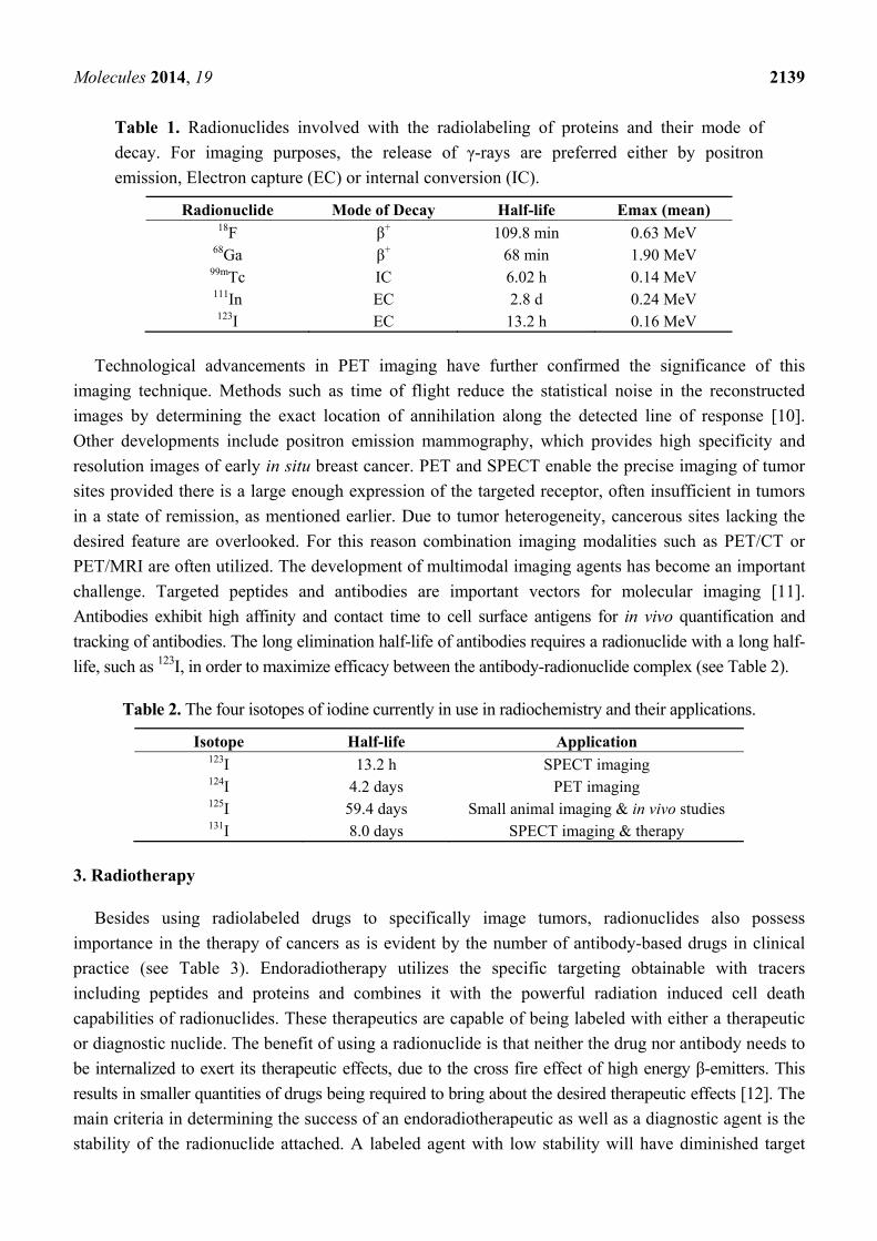
Table 1. Radionuclides involved with the radiolabeling of proteins and their mode of
decay. For imaging purposes, the release of γ-rays are preferred either by positron
emission, Electron capture (EC) or internal conversion (IC).
Radionuclide Mode of Decay Half-life Emax (mean) 18F β+ 109.8 min 0.63 MeV
68Ga β+ 68 min 1.90 MeV 99mTc IC 6.02 h 0.14 MeV 111In EC 2.8 d 0.24 MeV 123I EC 13.2 h 0.16 MeV
Technological advancements in PET imaging have further confirmed the significance of this
imaging technique. Methods such as time of flight reduce the statistical noise in the reconstructed
images by determining the exact location of annihilation along the detected line of response [10].
Other developments include positron emission mammography, which provides high specificity and
resolution images of early in situ breast cancer. PET and SPECT enable the precise imaging of tumor
sites provided there is a large enough expression of the targeted receptor, often insufficient in tumors
in a state of remission, as mentioned earlier. Due to tumor heterogeneity, cancerous sites lacking the
desired feature are overlooked. For this reason combination imaging modalities such as PET/CT or
PET/MRI are often utilized. The development of multimodal imaging agents has become an important
challenge. Targeted peptides and antibodies are important vectors for molecular imaging [11].
Antibodies exhibit high affinity and contact time to cell surface antigens for in vivo quantification and
tracking of antibodies. The long elimination half-life of antibodies requires a radionuclide with a long half-
life, such as 123I, in order to maximize efficacy between the antibody-radionuclide complex (see Table 2).
Table 2. The four isotopes of iodine currently in use in radiochemistry and their applications.
Isotope Half-life Application 123I 13.2 h SPECT imaging 124I 4.2 days PET imaging 125I 59.4 days Small animal imaging & in vivo studies 131I 8.0 days SPECT imaging & therapy
3. Radiotherapy
Besides using radiolabeled drugs to specifically image tumors, radionuclides also possess
importance in the therapy of cancers as is evident by the number of antibody-based drugs in clinical
practice (see Table 3). Endoradiotherapy utilizes the specific targeting obtainable with tracers
including peptides and proteins and combines it with the powerful radiation induced cell death
capabilities of radionuclides. These therapeutics are capable of being labeled with either a therapeutic
or diagnostic nuclide. The benefit of using a radionuclide is that neither the drug nor antibody needs to
be internalized to exert its therapeutic effects, due to the cross fire effect of high energy β-emitters. This
results in smaller quantities of drugs being required to bring about the desired therapeutic effects [12]. The
main criteria in determining the success of an endoradiotherapeutic as well as a diagnostic agent is the
stability of the radionuclide attached. A labeled agent with low stability will have diminished target

Molecules 2014, 19 2140
specificity and a greater potential harm to normal tissues, due to dissociation of the radionuclide.
The stability of a radiopharmaceutical drug is usually analyzed in human serum and measured using
radio-TLC, radio-HPLC and LC/MS. These techniques are based on the chromatographic separation of
the radiolabeled compounds and their subsequent evaluation [13].
Table 3. A selection of clinically used/trialed antibody based drugs, their targets and, if
applicable, method of radiolabel attachment.
Name Protein Antibody
Form
Radionuclide
attached Target Application
Clinical
Trial
Method of
Attachment
Rituximab Anti-CD20
mAB Chimeric N/A CD20
B-cell non-
Hodgkin’s
lymphoma
FDA
approved N/A
Trastuzumab Anti-HER2
mAB Humanized N/A HER2
HER2 positive
breast cancer
FDA
approved N/A
Pertuzumab Anti-HER2
mAB Humanized N/A HER2
HER2 Positive
breast cancer
FDA
approved N/A
Avastin®
(bevacizumab)
Anti-VEGF
mAB Humanized N/A VEGF-A
Angiogenesis
inhibitor
FDA
approved N/A
Zevalin®
(ibritumomab)
Anti-CD20
mAB mu IgG1 90Y CD20
B-cell non-
Hodgkin’s
lymphoma
FDA
approved MX-DTPA
Bexxar®
(tositumomab)
Anti-CD20
mAB mu IgG2a 131I CD20
B-cell non-
Hodgkin's
lymphoma
FDA
approved Iodination
Epratuzumab Anti-CD22
mAB Humanized 90Y CD22
B-cell non-
Hodgkin’s
lymphoma
Phase I/II DOTA
Veltuzumab Anti-CD20
mAB Humanized N/A CD20
B-cell non-
Hodgkin’s
lymphoma
Phase I/II N/A
Clivatuzumab Anti-MUC1 Humanized 90Y MUC1 Pancreatic cancer Phase I/II DOTA
A very relevant field is radioimmunotherapy (RIT) using antibody-based labeled drugs. It combines
the synergistic effects of radio- and immunotherapy and is an important tool in the treatment of
hematologic malignancies such as Non-Hodgkins lymphoma [14,15]. Like standard chemotherapy,
RIT has been optimized and standardized as a therapy for lymphomas, although not for solid tumors
due to its low penetration rate and lower radiosensitivity. Studies have shown that the larger the tumors
the less efficacy RIT will have. This is due to the fact that antibodies lack sufficient penetrative
capabilities to reach the center of a large tumor mass. Tumors larger than 3–4 cm in diameter currently
cannot be addressed with endoradiotherapy, with the most benefit possible in combination with
subtherapeutic chemotherapy doses [16,17]. Large doses of radiolabeled antibodies are required to
obtain any significant penetration required for these large solid tumors. Such large quantities prove
troublesome due to the low blood clearance of radiolabeled antibodies, leading to the potential
exposure of radiation sensitive tissues to the radionuclides [18].

Molecules 2014, 19 2141
4. Radionuclides
The main effect of endoradiotherapeutic agents is caused by the cytotoxic effects of radionuclides.
Ionizing radiation is emitted by radionuclides and is utilized in endoradiotherapy for ionization and
free radical formation. This emitted radiation results in subcellular damage by the energy deposition on
cells during transversion, inducing cell death [19].
The specific cytotoxic effects of an agent on the cancer tissue are dependent on the physical
properties of the radionuclide. The half-life of the radionuclide, its mode of decay and correspondingly
the linear transferred energy, as well as the rate of energy deposition, are all properties with an influence on
efficacy. Therefore the choice of appropriate radionuclide is of vital importance [18]. Other factors to
consider when selecting a radionuclide include tumor uptake and retention, blood clearance, rate of
radiation delivery, and the feasibility of large-scale production of the radionuclide in an economical
fashion. Besides the direct radiation induced effects, radiation also causes indirect, so called bystander
effects, on non-radiated cells. These bystander effects are mediated by gap-junction intracellular
communication and through irradiated cell secretion of factors, inducing transduction pathways [20].
Therapeutic radiopharmaceuticals use particulate radiation such as α- and β-particles, yet also
Auger electrons. Αlpha-radiation exhibits a high localized energy deposition combined with a short
tissue range. These characteristics lead to a high Linear Energy Transfer (LET [21]. Thus α-radiation is
the most damaging particulate radiation. Furthermore it can be applied in small concentrations since
only 1-4 α-particles need to hit the cell to induce cell death [22]. As such, α- radiation is often used in
residual tumors and hematological diseases, such as leukemia [23]. Alpha particles possess tissue
ranges of less than a cell diameter. Consequently no cross-fire occurs. On the one hand this reduces the
potential for unwanted side-effects on neighboring healthy tissue but increases the requirement for
effective cell penetration of the therapeutic [24].
Due to this obstacle in therapeutic design, radionuclides for therapy are preferentially β-particle
emitters (see Table 1). Compared to α-emitters, particulate β-emitters have a lower toxicity due to a
lower deposition energy. In contrast to α-emitters, their therapeutic effect is mainly caused by the
cross-firing bystander effects. This is due to β-emitters possessing a penetration range of several
millimeters. Thus even cancerous cells located in the inner mass of tumors can be affected by the
radiation, even if penetration rate of the pharmaceutical is poor. For example, long range β-emitters
like 90Y and 188Re are used in the therapy of large tumor masses, due to their excellent penetration
capabilities. [25] Conversely, a high penetration also accommodates an increased likelihood of
side-effects in healthy cells [26].
In comparison to nuclides used for therapy, imaging agents use non particulate emitters such as
γ-ray and positron emitters (Table 4). This is due to their low LET and rate of cell damage. The gamma
emission resulting from electron capture (EC) or internal conversion (IC), results in the emission of
Auger electrons. These Auger electrons traverse a very short range of only a few nanometers. Within
these an energy deposition takes place, which is significantly higher than that of β- emitters. This
results in a high LET which can be highly damaging when the emission occurs close to the DNA.
Studies have shown that auger electrons, attached to carriers targeting nuclear DNA, cause a
monoexponentional decrease in cell survival [22].

Molecules 2014, 19 2142
Table 4. List of radionuclide currently used in radiotherapy and their nuclear properties.
As visible, all decay via α- or β-particulate emissions.
Radionuclide Mode of Decay Half-life Eav Mean Tissue Range 90Y β 2.7 d 2.27 MeV 2.76 mm 131I β,γ 8.0 d 0.61 MeV 0.40 mm
177Lu β, γ 6.7 d 0.50 MeV 0.28 mm 225Ac α, β 10.0 d 6.83 MeV 0.04–0.1 mm 213Bi α 47.5 min 8.32 MeV 0.04–0.1 mm 212Bi α 1.0 h 6.21 MeV 0.04–0.1 mm 211At α 7.2 h 6.79 MeV 0.04–0.1 mm 212Pb β 10.6 h 0.57 MeV 0.6 mm
5. Proteins as Site-Specific Drugs
Proteinaceous drugs have gained importance as reflected by their impressive advent among the top
pharmaceutical products [27]. The size of therapeutically used protein formats can vary significantly.
While small proteins with molecular weights ≤ 30 kDa are efficiently eliminated within 1 hr, larger
proteins show very long circulation times. To date larger proteins dominate clinical use. For this reason
this review shall be focused on larger protein formats above the renal exclusion limit. The importance
of proteins is highlighted in the innovation of proteinaceous tumor-targeting systems and personalized
medicine. The target specificity achieved with antibodies, in particular antigens whose receptors are
overexpressed in tumor cells, allows for accurate drug delivery.
Tumors which develop through large numbers of mutations are often of a heterogeneous phenotype,
even among the same cancer types. The advanced development of modern diagnostic and therapeutic
techniques has taken advantage of the heterogeneity of tumors to benefit the patient. This is due to the
availability of a distinct receptor which can be specifically targeted, through the innovative model of
personalized medicine. Human Epidermal Growth Factor Receptor 2 (HER2) is a prime example of
this exploitation of heterogeneity. HER2 is overexpressed in 30% of breast cancers [28]. Trastuzumab
(Herceptin®, Genentech) is a specific HER2 targeting mAb which up-regulates the p27Kip1 protein,
resulting in the inhibition of CDK2 and halting cell proliferation [29]. Trastuzumab is the first line
treatment for breast cancer, but only in patients with a HER2 overexpression. However, cardiotoxicity
associated side effects of trastuzumab occur in approximately a third of patients, therefore screening of
patients to determine trastuzumab’s efficacy is of vital importance in risk analysis [30]. This prescreening
for the presence of HER2 positive cells allows for a determination of the response of a patient to the drug.
Thus, it is possible to distinguish which cancer drugs are most likely to benefit a patient, and then tailor the
medication regime to that patient. A current Phase 0 study being run by the NCI, is investigating the use of
trastuzumab labeled with 111-indium. By chelating the drug with a linker and the radionuclide, the dose
required as a diagnostic agent could be reduced to a clinical dose of 200 μg, compared to the current
recommended clinical dose of 2–4 mg/kg, so the safety implications are evident [31,32].
5.1. Design of Molecular Imaging Agents Based on Proteins
The specificity and success of radio-labeled proteinaceous drugs depends on both the antibody and
the radionuclide chosen. Briefly mentioned before, the half-life of the radionuclide should be

Molecules 2014, 19 2143
approximately equivalent to the drug half-life of the antibody. MAbs have generally long half-lives in
vivo, on the order of days, due to their large size preventing renal excretion. Furthermore their ability
to specifically interact with numerous receptor sites increases their retention time [33]. Thus
radionuclides with longer half-lives are preferred with full antibodies. Yet smaller antibodies such as
diabodies have a decreased retention time in the body, and can therefore be labeled with shorter lived
radionuclides [34]. Any antibody which is selected to be labeled with a radionuclide must be able to
withstand radiolysis, as an antibody subjected to a high amount of radioactivity may be altered and
lose its immunogenicity [35]. Thus, there is a vital importance in radiolabeled antibodies to ensure that
the correct antibody and radiolabel are selected.
5.2. Protein-Based Carrier System
As already described, the high selectivity of antibodies makes them a useful method of targeting in
cancer therapy. Targeted antibody therapy was first suggested by Paul Ehrlich when coining the term
‘Magic Bullet’ for a drug which can selectively target its site of action. Creating specific antibodies to
markers over-expressed on the surface of solid tumors creates a delivery system capable of
mono-targeting tissues possessing this marker. Thus to some extent mAbs are deserving of the title
‘Magic Bullets’. Since their inception mAbs have become increasingly expansive and successful in
targeting disease sites [36,37]. Exploiting the specificity of tumor biomarkers allows for the reduction
of cytotoxic side-effects associated with radionuclides. The first approved antibody-based drug and
diagnostic compounds were murine-based causing the human immune system to recognize them as
foreign and eliminate them from the body. Furthermore their low cell penetration and incorrect
interaction with the immune system resulted in a poor efficacy. The ability to create recombinant
chimeric antibodies was a key advancement in both antibody engineering and developing protein-
based carrier systems. By replacing portions of the murine antibody which provoke the immunogenic
response, but still preserving the variable binding domains, increased specific binding is attainable.
This introduction of human constant domains led to the generation of the first chimeric mAb [33].
There are currently many chimeric antibodies in clinical utility such as rituximab, used in the treatment
of CD20 positive B-cell non-Hodgkin’s lymphoma [38]. Additional developments allowed for further
decreases in immunogenicity by interchanging the murine hypervariable loops responsible for binding
activity with their counter hypervariable loops in the human antibody. This replacement led to the
generation of ‘humanized’ antibodies which are even less immunogenic than chimeric mAbs. There
are various humanized antibodies approved for medical use, such as bevacizumab, a VEGF antigen
used to treat metastatic colorectal and non-small cell lung carcinoma [33]. The engineering of
cytotoxic antibodies is simple in theory. By loading the antibody with a radioactive molecule, it is
possible to reach relatively high radiation counts at the tumor site. However this high irradiation is
possible at a low dose of drug, due to the targeting capabilities of the antibody. The increasing ability
to customize antibodies with a wide range of side chains allows for a greater potential for site specific
delivery of a radionuclide. This is owing to the functional groups which can be added resulting in an
improved targeting to specific receptors [39,40].
mAbs can act in various ways to produce their cytotoxic effects. Many (such as trasutzumab,
rituximab [41–43]) work together with the parts of the human immune system inducing either
Molecules 2014, 19 2144
antibody-dependent cellular cytotoxity (ADCC) by binding to Fcγ receptor and target cell or
complement dependent cytotoxicity [44]. The high sensitivity and specificity attained with mAbs are
their greatest features and are therefore a vital requirement for successful therapy. Thus one of the
main obstacles of mAb therapy is the heterogeneous distribution of antigen on tumor cells. Additional
requirements of mAbs to ensure effective therapy are: fast clearance from blood compartment and
sufficient and homogenous tumor penetration [41]. Other than inducing the human immune system,
antibodies are also used as carriers of a pharmaceutical to the tumor site. In this use their main function
is the transport of radionuclides to the target site. However, the labeled antibody often cannot deliver
an appropriate therapeutic dose to the tumor, therefore attachment of more than one radionuclide to the
protein has been attempted. However when randomly distributed, a loss of immunoreactivity is caused
as seen in the iodine-labeling of IgG antibodies [42]. Antibody-dendrimer conjugates were
successfully investigated producing anti-EGFR DOTA derived PAMAM dendrimers [11]. To further
increase permeability, attempts in molecular engineering have been made to develop bispecific
antibodies with lower molecular weight [45,46].
6. Radiolabeling Strategies of Proteins
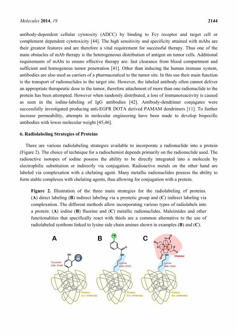
There are various radiolabeling strategies available to incorporate a radionuclide into a protein
(Figure 2). The choice of technique for a radiochemist depends primarily on the radionuclide used. The
radioactive isotopes of iodine possess the ability to be directly integrated into a molecule by
electrophilic substitution or indirectly via conjugation. Radioactive metals on the other hand are
labeled via complexation with a chelating agent. Many metallic radionuclides possess the ability to
form stable complexes with chelating agents, thus allowing for conjugation with a protein.
Figure 2. Illustration of the three main strategies for the radiolabeling of proteins.
(A) direct labeling (B) indirect labeling via a prostetic group and (C) indirect labeling via
complexation. The different methods allow incorporating various types of radiolabels into
a protein: (A) iodine (B) fluorine and (C) metallic radionuclides. Maleimides and other
functionalities that specifically react with thiols are a common alternative to the use of
radiolabeled synthons linked to lysine side chain amines shown in examples (B) and (C).
131IOH
Tyrosineside chain
Protein(i.e. antibody)
Protein(i.e. antibody)
NH
Lysineside chain
O
18F
Chelator
Protein(i.e. antibody)
Lysineside chain
NH
N N
NN
O
O O
O
O
O
O
90Y
A B C

Molecules 2014, 19 2145
6.1. Methods of Radiolabelling
6.1.1 Radiohalogenation
6.1.1.1. Radioiodination of Proteins
Radiolabeling molecules with iodine nuclides is of great importance in pharmaceutical
radiochemistry. There are over thirty different identified iodine isotopes, but only four are commonly
used in radioiodine chemistry: 123I, 124I, 125I and 131I. They all possess vital properties, allowing for
their application in radiochemistry.
The differing half-lives of all four allow for a variation in their usages. As seen in Table 2, the most
popular use of iodine radionuclides is in molecular imagining providing valuable information on
physiological processes. 123I is the most suitable radioisotope for molecular imaging and is an ideal
choice radionuclide for SPECT imaging. 131I can also be used as a γ-emitter for SPECT imaging,
however it also is a β-emitter (see Table 1) and is no longer recommended due to its irradiation
potential. [47] 124I is a positron emitting nuclide, therefore having an application in PET imaging, such
as the diagnosis of thyroid cancer [48]. Owing to its long half-life of over 4 days, it could also be
utilized in order to monitor physiological processes which occur over long periods. 124I has a half-life
similar to that of a mAb, therefore making it a key candidate in radioimmunotherapy [49].
The direct radioiodination of a protein is a key method for the synthesis of tumor-targeting
radiopharmaceuticals, like 131I-labeled rituximab [50] (Table 3). Generally there are two basic
approaches of protein radioiodination. The most straightforward approach is direct protein labeling
using electrophilic substitution at tyrosine and histidine residues. The radioiodide is oxidized in situ
creating the electrophile *I+. This is done using oxidizing agents like chloramine T, Iodogen® and N-
halosuccinimides [51]. The generated electrophile attacks the electron rich of aromatic ring of the
amino acid tyrosine, forming a σ-complex [51,52] This substitution is performed at the tyrosine
residue due to the electron donating hydroxyl group which stabilizes the σ-complex (see Figure 3). As
the labeling of proteins must take place under mild conditions, the attachment of iodine to the tyrosine
is highly suitable.
Figure 3. Mechanism of direct electrophilic radioiodination of an iodide electrophile to
produce the iodinated product.
This straightforward method is performed under mild conditions, which is optimal for the labeling
of proteins. Unfortunately, this is only possible when the protein contains accessible tyrosine or
histidine residues. A further disadvantage is the accumulation of iodine in the thyroid and stomach
when using direct labeling strategies. This is due to the naturally occurring catabolism of proteins by

Molecules 2014, 19 2146
the enzyme tyrosine deiodase, increasing in vivo toxicity [53]. Tositumomab (131I-B1 mAb) is an anti
CD20 antibody conjugated with the mixed β/γ-emitter 131I. It received FDA approval for the treatment
of Non-Hodgkin’s lymphoma in 2003. This drug is synthesized via direct labeling, using the
electrophilic substitution method above with Iodogen®. Tositumomab benefits from good response
rates, and is still being used in clinical practice [54].
However, due to the problems mentioned above, the indirect iodination of proteins via conjugation
is a frequently used alternative method. In this approach iodine is incorporated by the application of
prosthetic groups containing two functional groups to enable both radioiodination and incorporation to
the protein. There are a variety of prosthetic groups used for radioiodination, but the most frequently
used are N-succinimidyl 5-[*1]iodo-3-pyridinecarboxyl ([131I]SIPC) [55] and N-succinimidyl-3-[*I]-
iodobenzoate ([*I]SIB) [56] (Figure 4).
Figure 4. Chemical structures of the chelating agents SIB, SIPC and SGMIB.
Both active esters are conjugated to amino groups of the protein and exhibit a high in vivo stability.
This was successfully shown in the radiolabeling of insulin-attached [131I]SIPC, which exhibited a
higher in vivo stability than directly coupled [131I] [53,57]. Another prosthetic group for the acylation
of aromatic groups is N-succinimidyl-4-guanidinomethyl-3-[I-131]iodobenzbate ([I-131]SGMIB)
(Figure 4). It has been successfully applied to internalizing antibodies with a 4-fold increased uptake
compared to labeled antibodies with [131I]SIPC [58].
6.1.1.2. Protein-Labeling with 18F
18F, a positron emitter, is often considered the preferred radionuclide for PET imaging due to its
many desirable qualities as described above. Similarly to iodination there are two main synthetic
strategies to label a protein with 18F. The most straightforward method is the direct incorporation of 18F
into the protein by direct attachment to a tyrosine residue via electrophilic or nucleophilic attacks. 18F2
is most commonly used in electrophilic fluorination due to its high reactivity. Mild conditions are
essential for this highly exothermic reaction which must be tightly controlled using low temperatures.
The first 18F labeled molecules were obtained using this method. However, in modern times this
method is less commonly used, due to the lack of regioselectivity leading to the formation of a mixture
of products which exhibit low specific activity oft due to an exchange with 19F [8,59].
Nucleophilic fluorination achieved with fluoride ions is now the most routinely used method. The
nucleophile is obtained as an aqueous solution, but due to polar solvation the fluoride is a poor

Molecules 2014, 19 2147
nucleophile. Therefore the fluoride ions are transferred to a non-polar, organic solvent, like DMSO
under assistance of phase transfer agents such as [18F]KF·K222. Nucleophilic substitution is not
possible under mild conditions due to lack of reactivity. Therefore elevated temperatures and basic
conditions are used to initiate nucleophilic substitution. However under these harsh conditions most
macromolecules, including proteins, are not stable [60,61], so this method is often incompatible with
large, temperature sensitive biomolecules such as proteins.
As an alternative to the direct methods, indirect incorporation of 18F can occur via conjugation of 18F-labeled reactive precursors (Figure 5). These precursors contain prosthetic groups allowing for the
fluoridation and also conjugation with proteins under mild conditions.
Figure 5. Structures of prosthetic groups used to indirectly incorporate 18F proteins.
Thus the reaction can take place at room temperature and in aqueous solvents, preferable conditions
compared to nucleophilic substitution. The conjugation of the precursors occurs mostly at an amino
group on the protein, however can also take place on carboxy or sulfhydryl groups. The attachment of
a prosthetic group to an amino group (lysine side chains or the N-terminus of a protein) is carried out
with the active ester N-succinimidyl-4-[18F]fluorobenzoate ([18F]SFB) by alkylation with a good
coupling rate of 40%–60% [58]. A disadvantage of using prosthetic groups to incorporate 18F is that
the formation of prosthetic groups often requires multistep complex methods, likely to result in
decreased yields. Some groups have tried to reduce the numerous steps and complexity involved such
as the one pot synthesis method suggested by Hou et al., however the yields obtained were poor [62].
In order to decrease the work load, a recent study suggested the use of N-succinimidyl 3-(di-tert-
butyl[18F]fluorosilyl)benzoate ([18F]SiFB), a prosthetic group with properties similar to [18F]SFB but
with a simplified one step radiolabeling procedure[63]. In contrast to direct labeling approaches,
indirect labeling with precursors results in the generation of a greater number of radiolabeled
molecules containing several modifications and localization and thus varying in structure. The bio-
activity of these structures is often difficult to characterize, which has led to the pursuit of a site-
specific process producing a homogenous product [64]. The first chemoselective labeling was achieved
by Flavell et al. in 2008 using [18F]fluorobenzaldehyde ([18F]FBA). This precursor was used to
incorporate an aminooxy functional group into the C terminus of leptin in an aniline-accelerated
radiochemical oximation reaction, allowing for unambiguous bioactivity determination [65]. Further
site-specific labeling has been obtained by using a silicon-fluoride acceptor (SiFA) reagent such as
[18F]SiFA-SH. The number of incorporable molecules depends on the number of introduced maleimide
groups. In order to preserve the biological activity of the antibody, it is vital that no more than two

Molecules 2014, 19 2148
maleimide groups be introduced [66]. Secondly, the site of modification can affect the biological
activity [40]. Further chemoselective labeling can be obtained using an approach based on hydrazone
formation. The protein is conjugated with hydrazinonicotinic acid (HYNIC) and successively labeled
with [18F]fluorobenzaldehyde via hydrazone formation. This approach has been successfully used for
the fluorination of human serum albumin [67].
However these chemoselective reactions have drawbacks. The use of appropriate reaction conditions
is of utmost importance, with these reactions producing varying yields between 25%–99% [4].
Further difficulties arise when attempting to label proteins containing thiol groups. In order to
label proteins containing accessible thiol groups, [18F]maleimide reagents, like N-(6-[(4-[18F]fluoro-
benzylidene)aminooxy]hexyl)maleimide ([18F]FBAM) can be used for labeling low-density
lipoproteins [64]. The excellent orthogonality of the copper-catalyzed 1,3-dipolar cycloaddition
between azides and alkynes yielding triazoles has caused pervasive interest in this reaction to trigger
conjugation reactions. This reaction is referred to as click chemistry. In a recent approach
4-[18F]fluoro-N-methyl-N-(propyl-2-yn-1-yl)benzenesulfonamide ([18F]F-SA) was incorporated into
human serum albumin using click chemistry. Ramenda et al. successfully demonstrated the promising
utility of click chemistry as a reliable method of radiolabelling proteins [61]. The reaction results in the
formation of a triazole moiety that has to be considered to influence the properties of conjugates.
However, in the case of proteins this relatively small modification is not likely to influence the overall
pharmacokinetic properties of large proteins.
6.1.2. Chelation
The attachment of radionuclides through direct labeling, as explained for iodine and fluorine, is
only possible if the nuclides can undergo electrophilic and nucleophilic substitutions. Furthermore
these radionuclides must be capable of being attached under physiological conditions due to the heat-
labile nature or proteins.
Direct labeling is thought of as advantageous owing to the simplicity of such a process. However
radioactive metal ions are generally more difficult to attach via direct labeling strategies due to their
reduced reactivity compared to halogens. Nevertheless there have been direct approaches for labeling
proteins with metallic radionuclides. Proteins, such as antibodies, often contain the amino acid cysteine
along their amino acid backbone. Cysteine is unique as it is the only natural amino acid containing a
thiol group, a key target for complexation with a radionuclide via the formation of a coordinate bond.
These thiol groups possess the ability to form disulfide linkages between cysteine groups and are one
of the primary mechanisms of polypeptide folding. As such any attempt to label cysteine residues with
a radionuclide must first overcome these disulfide cross-linkages. For successful radiolabeling to occur
sufficient available thiol groups are necessary to complex with the generated metal radionuclide. Poor
site specificity, and lack of stability when coupling with thiol groups are major shortcomings of direct
radiolabeling of antibodies with metal nuclides [68]. Approaches to counter this have involved
reducing the disulfide linkages to produce thiol groups. However this method if uncontrolled is likely
to alter the spatial conformation of the protein [69]. These disadvantages, along with the good
chelating properties of metal ions, results in an indirect radiolabeling approach being preferred. These
indirect methods link radio-metals to proteins using bifunctional chelators [BFCs]. As their name

Molecules 2014, 19 2149
already indicates, these molecules consists of two functional groups which serve different purposes; one
functional group for the binding to the protein and a chelating unit which carries the radionuclide [70,71].
A BFC is essentially made up of three sections, a radionuclide binding unit, a unique ligand framework
and a conjugating group for attachment to the antibody [72]. The attachment of metallic radionuclides
using chelation is a widely accepted field, with FDA approved radiopharmaceuticals currently in use,
like Zevalin® (Spectrum Pharmaceuticals).
Besides the functional requirements, BFCs also have to meet synthetic and chemical requirements.
As with prosthetic groups, chelators have to exhibit a high in vivo stability in order to ensure patient
safety yet also obtain good therapeutic and diagnostic results [73]. To deliver this inertness, BFCs have
to be both stable against hydrolysis under physiological conditions and exhibit a strong affinity
towards the radionuclide. Furthermore the BFC must be able to withstand radiolysis, as they are used
to deliver loads of high radio activity [74]. In addition to those requirements, BFCs are further required
to not alter the biological properties and specificity of the protein. Consequently, isomerism is an
important characteristic to be considered due to the specific structure of the proteins required for target
binding [75].
Owing to the diversity of the metallic radionuclides there is no universal BFC to chelate to all of the
radiometals. Therefore many different variations of BFCs are used depending on the choice of
radionuclide. The size, charge, and electron configuration of the radiometals will determine the
coordination number required of a BFC. The BFC can differ from coordination numbers varying from
2–8 in order to accommodate the radionuclide [76].
6.1.2.1. Acyclic Chelators
The first chelating agents developed for coupling radionuclides to biomolecules were the acyclic
chelating agents. These molecules lack a ring system in their coordinating form as compared to
macrocyclic chelators which have a preformed ring system. The most common acyclic chelators are
derivatives of diethylenetriaminepentaacetic acid (DTPA), which itself was derived from the common
metal chelator, ethylenediaminetetraacetic acid (EDTA). EDTA was found initially to lack stability
and appropriate coordination values for metallic radionuclides, hence the synthesis of DTPA and its
derivatives [77]. Binding of DTPA to proteins, such as an antibody, is possible by the use of the
coupling agent isobutyl chloroformate [78,79]. DTPA is capable of binding a wide range of metals
including the widely used 111In, 90Y and 99mTc [60]. Attached to the proteinaceous primary amine, the
conjugated acyclic chelating system opens to reveal its octadentate nature. That is, it possesses eight
donor sites for interaction with a ligand, therefore DTPA can be considered of hexa- or octa-
coordination depending on the radiometal [80].
A major advantage of using DTPA analogs as BFCs is the fact that the reactions can occur under
mild conditions [81,82]. This is of particular benefit owing to the labile nature of proteins. Reaction
conditions and the BFC used must be chosen and scrutinized in order for the antibody to retain its
immunogenicity [83]. The DTPA derivative tiuxetan is an example of successful conjugation of a BFC
leading to a useful drug molecule. Zevalin® (90Y-ibritumomab tiuxetan) therapy uses the mAb
ibritumomab, radiolabeled with yttrium and conjugated via the BFC tiuxetan. As stated above,
Zevalin® received FDA approval in 2002 for the treatment of Non-Hodgkin’s lymphoma and is

Molecules 2014, 19 2150
currently still being marketed. Compared to the antibody drug rituximab, it shows greater response
rates and highlighted the use of anti CD20 radiotherapy [84]. The use of side chains with DTPA
derivatives allows for improved binding to proteins, as seen with p-isothiocyanatobenzyl
diethylenetriaminepentaacetic acid (p-SCN-Bz-DTPA). This derivative utilizes an isothiocyanate
group, which binds to a free amine on the protein to form a thiourea linkage [85].
Although DTPA has been extensively studied, its low in vivo stability diminishes its clinical
potential due to dissociation of the radionuclide from the chelating agent. This could cause a particular
harm upon radiation sensitive bone marrow [72]. Another negative aspect of conjugation with DTPA is
the synthesis of undesired conjugates, such as double substituted DTPA analogues obtained with the
DTPA dianhydride. However Hnatowich et al. overcame this by using gel chromatography in order to
separate the lead product [86]. These negatives have led to the synthesis of DTPA derivatives such as
tetra-t-Bu-DTPA (see Figure 6) [87] which contains only one free carboxy group, thus eliminating the
possibility of forming double substituted DTPA derivatives [88]. An advantage of this BFC is its use
in solid phase synthesis owing to its high solubility in a variety of solvents. Another acyclic chelating
agent is mercaptoacetyltriglycine (MAG3). This chelating agent is useful at binding 183Re and is
another alternative to DTPA [89].
Figure 6. Structures of four DTPA derivatives. Tetra-t-Bu-DTPA, containing the protecting
groups required to ensure no double substituted products form compared to p-SCN-Bz-
DTPA which highlights the use of side chains in order to improve binding of the BFC.
However the same radionuclide dissociation issues associated with DTPA are still as problematic in
the derivatives of DTPA [60]. The dissociation of the radionuclide from DTPA occurs due to the
opening of the chelate ring. Methods and modifications to DTPA have been sought in order to restrict
the freedom of motion of this chelate ring and provide an energetic hindrance to the opening of the
acyclic backbone [90]. Prevention of this dissociation results in reduced accumulation of radioactive
material in the blood stream and increased uptake in the tumor site [91]. This has resulted in the rise of
backbone stabilized acyclic chelators which looks to be the way forward for radiolabeling of metals.
These acyclic chelators benefit from still having the favorable reaction kinetics associated with DTPA,
but at a more appropriate inertness once conjugated. An example of this is cyclohexane-1,2-diamine-

Molecules 2014, 19 2151
N,N,N',N'-tetraacetate (CHX-DTPA). CHX-DTPA (Figure 6) contains a trans-cyclohexylene bridge
which constrains the acyclic chelator by preventing rotation [92]. The radionuclide is stabilized within
the chelate ring system resulting in a decreased likelihood of dissociation. Compared to DTPA, CHX-
DTPA and the methyl derivative MX-DTPA (Figure 6) benefit from a 2-3 order greater stability when
bound to a radioligand [72]. There are two isomeric forms of CHX-DTPA available, CHX-A and
CHX-B, with CHX-A possessing greater in vivo stability, and is generally preferred in the
radiolabeling of mAbs [93,94]. An antibody mimetic aimed at targeting HER2, possessing a single
cysteine residue in its amino acid scaffold, has been conjugated to a variant of the maleimido
derivative of CHX-A-DTPA. This conjugated ‘affibody’ has been used to provide imaging of high
contrast in breast cancer, suggesting that CHX-A-DTPA could have a vital role in molecular imaging
and later radiotherapy [95]. Another backbone-stabilized acyclic chelator is mDTPA (Figure 7). This
BFC possesses efficient reaction kinetics allowing the reaction to proceed in mild conditions, and
deliver DTPA to the antibody in high yield [78].
Figure 7. DTPA dianhydride chelating with a metal radionuclide. The mechanism shows
the use of a free amine group to open and conjugate to the BFC for attachment of the
radionuclide.
6.1.2.2. Macrocyclic Chelators
Another strategy to overcome the dissociation problems with bifunctional DTPA derivatives was
the search for more stable chelating agents, leading to the use of macrocyclic chelators. Cyclic
polyaminoploycarboxylates, also known as macrocyclic chelators, are made up of a tetraza- or triaza-
macrocyclic ring. The macrocyclic framework allows for formation of high thermodynamically stable
metal complexes with great kinetic inertness. 1,4,7,10-Tetraazacyclododecane-N,N',N'',N'''-tetraacetic
acid (DOTA, Figure 8) is a valuable substitute for DTPA. DOTA and its derivatives can form stable
complexes with divalent and trivalent metals and have been used to chelate a wide range of
radiometals, including 111In, 86Y, 90Y, 213Bi, and 225Ac [96]. Radiolabeled DOTA derivatives possess a
greater in vivo stability than DTPA, owing to their kinetic inertness, a quality which makes them very
useful in molecular imaging and radio therapy [97,98]. The ability to secure a radioactive payload is
vital for the safety of radiolabeled molecules. However there are also consequences to this beneficial
stability. In comparison to DTPA, incorporation of the radiolabel to DOTA occurs slower and with a
lower yield. This is thought to be due to poorer formation kinetics, owing to DOTA’s rigid structure,
and competition with trace metals [81,98,99]. Labeling of DOTA is a one step process which can be
improved by the use of a 4-(2-hydroxyethyl)-1-piperazineethanesulfonic acid (HEPES) or 2-(N-
morpholino)ethanesulfonic acid (MES) buffer [81,100]. The first approach to conjugate DOTA to a

Molecules 2014, 19 2152
protein was by creating an amide linkage via the activation of one of the carboxy groups in
DOTA [90]. However derivatives of DOTA have improved upon this conjugation by using added
linker side chains which will bind to the protein. By using X ray crystallography, it is possible to
determine which functional groups are not significant in radiolabeled complexes [101]. One such
example is the use of an amino acid-substituted BFC, DOTA-Phe-Ala, to bind to an anti EGFR mAb.
The amino acid side chain will allow DOTA-Phe-Ala to have a free amine which can be utilized to
bind to a wide array of proteins without altering their biological activity [102]. The addition of a side
chain also allows for better delivery and targeting properties of the radiolabeled antibody. An example
of this are the maleimidocysteineamido-DOTA derivatives which inhibit the release of the BFC-
radiometal complex in a pH dependent fashion. This is thought to improve tumor uptake owing to the
acidic pH often found in solid tumors, causing an accumulation of radiometal at the tumor site [99].
The ability of DOTA derivatives to complex with 67Ga, 90Y and 111In lends itself for use in
radiotherapy. Conjugating a metal radiolabeled DOTA derivative with a protein as a radiotherapeutic
is termed antibody guided radiation therapy. Wong et al. showed these capabilities by adapting the
conjugation of their chimeric anti-CEA antibody to use DOTA instead of the DTPA derivative
mentioned above. However the DOTA conjugated antibody had a greater immunogenic host response.
This may be due to the chimeric nature of the antibody and not be significantly influenced by the
conjugated DOTA [103]. The use of DOTA, however, does not automatically suggest and provide a
high level of radionuclide stability in the radiolabel-protein complex. DOTAs ability to complex with 64Cu is questionable [104]. As mentioned above, 64Cu is a useful radiolabel owing to its appropriate
half-life which is similar to that of mAb in vivo [105]. However it has a labile nature and when
complexed with DOTA it is known to dissociate and accumulate in non-target organs. 1,4,7-Triaza-
cyclononane-1,4,7-triacetic acid (NOTA, Figure 8) is a DOTA derivative which has seemingly
overcome this problem. It has been widely reported that NOTA has a much better stability with 64Cu
than DOTA, with less accumulation of the radionuclide appearing in other organs [106,107].
Another common chelating agent is 1,4,8,11-tetraazacyclotetradecane-1,4,8,11-tetraacetic acid
(TETA, Figure 8), a macrocyclic BFC primarily used in radiolabeling molecules with 64Cu
radionuclides. [90,108]. Compared to the acyclic BFCs and DOTA, TETA is a more stable chelating
agent when dealing with copper nuclides labeled onto antibodies, in particular the benzyl TETA
derivative, as shown by Cole et al. [109]. Use of TETA with 64Cu in peptides has led to suggestions that
TETA could have potential use in the future of molecular imaging of neuroendocrine tumors [110].
Figure 8. Macrocyclic bifunctional chelating agents: DOTA, NOTA and TETA.

Molecules 2014, 19 2153
7. Metallic Nuclide Labeled Pharmaceuticals
7.1. 99mTc-Labeled Antibody Radiopharmaceuticals
As stated earlier, 99mTc holds great value in SPECT imaging. There are currently more than 20
discovered isotopes of technetium (Tc), with the most valuable isotope being 99mTc. 99mTc is so useful
due to its beneficial nuclear properties, in particular its radioactive half-life of 6.02 h which, together
with the excellent purity of the isotope obtained from the 99Mo/99mTc generator, allows one to attain
the specific radioactivity required for the administration of high radiation counts at a small
biological dose. 99mTc is readily available at a low production cost through commercial 99Mo/99mTc
generators [111]. 99mTc radiolabeled antibodies are becoming of increasing interest due to the targeting
capabilities of mAb antibodies. One such example is that of 99mTc- labeled mAb girentuximab 250
(mAbG250), an antibody used in the treatment of renal cell carcinoma. By conjugating the antibody
with HYNIC and then radiolabeling with 99mTc, a 99mTc-HYNIC-G250 is developed which shows
great promise in the radioimmunodetection of renal cell carcinoma [112]. Oosterwijk-Wakka et al.
furthered this by investigating the potential use of mAbG250 in RIT and also as in combination
therapy with tyrosine kinase inhibitors. The potential of this radiolabeled antibody appears to be
increasing with positive results in early clinical trials [113]. 99mTc is considered to be a key radiometal
for radioimmunoscintigraphy (RIS). 99mTc-MAG3-biotin conjugates have been synthesized and
evaluated in vivo, to determine their use. Whilst they were unsuitable for RIT due to their excretion via
the intestines, the 99mTc-MAG3-biotin conjugates could still find use in RIS [114].
7.2. Metallic Radionuclides Other than Tc
Previously we have discussed the different coordination chemistry associated with 111In and 67/68Ga
and the utilization of these radiolabels in molecular imaging when conjugated to peptides [60]. Both of
these radionuclides are used in the specific imaging of tumors due to their appropriate range of
emissions. As discussed earlier, trastuzumab has successfully been radiolabeled with indium-111. It
has been shown that 111In-DTPA has been conjugated with trastuzumab in order to determine if it can be
used in molecular imaging of HER2 positive breast cancer. Lub-de Hooge et al. successfully radiolabeled
this antibody which selectively targets the HER2 receptor with high in vivo stability [115]. This new
radiopharmaceutical is currently in clinical trials. 68Ga is a short lived radionuclide, with a half-life just over an hour (see Table 1). Despite this, its
use in immuno-PET imaging has been explored. Diabodies as carriers of radionuclides can be utilized
for PET imaging due to their rapid clearance from the blood system even with the short-lived
radiometal 68Ga [116]. This suggests that radiolabeled diabodies may have prospects in the
management of immunotherapy regarding receptor expression of solid tumors [117].
As discussed earlier, 90Y is a long range β-emitter and therefore lends itself to use in RIT. Wong et al.
showed the capabilities of DTPA derivatives as BFCs by use of a chimeric anti-CEA antibody
conjugated with 90Y isothiocyanatobenzyl DTPA. This yttrium radiolabeled antibody was tested in
Phase I clinical trials for the treatment of CEA-producing metastases. This study showed promising
results in the large burden tumors but the chimeric nature of the antibody generated some host
immunogenic responses [118]. The group then furthered this study by trialing the conjugated antibody

Molecules 2014, 19 2154
in a combination therapy trial with 5-fluorouracil. The results showed that 5-fluorouracil may decrease
the immunogenic host response when using this chimeric antibody. This signifies that yttrium
radiolabeled proteins may have a future role in combination therapy of tumors [119]. This method was
then further enhanced by the use of pretargeting methods as discussed below. Radiolabeling of
antibodies with 90Y is an avenue well researched as shown by Zevalin®, epratuzumab, veltuzumab and
clivatuzumab, all featured in the list of antibodies on the market in Table 3.
Rhenium-188 is another radionuclide used in RIT. Methods to conjugate this metal nuclide includes
using the aforementioned chelating agent MAG3. 188Re can be produced in a 188W/188Re generator. It
decays via the emission β-particles at a high enough energy, making it appropriate for radiotherapy.
After this, 188Re then decays further by the emission of gamma photons. This allows for RIT
potentially followed by monitoring and analysis of biodistribution [120].
8. In-Vivo Pretargeting Strategies
A major cause of side-effects of radiolabeled proteins is the accumulation of radioactive materials
as a result of the low blood clearance of radiolabeled antibodies. This is especially troublesome in the
presence of radiation sensitive bone marrow. To decrease this risk factor, new technologies decouple
the targeting of the tumor and the delivery of the radionuclide which improves the blood/tumor ratio,
decreasing the irradiation delivered to marrow. Such approaches are known as pretargeting strategies.
Despite this being a multi-step process, the advantages prove to be worth the increasing
complexity [121,122]. All pretargeting strategies are based on modified bi-specific antibodies which
recognize target antigens and can also specifically bind to a second component. Firstly an injection of a
large saturating dose of an unlabeled antibody is administered [16]. Then, after sufficient blood
clearance of the unlabeled antibody, around two to four days, administration of the radiolabeled
effector occurs. This wait is to ensure that adequate unlabeled antibodies have been cleared from the
body ensuring that the labeled effector does not bind to circulating antibodies in the blood stream and
of sites of low binding affinity, thus reducing unwanted irradiation [121]. Pretargeting is facilitated by
the innovative dock and lock technology which allows for the production of many mAB targeting
tumor-associated antigens [123]. There are two main approaches using bispecific antibodies: (i)
bispecific antihapten antibody-hapten binding [122] and (ii) bispecific antibody conjugated
avidin/streptavidin(sAv)-biotin binding. [124]
Improvements have been made to bispecific antibody-hapten binding to enhance the binding
affinity. It was discovered that a divalent-hapten molecule would greatly improve tumor uptake. This
use of a divalent molecule instead of a monovalent is known as an affinity enhancement
system [125,126]. This involves introducing equimolar amounts of Fab’ fragments against the tumor
and Fab’ fragments of an anti-DTPA-indium antibody, followed by a bivalent hapten injected once
sufficient blood clearance has occurred. This approach is currently used in a preclinical trial of non-
Hodgkin’s lymphoma: TF4, an AB fusion protein with two Fabs specific for the CD20 antigen
(injected first for tumor targeting). After this, application of 90Y-IMP-288 (with 2 histamine-succinyl-
glycine residues) improved cure rates compared to therapy of NHL with 90Y-veltuzumab [123].
In pretargeting with streptavidin (sAv)-biotin, streptavidin is conjugated to antitumor IgG, followed
by an injection of radiolabeled biotin. The biotin molecule is conjugated with a BFC such as DOTA

Molecules 2014, 19 2155
allowing for the introduction of a variety of metallic radiolabels [124,127]. In contrast to bispecific
antibody-hapten binding, which utilizes the antibody’s affinity to ensure retention, sAv-biotin benefits
from a remarkably high affinity between biotin and sAv. This affinity is due to the four biotin binding
arms present on sAv [121. However as they’re large molecules, IgG and streptavidin reside in the
blood stream for a long period of time, and strategies have been implemented to improve the clearance
of these molecules. This clearance occurs prior to injection of labeled biotin, to prevent the presence of
conjugated sAv and radiolabeled biotin in the circulation. A clearing agent, biotin-galactose-human
serum albumin, is injected to remove the non-localized antibody-streptavidin conjugate from blood via
the liver [127]. A variation of this coupling can also be implemented by using a multi-step avidin-
biotin pretargeting method. As avidin is glycosylated and cleared by the liver, it is possible to decrease
the blood circulation concentration of the modified biotin conjugated antibody. Avidin binds to the
circulating mAb-biotin complexes and gets cleared by the liver. Then sAv, which is not glycosylated,
is administered which upon reaching the tumor site binds to the biotin conjugated IgG selectively.
Finally after sufficient clearance of avidin and sAv, radiolabeled biotin is administered which binds to
one of the vacant binding arms on the conjugated sAv-biotin-IgG [121]. Phase I – II clinical trials, of
post-surgical treatment of stage III-IV glioma, with this avidin-biotin pretargeting approach have
produced promising results. Using 90Y-biotin after surgery produced a fourfold increased survival rate
compared to the control group [128]. The continuation of this study led to suggestions that RIT could be
utilized immediately after surgery, and boasted improved survival rates in a cohort of 504 patients [129].
The potential use of pretargeting strategies in tumor therapy has been successfully investigated in
preclinical studies, such as the improvement of targeting solid tumors. By targeting the angiogenesis
marker, extra domain B fibronectin, with an antibody hapten system labeled with 111In gave a three-
fold improvement compared to non-targeted endoradiotherapy [130]. Pre-targeting can also improve
molecular imaging [122]. Another benefit with this increased selectivity and tumor retention is the
possibility to administer larger radioactive quantities to the tumor. Up to five times greater activity is
obtainable while also maintaining a low radiation concentration in the bloodstream [16].
9. Conclusions
Vaidyanathan et al. are currently investigating the use of trifunctional chelating agents. [130] That
is, a chelating agent which binds to the antibody as normal but has two groups to bind to radiolabels.
The benefit of this is that it would allow the delivery of two different halogens, radiometals or a
combination of the two. This could prove vitally important as it would allow for an α- and a β-emitter
to be delivered to the same tumor. Thus the long range capabilities of the β-emitter would combine
with the devastating effects of α-particulates. Currently a SIB-DOTA prosthetic group is under
investigation. This chelating agent allows for the radiolabeling of a halogen and a metal within the
same molecule. [131] The potential of delivering a mixture of radiolabels, each with their own unique
properties, may be of great benefit in the future of RIT.
As discussed in this paper, antibodies are large macromolecules which possess all the features you
would expect. The innovation of nanobodies, could overcome the long half-lives and poor uptake
associated with these proteinaceous drugs. Above we have discussed diabodies, however nanobodies is
a step further with the antibody fragment containing only the variable region, responsible for the

Molecules 2014, 19 2156
binding ability of mAbs. Use of nanobodies would provide rapid uptake into tumors and short
clearance times, improving patient safety. [132,133] Affibodies, chemically accessible peptides with
antibody-like properties, consist of a 58 amino acid scaffold derived from the Z domain of the
staphylococcal protein A.[134] Due to their favorable in vivo properties this class of molecules allows
to develop small molecules with excellent targeting properties.[135] The use of α-emitters in
α-therapy poses a potential outlet for α-therapy. Currently the radionuclides bismuth-213 and actinium-
225 are being investigated for therapeutic application. Combining this with an antibody, would allow
for specific targeting, delivering the alpha emitter selectively to the tumor, improving safety. However, the
half-life of 213Bi is 45 min, not ideal for antibody conjugation. 225Ac on the other hand has a half-life of 1
week, much more suitable for antibody therapy. Thus combining α-therapy with proteinaceous targeting
systems may provide future benefit in the treatment of tumors [136].
Acknowledgments
The authors gratefully acknowledge the ERASMUS programme for financial support of G.S.
Author Contributions
Uwe Haberkorn and Walter Mier designed the manuscript, wrote the vital passages of the text and
revised the manuscript. Grant Sugiura, Helen Kühn and Max Sauter composed the main part of the
manuscript and contributed to the design of the figures.
Conflicts of Interest
The authors declare no conflict of interest.
References
1. Mankoff, D.A.; Link, J.M.; Linden, H.M.; Sundararajan, L.; Krohn, K.A. Tumor receptor
imaging. J. Nucl. Med. 2008, 49 Suppl 2, 149S–163S.
2. Heston, T.F.; Wahl, R.L. Molecular imaging in thyroid cancer. Cancer Imaging 2010, 10, 1–7.
3. Selzner, M.; Hany, T.F.; Wildbrett, P.; McCormack, L.; Kadry, Z.; Clavien, P.-A. Does the novel
PET/CT imaging modality impact on the treatment of patients with metastatic colorectal cancer
of the liver? Ann. Surg. 2004, 240, 1027–1036.
4. Frangioni, J.V. New technologies for human cancer imaging. J. Clin Oncol. 2008, 26, 4012–4021.
5. Belhocine, T.; Steinmetz, N.; Hustinx, R.; Bartsch, P.; Jerusalem, G.; Seidel, L.; Rigo, P.; Green, A.
Increased uptake of the apoptosis-imaging agent 99mTc recombinant human annexin V in human
tumors after one course of chemotherapy as a predictor of tumor. Clin. Cancer Res. 2002, 8,
2766–2774.
6. Schrevens, L.; Lorent, N.; Dooms, C.; Vansteenkiste, J. The role of PET scan in diagnosis,
staging, and management of non-small cell lung cancer. Oncologist 2004, 9, 633–643.
7. Daniels, S.; Tohid, S.F.M.; Velanguparackel, W.; Westwell, A.D. The role and future potential of
fluorinated biomarkers in positron emission tomography. Expert Opin. Drug Discov. 2010, 5,
291–304.

Molecules 2014, 19 2157
8. Li, Z.; Conti, P.S. Radiopharmaceutical chemistry for positron emission tomography. Adv. Drug
Delivery Rev. 2010, 62, 1031–1051.
9. Müller, K.; Faeh, C.; Diederich, F. Fluorine in pharmaceuticals: looking beyond intuition.
Science 2007, 317, 1881–1886.
10. Conti, M. State of the art and challenges of time-of-flight PET. Phys. Med. 2009, 25, 1–11.
11. Wängler, C.; Moldenhauer, G.; Eisenhut, M.; Haberkorn, U.; Mier, W. Antibody-dendrimer
conjugates: the number, not the size of the dendrimers, determines the immunoreactivity.
Bioconjug. Chem. 2008, 19, 813–820.
12. Wängler, C.; Buchmann, I.; Eisenhut, M.; Haberkorn, U.; Mier, W. Radiolabeled peptides and
proteins in cancer therapy. Protein Pept. Lett. 2007, 14, 273–279.
13. Boswell, C.A.; McQuade, P.; Weisman, G.R.; Wong, E.H.; Anderson, C.J. Optimization of
labeling and metabolite analysis of copper-64-labeled azamacrocyclic chelators by radio-LC-MS.
Nucl. Med. Biol. 2005, 32, 29–38.
14. Chamarthy, M. Radioimmunotherapy of non-Hodgkin’s lymphoma: From the ‘magic bullets’ to
‘radioactive magic bullets’. Yale J. Biol. Med. 2011, 84, 391–407.
15. Rao, A.V.; Akabani, G.; Rizzieri, D.A. Radioimmunotherapy for Non-Hodgkin’s Lymphoma.
Clin. Med. Res. 2005, 3, 157–165.
16. Frampas, E.; Rousseau, C.; Bodet-Milin, C.; Barbet, J.; Chatal, J.-F.; Kraeber-Bodéré, F.
Improvement of radioimmunotherapy using pretargeting. Front. Oncol. 2013, 3, 159.
17. Sharkey, R.M.; Goldenberg, D.M. Cancer radioimmunotherapy. Future Oncol. 2012, 3, 349–370.
18. Oriuchi, N.; Higuchi, T.; Hanaoka, H.; Iida, Y.; Endo, K. Current status of cancer therapy with
radiolabeled monoclonal antibody. Ann. Nucl. Med. 2005, 19, 355–365.
19. Yuan, J.; Yeasky, T.M.; Rhee, M.C.; Glazer, P.M. Frequent T:A-->G:C transversions in X-
irradiated mouse cells. Carcinogenesis 1995, 16, 83–88.
20. Snyder, A.R. Review of radiation-induced bystander effects. Hum. Exp. Toxicol. 2004, 23, 87–89.
21. Sofou, S. Radionuclide carriers for targeting of cancer. Int. J. Nanomedicine 2008, 3, 181–199.
22. Kassis, A. Therapeutic radionuclides: Biophysical and radiobiologic principles. Semin. Nucl.
Med. 2008, 38, 358–366.
23. Zoller, F.; Eisenhut, M.; Haberkorn, U.; Mier, W. Endoradiotherapy in cancer treatment--basic
concepts and future trends. Eur. J. Pharmacol. 2009, 625, 55–62.
24. Couturier, O.; Supiot, S.; Degraef-Mougin, M.; Faivre-Chauvet, A.; Carlier, T.; Chatal, J.-F.;
Davodeau, F.; Cherel, M. Cancer radioimmunotherapy with alpha-emitting nuclides. Eur. J.
Nucl. Med. Mol. Imaging 2005, 32, 601–614.
25. Miao, Y.; Owen, N.; Fisher, D. Therapeutic efficacy of a 188RE-Labeled alpha-melanocyte-
stimulating hormone peptide analog in murine and human melanoma-bearing mouse models.
J. Nucl. Med. 2005, 121–129.
26. Volkert, W.A.; Hoffman, T.J. Therapeutic radiopharmaceuticals. Chem. Rev. 1999, 99, 2269–2292.
27. McGrath, N. A graphical journey of innovative organic architectures that have improved our
lives. J. Chem. Educ. 2010, 87, 1348–1349.
28. Slamon, D.J.; Godolphin, W.; Jones, L.A.; Holt, J.A.; Wong, S.G.; Keth, D.E.; Levin, W.J.;
Stuart, S.G.; Udove, J.; Ullrich, A.; et al. Studies of the HER-2/neu proto-oncogene in human
breast and ovarian cancer. Science 1989, 244, 707–712.

Molecules 2014, 19 2158
29. Le, X.-F.; Pruefer, F.; Bast, R.C. HER2-Targeting Antibodies Modulate the Cyclin-Dependent
Kinase inhibitor p27Kip1 via multiple signaling pathways. Cell. Cycle 2005, 4, 87–95.
30. Cardinale, D.; Colombo, A.; Torrisi, R.; Sandri, M.T.; Civelli, M.; Salvatici, M.; Lamantia, G.;
Colombo, N.; Cortinovis, S.; Dessanai, M.A.; et al. Trastuzumab-induced cardiotoxicity: clinical
and prognostic implications of troponin I evaluation. J. Clin. Oncol. 2010, 28, 3910–3916.
31. Kinders, R.; Parchment, R.; Ji, J.; Kummar, S.; Murgo, A.; Gutierrez, M. Phase 0 clinical
trials in cancer drug development: From FDA guidance to clinical practice. Mol. Interv. 2007, 7,
325–334.
32. Wong, J.Y.; Raubitschek, A.; Yamauchi, D.; Williams, L.E.; Wu, A.M.; Yazaki, P.; Shively, J.E.;
Colcher, D.; Somlo, G. A pretherapy biodistribution and dosimetry study of indium-111-
radiolabeled trastuzumab in patients with human epidermal growth factor receptor 2-
overexpressing breast cancer. Cancer Biother. Radiopharm. 2010, 25, 387–394.
33. Chames, P.; van Regenmortel, M.; Weiss, E.; Baty, D. Therapeutic antibodies: Successes,
limitations and hopes for the future. Brit. J. Pharmacol. 2009, 157, 220–233.
34. Barbet, J.; Bardiès, M.; Bourgeois, M.; Chérel, M.; Davodeau, F.; Faivre-Chauvet, A.; Gestin, J.-F.;
Kraeber-Bodéré, F. Radiolabeled antibodies for cancer imaging and therapy. Methods Mol. Biol.
2012, 907, 681–697.
35. Chakrabarti, M.C.; Le, N.; Paik, C.H.; de Graff, W.G.; Carrasquillo, J.A. Prevention of radiolysis
of monoclonal antibody during labeling. J. Nucl. Med. 1996, 37, 1384–1388.
36. Booy, E.P.; Johar, D.; Maddika, S.; Pirzada, H.; Sahib, M.M.; Gehrke, I.; Loewen, S.;
Louis, S.F.; Kadkhoda, K.; Mowat, M.; et al. Monoclonal and bispecific antibodies as novel
therapeutics. Arch. Immunol. Ther. Exp. 2006, 54, 85–101.
37. Beck, A.; Wurch, T.; Bailly, C.; Corvaia, N. Strategies and challenges for the next generation of
therapeutic antibodies. Nat. Rev. Immunol. 2010, 10, 345–352.
38. Blagosklonny, M. Analysis of FDA approved anticancer drugs reveals the future of cancer
therapy. Cell. Cycle 2004, 1035–1042.
39. Grunberg, J.; Jeger, S.; Sarko, D.; Dennler, P.; Zimmermann, K.; Mier, W.; Schibli, R.
DOTA-functionalized polylysine: A high number of DOTA chelates positively influences the
biodistribution of enzymatic conjugated anti-tumor antibody chCE7agl. PLoS One 2013, 8,
e60350.
40. Jeger, S.; Zimmermann, K.; Blanc, A.; Grunberg, J.; Honer, M.; Hunziker, P.; Struthers, H.;
Schibli, R. Site-specific and stoichiometric modification of antibodies by bacterial transglutaminase.
Angew. Chem. Int. Ed. Engl. 2010, 49, 9995–9997.
41. Milenic, D.; Brechbiel, M. Targeting of Radio-Isotopes for Cancer Therapy. Cancer Biol. Ther.
2004, 361–370.
42. Nikula, T.K.; Bocchia, M.; Curcio, M.J.; Sgouros, G.; Ma, Y.; Finn, R.D.; Scheinberg, D.A.
Impact of the high tyrosine fraction in complementarity determining regions: Measured and
predicted effects of radioiodination on IgG immunoreactivity. Mol. Immunol. 1995, 32, 865–872.
43. Tai, W.; Mahato, R.; Cheng, K. The role of HER2 in cancer therapy and targeted drug delivery.
J. Control. Release 2010, 146, 264–275.
44. McLaughlin, P.; Grillo-López, A.J.; Link, B.K.; Levy, R.; Czuczman, M.S.; Williams, M.E.;
Heyman, M.R.; Bence-Bruckler, I.; White, C.A.; Cabanillas, F.; et al. Rituximab chimeric

Molecules 2014, 19 2159
anti-CD20 monoclonal antibody therapy for relapsed indolent lymphoma: half of patients
respond to a four-dose treatment program. J. Clin. Oncol. 1998, 16, 2825–2833.
45. Chang, C.-H.; Rossi, E.; Wang, Y.; Cardillo, T.; Goldenberg, D. The development of bispecific
hexavalent antibodies as a novel class of DOCK-AND-LOCKTM (DNLTM) complexes.
Antibodies 2013, 2, 353–370.
46. Rossi, E.A.; Chang, C.-H.; Losman, M.J.; Sharkey, R.M.; Karacay, H.; McBride, W.;
Cardillo, T.M.; Hansen, H.J.; Qu, Z.; Horak, I.D.; et al. Pretargeting of carcinoembryonic
antigen-expressing cancers with a trivalent bispecific fusion protein produced in myeloma cells.
Clin. Cancer Res. 2005, 11, 7122s–7129s.
47. Gomes, C.M.; Abrunhosa, A.J.; Ramos, P.; Pauwels, E.K.J. Molecular imaging with SPECT as a
tool for drug development. Adv. Drug Delivery Rev. 2011, 63, 547–554.
48. Phan, H.T.T.; Jager, P.L.; Paans, A.M.J.; Plukker, J.T.M.; Sturkenboom, M.G.G.; Sluiter, W.J.;
Wolffenbuttel, B.H.R.; Dierckx, R.A.J.O.; Links, T.P. The diagnostic value of 124I-PET in
patients with differentiated thyroid cancer. Eur. J. Nucl. Med. Mol. Imaging 2008, 35, 958–965.
49. Verel, I.; Visser, G.W.M.; Boerman, O.C.; van Eerd, J.E.M.; Finn, R.; Boellaard, R.; Vosjan,
M.J.W.D.; Stigter-van Walsum, M.; Snow, G.B.; van Dongen, G.A.M.S. Long-lived positron
emitters zirconium-89 and iodine-124 for scouting of therapeutic radioimmunoconjugates with
PET. Cancer Biother. Radiopharm. 2003, 18, 655–661.
50. Juweid, M.E. Radioimmunotherapy with 131I-rituximab: What we know and what we don’t
know. Cancer Biother. Radiopharm. 2003, 18, 489–495.
51. Tolmachev, V. Choice of Radionuclides and Radiolabelling Techniques; Springer: Dordrecht,
The Netherlands, 2008.
52. Wilbur, D.S. Radiohalogenation of proteins: an overview of radionuclides, labeling methods, and
reagents for conjugate labeling. Bioconjug. Chem. 1992, 3, 433–470.
53. Amartey, J.K.; Esguerra, C.; Al-Otaibi, B.; Al-Jammaz, I.; Al-Qahtani, M.; Parhar, R.S.
Prosthetic radioiodination of interleukin-8 ([(123/131)I]-IL-8): Biological behavior in a mouse
infection model. Appl. Radiat. Isotopes 2005, 62, 39–47.
54. Dillman, R.O. Radiolabeled Anti-CD20 Monoclonal Antibodies for the Treatment of B-Cell
Lymphoma. J. Clin. Oncol. 2002, 20, 3545–3557.
55. Garg, P.K.; Garg, S.; Zalutsky, M.R. Fluorine-18 labeling of monoclonal antibodies and
fragments with preservation of immunoreactivity. Bioconjug. Chem.1991, 2, 44–49.
56. Zalutsky, M.R. Current status of therapy of solid tumors: brain tumor therapy. J. Nucl. Med.
2005, 46, 151S–156S.
57. Yang, Y.; Liu, N.; Zan, L.; Liao, J.; Jin, J. Radioiodination of insulin using N-succinimidyl
5-(tributylstannyl)-3-pyridinecarboxylate (SPC) as a bi-functional linker: Synthesis and
biodistribution in mice. J. Radioanal. Nucl. Chem. 2006, 268, 205–210.
58. Vaidyanathan, G.; Affleck, D.J.; Li, J.; Welsh, P.; Zalutsky, M.R. A polar substituent-containing
acylation agent for the radioiodination of internalizing monoclonal antibodies: N-succinimidyl
4-guanidinomethyl-3-[131I]iodobenzoate ([131I]SGMIB). Bioconjug. Chem. 2001, 12, 428–438.
59. Miller, P.W.; Long, N.J.; Vilar, R.; Gee, A.D. Synthesis of 11C, 18F, 15O, and 13N radiolabels
for positron emission tomography. Angew. Chem. Int. Ed. Engl. 2008, 47, 8998–9033.

Molecules 2014, 19 2160
60. Jamous, M.; Haberkorn, U.; Mier, W. Synthesis of peptide radiopharmaceuticals for the therapy
and diagnosis of tumor diseases. Molecules 2013, 18, 3379–3409.
61. Ramenda, T.; Steinbach, J.; Wuest, F. 4-[18F]Fluoro-N-methyl-N-(propyl-2-yn-1-yl)benzene-
sulfonamide ([18F]F-SA): A versatile building block for labeling of peptides, proteins and
oligonucleotides with fluorine-18 via Cu(I)-mediated click chemistry. Amino Acids 2013, 44,
1167–1180.
62. Hou, S.; Phung, D.L.; Lin, W.-Y.; Wang, M.-W.; Liu, K.; Shen, C.K.-F. Microwave-assisted
one-pot synthesis of N-succinimidyl-4[ 18F]fluorobenzoate ([18F]SFB). JoVE 2011, 1, 4–9.
63. Kostikov, A.P.; Chin, J.; Orchowski, K.; Schirrmacher, E.; Niedermoser, S.; Jurkschat, K.;
Iovkova-Berends, L.; Wängler, C.; Wängler, B.; Schirrmacher, R. Synthesis of [(18)F]SiFB: A
prosthetic group for direct protein radiolabeling for application in positron emission tomography.
Nat. Protoc. 2012, 7, 1956–1963.
64. Berndt, M.; Pietzsch, J.; Wuest, F. Labeling of low-density lipoproteins using the 18F-labeled
thiol-reactive reagent N-[6-(4-[18F]fluorobenzylidene)aminooxyhexyl]maleimide. Nucl. Med.
Biol. 2007, 34, 5–15.
65. Flavell, R.R.; Kothari, P.; Bar-Dagan, M.; Synan, M.; Vallabhajosula, S.; Friedman, J.M.;
Muir, T.W.; Ceccarini, G. Site-specific (18)F-labeling of the protein hormone leptin using a
general two-step ligation procedure. J. Am. Chem. Soc. 2008, 130, 9106–9112.
66. Wängler, B.; Kostikov, A.P.; Niedermoser, S.; Chin, J.; Orchowski, K.; Schirrmacher, E.;
Iovkova-Berends, L.; Jurkschat, K.; Wängler, C.; Schirrmacher, R. Protein labeling with the
labeling precursor [(18)F]SiFA-SH for positron emission tomography. Nat. Protoc. 2012, 7,
1964–1969.
67. Chang, Y.S.; Jeong, J.M.; Lee, Y.-S.; Kim, H.W.; Rai, G.B.; Lee, S.J.; Lee, D.S.; Chung, J.-K.;
Lee, M.C. Preparation of 18F-human serum albumin: a simple and efficient protein labeling
method with 18F using a hydrazone-formation method. Bioconjug.Chem. 2005, 16, 1329–1333.
68. Ranadive, G.N.; Rosenzweig, H.S.; Epperly, M.W.; Seskey, T.; Bloomer, W.D. A new method
of technetium-99m labeling of monoclonal antibodies through sugar residues. A study with
TAG-72 specific CC-49 antibody. Nucl. Med. Biol. 1993, 20, 719–726.
69. Rhodes, B.A. Direct labeling of proteins with 99mTc. Int. J. Rad. Appl. Instrum. B 1991, 18,
667–676.
70. Bartholomä, M.D.; Gottumukkala, V.; Zhang, S.; Baker, A.; Dunning, P.; Fahey, F.H.;
Treves, S.T.; Packard, A.B. Effect of the prosthetic group on the pharmacologic properties of
18F-labeled rhodamine B, a potential myocardial perfusion agent for positron emission
tomography (PET). J. Med. Chem. 2012, 55, 11004–11012.
71. Pruszyński, M.; Bilewicz, A.; Zalutsky, M. Preparation of Rh[16aneS4-diol]211At complexes as
potential precursors for astatine radiopharmaceuticals. Part I: Synthesis. Bioconjug. Chem. 2008,
19, 958–965.
72. Liu, S.; Edwards, D. Bifunctional chelators for therapeutic lanthanide radiopharmaceuticals.
Bioconjug. Chem. 2001, 12, 7–34.
73. Heppeler, A.; Froidevaux, S.; Mäcke, H.R.; Jermann, E.; Béhé, M.; Powell, P.; Hennig, M.
Radiometal-labelled macrocyclic chelator-derivatised somatostatin analogue with superb

Molecules 2014, 19 2161
tumour-targeting properties and potential for receptor-mediated internal radiotherapy. Chem-Eur. J.
1999, 5, 1974–1981.
74. Liu, G.; Hnatowich, D. Labeling biomolecules with radiorhenium—A review of the bifunctional
chelators. Anti-Cancer Agents Med. Chem. 2007, 7, 367–377.
75. Wu, C.; Kobayashi, H.; Sun, B.; Yoo, T. Stereochemical influence on the stability of radio-metal
complexes in vivo. Synthesis and Evaluation of the Four Stereoisomers of 2-(p-Nitrobenzyl)-
trans--CyDTPA. Bioorg. Med. Chem. 1997, 5, 1925–1934.
76. Brechbiel, M. Bifunctional Chelates for Metal Nuclides. Q. J. Nucl. Med. Mol. Imaging 2008, 52,
166–173.
77. Brechbiel, M.; Gansow, O. Synthesis of 1-(p-isothiocyanatobenzyl) derivatives of DTPA and
EDTA. Antibody labeling and tumor-imaging studies. Inorg. Chem. 1986, 20892, 1511–1513.
78. Arano, Y.; Akizawa, H. Conventional and high-yield synthesis of DTPA-Conjugated
peptides: Application of a monoreactive DTPA to DTPA-D-Phe-Ocreotide synthesis. Bioconjug.
Chem. 1997, 1802, 442–446.
79. McMurry, T.J.; Pippin, C.G.; Wu, C.; Deal, K.A.; Brechbiel, M.W.; Mirzadeh, S.; Gansow, O.A.
Physical parameters and biological stability of yttrium(III) diethylenetriaminepentaacetic acid
derivative conjugates. J. Med. Chem. 1998, 41, 3546–3549.
80. Liu, S. Bifunctional coupling agents for radiolabeling of biomolecules and target-specific
delivery of metallic radionuclides. Adv. Drug Delivery Rev. 2008, 60, 1347–1370.
81. Brom, M.; Joosten, L.; Oyen, W.J.; Gotthardt, M.; Boerman, O.C. Improved labelling of
DTPA- and DOTA-conjugated peptides and antibodies with 111In in HEPES and MES buffer.
EJNMMI Res. 2012, 2, 4.
82. Hnatowich, D.J.; Virzi, F.; Doherty, P.W. DTPA-coupled antibodies labeled with yttrium-90.
J. Nucl. Med. 1985, 26, 503–509.
83. Meares, C.F.; McCall, M.J.; Reardan, D.T.; Goodwin, D.A.; Diamanti, C.I.; McTigue, M.
Conjugation of antibodies with bifunctional chelating agents: isothiocyanate and bromoacetamide
reagents, methods of analysis, and subsequent addition of metal ions. Anal. Biochem. 1984, 142,
68–78.
84. Dillman, R.O., Radioimmunotherapy of B-cell lymphoma with radiolabelled anti-CD20
monoclonal antibodies. Clin. Exp. Med. 2006, 6, 1–12.
85. Perk, L.; Visser, G. 89Zr as a PET Surrogate Radioisotope for Scouting Biodistribution of the
Therapeutic Radiometals 90Y and 177Lu in Tumor-Bearing Nude Mice After Coupling to the
Internalizing Antibody Cetuximab. J. Nucl. Med 2005, 46, 1898–1906.
86. Hnatowich, D.J.; Layne, W.; Childs, R.; Lanteigne, D.; Davis, M. Radioactive Labeling of
Antibody: A simple and Efficient Method. Science 1983, 220, 613–615.
87. Frullano, L.; Caravan, P. Strategies for the preparation of bifunctional gadolinium(III) Chelators.
Curr. Org. Synth. 2011, 8, 535–565.
88. Arano, Y.; Uezono, T.; Akizawa, H.; Ono, M.; Wakisaka, K.; Nakayama, M.; Sakahara, H.;
Konishi, J.; Yokoyama, A. Reassessment of diethylenetriaminepentaacetic acid (DTPA) as a
chelating agent for indium-111 labeling of polypeptides using a newly synthesized monoreactive
DTPA derivative. J. Med. Chem. 1996, 39, 3451–3460.

Molecules 2014, 19 2162
89. Visser, G.W.; Gerretsen, M.; Herscheid, J.D.; Snow, G.B.; van Dongen, G. Labeling of
monoclonal antibodies with rhenium-186 using the MAG3 chelate for radioimmunotherapy of
cancer: a technical protocol. J. Nucl. Med. 1993, 34, 1953–1963.
90. Fichna, J.; Janecka, A. Synthesis of target-specific radiolabeled peptides for diagnostic imaging.
Bioconjug. Chem. 2003, 14, 3–17.
91. Roselli, M.; Schlom, J.; Gansow, O.A.; Brechbiel, M.W.; Mirzadeh, S.; Pippin, C.G.; Milenic, D.E.;
Colcher, D. Comparative biodistribution studies of DTPA-derivative bifunctional chelates for
radiometal labeled monoclonal antibodies. Int. J. Radiat. Appl. Instrum. B Nucl. Med. Biol. 1991,
18, 389–394.
92. Hosken, G.; Allan, C. Structure of the copper (II) complex of a highly preorganised tetradentate
ligand based on bispidine (3,7-diazabicyclo-[3.3.1]nonane). Dalton Trans. 1995, 22, 3705–3708.
93. Camera, L.; Kinuya, S.; Garmestani, K.; Wu, C.; Brechbiel, M.W.; Pai, L.H.; McMurry, T.J.;
Gansow, O.A.; Pastan, I.; Paik, C.H. Evaluation of the serum stability and in vivo biodistribution
of CHX-DTPA and other ligands for yttrium labeling of monoclonal antibodies. J. Nucl. Med.
1994, 35, 882–889.
94. Kobayashi, H.; Wu, C.; Yoo, T.M.; Sun, B.F.; Drumm, D.; Pastan, I.; Paik, C.H.; Gansow, O.A.;
Carrasquillo, J.A.; Brechbiel, M.W. Evaluation of the in vivo biodistribution of yttrium-labeled
isomers of CHX-DTPA-conjugated monoclonal antibodies. J. Nucl. Med. 1998, 39, 829–836.
95. Tolmachev, V.; Xu, H.; Wållberg, H.; Ahlgren, S.; Hjertman, M.; Sjöberg, A.; Sandström, M.;
Abrahmsén, L.; Brechbiel, M.W. Evaluation of a maleimido derivative of CH-A" DTPA for
site-specific labeling of Affibody molecules. Bioconjug. Chem. 2009, 19, 1579–1587.
96. Lewis, M.R.; Kao, J.Y.; Anderson, A.L.; Shively, J.E.; Raubitschek, A. An improved method for
conjugating monoclonal antibodies with N-hydroxysulfosuccinimidyl DOTA. Bioconjug. Chem.
2001, 12, 320–324.
97. Magerstädt, M.; Gansow, O.A.; Brechbiel, M.W.; Colcher, D.; Baltzer, L.; Knop, R.H.;
Girton, M.E.; Naegele, M. Gd(DOTA): An alternative to Gd(DTPA) as a T1,2 relaxation agent
for NMR imaging or spectroscopy. Magn. Reson. Med. 1986, 3, 808–812.
98. Wang, X.; Jin, T.; Comblin, V. A kinetic investigation of the lanthanide dota chelates.
Stability and rates of formation and of dissociation of a macrocyclic gadolinium(111) polyaza
polycarboxylic mri contrast agent. Inorg. Chem. 1992, 31, 1095–1099.
99. Lewis, M.R.; Shively, J.E. Maleimidocysteineamido-DOTA derivatives: new reagents for
radiometal chelate conjugation to antibody sulfhydryl groups undergo pH-dependent cleavage
reactions. Bioconjug. Chem. 1998, 9, 72–86.
100. Jalilian, A., Preparation Quality Control and Biodistribution Studies of two [111In]-Rituximab
Immunoconjugates. Sci. Pharm. 2008, 76, 151–170.
101. Sano, K.; Okada, M.; Hisada, H.; Shimokawa, K.; Saji, H.; Maeda, M.; Mukai, T. In vivo
evaluation of a radiogallium-labeled bifunctional radiopharmaceutical, Ga-DOTA-MN2, for
hypoxic tumor imaging. Biol. Pharm. Bull. 2013, 36, 602–608.
102. Panwar, P.; Iznaga-Escobar, N. Radiolabeling and biological evaluation of dota-ph-al derivative
conjugated to anti-EGFR antibody ior egf/r3 for targeted tumor imaging and therapy. Cancer
Biol. Ther. 2005, 854–860.

Molecules 2014, 19 2163
103. Wong, J.Y.; Chu, D.Z.; Williams, L.E.; Liu, A.; Zhan, J.; Yamauchi, D.M.; Wilczynski, S.;
Wu, A.M.; Yazaki, P.J.; Shively, J.E.; et al. A phase I trial of (90)Y-DOTA-anti-CEA chimeric
T84.66 (cT84.66) radioimmunotherapy in patients with metastatic CEA-producing malignancies.
Cancer Biother. Radiopharm. 2006, 21, 88–100.
104. Bass, L.a.; Wang, M.; Welch, M.J.; Anderson, C.J. In vivo transchelation of copper-64 from
TETA-octreotide to superoxide dismutase in rat liver. Bioconjug. Chem.2000, 11, 527–532.
105. Blower, P.J.; Lewis, J.S.; Zweit, J. Copper radionuclides and radiopharmaceuticals in nuclear
medicine. Nucl. Med. Biol. 1996, 23, 957–980.
106. Dearling, J.L.J.; Voss, S.D.; Dunning, P.; Snay, E.; Fahey, F.; Smith, S.V.; Huston, J.S.;
Meares, C.F.; Treves, S.T.; Packard, A.B. Imaging cancer using PET--the effect of the
bifunctional chelator on the biodistribution of a (64)Cu-labeled antibody. Nucl. Med. Biol. 2011,
38, 29–38.
107. Zhang, Y.; Hong, H.; Engle, J.W.; Bean, J.; Yang, Y.; Leigh, B.R.; Barnhart, T.E.; Cai, W.
Positron emission tomography imaging of CD105 expression with a 64Cu-labeled monoclonal
antibody: NOTA is superior to DOTA. PLoS One 2011, 6, e28005.
108. Lewis, M.R.; Boswell, C.a.; Laforest, R.; Buettner, T.L.; Ye, D.; Connett, J.M.; Anderson, C.J.
Conjugation of monoclonal antibodies with TETA using activated esters: biological comparison
of 64Cu-TETA-1A3 with 64Cu-BAT-2IT-1A3. Cancer Biother. Radiopharm. 2001, 16, 483–494.
109. Cole, W.C.; DeNardo, S.J.; Meares, C.F.; McCall, M.J.; DeNardo, G.L.; Epstein, A.L.;
O’Brien, H.A.; Moi, M.K., Comparative serum stability of radiochelates for antibody
radiopharmaceuticals. J. Nucl. Med. 1987, 28, 83–90.
110. Anderson, C.; Dehdashti, F. 64Cu-TETA-Octreotide as a PET Imaging Agent for Patients with
Neuroendocrine Tumors. J. Nucl. Med. 2001, 213–221.
111. Clarke, M.; Podbielski, L. Medical Diagnostic Imaging With Complexes of 99mTC. Coord.
Chem. Rev. 1987, 78, 253–331.
112. Steffens, M.G.; Oosterwijk, E.; Kranenborg, M.H.; Manders, J.M.; Debruyne, F.M.; Corstens, F.H.;
Boerman, O.C. In vivo and in vitro characterizations of three 99mTc-labeled monoclonal
antibody G250 preparations. J. Nucl. Med. 1999, 40, 829–836.
113. Oosterwijk-Wakka, J.C.; Boerman, O.C.; Mulders, P.F.A.; Oosterwijk, E. Application of
monoclonal antibody G250 recognizing carbonic anhydrase IX in renal cell carcinoma. Int. J.
Mol. Sci. 2013, 14, 11402–11423.
114. Van Gog, F.B.; Visser, G.W.; Gowrising, R.W.; Snow, G.B.; van Dongen, G.A. Synthesis and
evaluation of 99mTc/99Tc-MAG3-biotin conjugates for antibody pretargeting strategies.
Nucl. Med. Biol. 1998, 25, 611–619.
115. Lub-de Hooge, M.N.; Kosterink, J.G.W.; Perik, P.J.; Nijnuis, H.; Tran, L.; Bart, J.; Suurmeijer,
A.J.H.; de Jong, S.; Jager, P.L.; de Vries, E.G.E. Preclinical characterisation of 111In-DTPA-
trastuzumab. Brit. J. Pharmacol. 2004, 143, 99–106.
116. Todorovska, A.; Roovers, R.C.; Dolezal, O.; Kortt, A.A.; Hoogenboom, H.R.; Hudson, P.J.
Design and application of diabodies, triabodies and tetrabodies for cancer targeting. J. Immunol.
Meth. 2001, 248, 47–66.

Molecules 2014, 19 2164
117. Eder, M.; Knackmuss, S.; Le Gall, F.; Reusch, U.; Rybin, V.; Little, M.; Haberkorn, U.; Mier, W.;
Eisenhut, M. 68Ga-labelled recombinant antibody variants for immuno-PET imaging of solid
tumours. Eur. J. Nucl. Med. Mol. Imaging 2010, 37, 1397–1407.
118. Wong, J.Y.C.; Chu, D.Z.; Yamauchi, D.M.; Williams, L.E.; Liu, A.; Wilczynski, S.; Wu, A.M.;
Shively, J.E.; Doroshow, J.H.; Raubitschek, A.A. A phase I radioimmunotherapy trial evaluating
90 yttrium-labeled anti-carcinoembryonic antigen (CEA) chimeric T84.66 in patients with
metastatic CEA-producing malignancies. Clin. Cancer Res. 2000, 6, 38–55.
119. Wong, J.; Shibata, S.; Williams, L.; Kwok, C. A phase I trial of 90Y-anti-carcinoembryonic
antigen chimeric T84.66 radioimmunotherapy with 5-fluorouracil in patients with metastatic
colorectal cancer. Clin. Cancer Res. 2003, 5842–5852.
120. Crudo, J.L.; Edreira, M.M.; Obenaus, E.R.; Chinol, M.; Paganelli, G.; de Castiglia, S.G.
Optimization of antibody labeling with rhenium-188 using a prelabeled MAG3 chelate. Int. J.
Pharm. 2002, 248, 173–182.
121. Goldenberg, D.M.; Sharkey, R.M.; Paganelli, G.; Barbet, J.; Chatal, J.-F. Antibody pretargeting
advances cancer radioimmunodetection and radioimmunotherapy. J. Clin. Oncol. 2006, 24, 823–834.
122. McBride, W.; Zanzonico, P. Bispecific Antibody Pretargeting PET (ImmunoPET) with an
124I-Labeled Hapten-peptide. J. Nucl. Med. 2006, 47, 1678–1688.
123. Govindan, S.V.; Goldenberg, D.M. New antibody conjugates in cancer therapy.
ScientificWorldJournal 2010, 10, 2070–2089.
124. Moro, M.; Pelagi, M.; Fulci, G.; Paganelli, G. Tumor cell targeting with antibody-avidin
complexes and biotinylated tumor necrosis factor α. Cancer Res. 1997, 57, 1922–1928.
125. Goodwin, D.A.; Meares, C.F.; McTigue, M.; Chaovapong, W.; Diamanti, C.I.; Ransone, C.H.;
McCall, M.J. Pretargeted immunoscintigraphy: effect of hapten valency on murine tumor uptake.
J. Nucl. Med. 1992, 33, 2006–2013.
126. Le Doussal, J.M.; Martin, M.; Gautherot, E.; Delaage, M.; Barbet, J. In vitro and in vivo targeting
of radiolabeled monovalent and divalent haptens with dual specificity monoclonal antibody
conjugates: enhanced divalent hapten affinity for cell-bound antibody conjugate. J. Nucl. Med.
1989, 30, 1358–1366.
127. Axworthy, D.B.; Reno, J.M.; Hylarides, M.D.; Mallett, R.W.; Theodore, L.J.; Gustavson, L.M.;
Su, F.; Hobson, L.J.; Beaumier, P.L.; Fritzberg, A.R. Cure of human carcinoma xenografts by a
single dose of pretargeted yttrium-90 with negligible toxicity. Proc. Natl. Acad. Sci. USA 2000,
97, 1802–1807.
128. Grana, C.; Chinol, M.; Robertson, C. Pretargeted adjuvant radioimmunotherapy with yttrium-90-
biotin in malignant glioma patients: a pilot study. Brit. J. Cancer 2002, 207–212.
129. Grana, C.M.; Chinol, M.; Cicco, C.D.; Bartolomei, M.; Cremonesi, M.; Bodei, L.; Rocca, P.A.;
Pacifici, M.; Tiberini, S.; Baio, S.M.; et al. Eleven-year experience with the avidin-biotin
pretargeting system in glioblastoma: Toxicity, Efficacy and survival. Open Nucl. Med. J. 2012, 4,
14–20.
130. Moosmayer, D.; Berndorff, D.; Chang, C.-H.; Sharkey, R.M.; Rother, A.; Borkowski, S.;
Rossi, E.A.; McBride, W.J.; Cardillo, T.M.; Goldenberg, D.M. et al. Bispecific antibody
pretargeting of tumor neovasculature for improved systemic radiotherapy of solid tumors.
Clin. Cancer Res. 2006, 12, 5587–5595.
Molecules 2014, 19 2165
131. Vaidyanathan, G.; White, B.J.; Affleck, D.J.; Zhao, X.G.; Welsh, P.C.; McDougald, D.;
Choi, J.; Zalutsky, M.R. SIB-DOTA: A trifunctional prosthetic group potentially amenable for
multi-modal labeling that enhances tumor uptake of internalizing monoclonal antibodies. Bioorg.
Med. Chem. 2012, 20, 6929–6939.
132. Vaneycken, I.; Devoogdt, N.; van Gassen, N.; Vincke, C.; Xavier, C.; Wernery, U.;
Muyldermans, S.; Lahoutte, T.; Caveliers, V. Preclinical screening of anti-HER2 nanobodies for
molecular imaging of breast cancer. FASEB J. 2011, 25, 2433–2446.
133. Vaneycken, I.; D’Huyvetter, M.; Hernot, S.; de Vos, J.; Xavier, C.; Devoogdt, N.; Caveliers, V.;
Lahoutte, T. Immuno-imaging using nanobodies. Curr. Opin. Biotechnol. 2011, 22, 877–881.
134. Nilsson, B.; Moks, T.; Jansson, B.; Abrahmsen, L.; Elmblad, A.; Holmgren, E.; Henrichson, C.;
Jones, T.A.; Uhlen, M. A synthetic IgG-binding domain based on staphylococcal protein A.
Protein Eng. 1987, 1, 107–113.
135. Glaser, M.; Iveson, P.; Hoppmann, S.; Indrevoll, B.; Wilson, A.; Arukwe, J.; Danikas, A.;
Bhalla, R.; Hiscock, D. Three methods for 18F labeling of the HER2-binding affibody molecule
ZHER2:2891 including preclinical assessment. J. Nucl. Med. 2013, 54, 1981–1988.
136. Morgenstern, A.; Bruchertseifer, F.; Apostolidis, C. Bismuth-213 and actinium-225—generator
performance and evolving therapeutic applications of two generator-derived alpha-emitting
radioisotopes. Curr. Radiopharm. 2012, 5, 221–227.
© 2014 by the authors; licensee MDPI, Basel, Switzerland. This article is an open access article
distributed under the terms and conditions of the Creative Commons Attribution license
(http://creativecommons.org/licenses/by/3.0/).
Copyright © 2022 FDOKUMEN