Radiation-induced small bowel disease: latest developments and clinical guidance
15
Ther Adv Chronic Dis (2014) 5(1) 15–29 DOI: 10.1177/ 2040622313510730 © The Author(s), 2013. Reprints and permissions: http://www.sagepub.co.uk/ journalsPermissions.nav Therapeutic Advances in Chronic Disease Review http://taj.sagepub.com 15 Introduction Radiotherapy is a mainstay of oncological treat- ment for a variety of malignant diseases and is commonly administered to the abdomen and pel- vis of patients with gastrointestinal (GI), urologi- cal and gynaecological cancers. It is recognised that patients may subsequently develop a range of GI side effects. It is important that these symp- toms are both recognized and then acted upon by the various healthcare professionals who may encounter these patients in primary care and hos- pital practice. This review outlines the pathophysiology of radia- tion enteritis, discusses how its incidence may be reduced and details the current management for both acute and chronic presentations. What is radiation-induced small bowel disease? ‘Radiation enteritis’ is a term traditionally used to define injury to the small intestine resulting from radiotherapy. This excludes injury to the colon and rectum which are described as ‘radiation coli- tis’, ‘radiation proctitis’ or ‘radiation proctopa- thy’, respectively. These presentations are not covered in this review, but it is important for the clinician to remember the overlap between vari- ous radiation-induced GI injuries given the prox- imity of the colon and rectum to the small bowel. It is also important to recognize that patients may also have co-existing urological, sexual and psy- chological problems [Andreyev, 2007a]. The common term ‘radiation enteritis’ is a misnomer, and the terms ‘radiation enteropathy’ or ‘radia- tion mucositis’ have been used as a more accurate description of the disease process. There has been a recent consensus that ‘pelvic radiation disease’ most accurately describes the phenomena of GI injury secondary to radiotherapy, however ‘radia- tion-induced small bowel disease’ is probably the most accurate description of the disease process and will be used within this paper. Radiation injury to the small bowel can be subdivided into acute and chronic forms. Acute radiation-induced small bowel disease usually presents with colicky abdominal pain, bloating, loss of appetite, nausea, diarrhoea and faecal urgency during or shortly after a course of radiotherapy. Almost all patients receiving pelvic or abdominal radiotherapy expe- rience some form of GI symptoms [Andreyev, 2007b]. Patients usually notice these symptoms during the second week of treatment (when tissue Radiation-induced small bowel disease: latest developments and clinical guidance Rhodri Stacey and John T. Green Abstract: Ionizing radiation is commonly used to treat a number of malignancies. Although highly effective and now more targeted, many patients suffer side effects. The number of cancer survivors has increased and so there are more patients presenting with symptoms that have arisen as a result of radiotherapy. Radiation damage to small bowel tissue can cause acute or chronic radiation enteritis producing symptoms such as pain, bloating, nausea, faecal urgency, diarrhoea and rectal bleeding which can have a significant impact on patient’s quality of life. This review outlines the pathogenesis of radiation injury to the small bowel along with the prevention of radiation damage via radiotherapy techniques plus medications such as angiotensin-converting enzyme inhibitors, statins and probiotics. It also covers the treatment of both acute and chronic radiation enteritis via a variety of medical (including hyperbaric oxygen), dietetic, endoscopic and surgical therapies. Keywords: chronic radiation enteritis, pelvic radiation disease, radiation enteritis treatment, radiation enteropathy Correspondence to: John T. Green, MB Bch, MD, FRCP, PGCME Consultant Gastroenterologist, Department of Gastroenterology, University Hospital Llandough, Penlan Road, Penarth CF64 2XX, UK John.Green2@wales. nhs.uk Rhodri Stacey, MBBS, MRCP Gastroenterology Registrar, University Hospital Llandough, Cardiff and Vale University Health Board, South Wales, UK 510730TAJ 5 1 10.1177/2040622313510730Therapeutic Advances in Chronic DiseaseR Stacey and JT Green 2013 510730
-
Upload
independent -
Category
Documents
-
view
0 -
download
0
Transcript of Radiation-induced small bowel disease: latest developments and clinical guidance
Ther Adv Chronic Dis
(2014) 5(1) 15 –29
DOI: 10.1177/ 2040622313510730
© The Author(s), 2013. Reprints and permissions: http://www.sagepub.co.uk/ journalsPermissions.nav
Therapeutic Advances in Chronic Disease Review
http://taj.sagepub.com 15
IntroductionRadiotherapy is a mainstay of oncological treat-ment for a variety of malignant diseases and is commonly administered to the abdomen and pel-vis of patients with gastrointestinal (GI), urologi-cal and gynaecological cancers. It is recognised that patients may subsequently develop a range of GI side effects. It is important that these symp-toms are both recognized and then acted upon by the various healthcare professionals who may encounter these patients in primary care and hos-pital practice.
This review outlines the pathophysiology of radia-tion enteritis, discusses how its incidence may be reduced and details the current management for both acute and chronic presentations.
What is radiation-induced small bowel disease?‘Radiation enteritis’ is a term traditionally used to define injury to the small intestine resulting from radiotherapy. This excludes injury to the colon and rectum which are described as ‘radiation coli-tis’, ‘radiation proctitis’ or ‘radiation proctopa-thy’, respectively. These presentations are not
covered in this review, but it is important for the clinician to remember the overlap between vari-ous radiation-induced GI injuries given the prox-imity of the colon and rectum to the small bowel. It is also important to recognize that patients may also have co-existing urological, sexual and psy-chological problems [Andreyev, 2007a]. The common term ‘radiation enteritis’ is a misnomer, and the terms ‘radiation enteropathy’ or ‘radia-tion mucositis’ have been used as a more accurate description of the disease process. There has been a recent consensus that ‘pelvic radiation disease’ most accurately describes the phenomena of GI injury secondary to radiotherapy, however ‘radia-tion-induced small bowel disease’ is probably the most accurate description of the disease process and will be used within this paper. Radiation injury to the small bowel can be subdivided into acute and chronic forms. Acute radiation-induced small bowel disease usually presents with colicky abdominal pain, bloating, loss of appetite, nausea, diarrhoea and faecal urgency during or shortly after a course of radiotherapy. Almost all patients receiving pelvic or abdominal radiotherapy expe-rience some form of GI symptoms [Andreyev, 2007b]. Patients usually notice these symptoms during the second week of treatment (when tissue
Radiation-induced small bowel disease: latest developments and clinical guidanceRhodri Stacey and John T. Green
Abstract: Ionizing radiation is commonly used to treat a number of malignancies. Although highly effective and now more targeted, many patients suffer side effects. The number of cancer survivors has increased and so there are more patients presenting with symptoms that have arisen as a result of radiotherapy. Radiation damage to small bowel tissue can cause acute or chronic radiation enteritis producing symptoms such as pain, bloating, nausea, faecal urgency, diarrhoea and rectal bleeding which can have a significant impact on patient’s quality of life. This review outlines the pathogenesis of radiation injury to the small bowel along with the prevention of radiation damage via radiotherapy techniques plus medications such as angiotensin-converting enzyme inhibitors, statins and probiotics. It also covers the treatment of both acute and chronic radiation enteritis via a variety of medical (including hyperbaric oxygen), dietetic, endoscopic and surgical therapies.
Keywords: chronic radiation enteritis, pelvic radiation disease, radiation enteritis treatment, radiation enteropathy
Correspondence to: John T. Green, MB Bch, MD, FRCP, PGCME Consultant Gastroenterologist, Department of Gastroenterology, University Hospital Llandough, Penlan Road, Penarth CF64 2XX, UK [email protected]
Rhodri Stacey, MBBS, MRCP Gastroenterology Registrar, University Hospital Llandough, Cardiff and Vale University Health Board, South Wales, UK
510730 TAJ5110.1177/2040622313510730Therapeutic Advances in Chronic DiseaseR Stacey and JT Green2013510730
Therapeutic Advances in Chronic Disease 5 (1)
16 http://taj.sagepub.com
damage and inflammation is probably at a maxi-mum), and they characteristically peak by the fourth to fifth week (when histological changes are stable or improving) [Khalid et al. 2006].
Severity varies, with approximately 15–20% of patients requiring an altered therapeutic course. It is usually self-limiting, often resolves within 3 months and frequently only requires supportive measures [Do et al. 2011].
Chronic small bowel radiation disease typically develops between 18 months and 6 years after a completed course of radiotherapy, but has been reported to present up to 30 years later [Kountouras and Zavos, 2008]. It is a more com-mon entity than many doctors think: 90% of patients who receive pelvic radiotherapy develop a permanent change in their bowel habit [Olopade et al. 2005]. It is also problematic, 50% of patients with pelvic irradiation describe their quality of life has been adversely affected by a variety of GI symptoms [Widmark et al. 1994; Crook et al. 1996; Gami et al. 2003] with 20–40% (depending on tumour type) rating the effect on quality of life as moderate or severe [Andreyev, 2007b].
Chronic enteropathy presents in many different ways including post-prandial pain, acute or inter-mittent small bowel obstruction, nausea, ano-rexia, weight loss, bloating, diarrhoea, steatorrhoea and malabsorption of selected or multiple nutri-ents [Theis et al. 2010]. These can arise from damage to the small bowel itself or associated phenomena such as bile salt malabsorption, bac-terial overgrowth or lactose intolerance.
PathogenesisRadiotherapeutic injury is complex and healing varies from normal wound healing as a result of repetitive injuries [Denham and Hauer-Jensen, 2002]. Ionizing radiation causes several typical changes in tissues in the bowel. These are charac-terized by inflammation or cell death including mucosal cell loss, acute inflammation in the lam-ina propria, eosinophilic crypt abscess formation and swelling of the endothelial lining of arterioles [Theis et al. 2010]. These may resolve but can develop into a more chronic change with persis-tent cytokine activation in the submucosa and fibrosis of connective tissue with arteriolar endarteritis [Wong et al. 2010]. These changes result in tissue ischaemia, leading to mucosal friability and neovascularization as well as
progressive fibrosis [Theis et al. 2010]. This can lead to multiple areas of small bowel dysfunction plus stricturing disease. Clinical presentation will depend on the degree and extent of tissue damage together with the site of injury [Lange et al. 2009; Kennedy and Heise, 2005]. This article concen-trates on the therapeutic aspects of radiation-induced small bowel disease, however it is important to recognize the complexities of the underlying pathogenesis beyond that of which we have described above.
Symptom severity is related to the amount of radiation encountered. Symptoms may occur after just 5–12 Gy in a fractionated course, but usually occur at higher doses [Theis et al. 2010]. By way of illustration, the Royal College of Radiologists recommend that an acceptable treat-ment regimen for prostate cancer is 74–78 Gy to the prostate in 37–39 fractions over 7.5–8 weeks [Board of the Faculty of Clinical Oncology of the Royal College of Radiologists, 2006]. Intestinal damage is also related to the radiation regime, the size and site of the treatment field, the area of normal bowel that is exposed, the use of concur-rent chemotherapy and the presence of radiation implants [Kennedy and Heise, 2005]. Other patient factors affecting the severity of symptoms include previous surgery to the abdomen or pel-vis, diverticular or pelvic inflammatory disease, hypertension, smoking, diabetes and poor nutri-tion. These may all decrease blood flow to the bowel wall, increasing the risk of radiation injury [Kennedy and Heise, 2005].
Clinical assessmentAlthough GI symptoms, including those from radiation-induced small bowel disease are the most common of all of the chronic physical side effects of cancer treatment and have the greatest impact on quality of life [Andreyev, 2007b], fewer than 20% of affected patients are referred to a gastroenterologist [Andreyev et al. 2003]. Problems are under-reported by patients who may be embarrassed, feel they are not related to their prior oncological treatment or may accept them as inevitable consequences of successful cancer therapy. Patients receiving radiotherapy should be thoroughly educated to look for poten-tial GI side effects, including radiation-induced small bowel disease and self-presentation should be encouraged. GI symptoms are also under-rec-ognized by doctors who may not specifically ask about patient’s ‘bowels’ [Andreyev et al. 2012].
R Stacey and JT Green
http://taj.sagepub.com 17
Groups such as Macmillan Cancer Support have aimed to increase the knowledge of both the med-ical profession and the general public of the long term morbidity that can occur after treatment through ‘Cancer Survivorship’ initiatives and joint guidance was published in 2012 by the British Society of Gastroenterology, Association of Coloproctology of Great Britain and Ireland, the Royal College of Radiologists and Macmillan [Andreyev et al. 2012].
Acute problems can be recognized and managed by oncologists who need to exclude other causes such as infection. If systematic enquiry reveals that a patient has chronic abdominal symptoms that are adversely affecting their quality of life or have ‘alarm’ features such as rectal bleeding or weight loss then they should be referred to a gastroenterologist for prompt assessment. Practitioners should consider using the Royal Marsden algorithm [Andreyev, 2007b] which directs investigations on the basis of symptoms. It is vital to realize that each symptom may have several underlying causes and many patients have numerous symptoms. It is also important to consider the possibility of recurrent cancer or a malignancy at a different site. Surgeons should also be aware that prior radiation therapy is a risk factor for stricturing disease and adhesions which can present as subacute or intermittent small bowel obstruction.
As well as symptoms resulting from GI damage, there are secondary phenomena that are directly related to the radiotherapy. For example, diarrhoea can arise solely from dysfunction of the large and/or small bowel with decreased transit time from prior irradiation [Theis et al. 2010]. In addition, it could arise from any of the following: small bowel bacterial overgrowth, bile salt malabsorption from terminal ileal damage, malabsorption of lactose or other fermentable sugars, pancreatic exocrine insufficiency, or colitis. It could also be due to colo-rectal cancer and a range of other causes not directly linked to the prior oncological treatment including coeliac disease, inflammatory bowel dis-ease, thyrotoxicosis, psychological issues, side effects of medication and alcohol excess [Andreyev, 2007b; Theis et al. 2010].
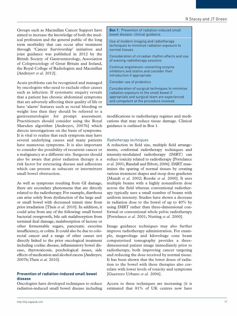
Prevention of radiation-induced small bowel diseaseOncologists have developed techniques to reduce radiation-induced small bowel disease including
modifications to radiotherapy regimes and medi-cations that may reduce tissue damage. Clinical guidance is outlined in Box 1.
Radiotherapy techniquesA reduction in field size, multiple field arrange-ments, conformal radiotherapy techniques and intensity-modulated radiotherapy (IMRT) can reduce toxicity related to radiotherapy [Portelance et al. 2001; Randall and Ibbott, 2006]. IMRT max-imizes the sparing of normal tissues by creating various treatment shapes and steep dose gradients [Mundt et al. 2002; Roeske et al. 2000]. It uses multiple beams with a highly nonuniform dose across the field whereas conventional radiother-apy typically uses a small number of beams with uniform intensity. Studies have shown a decrease in radiation dose to the bowel of up to 40% by using IMRT rather than three-dimensional con-formal or conventional whole pelvic radiotherapy [Portelance et al. 2001; Nutting et al. 2000].
Image guidance techniques may also further improve radiotherapy administration. For exam-ple, megavoltage and kilovoltage cone beam computerized tomography provides a three-dimensional patient image immediately prior to radiotherapy, both improving cancer targeting and reducing the dose received by normal tissue. It has been shown that the lower doses of radia-tion to the bowel with these therapies also cor-relate with lower levels of toxicity and symptoms [Guerrero Urbano et al. 2006].
Access to these techniques are increasing (it is estimated that 81% of UK centres now have
Box 1. Prevention of radiation-induced small bowel disease: clinical guidance.
Use of modern imaging and radiotherapy techniques to minimize radiation exposure to normal tissues
Consideration of circadian rhythm effects and use of evening radiotherapy sessions
Continue angiotensin-converting enzyme inhibitors and statins and consider their introduction if appropriate
Consider use of probiotics
Consideration of surgical techniques to minimise radiation exposure to the small bowel if appropriate and surgical team are experienced and competent at the procedure involved.
Therapeutic Advances in Chronic Disease 5 (1)
18 http://taj.sagepub.com
access to IMRT) [Ahmad et al. 2012]. Although these methods are extremely promising, chronic symptoms can present up to three decades after radiotherapy, therefore long-term data will take many years to be realized.
Patient positioning and positioning devicesA variety of patient positions and positioning devices (such as a belly board, a positioning device designed to reduce the irradiated small bowel vol-ume of patients undergoing treatment in the pel-vic region by small bowel displacement whilst lying prone) have been trialled attempting to min-imize inadvertent exposure of normal bowel to ionizing radiation.
A review analysing 46 papers concerning the influence of the patient position during the treat-ment of pelvic malignancies showed that a prone position generally results in a lower irradiated small bowel volume than the supine position [Wiesendanger-Wittmer et al. 2012]. However, a more significant reduction of the irradiated small bowel volume can be achieved by the additional use of a belly board in prone position, for both 3D-CRT (Conformal Radiotherapy) and IMRT treatment plans. It was also noted that a full blad-der can also reduce the irradiated small bowel vol-ume. However, there are little long-term data available at present assessing the long-term effect of these treatments on the GI tract.
Circadian rhythmAnimal studies have shown that when mice were irradiated at different times of the day, a clear cir-cadian rhythm was observed in the number of apoptotic cells in the intestinal crypt [Ijiri and Potten, 1988]. This is thought to be due to the effect of circadian rhythm on cell proliferation cycles, and tissue appears to be more radiosensi-tive if its cells have greater proliferative capacity and divide more rapidly. Studies of cellular prolif-eration in the human rectal mucosa have shown the highest proliferative activity occurring in the morning between 03:00 and 11:30 and the least activity occurring 12 hours later [Ijiri and Potten, 1990; Buchi et al. 1991].
A prospective trial randomized 229 patients who received radiotherapy for cervical carcinoma to treatment in the morning (08:00–10:00) or even-ing (18:00–20:00) [Shukla et al. 2010]. The inci-dence of acute radiation-induced small bowel
disease in the two arms was assessed and reported in terms of various grades of diarrhoea. Total number of patients with diarrhoea as well as those with more severe diarrhoea were found to be sig-nificantly greater in patients treated in the morn-ing when compared with those receiving the identical radiation regime in the evening. The oncological therapeutic response in the two arms was similar.
Although timing of treatment may reduce mor-bidity from radiotherapy, a logistical problem exists in centres with limited capacity for evening treatments.
MedicationsStatins and angiotensin-converting enzyme inhibitors. It has been noted that GI toxicity from radiotherapy is lower in patients taking antihypertensive and cholesterol-lowering agents (specifically angiotensin-converting enzyme [ACE] inhibitors and statins). ACE inhibitors block enzymatic conversion of angiotensin I to angiotensin II which plays a critical role in blood pressure homeostasis. In vitro studies have confirmed the anti-inflam-matory, antifibrotic and antithrombotic poten-tial of statins in irradiated human cells [Gaugler et al. 2005; Haydont et al. 2005, 2007] and low-dose lovastatin has been shown to be radioprotective in human endothelial cells [Ostrau et al. 2009].
A retrospective, nonrandomized cohort study of 308 pelvic radiotherapy patients assessed the impact of statins and ACE inhibitors on the devel-opment of GI symptoms [Wedlake et al. 2012]. GI symptomatology was recorded prospectively before radiotherapy, weekly during treatment and 1 year later using the Inflammatory Bowel Disease Questionnaire. Use of statin or statin + ACE inhibitor during radical pelvic radiotherapy sig-nificantly reduced acute GI symptoms.
Probiotics. A probiotic is a preparation contain-ing viable and defined microorganisms in large numbers sufficient to alter the host’s microflora [Kligler and Cohrssen, 2008].
Radiation therapy may disturb the indigenous gut flora which are important in maintaining a nor-mal mucosal function [Berthrong, 1986] and there is emerging evidence that probiotics may have a radio-protective effect.
R Stacey and JT Green
http://taj.sagepub.com 19
There have been a total of five randomized con-trolled trials of varying quality and size regard-ing probiotics in radiation-induced small bowel disease [Salminen et al. 1988; Delia et al. 2007; Urbancsek et al. 2001; Giralt et al. 2008; Chitapanarux et al. 2010]. Agents investigated include the probiotic preparation VSL#3 (a probiotic containing eight strains of live lactic acid bacteria and bifidobacteria) and live lacto-bacillus acidophilus plus bifidobacterium bifi-dum. Although some trials have shown a significant improvement in diarrhoeal symp-toms and decreased antidiarrhoeal medication use, study design and patient numbers do not enable us to fully advocate probiotics at this time. Full safety testing of individual com-pounds followed by larger, well-designed dou-ble-blinded randomized controlled trials are required.
Amifostine. Amifostine is a cytoprotective adju-vant used in cancer chemotherapy. It reduces rates of xerostomia when administered before head and neck cancer radiotherapy [Jha et al. 2012]. Preliminary studies suggest amifostine may also protect against radiation-induced bowel toxicity [Athanassiou et al. 2003; Ben-Josef et al. 2002; Leonard et al. 2005] but further research is required to define its true value.
Antioxidants. Cytotoxic effects of ionizing radia-tion on GI epithelium have been hypothesized to be related to oxidative stress. Animal studies have shown that vitamin E and/or selenium treatment prior to radiotherapy help to minimize oxidative stress [Felemovicius et al. 1995; Mutlu-Türkoğlu et al. 2000] indicating that antioxidant pretreat-ments may have some beneficial effects against radiation induced intestinal injury [Empey et al. 1992]. Further studies of these agents are required.
Teduglutide. Teduglutide is a glucagon-like pep-tide-2 analogue. Animal studies have shown increased intestinal crypt stem cell survival when given prior to whole-body irradiation in mice [Booth et al. 2004]. This suggests in theory that it may provide a useful protective role in preventing radiation-induced intestinal injury but further work is required in humans.
Dietary supplementation. Glutamine and argi-nine have been shown to have a protective effect on the intestinal mucosa of rats treated with radiotherapy [Yavas et al. 2012]. Clinical studies,
however, have shown that glutamine did not pro-tect against acute radiation-induced small bowel disease in humans [Kozelsky et al. 2003; Vidal-Casariego et al. 2013].
There is no evidence that a lactose-restricted diet will prevent radiation-induced small bowel dis-ease despite its utility in the treatment of some patients diarrhoea arising from radiotherapy [Stryker and Bartholomew, 1986]. A recent review of 22 studies regarding the efficacy of nutritional interventions to counteract acute gas-trointestinal toxicity during therapeutic pelvic radiotherapy [Wedlake et al. 2013] looked at the evidence for elemental formula, low- or modified-fat diets, low- or high-fibre diets, low-lactose diets, probiotics and symbiotics. The authors found that there is insufficient high-grade evi-dence to recommend nutritional interventions at present, and that further high-quality trials are required.
Sucralfate. Sucralfate is a highly sulphated polyanionic disaccharide used to treat dyspep-sia. It is thought to stimulate epithelial healing and form a protective barrier over damaged mucosal surfaces [Denton et al. 2002]. There is randomized controlled evidence that sucralfate can help in the treatment of bleeding in radia-tion proctitis, but there is no evidence that it is of use in the prevention of radiation-induced small bowel disease. A randomized double-blind study showed no significant difference between sucralfate and placebo in this setting [Marten-son et al. 2000].
Surgical techniquesSurgical placement of absorbable mesh slings and silicone prostheses have been described to prevent radiation-induced small bowel disease [Devereux et al. 1984; Kavanah et al. 1985; Sener et al. 1989; Rodier et al. 1991; Beitler et al. 1997; Sugarbaker, 1983]. These interven-tions are aimed at reducing toxicity by exclud-ing the small bowel from irradiated areas. However, the results have not been consistently reproduced in clinical practice and are not rou-tinely used in many centres.
Complete exclusion of the small bowel by mesh sling in the early postoperative period should prevent the small bowel from becoming adhered into the pelvis. After mesh absorption, it is thought that the small bowel retains enough
Therapeutic Advances in Chronic Disease 5 (1)
20 http://taj.sagepub.com
mobility that it may be temporarily excluded from the pelvis by simple positioning methods [Waddell et al. 1999]. One case series of 60 patients had polyglycolic acid mesh slings inserted after resection of rectal or gynaeco-logical malignancies. All patients received post-operative radiotherapy in standard fractions. At a mean follow up of 28 months, no cases of radiation-induced small bowel disease were seen [Devereux et al. 1988]. A study of 45 patients with resectable carcinoma of the rec-tum showed similar results [Dasmahapatra and Swaminathan, 1991].
Space-occupying silicone prostheses have been used to exclude the small bowel from the pelvis [McGinley et al. 1980], however they may develop a mass effect on surrounding structures resulting in moderate hydronephrosis [Nguyen and Hamper, 1997].
Repeat surgery may be necessary to remove the prosthesis after completion of the radiation ther-apy, although inflatable saline implants have been developed to reduce this problem [Sezeur et al. 1990].
Treatment of acute radiation-induced small bowel diseasePatients with acute enteritis may experience a wide variety of symptoms.
Treatment can be divided into supportive and dietary interventions as well as specific medical and surgical therapies. In severe cases, the subse-quent oncological regimen may have to be revised.
Supportive treatmentsNumerous medications can be prescribed that have no role in correcting the underlying patho-physiology of the condition but are aimed at mini-mizing symptoms.
The first-line treatment for acute radiation induced diarrhoea is antidiarrhoeal medication such as loperamide or cophenotrope [Wadler et al. 1998]. Bismuth subsalicylate has also been recommended for diarrhoea and nausea [National Cancer Institute, 2012], but as with many of these supportive treatments, the evi-dence base comes from clinical experience and consensus opinions only. Patients may also
benefit from an anticholinergic antispasmodic agent to alleviate bowel cramping, analgesics for pain or anti-emetics for nausea [National Cancer Institute, 2012]. It is important to note that symptoms often stop upon completion of the radiotherapy regimen. Clinicians should provide reassurance along with education about the potential for chronic problems [Andreyev et al. 2012]. There is also emerging evidence that bile acid malabsorption can occur in the acute setting and this should be considered by clinicians [Harris et al. 2012].
Dietary treatmentsIntestinal villi can be damaged by radiation ther-apy resulting in a reduction or loss of digestive enzymes leading to malabsorption of nutrients [Czito and Willett, 2010]. It is important to ensure sufficient calorific and fluid intake which may be difficult in this setting; a dietician can pro-vide targeted advice. A number of dietary modifi-cations have been suggested for the treatment of symptoms of radiation-induced small bowel dis-ease but there is only limited evidence to say that they are beneficial.
A diet that is lactose free, low fat and low residue may have a benefit on patients’ symptoms. However, results from other trials evaluating the effect of lactose-restricted diets on radiation-induced diarrhoea have provided contradictory results [Stryker and Bartholomew, 1986; Bye et al. 1992]. It is important to consider that if tak-ing this approach then lactose-free nutritional supplements should be used.
In clinical practice, a pragmatic approach is sug-gested which may be assisted by keeping a food diary correlating to symptoms.
OctreotideThe somatostatin analogue octreotide is used in the treatment of chemotherapy-induced diar-rhoea and radiation-induced small bowel disease [Yavuz et al. 2002]. It is an octapeptide that mim-ics natural somatostatin and decreases gut motil-ity. A randomized controlled trial comparing octreotide acetate (100 µg three times daily) with diphenoxylate hydrochloride plus atropine sul-phate (2.5 mg four times daily) in acute radiation-induced small bowel disease showed that diarrhoea resolved more quickly and a decrease in the number of patients needing to discontinue
R Stacey and JT Green
http://taj.sagepub.com 21
pelvic radiotherapy in the octreotide arm [Yavuz et al. 2002].
5-Aminosalicylic acidsThere is no evidence of the benefit of 5-amino-salicylic acids (5-ASAs) in acute or chronic radia-tion-induced small bowel disease and it has been shown that they may increase symptoms in the acute setting [Gibson et al. 2013].
SurgerySurgery is very rarely required in acute enteritis. Where possible, it should be avoided because of poor wound healing and concerns about leakage from surgical anastomoses [Galland and Spencer, 1986]. It may of course be necessary to operate in some patients who have had recent radiotherapy but surgeons should be wary and cautious in doing so.
Treatment of chronic radiation-induced small bowel diseasePatients with abdominal symptoms that occur after prior radiotherapy need thorough assess-ment and investigation by a gastroenterologist and a treatment plan which may involve other healthcare specialists. Treatments can be divided into those that target specific secondary entities that commonly occur after radiotherapy and sup-portive, nutritional, mediations and other inter-ventions that aim to counteract the effect of the enteropathy.
Supportive treatmentsAs per the acute setting, patients may need symp-tom-based medications either intermittently or on a regular basis. Again, this includes antimotil-ity agents, analgesics and anti-emetics. One small trial has assessed the efficacy of loperamide in patients with chronic radiation-induced small bowel disease, showing improvement in intestinal transit times, bile salt absorption and diarrhoea [Yeoh et al. 1993].
Secondary effects of chronic radiation-induced small bowel disease
Antibiotics for small bowel bacterial overgrowthDamage to the small bowel creates areas of dys-motility and stasis leading to bacterial overgrowth
[Husebye et al. 1995]. Unlike the colon, which is rich in bacteria, the small bowel usually has fewer than 104 organisms per millilitre [Quigley and Quera, 2006]. When bacterial overgrowth occurs, the most common isolates from the jejunum are Escherichia coli, Streptococcus, Lactobacillus, Bacteroides and Enterococcus species [Bouhnik et al. 1999].
Broad-spectrum antibiotics are also utilized: these include tetracycline, co-amoxiclav, ciprofloxacin and rifaximin. Local antibiotic guidance should be followed. Patients often need to have repeated courses and many require long-term maintenance therapy at a lower dose. Some clinicians advocate the use of a rotation of different antibiotics to reduce the risk of resistance [Quigley and Abu-Shanab, 2010].
Although probiotic therapy has been used in the prevention and treatment of acute radiation-induced small bowel disease, there is currently no evidence of their effectiveness in the chronic setting.
Cholestyramine and colesevelam for bile salt malabsorptionA total of 95% of bile acids are absorbed in the terminal ileum and radiation damage to this area can cause bile acid malabsorption (BAM) [Andersson et al. 1978]. This can be tested for by a Se-HCAT study, however this investigation is not widely used and many clinicians advocate empirical treatment. BAM is thought to be responsible for symptoms in 35–72% of patients with chronic radiation-induced small bowel dis-ease suffering from diarrhoea [Theis et al. 2010; Andreyev et al. 2005; Danielsson et al. 1991; Ludgate and Merrick, 1985; Arlow et al. 1987]. It responds well to cholestyramine, however this is not very palatable and 68% of patients discon-tinue it after 1 year [Kamal-Bahl et al. 2007]. Alternatives are colestipol and colesevelam which also bind bile salts. Colesevelam is better toler-ated and there is evidence for its benefit in this setting; however, it is not currently licensed for this indication and is relatively expensive com-pared with other agents [Puleston et al. 2005; Wedlake et al. 2009].
Nutrition and related therapiesThere has been research into the exclusion of certain foods and the use of nutritional
Therapeutic Advances in Chronic Disease 5 (1)
22 http://taj.sagepub.com
supplements in chronic radiation-induced small bowel disease, but again evidence for their ben-efit is variable.
When considering exclusion diets, it is impor-tant to get dietetic input. Patients can be assessed for lactose as well as other carbohy-drate intolerances using techniques such as breath testing. The reduced sensitivity of these noninvasive tests mean that an empirical but guided trial of exclusion may be necessary. Lactose-free diets in patients with lactose intol-erance have been shown to be effective [Beer et al. 1985]. We have also used a FODMAP (fermentable oligosaccharides, disaccharides, monosaccharides and pylols) exclusion diet in some of these patients who have subsequently reported an improvement in their symptoms, however there is currently no published trial assessing this approach.
Anecdotally, some patients do seem to relate exacerbations of their symptoms with the inges-tion of specific foods. One study of 26 women with chronic radiation-induced small bowel disease compared with 21 normal controls showed that 50% of the patients noticed an increase in symptoms upon consumption of bran muffins, berries, cabbage, brussel sprouts, broccoli, tossed salad, Caesar salad, baked beans, lentils and nuts in comparison with 14% in controls. A total of 85% of these patients could only tolerate smaller portions of these foods. The study suggests that smaller, more frequent portions may improve tolerance of certain foods and reinforces careful history taking and an individualized approach for all patients [Sekhon, 2000].
It is important that patients receive sufficient calorific intake and where possible support is given via the enteral route. Some patients will require the long term use of highly calorific nutritional supplements: so-called ‘sip’ feeds. Regular measurement and supplementation of vitamins and minerals including iron, folic acid, vitamin B12, vitamin D, magnesium, cal-cium, trace elements and fat-soluble vitamins are important.
In some patients, parenteral support with fluid and electrolytes is necessary. Intestinal failure due to extensive enteropathy from prior radio-therapy is a recognized and relatively common indication for home parenteral nutrition (PN).
The management of these patients should be coordinated at specialist centres. The latest British artificial nutrition survey showed that 3.8% of patients in the UK on home PN had radiation enteropathy [Smith et al. 2011].
It has been shown that intestinal rest with PN can improve clinical and radiological findings in patients with small bowel radiation injury [Loiudice and Lang, 1983] and that nutritional autonomy and survival may be improved if patients are treated initially with intestinal rest and home PN [Gavazzi et al. 2006]. However, it has also been reported [Silvain, 1992] that patients with chronic radiation-induced small bowel disease may be more likely to suffer clin-ical recurrence if treated conservatively with PN support compared with those undergoing surgical intervention [Scolapio et al. 1999, 2002].
Hyperbaric oxygenHyperbaric oxygen (HBO) decreases tissue hypoxia in bowel affected by ischaemic damage from ionising radiation by encouraging angio-genesis. An antibacterial effect has also been hypothesized [Bennett et al. 2012]. HBO is the only therapy found to increase the number of blood vessels in irradiated tissue [Bennett et al. 2012] and may allow the treatment of multiple sites of small bowel. The treatment is administered over several weeks in hyperbaric chambers.
A systematic literature review showed that 67 of 74 publications reported positive results when HBO was delivered as treatment for, or preven-tion of, delayed radiation injury [Feldmeier and Hampson, 2002].
A large multicentre study in the UK, the Hyperbaric Oxygen Therapy II (HOT-II) study, has finished recruiting and its results are eagerly awaited. Although HBO therapy is largely safe to use, it is limited by the availability of chambers.
Pentoxifylline and tocopherolPentoxifylline is a xanthine derivative and toco-pherols are a class of chemical compounds with vitamin E activity [Hamama et al. 2012]. It has been suggested that the combination of these medications may decrease radiation-induced
R Stacey and JT Green
http://taj.sagepub.com 23
fibrosis through antioxidant effects [Gothard et al. 2005]. A study of 30 patients with chronic radiation enteritis or proctitis showed sympto-matic improvement in 71% of patients treated with this combination therapy, compared with 33% of patients who received supportive treatment alone [Hille et al. 2005]. Further research is required to fully evaluate these therapies.
Anti-inflammatory agentsStudies regarding the use of aminosalicylates in chronic radiation-induced small bowel disease are limited. Sulphasalazine has been examined in a case series of four patients with chronic radiation-induced small bowel disease, all patients showed clinical and radiological improvement over the course of 1 year of ther-apy [Goldstein et al. 1976]. Another study has shown that methylprednisolone may enhance the effect of parenteral nutrition-facilitated ‘intestinal rest’, but there is no significant evidence to suggest that corticosteroids are of use in these patients [Loiudice and Lang, 1983].
Endoscopic therapiesOccasionally patients may present with melaena or iron-deficient anaemia. Telangiectasia may form at any site in the intestine which has been irradiated and can be difficult to locate. Capsule endoscopy may be helpful but should be avoided in those with strictures. Argon plasma coagula-tion is well described in the management of radiation proctopathy [Leiper and Morris, 2007], but has also been successfully used for radiation-induced lesions in the distal duode-num [Toyoda et al. 2004] and ileum [Pasha et al. 2007] and can be administered into the jejunum as well via double balloon enteroscopy. It should be used judiciously given the risk of perforation particularly in abnormal tissue. Enteroscopy may also prove to be an alternative to surgery to treat small bowel strictures [Haruta et al. 2005; Kita et al. 2007].
SurgerySurgery is challenging in patients with prior abdominal radiotherapy. There are often adhe-sions and the intestine may be very fibrotic and difficult to handle [Jao et al. 1986; Luna-Pérez et al. 2001]. Wound healing can also be difficult
[Luna-Pérez et al. 2001]. There are certain patients, particularly those with strictures and obstructive symptoms, who need surgery as they cannot get symptomatic control or are persis-tently nutritionally compromised by their small bowel disease. Patients with extensive small bowel involvement are at risk of short bowel syndrome or a high-output proximal stoma both of which are significant management challenges. It is important to note that one operation can lead to another further increasing the risks of a short bowel. It is imperative that those consid-ered for surgery have a thorough evaluation by radiological and other techniques and are man-aged by a multidisciplinary team that involves nutritional support and a surgeon who has expe-rience in managing these patients [Andreyev et al. 2012].
A study of surgical treatment for radiation-induced small bowel disease assessed 48 patients who underwent extended intestinal resection with anastomosis showed a significant postoperative morbidity of 21.7%. Overall sur-vival after radiation-related complication in patients without neoplastic disease recurrence was 89%, 79% and 69%, at 1, 3 and 5 years after surgery, respectively [Onodera et al. 2005].
ConclusionA clearer picture of the management of radiation-induced small bowel disease is beginning to emerge. However, the optimal ways of preventing radiation induced damage to the small bowel as well as effective management for all patients is still unclear. New radiotherapy techniques con-tinue to decrease inadvertent exposure to adja-cent normal tissue and preventative agents including ACE inhibitors and statins are exciting areas of future research. The treatment of radia-tion-induced small bowel disease is largely sup-portive in the acute phase. For those with GI problems that have arisen in the years following radiotherapy, the key is recognition and referral for specialist advice from a gastroenterologist who has an interest in this field. Suggested clinical guidelines are outlined in Table 1. Patients need a targeted work-up for each of the symptoms they have, as many of these have several potential causes. Specific treatments can be offered as well as considering the valuable input from other healthcare professionals, e.g. dieticians. Emerging treatments such as HBO offer promise. The small
Therapeutic Advances in Chronic Disease 5 (1)
24 http://taj.sagepub.com
subset of patients who require an operation to alleviate their problems should be directed towards surgeons who have an experience in deal-ing with this challenging situation.
FundingThis research received no specific grant from any funding agency in the public, commercial, or not-for-profit sectors.
Conflict of interest statementThe authors have no conflicts of interest to declare.
ReferencesAhmad, S., Duke, S., Jena, R., Williams, M. and Burnet, N. (2012) Advances in radiotherapy. BMJ (Clin Res Ed) 345: e7765.
Andersson, H., Bosaeus, I. and Nystrom, C. (1978) Bile salt malabsorption in the radiation syndrome. Acta Radiologica 17: 312–318.
Andreyev, H. (2007a) Gastrointestinal problems after pelvic radiotherapy: the past, the present and the future. Clin Oncol 19: 790–799.
Andreyev, H. (2007b) Gastrointestinal symptoms after pelvic radiotherapy: a new understanding to improve management of symptomatic patients. Lancet Oncol 8: 1007–1017.
Andreyev, H., Amin, Z., Blake, P., Dearnaley, D., Tait, D. and Vlavianos, P. (2003) GI symptoms developing after pelvic radiotherapy require gastroenterological review but is that happening in the UK? Clin Oncol 15: S12 (abstract).
Andreyev, H., Davidson, S., Gillespie, C., Allum, W. and Swarbrick, E. for the British Society of Gastroenterology, Association of Colo-Proctology of Great Britain and Ireland (2012) Practice guidance on the management of acute and chronic gastrointestinal problems arising as a result of treatment for cancer. Gut 61: 179–192.
Andreyev, H., Vlavianos, P., Blake, P., Dearnaley, D., Norman, A. and Tait, D. (2005) Gastrointestinal symptoms after pelvic radiotherapy: role for the gastroenterologist? Int J Radiation Oncol Biol Phys 62: 1464–1471.
Arlow, F., Dekovich, A., Priest, R. and Beher, W. (1987) Bile acids in radiation-induced diarrhea. Southern Med J 80: 1259–1261.
Athanassiou, H., Antonadou, D., Coliarakis, N., Kouveli, A., Synodinou, M., Paraskevaidis, M. et al. (2003) Protective effect of amifostine during fractionated radiotherapy in patients with pelvic carcinomas: results of a randomized trial. Int J Radiat Oncol Biol Phys 56: 1154–1160.
Beer, W., Fan, A. and Halsted, C. (1985) Clinical and nutritional implications of radiation enteritis. Am J Clin Nutr 41: 85–91.
Beitler, A., Rodriguez-Bigas, M., Weber, T., Lee, R., Cuenca, R. and Petrelli, N. (1997) Complications of
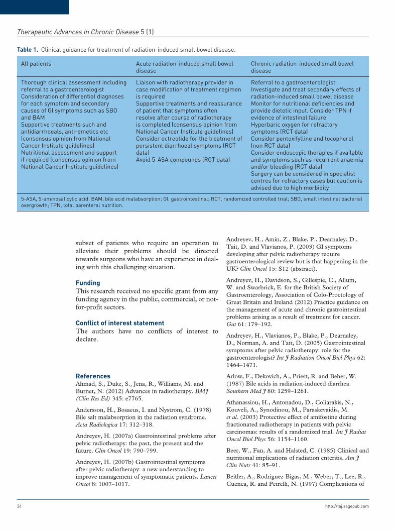
Table 1. Clinical guidance for treatment of radiation-induced small bowel disease.
All patients Acute radiation-induced small bowel disease
Chronic radiation-induced small bowel disease
Thorough clinical assessment including referral to a gastroenterologistConsideration of differential diagnoses for each symptom and secondary causes of GI symptoms such as SBO and BAMSupportive treatments such and antidiarrhoeals, anti-emetics etc (consensus opinion from National Cancer Institute guidelines)Nutritional assessment and support if required (consensus opinion from National Cancer Institute guidelines)
Liaison with radiotherapy provider in case modification of treatment regimen is requiredSupportive treatments and reassurance of patient that symptoms often resolve after course of radiotherapy is completed (consensus opinion from National Cancer Institute guidelines)Consider octreotide for the treatment of persistent diarrhoeal symptoms (RCT data)Avoid 5-ASA compounds (RCT data)
Referral to a gastroenterologistInvestigate and treat secondary effects of radiation-induced small bowel diseaseMonitor for nutritional deficiencies and provide dietetic input. Consider TPN if evidence of intestinal failureHyperbaric oxygen for refractory symptoms (RCT data)Consider pentoxifylline and tocopherol (non RCT data)Consider endoscopic therapies if available and symptoms such as recurrent anaemia and/or bleeding (RCT data)Surgery can be considered in specialist centres for refractory cases but caution is advised due to high morbidity
5-ASA, 5-aminosalicylic acid; BAM, bile acid malabsorption; GI, gastrointestinal; RCT, randomized controlled trial; SBO, small intestinal bacterial overgrowth; TPN, total parenteral nutrition.
R Stacey and JT Green
http://taj.sagepub.com 25
absorbable pelvic mesh slings following surgery for rectal carcinoma. Dis Colon Rectum 40: 1336–1341.
Ben-Josef, E., Han, S., Tobi, M., Shaw, L., Bonner, H., Vargas, B. et al. (2002) A pilot study of topical intrarectal application of amifostine for prevention of late radiation rectal injury. Int J Radiat Oncol Biol Phys 53: 1160–1164.
Bennett, M., Feldmeier, J., Hampson, N., Smee, R. and Milross, C. (2012) Hyperbaric oxygen therapy for late radiation tissue injury. Cochrane Database Syst Rev 5: CD005005.
Berthrong, M. (1986) Pathologic changes secondary to radiation. World J Surg 10: 155–170.
Board of the Faculty of Clinical Oncology of the Royal College of Radiologists (2006) Radiotherapy dose–fractionation. Royal College of Radiologists, London. Available at http://www.rcr.ac.uk/docs/oncology/pdf/Dose-Fractionation_Final.pdf
Booth, C., Booth, D., Williamson, S., Demchyshyn, L. and Potten, C. (2004) Teduglutide ([Gly2]GLP-2) protects small intestinal stem cells from radiation damage. Cell Proliferation 37: 385–400.
Bouhnik, Y., Alain, S., Attar, A., Flourié, B., Raskine, L., Sanson-Le Pors, M. et al. (1999) Bacterial populations contaminating the upper gut in patients with small intestinal bacterial overgrowth syndrome. Am J Gastroenterol 94: 1327–1331.
Buchi, K., Moore, J., Hrushesky, W., Sothern, R. and Rubin, N. (1991) Circadian rhythm of cellular proliferation in the human rectal mucosa. Gastroenterology 101: 410–415.
Bye, A., Kaasa, S., Ose, T., Sundfør, K. and Tropé, C. (1992) The influence of low fat, low lactose diet on diarrhoea during pelvic radiotherapy. Clin Nutrition 11: 147–153.
Chitapanarux, I., Chitapanarux, T., Traisathit, P., Kudumpee, S., Tharavichitkul, E. and Lorvidhaya, V. (2010) Randomized controlled trial of live lactobacillus acidophilus plus bifidobacterium bifidum in prophylaxis of diarrhea during radiotherapy in cervical cancer patients. Radiat Oncol 5: 31.
Crook, J., Esche, B. and Futter, N. (1996) Effect of pelvic radiotherapy for prostate cancer on bowel, bladder, and sexual function: the patient’s perspective. URL 47: 387–394.
Czito, B. and Willett, C. (2010) Radiation injury. In Feldman, M., Lawrence, S., Friedman, L. and Brandt, L. (eds), Sleisenger and Fordtran’s Gastrointestinal and Liver Disease. Pathophysiology, Diagnosis, Management (9th edn). Philadelphia, PA: Saunders.
Danielsson, A., Nyhlin, H., Persson, H., Stendahl, U., Stenling, R. and Suhr, O. (1991) Chronic
diarrhoea after radiotherapy for gynaecological cancer: occurrence and aetiology. Gut 32: 1180–1187.
Dasmahapatra, K. and Swaminathan, A. (1991) The use of a biodegradable mesh to prevent radiation-associated small-bowel injury. Arch Surg 126: 366–369.
Delia, P., Sansotta, G., Donato, V., Frosina, P., Messina, G., De Renzis, C. et al. (2007) Use of probiotics for prevention of radiation-induced diarrhea. World J Gastroenterol 13: 912–915.
Denham, J. and Hauer-Jensen, M. (2002) The radiotherapeutic injury--a complex ‘wound’. Radiother Oncol 63: 129–145.
Denton, A., Forbes, A., Andreyev, J. and Maher, E. (2002) Non surgical interventions for late radiation proctitis in patients who have received radical radiotherapy to the pelvis. Cochrane Database Syst Rev 1: CD003455.
Devereux, D., Chandler, J., Eisenstat, T. and Zinkin, L. (1988) Efficacy of an absorbable mesh in keeping the small bowel out of the human pelvis following surgery. Dis Colon Rectum 31: 17–21.
Devereux, D., Kavanah, M., Feldman, M., Kondi, E., Hull, D., O’Brien, M. et al. (1984) Small bowel exclusion from the pelvis by a polyglycolic acid mesh sling. J Surg Oncol 26: 107–112.
Do, N., Nagle, D. and Poylin, V. (2011) Radiation proctitis: current strategies in management. Gastroenterol Res Practice 2011: 917941.
Empey, L., Papp, J., Jewell, L. and Fedorak, R. (1992) Mucosal protective effects of vitamin E and misoprostol during acute radiation-induced enteritis in rats. Digest Dis Sci 37: 205–214.
Feldmeier, J. and Hampson, N. (2002) A systematic review of the literature reporting the application of hyperbaric oxygen prevention and treatment of delayed radiation injuries: an evidence based approach. Undersea Hyperbaric Med 29: 4–30.
Felemovicius, I., Bonsack, M., Baptista, M. and Delaney, J. (1995) Intestinal radioprotection by vitamin E (alpha-tocopherol). Ann Surg 222: 504–510.
Galland, R. and Spencer, J. (1986) Surgical management of radiation enteritis. Surgery 99: 133–139.
Gami, B., Harrington, K., Blake, P., Dearnaley, D., Tait, D., Davies, J. et al. (2003) How patients manage gastrointestinal symptoms after pelvic radiotherapy. Aliment Pharmacol Therapeut 18(10), pp. 987–994.
Gaugler, M., Vereycken-Holler, V., Squiban, C., Vandamme, M., Vozenin-Brotons, M. and Benderitter, M. (2005) Pravastatin limits endothelial activation after irradiation and decreases the resulting
Therapeutic Advances in Chronic Disease 5 (1)
26 http://taj.sagepub.com
inflammatory and thrombotic responses. Radiation Res 163(5), pp. 479–487.
Gavazzi, C., Bhoori, S., Lovullo, S., Cozzi, G. and Mariani, L. (2006) Role of home parenteral nutrition in chronic radiation enteritis. Am J Gastroenterol 101: 374–379.
Gibson, R., Keefe, D., Lalla, R., Bateman, E., Blijlevens, N., Fijlstra, M. et al. (2013) Systematic review of agents for the management of gastrointestinal mucositis in cancer patients. Supportive Care Cancer 21: 313–326.
Giralt, J., Regadera, J., Verges, R., Romero, J., de la, Fuente, I., Biete, A. et al. (2008) Effects of probiotic Lactobacillus casei DN-114 001 in prevention of radiation-induced diarrhea: results from multicenter, randomized, placebo-controlled nutritional trial. Int J Radiat Oncol Biol Phys 71: 1213–1219.
Goldstein, F., Khoury, J. and Thornton, J. (1976) Treatment of chronic radiation enteritis and colitis with salicylazosulfapyridine and systemic corticosteroids. A pilot study. Am J Gastroenterol 65: 201–208
Gothard, L., Cornes, P., Brooker, S., Earl, J., Glees, J., Hall, E., Peckitt, C. et al. (2005) Phase II study of vitamin E and pentoxifylline in patients with late side effects of pelvic radiotherapy. Radiother Oncol 75: 334–341.
Guerrero Urbano, M., Henrys, A., Adams, E., Norman, A., Bedford, J., Harrington, K. et al. (2006) Intensity-modulated radiotherapy in patients with locally advanced rectal cancer reduces volume of bowel treated to high dose levels. Int J Radiat Oncol Biol Phys 65: 907–916.
Hamama, S., Delanian, S., Monceau, V. and Vozenin, M. (2012) Therapeutic management of intestinal fibrosis induced by radiation therapy: from molecular profiling to new intervention strategies et vice et versa. Fibrogenesis Tissue Repair 5(Suppl. 1): S13.
Harris, V., Benton, B., Sohaib, A., Dearnaley, D. and Andreyev, H. (2012) Bile acid malabsorption after pelvic and prostate intensity modulated radiation therapy: an uncommon but treatable condition. Int J Radiat Oncol Biol Phys 84: 601–606.
Haruta, H., Yamamoto, H., Mizuta, K., Kita, Y., Uno, T., Egami, S. et al. (2005) A case of successful enteroscopic balloon dilation for late anastomotic stricture of choledochojejunostomy after living donor liver transplantation. Liver Transplantation 11: 1608–1610.
Haydont, V., Bourgier, C., Pocard, M., Lusinchi, A., Aigueperse, J., Mathé, D. et al. (2007) Pravastatin Inhibits the Rho/CCN2/extracellular matrix cascade in human fibrosis explants and improves radiation-induced intestinal fibrosis in rats. Clinical Cancer Res 13: 5331–5340.
Haydont, V., Mathé, D., Bourgier, C., Abdelali, J., Aigueperse, J., Bourhis, J. et al. (2005) Induction of CTGF by TGF-beta1 in normal and radiation enteritis human smooth muscle cells: Smad/Rho balance and therapeutic perspectives. Radiother Oncol 76: 219–225.
Hille, A., Christiansen, H., Pradier, O., Hermann, R., Siekmeyer, B., Weiss, E. et al. (2005) Effect of pentoxifylline and tocopherol on radiation proctitis/enteritis. Strahlenther Onkol 181: 606–614.
Husebye, E., Skar, V., Høverstad, T., Iversen, T. and Melby, K. (1995) Abnormal intestinal motor patterns explain enteric colonization with gram-negative bacilli in late radiation enteropathy. Gastroenterology 109: 1078–1089.
Ijiri, K. and Potten, C. (1988) Circadian rhythms in the incidence of apoptotic cells and number of clonogenic cells in intestinal crypts after radiation using normal and reversed light conditions. Int J Radiat Biol Relat Stud Phys Chem Med 53: 717–727.
Ijiri, K. and Potten, C. (1990) The circadian rhythm for the number and sensitivity of radiation-induced apoptosis in the crypts of mouse small intestine. Int J Radiat Biol 58: 165–175.
Jao, S., Beart, R. and Gunderson, L. (1986) Surgical treatment of radiation injuries of the colon and rectum. Am J Surg 151: 272–277.
Jha, N., Harris, J., Seikaly, H., Jacobs, J., McEwan, A., Robbins, K. et al. (2012) A phase II study of submandibular gland transfer prior to radiation for prevention of radiation-induced xerostomia in head-and-neck cancer (RTOG 0244). Int J Radiat Oncol Biol Phys 84: 437–442.
Kamal-Bahl, S., Burke, T., Watson, D. and Wentworth, C. (2007) Discontinuation of lipid modifying drugs among commercially insured United States patients in recent clinical practice. Am J Cardiol 99: 530–534.
Kavanah, M., Feldman, M., Devereux, D. and Kondi, E. (1985) New surgical approach to minimize radiation-associated small bowel injury in patients with pelvic malignancies requiring surgery and high-dose irradiation. A preliminary report. Cancer 56: 1300–1304.
Kennedy, G. and Heise, C. (2005) Radiation colitis and proctitis. Clin Colon Rectal Surg 20: 64–72.
Khalid, U., McGough, C., Hackett, C., Blake, P., Harrington, K., Khoo, V. et al. (2006) A modified inflammatory bowel disease questionnaire and the Vaizey Incontinence questionnaire are more sensitive measures of acute gastrointestinal toxicity during pelvic radiotherapy than RTOG grading. Int J Radiat Oncol Biol Phys 64: 1432–1441.
R Stacey and JT Green
http://taj.sagepub.com 27
Kita, H., Yamamoto, H., Yano, T., Miyata, T., Iwamoto, M., Sunada, K. et al. (2007) Double balloon endoscopy in two hundred fifty cases for the diagnosis and treatment of small intestinal disorders. Inflammopharmacology 15(2): 74–77.
Kligler, B. and Cohrssen, A. (2008) Probiotics. Am Fam Physician 78: 1073–1078.
Kountouras, J. and Zavos, C. (2008) Recent advances in the management of radiation colitis. World J Gastroenterol 14: 7289–7301.
Kozelsky, T., Meyers, G., Sloan, J., Shanahan, T., Dick, S., Moore, R. et al. (2003) Phase III double-blind study of glutamine versus placebo for the prevention of acute diarrhea in patients receiving pelvic radiation therapy. J Clin Oncol 21: 1669–1674.
Lange, M., Marijnen, C., Maas, C., Putter, H., Rutten, H., Stiggelbout, A. et al. (2009) Risk factors for sexual dysfunction after rectal cancer treatment. Eur J Cancer 45: 1578–1588.
Leiper, K. and Morris, A. (2007) Treatment of radiation proctitis. Clin Oncol 19: 724–729.
Leonard, C., Shapiro, H., Henkenberns, Cornish, P. and Dahl, K. (2005) Amifostine used as a normal tissue protectant in patients receiving pelvic radiotherapy. Int J Radiat Oncol Biol Phys 63: S448.
Loiudice, T. and Lang, J. (1983) Treatment of radiation enteritis: a comparison study. Am J Gastroenterol 78: 481–487.
Ludgate, S. and Merrick, M. (1985) The pathogenesis of post-irradiation chronic diarrhoea: measurement of SeHCAT and B12 absorption for differential diagnosis determines treatment. Clin Radiol 36: 275–278.
Luna-Pérez, P., Rodríguez-Ramírez, S., Vega, J., Sandoval, E. and Labastida, S. (2001) Morbidity and mortality following abdominoperineal resection for low rectal adenocarcinoma. Rev Investigación Clínica 53: 388–395.
Martenson, J., Bollinger, J., Sloan, J., Novotny, P., Urias, R., Michalak, J. et al. (2000) Sucralfate in the prevention of treatment-induced diarrhea in patients receiving pelvic radiation therapy: A North Central Cancer Treatment Group phase III double-blind placebo-controlled trial. J Clin Oncol 18: 1239–1245.
McGinley, P., Powell, W. and Bostwick, J. (1980) Dosimetry of a silicone breast prosthesis. Radiology 135: 223–224.
Mundt, A., Roeske, J. and Lujan, A. (2002) Intensity-modulated radiation therapy in gynecologic malignancies. Med Dosimetry 27: 131–136.
Mutlu-Türkoğlu, U., Erbil, Y., Oztezcan, S., Olgaç, V., Toker, G. and Uysal, M. (2000) The effect of selenium and/or vitamin E treatments on
radiation-induced intestinal injury in rats. Life Sci 66: 1905–1913.
National Cancer Institute (2012) PDQ® Gastrointestinal Complications. Bethesda, MD: National Cancer Institute. Available at: http://cancer.gov/cancertopics/pdq/supportivecare/gastrointestinalcomplications/HealthProfessional (last modified 18 July 2012).
Nguyen, B. and Hamper, U. (1997) Pelvic silicone prosthesis for prevention of radiation enteritis: US and CT features. Abdominal Imaging 22: 175–177.
Nutting, C., Convery, D., Cosgrove, V., Rowbottom, C., Padhani, A., Webb, S. et al. (2000) Reduction of small and large bowel irradiation using an optimized intensity-modulated pelvic radiotherapy technique in patients with prostate cancer. Int J Radiat Oncol Biol Phys 48: 649–656.
Olopade, F., Norman, A., Blake, P., Dearnaley, D., Harrington, K., Khoo, V. et al. (2005) A modified Inflammatory Bowel Disease questionnaire and the Vaizey Incontinence questionnaire are simple ways to identify patients with significant gastrointestinal symptoms after pelvic radiotherapy. Br J Cancer 92: 1663–1670.
Onodera, H., Nagayama, S., Mori, A., Fujimoto, A., Tachibana, T. and Yonenaga, Y. (2005) Reappraisal of surgical treatment for radiation enteritis. World J Surg 29: 459–463.
Ostrau, C., Hülsenbeck, J., Herzog, M., Schad, A., Torzewski, M., Lackner, K. et al. (2009) Lovastatin attenuates ionizing radiation-induced normal tissue damage in vivo. Radiother Oncol 92: 492–499.
Pasha, S., Harrison, M. and Leighton, J. (2007) Obscure GI bleeding secondary to radiation enteritis diagnosed and successfully treated with retrograde double-balloon enteroscopy. Gastrointest Endosc 65: 552–554.
Portelance, L., Chao, K., Grigsby, P., Bennet, H. and Low, D. (2001) Intensity-modulated radiation therapy (IMRT) reduces small bowel, rectum, and bladder doses in patients with cervical cancer receiving pelvic and para-aortic irradiation. Int J Radiat Oncol Biol Phys 51: 261–266.
Puleston, J., Morgan, H. and Andreyev, J. (2005) New treatment for bile salt malabsorption. Gut 54: 441–442.
Quigley, E. and Abu-Shanab, A. (2010) Small intestinal bacterial overgrowth. Infect Dis Clin N Am 24: 943–959.
Quigley, E. and Quera, R. (2006) Small intestinal bacterial overgrowth: roles of antibiotics, prebiotics, and probiotics. Gastroenterology 130(2 Suppl. 1): S78–S90.
Therapeutic Advances in Chronic Disease 5 (1)
28 http://taj.sagepub.com
Randall, M. and Ibbott, G. (2006) Intensity-modulated radiation therapy for gynecologic cancers: pitfalls, hazards, and cautions to be considered. Sem Radiat Oncol 16: 138–143.
Rodier, J., Janser, J., Rodier, D., Dauplat, J., Kauffmann, P., Le Bouedec, G. et al. (1991) Prevention of radiation enteritis by an absorbable polyglycolic acid mesh sling. A 60-case multicentric study. Cancer 68: 2545–2549.
Roeske, J., Lujan, A., Rotmensch, J., Waggoner, S., Yamada, D. and Mundt, A. (2000) Intensity-modulated whole pelvic radiation therapy in patients with gynecologic malignancies. Int J Radiat Oncol Biol Phys 48: 1613–1621.
Salminen, E., Elomaa, I., Minkkinen, J., Vapaatalo, H. and Salminen, S. (1988) Preservation of intestinal integrity during radiotherapy using live Lactobacillus acidophilis cultures. Clin Radiol 39: 435–437.
Scolapio, J., Fleming, C., Kelly, D., Wick, D. and Zinsmeister, A. (1999) Survival of home parenteral nutrition-treated patients: 20 years of experience at the Mayo Clinic. Mayo Clinic Proc 74: 217–222.
Scolapio, J., Ukleja, A., Burnes, J. and Kelly, D. (2002) Outcome of patients with radiation enteritis treated with home parenteral nutrition. Am J Gastroenterol 97: 662–666.
Sekhon, S. (2000) Chronic radiation enteritis women’s food tolerances after radiation treatment for gynecologic cancer. J Am Dietetic Assoc 100: 941–943.
Sener, S., Imperato, J., Blum, M., Ignatoff, J., Soper, T., Winchester, D. et al. (1989) Technique and complications of reconstruction of the pelvic floor with polyglactin mesh. Surg Gynecol Obstetrics 168: 475–480.
Sezeur, A., Abbou, C., Chopin, D., Rey, P. and Leandri, J. (1990) Protection of the small intestine against irradiation by means of a removable prosthesis. ASAIO Trans 36(3): M681–M683.
Shukla, P., Gupta, D., Bisht, S., Pant, M., Bhatt, M., Gupta, R. et al. (2010) Circadian variation in radiation-induced intestinal mucositis in patients with cervical carcinoma. Cancer 116: 2031–2035.
Silvain, C., Besson, I., Ingrand, P., Beau, P., Fort, E., Matuchansky, C. et al. (1992) Long-term outcome of severe radiation enteritis treated by total parenteral nutrition. Dig Dis Sci 37: 1065–1071.
Smith, T., Micklewright, A., Hirst, A., Stratton, R. and Baxter, J. (2011) Annual BANS Report, 2011 artificial nutrition support in the UK 2000–2010; a report by the British Artificial Nutrition Survey (BANS), a committee of BAPEN (The British Association for Parenteral and Enteral Nutrition). Available at: http://www.bapen.org.uk.
Stryker, J. and Bartholomew, M. (1986) Failure of lactose-restricted diets to prevent radiation-induced diarrhea in patients undergoing whole pelvis irradiation. Int J Radiat Oncol Biol Phys 12: 789–792.
Sugarbaker, P. (1983) Intrapelvic prosthesis to prevent injury of the small intestine with high dosage pelvic irradiation. Surg Gynecol Obstetrics 157: 269–271.
Theis, V., Sripadam, R., Ramani, V. and Lal, S. (2010) Chronic radiation enteritis. Clin Oncol 22: 70–83.
Toyoda, H., Jaramillo, E., Mukai, K., Saito, T., Imai, N., Naota, H. et al. (2004) Treatment of radiation-induced hemorrhagic duodenitis with argon plasma coagulation. Endoscopy 36: 192.
Urbancsek, H., Kazar, T., Mezes, I. and Neumann, K. (2001) Results of a double-blind, randomised study to evaluate the efficacy and safety of Antibiophilus in patients with radiation-induced diarrhoea. Eur J Gastroenterol Hepatol 13: 391–396.
Vidal-Casariego, A., Calleja-Fernández, A., de Urbina-González, J., Cano-Rodríguez, I., Cordido, F. and Ballesteros-Pomar, M. (2013) Efficacy of glutamine in the prevention of acute radiation enteritis: a randomized controlled trial. JPEN [ePub ahead of print].
Waddell, B., Rodriguez-Bigas, M., Lee, R., Weber, T. and Petrelli, N. (1999) Prevention of chronic radiation enteritis. J Am Coll Surg 189: 611–624.
Wadler, S., Benson, A., Engelking, C., Catalano, R., Field, M., Kornblau, S. et al. (1998) Recommended guidelines for the treatment of chemotherapy-induced diarrhea. J Clin Oncol 16: 3169–3178.
Wedlake, L., Shaw, C., Whelan, K. and Andreyev, H. (2013) Systematic review: the efficacy of nutritional interventions to counteract acute gastrointestinal toxicity during therapeutic pelvic radiotherapy. Aliment Pharmacol Therapeut 37: 1046–1056.
Wedlake, L., Silia, F., Benton, B., Lalji, A., Thomas, K., Dearnaley, D. et al. (2012) Evaluating the efficacy of statins and ACE-inhibitors in reducing gastrointestinal toxicity in patients receiving radiotherapy for pelvic malignancies. Eur J Cancer 48: 2117–2124.
Wedlake, L., Thomas, K., Lalji, A., Anagnostopoulos, C. and Andreyev, H. (2009) Effectiveness and tolerability of colesevelam hydrochloride for bile-acid malabsorption in patients with cancer: a retrospective chart review and patient questionnaire. Clin Therapeut 31: 2549–2558.
Widmark, A., Fransson, P. and Tavelin, B. (1994) Self-assessment questionnaire for evaluating urinary and intestinal late side effects after pelvic radiotherapy in
R Stacey and JT Green
http://taj.sagepub.com 29
patients with prostate cancer compared with an age-matched control population. Cancer 74: 2520–2532.
Wiesendanger-Wittmer, E., Sijtsema, N., Muijs, C. and Beukema, J. (2012) Systematic review of the role of a belly board device in radiotherapy delivery in patients with pelvic malignancies. Radiother Oncol 102: 325–334.
Wong, M., Lim, J., Ho, K., Ooi, B., Tang, C. and Eu, K. (2010) Radiation proctitis: a decade’s experience. Singapore Med J 51: 315–319.
Yavas, C., Yavas, G., Acar, H., Toy, H., Yuce, D., Akyurek, S. et al. (2013) Amelioration of radiation-induced acute inflammation and mucosal atrophy
by beta-hydroxy-beta-methylbutyrate, L-glutamine, and L-arginine: results of an experimental study. Supportive Care Cancer 21: 883–888.
Yavuz, M., Yavuz, A., Aydin, F., Can, G. and Kavgaci, H. (2002) The efficacy of octreotide in the therapy of acute radiation-induced diarrhea: a randomized controlled study. Int J Radiat Oncol Biol Phys 54: 195–202.
Yeoh, E., Horowitz, M., Russo, A., Muecke, T., Robb, T. and Chatterton, B. (1993) Gastrointestinal function in chronic radiation enteritis - effects of loperamide-N-oxide. Gut 34: 476–482.
Visit SAGE journals online http://taj.sagepub.com
SAGE journals