Latest Developments with a Focus on Combinatory Strategies
56
pharmaceutics Review Antimicrobial Photodynamic Therapy: Latest Developments with a Focus on Combinatory Strategies Raphaëlle Youf 1 , Max Müller 2 , Ali Balasini 3 , Franck Thétiot 4, *, Mareike Müller 2 , Alizé Hascoët 1 , Ulrich Jonas 3 , Holger Schönherr 2, * , Gilles Lemercier 5, * , Tristan Montier 1,6 and Tony Le Gall 1, * Citation: Youf, R.; Müller, M.; Balasini, A.; Thétiot, F.; Müller, M.; Hascoët, A.; Jonas, U.; Schönherr, H.; Lemercier, G.; Montier, T.; et al. Antimicrobial Photodynamic Therapy: Latest Developments with a Focus on Combinatory Strategies. Pharmaceutics 2021, 13, 1995. https:// doi.org/10.3390/pharmaceutics13121995 Academic Editors: Udo Bakowsky, Matthias Wojcik, Eduard Preis and Gerhard Litscher Received: 7 October 2021 Accepted: 17 November 2021 Published: 24 November 2021 Publisher’s Note: MDPI stays neutral with regard to jurisdictional claims in published maps and institutional affil- iations. Copyright: © 2021 by the authors. Licensee MDPI, Basel, Switzerland. This article is an open access article distributed under the terms and conditions of the Creative Commons Attribution (CC BY) license (https:// creativecommons.org/licenses/by/ 4.0/). 1 Univ Brest, INSERM, EFS, UMR 1078, GGB-GTCA, F-29200 Brest, France; [email protected] (R.Y.); [email protected] (A.H.); [email protected] (T.M.) 2 Physical Chemistry I & Research Center of Micro- and Nanochemistry and (Bio)Technology of Micro and Nanochemistry and Engineering (Cμ), Department of Chemistry and Biology, University of Siegen, Adolf-Reichwein-Straße 2, 57076 Siegen, Germany; [email protected] (M.M.); [email protected] (M.M.) 3 Macromolecular Chemistry, Department of Chemistry and Biology, University of Siegen, Adolf-Reichwein-Straße 2, 57076 Siegen, Germany; [email protected] (A.B.); [email protected] (U.J.) 4 Unité Mixte de Recherche (UMR), Centre National de la Recherche Scientifique (CNRS) 6521, Université de Brest (UBO), CS 93837, 29238 Brest, France 5 Coordination Chemistry Team, Unité Mixte de Recherche (UMR), Centre National de la Recherche Scientifique (CNRS) 7312, Institut de Chimie Moléculaire de Reims (ICMR), Université de Reims Champagne-Ardenne, BP 1039, CEDEX 2, 51687 Reims, France 6 CHRU de Brest, Service de Génétique Médicale et de Biologie de la Reproduction, Centre de Référence des Maladies Rares Maladies Neuromusculaires, 29200 Brest, France * Correspondence: [email protected] (F.T.); [email protected] (H.S.); [email protected] (G.L.); [email protected] (T.L.G.) Abstract: Antimicrobial photodynamic therapy (aPDT) has become a fundamental tool in modern therapeutics, notably due to the expanding versatility of photosensitizers (PSs) and the numerous possibilities to combine aPDT with other antimicrobial treatments to combat localized infections. After revisiting the basic principles of aPDT, this review first highlights the current state of the art of curative or preventive aPDT applications with relevant clinical trials. In addition, the most recent developments in photochemistry and photophysics as well as advanced carrier systems in the context of aPDT are provided, with a focus on the latest generations of efficient and versatile PSs and the progress towards hybrid-multicomponent systems. In particular, deeper insight into combinatory aPDT approaches is afforded, involving non-radiative or other light-based modalities. Selected aPDT perspectives are outlined, pointing out new strategies to target and treat microorganisms. Finally, the review works out the evolution of the conceptually simple PDT methodology towards a much more sophisticated, integrated, and innovative technology as an important element of potent antimicrobial strategies. Keywords: antimicrobials; ROS; combinatory strategies; photodynamic therapy; multidrug resistance; nanoparticles; photosensitizers 1. Introduction Antimicrobial resistance (AMR) occurring in bacteria, viruses, fungi, and parasites is a global health and development threat, declared by the WHO as one of the top 10 global public health threats facing humanity. The misuse and overuse of antimicrobials make almost all disease-causing microbes resistant to drugs commonly used to treat them [1]. Multidrug resistance (MDR) to critical classes of antibiotics has gradually increased in nosocomial pathogens, including Enterococcus faecium, Staphylococcus aureus, Klebsiella pneumoniae, Acinetobacter baumannii, Pseudomonas aeruginosa, and Enterobacter spp. (which are gathered in the so-called ESKAPE group) [2]. Currently, in Europe, AMR is estimated to be responsible for 33,000 deaths every year and the annual economic toll, covering extra Pharmaceutics 2021, 13, 1995. https://doi.org/10.3390/pharmaceutics13121995 https://www.mdpi.com/journal/pharmaceutics
-
Upload
khangminh22 -
Category
Documents
-
view
1 -
download
0
Transcript of Latest Developments with a Focus on Combinatory Strategies

pharmaceutics
Review
Antimicrobial Photodynamic Therapy: Latest Developmentswith a Focus on Combinatory Strategies
Raphaëlle Youf 1, Max Müller 2 , Ali Balasini 3 , Franck Thétiot 4,*, Mareike Müller 2 , Alizé Hascoët 1,Ulrich Jonas 3 , Holger Schönherr 2,* , Gilles Lemercier 5,* , Tristan Montier 1,6 and Tony Le Gall 1,*
�����������������
Citation: Youf, R.; Müller, M.;
Balasini, A.; Thétiot, F.; Müller, M.;
Hascoët, A.; Jonas, U.; Schönherr, H.;
Lemercier, G.; Montier, T.; et al.
Antimicrobial Photodynamic
Therapy: Latest Developments with a
Focus on Combinatory Strategies.
Pharmaceutics 2021, 13, 1995. https://
doi.org/10.3390/pharmaceutics13121995
Academic Editors: Udo Bakowsky,
Matthias Wojcik, Eduard Preis
and Gerhard Litscher
Received: 7 October 2021
Accepted: 17 November 2021
Published: 24 November 2021
Publisher’s Note: MDPI stays neutral
with regard to jurisdictional claims in
published maps and institutional affil-
iations.
Copyright: © 2021 by the authors.
Licensee MDPI, Basel, Switzerland.
This article is an open access article
distributed under the terms and
conditions of the Creative Commons
Attribution (CC BY) license (https://
creativecommons.org/licenses/by/
4.0/).
1 Univ Brest, INSERM, EFS, UMR 1078, GGB-GTCA, F-29200 Brest, France; [email protected] (R.Y.);[email protected] (A.H.); [email protected] (T.M.)
2 Physical Chemistry I & Research Center of Micro- and Nanochemistry and (Bio)Technology of Micro andNanochemistry and Engineering (Cµ), Department of Chemistry and Biology, University of Siegen,Adolf-Reichwein-Straße 2, 57076 Siegen, Germany; [email protected] (M.M.);[email protected] (M.M.)
3 Macromolecular Chemistry, Department of Chemistry and Biology, University of Siegen,Adolf-Reichwein-Straße 2, 57076 Siegen, Germany; [email protected] (A.B.);[email protected] (U.J.)
4 Unité Mixte de Recherche (UMR), Centre National de la Recherche Scientifique (CNRS) 6521, Université deBrest (UBO), CS 93837, 29238 Brest, France
5 Coordination Chemistry Team, Unité Mixte de Recherche (UMR), Centre National de la RechercheScientifique (CNRS) 7312, Institut de Chimie Moléculaire de Reims (ICMR), Université de ReimsChampagne-Ardenne, BP 1039, CEDEX 2, 51687 Reims, France
6 CHRU de Brest, Service de Génétique Médicale et de Biologie de la Reproduction, Centre de Référence desMaladies Rares Maladies Neuromusculaires, 29200 Brest, France
* Correspondence: [email protected] (F.T.); [email protected] (H.S.);[email protected] (G.L.); [email protected] (T.L.G.)
Abstract: Antimicrobial photodynamic therapy (aPDT) has become a fundamental tool in moderntherapeutics, notably due to the expanding versatility of photosensitizers (PSs) and the numerouspossibilities to combine aPDT with other antimicrobial treatments to combat localized infections. Afterrevisiting the basic principles of aPDT, this review first highlights the current state of the art of curativeor preventive aPDT applications with relevant clinical trials. In addition, the most recent developmentsin photochemistry and photophysics as well as advanced carrier systems in the context of aPDT areprovided, with a focus on the latest generations of efficient and versatile PSs and the progress towardshybrid-multicomponent systems. In particular, deeper insight into combinatory aPDT approachesis afforded, involving non-radiative or other light-based modalities. Selected aPDT perspectives areoutlined, pointing out new strategies to target and treat microorganisms. Finally, the review works out theevolution of the conceptually simple PDT methodology towards a much more sophisticated, integrated,and innovative technology as an important element of potent antimicrobial strategies.
Keywords: antimicrobials; ROS; combinatory strategies; photodynamic therapy; multidrug resistance;nanoparticles; photosensitizers
1. Introduction
Antimicrobial resistance (AMR) occurring in bacteria, viruses, fungi, and parasites isa global health and development threat, declared by the WHO as one of the top 10 globalpublic health threats facing humanity. The misuse and overuse of antimicrobials makealmost all disease-causing microbes resistant to drugs commonly used to treat them [1].Multidrug resistance (MDR) to critical classes of antibiotics has gradually increased innosocomial pathogens, including Enterococcus faecium, Staphylococcus aureus, Klebsiellapneumoniae, Acinetobacter baumannii, Pseudomonas aeruginosa, and Enterobacter spp. (whichare gathered in the so-called ESKAPE group) [2]. Currently, in Europe, AMR is estimatedto be responsible for 33,000 deaths every year and the annual economic toll, covering extra
Pharmaceutics 2021, 13, 1995. https://doi.org/10.3390/pharmaceutics13121995 https://www.mdpi.com/journal/pharmaceutics

Pharmaceutics 2021, 13, 1995 2 of 56
healthcare costs and productivity losses, amounts to at least EUR 1.5 billion [3,4]. Unlessadequately tackled, 10 million people a year will die from drug-resistant infections by 2050,according to the predictions of the government-commissioned O’Neill report [5].
With the decline in the discovery of new antimicrobials since 1970s, the mainstreamapproach for the development of new drugs to combat emerging and re-emerging resis-tant pathogens has focused on the modification of existing compounds. However, onekey recommendation encourages stimulation of early stage drug discovery [6]. Amongemerging antimicrobial therapeutic alternatives, light-based approaches show particularpromise [7]. Traditionally, phototherapy was already a common practice in ancient Greece,Egypt, and India to treat skin diseases [8]. At the beginning of the 20th century, Oscar Raabfirst described the phototoxicity of the dye acridine red against Paramecium caudatum, andTappenier and Jesionek reported the photodynamic effects of eosin suitable for treatingdiverse cutaneous diseases. Since then, PDT was established as the administration of anon-toxic photosensitizer (PS) followed by exposure to light irradiation at an appropriatewavelength focused on an area to treat [7]. While anti-cancer PDT is a clinical realityfor 25 years [9], PDT as an antimicrobial treatment was demonstrated for the first timeagainst drug-resistant infections in the healthcare sector in the early 1990s, leading to a“photo-antimicrobial renaissance era” [7]. Major MDR bacteria have been found susceptibleto antimicrobial PDT (aPDT), independently of their drug-resistance profiles [10,11]. Todate, resistance to aPDT is rarely reported, indicating that the possibility for microbesto adapt and escape this treatment can occur but is still contained. More effective aPDTsystems are continuously developed, notably via combinatory approaches using multiplechemical systems and/or modalities. At the current stage of development, aPDT cannotaddress systemic infections but it holds great promise for treating localized infections andto fight AMR.
While excellent earlier authoritative reviews provide a detailed description ofaPDT [12–15], the present review focuses on most recent developments in the field for thelast 5 years, with a focus on aPDT combinatory strategies. It should be noticed that aPDT issometimes referred to as photodynamic antimicrobial chemotherapy, light-based antimicro-bial therapy, photo-controlled antimicrobial therapy, or antimicrobial photo-inactivation.In this review, all these synonyms were considered to present (i) the current state of aPDTapplications in preclinical and clinical settings, (ii) a state of the art with recent develop-ments in photosystems, (iii) the implementation of multicomponent nanotechnologies andrecent molecular engineering, and (iv) the exploration of combinatory aPDT approachestowards possible future therapeutic innovations.
2. PDT: General Presentation and Features2.1. Photochemical Pathways and Reactive Oxygen Species Production
Generally speaking, a given PS has the potential to produce reactive oxygen species(ROS) under specific conditions (Figure 1A). Typically, it possesses a stable electronicconfiguration called ground state level. Following irradiation and absorption of a photon,the PS is converted from a low (fundamental) energy level (1PS) to a “Frank Condon”short-lived, very reactive, excited singlet state 1PS* [16–18]. Subsequently, the PS can loseenergy by emitting fluorescence or heat via internal conversion (IC), thereby returning toits initial ground state level; alternatively, it can be converted by a so-called inter-systemcrossing (ISC) to a longer-living excited triplet state 3PS*. From this state, two types ofchemical reaction pathways can occur, known as Type I electron transfer and Type II energytransfer, which can take place simultaneously [19]. In the Type I reaction, the 3PS* capturesan electron (e−) from a reducing molecule (R) in its vicinity, which induces an electrontransfer producing the superoxide anion radical (O2
•−) and, after the subsequent reduction,leads to the generation of more cytotoxic ROS including hydrogen peroxide (H2O2) andhydroxyl radical (HO•). In the Type II reaction, a direct energy transfer occurs from the3PS* to the ground state molecular oxygen (3O2) that is then converted to singlet oxygen(1O2). The ROS thus produced encompass O2
•−, H2O2, HO•, and 1O2, the last two being

Pharmaceutics 2021, 13, 1995 3 of 56
the most reactive and most cytotoxic species but also those with the shortest diffusiondistance. One PS molecule can generate thousands of molecules of 1O2, depending notablyon its 1O2 quantum yield, the surrounding environment, and the respective occurrence ofType I and Type II mechanisms [12,17,20].
Pharmaceutics 2021, 13, x 3 of 60
leads to the generation of more cytotoxic ROS including hydrogen peroxide (H2O2) and hydroxyl radical (HO●). In the Type II reaction, a direct energy transfer occurs from the 3PS* to the ground state molecular oxygen (3O2) that is then converted to singlet oxygen (1O2). The ROS thus produced encompass O2●−, H2O2, HO●, and 1O2, the last two being the most reactive and most cytotoxic species but also those with the shortest diffusion dis-tance. One PS molecule can generate thousands of molecules of 1O2, depending notably on its 1O2 quantum yield, the surrounding environment, and the respective occurrence of Type I and Type II mechanisms [12,17,20].
Figure 1. (A) Modified Jablonski diagram describing the photochemical and photophysical mecha-nisms leading to ROS production during PDT. (B) Overview of aPDT already applied to the critical category of pathogens, as defined by the NIAID (https://www.niaid.nih.gov/research/emerging-in-fectious-diseases-pathogens, accessed date: 1 September 2021). For each category, the chart specifies the number and the proportion (percent of pathogens already assayed in at least one aPDT study either before or after 2015).
In addition to the above-mentioned Type I and Type II mechanisms, Hamblin et al. recently proposed introduction of a Type III photochemical pathway, following which the radical anion PS•− and/or inorganic radicals formed in absence of oxygen could also lead to photoinactivation [21]. These authors indeed identified several circumstances in which oxygen-independent photoinactivation of bacteria using specific PSs can be obtained.
2.2. Biological Effects of aPDT: Potential Targets and Related Mechanisms The main first targets of aPDT are external microbial structures, i.e., the cell wall, cell
membrane, or virus capsid and envelope [22,23]. Photodynamic inactivation (PDI) can be
Figure 1. (A) Modified Jablonski diagram describing the photochemical and photophysical mecha-nisms leading to ROS production during PDT. (B) Overview of aPDT already applied to the criticalcategory of pathogens, as defined by the NIAID (https://www.niaid.nih.gov/research/emerging-infectious-diseases-pathogens, accessed date: 1 September 2021). For each category, the chart speci-fies the number and the proportion (percent of pathogens already assayed in at least one aPDT studyeither before or after 2015).
In addition to the above-mentioned Type I and Type II mechanisms, Hamblin et al.recently proposed introduction of a Type III photochemical pathway, following which theradical anion PS•− and/or inorganic radicals formed in absence of oxygen could also leadto photoinactivation [21]. These authors indeed identified several circumstances in whichoxygen-independent photoinactivation of bacteria using specific PSs can be obtained.
2.2. Biological Effects of aPDT: Potential Targets and Related Mechanisms
The main first targets of aPDT are external microbial structures, i.e., the cell wall, cellmembrane, or virus capsid and envelope [22,23]. Photodynamic inactivation (PDI) can beobtained against microorganisms growing as planktonic cells and/or in biofilms [13]. Inbiofilm matrices, the diffusion of PSs can be delayed or PSs can be sequestered, in spite ofphotodamage induced on various components such as polysaccharides and extracellular

Pharmaceutics 2021, 13, 1995 4 of 56
DNA [24,25]. The diffusion potential of ROS depends on (i) the maximal time-limiteddiffusion length, especially for 1O2 that possesses a shorter half-life compared with otherROS, (ii) the photostability in a given environmental medium, and (iii) the chemicalproperties of PSs (e.g., molecular size, charge, lipophilicity, stability), which influence theinteractions of the latter with target microorganisms [26]. Photoinactivation of Gram(+)bacteria can be obtained with a given PS, irrespective of its charge, whereas that of Gram(−)bacteria generally requires a cationic PS, or a combination of a neutral PS with membrane-disrupting agents [27].
Internalization of PSs in prokaryotic or eukaryotic cells can also occur, thus causing var-ious intracellular oxidative damage (such as in organelles in eukaryotic cells, e.g., nucleusand mitochondria in fungal cells) [28]. To protect their intracellular components, microbialcells can induce the production of antioxidant defenses such as protective enzymes (such assuperoxide dismutase (SOD), catalase and glutathion (GSH)-peroxidase) or pigments (suchas carotenoids acting as nonphotochemical 1O2 quenchers). Nevertheless, these mecha-nisms can be insufficient to thwart aPDT-induced oxidative stress because intracellularcomponents (including antioxidant defenses) can be also irreversibly photodamaged byROS [29]. The latter can act on the DNA level through two mechanisms, i.e., alteration ormodulation. Breaks in single-strand and double-strand DNA, and the disappearance ofthe super-coiled form of plasmid DNA have been reported in both Gram(+) and Gram(−)species. Indeed, PSs can interact with nucleic acids via electrostatic interactions and inducereduction of guanine residues causing DNA cleavage [30]. Again, microorganisms caninduce the overproduction of proteins involved in the repair of photodamaged DNA;however, some bacteria, such as Helicobacter pylori, possess only a few of such protectiverepair mechanisms [31]. Upon PDT treatment, ROS and 1O2 can modify bacterial geneexpression profiles by modulating (i) the quorum sensing pathway, therefore inhibitingbiofilm formation as shown in in vitro models, and/or (ii) the anti-virulence activities byreducing the gene expression of virulence factors in diverse clinical pathogens [32–35].
Given the multi-targeted nature of aPDT, the possibility for microorganisms to developresistance is supposed to be very limited [36]. However, they may be able to respondto aPDT in different ways. For instance, light response adaptation can occur in somemicroorganisms, such as E. coli upon exposure to blue light inducing the production ofa biofilm polysaccharide colonic acid [37]. Moreover, some PSs are substrates of effluxsystems that may be overproduced; specific inhibitors of the latter can be used to restorephototoxicity [38–40]. After sublethal aPDT, the biofilm-forming ability of bacteria canincrease, making them less susceptible to the same treatment [41]. Each strategy shouldthus be carefully examined with regard to the ability of target microorganisms to adaptand escape treatments. The latter may be noticeably reduced when using at the same timemultiple antimicrobial molecular partners and modalities (read below).
2.3. Important Parameters and Requirements for an “Ideal” aPDT
According to Cieplik et al. [12], an “ideal” aPDT system should meet the following setof general requirements: (i) PS physicochemical properties: efficient PSs for aPDT possessmost frequently a high hydrophilicity index and at least one cationic charge promotinginteractions with pathogens, especially Gram(−) bacteria; (ii) PS photosensitivity: follow-ing irradiation, a good PS produces a high rate of cytotoxic oxygen species (Figure 1A),depending notably on its 1O2 quantum yield, its stability, and the environmental media;(iii) light source parameters: efficient irradiation of PSs must take into account a coherentlight exposure ensuring a good transmittance efficiency with no side effects; (iv) safety: thePS has to be specific to target microorganism(s), inducing insignificant or only a few side ef-fects for the host, including no or few immunity responses; and (v) ease for implementationin clinical practice: aPDT has to be relatively easy to use (due to the rapid, non-aggressive,and non-invasive light application), cost-effective, and accessible. It is obvious that thedetailed specification of requirements can vary depending on the target applications. The

Pharmaceutics 2021, 13, 1995 5 of 56
improvement of PSs is an ongoing challenge that implies moving towards more rationalchemical engineering and biological investigations [11,42], as discussed in this review.
3. Positioning of aPDT in Current Human Healthcare Treatments
Over the past years, the number of studies dealing with aPDT has dramatically in-creased, emphasizing the potential of this therapeutic approach to treat a broad spectrumof microorganisms including bacteria, fungi, viruses, and parasites (Figure 1B). In this part,recent in vitro screening studies, preclinical (using animal models) and clinical investiga-tions are briefly reviewed. Details about the PSs involved and their structures are providedin Section 4 “State of the art with recent photo(nano)system developments”.
3.1. Curative Preclinical aPDT3.1.1. Treatment of Bacterial Infections
PDT is a promising alternative approach to antibiotherapy for photoinactivating abroad spectrum of bacterial pathogens, either Gram(+) or Gram(−), responsible for diverseinfections in humans. The antibacterial versatility of PDT can be highlighted in differentways, notably by considering lists of critically important human pathogens. First, in recentyears, more attention has been paid to the potential of aPDT to fight against bacteriainvolved in hard-to-treat infections, especially those forming the ESKAPE group [2,43].Second, other critical pathogen lists can be considered, such as the NIAID emerginginfectious diseases/pathogens category that includes biodefense research and additionalemerging infectious diseases/pathogens. To our knowledge, the susceptibility of morethan 50% of the bacteria in the NIAID critical pathogen list has been considered in atleast one aPDT study. In other words, bacteria causing the worst endemic infectionsincluding anthrax, botulism, melioidosis, cholerae, plague, and tuberculosis have alreadybeen considered. On the opposite, vector-borne diseases transmitted by human parasites,such as Borrelia mayonii and Bartonella henselae have not been addressed in that regardyet. Multiple experimental settings have been considered to demonstrate the potentialof aPDT to photoinactivate pathogenic bacteria, growing as planktonic forms, but alsoin biofilm matrices, and using diverse animal models [44,45]. Among human pathogens,bacteria implicated in oral infections, especially cariogenic, periodontic, and endodonticinjuries, have probably been the most intensely investigated [46]. Although less considered,other indications have also been evaluated with aPDT, including osteomyelitis, meningitis,pneumonia, lung abscess, and emphysema [47,48].
3.1.2. Treatment of Fungal Infections
Fungal infections caused by invasive candidiasis are widely recognized as a majorcause of morbidity and mortality in the health care environment [49]. In addition to theopportunistic features of some fungi, resistance to first-line antifungals (such as echinocan-dins and fluconazole) is spreading, compromising the efficiency of conventional antifungaltherapies. Despite the fact that yeasts are naturally more resistant to PDT than bacteria,noticeable in vitro and in vivo effects against fungal infections have been reported, includ-ing germ load reduction, biofilm inhibition and clearance, and eradication of persistentcolonization [50–53]. Furthermore, antifungal PDT has demonstrated its potential as anadjunctive (potentially synergistic) treatment procedure to the conventional fungicideNystatin [54]. Chen et al. identified and summarized other fungal infections that may betreated with aPDT, including onychomycosis, tinea cruris, pityriasis versicolor, chromoblastomy-cosis, and the cutaneous sporothricosis [55]. Recently, other fungal infections, such as fungiassociated to mucormycosis (recognized as emerging critical pathogens in the NIAID) weresuccessfully treated with PDT [56].
3.1.3. Treatment of Viral Infections
Although vaccines have drastically reduced the spreading of some of the most virulentviruses around the world, antiviral research and development remain a healthcare priority

Pharmaceutics 2021, 13, 1995 6 of 56
notably due to emerging viral infectious diseases [57]. PDT holds promises to help treatthe latter, as well as other viruses implicated in complications of some cancers. The oldest,but also the most current, application of antiviral PDT concerns the decontaminationof blood products potentially containing hepatitis B/C or West Nile virus [23,58]. ThePDI of viral infections was explored in many studies considering other various virusesincluding arbovirus, SV40, poliovirus, encephalitis virus, phages, and HSV [59,60]. Inaddition, emerging viruses such as Zika, Ebola, or Tickborne hemorrhagic fever viruseshave been considered [23], as well as viruses responsible for epidemic/pandemic crisessuch as influenza virus, SARS-CoV-2 virus, and its mutants/variants [61,62].
3.1.4. Treatment of Parasite Infections
Drug resistance is also rapidly spreading in parasites. For example, resistance toartemisinin (which is used to treat plasmodium infections causing malaria) increases dras-tically, even when combined with other drugs (WHO, https://www.who.int/news-room/fact-sheets/detail/antimicrobial-resistance, accessed date: 1 September 2021). Antipar-asital effect of PDT was demonstrated toward critical parasites in public health such astropical pathogens including Leishmania, African trypanosoma, and Plasmodium [63,64].Another way to limit the propagation of the vectors is to rely on photoinductible biolar-vicides. Such an approach was investigated in order to control Aedes, Anopheles, andCulex, which are tropical disease-carrying species [65,66]. This was also investigated inLyme disease using safranin-PDT for reducing the reproduction of ticks [67].
3.1.5. Treatment of Polymicrobial Infections
Quite recently, interest has grown to explore the potential of aPDT against polymicro-bial infections involving multispecies pathogens. A study demonstrated that the suscepti-bility to PDI of S. aureus and C. albicans growing in mixed biofilms is lower compared withsingle-species biofilms, which may be due to the difference in the chemical composition andviscosity of the composed matrix [24]. Nevertheless, aPDT applications are of interest re-garding hard-to-treat infections due to polymicrobial biofilms colonization, such as chronicrhinosinusitis. They could effectively be eradicated by aPDT in a maxillary sinus cavitymodel [68]. Moreover, Biel et al. showed that aPDT can eradicate polymicrobial biofilmsin the endotracheal tubes, which are factors leading to ventilator-associated pneumonia [69].More recently, aPDT was shown capable of significantly improving wound healing in micewith polymicrobial infections [70]. However, such an approach remains a challenge due to therespective affinity of PSs to each species being usually reduced in polymicrobial systems.
3.2. Current Clinical aPDT Practices
Many clinical trials have been done for evaluating aPDT approaches in the treatmentof bacterial/fungal oral infections. This is facilitated by the development of easy to use lightsources in dentistry. On the opposite, it can be compromised by the development of persistent(multispecies) biofilms. Skin infections such as Acnes vulgaris, caused by Propionibacteriumacnes, was one of the first microbial infections to reach the stage of aPDT clinical trial. Afew clinical trials demonstrated that onychomycosis, such as tinea cruris, tinea pedis, andinterdigital mycoses, could be treated with aPDT. Results demonstrated that aPDT is effectiveand well-tolerated, but infections can recur frequently [71,72]. Among cutaneous infections,non-healing chronic wounds in patients with chronic leg and/or foot ulcers were efficientlytreated with aPDT, inducing a significant reduction in microbial load (even immediately afterthe treatment), a better wound healing, and no safety issues [73,74]. Osteomyelitis in patientswith chronic ulcers can be treated with aPDT to prevent gangrene and amputations in theextremities of diabetic patients [75]. Clinical studies for treating H. pylori in gastric ulcers can beconducted using phototherapy without any PS administration; H. pylori naturally accumulatesintracellular PSs (porphyrins) and therefore could be inactivated by phototherapy thanks to aningenious blue/violet light delivery system [76]. A list of recently closed aPDT clinical trials isprovided in Table 1; many other trials (not shown here) are still ongoing.

Pharmaceutics 2021, 13, 1995 7 of 56
Table 1. List of some recently completed or terminated clinical trials that evaluated aPDT to treat diverse infectious diseases.
Medical Conditions Target Micro-Organism(s) Photosensitizer Trial Phase Number and Year
Acne Propionibacterium acnes
Butenyl ALA N.A. NCT02313467, 2014
Lemuteporfin Phase 1/2 NCT01490736, 2011
5-ALA Phase 2 NCT01689935, 2012
Methyl aminolevulinate Phase 2 NCT00673933, 2013
Dental cariesStreptococcus mutans, Streptococcus sobrinus, Lactobacillus casei,
Fusobacterium nucleatum, and Atopobium rimaeTBO Phase 1 NCT02479958, 2015
MB Phase 1 NCT02479958, 2015
Aggregatibacter actinomycetemcomitans, Tannerella forsythia andPorphyromonas gingivalis N.C. N.A. NCT03309748, 2017
Denture-relatedstomatitis Candida albicans MB Phase 4 NCT02642900, 2015
Orthodontic N.D. Curcumin Phase 1 NCT02337192, 2015
Peri-implantitis N.D. N.D. Phase 3 NCT02848482, 2016
PeriodonticAggregatibacter Actinomycetemcomitans, Porphyromonas gingivalis, Prevotella
intermedia, Tannerella forsythia and Treponema denticola
MB N.A. NCT03750162, 2018
ICG Phase 2 NCT02043340, 2014
Methyl aminolevulinate Phase 2 NCT00933543, 2013
MB N.A. NCT03262077, 2017
MB Phase 2 NCT03074136, 2017
Phenothiazinehydrochloride Phase 4 NCT03498404, 2018
TB Phase 4 NCT03412331, 2018
Distal subungualonychomycosis Fungi infecting nails 5-ALA Phase 2 NCT02355899, 2015
Endodontic E. faecalis and C. albicans MB Phase 2 NCT02824601, 2016
HPV infection Human Papillomavirus (HPV) 5-ALA Phase 2 NCT02631863, 2015
Leg ulcers Streptococci, anaerobes, coliform, S. aureus, P. aeruginosa, yeast, anddiphtheroids PPA904 Phase 2 NCT00825760, 2009
Studies collected from ClinicalTrials.gov (https://clinicaltrials.gov/ct2/results?cond=photodynamic+therapy, Accessed Date: 1 March 2021). ALA, alanine; MB, methylene blue; N.A., not applicable; N.C., notcommunicated; N.D., not determined; PPA904, 3,7-bis(di-n-butylamino)phenothiazin-5-ium bromide; and TB (or TBO), toluidine blue.

Pharmaceutics 2021, 13, 1995 8 of 56
3.3. Toward Preventive/Prophylactic Treatments
Beside curative antimicrobial treatments, PDT may be also used to decontaminatemedical equipment and tools in hospitals, for preventive/prophylactic aims [27]. Forexample, photoantimicrobial textiles were reported to efficiently photoinactivate bacte-ria and viruses, suggesting that self-sterilizing medical gowns could be developed [77].PDT can also decontaminate medical tools similarly to conventional chemical agents, asdemonstrated in a recent comparative study [78]. Its application for the decontamination ofroutine informatics tools, office equipment, or packing materials demonstrates sterilizationpotential that could be useful to avoid hospital-acquired infections and to protect healthcareworkers. Furthermore, photodisinfection of water and photoinactivation of food-bornepathogens can bring substantial benefits to people’s daily lives [65].
4. State of the Art with Recent Photo(nano)System Developments4.1. Single PSs4.1.1. Organic PSs and Their Derivatives
Organic PSs used in aPDT have been well described in some recent reviews [79–81](Figure 2A). Briefly, since the first use of eosin in 1904, various PSs were investigated, espe-cially in the phenothiazinium group, which includes methylene blue (MB) and toluidineblue O (TBO). Thanks to an absorption spectrum in the red region of light, these PSs canbe effective in tissues while being less toxic than other PSs. Their aPDT properties aremostly due to a high ROS production following Type I mechanism (Figure 1A). Structuralderivatives have been also reported, including new MB and dimethyl-MB [42]. Anothergroup gathers compounds featuring a macrocyclic structure composed of pyrroles, suchas porphyrins and its precursor 5-aminolevulinic acid (5-ALA), phthalocyanines, andchlorins. Macrocyclic compounds are generally hydrophilic, positively charged, and theyexhibit a good singlet oxygen quantum yield. Modifications of their chemical structurewere intensively studied, especially for porphyrins and phthalocyanines [82–87]. Thehalogenated xanthenes gather PSs with a structure similar to that of fluorescein. Amongthem, eosin Y, erythrosine, and rose bengal (RB) were the most studied [88–90]. Thesecompounds are anionic, which can limit their interaction with bacterial cells and theiraPDT effect in spite of good singlet oxygen quantum yields. Natural compounds, includingcoumarins, furanocoumarins, benzofurans, anthraquinones, and flavin derivatives areoften found in plants and other organisms. They are characterized by an absorption spec-trum in white light or UVA. The most used are curcumin, riboflavin, and hypericin [7,80].Nanostructures such as fullerenes are interesting PSs because of their ability to modulateType I and Type II reactions (Figure 1A), depending on the near environment and thelight source applied [91,92]. In this family, some quantum dots (QDs) can act as photoan-timicrobials [80]. Other synthetic fluorescent dyes such as organoboron compounds (e.g.,boron–dipyrromethene (BODIPY)), and cyanine dyes (e.g., indocyanine green (ICG)) areknown for their high photostability, high extinction coefficients, and high fluorescencequantum yields [93].
Over the past five years, some new organic PSs were reported. Among them, opti-mized natural PSs such as anthraquinones and diacethylcurcumin can be listed. Others in-clude derivatives of synthetic dyes such as monobrominated neutral red or azure B [79–81](Figure 2A).

Pharmaceutics 2021, 13, 1995 9 of 56Pharmaceutics 2021, 13, x 11 of 60
Figure 2. Representative compounds in various classes of PSs used in aPDT. (A) Examples of some organic PSs and their derivatives. (B) Examples of metallic-based PSs. (C) Different types of poly-mer-based PS carriers, which can be functionalized with ligands for specific target delivery (Adapted from [94], published by MDPI, 2020).
4.1.2. Coordination and Organometallic Complexes-Based PSs Distinctly from metal nanoparticles (NPs; see Section 4.2.1 “Metal-based systems”),
metal complexes, either coordination or organometallic complexes, are of increasing in-terest as PSs in PDT [95] (Figure 2B). They generally consist of a central metal core com-bined with ligands, involving coordinate covalent bonds (in coordination compounds) or at least one metal–carbon bond (in organometallic compounds). Compared with organic compounds, metal complexes have been notably much less considered and still remain largely underexploited regarding their potential use as new antibiotics [96]. Frei et al. re-cently reported on the antimicrobial activity of 906 metal-containing compounds. They
Figure 2. Representative compounds in various classes of PSs used in aPDT. (A) Examples of someorganic PSs and their derivatives. (B) Examples of metallic-based PSs. (C) Different types of polymer-based PS carriers, which can be functionalized with ligands for specific target delivery (Adaptedfrom [94], published by MDPI, 2020).
4.1.2. Coordination and Organometallic Complexes-Based PSs
Distinctly from metal nanoparticles (NPs; see Section 4.2.1 “Metal-based systems”),metal complexes, either coordination or organometallic complexes, are of increasing interestas PSs in PDT [95] (Figure 2B). They generally consist of a central metal core combined withligands, involving coordinate covalent bonds (in coordination compounds) or at least onemetal–carbon bond (in organometallic compounds). Compared with organic compounds,metal complexes have been notably much less considered and still remain largely underex-ploited regarding their potential use as new antibiotics [96]. Frei et al. recently reported onthe antimicrobial activity of 906 metal-containing compounds. They showed that, consid-

Pharmaceutics 2021, 13, 1995 10 of 56
ering antimicrobial activity against critical antibiotic-resistant pathogens, metal-bearingcompounds displayed hit rates about 10 times higher than purely organic molecules [97].
Metal complexes display a panel of specific properties that make them promising PSscandidates. The variety of metal ions and ligands can be assembled in scaffolds featuringvery diverse geometries [97,98]. Whereas most organic PSs are linear or planar molecules,metal complexes can exhibit much more complex—three-dimensional—geometries, whichcan improve interaction and molecular recognition with cellular targets, enlarge the spec-trum of activity, and impact on biological fate [98–100]. Furthermore, the modulation ofthe design of metal complexes allows to fine tune their hydrophilic–lipophilic balance,solubility, photophysical properties, and eventual “dark toxicity” (i.e., the toxicity in theabsence of specific irradiation). Metal complexes can display many excited-state electronicconfigurations associated with the central metal, the ligands, or involving both the metaland the ligand(s) in charge-transfer states (metal-to-ligand charge transfer or ligand-to-metal charge transfer). Although it is not always considered, the investigation of excitedstates of metal complexes (Figure 1A) is however of prime importance. Triplet states canbe more easily accessed due to the enhanced spin-orbit coupling induced by the presenceof the heavy metallic atom. Compared with natural PSs, metal complexes can act, besidesROS generation, via other mechanisms including redox activation, ligand exchange, anddepletion of substrates involved in vital cellular processes [96,97,101].
Besides their first intended development as anticancer compounds, metal complexeshave also been envisaged as potential “metallo-antibiotics”, benefiting from the knowledgeaccumulated about their chemical properties and biological behaviors [102]. Quite recently,several metal compounds were characterized for their activity in aPDT. For instance, plat-inum(II), molybdenum(II), ruthenium(II), cobalt(II), and iridium(III) were proposed as newclasses of stable photo-activatable metal complexes capable of combating AMR [11,103–108].In particular, many mononuclear and polynuclear Ru(II/III) complexes have been con-sidered as potential antibiotics, antifungals, antiparasitics, or antivirals, which have beenrecently extensively reviewed [107]. It is worth noticing here that, within a series of906 metal compounds, ruthenium was the most frequent element in active antimicrobialcompounds that are nontoxic to eukaryotic cells, followed by silver, palladium, and irid-ium [97]. Ru(II) polypyridyl complexes were assayed in several aPDT studies. For instance,Ru(DIP)2(bdt) and Ru(dqpCO2Me)2(ptpy)2+ (DIP = 4,7-diphenyl-1,10-phenanthroline, bdt= 1,2-benzenedithiolate, dqpCO2Me = 4-methylcarboxy-2,6-di(quinolin-8-yl)pyridine) andptpy = 4′-phenyl-2,2′:6′, 2” -terpyridine) were tested with S. aureus and E. coli [109]. Thecomplexes were found active against the Gram(+) strain and to a lesser extent againstthe Gram(−) strain, such difference in susceptibility being commonly reported in studiesusing other PSs [110] (Figure 2B). This observation was further detailed and rationalizedby us when investigating a collection of 17 metal-bearing derivatives; two neutral Ru(II)complexes (Ru(phen)2Cl2 and Ru(phen-Fluorene)2Cl2) as well as a mono-cationic Ir(III)complex (Ir(phen-Fluorene)(ppy)2+; ppy = phenypyridyl ligand) were found almost in-active, whereas a dicationic Ru(II) (Ru(phen-Fluorene)(phen)2
2+) was found to be themost active against a panel of clinical bacterial strains [11]. More recently, Sun et al. de-scribed a Ru(II) complex bearing photolabile ligands; they showed its ability to safelyphotoinactivate intracellular MRSA while inducing only negligible resistance after bacterialexposure for up to 700 generations [111]. Although the precise mechanism(s) of action isnot well-established in every case, Ru(II/III) compounds are also increasingly consideredfor their potential anti-parasitic activity for combating neglected tropical diseases suchas malaria, Chagas’ disease, and leishmaniasis. Moreover, potent antiviral activities havebeen noted for the ruthenium complex BOLD-100, particularly against HIV and SARS-CoV-2 [112]; importantly, this compound appears to retain its activity on all mutant strainsof the SARS-CoV-2 [107]. All combined, metal complexes—especially ruthenium-basedcompounds—can display antimicrobial activity via multiple, likely synergistic, mecha-nisms, involving notably their ability to produce ROS. Therefore, they are of major interestfor a wide range of aPDT-related applications.
Pharmaceutics 2021, 13, 1995 11 of 56
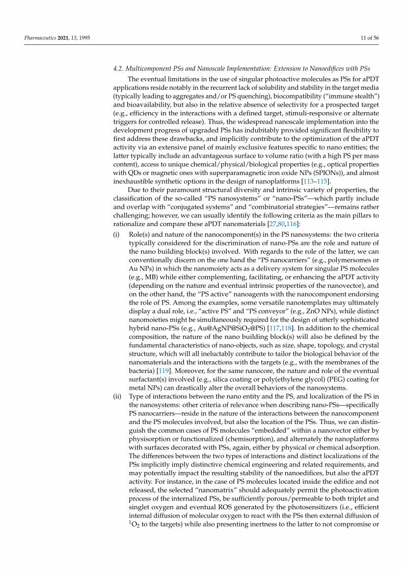
4.2. Multicomponent PSs and Nanoscale Implementation: Extension to Nanoedifices with PSs
The eventual limitations in the use of singular photoactive molecules as PSs for aPDTapplications reside notably in the recurrent lack of solubility and stability in the target media(typically leading to aggregates and/or PS quenching), biocompatibility (“immune stealth”)and bioavailability, but also in the relative absence of selectivity for a prospected target(e.g., efficiency in the interactions with a defined target, stimuli-responsive or alternatetriggers for controlled release). Thus, the widespread nanoscale implementation into thedevelopment progress of upgraded PSs has indubitably provided significant flexibility tofirst address these drawbacks, and implicitly contribute to the optimization of the aPDTactivity via an extensive panel of mainly exclusive features specific to nano entities; thelatter typically include an advantageous surface to volume ratio (with a high PS per masscontent), access to unique chemical/physical/biological properties (e.g., optical propertieswith QDs or magnetic ones with superparamagnetic iron oxide NPs (SPIONs)), and almostinexhaustible synthetic options in the design of nanoplatforms [113–115].
Due to their paramount structural diversity and intrinsic variety of properties, theclassification of the so-called “PS nanosystems” or “nano-PSs”—which partly includeand overlap with “conjugated systems” and “combinatorial strategies”—remains ratherchallenging; however, we can usually identify the following criteria as the main pillars torationalize and compare these aPDT nanomaterials [27,80,116]:
(i) Role(s) and nature of the nanocomponent(s) in the PS nanosystems: the two criteriatypically considered for the discrimination of nano-PSs are the role and nature ofthe nano building block(s) involved. With regards to the role of the latter, we canconventionally discern on the one hand the “PS nanocarriers” (e.g., polymersomes orAu NPs) in which the nanomoiety acts as a delivery system for singular PS molecules(e.g., MB) while either complementing, facilitating, or enhancing the aPDT activity(depending on the nature and eventual intrinsic properties of the nanovector), andon the other hand, the “PS active” nanoagents with the nanocomponent endorsingthe role of PS. Among the examples, some versatile nanotemplates may ultimatelydisplay a dual role, i.e., “active PS” and “PS conveyor” (e.g., ZnO NPs), while distinctnanomoieties might be simultaneously required for the design of utterly sophisticatedhybrid nano-PSs (e.g., Au@AgNP@SiO2@PS) [117,118]. In addition to the chemicalcomposition, the nature of the nano building block(s) will also be defined by thefundamental characteristics of nano-objects, such as size, shape, topology, and crystalstructure, which will all ineluctably contribute to tailor the biological behavior of thenanomaterials and the interactions with the targets (e.g., with the membranes of thebacteria) [119]. Moreover, for the same nanocore, the nature and role of the eventualsurfactant(s) involved (e.g., silica coating or poly(ethylene glycol) (PEG) coating formetal NPs) can drastically alter the overall behaviors of the nanosystems.
(ii) Type of interactions between the nano entity and the PS, and localization of the PS inthe nanosystems: other criteria of relevance when describing nano-PSs—specificallyPS nanocarriers—reside in the nature of the interactions between the nanocomponentand the PS molecules involved, but also the location of the PSs. Thus, we can distin-guish the common cases of PS molecules “embedded” within a nanovector either byphysisorption or functionalized (chemisorption), and alternately the nanoplatformswith surfaces decorated with PSs, again, either by physical or chemical adsorption.The differences between the two types of interactions and distinct localizations of thePSs implicitly imply distinctive chemical engineering and related requirements, andmay potentially impact the resulting stability of the nanoedifices, but also the aPDTactivity. For instance, in the case of PS molecules located inside the edifice and notreleased, the selected “nanomatrix” should adequately permit the photoactivationprocess of the internalized PSs, be sufficiently porous/permeable to both triplet andsinglet oxygen and eventual ROS generated by the photosensitizers (i.e., efficientinternal diffusion of molecular oxygen to react with the PSs then external diffusion of1O2 to the targets) while also presenting inertness to the latter to not compromise or

Pharmaceutics 2021, 13, 1995 12 of 56
quench the aPDT activity. Meanwhile, with surfaces of nano-objects decorated withPSs, the PSs may then contribute to some extent as an interface with the biologicalmedium or the target.
(iii) Biological impacts of the PS nanosystems: in addition to the biocompatibility andaPDT efficiency (including the critical concentrations just as the half-maximal effectiveconcentration EC50, minimum inhibitory concentration MIC, or 50% growth inhibitionconcentration GIC50), the eventual biodegradability, elimination process, or ecotoxicity ofthe aPDT nanomaterials can markedly vary from one system to another (based on factorssuch as composition and size/shape), but are rather difficult to evaluate or compare; ergo,these factors are not systematically addressed in the reports.
(iv) Relative sustainability of the nano-PSs for aPDT applications: the reproducibility,eco-friendliness, and cost-effectiveness parameters of the synthetic protocols andproduction of aPDT nanomaterials, as well as the ease of storage and use, and thestability over time are also ultimately to be evaluated for any system aiming to beviable and reasonably applied; however, similar to (iii), these parameters are complexand so scarcely investigated.
Thus, in the overview presentation of the different PS nanosystems hereafter, thechemical features (i) and (ii) have been conventionally defined as the main criteria for theclassification. Alternatively, the nature of the aPDT applications has also been used as themain criterion for classification in some references [120]. Another approach consists ofsystemizing all the nanosystems dedicated to a given PS (e.g., curcumin) [121].
Within the extensive collection of aPDT nanomaterials reported to date, the majoritybelongs to the category labeled as “PS nanocarriers” with the nanocomponent acting as adelivery system for PS molecules; however, increasing examples involving PS-active nanobuilding blocks have emerged as well. Overall, this multipurpose role of the nanomoi-ety may include avoiding aggregation (e.g., dimerization, trimerization) and correlatedPS quenching, enhancing “solubility” (i.e., dispersibility), stability and bioavailability,allowing “biological stealth”, on-demand release and target specificity, and ultimately trig-gering eventual synergistic aPDT activity with the complementary or ameliorative intrinsicproperties of the nanocomponent; although irrevocably confirmed in many nano-PSs, themechanisms involved in the synergy may differ from one system to another, and oftenremain partly or integrally unresolved due to the complexity of these tacit multiparametercontextures [113]. It is noteworthy that most systems comprise “classic”/”traditional”organic PSs (natural or synthetic, e.g., curcumin, MB) with fewer examples involving metal-lated PS molecules such as the recent review from Jain et al. dedicated to ruthenium-basedphotoactive metalloantibiotics [108]. Among aPDT nanomaterials, we can thus identifyvarious families of nanoplatforms based on the nature of the nanocore, starting here withthe inorganic vectors followed by the organic templates; as an indication, in the commoncases of “multi-component nano-PSs”, the classification has been defined hereafter ac-cording to the main/prominent nano building block involved in the composition, i.e.,metal-based systems, silicon-based systems, carbon-based systems, lipid-based systems,and polymer-based systems. Regarding the following presentation of aPDT nanosystems,it is important to specify that it is not exhaustive, but instead provides a panorama of themain categories of nanosystems—either colloids or surfaces [27,122]—and their relatedspecificities, with an emphasis on recent developments.
4.2.1. Metal-Based Systems
Metal-based nanostructures have been extensively investigated—both as “PS cargo”and PS active entities—through the exploration of the richness and diversity of the re-spective subcategories related to this class of compounds, as detailed below. Each of thebelow-mentioned inorganic classes presents distinct specificities of relevance for aPDTapplications, with a choice to be defined on a case-by-case basis according to the target, thenature of the PSs involved (with possible preferential affinity), or the anticipated comple-mental or synergistic role of the selected nano entity (based upon its chemical, physical,

Pharmaceutics 2021, 13, 1995 13 of 56
and/or biological features, with the eventual PS molecules located either at the surfaceof the NPs or within, when present). It is important to notice that the nanocores fromeach subcategory can be either further implemented, with silica or polymer coating forexample, or combined (multicomponent nano-PSs) in order to adequately optimize theefficiency of the systems [117]; however, the outcomes of these hybridity processes arecomplex to anticipate with systematic rationality, with either enhancement or quenching ofthe properties observed depending on the composition of the combinations.
Metal NPs
Metal NPs—mainly gold, but also silver—maybe considered among the “gold stan-dards” in nanomaterials through the history and expansion of nanosciences in terms ofdedicated publications and vastness of related possible applications [123]. As already welldocumented for Au and Ag NPs, the reasons are numerous and reside notably in theirrelatively easy accessibility with low-cost and highly reproducible (large scale) biogenicand chemical synthetic routes, in addition to the flexibility to finely tailor the propertiesvia a refined size and shape control (with narrow size distribution and diverse shapes),and the facility for functionalization with various types of molecules. Moreover, goldNPs display biocompatibility, low toxicity, and immunogenicity, almost chemical inertness(distinctly from their inherent catalytic properties), while silver nanomaterials present in-trinsic antimicrobial activity against a broad spectrum of microorganisms and related MDRinfections (e.g., towards Gram(−) and Gram(+) mature biofilms of MRSA), and disruptionof biofilm formations while being safe for mammalian cells [124–126].Ultimately, both Auand Ag NPs share nano features specific to noble metal systems, i.e., localized surfaceplasmon resonance (LSPR, arising from their resonant oscillation of their free electronsupon light exposure) and resonance energy transfer (RET), with subsequent optical andphotothermal properties of enhancing appositeness in aPDT applications and PDI efficacy(e.g., ROS production) [127–129]. For the most part, Au and Ag NPs of various shapes (e.g.,spheres, rods, cubes) are combined with organic PS molecules such as RB and MB [128–136],but also with metallated PSs such as ruthenium complexes, metallophthalocyanines, andmetalloporphyrins [108,137,138]; the corresponding PS nanovehicles can also be labeledas “conjugates” but they strictly differ from the “mixtures” involving metal NPs and PSmolecules [139]. Other noble metal NPs, viz., platinum, have also been employed in aPDTapplications due to their multitarget action to inactivate microbes, although to a lowerdegree up to now owing to synthetic limitations [27,140]. Alternately, redox-active cop-per NPs are typically less costly and easier to access and present unique features amongwhich the faculty to generate oxidative stress to various microbes through the genesis ofROS [141], such as in the recently developed copper–cysteamine (Cu–Cy) nano-PS thatcan be activated either by UV, X-ray, microwave, or ultrasound, to produce ROS againstcancer cells and bacteria [142,143]. More unwontedly, approaches to treat subcutaneousabscesses lead to the use of acetylcholine (Ach) ruthenium composite NPs (Ach@RuNPs) asan effective appealing PDT/PTT dual-modal phototherapeutic killing agent of pathogenicbacteria, with Ach playing a role in targeting the bacteria and promoting the entry into thebacterial cells [108]. While belonging to the same category, each metal displays particularspecificities; consequently, with the objective of optimization, nano-PSs resulting from al-loys or multimetallic NPs have been further designed such as the Au@AgNP@SiO2@PS andAA@Ru@HA-MoS2 (AA: ascorbic acid, HA: hyaluronic acid) nanocomposites [117,118].
Metal Oxides
Similar to gold and silver, metal oxides such as iron oxide, titanium oxide and zincoxide have been cornerstone contributors in the global evolution of applied nanomaterials(particularly in medicine), due notably to intrinsic magnetic and optical nanoscale features,with the latter typically available at “room temperature” and commonly finely tunablevia shape, size, and crystal structure parameters [144]. Indeed, specific single-domainsuperparamagnetic iron oxide NPs (SPIONs)—either magnetite (Fe3O4) or maghemite

Pharmaceutics 2021, 13, 1995 14 of 56
(γ-Fe2O3) of various shapes and sizes, including ferrite or doped derivatives—exhibit anoutstanding magnetization behavior with no remanent or coercive responses upon expo-sure to a magnetic field. As a result, such magnetic NPs have legitimately generated interestand use as magnetic resonance imaging (MRI) and magnetic particle imaging (MPI) agents,as well as magnetic fluid hyperthermia (MFH), magnetic cell separators [145], or drugdelivery conveyers with the possibility to guide the NPs to the targeted area via externalmagnetic fields [146]. More recently, iron oxide nano-objects proved to be also of perti-nence for aPDT applications not only as a magnetic “nanocargo” for various organic andinorganic PSs such as curcumin, MB, ICG, BODIPYs, porphyrins, metallophtalocyanines,or ruthenium derivatives among others [27,108,147–155], but also with peroxidase-likeactivity to enhance the cleavage of biological macromolecules for biofilm elimination [156].Extension in the design of more elaborate multicomponent architectures involving hybridiron oxide nanocore led inter alia to Ag/Fe3O4, Ag/CuFe2O4, CoFe2O4, and Fe3O4/MnO2NPs conjugated with different PSs [27,117,147,150,151,157–160].
Other oxides have also drawn heavy attention, in particular zinc oxide and titaniumoxide, as the photophysical properties of these wide bandgap semiconducting nanoma-terials efficiently translate into a multi-level antimicrobial activity including PS vesseland/or PS active agent (with possible coupled aPDT response), and/or membrane dis-ruptor [161–164]. Thus, ZnO and TiO2 nanoplatforms possess the ability to alter microbes’integrity—through alternate mechanisms involving ROS and/or metallic ions—in the darkor via photoactivation [165]. The latter is customarily triggered by UV or X-ray irradia-tion, with the eventual possibility to adequately shift to other wavelengths such as visiblelight irradiation in virtue of diverse modification methods of the oxides encompassingnotably: doping or surface alteration (e.g., F-doped ZnO, coatings or oxygen deficiency),coupling with other bandgap semiconductors (e.g., ZnO/TiO2) or sensitizing dyes, andcomposites [121,156,164,166–171]. Furthermore, beside the size and shape of the NPs, thecrystallographic phase appears to be a tuning parameter of particular importance for theantimicrobial effects of some oxides, especially for TiO2 with the distinction between theanatase, rutile, and brookite structures [172–175]. Although reported to a lesser extent, thelist of alternate oxides exhibiting potential in aPDT applications comprises CuO/Cu2O,MnO2, and rare earth oxides to mention just a few [141,176–180].
QDs and Metal Chalcogenide Nanomaterials
Aside from zinc/titanium oxides, distinct semiconductors such as QDs and metalchalcogenide (e.g., metal sulfide) nanomaterials (involving elements from different groupsin the periodic table) proved to be efficient disruptors against various multi-drug-resistantmicroorganisms. Due to their smaller size of a few nanometers (ca. up to 10 nm), QDsdiffer from other nano-objects with physical and optoelectronic properties governed by therules of quantum mechanics, high chemical stability and resistance to photobleaching, andnear-infrared (NIR) emission (typically above 700 nm) notably allowing for deep-tissueimaging [181]. Appositely comparable, metal chalcogenide likewise reveals unorthodoxphysio and physicochemical properties, accordingly garnering a legit interest for antimi-crobial applications [182]. Consequently, not only can these nano building blocks carryPS molecules and alter the integrity of microbial walls/membranes or gene expression,but they may as well act as PSs; when coupled with other PSs, synergistic interactionsin the QD-PS edifices might occur resulting from mechanisms such as Förster resonanceenergy transfer (FRET, non-radiative energy transfer from QD donors to PS acceptors) togenerate free radicals and ROS. Among examples of such QDs and metal chalcogenideaPDT systems can be cited CdTe QDs and related CdTe-PS conjugates, CdSe/ZnS QDscombined with PSs, InP and InP-PS, Mn-doped ZnS, MoS2, but also CuS and CdS nanocrys-tals, with ultimately hybrid systems involving for instance CoZnO/MoS2 or AgBiS2–TiO2composites [123,183–196].

Pharmaceutics 2021, 13, 1995 15 of 56
Metal–Organic Framework (MOF) Nanoscaffolds, Upconversion Nanomaterials and OtherMetal Ion Nanostructures
Although less investigated than the above-mentioned alternatives, other originalmetal-based nanostructures identified as MOF nanostructures, upconversion nanoplat-forms, and alternate metal ion nanomaterials tend to further consolidate their promisingpotential for aPDT applications. Considering their towering surface area and porousordered structure with substantial loading capacity (e.g., adsorption of O2 and ensuingphotocatalytic production of 1O2 via a heterogeneous process), stable versions of col-loidal nano-MOFs—or less common covalent organic frameworks (COFs), i.e., reticulationvariants typically defined by non-metal “nodes” instead of metal ones—have emergedas efficient heterogeneous photosensitizers—with frameworks acting as PSs or entrap-ping PSs—towards antimicrobial applications (e.g., enhanced penetration for bacterialbiofilms eradication), with porphyrin-based or porphyrin-containing MOFs and COFs,or Cu-based MOFs embedded with CuS NPs for rapid NIR sterilization among recentlyreported solutions [197–205].
Moreover, upconversion NPs (UCNPs) generally involve actinide- or lanthanide-doped transition metals and refer to the nonlinear process of photon upconversion, viz., asequential absorption of two or more photons resulting in an anti-Stokes type emission (i.e.,emission of light at a shorter wavelength than the excitation wavelength); when translatedinto biomedical context, UCNPs can be typically activated by NIR light—characterized bydeeper tissue penetration and reduced autofluorescence, phototoxicity, and photodamagewhen compared with UV or blue light—and produce high energy photons for optical imag-ing or more recently aPDT when combined with PSs [206–209]. Intrinsically limited by lowupconversion quantum yield, the current focus consists of developing hybrid UCNPs to im-prove aPDT efficiency; thus, auspicious progress has been achieved with examples such as{UCNPs (NaYF4:Mn/Yb/Er)/MB/CuS-chitosan)} multicomponent nanostructured systemrevealing a superior antibacterial activity with the UCNPs enhancing the energy transferto MB, the CuS triggering synergistic PDT/PTT effects, and chitosan assuring stability andbiocompatibility [156]. Other examples also include silica coating β-NaYF4:Yb, Er@NaYF4UCNPs loaded with MB as PS and lysozyme as a natural protein-inducing bacterial au-tolysis, Fe3O4@NaGdF4:Yb:Er combined with the photo/sonosensitizer hematoporphyrinmonomethyl ether (HMME), UCNPs@TiO2, N-octyl chitosan (OC) coated UCNP loadedwith the photosensitizer zinc phthalocyanine (OC-UCNP-ZnPc), or the UCNPs-CPZ-PVPsystem (CPZ: β-carboxy-phthalocyanine zinc, PVP: polyvinylpyrrolidone) to name but afew [206,210–213].
Alternatively, more disparate metal-ion aPDT systems have been reported such as PSencapsulated dual-functional metallocatanionic vesicles against drug-resistant bacteria in-volving copper-based cationic metallosurfactant, or self-assembled porphyrin nanoparticlePSs ZnTPyP@NO using zinc meso-tetra(4-pyridyl)porphyrin (ZnTPyP) and nitric oxide(NO) [214–216].
4.2.2. Silicon-Based NPs
This category will be divided hereafter into two main subclasses—porous silicon(pSi) and (mesoporous) silica (SiO2)—which differ in the oxidation state of the siliconand display distinctive properties, more specifically different quenching behavior andphotodynamic activity (singlet oxygen quantum yield under irradiation).
Porous silicon NPs (pSi NPs) are among the most promising types of inorganic nanocar-riers for biomedical applications and have been intensively investigated since the firstpublication by Sailor et al. in 2009 regarding their application for in vivo treatment ofovarian cancer. Composed of pure silicon, pSi NPs indeed exhibit relevant features encom-passing not only pores with large capacity for drug loading combined with specific surfacearea allowing for implemental functionalization, but also degradability in an aqueousenvironment, and biocompatibility [217]. Moreover, porous silicon particles are known tobe photodynamically active with related inherent antimicrobial properties by generation

Pharmaceutics 2021, 13, 1995 16 of 56
of ROS under irradiation with light of a specific wavelength [218,219]. Because of the lowquantum yield of singlet oxygen production from porous silicon itself, particles can begrafted with additional PSs, such as porphyrins, to enhance the yield of singlet oxygen gen-eration and thereby antimicrobial properties for PDT applications [219,220]. Consequently,several silicon-based systems have been reported in recent years, mainly for PDT applica-tions [221,222]. Furthermore, pSi NPs display intrinsic fluorescence, which can be appliedfor imaging and real-time diagnostics regardless of any surface functionalization [220,223].
As previously mentioned, pSi has a low singlet oxygen quantum yield due to quench-ing, which makes silica particles in comparison a more suitable substitutional systemcombining similar biocompatibility with improved optical properties. On the other hand,one of the pivotal advantages to use silica (SiO2) conjugates with PSs is to achieve a better“solubilization”—or more accurately, dispersibility—of hydrophobic dyes and a better pho-tostabilization, thus limiting the self-photobleaching of PSs. Further advantages to nameas the most important features of silica are high biocompatibility, antimicrobial properties,and high surface area for mesoporous silica that can be synthesized easily from commer-cially available precursors [27,224,225]. Furthermore, SiO2 exhibits an effective PS-graftingcapacity [226]; the latter can be accomplished via adsorption, covalent bonding, binding tothe hydroxyl groups from silica surface, and entrapment during formation in silica particlesor matrix [27]. Recently, Dube et al. reported about the photo-physicochemical behavior ofsilica NPs with (3-aminopropyl)triethoxysilane (APTES), and subsequently PS-modifiedsurfaces for aPDT [150]. In addition, silica coatings have also been reported to preventthe degradation of nanocarriers (magnetite) and prolong the stability and functionality ofPS systems [227]. Interestingly, coupling PSs to silica or Merrifield resin leads to distinctadvantages; indeed, immobilized Ce6 notably displays significantly higher aPDT efficiencyin comparison with the free form, which is probably due to an enhancement of the ad-hesion of PSs to bacterial cells resulting in a stronger cell wall disorganization [228,229].Unsurprisingly, many approaches with encapsulated PSs in silica NPs for potential aPDTapplications have been reported in recent years [229–232].
Distinctly, combinatory approaches involving other nanocomponents, such as silica-containing core-shell particles or silica-coated inorganic NPs, have emerged to furtherimplement the properties of silica with the specific features (e.g., magnetic, photoactive,or antimicrobial properties) from other nanomaterials of relevance [118]. Thus, since thesurface of silica can be easily grafted with PSs and is highly biocompatible, the surfacemodification of silica particles using metallic NPs (e.g., Ag NPs) to enhance the antibacterialphotodynamic activity, has been developed for improved aPDT [233] while combinationswith carbon quantum dots have been assessed for imaging-guided aPDT [234]. Anotherexample refers to sufficient aPDT/PDI systems, more precisely to mesoporous silica-coatedNaYF4:Yb:Er NPs with the PSs (silicon 2,9,16,23-tetra-tert-butyl-29H,31H-phthalocyaninedihydroxide) loaded in the silica shell to enhance bacterial targeting of E. coli and S. au-reus [235].
In addition to silica-based NPs or silica-containing core-shell particles, lesser-knownsilica nanofibers also proved to be suitable substrates for potential aPDT, PDT, and PDIapplications. As an illustration, Mapukata et al. [236] recently reported silver NP-modifiedsilica nanofibers with embedded zinc phthalocyanine as PS for aPDT applications. Thenanofiber-based substrates offer the advantage of fast removal after application, which canallow limiting any dark toxicity [236–238].
Silica NPs and fibrous or dendritic fibrous nanosilica have also been reported for theformation of nanocomposites to create antimicrobial photodynamically active surfaces foraPDT or PDI; to create such surfaces, silica NPs can be embedded into polymeric matricesfor enhanced biocompatibility and complementary surface properties from the selectedpolymer [239].
Additionally, silica substrates and nanoconjugates offer a suitable platform for combi-natory approaches since they can be easily modified. For example, Zhao et al. describedpolyelectrolyte-coated silica NPs modified with Ce6 [240]. These complexes could be

Pharmaceutics 2021, 13, 1995 17 of 56
extracted, by bacteria, from silica NPs to form stable binding on the bacterial surface,changing the aggregation state of Ce6 and leading to both the recovery of PS fluorescenceand 1O2 generation. Such bacteria-responsive multifunctional nanomaterials allowed forsimultaneous sensing and treating of MRSA. Another approach is illustrated by the photo-induced antibacterial activity of amino- and mannose-decorated silica NPs loaded with MBagainst E. coli and P. aeruginosa strains [241]. The modification of silica substrates with mannoseled to an increased targeting of P. aeruginosa and reduced dark toxicity of the systems.
4.2.3. Carbon-Based Nanomaterials
Akin to silicon-based nano-objects, the notable diversity of allotropic customizablecarbon-based nanostructures—either conveyers for traditional PS molecules or intrinsicallyPS active—legitimizes their distinct consideration in the actual classification of nano-PSs [242–244], with the following subcategories.
Fullerenes, Carbon Nanotubes (CNTs), and Nanodiamonds
In the evolution of carbon-based nano-objects, fullerenes and carbon nanotubes deriva-tives may be chronologically introduced as the “first generation”. Discovered in the mid-1980s, the proper Cn (n = 60–100) spheroidal “soccer ball” π-conjugated structures ofthe fullerenes yield tremendous chemical modularity and electrochemical and physicalproperties, including photostability, the propensity to act as a PS via Type I or Type IIpathways (Figure 1A) with high ROS quantum yield, and oxygen-independent photo-killing by electron transfer. Despite their intrinsic hydrophobicity typically requiringsurface functionalization for biocompatibility and related dispersibility, they have proveneven nowadays their effectiveness as broad-spectrum photodynamic antimicrobial agents,with photoactive antimicrobial coating based on a PEDOT-fullerene C60 polymeric dyad(PEDOT: poly(3,4-ethylenedioxythiophene)), BODIPY-fullerene C60, diketopyrrolopyrrole–fullerene C60, and cationic fullerene derivatives among recent examples [80,92,156,245–251].Mainly developed a few years later, carbon nanotubes (CNTs)—either single wall CNTs(SWCNTs) describable simply as a single-layer sheet of a hexagonal arrangement of hy-bridized carbon atoms (graphene) rolled up into a hollow cylindrical nanostructure, ormultiwall CNTs (MWCNTs) consisting of nested SWCNTs—unveil both independent ca-pacities to produce ROS upon irradiation and high surface area for decoration with PSmolecules [156]. Neoteric specimen of PS-CNTs encompass toluidine blue, polypyrrole,malachite green, MB, RB, and porphyrins [156,252–257]. Although purportedly older sinceit was discovered in the 1960s, diamond NPs or nanodiamonds seemingly remain the lesserknown carbon-based nanomaterials to date; nevertheless, the latter dispose of legit aPDTarguments with their fluorescence, photostability, proclivity for conjugation with diversePSs such as porphyrins or metallated phthalocyanines and silver NPs, but also inherentantibacterial activity [120,258–262].
Carbon QDs (CQDs)
As the next momentum in the blossoming of “nano-carbon” era, the carbon QDs werediscovered in the early 2000s [263,264]. The physical and chemical properties of thesefluorescent particles, commonly quasi-spherical with less than 10 nm in diameter, canbe finely tuned upon size/shape variations or doping with heteroatoms (e.g., B, N, O, P,S) [263,265]. By virtue of their biocompatibility and dispersibility, photostability, low toxic-ity and related eco-friendliness, good quantum yield and conductivity, CQDs have beeninvestigated for various applications, and more recently as antimicrobial agents; withal,their environment-friendly features combined with low cost and rather ecological biogenicor synthetic routes (from natural or synthetic precursors) place them advantageously asa viable scalable photocatalytic disinfection material compared with alternate nano-PSs.Late cases involve doped or hybrid CQDs or more conventional conjugates of CQDs withPSs [121,266–271].

Pharmaceutics 2021, 13, 1995 18 of 56
Graphene, Graphene QDs (GQDs), and Graphene Oxide (GO) Nanostructures
In a similar timescale to CQDs is the quantum leap discovery and blooming ofgraphene and graphene oxide materials. Graphene can be defined as a 2D allotropeof carbon, more accurately a monolayer of atoms with a hexagonal lattice structure (orsingle-layered graphite) and identifiable as the “building block” for the discrete fullerenes,1D carbon nanotubes, and 3D graphite. Despite its stunning mechanical/electronic prop-erties and chemical inertness, the limitations of graphene, such as zero bandgap and lowabsorptivity, lead to the ulterior conversion of the 2D graphene into “0D” GQDs [272,273].Due to quantum confinement and edge effects, GQDs exhibit different chemical and physi-cal properties when compared with other carbon-based materials, as well as a non-zerobandgap, good dispersibility, and propensity for functionalization and doping. Structurally,GQDs differ from CQDs because they comprise graphene nanosheets with a plane size lessthan 100 nm [272,274]. Likewise, graphene oxide (GO) is the oxidized form of graphenei.e., a single atomic sheet of graphite with various oxygen-containing moieties either on thebasal plane or at the edges. Meanwhile, reduced graphene oxide (rGO) can be summarilydescribed as an “intermediate” structure between graphene and GO, with variable andhigher C/O elemental ratios compared with GO, but remaining residual oxygen and struc-tural defects with reference to the pristine graphene structure. Although GO was reporteda couple of centuries ago, GO and rGO nanomaterials have mainly emerged for variousapplications after the discovery of graphene since GO is a precursor to prepare graphene,and both present distinctive physical and chemical properties that differ from graphene.As a result, countless and rising fast illustrations of graphene derivatives for antibacterialapplications are regularly reported [121,275–280].
4.2.4. Lipid-Based Systems
Due to their amphiphilic nature (typically hydrophilic “head” and hydrophobic “tail”),some lipids—natural and synthetic—have been extensively studied to develop efficientbiocompatible delivery systems—initially for drugs and DNA/RNA, but also for aPDTPSs—with synthetic flexibility and structural diversity. Among the prevalent examples, wecan distinguish the micelles (lipid monolayers with polar units at the surface and hydropho-bic core) and the liposomes (one or more concentric lipid bilayer with a hydrophilic surfaceand an internal aqueous compartment). Although sharing similar chemical constituents,the micelles and liposomes present significant differences to be taken into considerationdepending on the intended application (nature of the target) and the nature of the PSs.Indeed, the micelles are typically smaller than liposomes (with a diameter starting from afew nanometers for the micelles, and ca. 20 nm for the liposomes), with distinct stabilityand permeability in biological medium and uptake pathways for the PSs into bacteria.With reference to the nature of the transported PSs, the liposomes display the additionalflexibility to carry both hydrophilic PSs (in the core compartment or between the bilayers)or/and hydrophobic PSs (within the lipid bilayer), while the micelles are usually easierand cheaper to prepare [80,156]. As often critical to address for biomedical applications,the surface charge of these nano-objects can be tailored to further optimize the interactionswith the bacteria, with cationic modification of liposomes identified as a promising aPDTefficiency “amplifier” [80,156,281]. In addition to recent examples such as the hypericinloaded liposomes against Gram(+) bacteria [282–286], another emerging and promisingalternative includes the development of modified liposome-like derivatives labeled ei-ther as “ethosomes”, “transfersomes”, or “invasomes”, which can be briefly described asultra-deformable vesicular carriers with upgraded transdermal penetration and increasedpermeability into the skin for the PSs compared with conventional liposomes [287–290].On the other hand, recently reported aPDT micellar systems refer to micelles loaded withvarious hydrophobic PSs such as curcumin, BODIPY, porphyrins, hypocrellin A, or hy-pericin among others [121,291–293]. Furthermore, solid lipid NPs (SLN) composed ofsolid biodegradable lipids have been recently highlighted as delivery systems used foractual mRNA COVID-19 vaccines [294], but they also have been reported as transporters

Pharmaceutics 2021, 13, 1995 19 of 56
for curcumin for the treatment of oral mucosal infection [121]. Besides, we may alsoinclude nanoemulsions in this category of lipid-based nanovehicles since the latter con-ventionally involve lipids in the oily phase during the formulation process, with recentcurcumin/curcuminoid nanoemulsions [121,295].
4.2.5. Polymer-Based Systems
In direct correlation with the above-mentioned lipid-based systems, polymer-basednanocarriers have been positioned as a logical extension with the objective to implementa “degree of freedom” in the synthesis flexibility while expanding the panel of buildingblocks available in the design of aPDT nanostructures. A categorization of polymericsystems for the delivery of PSs to therapeutically relevant sites can be done using as criteriaeither the nature of the polymer(s) involved or the type of polymeric (nano)structures.Thus, in the following, we will use a classification primarily based on the structure of thepolymeric systems with differentiation between NPs (including hydrogels, biopolymers,and aerogels), polymersomes, polymeric micelles, dendrimers, fibers, and polymeric filmsand layers (including hybrid systems and nanocomposites). The nanostructure of polymericsystems is implicitly highly correlated to the molecular structure, i.e., composition (natureof the polymer(s), ratios, and distribution and amount of hydrophilic and hydrophobicmoieties), charge, and size, as reviewed recently by Osorno et al. [296]. The refined controlof these parameters enables the design and fine-tuning of specific polymeric nanostructuresspanning from long-ranged ordered lamellar sheets, tubes, and fibers to oval or sphericalparticles, micelles, and polymersomes [296] (Figure 2C).
Conjugated Polymers as PSs or Polymer-Functionalized PSs
One of the easiest approaches reported to develop polymeric carrier systems is toenhance water solubility and biocompatibility of PS molecules through the use of function-alized polymers, by introducing the PSs in a post-modification reaction to a hydrophilicand/or biocompatible polymer, or to modify the PS with a polymerizable group (e.g.,acrylate) to react as a monomer for further polymerization with suitable monomers. Otherapt options are conjugated polymers incorporating a backbone with alternating doubleand single bonds which provide photodynamic and optical properties, and thereforemight act as PSs themselves [297,298]. For example, poly[(9,9-bis{6′-[N-(triethylene glycolmethyl ether)-di(1H-imidazolium)-methane]hexyl}-2,7-fluorene)-co-4,7-di-2-thienyl-2,1,3-benzo-thiadiazole] tetrabromide (PFDBT-BIMEG) is a conjugated polymer which affordssalt bridges and electrostatic interactions with microorganisms; these interactions enablethe simultaneous detection and inhibition of microorganisms [297]. Conjugated polymerswere intensively investigated for the development of multifunctional NPs in antibacterialapplications because of their generally low-toxicity toward eukaryotic cells, their flexibil-ity, and their high potential to vehicle versatile therapeutic molecules [298]. ImprovedPSs based on conjugated polymers or polymerized PSs with antimicrobial photodynamicproperties have been amply reported in recent years [299–303]. For example, Huanget al. demonstrated the efficiency and selectivity of polyethyleneimine-Ce6 conjugated inpotential aPDT/PDI applications [304].
Other polymeric systems also show antimicrobial properties or preferential targetinfection sites and are thus suitable for aPDT applications when combined with PSs. For ex-ample, cationic polymers, which are known to be highly hydrophilic, can be used to targetcell walls and thus microbial infections. Hence, such polymers are adequate for potentialaPDT applications [305,306]. Exemplary amphiphilic or cationic poly(oxanorbornene)sdoped with PSs, which exhibit pronounced antimicrobial activity (99.9999% efficiency)against E. coli and S. aureus strains, have been reported [307]. By contrast, anionic polymerparticles and nanocarriers with negatively charged surfaces and membranes invade thereticuloendothelial system, which leads to elongated blood circulation times [308–310].Polymers with large fractions of functional groups, such as the biopolymers mentionedabove, can be easily modified and offer many loading sites for suitable PSs. Thus, among

Pharmaceutics 2021, 13, 1995 20 of 56
many suitable materials, hyperbranched and dendrimeric polymers are particularly promis-ing candidates for PS nanocarriers.
Dendrimers
Dendrimers, dendrimeric polymers, or dendrons are highly ordered and highlybranched polymers, which may form spherical three-dimensional structures with diam-eters typically ranging from 1–10 nm [311]. The dimensions of dendrimers are relativelysmall compared with other drug delivery systems, but as they consist of individual well-defined molecules, the drug loading can be subtly established with reproducibility. Theincorporation or encapsulation of drugs may be achieved by covalent binding of the PSsor drug molecules to reactive functional groups along the dendrimeric structure [312,313].Alternatively, the PS or drug molecule may act as a scaffold from which the dendrimeris synthesized. Finally, drug or PS encapsulation may occur inside the voids of the den-drimer [311,314,315]. Thus, dendrimers offer a substrate for PSs with many reaction sites,and controllable sterical and hydrophobic properties depending on the backbone of thedendrimer. Approaches using hyperbranched polymers for stimuli-responsive releaseof PSs, such as porphyrins, under acidic or reductive conditions, with improved target-ing of bacterial sites have been reported, such as mannose-functionalized polymers byStaegemann et al. [316,317].
Polymeric NPs and Nanocomposites
In many cases, polymeric nano-systems are not clearly classified, but instead typicallygrouped under the terminology “polymeric NPs”, since the exact structure may not befully characterized or considered less relevant with respect to the application efficiency.Polymeric NPs are commonly defined as physically or chemically crosslinked polymernetworks with a size in the range of 1 to 1000 nm, but if not further defined may also includenanocapsules, such as polymersomes, micelles, chitosomes, and even highly branchedpolymers and dendrimers [318].
In aPDT, polymeric NPs may be either used to encapsulate PS molecules (loadingwith PSs), or built from inherent photoactive polymers acting as PSs [303]. Various PSmolecules (e.g., RB, porphyrin derivatives, or curcumin) encapsulated in biocompatiblepolymeric NPs, such as polystyrene-, PEG-, polyester- (including poly-beta-amino esters),or polyacrylamide-based NPs, are widely used in aPDT/PDI approaches, as recentlyreported [319–321]. In addition to the role of nanovehicle, the polymeric particles mayalso help to address the lack of solubility in the biological medium from the PSs and/orreduce their toxicity, as reported among others by Gualdesi et al. [320,322]. For instance,polystyrene NPs with encapsulated hydrophobic TPP-NP(5,10,15,20-tetraphenylporphyrin)have been reported as nano-PS for efficient aPDI approaches towards multi-resistant bacte-ria [321]. In addition, biocompatible PLGA (polylactic-co-glycolic acid) NPs were employedto encapsulate curcumin, which could potentially serve as an orthodontic adhesive an-timicrobial additive [322]. Alternatively, combinatory approaches such as polymeric NPsmerging a polymeric PS, a photothermal polymeric agent, and poly(styrene-co-maleic anhy-dride) as dispersants have recently been reported for coupled aPDT/PTT nano-platformsin aqueous media [323,324]. Furthermore, Kubát et al. reported an increase in the stabilityof physically crosslinked polymeric NPs (polystyrene) to comparable nanocapsules (PEG-poly(ε-caprolactone) (PCL) micelles) equipped with identical PS [319].
The term polymeric NP also includes NPs obtained from natural polymers (e.g.,chitosan and alginate), hydrogels, or aerogels. Hydrogels are chemically or physicallycrosslinked polymeric networks, which are able to absorb large amounts of water due totheir pronounced hydrophilicity, but do not dissolve because of the crosslinking, whereasaerogels are formed by replacement of liquid with a gas resulting in low-density polymericstructures [325]. Recently, Kirar et al. reported PS-loaded biodegradable NPs fabricatedfrom gelatin, a naturally occurring biopolymer, and hydrogel, for application in aPDT [326].Moreover, also recently, some polymers such as Carbopol-forming hydrogel matrix en-
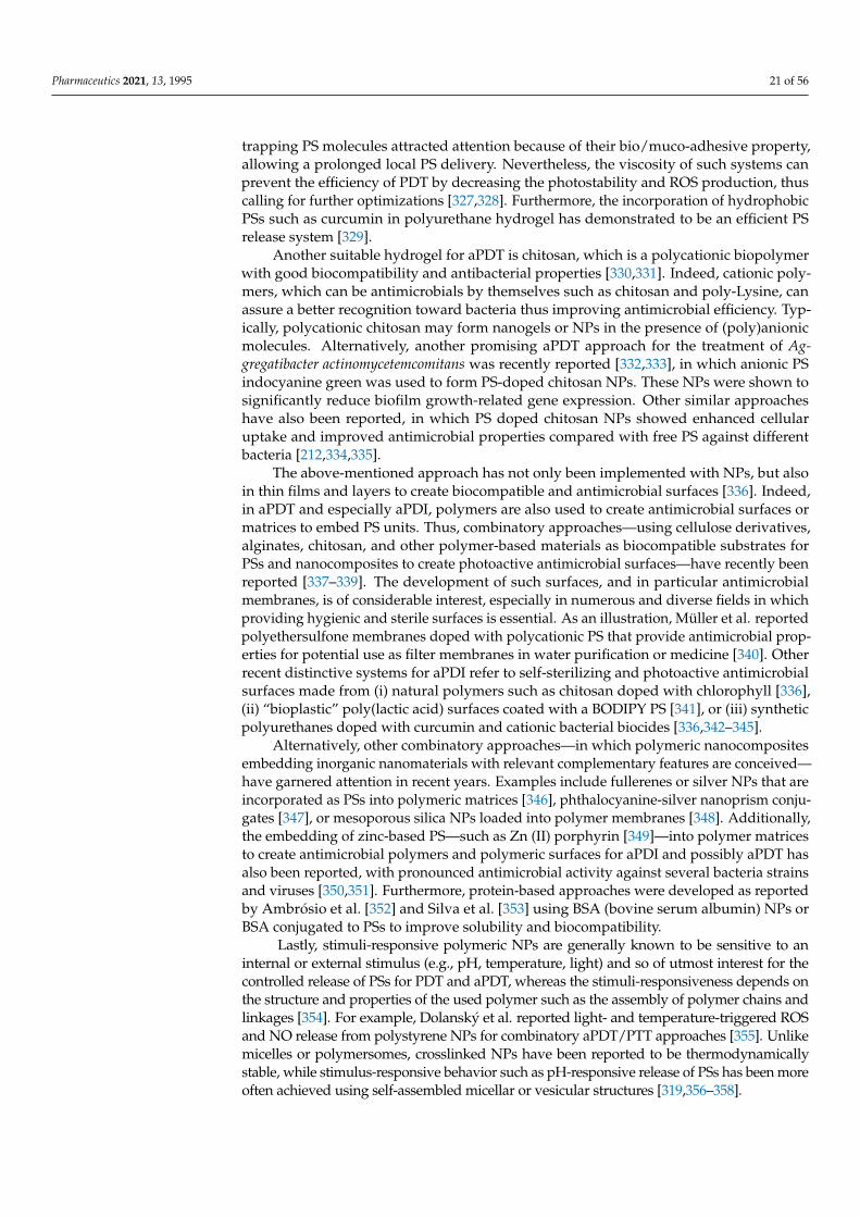
Pharmaceutics 2021, 13, 1995 21 of 56
trapping PS molecules attracted attention because of their bio/muco-adhesive property,allowing a prolonged local PS delivery. Nevertheless, the viscosity of such systems canprevent the efficiency of PDT by decreasing the photostability and ROS production, thuscalling for further optimizations [327,328]. Furthermore, the incorporation of hydrophobicPSs such as curcumin in polyurethane hydrogel has demonstrated to be an efficient PSrelease system [329].
Another suitable hydrogel for aPDT is chitosan, which is a polycationic biopolymerwith good biocompatibility and antibacterial properties [330,331]. Indeed, cationic poly-mers, which can be antimicrobials by themselves such as chitosan and poly-Lysine, canassure a better recognition toward bacteria thus improving antimicrobial efficiency. Typ-ically, polycationic chitosan may form nanogels or NPs in the presence of (poly)anionicmolecules. Alternatively, another promising aPDT approach for the treatment of Ag-gregatibacter actinomycetemcomitans was recently reported [332,333], in which anionic PSindocyanine green was used to form PS-doped chitosan NPs. These NPs were shown tosignificantly reduce biofilm growth-related gene expression. Other similar approacheshave also been reported, in which PS doped chitosan NPs showed enhanced cellularuptake and improved antimicrobial properties compared with free PS against differentbacteria [212,334,335].
The above-mentioned approach has not only been implemented with NPs, but alsoin thin films and layers to create biocompatible and antimicrobial surfaces [336]. Indeed,in aPDT and especially aPDI, polymers are also used to create antimicrobial surfaces ormatrices to embed PS units. Thus, combinatory approaches—using cellulose derivatives,alginates, chitosan, and other polymer-based materials as biocompatible substrates forPSs and nanocomposites to create photoactive antimicrobial surfaces—have recently beenreported [337–339]. The development of such surfaces, and in particular antimicrobialmembranes, is of considerable interest, especially in numerous and diverse fields in whichproviding hygienic and sterile surfaces is essential. As an illustration, Müller et al. reportedpolyethersulfone membranes doped with polycationic PS that provide antimicrobial prop-erties for potential use as filter membranes in water purification or medicine [340]. Otherrecent distinctive systems for aPDI refer to self-sterilizing and photoactive antimicrobialsurfaces made from (i) natural polymers such as chitosan doped with chlorophyll [336],(ii) “bioplastic” poly(lactic acid) surfaces coated with a BODIPY PS [341], or (iii) syntheticpolyurethanes doped with curcumin and cationic bacterial biocides [336,342–345].
Alternatively, other combinatory approaches—in which polymeric nanocompositesembedding inorganic nanomaterials with relevant complementary features are conceived—have garnered attention in recent years. Examples include fullerenes or silver NPs that areincorporated as PSs into polymeric matrices [346], phthalocyanine-silver nanoprism conju-gates [347], or mesoporous silica NPs loaded into polymer membranes [348]. Additionally,the embedding of zinc-based PS—such as Zn (II) porphyrin [349]—into polymer matricesto create antimicrobial polymers and polymeric surfaces for aPDI and possibly aPDT hasalso been reported, with pronounced antimicrobial activity against several bacteria strainsand viruses [350,351]. Furthermore, protein-based approaches were developed as reportedby Ambrósio et al. [352] and Silva et al. [353] using BSA (bovine serum albumin) NPs orBSA conjugated to PSs to improve solubility and biocompatibility.
Lastly, stimuli-responsive polymeric NPs are generally known to be sensitive to aninternal or external stimulus (e.g., pH, temperature, light) and so of utmost interest for thecontrolled release of PSs for PDT and aPDT, whereas the stimuli-responsiveness depends onthe structure and properties of the used polymer such as the assembly of polymer chains andlinkages [354]. For example, Dolanský et al. reported light- and temperature-triggered ROSand NO release from polystyrene NPs for combinatory aPDT/PTT approaches [355]. Unlikemicelles or polymersomes, crosslinked NPs have been reported to be thermodynamicallystable, while stimulus-responsive behavior such as pH-responsive release of PSs has been moreoften achieved using self-assembled micellar or vesicular structures [319,356–358].

Pharmaceutics 2021, 13, 1995 22 of 56
Polymersomes
Polymersomes are polymeric vesicles that resemble liposomes, which were previouslydescribed in Section 4.2.4 dedicated to lipid-based systems. Polymersomes are formedfrom amphiphilic copolymers, which self-assemble in aqueous media, resulting in capsules.The lumen of the polymersomes is filled with an aqueous medium and the wall comprisesa hydrophobic interior with a hydrophilic corona on both inner and outer interfaces.Polymersomes typically form when the weight fraction of the hydrophilic parts (e.g., PEG ina PEG-b-poly(lactic acid) (PLA) block copolymers) comprises up to 20–40% of the polymer.They also form at comparatively large water fractions in the solvent shift method [359–361].At higher weight fractions, the system tends to form micellar structures [296,362,363]. Thehydrophilic and hydrophobic compartments inside the polymersomes enable the uptakeand encapsulation of both hydrophobic and hydrophilic molecules [364–367], makingpolymersomes a relevant candidate for the development of advanced nanocarriers forPSs [368]. In aPDT and similar approaches, efficient delivery of the PSs to the targetedtissue is essential, not only to minimize toxic side effects and overcome low solubilityin body fluids, but also to enable elongated circulation in the blood stream and preventdimerization and quenching of the PSs. Li et al. reported that PSs encapsulated in polymericnanocarriers exhibit an increased singlet oxygen 1O2 quantum yield compared with non-encapsulated PSs [308]; by contrast, non-encapsulated PSs may tend to aggregate andlose efficiency [308,369,370]. Additionally, most polymersomes offer certain advantagescompared with the established liposome-based systems such as enhanced biocompatibility,lower immune response, controlled membrane properties, stimuli-responsive drug release,biodegradability, and higher stability, with those mainly resulting from the individualdesign of polymers and polymersomes for the anticipated applications [296,371].
The encapsulation of PSs into specifically designed nanosized polymersomes withstimulus-triggered release of PSs has been reported in recent years, including tempera-ture, pH, and light-induced release of the PSs [296,372,373]. For instance, Lanzilotto et al.recently reported a system used for aPDT consisting of polymersomes of the tri-block-copolymer poly(2-methyl-2-oxazoline)-block-poly-(dimethylsiloxane)-block-poly(2-methyl-2-oxazoline) (PMOXA34–PDMS6–PMOXA34) encapsulating water-soluble porphyrin deriva-tives [372].
Polymeric Micelles
In contrast to polymersomes or liposomes, micelles are particles that contain a hy-drophobic core. Typical micelles range between 10 to 100 nm in size. More specifically,polymeric micelles are formed of amphiphilic polymers, with a higher ratio of hydrophilicparts in the case of block copolymers compared with polymersomes [296,362,363]. Theyalso form at comparatively small water fractions in the solvent shift method [359–361]. Dueto their structures, micelles are implicitly used to encapsulate hydrophobic drugs or agents,such as PSs, to convey the latter to the target (e.g., cancer cells or microbial infections)while overcoming their low solubility in aqueous media [374]. Additionally, encapsulationmay reduce the toxicity of the PSs [375,376]. When compared with polymersomes, onedisadvantage is the lack of flexibility to encapsulate or transport both hydrophilic andhydrophobic molecules. On the other hand, as reported in the review by Kashef et al. [377],polymeric micelles are easier to produce, hence they are more cost-efficient than lipo-somes and potentially polymersomes, while providing similar applicable features [378,379].Among examples, an aPDT system of polymeric micelles, fabricated from methoxy-PEGand PCL and loaded with the hydrophobic PS curcumin and ketoconazole for the PDI offungal biofilms, has been reported with an increased water solubility and controlled releaseof the PSs on display [380]. Additionally, Caruso et al. recently reported the synthesis ofthermodynamically stable PEG-PLA micelles for efficient aPDI of S. aureus, suggestingthat this delivery system is promising in aPDI applications, which also reduces toxicitycompared with pure PSs [291]. Moreover, a significant advantage of polymeric micellescompared with lipid-based micelles resides notably in the adjustability of properties by

Pharmaceutics 2021, 13, 1995 23 of 56
individual design of the polymer and membrane surface with respect to the selected appli-cation. Hence, the polymeric micelles can be equipped with target ligands and/or theirmorphology can be tuned to increase the cellular uptake of the micelle, PS, or other thera-peutic agents in a specific tissue, such as in tumors [381]. Like polymersomes, polymericmicelles can be tuned to release drugs or collapse by application of external stimuli, suchas light, temperature or pH [357,358].
Niosomes
Niosomes are closely related to liposomes. They are vesicular structures consistingof non-ionic surfactants, including polymers, and lipids such as cholesterol. The additionof lipids to the non-ionic surfactants leads to increased rigidity of the membrane and thevesicular structure. Niosomes range from 10 to 3000 nm in size, and also may includemulti-layered systems typically consisting of more than one bilayer. Niosomes offerenhanced stability and biocompatibility compared with vesicular structures based on ionicsurfactants [288]. Due to the bilayer structure and adjustability of the selected polymersfor the applications of choice, they exhibit similar properties and offer similar advantagesas polymersomes, and can be used for encapsulation of a variety of hydrophobic andhydrophilic molecules [288,358]. They may also be equipped with targeting ligands, suchas folic acid that enhances uptake into cancer cells for PDT of cervical cancer [382]. Aniosome-based system, using MB as encapsulated PS, has been reported for aPDT treatmentof hidradenitis suppurativa [383].
Polymeric Fibers
Another approach to obtain PS carrier systems relies on polymeric or polymer-hybridfibers with diameters in micro and nano range [384–386]. The fibers can either be loadedafter formation with various PSs, such as cationic yellow or RB [387] incorporated intowool/acrylic blended fabrics to obtain antimicrobial properties, or they can be electrospunfrom PS containing polymer solution to form polymeric fibers loaded with PSs [388]. Sincemany polymers (such as nylon, cellulose acetate, or polyacrylonitrile) are used in thetextile industry, those materials may offer a novel approach to create substrates or textileswith self-sterilizing and antimicrobial properties in the presence of visible light [386,389].Furthermore, combinatory approaches using various NPs, such as magnetite NPs, toform polymeric fiber-based nanocomposites have shown some efficiency for antimicrobialphotodynamic chemotherapy [390,391].
To conclude this Section 4.2, the list of above-mentioned aPDT nanosystems is nonexhaustive and aims at providing an overview of the diversity and richness in the composi-tion of aPDT nanomaterials. The next section focusing on “combinatorial strategies” partlyoverlaps with the description of aPDT systems, which further complicates the classificationprocess. Moreover, a distinction must be settled between strict “mixtures” of componentsand “chemical combinations” of components in the development of aPDT treatments withpossible synergistic behaviors.
5. Focus on Combinatory aPDT Approaches
Given its intrinsic characteristics, PDT is highly amenable to versatile combinationswith other drugs, treatments, or modalities in view to potentiate therapeutic effects, whichinclude enhanced efficacy, limitation of side effects, and reduction in the risk of resistanceemergence. Proof-of-concept for various combinatory aPDT are thus being increasinglyrecorded, exploiting the additive/synergistic effects arising from single or multiple thera-peutic species acting via different mechanistic pathways. The following part aims to reviewrecent works following such strategies (Table 2).

Pharmaceutics 2021, 13, 1995 24 of 56
Table 2. Examples of recent studies that combined aPDT with other antimicrobial actives or treatments.
Combination with Antibiotics Target(s) In Vitro and/or In Vivo Effect(s) Reference5-ALA + Gentamicin S. aureus and S. epidermidis In vitro: antibiofilm synergistic effect [392]
Photodithazine + Metronidazole F. nucleatum and P. gingivalis In vitro: improvement of antibiofilm effect [393]
Ce6 NP + Tinidazole Periodontal pathogenic bacteria In vitro: synergistic antiperiodontitis effects; in vivo: reduced adsorptionof alveolar bone in a rat model of periodontitis [394]
MB + Clindamycin/Amoxicillin E. coli In vitro: enhancement of antibiotic susceptibility following aPDTtreatment; in vivo: prolonged survival of infected G. mellonella larvae [43]
MB + Gentamicin S. aureus and P. aeruginosa In vitro: synergistic effect on planctonik cultures of both bacteria; positiveeffect on P. aeruginosa biofilm [395]
MB + Carbapenem S. marcescens, K. pneumoniae and E. aerogenes In vitro: impairment of the enzymatic activity and genetic determinants ofcarbapenemases; restoration of the susceptibility to Carbapenem [396]
[Ir(ppy)2(ppdh)]PF6) + Cefotaxime K. pneumoniae In vitro: synergistic aPDI effect with Cefotaxime [397]
Combination with other antibacterialcompounds Target(s) In vitro and/or in vivo effect(s) Reference
MB or Ce6 + aurein 1.2 monomer or aurein1.2 C-terminal dimer E. faecalis In vitro: prevention of biofilm formation with all treatments;
improvement of aurein monomer effect when combined with Ce6-PDT [398]
RB + Concanavalin A E. coli In vitro: improvement of RB uptake, increased membrane damages andenhanced PDT effect [399]
MB@GNPDEX-ConA + Carbonyl cyanidem-chlorophenylhydrazone K. pneumoniae In vitro: enhancement of the MB-NPs mediated phototoxicity with the
efflux pump inhibitor CCCP [40]
Quinine hydrochloride + antimicrobial bluelight MDR P. aeruginosa and A. baumannii In vitro: photo-inactivation of planktonic cells and biofilms; in vivo:
potentiation of aBL effect in a mouse skin abrasion infection model [400]
Combination with other antifungaltreatment compounds Target(s) In vitro and/or in vivo effect(s) Reference
5-ALA + ITZ, itraconazole; TBF, terbinafine;VOR, voriconazole
Candida species, dermatophytes, A. fumigatusand F. monophora In vitro: reduction/improvement of lesions, disappearance of plaque [401]
Photodithazine + Nystatin Fluconazole-resistant C. albicans In vitro: reduction of fungal viability, decrease in oral lesions andinflammatory reaction; in vivo: decrease in tongue lesions [54]
5-ALA + Itraconazole Trichosporon asahii In vitro: better elimination of planktonic and biofilms fungi than singletherapy [402]

Pharmaceutics 2021, 13, 1995 25 of 56
Table 2. Cont.
Combination with immunotherapy Target(s) In vitro and/or in vivo effect(s) ReferenceSchiff base complexes E. coli et S. aureus In vitro: blockage of the production of inflammatory TNFα cytokine [403]
Porphyrin + phtalocyanine HIV-infected cells In vitro: specific phototoxicity against infected cells [404]Combination with sonodynamic therapy
(SDT) Target(s) In vitro and/or in vivo effect(s) Reference
Ce6 derivative Photodithazine + RB C. albicans In vitro: inactivation of biofilm (viability and total biomass) [405]
UCNPs + hematoporphyrin + SiO2-RB 1 Antibiotic-resistant bacteria In vitro: greater antibacterial effect with SDT and PDT at once [406]Combination with electrochemotherapy Target(s) In vitro and/or in vivo effect(s) Reference
Hypericin E. coli and S. aureus In vitro: better bacterial inactivation with combined therapies [407]Combination with viral NPs Target(s) In vitro and/or in vivo effect(s) Reference
TVP-A (luminogen) + PAP phage P. aeruginosa In vitro: synergistic bacterial recognizing and killing; in vivo: accelerationof healing rates [408]
Pheophorbide A (chlorophyll) + JM-phage C. albicans In vitro: better specificity of PS targeting [409]
Ru(bpy2)phen-IA + Cowpea chlorotic mottlevirus S. aureus In vitro: targeted bacterial photodynamic inactivation [410]
Combination of several PSs Target(s) In vitro and/or in vivo effect(s) ReferenceCarboxypterin + MB K. pneumoniae In vitro: better biofilm eradication [411]
Phthalocyanines + Graphene QDs S. aureus In vitro: better bacterial photoinactivation [412]
ICG + Metformin + Curcumin E. faecalis In vitro: better biofilm eradication [35]
Porphyrin + Phthalocyanine Leishmania braziliensis In vitro: better assimilation of photo-inactivated parasites bymacrophages [413]
Combination with photothermal therapy(PTT) Target(s) In vitro and/or in vivo effect(s) Reference
Ruthenium NPs Pathogenic bacteria In vitro: bacterial inhibition; in vivo: reduction of bacterial load andrepair of infected wounds [414]
Graphene oxide E. coli and S. aureus In vitro: efficient vector for both PDT and PTT [415]
ICG + SPIONs E. coli, K. pneumoniae, P. aeruginosa, and S.epidermis In vitro: antimicrobial and antibiofilm activity at a low dose [148]
Ag-conjugated graphene QDs E. coli and S. aureus In vitro: efficient photoinactivation by PDT and PTT; in vivo: promotedhealing in bacteria-infected rat wounds [280]
PDPPTT (photothermal agent) + MEH-PPV(PS) 2 E. coli In vitro: better inhibition rate than PTT/PDT systems used alone [324]
Mesoporous polydopamine NPs + ICG S. aureus In vivo: eradication of S. aureus biofilm on titanium implant [416]

Pharmaceutics 2021, 13, 1995 26 of 56
Table 2. Cont.
Combination with NO phototherapy Target(s) In vitro and/or in vivo effect(s) ReferenceN-(3-aminopropyl)-3-(trifluoromethyl)-4-
nitrobenzenamine +TMPyP/ZnPc
E. coli In vitro: dual-mode photoantibacterial action [417]
Sulfonated polystyrene NPs (NO photodonor+ porphyrin/phthalocyanine) E. coli In vitro: strong antibacterial action [355]
[Ru(bpy)3]Cl2 P. aeruginosa In vitro: PDT/NO synergistic antibiofilm effect [418]
ALA, alanine; MB, methylene blue; RB, rose bengal; Ce6, chlorin e6; ICG, indocyanine green; SPION, superparamagnetic iron oxide NP. 1, hematoporphyrin monomethyl ether enclosed into yolk-structuredup-conversion core and covalently linked RB on SiO2 shell; 2, photothermal agent poly(diketopyrrolopyrrole-thienothiophene) (PDPPTT) and the photosensitizer poly(2-methoxy-5-((2-ethylhexyl)oxy)-p-phenylenevinylene) (MEH-PPV) in the presence of poly(styrene-co-maleic anhydride).

Pharmaceutics 2021, 13, 1995 27 of 56
5.1. “Basic” aPDT Combinations5.1.1. Combination of Several PSs
The simplest combinatory aPDT approach probably consists in combining two ormore PSs in a single treatment. Thus, besides aPDT making use of a single PS, aPDTmay rely on the simultaneous use of several PSs in an attempt to obtain additive orsynergistic antimicrobial effects. The PSs combined may exhibit different photophysicalcharacters showing complementarities. For example, carboxypterin-based aPDT uponsunlight irradiation demonstrates a significant planktonic bacterial load reduction [419].However, eradication of biofilm formation needs a PS concentration 500 times higher thanassays performed with planktonic forms. When combined with MB, Tosato et al. showedthat reasonable concentrations of both PSs exert synergistic effect on both biofilm andplanktonic MDR bacteria [411]. In other studies, alternative dual-PSs aPDT systems haveshown even better antibacterial and antibiofilm properties [35,412].
5.1.2. Addition of Inorganic Salts
The combination of PSs with inorganic salts can modulate the PDT effectiveness,potentiating or inhibiting the antimicrobial activity through the production of additionalreactive species or quenching 1O2 [420]. As an example, azide sodium can modulate aPDTeffectiveness by promoting or inhibiting the binding of bacteria with PSs, depending onlipophilicity of the latter. Indeed, azide sodium is a 1O2 quencher, but can also producehighly reactive azide radicals, via electron transfer from PS at the excited state. This hasbeen reported with many phenothiazinium dyes and also fullerenes [93,421,422]. Othersalts can amplify the bacterial killing mediated by MB-PDT such as potassium iodide(KI), a very versatile salt, involved in the generation of short-lived reactive iodine radicals(I•/I2
•−) [423,424]; similar findings have been obtained when using KI with cationic BOD-IPY derivatives [425] or porphyrin–Schiff base conjugates bearing basic amino groups [426].Potassium thiocyanate and potassium selenocyanate could also form reactive species, suchas the sulfur trioxide radical anion and selenocyanogen (SeCN)2, respectively [427]. Inaddition, interactions between PSs and target microorganisms could be improved thanksto inorganic salts. For example, calcium and magnesium cations can modulate the elec-trostatic interaction between PSs and bacterial membranes [428]. It is worth noting herethat most of the above-mentioned studies were conducted under in vitro settings. Whileinorganic salts can be useful to enhance in vitro or ex vivo aPDT effects, the use of suchadditives in animals or human beings would need to be carefully examined, consideringthe dose to be used and potential concomitant side effects.
5.2. Combinations of aPDT with Other Antimicrobial Drugs or Antimicrobial Therapies
Various classes of antimicrobial drugs or other antimicrobial therapies, which actiondoes not depend on light, have been considered with regard to their aPDT compatibility.
5.2.1. Antibiotics
Antibiotherapy is an obvious complementary therapy to aPDT, which may allowobtaining stronger antimicrobial effects and/or restore antibiotic susceptibility. Combi-nations of PSs with antibiotics have been investigated in numerous studies addressingvarious infectious diseases, such as skin and mucosal infections as recently reviewed [429].Aminosides are the most common antibiotics used in combination with a PS. For instance,kanamycin, tobramycin, and gentamicin combined respectively to RB, porphyrin, and5-Alanine have been reported, showing potent effects against bacteria of clinical interest,notably by improving biofilm clearance and reducing microbial loads [392,430–433]. Inaddition, combinations with other antibiotic classes, such as nitroimidazoles (e.g., metron-idazole) or glycopeptides (e.g., vancomycine), also showed some effect against biofilmsof F. nucleatum and P. gingivalis [393], as well as S. aureus [434]. Further, PSs can also beuseful to combine with antibiotics in order to restore the susceptibility of bacteria to thelatter, notably to last-resort antibiotics. As a recent example, Feng et al. reported the photo-

Pharmaceutics 2021, 13, 1995 28 of 56
dynamic inactivation of bacterial carbapenemases, both restoring bacterial susceptibilityto carbapenems and enhancing the effectiveness of these antibiotics [396]. However, allcombinations of PSs and antibiotics may not be effective, since in some cases antibioticscan have an antagonistic effect on PS activity [435]. Finally, it is noteworthy that someantibiotics can act themselves as PSs; Jiang et al. indeed reported light-excited antibioticsfor potentiating bacterial killing via ROS generation [436].
5.2.2. Antifungals
The combination of PSs with antifungals is a powerful approach to thwart growingantifungal resistance, especially to fluconazole, which is commonly observed in C. albicansstrains [437]. Quiroga et al. showed that sublethal photoinactivation mediated by a tetracationictentacle porphyrin allowed to reduce the MIC of fluconazole in C. albicans [438]. Nystatin, acommon antifungal used to prevent and treat candidiasis, was combined with photodithazine-based PS and red light in C. albicans-infected mice. The combined therapy reduced thefungal viability and decreased the oral lesions and the inflammatory reaction [54]. Moreover,other antifungals (e.g., terbinafine, itraconazole and voriconazole) combined to ALA reportedpromising results. These combinations could be alternative methods for the treatment ofrefractory and complex cases of chromoblastomycosis [401,439].
5.2.3. Other Antimicrobial Compounds
Many other antimicrobial compounds may be used in combination with PSs. Antimi-crobial peptides (AMPs) are oligopeptides (commonly consisting of 10–50 amino acids)with high affinity for bacterial cells thanks to an overall positive charge. Their antimicro-bial spectrum can be modulated through variation of their amino acids sequence. AMPsencompass a large set of natural compounds that may be used in aPDT. For example,de Freitas et al. reported that sub-lethal doses of PSs (either Ce6 or MB) and aurein pep-tides (either aurein 1.2 monomer or aurein 1.2 C-terminal dimer) were able to preventbiofilm development by E. faecalis [398]. In another study, a membrane-anchoring PS,named TBD-anchor, demonstrated both bacterial membrane-anchoring abilities and ROSproduction [440]. In a similar way to peptide therapy, LPS-binding proteins were usedto improve the contact of PSs with the cytoplasmic membrane of bacteria. For instance,the antibacterial efficacy of a complex consisting of a combination of RB with the lectinconcanavalin A (ConA) was demonstrated in a planktonic culture of E. coli; ConA-RB con-jugates increased membrane damages and enhanced the RB efficacy up to 117-fold [399].In addition, coupling pump efflux inhibitors, such as CCCP EPI (carbonyl cyanide m-chlorophenylhydrazone), to PSs was also investigated. This highlighted the interest tocombine PSs with molecules acting on targets susceptible to induce resistance modula-tions [40]. Other antimicrobial molecules may be good candidates to be used in aPDTstrategies. For example, quinine used in combination with antimicrobial blue light wasshown efficient to photo-inactivate MDR P. aeruginosa and A. baumannii [400]. Some cationicmolecules, such as cationic lipids, can have a good affinity for bacterial cell membraneand a good antibacterial activity [126,441]. Thus, PS-amphiphiles conjugates would alsodeserve to be investigated for aPDT applications.
5.2.4. Viral NPs and Phagotherapy
In recent years, viral NPs (VNPs) deriving from phages, animal, or plant viruses havebeen proposed as biological vehicles for delivery of PSs. Such carriers can exhibit a seriesof advantageous properties including natural targeting, easy manufacture and good safetyprofile [442]. Compared with nanomaterials used as PS carriers, VNPs are natural protein-based NPs that may display higher biocompatibility and tissue specificity. In addition,VNPs can be tuned through genetic and synthetic engineering with appropriate biologicaland chemical modifications (e.g., surface decoration). Alternatively, the combination ofaPDT with so-called phagotherapy may be useful for various reasons, following differentstrategies. One study suggests that, following a PDT treatment, ROS damages can cause

Pharmaceutics 2021, 13, 1995 29 of 56
quorum sensing and virulence pathway alterations rendering micro-organisms more sus-ceptible to other therapies. Among these, phagotherapy is known to be modulated bymultiple virulence factors [443]. Another approach can consist of conjugating phages withPSs, in order to improve interaction/delivery of the latter into target microorganisms. Forexample, Dong et al. showed that a phage carrying the chlorophyll-based PS pheophorbideefficiently induced apoptosis in C. albicans, thus demonstrating the potential of photother-apeutic nanostructures for fungal inactivation [409]. However, such an approach has tobe prudently considered regarding the occurrence of phage resistance already reported innumerous investigations [444].
5.3. Combinations of aPDT with Other Light-Based Treatments
Multiple modalities entirely controlled by light stimuli may be combined with aPDT inmultifunctional antimicrobial treatments. The following part reports some very recent studiesillustrating multiple light-based antimicrobial strategies combined to operate independently,with potential additive or synergistic effects and without reciprocal interferences. This may beobtained with a single PS displaying such multiple properties and/or through combination ofPSs with other light-activatable compounds exhibiting complementary properties.
5.3.1. aPDT and Photothermal Therapy
Photothermal therapy (PTT) is a local treatment modality relying on the property ofa PS to absorb energy and convert it into heat upon stimulation with an electromagneticradiation, such as radiofrequency, microwaves, near-infrared irradiation, or visible light.The localized hyperthermia can lead to various damages resulting in microbial inactiva-tion in the treatment area. Following this principle, ruthenium NPs have been used forPDT/PTT dual-modal phototherapeutic killing of pathogenic bacteria [414]. Moreover, GOdemonstrated antibacterial effect against E. coli and S. aureus as a result of both PDT andPTT effects following irradiation with ultra-low doses (65 mW/cm2) of 630 nm light [415].Furthermore, combination of sonodynamic, photodynamic, and photothermal therapieswith an external controllable source recently reported against breast cancer [445] mayalso show promising applications for treating bacterial infection. Mai et al. reported aFDA-approved sinoporphyrin sodium (DVDMS) for photo- and sono-dynamic therapyin cancer cells and photoinactivation of S. aureus strains, in in vitro and in vivo models.However, no bacterial sonoinactivation by DVDMS was obtained [446].
5.3.2. aPDT and NO Phototherapy
Combination of aPDT with NO phototherapy is gaining increasing interest for antimi-crobial applications [447]. For instance, light-responsive dual NO and 1O2 releasing materi-als showed phototoxicities against E. coli [355,417]. More recently, Parisi et al. developed amolecular hybrid based on a BODIPY light-harvesting antenna producing simultaneouslyNO and 1O2 upon single photon excitation with green light for anticancer applications;according to the authors, this system may also act as an effective PS and NO photodonorantibacterial agent [448].
5.3.3. aPDT and Low Laser Therapy
Photobiomodulation (PBM), also called low-level laser therapy, is a non-destructiveprocess that may alleviate pain and inflammation or promote tissue healing and regenera-tion. The use of this method coupled to aPDT is a very recent approach. A concomitantuse of aPDT and PBM was reported as an adjunct treatment for palatal ulcer [449]. In aclinic-laboratory study, aPDT and PBM showed similar improvement in gingival inflamma-tory and microbiological parameters compared with conventional treatment [450]. Morerecently, some benefits of this combined therapy were reported such as the modulation of in-flammatory state, pain relief, and acceleration of tissue repair of patients contracting herpessimplex labialis virus or orofacial lesions in patients suffering from COVID-19 [451–453].

Pharmaceutics 2021, 13, 1995 30 of 56
5.4. Coupling of aPDT with Other Physical Treatments5.4.1. aPDT and Sonodynamic Therapy
Sonodynamic therapy (SDT), combining so-called sonosensitizers (SS) and ultrasounds(US) is a relatively new approach for treating microbial infections [454,455]. The ultrasonicwaves have the property to induce a cavitation phenomenon thus enhancing the efficacyof combined antimicrobial treatments. The rationale for combining PDT with SDT relieson specific advantages of the latter, notably, a deeper propagation of US into the tissuethan light; therefore, PDT/SDT may be used to treat deeper lesions in vivo, alleviating thelimitations of light propagation and delivery presented by aPDT [405,456]. An approachcombining PDT and SDT, called sonophotodynamic therapy (SPDT), has been reportedto improve microbial inactivation compared with individual aPDT or SDT [457]. Becauseof the complicated system of SPDT, its mechanisms have not been clearly revealed yet.Some studies have demonstrated that sonoporation mechanism induced by US improvesthe transfer of large molecules into the bacteria by forming transient pores. Moreover,US waves could potentiate the microorganisms dispersion in the medium resulting in(i) a better biodisponibility of therapeutic agent and light diffusion and (ii) a reductionof microbial aggregation and networks, such as biofilm [457,458]. The mention of a newclass of PS characterized by the dual ability to be activated by both US and light, for SPDTapplication, has been questioned. Indeed, Harris et al. recently suggested that this specificPS/SS class could be useful for antimicrobial application – beside previously reportedanticancer application – with initial investigation using chlorins as dual PS/SS agent [459].Since this study by Harris et al., a few dual-activated PS/SS have been described thatwould warrant further investigations [460,461].
5.4.2. aPDT and Electrochemotherapy
Electrochemotherapy, also called pulsed electromagnetic fields (PEMFs) or electrop-ermeabilization, is a method consisting in applying an electrical field to cells in orderto enhance their permeability to therapeutic molecules (often chemical drugs or DNA).Combination of PDT with electrochemotherapy has been used many times to treat cancerdiseases [14,462,463]. One study showed that, compared with aPDT used alone, hypericincombined with electrochemotherapy allows to achieve more than 2 to 3 log10 CFU reduc-tion in E. coli and S. aureus, respectively [407]. To our knowledge, no other study combiningelectrochemotherapy with aPDT was recorded since then. However, combination of aPDTand a cell-permeabilization technique with a controllable toxicity degree may be highlyrelevant since most PSs can act in extracellular medium without having a specific target.Accordingly, electrochemotherapy may allow boosting aPDT activity by promoting PSsinternalization into target microorganisms.
5.5. aPDT and Other Antimicrobial-Related Therapies
Immunotherapeutic effects may be obtained as a result of PDT itself or due to othertreatments used in combination. For instance, Schiff base complexes with differentialimmune-stimulatory and immune-modulatory activities were reported efficient to elim-inate both Gram(+) and Gram(−) bacteria. Furthermore, upon photoactivation, thesecomplexes blocked the production of the inflammatory cytokine TNFα, thereby allowingto treat at once bacterial infections associated with damaging inflammation [403]. Onerecent study proposed the first application of antimicrobial photoimmunotherapy (PIT)by developing a PS-antibody complex, selective to the HIV antigen anchored to the in-fected cell membranes [404]. Such an approach supports the therapeutic applicability ofPDT against antimicrobial infections, especially those mediated by intracellular pathogens.In addition, photodynamic therapy using PSs at sub-lethal concentrations may exhibitinteresting properties for inflammatory and infectious conditions [464]. It was showneffective to alter immune cell function and alleviate immune-mediated disease, to hastenthe process of wound healing, and to enhance antibacterial immunity. PDT thus appears as

Pharmaceutics 2021, 13, 1995 31 of 56
a promising therapeutic modality in infectious and chronic inflammatory diseases such asinflammatory bowel disease and arthritis.
6. Other aPDT Perspectives: New Strategies to Efficiently Target Bacteria
Irrespective of the biomedical applications, achieving a precise targeting is crucialto guarantee both efficiency and specificity. For anticancer PDT, many targeting studieshave been done, notably for evaluating PSs covalently attached to molecules havingaffinity for neoplasia or ligands for receptors expressed on tumors. By this way, PSsmay be chosen considering primarily their ability to achieve high PDT effects rather thandepending on their intrinsic targeting properties. Following the same rationale, aPDT-based combinatory systems can be developed, benefiting from earlier studies performed inmultimodal oncology [465].
6.1. Aggregation-Induced Emission (AIE) Luminogens
AIE luminogens exhibit, in the aggregated state, nonradiative decay and show brightfluorescence due to the restriction of intramolecular motions [9]. Recently, their interestsfor antimicrobial applications have been reported, showing the possibility to simulta-neously perform detection and image-guided elimination of bacteria for theranosticsapplications [466]. In comparison with classical PSs, AIE luminogens in an aggregated statedo not exhibit self-quenched fluorescence and ROS production is better. For instance, Gaoet al. reported a tetraphenylethylene-based discrete organoplatinum(II) metallacycle elec-trostatically assembled with a peptide-decorated virus coat protein. This assembly showedstrong membrane-intercalating ability, especially in Gram(−) bacteria, and behaved as apotent AIE-PS upon light irradiation [467].
6.2. Photochemical Internalization (PCI)
PCI may be used to enhance cell internalization of diverse macromolecules. It consistsof PDT-induced disruption of endocytic vesicles and lysosomes improving the release oftheir payloads into the cytoplasm of target cells. Although most PCI applications relateto cancer treatments, PCI could be extended to treat intracellular infections by deliveringantimicrobials into infected cells [468]. For instance, Zhang et al. reported PCI as an antibi-otic delivery strategy allowing to enhance cytoplasmic release of Gentamicin, to counterintracellular staphylococcal infection in eukaryotic cells and in zebrafish embryos [469].
6.3. Genetically-Encoded PSs
Internalization of PSs inside target microorganisms could be facilitated thanks to theirconjugation with adjuvants, as mentioned before. Alternatively, it could be possible touse genetically-encoded ROS-generating proteins (RGPs), also called genetically-encodedPSs. Such an approach represents a powerful way to “completely localize” PSs insidetarget microorganisms for highly specific antimicrobial phototoxicity. Furthermore, insitu production of RGPs allows to enhance interaction with intracellular targets and bettercontrol the biodistribution of PSs, while limiting side-effects for the host tissues and envi-ronments [470]. To date, two groups of RGPs have been reported; those that belong to thegreen fluorescent protein (GFP) family and form their chromophores auto catalytically, andthose that use external ubiquitous co-factors (flavins) as chromophores [471]. For exam-ple, Endres et al. compared eleven light-oxygen-voltage-based flavin binding fluorescentproteins and showed that most were potent PSs for light-controlled killing of bacteria [472].
6.4. pH-Sensitive aPDT
Some studies have reported smart photoactive systems consisting in PSs assembledin nanoconjugates with acid-cleavable linkers. For instance, Staegemann et al. describedporphyrins conjugated with acid-labile benzacetal linkers and demonstrated the cleavageof the active PS agents from the polymer carrier in the acidic bacterial environment [316].In addition, photoacids may be useful to design pH-sensitive aPDT systems. Upon light

Pharmaceutics 2021, 13, 1995 32 of 56
irradiation, such agents promote the spatial and temporal control of proton-release pro-cesses and could provide a way to convert photoenergy into other types of energy [473].Thus, proof-of-concept was reported for the use of reversible photo-switchable chemicalsas antimicrobials inducing MDR bacteria photoinactivation mediated by the acidificationof intercellular environment [474]. To our knowledge, no studies have yet reported the po-tential of photoacids in combination with aPDT systems. However, some pH-sensitive PSscan induce remarkable variations of antimicrobial photoinactivation levels under differentenvironmental pH [475]. These observations suggest the potential of photoacids as PDTpotentiators for enhanced antimicrobial applications.
6.5. DNA Origami as PS Carriers
The quite recent development of DNA origami based on well-established DNA nan-otechnology can serve as an excellent scaffold for the functionalization with differentkinds of molecules and could be a powerful tool, as described by Yang at al., to study in areal-time conditions the assemble/disassemble of photo-controllable nanostructures [476].Oligonucleotides organized as DNA origami could thus be used as PS-carrying nanostruc-tures featuring numerous and dense intercalation sites. In addition, the tightly packeddouble helices can avoid the degradation by DNA hydrolases in the cellular environment.For instance, Zhuang et al. reported the uptake in tumor cells of a PS-loaded DNA origaminanostructure where it generated free radicals, releasing PSs due to DNA photocleavage,and induced cell apoptosis [477]. To our knowledge, such an approach has not yet beeninvestigated for antimicrobial purposes.
7. Discussion
Antimicrobial PDT has the potential to fight against a wide spectrum of infectiveagents, including those resistant to conventional antimicrobials, under non-clinical andclinical settings. Rather than replacement, aPDT may be a complementary approach toreduce the use of current, especially last resort, antimicrobials. This review aims to givea non-exhaustive overview of the diversity and richness of synthetic, natural, or hybridsingle PSs and aPDT nanosystems that were recently reported, with respect to their specificadvantages, limitations, and possible evolutions. It is noteworthy that many systems andstrategies primarily developed for anticancer PDT have been or could be applied—per se orfollowing adaptations—to antimicrobial applications.
Beyond the “chemical space” that can be explored with individual PSs, versatilecombinations with other compounds can allow the design of multimodular/multimodalsystems. Along this line, the various PSs available may be considered as “basic ingredients”.Apart from offering alternative possibilities for overcoming the most common limitationsof PSs (i.e., solubility, delivery, and specificity), reasons for implementing PSs in complexsystems can be related to (i) the ability to target several types of pathogens at once (extendedantimicrobial spectrum), (ii) synergistic antimicrobial effects, (iii) reduction in the dose ofeach combined component, (iv) beneficial effects in severe poly-pathogenic infections, and(v) reduction in the risk of resistance emergence.
In addition to the many possible variations concerning PSs, optimizations can also con-sider other critical parameters in PDT, namely light irradiation and oxygenation. The latterwas considered for a long time as an indispensable component. However, control of theoxygen level in the aPDT system is questionable. Indeed, additional oxygen-independentphototoxic mechanisms have been reported, for example with psoralens, which can pro-duce more effective aPDT without oxygen [21]. Furthermore, recent studies suggest thatvarious strategies could be used to reduce or bypass the limitations of oxygen and lightsupplies (read below). All combined, optimizations targeting not only the PSs, but alsolight irradiation and oxygen supply, could allow to evolve toward integrative, highlysophisticated, antimicrobial photodynamic therapy.
Light irradiation and its various modalities have been reviewed in depth by differentauthors [12,478–480]. Typically, the irradiation in PDT occurs in the UV (200–400 nm) or

Pharmaceutics 2021, 13, 1995 33 of 56
in the visible light (400–700 nm) with a power of ≤ 100 mW [479]. However, the lowlight-penetration depth (around mm) and possible occurrence of tissue photodamage limitthe applicability of this spectral range for PDT. This can be circumvented by application ofa near-infrared (NIR) irradiation (750–1100 nm), particularly via a two-photon excitation,which is emerging for PDT applications [223,481]. Being a third-order nonlinear opticphenomenon and corresponding to the simultaneous absorption of two photons withhalf the resonant energy, it allows deeper penetration in biological tissues (around 2 cm),lower scattering losses, and a three-dimensional spatial resolution [482]. Light sourcesare also constantly improving. Laser is an exceptional source of radiation, capable ofproducing extremely fine spectral bands, intense, coherent electromagnetic fields rangingfrom NIR to UV. In comparison, LEDs feature other advantages, notably the possibility tobe arranged in many ways, in large quantity, for irradiating wide areas while inducingnegligible heating [478,479].
Considering that light-emitted sources can become a brake to any PDT applications,several promising options have been envisaged quite recently. Notably, Blum et al. haveidentified a series of “self-exiting way” that allows to abolish the need of light to achieveefficient PDT [483]. Among them, chemiluminescence was extensively investigated byusing luminol or luciferase energy transfer to induce a chemiexcitation of PS as antibacterialtherapeutics [484]. In addition, other methods may be based on Cherenkov radiation,which occurs when an emitted charged particle, such as an electron, moves with highspeed through a dielectric medium, such as water. Thus, the polarization of electronsin the medium produces electromagnetic waves in the visible wavelengths that couldactivate PDT reaction. Another way to induce Cherenkov radiation is to use radioactiveisotopes with high beta emissions (e.g., 18F, 64Cu, or 68Ga) as an electronic excitationsource [483]. The relevance of such approaches for the design of a self-induced aPDTsystem remains to be evaluated. In addition, another external source of excitation, such asmicrowaves, could activate photoactive molecules such as Fe3O4 when they are complexedwith carbon nanotubes. This was recently evaluated by Qiao et al. for treating MRSA-infected osteomyelitis [48]. Furthermore, X-ray as an activated source can also facilitatethe activation of the PDT system by transferring energy harvested from X-ray irradiationto the PS used [485]. Kamanli et al. compared pulse and superpulse radiation modes,showing that the latter is more effective to produce 1O2 and S. aureus eradication than theformer [486].
Oxygen is also a critical limiting factor that determines PDT efficiency, especially inpoorly oxygenated environments. However, it is noteworthy that PDT can be achieved incancers that typically feature a low oxygenation rate. To alleviate the possible limitationsof oxygen supply in PDT systems, several strategies have also been recently considered.One is based on catalase grafting to achieve oxygen self-sufficient NPs in order to convertH2O2 into available dissolved oxygen in the tumor environment. The abundant ROSin tumors compared with normal tissues provide a coherent substrate for catalase andthus allows an improvement of PDT activity [487]. Some multifunctional nanomaterials,called nanozymes, can also be used in combination with PSs to achieve a catalase-likeactivity supplying an oxygen source for the PS functioning [488,489]. In addition, it wasalso reported that noble metal NPs – such as Ru, Pt, and Au NPs – exhibit catalase-likenanozyme activities [490]. The use of catalases or nanozymes may have the crucial role ofoxygen helpers in aPDT for treating deep infections.
The possibilities to design combinatorial aPDT strategies seem unlimited. Thosepresented in this review partly overlap with the description of aPDT systems, showingthe complexity of any classification process. Awareness and caution may be raised aboutsome sort of “paradox” or “dilemma”; indeed, while the seemingly boundless collectionof chemical options and modular tools to develop nanoscale aPDT therapeutics implicitlydefines extensive design flexibility, it staggeringly complicates and bewilders at the sametime the optimization process aiming to compare, rationalize, and identify the “best” optionfor each application. Along this line, data concerning structure-activity relationships are

Pharmaceutics 2021, 13, 1995 34 of 56
usually missing, with not many studies yet dedicated to this matter [11,42,91,491]. Similarto the CLSI guidelines defined for antibiotics, uniform research methodologies would beuseful to assay aPDT systems under well-defined standards and guidelines, consideringnotably (i) illumination settings, (ii) positive controls [492], (iii) microorganisms and celllines relevant for a given application, and (iv) assessment of antimicrobial efficacy; thiswould guarantee better-conducted preclinical and clinical trials of aPDT systems used asmono or combinatory therapy. Furthermore, a public database compiling the efficacy/sideeffects of various systems would also be useful, facilitating meta-analyses for delineating(quantitative) structure/activity relationships and computational simulations [493].
Finally, in view of translation to clinical practice, a series of precautions and potentiallimitations must also be considered, especially when dealing with combinatory strategies.Beside possible reciprocal interferences (antagonisms) between combined partners, safetyand specificity parameters must also be carefully examined. One main advantage ofaPDT is the possibility to control the production of ROS thanks to the use of nontoxicPSs triggered with inducers (specific light and free oxygen) and/or enhancers. The vastmajority of PSs are considered safe and dark cytotoxic side effects toward non-targeteukaryotic cells are rarely reported. However, in many cases, more studies are neededfor examining—beyond potential side-effects—other parameters such as bioavailability,biodispersion, persistence in host cells and body, and elimination pathways. With regardto combinatory strategies, it is noteworthy that studies conducted to date were mostlybased on in vitro evaluation or using animal models bearing well-defined sites of infection(Table 2). Thus, much more data in preclinical and clinical settings are required to supportthe actual potential of such strategies. Moreover, considering that many microbial infectionsare systemic, the use of modalities such as sonodynamic therapy or electrochemotherapyis at present not realistically feasible. Therefore, in spite of significant progress and realpromise, further important work/innovations are needed to effectively broaden the rangeof infectious conditions that could be treated via aPDT approaches. Lastly, cost effectivenessof multiplexed aPDT therapies over monomodal conventional antimicrobial agents isanother crucial point to be considered in view of clinical applications. Potent, but tooexpensive solutions may fail to be used in practice, especially in under-developed countrieswhere conventional antimicrobials will continue to be used, allowing microorganisms toincrease in resistance.
8. Concluding Remarks
In conclusion, aPDT is a versatile approach that tends to evolve from quite a simplemethod to reach much higher degrees of complexity, with several expected advantages,but also possible drawbacks or undesirable effects. Among the latter, any direct/indirectimpact on AMR should be more thoroughly considered. It is noteworthy that aPDT clinicaltrials conducted to date evaluated quite simple PSs. Any increase in the complexity oftherapeutic systems would lead to an increase in difficulty before being able to reach clinicalapplications. The development of effective and safe aPDT treatments requires expertisein many fields of research, including biology (microbiology, cell biology, biochemistry,pharmacology), chemistry, physics (optical physics), and engineering. This is even morethe case with combinatory strategies involving different modalities as reviewed in thisarticle. Translation to practical applications also implies strong collaborations with thedifferent sectors of health care and pharmaceutical companies. In these conditions, aPDTand its many therapeutic combinations could become a frontline routine treatment to fightagainst microorganisms possibly responsible for the next healthcare crises [61].
Funding: This work was supported by grants from ANR/BMBF (TARGET-THERAPY; PIs: TonyLe Gall and Holger Schönherr; ANR grant number: ANR-20-AMRB-0009, RPV21103NNA; BMBFFörderkennzeichen: 16GW0342). We also thank the “Association de Transfusion Sanguine et deBiogénétique Gaétan Saleün” (France) and the “Conseil Régional de Bretagne” (France) for theirfinancial support. Raphaëlle Youf is recipient of a PhD fellowship from the French “Ministère del’Enseignement supérieur, de la Recherche et de l’Innovation” (Paris, France).

Pharmaceutics 2021, 13, 1995 35 of 56
Conflicts of Interest: The authors declare no conflict of interest. The funders had no role in the designof the study; in the collection, analyses, or interpretation of data; in the writing of the manuscript, orin the decision to publish the results.
Abbreviations
AIE aggregation-induced emissionALA alanineAMP antimicrobial peptideAMR antimicrobial resistanceaPDT antimicrobial PDTConA concanavalin ACNTs carbon nanotubesCe6 chlorin e6CFU colony forming unite− electronESKAPE Enterococcus faecium, Staphylococcus aureus, Klebsiella pneumoniae, Acinetobacter baumannii,
Pseudomonas aeruginosa, and Enterobacter spp.IC internal conversionICG indocyanine greenISC inter-system crossingGO graphene oxideH2O2 hydrogen peroxideHO• hydroxyl radicalMB methylene blueMDR multidrug resistanceMRSA methicillin resistant S. aureusMOF metal organic frameworkNIR near infraredNO nitic oxideNP nanoparticleO2 dioxygenO2•− superoxide anion radical
1O2 singlet oxygen3O2 ground state molecular oxygenPDT photodynamic therapyPS photosensitizerPS•− PS radical anion1PS PS in the ground state1PS* PS in a first excited singlet state3PS* PS in a triplet excited statePEG poly(ethylene glycol)PCI photochemical internalizationPCL poly(ε-caprolactone)PLA poly(lactic acid)pSi porous siliconPTT photothermal therapyQD quantum dotR reduced moleculeR•+ oxidized moleculeRB rose bengalROS reactive oxygen speciesSS sonosensitizerSDT sonodynamic therapySPDT sonophotodynamic therapySPION superparamagnetic iron oxide NPTB(O) toluidine blue

Pharmaceutics 2021, 13, 1995 36 of 56
UCNP upconversion NPUV ultravioletWHO world health organisation
References1. Laxminarayan, R.; Matsoso, P.; Pant, S.; Brower, C.; Røttingen, J.-A.; Klugman, K.; Davies, S. Access to Effective Antimicrobials: A
Worldwide Challenge. Lancet 2016, 387, 168–175. [CrossRef]2. Mulani, M.S.; Kamble, E.E.; Kumkar, S.N.; Tawre, M.S.; Pardesi, K.R. Emerging Strategies to Combat ESKAPE Pathogens in the
Era of Antimicrobial Resistance: A Review. Front. Microbiol. 2019, 10, 539. [CrossRef]3. Antoñanzas, F.; Goossens, H. The Economics of Antibiotic Resistance: A Claim for Personalised Treatments. Eur. J. Health Econ.
2019, 20, 483–485. [CrossRef]4. Cassini, A.; Högberg, L.D.; Plachouras, D.; Quattrocchi, A.; Hoxha, A.; Simonsen, G.S.; Colomb-Cotinat, M.; Kretzschmar, M.E.;
Devleesschauwer, B.; Cecchini, M.; et al. Attributable Deaths and Disability-Adjusted Life-Years Caused by Infections withAntibiotic-Resistant Bacteria in the EU and the European Economic Area in 2015: A Population-Level Modelling Analysis. LancetInfect. Dis. 2019, 19, 56–66. [CrossRef]
5. Dadgostar, P. Antimicrobial Resistance: Implications and Costs. Infect. Drug Resist. 2019, 12, 3903–3910. [CrossRef] [PubMed]6. Hutchings, M.I.; Truman, A.W.; Wilkinson, B. Antibiotics: Past, Present and Future. Curr. Opin. Microbiol. 2019, 51, 72–80.
[CrossRef]7. Wainwright, M.; Maisch, T.; Nonell, S.; Plaetzer, K.; Almeida, A.; Tegos, G.P.; Hamblin, M.R. Photoantimicrobials—Are We Afraid
of the Light? Lancet Infect. Dis. 2017, 17, e49–e55. [CrossRef]8. Daniell, M.D.; Hill, J.S. A History of Photodynamic Therapy. Aust. N. Z. J. Surg. 1991, 61, 340–348. [CrossRef]9. Shi, X.; Zhang, C.Y.; Gao, J.; Wang, Z. Recent Advances in Photodynamic Therapy for Cancer and Infectious Diseases. WIREs
Nanomed. NanoBiotechnol. 2019, 11, e1560. [CrossRef] [PubMed]10. Sabino, C.P.; Wainwright, M.; Ribeiro, M.S.; Sellera, F.P.; dos Anjos, C.; Baptista, M.d.S.; Lincopan, N. Global Priority Multidrug-
Resistant Pathogens Do Not Resist Photodynamic Therapy. J. Photochem. Photobiol. B Biol. 2020, 208, 111893. [CrossRef][PubMed]
11. Le Gall, T.; Lemercier, G.; Chevreux, S.; Tücking, K.-S.; Ravel, J.; Thétiot, F.; Jonas, U.; Schönherr, H.; Montier, T. Ruthenium(II)Polypyridyl Complexes as Photosensitizers for Antibacterial Photodynamic Therapy: A Structure-Activity Study on ClinicalBacterial Strains. ChemMedChem 2018, 13, 2229–2239. [CrossRef]
12. Cieplik, F.; Deng, D.; Crielaard, W.; Buchalla, W.; Hellwig, E.; Al-Ahmad, A.; Maisch, T. Antimicrobial Photodynamic Therapy—What We Know and What We Don’t. Crit. Rev. Microbiol. 2018, 44, 571–589. [CrossRef]
13. Hu, X.; Huang, Y.-Y.; Wang, Y.; Wang, X.; Hamblin, M.R. Antimicrobial Photodynamic Therapy to Control Clinically RelevantBiofilm Infections. Front. Microbiol. 2018, 9, 1299. [CrossRef]
14. Kwiatkowski, S.; Knap, B.; Przystupski, D.; Saczko, J.; Kedzierska, E.; Knap-Czop, K.; Kotlinska, J.; Michel, O.; Kotowski,K.; Kulbacka, J. Photodynamic Therapy—Mechanisms, Photosensitizers and Combinations. Biomed. Pharmacother. 2018, 106,1098–1107. [CrossRef] [PubMed]
15. Wozniak, A.; Grinholc, M. Combined Antimicrobial Activity of Photodynamic Inactivation and Antimicrobials—State of the Art.Front. Microbiol. 2018, 9, 930. [CrossRef]
16. Baier, J.; Maier, M.; Engl, R.; Landthaler, M.; Bäumler, W. Time-Resolved Investigations of Singlet Oxygen Luminescence in Water,in Phosphatidylcholine, and in Aqueous Suspensions of Phosphatidylcholine or HT29 Cells. J. Phys. Chem. B 2005, 109, 3041–3046.[CrossRef] [PubMed]
17. Maisch, T.; Baier, J.; Franz, B.; Maier, M.; Landthaler, M.; Szeimies, R.-M.; Bäumler, W. The Role of Singlet Oxygen and OxygenConcentration in Photodynamic Inactivation of Bacteria. Proc. Natl. Acad. Sci. USA 2007, 104, 7223–7228. [CrossRef]
18. Ogilby, P.R. Singlet Oxygen: There Is Indeed Something New under the Sun. Chem. Soc. Rev. 2010, 39, 3181. [CrossRef]19. Foote, C.S. Definition of Type I and Type II Photosensitized Oxidation. Photochem. Photobiol. 1991, 54, 659. [CrossRef]20. Baptista, M.d.S.; Cadet, J.; Di Mascio, P.; Ghogare, A.A.; Greer, A.; Hamblin, M.R.; Lorente, C.; Nunez, S.C.; Ribeiro, M.S.; Thomas,
A.H.; et al. Type I and II Photosensitized Oxidation Reactions: Guidelines and Mechanistic Pathways. Photochem. Photobiol. 2017,93, 912–919. [CrossRef]
21. Hamblin, M.R.; Abrahamse, H. Oxygen-Independent Antimicrobial Photoinactivation: Type III Photochemical Mechanism?Antibiotics 2020, 9, 53. [CrossRef]
22. Quiroga, E.D.; Cormick, M.P.; Pons, P.; Alvarez, M.G.; Durantini, E.N. Mechanistic Aspects of the Photodynamic Inactivation ofCandida Albicans Induced by Cationic Porphyrin Derivatives. Eur. J. Med. Chem. 2012, 58, 332–339. [CrossRef]
23. Wiehe, A.; O’Brien, J.M.; Senge, M.O. Trends and Targets in Antiviral Phototherapy. Photochem. Photobiol. Sci. 2019, 18, 2565–2612.[CrossRef]
24. Beirão, S.; Fernandes, S.; Coelho, J.; Faustino, M.A.F.; Tomé, J.P.C.; Neves, M.G.P.M.S.; Tomé, A.C.; Almeida, A.; Cunha, A.Photodynamic Inactivation of Bacterial and Yeast Biofilms with a Cationic Porphyrin. Photochem. Photobiol. 2014, 90, 1387–1396.[CrossRef]

Pharmaceutics 2021, 13, 1995 37 of 56
25. Garcez, A.S.; Núñez, S.C.; Azambuja, N.; Fregnani, E.R.; Rodriguez, H.M.H.; Hamblin, M.R.; Suzuki, H.; Ribeiro, M.S. Effects ofPhotodynamic Therapy on Gram-Positive and Gram-Negative Bacterial Biofilms by Bioluminescence Imaging and ScanningElectron Microscopic Analysis. Photomed. Laser Surg. 2013, 31, 519–525. [CrossRef] [PubMed]
26. Alves, E.; Faustino, M.A.; Neves, M.G.; Cunha, A.; Tome, J.; Almeida, A. An Insight on Bacterial Cellular Targets of PhotodynamicInactivation. Future Med. Chem. 2014, 6, 141–164. [CrossRef]
27. Mesquita, M.Q.; Dias, C.J.; Neves, M.G.P.M.S.; Almeida, A.; Faustino, M.A.F. Revisiting Current Photoactive Materials forAntimicrobial Photodynamic Therapy. Molecules 2018, 23, 2424. [CrossRef]
28. Lam, M.; Jou, P.C.; Lattif, A.A.; Lee, Y.; Malbasa, C.L.; Mukherjee, P.K.; Oleinick, N.L.; Ghannoum, M.A.; Cooper, K.D.; Baron, E.D.Photodynamic Therapy with Pc 4 Induces Apoptosis of Candida Albicans. Photochem. Photobiol. 2011, 87, 904–909. [CrossRef][PubMed]
29. Konopka, K.; Goslinski, T. Photodynamic Therapy in Dentistry. J. Dent. Res. 2007, 86, 694–707. [CrossRef]30. Hirakawa, K.; Ota, K.; Hirayama, J.; Oikawa, S.; Kawanishi, S. Nile Blue Can Photosensitize DNA Damage through Electron
Transfer. Chem. Res. Toxicol. 2014, 27, 649–655. [CrossRef]31. Choi, S.S.; Lee, H.K.; Chae, H.S. In Vitro Photodynamic Antimicrobial Activity of Methylene Blue and Endoscopic White Light
against Helicobacter Pylori 26695. J. Photochem. Photobiol. B Biol. 2010, 101, 206–209. [CrossRef] [PubMed]32. Abdulrahman, H.; Misba, L.; Ahmad, S.; Khan, A.U. Curcumin Induced Photodynamic Therapy Mediated Suppression of
Quorum Sensing Pathway of Pseudomonas Aeruginosa: An Approach to Inhibit Biofilm In Vitro. Photodiagn. Photodyn. Ther. 2020,30, 101645. [CrossRef] [PubMed]
33. Boluki, E.; Moradi, M.; Azar, P.S.; Fekrazad, R.; Pourhajibagher, M.; Bahador, A. The Effect of Antimicrobial PhotodynamicTherapy against Virulence Genes Expression in Colistin-Resistance Acinetobacter Baumannii: APDT in Genes Expression ofColistin-Resistance A. Baumannii. Laser Ther. 2019, 28, 27–33. [CrossRef]
34. Ghorbanzadeh, R.; Assadian, H.; Chiniforush, N.; Parker, S.; Pourakbari, B.; Ehsani, B.; Alikhani, M.Y.; Bahador, A. Modulationof Virulence in Enterococcus Faecalis Cells Surviving Antimicrobial Photodynamic Inactivation with Reduced Graphene Oxide-Curcumin: An Ex Vivo Biofilm Model. Photodiagn. Photodyn. Ther. 2020, 29, 101643. [CrossRef] [PubMed]
35. Pourhajibagher, M.; Plotino, G.; Chiniforush, N.; Bahador, A. Dual Wavelength Irradiation Antimicrobial Photodynamic TherapyUsing Indocyanine Green and Metformin Doped with Nano-Curcumin as an Efficient Adjunctive Endodontic Treatment Modality.Photodiagn. Photodyn. Ther. 2020, 29, 101628. [CrossRef] [PubMed]
36. Marasini, S.; Leanse, L.G.; Dai, T. Can Microorganisms Develop Resistance against Light Based Anti-Infective Agents? Adv. DrugDeliv. Rev. 2021, 175, 113822. [CrossRef] [PubMed]
37. Tschowri, N.; Lindenberg, S.; Hengge, R. Molecular Function and Potential Evolution of the Biofilm-Modulating Blue Light-Signalling Pathway of Escherichia Coli. Mol. Microbiol. 2012, 85, 893–906. [CrossRef]
38. Prates, R.A.; Kato, I.T.; Ribeiro, M.S.; Tegos, G.P.; Hamblin, M.R. Influence of Multidrug Efflux Systems on Methylene Blue-Mediated Photodynamic Inactivation of Candida Albicans. J. Antimicrob. Chemother. 2011, 66, 1525–1532. [CrossRef] [PubMed]
39. Tegos, G.P.; Masago, K.; Aziz, F.; Higginbotham, A.; Stermitz, F.R.; Hamblin, M.R. Inhibitors of Bacterial Multidrug Efflux PumpsPotentiate Antimicrobial Photoinactivation. Antimicrob. Agents Chemother. 2008, 52, 3202–3209. [CrossRef] [PubMed]
40. Khan, S.; Khan, S.N.; Akhtar, F.; Misba, L.; Meena, R.; Khan, A.U. Inhibition of Multi-Drug Resistant Klebsiella Pneumoniae:Nanoparticles Induced Photoinactivation in Presence of Efflux Pump Inhibitor. Eur. J. Pharm. Biopharm. 2020, 157, 165–174.[CrossRef]
41. Kashef, N.; Akbarizare, M.; Kamrava, S.K. Effect of Sub-Lethal Photodynamic Inactivation on the Antibiotic Susceptibility andBiofilm Formation of Clinical Staphylococcus Aureus Isolates. Photodiagn. Photodyn. Ther. 2013, 10, 368–373. [CrossRef] [PubMed]
42. Gollmer, A.; Felgenträger, A.; Bäumler, W.; Maisch, T.; Späth, A. A Novel Set of Symmetric Methylene Blue Derivatives ExhibitsEffective Bacteria Photokilling—A Structure–Response Study. Photochem. Photobiol. Sci. 2015, 14, 335–351. [CrossRef] [PubMed]
43. Garcez, A.S.; Kaplan, M.; Jensen, G.J.; Scheidt, F.R.; Oliveira, E.M.; Suzuki, S.S. Effects of Antimicrobial Photodynamic Therapyon Antibiotic-Resistant Escherichia Coli. Photodiagn. Photodyn. Ther. 2020, 32, 102029. [CrossRef]
44. Paziani, M.H.; Tonani, L.; de Menezes, H.D.; Bachmann, L.; Wainwright, M.; Braga, G.Ú.L.; von Zeska Kress, M.R. AntimicrobialPhotodynamic Therapy with Phenothiazinium Photosensitizers in Non-Vertebrate Model Galleria Mellonella Infected withFusarium Keratoplasticum and Fusarium Moniliforme. Photodiagn. Photodyn. Ther. 2019, 25, 197–203. [CrossRef] [PubMed]
45. Santezi, C.; Reina, B.D.; Dovigo, L.N. Curcumin-Mediated Photodynamic Therapy for the Treatment of Oral Infections—AReview. Photodiagn. Photodyn. Ther. 2018, 21, 409–415. [CrossRef] [PubMed]
46. Cieplik, F.; Buchalla, W.; Hellwig, E.; Al-Ahmad, A.; Hiller, K.-A.; Maisch, T.; Karygianni, L. Antimicrobial PhotodynamicTherapy as an Adjunct for Treatment of Deep Carious Lesions—A Systematic Review. Photodiagn. Photodyn. Ther. 2017, 18, 54–62.[CrossRef] [PubMed]
47. Briggs, T.; Blunn, G.; Hislop, S.; Ramalhete, R.; Bagley, C.; McKenna, D.; Coathup, M. Antimicrobial Photodynamic Therapy—aPromising Treatment for Prosthetic Joint Infections. Lasers Med. Sci. 2018, 33, 523–532. [CrossRef]
48. Qiao, Y.; Liu, X.; Li, B.; Han, Y.; Zheng, Y.; Yeung, K.W.K.; Li, C.; Cui, Z.; Liang, Y.; Li, Z.; et al. Treatment of MRSA-InfectedOsteomyelitis Using Bacterial Capturing, Magnetically Targeted Composites with Microwave-Assisted Bacterial Killing. Nat.Commun. 2020, 11, 4446. [CrossRef] [PubMed]
49. Pappas, P.G.; Lionakis, M.S.; Arendrup, M.C.; Ostrosky-Zeichner, L.; Kullberg, B.J. Invasive Candidiasis. Nat. Rev. Dis. Primers2018, 4, 18026. [CrossRef] [PubMed]

Pharmaceutics 2021, 13, 1995 38 of 56
50. Baltazar, L.M.; Ray, A.; Santos, D.A.; Cisalpino, P.S.; Friedman, A.J.; Nosanchuk, J.D. Antimicrobial Photodynamic Therapy: AnEffective Alternative Approach to Control Fungal Infections. Front. Microbiol. 2015, 6, 202. [CrossRef] [PubMed]
51. Cabrini Carmello, J.; Alves, F.; Basso, F.G.; de Souza Costa, C.A.; Tedesco, A.C.; Lucas Primo, F.; Mima, E.G.d.O.; Pavarina, A.C.Antimicrobial Photodynamic Therapy Reduces Adhesion Capacity and Biofilm Formation of Candida Albicans from Induced OralCandidiasis in Mice. Photodiagn. Photodyn. Ther. 2019, 27, 402–407. [CrossRef]
52. Freire, F.; Ferraresi, C.; Jorge, A.O.C.; Hamblin, M.R. Photodynamic Therapy of Oral Candida Infection in a Mouse Model. J.Photochem. Photobiol. B 2016, 159, 161–168. [CrossRef] [PubMed]
53. Leanse, L.G.; Goh, X.S.; Dai, T. Quinine Improves the Fungicidal Effects of Antimicrobial Blue Light: Implications for theTreatment of Cutaneous Candidiasis. Lasers Surg. Med. 2020, 52, 569–575. [CrossRef] [PubMed]
54. Janeth Rimachi Hidalgo, K.; Cabrini Carmello, J.; Carolina Jordão, C.; Aboud Barbugli, P.; de Sousa Costa, C.A.; Mima, E.G.d.O.;Pavarina, A.C. Antimicrobial Photodynamic Therapy in Combination with Nystatin in the Treatment of Experimental OralCandidiasis Induced by Candida Albicans Resistant to Fluconazole. Pharmaceuticals 2019, 12, 140. [CrossRef] [PubMed]
55. Shen, J.J.; Jemec, G.B.E.; Arendrup, M.C.; Saunte, D.M.L. Photodynamic Therapy Treatment of Superficial Fungal Infections: ASystematic Review. Photodiagn. Photodyn. Ther. 2020, 31, 101774. [CrossRef] [PubMed]
56. Liu, Z.; Tang, J.; Sun, Y.; Gao, L. Effects of Photodynamic Inactivation on the Growth and Antifungal Susceptibility of RhizopusOryzae. Mycopathologia 2019, 184, 315–319. [CrossRef]
57. De Clercq, E.; Li, G. Approved Antiviral Drugs over the Past 50 Years. Clin. Microbiol. Rev. 2016, 29, 695–747. [CrossRef] [PubMed]58. Mohr, H.; Knüver-Hopf, J.; Gravemann, U.; Redecker-Klein, A.; Müller, T.H. West Nile Virus in Plasma Is Highly Sensitive to
Methylene Blue-Light Treatment. Transfusion 2004, 44, 886–890. [CrossRef] [PubMed]59. Ohtsuki, A.; Hasegawa, T.; Hirasawa, Y.; Tsuchihashi, H.; Ikeda, S. Photodynamic Therapy Using Light-Emitting Diodes for the
Treatment of Viral Warts. J. Dermatol. 2009, 36, 525–528. [CrossRef]60. Zhang, W.; Zhang, A.; Sun, W.; Yue, Y.; Li, H. Efficacy and Safety of Photodynamic Therapy for Cervical Intraepithelial Neoplasia
and Human Papilloma Virus Infection. Medicine 2018, 97, e10864. [CrossRef]61. Almeida, A.; Faustino, M.A.F.; Neves, M.G.P.M.S. Antimicrobial Photodynamic Therapy in the Control of COVID-19. Antibiotics
2020, 9, 320. [CrossRef]62. Zarubaev, V.V.; Belousova, I.M.; Kiselev, O.I.; Piotrovsky, L.B.; Anfimov, P.M.; Krisko, T.C.; Muraviova, T.D.; Rylkov, V.V.;
Starodubzev, A.M.; Sirotkin, A.C. Photodynamic Inactivation of Influenza Virus with Fullerene C60 Suspension in Allantoic Fluid.Photodiagn. Photodyn. Ther. 2007, 4, 31–35. [CrossRef] [PubMed]
63. Abada, Z.; Cojean, S.; Pomel, S.; Ferrié, L.; Akagah, B.; Lormier, A.T.; Loiseau, P.M.; Figadère, B. Synthesis and AntiprotozoalActivity of Original Porphyrin Precursors and Derivatives. Eur. J. Med. Chem. 2013, 67, 158–165. [CrossRef]
64. Espitia-Almeida, F.; Díaz-Uribe, C.; Vallejo, W.; Gómez-Camargo, D.; Romero Bohórquez, A.R. In Vitro Anti-Leishmanial Effectof Metallic Meso-Substituted Porphyrin Derivatives against Leishmania Braziliensis and Leishmania Panamensis PromastigotesProperties. Molecules 2020, 25, 1887. [CrossRef]
65. Alves, E.; Faustino, M.A.F.; Neves, M.G.P.M.S.; Cunha, Â.; Nadais, H.; Almeida, A. Potential Applications of Porphyrins inPhotodynamic Inactivation beyond the Medical Scope. J. Photochem. Photobiol. C Photochem. Rev. 2015, 22, 34–57. [CrossRef]
66. de Souza, L.M.; Venturini, F.P.; Inada, N.M.; Iermak, I.; Garbuio, M.; Mezzacappo, N.F.; de Oliveira, K.T.; Bagnato, V.S. Curcuminin Formulations against Aedes Aegypti: Mode of Action, Photolarvicidal and Ovicidal Activity. Photodiagn. Photodyn. Ther. 2020,31, 101840. [CrossRef]
67. Khater, H.; Hendawy, N.; Govindarajan, M.; Murugan, K.; Benelli, G. Photosensitizers in the Fight against Ticks: Safranin as aNovel Photodynamic Fluorescent Acaricide to Control the Camel Tick Hyalomma Dromedarii (Ixodidae). Parasitol. Res. 2016, 115,3747–3758. [CrossRef] [PubMed]
68. Biel, M.A.; Pedigo, L.; Gibbs, A.; Loebel, N. Photodynamic Therapy of Antibiotic Resistant Biofilms in a Maxillary Sinus Model.Int. Forum Allergy Rhinol. 2013, 3, 468–473. [CrossRef] [PubMed]
69. Biel, M.A.; Sievert, C.; Usacheva, M.; Teichert, M.; Wedell, E.; Loebel, N.; Rose, A.; Zimmermann, R. Reduction of EndotrachealTube Biofilms Using Antimicrobial Photodynamic Therapy. Lasers Surg. Med. 2011, 43, 586–590. [CrossRef] [PubMed]
70. Karner, L.; Drechsler, S.; Metzger, M.; Hacobian, A.; Schädl, B.; Slezak, P.; Grillari, J.; Dungel, P. Antimicrobial PhotodynamicTherapy Fighting Polymicrobial Infections—A Journey from In Vitro to In Vivo. Photochem. Photobiol. Sci. 2020, 19, 1332–1343.[CrossRef] [PubMed]
71. Calzavara-Pinton, P.G.; Venturini, M.; Capezzera, R.; Sala, R.; Zane, C. Photodynamic Therapy of Interdigital Mycoses of theFeet with Topical Application of 5-Aminolevulinic Acid. Photodermatol. Photoimmunol. Photomed. 2004, 20, 144–147. [CrossRef][PubMed]
72. Lin, J.; Wan, M.T. Current Evidence and Applications of Photodynamic Therapy in Dermatology. Clin. Cosmet. Investig. Dermatol.2014, 7, 145. [CrossRef] [PubMed]
73. Mannucci, E.; Genovese, S.; Monami, M.; Navalesi, G.; Dotta, F.; Anichini, R.; Romagnoli, F.; Gensini, G. Photodynamic TopicalAntimicrobial Therapy for Infected Foot Ulcers in Patients with Diabetes: A Randomized, Double-Blind, Placebo-ControlledStudy—The D.A.N.T.E (Diabetic Ulcer Antimicrobial New Topical Treatment Evaluation) Study. Acta Diabetol. 2014, 51, 435–440.[CrossRef]
74. Morley, S.; Griffiths, J.; Philips, G.; Moseley, H.; O’Grady, C.; Mellish, K.; Lankester, C.L.; Faris, B.; Young, R.J.; Brown, S.B.; et al.Phase IIa Randomized, Placebo-Controlled Study of Antimicrobial Photodynamic Therapy in Bacterially Colonized, Chronic

Pharmaceutics 2021, 13, 1995 39 of 56
Leg Ulcers and Diabetic Foot Ulcers: A New Approach to Antimicrobial Therapy. Br. J. Dermatol. 2013, 168, 617–624. [CrossRef][PubMed]
75. Tardivo, J.P.; Adami, F.; Correa, J.A.; Pinhal, M.A.S.; Baptista, M.S. A Clinical Trial Testing the Efficacy of PDT in PreventingAmputation in Diabetic Patients. Photodiagn. Photodyn. Ther. 2014, 11, 342–350. [CrossRef] [PubMed]
76. Hamblin, M.R.; Viveiros, J.; Yang, C.; Ahmadi, A.; Ganz, R.A.; Tolkoff, M.J. Helicobacter Pylori Accumulates PhotoactivePorphyrins and Is Killed by Visible Light. Antimicrob. Agents Chemother. 2005, 49, 2822–2827. [CrossRef]
77. Rahimi, R.; Fayyaz, F.; Rassa, M. The Study of Cellulosic Fabrics Impregnated with Porphyrin Compounds for Use as Photo-Bactericidal Polymers. Mater. Sci. Eng. C 2016, 59, 661–668. [CrossRef]
78. Fonseca, G.A.M.D.; Dourado, D.C.; Barreto, M.P.; Cavalcanti, M.F.X.B.; Pavelski, M.D.; Ribeiro, L.B.Q.; Frigo, L. AntimicrobialPhotodynamic Therapy (APDT) for Decontamination of High-Speed Handpieces: A Comparative Study. Photodiagn. Photodyn.Ther. 2020, 30, 101686. [CrossRef] [PubMed]
79. Abrahamse, H.; Hamblin, M.R. New Photosensitizers for Photodynamic Therapy. Biochem. J. 2016, 473, 347–364. [CrossRef][PubMed]
80. Ghorbani, J.; Rahban, D.; Aghamiri, S.; Teymouri, A.; Bahador, A. Photosensitizers in Antibacterial Photodynamic Therapy: AnOverview. Laser Ther. 2018, 27, 293–302. [CrossRef] [PubMed]
81. Polat, E.; Kang, K. Natural Photosensitizers in Antimicrobial Photodynamic Therapy. Biomedicines 2021, 9, 584. [CrossRef][PubMed]
82. Ahmad, A. Phthalocyanines Derivatives as Control Approach for Antimicrobial Photodynamic Therapy. J. Am. J. Clin. Microbiol.Antimicrob. 2019, 2, 8.
83. Amos-Tautua, B.M.; Songca, S.P.; Oluwafemi, O.S. Application of Porphyrins in Antibacterial Photodynamic Therapy. Molecules2019, 24, 2456. [CrossRef]
84. Jeon, Y.-M.; Lee, H.-S.; Jeong, D.; Oh, H.-K.; Ra, K.-H.; Lee, M.-Y. Antimicrobial Photodynamic Therapy Using Chlorin E6 withHalogen Light for Acne Bacteria-Induced Inflammation. Life Sci. 2015, 124, 56–63. [CrossRef]
85. Li, X.; Lee, D.; Huang, J.-D.; Yoon, J. Phthalocyanine-Assembled Nanodots as Photosensitizers for Highly Efficient Type IPhotoreactions in Photodynamic Therapy. Angew. Chem. Int. Ed. 2018, 57, 9885–9890. [CrossRef]
86. Terra Garcia, M.; Correia Pereira, A.H.; Figueiredo-Godoi, L.M.A.; Jorge, A.O.C.; Strixino, J.F.; Junqueira, J.C. PhotodynamicTherapy Mediated by Chlorin-Type Photosensitizers against Streptococcus Mutans Biofilms. Photodiagn. Photodyn. Ther. 2018, 24,256–261. [CrossRef] [PubMed]
87. Zhang, Y.; Lovell, J.F. Recent Applications of Phthalocyanines and Naphthalocyanines for Imaging and Therapy. Wiley Interdiscip.Rev. Nanomed. Nanobiotechnol. 2017, 9, e1420. [CrossRef]
88. Hirose, M.; Yoshida, Y.; Horii, K.; Hasegawa, Y.; Shibuya, Y. Efficacy of Antimicrobial Photodynamic Therapy with Rose Bengaland Blue Light against Cariogenic Bacteria. Arch Oral Biol. 2021, 122, 105024. [CrossRef] [PubMed]
89. Galdino, D.Y.T.; da Rocha Leódido, G.; Pavani, C.; Gonçalves, L.M.; Bussadori, S.K.; Benini Paschoal, M.A. PhotodynamicOptimization by Combination of Xanthene Dyes on Different Forms of Streptococcus Mutans: An In Vitro Study. PhotodiagnosisPhotodyn. Ther. 2021, 33, 102191. [CrossRef]
90. Silva, A.F.; Dos Santos, A.R.; Trevisan, D.A.C.; Bonin, E.; Freitas, C.F.; Batista, A.F.P.; Hioka, N.; Simões, M.; Graton Mikcha, J.M.Xanthene Dyes and Green LED for the Inactivation of Foodborne Pathogens in Planktonic and Biofilm States. Photochem Photobiol2019, 95, 1230–1238. [CrossRef]
91. Mizuno, K.; Zhiyentayev, T.; Huang, L.; Khalil, S.; Nasim, F.; Tegos, G.P.; Gali, H.; Jahnke, A.; Wharton, T.; Hamblin, M.R.Antimicrobial Photodynamic Therapy with Functionalized Fullerenes: Quantitative Structure-Activity Relationships. J. Nanomed.Nanotechnol. 2011, 2, 1–9. [CrossRef] [PubMed]
92. Sharma, S.K.; Chiang, L.Y.; Hamblin, M.R. Photodynamic Therapy with Fullerenes In Vivo: Reality or a Dream? Nanomedicine2011, 6, 1813–1825. [CrossRef] [PubMed]
93. Yin, R.; Hamblin, M. Antimicrobial Photosensitizers: Drug Discovery Under the Spotlight. Curr. Med. Chem. 2015, 22, 2159–2185.[CrossRef] [PubMed]
94. Ali, I.; Alsehli, M.; Scotti, L.; Tullius Scotti, M.; Tsai, S.-T.; Yu, R.-S.; Hsieh, M.F.; Chen, J.-C. Progress in Polymeric Nano-Medicinesfor Theranostic Cancer Treatment. Polymers 2020, 12, 598. [CrossRef]
95. Josefsen, L.B.; Boyle, R.W. Photodynamic Therapy and the Development of Metal-Based Photosensitisers. Met.-Based Drugs 2008,2008, 1–23. [CrossRef]
96. Frei, A. Metal Complexes, an Untapped Source of Antibiotic Potential? Antibiotics 2020, 9, 90. [CrossRef] [PubMed]97. Frei, A.; Zuegg, J.; Elliott, A.G.; Baker, M.; Braese, S.; Brown, C.; Chen, F.; Dowson, C.G.; Dujardin, G.; Jung, N.; et al. Metal
Complexes as a Promising Source for New Antibiotics. Chem. Sci. 2020, 11, 2627–2639. [CrossRef] [PubMed]98. Morrison, C.N.; Prosser, K.E.; Stokes, R.W.; Cordes, A.; Metzler-Nolte, N.; Cohen, S.M. Expanding Medicinal Chemistry into
3D Space: Metallofragments as 3D Scaffolds for Fragment-Based Drug Discovery. Chem. Sci. 2020, 11, 1216–1225. [CrossRef][PubMed]
99. Galloway, W.R.J.D.; Isidro-Llobet, A.; Spring, D.R. Diversity-Oriented Synthesis as a Tool for the Discovery of Novel BiologicallyActive Small Molecules. Nat. Commun. 2010, 1, 80. [CrossRef]
100. Hung, A.W.; Ramek, A.; Wang, Y.; Kaya, T.; Wilson, J.A.; Clemons, P.A.; Young, D.W. Route to Three-Dimensional FragmentsUsing Diversity-Oriented Synthesis. Proc. Natl. Acad. Sci. USA 2011, 108, 6799–6804. [CrossRef]

Pharmaceutics 2021, 13, 1995 40 of 56
101. Claudel, M.; Schwarte, J.V.; Fromm, K.M. New Antimicrobial Strategies Based on Metal Complexes. Chemistry 2020, 2, 56.[CrossRef]
102. Li, F.; Collins, J.G.; Keene, F.R. Ruthenium Complexes as Antimicrobial Agents. Chem. Soc. Rev. 2015, 44, 2529–2542. [CrossRef]103. Deng, T.; Zhao, H.; Shi, M.; Qiu, Y.; Jiang, S.; Yang, X.; Zhao, Y.; Zhang, Y. Photoactivated Trifunctional Platinum Nanobiotics for
Precise Synergism of Multiple Antibacterial Modes. Small 2019, 15, 1902647. [CrossRef] [PubMed]104. Kirakci, K.; Zelenka, J.; Rumlová, M.; Cvacka, J.; Ruml, T.; Lang, K. Cationic Octahedral Molybdenum Cluster Complexes
Functionalized with Mitochondria-Targeting Ligands: Photodynamic Anticancer and Antibacterial Activities. Biomater. Sci. 2019,7, 1386–1392. [CrossRef] [PubMed]
105. Sudhamani, C.N.; Bhojya Naik, H.S.; Sangeetha Gowda, K.R.; Girija, D.; Giridhar, M. DNA Binding, Prominent PhotonucleaseActivity and Antibacterial PDT of Cobalt(II) Complexes of Phenanthroline Based Photosensitizers. Nucleosides Nucleotides NucleicAcids 2018, 37, 546–562. [CrossRef] [PubMed]
106. Wang, L.; Monro, S.; Cui, P.; Yin, H.; Liu, B.; Cameron, C.G.; Xu, W.; Hetu, M.; Fuller, A.; Kilina, S.; et al. Heteroleptic Ir(III)N 6Complexes with Long-Lived Triplet Excited States and In Vitro Photobiological Activities. ACS Appl. Mater. Interfaces 2019, 11,3629–3644. [CrossRef]
107. Munteanu, A.-C.; Uivarosi, V. Ruthenium Complexes in the Fight against Pathogenic Microorganisms. An Extensive Review.Pharmaceutics 2021, 13, 874. [CrossRef]
108. Jain, A.; Garrett, N.T.; Malone, Z.P. Ruthenium-based Photoactive Metalloantibiotics. Photochem. Photobiol. 2021, php.13435.[CrossRef] [PubMed]
109. Frei, A.; Rubbiani, R.; Tubafard, S.; Blacque, O.; Anstaett, P.; Felgenträger, A.; Maisch, T.; Spiccia, L.; Gasser, G. Synthesis,Characterization, and Biological Evaluation of New Ru(II) Polypyridyl Photosensitizers for Photodynamic Therapy. J. Med. Chem.2014, 57, 7280–7292. [CrossRef] [PubMed]
110. Hamblin, M.R.; Hasan, T. Photodynamic Therapy: A New Antimicrobial Approach to Infectious Disease? Photochem. Photobiol.Sci. 2004, 3, 436–450. [CrossRef]
111. Sun, W.; Jian, Y.; Zhou, M.; Yao, Y.; Tian, N.; Li, C.; Chen, J.; Wang, X.; Zhou, Q. Selective and Efficient Photoinactivation ofIntracellular Staphylococcus Aureus and MRSA with Little Accumulation of Drug Resistance: Application of a Ru(II) Complexwith Photolabile Ligands. J. Med. Chem. 2021, 64, 7359–7370. [CrossRef] [PubMed]
112. Ibrahim, I.M.; Abdelmalek, D.H.; Elshahat, M.E.; Elfiky, A.A. COVID-19 Spike-Host Cell Receptor GRP78 Binding Site Prediction.J. Infect. 2020, 80, 554–562. [CrossRef] [PubMed]
113. Wang, L.; Hu, C.; Shao, L. The Antimicrobial Activity of Nanoparticles: Present Situation and Prospects for the Future. Int. J.Nanomed. 2017, 12, 1227–1249. [CrossRef]
114. Eleraky, N.E.; Allam, A.; Hassan, S.B.; Omar, M.M. Nanomedicine Fight against Antibacterial Resistance: An Overview of theRecent Pharmaceutical Innovations. Pharmaceutics 2020, 12, 142. [CrossRef]
115. Khorsandi, K.; Fekrazad, S.; Vahdatinia, F.; Farmany, A.; Fekrazad, R. Nano Antiviral Photodynamic Therapy: A ProbableBiophysicochemical Management Modality in SARS-CoV-2. Expert Opin. Drug Deliv. 2021, 18, 265–272. [CrossRef]
116. Perni, S.; Prokopovich, P.; Pratten, J.; Parkin, I.P.; Wilson, M. Nanoparticles: Their Potential Use in Antibacterial PhotodynamicTherapy. Photochem. Photobiol. Sci. 2011, 10, 712–720. [CrossRef] [PubMed]
117. Basavegowda, N.; Baek, K.-H. Multimetallic Nanoparticles as Alternative Antimicrobial Agents: Challenges and Perspectives.Molecules 2021, 26, 912. [CrossRef]
118. Zhou, Z.; Peng, S.; Sui, M.; Chen, S.; Huang, L.; Xu, H.; Jiang, T. Multifunctional Nanocomplex for Surface-Enhanced RamanScattering Imaging and near-Infrared Photodynamic Antimicrobial Therapy of Vancomycin-Resistant Bacteria. Colloids Surf. BBiointerfaces 2018, 161, 394–402. [CrossRef]
119. Gupta, A.; Landis, R.F.; Rotello, V.M. Nanoparticle-Based Antimicrobials: Surface Functionality Is Critical. F1000Res 2016, 5.[CrossRef] [PubMed]
120. Alfirdous, R.A.; Garcia, I.M.; Balhaddad, A.A.; Collares, F.M.; Martinho, F.C.; Melo, M.A.S. Advancing Photodynamic Therapy forEndodontic Disinfection with Nanoparticles: Present Evidence and Upcoming Approaches. Appl. Sci. 2021, 11, 4759. [CrossRef]
121. Trigo-Gutierrez, J.K.; Vega-Chacón, Y.; Soares, A.B.; Mima, E.G.d.O. Antimicrobial Activity of Curcumin in Nanoformulations: AComprehensive Review. Int. J. Mol. Sci. 2021, 22, 7130. [CrossRef] [PubMed]
122. Yetisgin, A.A.; Cetinel, S.; Zuvin, M.; Kosar, A.; Kutlu, O. Therapeutic Nanoparticles and Their Targeted Delivery Applications.Molecules 2020, 25, 2193. [CrossRef] [PubMed]
123. Nair, A.B.; Morsy, M.A.; Shinu, P.; Kotta, S.; Chandrasekaran, M.; Tahir, A. Advances of Non-iron Metal Nanoparticles inBiomedicine. J. Pharm. Pharm. Sci. 2021, 24, 41–61. [CrossRef] [PubMed]
124. Belekov, E.; Kholikov, K.; Cooper, L.; Banga, S.; Er, A.O. Improved Antimicrobial Properties of Methylene Blue Attached to SilverNanoparticles. Photodiagn. Photodyn. Ther. 2020, 32, 102012. [CrossRef]
125. Franci, G.; Falanga, A.; Galdiero, S.; Palomba, L.; Rai, M.; Morelli, G.; Galdiero, M. Silver Nanoparticles as Potential AntibacterialAgents. Molecules 2015, 20, 8856–8874. [CrossRef] [PubMed]
126. Mottais, A.; Berchel, M.; Sibiril, Y.; Laurent, V.; Gill, D.; Hyde, S.; Jaffrès, P.-A.; Montier, T.; Le Gall, T. Antibacterial Effect andDNA Delivery Using a Combination of an Arsonium-Containing Lipophosphoramide with an N-Heterocyclic Carbene-SilverComplex—Potential Benefits for Cystic Fibrosis Lung Gene Therapy. Int. J. Pharm. 2018, 536, 29–41. [CrossRef]

Pharmaceutics 2021, 13, 1995 41 of 56
127. Ribeiro, M.S.; de Melo, L.S.A.; Farooq, S.; Baptista, A.; Kato, I.T.; Núñez, S.C.; de Araujo, R.E. Photodynamic Inactivation Assistedby Localized Surface Plasmon Resonance of Silver Nanoparticles: In Vitro Evaluation on Escherichia Coli and Streptococcus Mutans.Photodiagn. Photodyn. Ther. 2018, 22, 191–196. [CrossRef] [PubMed]
128. Yamamoto, M.; Shitomi, K.; Miyata, S.; Miyaji, H.; Aota, H.; Kawasaki, H. Bovine Serum Albumin-Capped Gold NanoclustersConjugating with Methylene Blue for Efficient 1O2 Generation via Energy Transfer. J. Colloid Interface Sci. 2018, 510, 221–227.[CrossRef]
129. Rizzi, V.; Vurro, D.; Placido, T.; Fini, P.; Petrella, A.; Semeraro, P.; Cosma, P. Gold-Chlorophyll a-Hybrid Nanoparticles andChlorophyll a/Cetyltrimethylammonium Chloride Self-Assembled-Suprastructures as Novel Carriers for Chlorophyll a Deliveryin Water Medium: Photoactivity and Photostability. Colloids Surf. B Biointerfaces 2018, 161, 555–562. [CrossRef] [PubMed]
130. Miyata, S.; Miyaji, H.; Kawasaki, H.; Yamamoto, M.; Nishida, E.; Takita, H.; Akasaka, T.; Ushijima, N.; Iwanaga, T.; Sugaya, T.Antimicrobial Photodynamic Activity and Cytocompatibility of Au25(Capt)18 Clusters Photoexcited by Blue LED Light Irradiation.Int. J. Nanomed. 2017, 12, 2703–2716. [CrossRef] [PubMed]
131. Kubheka, G.; Uddin, I.; Amuhaya, E.; Mack, J.; Nyokong, T. Synthesis and Photophysicochemical Properties of BODIPY DyeFunctionalized Gold Nanorods for Use in Antimicrobial Photodynamic Therapy. J. Porphyr. Phthalocyanines 2016, 20, 1016–1024.[CrossRef]
132. Okamoto, I.; Miyaji, H.; Miyata, S.; Shitomi, K.; Sugaya, T.; Ushijima, N.; Akasaka, T.; Enya, S.; Saita, S.; Kawasaki, H. Antibacterialand Antibiofilm Photodynamic Activities of Lysozyme-Au Nanoclusters/Rose Bengal Conjugates. ACS Omega 2021, 6, 9279–9290.[CrossRef]
133. Rigotto Caruso, G.; Tonani, L.; Marcato, P.D.; von Zeska Kress, M.R. Phenothiazinium Photosensitizers Associated with SilverNanoparticles in Enhancement of Antimicrobial Photodynamic Therapy. Antibiotics 2021, 10, 569. [CrossRef]
134. Morales-de-Echegaray, A.V.; Lin, L.; Sivasubramaniam, B.; Yermembetova, A.; Wang, Q.; Abutaleb, N.S.; Seleem, M.N.; Wei,A. Antimicrobial Photodynamic Activity of Gallium-Substituted Haemoglobin on Silver Nanoparticles. Nanoscale 2020, 12,21734–21742. [CrossRef] [PubMed]
135. Parasuraman, P.; Thamanna, R.Y.; Shaji, C.; Sharan, A.; Bahkali, A.H.; Al-Harthi, H.F.; Syed, A.; Anju, V.T.; Dyavaiah, M.;Siddhardha, B. Biogenic Silver Nanoparticles Decorated with Methylene Blue Potentiated the Photodynamic Inactivation ofPseudomonas Aeruginosa and Staphylococcus Aureus. Pharmaceutics 2020, 12, 709. [CrossRef] [PubMed]
136. Chen, J.; Yang, L.; Chen, J.; Liu, W.; Zhang, D.; Xu, P.; Dai, T.; Shang, L.; Yang, Y.; Tang, S.; et al. Composite of Silver Nanoparticlesand Photosensitizer Leads to Mutual Enhancement of Antimicrobial Efficacy and Promotes Wound Healing. Chem. Eng. J. 2019,374, 1373–1381. [CrossRef]
137. Sen, P.; Nyokong, T. Enhanced Photodynamic Inactivation of Staphylococcus Aureus with Schiff Base Substituted Zinc Phthalocya-nines through Conjugation to Silver Nanoparticles. J. Mol. Struct. 2021, 1232, 130012. [CrossRef]
138. Shabangu, S.M.; Babu, B.; Soy, R.C.; Managa, M.; Sekhosana, K.E.; Nyokong, T. Photodynamic Antimicrobial Chemotherapy ofAsymmetric Porphyrin-Silver Conjugates towards Photoinactivation of Staphylococcus Aureus. J. Coord. Chem. 2020, 73, 593–608.[CrossRef]
139. Wanarska, E.; Maliszewska, I. Gold Nanoparticles in an Enhancement of Antimicrobial Activity. Physicochem. Probl. Miner. Process.2020, 56, 269–279. [CrossRef]
140. Managa, M.; Antunes, E.; Nyokong, T. Conjugates of Platinum Nanoparticles with Gallium Tetra—(4-Carboxyphenyl) Porphyrinand Their Use in Photodynamic Antimicrobial Chemotherapy When in Solution or Embedded in Electrospun Fiber. Polyhedron2014, 76, 94–101. [CrossRef]
141. Ermini, M.L.; Voliani, V. Antimicrobial Nano-Agents: The Copper Age. ACS Nano 2021, 15, 6008–6029. [CrossRef]142. Zhen, X.; Chudal, L.; Pandey, N.K.; Phan, J.; Ran, X.; Amador, E.; Huang, X.; Johnson, O.; Ran, Y.; Chen, W.; et al. A Powerful
Combination of Copper-Cysteamine Nanoparticles with Potassium Iodide for Bacterial Destruction. Mater. Sci. Eng. C Mater. Biol.Appl. 2020, 110, 110659. [CrossRef] [PubMed]
143. Sah, B.; Wu, J.; Vanasse, A.; Pandey, N.K.; Chudal, L.; Huang, Z.; Song, W.; Yu, H.; Ma, L.; Chen, W.; et al. Effects of NanoparticleSize and Radiation Energy on Copper-Cysteamine Nanoparticles for X-Ray Induced Photodynamic Therapy. Nanomaterials 2020,10, 1087. [CrossRef] [PubMed]
144. Shkodenko, L.; Kassirov, I.; Koshel, E. Metal Oxide Nanoparticles Against Bacterial Biofilms: Perspectives and Limitations.Microorganisms 2020, 8, 1545. [CrossRef] [PubMed]
145. Namanga, J.; Foba, J.; Ndinteh, D.T.; Yufanyi, D.M.; Krause, R.W.M. Synthesis and Magnetic Properties of a SuperparamagneticNanocomposite "Pectin-Magnetite Nanocomposite". J. Nanomater. 2013, 2013, 87. [CrossRef]
146. Dadfar, S.M.; Camozzi, D.; Darguzyte, M.; Roemhild, K.; Varvarà, P.; Metselaar, J.; Banala, S.; Straub, M.; Güvener, N.; Engelmann,U.; et al. Size-Isolation of Superparamagnetic Iron Oxide Nanoparticles Improves MRI, MPI and Hyperthermia Performance. J.Nanobiotechnol. 2020, 18, 22. [CrossRef]
147. Alves, E.; Rodrigues, J.M.M.; Faustino, M.A.F.; Neves, M.G.P.M.S.; Cavaleiro, J.A.S.; Lin, Z.; Cunha, Â.; Nadais, M.H.; Tomé,J.P.C.; Almeida, A. A New Insight on Nanomagnet–Porphyrin Hybrids for Photodynamic Inactivation of Microorganisms. Dye.Pigment. 2014, 110, 80–88. [CrossRef]
148. Bilici, K.; Atac, N.; Muti, A.; Baylam, I.; Dogan, O.; Sennaroglu, A.; Can, F.; Yagci Acar, H. Broad Spectrum AntibacterialPhotodynamic and Photothermal Therapy Achieved with Indocyanine Green Loaded SPIONs under near Infrared Irradiation.Biomater. Sci. 2020, 8, 4616–4625. [CrossRef]

Pharmaceutics 2021, 13, 1995 42 of 56
149. de Santana, W.M.O.S.; Caetano, B.L.; de Annunzio, S.R.; Pulcinelli, S.H.; Ménager, C.; Fontana, C.R.; Santilli, C.V. Conjugation ofSuperparamagnetic Iron Oxide Nanoparticles and Curcumin Photosensitizer to Assist in Photodynamic Therapy. Colloids Surf. BBiointerfaces 2020, 196, 111297. [CrossRef] [PubMed]
150. Dube, E.; Soy, R.; Shumba, M.; Nyokong, T. Photophysicochemical Behaviour of Phenoxy Propanoic Acid Functionalised ZincPhthalocyanines When Grafted onto Iron Oxide and Silica Nanoparticles: Effects in Photodynamic Antimicrobial Chemotherapy.J. Lumin. 2021, 234, 117939. [CrossRef]
151. Mapukata, S.; Nwahara, N.; Nyokong, T. The Photodynamic Antimicrobial Chemotherapy of Stapphylococcus Aureus Using anAsymmetrical Zinc Phthalocyanine Conjugated to Silver and Iron Oxide Based Nanoparticles. J. Photochem. Photobiol. A Chem.2020, 402, 112813. [CrossRef]
152. Scanone, A.C.; Gsponer, N.S.; Alvarez, M.G.; Heredia, D.A.; Durantini, A.M.; Durantini, E.N. Magnetic Nanoplatforms for In SituModification of Macromolecules: Synthesis, Characterization, and Photoinactivating Power of Cationic Nanoiman–PorphyrinConjugates. ACS Appl. Bio Mater. 2020, 3, 5930–5940. [CrossRef]
153. Scanone, A.C.; Santamarina, S.C.; Heredia, D.A.; Durantini, E.N.; Durantini, A.M. Functionalized Magnetic Nanoparticles withBODIPYs for Bioimaging and Antimicrobial Therapy Applications. ACS Appl. Bio Mater. 2020, 3, 1061–1070. [CrossRef]
154. Sun, X.; Wang, L.; Lynch, C.D.; Sun, X.; Li, X.; Qi, M.; Ma, C.; Li, C.; Dong, B.; Zhou, Y.; et al. Nanoparticles Having AmphiphilicSilane Containing Chlorin E6 with Strong Anti-Biofilm Activity against Periodontitis-Related Pathogens. J. Dent. 2019, 81, 70–84.[CrossRef]
155. Toledo, V.H.; Yoshimura, T.M.; Pereira, S.T.; Castro, C.E.; Ferreira, F.F.; Ribeiro, M.S.; Haddad, P.S. Methylene Blue-CoveredSuperparamagnetic Iron Oxide Nanoparticles Combined with Red Light as a Novel Platform to Fight Non-Local BacterialInfections: A Proof of Concept Study against Escherichia Coli. J. Photochem. Photobiol. B 2020, 209, 111956. [CrossRef]
156. Qi, M.; Chi, M.; Sun, X.; Xie, X.; Weir, M.D.; Oates, T.W.; Zhou, Y.; Wang, L.; Bai, Y.; Xu, H.H. Novel Nanomaterial-BasedAntibacterial Photodynamic Therapies to Combat Oral Bacterial Biofilms and Infectious Diseases. Int. J. Nanomed. 2019, 14,6937–6956. [CrossRef]
157. Collen Makola, L.; Nyokong, T.; Amuhaya, E.K. Impact of Axial Ligation on Photophysical and Photodynamic AntimicrobialProperties of Indium (III) Methylsulfanylphenyl Porphyrin Complexes Linked to Silver-Capped Copper Ferrite MagneticNanoparticles. Polyhedron 2021, 193, 114882. [CrossRef]
158. Makola, L.C.; Managa, M.; Nyokong, T. Enhancement of Photodynamic Antimicrobialtherapy through the Use of Cationic IndiumPorphyrin Conjugated to Ag/CuFe2O4 Nanoparticles. Photodiagn. Photodyn. Ther. 2020, 30, 101736. [CrossRef]
159. Philip, S.; Kuriakose, S. Photodynamic Antifungal Activity of a Superparamagnetic and Fluorescent Drug Carrier System againstAntibiotic-Resistant Fungal Strains. Cellulose 2021, 28, 9091–9102. [CrossRef]
160. Sun, X.; Sun, J.; Sun, Y.; Li, C.; Fang, J.; Zhang, T.; Wan, Y.; Xu, L.; Zhou, Y.; Wang, L.; et al. Oxygen Self-Sufficient Nanoplatformfor Enhanced and Selective Antibacterial Photodynamic Therapy against Anaerobe-Induced Periodontal Disease. Adv. Funct.Mater. 2021, 31, 2101040. [CrossRef]
161. Siddiqi, K.S.; Ur Rahman, A.; Tajuddin; Husen, A. Properties of Zinc Oxide Nanoparticles and Their Activity Against Microbes.Nanoscale Res. Lett. 2018, 13, 141. [CrossRef] [PubMed]
162. Gudkov, S.V.; Burmistrov, D.E.; Serov, D.A.; Rebezov, M.B.; Semenova, A.A.; Lisitsyn, A.B. A Mini Review of AntibacterialProperties of ZnO Nanoparticles. Front. Phys. 2021, 9, 641481. [CrossRef]
163. Da Silva, B.L.; Abuçafy, M.P.; Manaia, E.B.; Junior, J.A.O.; Chiari-Andréo, B.G.; Pietro, R.C.R.; Chiavacci, L.A. RelationshipBetween Structure and Antimicrobial Activity of Zinc Oxide Nanoparticles: An Overview. Int. J. Nanomed. 2019, 14, 9395–9410.[CrossRef] [PubMed]
164. Ziental, D.; Czarczynska-Goslinska, B.; Mlynarczyk, D.T.; Glowacka-Sobotta, A.; Stanisz, B.; Goslinski, T.; Sobotta, L. TitaniumDioxide Nanoparticles: Prospects and Applications in Medicine. Nanomaterials 2020, 10, 387. [CrossRef] [PubMed]
165. Joe, A.; Park, S.-H.; Shim, K.-D.; Kim, D.-J.; Jhee, K.-H.; Lee, H.-W.; Heo, C.-H.; Kim, H.-M.; Jang, E.-S. Antibacterial Mechanismof ZnO Nanoparticles under Dark Conditions. J. Ind. Eng. Chem. 2017, 45, 430–439. [CrossRef]
166. Liou, J.-W.; Chang, H.-H. Bactericidal Effects and Mechanisms of Visible Light-Responsive Titanium Dioxide Photocatalysts onPathogenic Bacteria. Arch. Immunol. Ther. Exp. 2012, 60, 267–275. [CrossRef]
167. Oves, M.; Arshad, M.; Khan, M.S.; Ahmed, A.S.; Azam, A.; Ismail, I.M.I. Anti-Microbial Activity of Cobalt Doped Zinc OxideNanoparticles: Targeting Water Borne Bacteria. J. Saudi Chem. Soc. 2015, 19, 581–588. [CrossRef]
168. Raut, A.V.; Yadav, H.M.; Gnanamani, A.; Pushpavanam, S.; Pawar, S.H. Synthesis and Characterization of Chitosan-TiO2:CuNanocomposite and Their Enhanced Antimicrobial Activity with Visible Light. Colloids Surf. B Biointerfaces 2016, 148, 566–575.[CrossRef] [PubMed]
169. Xu, X.; Dutta, A.; Khurgin, J.; Wei, A.; Shalaev, V.M.; Boltasseva, A. TiN@TiO2 Core–Shell Nanoparticles as Plasmon-EnhancedPhotosensitizers: The Role of Hot Electron Injection. Laser Photonics Rev. 2020, 14, 1900376. [CrossRef]
170. Page, K.; Wilson, M.; Parkin, I.P. Antimicrobial Surfaces and Their Potential in Reducing the Role of the Inanimate Environmentin the Incidence of Hospital-Acquired Infections. J. Mater. Chem. 2009, 19, 3819–3831. [CrossRef]
171. Karthikeyan, K.T.; Nithya, A.; Jothivenkatachalam, K. Photocatalytic and Antimicrobial Activities of Chitosan-TiO2 Nanocompos-ite. Int. J. Biol. Macromol. 2017, 104, 1762–1773. [CrossRef]
Pharmaceutics 2021, 13, 1995 43 of 56
172. Pantaroto, H.N.; Ricomini-Filho, A.P.; Bertolini, M.M.; Dias da Silva, J.H.; Azevedo Neto, N.F.; Sukotjo, C.; Rangel, E.C.; Barão,V.A.R. Antibacterial Photocatalytic Activity of Different Crystalline TiO2 Phases in Oral Multispecies Biofilm. Dent. Mater. 2018,34, e182–e195. [CrossRef] [PubMed]
173. Suketa, N.; Sawase, T.; Kitaura, H.; Naito, M.; Baba, K.; Nakayama, K.; Wennerberg, A.; Atsuta, M. An Antibacterial Surface onDental Implants, Based on the Photocatalytic Bactericidal Effect. Clin. Implant. Dent. Relat. Res. 2005, 7, 105–111. [CrossRef]
174. Kumar, S.G.; Devi, L.G. Review on Modified TiO2 Photocatalysis under UV/Visible Light: Selected Results and RelatedMechanisms on Interfacial Charge Carrier Transfer Dynamics. J. Phys. Chem. A 2011, 115, 13211–13241. [CrossRef] [PubMed]
175. De Dicastillo, C.L.; Correa, M.G.; Martínez, F.B.; Streitt, C.; Galotto, M.J. Antimicrobial Effect of Titanium Dioxide Nanoparticles;IntechOpen: London, UK, 2020; ISBN 978-1-83962-433-9.
176. Farkhonde Masoule, S.; Pourhajibagher, M.; Safari, J.; Khoobi, M. Base-Free Green Synthesis of Copper(II) Oxide NanoparticlesUsing Highly Cross-Linked Poly(Curcumin) Nanospheres: Synergistically Improved Antimicrobial Activity. Res. Chem. Intermed.2019, 45, 4449–4462. [CrossRef]
177. Varaprasad, K.; López, M.; Núñez, D.; Jayaramudu, T.; Sadiku, E.R.; Karthikeyan, C.; Oyarzúnc, P. Antibiotic Copper Oxide-Curcumin Nanomaterials for Antibacterial Applications. J. Mol. Liq. 2020, 300, 112353. [CrossRef]
178. Xiu, W.; Gan, S.; Wen, Q.; Qiu, Q.; Dai, S.; Dong, H.; Li, Q.; Yuwen, L.; Weng, L.; Teng, Z.; et al. Biofilm Microenvironment-Responsive Nanotheranostics for Dual-Mode Imaging and Hypoxia-Relief-Enhanced Photodynamic Therapy of BacterialInfections. Research 2020, 2020, 9426453. [CrossRef] [PubMed]
179. Sambhaji, R.B.; Vijay, J.S. Folate Tethered Gd2O3 Nanoparticles Exhibit Photoactive Antimicrobial Effects and pH ResponsiveDelivery of 5-Fluorouracil into MCF-7 Cells. Drug Deliv. Lett. 2019, 9, 58–68.
180. Hossain, M.K.; Khan, M.I.; El-Denglawey, A. A Review on Biomedical Applications, Prospects, and Challenges of Rare EarthOxides. Appl. Mater. Today 2021, 24, 101104. [CrossRef]
181. Matea, C.T.; Mocan, T.; Tabaran, F.; Pop, T.; Mosteanu, O.; Puia, C.; Iancu, C.; Mocan, L. Quantum Dots in Imaging, Drug Deliveryand Sensor Applications. Int. J. Nanomed. 2017, 12, 5421–5431. [CrossRef] [PubMed]
182. Tang, Y.; Qin, Z.; Yin, S.; Sun, H. Transition Metal Oxide and Chalcogenide-Based Nanomaterials for Antibacterial Activities: AnOverview. Nanoscale 2021, 13, 6373–6388. [CrossRef] [PubMed]
183. Rajendiran, K.; Zhao, Z.; Pei, D.-S.; Fu, A. Antimicrobial Activity and Mechanism of Functionalized Quantum Dots. Polymers2019, 11, 1670. [CrossRef] [PubMed]
184. McCollum, C.R.; Levy, M.; Bertram, J.R.; Nagpal, P.; Chatterjee, A. Photoexcited Quantum Dots as Efficacious and NontoxicAntibiotics in an Animal Model. ACS Biomater. Sci. Eng. 2021, 7, 1863–1875. [CrossRef] [PubMed]
185. Nain, A.; Wei, S.-C.; Lin, Y.-F.; Tseng, Y.-T.; Mandal, R.P.; Huang, Y.-F.; Huang, C.-C.; Tseng, F.-G.; Chang, H.-T. Copper SulfideNanoassemblies for Catalytic and Photoresponsive Eradication of Bacteria from Infected Wounds. ACS Appl. Mater. Interfaces2021, 13, 7865–7878. [CrossRef] [PubMed]
186. Jiang, C.; Scholle, F.; Ghiladi, R.A.; Dai, T.; Wu, M.X.; Popp, J. Mn-Doped Zn/S Quantum Dots as Photosensitizers for Antimi-crobial Photodynamic Inactivation. In Photonic Diagnosis and Treatment of Infections and Inflammatory Diseases II; SPIE BiOS: SanFrancisco, CA, USA, 2019; p. 10863. [CrossRef]
187. McCollum, C.R.; Bertram, J.R.; Nagpal, P.; Chatterjee, A. Photoactivated Indium Phosphide Quantum Dots Treat Multidrug-Resistant Bacterial Abscesses In Vivo. ACS Appl. Mater. Interfaces 2021, 13, 30404–30419. [CrossRef] [PubMed]
188. Tian, X.; Sun, Y.; Fan, S.; Boudreau, M.D.; Chen, C.; Ge, C.; Yin, J.-J. Photogenerated Charge Carriers in Molybdenum DisulfideQuantum Dots with Enhanced Antibacterial Activity. ACS Appl. Mater. Interfaces 2019, 11, 4858–4866. [CrossRef]
189. Narband, N.; Mubarak, M.; Ready, D.; Parkin, I.P.; Nair, S.P.; Green, M.A.; Beeby, A.; Wilson, M. Quantum Dots as Enhancers ofthe Efficacy of Bacterial Lethal Photosensitization. Nanotechnology 2008, 19, 445102. [CrossRef] [PubMed]
190. Nadeau, J.; Chibli, H.; Carlini, L. Photosensitization of InP/ZnS Quantum Dots for Anti-Cancer and Anti-Microbial Applications. InProceedings of the Colloidal Nanocrystals for Biomedical Applications VII; SPIE: San Francisco, CA, USA, 2012; Volume 8232, pp. 87–95.
191. Liu, J.; Cheng, W.; Wang, Y.; Fan, X.; Shen, J.; Liu, H.; Wang, A.; Hui, A.; Nichols, F.; Chen, S. Cobalt-Doped Zinc OxideNanoparticle–MoS2 Nanosheet Composites as Broad-Spectrum Bactericidal Agents. ACS Appl. Nano Mater. 2021, 4, 4361–4370.[CrossRef]
192. Courtney, C.M.; Goodman, S.M.; McDaniel, J.A.; Madinger, N.E.; Chatterjee, A.; Nagpal, P. Photoexcited Quantum Dots forKilling Multidrug-Resistant Bacteria. Nat. Mater. 2016, 15, 529–534. [CrossRef]
193. Goodman, S.M.; Levy, M.; Li, F.-F.; Ding, Y.; Courtney, C.M.; Chowdhury, P.P.; Erbse, A.; Chatterjee, A.; Nagpal, P. DesigningSuperoxide-Generating Quantum Dots for Selective Light-Activated Nanotherapy. Front. Chem. 2018, 6, 46. [CrossRef] [PubMed]
194. Ganguly, P.; Mathew, S.; Clarizia, L.; Kumar, R.S.; Akande, A.; Hinder, S.; Breen, A.; Pillai, S.C. Theoretical and ExperimentalInvestigation of Visible Light Responsive AgBiS2-TiO2 Heterojunctions for Enhanced Photocatalytic Applications. Appl. Catal. BEnviron. 2019, 253, 401–418. [CrossRef]
195. Nazir, M.; Aziz, M.I.; Ali, I.; Basit, M.A. Revealing Antimicrobial and Contrasting Photocatalytic Behavior of Metal ChalcogenideDeposited P25-TiO2 Nanoparticles. Photonics Nanostruct.-Fundam. Appl. 2019, 36, 100721. [CrossRef]
196. Suganya, M.; Balu, A.R.; Balamurugan, S.; Srivind, J.; Narasimman, V.; Manjula, N.; Rajashree, C.; Nagarethinam, V.S. Photocon-ductive, Photocatalytic and Antifungal Properties of PbS:Mo Nanoparticles Synthesized via Precipitation Method. Surf. Interfaces2018, 13, 148–156. [CrossRef]

Pharmaceutics 2021, 13, 1995 44 of 56
197. Schlachter, A.; Asselin, P.; Harvey, P.D. Porphyrin-Containing MOFs and COFs as Heterogeneous Photosensitizers for SingletOxygen-Based Antimicrobial Nanodevices. ACS Appl. Mater. Interfaces 2021, 13, 26651–26672. [CrossRef] [PubMed]
198. Qian, S.; Song, L.; Sun, L.; Zhang, X.; Xin, Z.; Yin, J.; Luan, S. Metal-Organic Framework/Poly (ε-Caprolactone) HybridElectrospun Nanofibrous Membranes with Effective Photodynamic Antibacterial Activities. J. Photochem. Photobiol. A Chem. 2020,400, 112626. [CrossRef]
199. Nie, X.; Wu, S.; Mensah, A.; Wang, Q.; Huang, F.; Wei, Q. FRET as a Novel Strategy to Enhance the Singlet Oxygen Generation ofPorphyrinic MOF Decorated Self-Disinfecting Fabrics. Chem. Eng. J. 2020, 395, 125012. [CrossRef]
200. Yu, P.; Han, Y.; Han, D.; Liu, X.; Liang, Y.; Li, Z.; Zhu, S.; Wu, S. In-Situ Sulfuration of Cu-Based Metal-Organic Framework forRapid near-Infrared Light Sterilization. J. Hazard. Mater. 2020, 390, 122126. [CrossRef]
201. Xiong, K.; Li, J.; Tan, L.; Cui, Z.; Li, Z.; Wu, S.; Liang, Y.; Zhu, S.; Liu, X. Ag2S Decorated Nanocubes with Enhanced Near-InfraredPhotothermal and Photodynamic Properties for Rapid Sterilization. Colloid Interface Sci. Commun. 2019, 33, 100201. [CrossRef]
202. Bagchi, D.; Bhattacharya, A.; Dutta, T.; Nag, S.; Wulferding, D.; Lemmens, P.; Pal, S.K. Nano MOF Entrapping HydrophobicPhotosensitizer for Dual-Stimuli-Responsive Unprecedented Therapeutic Action against Drug-Resistant Bacteria. ACS Appl. BioMater. 2019, 2, 1772–1780. [CrossRef]
203. Golmohamadpour, A.; Bahramian, B.; Khoobi, M.; Pourhajibagher, M.; Barikani, H.R.; Bahador, A. Antimicrobial PhotodynamicTherapy Assessment of Three Indocyanine Green-Loaded Metal-Organic Frameworks against Enterococcus Faecalis. Photodiagn.Photodyn. Ther. 2018, 23, 331–338. [CrossRef]
204. Wang, M.; Nian, L.; Cheng, Y.; Yuan, B.; Cheng, S.; Cao, C. Encapsulation of Colloidal Semiconductor Quantum Dots intoMetal-Organic Frameworks for Enhanced Antibacterial Activity through Interfacial Electron Transfer. Chem. Eng. J. 2021, 426,130832. [CrossRef]
205. Hynek, J.; Zelenka, J.; Rathouský, J.; Kubát, P.; Ruml, T.; Demel, J.; Lang, K. Designing Porphyrinic Covalent Organic Frameworksfor the Photodynamic Inactivation of Bacteria. ACS Appl. Mater. Interfaces 2018, 10, 8527–8535. [CrossRef] [PubMed]
206. Liang, G.; Wang, H.; Shi, H.; Wang, H.; Zhu, M.; Jing, A.; Li, J.; Li, G. Recent Progress in the Development of UpconversionNanomaterials in Bioimaging and Disease Treatment. J. Nanobiotechnol. 2020, 18, 154. [CrossRef] [PubMed]
207. Zhang, T.; Ying, D.; Qi, M.; Li, X.; Fu, L.; Sun, X.; Wang, L.; Zhou, Y. Anti-Biofilm Property of Bioactive UpconversionNanocomposites Containing Chlorin E6 against Periodontal Pathogens. Molecules 2019, 24, 2692. [CrossRef]
208. Chen, B.; Wang, F. Emerging Frontiers of Upconversion Nanoparticles. Trends Chem. 2020, 2, 427–439. [CrossRef]209. Yao, J.; Huang, C.; Liu, C.; Yang, M. Upconversion Luminescence Nanomaterials: A Versatile Platform for Imaging, Sensing, and
Therapy. Talanta 2020, 208, 120157. [CrossRef] [PubMed]210. Li, Z.; Lu, S.; Liu, W.; Dai, T.; Ke, J.; Li, X.; Li, R.; Zhang, Y.; Chen, Z.; Chen, X. Synergistic Lysozyme-Photodynamic Therapy
Against Resistant Bacteria Based on an Intelligent Upconversion Nanoplatform. Angew. Chem. Int. Ed. 2021, 60, 19201–19206.[CrossRef]
211. Wang, Z.; Liu, C.; Zhao, Y.; Hu, M.; Ma, D.; Zhang, P.; Xue, Y.; Li, X. Photomagnetic Nanoparticles in Dual-Modality Imaging andPhoto-Sonodynamic Activity against Bacteria. Chem. Eng. J. 2019, 356, 811–818. [CrossRef]
212. Li, S.; Cui, S.; Yin, D.; Zhu, Q.; Ma, Y.; Qian, Z.; Gu, Y. Dual Antibacterial Activities of a Chitosan-Modified UpconversionPhotodynamic Therapy System against Drug-Resistant Bacteria in Deep Tissue. Nanoscale 2017, 9, 3912–3924. [CrossRef]
213. Zhang, Y.; Huang, P.; Wang, D.; Chen, J.; Liu, W.; Hu, P.; Huang, M.; Chen, X.; Chen, Z. Near-Infrared-Triggered Antibacterial andAntifungal Photodynamic Therapy Based on Lanthanide-Doped Upconversion Nanoparticles. Nanoscale 2018, 10, 15485–15495.[CrossRef]
214. Sharma, B.; Kaur, G.; Chaudhary, G.R.; Gawali, S.L.; Hassan, P.A. High Antimicrobial Photodynamic Activity of PhotosensitizerEncapsulated Dual-Functional Metallocatanionic Vesicles against Drug-Resistant Bacteria S. Aureus. Biomater. Sci. 2020, 8,2905–2920. [CrossRef] [PubMed]
215. Kaur, G.; Berwal, K.; Sharma, B.; Chaudhary, G.R.; Gawali, S.L.; Hassan, P.A. Enhanced Antimicrobial Photodynamic Activity ofPhotosensitizer Encapsulated Copper Based Metallocatanionic Vesicles against E. coli Using Visible Light. J. Mol. Liq. 2021, 324,114688. [CrossRef]
216. Wang, D.; Niu, L.; Qiao, Z.-Y.; Cheng, D.-B.; Wang, J.; Zhong, Y.; Bai, F.; Wang, H.; Fan, H. Synthesis of Self-Assembled PorphyrinNanoparticle Photosensitizers. ACS Nano 2018, 12, 3796–3803. [CrossRef] [PubMed]
217. Park, Y.; Yoo, J.; Kang, M.-H.; Kwon, W.; Joo, J. Photoluminescent and Biodegradable Porous Silicon Nanoparticles for BiomedicalImaging. J. Mater. Chem. B 2019, 7, 6271–6292. [CrossRef]
218. Jabir, M.S.; Nayef, U.M.; Jawad, K.H.; Taqi, Z.J.; Buthenhia, A.H.; Ahmed, N.R. Porous Silicon Nanoparticles Prepared via anImproved Method: A Developing Strategy for a Successful Antimicrobial Agent against Escherichia Coli and Staphylococcus Aureus.IOP Conf. Ser. Mater. Sci. Eng. 2018, 454, 012077. [CrossRef]
219. Xiao, L.; Gu, L.; Howell, S.B.; Sailor, M.J. Porous Silicon Nanoparticle Photosensitizers for Singlet Oxygen and Their Phototoxicityagainst Cancer Cells. ACS Nano 2011, 5, 3651–3659. [CrossRef]
220. Secret, E.; Maynadier, M.; Gallud, A.; Gary-Bobo, M.; Chaix, A.; Belamie, E.; Maillard, P.; Sailor, M.J.; Garcia, M.; Durand, J.-O.;et al. Anionic Porphyrin-Grafted Porous Silicon Nanoparticles for Photodynamic Therapy. Chem. Commun. 2013, 49, 4202–4204.[CrossRef]

Pharmaceutics 2021, 13, 1995 45 of 56
221. Chaix, A.; Cheikh, K.E.; Bouffard, E.; Maynadier, M.; Aggad, D.; Stojanovic, V.; Knezevic, N.; Garcia, M.; Maillard, P.; Morère,A.; et al. Mesoporous Silicon Nanoparticles for Targeted Two-Photon Theranostics of Prostate Cancer. J. Mater. Chem. B 2016, 4,3639–3642. [CrossRef]
222. Näkki, S.; Martinez, J.O.; Evangelopoulos, M.; Xu, W.; Lehto, V.-P.; Tasciotti, E. Chlorin E6 Functionalized Theranostic MultistageNanovectors Transported by Stem Cells for Effective Photodynamic Therapy. ACS Appl. Mater. Interfaces 2017, 9, 23441–23449.[CrossRef]
223. Kneževic, N.Ž.; Stojanovic, V.; Chaix, A.; Bouffard, E.; Cheikh, K.E.; Morère, A.; Maynadier, M.; Lemercier, G.; Garcia, M.;Gary-Bobo, M.; et al. Ruthenium(II) Complex-Photosensitized Multifunctionalized Porous Silicon Nanoparticles for Two-Photonnear-Infrared Light Responsive Imaging and Photodynamic Cancer Therapy. J. Mater. Chem. B 2016, 4, 1337–1342. [CrossRef]
224. Capeletti, L.B.; de Oliveira, L.F.; Gonçalves, K.d.A.; de Oliveira, J.F.A.; Saito, Â.; Kobarg, J.; dos Santos, J.H.Z.; Cardoso, M.B.Tailored Silica–Antibiotic Nanoparticles: Overcoming Bacterial Resistance with Low Cytotoxicity. Langmuir 2014, 30, 7456–7464.[CrossRef] [PubMed]
225. Guo, Y.; Rogelj, S.; Zhang, P. Rose Bengal-Decorated Silica Nanoparticles as Photosensitizers for Inactivation of Gram-PositiveBacteria. Nanotechnology 2010, 21, 065102. [CrossRef] [PubMed]
226. Wang, C.; Chen, P.; Qiao, Y.; Kang, Y.; Yan, C.; Yu, Z.; Wang, J.; He, X.; Wu, H. PH Responsive Superporogen Combined withPDT Based on Poly Ce6 Ionic Liquid Grafted on SiO2 for Combating MRSA Biofilm Infection. Theranostics 2020, 10, 4795–4808.[CrossRef]
227. Scanone, A.C.; Gsponer, N.S.; Alvarez, M.G.; Durantini, E.N. Photodynamic Properties and Photoinactivation of MicroorganismsMediated by 5,10,15,20-Tetrakis(4-Carboxyphenyl)Porphyrin Covalently Linked to Silica-Coated Magnetite Nanoparticles. J.Photochem. Photobiol. A Chem. 2017, 346, 452–461. [CrossRef]
228. Mesquita, M.Q.; Menezes, J.C.J.M.D.S.; Pires, S.M.G.; Neves, M.G.P.M.S.; Simões, M.M.Q.; Tomé, A.C.; Cavaleiro, J.A.S.; Cunha,Â.; Daniel-da-Silva, A.L.; Almeida, A.; et al. Pyrrolidine-Fused Chlorin Photosensitizer Immobilized on Solid Supports for thePhotoinactivation of Gram Negative Bacteria. Dye. Pigment. 2014, 110, 123–133. [CrossRef]
229. Baigorria, E.; Reynoso, E.; Alvarez, M.G.; Milanesio, M.E.; Durantini, E.N. Silica Nanoparticles Embedded with Water InsolublePhthalocyanines for the Photoinactivation of Microorganisms. Photodiagn. Photodyn. Ther. 2018, 23, 261–269. [CrossRef] [PubMed]
230. Amin, A.; Kaduskar, D.V. Comparative Study on Photodynamic Activation of Ortho-Toluidine Blue and Methylene Blue LoadedMesoporous Silica Nanoparticles Against Resistant Microorganisms. Recent Pat. Drug Deliv. Formul. 2018, 12, 154–161. [CrossRef][PubMed]
231. Paramanantham, P.; Siddhardha, B.; Sb, S.L.; Sharan, A.; Alyousef, A.A.; Dosary, M.S.A.; Arshad, M.; Syed, A. AntimicrobialPhotodynamic Therapy on Staphylococcus Aureus and Escherichia Coli Using Malachite Green Encapsulated Mesoporous SilicaNanoparticles: An In Vitro Study. PeerJ 2019, 7, e7454. [CrossRef]
232. Parasuraman, P.; Antony, A.P.; S.B, S.L.; Sharan, A.; Siddhardha, B.; Kasinathan, K.; Bahkali, N.A.; Dawoud, T.M.S.; Syed, A.Antimicrobial Photodynamic Activity of Toluidine Blue Encapsulated in Mesoporous Silica Nanoparticles against PseudomonasAeruginosa and Staphylococcus Aureus. Biofouling 2019, 35, 89–103. [CrossRef]
233. Wysocka-Król, K.; Olsztynska-Janus, S.; Plesch, G.; Plecenik, A.; Podbielska, H.; Bauer, J. Nano-Silver Modified Silica Particles inAntibacterial Photodynamic Therapy. Appl. Surf. Sci. 2018, 461, 260–268. [CrossRef]
234. Liu, Y.; Liu, X.; Xiao, Y.; Chen, F.; Xiao, F. A Multifunctional Nanoplatform Based on Mesoporous Silica Nanoparticles forImaging-Guided Chemo/Photodynamic Synergetic Therapy. RSC Adv. 2017, 7, 31133–31141. [CrossRef]
235. Grüner, M.C.; Arai, M.S.; Carreira, M.; Inada, N.; de Camargo, A.S.S. Functionalizing the Mesoporous Silica Shell of UpconversionNanoparticles to Enhance Bacterial Targeting and Killing via Photosensitizer-Induced Antimicrobial Photodynamic Therapy. ACSAppl. Bio Mater. 2018, 1, 1028–1036. [CrossRef]
236. Mapukata, S.; Britton, J.; Osifeko, O.L.; Nyokong, T. The Improved Antibacterial Efficiency of a Zinc Phthalocyanine WhenEmbedded on Silver Nanoparticle Modified Silica Nanofibers. Photodiagn. Photodyn. Ther. 2021, 33, 102100. [CrossRef]
237. Al-Omari, S. Toward a Molecular Understanding of the Photosensitizer-Copper Interaction for Tumor Destruction. Biophys. Rev.2013, 5, 305–311. [CrossRef] [PubMed]
238. Eckl, D.B.; Dengler, L.; Nemmert, M.; Eichner, A.; Bäumler, W.; Huber, H. A Closer Look at Dark Toxicity of the PhotosensitizerTMPyP in Bacteria. Photochem. Photobiol. 2018, 94, 165–172. [CrossRef]
239. Ma, J.; Liu, C.; Yan, K. CQDs-MoS2 QDs Loaded on Dendritic Fibrous Nanosilica/Hydrophobic Waterborne PolyurethaneAcrylate for Antibacterial Coatings. Chem. Eng. J. 2022, 429, 132170. [CrossRef]
240. Zhao, Z.; Yan, R.; Wang, J.; Wu, H.; Wang, Y.; Chen, A.; Shao, S.; Li, Y.-Q. A Bacteria-Activated Photodynamic Nanosystem Basedon Polyelectrolyte-Coated Silica Nanoparticles. J. Mater. Chem. B 2017, 5, 3572–3579. [CrossRef] [PubMed]
241. Planas, O.; Bresolí-Obach, R.; Nos, J.; Gallavardin, T.; Ruiz-González, R.; Agut, M.; Nonell, S. Synthesis, Photophysical Characteri-zation, and Photoinduced Antibacterial Activity of Methylene Blue-Loaded Amino- and Mannose-Targeted Mesoporous SilicaNanoparticles. Molecules 2015, 20, 6284. [CrossRef] [PubMed]
242. Paramanantham, P.; Anju, V.T.; Dyavaiah, M.; Siddhardha, B. Applications of Carbon-Based Nanomaterials for AntimicrobialPhotodynamic Therapy. In Microbial Nanobionics: Volume 2, Basic Research and Applications; Prasad, R., Ed.; Nanotechnology in theLife Sciences; Springer International Publishing: Cham, Switzerland, 2019; pp. 237–259. ISBN 978-3-030-16534-5.
243. Alavi, M.; Jabari, E.; Jabbari, E. Functionalized Carbon-Based Nanomaterials and Quantum Dots with Antibacterial Activity: AReview. Expert Rev. Anti-Infect. Ther. 2021, 19, 35–44. [CrossRef] [PubMed]

Pharmaceutics 2021, 13, 1995 46 of 56
244. Bacakova, L.; Pajorova, J.; Tomkova, M.; Matejka, R.; Broz, A.; Stepanovska, J.; Prazak, S.; Skogberg, A.; Siljander, S.; Kallio, P.Applications of Nanocellulose/Nanocarbon Composites: Focus on Biotechnology and Medicine. Nanomaterials 2020, 10, 196.[CrossRef] [PubMed]
245. Agazzi, M.L.; Durantini, J.E.; Gsponer, N.S.; Durantini, A.M.; Bertolotti, S.G.; Durantini, E.N. Light-Harvesting Antenna andProton-Activated Photodynamic Effect of a Novel BODIPY−Fullerene C60 Dyad as Potential Antimicrobial Agent. ChemPhysChem2019, 20, 1110–1125. [CrossRef] [PubMed]
246. Agazzi, M.L.; Almodovar, V.A.S.; Gsponer, N.S.; Bertolotti, S.; Tomé, A.C.; Durantini, E.N. Diketopyrrolopyrrole–Fullerene C60Architectures as Highly Efficient Heavy Atom-Free Photosensitizers: Synthesis, Photophysical Properties and PhotodynamicActivity. Org. Biomol. Chem. 2020, 18, 1449–1461. [CrossRef] [PubMed]
247. Agazzi, M.L.; Durantini, J.E.; Quiroga, E.D.; Alvarez, M.G.; Durantini, E.N. A Novel Tricationic Fullerene C60 as Broad-SpectrumAntimicrobial Photosensitizer: Mechanisms of Action and Potentiation with Potassium Iodide. Photochem. Photobiol. Sci. 2021, 20,327–341. [CrossRef]
248. Huang, Y.-Y.; Sharma, S.K.; Yin, R.; Agrawal, T.; Chiang, L.Y.; Hamblin, M.R. Functionalized Fullerenes in Photodynamic Therapy.J. Biomed. Nanotechnol. 2014, 10, 1918–1936. [CrossRef] [PubMed]
249. Palacios, Y.B.; Durantini, J.E.; Heredia, D.A.; Martínez, S.R.; González de la Torre, L.; Durantini, A.M. Tuning the Polarity ofFullerene C60 Derivatives for Enhanced Photodynamic Inactivation. Photochem. Photobiol. 2021. [CrossRef] [PubMed]
250. Reynoso, E.; Durantini, A.M.; Solis, C.A.; Macor, L.P.; Otero, L.A.; Gervaldo, M.A.; Durantini, E.N.; Heredia, D.A. PhotoactiveAntimicrobial Coating Based on a PEDOT-Fullerene C60 Polymeric Dyad. RSC Adv. 2021, 11, 23519–23532. [CrossRef]
251. Spesia, M.B.; Milanesio, M.E.; Durantini, E.N. Chapter 18—Fullerene Derivatives in Photodynamic Inactivation of Microorganisms.In Nanostructures for Antimicrobial Therapy; Ficai, A., Grumezescu, A.M., Eds.; Micro and Nano Technologies; Elsevier: Amsterdam,The Netherlands, 2017; pp. 413–433. ISBN 978-0-323-46152-8.
252. Anju, V.T.; Paramanantham, P.; S.B, S.L.; Sharan, A.; Syed, A.; Bahkali, N.A.; Alsaedi, M.H.; K., K.; Busi, S. AntimicrobialPhotodynamic Activity of Toluidine Blue-Carbon Nanotube Conjugate against Pseudomonas Aeruginosa and StaphylococcusAureus—Understanding the Mechanism of Action. Photodiagn. Photodyn. Ther. 2019, 27, 305–316. [CrossRef]
253. Tondro, G.H.; Behzadpour, N.; Keykhaee, Z.; Akbari, N.; Sattarahmady, N. Carbon@polypyrrole Nanotubes as a Photosensitizerin Laser Phototherapy of Pseudomonas Aeruginosa. Colloids Surf. B Biointerfaces 2019, 180, 481–486. [CrossRef] [PubMed]
254. Anju, V.T.; Paramanantham, P.; Siddhardha, B.; Lal, S.S.; Sharan, A.; Alyousef, A.A.; Arshad, M.; Syed, A. Malachite Green-Conjugated Multi-Walled Carbon Nanotubes Potentiate Antimicrobial Photodynamic Inactivation of Planktonic Cells andBiofilms of Pseudomonas Aeruginosa and Staphylococcus Aureus. Int. J. Nanomed. 2019, 14, 3861–3874. [CrossRef]
255. Parasuraman, P.; Anju, V.T.; Lal, S.S.; Sharan, A.; Busi, S.; Kaviyarasu, K.; Arshad, M.; Dawoud, T.M.S.; Syed, A. Synthesisand Antimicrobial Photodynamic Effect of Methylene Blue Conjugated Carbon Nanotubes on E. Coli and S. Aureus. Photochem.Photobiol. Sci. 2019, 18, 563–576. [CrossRef]
256. Vt, A.; Paramanantham, P.; Sb, S.L.; Sharan, A.; Alsaedi, M.H.; Dawoud, T.M.S.; Asad, S.; Busi, S. Antimicrobial PhotodynamicActivity of Rose Bengal Conjugated Multi Walled Carbon Nanotubes against Planktonic Cells and Biofilm of Escherichia Coli.Photodiagn. Photodyn. Ther. 2018, 24, 300–310. [CrossRef]
257. Sah, U.; Sharma, K.; Chaudhri, N.; Sankar, M.; Gopinath, P. Antimicrobial Photodynamic Therapy: Single-Walled CarbonNanotube (SWCNT)-Porphyrin Conjugate for Visible Light Mediated Inactivation of Staphylococcus Aureus. Colloids Surf. BBiointerfaces 2018, 162, 108–117. [CrossRef]
258. Szunerits, S.; Barras, A.; Boukherroub, R. Antibacterial Applications of Nanodiamonds. Int. J. Environ. Res Public Health 2016, 13,413. [CrossRef]
259. Hurtado, C.R.; Hurtado, G.R.; de Cena, G.L.; Queiroz, R.C.; Silva, A.V.; Diniz, M.F.; dos Santos, V.R.; Trava-Airoldi, V.; Baptista,M.d.S.; Tsolekile, N.; et al. Diamond Nanoparticles-Porphyrin MTHPP Conjugate as Photosensitizing Platform: Cytotoxicity andAntibacterial Activity. Nanomaterials 2021, 11, 1393. [CrossRef]
260. Openda, Y.I.; Nyokong, T. Enhanced Photo-Ablation Effect of Positively Charged Phthalocyanines-Detonation NanodiamondsNanoplatforms for the Suppression of Staphylococcus Aureus and Escherichia Coli Planktonic Cells and Biofilms. J. Photochem.Photobiol. A Chem. 2021, 411, 113200. [CrossRef]
261. Openda, Y.I.; Nyokong, T. Detonation Nanodiamonds-Phthalocyanine Photosensitizers with Enhanced PhotophysicochemicalProperties and Effective Photoantibacterial Activity. Photodiagn. Photodyn. Ther. 2020, 32, 102072. [CrossRef]
262. Openda, Y.I.; Ngoy, B.P.; Nyokong, T. Photodynamic Antimicrobial Action of Asymmetrical Porphyrins Functionalized Silver-Detonation Nanodiamonds Nanoplatforms for the Suppression of Staphylococcus Aureus Planktonic Cells and Biofilms. Front.Chem. 2021, 9, 97. [CrossRef]
263. Gayen, B.; Palchoudhury, S.; Chowdhury, J. Carbon Dots: A Mystic Star in the World of Nanoscience. J. Nanomater. 2019, 2019,e3451307. [CrossRef]
264. Knoblauch, R.; Geddes, C.D. Carbon Nanodots in Photodynamic Antimicrobial Therapy: A Review. Materials 2020, 13, 4004.[CrossRef]
265. Moniruzzaman, M.; Lakshmi, B.A.; Kim, S.; Kim, J. Preparation of Shape-Specific (Trilateral and Quadrilateral) Carbon QuantumDots towards Multiple Color Emission. Nanoscale 2020, 12, 11947–11959. [CrossRef]

Pharmaceutics 2021, 13, 1995 47 of 56
266. Dong, X.; Ge, L.; Abu Rabe, D.I.; Mohammed, O.O.; Wang, P.; Tang, Y.; Kathariou, S.; Yang, L.; Sun, Y.-P. Photoexcited StateProperties and Antibacterial Activities of Carbon Dots Relevant to Mechanistic Features and Implications. Carbon 2020, 170,137–145. [CrossRef]
267. Knoblauch, R.; Harvey, A.; Geddes, C.D. Metal-Enhanced Photosensitization of Singlet Oxygen (ME1O2) from BrominatedCarbon Nanodots on Silver Nanoparticle Substrates. Plasmonics 2021, 16, 1765–1772. [CrossRef]
268. Kovácová, M.; Špitalská, E.; Markovic, Z.; Špitálský, Z. Carbon Quantum Dots As Antibacterial Photosensitizers and TheirPolymer Nanocomposite Applications. Part. Part. Syst. Charact. 2020, 37, 1900348. [CrossRef]
269. Markovic, Z.M.; Kovácová, M.; Humpolícek, P.; Budimir, M.D.; Vajd’ák, J.; Kubát, P.; Micušík, M.; Švajdlenková, H.; Danko, M.;Capáková, Z.; et al. Antibacterial Photodynamic Activity of Carbon Quantum Dots/Polydimethylsiloxane Nanocompositesagainst Staphylococcus Aureus, Escherichia Coli and Klebsiella Pneumoniae. Photodiagn. Photodyn. Ther. 2019, 26, 342–349. [CrossRef]
270. Tejwan, N.; Saini, A.K.; Sharma, A.; Singh, T.A.; Kumar, N.; Das, J. Metal-Doped and Hybrid Carbon Dots: A ComprehensiveReview on Their Synthesis and Biomedical Applications. J. Control. Release 2021, 330, 132–150. [CrossRef] [PubMed]
271. Zhang, J.; Lu, X.; Tang, D.; Wu, S.; Hou, X.; Liu, J.; Wu, P. Phosphorescent Carbon Dots for Highly Efficient Oxygen Photosensiti-zation and as Photo-Oxidative Nanozymes. ACS Appl. Mater. Interfaces 2018, 10, 40808–40814. [CrossRef]
272. Dong, Y.; Lin, J.; Chen, Y.; Fu, F.; Chi, Y.; Chen, G. Graphene Quantum Dots, Graphene Oxide, Carbon Quantum Dots andGraphite Nanocrystals in Coals. Nanoscale 2014, 6, 7410–7415. [CrossRef]
273. Tian, P.; Tang, L.; Teng, K.S.; Lau, S.P. Graphene Quantum Dots from Chemistry to Applications. Mater. Today Chem. 2018, 10,221–258. [CrossRef]
274. Cui, F.; Ye, Y.; Ping, J.; Sun, X. Carbon Dots: Current Advances in Pathogenic Bacteria Monitoring and Prospect Applications.Biosens. Bioelectron. 2020, 156, 112085. [CrossRef]
275. Anand, A.; Unnikrishnan, B.; Wei, S.-C.; Chou, C.P.; Zhang, L.-Z.; Huang, C.-C. Graphene Oxide and Carbon Dots as Broad-Spectrum Antimicrobial Agents—A Minireview. Nanoscale Horiz. 2019, 4, 117–137. [CrossRef] [PubMed]
276. Karahan, H.E.; Wiraja, C.; Xu, C.; Wei, J.; Wang, Y.; Wang, L.; Liu, F.; Chen, Y. Graphene Materials in Antimicrobial Nanomedicine:Current Status and Future Perspectives. Adv. Healthc. Mater. 2018, 7, 1701406. [CrossRef] [PubMed]
277. Kuo, W.-S.; Shao, Y.-T.; Huang, K.-S.; Chou, T.-M.; Yang, C.-H. Antimicrobial Amino-Functionalized Nitrogen-Doped GrapheneQuantum Dots for Eliminating Multidrug-Resistant Species in Dual-Modality Photodynamic Therapy and Bioimaging underTwo-Photon Excitation. ACS Appl. Mater. Interfaces 2018, 10, 14438–14446. [CrossRef]
278. Ristic, B.Z.; Milenkovic, M.M.; Dakic, I.R.; Todorovic-Markovic, B.M.; Milosavljevic, M.S.; Budimir, M.D.; Paunovic, V.G.;Dramicanin, M.D.; Markovic, Z.M.; Trajkovic, V.S. Photodynamic Antibacterial Effect of Graphene Quantum Dots. Biomaterials2014, 35, 4428–4435. [CrossRef] [PubMed]
279. Sen, P.; Nwahara, N.; Nyokong, T. Photodynamic Antimicrobial Activity of Benzimidazole Substituted Phthalocyanine WhenConjugated to Nitrogen Doped Graphene Quantum Dots against Staphylococcus Aureus. Main Group Chem. 2021, 20, 175–191.[CrossRef]
280. Yu, Y.; Mei, L.; Shi, Y.; Zhang, X.; Cheng, K.; Cao, F.; Zhang, L.; Xu, J.; Li, X.; Xu, Z. Ag-Conjugated Graphene Quantum Dots withBlue Light-Enhanced Singlet Oxygen Generation for Ternary-Mode Highly-Efficient Antimicrobial Therapy. J. Mater. Chem. B2020, 8, 1371–1382. [CrossRef] [PubMed]
281. Ferro, S.; Ricchelli, F.; Mancini, G.; Tognon, G.; Jori, G. Inactivation of Methicillin-Resistant Staphylococcus Aureus (MRSA) byLiposome-Delivered Photosensitising Agents. J. Photochem. Photobiol. B Biol. 2006, 83, 98–104. [CrossRef] [PubMed]
282. Jeong, S.; Lee, J.; Im, B.N.; Park, H.; Na, K. Combined Photodynamic and Antibiotic Therapy for Skin Disorder via Lipase-SensitiveLiposomes with Enhanced Antimicrobial Performance. Biomaterials 2017, 141, 243–250. [CrossRef] [PubMed]
283. Miretti, M.; Juri, L.; Cosiansi, M.C.; Tempesti, T.C.; Baumgartner, M.T. Antimicrobial Effects of ZnPc Delivered into Liposomes onMultidrug Resistant (MDR)-Mycobacterium Tuberculosis. ChemistrySelect 2019, 4, 9726–9730. [CrossRef]
284. Plenagl, N.; Seitz, B.S.; Duse, L.; Pinnapireddy, S.R.; Jedelska, J.; Brüßler, J.; Bakowsky, U. Hypericin Inclusion ComplexesEncapsulated in Liposomes for Antimicrobial Photodynamic Therapy. Int. J. Pharm. 2019, 570, 118666. [CrossRef] [PubMed]
285. Sobotta, L.; Dlugaszewska, J.; Ziental, D.; Szczolko, W.; Koczorowski, T.; Goslinski, T.; Mielcarek, J. Optical Properties of a Seriesof Pyrrolyl-Substituted Porphyrazines and Their Photoinactivation Potential against Enterococcus Faecalis after Incorporation intoLiposomes. J. Photochem. Photobiol. A Chem. 2019, 368, 104–109. [CrossRef]
286. Wieczorek, E.; Mlynarczyk, D.T.; Kucinska, M.; Dlugaszewska, J.; Piskorz, J.; Popenda, L.; Szczolko, W.; Jurga, S.; Murias, M.;Mielcarek, J.; et al. Photophysical Properties and Photocytotoxicity of Free and Liposome-Entrapped Diazepinoporphyrazines onLNCaP Cells under Normoxic and Hypoxic Conditions. Eur. J. Med. Chem. 2018, 150, 64–73. [CrossRef] [PubMed]
287. Babaie, S.; Bakhshayesh, A.R.D.; Ha, J.W.; Hamishehkar, H.; Kim, K.H. Invasome: A Novel Nanocarrier for Transdermal DrugDelivery. Nanomaterials 2020, 10, 341. [CrossRef] [PubMed]
288. Opatha, S.A.T.; Titapiwatanakun, V.; Chutoprapat, R. Transfersomes: A Promising Nanoencapsulation Technique for TransdermalDrug Delivery. Pharmaceutics 2020, 12, 855. [CrossRef] [PubMed]
289. Park, H.; Lee, J.; Jeong, S.; Im, B.N.; Kim, M.-K.; Yang, S.-G.; Na, K. Lipase-Sensitive Transfersomes Based on Photosensi-tizer/Polymerizable Lipid Conjugate for Selective Antimicrobial Photodynamic Therapy of Acne. Adv. Healthc. Mater. 2016, 5,3139–3147. [CrossRef]
290. Wang, Y.; Song, J.; Zhang, F.; Zeng, K.; Zhu, X. Antifungal Photodynamic Activity of Hexyl-Aminolevulinate Ethosomes AgainstCandida Albicans Biofilm. Front. Microbiol. 2020, 11, 2052. [CrossRef]

Pharmaceutics 2021, 13, 1995 48 of 56
291. Caruso, E.; Orlandi, V.T.; Malacarne, M.C.; Martegani, E.; Scanferla, C.; Pappalardo, D.; Vigliotta, G.; Izzo, L. Bodipy-LoadedMicelles Based on Polylactide as Surface Coating for Photodynamic Control of Staphylococcus Aureus. Coatings 2021, 11, 223.[CrossRef]
292. Castro, K.A.D.F.; Costa, L.D.; Prandini, J.A.; Biazzotto, J.C.; Tomé, A.C.; Hamblin, M.R.; da Graça, P.M.S. Neves, M.; Faustino,M.A.F.; da Silva, R.S. The Photosensitizing Efficacy of Micelles Containing a Porphyrinic Photosensitizer and KI against ResistantMelanoma Cells. Chem.—A Eur. J. 2021, 27, 1990–1994. [CrossRef]
293. Dantas Lopes dos Santos, D.; Besegato, J.F.; de Melo, P.B.G.; Oshiro Junior, J.A.; Chorilli, M.; Deng, D.; Bagnato, V.S.; Rastelli,A.N.d.S. Curcumin-Loaded Pluronic®F-127 Micelles as a Drug Delivery System for Curcumin-Mediated Photodynamic Therapyfor Oral Application. Photochem. Photobiol. 2021, 97, 1072–1088. [CrossRef]
294. Schoenmaker, L.; Witzigmann, D.; Kulkarni, J.A.; Verbeke, R.; Kersten, G.; Jiskoot, W.; Crommelin, D.J.A. MRNA-LipidNanoparticle COVID-19 Vaccines: Structure and Stability. Int. J. Pharm. 2021, 601, 120586. [CrossRef]
295. Do Bonfim, C.M.; Monteleoni, L.F.; Calmon, M.d.F.; Cândido, N.M.; Provazzi, P.J.S.; de Souza Lino, V.; Rabachini, T.; Sichero,L.; Villa, L.L.; Quintana, S.M.; et al. Antiviral Activity of Curcumin-Nanoemulsion Associated with Photodynamic Therapy inVulvar Cell Lines Transducing Different Variants of HPV-16. Artif. Cells Nanomed. Biotechnol. 2020, 48, 515–524. [CrossRef]
296. Osorno, L.L.; Brandley, A.N.; Maldonado, D.E.; Yiantsos, A.; Mosley, R.J.; Byrne, M.E. Review of Contemporary Self-AssembledSystems for the Controlled Delivery of Therapeutics in Medicine. Nanomaterials 2021, 11, 278. [CrossRef] [PubMed]
297. Zhu, S.; Wang, X.; Yang, Y.; Bai, H.; Cui, Q.; Sun, H.; Li, L.; Wang, S. Conjugated Polymer with Aggregation-Directed Intramolecu-lar Förster Resonance Energy Transfer Enabling Efficient Discrimination and Killing of Microbial Pathogens. Chem. Mater. 2018,30, 3244–3253. [CrossRef]
298. Meng, Z.; Hou, W.; Zhou, H.; Zhou, L.; Chen, H.; Wu, C. Therapeutic Considerations and Conjugated Polymer-Based Photosensi-tizers for Photodynamic Therapy. Macromol. Rapid Commun. 2018, 39, 1700614. [CrossRef] [PubMed]
299. Wang, S.; Wu, W.; Manghnani, P.; Xu, S.; Wang, Y.; Goh, C.C.; Ng, L.G.; Liu, B. Polymerization-Enhanced Two-PhotonPhotosensitization for Precise Photodynamic Therapy. ACS Nano 2019, 13, 3095–3105. [CrossRef] [PubMed]
300. Wu, W.; Mao, D.; Xu, S.; Kenry; Hu, F.; Li, X.; Kong, D.; Liu, B. Polymerization-Enhanced Photosensitization. Chem 2018, 4,1937–1951. [CrossRef]
301. Xu, Q.; He, P.; Wang, J.; Chen, H.; Lv, F.; Liu, L.; Wang, S.; Yoon, J. Antimicrobial Activity of a Conjugated Polymer with CationicBackbone. Dye. Pigment. 2019, 160, 519–523. [CrossRef]
302. Zhang, G.; Zhang, D. New Photosensitizer Design Concept: Polymerization-Enhanced Photosensitization. Chem 2018, 4,2013–2015. [CrossRef]
303. Zhou, T.; Hu, R.; Wang, L.; Qiu, Y.; Zhang, G.; Deng, Q.; Zhang, H.; Yin, P.; Situ, B.; Zhan, C.; et al. An AIE-Active ConjugatedPolymer with High ROS-Generation Ability and Biocompatibility for Efficient Photodynamic Therapy of Bacterial Infections.Angew. Chem. Int. Ed. 2020, 59, 9952–9956. [CrossRef]
304. Huang, L.; Zhiyentayev, T.; Xuan, Y.; Azhibek, D.; Kharkwal, G.B.; Hamblin, M.R. Photodynamic Inactivation of Bacteria UsingPolyethylenimine–Chlorin(E6) Conjugates: Effect of Polymer Molecular Weight, Substitution Ratio of Chlorin(E6) and PH. LasersSurg. Med. 2011, 43, 313–323. [CrossRef]
305. Gavara, R.; de Llanos, R.; Pérez-Laguna, V.; Arnau del Valle, C.; Miravet, J.F.; Rezusta, A.; Galindo, F. Broad-Spectrum Photo-Antimicrobial Polymers Based on Cationic Polystyrene and Rose Bengal. Front. Med. 2021, 8, 641646. [CrossRef]
306. Yang, Y.; Cai, Z.; Huang, Z.; Tang, X.; Zhang, X. Antimicrobial Cationic Polymers: From Structural Design to Functional Control.Polym. J. 2018, 50, 33–44. [CrossRef]
307. Ahmetali, E.; Sen, P.; Süer, N.C.; Aksu, B.; Nyokong, T.; Eren, T.; Sener, M.K. Enhanced Light-Driven Antimicrobial Activity ofCationic Poly(Oxanorbornene)s by Phthalocyanine Incorporation into Polymer as Pendants. Macromol. Chem. Phys. 2020, 221,2000386. [CrossRef]
308. Li, T.; Yan, L. Functional Polymer Nanocarriers for Photodynamic Therapy. Pharmaceuticals 2018, 11, 133. [CrossRef] [PubMed]309. Sun, C.-Y.; Shen, S.; Xu, C.-F.; Li, H.-J.; Liu, Y.; Cao, Z.-T.; Yang, X.-Z.; Xia, J.-X.; Wang, J. Tumor Acidity-Sensitive Polymeric Vector
for Active Targeted SiRNA Delivery. J. Am. Chem. Soc. 2015, 137, 15217–15224. [CrossRef]310. Zhou, J.; Li, T.; Zhang, C.; Xiao, J.; Cui, D.; Cheng, Y. Charge-Switchable Nanocapsules with Multistage PH-Responsive Behaviours
for Enhanced Tumour-Targeted Chemo/Photodynamic Therapy Guided by NIR/MR Imaging. Nanoscale 2018, 10, 9707–9719.[CrossRef] [PubMed]
311. Calixto, G.M.F.; Bernegossi, J.; De Freitas, L.M.; Fontana, C.R.; Chorilli, M. Nanotechnology-Based Drug Delivery Systems forPhotodynamic Therapy of Cancer: A Review. Molecules 2016, 21, 342. [CrossRef] [PubMed]
312. Choudhary, S.; Gupta, L.; Rani, S.; Dave, K.; Gupta, U. Impact of Dendrimers on Solubility of Hydrophobic Drug Molecules.Front. Pharmacol. 2017, 8, 261. [CrossRef]
313. Svenson, S. Dendrimers as Versatile Platform in Drug Delivery Applications. Eur. J. Pharm. Biopharm. 2009, 71, 445–462. [CrossRef]314. Tomalia, D.A.; Khanna, S.N. A Systematic Framework and Nanoperiodic Concept for Unifying Nanoscience: Hard/Soft
Nanoelements, Superatoms, Meta-Atoms, New Emerging Properties, Periodic Property Patterns, and Predictive Mendeleev-likeNanoperiodic Tables. Chem. Rev. 2016, 116, 2705–2774. [CrossRef]
315. Caminade, A.-M.; Turrin, C.-O.; Majoral, J.-P. Biological Properties of Water-Soluble Phosphorhydrazone Dendrimers. Braz. J.Pharm. Sci. 2013, 49, 33–44. [CrossRef]

Pharmaceutics 2021, 13, 1995 49 of 56
316. Staegemann, M.H.; Gräfe, S.; Gitter, B.; Achazi, K.; Quaas, E.; Haag, R.; Wiehe, A. Hyperbranched Polyglycerol Loaded with(Zinc-)Porphyrins: Photosensitizer Release Under Reductive and Acidic Conditions for Improved Photodynamic Therapy.Biomacromolecules 2018, 19, 222–238. [CrossRef] [PubMed]
317. Staegemann, M.H.; Gitter, B.; Dernedde, J.; Kuehne, C.; Haag, R.; Wiehe, A. Mannose-Functionalized Hyperbranched PolyglycerolLoaded with Zinc Porphyrin: Investigation of the Multivalency Effect in Antibacterial Photodynamic Therapy. Chem.—A Eur. J.2017, 23, 3918–3930. [CrossRef] [PubMed]
318. Zielinska, A.; Carreiró, F.; Oliveira, A.M.; Neves, A.; Pires, B.; Venkatesh, D.N.; Durazzo, A.; Lucarini, M.; Eder, P.; Silva, A.M.;et al. Polymeric Nanoparticles: Production, Characterization, Toxicology and Ecotoxicology. Molecules 2020, 25, 3731. [CrossRef][PubMed]
319. Kubát, P.; Henke, P.; Raya, R.K.; Štepánek, M.; Mosinger, J. Polystyrene and Poly(Ethylene Glycol)-b-Poly(ε-Caprolactone)Nanoparticles with Porphyrins: Structure, Size, and Photooxidation Properties. Langmuir 2020, 36, 302–310. [CrossRef] [PubMed]
320. Gualdesi, M.S.; Vara, J.; Aiassa, V.; Alvarez Igarzabal, C.I.; Ortiz, C.S. New Poly(Acrylamide) Nanoparticles in the Developmentof Third Generation Photosensitizers. Dye. Pigment. 2021, 184, 108856. [CrossRef]
321. Malá, Z.; Žárská, L.; Malina, L.; Langová, K.; Vecerová, R.; Kolár, M.; Henke, P.; Mosinger, J.; Kolárová, H. Photodynamic Effect ofTPP Encapsulated in Polystyrene Nanoparticles toward Multi-Resistant Pathogenic Bacterial Strains: AFM Evaluation. Sci. Rep.2021, 11, 6786. [CrossRef]
322. Ahmadi, H.; Haddadi-Asl, V.; Ghafari, H.-A.; Ghorbanzadeh, R.; Mazlum, Y.; Bahador, A. Shear Bond Strength, AdhesiveRemnant Index, and Anti-Biofilm Effects of a Photoexcited Modified Orthodontic Adhesive Containing Curcumin Doped PolyLactic-Co-Glycolic Acid Nanoparticles: An Ex-Vivo Biofilm Model of S. Mutans on the Enamel Slab Bonded Brackets. Photodiagn.Photodyn. Ther. 2020, 30, 101674. [CrossRef]
323. Liang, Y.; Zhang, H.; Yuan, H.; Lu, W.; Li, Z.; Wang, L.; Gao, L.-H. Conjugated Polymer and Triphenylamine Derivative CodopedNanoparticles for Photothermal and Photodynamic Antimicrobial Therapy. ACS Appl. Bio. Mater. 2020, 3, 3494–3499. [CrossRef]
324. Zhang, H.; Liang, Y.; Zhao, H.; Qi, R.; Chen, Z.; Yuan, H.; Liang, H.; Wang, L. Dual-Mode Antibacterial Conjugated PolymerNanoparticles for Photothermal and Photodynamic Therapy. Macromol. Biosci. 2020, 20, e1900301. [CrossRef]
325. Ahmed, E.M. Hydrogel: Preparation, Characterization, and Applications: A Review. J. Adv. Res. 2015, 6, 105–121. [CrossRef]326. Kirar, S.; Thakur, N.S.; Laha, J.K.; Banerjee, U.C. Porphyrin Functionalized Gelatin Nanoparticle-Based Biodegradable Photothera-
nostics: Potential Tools for Antimicrobial Photodynamic Therapy. ACS Appl. Bio Mater. 2019, 2, 4202–4212. [CrossRef]327. Barbosa, P.M.; Campanholi, K.d.S.S.; Vilsinski, B.H.; Ferreira, S.B.d.S.; Gonçalves, R.S.; de Castro-Hoshino, L.V.; de Oliveira,
A.C.V.; Sato, F.; Baesso, M.L.; Bruschi, M.L.; et al. Aluminum Phthalocyanine Hydroxide-Loaded Thermoresponsive BiomedicalHydrogel: A Design for Targeted Photosensitizing Drug Delivery. J. Mol. Liq. 2021, 341, 117421. [CrossRef]
328. Zheng, Y.; Yu, E.; Weng, Q.; Zhou, L.; Li, Q. Optimization of Hydrogel Containing Toluidine Blue O for Photodynamic Therapy inTreating Acne. Lasers Med. Sci. 2019, 34, 1535–1545. [CrossRef] [PubMed]
329. Feng, Y.; Xiao, K.; He, Y.; Du, B.; Hong, J.; Yin, H.; Lu, D.; Luo, F.; Li, Z.; Li, J.; et al. Tough and Biodegradable Polyurethane-Curcumin Composited Hydrogel with Antioxidant, Antibacterial and Antitumor Properties. Mater. Sci. Eng. C 2021, 121, 111820.[CrossRef] [PubMed]
330. de Souza, C.M.; Garcia, M.T.; de Barros, P.P.; Pedroso, L.L.C.; Ward, R.A.d.C.; Strixino, J.F.; Melo, V.M.M.; Junqueira, J.C. ChitosanEnhances the Antimicrobial Photodynamic Inactivation Mediated by Photoditazine®against Streptococcus Mutans. Photodiagn.Photodyn. Ther. 2020, 32, 102001. [CrossRef] [PubMed]
331. Yilmaz Atay, H. Antibacterial Activity of Chitosan-Based Systems. In Functional Chitosan: Drug Delivery and Biomedical Applications;Jana, S., Jana, S., Eds.; Springer: Singapore, 2019; pp. 457–489. ISBN 9789811502637.
332. Pourhajibagher, M.; Rokn, A.R.; Rostami-Rad, M.; Barikani, H.R.; Bahador, A. Monitoring of Virulence Factors and MetabolicActivity in Aggregatibacter Actinomycetemcomitans Cells Surviving Antimicrobial Photodynamic Therapy via Nano-ChitosanEncapsulated Indocyanine Green. Front. Phys. 2018, 6, 124. [CrossRef]
333. Rad, M.R.; Pourhajibagher, M.; Rokn, A.R.; Barikani, H.R.; Bahador, A. Effect of Antimicrobial Photodynamic Therapy UsingIndocyanine Green Doped with Chitosan Nanoparticles on Biofilm Formation-Related Gene Expression of AggregatibacterActinomycetemcomitans. Front. Dent. 2019, 16, 187–193. [CrossRef] [PubMed]
334. Shrestha, A.; Kishen, A. Polycationic Chitosan-Conjugated Photosensitizer for Antibacterial Photodynamic Therapy. Photochem.Photobiol. 2012, 88, 577–583. [CrossRef] [PubMed]
335. Zhang, R.; Li, Y.; Zhou, M.; Wang, C.; Feng, P.; Miao, W.; Huang, H. Photodynamic Chitosan Nano-Assembly as a PotentAlternative Candidate for Combating Antibiotic-Resistant Bacteria. ACS Appl. Mater. Interfaces 2019, 11, 26711–26721. [CrossRef]
336. Rizzi, V.; Fini, P.; Fanelli, F.; Placido, T.; Semeraro, P.; Sibillano, T.; Fraix, A.; Sortino, S.; Agostiano, A.; Giannini, C.; et al. MolecularInteractions, Characterization and Photoactivity of Chlorophyll a/Chitosan/2-HP-β-Cyclodextrin Composite Films as Functionaland Active Surfaces for ROS Production. Food Hydrocoll. 2016, 58, 98–112. [CrossRef]
337. Sharma, M.; Dube, A.; Majumder, S.K. Antibacterial Photodynamic Activity of Photosensitizer-Embedded Alginate-Pectin-Carboxymethyl Cellulose Composite Biopolymer Films. Lasers Med. Sci. 2021, 36, 763–772. [CrossRef]
338. Hasanin, M.S.; Abdelraof, M.; Fikry, M.; Shaker, Y.M.; Sweed, A.M.K.; Senge, M.O. Development of Antimicrobial Laser-InducedPhotodynamic Therapy Based on Ethylcellulose/Chitosan Nanocomposite with 5,10,15,20-Tetrakis(m-Hydroxyphenyl)Porphyrin.Molecules 2021, 26, 3551. [CrossRef]

Pharmaceutics 2021, 13, 1995 50 of 56
339. Mai, B.; Jia, M.; Liu, S.; Sheng, Z.; Li, M.; Gao, Y.; Wang, X.; Liu, Q.; Wang, P. Smart Hydrogel-Based DVDMS/BFGF Nanohybridsfor Antibacterial Phototherapy with Multiple Damaging Sites and Accelerated Wound Healing. ACS Appl. Mater. Interfaces 2020,12, 10156–10169. [CrossRef]
340. Müller, A.; Preuß, A.; Bornhütter, T.; Thomas, I.; Prager, A.; Schulze, A.; Röder, B. Electron Beam Functionalized PhotodynamicPolyethersulfone Membranes—Photophysical Characterization and Antimicrobial Activity. Photochem. Photobiol. Sci. 2018, 17,1346–1354. [CrossRef]
341. Martínez, S.R.; Palacios, Y.B.; Heredia, D.A.; Aiassa, V.; Bartolilla, A.; Durantini, A.M. Self-Sterilizing 3D-Printed Polylactic AcidSurfaces Coated with a BODIPY Photosensitizer. ACS Appl. Mater. Interfaces 2021, 13, 11597–11608. [CrossRef]
342. Santos, M.R.E.; Mendonça, P.V.; Branco, R.; Sousa, R.; Dias, C.; Serra, A.C.; Fernandes, J.R.; Magalhães, F.D.; Morais, P.V.; Coelho,J.F.J. Light-Activated Antimicrobial Surfaces Using Industrial Varnish Formulations to Mitigate the Incidence of NosocomialInfections. ACS Appl. Mater. Interfaces 2021, 13, 7567–7579. [CrossRef]
343. Sautrot-Ba, P.; Contreras, A.; Andaloussi, S.A.; Coradin, T.; Hélary, C.; Razza, N.; Sangermano, M.; Mazeran, P.-E.; Malval,J.-P.; Versace, D.-L. Eosin-Mediated Synthesis of Polymer Coatings Combining Photodynamic Inactivation and AntimicrobialProperties. J. Mater. Chem. B 2017, 5, 7572–7582. [CrossRef] [PubMed]
344. Kovácová, M.; Kleinová, A.; Vajd’ák, J.; Humpolícek, P.; Kubát, P.; Bodík, M.; Markovic, Z.; Špitálský, Z. Photodynamic-ActiveSmart Biocompatible Material for an Antibacterial Surface Coating. J. Photochem. Photobiol. B Biol. 2020, 211, 112012. [CrossRef][PubMed]
345. Wylie, M.P.; Irwin, N.J.; Howard, D.; Heydon, K.; McCoy, C.P. Hot-Melt Extrusion of Photodynamic Antimicrobial Polymers forPrevention of Microbial Contamination. J. Photochem. Photobiol. B Biol. 2021, 214, 112098. [CrossRef] [PubMed]
346. Moor, K.J.; Osuji, C.O.; Kim, J.-H. Dual-Functionality Fullerene and Silver Nanoparticle Antimicrobial Composites via BlockCopolymer Templates. ACS Appl. Mater. Interfaces 2016, 8, 33583–33591. [CrossRef]
347. Mafukidze, D.M.; Sindelo, A.; Nyokong, T. Spectroscopic Characterization and Photodynamic Antimicrobial Chemotherapy ofPhthalocyanine-Silver Triangular Nanoprism Conjugates When Supported on Asymmetric Polymer Membranes. Spectrochim.Acta A Mol. Biomol. Spectrosc. 2019, 219, 333–345. [CrossRef] [PubMed]
348. Sun, J.; Fan, Y.; Zhang, P.; Zhang, X.; Zhou, Q.; Zhao, J.; Ren, L. Self-Enriched Mesoporous Silica Nanoparticle CompositeMembrane with Remarkable Photodynamic Antimicrobial Performances. J. Colloid Interface Sci. 2020, 559, 197–205. [CrossRef]
349. Heredia, D.A.; Martínez, S.R.; Durantini, A.M.; Pérez, M.E.; Mangione, M.I.; Durantini, J.E.; Gervaldo, M.A.; Otero, L.A.;Durantini, E.N. Antimicrobial Photodynamic Polymeric Films Bearing Biscarbazol Triphenylamine End-Capped DendrimericZn(II) Porphyrin. ACS Appl. Mater. Interfaces 2019, 11, 27574–27587. [CrossRef] [PubMed]
350. Peddinti, B.S.T.; Scholle, F.; Ghiladi, R.A.; Spontak, R.J. Photodynamic Polymers as Comprehensive Anti-Infective Materials:Staying Ahead of a Growing Global Threat. ACS Appl. Mater. Interfaces 2018, 10, 25955–25959. [CrossRef]
351. Baigorria, E.; Milanesio, M.E.; Durantini, E.N. Synthesis, Spectroscopic Properties and Photodynamic Activity of Zn(II)Phthalocyanine-Polymer Conjugates as Antimicrobial Agents. Eur. Polym. J. 2020, 134, 109816. [CrossRef]
352. Ambrósio, J.A.R.; Pinto, B.C.D.S.; da Silva, B.G.M.; Passos, J.C.d.S.; Beltrame Junior, M.; Costa, M.S.; Simioni, A.R. BSANanoparticles Loaded-Methylene Blue for Photodynamic Antimicrobial Chemotherapy (PACT): Effect on Both Growth andBiofilm Formation by Candida Albicans. J. Biomater. Sci. Polym. Ed. 2020, 31, 2182–2198. [CrossRef]
353. Silva, E.P.O.; Ribeiro, N.M.; Cardoso, M.A.G.; Pacheco-Soares, C.; Jr, M.B. Photodynamic Therapy Using Silicon PhthalocyanineConjugated with Bovine Serum Albumin as a Drug Delivery System. Laser Phys. 2021, 31, 075601. [CrossRef]
354. Cheng, R.; Meng, F.; Deng, C.; Klok, H.-A.; Zhong, Z. Dual and Multi-Stimuli Responsive Polymeric Nanoparticles for Pro-grammed Site-Specific Drug Delivery. Biomaterials 2013, 34, 3647–3657. [CrossRef]
355. Dolanský, J.; Henke, P.; Malá, Z.; Žárská, L.; Kubát, P.; Mosinger, J. Antibacterial Nitric Oxide- and Singlet Oxygen-ReleasingPolystyrene Nanoparticles Responsive to Light and Temperature Triggers. Nanoscale 2018, 10, 2639–2648. [CrossRef]
356. Ren, B.; Li, K.; Liu, Z.; Liu, G.; Wang, H. White Light-Triggered Zwitterionic Polymer Nanoparticles Based on an AIE-ActivePhotosensitizer for Photodynamic Antimicrobial Therapy. J. Mater. Chem. B 2020, 8, 10754–10763. [CrossRef]
357. Wang, H.; He, J.; Zhang, M.; Tao, Y.; Li, F.; Tam, K.C.; Ni, P. Biocompatible and Acid-Cleavable Poly(ε-Caprolactone)-Acetal-Poly(Ethylene Glycol)-Acetal-Poly(ε-Caprolactone) Triblock Copolymers: Synthesis, Characterization and PH-Triggered Doxoru-bicin Delivery. J. Mater. Chem. B 2013, 1, 6596–6607. [CrossRef] [PubMed]
358. Zhou, Q.; Zhang, L.; Yang, T.; Wu, H. Stimuli-Responsive Polymeric Micelles for Drug Delivery and Cancer Therapy. IJN 2018, 13,2921–2942. [CrossRef] [PubMed]
359. Mai, Y.; Eisenberg, A. Self-Assembly of Block Copolymers. Chem. Soc. Rev. 2012, 41, 5969–5985. [CrossRef]360. Shen, H.; Eisenberg, A. Morphological Phase Diagram for a Ternary System of Block Copolymer PS310-b-PAA52/Dioxane/H2O.
J. Phys. Chem. B 1999, 103, 9473–9487. [CrossRef]361. Zhang, L.; Eisenberg, A. Multiple Morphologies of “Crew-Cut” Aggregates of Polystyrene-b-Poly(Acrylic Acid) Block Copolymers.
Science 1995, 268, 1728–1731. [CrossRef]362. Lombardo, D.; Kiselev, M.A.; Caccamo, M.T. Smart Nanoparticles for Drug Delivery Application: Development of Versatile
Nanocarrier Platforms in Biotechnology and Nanomedicine. J. Nanomater. 2019, 2019, e3702518. [CrossRef]363. Won, Y.-Y.; Brannan, A.K.; Davis, H.T.; Bates, F.S. Cryogenic Transmission Electron Microscopy (Cryo-TEM) of Micelles and
Vesicles Formed in Water by Poly(Ethylene Oxide)-Based Block Copolymers. J. Phys. Chem. B 2002, 106, 3354–3364. [CrossRef]

Pharmaceutics 2021, 13, 1995 51 of 56
364. Haas, S.; Chen, Y.; Fuchs, C.; Handschuh, S.; Steuber, M.; Schönherr, H. Amphiphilic Block Copolymer Vesicles for Active WoundDressings: Synthesis of Model Systems and Studies of Encapsulation and Release. Macromol. Symp. 2013, 328, 73–79. [CrossRef]
365. Haas, S.; Hain, N.; Raoufi, M.; Handschuh-Wang, S.; Wang, T.; Jiang, X.; Schönherr, H. Enzyme Degradable Polymersomes fromHyaluronic Acid-Block-Poly(ε-Caprolactone) Copolymers for the Detection of Enzymes of Pathogenic Bacteria. Biomacromolecules2015, 16, 832–841. [CrossRef]
366. Handschuh-Wang, S.; Wesner, D.; Wang, T.; Lu, P.; Tücking, K.-S.; Haas, S.; Druzhinin, S.I.; Jiang, X.; Schönherr, H. Determinationof the Wall Thickness of Block Copolymer Vesicles by Fluorescence Lifetime Imaging Microscopy. Macromol. Chem. Phys. 2017,218, 1600454. [CrossRef]
367. Tücking, K.-S.; Handschuh-Wang, S.; Schönherr, H.; Tücking, K.-S.; Handschuh-Wang, S.; Schönherr, H. Bacterial EnzymeResponsive Polymersomes: A Closer Look at the Degradation Mechanism of PEG-Block-PLA Vesicles. Aust. J. Chem. 2014, 67,578–584. [CrossRef]
368. Tang, Q.; Hu, P.; Peng, H.; Zhang, N.; Zheng, Q.; He, Y. Near-Infrared Laser-Triggered, Self-Immolative Smart Polymersomes forin Vivo Cancer Therapy. Int. J. Nanomed. 2020, 15, 137–149. [CrossRef]
369. Knop, K.; Mingotaud, A.-F.; El-Akra, N.; Violleau, F.; Souchard, J.-P. Monomeric Pheophorbide(a)-Containing Poly(Ethyleneglycol-b-ε-Caprolactone) Micelles for Photodynamic Therapy. Photochem. Photobiol. Sci. 2009, 8, 396–404. [CrossRef]
370. Zhang, G.-D.; Harada, A.; Nishiyama, N.; Jiang, D.-L.; Koyama, H.; Aida, T.; Kataoka, K. Polyion Complex Micelles EntrappingCationic Dendrimer Porphyrin: Effective Photosensitizer for Photodynamic Therapy of Cancer. J. Control. Release 2003, 93,141–150. [CrossRef] [PubMed]
371. Xiao-ying, Z.; Pei-ying, Z. Polymersomes in Nanomedicine—A Review. Curr. Nanosci. 2017, 13, 124–129.372. Lanzilotto, A.; Kyropoulou, M.; Constable, E.C.; Housecroft, C.E.; Meier, W.P.; Palivan, C.G. Porphyrin-Polymer Nanocompart-
ments: Singlet Oxygen Generation and Antimicrobial Activity. J. Biol. Inorg. Chem. 2018, 23, 109–122. [CrossRef]373. Li, Z.; Fan, F.; Ma, J.; Yin, W.; Zhu, D.; Zhang, L.; Wang, Z. Oxygen- and Bubble-Generating Polymersomes for Tumor-Targeted
and Enhanced Photothermal–Photodynamic Combination Therapy. Biomater. Sci. 2021, 9, 5841–5853. [CrossRef]374. Avci, P.; Erdem, S.S.; Hamblin, M.R. Photodynamic Therapy: One Step Ahead with Self-Assembled Nanoparticles. J. Biomed.
Nanotechnol. 2014, 10, 1937–1952. [CrossRef] [PubMed]375. Gibot, L.; Demazeau, M.; Pimienta, V.; Mingotaud, A.-F.; Vicendo, P.; Collin, F.; Martins-Froment, N.; Dejean, S.; Nottelet, B.;
Roux, C.; et al. Role of Polymer Micelles in the Delivery of Photodynamic Therapy Agent to Liposomes and Cells. Cancers 2020,12, 384. [CrossRef] [PubMed]
376. Karges, J.; Tharaud, M.; Gasser, G. Polymeric Encapsulation of a Ru(II)-Based Photosensitizer for Folate-Targeted PhotodynamicTherapy of Drug Resistant Cancers. J. Med. Chem. 2021, 64, 4612–4622. [CrossRef] [PubMed]
377. Kashef, N.; Huang, Y.-Y.; Hamblin, M.R. Advances in Antimicrobial Photodynamic Inactivation at the Nanoscale. Nanophotonics2017, 6, 853–879. [CrossRef]
378. Vilsinski, B.H.; Gerola, A.P.; Enumo, J.A.; Campanholi, K.d.S.S.; Pereira, P.C.d.S.; Braga, G.; Hioka, N.; Kimura, E.; Tessaro, A.L.;Caetano, W. Formulation of Aluminum Chloride Phthalocyanine in Pluronic(TM) P-123 and F-127 Block Copolymer Micelles:Photophysical Properties and Photodynamic Inactivation of Microorganisms. Photochem. Photobiol. 2015, 91, 518–525. [CrossRef]
379. Tsai, T.; Yang, Y.-T.; Wang, T.-H.; Chien, H.-F.; Chen, C.-T. Improved Photodynamic Inactivation of Gram-Positive Bacteria UsingHematoporphyrin Encapsulated in Liposomes and Micelles. Lasers Surg. Med. 2009, 41, 316–322. [CrossRef]
380. Teng, F.; Deng, P.; Song, Z.; Zhou, F.; Feng, R. Enhanced Effect in Combination of Curcumin- and Ketoconazole-Loaded MethoxyPoly (Ethylene Glycol)-Poly (ε-Caprolactone) Micelles. Biomed. Pharmacother. 2017, 88, 43–51. [CrossRef]
381. Yi, X.; Hu, J.-J.; Dai, J.; Lou, X.; Zhao, Z.; Xia, F.; Tang, B.Z. Self-Guiding Polymeric Prodrug Micelles with Two Aggregation-Induced Emission Photosensitizers for Enhanced Chemo-Photodynamic Therapy. ACS Nano 2021, 15, 3026–3037. [CrossRef]
382. Demir, B.; Barlas, F.B.; Gumus, Z.P.; Unak, P.; Timur, S. Theranostic Niosomes as a Promising Tool for Combined Therapy andDiagnosis: “All-in-One” Approach. ACS Appl. Nano Mater. 2018, 1, 2827–2835. [CrossRef]
383. Fadel, M.A.; Tawfik, A.A. New Topical Photodynamic Therapy for Treatment of Hidradenitis Suppurativa Using Methylene BlueNiosomal Gel: A Single-Blind, Randomized, Comparative Study. Clin. Exp. Dermatol. 2015, 40, 116–122. [CrossRef]
384. Belali, S.; Karimi, A.R.; Hadizadeh, M. Novel Nanostructured Smart, Photodynamic Hydrogels Based on Poly(N-Isopropylacrylamide) Bearing Porphyrin Units in Their Crosslink Chains: A Potential Sensitizer System in Cancer Therapy.Polymer 2017, 109, 93–105. [CrossRef]
385. Preis, E.; Anders, T.; Širc, J.; Hobzova, R.; Cocarta, A.-I.; Bakowsky, U.; Jedelská, J. Biocompatible Indocyanine Green Loaded PLANanofibers for In Situ Antimicrobial Photodynamic Therapy. Mater. Sci. Eng. C 2020, 115, 111068. [CrossRef]
386. Stoll, K.R.; Scholle, F.; Zhu, J.; Zhang, X.; Ghiladi, R.A. BODIPY-Embedded Electrospun Materials in Antimicrobial PhotodynamicInactivation. Photochem. Photobiol. Sci. 2019, 18, 1923–1932. [CrossRef]
387. Chen, W.; Chen, J.; Li, L.; Wang, X.; Wei, Q.; Ghiladi, R.A.; Wang, Q. Wool/Acrylic Blended Fabrics as Next-GenerationPhotodynamic Antimicrobial Materials. ACS Appl. Mater. Interfaces 2019, 11, 29557–29568. [CrossRef] [PubMed]
388. Contreras, A.; Raxworthy, M.J.; Wood, S.; Schiffman, J.D.; Tronci, G. Photodynamically Active Electrospun Fibers for Antibiotic-Free Infection Control. ACS Appl. Bio Mater. 2019, 2, 4258–4270. [CrossRef]
389. Czapka, T.; Winkler, A.; Maliszewska, I.; Kacprzyk, R. Fabrication of Photoactive Electrospun Cellulose Acetate Nanofibers forAntibacterial Applications. Energies 2021, 14, 2598. [CrossRef]

Pharmaceutics 2021, 13, 1995 52 of 56
390. Managa, M.; Amuhaya, E.K.; Nyokong, T. Photodynamic Antimicrobial Chemotherapy Activity of (5,10,15,20-Tetrakis(4-(4-Carboxyphenycarbonoimidoyl)Phenyl)Porphyrinato) Chloro Gallium(III). Spectrochim. Acta A Mol. Biomol. Spectrosc. 2015, 151,867–874. [CrossRef] [PubMed]
391. Sindelo, A.; Nyokong, T. Magnetic Nanoparticle—Indium Phthalocyanine Conjugate Embedded in Electrospun Fiber forPhotodynamic Antimicrobial Chemotherapy and Photodegradation of Methyl Red. Heliyon 2019, 5, e02352. [CrossRef]
392. Barra, F.; Roscetto, E.; Soriano, A.A.; Vollaro, A.; Postiglione, I.; Pierantoni, G.M.; Palumbo, G.; Catania, M.R. Photodynamicand Antibiotic Therapy in Combination to Fight Biofilms and Resistant Surface Bacterial Infections. Int. J. Mol. Sci. 2015, 16,20417–20430. [CrossRef] [PubMed]
393. Tavares, L.J.; de Avila, E.D.; Klein, M.I.; Panariello, B.H.D.; Spolidório, D.M.P.; Pavarina, A.C. Antimicrobial PhotodynamicTherapy Alone or in Combination with Antibiotic Local Administration against Biofilms of Fusobacterium Nucleatum andPorphyromonas Gingivalis. J. Photochem. Photobiol. B Biol. 2018, 188, 135–145. [CrossRef] [PubMed]
394. Li, Z.; Pan, W.; Shi, E.; Bai, L.; Liu, H.; Li, C.; Wang, Y.; Deng, J.; Wang, Y. A Multifunctional Nanosystem Based on BacterialCell-Penetrating Photosensitizer for Fighting Periodontitis via Combining Photodynamic and Antibiotic Therapies. ACS Biomater.Sci. Eng. 2021, 7, 772–786. [CrossRef] [PubMed]
395. Pérez-Laguna, V.; García-Luque, I.; Ballesta, S.; Pérez-Artiaga, L.; Lampaya-Pérez, V.; Rezusta, A.; Gilaberte, Y. PhotodynamicTherapy Using Methylene Blue, Combined or Not with Gentamicin, against Staphylococcus Aureus and Pseudomonas Aeruginosa.Photodiagn. Photodyn. Ther. 2020, 31, 101810. [CrossRef] [PubMed]
396. Feng, Y.; Palanisami, A.; Ashraf, S.; Bhayana, B.; Hasan, T. Photodynamic Inactivation of Bacterial Carbapenemases RestoresBacterial Carbapenem Susceptibility and Enhances Carbapenem Antibiotic Effectiveness. Photodiagn. Photodyn. Ther. 2020, 30,101693. [CrossRef]
397. Núñez, C.; Palavecino, A.; González, I.A.; Dreyse, P.; Palavecino, C.E. Effective Photodynamic Therapy with Ir(III) for VirulentClinical Isolates of Extended-Spectrum Beta-Lactamase Klebsiella Pneumoniae. Pharmaceutics 2021, 13, 603. [CrossRef] [PubMed]
398. de Freitas, L.M.; Lorenzón, E.N.; Cilli, E.M.; de Oliveira, K.T.; Fontana, C.R.; Mang, T.S. Photodynamic and Peptide-Based Strategyto Inhibit Gram-Positive Bacterial Biofilm Formation. Biofouling 2019, 35, 742–757. [CrossRef]
399. Cantelli, A.; Piro, F.; Pecchini, P.; Di Giosia, M.; Danielli, A.; Calvaresi, M. Concanavalin A-Rose Bengal Bioconjugate for TargetedGram-Negative Antimicrobial Photodynamic Therapy. J. Photochem. Photobiol. B Biol. 2020, 206, 111852. [CrossRef] [PubMed]
400. Leanse, L.G.; Dong, P.-T.; Goh, X.S.; Lu, M.; Cheng, J.-X.; Hooper, D.C.; Dai, T. Quinine Enhances Photo-Inactivation ofGram-Negative Bacteria. J. Infect. Dis. 2020, 221, 618–626. [CrossRef] [PubMed]
401. Hu, Y.; Huang, X.; Lu, S.; Hamblin, M.R.; Mylonakis, E.; Zhang, J.; Xi, L. Photodynamic Therapy Combined with TerbinafineAgainst Chromoblastomycosis and the Effect of PDT on Fonsecaea Monophora In Vitro. Mycopathologia 2015, 179, 103–109.[CrossRef]
402. Lan, Y.; Lu, S.; Zheng, B.; Tang, Z.; Li, J.; Zhang, J. Combinatory Effect of ALA-PDT and Itraconazole Treatment for TrichosporonAsahii. Lasers Surg. Med. 2020, 53, 722–730. [CrossRef]
403. Ayaz, F.; Gonul, I.; Demirbag, B.; Ocakoglu, K. Differential Immunomodulatory Activities of Schiff Base Complexes Dependingon Their Metal Conjugation. Inflammation 2019, 42, 1878–1885. [CrossRef] [PubMed]
404. Sadraeian, M.; Bahou, C.; da Cruz, E.F.; Janini, L.M.R.; Sobhie Diaz, R.; Boyle, R.W.; Chudasama, V.; Eduardo Gontijo Guimarães,F. Photoimmunotherapy Using Cationic and Anionic Photosensitizer-Antibody Conjugates against HIV Env-Expressing Cells.Int. J. Mol. Sci. 2020, 21, 9151. [CrossRef] [PubMed]
405. Alves, F.; Pavarina, A.C.; Mima, E.G.d.O.; McHale, A.P.; Callan, J.F. Antimicrobial Sonodynamic and Photodynamic Therapiesagainst Candida Albicans. Biofouling 2018, 34, 357–367. [CrossRef]
406. Xu, F.; Hu, M.; Liu, C.; Choi, S.K. Yolk-Structured Multifunctional Up-Conversion Nanoparticles for Synergistic Photodynamic–Sonodynamic Antibacterial Resistance Therapy. Biomater. Sci. 2017, 5, 678–685. [CrossRef] [PubMed]
407. de Melo, W.d.C.M.A.; Lee, A.N.; Perussi, J.R.; Hamblin, M.R. Electroporation Enhances Antimicrobial Photodynamic TherapyMediated by the Hydrophobic Photosensitizer, Hypericin. Photodiagn. Photodyn. Ther. 2013, 10, 647–650. [CrossRef] [PubMed]
408. He, X.; Yang, Y.; Guo, Y.; Lu, S.; Du, Y.; Li, J.-J.; Zhang, X.; Leung, N.L.C.; Zhao, Z.; Niu, G.; et al. Phage-Guided Targeting,Discriminative Imaging, and Synergistic Killing of Bacteria by AIE Bioconjugates. J. Am. Chem. Soc. 2020, 142, 3959–3969.[CrossRef] [PubMed]
409. Dong, S.; Shi, H.; Zhang, X.; Chen, X.; Cao, D.; Mao, C.; Gao, X.; Wang, L. Difunctional Bacteriophage Conjugated withPhotosensitizers for Candida Albicans-Targeting Photodynamic Inactivation. Int. J. Nanomed. 2018, 13, 2199–2216. [CrossRef][PubMed]
410. Suci, P.A.; Varpness, Z.; Gillitzer, E.; Douglas, T.; Young, M. Targeting and Photodynamic Killing of a Microbial Pathogen UsingProtein Cage Architectures Functionalized with a Photosensitizer. Langmuir 2007, 23, 12280–12286. [CrossRef]
411. Tosato, M.G.; Schilardi, P.; Lorenzo de Mele, M.F.; Thomas, A.H.; Lorente, C.; Miñán, A. Synergistic Effect of Carboxypterin andMethylene Blue Applied to Antimicrobial Photodynamic Therapy against Mature Biofilm of Klebsiella Pneumoniae. Heliyon 2020, 6,e03522. [CrossRef] [PubMed]
412. Openda, Y.I.; Sen, P.; Managa, M.; Nyokong, T. Acetophenone Substituted Phthalocyanines and Their Graphene QuantumDots Conjugates as Photosensitizers for Photodynamic Antimicrobial Chemotherapy against Staphylococcus Aureus. Photodiagn.Photodyn. Ther. 2020, 29, 101607. [CrossRef] [PubMed]

Pharmaceutics 2021, 13, 1995 53 of 56
413. Sharma, R.; Viana, S.M.; Ng, D.K.P.; Kolli, B.K.; Chang, K.P.; de Oliveira, C.I. Photodynamic Inactivation of Leishmania BraziliensisDoubly Sensitized with Uroporphyrin and Diamino-Phthalocyanine Activates Effector Functions of Macrophages In Vitro. Sci.Rep. 2020, 10, 17065. [CrossRef] [PubMed]
414. Huang, X.; Chen, G.; Pan, J.; Chen, X.; Huang, N.; Wang, X.; Liu, J. Effective PDT/PTT Dual-Modal Phototherapeutic Killing ofPathogenic Bacteria by Using Ruthenium Nanoparticles. J. Mater. Chem. B 2016, 4, 6258–6270. [CrossRef] [PubMed]
415. Romero, M.P.; Marangoni, V.S.; de Faria, C.G.; Leite, I.S.; Silva, C.; de Carvalho Castro e Silva, C.; Maroneze, C.M.; Pereira-da-Silva, M.A.; Bagnato, V.S.; Inada, N.M. Graphene Oxide Mediated Broad-Spectrum Antibacterial Based on Bimodal Action ofPhotodynamic and Photothermal Effects. Front. Microbiol. 2020, 10, 2995. [CrossRef]
416. Yuan, Z.; Tao, B.; He, Y.; Mu, C.; Liu, G.; Zhang, J.; Liao, Q.; Liu, P.; Cai, K. Remote Eradication of Biofilm on Titanium Implant viaNear-Infrared Light Triggered Photothermal/Photodynamic Therapy Strategy. Biomaterials 2019, 223, 119479. [CrossRef]
417. Dolanský, J.; Henke, P.; Kubát, P.; Fraix, A.; Sortino, S.; Mosinger, J. Polystyrene Nanofiber Materials for Visible-Light-Driven DualAntibacterial Action via Simultaneous Photogeneration of NO and O2((1)∆g). ACS Appl. Mater. Interfaces 2015, 7, 22980–22989.[CrossRef] [PubMed]
418. Zhao, Z.; Li, H.; Tao, X.; Xie, Y.; Yang, L.; Mao, Z.; Xia, W. Light-Triggered Nitric Oxide Release by a Photosensitizer to CombatBacterial Biofilm Infections. Chem. Eur. J. 2021, 27, 5453–5460. [CrossRef]
419. Miñán, A.; Lorente, C.; Ipiña, A.; Thomas, A.H.; Fernández Lorenzo de Mele, M.; Schilardi, P.L. Photodynamic InactivationInduced by Carboxypterin: A Novel Non-Toxic Bactericidal Strategy against Planktonic Cells and Biofilms of StaphylococcusAureus. Biofouling 2015, 31, 459–468. [CrossRef] [PubMed]
420. Hamblin, M.R. Potentiation of Antimicrobial Photodynamic Inactivation by Inorganic Salts. Expert Rev. Anti-Infect. Ther. 2017, 15,1059–1069. [CrossRef] [PubMed]
421. Huang, L.; St. Denis, T.G.; Xuan, Y.; Huang, Y.-Y.; Tanaka, M.; Zadlo, A.; Sarna, T.; Hamblin, M.R. Paradoxical Potentiation ofMethylene Blue-Mediated Antimicrobial Photodynamic Inactivation by Sodium Azide: Role of Ambient Oxygen and AzideRadicals. Free Radic. Biol. Med. 2012, 53, 2062–2071. [CrossRef] [PubMed]
422. Kasimova, K.R.; Sadasivam, M.; Landi, G.; Sarna, T.; Hamblin, M.R. Potentiation of Photoinactivation of Gram-Positive andGram-Negative Bacteria Mediated by Six Phenothiazinium Dyes by Addition of Azide Ion. Photochem. Photobiol. Sci. 2014, 13,1541–1548. [CrossRef]
423. Vecchio, D.; Gupta, A.; Huang, L.; Landi, G.; Avci, P.; Rodas, A.; Hamblin, M.R. Bacterial Photodynamic Inactivation Mediated byMethylene Blue and Red Light Is Enhanced by Synergistic Effect of Potassium Iodide. Antimicrob. Agents Chemother. 2015, 59,5203–5212. [CrossRef]
424. Vieira, C.; Gomes, A.T.P.C.; Mesquita, M.Q.; Moura, N.M.M.; Neves, M.G.P.M.S.; Faustino, M.A.F.; Almeida, A. An Insight Intothe Potentiation Effect of Potassium Iodide on APDT Efficacy. Front. Microbiol. 2018, 9, 2665. [CrossRef]
425. Reynoso, E.; Quiroga, E.D.; Agazzi, M.L.; Ballatore, M.B.; Bertolotti, S.G.; Durantini, E.N. Photodynamic Inactivation ofMicroorganisms Sensitized by Cationic BODIPY Derivatives Potentiated by Potassium Iodide. Photochem. Photobiol. Sci. 2017, 16,1524–1536. [CrossRef]
426. Pérez, M.E.; Durantini, J.E.; Reynoso, E.; Alvarez, M.G.; Milanesio, M.E.; Durantini, E.N. Porphyrin–Schiff Base ConjugatesBearing Basic Amino Groups as Antimicrobial Phototherapeutic Agents. Molecules 2021, 26, 5877. [CrossRef]
427. Hamblin, M.R.; Abrahamse, H. Inorganic Salts and Antimicrobial Photodynamic Therapy: Mechanistic Conundrums? Molecules2018, 23, 3190. [CrossRef]
428. Gsponer, N.S.; Spesia, M.B.; Durantini, E.N. Effects of Divalent Cations, EDTA and Chitosan on the Uptake and Photoinactivationof Escherichia Coli Mediated by Cationic and Anionic Porphyrins. Photodiagn. Photodyn. Ther. 2015, 12, 67–75. [CrossRef] [PubMed]
429. Pérez-Laguna, V.; Gilaberte, Y.; Millán-Lou, M.I.; Agut, M.; Nonell, S.; Rezusta, A.; Hamblin, M.R. A Combination of Photody-namic Therapy and Antimicrobial Compounds to Treat Skin and Mucosal Infections: A Systematic Review. Photochem. Photobiol.Sci. 2019, 18, 1020–1029. [CrossRef] [PubMed]
430. Cahan, R.; Swissa, N.; Gellerman, G.; Nitzan, Y. Photosensitizer–Antibiotic Conjugates: A Novel Class of Antibacterial Molecules.Photochem. Photobiol. 2010, 86, 418–425. [CrossRef]
431. Lu, Z.; Dai, T.; Huang, L.; Kurup, D.B.; Tegos, G.P.; Jahnke, A.; Wharton, T.; Hamblin, M.R. Photodynamic Therapy with a CationicFunctionalized Fullerene Rescues Mice from Fatal Wound Infections. Nanomedicine 2010, 5, 1525–1533. [CrossRef] [PubMed]
432. Collins, T.L.; Markus, E.A.; Hassett, D.J.; Robinson, J.B. The Effect of a Cationic Porphyrin on Pseudomonas Aeruginosa Biofilms.Curr. Microbiol. 2010, 61, 411–416. [CrossRef]
433. Almeida, J.; Tomé, J.P.C.; Neves, M.G.P.M.S.; Tomé, A.C.; Cavaleiro, J.A.S.; Cunha, Â.; Costa, L.; Faustino, M.A.F.; Almeida, A.Photodynamic Inactivation of Multidrug-Resistant Bacteria in Hospital Wastewaters: Influence of Residual Antibiotics. Photochem.Photobiol. Sci. 2014, 13, 626. [CrossRef] [PubMed]
434. Di Poto, A.; Sbarra, M.S.; Provenza, G.; Visai, L.; Speziale, P. The Effect of Photodynamic Treatment Combined with AntibioticAction or Host Defence Mechanisms on Staphylococcus Aureus Biofilms. Biomaterials 2009, 30, 3158–3166. [CrossRef] [PubMed]
435. Tanaka, M.; Mroz, P.; Dai, T.; Huang, L.; Morimoto, Y.; Kinoshita, M.; Yoshihara, Y.; Shinomiya, N.; Seki, S.; Nemoto, K.; et al.Linezolid and Vancomycin Decrease the Therapeutic Effect of Methylene Blue-Photodynamic Therapy in a Mouse Model ofMRSA Bacterial Arthritis. Photochem. Photobiol. 2013, 89, 679–682. [CrossRef]
436. Jiang, Q.; Fangjie, E.; Tian, J.; Yang, J.; Zhang, J.; Cheng, Y. Light-Excited Antibiotics for Potentiating Bacterial Killing via ReactiveOxygen Species Generation. ACS Appl. Mater. Interfaces 2020, 12, 16150–16158. [CrossRef] [PubMed]

Pharmaceutics 2021, 13, 1995 54 of 56
437. Morschhäuser, J. The Development of Fluconazole Resistance in Candida Albicans—An Example of Microevolution of a FungalPathogen. J. Microbiol. 2016, 54, 192–201. [CrossRef] [PubMed]
438. Quiroga, E.D.; Mora, S.J.; Alvarez, M.G.; Durantini, E.N. Photodynamic Inactivation of Candida Albicans by a Tetracationic TentaclePorphyrin and Its Analogue without Intrinsic Charges in Presence of Fluconazole. Photodiagn. Photodyn. Ther. 2016, 13, 334–340.[CrossRef]
439. Hu, Y.; Qi, X.; Sun, H.; Lu, Y.; Hu, Y.; Chen, X.; Liu, K.; Yang, Y.; Mao, Z.; Wu, Z.; et al. Photodynamic Therapy Combined withAntifungal Drugs against Chromoblastomycosis and the Effect of ALA-PDT on Fonsecaea In Vitro. PLoS Negl. Trop. Dis. 2019, 13,e0007849. [CrossRef] [PubMed]
440. Chen, H.; Li, S.; Wu, M.; Kenry; Huang, Z.; Lee, C.; Liu, B. Membrane-Anchoring Photosensitizer with Aggregation-InducedEmission Characteristics for Combating Multidrug-Resistant Bacteria. Angew. Chem. Int. Ed. 2020, 59, 632–636. [CrossRef][PubMed]
441. Le Gall, T.; Berchel, M.; Le Hir, S.; Fraix, A.; Salaün, J.Y.; Férec, C.; Lehn, P.; Jaffrès, P.-A.; Montier, T. Arsonium-ContainingLipophosphoramides, Poly-Functional Nano-Carriers for Simultaneous Antibacterial Action and Eukaryotic Cell Transfection.Adv. Healthc. Mater. 2013, 2, 1513–1524. [CrossRef] [PubMed]
442. Lin, S.; Liu, C.; Han, X.; Zhong, H.; Cheng, C. Viral Nanoparticle System: An Effective Platform for Photodynamic Therapy. Int. J.Mol. Sci. 2021, 22, 1728. [CrossRef]
443. Hosseini, N.; Pourhajibagher, M.; Chiniforush, N.; Hosseinkhan, N.; Rezaie, P.; Bahador, A. Modulation of Toxin-AntitoxinSystem Rnl AB Type II in Phage-Resistant Gammaproteobacteria Surviving Photodynamic Treatment. J. Lasers Med. Sci. 2018, 10,21–28. [CrossRef]
444. Luong, T.; Salabarria, A.-C.; Roach, D.R. Phage Therapy in the Resistance Era: Where Do We Stand and Where Are We Going?Clin. Ther. 2020, 42, 1659–1680. [CrossRef]
445. Liu, Z.; Li, J.; Chen, W.; Liu, L.; Yu, F. Light and Sound to Trigger the Pandora’s Box against Breast Cancer: A CombinationStrategy of Sonodynamic, Photodynamic and Photothermal Therapies. Biomaterials 2020, 232, 119685. [CrossRef] [PubMed]
446. Mai, B.; Wang, X.; Liu, Q.; Zhang, K.; Wang, P. The Application of DVDMS as a Sensitizing Agent for Sono-/Photo-Therapy. Front.Pharm. 2020, 11, 19. [CrossRef]
447. Fraix, A.; Sortino, S. Combination of PDT Photosensitizers with NO Photodononors. Photochem. Photobiol. Sci. 2018, 17, 1709–1727.[CrossRef]
448. Parisi, C.; Failla, M.; Fraix, A.; Rescifina, A.; Rolando, B.; Lazzarato, L.; Cardile, V.; Graziano, A.C.E.; Fruttero, R.; Gasco, A.; et al.A Molecular Hybrid Producing Simultaneously Singlet Oxygen and Nitric Oxide by Single Photon Excitation with Green Light.Bioorganic Chem. 2019, 85, 18–22. [CrossRef] [PubMed]
449. Maya, R.; Ladeira, L.L.C.; Maya, J.E.P.; Mail, L.M.G.; Bussadori, S.K.; Paschoal, M.A.B. The Combination of AntimicrobialPhotodynamic Therapy and Photobiomodulation Therapy for the Treatment of Palatal Ulcers: A Case Report. J. Lasers Med. Sci.2020, 11, 228–233. [CrossRef] [PubMed]
450. Alqerban, A. Efficacy of Antimicrobial Photodynamic and Photobiomodulation Therapy against Treponema Denticola, Fusobac-terium Nucleatum and Human Beta Defensin-2 Levels in Patients with Gingivitis Undergoing Fixed Orthodontic Treatment: AClinic-Laboratory Study. Photodiagn. Photodyn. Ther. 2020, 29, 101659. [CrossRef]
451. Fekrazad, R. Photobiomodulation and Antiviral Photodynamic Therapy as a Possible Novel Approach in COVID-19 Management.Photobiomodulation Photomed. Laser Surg. 2020, 38, 255–257. [CrossRef] [PubMed]
452. Lago, A.D.N.; Fortes, A.B.C.; Furtado, G.S.; Menezes, C.F.S.; Gonçalves, L.M. Association of Antimicrobial Photodynamic Therapyand Photobiomodulation for Herpes Simplex Labialis Resolution: Case Series. Photodiagn. Photodyn. Ther. 2020, 32, 102070.[CrossRef] [PubMed]
453. Teixeira, I.S.; Leal, F.S.; Tateno, R.Y.; Palma, L.F.; Campos, L. Photobiomodulation Therapy and Antimicrobial PhotodynamicTherapy for Orofacial Lesions in Patients with COVID-19: A Case Series. Photodiagn. Photodyn. Ther. 2021, 34, 102281. [CrossRef]
454. Ma, X.; Pan, H.; Wu, G.; Yang, Z.; Yi, J. Ultrasound May Be Exploited for the Treatment of Microbial Diseases. Med. Hypotheses2009, 73, 18–19. [CrossRef] [PubMed]
455. Serpe, L.; Giuntini, F. Sonodynamic Antimicrobial Chemotherapy: First Steps towards a Sound Approach for Microbe Inactivation.J. Photochem. Photobiol. B Biol. 2015, 150, 44–49. [CrossRef] [PubMed]
456. Costley, D.; Ewan, C.M.; Fowley, C.; McHale, A.P.; Atchison, J.; Nomikou, N.; Callan, J.F. Treating Cancer with SonodynamicTherapy: A Review. Int. J. Hyperth. 2015, 31, 107–117. [CrossRef]
457. Alves, F.; Gomes Guimarães, G.; Mayumi Inada, N.; Pratavieira, S.; Salvador Bagnato, V.; Kurachi, C. Strategies to Improve theAntimicrobial Efficacy of Photodynamic, Sonodynamic, and Sonophotodynamic Therapies. Lasers Surg. Med. 2021, lsm.23383.[CrossRef]
458. Erriu, M.; Blus, C.; Szmukler-Moncler, S.; Buogo, S.; Levi, R.; Barbato, G.; Madonnaripa, D.; Denotti, G.; Piras, V.; Orrù, G.Microbial Biofilm Modulation by Ultrasound: Current Concepts and Controversies. Ultrason. Sonochemistry 2014, 21, 15–22.[CrossRef] [PubMed]
459. Harris, F.; Dennison, S.R.; Phoenix, D.A. Using Sound for Microbial Eradication—Light at the End of the Tunnel? FEMS Microbiol.Lett. 2014, 356, 20–22. [CrossRef] [PubMed]

Pharmaceutics 2021, 13, 1995 55 of 56
460. Pourhajibagher, M.; reza Rokn, A.; reza Barikani, H.; Bahador, A. Photo-Sonodynamic Antimicrobial Chemotherapy via ChitosanNanoparticles-Indocyanine Green against Polymicrobial Periopathogenic Biofilms: Ex Vivo Study on Dental Implants. Photodiagn.Photodyn. Ther. 2020, 31, 101834. [CrossRef]
461. Pourhajibagher, M.; Bahador, A. Attenuation of Aggregatibacter Actinomycetemcomitans Virulence Using Curcumin-DecoratedNanophytosomes-Mediated Photo-Sonoantimicrobial Chemotherapy. Sci. Rep. 2021, 11, 6012. [CrossRef] [PubMed]
462. Traitcheva, N.; Berg, H. Electroporation and Alternating Current Cause Membrane Permeation of Photodynamic CytotoxinsYielding Necrosis and Apoptosis of Cancer Cells. Bioelectrochemistry 2010, 79, 257–260. [CrossRef]
463. Wezgowiec, J.; Kulbacka, J.; Saczko, J.; Rossowska, J.; Chodaczek, G.; Kotulska, M. Biological Effects in Photodynamic TreatmentCombined with Electropermeabilization in Wild and Drug Resistant Breast Cancer Cells. Bioelectrochemistry 2018, 123, 9–18.[CrossRef]
464. Reinhard, A.; Sandborn, W.J.; Melhem, H.; Bolotine, L.; Chamaillard, M.; Peyrin-Biroulet, L. Photodynamic Therapy as a NewTreatment Modality for Inflammatory and Infectious Conditions. Expert Rev. Clin. Immunol. 2015, 11, 637–657. [CrossRef][PubMed]
465. McFarland, S.A.; Mandel, A.; Dumoulin-White, R.; Gasser, G. Metal-Based Photosensitizers for Photodynamic Therapy: TheFuture of Multimodal Oncology? Curr. Opin. Chem. Biol. 2019, 56, 23–27. [CrossRef] [PubMed]
466. Prasad, P.; Gupta, A.; Sasmal, P.K. Aggregation-Induced Emission Active Metal Complexes: A Promising Strategy to TackleBacterial Infections. Chem. Commun. 2021, 57, 174–186. [CrossRef]
467. Gao, S.; Yan, X.; Xie, G.; Zhu, M.; Ju, X.; Stang, P.J.; Tian, Y.; Niu, Z. Membrane Intercalation-Enhanced Photodynamic Inactivationof Bacteria by a Metallacycle and TAT-Decorated Virus Coat Protein. Proc. Natl. Acad. Sci. USA 2019, 116, 23437–23443. [CrossRef]
468. Jerjes, W.; Theodossiou, T.A.; Hirschberg, H.; Høgset, A.; Weyergang, A.; Selbo, P.K.; Hamdoon, Z.; Hopper, C.; Berg, K.Photochemical Internalization for Intracellular Drug Delivery. From Basic Mechanisms to Clinical Research. J. Clin. Med. 2020, 9,528. [CrossRef]
469. Zhang, X.; de Boer, L.; Heiliegers, L.; Man-Bovenkerk, S.; Selbo, P.K.; Drijfhout, J.W.; Høgset, A.; Zaat, S.A.J. PhotochemicalInternalization Enhances Cytosolic Release of Antibiotic and Increases Its Efficacy against Staphylococcal Infection. J. Control.Release 2018, 283, 214–222. [CrossRef]
470. Hilgers, F.; Bitzenhofer, N.L.; Ackermann, Y.; Burmeister, A.; Grünberger, A.; Jaeger, K.-E.; Drepper, T. Genetically EncodedPhotosensitizers as Light-Triggered Antimicrobial Agents. Int. J. Mol. Sci. 2019, 20, 4608. [CrossRef] [PubMed]
471. Gorbachev, D.A.; Staroverov, D.B.; Lukyanov, K.A.; Sarkisyan, K.S. Genetically Encoded Red Photosensitizers with EnhancedPhototoxicity. Int. J. Mol. Sci. 2020, 21, 8800. [CrossRef] [PubMed]
472. Endres, S.; Wingen, M.; Torra, J.; Ruiz-González, R.; Polen, T.; Bosio, G.; Bitzenhofer, N.L.; Hilgers, F.; Gensch, T.; Nonell, S.; et al.An Optogenetic Toolbox of LOV-Based Photosensitizers for Light-Driven Killing of Bacteria. Sci. Rep. 2018, 8, 15021. [CrossRef][PubMed]
473. Liao, Y. Design and Applications of Metastable-State Photoacids. Acc. Chem. Res. 2017, 50, 1956–1964. [CrossRef] [PubMed]474. Luo, Y.; Wang, C.; Peng, P.; Hossain, M.; Jiang, T.; Fu, W.; Liao, Y.; Su, M. Visible Light Mediated Killing of Multidrug-Resistant
Bacteria Using Photoacids. J. Mater. Chem. B 2013, 1, 997–1001. [CrossRef] [PubMed]475. Huang, T.-C.; Chen, C.-J.; Ding, S.-J.; Chen, C.-C. Antimicrobial Efficacy of Methylene Blue-Mediated Photodynamic Therapy on
Titanium Alloy Surfaces In Vitro. Photodiagn. Photodyn. Ther. 2019, 25, 7–16. [CrossRef] [PubMed]476. Yang, Y.; Goetzfried, M.A.; Hidaka, K.; You, M.; Tan, W.; Sugiyama, H.; Endo, M. Direct Visualization of Walking Motions of
Photocontrolled Nanomachine on the DNA Nanostructure. Nano Lett. 2015, 15, 6672–6676. [CrossRef]477. Zhuang, X.; Ma, X.; Xue, X.; Jiang, Q.; Song, L.; Dai, L.; Zhang, C.; Jin, S.; Yang, K.; Ding, B.; et al. A Photosensitizer-Loaded DNA
Origami Nanosystem for Photodynamic Therapy. ACS Nano 2016, 10, 3486–3495. [CrossRef] [PubMed]478. Donnelly, R.F.; McCarron, P.A.; Tunney, M.M. Antifungal Photodynamic Therapy. Microbiol. Res. 2008, 163, 1–12. [CrossRef]
[PubMed]479. Pereira Rosa, L. Antimicrobial Photodynamic Therapy: A New Therapeutic Option to Combat Infections. J. Med. Microb. Diagn.
2014, 3, 1. [CrossRef]480. Kim, M.M.; Darafsheh, A. Light Sources and Dosimetry Techniques for Photodynamic Therapy. Photochem. Photobiol. 2020, 96,
280–294. [CrossRef] [PubMed]481. Heinemann, F.; Karges, J.; Gasser, G. Critical Overview of the Use of Ru(II) Polypyridyl Complexes as Photosensitizers in
One-Photon and Two-Photon Photodynamic Therapy. Acc. Chem. Res. 2017, 50, 2727–2736. [CrossRef]482. Boca, S.C.; Four, M.; Bonne, A.; van der Sanden, B.; Astilean, S.; Baldeck, P.L.; Lemercier, G. An Ethylene-Glycol Decorated
Ruthenium(II) Complex for Two-Photon Photodynamic Therapy. Chem. Commun. 2009, 30, 4590–4592. [CrossRef] [PubMed]483. Blum, N.T.; Zhang, Y.; Qu, J.; Lin, J.; Huang, P. Recent Advances in Self-Exciting Photodynamic Therapy. Front. Bioeng. Biotechnol.
2020, 8, 594491. [CrossRef] [PubMed]484. Liu, S.; Yuan, H.; Bai, H.; Zhang, P.; Lv, F.; Liu, L.; Dai, Z.; Bao, J.; Wang, S. Electrochemiluminescence for Electric-Driven
Antibacterial Therapeutics. J. Am. Chem. Soc. 2018, 140, 2284–2291. [CrossRef]485. Kamkaew, A.; Chen, F.; Zhan, Y.; Majewski, R.L.; Cai, W. Scintillating Nanoparticles as Energy Mediators for Enhanced
Photodynamic Therapy. ACS Nano 2016, 10, 3918–3935. [CrossRef] [PubMed]486. Kamanli, A.F.; Çetinel, G. Comparison of Pulse and Super Pulse Radiation Modes’ Singlet Oxygen Production Effect in Antimi-
crobial Photodynamic Therapy (AmPDT). Photodiagn. Photodyn Ther. 2020, 30, 101706. [CrossRef]

Pharmaceutics 2021, 13, 1995 56 of 56
487. Hou, X.; Tao, Y.; Li, X.; Pang, Y.; Yang, C.; Jiang, G.; Liu, Y. CD44-Targeting Oxygen Self-Sufficient Nanoparticles for EnhancedPhotodynamic Therapy Against Malignant Melanoma. Int. J. Nanomed. 2020, 15, 10401–10416. [CrossRef] [PubMed]
488. Liang, M.; Yan, X. Nanozymes: From New Concepts, Mechanisms, and Standards to Applications. Acc. Chem. Res. 2019, 52,2190–2200. [CrossRef]
489. Liu, X.; Liu, J.; Chen, S.; Xie, Y.; Fan, Q.; Zhou, J.; Bao, J.; Wei, T.; Dai, Z. Dual-Path Modulation of Hydrogen Peroxide to AmeliorateHypoxia for Enhancing Photodynamic/Starvation Synergistic Therapy. J. Mater. Chem. B 2020, 8, 9933–9942. [CrossRef] [PubMed]
490. Wang, D.; Wu, H.; Phua, S.Z.F.; Yang, G.; Qi Lim, W.; Gu, L.; Qian, C.; Wang, H.; Guo, Z.; Chen, H.; et al. Self-AssembledSingle-Atom Nanozyme for Enhanced Photodynamic Therapy Treatment of Tumor. Nat. Commun. 2020, 11, 357. [CrossRef][PubMed]
491. Nonell, S.; Ferreras, L.R.; Cañete, A.; Lemp, E.; Günther, G.; Pizarro, N.; Zanocco, A.L. Photophysics and Photochemistry ofNaphthoxazinone Derivatives. J. Org. Chem. 2008, 73, 5371–5378. [CrossRef]
492. Lamarque, G.C.C.; Méndez, D.A.C.; Gutierrez, E.; Dionisio, E.J.; Machado, M.A.A.M.; Oliveira, T.M.; Rios, D.; Cruvinel, T.Could Chlorhexidine Be an Adequate Positive Control for Antimicrobial Photodynamic Therapy in-in Vitro Studies? Photodiagn.Photodyn. Ther. 2019, 25, 58–62. [CrossRef] [PubMed]
493. Pourhajibagher, M.; Bahador, A. Computational Biology Analysis of COVID-19 Receptor-Binding Domains: A Target Site forIndocyanine Green Through Antimicrobial Photodynamic Therapy. J. Lasers Med. Sci. 2020, 11, 433–441. [CrossRef]