Response of Porphyromonas gingivalis to Heme Limitation in Continuous Culture

Jayaprakash et al. BMC Microbiology 2014, 14:193http://www.biomedcentral.com/1471-2180/14/193
RESEARCH ARTICLE Open Access
Gingipains from Porphyromonas gingivalis play asignificant role in induction and regulation ofCXCL8 in THP-1 cellsKartheyaene Jayaprakash1,2*, Hazem Khalaf1 and Torbjörn Bengtsson1
Abstract
Background: Porphyromonas gingivalis is an important bacterial etiological agent involved in periodontitis. Thebacterium expresses two kinds of cysteine proteases called gingipains: arginine gingipains (RgpA/B) and lysinegingipain (Kgp). This study evaluated the interaction between P. gingivalis and THP-1 cells, a widely usedmonocytic cell line, in vitro with a focus on CXCL8 at the gene and protein levels and its fate thereafter incell culture supernatants. THP-1 cells were stimulated with viable and heat-killed wild-type strains ATCC33277 or W50 or viable isogenic gingipain mutants of W50, E8 (Rgp mutant) or K1A (Kgp mutant), for 24 hours.
Results: ELISA and qPCR results show an elevated CXCL8 expression and secretion in THP-1 cells in response toP. gingivalis, where the heat-killed ATCC33277 and W50 induced higher levels of CXCL8 in comparison to theirviable counterparts. Furthermore, the Kgp-deficient mutant K1A caused a higher CXCL8 response compared tothe Rgp-deficient E8. Chromogenic quantification of lipopolysaccharide (LPS) in supernatant showed no significantdifferences between viable and heat killed bacteria except that W50 shed highest levels of LPS. The wild-typestrains secreted relatively more Rgp during the co-culture with THP-1 cells. The CXCL8 degradation assay offilter-sterilized supernatant from heat-killed W50 treated cells showed that Rgp was most efficient at CXCL8hydrolysis. Of all tested P. gingivalis strains, adhesion and internalization in THP-1 cells was least conspicuousby Rgp-deficient P. gingivalis (E8), as demonstrated by confocal imaging.
Conclusions: W50 and its Kgp mutant K1A exhibit a higher immunogenic and proteolytic function in comparisonto the Rgp mutant E8. Since K1A differs from E8 in the expression of Rgp, it is rational to conclude that Rgpcontributes to immunomodulation in a more dynamic manner in comparison to Kgp. Also, W50 is a more virulentstrain when compared to the laboratory strain ATCC33277.
Keywords: Porphyromonas gingivalis, THP-1 cells, Gingipains, Mutants, CXCL8 degradation
BackgroundPeriodontitis is a disease affecting the supporting hardand soft tissues of the teeth characterized by looseningand eventual tooth loss [1]. Mounting epidemiologicalevidence has linked periodontitis to systemic illness suchas atherosclerosis, and rheumatoid arthritis increasingthe morbidity associated with the disease [2,3]. Porphyr-omonas gingivalis (P. gingivalis) is a natural member ofthe oral microflora but can be detected in great numbersin at least 85% of the periodontal lesions. There is a
* Correspondence: [email protected] of Medicine and Health, Örebro University, Örebro, Sweden2School Of Health and Medical Sciences, Örebro University, SE-701 82Örebro, Sweden
© 2014 Jayaprakash et al.; licensee BioMed CeCreative Commons Attribution License (http:/distribution, and reproduction in any mediumDomain Dedication waiver (http://creativecomarticle, unless otherwise stated.
paradigm shift of the microbiome from health to diseaseand P. gingivalis has been identified as one of the keyanaerobic proteolytic species instrumental in periodontaldisease progression [4,5]. P. gingivalis is a gram negative,assacharolytic, black pigmented bacterium armed with apleothera of virulence factors such as lipopolysaccharide(LPS), gingipains, peptidyl arginine deiminase, haemag-glutinins, fimbriae and outer membrane proteins. Thesefactors are indispensable for the persistence of the or-ganism by enhancing biofilm formation and evading hostdefense mechanisms [6]. Gingipains are typsin-like cyst-eine proteases that are broadly classified into two main
ntral Ltd. This is an Open Access article distributed under the terms of the/creativecommons.org/licenses/by/4.0), which permits unrestricted use,, provided the original work is properly credited. The Creative Commons Publicmons.org/publicdomain/zero/1.0/) applies to the data made available in this
Jayaprakash et al. BMC Microbiology 2014, 14:193 Page 2 of 9http://www.biomedcentral.com/1471-2180/14/193
categories – (i) arginine gingipains (RgpA and RgpB) and(ii) lysine gingipain (Kgp), which can exist in soluble andmembrane-bound forms [7].Monocytes and neutrophils are sentinel cells of innate
immunity and are found in abundance during periodon-tal infection [8]. THP-1 cells have been widely acceptedand used as a surrogate for primary monocytes in bio-medical research [9,10]. Toll-like recptors (TLRs) aregermline encoded pattern recognition receptors (PRRs)present on various cells and they have been evolved torecognize conserved products unique to microbial me-tabolism and alert the immune system along signalingcascades. However, in chronic infections like periodon-titis, a large number of these pathways converge on arelatively limited set of interaction mechanisms. Poly-morphisms of the TLRs have been implicated in variousdiseases and susceptibility to infections [11,12]. Mono-cytes have various protease-activated receptors (PARs)that are activated by gingipains and other componentsof the bacteria. The PARs are a unique category of trans-membrane receptors that are activated on the cleavage ofthe receptor N-terminal part to expose a new, previouslycryptic sequence. The exposed sequence remains tetheredto the receptor and acts further as a receptor-activatingligand which results in Ca2+ increase and production ofCXCL8 [13]. It has been shown that platelet activationwith gingipains is associated with PARs and PAR1 and 4are specifically involved in response to Rgp [14,15].CXCL8 is an important signaling chemokine which is
secreted in copious amounts by monocytes in responseto infection and it serves to recruit neutrophils to thesite of infection along a chemotactic gradient. Stathopolouand colleagues have studied modification of host cytokineresponses by P. gingivalis using human gingival epithelialcells [16]. It has been documented that CXCL8 exists intwo forms: (i) 72 aminoacid CXCL8 secreted by mono-cytes and lymphocytes and (ii) 77 aminoacid CXCL8 se-creted by various cells of non-immunological origin. Thelatter form is broken down into a more potent truncatedaction, but on prolonged exposure, they are completelyinactivated [17].Gingipains have also been known stimulate an innate
immune response followed by a potent down regulationof its effects by proteolytic degradation of complementcomponents, anti-bacterial peptides, cytokines and che-mokines thereby preventing the resolution of the infec-tion [18,19]. Several studies documenting the effect ofpurified gingipains or specific gingipain mutants on cellsand secretory proteins from the cells have been carriedout so far. Previous studies have demonstrated cytokineand chemokine production in THP-1 cells and variousother cell lines when stimulated with the whole bacter-ium or components of P. gingivalis [16,20-22]. However,the mechanisms involved are still poorly understood.
We hypothesize that, arginine and lysine gingipains dif-ferentially regulate CXCL8 expression and secretion inmonocytes. The aim of this study is to clarify the role ofgingipains in the expression, release and degradation ofCXCL8 in P. gingivalis- stimulated THP-1 cells by usingspecific Kgp or Rgp mutants.
MethodsCell cultureTHP-1 (TIB-202, American Type culture collection,Manassas, VA, USA) cells were grown in RPMI-1640containing 10% fetal bovine serum at 37°C, 5% CO2.The logarithmic growth of the cells was maintained be-tween 2 × 10⁵ to 1 × 10⁶ cells/ml by passage, every 3–4days. A cell concentration of 1 × 10⁶ cells /ml per wellwas used in a six well plate during each experiment.
Bacterial culture and preparationPorphyromonas gingivalis ATCC 33277(American TypeCulture Collection, Manassas, VA, USA), W50 and itsisogenic mutant strains: arginine gingipain (Rgp) mutantstrain E8 and lysine gingipain (Kgp) mutant strain K1Awere kind gifts from Dr. M. Curtis (Barts and TheLondon, Queen Mary's School of Medicine and Dentis-try, UK), were grown in fastidious anaerobic broth (LabM Limited, Lancashire, UK). The bacteria were culturedfor 72 hours in an anaerobic chamber (Concept 400 An-aerobic Workstation; Ruskinn Technology Ltd., Leeds,UK) containing 80% N2, 10% CO2, and 10% H2 at 37°C.The bacteria was centrifuged at 9300 × g for 10 mi-
nutes at room temperature, washed twice with Krebs-Ringer glucose buffer (KRG) free of calcium (120 mMNaCl, 4.9 mM KCl, 1.2 mM MgSO4, 1.7 mM KH2PO4,8.3 mM Na2HPO4, and 10 mM glucose, pH 7.3) and re-suspended in fresh KRG buffer without calcium and theconcentration was adjusted to 1 × 10⁹ CFU/ml. thirtymicrolitres of each serial dilution suspensions wereplated on fastidious anaerobic agar (Acumedia, Neogen,Lansing, USA) plate enriched with 5% defibrinated horseblood and incubated for 7 days in the anaerobic cham-ber for a subsequent colony count. For heat-killing thebacteria, the prepared bacteria was placed in 70°C heat-ing block for one hour. To ensure heat-killing, 10μl ofthis bacterial suspension was streaked on to a fastidiousanaerobic agar plate plate enriched with 5% defibrinatedhorse blood and incubated for 7 days in the anaerobicchamber with no observed growth. In order to stimulatethe THP-1 cells, a MOI (multiplicity of infection) 100bacteria was used and incubated for 24 hours in a stableenvironment of 37°C, 5% CO2 and 95% air.
Enzyme-linked immunosorbant assay (ELISA)ELISA was performed on culture supernatant of THP-1cells challenged with the different strains of P. gingivalis

Jayaprakash et al. BMC Microbiology 2014, 14:193 Page 3 of 9http://www.biomedcentral.com/1471-2180/14/193
to measure CXCL8. After the 24 hour treatment, the cellculture suspension was centrifuged at 1200 × g for 5 mi-nutes at room temperature and the cell - free supernatantswere aliquoted and stored at −80°C until further analysisusing Human CXCL8 ELISA kit (Biolegend, Sandiego,USA).
Reverse transcriptase quantitative real-time PCR (RT- qPCR)The CXCL8 expression of THP-1 cells in response to thevarious strains of P. gingivalis was measured using RT-qPCR. RNA isolation was carried out using GenejetRNA isolation kit (Fermentas, Sweden) according tomanufacturer’s protocol. Reverse transcription was donewith equal amounts of RNA using cDNA synthesis kit(BioRad, Sweden). The primer sequences for CXCL8 areas follows: forward sequence: GAGAGTGATTGAGAGTGGACCAC; reverse sequence: CACAACCCTCTG-CACCCAGTTT (product length: 112bp). Thermal cyc-ling conditions for SYBR Green (Fermentas, Sweden)consisted of a denaturation step at 95°C for 10 minfollowed by 40 cycles of 95°C for 15 s and 60°C for 60 s.Gene expression was analyzed using a 7900 HT real-time PCR instrument (Applied Biosystems, UK). The ob-tained Ct values were normalized against GAPDH. Theprimer sequences for GAPDH are as follows: forward se-quence: GTCTCCTCTGACTTCAACA GCG; reversesequence: ACCACCCTGTTGCTGTAGCCAA (productlength:131bp). All primer sets were specific for theirproducts (Eurofins, Ebersberg, Germany). Relative quan-tification of gene-expression was determined by usingthe ΔΔCt method. The ΔCt was calculated by subtract-ing the Ct of GAPDH from the Ct of CXCL8 for eachsample. The ΔΔCt was calculated by subtracting the ΔCtof the control sample from the ΔCt of each treated sam-ple. Fold change was generated by using the equation2ΔΔCt.
CXCL8 degradation assayThe cell-free culture supernatants from THP-1 cellstreated with heat-killed W50 were filtered using 0.2 μmfilter to ensure the supernatants were free of bacterialcontamination. The supernatant was diluted 10 timesusing RPMI1640. Several 500 μl aliquots were made andthe tubes were incubated with viable or heat killedATCC 33277 or W50 or viable E8 and K1A for 4 hoursat 37°C, 5% CO2. Three serially diluted concentrations(107 -109 CFU/ml) of viable and heat killed bacteria wereused. After 4 hour incubation, the samples were centri-fuged at 9300 x g for 10 minutes at room temperatureand the supernatant was transferred to a fresh tube andstored at −80°C until further analysis using HumanCXCL8 ELISA kit (Biolegend, Sandiego, USA).
Endotoxin quantificationThe cell culture supernatants of THP-1 cells treated withdifferent strains of bacteria were centrifuged at 9300 × gto get rid of cells, filtered through a 0.2 μm filter to elim-inate bacteria and stored at −80°C until further quantifiedfor endotoxin levels using LAL Chromogenic EndotoxinQuantitation Kit, according to manufacturer’s instruc-tions. (Thermoscientific fisher, Sweden).
Gingipain quantificationThe cell culture supernatants of THP-1 cells treatedwith different strains of bacteria were collected andstored at −80°C until further quantified for argine andlysine gingipains using arginine and lysine substrates(Peptanova, Sandhausen, Germany). The arginine gingipainsubstrate peptide sequence is Boc-Phe-Ser -Arg-AMC(t-Butyloxycarbonyl- L-phenylalanyl- L-seryl- L-argin-ine- 4-methylcoumaryl- 7-amide) and the lysine gingi-pain substrate peptide sequence is Z-His-Glu-Lys-AMC(Benzyloxycarbonyl- L-histidyl- L-glutamyl- L-lysine- 4-methylcoumaryl-7-amide). The supernatants (200μl) wereincubated with either of the substrates at a final concen-tration of 100μM for one hour at 37°C and the enzymeactivity was read in a fluorescence microplate reader(Fluostar Optima, Ortenberg, Germany) at excitation/emission wavelength settings of 380/ 460 nm.
Scanning laser confocal microscopyFluorescein isothiocyanate (FITC, Sigma Aldrich, Germany)labeled P. gingivalis was used to stimulate THP-1 cells (100MOI) for 24 hours. The cells were then fixed using 4%ice-cold paraformaldehyde for 30 minutes and F-actinvisualization was enabled by staining with rhodaminephalloidin (Invitrogen, Stockholm, Sweden). Nucleuswas stained using 4', 6-diamidino-2-phenylindole (DAPI;Sigma Aldrich, Seelze, Germany). THP-1 cell and P. gingi-valis interaction was analyzed by confocal microscopy(Olympus Fluoview, Hamburg, Germany).
Statistical AnalysesThe statistically significant differences of data betweengroups and over time, obtained from real-time qPCR,ELISA, LPS quantification and CXCL8 degradation assaywere assessed using one-way ANOVA with Bonferronipost-hoc corrections. All data were represented as meanvalues with standard deviation. P-value < 0.05 was con-sidered significant.
ResultsPorphyromonas gingivalis induces CXCL8 production inTHP-1 cellsTHP-1 cells were stimulated with viable or heat-killed P.gingivalis at a MOI 100 for 24 hours. The cell-culturesupernatants were collected and quantified for CXCL8
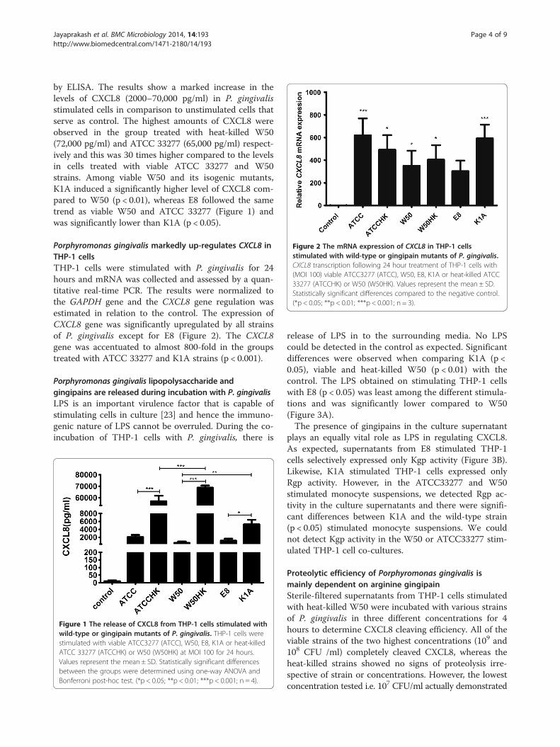
Figure 2 The mRNA expression of CXCL8 in THP-1 cellsstimulated with wild-type or gingipain mutants of P. gingivalis.CXCL8 transcription following 24 hour treatment of THP-1 cells with(MOI 100) viable ATCC3277 (ATCC), W50, E8, K1A or heat-killed ATCC33277 (ATCCHK) or W50 (W50HK). Values represent the mean ± SD.Statistically significant differences compared to the negative control.(*p < 0.05; **p < 0.01; ***p < 0.001; n = 3).
Jayaprakash et al. BMC Microbiology 2014, 14:193 Page 4 of 9http://www.biomedcentral.com/1471-2180/14/193
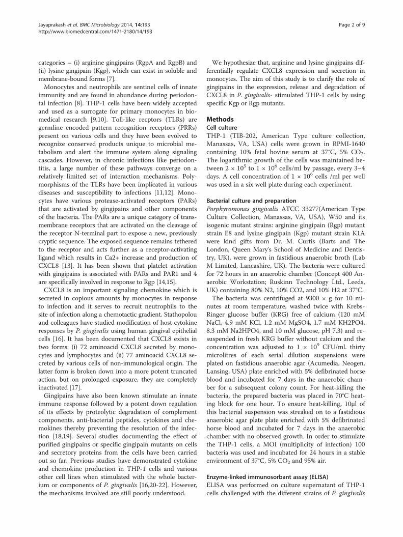
by ELISA. The results show a marked increase in thelevels of CXCL8 (2000–70,000 pg/ml) in P. gingivalisstimulated cells in comparison to unstimulated cells thatserve as control. The highest amounts of CXCL8 wereobserved in the group treated with heat-killed W50(72,000 pg/ml) and ATCC 33277 (65,000 pg/ml) respect-ively and this was 30 times higher compared to the levelsin cells treated with viable ATCC 33277 and W50strains. Among viable W50 and its isogenic mutants,K1A induced a significantly higher level of CXCL8 com-pared to W50 (p < 0.01), whereas E8 followed the sametrend as viable W50 and ATCC 33277 (Figure 1) andwas significantly lower than K1A (p < 0.05).
Porphyromonas gingivalis markedly up-regulates CXCL8 inTHP-1 cellsTHP-1 cells were stimulated with P. gingivalis for 24hours and mRNA was collected and assessed by a quan-titative real-time PCR. The results were normalized tothe GAPDH gene and the CXCL8 gene regulation wasestimated in relation to the control. The expression ofCXCL8 gene was significantly upregulated by all strainsof P. gingivalis except for E8 (Figure 2). The CXCL8gene was accentuated to almost 800-fold in the groupstreated with ATCC 33277 and K1A strains (p < 0.001).
Porphyromonas gingivalis lipopolysaccharide andgingipains are released during incubation with P. gingivalisLPS is an important virulence factor that is capable ofstimulating cells in culture [23] and hence the immuno-genic nature of LPS cannot be overruled. During the co-incubation of THP-1 cells with P. gingivalis, there is
Figure 1 The release of CXCL8 from THP-1 cells stimulated withwild-type or gingipain mutants of P. gingivalis. THP-1 cells werestimulated with viable ATCC3277 (ATCC), W50, E8, K1A or heat-killedATCC 33277 (ATCCHK) or W50 (W50HK) at MOI 100 for 24 hours.Values represent the mean ± SD. Statistically significant differencesbetween the groups were determined using one-way ANOVA andBonferroni post-hoc test. (*p < 0.05; **p < 0.01; ***p < 0.001; n = 4).
release of LPS in to the surrounding media. No LPScould be detected in the control as expected. Significantdifferences were observed when comparing K1A (p <0.05), viable and heat-killed W50 (p < 0.01) with thecontrol. The LPS obtained on stimulating THP-1 cellswith E8 (p < 0.05) was least among the different stimula-tions and was significantly lower compared to W50(Figure 3A).The presence of gingipains in the culture supernatant
plays an equally vital role as LPS in regulating CXCL8.As expected, supernatants from E8 stimulated THP-1cells selectively expressed only Kgp activity (Figure 3B).Likewise, K1A stimulated THP-1 cells expressed onlyRgp activity. However, in the ATCC33277 and W50stimulated monocyte suspensions, we detected Rgp ac-tivity in the culture supernatants and there were signifi-cant differences between K1A and the wild-type strain(p < 0.05) stimulated monocyte suspensions. We couldnot detect Kgp activity in the W50 or ATCC33277 stim-ulated THP-1 cell co-cultures.
Proteolytic efficiency of Porphyromonas gingivalis ismainly dependent on arginine gingipainSterile-filtered supernatants from THP-1 cells stimulatedwith heat-killed W50 were incubated with various strainsof P. gingivalis in three different concentrations for 4hours to determine CXCL8 cleaving efficiency. All of theviable strains of the two highest concentrations (109 and108 CFU /ml) completely cleaved CXCL8, whereas theheat-killed strains showed no signs of proteolysis irre-spective of strain or concentrations. However, the lowestconcentration tested i.e. 107 CFU/ml actually demonstrated
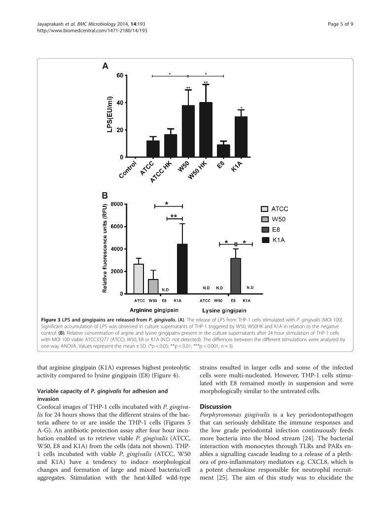
Figure 3 LPS and gingipains are released from P. gingivalis. (A). The release of LPS from THP-1 cells stimulated with P. gingivalis (MOI 100).Significant accumulation of LPS was observed in culture supernatants of THP-1 triggered by W50, W50HK and K1A in relation to the negativecontrol. (B). Relative concentration of argine and lysine gingipains present in the culture supernatants after 24 hour stimulation of THP-1 cellswith MOI 100 viable ATCC33277 (ATCC), W50, E8 or K1A (N.D: not detected). The differnces between the different stimulations were analyzed byone-way ANOVA. Values represent the mean ± SD. (*p < 0.05; **p < 0.01; ***p < 0.001; n = 3).
Jayaprakash et al. BMC Microbiology 2014, 14:193 Page 5 of 9http://www.biomedcentral.com/1471-2180/14/193
that arginine gingipain (K1A) expresses highest proteolyticactivity compared to lysine gingipain (E8) (Figure 4).
Variable capacity of P. gingivalis for adhesion andinvasionConfocal images of THP-1 cells incubated with P. gingiva-lis for 24 hours shows that the different strains of the bac-teria adhere to or are inside the THP-1 cells (Figures 5A-G). An antibiotic protection assay after four hour incu-bation enabled us to retrieve viable P. gingivalis (ATCC,W50, E8 and K1A) from the cells (data not shown). THP-1 cells incubated with viable P. gingivalis (ATCC, W50and K1A) have a tendency to induce morphologicalchanges and formation of large and mixed bacteria/cellaggregates. Stimulation with the heat-killed wild-type
strains resulted in larger cells and some of the infectedcells were multi-nucleated. However, THP-1 cells stimu-lated with E8 remained mostly in suspension and weremorphologically similar to the untreated cells.
DiscussionPorphyromonas gingivalis is a key periodontopathogenthat can seriously debilitate the immune responses andthe low grade periodontal infection continuously feedsmore bacteria into the blood stream [24]. The bacterialinteraction with monocytes through TLRs and PARs en-ables a signalling cascade leading to a release of a pleth-ora of pro-inflammatory mediators e.g. CXCL8, which isa potent chemokine responsible for neutrophil recruit-ment [25]. The aim of this study was to elucidate the

Figure 4 P. gingivalis degrades CXCL8 through gingipains. Theculture supernatant of heat-killed W50 (MOI 100) treated THP-1 cellsfor 24 hours was filtered and incubated with 107/ml, 108/ml, 109/mlviable ATCC3277 (ATCC), W50, E8, K1A or heat-killed ATCC 33277(ATCCHK) or W50 (W50HK) for 4 hours and CXCL8 was quantified byELISA. Significant differences among each group were analyzed inrelation to the control and statistics were done using one-way ANOVA.Values represent the mean ± SD. (*p < 0.05; **p < 0.01; ***p < 0.001;n = 3).
Figure 5 THP-1 cells were incubated with different strains andmutants of FITC-labelled (green) P. gingivalis (MOI 100) for 24hours and then stained for F-actin with rhodamine phalloidin(red) and nucleus with DAPI (blue), followed by confocalfluorosence microscopy. Confocal images show (A) Control(untreated) THP-1 cells (B) ATCC 33277 (C) ATCC 33277 heat-killed(D) W50 (E) W50 heat-killed (F) E8 and (G) K1A adhere to and arefound within monocytes. Large, multi-nucleated cells are indicatedby white arrows in figures B, C, D and E. Bar = 40μm. 60X objective,oil immersion. Representative images from three independentexperiments are shown.
Jayaprakash et al. BMC Microbiology 2014, 14:193 Page 6 of 9http://www.biomedcentral.com/1471-2180/14/193
mechanisms of P. gingivalis interaction with monocytes interms of induction and expression of CXCL8. By usinggingipain mutants, we found that Rgp and Kgp help P.gingivalis evade the immune system both in an antagonis-tic and synergistic manner. The results are consistent withthe previous studies and would be a rational to direct fu-ture studies using peripheral blood monocytes.Both viable and heat-killed W50 strains of P. gingivalis
shed significant amounts of LPS during interaction withTHP-1 cells and induced release of CXCL8 indicatingthat the TLRs are involved as LPS is heat-stable and isan agonist for TLR2 activation. We observed that thelevels of CXCL8 are 30–40 times higher in THP-1 cellsstimulated with heat-killed ATCC and W50 compared tocells exposed to viable bacteria. TLR2 has previouslybeen shown to be activated by both viable and heat-killed P. gingivalis [26]. If the bacteria-induced release ofCXCL8 is based only on LPS, there would have been nodifference in CXCL8 response between viable and heat-killed bacteria, however, this was not the case.Gingipains are trypsin-like cysteine proteases that can
cleave the proteins at arginine and lysine specific sites[27]. We observed that the different strains of P. gingiva-lis (viable or heat-killed) upregulated CXCL8 gene sev-eral hundred-folds. Uehara and colleagues investigatedthe possible synergistic effects of PARs, TLRs and NODson CXCL8 production in THP-1 cells using syntheticpeptide ligands and showed that gingipains stimulate thesecretion of cytokines from monocytic cells through theactivation of PARs with synergistic effects by PRRs [20].Consequently, the inflammatory response induced by
gingipains was not exclusively dependent on their cata-lytic activity since heat-inactivated bacterial preparationswere still effective whereas proteolytic activity was ab-sent by denaturation [28]. The loss of protease activityon heat-treatment was validated by a cytokine degrad-ation functional assay and this also explains the reduced

Jayaprakash et al. BMC Microbiology 2014, 14:193 Page 7 of 9http://www.biomedcentral.com/1471-2180/14/193
levels of extracellular CXCL8 in THP-1 cells stimulatedwith viable bacteria. Measuring the gingipain activity in thesupernatant served as a functional validation of the iso-genic gingipain mutants as well as the relative expressionof arginine and lysine gingipains by the wild-type strains.Aduse-Opoku and colleagues showed that Rgp and Kgp
are secreted independent of each other [29]. It is possiblethat ATCC33277 and W50 could be selectively inclined tosecreting Rgp in aerobic cell culture conditions. It is evidentthat the gene transcription patterns do not follow the pro-tein levels and could imply that the post-secretory CXCL8protein is being degraded by gingipains. The inflammatorycharacter of fimbriae and bacterial DNA cannot be disre-garded. Thermal inactivation of microorganisms could re-lease bacterial DNA due to possible membrane ruptureactivating TLR9 and P. gingivalis fimbriae are also docu-mented to activate TLR2 and aid in CXCL8 release in hu-man aortic endothelial cells [30,31].We show that W50 cleaves CXCL8 with the same effi-
ciency as K1A, while ATCC 33277 is less effective. It isclear that there is variation in protease activity amongthe various wild-type strains of P. gingivalis. The degrad-ation assay shows that at high titres, both Rgp and Kgpwere equally effective in inactivating CXCL8, whereas atmoderate and low titres, Rgp was more effective at de-grading CXCL8. We have previously shown that CXCL8hydrolysis can be salvaged by using leupeptin which isan arginine-specific gingipain inhibitor [19]. However,all cytokines are not cleaved alike. Stathopolou and col-leagues have shown that the rate of IL-1β degradation islower compared to that of IL-6 or CXCL8, which couldbe due to the primary, secondary and tertiary molecularstructure of IL-1β which makes it relatively resistant tohydrolysis by Kgp. It has been shown that interleukin-6is rapidly cleaved by both gingipains, although Kgp wasfound to be more effective [16,32].The degradation assay makes it evident that Rgp is a
more efficient protease at cleaving CXCL8, however, P.gingivalis expressing Rgp markedly amplifies CXCL8transcription and significantly higher level of CXCL8was present in culture supernatants of THP-1 treatedwith P. gingivalis expressing only Rgp (K1A). This couldbe due to a complex balance and interaction betweeninduction and degradation. Induction of the CXCL8 geneis due to LPS, fimbriae, bacterial outer membraneproteins, DNA and gingipains, whereas degradation ofCXCL8 is only protease dependent. In addition, it hasbeen shown that CXCL8 is a secondary cytokine, i.e. itsrelease could also be mediated by primary cytokines, likeIL-1β and TNF in an autocrine manner [33]. Hence, it isobvious that CXCL8 regulation during host pathogeninteraction is very elaborate and the in vivo conditionsare more dynamic compared to the situation in vitro.Also, gram negative bacteria can shed LPS in to the
surrounding during cell death, outer membrane vesiclebiogenesis and stress [34-36].Confocal fluorescent images showed that THP-1 cells
form aggregates when treated with P. gingivalis. Besidesthe changes in the actin cytoskeleton, P. gingivalis prob-ably increased the expression of cell adhesion moleculesin monocytes leading to cell aggregation [37]. THP-1cells stimulated with Kgp-deficient K1A appear to bemore differentiated and aggregated, compared to the E8(lacks Rgp)-infected THP-1 cells, which could be sug-gestive of a role exhibited by Rgps in modulating THP-1cell aggregation and adhesion. Hashizume and col-leagues showed that cytoadherence between THP-1 cellsand endothelial cells were enhanced by treatment withP. gingivalis [38]. P. gingivalis invades or is internalizedwithin the cells. We were able to culture intracellularbacteria by the antibiotic protection assay after 4 hourincubation showing that P. gingivalis survives intracellu-larly. The ability of ATCC 33277 to invade endothelialcells has previously been demonstrated in our lab [39].Fimbriae possibly contributes to the intracellular pres-ence of the ATCC strain and even sparsely fimbriatedstrains successfully invaded although in varying degreesindicating there could be other factors supporting adhe-sion and infection of cells [40]. W50 and its mutantspossess type IV fimA and previous studies have con-firmed that the precursor fimA can only become matureby the action of Rgp and, consequently, the lack of Rgpin E8 likely renders fimA ineffective [29,41]. The giantmulti-nucleated cells that formed on interaction ofTHP-1 cells with viable or heat-killed ATCC 33277 andW50 is of additional interest and one could speculatethat these giant cells could be of osteoclastic phenotypeof importance in the pathogenesis of periodontitis andneeds further clarification.
ConclusionIn conclusion, our results demonstrate that both argin-ine and lysine gingipains could play a vital role in P. gin-givalis-mediated immunomodulation and that Rgpappears to be cruciual in the context of CXCL8 regula-tion even at low concentrations. This study establishesthat gingipains are capable of post-secretory chemokineparalysis which would most likely result in abberantneutrophil recruitment to the site of infection and en-suring that a low grade infection would continue un-hampered. The invasion and survival of P. gingivaliswithin the monocytes could account for the safe passageof the bacteria from the site of periodontal infection toother inflamed sites. Periodontal infections are inde-pendent risk factors for several chronic systemic illnessand persistent recurrent bacteremia due to denuded oralepithelium results in systemic dissemination of period-ontopathogens via the vascular compartment.

Jayaprakash et al. BMC Microbiology 2014, 14:193 Page 8 of 9http://www.biomedcentral.com/1471-2180/14/193
Competing interestsThe authors declare that they have no competing interests.
Author’s contributionsTB, HK and KJ design the study, KJ conducted the experiments. KJ, HK andTB analyzed the data. KJ, HK and TB drafted the article. All authors read andapproved the final manuscript.
AcknowledgementThe authors acknowledge the support from Swedish Heart and LungFoundation, the Foundation of Olle Engkvist and the Knowledge Foundation.The W50 and its isogenic mutants were a kind gift from Dr. M. Curtis (Bartsand The London, Queen Mary's School of Medicine and Dentistry, UK).
Received: 5 May 2014 Accepted: 10 July 2014Published: 18 July 2014
References1. Feng Z, Weinberg A: Role of bacteria in health and disease of periodontal
tissues. Periodontol 2006, 40:50–76.2. Kebschull M, Demmer RT, Papapanou PN: "Gum bug, leave my heart
alone!"–epidemiologic and mechanistic evidence linking periodontalinfections and atherosclerosis. J Dent Res 2010, 89(9):879–902.
3. Ogrendik M: Rheumatoid arthritis is an autoimmune disease caused byperiodontal pathogens. Int J Gen Med 2013, 6:383–386.
4. Yang HW, Huang YF, Chou MY: Occurrence of Porphyromonas gingivalisand Tannerella forsythensis in periodontally diseased and healthysubjects. J Periodontol 2004, 75(8):1077–1083.
5. Ximenez-Fyvie LA, Haffajee AD, Socransky SS: Comparison of themicrobiota of supra- and subgingival plaque in health and periodontitis.J Clin Periodontol 2000, 27(9):648–657.
6. Bostanci N, Belibasakis GN: Porphyromonas gingivalis: an invasive andevasive opportunistic oral pathogen. FEMS Microbiol Lett 2012, 333(1):1–9.
7. Guo Y, Nguyen KA, Potempa J: Dichotomy of gingipains action asvirulence factors: from cleaving substrates with the precision of asurgeon's knife to a meat chopper-like brutal degradation of proteins.Periodontol 2010, 54(1):15–44.
8. Fokkema SJ: Peripheral blood monocyte responses in periodontitis. Int JDent Hyg 2012, 10(3):229–235.
9. Markotic A, Hensley L, Daddario K, Spik K, Anderson K, Schmaljohn C:Pathogenic hantaviruses elicit different immunoreactions in THP-1 cellsand primary monocytes and induce differentiation of human monocytesto dendritic-like cells. Coll Antropol 2007, 31(4):1159–1167.
10. Campbell JE, Brummel-Ziedins KE, Butenas S, Mann KG: Cellularregulation of blood coagulation: a model for venous stasis. Blood 2010,116(26):6082–6091.
11. Hajishengallis G, Sharma A, Russell MW, Genco RJ: Interactions of oralpathogens with toll-like receptors: possible role in atherosclerosis.Ann Periodontol 2002, 7(1):72–78.
12. Medvedev AE: Toll-like receptor polymorphisms, inflammatory andinfectious diseases, allergies, and cancer. J Interferon Cytokine Res 2013,33(9):467–484.
13. Colognato R, Slupsky JR, Jendrach M, Burysek L, Syrovets T, Simmet T:Differential expression and regulation of protease-activated receptors inhuman peripheral monocytes and monocyte-derived antigen-presentingcells. Blood 2003, 102(7):2645–2652.
14. Nylander M, Lindahl TL, Bengtsson T, Grenegard M: The periodontalpathogen Porphyromonas gingivalis sensitises human blood platelets toepinephrine. Platelets 2008, 19(5):352–358.
15. Lourbakos A, Yuan YP, Jenkins AL, Travis J, Andrade-Gordon P, Santulli R,Potempa J, Pike RN: Activation of protease-activated receptors bygingipains from Porphyromonas gingivalis leads to platelet aggregation:a new trait in microbial pathogenicity. Blood 2001, 97(12):3790–3797.
16. Stathopoulou PG, Benakanakere MR, Galicia JC, Kinane DF: The hostcytokine response to Porphyromonas gingivalis is modified bygingipains. Oral Microbiol Immunol 2009, 24(1):11–17.
17. Mikolajczyk-Pawlinska J, Travis J, Potempa J: Modulation of interleukin-8activity by gingipains from Porphyromonas gingivalis: implications forpathogenicity of periodontal disease. FEBS Lett 1998, 440(3):282–286.
18. Curtis MA, Kuramitsu HK, Lantz M, Macrina FL, Nakayama K, Potempa J,Reynolds EC, Aduse-Opoku J: Molecular genetics and nomenclature of
proteases of Porphyromonas gingivalis. J Periodontal Res 1999,34(8):464–472.
19. Palm E, Khalaf H, Bengtsson T: Porphyromonas gingivalis downregulatesthe immune response of fibroblasts. BMC Microbiol 2013, 13:155.
20. Uehara A, Takada H: Synergism between TLRs and NOD1/2 in oralepithelial cells. J Dent Res 2008, 87(7):682–686.
21. Diya Z, Lili C, Shenglai L, Zhiyuan G, Jie Y: Lipopolysaccharide (LPS)of Porphyromonas gingivalis induces IL-1beta, TNF-alpha and IL-6production by THP-1 cells in a way different from that of Escherichiacoli LPS. Innate immunity 2008, 14(2):99–107.
22. Zaric S, Shelburne C, Darveau R, Quinn DJ, Weldon S, Taggart CC,Coulter WA: Impaired immune tolerance to Porphyromonas gingivalislipopolysaccharide promotes neutrophil migration and decreasedapoptosis. Infect Immun 2010, 78(10):4151–4156.
23. Dias IH, Marshall L, Lambert PA, Chapple IL, Matthews JB, Griffiths HR:Gingipains from Porphyromonas gingivalis increase the chemotactic andrespiratory burst-priming properties of the 77-amino-acid interleukin-8variant. Infect Immun 2008, 76(1):317–323.
24. Carrion J, Scisci E, Miles B, Sabino GJ, Zeituni AE, Gu Y, Bear A, Genco CA,Brown DL, Cutler CW: Microbial carriage state of peripheral blooddendritic cells (DCs) in chronic periodontitis influences DCdifferentiation, atherogenic potential. J Immunol 2012, 189(6):3178–3187.
25. Nakagawa H, Hatakeyama S, Ikesue A, Miyai H: Generation of interleukin-8by plasmin from AVLPR-interleukin-8, the human fibroblast-derivedneutrophil chemotactic factor. FEBS Lett 1991, 282(2):412–414.
26. Giacaman RA, Asrani AC, Ross KF, Herzberg MC: Cleavage of protease-activated receptors on an immortalized oral epithelial cell lineby Porphyromonas gingivalis gingipains. Microbiology 2009,155(Pt 10):3238–3246.
27. Imamura T, Travis J, Potempa J: The biphasic virulence activities ofgingipains: activation and inactivation of host proteins. Curr Protein PeptSci 2003, 4(6):443–450.
28. Grenier D, Tanabe S: Porphyromonas gingivalis gingipains trigger aproinflammatory response in human monocyte-derived macrophagesthrough the p38alpha mitogen-activated protein kinase signaltransduction pathway. Toxins 2010, 2(3):341–352.
29. Aduse-Opoku J, Davies NN, Gallagher A, Hashim A, Evans HE, Rangarajan M,Slaney JM, Curtis MA: Generation of lys-gingipain protease activity inPorphyromonas gingivalis W50 is independent of Arg-gingipain proteaseactivities. Microbiology 2000, 146(Pt 8):1933–1940.
30. Davey M, Liu X, Ukai T, Jain V, Gudino C, Gibson FC 3rd, Golenbock D,Visintin A, Genco CA: Bacterial fimbriae stimulate proinflammatoryactivation in the endothelium through distinct TLRs. J Immunol 2008,180(4):2187–2195.
31. Beutler BA: TLRs and innate immunity. Blood 2009, 113(7):1399–1407.32. Banbula A, Bugno M, Kuster A, Heinrich PC, Travis J, Potempa J: Rapid and
efficient inactivation of IL-6 gingipains, lysine- and arginine-specificproteinases from Porphyromonas gingivalis. Biochem Biophys ResCommun 1999, 261(3):598–602.
33. Eskan MA, Benakanakere MR, Rose BG, Zhang P, Zhao J, Stathopoulou P,Fujioka D, Kinane DF: Interleukin-1beta modulates proinflammatorycytokine production in human epithelial cells. Infect Immun 2008,76(5):2080–2089.
34. Kulp A, Kuehn MJ: Biological functions and biogenesis of secretedbacterial outer membrane vesicles. Annu Rev Microbiol 2010,64:163–184.
35. McBroom AJ, Kuehn MJ: Release of outer membrane vesicles byGram-negative bacteria is a novel envelope stress response. Mol Microbiol2007, 63(2):545–558.
36. Kato S, Kowashi Y, Demuth DR: Outer membrane-like vesicles secreted byActinobacillus actinomycetemcomitans are enriched in leukotoxin.Microb Pathog 2002, 32(1):1–13.
37. Yamaguchi H, Haranaga S, Widen R, Friedman H, Yamamoto Y: Chlamydiapneumoniae infection induces differentiation of monocytes intomacrophages. Infect Immun 2002, 70(5):2392–2398.
38. Hashizume T, Kurita-Ochiai T, Yamamoto M: Porphyromonas gingivalisstimulates monocyte adhesion to human umbilical vein endothelial cells.FEMS Immunol Med Microbiol 2011, 62(1):57–65.
39. Zhang B, Elmabsout AA, Khalaf H, Basic VT, Jayprakash K, Kruse R, Bengtsson T,Sirsjo A: The periodontal pathogen Porphyromonas gingivalis changesthe gene expression in vascular smooth muscle cells involving the
Jayaprakash et al. BMC Microbiology 2014, 14:193 Page 9 of 9http://www.biomedcentral.com/1471-2180/14/193
TGFbeta/Notch signalling pathway and increased cell proliferation. BMCGenomics 2013, 14(1):770.
40. Sojar HT, Hamada N, Genco RJ: Isolation and characterization offimbriae from a sparsely fimbriated strain of Porphyromonas gingivalis.Appl Environ Microbiol 1997, 63(6):2318–2323.
41. Shoji M, Naito M, Yukitake H, Sato K, Sakai E, Ohara N, Nakayama K: Themajor structural components of two cell surface filaments ofPorphyromonas gingivalis are matured through lipoprotein precursors.Mol Microbiol 2004, 52(5):1513–1525.
doi:10.1186/1471-2180-14-193Cite this article as: Jayaprakash et al.: Gingipains from Porphyromonasgingivalis play a significant role in induction and regulation of CXCL8 inTHP-1 cells. BMC Microbiology 2014 14:193.
Submit your next manuscript to BioMed Centraland take full advantage of:
• Convenient online submission
• Thorough peer review
• No space constraints or color figure charges
• Immediate publication on acceptance
• Inclusion in PubMed, CAS, Scopus and Google Scholar
• Research which is freely available for redistribution
Submit your manuscript at www.biomedcentral.com/submit
Copyright © 2022 FDOKUMEN