Tnrs Index contains no reference to the Introductory Tables ...
Upload
independentCategory
view
0download
0
10.1128/IAI.72.9.5041-5051.2004.
2004, 72(9):5041. DOI:Infect. Immun. William N. Howald, Sing Sing Way and Adeline M. HajjarRobert A. Reife, Brian W. Bainbridge, Stephen R. Coats, Richard P. Darveau, Thu-Thao T. Pham, Kayde Lemley, Both Toll-Like Receptors 2 and 4A Species That Functionally Interact with
LipidLipopolysaccharide Contains Multiple Porphyromonas gingivalis
http://iai.asm.org/content/72/9/5041Updated information and services can be found at:
These include:
REFERENCEShttp://iai.asm.org/content/72/9/5041#ref-list-1at:
This article cites 66 articles, 37 of which can be accessed free
CONTENT ALERTS more»articles cite this article),
Receive: RSS Feeds, eTOCs, free email alerts (when new
http://journals.asm.org/site/misc/reprints.xhtmlInformation about commercial reprint orders: http://journals.asm.org/site/subscriptions/To subscribe to to another ASM Journal go to:
on February 15, 2014 by guest
http://iai.asm.org/
Dow
nloaded from
on February 15, 2014 by guest
http://iai.asm.org/
Dow
nloaded from

INFECTION AND IMMUNITY, Sept. 2004, p. 5041–5051 Vol. 72, No. 90019-9567/04/$08.00�0 DOI: 10.1128/IAI.72.9.5041–5051.2004Copyright © 2004, American Society for Microbiology. All Rights Reserved.
Porphyromonas gingivalis Lipopolysaccharide Contains Multiple LipidA Species That Functionally Interact with Both Toll-Like
Receptors 2 and 4Richard P. Darveau,1* Thu-Thao T. Pham,1 Kayde Lemley,2 Robert A. Reife,1
Brian W. Bainbridge,1 Stephen R. Coats,1 William N. Howald,3Sing Sing Way,2 and Adeline M. Hajjar2
Departments of Periodontics,1 Immunology,2 and Medicinal Chemistry,3 University ofWashington, Seattle, Washington
Received 12 March 2004/Returned for modification 19 April 2004/Accepted 26 May 2004
The innate host response to lipopolysaccharide (LPS) obtained from Porphyromonas gingivalis is unusual inthat different studies have reported that it can be an agonist for Toll-like receptor 2 (TLR2) as well as anantagonist or agonist for TLR4. In this report it is shown that P. gingivalis LPS is highly heterogeneous,containing more lipid A species than previously described. In addition, purification of LPS can preferentiallyfractionate these lipid A species. It is shown that an LPS preparation enriched for lipid A species at m/z 1,435and 1,450 activates human and mouse TLR2, TLR2 plus TLR1, and TLR4 in transiently transfected HEK 293cells coexpressing membrane-associated CD14. The HEK cell experiments further demonstrated that cofactorMD-2 was required for functional engagement of TLR4 but not of TLR2 nor TLR2 plus TLR1. In addition,serum-soluble CD14 effectively transferred P. gingivalis LPS to TLR2 plus TLR1, but poorly to TLR4. Impor-tantly, bone marrow cells obtained from TLR2�/� and TLR4�/� mice also responded to P. gingivalis LPS in amanor consistent with the HEK results, demonstrating that P. gingivalis LPS can utilize both TLR2 and TLR4.No response was observed from bone marrow cells obtained from TLR2 and TLR4 double-knockout mice,demonstrating that P. gingivalis LPS activation occurred exclusively through either TLR2 or TLR4. Althoughthe biological significance of the different lipid A species found in P. gingivalis LPS preparations is not currentlyunderstood, it is proposed that the presence of multiple lipid A species contributes to cell activation throughboth TLR2 and TLR4.
A functional innate response system consists of differenthost components that act coordinately to recognize microbialcolonization and elicit appropriate immediate responses (27,31). Host responses to lipopolysaccharide (LPS), an essentialconstituent of the cell wall of gram-negative bacteria, can bevery potent, such that host sampling of this molecule can con-tribute significantly to the overall innate response to bacterialinfection (6, 60). For example, in vitro studies have confirmedthat whole bacteria and their respective isolated LPSs yieldsimilar responses (12, 56), and in vivo studies have validatedthe important role of LPS in triggering inflammation in re-sponse to bacterial infection (25, 30, 55).
A well-characterized innate host recognition pathway is thatfor Escherichia coli LPS, which starts with a series of initialbinding and transfer reactions between LPS binding protein(LBP), CD14, and other host proteins (49, 64). CD14 is locatedin the cell membrane of certain host cells (mCD14) and is alsopresent in serum (sCD14) and gingival crevicular fluid, a serumexudate (28, 45). Transfer of E. coli LPS by either mCD14 orsCD14 to a cell-associated Toll-like receptor 4 (TLR4) andMD-2 protein complex (15, 40) initiates host cell activationpathways, leading to innate host defense mediator production.
In the periodontium, innate host responses to microbial col-
onization are important in both health and disease (14, 59). Inclinically healthy periodontal tissue, the highly orchestratedexpression of select innate host defense mediators is believedto be associated with commensal microbial colonization (59).These mediators facilitate neutrophil transit through this tissueand into the gingival crevice, where they play a key role in theprevention of disease (24). Periodontitis is an inflammatorydisease that is characterized by loss of alveolar bone support-ing the tooth root and is the leading cause for tooth loss.Strong evidence for the role of the innate host response tomicrobial colonization in periodontitis comes from the obser-vation that removal of the dental plaque microbial biofilmremains the most effective treatment for the disease (14). Al-though the microbial composition of dental plaque associatedwith health and periodontitis is well characterized (66), little isknown about how these different compositions influence theinflammatory response.
Porphyromonas gingivalis is a gram-negative bacterium thatis an important etiologic agent of human adult-type periodon-titis (54). This bacterium releases copious amounts of outermembrane vesicles containing LPS (19, 20), which can pene-trate periodontal tissue (38, 39, 50) and thus participate in thedestructive innate host response associated with disease. Thepotential contribution of P. gingivalis LPS to the disease pro-cess is not clear, however, due to complex innate host re-sponses to this cell wall component (4). P. gingivalis LPS is ableto activate human monocytes by a CD14-dependent mecha-nism (51) and binds sCD14 (9); however, it does not facilitate
* Corresponding author. Mailing address: Department of Periodon-tics, University of Washington, Health Sciences Center, Box 357444,Seattle, WA 98195. Phone: (206) 543-9514. Fax: (206) 616-7478. E-mail: [email protected].
5041
on February 15, 2014 by guest
http://iai.asm.org/
Dow
nloaded from

sCD14-dependent E-selectin expression nor interleukin-8(IL-8) secretion from human umbilical cord vein vascular en-dothelial cells (11). In fact, this LPS is a natural antagonist forthe human endothelial E-selectin and IL-8 responses to E. coliLPS and other oral bacteria (11) and has recently been re-ported to be a TLR4 antagonist in some cell types (8, 10, 68).Furthermore, although several reports have demonstrated thatthis LPS utilizes TLR2 instead of TLR4 for host cell activation(5, 26, 37), it has also been reported to engage TLR4 (42, 57)to facilitate gingival fibroblast activation through mCD14 (46,62).
In this report it is shown that a P. gingivalis LPS preparationenriched for two major lipid A mass ions at m/z 1,435 and 1,450activates both human and mouse TLR2 and TLR2 plus TLR1,as well as TLR4 in transiently transfected HEK 293 cells co-expressing mCD14. In addition, with the use of primary bonemarrow cells obtained from TLR knockout mice, it is shownthat this LPS preparation exclusively utilizes either mouseTLR2 or TLR4 to induce tumor necrosis factor alpha (TNF-�)production. The ability of a P. gingivalis LPS preparation toactivate cells through multiple TLRs helps to reconcile previ-ous possible conflicting observations that have demonstratedthat P. gingivalis LPS preparations can activate either TLR2 orTLR4.
MATERIALS AND METHODS
Bacterial strains and preparation. P. gingivalis ATCC 33277 was obtainedfrom the American Type Culture Collection, Rockville, Md.; it was examined forpurity, properly identified, and stored at �70°C. Cultures were made from frozenbacterial stocks to avoid repetitive subculture. Bacterial cells were grown for LPS
isolation as follows: P. gingivalis was grown anaerobically at 37°C for 2 to 3 daysin Trypticase soy broth (30 g/liter) containing yeast extract (1 g/liter; Difco),glucose at 1 g/liter, potassium nitrate at 0.5 g/liter, sodium lactate (Sigma L-1375)at 1 ml/liter, sodium succinate at 0.5 g/liter, and sodium fumerate at 1 g/liter;after autoclaving, filter-sterilized supplements were added (sodium carbonate,0.4 g/liter; hemin [� H-2250], 0.005 g/liter; cysteine, 0.4 g/liter; and vitamin K [�M-5625], 0.001 g/liter). Stationary-phase cells were employed for LPS isolation.
Purification and characterization of LPS. P. gingivalis LPS was prepared bythe cold MgCl2-ethanol (EtOH) procedure (13) followed by lipid extraction (18)and conversion to sodium salts (43). E. coli 0111:B4 LPS (Sigma, St. Louis, Mo.)was subjected to a Folch extraction (18) to remove contaminating phospholipids.All LPS preparations were further treated to remove trace amounts of endotoxinprotein as described by Manthey and Vogel (36) with the following modification.Following the final EtOH precipitation, LPS was lyophilized to determine theyield and was resuspended in distilled water to 1 mg/ml without the addition oftriethanolamine. LPS was subjected to sodium dodecyl sulfate-polyacrylamidegel electrophoresis (SDS-PAGE) and stained for protein by the enhanced col-loidal gold procedure, as described in reference 36. The stain was able to detect100 pg of protein, employing bovine serum albumin as a standard. The presenceof nucleic acid was determined by ethidium bromide fluorescence quantificationof the amount of double-stranded DNA by using the plastic wrap method (35)and ImageQuant software. The P. gingivalis LPS preparation shown in Fig. 1Awas obtained with the use of TRI reagent as previously described (67).
GC/MS analysis of LPS fatty acids. Each LPS sample (0.25 mg [dry weight])and fatty acid methyl ester standards C15:0, C16:OH, C17:OH (Matreya, Inc.,Pleasant Gap, Pa.), and C16:0 (Sigma) were analyzed twice: once as their methylesters in hexane with free alcohol functional groups after derivitization withmethanolic HCl as described by the manufacturer (Alltech, Deerfield, Ill.), andthen as their trimethylsilyl ethers after transmethylation with N,O,-bis(trimeth-ylsilyl)trifluoroacetimide (BSTFA) containing 1% trimethylchlorosilane (Pierce,Rockford, Ill.). Ether formation was accomplished by the transfer of 50-�laliquots of each sample and standard to 100-�l glass liners followed by theaddition of an equal volume of BSTFA-hexane (1:1, vol/vol). The liners werethen sealed in autosampler vials and allowed to stand at room temperature for3 h prior to gas chromatographic-mass spectroscopy (GC/MS) analyses of themethylated fatty acid extracts, performed on a Finnigan Trio 1000 quadrupole
FIG. 1. Characterization of P. gingivalis lipid A species by negative ion mass spectrometry. P. gingivalis LPS was obtained by either a TRIreagent procedure (67) (A) or a cold MgCl2-EtOH procedure (13) (B). Lipid A was cleaved and separated from the LPS as described by Caroffet al. (7). MALDI-TOF was performed as previously described (21). All values given are averag masses rounded to the nearest whole number forsingly charged deprotonated molecules. TRI reagent-extracted LPS yielded two major clusters of lipid A mass ions centered at m/z 1,690 and 1,770,which were missing or significantly reduced when the MgCl2-EtOH (B) procedure was used to purify the LPS.
5042 DARVEAU ET AL. INFECT. IMMUN.
on February 15, 2014 by guest
http://iai.asm.org/
Dow
nloaded from

mass spectrometer (Thermo Electron Corp., San Jose, Calif.) fitted with aHewlett-Packard 5890 series II gas chromatograph equipped with a capillarysplitless injector and a Hewlett-Packard 7673A autosampler (Agilent Technol-ogies, Palo Alto, Calif.). A fused silica capillary GC column (30 m by 0.32 mm[inner diameter], 0.25-�m film thickness) was used, coated with the bondedstationary phase (J&W DB-5; Agilent Technologies, Folsom, Calif.), and oper-ated with helium (head pressure, 5 lb/in2) as carrier gas with a 3-ml min�1
septum purge through the injector. Samples were injected in the splitless mode(injector temperature, 250°C) and cold trapped on the column at 40°C. After 1min, the injector was purged and the column oven temperature was programmedlinearly to 280°C at a rate of 10°C/min and held for 5 min. The mass spectrometerwas operated in the electron ionization mode, with a filament emission currentand electron energy of 150 �A and 70 eV, respectively. The ion source temper-ature was 200°C, and the GC interface was held at 250°C. Tuning and masscalibration were performed daily using perfluorotributylamine in the repetitivescan mode (scanning from 35 to 450 Da once every second). All data acquisitionand processing, including mass spectral database searches against both the WileyRegistry of MS data (6th ed., 1996) and the NIST/EPA/NIH Mass SpectralLibrary (1992) were performed using Windows-based Finnigan MassLab 1.3software. GC/MS analyses of the extracted transmethylated fatty acid samplesand fatty acid methyl ester standards as well as their trimethylsilyl ether deriv-atives were carried out using 2-�l injection volumes.
Matrix assisted laser desorption–time-of-flight (MALDI-TOF) MS was per-formed as previously described (21). Two separate extractions of P. gingivalis LPSwere produced and analyzed.
Cells and reagents. Human embryonic kidney (HEK) 293 cells were main-tained in Dulbecco’s modified Eagle’s medium (GibcoBRL, Rockville, Md.) with10% heat-inactivated fetal calf serum (HyClone, Logan, Utah). The NF-�Breporter construct (ELAM-1 firefly luciferase), the �-actin–Renilla luciferasereporter construct, the modified pDisplay expression vector, and the expressionconstructs for murine TLR2 (pmuTLR2), murine TLR1 (pmuTLR1), murineTLR4 (pmuTLR4), and human TLR4 (phuTLR4) and mCD14 (phumCD14)have been described previously (22, 23). Human TLR1 (phuTLR1) and TLR2(phuTLR2) open reading frames were cloned into the modified pDisplay expres-sion vector. The human and murine MD-2 plasmids were kindly provided byKensuke Miyake (The University of Tokyo, Tokyo, Japan). CD14-depleted se-rum was prepared as described previously (9).
Luciferase assays. HEK 293 cells were transfected by calcium phosphateprecipitation and stimulated as described previously (23) with the modificationsreported for a 96-well plate assay format (22). Cells were washed twice withmedium 3 h after transfection and stimulated 20 to 24 h posttransfection. Stim-ulations were performed in stimulation medium (Dulbecco’s modified Eagle’smedium containing 10% fetal bovine serum (FBS), 10% human serum, orsCD14-depleted normal human serum) for 4 h at 37°C, using concentrations ofligands as indicated below in the text and figure legends. After stimulation, cellswere rinsed with phosphate-buffered saline (BioWhittaker, Walkersville, Md.)and lysed with 50 �l of passive lysis buffer (Promega, Madison, Wis.). Reportergene expression in each lysate (10 �l) was measured using the Dual Luciferasereporter assay system (Promega). Data are expressed as the fold increase inrelative light units (which represents the ratio of ELAM-luciferase to �-actinRenilla-luciferase expression) relative to that of a no-stimulation control. Allexperiments were performed a minimum of three separate times with similarresults. Results from one experiment performed with data from triplicate wellsare presented.
Stimulation of primary bone marrow cells obtained from TLR knockout mice.TLR2�/� and TLR4�/� mice were backcrossed for six generations to C57BL/6mice and then intercrossed to obtain TLR2/4 double-knockout mice. Bone mar-row cells were isolated from femurs and tibias of mice bred at the University ofWashington. Following red cell lysis, 1 � 106 to 2 � 106 cells were plated per wellin round-bottom 96-well plates. The cells were stimulated with concentrations ofligands as indicated below in the text and figure legends for 5 h in the presenceof GolgiStop (Pharmingen). Cells were then stained for surface expression ofCD11b (Pharmingen), permeabilized with Cytoperm/Cytofix (Pharmingen), andstained for intracellular TNF-� (Caltag). Cells were analyzed on a Becton Dick-inson FACScan flow cytometer using CellQuest software (BD Biosciences).
RESULTS
P. gingivalis LPS contains multiple forms of lipid A. Studieshave reported several different structures for the lipid A ob-tained from P. gingivalis (32, 41, 67). Ogawa (41), employingstrain 381, reported that P. gingivalis LPS contained one pre-
dominant lipid A mass ion at m/z 1,195, while Kumada et al.,examining a clinical isolate designated SU63 (32), reportedmultiple lipid A mass ions, with the most predominant onesfound at m/z 1,435 and 1,450, and Yi and Hackett (67), em-ploying strain 33277, reported that a lipid A species at m/z1,691 was the dominant structure found among multiple lipidA species. Consistent with the notion that P. gingivalis LPSdisplays lipid A heterogeneity, our investigators have reportedthat a purified P. gingivalis LPS preparation from strain 33277contained lipid A mass ions at m/z 1,195, 1,435, and 1,450 (3).
In this report, the degree and extent of P. gingivalis LPS lipidA heterogeneity was examined with the use of a new crude LPSextraction procedure which employs commercially availableTRI reagent (67). It was found that P. gingivalis LPS containsmore major lipid A mass ions than previously described (Fig.1A) and that the MgCL2-EtOH LPS purification procedurefailed to extract all of the different lipid A mass ions equally(Fig. 1B). TRI reagent extraction of LPS from P. gingivalis andsubsequent lipid A cleavage (7) revealed numerous major lipidA mass ions that clustered around m/z 1,450, 1,690, and 1,770.The lipid A species found clustered around each of these massions differed by smaller single methylene units (m/z 1,420 and1,435 adjacent to 1,450; 1,675 adjacent to 1,690; and 1,705 and1,755 adjacent to 1,770) and larger single methylene units (m/z1,465 and 1,480 adjacent to 1,450; 1,705 adjacent to 1,690; and1,785 and 1,800 adjacent to 1,770). This pattern of differentlipid A mass ions is indicative of fatty acid chain length heter-ogeneity, accounting for some of the different lipid A species.In contrast, LPS extracted from whole cells by the MgCl2-EtOH method was significantly reduced in both clusters oflipid A mass ions centered at m/z 1,690 and m/z 1,770. Thispreparation, designated Pg LPS1435/1450 (see below), revealedmajor lipid A mass ions at m/z 1,435 and 1,450 (Fig. 1B); thestructures for both of these P. gingivalis lipid A species havebeen previously elucidated (32) (Fig. 2). In addition, two minorpeaks at m/z 1,420 and 1,465 were observed and are suspectedto represent structurally related lipid A mass ions that differ intheir fatty acid content.
In this study, the triacylated monophosphorylated lipid A atm/z 1,195 was not detected in either the TRI reagent or MgCl2-EtOH LPS preparations. The reasons for the lack of this lipidA mass ion, which was originally described by Ogawa (41) andpreviously detected by our group (3), are currently not under-stood. However, initial studies in our laboratory have revealedthat different media compositions may influence the appear-ance of this lipid A mass ion (data not shown).
Characterization of the P. gingivalis LPS preparation em-ployed to examine TLR utilization. The MgCl2-EtOH LPSpreparation was characterized with respect to possible contam-inants that could interfere with the TLR utilization studies. Inparticular, bacterial lipoproteins that remain tightly associatedwith LPS during purification have been shown to utilize TLR2(34). The P. gingivalis LPS preparation obtained by the coldMgCl2-EtOH procedure was further treated to remove traceamounts of endotoxin protein as described by Manthey andVogel (36). Similar to our group’s previous results (3), colloi-dal gold staining (to detect protein) of the P. gingivalis LPSpreparations before and after phenol extraction revealed thatprotein was removed from the MgCl2-ETOH P. gingivalis LPSpreparation (designated Pg0 in Fig. 3) to yield a highly purified
VOL. 72, 2004 P. GINGIVALIS LPS 5043
on February 15, 2014 by guest
http://iai.asm.org/
Dow
nloaded from

Pg LPS1435/1450 preparation (Fig. 3). Determination of therelative amount of protein contamination by comparison toknown bovine serum albumin standards (based upon theamount of LPS loaded into the gel and the relative intensity ofthe major protein band) revealed between 0.1 and 1% proteinin the P. gingivalis LPS preparation before extraction and lessthan 0.1% protein contamination in the extracted PgLPS1435/1450 preparation.
In addition, GC/MS of fatty acids present in the PgLPS1435/1450 preparation was performed. The identification ofall the peaks found in the preparation after transmethylationwas performed by identification of the mass spectral patterns.The fatty acids previously identified by Kumada et al. (32) anddepicted in Fig. 2 were found along with trace amounts ofC14:0 and C18:0. No other fatty acid peaks were detected.
These data demonstrate that there was little or no phospho-lipid, glycolipid, or lipoprotein contamination in the PgLPS1435/1450 preparation. The Pg LPS1435/1450 preparation wasemployed to examine TLR utilization.
P. gingivalis LPS enriched in lipid A mass ions of m/z 1,435and 1,450 activates HEK cells through TLR2, TLR2 plusTLR1, and TLR4. The ability of the Pg LPS1435/1450 P. gingivalispreparation to activate cells through specific TLRs was exam-ined by employing HEK cell transient transfections with dif-ferent components of the TLR2 and TLR4 activation com-plexes. Initially, a control experiment was performed todetermine if endogenous receptors on HEK cells were capableof responding to the P. gingivalis LPS preparation. HEK cellswere cotransfected with the control plasmid (�-actin–Renillaluciferase) and the NF-�B-dependent reporter plasmid(ELAM-firefly luciferase), and various concentrations of PgLPS1435/1450 were added. This LPS preparation did not signif-icantly induce the reporter construct, validating HEK 293 cellsas a suitable cell line to examine the interactions of P. gingivalisLPS with different exogenously added TLRs (data not shown).Therefore, experiments described below report the ability ofthe Pg LPS1435/1450 preparation to activate HEK cells abovebackground levels and compare Pg LPS1435/1450 activation tothat of known TLR ligands.
The abilities of the Pg LPS1435/1450 preparation and the
FIG. 2. Structure of P. gingivalis lipid A mass ions at m/z 1,435 and1,450 found in purified P. gingivalis LPS preparations. Kumada et al.(32) have elucidated the structures of several of the major lipid A massions, including the mass ion at m/z 1,450 (A) and m/z 1,435 (B).
FIG. 3. Colloidal gold staining of purified P. gingivalis LPS prepa-rations. P. gingivalis LPS was extracted with MgCl2-EtOH as describedin the text (this LPS preparation is designated Pg0) and further ex-tracted with phenol (36) to remove contaminating protein (the result-ing LPS preparation was designated Pg LPS1435/1450). Pg0 (10 �g ofLPS), the phenol-soluble fraction (phenol phase [10 �g, dry weight]and Pg LPS1435/1450 [10 �g of LPS]) were subjected to SDS-PAGE andstained for protein with the colloidal gold method as described in thetext. Note: two proteins found in the Pg0 preparation (indicated byarrows) were removed by the phenol extraction and were not found inthe Pg LPS1435/1450 preparation. One protein (lower-molecular-massband) was found in the phenol phase.
5044 DARVEAU ET AL. INFECT. IMMUN.
on February 15, 2014 by guest
http://iai.asm.org/
Dow
nloaded from

synthetic lipopeptide (Pam3CSK4), a known TLR2 agonist (1),to stimulate HEK 293 cells transiently transfected with humanTLR2 (huTLR2) were determined (Fig. 4A and B). In theseexperiments, HEK cells were transfected with human mCD14with and without MD-2, components of the innate host re-sponse known to optimize LPS interactions with TLRs (16, 52,64). P. gingivalis LPS significantly (P 0.001; two-sample ttest) activated HEK cells through huTLR2 (an eightfold in-crease at 1 �g of LPS/ml, with or without the addition ofMD-2), although it was significantly (P 0.001; two-sampletest) less active than Pam3CSK4 (Fig. 4A and B). Additionalexperiments employing murine TLR2 (muTLR2) with murineMD-2 also demonstrated significant HEK cell activation (P 0.001; two-sample t test) that was significantly (P 0.001;
two-sample t test) less than that with Pam3CSK4 (data notshown).
The contribution of huTLR1 was examined, since a previousreport demonstrated that TLR2 can form functional het-erodimers with TLR1 (65). In addition, in these experiments E.coli LPS was also examined. It was found that the combinationof huTLR2 plus TLR1 resulted in greater P. gingivalis LPS-dependent HEK cell activation, as evidenced by the observa-tion that the maximal induction of the reporter construct wasnearly equivalent for Pg LPS1435/1450 and the Pam3CSK4 li-popeptide. There was no significant difference in HEK cellactivation at any concentration of ligand tested when MD-2was present (Fig. 4C) (P 0.001; two-sample t test), and asignificant difference was observed only at 1,000 ng of li-
FIG. 4. TLR2 and TLR2 plus TLR1 activation with P. gingivalis LPS preparations. HEK 293 cells were transiently transfected with humanmCD14 and either huTLR2 (A and B) or huTLR2 plus TLR1 (C and D), with (A and C) or without (B and D) huMD-2. Each transfectionexperiment also contained the NF-�B reporter (ELAM-1–firefly luciferase) and the transfection control (�-actin–Renilla luciferase). Cells werethen stimulated with various concentrations of Pam3CSK4, a TLR2 agonist, and Pg LPS1435/1450 (some cells were also stimulated with variousconcentrations of E. coli LPS [C and D]) for 4 h and lysed, and the amount of luciferase produced was determined. Values are reported as thefold increase of relative luciferase units (firefly luciferase/Renilla luciferase) compared to the nonstimulated control response, which was set at 1.The data presented represent the means and standard deviations from triplicate wells from one experiment and are representative of at least threeseparate experiments.
VOL. 72, 2004 P. GINGIVALIS LPS 5045
on February 15, 2014 by guest
http://iai.asm.org/
Dow
nloaded from
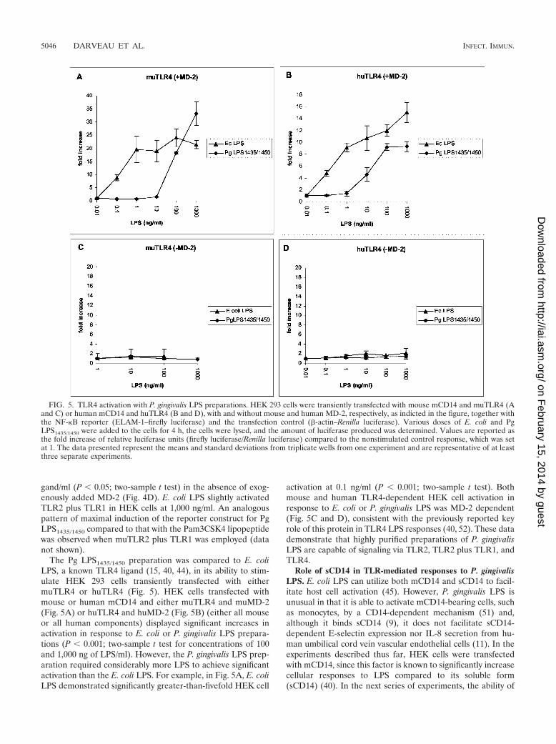
gand/ml (P 0.05; two-sample t test) in the absence of exog-enously added MD-2 (Fig. 4D). E. coli LPS slightly activatedTLR2 plus TLR1 in HEK cells at 1,000 ng/ml. An analogouspattern of maximal induction of the reporter construct for PgLPS1435/1450 compared to that with the Pam3CSK4 lipopeptidewas observed when muTLR2 plus TLR1 was employed (datanot shown).
The Pg LPS1435/1450 preparation was compared to E. coliLPS, a known TLR4 ligand (15, 40, 44), in its ability to stim-ulate HEK 293 cells transiently transfected with eithermuTLR4 or huTLR4 (Fig. 5). HEK cells transfected withmouse or human mCD14 and either muTLR4 and muMD-2(Fig. 5A) or huTLR4 and huMD-2 (Fig. 5B) (either all mouseor all human components) displayed significant increases inactivation in response to E. coli or P. gingivalis LPS prepara-tions (P 0.001; two-sample t test for concentrations of 100and 1,000 ng of LPS/ml). However, the P. gingivalis LPS prep-aration required considerably more LPS to achieve significantactivation than the E. coli LPS. For example, in Fig. 5A, E. coliLPS demonstrated significantly greater-than-fivefold HEK cell
activation at 0.1 ng/ml (P 0.001; two-sample t test). Bothmouse and human TLR4-dependent HEK cell activation inresponse to E. coli or P. gingivalis LPS was MD-2 dependent(Fig. 5C and D), consistent with the previously reported keyrole of this protein in TLR4 LPS responses (40, 52). These datademonstrate that highly purified preparations of P. gingivalisLPS are capable of signaling via TLR2, TLR2 plus TLR1, andTLR4.
Role of sCD14 in TLR-mediated responses to P. gingivalisLPS. E. coli LPS can utilize both mCD14 and sCD14 to facil-itate host cell activation (45). However, P. gingivalis LPS isunusual in that it is able to activate mCD14-bearing cells, suchas monocytes, by a CD14-dependent mechanism (51) and,although it binds sCD14 (9), it does not facilitate sCD14-dependent E-selectin expression nor IL-8 secretion from hu-man umbilical cord vein vascular endothelial cells (11). In theexperiments described thus far, HEK cells were transfectedwith mCD14, since this factor is known to significantly increasecellular responses to LPS compared to its soluble form(sCD14) (40). In the next series of experiments, the ability of
FIG. 5. TLR4 activation with P. gingivalis LPS preparations. HEK 293 cells were transiently transfected with mouse mCD14 and muTLR4 (Aand C) or human mCD14 and huTLR4 (B and D), with and without mouse and human MD-2, respectively, as indicted in the figure, together withthe NF-�B reporter (ELAM-1–firefly luciferase) and the transfection control (�-actin–Renilla luciferase). Various doses of E. coli and PgLPS1435/1450 were added to the cells for 4 h, the cells were lysed, and the amount of luciferase produced was determined. Values are reported asthe fold increase of relative luciferase units (firefly luciferase/Renilla luciferase) compared to the nonstimulated control response, which was setat 1. The data presented represent the means and standard deviations from triplicate wells from one experiment and are representative of at leastthree separate experiments.
5046 DARVEAU ET AL. INFECT. IMMUN.
on February 15, 2014 by guest
http://iai.asm.org/
Dow
nloaded from

P. gingivalis LPS to utilize sCD14 for TLR activation was ex-amined. Previous experiments have shown that FBS has suffi-cient bovine LBP and sCD14 to facilitate HEK cell activationwith both murine and human TLRs (data not shown). The roleof sCD14 in facilitating LPS activation of TLR-transfectedHEK cells was examined by transfecting HEK cells withoutmCD14 and employing sCD14-depleted human serum as asource for LBP (Fig. 6).
Human serum depleted of sCD14 demonstrated significantlylower muTLR2-plus-TLR1-dependent HEK cell activation forP. gingivalis LPS compared to the same transfection that em-ployed FBS during the cell activation (Fig. 6A). In addition, inother experiments (data not shown) recombinant sCD14 wasadded back to the sCD14-depleted serum and HEK cell acti-vation was restored to similar levels as observed when FBS waspresent in the activation buffer. These experiments demon-strated that sCD14 (human and bovine) can efficiently transferP. gingivalis LPS to muTLR2 plus muTLR1 to facilitate HEKcell activation. Next, the contribution of sCD14 for P. gingivalis
LPS activation of cells through murine and human TLR4 wasexamined (Fig. 6B and C). When FBS was added as a source ofboth LBP and sCD14, the Pg LPS1435/1450 preparation wasunable to mediate effective TLR4-dependent HEK cell activa-tion in comparison to that with E. coli LPS (Fig. 6B and C).This difference in HEK cell activation through TLR4 was mostdramatic for huTLR4, where P. gingivalis LPS yielded less thanfivefold activation, compared to approximately 20-fold for E.coli LPS (Fig. 6C). These findings are in contrast to the dataobtained with mCD14 (Fig. 5), where maximal TLR4 activa-tion was nearly equivalent for both E. coli and P. gingivalis LPS.Thus, these data reveal a relatively poor ability of P. gingivalisLPS to utilize human sCD14 for huTLR4-dependent HEK cellactivation.
P. gingivalis LPS enriched in lipid A mass ions of m/z 1,435and 1,450 activates primary bone marrow cells obtained fromboth TLR2 and TLR4 knockout mice. The HEK transient-transfection experiments with either TLR2 or TLR4 demon-strated that the Pg LPS1435/1450 preparation was able to engage
FIG. 6. Contribution of sCD14 for TLR activation by P. gingivalis LPS preparations. HEK 293 cells were transiently transfected with MD-2 andwith muTLR2 plus TLR1 (A), muTLR4 (B), or huTLR4 (C), together with the NF-�B reporter (ELAM-1–firefly luciferase) and the transfectioncontrol (�-actin–Renilla luciferase). Various concentrations Pg LPS1435/1450 and E. coli LPS (B and C) were added to the cells in activation buffercontaining either sCD14-depleted human serum as a source for LBP (dotted lines) or FBS as a source for both LBP and sCD14 (solid lines). TheHEK cells were incubated with the LPS preparations for 4 h, the cells were lysed, and the amount of luciferase produced was determined. Thedata presented represent the means and standard deviations from triplicate wells from one experiment and are representative of at least threeseparate experiments.
VOL. 72, 2004 P. GINGIVALIS LPS 5047
on February 15, 2014 by guest
http://iai.asm.org/
Dow
nloaded from

either TLR receptor to activate the NF-�B-dependent reporterconstruct. The ability of this P. gingivalis LPS preparation toactivate mouse bone marrow cells obtained from TLR2�/�,TLR4�/�, and TLR2/4�/� knockout mice was examined inorder to determine if endogenous TLR2 and TLR4 were uti-lized. Activation was determined by intracellular TNF-� stain-ing and fluorescence-activated cell sorter analysis (Fig. 7). Itwas found that bone marrow cells obtained from eitherTLR2�/� or TLR4�/� mice responded to the Pg LPS1435/1450
preparation. These data are consistent with the observationsmade in HEK cells that demonstrated this P. gingivalis LPSpreparation could utilize either TLR2 or TLR4 for host cellactivation. Furthermore, cells obtained from TLR2/4 double-knockout mice did not respond to the P. gingivalis LPS prep-aration, demonstrating that the LPS activity observed with thiscell type was mediated exclusively through TLR2 and TLR4. Incontrast, bone marrow cell responses to Salmonella minnesotaLPS (a TLR4 ligand) and Pam3CSK4 (a TLR2 ligand) dem-onstrated that these microbial ligands utilized their respectivecognate TLR. These data provide good evidence that P. gingi-valis LPS can utilize TLR2 and TLR4 under conditions wherethey are expressed at endogenous levels.
DISCUSSION
Different structures have been reported for the lipid A ob-tained from P. gingivalis LPS (32, 41). Ogawa (41) reported the
major lipid A species as a triacylated monophosphorylatedform with a mass of m/z 1,195. In contrast, Kumada et al. (32)reported that P. gingivalis LPS preparations contain multiplelipid A species, with the largest being a pentacylated diphos-phorylated species displaying a mass ion of m/z 1,770. Both ofthe m/z 1,195 and 1,770 lipid A mass ions have been chemicallysynthesized, and evidence that they activate cells throughTLR4 and not TLR2 has been reported (33, 42). However,highly purified native P. gingivalis LPS or lipid A preparationsconsistently demonstrate TLR2 activity (5, 26, 33, 42). Traceamounts of tightly associated endotoxin proteins (34) or an-other as-yet-undefined P. gingivalis component (42) have beenproposed as being responsible for the TLR 2 activity in nativeP. gingivalis LPS preparations. In this report, both the proteincontent and fatty acid composition of the P. gingivalis LPS PgLPS1435/1450 preparation did not support contaminants as be-ing responsible for the TLR2 activity that was observed in theP. gingivalis LPS preparation. No detectable endotoxin-associ-ated protein was observed in SDS-PAGE. Rather, we proposethat the P. gingivalis LPS TLR2 activities observed both in theHEK cell and mouse bone marrow cell systems in this studywere due to the lipid A mass ions at m/z 1,435 and/or 1,450.These mass ions have been reported to be the major lipid Aspecies found in highly purified P. gingivalis LPS preparationsby both our laboratory (3) and Kumada et al. (32) and repre-sent the most likely candidates for the apparent discrepancybetween studies employing chemically synthesized lipid A spe-
FIG. 7. Pg LPS1435/1450 stimulates TNF-� production in primary bone marrow cells via TLR2 or TLR4. Bone marrow cells from the indicatedmice were stimulated with 10 �g of Pg LPS1435/1450/ml, 10 ng of S. minnesota Re595 LPS/ml, or 1 �g of Pam3CSK4/ml. The numbers in eachhistogram indicate the mean ( range) of the percentage of cells producing TNF-� from duplicate wells from a single mouse. The data are fromone experiment, and similar results were obtained in three separate experiments.
5048 DARVEAU ET AL. INFECT. IMMUN.
on February 15, 2014 by guest
http://iai.asm.org/
Dow
nloaded from

cies and native preparations. The structures for the lipid Amass ions at m/z 1,435 and 1,450 have been elucidated as beingtetra-acylated monophosphorylated species (32). However, inthis report, it was also found that the same highly purified P.gingivalis LPS preparation was capable of activating HEK andmouse bone marrow cells through TLR4. Both murine andhuman TLR4/MD-2 systems were capable of responding to P.gingivalis LPS. This is unusual, since it has been shown thatmuTLR4 but not huTLR4 can respond to tetra-acylated E. coliLPS (44). Since the major P. gingivalis lipid A species examinedin this report were tetra-acylated (m/z 1,435 and 1,450), it ispossible that human TLR4 can detect tetra-acylated LPS fromdifferent species of bacteria. Alternatively, since the MALDI-TOF analysis of lipid A was not quantitative, it is not known ifthe major lipid A mass ions observed were responsible forTLR4 activation or if minor amounts of other lipid A species(e.g., m/z 1,770) facilitated activation through this receptor.
The biological significance of the multiple lipid A speciesfound in P. gingivalis LPS preparations is not currently under-stood. A preliminary examination has revealed that severaldifferent P. gingivalis laboratory strains and clinical isolatesdisplay a similar pattern of lipid A heterogeneity as that shownin this work, demonstrating that the major lipid A mass ionsare not strain specific (data not shown). Several other bacterialspecies have been shown to contain multiple lipid A forms thatexist in a single bacterial population (2, 47, 48, 53, 58) and thatcan be regulated by incubation temperature (29) or the con-centration of Mg2� in the growth medium (17). In Salmonellaenterica serovar Typhimurium, additional lipid A species thatalter the human endothelial cell response of purified LPS havebeen shown to be under the control of a phoP/phoQ two-component regulatory system (21). Similarly, P. gingivalis mayalso synthesize multiple lipid A species, accounting for theheterogeneity observed in purified LPS preparations (5, 32).Conversely, P. gingivalis lipid A heterogeneity may arise due toisolation and characterization procedures. For example, isola-tion of LPS often involves heating in phenol, and examinationof lipid A requires acid hydrolysis of the lipid A from the3-deoxy-D-manno-octulosonic acid core sugar, and these pro-cedures may result in cleavage of acid-labile fatty acids andphosphate residues. However, our laboratory has found that P.gingivalis LPS preparations contain the 1,435 and 1,450 lipid Aspecies as the major peaks in MALDI-TOF analysis wheneither the phenol-water (63) or MgCl2 LPS (13) extractionprocedures are employed or if the LPS is subjected to twodifferent lipid A hydrolysis procedures (7, 41) (data notshown). This strongly suggests that the lipid A mass ions foundat m/z 1,435 and 1,450 did not arise due to the type of LPSextraction or lipid A hydrolysis procedures. Additional studieswill be required to determine the origin of P. gingivalis lipid Aheterogeneity; nevertheless, it is clear that purified P. gingivalisLPS preparations may contain more than one lipid A species,and this may account for the discrepancy in host cell activationassays observed between chemically synthesized lipid As andnative highly purified preparations.
Transient HEK cell transfection experiments provide an ex-cellent means to determine the components of the TLR com-plex necessary to facilitate host cell responses. In this report, itwas found that MD-2 was not required for P. gingivalis LPSTLR2-dependent, or TLR2-plus-TLR1-dependent, HEK cell
activation but was necessary for activation with mouse or hu-man TLR4. It is interesting that in another HEK system, en-dogenous levels of MD-2 were sufficient to facilitate TLR4 E.coli LPS activation (16). This may explain why in a studyemploying transiently transfected HEK cells (where the endog-enous levels of MD-2 were not reported), TLR4-dependentcell activation with phenol-extracted P. gingivalis LPS was notobserved (26). The requirement for different TLR cofactorsfor cell stimulation is consistent with other observations (16,40, 61) that have shown each bacterial ligand may requiredifferent components of the TLR signaling complex to facili-tate host cell activation.
Furthermore, sCD14 amply replaced mCD14 to yield highlysignificant P. gingivalis LPS TLR2-plus-TLR1-dependent HEKcell activation, as demonstrated both by significant activationat low LPS concentrations (10 ng/ml) and a greater-than-15-fold increase over background at higher concentrations of LPS.However, huTLR4-dependent HEK cell activation employingsCD14 revealed that the Pg LPS1435/1450 preparation onlyslightly activated these cells above background control levels at100 and 1,000 ng of LPS/ml. These data demonstrate that theTLR activity of P. gingivalis LPS preparations is particularlyprone to the presence of key accessory molecules, such assoluble or membrane CD14. In addition, it has previously beenshown that P. gingivalis LPS can utilize hamster but not humanTLR4 when combined with endogenous hamster MD-2 (68),further emphasizing that P. gingivalis LPS-dependent host cellactivation, in contrast to that with E. coli LPS, is more sensitiveto the species specificity of the TLR complex components.These factors, combined with the potential for multiple lipid Aspecies in P. gingivalis LPS preparations, almost certainly havecontributed to the variability reported for TLR utilization by P.gingivalis LPS.
One implication of the work presented here is that P. gingi-valis LPS may activate host cells through either a TLR2- orTLR4-dependent pathway. The results of HEK cell transfec-tion assays and bone marrow cell activation experiments dem-onstrate that certain P. gingivalis LPS preparations have theability to interact with either TLR2 or TLR4. Another relatedimplication is that the lipid A heterogeneity observed in P.gingivalis LPS preparations may reflect an ability of this bac-terium to synthesize and express multiple, structurally differentforms of lipid A. Alterations in the lipid A structural compo-sition and utilization of multiple TLRs may affect host cellsignaling, contributing to the ability of P. gingivalis to remain apersistent colonizer of the oral cavity as well as to induceinflammatory disease.
ACKNOWLEDGMENTS
We thank Christopher B. Wilson for his advice and generosity withthe TLR expression reagents. We also thank Shizuo Akira for the TLRknockout mice.
This work was supported in part by NIDCR grant DE12768 toR.P.D.
REFERENCES
1. Aliprantis, A. O., R. B. Yang, M. R. Mark, S. Suggett, B. Devaux, J. D.Radolf, G. R. Klimpel, P. Godowski, and A. Zychlinsky. 1999. Cell activationand apoptosis by bacterial lipoproteins through toll-like receptor-2. Science285:736–739.
2. Aussel, L., J. R. Brisson, M. B. Perry, and M. Caroff. 2000. Structure of thelipid A of Bordetella hinzii ATCC 51730. Rapid Commun. Mass Spectrom.14:595–599.
VOL. 72, 2004 P. GINGIVALIS LPS 5049
on February 15, 2014 by guest
http://iai.asm.org/
Dow
nloaded from

3. Bainbridge, B. W., S. R. Coats, and R. P. Darveau. 2002. Porphyromonasgingivalis lipopolysaccharide displays functionally diverse interactions withthe innate host defense system. Ann. Periodontol. 7:1–9.
4. Bainbridge, B. W., and R. P. Darveau. 1997. Lipopolysaccharide from oralbacteria: role in innate host defense and chronic inflammatory disease, p.899–913. In D. Morrison (ed.), Endotoxin in health and disease. MarcelDekker, New York, N.Y.
5. Bainbridge, B. W., and R. P. Darveau. 2001. Porphyromonas gingivalis lipo-polysaccharide: an unusual pattern recognition receptor ligand for the innatehost defense system. Acta Odontol. Scand. 59:131–138.
6. Beutler, B. 2000. Tlr4: central component of the sole mammalian LPS sen-sor. Curr. Opin. Immunol. 12:20–26.
7. Caroff, M., A. Tacken, and L. Szabo. 1988. Detergent-accelerated hydrolysisof bacterial endotoxins and determination of the anomeric configuration ofthe glycosyl phosphate present in the “isolated lipid A” fragment of theBordetella pertussis endotoxin. Carbohydr. Res. 175:273–282.
8. Coats, S. R., R. A. Reife, B. W. Bainbridge, T. T. Pham, and R. P. Darveau.2003. Porphyromonas gingivalis lipopolysaccharide antagonizes Escherichiacoli lipopolysaccharide at toll-like receptor 4 in human endothelial cells.Infect. Immun. 71:6799–6807.
9. Cunningham, M. D., C. Seachord, K. Ratcliffe, B. Bainbridge, A. Aruffo, andR. P. Darveau. 1996. Helicobacter pylori and Porphyromonas gingivalis lipo-polysaccharides are poorly transferred to recombinant soluble CD14. Infect.Immun. 64:3601–3608.
10. Darveau, R. P., S. Arbabi, I. Garcia, B. Bainbridge, and R. V. Maier. 2002.Porphyromonas gingivalis lipopolysaccharide is both an agonist and antago-nist for p38 mitogen-activated protein kinase activation. Infect. Immun.70:1867–1873.
11. Darveau, R. P., M. D. Cunningham, T. Bailey, C. Seachord, K. Ratcliffe, B.Bainbridge, M. Dietsch, R. C. Page, and A. Aruffo. 1995. Ability of bacteriaassociated with chronic inflammatory disease to stimulate E-selectin expres-sion and promote neutrophil adhesion. Infect. Immun. 63:1311–1317.
12. Darveau, R. P., M. D. Cunningham, C. L. Seachord, L. Cassiano-Clough,W. L. Cosand, J. Blake, and C. S. Watkins. 1991. �-Lactam antibioticspotentiate magainin 2 antimicrobial activity in vitro and in vivo. Antimicrob.Agents Chemother. 35:1153–1159.
13. Darveau, R. P., and R. E. Hancock. 1983. Procedure for isolation of bacteriallipopolysaccharides from both smooth and rough Pseudomonas aeruginosaand Salmonella typhimurium strains. J. Bacteriol. 155:831–838.
14. Darveau, R. P., A. Tanner, and R. C. Page. 1997. The microbial challenge inperiodontitis. Periodontology 2000 14:12–32.
15. da Silva Correia, J., K. Soldau, U. Christen, P. S. Tobias, and R. J. Ulevitch.2001. Lipopolysaccharide is in close proximity to each of the proteins in itsmembrane receptor complex. Transfer from CD14 to TLR4 and MD-2.J. Biol. Chem. 276:21129–21135.
16. Dziarski, R., Q. Wang, K. Miyake, C. J. Kirschning, and D. Gupta. 2001.MD-2 enables Toll-like receptor 2 (TLR2)-mediated responses to lipopoly-saccharide and enhances TLR2-mediated responses to gram-positive andgram-negative bacteria and their cell wall components. J. Immunol. 166:1938–1944.
17. Ernst, R. K., E. C. Yi, L. Guo, K. B. Lim, J. L. Burns, M. Hackett, and S. I.Miller. 1999. Specific lipopolysaccharide found in cystic fibrosis airwayPseudomonas aeruginosa. Science 286:1561–1565.
18. Folch, J., M. Lees, and G. H. S. Stanley. 1957. A simple method for theisolation and purification of total lipids from animal tissues. J. Biol. Chem.226:497–509.
19. Grenier, D., J. Bertrand, and D. Mayrand. 1995. Porphyromonas gingivalisouter membrane vesicles promote bacterial resistance to chlorhexidine. OralMicrobiol. Immunol. 10:319–320.
20. Grenier, D., and D. Mayrand. 1987. Functional characterization of extracel-lular vesicles produced by Bacteroides gingivalis. Infect. Immun. 55:111–117.
21. Guo, L., K. B. Lim, J. S. Gunn, B. Bainbridge, R. P. Darveau, M. Hackett,and S. I. Miller. 1997. Regulation of lipid A modifications by Salmonellatyphimurium virulence genes, phoP-phoQ. Science 276:250–253.
22. Hajjar, A. M., R. K. Ernst, J. H. Tsai, C. B. Wilson, and S. I. Miller. 2002.Human Toll-like receptor 4 recognizes host-specific LPS modifications. Nat.Immunol. 3:354–359.
23. Hajjar, A. M., D. S. O’Mahony, A. Ozinsky, D. M. Underhill, A. Aderem, S. J.Klebanoff, and C. B. Wilson. 2001. Cutting edge: functional interactionsbetween Toll-like receptor (TLR) 2 and TLR1 or TLR6 in response tophenol-soluble modulin. J. Immunol. 166:15–19.
24. Hart, T. C., L. Shapira, and T. E. Van Dyke. 1994. Neutrophil defects as riskfactors for periodontal diseases. J. Periodontol. 65:521–529.
25. Haziot, A., N. Hijiya, S. C. Gangloff, J. Silver, and S. M. Goyert. 2001.Induction of a novel mechanism of accelerated bacterial clearance by lipo-polysaccharide in CD14-deficient and Toll-like receptor 4-deficient mice.J. Immunol. 166:1075–1078.
26. Hirschfeld, M., J. J. Weis, V. Toshchakov, C. A. Salkowski, M. J. Cody, D. C.Ward, N. Qureshi, S. M. Michalek, and S. N. Vogel. 2001. Signaling bytoll-like receptor 2 and 4 agonists results in differential gene expression inmurine macrophages. Infect. Immun. 69:1477–1482.
27. Janeway, C. J. 1992. The immune system evolved to discriminate infectiousnonself from noninfectious self. Immunol. Today 13:11–16.
28. Jin, L., and R. P. Darveau. 2001. Soluble CD14 levels in gingival crevicularfluid of subjects with untreated adult periodontitis. J. Periodontol. 72:634–640.
29. Kawahara, K., H. Tsukano, H. Watanabe, B. Lindner, and M. Matsuura.2002. Modification of the structure and activity of lipid A in Yersinia pestislipopolysaccharide by growth temperature. Infect. Immun. 70:4092–4098.
30. Khan, S. A., P. Everest, S. Servos, N. Foxwell, U. Zahringer, H. Brade, E. T.Rietschel, G. Dougan, I. G. Charles, and D. J. Maskell. 1998. A lethal rolefor lipid A in Salmonella infections. Mol. Microbiol. 29:571–579.
31. Kopp, E. B., and R. Medzhitov. 1999. The Toll-receptor family and controlof innate immunity. Curr. Opin. Immunol. 11:13–18.
32. Kumada, H., Y. Haishima, T. Umemoto, and K.-I. Tanamoto. 1995. Struc-tural study on the free lipid A isolated from lipopolysaccharide of Porphy-romonas gingivalis. J. Bacteriol. 177:2098–2106.
33. Kumada, H., T. Umemoto, K. Watanabe, H. Tamura, T. Murai, Y. Naka-gawa, K.-I. Tanamoto, and Y. Haishima. 2002. Biological properties of thenative and synthetic lipid A of Porphyromonas gingivalis lipopolysaccharide.J. Dent. Res. 81:A-197.
34. Lee, H. K., J. Lee, and P. S. Tobias. 2002. Two lipoproteins extracted fromEscherichia coli K-12 LCD25 lipopolysaccharide are the major componentsresponsible for Toll-like receptor 2-mediated signaling. J. Immunol. 168:4012–4017.
35. Maniatis, T., E. F. Fritsch, and J. Sambrook. 1982. Molecular cloning: alaboratory manual, p. 468–492. Cold Spring Harbor Laboratory Press, ColdSpring Harbor, N.Y.
36. Manthey, C. L., and S. N. Vogel. 1994. Elimination of trace endotoxinprotein from rough chemotype LPS. J. Endotoxin Res. 1:84–91.
37. Martin, M., J. Katz, S. N. Vogel, and S. M. Michalek. 2001. Differentialinduction of endotoxin tolerance by lipopolysaccharides derived from Por-phyromonas gingivalis and Escherichia coli. J. Immunol. 167:5278–5285.
38. McCoy, S. A., H. R. Creamer, M. Kawanami, and D. F. Adams. 1987. Theconcentration of lipopolysaccharide on individual root surfaces at varyingtimes following in vivo root planing. J. Periodontol. 58:393–399.
39. Moore, J., M. Wilson, and J. B. Kieser. 1986. The distribution of bacteriallipopolysaccharide (endotoxin) in relation to periodontally involved rootsurfaces. J. Clin. Periodontol. 13:748–751.
40. Muta, T., and K. Takeshige. 2001. Essential roles of CD14 and lipopolysac-charide-binding protein for activation of toll-like receptor (TLR)2 as well asTLR4. Reconstitution of TLR2- and TLR4-activation by distinguishableligands in LPS preparations. Eur. J. Biochem. 268:4580–4589.
41. Ogawa, T. 1993. Chemical structure of lipid A from Porphyromonas (Bacte-roides) gingivalis lipopolysaccharide. FEBS Lett. 332:197–201.
42. Ogawa, T., Y. Asai, M. Hashimoto, O. Takeuchi, T. Kurita, Y. Yoshikai, K.Miyake, and S. Akira. 2002. Cell activation by Porphyromonas gingivalis lipidA molecule through Toll-like receptor 4- and myeloid differentiation factor88-dependent signaling pathway. Int. Immunol. 14:1325–1332.
43. Peterson, A. A., A. Haug, and E. J. McGroarty. 1986. Physical properties ofshort- and long-O-antigen-containing fractions of lipopolysaccharide fromEscherichia coli O111:B4. J. Bacteriol. 165:116–122.
44. Poltorak, A., P. Ricciardi-Castagnoli, S. Citterio, and B. Beutler. 2000.Physical contact between lipopolysaccharide and toll-like receptor 4 revealedby genetic complementation. Proc. Natl. Acad. Sci. USA 97:2163–2167.
45. Pugin, J., C.-C. Schurer-Maly, D. Leturcq, A. Moriarty, R. J. Ulevitch, andP. S. Tobias. 1993. Lipopolysaccharide activation of human endothelial andepithelial cells is mediated by lipopolysaccharide-binding protein and solubleCD14. Proc. Natl. Acad. Sci. USA 90:2744–2748.
46. Putnins, E. E., A.-R. Sanaie, Q. Wu, and J. D. Firth. 2002. Induction ofkeratinocyte growth factor 1 expression by lipopolysaccharide is regulated byCD14 and Toll-like receptors 2 and 4. Infect. Immun. 70:6541–6548.
47. Que, N. L., S. Lin, R. J. Cotter, and C. R. Raetz. 2000. Purification and massspectrometry of six lipid A species from the bacterial endosymbiont Rhizo-bium etli. Demonstration of a conserved distal unit and a variable proximalportion. J. Biol. Chem. 275:28006–28016.
48. Rund, S., B. Lindner, H. Brade, and O. Holst. 1999. Structural analysis of thelipopolysaccharide from Chlamydia trachomatis serotype L2. J. Biol. Chem.274:16819–16824.
49. Schumann, R. R., S. R. Leong, G. W. Flaggs, P. W. Gray, S. D. Wright, J. C.Mathison, P. S. Tobias, and R. J. Ulevitch. 1990. Structure and function oflipopolysaccharide binding protein. Science 249:1429–1431.
50. Schwartz, J., F. L. Stinson, and R. B. Parker. 1972. The passage of tritiatedbacterial endotoxin across intact gingival crevicular epithelium. J. Periodon-tol. 43:270–276.
51. Shapira, L., S. Takashiba, S. Amar, and T. E. Van Dyke. 1994. Porphyromo-nas gingivalis lipopolysaccharide stimulation of human monocytes: depen-dence on serum and CD14 receptor. Oral Microbiol. Immunol. 9:112–117.
52. Shimazu, R., S. Akashi, H. Ogata, Y. Nagai, K. Fukudome, K. Miyake, andM. Kimoto. 1999. MD-2, a molecule that confers lipopolysaccharide respon-siveness on Toll-like receptor 4. J. Exp. Med. 189:1777–1782.
53. Silipo, A., R. Lanzetta, A. Amoresano, M. Parrilli, and A. Molinaro. 2002.Ammonium hydroxide hydrolysis: a valuable support in the MALDI-TOF
5050 DARVEAU ET AL. INFECT. IMMUN.
on February 15, 2014 by guest
http://iai.asm.org/
Dow
nloaded from

mass spectrometry analysis of lipid A fatty acid distribution. J. Lipid Res.43:2188–2195.
54. Socransky, S. S., and A. D. Haffajee. 1992. The bacterial etiology of destruc-tive periodontal disease: current concepts. J. Periodontol. 63:322–331.
55. Somerville, J. E., Jr., L. Cassiano, and R. P. Darveau. 1999. Escherichia colimsbB gene as a virulence factor and a therapeutic target. Infect. Immun.67:6583–6590.
56. Somerville, J. E. J., L. Cassiano, B. Bainbridge, M. D. Cunningham, andR. P. Darveau. 1996. A novel Escherichia coli lipid A mutant that producesan antiinflammatory lipopolysaccharide. J. Clin. Investig. 97:359–365.
57. Tabeta, K., K. Yamazaki, S. Akashi, K. Miyake, H. Kumada, T. Umemoto,and H. Yoshie. 2000. Toll-like receptors confer responsiveness to lipopoly-saccharide from Porphyromonas gingivalis in human gingival fibroblasts. In-fect. Immun. 68:3731–3735.
58. Therisod, H., M. A. Monteiro, M. B. Perry, and M. Caroff. 2001. Helicobactermustelae lipid A structure differs from that of Helicobacter pylori. FEBS Lett.499:1–5.
59. Tonetti, M. S., M. A. Imboden, and N. P. Lang. 1998. Neutrophil migrationinto the gingival sulcus is associated with transepithelial gradients of inter-leukin-8 and ICAM-1. J. Periodontol. 69:1139–1147.
60. Ulevitch, R. J., and P. S. Tobias. 1999. Recognition of gram-negative bac-teria and endotoxin by the innate immune system. Curr. Opin. Immunol.11:19–22.
61. Viriyakosol, S., J. C. Mathison, P. S. Tobias, and T. N. Kirkland. 2000.
Structure-function analysis of CD14 as a soluble receptor for lipopolysac-charide. J. Biol. Chem. 275:3144–3149.
62. Wang, P. L., and K. Ohura. 2002. Porphyromonas gingivalis lipopolysaccha-ride signaling in gingival fibroblasts—CD14 and Toll-like receptors. Crit.Rev. Oral Biol. Med. 13:132–142.
63. Westphal, O., and K. Jann. 1965. Bacterial lipopolysaccharides: extractionwith phenol-water and further applications of the procedure, p. 83. In R.Whistler (ed.), Methods in carbohydrate chemistry, vol. 5. Academic Press,Inc., New York, N.Y.
64. Wright, S. D., R. A. Ramos, P. S. Tobias, R. J. Ulevitch, and J. C. Mathison.1990. CD14, a receptor for complexes of lipopolysaccharide (LPS) and LPSbinding protein. Science 249:1431–1433.
65. Wyllie, D. H., E. Kiss-Toth, A. Visintin, S. C. Smith, S. Boussouf, D. M.Segal, G. W. Duff, and S. K. Dower. 2000. Evidence for an accessory proteinfunction for the Toll-like receptor TLR1 in lipopolysaccharide responses.J. Immunol. 165:7125–7132.
66. Ximenez-Fyvie, L. A., A. D. Haffajee, and S. S. Socransky. 2000. Comparisonof the microbiota of supra- and subgingival plaque in health and periodon-titis. J. Clin. Periodontol. 27:648–657.
67. Yi, E. C., and M. Hackett. 2000. Rapid isolation method for lipopolysaccha-ride and lipid A from gram-negative bacteria. Analyst 125:651–656.
68. Yoshimura, A., T. Kaneko, Y. Kato, D. T. Golenbock, and Y. Hara. 2002.Lipopolysaccharides from periodontopathic bacteria Porphyromonas gingiva-lis and Capnocytophaga ochracea are antagonists for human toll-like receptor4. Infect. Immun. 70:218–225.
Editor: J. D. Clements
VOL. 72, 2004 P. GINGIVALIS LPS 5051
on February 15, 2014 by guest
http://iai.asm.org/
Dow
nloaded from
Copyright © 2022 FDOKUMEN