Photonic crystal to photonic crystal surface modes: narrow-bandpass filters

Journal of Fluorine Chemistry 163 (2014) 16–22
Quantitative crystal structure analysis of fluorinated porphyrins
Rahul Soman, Subramaniam Sujatha, Chellaiah Arunkumar *
Bioinorganic Materials Research Laboratory, Department of Chemistry, National Institute of Technology Calicut, Kozhikode 673 601, Kerala, India
A R T I C L E I N F O
Article history:
Received 18 February 2014
Received in revised form 31 March 2014
Accepted 2 April 2014
Available online 13 April 2014
Keywords:
Supramolecular chemistry
Metalloporphyrins
Crystal structure analysis
Intermolecular interactions
Hirshfeld surface analysis
A B S T R A C T
Herein, we report a series of fluorinated porphyrins, 5,10,15,20-tetrakis(20,60-difluorophenyl)porphyrin,
MT(20,60-DFP)P where M = 2H, 1; Co(II)�(MeOH)2, 2; Cu(II), 3; Zn(II)�(MeOH)2, 4 and Zn(II)�(THF)3, 5 have
been structurally characterized by single crystal X-ray diffraction analysis. All the compounds are
crystallized in monoclinic crystal system and the crystal structures of 2, 4 and 5 features octahedral
geometry whereas 3 exhibits square planar geometry. The compounds 1, 3 and 2, 4 are isostructural,
show similar molecular crystal packing and comparable intermolecular interactions. The supramolecu-
lar self assembly of compounds 1–5 is dominated by a variety of intermolecular interactions such as C–
H� � �F, C–H� � �p, C–F� � �p and F� � �F. Furthermore, the role of weak intermolecular interactions in the
crystal packing has been analysed and quantified using Hirshfeld surface analysis.
� 2014 Elsevier B.V. All rights reserved.
Contents lists available at ScienceDirect
Journal of Fluorine Chemistry
jo ur n al h o mep ag e: www .e lsev ier . c om / loc ate / f luo r
1. Introduction
The well-known definition of crystal engineering is quoted byDesiraju as ‘‘the understanding of intermolecular interactions in the
context of crystal packing and the utilization of such understanding in
design of new solids with desired physical and chemical properties’’[1]. Intermolecular interactions in organic/inorganic compoundsplay an important role in the crystal packing of molecules whichattracts intense attention in various research fields includingsupramolecular chemistry [2–4]. Such interactions involvingfluorine are of mainly three kinds, namely C–F� � �H, F� � �F and C–F� � �p which provides stability to form molecular self-assembliesespecially in the absence of strong intermolecular forces [5–8].Even though these individual interactions are energetically weak,the net result may become considerably strong due to the co-operative effect and thus may provide a higher stability to thecrystalline lattice. In addition, supramolecular assembly is framedby suitable combination of various types of weak intermolecularinteractions like C–H� � �p and the geometrical preference of themetal ion. Owing to the high electronegativity of fluorine, itrenders the C–F bond highly polar and by introducing the fluorineatoms into the organic moieties result in significant changes in thephysical properties and biological activities compared with non-fluorinated precursors [9–11]. It is well known that the fluorinateddrugs are innumerable and the study of weak intermolecularinteractions involving fluorine is essential in the field of medicinal
* Corresponding author. Tel.: +91 9746244507; fax: +91 495 228 7250.
E-mail address: [email protected] (C. Arunkumar).
http://dx.doi.org/10.1016/j.jfluchem.2014.04.002
0022-1139/� 2014 Elsevier B.V. All rights reserved.
chemistry. Porphyrins and their metal derivatives have beenwidely studied due to their distinct spectral, electrochemicalproperties and novel biological activities. Synthetic porphyrinanalogues are considered to be the ligands of choice to mimicnatural processes, because of their close resemblance to naturalsystems in both structure and properties [12]. Study of weakinteractions involving halogenated porphyrins offers an emergingfield of research in crystal engineering [13]. A diverse range of self-assembly patterns of porphyrin have been discovered, heldtogether by van der Waals interactions, hydrogen bonding, ormetal coordination [14–16]. In order to explore the betterunderstanding of intermolecular interactions to the supramolecu-lar assembly, it is crucial to get quantitative measurements of theseinteractions. Hirshfeld surface analysis [17,18] is becoming avaluable tool for elucidating molecular crystal structures quanti-tatively and such quantitative measures of weak interactions inporphyrins are poorly documented in the literature [16,19]. In thiscontext, we hereby report the crystal structures and Hirshfeldsurface analysis of 5,10,15,20-tetrakis(20,60-difluorophenyl) por-phyrin, H2T(20,60-DFP)P and its metal complexes (Fig. 1). And also,the effect of 2,6-difluorphenyl groups is discussed on theevaluation of intermolecular interactions.
2. Experimental
2.1. Materials and synthetic procedures
All the chemicals employed here for the synthesis werecommercially available reagents of analytical grade and wereused without further purification. Solvents used for the synthesis
N
N
N
N
F
F
F
F
F
FF
F
M
M = 2H,1; Co( II), 2; Cu( II), 3 and Zn(II), 4
Fig. 1. Molecular structure of porphyrins under study.
R. Soman et al. / Journal of Fluorine Chemistry 163 (2014) 16–22 17
were purified using the available literature methods [20].5,10,15,20-Tetrakis(20,60-difluorophenyl) porphyrin, H2T(20,60-DFP)P ligand, 1, was synthesized using the method reported byLindsey et al. [21] and the metal complexes were prepared by theliterature method [22] to afford the desired complexes yielded in92–95%.
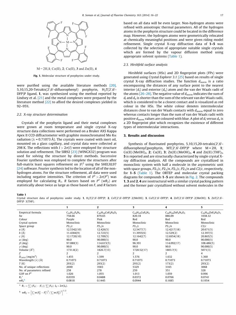
2.2. X-ray structure determination
Crystals of the porphyrin ligand and their metal complexeswere grown at room temperature and single crystal X-raystructure data collections were performed on a Bruker AXS KappaApex II CCD diffractometer with graphite monochromated Mo Karadiation (l = 0.71073 A). The crystals were coated with inert oil,mounted on a glass capillary, and crystal data were collected at298 K. The reflections with I > 2s(I) were employed for structuresolution and refinement. The SIR92 [23] (WINGX32) program wasused for solving the structure by direct methods. SuccessiveFourier synthesis was employed to complete the structures afterfull-matrix least squares refinement on jFj2 using the SHELXL97[24] software. Fourier synthesis led to the location of all of the non-hydrogen atoms. For the structure refinement, all data were usedincluding negative intensities. The criterion of F2 > 2s(F2) wasemployed for calculating R1. R factors based on F2 (wR2) arestatistically about twice as large as those based on F, and R factors
Table 1Crystal structure data of porphyrins under study. 1, H2T(20 ,60-DFP)P; 2, CoT(20 ,60-
DFP)P � 3(THF).
1 2
Empirical formula C44H22F8N4 C46H28CoF8N4O2
fw 758.66 879.65
Colour Purple Pink
Crystal system Monoclinic Monoclinic
Space group P21/c P21/n
a (A) 12.5342(10) 12.426(5)
b (A) 11.4384(9) 12.723(5)
c (A) 12.1720(10) 12.709(5)
a (deg) 90.0 90.000(5)
b (deg) 97.080(3) 114.615(5)
g (deg) 90.0 90.000(5)
Volume (A3) 1731.8(2) 1826.7(13)
Z 2 2
Dcalcd (mg/m3) 1.455 1.599
Wavelength (l) (A) 0.71073 0.71073
T (K) 293(2) 293(2)
No. of unique reflections 2690 3583
No. of parameters refined 258 278
GOF on F2 1.026 1.055
R1a 0.0359 0.0488
wR2b 0.0818 0.1445
a R1 ¼PjjF0j � jFcjj=
PjF0j; I0> 2s(I0).
b wR2 ¼P
wðF20 � F2
c Þ2=P
wðF20 Þ
2h i1=2
:
based on all data will be even larger. Non-hydrogen atoms wererefined with anisotropic thermal parameters. All of the hydrogenatoms in the porphyrin structure could be located in the differencemap. However, the hydrogen atoms were geometrically relocatedat chemically meaningful positions and were given riding modelrefinement. Single crystal X-ray diffraction data of 1–5 wascollected by the selection of appropriate suitable single crystalswhich are formed by the vapour diffusion method usingappropriate solvent systems (Table 1).
2.3. Hirshfeld surface analysis
Hirshfeld surfaces (HSs) and 2D fingerprint plots (FPs) weregenerated using Crystal Explorer 3.1 [25] based on results of singlecrystal X-ray diffraction studies. The function dnorm is a ratioencompassing the distances of any surface point to the nearestinterior (di) and exterior (de) atom and the van der Waals radii ofthe atoms [26–28]. The negative value of dnorm indicates the sum ofdi and de is shorter than the sum of the relevant van der Waals radii,which is considered to be a closest contact and is visualized as redcolour in the HSs. The white colour denotes intermoleculardistances close to van der Waals contacts with dnorm equal to zerowhereas contacts longer than the sum of van der Waals radii withpositive dnorm values are coloured with blue. A plot of di versus de isa 2D fingerprint plot which recognizes the existence of differenttypes of intermolecular interactions.
3. Results and discussion
Synthesis of fluorinated porphyrins, 5,10,15,20-tetrakis(20,60-difluorophenyl)porphyrin, MT(20,60-DFP)P where M = 2H, 1;Co(II)�(MeOH)2, 2; Cu(II), 3; Zn(II)�(MeOH)2, 4 and Zn(II)�(THF)3,5 is reported and are structurally characterized by single crystal X-ray diffraction analysis. All the compounds are crystallized inmonoclinic system with half a molecule in the asymmetric unitwith space groups, P21/c, P21/n, P21/c, P21/n and C2/c, respectively,for 1–5 (Table 1). The ORTEP and molecular crystal packingdiagrams for compounds 1–5 are shown in Fig. 2. The compounds1, 3 and 2, 4 are isostructural with a similar crystal packing patternand the former pair crystallized without solvent molecules in the
DFP)P � 2(MeOH); 3, CuT(20,60-DFP)P; 4, ZnT(20 ,60-DFP)P � 2(MeOH); 5, ZnT(20 ,60-
3 4 5
C44H20CuF8N4 C46H28ZnF8N4O2 C56H44ZnF8N4O3
820.18 886.09 1038.32
Red Red Red
Monoclinic Monoclinic Monoclinic
P21/c P21/n C2/c
12.5477(7) 12.4217(18) 20.671(5)
11.3955(6) 12.529(2) 12.397(5)
12.1642(7) 12.6954(18) 20.865(5)
90.0 90.0 90.000(5)
96.391 114.092(17) 108.486(5)
90.0 90.0 90.000(5)
1728.52(17) 1803.7(5) 5071(3)
2 2 4
1.576 1.632 1.360
0.71073 0.71073 0.71073
293(2) 173(2) 293(2)
5329 3165 3068
259 351 328
1.013 1.059 0.996
0.0372 0.0766 0.0741
0.0944 0.1685 0.1954

Fig. 2. (a) ORTEP diagram (solvent molecules and disordered components removed for clarity and shown in 40% probability ellipsoids) and (b) molecular crystal packing
diagram for 1, 2, 3 and 4 viewed down ‘c’, ‘a’, ‘c’ and ‘a’ axes, respectively.
R. Soman et al. / Journal of Fluorine Chemistry 163 (2014) 16–2218
crystal lattice. Attempts to collect the single crystals of THFsolvated cobalt complex 2 with suitable size for the X-raydiffraction analysis were unsuccessful. The mean plane calcula-tions of compound 1 shows the porphyrin plane is not to be ideallyplanar and with maximum deviation of 0.081(1) A from the meanplane for N2 atom alone. The two difluorophenyl moieties,‘C11–C16’ and ‘C17–C22’ are inclined to each other through adihedral angle of 82.718(6) and 65.858(5). Table S1 shows thehydrogen bonding interactions present in 1 and the moleculesrelated by c-glide (Symm #1: x, ½ � y, �½ + z) are linked throughC–H� � �p interactions with the centroid of the ring (N2�C6�C7�C8�C9)having a distance of 2.686 A to H19 #1. In addition, there is a C–H� � �F(C20–H20� � �F3) interaction (Table 2) and the above mentionedtwo interactions spread in the plane linking the molecules to forma 2D sheet of zigzag motifs (Fig. S3). Furthermore, the C–H� � �F (C15–H15� � �F1) interaction links the 2D sheets to form the 3D networkstabilizing the crystal lattice (Fig. S4). In compound 3 (counter partof 1), the tetra coordinated copper atom lies in the porphyrin coreand exhibits perfect square planar geometry with N1–Cu1–N2 bitangle of 908 which makes the porphyrin moiety more planar thanthe free ligand, 1.
In 2, the porphyrin macrocycle is planar within the limits ofstandard deviation and the cobalt atom is octahedrally coordinatedwith the core nitrogen atoms and two centro-symmetrically
Table 2Distances (in A) for the different types of interactions in the crystal packing of 1–5.
Interactionsa 1b 2b
(ph)C–H� � �F 2.499–2.652 (6) 2.651 (2)
(sol)C–H� � �F – 2.557, 2.566 (4)
C–H� � �p(ph) – 2.765–2.868 (8)
C–H� � �p(pyr) 2.760 (2) 2.844 (2)
O–H� � �p(ph) – 2.485–2.829 (6)
O–H� � �p(pyr) – –
C–F� � �p(ph) 3.091 (2) –
C–F� � �C(sol) – –
F� � �F – 2.901 (2)
C–H� � �O – 2.670 (2)
C–H� � �N 2.741 (2) –
a Different types.b Value in parenthesis gives the number of interactions of each types per molecule.
related methanol molecules which are coordinated at the apexposition of cobalt atom stayed in the porphyrin plane itself. Thedihedral angle between the porphyrin moiety and the twodifluorophenyl moieties are 68.078(7) for ‘C11–C16’ ring and88.998 (8) for ‘C17–C22’ ring, respectively. The Co–N distancesare equal within the limits of standard deviation and inagreement with the reported values [29]. The hydrogen of thecoordinated methanol forms OH� � �p interactions with thephenyl ring ‘C17–C22’, the shortest interaction distance is givenby O1–H1� � �C19 #1 equals 2.485 (5) A [Symm: #1: ½ � x, ½ + y,½ � z]. However, the average distance between the hydrogen tothat of the centroid of phenyl ring is 2.562 A. In addition, thereare two more kinds of intermolecular interactions such as C–H� � �F and C–H� � �p also shown in Table 2. Apart from these, F� � �Fand C–H� � �O contacts also contribute to the crystal packing of 2.In 4, the porphyrin core is planar and the zinc(II) ion is hexa-coordinated and stays in the plane of porphyrin moiety. Thedihedral angle between the porphyrin core and difluorophenylrings, ‘C11–C16’ & ‘C17–C22’ are 79.6(2)8 and 65.0(2)8, respec-tively, and the coordination geometry around zinc is same as thatof compound 2. The molecule and its 21 screw equivalentsinteract through O–H� � �p interaction [(O–H) Symm: #1: � � �p(C11–C16), #1: ½ � x, ½ + y, ½ � z] and the H1#1 distance withthe centroid of ‘C11–C16’ phenyl ring is 2.761 A.
3b 4b 5b
2.471–2.636 (6) 2.540 (2) –
– 2.489 (2) 2.638 (2)
– 2.471–2.828 (8) 2.890 (2)
2.766 (2) 2.740, 2.828 (4) 2.774 (2)
– 2.795 (2) –
– 2.471, 2.512 (4) –
3.063, 3.165 (4) – –
– 3.043 (2) –
– 2.782 (2) –
– 2.626 (2) 2.672 (2)
– – –

R. Soman et al. / Journal of Fluorine Chemistry 163 (2014) 16–22 19
In 5, the zinc(II) metal is hexa-coordinated with core nitrogensin the equatorial plane and two centres of symmetrically relatedtetrahydrofuran (THF) molecules at the apex position. In addition,there is an extra THF molecule present in the crystal lattice playinga key role in crystal packing of 5 (Fig. S5). The presence of zinc(II)ion makes porphyrin moiety planar within the limits of standarddeviation and it stays within the porphyrin plane. There is a weakC–H� � �p [C14–H14� � �p (N2�C6�C7�C8�C9)] interaction between the21 screw related molecules with a H14 centroid distance equal to2.872 A. However, the normal distance between the pyrrole ringand hydrogen atom is 2.75 A. The dihedral angle between the twodifluorophenyl planes of asymmetric unit is 89.67(3)8 and thedihedral angle between the porphyrin moiety and the twodifluorophenyl moieties of asymmetric unit are 81.78(3)8 for‘C11–C16’ ring and 88.20(2)8 for ‘C17–C22’ ring. The lattice THFmolecule mediates intermolecular link between molecules of 5through C–H� � �F (C27–H27A� � �F4 #1, Symm: #1: �½ + x, ½ + y, z)interaction. Note that, the crystal packing of the reported toluenesolvated FeIIT(20,60-DFP)P is stabilized mainly by F� � �H/C and M� � �Cinteractions [30].
The average Zn–N distances in 4 and 5 are 2.054(5) A and2.042(6) A respectively which is relatively higher than the reportedfour coordinated ZnTPP (2.037(2) A) and is quite similar to that offive and six coordinated ZnCPTBPP(THF), ZnTPP(THF)2 complexes(2.052 (4) A and 2.057(4) A), respectively [31–33]. The zinc(II) ionin 4 and 5 lies almost in the plane of the four nitrogen atoms andthe Zn–O bond length observed are 2.324(5) A and 2.430(8) Awhich are very well comparable with that of reported ZnTPP(THF)2
Fig. 3. (a) Hirshfeld surfaces mapped with (a) dnorm ranging from �0.40 A (red) to 2.50 A
ranging from 1.0 to 2.8 A for 1–5. (For interpretation of the references to colour in this
complex (2.380(2) A). The average N–Zn–O and N–Zn–N angles arefound to be 90.08(2) and 1808 in 4 and 5 showing no deviation ofthe planarity of the porphyrin macrocycle with central zinc(II)atom. The bound-THF solvate molecules in 5 show the well knownenvelope conformation as observed in the literature [19,33,34].
The geometrical analysis on the crystal structures of porphyrins1–5 reveals that there are no direct p–p interactions as well asintramolecular interactions. However, the crystal packing in theporphyrins mainly consists of five different kinds of weakinteractions involving carbon, fluorine, hydrogen and oxygen,viz., (ph,sol)C–H� � �F, C–H� � �p(ph,pyr), O–H� � �p(ph,pyr), C–F� � �p(ph,sol)
and C–H� � �O (Table 2). Apart from this, there are C–H� � �N and F� � �Finteractions present in 1 and 2, 4 respectively. The F� � �F contactdistance in 2 and 4 is found to be 2.901 A and 2.782 A, respectively,which is shorter than the reported values in non-porphyrinicfluorinated coordination complexes (2.7–3.6 A) [35,36]. Thezinc(II) complex of methanol solvate, 4 is associated with largenumber of interactions compared to that of THF solvate, 5. TheC� � �F interaction involving methanolic carbon is having shorterdistance (3.043 A) than the carbon involving phenyl ring (3.063–3.165 A) in 1 and 3. The crystal structures of 1 and 3 mainlycontains three types of interactions in the crystal packing such as
(ph)C–H� � �F, C–H� � �p(pyr), C–F� � �p(ph) and their distances is in therange of 2.471–2.652 A, 2.760–2.766 A and 3.063–3.165 A respec-tively. While in 2 and 4, the types are (ph,sol)C–H� � �F, C–H� � �p(ph,pyr),O–H� � �p(ph,pyr), F� � �F and C–H� � �O interactions. In 5, there are onlyfour types of interactions namely (sol)C–H� � �F, C–H� � �p(ph), C–H� � �p(pyr) and C–H� � �O. Out of the eight fluorine atoms present in
˚ (blue), (b) shape-index, (c) curvedness and (d) 2D finger print plots with di and de
figure legend, the reader is referred to the web version of the article.)

R. Soman et al. / Journal of Fluorine Chemistry 163 (2014) 16–2220
the porphyrin, only six of them are involved in the crystal packingof 2, 3 and 4, whereas in 1 and 5, it is of four and two respectively.Note that, the porphyrin ligand, 1 shows C–H� � �N interactionwhich is not seen in any of the other metal derivatives. Similarly,the carbon of methanol which is coordinated to the zinc(II) metalatom in 4 is having close contact with fluorine (F3) which is absentin other porphyrins. Insertion of copper(II) metal at the centre ofporphyrin did not make any appreciable change in geometry of themolecule except that the porphyrin moiety made more planar thanthe ligand. This unaltered geometry of the molecule is responsiblefor intermolecular interactions which remain same as that ofligand, 1. Hence, the copper complex and the porphyrin ligand areisostructural similar to that of reported 5,10,15,20-tetra-kis(20,30,40,50,60-pentafluorophenyl)porphyrin, H2TF5PP and itscopper complex [37]. The synthesis of isostructural Mo2+ porphy-rin and Mo2+ N-confused porphyrin complexes are also reported inthe literature [38].
In order to quantify the various intermolecular interactions, HSsand their associated finger print plots were calculated using Crystal
Explorer 3.1. The HSs of porphyrins, 1–5 are illustrated in Fig. 3showing surfaces that have been mapped with dnorm, shape indexand curvedness. For a given crystal structure, the Hirshfeld surfaceis unique and it suggests the possibility of obtaining additionalinsight into the crystal [39]. The quantitative measures like volume(VH), area (SH), globularity (G) and asphericity (V) were alsocomputed using HSs. The term, globularity [40] is found to be <1for all the porphyrins, 1–5 which indicates that the molecularsurface is more structured, not a sphere. Furthermore, theasphericity [41,42] is a measure of anisotropy which decreasesin the following order for the compounds: 1 > 3 > 4 > 2 > 5. Themolecular HSs generated for 1, 3 and 2, 4 have almost similarshapes reflecting the similar kind of crystal packing modes whichindicate the isostructural nature of 1, 3 and 2, 4. Although 2 and 4are isostructural, a closer look on Table 2 shows that there are fewdifferences in the (pyr/ph)C–H(ph/MeOH) contacts. Note that, in 4, thepyrrole ring contributes more towards C–H interaction (8 permolecule) seen as medium intense red spot in the HS compared tothat of 2 (2 per molecule) which is observed as light red spot (Fig.S7). Owing to the bulky nature of the THF molecule which is axiallycoordinated to the zinc centre in 5, makes it more isotropic, ie., thespherical nature of the whole entity compared to that of methanolsolvated porphyrins, 2 and 4. Further, the HSs of 1, 3 and 5 showsintense red spot on the de surface near the phenyl ring which is dueto close (ph/pyr)C� � �H(ph) contacts. In addition to this, N� � �H and C–F� � �H contacts are also seen as intense red spots in 1 and 3,respectively. The crystal packing of 2 is mainly controlled bydominant interactions between methanolic OH and phenyl carbonatom whereas in 4, between methanolic OH with pyrrole carbonatom observed as strong red spots. The medium intense red spotsare observed for F� � �H(ph) contacts in 1, 3 and (pyr)H� � �O(MeOH)
contacts in 2, 4.
Fig. 4. Distribution of individual intermolecular interactio
Shape index and curvedness can also be used to identify thecharacteristic packing modes and the ways in which the nearbymolecules contact one another. The front and back view of the HSsfor 1–4 are quite similar indicating that the two sides of themolecules involved in quite similar crystal packing whereas in 5, itis different due to the presence of extra lattice THF molecule (frontview) which effectively involves in crystal packing (Fig. S8). Theshape index of 1–5 shows a red concave region on the surfacearound the acceptor atom and a blue region around the donor H-atom [43]. Curvedness is a function of the root mean squarecurvature of the surface and the maps of curvedness on the HSs for1–5 show no flat surface patches which indicates that there is nostacking interaction between the molecules (Fig. 3c) [43]. Fig. 3dillustrates the breakdown of fingerprint plots (FPs) of the HSs forstructures 1–5. From the FP plots, the division of contributions ispossible for different interactions including C� � �H, F� � �H/C/F, H� � �Hand N� � �H [for all porphyrins], C� � �C and C� � �N [in 1–4] and M� � �H/C/F [in 3], O� � �H [in 2, 4, 5], F� � �N [in 1, 3] through interactivecomputer graphics which commonly overlap in the full FP plots. Asnoted earlier, the shapes of FP plots of 1, 3 and 2, 4 are comparableindicating these two pairs are isostructural. A major contributionto HS of 1–5 comes from three kinds of contacts, i.e., C� � �H, H� � �Fand H� � �H.
The fingerprint plots of 1–4 features spike of various lengthsand thickness, and the most prominent being the presence of wing-like peripheral spikes for C� � �H contact at the top left and bottomright of each plot. The spike at the top left corresponds to the pointson the surface around the C–H donor, while those at the bottomright correspond to the surface around the p acceptor and a similarfeature for C� � �H contact is also observed in FPs of small organicmolecules [27,44]. In 1–4, 26–28% of intermolecular contacts areassociated with C� � �H whereas in 5 it is of only 18% of the HSs withdistance (di + de) is about 2.65 A for 1, 3, 5, and 2 A for 2, 4. TheH� � �F contact is characterized by a pair of small spikes which alsoaccounts 27–28% of the HSs in 1–5. A similar pattern ofdecomposed FP for H� � �F contact is observed in fluorinatedthiazole derivatives also [45]. It is well known from the literaturethat the Pauling’s, Bondi’s and other’s van der Waals radii forfluorine atom is in the range of 1.35–1.50 A, respectively [46–48].Interestingly, the F� � �H contact distances are significantly shorterthan the corresponding sum of van der Walls radii (2.45–2.70 A)for all porphyrins indicating its prominent role in crystal packing.The non-directional H� � �H contacts are characterized by broaderspikes in 1–4 and relatively sharper spikes in 5 [27,34, 24, 33 and47% in 1–5 respectively]. The H� � �H contact distances of porphyrins1–4 are in the range of 2.52–2.70 A whereas it is about 2.4 A in 5indicating that in the latter case it is somewhat closer in nature.And, the percentage contribution of H� � �H contact is also a measureof strength of the crystal lattice [49]. The interactions involvingfluorine other than H� � �F contacts are F� � �F [4–6% in 1–4; 2% in 5],characterized by broad peak; C� � �F [7% in 1, 3; 2% in 2, 4; 1% in 5],
ns on the basis of Hirshfeld surface analysis of 1–5.

R. Soman et al. / Journal of Fluorine Chemistry 163 (2014) 16–22 21
characterized by small curved lines for 1 and 3, small straightpeaks for 2, 4. The di + de value observed for C� � �F and F� � �F contactsare in the range of 3.28–3.48 A and 2.80–3.02 A, respectively andthe values are close to sum of the van der Walls radii (3.03–3.27 Afor F� � �C and 2.70–3.00 A for F� � �F) [46–48]. Like, F� � �F contact, theFP plot of Cl� � �Cl contacts is reported in the literature which isobserved as a long and sharp single peak of great spot intensity[50].
Moreover, the weak interactions involving fluorine is 35–40%,they can play a pivotal role in directing the supramolecularorganization of porphyrins. The longer and thinner spikes reflectthe O� � �H contacts (2–4%) of the hydrogen bonds C2–H2� � �O1,C13–H13� � �O1 and C3–O2� � �H3 for 2, 4 and 5, respectively. TheC� � �C and N� � �H contacts are present only with a negligiblecontribution of 1–2% and 1–5%, respectively, for all the porphyrins1–5. Fig. 4 shows the distribution of individual intermolecularinteractions on the basis of HS analysis for the related compounds.By comparing the brick red and blue region in Fig. 4, it is seen thatthe relative contribution of C� � �H and H� � �H contacts arecomparable for 1–4, while the contribution is entirely differentfor 5. A visual inspection of FPs for zinc complexes 4 and 5 revealsthat the two molecules are distinct from each other and the maindifference is the shortening of C� � �H contact of 4 relative to 5 withconcomitant increase in the H� � �H contact distance.
4. Conclusion
We have demonstrated the results of experimental crystallo-graphic studies combined with Hirshfeld surface analysis of aseries of fluorinated porphyrins, MT(20,60-DFP)P where M = 2H, 1;Co(II)�(MeOH)2, 2; Cu(II), 3; Zn(II)�(MeOH)2, 4 and Zn(II)�(THF)3, 5.Based on our results, we report here for the first time a newinteresting isostructural couple in porphyrins, particularly theoctahedral geometry of Zn(II) and Co(II) complexes containingmethanol as axial ligands staying at apex position. In addition tothis, the porphyrin ligand, 1 and its copper complex, 3 are alsofound to be isostructural species. The results reveal the presence ofsimilar kind of crystal packing in solid state as well as withcomparable intermolecular interactions in both the couples. Theporphyrin complex, 5 shows an entirely different packing patternwith two THF molecules at the apex position and an extra THFmolecule in the lattice. Among all the porphyrins, the isostructuralpair 2 and 4 having methanol at the apex position shows severalintermolecular interactions different from 1, 3 and 5. From the FPplots, it can be concluded that the back bone of the netsupramolecular arrangements are dictated by the close contactsinvolving organic fluorine [40% for 1, 3; 34% for 2, 4; 30% for 5]. Inaddition to this, the more efficient close contact C� � �H [25–28% for1–4; 18% for 5] and the cooperativity of weak H� � �H contacts [24–27% for 1, 3; 33–34% for 2, 4; 47% for 5] also play a key role in selfassembled supramolecular structure. Overall, the close contactsare dominated by C� � �H, F� � �H and H� � �H contacts which arevisualized through Hirshfeld surface analysis.
Supplementary data
Crystallographic data for the structures reported in this paperhave been deposited with the Cambridge Crystallographic DataCentre, CCDC reference numbers are 956523, 974213, 958814,974219 and 974220. Copies of this information may be obtainedfree of charge from The Director, CCDC, 12 Union Road, Cambridge,CB2 1EZ, UK (fax: +44 1233336 033; email: [email protected]).
Acknowledgements
SS (SR/WOS-A/CS-146/2011) and CA (SR/FT/CS-25/2011, SB/EMEQ-016/2013) thank DST, New Delhi for the financial support.We wish to thank Dr. Babu Varghese for the structure solution andrefinement. Also, we thank SAIF, IIT Madras for single crystal XRDdata collection. The authors also thank Prof. T. N. Guru Row and Mr.Vijith kumar, Solid State and Structural Chemistry Unit, IndianInstitute of Science, Bangalore for their help for obtaining one ofthe single crystal data collections.
Appendix A. Supplementary data
Supplementary data associated with this article can be found, in the
online version, at http://dx.doi.org/10.1016/j.jfluchem.2014.04.002.
References
[1] G.R. Desiraju, Crystal Engineering. The Design of Organic Solids, Elsevier, Amster-dam, 1989.
[2] D. Braga, F. Grepioni, Acc. Chem. Res. 33 (2000) 601–608.[3] T. Korenag, H. Tanaka, T. Ema, T. Sakai, J. Fluorine Chem. 122 (2003) 201–205.[4] M. Gonzalez, A.A. Lemus-Santana, J. Rodrıguez-Hernandez, C.I. Aguirre-Velez, M.
Knobel, E. Reguera, J. Solid State Chem. 204 (2013) 128–135.[5] K. Reichenbacher, H.I. Suss, J. Hulliger, Chem. Soc. Rev. 34 (2005) 22–30.[6] N. Chopin, M. Debielle, G. Pilet, J. Fluorine Chem. 155 (2013) 89–96.[7] M.D. Prasanna, T.N. Guru Row, Cryst. Eng. 3 (2000) 135–154.[8] G. Kaur, P. Panini, D. Chopra, A.R. Choudhury, Cryst. Growth Des. 12 (2012)
5096–5110.[9] M. Schlosser, in: A.V. Soloshonok (Ed.), The Chemical and Physiological Size of
Fluorine, in Enantiocontrolled Synthesis of Fluoro-Organic Compounds, Wiley,Chichester, 1999, pp. 613–659.
[10] T. Goslinski, J. Piskorz, J. Photochem. Photobiol. C: Photochem. Rev. 12 (2011)304–321.
[11] D. Samaroo, M. Vinodu, X. Chen, C.M. Drain, J. Comb. Chem. 9 (2007) 998–1011.[12] K.S. Suslick, Comprehensive Supramolecular Chemistry: Bioinorganic Systems,
Elsevier, Oxford, 1996.[13] A. Hosseini, M.C. Hodgson, F.S. Tham, C.A. Reed, P.D.W. Boyd, Cryst. Growth Des. 6
(2006) 397–403.[14] M.E. Kosal, K.S. Suslick, J. Solid State Chem. 152 (2000) 87–98.[15] M. Shmilovits, M. Vinodu, I. Goldberg, New J. Chem. 28 (2004) 223–227.[16] H.M. Titi, R. Patra, I. Goldberg, Chem. Eur. J. 19 (2013) 14941–14949.[17] M.A. Spackman, P.G. Byrom, Chem. Phys. Lett. 267 (1997) 215–220.[18] J.J. McKinnon, A.S. Mitchell, M.A. Spackman, Chem. Eur. J. 4 (1998) 2136–2141.[19] R. Soman, S. Sujatha, S. De, V.C. Rojisha, P. Parameswaran, B. Varghese, C.
Arunkumar, Eur. J. Inorg. Chem. (2014), http://dx.doi.org/10.1002/ejic.201402008.
[20] D.D. Perrin, W.L.F. Armarego, Purification of Organic Solvents, Pergamon Press,Oxford, 1988.
[21] J.S. Lindsey, I.C. Schreiman, H.C. Hsu, P.C. Kearney, A.M. Marguerettaz, J. Org.Chem. 52 (1987) 827–836.
[22] A.D. Adler, F.R. Longo, F. Kampas, J. Kim, J. Inorg. Nucl. Chem. 32 (1970) 2443–2445.
[23] A.G. Altomare, G. Cascarano, C. Giacovazzo, A. Gualardi, J. Appl. Crystallogr. 26(1993) 343–350.
[24] G.M. Sheldrick, SHELXL97, University of Goettingen, Goettingen, Germany, 1997.[25] S.K. Wolff, D.J. Grimwood, J.J. McKinnon, M.J. Turner, D. Jayatilaka, M.A. Spackman,
CrystalExplorer 3.1 (2013), University of Western Australia, Crawley, WesternAustralia, 2005–2013, http://hirshfeldsurface.net/CrystalExplorer.
[26] F.L. Hirshfeld, Theor. Chim. Acta 44 (1977) 129–138.[27] M.A. Spackman, D. Jayatilaka, CrystEngComm 11 (2009) 19–32.[28] M.A. Spackman, J.J. McKinnon, CrystEngComm 4 (2002) 378–392.[29] K.M. Kadish, C. Araullo-McAdams, B.C. Han, M.M. Franzen, J. Am. Chem. Soc. 112
(1990) 8364–8368.[30] R.A. Ghiladi, R.M. Kretzer, I. Guzei, A.L. Rheingold, Y-M. Neuhold, K.R. Hatwell, A.D.
Zuberbuhler, K.D. Karlin, Inorg. Chem. 40 (2001) 5754–5767.[31] W.R. Scheidt, J.U. Mondal, C.W. Eigenbrot, A. Adler, L.J. Radonovich, J.L. Hoard,
Inorg. Chem. 25 (1986) 795–799.[32] P. Bhyrappa, C. Arunkumar, J.J. Vittal, J. Chem. Sci. 117 (2005) 139–143.[33] C.K. Schauer, O.P. Anderson, S.S. Eaton, G.R. Eaton, Inorg. Chem. 24 (1985) 4082–
4086.[34] C.A. Reed, T. Mashiko, W.R. Scheidt, K. Spartalian, G. Lang, J. Am. Chem. Soc. 102
(1980) 2302–2306.[35] G. Liu, C. Liu, H. Li, J. Solid State Chem. 184 (2011) 481–487.[36] J. Granifo, D. Toledo, M.T. Garland, R. Baggio, J. Fluorine Chem. 131 (2010)
510–516.[37] E.R. Birnbaum, J.A. Hodge, M.W. Grinstaff, W.P. Schaefer, L. Henling, J.A. Labinger,
J.E. Bercaw, H.B. Gray, Inorg. Chem. 34 (1995) 3625–3632.[38] J.D. Harvey, J.L. Shaw, R.S. Herrick, C.J. Ziegler, Chem. Commun. (2005)
4663–4665.[39] J.J. McKinnon, M.A. Spackman, A.S. Mitchell, Acta Crystallogr. B60 (2004) 627–668.

R. Soman et al. / Journal of Fluorine Chemistry 163 (2014) 16–2222
[40] A.Y. Meyer, Chem. Soc. Rev. 15 (1986) 449–474.[41] J. Rudnick, G. Gaspari, J. Phys. A: Math. Gen. Phys. 19 (1986) L191–L193.[42] A. Baumgartner, J. Chem. Phys. 99 (1993) 7496–7501.[43] S.K. Seth, D. Sarkar, A. Roy, T. Kar, CrystEngComm 13 (2011) 6728–6741.[44] A. Tarahhomi, M. Pourayoubi, J.A. Golen, P. Zargaran, B. Elahi, A.L. Rheingold, M.A.
Leyva Ramırezc, T.M. Percino, Acta Crystallogr. B69 (2013) 260–270.[45] P. Panini, T.P. Mohan, U. Gangwar, R. Sankollid, D. Chopra, CrystEngComm 15
(2013) 4549–4564.
[46] A. Bondi, J. Phys. Chem. 68 (1964) 441–451.[47] L. Pauling, The Nature of the Chemical Bond, 3rd ed., Cornell Univ., Ithaca, 1960.[48] S.S. Batsanov, Inorg. Mater. 37 (2001) 871–885.[49] S. Grabowsky, P.M. Dean, B.W. Skelton, A.N. Sobolev, M.A. Spackman, A.H. White,
CrystEngComm 14 (2012) 1083–1093.[50] A.J. Rybarczyk-Pirek, L. Checinnska, M. Malecka, S. Wojtulewski, Cryst. Growth
Des. 13 (2013) 3913–3924.
Copyright © 2022 FDOKUMEN