Putative Porcine Embryonic Stem Cell Lines Derived from Aggregated Four-Celled Cloned Embryos...
21
RESEARCH ARTICLE Putative Porcine Embryonic Stem Cell Lines Derived from Aggregated Four-Celled Cloned Embryos Produced by Oocyte Bisection Cloning Chawalit Siriboon 1,2 , Yu-Hsuan Lin 2 , Michel Kere 2 , Chun-Da Chen 2 , Lih-Ren Chen 3 , Chien-Hong Chen 4 , Ching-Fu Tu 4 , Neng-Wen Lo 5 , Jyh-Cherng Ju 1,2,6,7,8 * 1 Graduate Institute of Basic Medical Science, China Medical University, Taichung, Taiwan, ROC, 2 Department of Animal Science, National Chung Hsing University, Taichung, Taiwan, ROC, 3 Division of Physiology, Livestock Research Institute, Council of Agriculture, Executive Yuan, Tainan, Taiwan, ROC, 4 Agriculture Technology Research Institute 1, Ln. 51, Dahu Rd., Xiangshan Dist., Hsinchu City, 300, Taiwan, ROC, 5 Department of Animal Science and Biotechnology, Tunghai University 181, Sec. 3, Taichung Harbor Road, Taichung, 407, Taiwan, ROC, 6 Core Laboratory for Stem Cell Research, Medical Research Department, China Medical University Hospital, Taichung, Taiwan, ROC, 7 Agricultural Biotechnology and Biotechnology Centers, National Chung Hsing University, Taichung, Taiwan, ROC, 8 Department of Biomedical Informatics, College of Computer Science, Asia University, Taichung, Taiwan, ROC * [email protected] Abstract We attempted to isolate ES cell lines using inner cell masses from high-quality cloned porcine blastocysts. After being seeded onto feeders, embryos had better (P < 0.05) attachment, out- growth formation and primary colonization in both 2× and 3× aggregated cloned embryos (62.8, 42.6 and12.8% vs. 76.2, 55.2 and 26.2%, respectively) compared to the non-aggregat- ed group (41.6, 23.4 and 3.9%). Effects of feeder types (STO vs. MEF) and serum sources (FBS vs. KSR) on extraction of cloned embryo-derived porcine ES cells were examined. More (17.1%) ntES cell lines over Passage 3 were generated in the MEF/KSR group. Howev- er, ntES cells cultured in KSR-supplemented medium had a low proliferation rate with defec- tive morphology, and eventually underwent differentiation or apoptosis subsequently. Approximately 26.1, 22.7 and 35.7% of primary colonies were formed after plating embryos in DMEM, DMEM/F12 and α-MEM media, respectively. Survival rates of ntES cells cultured in α-MEM, DMEM and DMEM/F12 were 16.7, 4.3 and 6.8%, respectively (P > 0.05). We further examined the beneficial effect of TSA treatment of 3× aggregated cloned embryos on estab- lishment of ntES cell lines. Primary colony numbers and survival rates of ntES cells beyond passage 3 were higher (P < 0.05) in those derived from TSA-treated 3× blastocysts (36.7 and 26.7%) than from the non-treated aggregated group (23.1 and 11.5%). These cells, remaining undifferentiated over 25 passages, had alkaline phosphatase activity and expressed ES spe- cific markers Oct4, Nanog, Sox2, and Rex01. Moreover, these ntES cells successfully differ- entiated into embryoid bodies (EBs) that expressed specific genes of all three germ layers after being cultured in LIF-free medium. In conclusion, we have successfully derived putative porcine ntES cells with high efficiency from quality cloned embryos produced by embryo ag- gregation, and optimized the ES cell culture system suitable for establishing and maintaining ntES cell lines in undifferentiated state. PLOS ONE | DOI:10.1371/journal.pone.0118165 February 13, 2015 1 / 21 OPEN ACCESS Citation: Siriboon C, Lin Y-H, Kere M, Chen C-D, Chen L-R, Chen C-H, et al. (2015) Putative Porcine Embryonic Stem Cell Lines Derived from Aggregated Four-Celled Cloned Embryos Produced by Oocyte Bisection Cloning. PLoS ONE 10(2): e0118165. doi:10.1371/journal.pone.0118165 Academic Editor: Austin John Cooney, Baylor College of Medicine, UNITED STATES Received: June 24, 2014 Accepted: January 5, 2015 Published: February 13, 2015 Copyright: © 2015 Siriboon et al. This is an open access article distributed under the terms of the Creative Commons Attribution License, which permits unrestricted use, distribution, and reproduction in any medium, provided the original author and source are credited. Data Availability Statement: All relevant data are within the paper. Funding: This study was supported in part by grants from China Medical University Hospital (DMR-103- 104), the National Science Council (NSC# 96-2313- B-005-013, NSC# 98-2628-B005-019-MY3, and NSC# 101-2313-B-005 -013 -MY3), Executive Yuan and the Ministry of Education, Taiwan, Republic of China, under the ATU plan. The funders had no role in study design, data collection and analysis, decision to publish, or preparation of the manuscript.
Transcript of Putative Porcine Embryonic Stem Cell Lines Derived from Aggregated Four-Celled Cloned Embryos...

RESEARCH ARTICLE
Putative Porcine Embryonic Stem Cell LinesDerived from Aggregated Four-Celled ClonedEmbryos Produced by Oocyte Bisection CloningChawalit Siriboon1,2, Yu-Hsuan Lin2, Michel Kere2, Chun-Da Chen2, Lih-Ren Chen3,Chien-Hong Chen4, Ching-Fu Tu4, Neng-Wen Lo5, Jyh-Cherng Ju1,2,6,7,8*
1 Graduate Institute of Basic Medical Science, China Medical University, Taichung, Taiwan, ROC,2 Department of Animal Science, National Chung Hsing University, Taichung, Taiwan, ROC, 3 Division ofPhysiology, Livestock Research Institute, Council of Agriculture, Executive Yuan, Tainan, Taiwan, ROC,4 Agriculture Technology Research Institute 1, Ln. 51, Dahu Rd., Xiangshan Dist., Hsinchu City, 300,Taiwan, ROC, 5 Department of Animal Science and Biotechnology, Tunghai University 181, Sec. 3,TaichungHarbor Road, Taichung, 407, Taiwan, ROC, 6 Core Laboratory for Stem Cell Research, MedicalResearch Department, China Medical University Hospital, Taichung, Taiwan, ROC, 7 AgriculturalBiotechnology and Biotechnology Centers, National Chung Hsing University, Taichung, Taiwan, ROC,8 Department of Biomedical Informatics, College of Computer Science, Asia University, Taichung, Taiwan, ROC
AbstractWe attempted to isolate ES cell lines using inner cell masses from high-quality cloned porcine
blastocysts. After being seeded onto feeders, embryos had better (P< 0.05) attachment, out-
growth formation and primary colonization in both 2× and 3× aggregated cloned embryos
(62.8, 42.6 and12.8% vs. 76.2, 55.2 and 26.2%, respectively) compared to the non-aggregat-
ed group (41.6, 23.4 and 3.9%). Effects of feeder types (STO vs. MEF) and serum sources
(FBS vs. KSR) on extraction of cloned embryo-derived porcine ES cells were examined.
More (17.1%) ntES cell lines over Passage 3 were generated in the MEF/KSR group. Howev-
er, ntES cells cultured in KSR-supplemented medium had a low proliferation rate with defec-
tive morphology, and eventually underwent differentiation or apoptosis subsequently.
Approximately 26.1, 22.7 and 35.7% of primary colonies were formed after plating embryos in
DMEM, DMEM/F12 and α-MEMmedia, respectively. Survival rates of ntES cells cultured in
α-MEM, DMEM and DMEM/F12 were 16.7, 4.3 and 6.8%, respectively (P> 0.05). We further
examined the beneficial effect of TSA treatment of 3× aggregated cloned embryos on estab-
lishment of ntES cell lines. Primary colony numbers and survival rates of ntES cells beyond
passage 3 were higher (P< 0.05) in those derived from TSA-treated 3× blastocysts (36.7 and
26.7%) than from the non-treated aggregated group (23.1 and 11.5%). These cells, remaining
undifferentiated over 25 passages, had alkaline phosphatase activity and expressed ES spe-
cific markers Oct4, Nanog, Sox2, and Rex01. Moreover, these ntES cells successfully differ-
entiated into embryoid bodies (EBs) that expressed specific genes of all three germ layers
after being cultured in LIF-free medium. In conclusion, we have successfully derived putative
porcine ntES cells with high efficiency from quality cloned embryos produced by embryo ag-
gregation, and optimized the ES cell culture system suitable for establishing and maintaining
ntES cell lines in undifferentiated state.
PLOS ONE | DOI:10.1371/journal.pone.0118165 February 13, 2015 1 / 21
OPEN ACCESS
Citation: Siriboon C, Lin Y-H, Kere M, Chen C-D,Chen L-R, Chen C-H, et al. (2015) Putative PorcineEmbryonic Stem Cell Lines Derived from AggregatedFour-Celled Cloned Embryos Produced by OocyteBisection Cloning. PLoS ONE 10(2): e0118165.doi:10.1371/journal.pone.0118165
Academic Editor: Austin John Cooney, BaylorCollege of Medicine, UNITED STATES
Received: June 24, 2014
Accepted: January 5, 2015
Published: February 13, 2015
Copyright: © 2015 Siriboon et al. This is an openaccess article distributed under the terms of theCreative Commons Attribution License, which permitsunrestricted use, distribution, and reproduction in anymedium, provided the original author and source arecredited.
Data Availability Statement: All relevant data arewithin the paper.
Funding: This study was supported in part by grantsfrom China Medical University Hospital (DMR-103-104), the National Science Council (NSC# 96-2313-B-005-013, NSC# 98-2628-B005-019-MY3, andNSC# 101-2313-B-005 -013 -MY3), Executive Yuanand the Ministry of Education, Taiwan, Republic ofChina, under the ATU plan. The funders had no rolein study design, data collection and analysis, decisionto publish, or preparation of the manuscript.
IntroductionEmbryonic stem (ES) cells, a pluripotent cell population with the capacity of self-renewal and dif-ferentiation into all body cell types and lineages, have great potential for use in regenerative medi-cine, research, and production of transgenic animals for xenotransplantation, e.g. the α-galknockout pig [1–3]. Recently, ES or ES-like cells were derived from somatic cell nuclear transfer(SCNT) embryos in mice [4], rabbits [5], cattle [6], primates [7], and pigs [8,9]. The combinationof SCNT and stem cell technology has numerous clinical applications in cell therapy and xeno-transplantation, including mass-production of organs suitable for xenotransplantation [8].
Limited success of establishing porcine ntES cell lines is mainly attributed to the low efficiencyof SCNT due to poor embryonic development, presumably as a result of incomplete cellular re-programming and inadequate support from the in vitro culture system [10]. That the develop-mental potential of in vitro-derived blastocysts was lower than that of in vivo blastocysts [11,12],these cloned blastocysts had less total cell numbers and low ratio of inner cell mass (ICM) totrophectoderm (TE) cells than their in vivo counterparts [13]. Therefore, to improve cloning effi-ciency in pigs and to establish competent ntES cells, it is necessary to produce high-quality clonedblastocyst embryos. We previously reported that cloned porcine embryos treated with a histonedeacetylation inhibitor (TSA) had enhanced histone acetylation and superior development com-pared to control embryos [14]. It is well known that reconstructed porcine embryos treated withTSA have an altered acetylation status of histone proteins, leading to enhanced reprogrammingof the somatic genome and improved cloning efficiency [15,16].
The other crucial factor causing failure of embryo development is a suboptimal ratio of ICMand/or TE to total cell numbers [17,18]. However, in some studies, embryo aggregation im-proved embryo development in vitro [19]. Lee et al. [20] reported that aggregation of porcineembryos improved blastocyst rates, total cell numbers, and Oct4 expression. Similarly, we pre-viously demonstrated that embryo aggregation improved developmental competency and theratio of ICM to total cell numbers in cloned porcine embryos [29]. Although there were someclaims that clone-clone aggregations did not improve embryo development in mice, it wasnoteworthy that total cell numbers and Oct4 expression levels were increased [21,22].
Putative porcine ES cells can be derived from early embryos, but they subsequently losetheir pluripotency with repeated culture [23–27]. In that regard, most of these ES-like cellslacking definitive pluripotency markers failed to maintain their undifferentiated status [27],perhaps due to suboptimal culture conditions, since a great majority of the cells did not surviveexpansion during long-term culture. It is noteworthy that successful establishment of ES cellsby SCNT with subsequent production of germline chimeras has apparently only been reportedfor mice and cattle [4,28].
The aim of this study was to improve the efficiency of establishing ntES cell lines by aggregationof genetically identical clones at the four-cell stage. After determining the beneficial effects of em-bryo aggregation and TSA treatment of cloned embryos on establishment of ntES cell lines, cultureconditions were optimized to maintain putative porcine ntES cell lines and their pluripotency.
Materials and Methods
Animal Use and Ovary CollectionOocytes were all aspirated from the ovaries collected from a local abattoir, where pigs wereslaughtered with an electric shock and with guideline tightly stipulated by governmental stan-dard operation procedure. This study was carried out in strict accordance with the Guidelinerecommended and approved (Permit Number: 97–95) by the Institutional Animal Care andUse Committee (IACUC) of the National Chung Hsing University.
Porcine ntES Cells by OBCT Embryos
PLOS ONE | DOI:10.1371/journal.pone.0118165 February 13, 2015 2 / 21
Competing Interests: The authors have declaredthat no competing interests exist.
Production of Cloned and Parthenogenetic EmbryosCumulus-oocyte complexes (COCs), aspirated from abattoir-derived ovarian follicles, were invitromatured (IVM) in a 100-μL droplet of maturation medium (TCM 199 supplemented with10% porcine follicular fluid and 10% FBS) containing gonadotropins (10 IU/mL hCG and10 IU/mL PMSG) at 39°C under 5% CO2.
After IVM for 41 hours, matured oocytes with first polar body were incubated in 3.3 mg/mLpronase in HEPES-buffered TCM 199 supplemented with 33% fetal bovine serum (FBS) for20 seconds and washed twice with HEPES-buffered TCM-199 (with 10% FBS; designated T10).After washing, oocytes were placed in 40 μL of T10 medium containing 2.5 mg/mL cytochala-sin B (10 oocytes per droplet). For cloning with handmade cloning (HMC) or oocyte bisectiontechnique (OBCT), oocytes were rotated with a fire-polished glass pipette to identify themembrane protrusion or first polar body for oriented bisection with a microblade, as described[29] under a stereomicroscope. After bisection, demi-ooplasts were washed twice in T10.
Cell fusion was performed with a two-step protocol consisting of two consecutive electricpulses. First, the enucleated cytoplast was transferred to the HEPES-TCM-199 droplet contain-ing 1 mg/mL phytohaemagglutinin (PHA) for 5 seconds, and then moved to a T10 dropletholding fibroblasts. Each cytoplast was then allowed to pair with one fibroblast cell. Thecytoplast-fibroblast pairs were incubated in the fusion medium (0.3 M mannitol and 0.01%PVA) for 20 seconds, and then transferred to the fusion chamber (two electrodes, 1 mm apart).Under a 0.6 kV/cm AC, cell pairs were aligned to the wire, with the fibroblasts farthest fromthe wire. Cell fusion was performed with one DC pulse at 2.0 kV/cm for 9 μseconds. The pairswere then transferred from the fusion chamber to the T10 drop and incubated for 1 hourbefore the second fusion.
For the second fusion, the remaining cytoplasts and the fused cytoplast-fibroblast pairs weretransferred to the activation medium droplet (0.3 M mannitol, 0.1 mMMgSO4, 0.1 mM CaCl2and 0.01% PVA) for equilibration. Then, they were aligned (0.6 kV/cm AC) with the fusedpairs farthest from the wire, followed by a DC pulse (0.85 kV/cm) for 80 μseconds for the sec-ond fusion and initial activation. After elecrofusion and activation simultaneously, cytoplast-fibroblast triplets were incubated in T10 to allow complete fusion prior to chemical activationwith 6-DMAP.
For production of parthenogenetic embryos, matured oocytes were activated by a DC pulse(2.2 kV/cm, 30 μseconds) in an activation chamber and then incubated in 6-DMAP for 4 hoursunder culture conditions as described [30].
Embryo AggregationAfter parthenogenetic activation, reconstructed embryos were washed three times with 200 μLporcine zygote medium-3 (PZM-3). Parthenogenetic and OBCT embryos were cultured indi-vidually for 2 days up to the four-cell stage in the WOW system covered with mineral oil at39°C in an incubator containing 5% CO2 in air. Embryo aggregation was performed with thefour-celled embryos, and aggregated embryos were cultured continuously for an additional5 days
Preparation of STO and MEF FeedersThe STO fibroblasts were routinely cultured in DMEM supplemented with 10% FBS. Feedercells were prepared using STO cells at less than 10 passages. After 80% confluency, the STOmonolayers were exposed to 10 μg/mL mitomycin C (Sigma-Aldrich) for 2 hours, and then mi-tomycin C was washed off with PBS. Those STO cells were re-suspended in culture mediumand seeded at a density of 0.8 × 10⁵ cells/cm2 in four-well dishes pre-coated with 0.1% gelatin.
Porcine ntES Cells by OBCT Embryos
PLOS ONE | DOI:10.1371/journal.pone.0118165 February 13, 2015 3 / 21
Mouse embryonic fetal fibroblasts (MEFs) were isolated from fetuses at 13.5 day postcoitum. The head, legs, and internal organs were removed by forceps and scissors in a 500 μLdrop of 1% ABAM/PBS. After washing twice with 1% ABAM/PBS, the carcass was minced intosmall pieces with a surgical blade. Minced fetal tissues were washed by centrifugation (300 ×gfor 10 min) and subsequently seeded into culture dishes with DMEM supplemented with 10%FBS at 37°C for 5–7 days. Derived MEF cells were cultured in DMEMmedium supplementedwith 10% FBS and sub-cultured when cells proliferated up to 80% confluence. At passages3 and 4, once MEFs reached 80% confluence, they were treated with 10 μg/mL mitomycin-C(Sigma-Aldrich) for 3 hours to arrest mitosis, washed several times with PBS, and re-plated(0.8 × 10⁵ cells/well) in gelatin-coated, four-well culture dishes.
Inactivation of both STO and MEFs feeders was done 24 h before plating blastocysts orpassaging pluripotent cell lines. Two hours before using the feeder cells, the medium was re-moved and replaced with pES cell medium.
Outgrowth Culture ConditionsOn Day 7, OBCT-derived and PA blastocysts were transferred from the culture dish to 200 μLdrops of 3% ABAM/PBS for several washings. Thereafter, blastocysts were seeded onto a four-well dish containing a feeder layers with ES cell medium, according to experimental designs.The ES cell medium was supplemented with 1 mM L-glutamine (Sigma-Aldrich), 0.1 mMβ-mercaptoethanol (Sigma-Aldrich), 10 mMMEM non-essential amino acids (Sigma-Aldrich),0.03 mM adenosine (Sigma-Aldrich), 0.03 mM guanosine (Sigma-Aldrich), 0.03 mM cytidine(Sigma-Aldrich), 0.03 mM uridine (Sigma-Aldrich), 0.01 mM thymidine (Sigma-Aldrich), an-tibiotics (50 units/mL penicillin G, and 50 mg/mL streptomycin sulfate: Invitrogen), 5 ng/mLbasic FGF, 20 ng/mL human recombinant LIF, 10 μMROCK inhibitor (Y-27632; 1596–5;Biovision, Milpitas, CA, USA), and protein sources were either 15% FBS (Invitrogen) or 20%knock out serum replacement (KSR). Blastocysts were cultured for 5–7 days at 38.5°C in a 5%CO2 incubator and culture medium replaced with fresh ES cell medium every second day.When a colony enlarged to its full dimension, primary outgrowths were mechanically removedusing a sterile glass pipette, and were transferred to a 100 μL drop of fresh medium for cuttinginto small pieces (30–50 cells per piece) with a microblade (ESE020, Bioniche Animal Health,Inc., Pullman, WA, USA) and mechanical pipetting under a stereomicroscope. Pieces of cellswere then passaged and cultured on freshly prepared feeder-layers.
Subculture, Freezing, and Thawing of ntES CellsThe ntES cell colonies were passaged every 8–10 days by picking up an individual undifferenti-ated ES cell colony and transferring it to 100 μL ES cell medium, where colonies were mechani-cally dissociated (microblade) into small pieces. The ES cell pieces were transferred andcultured onto new fresh feeders in pES cell medium. Medium was changed after 42 hours ofculture and reattached ES cell colonies were examined.
Protocols for freezing and thawing of pES cells were as described [31]. Briefly, ES cellcolonies were dissected into smaller pieces and suspended in 100 mL freezing medium, consist-ing of 10% dimethylsulfoxide (DMSO, Sigma-Aldrich) and 90% FBS, and then placed intoa cryotube. The cryotubes were transferred to -20°C and kept for 4 hours, prior to preservingat -80°C overnight. On the following day, the cryotubes were directly plunged into -196°Cliquid nitrogen for long-term storage. To thaw pES cells, cryotubes were removed from liquidnitrogen and submerged in a 37°C water bath for 30 seconds. The thawed cells were washedwith 1 mL pES cell medium by centrifugation at 3,000 rpm for 3 minutes. Supernatant was
Porcine ntES Cells by OBCT Embryos
PLOS ONE | DOI:10.1371/journal.pone.0118165 February 13, 2015 4 / 21
removed and new fresh pES cell medium was added to re-suspend pES cells for plating onnew feeders.
Pluripotency Marker AssayAlkaline phosphate (AP) staining was determined as described [32]. After culture, plates wererinsed twice in PBS and fixed in 4% formaldehyde in PBS for 15 minutes at room temperature;fixed cells were washed twice with PBS and then stained in AP solution for 15–30 minutes(until color development occurred). The AP staining buffer (100 mM Tris-HCl, 100 mMNaCl,and 50 mMMgCl2, pH 9.5) consisted of 0.25 M trizma maleate (Sigma T3128), 0.008 MMgCl2(Sigma M8266), 0.17 g/L Fast Red TR salt (Sigma F8764), and 0.4 g/L α-naphthyl phosphate(Sigma N7255). Stained cells were observed under an inverted microscope after washing withPBS.
To analyze specific marker gene expressions, putative ES cell colonies were rinsed twicewith Dulbecco’s phosphate buffered saline (DPBS, Gibco 21600–010) and then fixed in 4%paraformaldehyde for 30 minutes at room temperature. After fixation, colonies were rinsedthree times with DPBS for 3 minutes each, and the cells were made permeable in 0.3% TritonX-100 (Sigma X100)/DPBS for 1 hour, followed by three washings with DPBS prior to incuba-tion with blocking solution (DPBS + 2% skimmed milk) for 1 hour. Putative ES cells wereincubated with primary antibodies, anti-Oct4 (Chemicon; AB3209), anti-Nanog (Abcam;ab115589), and anti-Sox2 (Abcam; ab97959). All antibodies were diluted in blocking solution(1:200) and incubated with samples overnight at 4°C after an additional three washes with0.05% Tween-20/DPBS (15 minutes/wash). After incubation with primary antibody, ES cellswere washed three times with 0.05% Tween-20/DPBS and then incubated with secondaryantibodies for Oct4 (Alexa Fluor 546-conjugated goat anti-rabbit IgG; Invitrogen), Nanog(Alexa Fluor 594-conjugated donkey anti-goat IgG; Invitrogen), and Sox2 (Alexa Fluor 546-conjugated goat anti-rabbit IgG; Invitrogen) for 1 to 2 hours. Finally, fluorescent dye DAPI(1 μg/mL) was added to DPBS for nuclear staining, followed by two washes with DPBS forepifluorescence microscopy.
KaryotypingChromosomes of established pES1 and pES3 cells were assessed at passages 20 and 25, respec-tively. The ES cells were incubated in medium supplemented with 0.5 μg/mL colchicine (SigmaAldrich; C9754) overnight at 37°C in an incubator with 5% CO2 in air. After trypsinization andtreatment with hypotonic KCl (0.56%) for 20 minutes, cells were placed in a hypotonic solu-tion, fixed in a 3:1 (v/v) mixture of methanol and acetic acid, and spread on clean microscopicslides by gentle dropping. After staining with Giemsa (1:20 dilution; Sigma Aldrich) for20 minutes, numbers of chromosomes were examined at ×1000 magnification.
Gene Expression Analysis of ntES CellsA small aliquot of the cell suspension was isolated and subjected to Reverse Transcription Poly-merase Chain Reaction (RT-PCR) for expressions of genes listed in Table 1. Total RNAs wereextracted using a total RNA extraction kit (Geneaid RT050). Cytoplasmic RNAs from pES cellswere reverse-transcribed to generate first-strand cDNA. For this, 20 μL of total RNA, to which2 μL of DNase I, 3 μL of DNase I 10× buffer, 0.25 μL of RNasin, 4.75 μL of ddH2O were addedand reacted at 37°C for 25 minutes. After the first step, 1 μL EDTA and oligo-(dT) was addedto the tube and reacted at 70°C for 5 minutes. Then, 1 μL RT enzyme (Promega M289A), 10 μLRT 5× buffer, 0.25 μL RNasin, 1 μL dNTP, 2.5 μL MgCl2, 3.25 μL ddH2O were added to the re-action and incubated for 1 hour. Aliquoted cDNAs were stored at -20°C until used.
Porcine ntES Cells by OBCT Embryos
PLOS ONE | DOI:10.1371/journal.pone.0118165 February 13, 2015 5 / 21
For PCR, a total volume of 25 μL reaction solution containing 2 μL of the RT reaction mixwere added along with 18.7 μL of ddH2O, 2.5 μL of 10× PCR buffer (2.5 mMMgCl2), 0.5 μL ofdNTPs (10 mM each; Fermentas R0182), 0.3 μL of Taq DNA polymerase (5 U/μL, Geneaid),0.5 μL forward primer (10 μM), and 0.5 μL reverse primer (10 μM). The PCR was done byspecific primer sets derived from the GenBank database. Primer sequences including sense andantisense, annealing temperatures used in the PCR reactions, and the expected product size arelisted (Table 1). An aliquot (4–10 μL) of PCR products were separated on a 1.5% agarose geland visualized under UV light after ethidium bromide staining.
Differentiation of pES CellsFor derivation of embryoid bodies (EBs), cells were cultured in pES cell medium without LIFand bFGF. The medium was refreshed daily for 10 days. Differentiation of EBs was confirmed,through RT-PCR screening for expression of markers (Table 1) associated with mesoderm(Bone Morphogenetic Protein-4, BMP-4), ectoderm (β-III tubulin) and endoderm (GATA4).
For spontaneous in vitro differentiation, day 7 EBs were mechanically dissociated and cellswere plated directly onto gelatin-coated 4-well dishes in ES cell medium without inhibitors foradherent and spontaneous differentiation. After one week, cells were subjected to immunocy-tochemical analysis for embryonic germ layer lineages of the differentiated EB and werescreened for the presence of differentiation markers: troponin I (for mesoderm; GeneTex;GTX113028), neurofilament light (NFL, for ectoderm; Millipore; AB9568), cytokeratin (forectoderm; Sigma; C-2562), and α-fetoprotein (AFP, for endoderm; Santa Cruz; SC-8108).
Experimental DesignsTo determine optimal conditions for in vitro culture of porcine ICM cells, a series ofexperiments were conducted, as follows:
Experiment 1: Competency of Aggregated Cloned and PA Porcine Blastocysts forDerivation of ES Cell Colonies. Blastocysts were derived from aggregated OBCT embryos(1×, one-embryo; 2×, two-embryo aggregation; 3×, three-embryo aggregation) and 3× PA
Table 1. Primer Sequences and Related Information for PCR Analyses.
Target genes Primers Sequences (5’-3’) Annealing temperature (°C) Product size (bp)
Oct4 Forward agtgagaggcaacctggaga 55 166
Reverse tcgttgcgaatagtcactgc
Nanog Forward ttccttcctccatggatctg 56 214
Reverse atctgctggaggctgaggta
Sox2 Forward gccctgcagtacaactccat 61 216
Reverse gctgatcatgtcccgtaggt
Rex01 Forward cttcaaggagagcgcaaaac 52 299
Reverse tgtccccaatcaaaaatgct
GATA4 Forward ctcagaaggcagagagtgtg 59 281
Reverse ccgcattgcaagaggcctgg
BMP-4 Forward tgagcctttccagcaagttt 60 180
Reverse cttccccgtctcaggtatca
β-III tubulin Forward ttccagctcacccactctct 52 214
Reverse tgtcgatgcagtaggtctcg
β-actin Forward tgaaccctaaggccaaccgtg 60 267
Reverse tgtagccacgctcggtcagga
doi:10.1371/journal.pone.0118165.t001
Porcine ntES Cells by OBCT Embryos
PLOS ONE | DOI:10.1371/journal.pone.0118165 February 13, 2015 6 / 21

embryos at Day 7 of in vitro culture. After being washed four times with 3% ABAM in PBS,they were seeded onto mitomycin C-inactivated mouse STO feeder cells in a four-well culturedish containing ES cell medium (DMEMmedium supplemented with 1 mM L-glutamine,0.1 mM b-2-mercaptoethanol, 10 mMMEM nonessential amino acids, 0.03 mM adenosine,0.03 mM guanosine, 0.03 mM cytidine, 0.03 mM uridine, 0.01 mM thymidine, 50 units/mLpenicillin G, 50 mg/mL, 20 ng/mL bFGF, 20 ng/mL human recombinant LIF (hLIF), strepto-mycin, and 15% FBS). Blastocysts were cultured for 5–7 days to derive primary outgrowthsand colonies.
Experiment 2: Competency of Aggregated Cloned Blastocysts for Derivation of ES CellsCultured with Various Feeder Cells and Sera. Two feeder cells were used for culture of 3×cloned embryos to derive primary outgrowths and ES cell colonies. Thereafter, 3× cloned Day-7 embryos were either seeded onto mitomycin C-inactivated STO cells or MEF feeders in afour-well dish containing ES cell medium (DMEMmedium supplemented with either 15%FBS or 20% KSR). Blastocysts were cultured for 5–7 days to derive primary outgrowthsand colonies.
Experiment 3: Competency of Aggregated Cloned Blastocysts to Derive ES Cell Lines inVarious Culture Media. Three culture media (DMEM, DMEM/F12 and alpha-MEM) weretested in this experiment. First, 3× cloned embryos of Day 7 were cultured onto mitomycinC-inactivated MEF feeder cells in a four-well culture dish containing DMEM, DMEM/F12 orα-MEM ES cell medium supplemented with 15% FBS. After 5–7 days of culture, numbers ofprimary outgrowths and ES cell colonies were observed and recorded.
Experiment 4: Effect of TSA Treatment on Establishment of ntES Cell Lines. In this ex-periment, 3× aggregated cloned blastocysts derived from TSA-treatment (for 24 hours) andnon-treated embryos after activation were used to derive ntES cell lines, which were consideredas being established if they survived repeated passaging and freezing. These ES cells were char-acterized by expressions of alkaline phosphatase activity, pluripotency marker genes (Oct4,Nanog, Sox2, and Rex01), and differentiation capacity.
Statistical AnalysesAll data were subjected to analysis of variance (ANOVA) using the General Linear Model(GLM) procedure in SAS, Version 9 (SAS Institute, Cary NC, USA), followed by Tukey’s test.Percentile data were arcsine-transformed before ANOVA and differences between treatmentgroups at P< 0.05 was considered significant.
Results
Experiment 1: Competency of Aggregated Cloned and PA PorcineBlastocysts for Derivation of ES Cell ColoniesDay-7 aggregated embryos derived by OBCT and PA processes (Fig. 1A-B) were tested fortheir ability to form outgrowths and primary ES cell colonies. Attached embryos in 2× and 3×aggregated groups (62.8 and 76.2%, respectively) performed better (P< 0.05) than those of 1×and 3×PA group (41.6 and 44.3%, respectively; Table 2). Outgrowths and primary colonieswere detected 5–8 days after initial seeding. Similarly, more outgrowths were formed in 2× and3× aggregated (42.6 and 55.2%, respectively) groups than those in 1× and 3×PA groups (23.4and 25.3%; P< 0.05). In addition, 12.8 to 26.2% of primary colonies were observed in 2× and3× groups, higher than those from 1× group (3.9%), but similar to that of 3×PA group (13.9%).The putative porcine ES cells had typical ES cell morphology, with compact colonies and dis-tinct borders (Fig. 2B-C). However, most of the colonies only maintained typical ES
Porcine ntES Cells by OBCT Embryos
PLOS ONE | DOI:10.1371/journal.pone.0118165 February 13, 2015 7 / 21
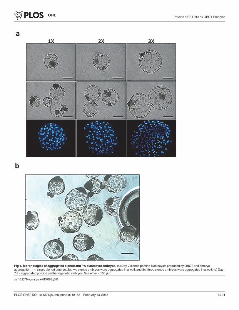
Fig 1. Morphologies of aggregated cloned and PA blastocyst embryos. (a) Day-7 cloned porcine blastocysts produced by OBCT and embryoaggregation. 1×: single cloned embryo; 2×: two cloned embryos were aggregated in a well, and 3×: three cloned embryos were aggregated in a well. (b) Day-7 3× aggregated porcine parthenogenetic embryos. Scale bar = 100 μm.
doi:10.1371/journal.pone.0118165.g001
Porcine ntES Cells by OBCT Embryos
PLOS ONE | DOI:10.1371/journal.pone.0118165 February 13, 2015 8 / 21
morphology for one or two passages and then either differentiated or degenerated. However,five ES cell lines derived from the 3× group maintained proliferation without differentiation be-yond three passages (Table 2).
Experiment 2: Competency of Aggregated Cloned Blastocysts forDerivation of ES Cells Cultured with Various Feeder Cells and SeraWe further investigated the effects of various feeder cells and types of serum on derivingprimary outgrowths and ES cell colonies. The Day-7 3× OBCT embryos were either seededonto mitomycin C-inactivated mouse STO cells or MEF feeder cell layers in a four-well culturedish with ES cell medium (DMEMmedium supplemented with either 15% FBS or 20% KSR).Various feeders and sera had no effect (P> 0.05) on ES cell establishment efficiency in termsof embryo attachment, outgrowths, and primary colony formation. More ES-like cell lines pro-liferated beyond passage 3 (17.1%) in MEF feeders and in ES cell medium supplemented with20% KSR (Table 3). However, ES-like cell cultured in the medium supplemented with KSR hadcompromised pluripotency, defective morphology, and eventually became differentiated orsubsequently died.
Experiment 3: Competency of Aggregated Cloned Blastocysts to DeriveES Cell Lines in Various Culture MediaEffects of various culture media on the growth of porcine ICM cells were compared (Table 4).There were no differences (P> 0.05) among treatment groups in embryo attachment rate,outgrowths, and primary colony formation. Percentages of ES-like cell lines beyond passage3 in DMEM, DMEM/F12, and α-MEMmedium were 4.3, 6.8, and 16.7%, respectively.
Experiment 4: Effect of TSA Treatment in 3× Aggregated Embryos onDerivation of Porcine ntES Cell LinesThe effect of TSA treatment in 3× aggregated embryos on the establishment of ntES-like cells isshown in Table 5. There was no difference in the efficiency with which outgrowth formationcould be established from the TSA-treated blastocysts compared to the control (38.5 and46.7%, respectively; P> 0.05). Primary outgrowths from the cloned embryos started to develop4–6 days after plating onto MEF layers. Over the next 8–10 days, these colonies grew to2–5 mm in diameter without changes in their morphology. The primary colony formation ratewas greater (P< 0.05) in the TSA-treated group (36.7%) than the control (23.1%). Eight of the
Table 2. Competency of Aggregated Cloned and Parthenogenetic (PA) Porcine Blastocysts for Derivation of ES Cell Colonies.
Aggregated embryos No. embryos Primary colonies No. lines > passage 3 (%)
Total embryos Attached embryos (%) Outgrowth (%)
1× 77 32 (41.6)b 18 (23.4)b 3 (3.9)c 0
2× 94 59 (62.8)a 40 (42.6)a 12 (12.8)b 0
3× 172 131 (76.2)a 95 (55.2)a 45 (26.2)a 5 (2.9)
3×PA 79 35 (44.3)b 20 (25.3)b 11 (13.9)ab 0
Number of replicates = 12a-cWithin a column, means without a common superscript differed (P < 0.05).
1× = one OBCT cloned embryo was cultured in a well; 2× = two OBCT cloned embryos were aggregated in a well; 3× = three OBCT cloned embryos were
aggregated in a well; 3×PA = three PA embryos were aggregated in a well
doi:10.1371/journal.pone.0118165.t002
Porcine ntES Cells by OBCT Embryos
PLOS ONE | DOI:10.1371/journal.pone.0118165 February 13, 2015 9 / 21
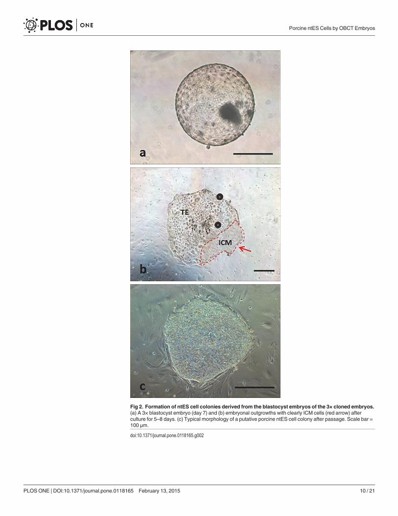
Fig 2. Formation of ntES cell colonies derived from the blastocyst embryos of the 3× cloned embryos.(a) A 3× blastocyst embryo (day 7) and (b) embryonal outgrowths with clearly ICM cells (red arrow) afterculture for 5–8 days. (c) Typical morphology of a putative porcine ntES cell colony after passage. Scale bar =100 μm.
doi:10.1371/journal.pone.0118165.g002
Porcine ntES Cells by OBCT Embryos
PLOS ONE | DOI:10.1371/journal.pone.0118165 February 13, 2015 10 / 21

11 (26.7% of plated embryos) primary colonies obtained from 30 TSA-treated 3× aggregatedcloned embryos survived repeated passaging and freezing, and continued to grow without ap-parent changes in their morphology, thereby outperforming the control (11.5%, P< 0.05).
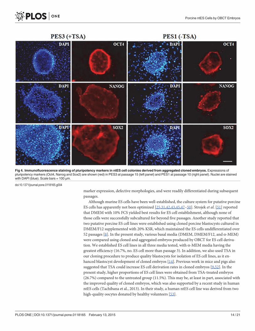
The ES-like cell colonies also had typical ES cell morphology including densely packed,small and round nuclei with well-defined boundaries and expression of AP activity (Fig. 3).Based on immunofluorescence staining and RT-PCR analysis, our ES-like cell colonies con-sisted entirely of those cells expressing Oct4, Nanog, Sox2, and Rex01 (Figs. 4 and 5).
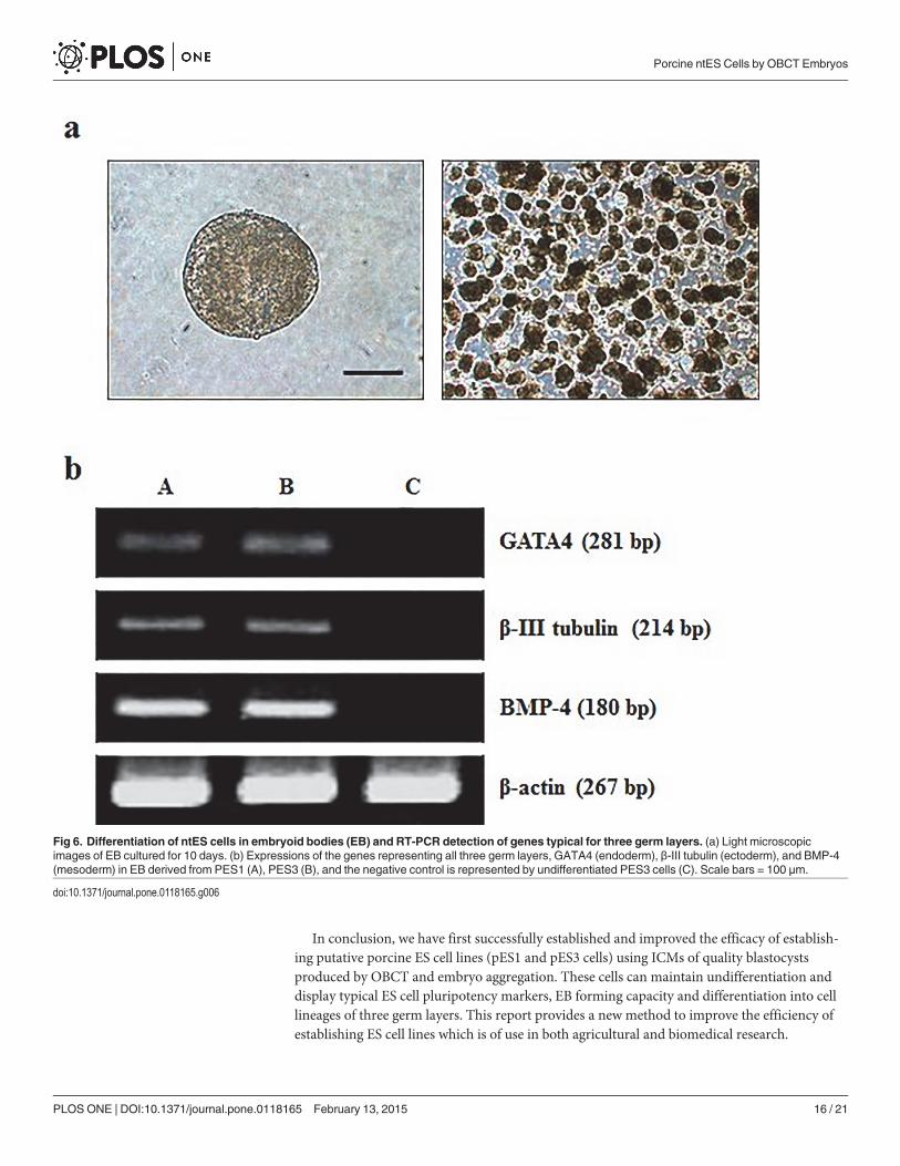
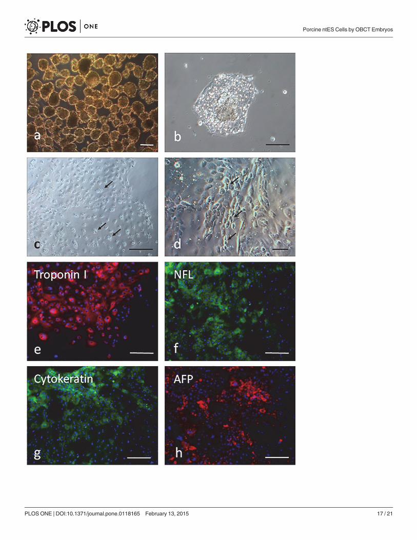
When the ES cells were cultured for 10 days on plastic petri dishes, cystic embryoid bodiesformed (Fig. 6A). Expression levels of several ES cell specific genes of three embryonic germlayer cells were assessed for 10-day-old embryoid bodies. The mRNA transcripts of mesoderm(BMP-4), ectoderm (β-III tubulin) and endoderm (GATA4) were detected by RT-PCR(Fig. 6B). In addition, when day 7 EBs (Fig. 7A) were disaggregated and grown in gelatin-coated surface, they attached to culture dishes (Fig. 7B) after one day of culture. Some neuron-like cells showed obvious Nissl bodies (Fig. 7C) after cultured for 3 days, but became moreepithelial cell-like morphology (Fig. 7D) after cultured for 7 days. Other cells showed the ex-pression of troponin I, NFL, cytokeratin and AFP (Fig. 7E-H), respectively, indicating thatsubpopulations of cells representative of all three germ layers could be obtained.
DiscussionThe objective of the present study was to optimize production and culture conditions support-ing primary culture of porcine ICM cells and stably passaged ES cell lines derived from clonedblastocysts. It is noteworthy that the success of establishing putative porcine ES cell lines isaffected by several factors, including sources of embryos, ICM isolation methods, feeder cells,culture conditions, and culture media [33–39]. To establish putative porcine ES cell lines,blastocyst quality is among the most crucial factors affecting outgrowth and colony formation.
Table 3. Competency of Aggregated Cloned Blastocysts for Derivation of ES Cells Cultured on Different Feeder Cells with Sera.
Feeder cells Serum* No. embryos Primary colonies No. more than passage 3 (%)
Total embryos Attached embryos (%) Outgrowth (%)
STO FBS 39 30 (76.9) 24 (61.1) 18 (46.2) 1 (2.6)
KSR 37 22 (59.5) 15 (40.5) 11 (29.7) 3 (8.1)
MEF FBS 36 28 (77.8) 21 (58.3) 17 (47.2) 2 (5.6)
KSR 35 19 (54.3) 16 (45.7) 12 (34.3) 6 (17.1)
Number of replicates = 7
*ES cell medium was supplemented with 15% FBS or 20% KSR
doi:10.1371/journal.pone.0118165.t003
Table 4. Competency of Aggregated (3×) Cloned Porcine Blastocysts to Derive ES Cell Lines in Various Culture Media.
Medium No. embryos Primary colonies No. more than passage 3 (%)
Total embryos Attached embryos (%) Outgrowth (%)
DMEM 46 25 (54.3) 18 (39.1) 12 (26.1) 2 (4.3)
DMEM/F12 44 24 (54.5) 17 (38.6) 10 (22.7) 3 (6.8)
α-MEM 42 26 (61.9) 18 (42.9) 15 (35.7) 7 (16.7)
Number of replicates = 7
doi:10.1371/journal.pone.0118165.t004
Porcine ntES Cells by OBCT Embryos
PLOS ONE | DOI:10.1371/journal.pone.0118165 February 13, 2015 11 / 21
Although porcine blastocysts have been produced with SCNT [14,15], in general, their qualitywas inferior to those produced in vivo. In this study, embryo aggregation technique providedthe efficiency and quality to establishment of ntES cells, including embryo attachment rates,outgrowths, and primary colonies which explained that aggregation-derived blastocysts hadenhanced cell-cell communication and thus facilitated development of outgrowths and also ex-pression of pluripotency marker Oct4 of the established ntES cells [21]. Consistently, out-growth formation was observed after culture of aggregated porcine-canine interspeciesembryos onto feeder cells in ES cell medium [40]. Moreover, mouse parthenote (PA)-derivedES cells were established by aggregating PA embryos at the eight-cell stage [41].
The effects of feeders, sera, and media on putative porcine ES cell culture have been widelystudied. It is generally agreed that ES cell lines established from different labs had diverse adap-tion to different culture conditions. In addition to providing options of culture systems fromlab to lab, any subtle changes of potential factors affecting the outcome are worth to be re-examined and confirmed in a new system. Therefore, in this present study we attempted toderive and to optimize ES cell culture system by testing several crucial factors such as feeders,sera and media. The feeder cell is among the well-known essential factors for both establish-ment and maintenance of ES cell lines. One of the most important functions of feeders is toserve as an attachment matrix for cells and to secrete cytokines, e.g. LIF and other paracrinefactors that may stimulate ES cell growth and inhibit differentiation [42]. In the present study,we demonstrated that the establishment of putative ES cell lines was more efficient on MEFthan on STO feeders. This could be described that culture of putative porcine ES cells on STOfeeders had a low attachment rate with less primary colony formation; furthermore, the majori-ty of the cultured cells became differentiated after passaging [25,43]. According to Ropeteret al. [44], MEFs were better feeder cells for culturing ES cells which yielded more passages ofputative ES cell lines. In addition, porcine embryonic fibroblast (PEF) cells and were also betterthan the STO feeders [42]. This phenomenon could be attributed to the reduced secreting ac-tivity of STO, since fewer microtubular structures were present in the STO cells [42]. Putativeporcine ES cell lines have been derived by culturing cloned blastocysts onto MEF feeders forvarious passages [8,9]. Nevertheless, in some studies, STO feeder cells were also used to cultureputative porcine ES cells derived from fertilized embryos for more than 10 passages [31,45].
It has been widely reported that FBS is beneficial to maintaining the morphology and pluri-potency state of pig ES cells [31,38,46]. However, Goldsborough et al. (1998) also reported thatFBS contained some major factors causing differentiation of ES cell lines. Recently, severalstudies have demonstrated that the defined serum-free supplement or knockout serum replace-ment (KSR) can be used to maintain ES cells in an undifferentiated state [8,9]. However, in thepresent study, there was no difference observed in the efficiency of deriving ES cell when em-bryos were plated in ES cell culture medium supplemented with either FBS or KSR. However,putative ES cells cultured in the medium supplemented with KSR showed a lower pluripotency
Table 5. Effect of TSA Treatment in 3× Aggregated Embryos on the Establishment of Putative Porcine ntES-Like Cell Lines.
TSA (30 nM) Total embryos Primary outgrowth (%) Primary colonies (%) No. survived beyond passage 3 (%)
- 26 10 (38.5) 6 (23.1)b 3 (11.5)b
+ 30 14 (46.7) 11 (36.7)a 8 (26.7)a
Number of replicates = 4a,bWithin a column, means without a common superscript differed (P < 0.05).
doi:10.1371/journal.pone.0118165.t005
Porcine ntES Cells by OBCT Embryos
PLOS ONE | DOI:10.1371/journal.pone.0118165 February 13, 2015 12 / 21
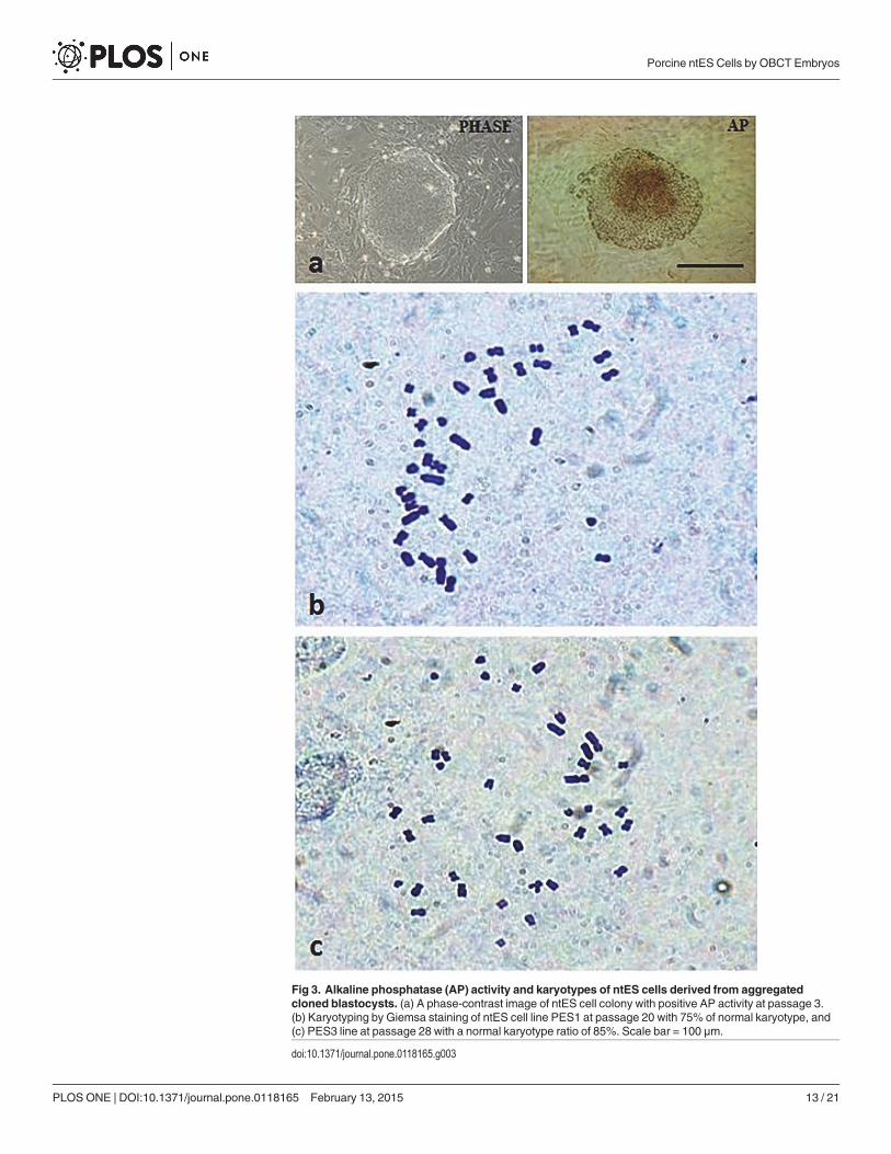
Fig 3. Alkaline phosphatase (AP) activity and karyotypes of ntES cells derived from aggregatedcloned blastocysts. (a) A phase-contrast image of ntES cell colony with positive AP activity at passage 3.(b) Karyotyping by Giemsa staining of ntES cell line PES1 at passage 20 with 75% of normal karyotype, and(c) PES3 line at passage 28 with a normal karyotype ratio of 85%. Scale bar = 100 μm.
doi:10.1371/journal.pone.0118165.g003
Porcine ntES Cells by OBCT Embryos
PLOS ONE | DOI:10.1371/journal.pone.0118165 February 13, 2015 13 / 21
marker expression, defective morphologies, and were readily differentiated during subsequentpassages.
Although murine ES cells have been well-established, the culture system for putative porcineES cells has apparently not been optimized [25,31,42,43,45,47–50]. Strojek et al. [51] reportedthat DMEM with 10% FCS yielded best results for ES cell establishment, although none ofthose cells were successfully subcultured for beyond five passages. Another study reported thattwo putative porcine ES cell lines were established using cloned porcine blastocysts cultured inDMEM/F12 supplemented with 20% KSR, which maintained the ES cells undifferentiated over52 passages [8]. In the present study, various basal media (DMEM, DMEM/F12, and α-MEM)were compared using cloned and aggregated embryos produced by OBCT for ES cell deriva-tion. We established ES cell lines in all three media tested, with α-MEMmedia having thegreatest efficiency (16.7%, no. ES cell more than passage 3). In addition, we also used TSA inour cloning procedure to produce quality blastocysts for isolation of ES cell lines, as it en-hanced blastocyst development of cloned embryos [14]. Previous work in mice and pigs alsosuggested that TSA could increase ES cell derivation rates in cloned embryos [9,52]. In thepresent study, higher proportions of ES cell lines were obtained from TSA-treated embryos(26.7%) compared to the untreated group (11.5%). This may be, at least in part, associated withthe improved quality of cloned embryos, which was also supported by a recent study in humanntES cells (Tachibana et al., 2013). In their study, a human ntES cell line was derived from twohigh-quality oocytes donated by healthy volunteers [53].
Fig 4. Immunofluorescence staining of pluripotency markers in ntES cell colonies derived from aggregated cloned embryos. Expressions ofpluripotency markers (Oct4, Nanog and Sox2) are shown (red) in PES3 at passage 15 (left panel) and PES1 at passage 10 (right panel). Nuclei are stainedwith DAPI (blue). Scale bars = 100 μm.
doi:10.1371/journal.pone.0118165.g004
Porcine ntES Cells by OBCT Embryos
PLOS ONE | DOI:10.1371/journal.pone.0118165 February 13, 2015 14 / 21

In total, 11 cell lines were established in this study, of which the PES3 (derived from TSA-treated embryos) and PES1 (from control embryos) propagated rapidly with normal colonymorphology upon repeated passaging. These cells expressed pluripotency marker genes (Oct4,Nanog, Sox2, and Rex01), and readily formed embryoid bodies in suspension culture, with theexpression of genes representing all three germ layers (endoderm: GATA4; mesoderm: BMP-4;ectoderm: β-III tubulin). Moreover, the EB derived-differentiated cells exhibited various typesof morphologies and expressed cell lineages of all three germ layers. Unfortunately, althoughteratoma formation is regarded as one of an essential pluripotency characteristics of ES cells,we failed to obtain teratomas after cell transfer into SCID mice (five replicates, 3–5×10⁶ mor-phologically undifferentiated ES cells), indicating these ES cell lines may not be sufficientlycompetent to become germline transmissible (data not shown). However, to date, there is noreport of teratoma formation using pluripotent cell lines derived from either cloned or non-cloned porcine embryos. For instance, putative porcine ES cells generated by Anderson et al.(1996) failed to form teratomas via transplantation into the kidney capsule of athymic mice[54]. Recently, subcutaneously injection of porcine ES cells into non-obese diabetic/severecombined immunodeficient (NOD/SCID) mice were also unable to demonstrate teratoma for-mation [55]. Similarly, Kim et al. [8] reported that transplanted porcine ntES cells did not formteratomas in immuno-compromised mice, indicating more studies are required to derive trulypluripotent ntES cells in pigs, most likely by the use of small molecule signaling inhibitors orforced expression of pluripotency-related genes, such as Oct4, Sox2, Klf4, and c-Myc [56–59].
Fig 5. Characterization of ntES cells by real time RT-PCR analysis. Expressions of Oct4, Nanog, Sox2, and Rex01genes in (A) PES1 at passages 8, (B)PES3 at passages 15 and (C) pig fibroblast cells.
doi:10.1371/journal.pone.0118165.g005
Porcine ntES Cells by OBCT Embryos
PLOS ONE | DOI:10.1371/journal.pone.0118165 February 13, 2015 15 / 21
In conclusion, we have first successfully established and improved the efficacy of establish-ing putative porcine ES cell lines (pES1 and pES3 cells) using ICMs of quality blastocystsproduced by OBCT and embryo aggregation. These cells can maintain undifferentiation anddisplay typical ES cell pluripotency markers, EB forming capacity and differentiation into celllineages of three germ layers. This report provides a new method to improve the efficiency ofestablishing ES cell lines which is of use in both agricultural and biomedical research.
Fig 6. Differentiation of ntES cells in embryoid bodies (EB) and RT-PCR detection of genes typical for three germ layers. (a) Light microscopicimages of EB cultured for 10 days. (b) Expressions of the genes representing all three germ layers, GATA4 (endoderm), β-III tubulin (ectoderm), and BMP-4(mesoderm) in EB derived from PES1 (A), PES3 (B), and the negative control is represented by undifferentiated PES3 cells (C). Scale bars = 100 μm.
doi:10.1371/journal.pone.0118165.g006
Porcine ntES Cells by OBCT Embryos
PLOS ONE | DOI:10.1371/journal.pone.0118165 February 13, 2015 16 / 21
Porcine ntES Cells by OBCT Embryos
PLOS ONE | DOI:10.1371/journal.pone.0118165 February 13, 2015 17 / 21
Nevertheless, it warrants more work to improve the quality of ES cell lines established, possiblyby reversing these cells to their naïve status.
AcknowledgmentsThis study was supported in part by grants from China Medical University Hospital(DMR-103-104), the National Science Council (NSC# 96-2313-B-005-013, NSC# 98-2628-B005-019-MY3, and NSC# 101-2313-B-005-013-MY3), Executive Yuan and the Ministry ofEducation, Taiwan, Republic of China, under the ATU plan. We are also grateful to Dr. JohnKastelic, Professor, University of Calgary, Canada, for his critical reading and suggestions toimprove this manuscript.
Author ContributionsConceived and designed the experiments: CS YHLMK JCJ. Performed the experiments: CSYHLMK CDC. Analyzed the data: CS YHL CHC CFT. Contributed reagents/materials/analysis tools: LRC CHC CFT NWL JCJ. Wrote the paper: CS JCJ. Provided initial training ofporcine ES cell culture: LRC.
References1. Lai L, Kolber-Simonds D, Park KW, Cheong HT, Greenstein JL, Im GS, et al. (2002) Production of
alpha-1, 3-galactosyltransferase knockout pigs by nuclear transfer cloning. Science 295: 1089–1092.PMID: 11778012
2. Chen G, Qian H, Starzl T, Sun H, Garcia B, Wang X, et al. (2005) Acute rejection is associated withantibodies to non-Gal antigens in baboons using Gal-knockout pig kidneys. Natural Medicine 11:1295–1298. PMID: 15974998
3. Yamada K, Yazawa K, Shimizu A, Iwanaga T, Hisashi Y, Nuhn M, et al. (2005) Marked prolongation ofporcine renal xenograft survival in baboons through the use of alpha1,3-galactosyltransferase gene-knockout donors and the cotransplantation of vascularized thymic tissue. Natural Medicine 11: 32–34.
4. Wakayama T, Tabar V, Rodriguez I, Perry AC, Studer L, Mombaerts P (2001) Differentiation ofembryonic stem cell lines generated from adult somatic cells by nuclear transfer. Science 292:740–743. PMID: 11326103
5. Fang ZF, Gai H, Huang YZ, Li SG, Chen XJ, Shi JJ, et al. (2006) Rabbit embryonic stem cell linesderived from fertilized, parthenogenetic or somatic cell nuclear transfer embryos. Experimental CellResearch 312: 3669–3682. PMID: 16996056
6. Wang L, Duan E, Sung LY, Jeong BS, Yang X, Tian XC (2005) Generation and characterization ofpluripotent stem cells from cloned bovine embryos. Biology of Reproduction 73: 149–155. PMID:15744021
7. Byrne JA, Pedersen DA, Clepper LL, Nelson M, Sanger WG, Gokhale S, et al. (2007) Producing pri-mate embryonic stem cells by somatic cell nuclear transfer. Nature 450: 497–502. PMID: 18004281
8. Kim S, Kim JH, Lee E, Jeong YW, Hossein MS, Park SM, et al. (2010) Establishment andcharacterization of embryonic stem-like cells from porcine somatic cell nuclear transfer blastocysts.Zygote 18: 93–101. doi: 10.1017/S0967199409990372 PMID: 20307349
9. Vassiliev I, Vassilieva S, Truong KP, Beebe LF, McIlfatrick SM, Harrison SJ, et al. (2011) Isolation andin vitro characterization of putative porcine embryonic stem cells from cloned embryos treated withtrichostatin A. Cellular Reprogramming 13: 205–213. doi: 10.1089/cell.2010.0102 PMID: 21548828
10. Pratt SL, Sherrer ES, Reeves DE, Stice SL (2006) Factors influencing the commercialization of cloningin the pork industry. Society of Reproduction and Fertility supplement 62: 303–315. PMID: 16866326
Fig 7. In vitro differentiation ability of porcine ntES cells after induction of EB formation. (a) Light microscopic images of EB cultured for 7 days. (b) EBattachment to gelatin-coated surface of culture dishes. (c) The neuron-like cells with obvious Nissl bodies after cultured for 3 days. (d) The epithelial-like cellsmorphology after cultured for 7 days. (e-h) Confirmation of the expression of differentiation marker troponin I (mesoderm), NFL and cytokeratin (ectoderm),and AFP (endoderm) from differentiated cells by the immunocytochemical analysis. Scale bars = 100 μm.
doi:10.1371/journal.pone.0118165.g007
Porcine ntES Cells by OBCT Embryos
PLOS ONE | DOI:10.1371/journal.pone.0118165 February 13, 2015 18 / 21
11. Thompson JG (1997) Comparison between in vivo derived and in vitro produced pre-elongationembryos from domestic ruminants. Reproduction, Fertility and Development 9: 341–354. PMID:9261882
12. WangWH, Abeydeera LR, Han YM, Prather RS, Day BN (1997) Morphologic evaluation and actinfilament distribution in porcine embryos produced in vitro and in vivo. Journal of Reproduction andFertility 111: 101–108. PMID: 9370973
13. Koo DB, Kang YK, Park JS, Park JK, ChangWK, Lee KK, et al. (2004) A paucity of structural integrity incloned porcine blastocysts produced in vitro. Theriogenology 62: 779–789. PMID: 15251229
14. Chawalit S, Nguyen NT, Tseng JK, Lo NW, Tu CF, Ju JC (2012) Trichostatin A and ascorbic acid assistin the development of porcine handmade cloned embryos via different physiologic pathways.Reproductive Science 19: 976–986. doi: 10.1177/1933719112440049 PMID: 22534331
15. Li J, Svarcova O, Villemoes K, Kragh PM, Schmidt M, Bogh IB, et al. (2008) High in vitro developmentafter somatic cell nuclear transfer and trichostatin A treatment of reconstructed porcine embryos.Theriogenology 70: 800–808. doi: 10.1016/j.theriogenology.2008.05.046 PMID: 18573521
16. Cervera RP, Martí-Gutiérrez N, Escorihuela E, Moreno R, Stojkovic M (2009) Trichostatin A affectshistone acetylation and gene expression in porcine somatic cell nucleus transfer embryos.Theriogenology 72: 1097–1110. doi: 10.1016/j.theriogenology.2009.06.030 PMID: 19765811
17. Giles JR, Foote RH (1995) Rabbit blastocyst: Allocation of cells to the inner cell mass andtrophectoderm. Molecular Reproduction and Development 41: 204–211. PMID: 7654374
18. Macháty Z, Prather RS (1998) Strategies for activating nuclear transfer oocytes. Reproduction, Fertilityand Development 10: 599–613. PMID: 10612466
19. Tang PC, West JD (2000) The effects of embryo stage and cell number on the composition of mouseaggregation chimaeras. Zygote 8: 235–243. PMID: 11014503
20. Lee SG, Park CH, Choi DH, Kim HS, Ka HH, Lee CK (2007) In vitro development and cell allocation ofporcine blastocysts derived by aggregation of in vitro fertilized embryos. Molecular Reproduction andDevelopment 74: 1436–1445. PMID: 17440970
21. Boiani M, Eckardt S, Leu NA, Schöler HR, McLaughlin KJ (2003) Pluripotency deficit in clone overcomeclone–clone aggregation: Epigenetic complementation? EMBO Journal 22: 5304–5312. PMID:14517267
22. Terashita Y, Sugimura S, Kudo Y, Amano R, Hiradate Y, Sato E (2011) Improving the quality of minia-ture pig somatic cell nuclear transfer blastocysts: aggregation of SCNT embryos at the four-cell stage.Reproduction in Domestic Animals 46: 189–196. doi: 10.1111/j.1439-0531.2010.01614.x PMID:20412512
23. Moore K, Piedrahita JA (1997) The effects of human leukemia inhibitory factor (hLIF) and culturemedium on in vitro differentiation of cultured porcine inner cell mass (pICM). In Vitro Cellular &Developmental Biology-Animal 33: 62–71. PMID: 15033587
24. Miyoshi K, Taguchi Y, Sendai Y, Hoshi H, Sato E (2000) Establishment of a porcine cell line from invitro-produced blastocysts and transfer of the cells into enucleated oocytes. Biology of Reproduction62: 1640–1646. PMID: 10819766
25. Li M, MaW, Hou Y, Sun XF, Sun QY, WangWH (2004) Improved isolation and culture of embryonicstem cells from Chinese miniature pig. Journal of Reproduction and Development 50: 237–244. PMID:15118251
26. Kim HS, Son HY, Kim S, Lee GS, Park CH, Kang SK, et al. (2007) Isolation and initial culture of porcineinner cell masses derived from in vitro-produced blastocysts. Zygote 15: 55–63. PMID: 17391546
27. Vassiliev I, Vassilieva S, Beebe LF, Mcilfatrick SM, Harrison SJ, Nottle MB (2010) Development of cul-ture conditions for the isolation of pluripotent porcine embryonal outgrowths from in vitro produced andin vivo derived embryos. Journal of Reproduction and Development. 56: 546–551. PMID: 20519828
28. Cibelli JB, Stice SL, Golueke PJ, Kane JJ, Jerry J, Blackwell C, et al. (1998) Transgenic bovine chimericoffspring produced from somatic cell-derived stem-like cells. Nature Biotechnology 167: 642–646.
29. Siriboon C, Tu CF, Kere M, Liu MS, Chang HJ, Ho LL, et al. (2013) Production of viable cloned minia-ture pigs by aggregation of handmade cloned embryos at the 4-cell stage. Reproduction, Fertility andDevelopment doi: 10.1071/RD12243.
30. Nguyen NT, Lin DP, Yen SY, Tseng JK, Chuang JF, Chen BY, et al. (2009) Sonic hedgehog promotesporcine oocyte maturation and early embryo development. Reproduction, Fertility and Development21: 805–815. doi: 10.1071/RD08277 PMID: 19567223
31. Chen LR, Shiue YL, Bertolini L, Medrano JF, BonDurant RH, Anderson GB (1999) Establishment of plu-ripotent cell lines from porcine preimplantation embryos. Theriogenology 52: 195–212. PMID:10734388
Porcine ntES Cells by OBCT Embryos
PLOS ONE | DOI:10.1371/journal.pone.0118165 February 13, 2015 19 / 21

32. Intawicha P, Ou YW, Lo NW, Zhang SC, Chen YZ, Lin TA, et al. (2009) Characterization of embryonicstem cell lines derived from New ZealandWhite Rabbit embryos. Cloning Stem Cells 11: 27–38. doi:10.1089/clo.2008.0040 PMID: 19220131
33. Brevini TA, Tosetti V, Crestan M, Antonini S, Gandolfi F (2007) Derivation and characterization of plu-ripotent cell lines from pig embryos of different origins. Theriogenology 67: 54–63. PMID: 17055567
34. Vackova I, Ungrova A, Lopes F (2007) Putative embryonic stem cell lines from pig embryos. Journal ofReproduction and Development 53: 1137–1149. PMID: 18198476
35. Hall V (2008) Porcine embryonic stem cells: a possible source for cell replacement therapy. Stem cellReviews 4: 275–282. doi: 10.1007/s12015-008-9040-2 PMID: 18770051
36. Telugu BP, Ezashi T, Roberts RM (2010) The promise of stem cell research in pig and other ungulatespecies. Stem Cell Reviews 6: 31–41. doi: 10.1007/s12015-009-9101-1 PMID: 19949895
37. Telugu BP, Ezashi T, Roberts RM (2010) Porcine induced pluripotent stem cells analogous to naïveand primed embryonic stem cells of the mouse. The International Journal of Developmental Biology54: 1703–1711. doi: 10.1387/ijdb.103200bt PMID: 21305472
38. Brevini TA, Pennarossa G, Attanasio L, Vanelli A, Gasparrini B, Gandolfi F (2010) Culture conditionsand signalling networks promoting the establishment of cell lines from parthenogenetic and biparentalpig embryos. Stem Cell Reviews 6: 484–495. doi: 10.1007/s12015-010-9153-2 PMID: 20407852
39. Brevini TA, Pennarossa G, Gandolfi F (2010) No shortcuts to pig embryonic stem cells. Theriogenology74: 544–550. doi: 10.1016/j.theriogenology.2010.04.020 PMID: 20570327
40. Sugimura S, Narita K, Yamashiro H, Sugawara A, Shoji T, Terashita Y, et al. (2009) Interspeciessomatic cell nuclear transfer with porcine oocytes as recipients: a novel bioassay system for assessingthe competence of canine somatic cells to develop into embryos. Theriogenology 72: 549–559. doi:10.1016/j.theriogenology.2009.04.011 PMID: 19524287
41. Shan ZY, Wu YS, Shen XH, Li X, Xue Y, Zheng Z, et al. (2012) Aggregation of pre-implantationembryos improves establishment of parthenogenetic stem cells and expression of imprinted genes.Development, Growth & Differentiation 54: 481–488. doi: 10.1111/bph.13094 PMID: 25625931
42. Li M, Zhang D, Hou Y, Jiao L, Zheng X, WangWH (2003) Isolation and culture of embryonic stem cellsfrom porcine blastocysts. Molecular Reproduction and Development 65: 429–434. PMID: 12840816
43. Talbot NC, Rexroad CE Jr, Pursel VG, Powell AM, Nel ND (1993) Culturing the epiblast cells of the pigblastocyst. In Vitro Cellular & Developmental Biology-Animal 29: 543–554. PMID: 15033587
44. Ropeter Scharfenstein M, Neubert N, Prelle K, Holt W (1996) Identification, isolation and culture ofpluripotent cells from the porcine inner cell mass. Journal of Animal Breeding and Genetics 113:427–436.
45. Piedrahita JA, Anderson GB, Bondurant RH (1990) On the isolation of embryonic stem cells:Comparative behavior of murine, porcine and ovine embryos. Theriogenology 34: 879–901. PMID:16726890
46. Yang JR, Shiue YL, Liao CH, Lin SZ, Chen LR (2009) Establishment and characterization of novelporcine embryonic stem cell lines expressing hrGFP. Cloning Stem Cells 11: 235–244. doi: 10.1089/clo.2008.0050 PMID: 19508116
47. Evans MJ, Notarianni E, Laurie S, Moor RM (1990) Derivation and preliminary characterization ofpluripotent cell lines from porcine and bovine blastocysts. Theriogenology 33: 125–128.
48. Hochereau de Reviers MT, Perreau C (1993) In vitro culture of embryonic disc cells from porcineblastocysts. Reproduction Nutrition Development 33: 475–483. PMID: 8142030
49. Anderson GB, Choi SJ, Bondurant RH (1994) Survival of porcine inner cell masses in culture and afterinjection into blastocysts. Theriogenology 42: 204–212. PMID: 16727526
50. Wianny F, Perreau C, Hochereau de Reviers MT (1997) Proliferation and differentiation of porcineinner cell mass and epiblast in vitro. Biology of Reproduction 57: 756–764. PMID: 9314577
51. Strojek RM, Reed MA, Hoover JL, Wagner TE (1990) A method for cultivating morphologically undiffer-entiated embryonic stem cells from porcine blastocysts. Theriogenology 33: 901–913. PMID:16726786
52. Kishigami S, Mizutani E, Ohta H, Hikichi T, Thuan NV, Wakayama S, et al. (2006) Significantimprovement of mouse cloning technique by treatment with trichostatin A after somatic nuclear transfer.Biochemical and Biophysical Research Communications 340: 183–189. PMID: 16356478
53. Tachibana M, Amato P, Sparman M, Gutierrez NM, Tippner-Hedges R, Ma H, et al. (2013) Humanembryonic stem cells derived by somatic cell nuclear transfer. Cell 153: 1228–1238. doi: 10.1016/j.cell.2013.05.006 PMID: 23683578
54. Anderson GB, BonDurant RH, Goff L, Groff J, Moyer AL (1996) Development of bovine and porcine em-bryonic teratomas in athymic mice. Animal Reproduction Science 45: 231–240. PMID: 9227925
Porcine ntES Cells by OBCT Embryos
PLOS ONE | DOI:10.1371/journal.pone.0118165 February 13, 2015 20 / 21

55. Park JK, Kim HS, Uh KJ, Choi KH, Kim HM, Lee T, et al. (2013) Primed pluripotent cell lines derivedfrom various embryonic origins and somatic cells in pig. PLOS One 8(1). doi: 10.1371/journal.pone.0052481 PMID: 23536748
56. Haraguchi S, Kikuchi K, Nakai M, Tokunaga T (2012) Establishment of self-renewing porcineembryonic stem cell-like cells by signal inhibition. Journal of Reproduction and Development 58:707–716. PMID: 22972236
57. Ezashi T, Telugu BP, Alexenko AP, Sachdev S, Sinha S, Roberts RM (2009) Derivation of inducedpluripotent stem cells from pig somatic cells. Proceedings of the National Academy of Sciences of theUnited States of America 106: 10993–10998. doi: 10.1073/pnas.0905284106 PMID: 19541600
58. Fujishiro SH, Nakano K, Mizukami Y, Azami T, Arai Y, Matsunari H, et al. (2013) Generation of naive-like porcine-induced pluripotent stem cells capable of contributing to embryonic and fetal development.Stem Cells and Development 22: 473–482. doi: 10.1089/scd.2012.0173 PMID: 22889279
59. Liao YJ, Liao CH, Liao JW, Yuan K, Liu YZ, Chen YS, et al. (2014) Establishment and characterizationof novel porcine induced pluripotent stem cells expressing hrGFP. Stem Cell Research & Therapy4(5). doi: 10.4172/2157-7633.1000208 PMID: 25625754
Porcine ntES Cells by OBCT Embryos
PLOS ONE | DOI:10.1371/journal.pone.0118165 February 13, 2015 21 / 21


![[sp1] oocyte collection from superstimulated disease-free ...](https://static.fdokumen.com/doc/165x107/631dd3361aedb9cd850f788f/sp1-oocyte-collection-from-superstimulated-disease-free-.jpg)