Substrate specificity of Staphylococcus aureus cysteine proteases – Staphopains A, B and C

Purification of a novel cysteine protease, procerain B, from Calotropis procerawith distinct characteristics compared to procerain
Abhay Narain Singh a, Anil Kumar Shukla a, M.V. Jagannadham b, Vikash Kumar Dubey a,*a Department of Biotechnology, Indian Institute of Technology Guwahati, Guwahati-781039, Assam, Indiab Molecular Biology Unit, Institute of Medical Sciences, Banaras Hindu University, Varanasi-221005, India
Process Biochemistry 45 (2010) 399–406
A R T I C L E I N F O
Article history:
Received 21 May 2009
Received in revised form 8 September 2009
Accepted 22 October 2009
Keywords:
Cysteine protease
ELISA
Food industry
Substrate specificity
Calotropis procera
A B S T R A C T
Proteases have applications in food, detergent and pharmaceutical industries. A novel protease has been
purified from the latex of Calotropis procera and characterized. As another cysteine protease, procerain, is
reported from the same source, the newly purified enzyme was named as procerain B. The enzyme
shows distinct properties compared to procerain, in terms of cleavage recognition site, immunological
properties and other physical properties like molecular weight, isoelectric point, etc. The newly purified
enzyme shows a broad optimum pH (6.5–8.5) as well as broad optimum temperature (40–60 8C).
Additionally, the enzyme retains its activity where most of other proteases are not active. Moreover, the
enzyme appeared to be very efficient in hydrolysis of blood stain and may have potential application in
detergent industries. Simple and economic purification of procerain B, together with easy availability of
latex, makes the large-scale production of procerain B possible, thus enables to explore various industrial
as well as biotechnological applications.
� 2009 Elsevier Ltd. All rights reserved.
Contents lists available at ScienceDirect
Process Biochemistry
journa l homepage: www.e lsev ier .com/ locate /procbio
1. Introduction
Bioinformatics analysis of the human and mouse genomesindicates at least 2% genes of the genome codes for proteases [1]which indicates physiological role of proteases. Proteases havetherapeutic and industrial applications as well. The annual sales ofproteases accounts 60% of the total world enzyme market andestimated to reach 220 billion US$ by the year 2009 [2]. Plantcysteine proteases are used in industry owing to their hightemperature stability and broad substrate specificity [3]. Plantgenomes encode hundreds of proteases, but little is known aboutwhat roles they play in the life of a plant. Proteases are thought tobe involved in a range of biological processes, includingsenescence, implicated in perception, signaling and executionleading to plant defense [4]. Few other cysteine proteases from theplant sources have been purified and characterized. As usefulnessof the proteases depends on its unique cleavage site, stability as
Abbreviations: BAPA, NR-benzoylarginine p-nitroanilide; DFP, diisopropylfluoro-
phosphate; DTNB, 5,50-dithiobis(2-nitrobenzoic acid); DTT, dithiothreitol; EDTA,
ethylenediaminetetraacetic acid; EGTA, glycol-bis(2-aminoethylether)-N,N,N0 ,N0-
tetraacetic acid; ELISA, enzyme-linked immunosorbent assay; GuHCl, guanidine
hydrochloride; NEM, N-ethyl-maleimide; PCMB, p-chloromercurybenzoate; PMSF,
phenyl-methanesulfonyl fluoride; SBTI, soybean trypsin inhibitor; TCA, trichlor-
oacetic acid; TFA, trifluoroacetic acid; TEMED, N,N,N,N-tetramethylethylenedia-
mines.
* Corresponding author. Tel.: +91 361 2582203; fax: +91 361 2582249.
E-mail address: [email protected] (V.K. Dubey).
1359-5113/$ – see front matter � 2009 Elsevier Ltd. All rights reserved.
doi:10.1016/j.procbio.2009.10.014
well as optimum activity condition search of a novel protease withunique properties is always on.
Calotropis procera (family Asclepiadaceae) is a tropical plantthat has been widely used in Indian traditional medicinal systemfor the treatment of various diseases namely leprosy, ulcers,tumors, piles and diseases of the spleen, liver and abdomen [5].Various parts of the plant show anti-microbial, anti-inflammatory,antipyretic and anti-malarial activities [6,7]. The latex of the plantshows antidiabetic, hepatoprotective, antiarthritic, cytotoxic andanticancerous properties [8]. Preliminary screening of the latex ofthe plant showed very high proteolytic activity. We have earlierreported purification and characterization of a novel proteaseprocerain, from the latex of the plant [9]. Here we reportpurification of another protease from the plant, which we namedas procerain B, with distinct properties and cleavage site.
2. Materials and methods
2.1. Materials
Superficial incisions on the C. procera yielded milk like latex. Fresh latex of the plant
was collected. CM-Sepharose FF was purchased from GE Healthcare. Azocasein, DTNB
(5,50-dithiobis-[2-nitro benzoic acid]), DTT (dithiothreitol), GuHCl (guanidine
hydrochloride), urea, o-phenanthroline, EDTA (ethylenediaminetetraacetic acid),
EGTA (glycol-bis(2-aminoethylether)-N,N,N0 ,N0-tetraacetic acid), leupeptin, SBTI
(soybean trypsin inhibitor), NEM (N-ethylmaleimide), b-mercaptoethanol, PMSF
(phenylmethanesulfonylfluoride), acrylamide,N,N-methylene bisacrylamide, Coo-
massie brilliant blue R-250, E-64 (l-trans-epoxysuccinylleucylamide (4-guanidino)
butane-N-[N-(L-3-trans-carboxyirane-2-carbonyl)-L-leucyl] agimatine), are obtained
from Sigma Chemical Co., USA. Sodium tetrathionate (Na2S4O6�2H2O) was

A.N. Singh et al. / Process Biochemistry 45 (2010) 399–406400
synthesized by the method of Gilman et al. [10]. All other chemicals were of highest
purity commercially available. Procerain was purified by the method of Dubey and
Jagannadham [9].
2.2. Methods
2.2.1. Purification
All purification steps were carried out in cold to minimize any complication
due to possible autodigestion or unfolding of the enzyme at higher temperature.
Most of the purification steps were carried out at 4 8C unless stated. Latex was
collected from the young stems of the plant in 0.01 M acetate buffer pH 4.5
containing 0.01 M sodium tetrathionate, a reversible inhibitor of cysteine
protease, and was stored at �20 8C. Frozen latex was thawed to room
temperature and centrifuged at 24,000 � g for 10 min to remove any insoluble
materials. The supernatant was then subjected to 50% ammonium sulfate
fractionation. The supernatant after 50% ammonium sulfate fractionation found
to have more enzyme activity which is dialyzed extensively and used for further
purification. The ion exchange chromatography of the dialyzed supernatant of
50% ammonium sulfate fractionation was performed at 4 8C. The protein was
loaded on CM-Sepharose cation exchange column pre-equilibrated with 0.01 M
acetate buffer pH 4.5. The column was washed with the same buffer until no
protein was detected in elute and the bound proteins were eluted with a linear
gradient of 0–0.8 M NaCl at a flow rate of 3 ml/min. Fractions of 6 ml were
collected; the absorbance at 280 nm as well as caseinolytic activities of the
protein in all fractions was checked using casein as a substrate. The partially
purified fractions of the ion exchange chromatography were further purified by
size exclusion chromatography using sephacryl S-300 column (1.6 cm � 40 cm).
The loaded protein was eluted by 0.01 M acetate buffer pH 4.5 containing
250 mM NaCl.
2.2.2. Protein concentration
Protein concentration at different stages of purification was determined by
absorbance at 280 nm as well as by the method of Bradford [11] using BSA as a
standard.
2.2.3. Protease activity
The hydrolyzing activity of the protease was determined using denatured
natural substrates casein or azocasein using the method of Dubey and
Jagannadham [9]. Enzyme solution (5 mg) was incubated in final volume of
500 ml of 50 mM Tris-Cl buffer pH 7.5 at 37 8C for 10 min. Casein solution (1%)
(w/v) was prepared at the same pH and added to the enzyme solution making
the final reaction volume to 1 ml and the reaction mixture was incubated for the
30 min at 37 8C. The reaction was stopped by adding 0.5 ml of 10% TCA,
incubated further for 10 min at room temperature and centrifuged (10,000 rpm
for 10 min). The absorbance of the soluble peptides in the supernatant was
measured at 280 nm. In the case of azocasein, as substrate, 0.5 ml of supernatant
after TCA precipitation was mixed with equal volume of 0.5 M NaOH and
incubated for 15 min. The development of colour was measured spectro-
photometrically by taking absorbance at 440 nm. A control assay, without the
enzyme was done and used as blank in all spectrophotometric measurements.
One unit of enzyme activity was defined as the amount of enzyme, under given
assay conditions that give rise to an increase of 0.001 unit of absorbance at
280 nm or an increase of 0.001 unit of absorbance at 440 nm per minute of
digestion. Number of units of activity per milligram of protein was taken as the
specific activity of the enzyme.
2.2.4. Electrophoresis
Homogeneity and intactness of the enzyme, during purification as well as
molecular mass determination of the purified enzyme, was assayed by using SDS-
PAGE [12] with little modification. The purified enzyme was inactivated to avoid
autolysis by treatment with cocktail inhibitor mixture. The gel was stained with
0.1% Coomassie brilliant blue R-250. Molecular weight markers used were
phosphorylase b (97.4 kDa), BSA (66.0 kDa), ovalbumin (45.0 kDa), carbonic
anhydrase (29.0 kDa), soybean tyrosine inhibitor (20.1 kDa), and lysozyme
(14.3 kDa) and a plot of log molecular weight vs mobility was generated to
extrapolate the molecular weight of the purified enzyme.
2.2.5. Isoelectric point
The isoelectric point of the purified enzyme was determined by isoelectric
focussing in tube gels as described for procerain [9]. Ampholines, in the pH range
3.5–10.0 were used to generate the pH gradient. A 5% polyacrylamide gel containing
2% desired ampholine was cast in tube gels. Anodic and cathodic chambers buffer
were 0.01 M-iminodiacetic acids and 0.01 M ethylenediamine, respectively. The
buffers were flushed with nitrogen gas before electrophoresis. The gel was
subjected to a pre-run at a constant current of 1 mA per rod for 2 h to develop the pH
gradient. A protein sample (100 mg) containing 10% (v/v) ampholine and 25%
glycerol was loaded on the gel and electrophoresed at constant current of 2 mA per
rod for 4 h. Protein bands were stained with 0.04% (w/v) Coomassie brilliant G-250
dissolved in 6% (w/v) perchloric acid [13].
2.2.6. Carbohydrate content
Carbohydrate content of procerain B was determined by phenol sulphuric acid
method [14,15].
2.2.7. Tryptophan and tyrosine content
Total numbers of tryptophan and tyrosine residues in the purified protein
was determined by the method of Goodwin and Morton [16]. An absorbance
spectrum of the purified enzyme in 0.1 M NaOH was recorded between 300 and
220 nm using a Beckman DU 640B spectrophotometer. Absorbance values at
280 nm and 294.4 nm were obtained from the spectra. For calculations, the
formula, w ¼ ðA280 � xeyÞ=ðew � eyÞ, was used where, w is the estimated
tryptophan content in moles per liter; A280 is the absorbance at 280 nm for
one molar protein; ew and ey are molar extinction coefficients of tryptophan and
tyrosine in 0.1 M NaOH at 280 nm (ey = 1576 and ew ¼ 5225), respectively. The
total tyrosine and tryptophan content in the protein, x was calculated using
e294.4 = 2375. The number of a particular amino acid residue per molecule of the
protein was calculated from the ratio of the molar concentrations of the amino
acid residues to that of the total protein.
2.2.8. Measurement of free and total sulfhydryl content
Free and total cysteine residues of procerain B were determined by using DTNB
method of Ellman [17]. For free cysteine content determination, the enzyme was
activated with 0.05 M b-ME in 50 mM phosphate buffer pH 7.5 at 37 8C for 30 min and
dialyzed extensively for 24 h against 500 ml of 0.1 M acetic acid with four changes of
the dialyzate. After dialysis, 50 ml of the dialyzed enzyme sample was taken in 700 ml
of 0.1 M Tris–HCl, pH 7.3 and the sample was allowed to stand for 10 min to attain the
pH. Subsequently, 50 ml of 5 mM DTNB solution was added and the reaction mixture
was thoroughly mixed. The liberated TNB anion after reaction of sulfhydryl group
with DTNB was monitored spectrophotometrically. The numbers of free cysteine
residues are assessed using extinction coefficient of TNB of 14,150 M�1 cm�1 at
412 nm [18]. For the estimation of the total number of cysteine residues, the enzyme
was denatured in 6 M GuHCl and reduced with 0.05 M DTT in 0.05 M Tris–HCl, pH 8.0.
The excess DTT in the reaction mixture was removed by dialysis against 500 ml of
0.1 M acetic acid with four changes of the dialyzate [19]. The liberated thiol groups
were estimated as described in the case of free cysteine estimation. The numbers of
disulfide bonds, in the protein, were deduced by the comparison of the number of free
and total cysteine residues. To validate the measurements, similar contents of papain,
ribonuclease A, and lysozyme were also determined.
2.2.9. Extinction coefficient
The extinction coefficient of procerain B was determined using spectro-
photometric method [20]. The extinction coefficient was determined using the
formula, e1%280 nm ¼ 10ð5690nw þ 1280ny þ 120ncÞ=M, where nw , ny, and nc are the
number of tryptophan, tyrosine, and cysteine residues in the protein; M is the
molecular mass of the protein and 5690, 1280, and 120 are the respective extinction
coefficients of tryptophan, tyrosine and cysteine residues. The total numbers of
tryptophan, tyrosine and cysteine residues in the protein are determined as
described below.
2.2.10. pH and temperature optima
The activity of the purified enzyme is measured as function of varying pH to
determine the pH optima of the enzyme. 10 mg of enzyme was used for activity
measurement. The buffers used were: 0.05 M KCl–HCl (pH 1.0–1.5); 0.05 M
Glycine–HCl (pH 2.0–3.5); 0.05 M Na-acetate (pH 4.0–5.5); 0.05 M Na-phosphate
(pH 6.0–7.0); 0.05 M Tris–HCl (pH 7.5–10.5) and 0.05 M sodium carbonate (pH 11–
12.5) Substrate solution of azoalbumin or hemoglobin was prepared in the
respective buffers. Procerain B was equilibrated in 0.5 ml of the buffer at a given pH
for 15 min and added to the substrate solution of the same pH. The assay procedure
is same as described above. Due to the insolubility of azocasein below pH 4.0,
hemoglobin was used as substrate for activity measurements at lower pH [21].
The effect of temperature on the activity of procerain B was also studied using
azocasein as substrate. 10 mg of enzyme was incubated at desired temperature in
the range of 10–95 8C for 15 min in 50 mM phosphate buffer pH 7.5 and an aliquot
was used for the activity measurement at the same temperature. Prior to the assays,
substrate solution was also equilibrated at the corresponding temperature in the
same buffer. At each temperature, a control assay was carried out without the
enzyme was used as a blank.
2.2.11. Stability
The ability of purified enzyme to retain its activity under various conditions such
as extreme pH, temperatures, strong denaturants and organic solvents were
studied. The 15 mg of enzyme was incubated at different pH in the range of 0.5–12.0
for 24 h at room temperature and residual activity was measured as described
earlier using azocasein as substrate. Similarly, enzyme samples were incubated at
temperatures from 10 8C to 95 8C for 15 min and assayed for residual activity. The
enzyme was also incubated for 24 h in the presence of chemical denaturants like
GuHCl, urea and different solvents such as methanol, acetonitrile, and dioxan. The
activity was measured after 24 h incubation as previously described.
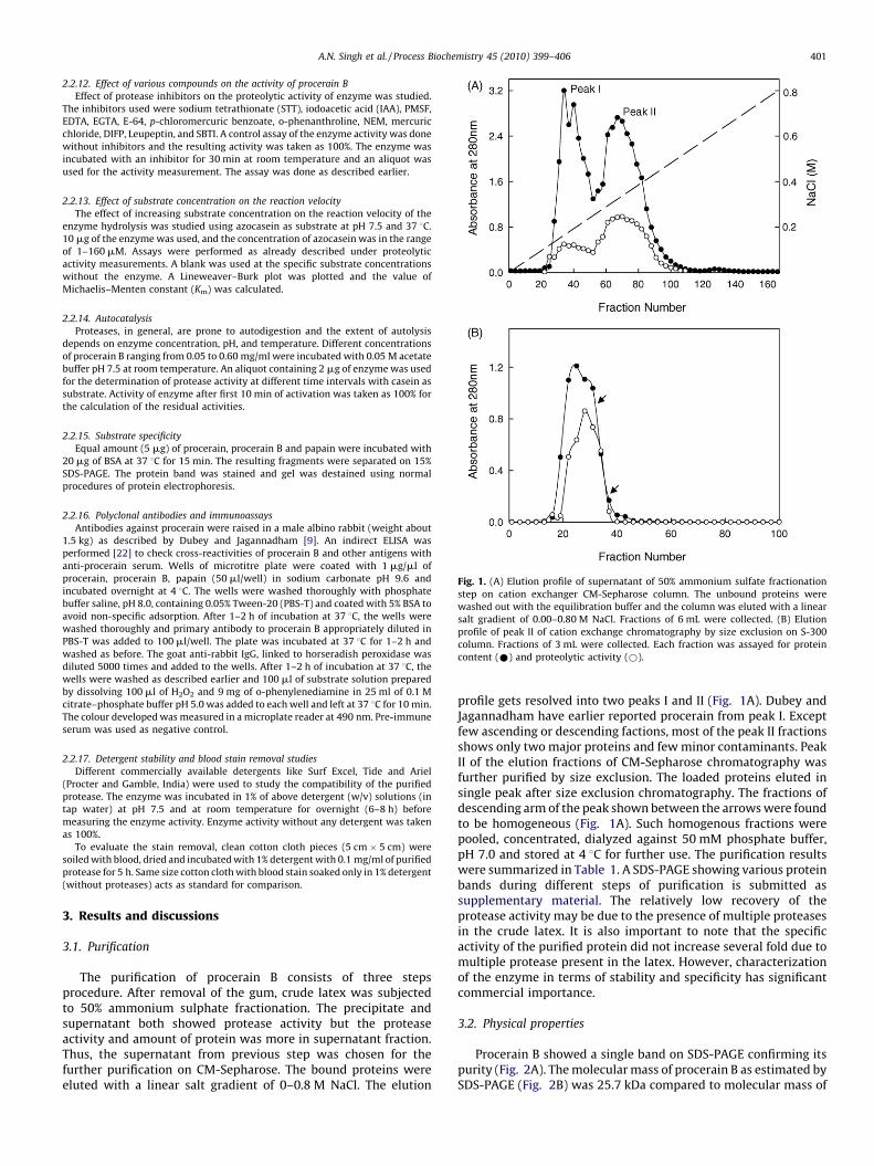
Fig. 1. (A) Elution profile of supernatant of 50% ammonium sulfate fractionation
step on cation exchanger CM-Sepharose column. The unbound proteins were
washed out with the equilibration buffer and the column was eluted with a linear
salt gradient of 0.00–0.80 M NaCl. Fractions of 6 mL were collected. (B) Elution
profile of peak II of cation exchange chromatography by size exclusion on S-300
column. Fractions of 3 mL were collected. Each fraction was assayed for protein
content (*) and proteolytic activity (*).
A.N. Singh et al. / Process Biochemistry 45 (2010) 399–406 401
2.2.12. Effect of various compounds on the activity of procerain B
Effect of protease inhibitors on the proteolytic activity of enzyme was studied.
The inhibitors used were sodium tetrathionate (STT), iodoacetic acid (IAA), PMSF,
EDTA, EGTA, E-64, p-chloromercuric benzoate, o-phenanthroline, NEM, mercuric
chloride, DIFP, Leupeptin, and SBTI. A control assay of the enzyme activity was done
without inhibitors and the resulting activity was taken as 100%. The enzyme was
incubated with an inhibitor for 30 min at room temperature and an aliquot was
used for the activity measurement. The assay was done as described earlier.
2.2.13. Effect of substrate concentration on the reaction velocity
The effect of increasing substrate concentration on the reaction velocity of the
enzyme hydrolysis was studied using azocasein as substrate at pH 7.5 and 37 8C.
10 mg of the enzyme was used, and the concentration of azocasein was in the range
of 1–160 mM. Assays were performed as already described under proteolytic
activity measurements. A blank was used at the specific substrate concentrations
without the enzyme. A Lineweaver–Burk plot was plotted and the value of
Michaelis–Menten constant (Km) was calculated.
2.2.14. Autocatalysis
Proteases, in general, are prone to autodigestion and the extent of autolysis
depends on enzyme concentration, pH, and temperature. Different concentrations
of procerain B ranging from 0.05 to 0.60 mg/ml were incubated with 0.05 M acetate
buffer pH 7.5 at room temperature. An aliquot containing 2 mg of enzyme was used
for the determination of protease activity at different time intervals with casein as
substrate. Activity of enzyme after first 10 min of activation was taken as 100% for
the calculation of the residual activities.
2.2.15. Substrate specificity
Equal amount (5 mg) of procerain, procerain B and papain were incubated with
20 mg of BSA at 37 8C for 15 min. The resulting fragments were separated on 15%
SDS-PAGE. The protein band was stained and gel was destained using normal
procedures of protein electrophoresis.
2.2.16. Polyclonal antibodies and immunoassays
Antibodies against procerain were raised in a male albino rabbit (weight about
1.5 kg) as described by Dubey and Jagannadham [9]. An indirect ELISA was
performed [22] to check cross-reactivities of procerain B and other antigens with
anti-procerain serum. Wells of microtitre plate were coated with 1 mg/ml of
procerain, procerain B, papain (50 ml/well) in sodium carbonate pH 9.6 and
incubated overnight at 4 8C. The wells were washed thoroughly with phosphate
buffer saline, pH 8.0, containing 0.05% Tween-20 (PBS-T) and coated with 5% BSA to
avoid non-specific adsorption. After 1–2 h of incubation at 37 8C, the wells were
washed thoroughly and primary antibody to procerain B appropriately diluted in
PBS-T was added to 100 ml/well. The plate was incubated at 37 8C for 1–2 h and
washed as before. The goat anti-rabbit IgG, linked to horseradish peroxidase was
diluted 5000 times and added to the wells. After 1–2 h of incubation at 37 8C, the
wells were washed as described earlier and 100 ml of substrate solution prepared
by dissolving 100 ml of H2O2 and 9 mg of o-phenylenediamine in 25 ml of 0.1 M
citrate–phosphate buffer pH 5.0 was added to each well and left at 37 8C for 10 min.
The colour developed was measured in a microplate reader at 490 nm. Pre-immune
serum was used as negative control.
2.2.17. Detergent stability and blood stain removal studies
Different commercially available detergents like Surf Excel, Tide and Ariel
(Procter and Gamble, India) were used to study the compatibility of the purified
protease. The enzyme was incubated in 1% of above detergent (w/v) solutions (in
tap water) at pH 7.5 and at room temperature for overnight (6–8 h) before
measuring the enzyme activity. Enzyme activity without any detergent was taken
as 100%.
To evaluate the stain removal, clean cotton cloth pieces (5 cm � 5 cm) were
soiled with blood, dried and incubated with 1% detergent with 0.1 mg/ml of purified
protease for 5 h. Same size cotton cloth with blood stain soaked only in 1% detergent
(without proteases) acts as standard for comparison.
3. Results and discussions
3.1. Purification
The purification of procerain B consists of three stepsprocedure. After removal of the gum, crude latex was subjectedto 50% ammonium sulphate fractionation. The precipitate andsupernatant both showed protease activity but the proteaseactivity and amount of protein was more in supernatant fraction.Thus, the supernatant from previous step was chosen for thefurther purification on CM-Sepharose. The bound proteins wereeluted with a linear salt gradient of 0–0.8 M NaCl. The elution
profile gets resolved into two peaks I and II (Fig. 1A). Dubey andJagannadham have earlier reported procerain from peak I. Exceptfew ascending or descending factions, most of the peak II fractionsshows only two major proteins and few minor contaminants. PeakII of the elution fractions of CM-Sepharose chromatography wasfurther purified by size exclusion. The loaded proteins eluted insingle peak after size exclusion chromatography. The fractions ofdescending arm of the peak shown between the arrows were foundto be homogeneous (Fig. 1A). Such homogenous fractions werepooled, concentrated, dialyzed against 50 mM phosphate buffer,pH 7.0 and stored at 4 8C for further use. The purification resultswere summarized in Table 1. A SDS-PAGE showing various proteinbands during different steps of purification is submitted assupplementary material. The relatively low recovery of theprotease activity may be due to the presence of multiple proteasesin the crude latex. It is also important to note that the specificactivity of the purified protein did not increase several fold due tomultiple protease present in the latex. However, characterizationof the enzyme in terms of stability and specificity has significantcommercial importance.
3.2. Physical properties
Procerain B showed a single band on SDS-PAGE confirming itspurity (Fig. 2A). The molecular mass of procerain B as estimated bySDS-PAGE (Fig. 2B) was 25.7 kDa compared to molecular mass of

Table 1Purification of procerain B from latex of Calotropis procera.
Steps Total protein
(mg)
Total activity
(unitsa)
Specific activity
(units/mg)
Crude latex 780 80,880 103
50% ammonium sulfate
supernatant
384 43,776 114
Ion exchange (peak II
descending arm)
120 17,400 145
Purified procerain B 70 15,750 212
Casein was used as substrate.a Definition of 1 unit: 1 unit of enzyme activity is defined as the amount of
enzyme under the assay conditions described, gives rise to an increase of 1 unit
absorbance at 280 nm/min of digestion.
Fig. 3. Effects of (A) temperature and (B), pH on activity (*) and stability (*) of
procerain. Stability was determined by overnight incubating the enzyme at room
temperature at different pH conditions and next day activity was taken as described
in Section 2.2. For temperature stability measurements, enzyme was incubated at
required temperature for 15 min, and activity was measured at 37 8C and pH 7.5.
A.N. Singh et al. / Process Biochemistry 45 (2010) 399–406402
procerain 28.8 kDa [9]. The molecular weight of cryptolepain fallswell in the range of the molecular weight of plant cysteineproteases.
The isoelectric point of procerain B was found to be 9.52. Mostof the cysteine proteases are reported to be basic in nature likeprocerain (pI 9.32), heynein (pI 10.8), papain (pI 8.75), ficin (pI 9.0),ervetamin C (pI 9.54), stem bromelin (pI 9.55) shows basicisoelectric points [9,23]. However, some acidic plant proteases likeactinidin (pI 3.0) and asclepain (pI 3.11) are also reported [24]. Theextinction coefficient (e1%
280 nm) of purified enzyme procerain B asdetermined by spectrometric methods was 10.1 and whileprocerain showed extinction coefficient (e1%
280 nm) of 24.9.The number of tryptophan was found to be 3 and number of
tyrosine was 6 as determined by spectroscopic method as explainedin Section 2.2. The total cysteine content of procerain B is found to benine (measured value 8.86) with one free cysteine residues(measured value 0.89), thus forming four disulphide bridges. Mostof the cysteine proteases, including procerain, reported to have totalseven cysteine, six cysteine forming three disulfides and onecatalytic cysteine [25–29]. However, 11 cysteine [5 disulfide and 1free] is also reported in plant cysteine protease, heynein [23].Procerain B, like procerain many other cysteine proteases does notcontain any detectable carbohydrate moiety [9,23,24]. However,few cysteine proteases like ginger proteases GP-I and GP-II reportedto have significant carbohydrate moieties [30].
3.3. pH and temperature optimum
Procerain also shows broad temperature optima in the range of40–60 8C (Fig. 3A). The enzyme shows 100% activity till 75 8C. Thisunusually high temperature stability broad temperature optima
Fig. 2. (A) SDS-PAGE of protein samples. Lanes 1 and 2 represent purified protein
and protein molecular weight markers, respectively. (B) Estimation of molecular
weight of procerain B on SDS-PAGE by comparing its relative mobility with
standard markers used in gel.
and broad pH optima of procerain B makes it a valuable proteasefor industrial application.
Procerain B shows broad pH optima for hydrolyzing activity andshows optimum activity in the range of pH 6.5–8.5 (Fig. 3B). Such abroad pH optima range of procerain B may make it a betterprotease molecule in various food and biotechnology industries.Further, the optimum pH of procerain B differs significantly withprocerain and other cysteine protease (Table 2). A sharp decreasein activity in the acidic region below pH 5.0 may be due to theformation of improper ionic forms of the enzyme active site orsubstrate or combination of the two.
3.4. Stability
The stability of the enzyme under extreme conditions is adecisive factor for its usefulness as a potent industrial enzyme.Procerain B is remarkably stable and retains its activity underthe conditions where most of the enzymes lose their activity.The enzyme is fully active till 70 8C for 15 min (Fig. 3A). Theenzyme retains complete activity in 30% methanol while in 80%methanol enzyme shows 15% activity. Additionally, enzyme isfully active in 35% acetonitrile and 2.0 M GuHCl at pH 7.5.The enzyme retains most of the activity in 8 M urea. Such ahigh stability of procerain B under various harsh conditionsmakes it an excellent enzyme for industrial and biotechnologicalapplications.

Table 2Comparison of biochemical properties of procerain with few other plant cysteine proteases.
Protein Plant Mol. weight
(kDa)
pH optimum Temperature
optimum (8C)
Isoelectric point (pI) No. of disulfide
bonds
Carbohydrate
Procerain Ba Calotropis procera 25.7 6.5–8.5 40–60 9.52 4 No
Procerainb Calotropis procera 28.8 7.0–9.0 55–60 9.32 3 No
Ervatamin Ac Ervatamia coronaria 27.6 8–8.5 50–55 8.37 3 No
Ervatamin Bd,e Ervatamia coronaria 26 6.0–6.5 50–55 9.35 3 No
Ervatamin Cf Ervatamia coronaria 25.5 7.5–8.0 50 9.54 3 No
Heyneing Ervatamia heyneana 23 8.0–8.5 50–55 10.8 5 No
GP-Ih,i Zingiber officinale 22.5 NR NR 4.6 3 Yes
GP-IIh,i Zingiber officinale 22.5 NR NR 4.7 3 Yes
Papainj Carica papaya 23 6.0–7.0. NR 9.6 3 No
Asclepain c-IIk Asclepias curassavica 23.6 9.4–10.2 NR 9.3 NR NR
NR: Not reported.a Current work.b [9].c [32].d [33].e [29].f [25].g [23].h [30].i [34].j http://www.worthington-biochem.com/pcyp/default.html.k [35].
A.N. Singh et al. / Process Biochemistry 45 (2010) 399–406 403
3.5. Effect of inhibitors on the activity of procerain B
Five catalytic types of proteases are recognized in which serine,threonine, cysteine, aspartic or metallo groups play primary rolesin enzyme catalysis. We have studied effect of inhibitors of variousclasses of proteases to determine the class of the purified protease,procerain B (Table 3). Maximum inhibition of activity of theenzyme occurs in the presence of iodoacetic acid, E-64, HgCl2,NEM, PCMB, leupeptine and sodium tetrathionate. Enzyme did notshow significant inhibitions in the presence of PMSF, SBTI, DFP.Lack of inhibition of the activity by proteinaceous inhibitors suchas SBTI, which are abundant in protein-rich foods like soybean,makes the enzyme a potential protease for food industry [31].Metalloprotease inhibitors like o-phenanthroline, EDTA and EGTAshowed no significant effect on the activity of procerain B rulingout the possibly of the protease being a metalloprotein. The resultsconfirm that the enzyme belongs to cysteine protease family.
3.6. Effect of substrate concentration on the reaction velocity
The effect of increasing substrate concentration on reactionvelocity of procerain B follows the Michaelis–Menton equation.
Table 3Effect of various compounds on proteolytic activity of procerain B and comparison wit
Inhibitor class Inhibitor Proceraina
[I]
Cysteine protease Iodoacetic acid 40 mM
E-64 3 mM
HgCl2 5 mM
NEM 30 mM
Sodium tetrathionate 30 mM
PCMB 5 mM
Ser/Cys Leupeptin 10 mM
Serine protease DFP 1 mM
PMSF 50 mM
SBTI 1 mM
Metalloprotease EDTA 1 mM
EGTA 1 mM
o-Phenanthroline 1 mM
a Data taken from Dubey and Jagannadham [9].b Average of three measurements.
The study was carried out using azocasein (Fig. 4A) and casein(Fig. 4B) as substrates. The value of Km was estimated fromLineweaver–Burk plot. The enzyme shows Km value of31 � 210 mM with both the substrates.
3.7. Substrate specificity
The specificity of the enzyme was further investigated by BSAdigestion. As shown in Fig. 5, procerain B shows very differentcleavage pattern compared to procerain and papain. Thus, procerainB appears to have distinct substrate specificity. Two major digestedfragments of 27 kDa and 30 kDa as well as minor bands around25 kDa are common. However, 21 kDa, 20 kDa and 14 kDa fragmentsare unique in procerain B. This suggests that procerain B can cleave atadditional sites which are not recognized by papain and procerain.However, the common fragments indicate that procerain B can alsocleave at sites recognized by papain and procerain.
3.8. Autocatalysis
The autodigestion property of proteases limits its usefulness.Autolysis resistance proteases are more useful for industrial or
h procerain.
Procerain B
Residual activity (%) [I] Residual activity (%)b
12 40 mM 5
0 3 mM 0
8 2 mM 9
21 50 mM 18
15 30 mM 22
10 5 mM 12
10 10 mM 15.2
95 1 mM 98.5
85 50 mM 92
100 1 mM 100
100 5 mM 100
100 5 mM 100
100 5 mM 100
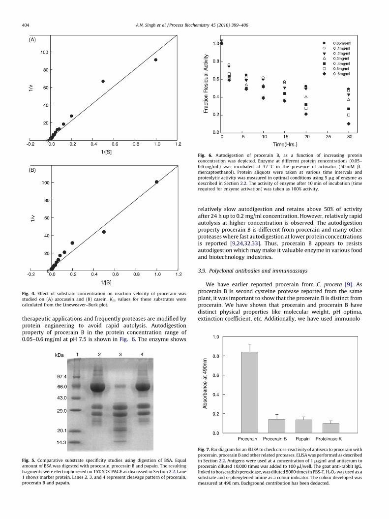
Fig. 4. Effect of substrate concentration on reaction velocity of procerain was
studied on (A) azocasein and (B) casein. Km values for these substrates were
calculated from the Lineweaver–Burk plot.
Fig. 6. Autodigestion of procerain B, as a function of increasing protein
concentration was depicted. Enzyme at different protein concentrations (0.05–
0.6 mg/mL) was incubated at 37 8C in the presence of activator (50 mM b-
mercaptoethanol). Protein aliquots were taken at various time intervals and
proteolytic activity was measured in optimal conditions using 5 mg of enzyme as
described in Section 2.2. The activity of enzyme after 10 min of incubation (time
required for enzyme activation) was taken as 100% activity.
A.N. Singh et al. / Process Biochemistry 45 (2010) 399–406404
therapeutic applications and frequently proteases are modified byprotein engineering to avoid rapid autolysis. Autodigestionproperty of procerain B in the protein concentration range of0.05–0.6 mg/ml at pH 7.5 is shown in Fig. 6. The enzyme shows
Fig. 5. Comparative substrate specificity studies using digestion of BSA. Equal
amount of BSA was digested with procerain, procerain B and papain. The resulting
fragments were electrophoresed on 15% SDS-PAGE as discussed in Section 2.2. Lane
1 shows marker protein. Lanes 2, 3, and 4 represent cleavage pattern of procerain,
procerain B and papain.
relatively slow autodigestion and retains above 50% of activityafter 24 h up to 0.2 mg/ml concentration. However, relatively rapidautolysis at higher concentration is observed. The autodigestionproperty procerain B is different from procerain and many otherproteases where fast autodigestion at lower protein concentrationsis reported [9,24,32,33]. Thus, procerain B appears to resistsautodigestion which may make it valuable enzyme in various foodand biotechnology industries.
3.9. Polyclonal antibodies and immunoassays
We have earlier reported procerain from C. procera [9]. Asprocerain B is second cysteine protease reported from the sameplant, it was important to show that the procerain B is distinct fromprocerain. We have shown that procerain and procerain B havedistinct physical properties like molecular weight, pH optima,extinction coefficient, etc. Additionally, we have used immunolo-
Fig. 7. Bar diagram for an ELISA to check cross-reactivity of antisera to procerain with
procerain, procerain B and other related proteases. ELISA was performed as described
in Section 2.2. Antigens were used at a concentration of 1 mg/ml and antiserum to
procerain diluted 10,000 times was added to 100 ml/well. The goat anti-rabbit IgG,
linked to horseradish peroxidase, was diluted 5000 times in PBS-T. H2O2 was used as a
substrate and o-phenylenediamine as a colour indicator. The colour developed was
measured at 490 nm. Background contribution has been deducted.

Fig. 8. To evaluate the stain removal, clean cotton cloth pieces were soiled with blood and dried. (A) Soaked only in 1% detergent (without proteases) acts as standard for
comparison. (B) Incubated with 1% detergent with 0.1 mg/ml of purified protease for 5 h.
A.N. Singh et al. / Process Biochemistry 45 (2010) 399–406 405
gical methodfor the same.Polyclonalantibodies specifictoprocerainhave been successfully raised in male albino rabbit and cross-reactivitywithprocerain B byindirectELISAwas checked. Thetypicalcolour development in indirect ELISA results from procerain anti-procerain complex formation further confirmed the presence ofantibodies to procerain (Fig. 7). The newly purified procerain B andfew other common proteases were also checked for cross-reactivityagainst anti-procerain. Like other protease and unlike procerain,procerain B did not show any cross-reactivity with anti-procerain.This confirms a distinct nature of procerain B and reveals thatpurified protein is antigenitically different from them.
3.10. Detergent stability and blood stain removal studies
The purified enzyme, procerain B, shows very high stability inall detergents used for the study. It shows 100% activity in 1%(w/v) in all detergents used in the current study. Moreover, itappears to improve blood strain cleaning property of thedetergent (Fig. 8). Thus, procerain B has potential applicationin detergent industry for improving the cleaning efficiency ofdetergents. Easy availability of the plant latex and simplepurification method of the enzyme make it also a cost-effectivedetergent enzyme.
Acknowledgements
Infrastructure funded to VKD by Department of Biotechnology,Department of Science and Technology and Department of
Information Technology,Government of India and facilities providedby Indian Institute of Technology Guwahati are acknowledged.
Appendix A. Supplementary data
Supplementary data associated with this article can be found, in
the online version, at doi:10.1016/j.procbio.2009.10.014.
References
[1] Puente XS, Sanchez LM, Overall CM, Lopez-Otin C. Human and mouse pro-teases: a comparative genomic approach. Nat Rev Genet 2003;4:544–58.
[2] Turk B. Targeting proteases: successes, failures and future prospects. Nat RevDrug Discov 2006;5:785–98.
[3] Dubey VK, Pande M, Singh BK, Jagannadham MV. Papain-like proteases:applications of their inhibitors. Afr J Biotechnol 2007;6:1077–86.
[4] Van der Hoorn RAL, Jones JDG. The plant proteolytic machinery and its role indefence. Curr Opin Plant Biol 2004;7:400–7.
[5] Kumar VL, Arya S. Medicinal uses and pharmacological properties of Calotropisprocera. In: Govil JN, editor. Recent Progress in Medicinal Plants, vol. 11.Houston, TX, USA: Studium Press; 2006. p. 373–88.
[6] Sharma P, Sharma JD. Evaluation of in vitro schizontocidal activity of plant partsof Calotropis procera—an ethanobotanical approach. J Ethnopharmacol 1999;68:83–95.
[7] Sharma P, Sharma JD. In vitro hydrolysis of erythrocytes—by plant extractswith anti plasmodial activity. J Ethnopharmacol 2001;74:239–43.
[8] Singhal A, Kumar VL. Effect of aqueous suspension of dried latex ofCalotropis procera on hepatorenal functions in rat. J Ethnopharmacol2009;122: 172–4.
[9] Dubey VK, Jagannadham MV. Procerain, a stable cysteine protease from thelatex of Calotropis procera. Phytochemistry 2003;62:1057–71.
[10] Gilman A, Philips FS, Koelle ES, Allen RP, John St E. The metabolic reduction andnephrotoxic action of tetrathionate in relation to a possible interaction withsulfhydryl compounds. Am J Physiol 1946;147:115–23.
A.N. Singh et al. / Process Biochemistry 45 (2010) 399–406406
[11] Bradford MM. A rapid and sensitive method for the quantitation of microgramquantities of protein utilizing the principle of protein–dye binding. AnalBiochem 1976;72:248–54.
[12] Laemmli UK. Cleavage of structural proteins during assembly of the head ofbacteriophage T4. Nature 1970;227:680–5.
[13] Merril CR. In: Deutscher MP, editor. Guide to Protein Purification in Gel-staining Techniques. San diego, CA: Academic Press, Inc.; 1990. p. 477–88.
[14] Van Teeffelen MMA, Broersen K, De Jongh HJ. Glucosylation of b-lactoglobulinlowers the capacity change of unfolding, a unique way to affect proteinthermodynamics. Protein Sci 2005;14:2187–94.
[15] Hounsell EF, Davies MJ, Smith KD. Chemical methods of analysis ofglycoproteins. In: Walker JM, editor. The Protein Protocol Hand Book. Totawa,NJ: Humana Press; 1997. p. 633–4.
[16] Goodwin W, Morton RA. The spectrophotometric determination of tyrosineand tryptophan in proteins. Biochem J 1946;40:628–32.
[17] Ellman L. Tissue sulfhydryl groups. Arch Biochem Biophys 1959;82:70–7.
[18] Disulfide bonds between protein residues. Creighton TE, editor. Protein Struc-ture—A Practical Approach. Oxford: IRL Press; 1989. p. 155–60.
[19] Riddles PW, Blakeley RL, Zerner B. Reassessment of Ellman’s reagent. MethodsEnzymol 1983;91:49–60.
[20] Aitken A, Learmoth M. Protein determination by UV absorption. In: WalkerJM, editor. The Protein Protocol Handbook. Totowa, NJ: Humana Press; 1997.p. 3–6.
[21] Sarath G, Motte RS, Wagner FW. Protease assay methods. In: Bond RJ, Beynon,editors. Proteolytic Enzymes—A Practical Approach. Oxford: IRL Press; 1989. p.25–55.
[22] Friguet B, Djavadi-Ohaniance L, Goldberg ME. Immuno-chemical analysis ofprotein concentration. In: Crieghton TE, editor. Protein Structure—A PracticalApproach. Oxford: IRL Press; 1989. p. 287–310.
[23] Patel BK, Jagannadham MV. A high cysteine containing thiol proteinasefrom the latex of Ervatamia heyneana: purification and comparison withervatamin B and C from Ervatamia coronaria. J Agric Food Chem 2003;51:6326–34.
[24] Mc Dowall MA. Anionic protenases from Actinidia chinensis preparation andproperties of the crystalline enzyme. Eur J Biochem 1970;14:214–21.
[25] Sundd M, Kundu S, Pal GP, Jagannadham MV. Purification and characterizationof a highly stable cysteine protease from the latex of Ervatamia coronaria.Biosci Biotechnol Biochem 1998;62:1947–55.
[26] Sumner IG, Harris GW, Taylor MAJ, Pickersgill RW, Owen AJ, Goodenough PW.Factors affecting the thermostability of cysteine proteases from Carica papaya.Eur J Biochem 1993;214:129–234.
[27] Brockbank WJ, Lynn KR. Purification and preliminary characterization of twoasclepains from the latex of Asclepais syriaca L. (milk weed). Biochim BiophysActa 1979;578:13–22.
[28] Pal G, Sinha NK. Isolation, crystallization and properties of Calotropis DI andDII from Calotropis gigentea. Arch Biochem Biophys 1980;202:321–9.
[29] Biswas S, Chakrabarti C, Kundu S, Jagannadham MV, Dattagupta JK. Proposedamino acid sequence and the 1.63 A X-ray crystal structure of a plant cysteineprotease, ervatamin B: some insights into the structural basis of its stabilityand substrate specificity. Proteins 2003;51:489–97.
[30] Choi KH, Laursen RA. Amino-acid sequence and glycan structures of cysteineproteases with proline specificity from ginger rhizome Zingiber officinale. Eur JBiochem 2002;267:1516–26.
[31] Kaneda M, Tominaga N. Isolation and characterization of a proteinase fromwhite gourd. Phytochemistry 1977;16:345–6.
[32] Pande M, Dubey VK, Yadav SC, Jagannadham MV. A novel serine proteasecryptolepain from Cryptolepis buchanani: purification and biochemical char-acterization. J Agric Food Chem 2006;54:10141–50.
[33] Bhowmick R, Kumari NK, Jagannadham MV, Kayastha AM. Purification andcharacterization of a novel protease from the latex of Pedilanthus tithyma-loids. Protein Pept Lett 2008;15(19):1009–16.
[34] Ohtsuki K, Taguchi K, Sato K, Kawabata M. Purification of ginger proteases byDEAE-Sepharose and isoelectric focusing. Biochim Biophys Acta 1995;1243:181–4.
[35] Liggieri C, Obregon W, Trejo S, Priolo N. Biochemical analysis of a papain-likeprotease isolated from the latex of Asclepias curassavica L.. Acta BiochimBiophys Sin (Shanghai) 2009;41:154–62.
Copyright © 2022 FDOKUMEN