PTIP Promotes Chromatin Changes Critical for Immunoglobulin Class Switch Recombination
15
PTIP Promotes Chromatin Changes Critical for Immunoglobulin Class Switch Recombination Jeremy A. Daniel 1 , Margarida Almeida Santos 1,* , Zhibin Wang 2,* , Chongzhi Zang 3,* , Kristopher R. Schwab 4 , Mila Jankovic 5 , Darius Filsuf 1 , Hua-Tang Chen 1 , Anna Gazumyan 5 , Arito Yamane 6 , Young-Wook Cho 7 , Hong-Wei Sun 8 , Kai Ge 7 , Weiqun Peng 3 , Michel C. Nussenzweig 5 , Rafael Casellas 6 , Gregory R. Dressler 4 , Keji Zhao 2 , and André Nussenzweig 1,† 1 Experimental Immunology Branch, National Cancer Institute, National Institutes of Health (NIH), Bethesda, MD 20892, USA 2 Laboratory of Molecular Immunology, National Heart, Lung and Blood Institute, NIH, Bethesda, MD 20892, USA 3 Department of Physics, The George Washington University, Washington, DC 20052, USA 4 Department of Pathology, University of Michigan, Ann Arbor, MI 48109, USA 5 Laboratory of Molecular Immunology, The Rockefeller University, and Howard Hughes Medical Institute, New York, NY 10065, USA 6 Genomics and Immunity, National Institute of Arthritis and Musculoskeletal and Skin Diseases (NIAMS), NIH, Bethesda, MD 20892, USA 7 Nuclear Receptor Biology Section, CEB, National Institute of Diabetes and Digestive and Kidney Diseases, NIH, Bethesda, MD 20892, USA 8 Biodata Mining and Discovery Section, NIAMS, NIH, Bethesda, MD 20892, USA Abstract Programmed genetic rearrangements in lymphocytes require transcription at antigen receptor genes to promote accessibility for initiating double-strand break (DSB) formation critical for DNA recombination and repair. Here, we showed that activated B cells deficient in the PTIP component of the MLL3 (mixed-lineage leukemia 3)–MLL4 complex display impaired trimethylation of histone 3 at lysine 4 (H3K4me3) and transcription initiation of downstream switch regions at the immunoglobulin heavy-chain (Igh) locus, leading to defective immunoglobulin class switching. We also showed that PTIP accumulation at DSBs contributes to class switch recombination (CSR) and genome stability independently of Igh switch transcription. These results demonstrate that PTIP promotes specific chromatin changes that control the accessibility of the Igh locus to CSR and suggest a nonredundant role for the MLL3-MLL4 complex in altering antibody effector function. † To whom correspondence should be addressed. [email protected]. * These authors contributed equally to this work. Supporting Online Material www.sciencemag.org/cgi/content/full/science.1187942/DC1 Materials and Methods Figs. S1 to S16 Tables S1 and S2 References NIH Public Access Author Manuscript Science. Author manuscript; available in PMC 2011 February 1. Published in final edited form as: Science. 2010 August 20; 329(5994): 917–923. doi:10.1126/science.1187942. NIH-PA Author Manuscript NIH-PA Author Manuscript NIH-PA Author Manuscript
-
Upload
independent -
Category
Documents
-
view
0 -
download
0
Transcript of PTIP Promotes Chromatin Changes Critical for Immunoglobulin Class Switch Recombination

PTIP Promotes Chromatin Changes Critical for ImmunoglobulinClass Switch Recombination
Jeremy A. Daniel1, Margarida Almeida Santos1,*, Zhibin Wang2,*, Chongzhi Zang3,*,Kristopher R. Schwab4, Mila Jankovic5, Darius Filsuf1, Hua-Tang Chen1, AnnaGazumyan5, Arito Yamane6, Young-Wook Cho7, Hong-Wei Sun8, Kai Ge7, Weiqun Peng3,Michel C. Nussenzweig5, Rafael Casellas6, Gregory R. Dressler4, Keji Zhao2, and AndréNussenzweig1,†1 Experimental Immunology Branch, National Cancer Institute, National Institutes of Health (NIH),Bethesda, MD 20892, USA2 Laboratory of Molecular Immunology, National Heart, Lung and Blood Institute, NIH, Bethesda,MD 20892, USA3 Department of Physics, The George Washington University, Washington, DC 20052, USA4 Department of Pathology, University of Michigan, Ann Arbor, MI 48109, USA5 Laboratory of Molecular Immunology, The Rockefeller University, and Howard Hughes MedicalInstitute, New York, NY 10065, USA6 Genomics and Immunity, National Institute of Arthritis and Musculoskeletal and Skin Diseases(NIAMS), NIH, Bethesda, MD 20892, USA7 Nuclear Receptor Biology Section, CEB, National Institute of Diabetes and Digestive and KidneyDiseases, NIH, Bethesda, MD 20892, USA8 Biodata Mining and Discovery Section, NIAMS, NIH, Bethesda, MD 20892, USA
AbstractProgrammed genetic rearrangements in lymphocytes require transcription at antigen receptorgenes to promote accessibility for initiating double-strand break (DSB) formation critical for DNArecombination and repair. Here, we showed that activated B cells deficient in the PTIP componentof the MLL3 (mixed-lineage leukemia 3)–MLL4 complex display impaired trimethylation ofhistone 3 at lysine 4 (H3K4me3) and transcription initiation of downstream switch regions at theimmunoglobulin heavy-chain (Igh) locus, leading to defective immunoglobulin class switching.We also showed that PTIP accumulation at DSBs contributes to class switch recombination (CSR)and genome stability independently of Igh switch transcription. These results demonstrate thatPTIP promotes specific chromatin changes that control the accessibility of the Igh locus to CSRand suggest a nonredundant role for the MLL3-MLL4 complex in altering antibody effectorfunction.
†To whom correspondence should be addressed. [email protected].*These authors contributed equally to this work.Supporting Online Materialwww.sciencemag.org/cgi/content/full/science.1187942/DC1Materials and MethodsFigs. S1 to S16Tables S1 and S2References
NIH Public AccessAuthor ManuscriptScience. Author manuscript; available in PMC 2011 February 1.
Published in final edited form as:Science. 2010 August 20; 329(5994): 917–923. doi:10.1126/science.1187942.
NIH
-PA Author Manuscript
NIH
-PA Author Manuscript
NIH
-PA Author Manuscript
Mixed-lineage leukemia (MLL)–like complexes catalyze mono-, di-, and tri-methylation ofhistone H3 at lysine 4 (H3K4me), marks which all strongly correlate with active geneexpression (1), but the direct role and specificity of these complexes during lymphocytematuration are unknown. The PTIP (Pax interaction with transcription-activation domainprotein-1, also known as Paxip1) protein stably associates with a subset of MLL-likecomplexes containing the MLL3 and MLL4 methyltransferases (2–4), suggesting a role forPTIP in transcriptional regulation. Embryonic lethality of Paxip1−/− mice at embryonic day9.5 (5), however, has precluded any further elucidation of its biological function, with theexception of a role in adipogenesis (6).
Along with possible functions in transcriptional regulation, PTIP contains six BRCT(BRCA1 carboxy terminal) domains, implicating the protein in the DNA damage response(7). Indeed, upon treatment with ionizing radiation (IR), PTIP forms nuclear foci dependenton the γ-H2AX/MDC1/RNF8 damage response pathway and its own BRCT function, andalso physically interacts with the DNA repair factor 53BP1 (7–9). PTIP-deficient cells arealso hypersensitive to irradiation (9–11) and defective in homologous recombination (12).To what extent PTIP functions directly in double-strand break (DSB) repair, however, isunclear because loss of PTIP is also associated with defective proliferation (5) and a globaldecrease in H3K4me3 (3,13), a chromatin mark of transcription initiation (14–16).
During V(D)J recombination in developing B cells in the bone marrow, recombination-activating gene 1/2 (RAG1/2)–mediated cleavage and DSB repair by the nonhomologousend-joining (NHEJ) pathway occur at accessible recombination signal sequence chromatinin antigen receptor genes and require actively transcribed gene elements (17). In response toligand engagement from lipopolysaccharide (LPS), B cell receptor cross-linking, and/orcytokine stimulation, mature naïve immunoglobulin M (IgM)–expressing B cells in theperiphery proliferate and become activated to undergo class-switch recombination (CSR)(17,18). This second rearrangement event promotes recombination of an antigen recognitiongene segment with different Igh constant regions to interact with different cell surfacereceptors for successful clearance of a pathogen (17,18). During CSR, transcription ofimmunoglobulin heavy-chain (Igh) switch region chromatin is required for accessibility ofAID (activation-induced cytidine deaminase) (19,20), which leads to formation of DSBsboth at the upstream switch (Sμ) region and one of the downstream switch regions (Sγ3,Sγ1, Sγ2b, Sγ2a, Sε, or Sα) (18). Subsequently, synapsis and DNA repair of the two switchregions are mediated by γ-H2AX/53BP1 and NHEJ proteins, leading to a recombined Ighlocus (21). Given that DNA rearrangements in B lymphocytes require transcription andDNA repair at immunoglobulin genes and that PTIP is implicated in both of these processes,we investigated PTIP function in CSR.
Genome-wide H3K4me3 in LPS-stimulated PaxipΔ/Δ and control B cellsTo determine how changes in histone H3K4 methylation regulate mature B celldifferentiation, we used chromatin immunoprecipitation coupled with Illumina sequencing(ChIP-Seq) to compare genome-wide H3K4me3 profiles of resting murine B cells with Bcells stimulated with LPS and antibody against IgD conjugated to dextran (αIgD-dextran,herein referred to as LPS stimulation), mitogens known to induce rapid proliferation andactivation ex vivo. We observed that 1014 regions across the genome display more than afourfold increase in H3K4me3 after LPS stimulation (fig. S1A and table S1). Only 13.2% ofthese LPS-induced H3K4me3 regions were localized within 4 kb of known transcriptionalstart sites, suggesting that LPS stimulation may initiate transcription at manyuncharacterized genomic regions (fig. S1B).
Daniel et al. Page 2
Science. Author manuscript; available in PMC 2011 February 1.
NIH
-PA Author Manuscript
NIH
-PA Author Manuscript
NIH
-PA Author Manuscript
Several genes whose transcripts are LPS inducible in B lymphocytes (18) displayedincreased H3K4me3 upon LPS stimulation, including Il6, the Igh switch regions, and Aicda(encodes activation-induced cytidine deaminase) (fig. S1, C to E). In contrast, c-myc, whichis induced during B cell activation but displays promoter-proximal polymerase II (Pol II)pausing (22), displayed similar H3K4me3 profiles in both resting and LPS-stimulated Bcells (fig. S1E). These data support the notion that although c-myc may be poised forinduced expression in mature B cells, Aicda, Il6, and the Igh switch regions are devoid ofH3K4me3 until B cell activation, thereby comprising a different group of tightly regulatedgenes that have predominant control mechanisms that normally prevent initiation (15).
To investigate the genetic requirements and biological impact of LPS-induced H3K4me3,we crossed Paxipfl/fl mice with CD19-cre mice to generate B cell–specific PTIP knockoutmice. Analysis of Cd19-cre Paxipfl/fl splenic B cells (herein referred to as PaxipΔ/Δ B cells)revealed reduced PTIP protein and transcript expression while displaying near-normalCD19+ B cell numbers and frequency of surface IgM+ cells (fig. S2, A to D). In contrast toprimary fibroblasts and embryonic stem cells (5), PTIP-deficient B cells proliferated in amanner similar to that of controls in response to either LPS or LPS and interleukin-4 (IL-4)(fig. S2E). Together these data suggest that PTIP is largely dispensable for B celldevelopment and cell cycle progression.
Normal proliferation of PaxipΔ/Δ B cells suggested the possibility of a more limited role forPTIP-mediated histone methylation in B cells compared to the global reduction ofH3K4me3 observed in developing tissues (3,13). We found that only six regions (Igh-γ2b,Igh-γ3, Slit3, Il6, Paxip1, and mRNA AK191783/Cacna1e) in PaxipΔ/Δ B cells displayedmore than a fourfold decrease in H3K4me3 compared to controls, and 10 regions displayedmore than a threefold decrease in H3K4me3 (Fig. 1A and fig. S3, A and B). No regionsdisplayed consistently increased H3K4me3 in PaxipΔ/Δ B cells (fig. S3A). Consistent withPTIP deficiency in PaxipΔ/Δ cells, the Paxip gene itself also displayed reduced H3K4me3(Fig. 1A) because of Cre-mediated deletion of its first exon (3). Taken together, our datademonstrate a selective role for PTIP in promoting histone methylation in activated B cells.
The Igh-γ2b and Igh-γ3 switch regions showed the greatest decrease in H3K4me3 fromLPS-induced PaxipΔ/Δ B cells (Fig. 1, A and B). Moreover, PaxipΔ/Δ and control B cellsstimulated with LPS and IL-4 displayed PTIP-dependent H3K4me3 at Igh-γ1 (fig. S4),indicating that PTIP also promotes H3K4me3 at other Igh switch regions under differentconditions of B cell activation. Notably, the PTIP-dependent H3K4me3 at activated Ighswitch regions occurs independently of AID-induced DNA damage (fig. S4), consistent withH3K4me3 associating with transcription rather than with DNA DSBs (14). PTIP deficiency,however, had no effect on H3K4me3 marking the Igh-μ enhancer (5′Eμ), μ switch repeatregion (Sμ), or Igh-ε switch repeat region (Sε) under either stimulation condition (Fig. 1Band fig. S4), demonstrating the specificity of PTIP-mediated histone methylation. NormalH3K4me3 across the 5′Eμ region is consistent with the normal surface IgM expressionobserved on PaxipΔ/Δ B cells (fig. S2D).
Ig class-switching defects in PaxipΔ/Δ B cellsAberrant regulation of Igh switch regions and Il6 have well-established implications formature B cell function (18,23). To understand the physiological relevance of PTIP-dependent H3K4me3 at Igh switch regions, we examined Ig class switching. Upon LPSstimulation, PaxipΔ/Δ B cells displayed 14- and 4.8-fold decreases in the frequency of IgG3and IgG2b switching, respectively (Fig. 2A and fig. S5, A and B). PaxipΔ/Δ B cells alsodisplayed a 2.6-fold decrease in IgG1 switching upon LPS and IL-4 stimulation (Fig. 2A andfig. S5, A and B). When CSR was assayed directly from genomic DNA of B cells stimulated
Daniel et al. Page 3
Science. Author manuscript; available in PMC 2011 February 1.
NIH
-PA Author Manuscript
NIH
-PA Author Manuscript
NIH
-PA Author Manuscript
with LPS and IL-4, PaxipΔ/Δ B cells displayed a similar defect in the level of Sμ-Sγ1 switchjunctions (fig. S5C). As expected, PaxipΔ/Δ B cells showed normal cell proliferation (figs.S2E and S5, A and B) and cell survival (fig. S5D). We conclude that PTIP is critical forIgG3, IgG2b, and to a lesser extent, IgG1 class switching.
IL-6 was further analyzed because it can function in both antibody secretion and thedevelopment and tumorigenesis of plasma cells (23) (Fig. 1A). We found that H3K4me3 atIl6 was both LPS inducible and PTIP dependent (fig. S1C and Fig. 2B) and that mRNAexpression of Il6 was also impaired in PaxipΔ/Δ B cells (Fig. 2C). These defects, however,had no apparent effect on plasma cell differentiation ex vivo, as monitored by CD138surface expression (Fig. 2D). Moreover, the IgG3 class-switching defect in PaxipΔ/Δ B cellswas not rescued with exogenous recombinant IL-6 in the culture medium (fig. S6).
PTIP promotes Ig switch region transcription initiationFor most actively transcribed genes, H3K4me3 and Pol II accumulate within 2 kb of thetranscription start sites (24). In contrast, H3K4me3 peaks at Igh switch regions are morebroad, spanning up to 7 kb downstream of the germline transcript promoter, and include themutagenic switch repeats and downstream of the switch region (Fig. 1B and figs. S4 and S7,A and B) (25). This broad H3K4me3 distribution at switch regions correlates with observedaccumulation of Pol II at switch regions (Fig. 3A) (25,26). Therefore, we consideredwhether PTIP might be important for elongation or splicing of switch transcripts. Toinvestigate whether PTIP regulates transcription of Igh switch regions, we measuredgermline switch transcripts that had been spliced from the initiating (I) exon located 5′ of theswitch region to the constant (C) exon located 3′ of the switch region from stimulatedPaxipΔ/Δ and control B cells (Fig. 3B). IgM-expressing B cells express a μ sterile transcript,and we found that PaxipΔ/Δ B cells displayed near-normal μ switch transcript amounts inboth conditions (Fig. 3B). This is consistent with the normal H3K4me3 patterns at Eμ andSμ in the absence of PTIP (Fig. 1B and fig. S4). In contrast, both γ3 and γ1 spliced switchtranscripts, important for IgG CSR, were severely impaired in PaxipΔ/Δ B cells under theirrespective stimulation conditions (Fig. 3B). Spliced transcripts at Igh-ε were unchanged inPaxipΔ/Δ B cells (fig. S7C), consistent with no change in H3K4me3 at this switch region inthe absence of PTIP (fig. S4).
To further delineate at which stage PTIP functions in promoting Igh-γ germline switchtranscript expression, we also assayed for unspliced and initiating switch transcripts.PaxipΔ/Δ B cells showed similar deficiencies both in unspliced (fig. S7D) as well as ininitiating switch transcripts (Fig. 3B) as were observed for spliced switch transcripts whenwe assayed across the IγCγ exon junction (Fig. 3B). We conclude that PTIP functions at astage preceding pre-mRNA splicing to promote initiating Igh switch transcript expression.
In yeast, evidence suggests that initiating Pol II recruits the H3K4 methylase activity tomark transcription start sites (14). To test whether PTIP promotes Igh switch regiontranscripts at the level of Pol II association or downstream of pre-initiation complexassembly, we performed Pol II ChIP-Seq on PaxipΔ/Δ and control B cells stimulated withLPS. At Igh, although no major changes were observed at the μ, δ, or 3′ α enhancer (Eα)regions, Pol II association was severely impaired at both Igh-γ2b and γ3 regions (Fig. 3A).Furthermore, at the other sites displaying PTIP-dependent H3K4me3, including Il6 andPaxip itself, Pol II association was also impaired (fig. S8). We conclude that, upon LPSstimulation, PTIP is required for Pol II association at downstream switch regions along witha subset of the other regions displaying PTIP-dependent H3K4me3.
Histone acetylation facilitates decondensation of chromatin, which may promotetranscriptional activation by increasing the accessibility of transcription factors and Pol II to
Daniel et al. Page 4
Science. Author manuscript; available in PMC 2011 February 1.
NIH
-PA Author Manuscript
NIH
-PA Author Manuscript
NIH
-PA Author Manuscript

promoters (27). To better understand the mechanism for PTIP-dependent Pol II associationat Igh switch regions and other PTIP-affected regions, we performed ChIP-Seq for a numberof histone acetylation marks (H2BK5ac, H3K9ac, H3K27ac, and H4K8ac) along with theH3K36me3 mark associated with transcription elongation (24). At Igh, our data indicate thatPTIP is required for all of these histone modifications at activated switch regions, and,similar to H3K4me3 and Pol II, PTIP is dispensable for these modifications at the μ, δ, and3′Eα regions (Fig. 4 and fig. S9). Thus, loss of H3K4me3 correlates with impaired histoneacetylation. Although PTIP is also required for H3K4me2 at activated Igh switch regions(fig. S9), the H3K4me1 mark was present similarly across the Igh locus in both PaxipΔ/Δ
and control B cells (fig. S9), indicating that PTIP is required for most but not all chromatinsignatures of Igh switch region transcription. This PTIP dependency for other histonemodifications largely holds true for all regions displaying PTIP-dependent H3K4me3 (fig.S8). Taken together, our results indicate that loss of PTIP correlates with impairedH3K4me2/3, histone acetylation, and Pol II association and suggest that chromatinaccessibility at Igh switch regions may be regulated by a hierarchy of histone modifications.
To test whether PTIP-dependent H3K4me3 in activated B cells is directly mediated by thePTIP-associated methyltransferase complex, we performed genome-wide associationanalysis of PTIP and the shared MLL-like complex component, ASH2, using ChIP-Seq.Substantial enrichment of PTIP was observed at 9647 islands in LPS-stimulated control Bcells, and localization was substantially enriched at promoter regions (fig. S10, A and B). Bycomparison, ASH2 binding in LPS-stimulated B cells was observed at 27,806 islands andwas also enriched at promoter regions (fig. S10, A and B). Although the majority (78.6%) ofLPS-induced H3K4me3 that we identified earlier (Fig. 1A and table S1) also displayedASH2 binding (fig. S10C), only 27.8% show direct PTIP binding (fig. S10C), suggestingthat PTIP function in gene expression during B cell activation may be very limited. Notably,nearly all (94.7%) of the LPS-induced H3K4me3 sites showing PTIP binding also displayedASH2 binding (fig. S10C). These data are consistent with PTIP associating with an MLL-like complex, which colocalizes with H3K4me3 marks at transcription start sites (1–4).These data also suggest that PTIP may be important for targeting the MLL3-MLL4 complexto a subset of genomic loci. Indeed, substantial PTIP and ASH2 ChIP-Seq colocalizationwas observed at five of the six regions displaying PTIP-dependent H3K4me3, including Igh-γ3, Igh-γ2b, Il6, Slit3, and Paxip itself (Fig. 4 and figs. S8 and S11A).
By comparing resting and stimulated B cells, we also found that PTIP and ASH2localization at Igh switch regions was dependent on LPS stimulation (Fig. 4 and fig. S11A),suggesting that the PTIP-associated complex is actively recruited to the Igh locus during Bcell activation. Consistent with these data, localization of PTIP and another shared MLL-likecomplex component, RBBP5, was also observed near the Igh-γ1 transcription start site inLPS- and IL-4–stimulated B cells using ChIP-qPCR (fig. S11B). Furthermore, RBBP5localization at Igh-γ1 was dependent on PTIP (fig. S11B), suggesting that PTIP is requiredfor targeting the methyltransferase complex to Igh switch regions. As a specificity control,we did not observe enrichment of PTIP by ChIP-qPCR (quantitative polymerase chainreaction) near the Igh-γ1 transcription start site in LPS- and IL-4–stimulated mouseembryonic fibroblasts (MEFs) (fig. S11C). We conclude that PTIP-dependent H3K4me3 atIgh-γ3, Igh-γ2b, and Igh-γ1 in stimulated B cells results from direct association of the PTIP-associated H3K4 methyltransferase complex nearby their transcription start sites.
PTIP accumulation at DNA breaks contributes to CSR independently of itsrole in transcription
AID expression is required for mutations at Igh switch regions and for CSR (28–31);however, AID protein expression was similar in PaxipΔ/Δ and control B cells (Fig. 5A),
Daniel et al. Page 5
Science. Author manuscript; available in PMC 2011 February 1.
NIH
-PA Author Manuscript
NIH
-PA Author Manuscript
NIH
-PA Author Manuscript
consistent with normal H3K4me3 and Pol II at the Aicda locus in PaxipΔ/Δ B cells (fig.S1E). Similarly, we observed no major differences in H3K4me3, Pol II association, orexpression of genes implicated in CSR, suggesting that disruption of Paxip does not causeindirect expression defects in known CSR-related factors (fig. S12, A and B). Moreover, asimilar frequency of mutations was observed in PaxipΔ/Δ B cells in a region immediately 5′of the Sμ repeat sequence (Fig. 5B), suggesting that AID targeting and mutation at Sμoccurs independently of PTIP.
PTIP-mediated changes in Igh chromatin structure could also affect NHEJ of broken switchregions. To directly assay for alterations in DNA repair, we analyzed switch recombinationjunctions from successfully class-switched IgG1-positive B cells. We found that Sμ-Sγ1junctions were indistinguishable between PaxipΔ/Δ and control, showing no differences inthe amount of donor/acceptor homology at the junctions, suggesting that classical NHEJ isproficient in PaxipΔ/Δ B cells (fig. S13). Nevertheless, B cells with CSR-related DNA repairdeficiencies show Igh-associated DNA breaks and general genomic instability (21,32,33).Indeed, we found that 8.3% of metaphases from PaxipΔ/Δ B cells showed general genomicinstability outside of the Igh locus, compared to 3.8% in control cells, marked bychromosome breaks, chromatid breaks, and translocations (Fig. 5C). Furthermore, 0.9% ofPaxipΔ/Δ metaphases displayed Igh-associated instability compared with none in controlcells (Fig. 5C). We conclude that PTIP suppresses both general and Igh-associated genomicinstability in B cells. DNA breaks and translocations at Igh may arise from AID-inducedmutations at the μ, γ3, and γ2b regions because these are the transcriptionally active switchloci upon LPS stimulation (17,18,21). Given that AID is targeted to the upstream Sμ region(Fig. 5B) while transcription (and presumably accessibility) of downstream (Sγ) switchregions is impaired (Fig. 3, A and B), we presume that the observed Igh instability in PTIP-deficient cells is the result of unresolved AID-dependent breaks at Sμ.
53BP1 accumulates at sites of AID-induced Igh breaks and promotes DNA end-joiningduring CSR (21). We further examined a role for PTIP in DNA repair of CSR-associatedbreaks because PTIP contains six BRCT domains and can physically interact with 53BP1. Amutation of Trp663 to Arg (W663R) within BRCT domain 3 (BRCT3) of PTIP abolishesirradiation-induced foci formation of PTIP (8,11), demonstrating the critical importance ofthis residue in targeting PTIP to DNA DSBs. To test whether accumulation of PTIP at sitesof AID-induced DNA damage plays a role in Ig class switching, we generated retroviralexpression constructs for B cell infection containing either control or W663R mutant PTIPand IRES-GFP (green fluorescent protein) to label the infected cells. After verifying that ourectopically expressed PTIP protein accumulated at sites of DSBs and the W663R mutantretrovirus expressed a DNA damage foci-defective PTIP mutant (Fig. 5D), we infectedstimulated PaxipΔ/Δ B cells with retrovirus expressing either wild-type control or W663Rmutant and measured class switching. First, comparing switching in GFP+ and GFP− cellsinfected with control retrovirus indicated that both IgG3 and IgG1 class-switching defectsobserved in PaxipΔ/Δ B cells are fully rescued (Fig. 5E), demonstrating that ectopic PTIPexpression is functional. In contrast, the W663R mutant retrovirus restored IgG3 and IgG1class switching only to 52% and 66%, respectively, of switching restored by controlretrovirus (Fig. 5, E and F). This difference in the efficiency of rescue is not caused byimpaired PTIP or γ3 spliced switch transcript amounts (Fig. 5G). These data indicate that thedefect in class switching observed in W663R mutant B cells, unlike that in PaxipΔ/Δ B cells,is not due to aberrant germline switch transcription.
Mutation of W663 or any of the four C-terminal BRCT domains abolishes IR-induced53BP1 coprecipitation with PTIP (8,11). To determine whether disruption of Paxip or W663impairs 53BP1 accumulation at Igh in B cells undergoing CSR, we infected PaxipΔ/Δ B cellswith either W663R mutant or control retrovirus and processed the cells for
Daniel et al. Page 6
Science. Author manuscript; available in PMC 2011 February 1.
NIH
-PA Author Manuscript
NIH
-PA Author Manuscript
NIH
-PA Author Manuscript
immunocytochemistry–fluorescence in situ hybridization (FISH) to visualize protein andDNA simultaneously (30). W663R mutant and control B cells showed a similar percentageof 53BP1/Igh colocalizing (Fig. 5H), suggesting that defective CSR in the W663R mutantwas not caused by impaired 53BP1 accumulation at Igh. These data support ourobservations (fig. S14) and those of others (11) that PTIP is dispensable for IR-induced53BP1 foci formation in MEFs.
DiscussionObservations from mice with Igh switch promoter deletions and subsequent biochemicalstudies (17–20) have together demonstrated that switch-region transcription targets AIDactivity to Igh and is required for AID-dependent DSB formation and class switching (fig.S15, step 3). Histone modifications at the Igh locus and interactions with the transcriptionmachinery have been suggested to be important for AID accessibility and targeting(25,34,35), but there has been little genetic evidence to support this notion. Our experimentscall attention to PTIP as a critical factor for directly mediating histone modifications andpromoting transcription initiation of germline Igh switch regions to target AID activity andalso functioning, subsequently, in the repair of AID-induced DNA breaks.
PTIP promotes Igh switch transcription initiationWe propose that the downstream Igh constant region locus, as a cluster of gene segmentsthat lack H3K4me3, Pol II, and transcription initiation, is an example of a tightly repressedgene cluster (15) that, upon B cell stimulation, requires PTIP to alleviate regulatorymechanisms that ensure proper targeting and stability of the switch regions (36). Becausedisruption of PTIP does not appear to alter MLL3-MLL4 complex integrity (2), our datasuggest that PTIP confers specificity for recruitment of MLL3-MLL4 activity to particulargenomic regions. Based on the observations that PTIP can directly interact with the PAX2transcription factor (37) and mediate recruitment of H3K4me activity to a PAX2-dependentpromoter (3), we speculate that PTIP-mediated H3K4me results from direct interaction withDNA-binding transcription factors (fig. S15, step 1) (36). In this model, upon PTIP-associated MLL3-MLL4 complex recruitment, H3K4me3 is catalyzed at transcription startsites (fig. S15, step 2). The H3K4me3 mark could then recruit acetylases and chromatinremodelers that alter chromatin structure at transcription start sites, promoting recruitmentand/or stabilization of Pol II to initiate transcription (fig. S15, step 2). Nevertheless, wecannot exclude an alternative model in which PTIP stabilizes a transcription factor–DNAcomplex at the Igh locus leading to more efficient transcription initiation by Pol II, which inturn promotes H3K4 di- and trimethylation.
PTIP functions in DNA repair during class switchingγ-H2AX amplifies the DNA damage signal, in part, by accumulating the E3 ubiquitin ligaseRNF8 to promote H2A/H2AX ubiquitination critical for subsequent accumulation of PTIPto DSBs (9,11). Because CSR-associated DSBs are marked by γ-H2AX foci (30) and bothH2AX- and RNF8-deficient mice also exhibit mild defects in CSR due to DSB repairdefects (38–40), we speculate that, after AID-dependent DSB formation (fig. S15, step 3),PTIP may act downstream of H2AX and RNF8 to facilitate synapsis and/or repair efficiencyof CSR-associated DNA breaks (fig. S15, step 4). Although DNA damage–inducedaccumulation of PTIP promotes efficient Ig class switching, this DNA repair function,however, cannot entirely account for the severe class-switching defects observed in PaxipΔ/Δ
B cells, which arise to a larger extent from impaired transcription initiation of downstreamIgh switch regions.
Daniel et al. Page 7
Science. Author manuscript; available in PMC 2011 February 1.
NIH
-PA Author Manuscript
NIH
-PA Author Manuscript
NIH
-PA Author Manuscript
Supplementary MaterialRefer to Web version on PubMed Central for supplementary material.
AcknowledgmentsWe thank G. Gutierrez-Cruz, V. Sartorelli, M. Kruhlak, K. Cui, and Q. Tang for technical assistance. This workwas supported by funding from the Intramural Research Program of the NIH, National Cancer Institute, and Centerfor Cancer Research to A.N.; the Division of Intramural Research at the National Heart, Lung, and Blood Instituteto K.Z.; and an NIH Director’s Challenge Award on “Epigenomic regulation of mammalian development anddifferentiation” to A.N., K.Z., and R.C. M.C.N. is a Howard Hughes Medical Institute investigator. The authorsdeclare no competing financial interests and apologize for not being able to cite all primary references due to spacelimitations. All sequence data have been deposited in the Gene Expression Omnibus database (accession numberGSE20852) at http://ncbi.nlm.nih.gov/geo/.
References and Notes1. Ruthenburg AJ, Allis CD, Wysocka J. Mol Cell 2007;25:15. [PubMed: 17218268]2. Cho YW, et al. J Biol Chem 2007;282:20395. [PubMed: 17500065]3. Patel SR, Kim D, Levitan I, Dressler GR. Dev Cell 2007;13:580. [PubMed: 17925232]4. Issaeva I, et al. Mol Cell Biol 2007;27:1889. [PubMed: 17178841]5. Cho EA, Prindle MJ, Dressler GR. Mol Cell Biol 2003;23:1666. [PubMed: 12588986]6. Cho YW, et al. Cell Metab 2009;10:27. [PubMed: 19583951]7. Manke IA, Lowery DM, Nguyen A, Yaffe MB. Science 2003;302:636. [PubMed: 14576432]8. Munoz IM, Jowsey PA, Toth R, Rouse J. Nucleic Acids Res 2007;35:5312. [PubMed: 17690115]9. Wu J, Prindle MJ, Dressler GR, Yu X. J Biol Chem 2009;284:18078. [PubMed: 19414588]10. Jowsey PA, Doherty AJ, Rouse J. J Biol Chem 2004;279:55562. [PubMed: 15456759]11. Gong Z, Cho YW, Kim JE, Ge K, Chen J. J Biol Chem 2009;284:7284. [PubMed: 19124460]12. Wang X, Takenaka K, Takeda S. Genes Cells 2010;15:243.13. Fang M, et al. Development 2009;136:1929. [PubMed: 19429789]14. Shilatifard A. Annu Rev Biochem 2006;75:243. [PubMed: 16756492]15. Guenther MG, Levine SS, Boyer LA, Jaenisch R, Young RA. Cell 2007;130:77. [PubMed:
17632057]16. Roh TY, Cuddapah S, Cui K, Zhao K. Proc Natl Acad Sci USA 2006;103:15782. [PubMed:
17043231]17. Dudley DD, Chaudhuri J, Bassing CH, Alt FW. Adv Immunol 2005;86:43. [PubMed: 15705419]18. Stavnezer J, Guikema JE, Schrader CE. Annu Rev Immunol 2008;26:261. [PubMed: 18370922]19. Ramiro AR, Stavropoulos P, Jankovic M, Nussenzweig MC. Nat Immunol 2003;4:452. [PubMed:
12692548]20. Chaudhuri J, et al. Nature 2003;422:726. [PubMed: 12692563]21. Nussenzweig A, Nussenzweig MC. Cell 2010;141:27. [PubMed: 20371343]22. Lis J. Cold Spring Harb Symp Quant Biol 1998;63:347. [PubMed: 10384299]23. Potter M. Immunol Rev 2003;194:177. [PubMed: 12846815]24. Wang Z, Schones DE, Zhao K. Curr Opin Genet Dev 2009;19:127. [PubMed: 19299119]25. Wang L, Wuerffel R, Feldman S, Khamlichi AA, Kenter AL. J Exp Med 2009;206:1817.
[PubMed: 19596805]26. Rajagopal D, et al. J Exp Med 2009;206:1237. [PubMed: 19433618]27. Li B, Carey M, Workman JL. Cell 2007;128:707. [PubMed: 17320508]28. Muramatsu M, et al. Cell 2000;102:553. [PubMed: 11007474]29. Revy P, et al. Cell 2000;102:565. [PubMed: 11007475]30. Petersen S, et al. Nature 2001;414:660. [PubMed: 11740565]31. Xue K, Rada C, Neuberger MS. J Exp Med 2006;203:2085. [PubMed: 16894013]
Daniel et al. Page 8
Science. Author manuscript; available in PMC 2011 February 1.
NIH
-PA Author Manuscript
NIH
-PA Author Manuscript
NIH
-PA Author Manuscript

32. Franco S, et al. Mol Cell 2006;21:201. [PubMed: 16427010]33. Callén E, et al. Cell 2007;130:63. [PubMed: 17599403]34. Nambu Y, et al. Science 2003;302:2137. [PubMed: 14684824]35. Kuang FL, Luo Z, Scharff MD. Proc Natl Acad Sci USA 2009;106:5288. [PubMed: 19276123]36. Materials and methods are available as supporting material on Science Online.37. Lechner MS, Levitan I, Dressler GR. Nucleic Acids Res 2000;28:2741. [PubMed: 10908331]38. Ramachandran S, et al. Proc Natl Acad Sci USA 2010;107:809. [PubMed: 20080757]39. Santos MA, et al. J Exp Med 2010;207:973. [PubMed: 20385748]40. Li L, et al. J Exp Med 2010;207:983. [PubMed: 20385750]
Daniel et al. Page 9
Science. Author manuscript; available in PMC 2011 February 1.
NIH
-PA Author Manuscript
NIH
-PA Author Manuscript
NIH
-PA Author Manuscript

Fig. 1.Genome-wide H3K4me3 changes in LPS-stimulated PaxipΔ/Δ B cells. (A) Table showingaverage fold change decrease of most affected regions from three independent H3K4me3ChIP-Seq experiments with multiple mice. Values represent control/PaxipΔ/Δ ratios ofsequence tag counts. (B) H3K4me3 ChIP-Seq profiles across the Igh constant region locusin B cells stimulated with LPS and α-IgD-dextran for 2 days. Data are representative of threeindependent experiments. In the illustration, green rectangles indicate constant (C) regionexon segments, blue circles indicate switch (S) regions, orange ovals indicate enhancers (E),and the black rectangle indicates the antigen recognition V(D)J gene segment. The μ, δ, γ3,γ1, γ2b, γ2a, ε, and α isotypes correspond to immunoglobulins M, D, G3, G1, G2b, G2a, E,and A. LPS-induced switch regions are highlighted with orange boxes.
Daniel et al. Page 10
Science. Author manuscript; available in PMC 2011 February 1.
NIH
-PA Author Manuscript
NIH
-PA Author Manuscript
NIH
-PA Author Manuscript
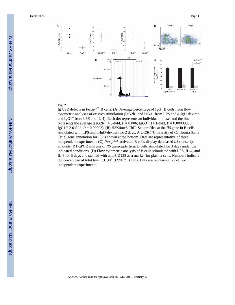
Fig. 2.Ig CSR defects in PaxipΔ/Δ B cells. (A) Average percentage of IgG+ B cells from flowcytometric analyses of ex vivo stimulation (IgG2b+ and IgG3+ from LPS and α-IgD-dextranand IgG1+ from LPS and IL-4). Each dot represents an individual mouse, and the linerepresents the average (IgG2b+: 4.8-fold, P = 0.006; IgG3+: 14.1-fold, P = 0.00000005;IgG1+: 2.6-fold, P = 0.00003). (B) H3K4me3 ChIP-Seq profiles at the Il6 gene in B cellsstimulated with LPS and α-IgD-dextran for 2 days. A UCSC (University of California SantaCruz) gene annotation for Il6 is shown at the bottom. Data are representative of threeindependent experiments. (C) PaxipΔ/Δ-activated B cells display decreased Il6 transcriptamounts. RT-qPCR analysis of Il6 transcripts from B cells stimulated for 3 days under theindicated conditions. (D) Flow cytometric analysis of B cells stimulated with LPS, IL-4, andIL-5 for 5 days and stained with anti-CD138 as a marker for plasma cells. Numbers indicatethe percentage of total live CD138+ B220low B cells. Data are representative of twoindependent experiments.
Daniel et al. Page 11
Science. Author manuscript; available in PMC 2011 February 1.
NIH
-PA Author Manuscript
NIH
-PA Author Manuscript
NIH
-PA Author Manuscript

Fig. 3.PTIP promotes Pol II association and transcription initiation at Igh switch regions. (A) Pol IIChIP-Seq profiles across the constant region locus in B cells stimulated with LPS and α-IgD-dextran for 2 days. Illustration is the same as in Fig. 1B. LPS-induced switch regionsare highlighted with orange boxes. The lower panel shows the expanded profiles across γ2b,γ1, and γ3. Black arrows indicate sites of LPS- and α-IgD-dextran–induced transcription atγ3 and γ2b switch regions. “I” indicates initiating exon segment. (B) Left, illustrationshowing location of oligonucleotides used in RT-qPCR. Right, RT-qPCR analysis of splicedand initiating Igh switch transcripts from B cells stimulated under the indicated conditionsfor 3 days. Data are representative of two independent experiments.
Daniel et al. Page 12
Science. Author manuscript; available in PMC 2011 February 1.
NIH
-PA Author Manuscript
NIH
-PA Author Manuscript
NIH
-PA Author Manuscript
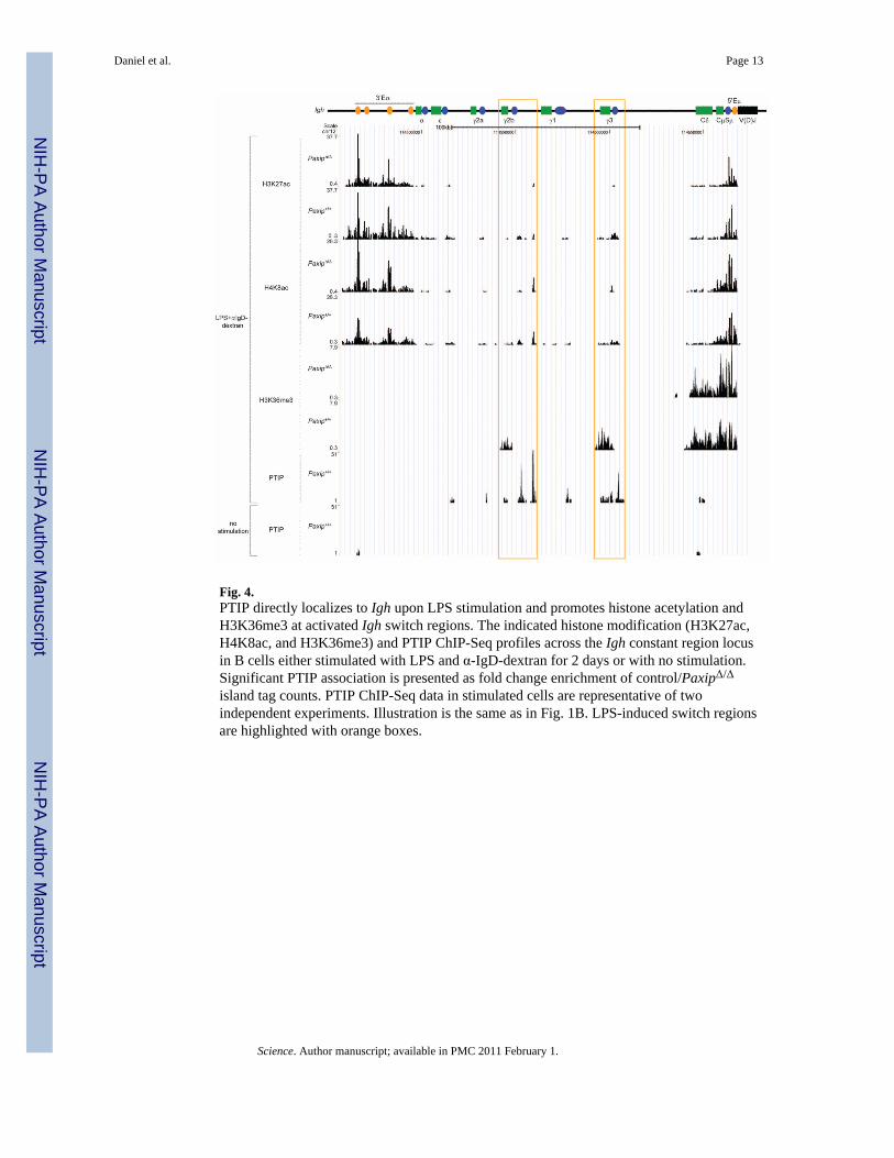
Fig. 4.PTIP directly localizes to Igh upon LPS stimulation and promotes histone acetylation andH3K36me3 at activated Igh switch regions. The indicated histone modification (H3K27ac,H4K8ac, and H3K36me3) and PTIP ChIP-Seq profiles across the Igh constant region locusin B cells either stimulated with LPS and α-IgD-dextran for 2 days or with no stimulation.Significant PTIP association is presented as fold change enrichment of control/PaxipΔ/Δ
island tag counts. PTIP ChIP-Seq data in stimulated cells are representative of twoindependent experiments. Illustration is the same as in Fig. 1B. LPS-induced switch regionsare highlighted with orange boxes.
Daniel et al. Page 13
Science. Author manuscript; available in PMC 2011 February 1.
NIH
-PA Author Manuscript
NIH
-PA Author Manuscript
NIH
-PA Author Manuscript

Fig. 5.PTIP foci formation contributes to Ig class-switching and genome stability. (A) Western blotof AID and α-TUBULIN from B cells stimulated for 3 days. (B) Sμ mutation analysis ofgenomic DNA from CFSE (carboxyfluorescein succinimydyl ester)–labeled B cells sortedfor IgM and five cell divisions after stimulation with LPS for 4 days. Segment sizes in thepie charts are proportional to the number of sequences carrying the number of mutationsindicated in the periphery of the charts. Frequency of mutations per base pair sequenced andthe total number of independent sequences analyzed are indicated below and in the center ofeach chart, respectively. Data are representative of two independent experiments. (C)Genomic instability analysis of metaphase spreads from B cells stimulated with LPS for 3days (two mice of each genotype, Paxip+/+: n = 261; PaxipΔ/Δ: n = 218). Abnormalitiesspecifically associated with the Igh locus (Igh-associated) or with all other chromosomes(general) are shown. (D) Immunofluorescent images of retrovirally infected MEFs exposedto 10-Gy irradiation with 4 hours recovery showing PTIP and 53BP1 foci formation. Bar, 20μm. (E) Flow cytometric analysis of retrovirally infected PaxipΔ/Δ stimulated B cells stained3 days after infection with IgG antibodies. Numbers indicate the percentages of total liveIgG+ cells in GFP− (no retroviral expression) or GFP+ (retroviral expression) gates. (F)Average percentage of IgG switching from infection experiments. Data are from fourindependent experiments for each condition (*P < 0.05). (G) RT-qPCR of Paxip (top) andIgh-γ3 switch (bottom) transcripts from retrovirally infected GFP+IgM+ sorted PaxipΔ/Δ Bcells stimulated with LPS. (H) Representative images of sorted B cells stimulated with LPSand IL-4 for 2 days and stained with α53BP1 antibody (red) before FISH detection of theIgh-Cα region (green). Among the total number of cells examined, those that contained53BP1 and Igh-Cα foci were analyzed [GFP− PaxipΔ/Δ, 100 cells; wild-type (WT) control
Daniel et al. Page 14
Science. Author manuscript; available in PMC 2011 February 1.
NIH
-PA Author Manuscript
NIH
-PA Author Manuscript
NIH
-PA Author Manuscript
GFP+, 89 cells; W663R mutant GFP+, 97 cells]. Coincidence of a 53BP1 focus with one orboth Igh-Cα alleles was detected in 42% of GFP− PaxipΔ/Δ, 30% of WT control GFP+, and43% of W663R mutant GFP+ cells. Bar, 5 μm.
Daniel et al. Page 15
Science. Author manuscript; available in PMC 2011 February 1.
NIH
-PA Author Manuscript
NIH
-PA Author Manuscript
NIH
-PA Author Manuscript