Pseudo half-molecules of the ABC transporter, COMATOSE, bind Pex19 and target to peroxisomes...
8
This article appeared in a journal published by Elsevier. The attached copy is furnished to the author for internal non-commercial research and education use, including for instruction at the authors institution and sharing with colleagues. Other uses, including reproduction and distribution, or selling or licensing copies, or posting to personal, institutional or third party websites are prohibited. In most cases authors are permitted to post their version of the article (e.g. in Word or Tex form) to their personal website or institutional repository. Authors requiring further information regarding Elsevier’s archiving and manuscript policies are encouraged to visit: http://www.elsevier.com/copyright
-
Upload
independent -
Category
Documents
-
view
3 -
download
0
Transcript of Pseudo half-molecules of the ABC transporter, COMATOSE, bind Pex19 and target to peroxisomes...
This article appeared in a journal published by Elsevier. The attachedcopy is furnished to the author for internal non-commercial researchand education use, including for instruction at the authors institution
and sharing with colleagues.
Other uses, including reproduction and distribution, or selling orlicensing copies, or posting to personal, institutional or third party
websites are prohibited.
In most cases authors are permitted to post their version of thearticle (e.g. in Word or Tex form) to their personal website orinstitutional repository. Authors requiring further information
regarding Elsevier’s archiving and manuscript policies areencouraged to visit:
http://www.elsevier.com/copyright

Author's personal copy
Pseudo half-molecules of the ABC transporter, COMATOSE, bind Pex19 and targetto peroxisomes independently but are both required for activity
Yvonne Nyathi a,1,2, Xuebin Zhang b,1,3, Jocelyn M. Baldwin c, Kristin Bernhardt b,d, Barbara Johnson a,Stephen A. Baldwin c, Frederica L. Theodoulou b,⇑, Alison Baker a
a Centre for Plant Sciences, Irene Manton Building, University of Leeds, Leeds LS2 9JT, UKb Biological Chemistry and Crop Protection Department, Rothamsted Research, Harpenden AL5 2JQ, UKc Institute of Membrane and Systems Biology, University of Leeds, Leeds LS2 9JT, UKd Institut für Biochemie der Pflanzen, Heinrich-Heine Universität Düsseldorf, 40225 Düsseldorf, Germany
a r t i c l e i n f o
Article history:Received 25 May 2012Accepted 30 May 2012Available online 16 June 2012
Edited by Ulf-Ingo Flügge
Keywords:ABC transporterPeroxisomePex19Protein targetingATPase
a b s t r a c t
Peroxisomal ABC transporters of animals and fungi are ‘‘half-size’’ proteins which dimerise to forma functional transporter. However, peroxisomal ABC transporters of land plants are synthesised as asingle polypeptide which represents a fused heterodimer. The N- and C-terminal pseudo-halves ofCOMATOSE (CTS; AtABCD1) were expressed as separate polypeptides which bound Pex19 in vitroand targeted independently to the peroxisome membrane in yeast, where they were stable butnot functional. When co-expressed, the pseudo-halves were fully functional as indicated by ATPaseactivity and rescue of the pxa1pxa2D mutant for growth on oleate. The functional significance ofheterodimer asymmetry is discussed.
Structured summary of protein interactions:PEX19-1 binds to CTS-N by pull down (View Interaction)PEX19-1 binds to CTS-C by pull down (View Interaction)PEX19-2 binds to CTS-N by pull down (View Interaction)PEX19-2 binds to CTS-N by pull down (View Interaction)
� 2012 Federation of European Biochemical Societies. Published by Elsevier B.V. All rights reserved.
1. Introduction
ABC transporters couple ATP hydrolysis to the trans-membranemovement of a wide range of substrates. All ABC transporters com-prise four functional domains: two sets of primarily a-helical trans-membrane domains (TMDs) which associate to form the transportpathway for the substrate and two nucleotide binding domains(NBDs) which dimerise and form two composite ATP binding sitesat the dimer interface.
Peroxisomes from all eukaryotes contain at least one member ofthe ABC subfamily D which transports lipophilic/amphipathic
substrates from the cytosol into the peroxisome for b-oxidation.Comatose (CTS; AtABCD1, also known as AtPXA1 and PED3) is a fullsize peroxisomal ABC transporter from Arabidopsis with the domainorganisation [TMD-NBD-TMD-NBD] [1–3]. Thus CTS can be consid-ered to be a fused heterodimer consisting of two homologous butdistinct halves. This gene structure is found in all land plants includ-ing bryophytes but is not present in algae, suggesting that this fusionhappened once in the land plant lineage more than 450 Ma ago(Fig. 1). In contrast, animal and fungal peroxisomal ABC transportersare all half transporters with the organisation [TMD-NBD] that haveto dimerise for activity [4]. Mammalian ABCD proteins are functionalas homodimers, but in Saccharomyces cerevisiae, the half transport-ers Pxa1p and Pxa2p heterodimerise to transport long chain acylCoAs [5,6]. We have shown recently that CTS targets to peroxisomeswhen expressed in oleate-grown S. cerevisiae and has the ability tocomplement the yeast pxa1 pxa2D mutant for b-oxidation of fattyacids [7].
Since only land plants have peroxisomal transporters with thefused heterodimer configuration, but this arrangement is functionalin S. cerevisiae, we investigated the targeting and functionality of the
0014-5793/$36.00 � 2012 Federation of European Biochemical Societies. Published by Elsevier B.V. All rights reserved.http://dx.doi.org/10.1016/j.febslet.2012.05.065
Abbreviations: ABC, ATP Binding Cassette; TMD, transmembrane domain; NBD,nucleotide binding domain⇑ Corresponding author.
E-mail address: [email protected] (F.L. Theodoulou).1 These authors contributed equally to this work.2 Current address: Faculty of Life Sciences, Michael Smith Building, Oxford Road,
Manchester M13 9PT, UK.3 Current address: Biology Department, Brookhaven National Laboratory, Upton,
NY 11973-5000, USA.
FEBS Letters 586 (2012) 2280–2286
journal homepage: www.FEBSLetters .org

Author's personal copy
two CTS pseudo half transporters CTS-N and CTS-C from Arabidopsiswhen expressed in yeast.
2. Materials and methods
2.1. Growth of yeast
Yeast strains were grown as described in [7]. Yeast strains aregiven in Table 1.
2.2. Pull-down assays
AtPEX19-1 was amplified with primer pair FT119/FT120 (TableS1), restricted with Bam HI/Not I and cloned in the correspondingsites of pGEX-4T-3 (GE Healthcare). AtPEX19-2 was amplified withprimer pair FT121/FT122, restricted with Sal I/Not I and cloned inthe corresponding sites of pGEX-4T-3. Expression and purificationof glutathione S-transferase (GST) fusion proteins was carried outas described in [8]. CTS-N and CTS-C were amplified from plasmidH1A6T7 using primer pairs FT187/FT189 and FT190/FT188 respec-
Fig. 1. Phylogenetic analysis of ABC family D transporters. Analysis by the Maximum Likelihood method was performed as described in Supplementary data. Upper and lowercase letters at the beginning of protein names indicate abbreviated Latin binomial names for species, as detailed in the species key (Table S2). Subfamily members within eachspecies are classified according to [18].
Y. Nyathi et al. / FEBS Letters 586 (2012) 2280–2286 2281
Author's personal copy
tively, restricted with Nco I and Sma I and cloned into the corre-sponding sites of pIVEX1.3 (www.5prime.com). In vitro translationwas carried out using a wheat germ lysate (5PRIME kit), accordingto manufacturer’s instructions. Aliquots were removed for immu-noblot analysis (typically 10–20% input) and equal volumes ofthe remainder were used for pull-down reactions using GST-At-PEX19-1, GST-AtPEX19-2 or GST alone. Pull-down assays were per-formed as described in [8], except that proteins were detected byimmunoblotting with an anti-penta-His antibody (Qiagen).
2.3. Plasmids for yeast expression
The ORF of AtPEX19-1 was amplified by PCR from plasmid pHD-1PEX19-1 [9] using primer pair AtPEX19F/AtPEX19R (Table S1) andcloned into pRS415-met25 as a Bam HI-Eco RI fragment. AtPEX19-1and ScPex19 were amplified from pHD-1PEX19_1 and pKat61 [10]with primer pairs FT163/FT137 and FT160/161, respectively and re-stricted with Bam HI/Sal I. AtPEX19-2 was amplified from cDNA cloneU13401 (Arabidopsis Biological Resource Center, Ohio) with primerpair FT138/FT139, and restricted with Bgl II/Sal I. The fragments wereligated into the Bam HI/Sal I sites of both pRS416/GPD and pRS415/
met25. Details of primer pairs are given in Table S1. To generate con-structs for the expression of pseudo-half transporters, the N-terminalhalf of CTS (encoding amino acids 1–676, designated ‘‘CTS-N’’) andthe C-terminal half (‘‘CTS-C’’, encoding amino acids 677–1337) wereamplified from CTS/pRS416-GPD [7] using primer pairs CTSAF/CTSARand CTSBF/CTSBR, respectively (Table S1). All products were restrictedwith Bam HI and Eco RI and ligated into pRS416-GPD [7] and alsopRS415/met25. Similarly, for complementation experiments, CTS-Nand CTS-C were amplified using primer pairs FT167/FT168 andFT169/FT170, respectively. Products were restricted with Bam HI andSal I and ligated into pEL30 and pIJL30.
2.4. Statistical analysis
Data were expressed as mean SI ± 95% CL, where 95% CL wasdetermined according to the formula: 95% CL = mean ± (t � SEM),where n = number of samples, t = value of the Student t-distribu-tion at (n�1) degrees of freedom and SEM is the standard errorof the mean. All the statistical analysis and calculations were doneusing Origin� 8.0 Data Analysis and Graphing Software statisticalpackage and Windows
�Microsoft Excel.
Table 1Yeast strains used in this study.
BSL1-11B (WT) MATa, lys2D0, his4D519, Ura3D0, leu2D2 [19]BSL1-11B (pxa1D) MATa, lys2D0, his4D519, Ura D0, leu2D2, YPL147w::kanMX4 [7]BY4742 (WT) Mat a; his3D1; leu2D00; lys2D0; ura3D0 [20]BY4742 (pex19D) Mat a; his3D1; leu2D00; lys2D0; ura3D0; YDL065c::kanMX4 [20]BJ1991 Mata, leu2, trp1, ura3-251, prb1-112,pex4-3 [5]BJ1991 (pxa1,2D) Mata, leu2, trp1, ura3-251, prb1-112,pex4-3; YPL147w::LEU2;YKL188c::kanMX4 [5]
B
A
20%
inpu
t
GST
GST
-AtP
EX19
-1
GST
-AtP
EX19
-2
20%
inpu
t
GST
GST
-AtP
EX19
-1
GST
-AtP
EX19
-2
CTS-N CTS-C
< CTS pseudo halves
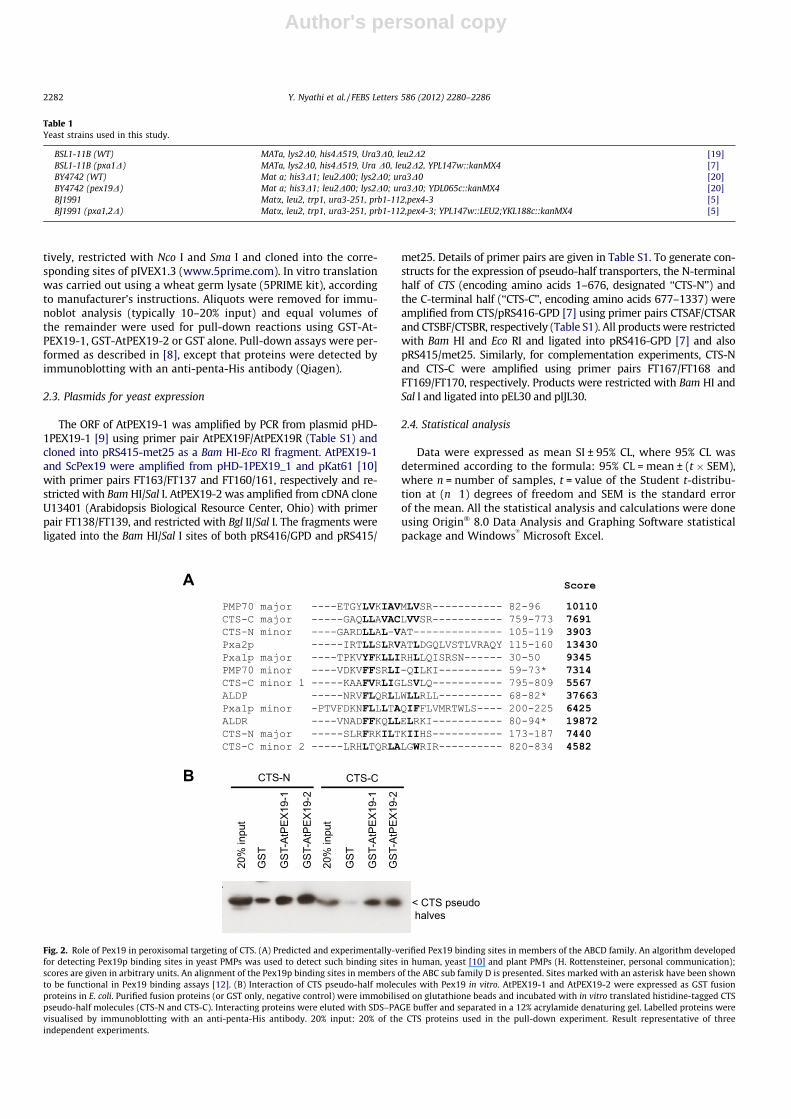
Fig. 2. Role of Pex19 in peroxisomal targeting of CTS. (A) Predicted and experimentally-verified Pex19 binding sites in members of the ABCD family. An algorithm developedfor detecting Pex19p binding sites in yeast PMPs was used to detect such binding sites in human, yeast [10] and plant PMPs (H. Rottensteiner, personal communication);scores are given in arbitrary units. An alignment of the Pex19p binding sites in members of the ABC sub family D is presented. Sites marked with an asterisk have been shownto be functional in Pex19 binding assays [12]. (B) Interaction of CTS pseudo-half molecules with Pex19 in vitro. AtPEX19-1 and AtPEX19-2 were expressed as GST fusionproteins in E. coli. Purified fusion proteins (or GST only, negative control) were immobilised on glutathione beads and incubated with in vitro translated histidine-tagged CTSpseudo-half molecules (CTS-N and CTS-C). Interacting proteins were eluted with SDS–PAGE buffer and separated in a 12% acrylamide denaturing gel. Labelled proteins werevisualised by immunoblotting with an anti-penta-His antibody. 20% input: 20% of the CTS proteins used in the pull-down experiment. Result representative of threeindependent experiments.
2282 Y. Nyathi et al. / FEBS Letters 586 (2012) 2280–2286
Author's personal copy
2.5. Miscellaneous
Peroxisome isolation, carbonate extractions, ATPase assays,immunoblotting and complementation for growth on oleate med-ium were performed as described in [7] and [9]. Pex19 binding siteprediction and homology modelling of CTS are described in [10]and [11], respectively.
3. Results and discussion
3.1. The two halves of CTS contain independent binding sites for bothArabidopsis and Saccharomyces Pex19
Pex19p is a universal and essential factor for the correct locali-sation and insertion of peroxisome membrane proteins. Althoughthere is debate over the exact role of Pex19p, recognition of perox-isome membrane proteins is central to all the proposed functions.Pex19p binding sites in a number of PMPs are short helical motifsthat overlap with the peroxisome targeting signal [10]. A Pex19pbinding motif predictor has been developed and used to identifyand experimentally verify a Pex19p binding site in human ALDP(HsABCD1) which was functional in both human and S. cerevisiae[10,12]. Analysis of CTS with the Pex19p binding site predictor re-vealed five potential Pex19p binding sites at amino acids 105–119,173–187, 759–773, 795–809 and 820–834. Based on the scoresshown in Fig. 2A, there is a major site in both the N-(CTS-N) andC-(CTS-C) terminal parts of CTS as well as one and two minor sitesin these regions, respectively. These sites all share features withthose from other ABCD subfamily proteins, especially the predom-inance of hydrophobic amino acid side chains, the absence of pro-line and the under representation of negatively charged aminoacids.
CTS was expressed as two pseudo-half transportersCTS-N (1–676) and CTS-C (677–1337), dividing the protein justafter NBD1, at the beginning of the predicted flexible linker region[7,11], and the ability of each half to interact with different Pex19isoforms in vitro was examined in a pull-down assay (Fig. 2B). CTS-N and -C were expressed by in vitro transcription and translation,incubated with purified GST-Pex19 fusion proteins and the Pex19protein recovered by affinity isolation on glutathione agarosebeads. GST alone was used as a negative control. Low or very lowlevels of non-specific binding to the GST control were seen forCTS-N and CTS-C, but much greater amounts (>20% of input) ofCTS-N and CTS-C re-isolated with AtPEX19-1, AtPEX19-2 (Fig. 2B)and ScPex19p (Figure S1), indicating that both halves of the proteincan interact with Pex19 from both species. The presence of Pex19binding sites in both halves of the protein likely reflects their dis-tant evolutionary history as separate proteins (see phylogenetictree in Fig 1). The fact that they have apparently retained function-ality over millions of years since the divergence of land plants, sug-gests that they might be functionally important and that Pex19interacts with full-length CTS at more than one site, perhaps tomaintain solubility prior to insertion in the peroxisomal mem-brane. Large membrane proteins are difficult for the cell to foldand are often subject to a high rate of misfolding, thus chaperoneswould be selected for. This has been very well documented for an-other ‘‘full-length’’ ABC transporter, the cystic fibrosis transmem-brane conductance regulator, CFTR, which is subject to multiplequality control mechanisms [13].
3.2. Arabidopsis PEX19 cannot complement the S. cerevisiae pex19Dmutant for growth on oleate
Since CTS can be recognised by ScPex19p and since correct tar-geting of human [12] and plant [7] PMPs has been demonstrated in
α-PEX19
Coomassie
• 49• 37 <Pex19
WT
pex19
Δ
pRS4
15
pRS4
15/A
tPEX
19-1
pRS4
16
pRS4
16/A
tPEX
19-2
pRS4
15 +
pR
S416
AtPE
X19-
1 +
AtPE
X19-
2
pRS4
16/S
cPex
19
A
B
WT
pex19Δ
pex19Δ::AtPEX19-1 + AtPEX19-2
pex19Δ::ScPex19
• 180
• 82 • 115
• 64
• 26 • 19
• 6 • 15
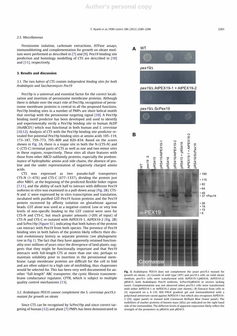
Fig. 3. Arabidopsis PEX19 does not complement the yeast pex19D mutant forgrowth on oleate. (A) Growth of wild type (WT) and pex19D cells on solid oleatemedium. pex19D cells were transformed with AtPEX19-1/pRS416, AtPEX19-2/pRS415, both Arabidopsis Pex19 isoforms, ScPex19/pRS416 or vectors lackinginsert. Complementation was not observed when pex19D cells were transformedwith either AtPEX19-1 or AtPEX19-2 alone (not shown). (B) Extracts from cells in(A) separated on a 4–12% SDS–PAGE gradient gel and immunoblotted with apolyclonal antiserum raised against AtPEX19-1 but which also recognises AtPEX19-2 ([9]; upper panel) or stained with Coomassie Brilliant Blue (lower panel). Themobilities of marker proteins of known mass (kDa) are indicated on the right handside of the immunoblot. The different levels of apparent expression likely reflect thestrength of the promoters in pRS416 and pRS415.
Y. Nyathi et al. / FEBS Letters 586 (2012) 2280–2286 2283

Author's personal copy
S. cerevisiae, we tested whether Arabidopsis PEX19 could comple-ment the S. cerevisiae pex19D mutant for growth on oleate. Thetwo PEX19 isoforms of Arabidopsis, PEX19-1 and PEX19-2, are79% identical at the amino acid level but share only ca. 25% identityto ScPex19p [9]. RNAi down regulation of these two Pex19 iso-forms in Arabidopsis suggested they are not completely function-ally redundant [14]. We expressed AtPEX19-1 and AtPEX19-2independently and together in the S. cerevisiae pex19D mutant(Fig. 3A). Neither protein alone or in combination was able to res-cue the oleate non-utilisation (onu) phenotype of the pex19D mu-tant, despite both proteins being expressed at detectable levels(Fig. 3B). This may be because the ability to complement the onuphenotype is a very stringent test of conservation of function andrequires not only recognition of the targeting signals of all PMPsnecessary (directly or indirectly) for oleate metabolism, but alsorescue of all Pex19 functions, including the capacity to be modifiedby the yeast farnesyl transferase and to interact productively withyeast Pex3p.
3.3. CTS-N and CTS-C target independently to peroxisomes and arecorrectly integrated into peroxisome membranes
CTS-N and CTS-C were expressed separately in yeast and oleate-grown cultures were subjected to fractionation on Optiprep™ gra-dients to prepare peroxisome- and mitochondria-enriched frac-tions, which were subsequently analysed by SDS PAGE andimmunoblotting (Fig. 4). Good separation between the organellarfractions was demonstrated by the enrichment of thiolase (perox-isomal marker) and porin (mitochondrial marker) in the appropri-ate fractions (Fig. 4A, lower two panels). Both CTS-N and CTS-Cwere almost exclusively detected in the peroxisome fraction(Fig. 4A upper panel), with a small amount being detected in themitochondrial fraction. When the peroxisome membrane is limit-ing in yeast cells (for example on lactate medium or in the pex19Dbackground), CTS mistargeted and was detected in the mitochon-drial fraction (data not shown). CTS-N and CTS-C were found tobe more stable than the full length CTS, which is subject to exten-sive proteolysis within the linker region joining the two pseudohalves [7]. This is in contrast to Pxa1p, which is unstable in thepxa2D background, although Pxa2p is stable in the absence ofPxa1p [15]. Similarly, the mammalian ER-located ABC half-trans-porter, TAP2, is unstable in the absence of its partner TAP1 [16].Both CTS-N and CTS-C were not extractable from the membranesby alkaline sodium carbonate treatment, consistent with the factthat they contain multiple transmembrane helices and are presum-ably integrated into the membrane (Fig. 4B). These results demon-strate that each pseudo half transporter contains all the necessaryinformation to be recognised and inserted into peroxisome mem-branes by the heterologous host cell machinery.
3.4. Co-expression of CTS-N and CTS-C is required for function
We investigated whether CTS-N and CTS-C are functional whenexpressed in yeast. Neither protein complemented the onu pheno-type of the pxa1,2D mutant when expressed alone, but when ex-pressed together permitted growth on oleate comparable to thatobtained with full-length CTS (Fig. 5A). We then tested whetherthe pseudo-halves could form homodimers or heterodimers withATPase activity. ATPase activity was measured in peroxisomes iso-lated from strains expressing full length CTS, CTS-N and CTS-C sep-arately, CTS-N and CTS-C co-expressed, and empty vectortransformed cells. Peroxisomes from cells co-expressing CTS-Nand CTS-C had CTS-specific basal ATPase activity (as measured in[7]) which was almost as high as that of peroxisomes containingthe full length CTS. In contrast, peroxisomes from cells whereCTS-N and CTS-C were expressed alone had no more ATPase activity
than control cells. Thus CTS-N and CTS-C can assemble to form anactive heterodimer. CTS-N and CTS-C alone are either unable tohomodimerise, or form an inactive homodimer.
As NBDs dimerise during the transport cycle, the two ATP bind-ing sites are composites, comprising residues from both NBDs.Each ATP site is made up of the Walker A, Walker B and Switch mo-tif (H-loop) from one NBD and the signature motif and D loop fromthe other NBD. NBD1 of CTS shows conservation of all functionallyimportant residues in the Walker A, Walker B and Switch or ‘H’loop and mutagenesis shows that Walker A and B residues ofNBD1 are essential for function in vivo [11]. In contrast, NBD2
T C M P T C M P
40
13095
72
55
43
30
<Thiolase
<Porin
< CTSpseudo-halves
CTS-N CTS-C
SN SNPelle
t
Pelle
t
CTS-N CTS-C
A
B
95
72
55
43
34
< CTSpseudo-halves
Fig. 4. CTS N- and C-terminal pseudo-half molecules target to peroxisomesindependently and are integrated into the peroxisome membrane. (A) BLS1-11Bcells transformed with the CTS-N/pRS416-GPD or CTS-C/pRS416-GPD were grownon WOYD medium and transferred to the oleate medium for peroxisome induction.A light mitochondrial fraction was prepared and separated on an iodixanol densitygradient. The fractions corresponding to the peroxisomes (P) and mitochondria (M)were separated by SDS–PAGE in a 7.5% gel (30 lg of protein/lane) and immuno-blotted with antibodies raised against CTS, thiolase (peroxisomal marker), andporin (mitochondrial marker) [7]. The fractions corresponding to the total celllysate (T) and cytosol (C) were also collected and treated as above. The mobilities ofmarker proteins of known mass (kDa) are indicated on the left hand side of thefigure. The upper band represents unrelated cross reaction with a mitochondrialprotein. (B) Organelles (‘‘light mitochondrial fraction’’) were isolated from oleate-grown BLS1-11B cells transformed with the CTS-N/pRS416-GPD or CTS-C/pRS416-GPD and were lysed in a hypotonic buffer in the presence of protease inhibitors.Membranes were separated from soluble proteins by centrifugation at 100,000gand then extracted with alkaline sodium carbonate buffer, pH 11.5, followed by re-centrifugation at 100,000g. Pellet and supernatant (SN) fractions were separated bySDS–PAGE (30 lg per lane) followed by immunoblotting with antibodies raisedagainst CTS. The mobilities of marker proteins of known mass (kDa) are indicatedon the left hand side of the figure.
2284 Y. Nyathi et al. / FEBS Letters 586 (2012) 2280–2286

Author's personal copy
has Q in place of H in the Switch loop. This histidine plays a criticalrole in contacting the c-phosphate of the bound ATP. Thus CTS willhave one consensus and one degenerate ATP binding site. Thisasymmetry is quite common in eukaryotic ABC transporters [17and references therein]; indeed Pxa1p and TAP1 also contain thesame H to Q substitution. In the light of this, CTS-C might be ex-pected to be non-functional when expressed in the absence ofCTS-N. However, CTS-N contains consensus residues which con-tribute to the ATP binding site and might be predicted to form acatalytically active homodimer when expressed alone. The factthat CTS-N expressed alone does not exhibit ATPase activity sug-gests either, that it is not able to homodimerise, and/or that a‘‘degenerate’’ ATP binding site is somehow important for the ATP-ase catalytic cycle of CTS. Alternatively, the lack of functionality ofCTS pseudo half molecules may be related to TMD structure. Amodel of CTS based on the structure of the homodimeric ABCtransporter Sav1866 predicts ‘‘domain-swapping’’, such thatTMD1 contacts NBD2 and vice versa [11]. Moreover, in the model,a-helices from the two distinct halves of the protein are shared be-tween the two TMDs, such that one is composed of TM helices 3–8and the other of TM helices 1, 2, and 9–12 (Fig. 6). Within theTMDs, the interface between the two halves of the protein involvesinteractions between TM2 and 11 and between TM8 and 5. In the
homodimeric molecule Sav1866 the equivalent interactions, be-tween TM2 of one subunit and TM5 of the other, are necessarilyidentical whereas this is not the case in native CTS. For example,a salt bridge is predicted between R834 (TM8) and D332 (TM5),while the equivalent positions in the other TMD are occupied byI180 (TM2) and K986 (TM11). In the CTS-C homodimer, the corre-sponding interface would contain R834 and K986, likely resultingin electrostatic repulsion, while in the CTS-N homodimer, theinterface would contain I180 and D332, precluding salt bridge for-mation or the likely hydrophobic interaction between the isoleu-cine side chain and the aliphatic portion of the lysine side chain.Similar differences in size and/or charge are observed between anumber of other conserved interfacial residues at equivalent posi-
Fig. 6. Homology model of CTS showing domain swapping of transmembranehelices. Modified from [11]. The N- and C-terminal halves of the CTS molecule arecoloured in green and blue, respectively. Residues R834 and D332, predicted toform a salt bridge in the interface between the halves, are shown in space-fillingrepresentation, as are bound AMP-PNP molecules within the NBDs.
A
B
BJ1991
pxa1,2Δ
CTS
CTS-N
CTS-C
CTS-N + C
Control CTS-N CTS-C CTSN+C
CTS FL
Pi (n
mol
/mg
prot
ein/
min
)
0
40
80
120
160
Fig. 5. Functional analysis of CTS pseudo-half molecules. (A) Growth of wild-typeand mutant cells transformed with the indicated constructs on solid oleatemedium. (B) ATPase activity in peroxisomes isolated from oleate-grown wild-typeBSL1-11B yeast, transformed with CTS-N/pRS416-GPD, CTS-C/pRS415-met25, sin-gly and in combination and CTS/pRS416-GPD (CTS FL) or vector lacking an insert(control). All samples were analysed in triplicate, and the experiment was repeatedthree times. The data were used to compute the means and the standard error of themean (SEM). Data are expressed as mean SI ± 95% CL, where 95% CL was determinedaccording to the formula: 95% CL = mean ± (t � SEM), where n = number of samples,t = value of the Student t-distribution at (n � 1) degrees of freedom and SEM is thestandard error of the mean.
Y. Nyathi et al. / FEBS Letters 586 (2012) 2280–2286 2285

Author's personal copy
tions elsewhere in the two halves of the molecule. These wouldlikely interfere with the association between, and/or activity of,the CTS homodimers. Conservation of a number of interfacial resi-dues is indicative of their probable collective importance in main-taining the interaction between the helices. In conclusion, whilethere is no apparent requirement for the two halves of CTS to becovalently joined to produce a functional molecule, both halvesare indispensable for biological activity.
Acknowledgements
We thank Dr. Hanspeter Rottensteiner for ScPEX19/pPC86 andfor Pex19 binding site predictions, Dr Ewald Hettema forpEW205, Euroscarf for pex19D, the Arabidopsis Biological ResourceCentre (Ohio) for U13403 and Dr Carlo van Roermund for pxa1,2Dand pIJL30. This work was funded by the Biotechnology and Biolog-ical Sciences Research Council (BBSRC) (Grants BB/F007108/1 toFLT and BB/F007299/1 to AB/SAB and a doctoral training student-ship to YN) and by a Boehringer Ingelheim Fonds travel grant toKB. We thank Dr Carine de Marcos Lousa for helpful discussions.
Appendix A. Supplementary data
Supplementary data associated with this article can be found, in theonline version, at http://dx.doi.org/10.1016/j.febslet.2012.05.065.
References
[1] Zolman, B.K., Silva, I.D. and Bartel, B. (2001) The Arabidopsis pxa1 mutant isdefective in an ATP-binding cassette transporter-like protein required forperoxisomal fatty acid beta-oxidation. Plant Physiol. 127, 1266–1278.
[2] Footitt, S., Slocombe, S.P., Larner, V., Kurup, S., Wu, Y., Larson, T., Graham, I.,Baker, A. and Holdsworth, M. (2002) Control of germination and lipidmobilization by COMATOSE, the Arabidopsis homologue of human ALDP.EMBO J. 21, 2912–2922.
[3] Hayashi, M., Nito, K., Takei-Hoshi, R., Yagi, M., Kondo, M., Suenaga, A., Yamaya,T. and Nishimura, M. (2002) Ped3p is a peroxisomal ATP-binding cassettetransporter that might supply substrates for fatty acid beta-oxidation. PlantCell Physiol. 43, 1–11.
[4] Rottensteiner, H. and Theodoulou, F.L. (2006) The ins and outs of peroxisomes:co-ordination of membrane transport and peroxisomal metabolism. Biochim.Biophys. Acta 1763, 1527–1540.
[5] van Roermund, C.W., Visser, W.F., Ijlst, L., van Cruchten, A., Boek, M., Kulik, W.,Waterham, H.R. and Wanders, R.J. (2008) The human peroxisomal ABC halftransporter ALDP functions as a homodimer and accepts acyl-CoA esters.FASEB J. 22, 4201–4208.
[6] Verleur, N., Hettema, E.H., van Roermund, C.W., Tabak, H.F. and Wanders, R.J.(1997) Transport of activated fatty acids by the peroxisomal ATP-binding-
cassette transporter Pxa2 in a semi-intact yeast cell system. Eur. J. Biochem.249, 657–661.
[7] Nyathi, Y., De Marcos Lousa, C., van Roermund, C.W., Wanders, R.J.A., Baldwin,S.A., Theodoulou, F.L. and Baker, A. (2010) The Arabidopsis peroxisomal ABCtransporter, COMATOSE, complements the Saccharomyces cerevisiae pxa1pxa2D double mutant for metabolism of long chain fatty acids and exhibitsfatty acyl-CoA stimulated ATPase activity. J. Biol. Chem. 285, 29892–29902.
[8] Zhang, X., De Marcos Lousa, C., Schutte-Lensink, N., Ofman, R., Wanders, R.J.,Baldwin, S.A., Baker, A., Kemp, S. and Theodoulou, F.L. (2011) Conservation oftargeting but divergence in function and quality control of peroxisomal ABCtransporters: an analysis using cross-kingdom expression. Biochem. J. 436,547–557.
[9] Hadden, D.A., Phillipson, B.A., Johnston, K.A., Brown, L.A., Manfield, I.W., El-Shami, M., Sparkes, I.A. and Baker, A. (2006) Arabidopsis PEX19 is a dimericprotein that binds the peroxin PEX10. Mol. Membr. Biol. 23, 325–336.
[10] Rottensteiner, H., Kramer, A., Lorenzen, S., Stein, K., Landgraf, C., Volkmer-Engert, R. and Erdmann, R. (2004) Peroxisomal membrane proteins containcommon Pex19p-binding sites that are an integral part of their targetingsignals. Mol. Biol. Cell. 15, 3406–3417.
[11] Dietrich, D., Schmuths, H., De Marcos, Lousa.C., Baldwin, J.M., Baldwin, S.A.,Baker, A., Theodoulou, F.L. and Holdsworth, M.J. (2009) Mutations in theArabidopsis peroxisomal ABC transporter COMATOSE allow differentiationbetween multiple functions in planta: insights from an allelic series. Mol. Biol.Cell. 20, 530–543.
[12] Halbach, A., Lorenzen, S., Landgraf, C., Volkmer-Engert, R., Erdmann, R. andRottensteiner, H. (2005) Function of the PEX19-binding site of humanadrenoleukodystrophy protein as targeting motif in man and yeast. PMPtargeting is evolutionarily conserved. J. Biol. Chem. 280, 21176–21182.
[13] Cheung, J.C. and Deber, C.M. (2008) Misfolding of the cystic fibrosistransmembrane conductance regulator and disease. Biochemistry 47, 1465–1473.
[14] Nito, K., Kamigaki, A., Kondo, M., Hayashi, M. and Nishimura, M. (2007)Functional classification of Arabidopsis peroxisome biogenesis factorsproposed from analyses of knockdown mutants. Plant Cell Physiol. 48, 763–774.
[15] Shani, N. and Valle, D. (1996) A Saccharomyces cerevisiae homolog of thehuman adrenoleukodystrophy transporter is a heterodimer of two half ATP-binding cassette transporters. Proc. Natl. Acad. Sci. U S A 93, 11901–11906.
[16] Keusekotten, K., Leonhardt, R.M., Ehses, S. and Knittler, M.R. (2006) Biogenesisof functional antigenic peptide transporter TAP requires assembly of pre-existing TAP1 with newly synthesized TAP2. J. Biol. Chem. 281, 17545–17551.
[17] Procko, E., O’Mara, M.L., Bennett, W.F., Tieleman, D.P. and Gaudet, R. (2009)The mechanism of ABC transporters: general lessons from structural andfunctional studies of an antigenic peptide transporter. FASEB J. 23, 1287–1302.
[18] Verrier, P.J., Bird, D., Burla, B., Dassa, E., Forestier, C., Geisler, M., Klein, M.,Kolukisaoglu, U., Lee, Y., Martinoia, E., Murphy, A., Rea, P.A., Samuels, L.,Schulz, B., Spalding, E.J., Yazaki, K. and Theodoulou, F.L. (2008) Plant ABCproteins–a unified nomenclature and updated inventory. Trends Plant Sci. 13,151–159.
[19] Taylor, K.M., Kaplan, C.P., Gao, X. and Baker, A. (1996) Localization andtargeting of isocitrate lyases in Saccharomyces cerevisiae. Biochem. J. 319, 255–262.
[20] Brachmann, C.B., Davies, A., Cost, G.J., Caputo, E., Li, J., Hieter, P. and Boeke, J.D.(1998) Designer deletion strains derived from Saccharomyces cerevisiae S288C:a useful set of strains and plasmids for PCR-mediated gene disruption andother applications. Yeast 14, 115–132.
2286 Y. Nyathi et al. / FEBS Letters 586 (2012) 2280–2286