Proteomic analysis of the reproductive tract fluids from tropically-adapted Santa Ines rams
21
Proteomic analysis of the reproductive tract fluids from tropically-adapted Santa Ines rams ☆ Carlos Eduardo A. Souza a , João Paulo A. Rego a , Carlos H. Lobo a , José Tadeu A. Oliveira b , Fábio C.S. Nogueira c , Gilberto B. Domont c , Mariana Fioramonte d , Fabio C. Gozzo d , Frederico B. Moreno e , Ana Cristina O. Monteiro-Moreira e , José Ricardo Figueiredo f , Arlindo A. Moura a, ⁎ a Department of Animal Science, Federal University of Ceará, Brazil b Department of Biochemistry, Federal University of Ceará, Brazil c Institute of Chemistry, Federal University of Rio de Janeiro, Brazil d Institute of Chemistry, University of Campinas, Brazil e School of Pharmacy, The University of Fortaleza, Brazil f School of Veterinary Medicine, Ceará State University, Brazil ARTICLE INFO ABSTRACT Available online 30 May 2012 The present study is focused on the proteome of reproductive tract fluids from tropically- adapted Santa Ines rams. Seminal plasma, cauda epididymal (CEF) and vesicular gland fluid (VGF) proteins were analyzed by 2-D electrophoresis and mass spectrometry. Seminal plasma maps contained 302±16 spots, within the 4–7 pH range. From these maps, 73 spots were identified, corresponding to 41 proteins. Ram Seminal Vesicle Proteins (RSVP) 14 and 22 kDa and bodhesins 1 and 2 represented the most abundant seminal components. Other seminal proteins included clusterin, angiotensin-converting enzyme, matrix metalloproteinase-2, tissue-inhibitor of metalloproteinase-2, plasma glutamate carboxypeptidase, albumin, lactofer- rin, alpha enolase, peroxiredoxin, leucine aminopeptidase, β-galactosidase, among others. Later, seminal plasma gels were run within narrow pH intervals (3.9–5.1; 4.7–5.9; 5.5–6.7), allowing the additional identification of 21 proteins not detected in 4–7 pH maps. Major proteins of CEF and VGF were albumin and transferrin, and RSVPs, respectively. Western blots confirmed that RSVPs were mainly present in VGF while bodhesins, in VGF and CEF. Based on RT-PCR, RSVP and bodhesin genes were primarily expressed in the vesicular glands. In summary, the reproductive tract fluids of Brazilian hairy rams contain several categories of proteins, with potential roles in sperm protection, capacitation, acrosome reaction and sperm–oocyte interaction. This article is part of a Special Issue entitled: Farm animal proteomics. © 2012 Elsevier B.V. All rights reserved. Keywords: Seminal plasma Binder of Sperm Protein Spermadhesin Ram Sperm 1. Introduction Spermatozoa are produced in the germinative epithelium of the testis, matured during epididymal transit and stored in the cauda epididymal region. Following ejaculation, spermatozoa and cauda epididymal fluid are mixed with secretions from the accessory sex glands and deposited in the female reproductive tract, where they are capacitated and start their journey to the fertilization site [1]. In this context, we suggest that constituents of the sperm themselves and JOURNAL OF PROTEOMICS 75 (2012) 4436 – 4456 ☆ This article is part of a Special Issue entitled: Farm animal proteomics. ⁎ Corresponding author. Tel./fax: + 55 8533669701. E-mail addresses: [email protected], [email protected] (A.A. Moura). 1874-3919/$ – see front matter © 2012 Elsevier B.V. All rights reserved. doi:10.1016/j.jprot.2012.05.039 Available online at www.sciencedirect.com www.elsevier.com/locate/jprot
-
Upload
independent -
Category
Documents
-
view
0 -
download
0
Transcript of Proteomic analysis of the reproductive tract fluids from tropically-adapted Santa Ines rams

J O U R N A L O F P R O T E O M I C S 7 5 ( 2 0 1 2 ) 4 4 3 6 – 4 4 5 6
Ava i l ab l e on l i ne a t www.sc i enced i r ec t . com
www.e l sev i e r . com/ loca te / j p ro t
Proteomic analysis of the reproductive tract fluids fromtropically-adapted Santa Ines rams☆
Carlos Eduardo A. Souzaa, João Paulo A. Regoa, Carlos H. Loboa, José Tadeu A. Oliveirab,Fábio C.S. Nogueirac, Gilberto B. Domontc, Mariana Fioramonted, Fabio C. Gozzod,Frederico B. Morenoe, Ana Cristina O. Monteiro-Moreirae,José Ricardo Figueiredo f, Arlindo A. Mouraa,⁎aDepartment of Animal Science, Federal University of Ceará, BrazilbDepartment of Biochemistry, Federal University of Ceará, BrazilcInstitute of Chemistry, Federal University of Rio de Janeiro, BrazildInstitute of Chemistry, University of Campinas, BrazileSchool of Pharmacy, The University of Fortaleza, BrazilfSchool of Veterinary Medicine, Ceará State University, Brazil
A R T I C L E I N F O
☆ This article is part of a Special Issue entit⁎ Corresponding author. Tel./fax: +55 8533669
E-mail addresses: [email protected]
1874-3919/$ – see front matter © 2012 Elseviedoi:10.1016/j.jprot.2012.05.039
A B S T R A C T
Available online 30 May 2012
The present study is focused on the proteome of reproductive tract fluids from tropically-adapted Santa Ines rams. Seminal plasma, cauda epididymal (CEF) and vesicular gland fluid(VGF) proteins were analyzed by 2-D electrophoresis andmass spectrometry. Seminal plasmamaps contained 302±16 spots, within the 4–7 pH range. From these maps, 73 spots wereidentified, corresponding to 41 proteins. Ram Seminal Vesicle Proteins (RSVP) 14 and 22 kDaand bodhesins 1 and 2 represented the most abundant seminal components. Other seminalproteins included clusterin, angiotensin-converting enzyme, matrix metalloproteinase-2,tissue-inhibitor of metalloproteinase-2, plasma glutamate carboxypeptidase, albumin, lactofer-rin, alpha enolase, peroxiredoxin, leucine aminopeptidase,β-galactosidase, amongothers. Later,seminal plasma gels were run within narrow pH intervals (3.9–5.1; 4.7–5.9; 5.5–6.7), allowing theadditional identification of 21 proteins not detected in 4–7 pH maps. Major proteins of CEF andVGFwere albumin and transferrin, and RSVPs, respectively.Western blots confirmed that RSVPswere mainly present in VGF while bodhesins, in VGF and CEF. Based on RT-PCR, RSVP andbodhesin genes were primarily expressed in the vesicular glands. In summary, the reproductivetract fluids of Brazilian hairy rams contain several categories of proteins, with potential roles insperm protection, capacitation, acrosome reaction and sperm–oocyte interaction.This article is part of a Special Issue entitled: Farm animal proteomics.© 2012 Elsevier B.V. All rights reserved.
Keywords:Seminal plasmaBinder of Sperm ProteinSpermadhesinRamSperm
1. Introduction
Spermatozoa are produced in the germinative epithelium ofthe testis, matured during epididymal transit and stored inthe cauda epididymal region. Following ejaculation,
led: Farm animal proteom701.om, [email protected] (A.A.
r B.V. All rights reserved
spermatozoa and cauda epididymal fluid are mixed withsecretions from the accessory sex glands and deposited in thefemale reproductive tract, where they are capacitated andstart their journey to the fertilization site [1]. In this context,we suggest that constituents of the sperm themselves and
ics.
Moura).
.

4437J O U R N A L O F P R O T E O M I C S 7 5 ( 2 0 1 2 ) 4 4 3 6 – 4 4 5 6
fluids of the male and female reproductive tracts potentiallymodulate each of these single steps that precede conception.For example, sperm proteins [2], DNA integrity [3] and mRNAprofile [4] are among the components that influence fertility.The seminal plasma is the media surrounding sperm cellsimmediately after ejaculation and it is basically composed bysecretions from the epididymis and accessory sex glands,in addition to some proteins detached from sperm cells [5].Seminal plasma proteins are associated with aspects of thereproductive potential in several species, such as the ability topenetrate oocytes in vitro [6] and fertility of sires [7–10]. Suchrelationships, according to those authors, are probably due tothe ability of seminal proteins to modulate several functions,including sperm capacitation, acrosome reaction and sperm–oocyte interaction, among others.
Proteomics-related techniques, such as 2-D PAGE (poly-acrylamide gel electrophoresis), liquid chromatography andmass spectrometry constitute a powerful arsenal to studyprotein composition as well as their biological functions incells, tissues and their secretions. This is especially useful inthe study of the mechanisms by which reproductive tractproteins affect sperm function. Knowledge about the compo-sition of seminal plasma of bulls is far more advanced thanfor other species of farm animals, especially those in tropicalareas of the world. In vast areas of Brazil, sheep are pre-dominantly without wool and, among these breeds of hairsheep, the Santa Ines is the most common. It originallyevolved in a semi-arid region of the Brazilian Northeast and,over the last three decades, spread to practically all regions ofthe country. The Santa Ines represents an important genotypebecause of its performance in medium-to-low external inputproduction systems, their capacity to graze local pastures andadaptability to heat stress [11]. Thus, the present study wasconducted to establish the identity of the major proteincomposition of the reproductive tract fluids from adult SantaInes rams. We also evaluated how major seminal plasmaproteins interact with membranes of ejaculated and epididy-mal sperm.
2. Material and methods
2.1. General procedure
Semen samples were collected from ten Santa Ines rams withnormal reproductive status. At a commercial slaughter house,we obtained cauda epididymal and vesicular gland secretionsfrom ten animals with the same phenotype. The seminalplasma and cauda epididymal fluid were separated fromsperm by centrifugation. Vesicular gland fluid samples werealso centrifuged to remove debris. All samples were subjectedto 2-Delectrophoresis andCoomassie blue-stained gels analyzedwith PDQuest software (Bio-Rad, Rockville, MD, USA). Spots ofinterest were excised from gels, digested with trypsin andprepared for protein identification by mass spectrometry.Western blots and RT-PCRwere carried out to confirm identitiesof specific proteins and evaluate gene expression, respectively.We relied on indirect immunofluorescence coupled with confo-cal microscopy to evaluate how major seminal fluid proteinsinteract with sperm. All rams used in the present study were
managed in accordance to International Guiding Principles forBiomedical Research Involving Animals.
2.2. Collection of semen and seminal plasma
For collection of semen, we used ten yearling Santa Ines ramswith average body weight and scrotal circumference of 58.0±1.8 kg and 34.3±1.8 cm, respectively. Semen samples wereobtained three times from each animal, every other day, andanalyzed as previously reported [12], withmodifications. Briefly,semen was harvested using an artificial vagina, immediatelyplaced in a water bath (37 °C) and aliquots taken for assessmentof spermcounts and thepercentages ofmotile spermandspermwith progressive motility. Spermatozoa were counted with theaid of a Neubauer hemocytometer, diluting semen samples(1:400) in a buffered saline-formol solution. Total number ofejaculated sperm was determined by multiplying sperm con-centration by ejaculated volume [12]. The remaining of semensamples were initially centrifuged at 700×g for 10 min (4 °C) toseparate the seminal plasma from sperm. Seminal plasma wasthen transferred to a clean tube and centrifuged once more(10,000×g, 60min, 4 °C). After the last centrifugation, seminalplasma was divided into aliquots and stored at −80 °C.
2.3. Collection of cauda epididymal and vesicular glandfluid
To obtain cauda epididymal and vesicular gland fluid, weselected ten other Santa Ines animals with similar phenotypeat a local slaughterhouse. Right after slaughter, the entireepididymis was dissected from the testis and cauda epididy-mal fluid (CEF) was obtained by micro-perfusion with PBS(at 37 °C), followed by centrifugation at 700×g for 10 min(at 4 °C) to separate fluid from sperm. The supernatant wasfurther centrifuged at 10,000×g for 60 min, at 4 °C, divided intoaliquots and kept at −80 °C. Vesicular gland fluid (VGF) wascollected by gentle massage of the vesicular glands right afteranimals were slaughtered. The fluid obtained as such wasmixed (1:1) with PBS, centrifuged, aliquoted and stored asabove.
2.4. Two-dimensional electrophoresis
Aliquots from seminal plasma, cauda epididymal and vesic-ular fluids were used to determine total protein concentration[13]. This protocol was run in triplicates and used bovineserum albumin (Sigma-Aldrich, Saint Louis, MO, USA) asstandards. Electrophoresis of seminal plasma, cauda epidid-ymal and vesicular fluid proteins was carried out as describedbefore [12], with modifications. In summary, one samplefrom each fluid, containing 400 μg of total protein, was mixedwith re-hydration buffer (7 M urea, 2 M thiourea, 65 mMDTT (dithiothreitol), 0.5% free ampholytes (IPG buffer, 4–7),0.5% CHAPS (3-[(3-cholamidopropyl)dimethylammonio]-1-propanesulfonate) and traces of bromophenol blue) sufficientto make 250 μL. Then, samples were loaded in the reswellingtray, incubated with 13 cm IPG (immobilized pH gradient)strips (pH 4 to 7, linear; GE Lifesciences, Piscataway, NJ, USA),and allowed to rehydrate for 20 h. Isoelectric focusing wascarried out in Ettan™ IPGphor 3™ apparatus (GE Lifesciences,
4438 J O U R N A L O F P R O T E O M I C S 7 5 ( 2 0 1 2 ) 4 4 3 6 – 4 4 5 6
Piscataway, NJ, USA) at 20 °C, according to the followingprogram: 200 V (200 V h), 500 V (1000 V h), 5000 V (10,000 V h)and 10,000 V (22,000 V h), with a total of 33,200 V h. Afterfocusing, IPG strips were incubated (15 min) in equilibrationbuffer I (6 M urea, 50 mM Tris–HCl pH 8.8, 29.3% glycerol, 2%SDS, 1% DTT) and re-equilibrated for additional 15 min inbuffer II (similar to equilibration buffer I, but containing 2.5%iodoacetamide instead of DTT). After equilibration, strips werefixed with agarose (5% in SDS-PAGE running buffer) on the topof homogeneous SDS-PAGE gels (15%) and run at 250 V, with30 mA per gel (Hoefer SE 600; GE Lifesciences, USA).
After all seminal plasmamapswere analyzed, other aliquotsof the same samples were subjected again to new 2-Delectrophoresis runs. In these cases, each sample (containing750 μg of total protein) was mixed with re-hydration buffer asbefore, and loaded in the reswelling tray with 18-cm IPG stripscovering three different linear pH ranges (3.9–5.1; 4.7–5.9 and5.5–6.7; GE Lifesciences, USA). Then, isoelectric focusing wascarried according to the following program: 200 V (200 V h),500 V (500Vh), 1000 V (600Vh), 5000 V (5000 V h) and 10,000(60,000 V h), with a total of 60,325 V h. After electrofocusing,strips with seminal plasma proteins were subjected to thesecond dimension as detailed above, in homogeneous SDS-PAGE gels (15%), run at 300 V, with 40 mA per gel, using anEttan™ DALTsix Large Vertical System (GE Lifesciences, USA).
2.5. Gel staining
All gels described in the present study were stained incolloidal Coomassie blue, adapting a protocol described byCandiano et al. [14]. In summary, after the second phase ofthe 2-D electrophoresis, gels were washed three times (20 mineach) in a solution containing phosphoric acid (2%) andethanol (30%). Then, three other washes in 2% phosphoricacid were followed and gels were then immersed for 72 h ina solution with phosphoric acid (2%), ethanol (18%) andammonium sulfate (15%), added with 2 mL of a Coomassieblue G-250 solution (2%). Reagents used for electrophoresisand gel staining were purchased from Bio-Rad Laboratories(USA), GE Lifesciences (USA) and/or Sigma-Aldrich (USA).
2.6. Analysis of gel images
Two-dimensional gels were scanned at 300 dpi (ImageScannerII; GE Lifesciences, USA), saved as tagged image file format(.tiff) files and analyzed using PDQuest software, version 7.3.0(Bio-Rad, USA). According to a strategy previously reported indetail [9,10,12,15,16], maps of seminal plasma, cauda epididy-mal and vesicular gland fluid were evaluated in separatematchsets. In the analysis, eachmatch set contained 10 gels fromeachsample type. We also analyzed in separate match sets eachgroup of seminal plasma gels obtainedwith strips of narrow pHrange (3.9–5.1; 4.7–5.9 and 5.5–6.7). For eachmatch set, a uniquemaster gel was generated based on a representative gel for eachtype of sample. In addition, spots consistently present in theremaining gels were added to the master, so that they could bematched to all samples. We used proteins in key regions of themaster gel as landmarks and achieved the matching of spotsafter several rounds of extensive comparisons. Also, final spotmatches were organized by checking each spot in each gel with
the respective pattern in the master. Protein quantities weregiven as parts per million of the total integrated optical densityof spots in the gels, according to PDQuest.
2.7. In-gel protein digestion
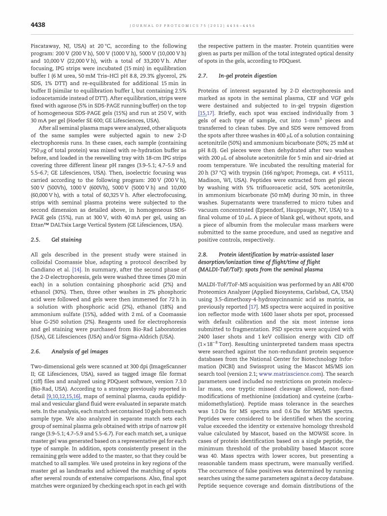
Proteins of interest separated by 2-D electrophoresis andmarked as spots in the seminal plasma, CEF and VGF gelswere destained and subjected to in-gel trypsin digestion[15,17]. Briefly, each spot was excised individually from 3gels of each type of sample, cut into 1-mm3 pieces andtransferred to clean tubes. Dye and SDS were removed fromthe spots after three washes in 400 μL of a solution containingacetonitrile (50%) and ammonium bicarbonate (50%; 25 mM atpH 8.0). Gel pieces were then dehydrated after two washeswith 200 μL of absolute acetonitrile for 5 min and air-dried atroom temperature. We incubated the resulting material for20 h (37 °C) with trypsin (166 ng/spot; Promega, cat. # v5111,Madison, WI, USA). Peptides were extracted from gel piecesby washing with 5% trifluoroacetic acid, 50% acetonitrile,in ammonium bicarbonate (50 mM) during 30 min, in threewashes. Supernatants were transferred to micro tubes andvacuum concentrated (Eppendorf, Hauppauge, NY, USA) to afinal volume of 10 μL. A piece of blank gel, without spots, anda piece of albumin from the molecular mass markers weresubmitted to the same procedure, and used as negative andpositive controls, respectively.
2.8. Protein identification by matrix-assisted laserdesorption/ionization time of flight/time of flight(MALDI-ToF/ToF): spots from the seminal plasma
MALDI-ToF/ToF-MS acquisition was performed by an ABI 4700Proteomics Analyzer (Applied Biosystems, Carlsbad, CA, USA)using 3.5-dimethoxy-4-hydroxycinnamic acid as matrix, aspreviously reported [17]. MS spectra were acquired in positiveion reflector mode with 1600 laser shots per spot, processedwith default calibration and the six most intense ionssubmitted to fragmentation. PSD spectra were acquired with2400 laser shots and 1 keV collision energy with CID off(1×18−8 Torr). Resulting uninterpreted tandem mass spectrawere searched against the non-redundant protein sequencedatabases from the National Center for Biotechnology Infor-mation (NCBI) and Swissprot using the Mascot MS/MS ionsearch tool (version 2.1; www.matrixscience.com). The searchparameters used included no restrictions on protein molecu-lar mass, one tryptic missed cleavage allowed, non-fixedmodifications of methionine (oxidation) and cysteine (carba-midomethylation). Peptide mass tolerance in the searcheswas 1.0 Da for MS spectra and 0.6 Da for MS/MS spectra.Peptides were considered to be identified when the scoringvalue exceeded the identity or extensive homology thresholdvalue calculated by Mascot, based on the MOWSE score. Incases of protein identification based on a single peptide, theminimum threshold of the probability based Mascot scorewas 40. Mass spectra with lower scores, but presenting areasonable tandem mass spectrum, were manually verified.The occurrence of false positives was determined by runningsearches using the same parameters against a decoy database.Peptide sequence coverage and domain distributions of the

4439J O U R N A L O F P R O T E O M I C S 7 5 ( 2 0 1 2 ) 4 4 3 6 – 4 4 5 6
major proteins and major trains identified by mass spectrom-etry were graphically localized in the primary sequences ofthe proteins using the Caititu software [18].
2.9. Protein identification by electrosprayionization-quadrupole-time of flight (ESI-Q-ToF) massspectrometry: spots from the seminal plasma, caudaepididymal and seminal vesicle fluids
For LC-MS/MS [19], the digested samples were injected usingthe nanoAcquity UPLC sample manager and the chromato-graphic separation was performed using a UPLC C18 column(75 μm×10 cm) with a flow of 0.6 μL/min. The mass spectrawere acquired in a Synapt G2 HDMS instrument (Waters Co.,Milford, MA, USA) using a data dependent acquisition (DDA),where the three top peaks were subjected to MS/MS. Mobilephases A and B consisted of 0.1% formic acid inwater and 0.1%formic acid in acetonitrile, respectively. The gradient condi-tions used were as follows: 0 min with 3% of B, increasinglinearly to 30% B in 20 min, then it increased up to 70% B in40 min where it remained until 50 min and in the next minuteit was decreased to 3% of B. The data were processed usingMascot Distiller (Matrix Science Ltd) and subjected to databasesearch using Mascot Server 2.3 [20]. The searches were madewith the assumption that therewas amaximumof onemissedtrypsin cleavage and that peptides were mono-isotopic andusing partially oxidized methionine residues, and completelycarbamidomethylated cysteine residues. Peptide mass toler-ance and fragmentmass tolerancewere initially set to ±0.1 Da,respectively, for MS/MS ion searching. However, candidatepeptide IDs were only accepted if the m/z values wereobserved within 0.1 Da (typically less than 0.05 Da) of thetheoretical mass of the candidate ID, as determined whenmanually reviewing MASCOT search results.
2.10. Gene ontology analysis
Protein data from the protein lists of seminal plasma, caudaepididymal and vesicular gland fluids obtained after MASCOTsearch were analyzed using the software for researchingannotations of proteins (STRAP), an open-source application[21]. Gene ontology terms for biological process and molecularfunctionwere obtained fromUniProtKB and EBI GOAdatabases.
2.11. Western blots
Seminal plasma, cauda epididymal and vesicular gland fluidproteins, representing a pool from 3 animals, were separatedby one dimensional SDS-PAGE and transferred to nitrocellu-lose membranes (Hybond ECL, GE Lifesciences, USA) usinga TE 70 transfer unit (GE Lifesciences, USA) at 208 mA for90 min. A total of 20 μg of protein were used for each transfer.Membranes were blocked overnight at 4 °C with PBS-T (50 mLof PBS with 0.5% Tween 20) containing BSA (5% w/v), undermild agitation, followed by a for 2-hour incubation withprimary antibodies against bovine Binder of Sperm Protein(BSP) 1 (1:1000) and bodhesin 2 (1:500). Membranes were thenwashed three times in PBS-T and incubated with donkeyanti-rabbit IgG coupled with alkaline phosphatase (Abcam,Cambridge, MA, USA) for another 2 h, washed again three
times in PBS-T and rinsed twice with Tris–HCl (50 mM). Immu-noreaction was visualized by exposing the membranes to asolution containing BCIP® (5-bromo-4-chloro-3-indolyl phos-phate, 0.15 mg/mL), NBT (nitro blue tetrazolium, 0.30 mg/mL),Tris (100 mM) and MgCl2 (5 mM), pH 9.5. Reaction was stoppedby washing the membranes several times with milli-Q water.Controls consisted of use of only the primary or secondaryantibodies with the sample and the use of a blocked mem-brane without sample exposed to both antibodies (data notshown). Antibodies against BSP 1 [22] were kindly providedby Dr. Puttaswamy Manjunath (Department of Medicine,University of Montréal, Canada). Binder of Sperm Proteins isexpressed in bulls and rams, among other species. The bovineBSP (BSP 1) and its ovine homolog (RSVP 14) share a highdegree of homology in amino acid sequence [23]. Polyclonalantibodies against bodhesin, commercially developed (RheaBiotech, Campinas, SP, Brazil), were raised in rabbits againsta peptide (SSNQPVSPFDIFYYERPSA) corresponding to theC-terminus sequence of bodhesin-2 (Bdh-2; gi: 121484235).
2.12. Immunocytochemistry
For immunocytochemistry, both ejaculated and cauda epidid-ymal sperm samples, each containing 5×10⁶ cells, were fixedin paraformaldehyde (2%) for 10 min and washed twice in PBS(700 g, 15 min, 4 °C), as previously described [24]. To blocknonspecific sites, spermatozoa were next incubated for 2 h,under gentle agitation and at 4 °C, in PBS-Tween 20 containing5% of bovine serum albumin — BSA (w/v; Sigma, USA). Spermcells were then incubated with the primary antibodies in thesame blocking solution for 2 h (under gentle agitation and4 °C), using the following antibody concentrations: 1:300 foranti-BSP 1 and 1:200 for anti-bodhesin 2. After this firstincubation, sperm were washed three times with PBS-Tween20 (500 g, 5 min, 4 °C) and incubated with the secondaryantibody, an Alexa 488-conjugated anti-rabbit IgG (1:300,Alexa Fluor 488 Goat Anti-Rabbit SFX kit (Life Technologies,Grand Island, NY, USA) for 1 h in a solution with PBS-Tween20 and 1% BSA (w/v). Three additional washes followed thisincubation (as described above) and smears were immediatelyprepared in a dark room, using an anti-fade reagent (ProlongGold, Life Technologies, Grand Island, NY, USA), for confocalanalysis. Antibodies used in this protocol were the same asdescribed for the Western blots.
Images of sperm incubated with Alexa 488-conjugatedantibodies were acquired by a laser scanning confocal micro-scope using an emission wavelength of 510 nm, and Zen™software (LSM 710 Confocal Microscope, Carl Zeiss Inc.,Thornwood, NY, USA). We obtained images as a series ofsequential planes taken every 0.125 μm, with a total depth of5 μm [24]. The image stacks were deconvoluted to improve theresolving power and to eliminate noises and out-of-focus blur,using Autoquant X2 (Media Cybernetics Inc., Silver Springs,MD, USA).
2.13. Isolation of total RNA from epididymis and seminalvesicles
We collected samples from animals at different developmentstates (pre-puberty and adulthood) to verify the sites of ovine

4440 J O U R N A L O F P R O T E O M I C S 7 5 ( 2 0 1 2 ) 4 4 3 6 – 4 4 5 6
BSP and spermadhesin gene expression in the reproductivetract of the Santa Ines rams. Immediately postmortem, testissamples measuring approximately 1 cm3 taken from theinnermost region of the organ, samples from the caudaepididymis (as described by Fouchécourt et al. [25]) andvesicular glandswere snap frozen in liquid N2 and transportedto the laboratory. Total RNA was isolated using approximately100 mg of the collected tissues. For this procedure, Trizol™Reagent (Life Technologies, USA) was used following manu-facturer's recommendations. Briefly, the frozen tissue wasmacerated in 1 mL of the Trizol reagent, and the organicportion was separated from the aqueous phase by adding200 μL of chloroform and centrifuged at 12,000 g for 15 min, at4 °C. After centrifugation of this mixture, we added to thesupernatant the same volume of 70% ethanol. Total RNA waspurified from this supernatant using the column systemPureLink™ RNA Mini Kit (Life Technologies, USA), alsoaccording to manufacturer's instructions. RNA preparationswere subjected to DNase I treatment (PureLink™ DNase; LifeTechnologies, USA), and the RNAwaswashed three timeswiththe provided Wash Buffer Solution II and then re-suspendedin diethylpyrocarbonate 0.1% (DEPC)-treated water. RNAquality and concentration were determined in a NanoDrop™2000 spectrophotometer (Thermo Scientific, Waltham, MA,USA). We considered that one unit of absorbance at 260 nmcorresponded to 40 μg/mL RNA.
2.14. Reverse transcription-PCR
Complementary DNA (cDNA) was synthesized using 1 μg oftotal RNA with SuperScript™ III Reverse Transcriptase (LifeTechnologies, USA) and PCR reactions were conducted in twosteps. In the first one, 1 μg of RNA, 50 ng/mL of randomhexamer primers, 10 mM dNTP mix and DEPC-treated water(for a total volume of 13 μL) were heated to 65 °C for 5 minand immediately placed on ice for at least 1 min. The secondstep consisted of adding to the first step solution: 200 U ofSuperScript™ III RT, 10× RT Buffer, 0.1 M DTT and 40 U RNaseout. Reverse transcription was performed at 25 °C for 5 min,50 °C for 40 min and 70 °C for 15 min. The resulting cDNA wasstored at −20 °C until later use.
Based on preliminary results, proteins identified as theRam Seminal Vesicle Proteins (RSVP) with 14 and 22 kDa andbodhesins represented the most abundant components of theseminal plasma of the Santa Ines rams. RSVPs are Binder ofSperm Proteins of the ovine species and bodhesins belong tothe spermadhesin family [23,26]. Thus, candidate genes werechosen on the basis of RSVP 14 and 22 kDa and bodhesins 1and 2. Gene sequences were obtained from the UniGenedatabase (NCBI). The primers were designed according to thepublished Ovis aries (RSVPs 14 and 22) and Capra hircus(bodhesins 1 and 2)mRNA sequences available at the UniGene,using the online, free access program Primer 3 (for sequencesof primers, see Table S1).
Quantitative real-time PCR (qPCR)was carried out on an iQ5cycler (Bio-Rad, USA). Reactions were performed in 20 μL with1 μg of each cDNA, 1× Power SYBR™ Green (Life Technologies,USA) PCR Master Mix, 0.4 μM of both sense and antisenseprimers and ultra-pure water completing the reaction volume.The qPCR protocol included a first denaturation at 95 °C for
10 min, then 40 PCR cycles (15 s 95 °C, 1 min 60 °C, 1 min 72 °C),followed by a final extension of 10 min at 72 °C. The specificityof each primer set was tested by generating a melting curve,carried out between 60 and 95 °C for all sequences, startingfluorescence acquisition at 60 °C and taking measurementsat 10-second intervals until the temperature reached 95 °C.GAPDH was used as reference gene to normalize the se-quences assayed. All samples were run in triplicates andqPCRs were repeated at least twice. As negative controls,samples with reverse transcriptase but without cDNA wereused. Relative gene expression was estimated using the ΔΔCt-method [27]. Threshold and Ct (cycle threshold) values wereautomatically determined by Bio-Rad iQ5 software, usinginternal parameters, and Ct and Tm (melting temperature).
3. Results
3.1. Seminal plasma proteins
Semen samples of the Santa Ines rams used on the presentstudy contained, on average, 3.6±0.4×10⁹ spermatozoa/mL and5.4±0.3×10⁹ total sperm/ejaculate, with 90±0.2% ofmotile cellsand 75±0.3% of sperm with progressive motility. On average,302±16 spots were detected by PDQuest software in the matchset of ten seminal plasma gels constructed within the 4–7 pHrange. Based on the master gel image (Fig. 1A), which includedproteins of the reference gel (Fig. 1B) and spots from additionalmembers of the match set, 143 spots consistently appeared inevery gel and intensities of these spots corresponded to 65.5±1.5% of the intensities of all spots depicted in the seminalplasma maps. Seventy three spots were identified by massspectrometry, corresponding to 41 different proteins (Table 1).Combined, intensities of the identified spots accounted for42.5±0.9% of the intensities of all spots and 65.1% of theintensities of spots presented in every protein map.
Two spots were identified as Ram Seminal Vesicle Protein14 kDa — RSVP 14 (spots #66 and #67) and eleven others asRam Seminal Vesicle Protein 22 kDa — RSVP 22 (spots #17, 18,51, 52, 53, 55, 56, 60, 61, 62 and 70; Fig. 1A; Table 1). RSVP spotscorresponded to 20.3% of the total intensity of all spotsdetected in the gels. Six other spots showed homology toisoforms of either bodhesin 1 (spot #71) or bodhesin 2 (spots#57, 63, 64, 68 and 69) and the intensities of such componentsrepresented 11.4% of all spot intensities quantified in the ramseminal plasma gels. The 19 spots recognized as RSVPs andbodhesins numerically represented only 6% of all spotsdetected in the PDQuest match set, but their combined opticaldensities corresponded to 31.8% of the total spot intensitiesestimated in the protein maps. Some of the proteins identifiedin the 2-D gels were present as multiple isoforms, includingmatrix metalloproteinase 2 (train 1), albumin (train 2), lacto-transferrin (train 3) and clusterin (train 4; Fig. 1B).
Conserved domains and other structural features ofselected proteins described in Fig. 1 and Table 1 aregraphically represented using the Caititu software (Fig. S1).The sequence of trypsin-digested peptides obtained fromRSVP 14 spots matched to both fibronectin type II (FNII)domains of the full protein. Tryptic peptides from five RSVP22 spots had one sequence matching to each of the FNII

Fig. 1 – Two-dimensional map of seminal plasma proteins from Santa Ines rams. (A) represents the master gel generated byPDQuest software (Bio Rad, USA), based on a match set with all gels used in the study. (B) corresponds to the reference map(from ram #3) fromwhich themaster wasmostly generated. Proteins were stainedwith Coomassie blue and identified bymassspectrometry (MALDI-ToF/ToF and ESI-Q-ToF). Spot numbers refer to those shown in Table 1.
4441J O U R N A L O F P R O T E O M I C S 7 5 ( 2 0 1 2 ) 4 4 3 6 – 4 4 5 6

Table 1 – Proteins of the seminal plasma of Santa Inesrams identified by two-dimensional electrophoresis andmass spectrometry (MALDI-ToF/ToF and ESI-Q-ToF).Table includes spots detected in 13-cm gels, within the4–7 pH range. Spot numbers refer to those in Fig. 1. Fulltable showing sequences of matched peptides, ion scores,m/z and z values is shown as Table S2.
Protein Experimental a
kDa/pINCBI
accessionnumber
MS/MSproteinscore
Sequencecovered
(%)
Dipeptidyl peptidase IIISpot 01 73.0/5.2 296471603 202 19
Matrix metalloproteinase 2Spot 01 73.0/5.2 27807447 59 9Spot 02 73.0/5.2 27807447 179 10Spot 03 72.7/5.3 27807447 111 7
Inositol-3-P-synthase 1Spot 04 71.3/5.6 114051253 221 14
Serum albuminSpot 05 71.3/6.0 57164373 259 9Spot 06 71.6/5.9 57164373 268 11Spot 07 71.6/5.8 57164373 273 11
Heat shock 70 kDa protein 1-likeSpot 08 70.7/6.2 268607682 327 12
Thioredoxin reductaseSpot 09 65.6/6.4 27807129 207 14
Beta-galactosidaseSpot 10 76.1/6.5 78042544 48 4
Alpha-2-macroglobulinSpot 11 80.0/6.6 157954061 76 1
14-3-3 protein zeta chainSpot 12 80.0/6.7 112687 62 5Spot 54 29.3/4.7 253706 233 22
LactoferrinSpot 13 76.6/6.9 32130549 62 1
Angiotensin-I converting enzymeSpot 14 79.9/6.8 66710717 166 10Spot 74 79.9/6.9 66710717 92 4Spot 75 79.9/7.0 331284213 52 2
TransferrinSpot 15 73.8/6.7 114053269 51 3Spot 16 73.8/6.6 114053269 58 5
Ram Seminal Vesicle 22 kDa ProteinSpot 17 54.9/4.1 219521812 67 15%Spot 18 61.4/4.2 219521812 146 20%Spot 51 29.4/4.3 219521812 68 15%Spot 52 28.4/4.4 219521812 161 20%Spot 53 28.2/4.5 219521812 176 20%Spot 55 28.2/5.0 219521812 52 12%Spot 60 25.0/4.3 219521812 145 20%Spot 61 23.6/4.6 219521812 157 26%Spot 62 23.4/4.8 219521812 167 20%Spot 56 25.4/5.5 219521812 200 20%Spot 70 15.3/6.1 219521812 93 20%
Arylsulfatase ASpot 19 66.7/5.2 47522624 78 4Spot 20 66.7/5.3 115497982 36 9
Plasma glutamate carboxypeptidase precursorSpot 21 62.5/5.4 115495837 116 11
V-type proton ATPase subunit BSpot 22 62.5/5.7 297129 115 11Spot 75 62.5/5.8 27806225 36 7
Alpha-L-fucosidase 2Spot 23 61.7/5.8 76626175 65 12
Chaperonin containing TCP‐1 subunit εSpot 24 67.7/6.0 74353962 66 8
Table 1 (continued)
Protein Experimental a
kDa/pINCBI
accessionnumber
MS/MSproteinscore
Sequencecovered
(%)
Chaperonin containing TCP-1 subunit αSpot 25 67.6/5.9 57032236 122 5
Leucine aminopeptidaseSpot 26 63.0/6.1 410689 183 12
Alpha enolaseSpot 27 42.2/6.4 4927286 184 12
Cathepsin FSpot 28 56.6/6.5 115495381 104 10
Betaine-homocysteine S-methyltransferase 2Spot 29 40.0/6.4 60687508 47 4
Chaperonin containing TCP-1 subunit βSpot 30 60.5/6.6 468546 90 15
ClusterinSpot 31 49.7/4.3 27806907 114 7Spot 32 48.9/4.4 27806907 247 12Spot 33 48.1/4.5 27806907 144 7Spot 34 45.2/4.2 27806907 51 5Spot 35 44.2/4.3 27806907 319 19Spot 37 43.4/5.2 27806907 450 16Spot 38 42.5/5.3 27806907 37 2Spot 42 41.3/5.4 27806907 64 3Spot 46 46.5/6.1 27806907 67 6Spot 76 48.7/6.1 27806907 225 11
Phosphoglycolate phosphataseSpot 36 41.5/5.1 84000329 167 13
ActinSpot 39 51.1/5.4 8809716 94 15Spot 40 51.2/5.4 8809716 85 15
Prostaglandin reductase 2Spot 41 49.1/5.4 115497482 133 12
Zinc-alpha-2-glycoproteinSpot 43 47.8/5.5 77735615 95 8Spot 77 49.9/5.7 77735615 52 8
Galactokinase 1Spot 45 41.3/5.7 95767475 78 4
Aldose reductaseSpot 47 48.1/6.2 113594 56 13
Malate dehydrogenaseSpot 48 40.0/6.4 77736203 86 6Spot 49 41.2/6.4 77736203 129 24
Palate, lung and nasal epithelium cloneSpot 50 32.9/4.4 27806071 102 7
Bodhesin-2Spot 57 24.4/5.7 121484235 217 18Spot 63 19.2/5.2 121484235 87 18Spot 64 15.1/4.3 121484235 34 18Spot 68 13.2/5.5 121484235 162 27Spot 69 14.1/5.6 121484235 57 18
Proteasome alpha 6 subunitSpot 58 28.8/6.6 6755198 78 11
Tissue inhibitor metallopeptidase (TIMP-2)Spot 59 25.4/6.6 75832065 190 13
MicroseminoproteinSpot 65 15.2/4.5 76656898 55 9Spot 77 15.2/4.5 76656898 155 26
Ram Seminal Vesicle 14 kDa ProteinSpot 66 14.9/5.0 219521810 215 48Spot 67 13.7/5.2 219521810 179 34
BodhesinSpot 71 16.3/6.3 77864607 73 9
4442 J O U R N A L O F P R O T E O M I C S 7 5 ( 2 0 1 2 ) 4 4 3 6 – 4 4 5 6

Table 1 (continued)
Protein Experimental a
kDa/pINCBI
accessionnumber
MS/MSproteinscore
Sequencecovered
(%)
Peroxiredoxin 5Spot 72 17.8/6.7 74354725 158 22
Epididymal secretory protein E1Spot 73 17.5/6.9 27806881 150 31
a Experimental values were deduced from the respective 2D mapby the PDQuest software.
4443J O U R N A L O F P R O T E O M I C S 7 5 ( 2 0 1 2 ) 4 4 3 6 – 4 4 5 6
domains; five other spots were found with one sequencebelonging to FNII 1 domain and two sequences to the FNII 2domain. One RSVP 22 spot had sequences matching only theFNII 2 domain. The sequence of trypsin-digested peptidesfrom Bdh-1 spot matched to one single region of the CUBdomain represented in the full protein. Peptides from threeBdh-2 spots also matched to only one region of the samedomain of the protein, while one Bdh-2 spot had peptidesequences equivalent to two different regions of this domain.
3.9
13
5.14.0 4.1 4.2 4.3 4.4 4.5 4.6 4.7 4.8 4.9 5.0
4.74.8 4.9 5.0 5.1 5.2
66
45
30
20.1
14.3
kDa
kDa
66
45
30
20.1
14.3
4.2 5.0 5.24.4 4.6 4.8 5.4
12
3
4
5
6
8
9
10
11
12
13
14
15
16
17
18
19
20
RSVP-14
Clusterin
RSVP-14
2122
23
7
26 25
24
27
28
29
30
31
323334353637
38
39
4041
4344
45
46
47
48
42
49
50
5152
53
6 364
A
B C
97
97
18 cm 18
Fig. 2 – Two-dimensional map of seminal plasma proteins fromPDQuest software (Bio Rad, USA), based on amatch set with all 13to the master gels based on match sets constructed with 18-cm g5.5–6.7). Proteins were stained with Coomassie blue and identifithose shown in Table 2.
Angiotensin-converting enzyme sequences all matched tothe main peptidase M2 domains, which comprise most of theprotein. Sequences of the seven clusterin spots were foundinserted in the beta chain and only one spot also in the alphachain. Peptide sequences obtained after tryptic digestion ofMMP-2 spots (#2 and 3) were part of the zinc-dependentmetalloproteinase domain but only one of these spots (#3) hadits sequence in the fibronectin type II and HX domains.
Electrofocusing seminal plasma proteins within pH rangesof 3.9–5.1, 4.7–5.9 and 5.5–6.7 allowed the detection of 104±8,159±7 and 81±5 spots in the 2-D gels (Fig. 2B–D). In the 3.9–5.1pH range, there were 45 spots identified by mass spectrom-etry, corresponding to 15 different proteins. From these, sevenproteins were not detected in the original map constructedwithin the 4–7 pH range (glucose–fructose oxidoreductasedomain-containing protein 2, proteasome subunit alpha 5,lysozyme, gelsolin, alpha-2-HS glycoprotein, ubiquitin carboxyl-terminal hydrolase isoenzyme L3 and alpha-1-antitrypsin). Forthe 4.7–5.9 pH interval, there were 76 spots identified, respectiveto 23 proteins, including seven that had not been detected beforein the original seminal plasma gels within the 4–7 pH range:
6.7
cm
5.9 5.55.3 5.4 5.5 5.6 5.7 5.8 5.6 5.7 5.8 5.9 6.0 6.1 6.2 6.3 6.4 6.5 6.6
6.0 6.25.6 5.8 6.4 6.6 6.7
1
2
3 4
5 6
7
8
9
10
Bdh-2Bdh-2
ERBP
ERBP
1415 11
12
13
16
17
65
D
cm 18 cm
pI
Santa Ines rams. (A) represents the master gel generated by-cm gels, within the 4–7 pH interval. (B), (C) and (D) correspondels, within three different pH intervals (3.9–5.1, 4.7–5.9 anded by tandem mass spectrometry. Spots numbers refer to

Table 2 – Proteins of the seminal plasma of Santa Inesrams identified by two-dimensional electrophoresis andmass spectrometry (MALDI-ToF/ToF and ESI-Q-ToF).Table includes spots detected in 18-cm gels, obtainedwithin the 3.9–5.1, 4.7–5.9 and 5.5–6.7 pH intervals. Spotnumbers refer to those in Fig. 2. Full table showingsequences of matched peptides, ion scores, m/z and zvalues is shown as Table S3.
Protein Experimental a
kDa/pINCBI
accessionnumber
MS/MSproteinscore
Sequencecovered
(%)
pH range: 3.9–5.1Ram Seminal Vesicle 14 kDa ProteinSpot 01 15.2/4.8 219521810 945 70Spot 02 15.4/5.0 219521810 47 17
Glucose-fructose oxidoreductase domain-containing protein 2Spot 03 18.9/4.1 62460418 41 2
Ram Seminal Vesicle 22 kDa ProteinSpot 04 24.9/4.9 219521812 143 36Spot 05 22.8/4.4 219521812 131 20Spot 06 26.7/4.3 219521812 188 20Spot 07 25.0/4.2 219521812 131 20Spot 08 28.3/4.2 219521812 176 20Spot 09 25.1/4.1 219521812 131 20Spot 10 28.0/4.1 219521812 118 20Spot 23 27.0/4.2 219521812 172 24Spot 26 22.9/4.4 219521812 115 20Spot 45 30.2/4.1 219521812 183 24
MicroseminoproteinSpot 11 15.3/4.4 296472032 62 50
Phosphoglycolate phosphataseSpot 12 41.6/5.1 84000329 254 17Spot 28 42.2/4.9 84000329 71 8
ClusterinSpot 13 43.5/5.1 27806907 569 18Spot 14 42.0/4.9 27806907 108 4Spot 15 42.6/4.9 27806907 54 7Spot 21 44.6/4.2 27806907 175 8Spot 22 44.8/4.1 27806907 155 12Spot 29 44.6/4.8 27806907 341 13
14-3-3 protein zeta chainSpot 16 29.5/4.7 253706 409 22
Proteasome subunit alpha 5Spot 17 28.4/4.6 296489341 187 34
Bodhesin 2Spot 18 17.6/5.0 121484235 101 18
LysozymeSpot 19 23.0/4.8 112030802 94 12
Proteasome subunit alpha 6Spot 20 26.8/4.8 77735687 170 12
GelsolinSpot 24 27.1/4.8 327346104 54 2Spot 25 25.8/4.8 327346104 63 2
Alpha-2-HS-glycoproteinSpot 27 15.4/4.7 27806751 47 6Spot 28 42.2/4.9 27806751 138 12Spot 31 33.2/4.7 27806751 108 12Spot 32 33.2/4.7 27806751 125 12Spot 33 69.5/4.8 27806751 126 12Spot 34 69.5/4.7 27806751 100 12Spot 36 69.5/4.6 27806751 99 12Spot 37 69.5/4.6 27806751 82 12Spot 38 31.8/4.3 27806751 124 12Spot 39 44.8/4.3 27806751 112 12Spot 40 25.3/4.4 27806751 116 12Spot 41 25.9/4.4 27806751 105 12
4444 J O U R N A L O F P R O T E O M I C S 7 5 ( 2 0 1 2 ) 4 4 3 6 – 4 4 5 6
alpha-N-acetylgalactosaminidase, aldose-1-epimarase, gelsolin,apolipoprotein A1, phosphoglycerate kinase 1, glutamate car-boxypeptidase and prostaglandin-H2 D-isomerase. As regard tothe 5.5–6.7 interval, 14 spotswere identified,which correspondedto 14 proteins. From this list, six had not appeared in the first setof gels: superoxide dismutase, phosphoglycerate kinase 1, spermacrosome membrane-associated protein 3, epididymis-specificERBP-like protein, polyubiquitin and alpha-2-HS glycoprotein(Table 2). Spots were highlighted in the overlapping regions ofthe gels with narrow pH range. As shown, spots identified asclusterin and RSVP 14 in the 3.9–5.1 pH interval in the first gel(Fig. 2B) also appear in the 4.7–5.1 interval of the second gel(Fig. 2C). Similarly, ERBP and bodhesin-2 spots identified in the5.5–5.8 pH interval of this gel (Fig. 2C) were also present in thethird map (Fig. 2D), in the same pH range.
3.2. Proteins of the cauda epididymal and vesicular glandfluid
Onaverage,wedetected 113±7 spots per gel of cauda epididymalfluid (Fig. 3A) and 62 spots were consistently present on allmaps. Combined, these spots represented 46.9±3.2% of theintensities of all spots detected in the 2-Dmaps. Twelve differentproteins were identified and the most abundant proteins inthese gels appeared as albumin and transferrin. These proteinswere present as trains of multiple isoforms and togethercorresponded to 27.3% of all spot intensities. Other proteinsidentified include clusterin, alpha-1-anti-trypsin, prostaglandinD synthase, alpha-2-HS glycoprotein and actin (Table 3).
Concerning the vesicular gland fluid (VGF), 215±12 spotswere detected per gel (Fig. 3B) and 104 spots consistentlyexpressed in all maps. The combined intensity of these spotsrepresented 38.0±2.2% of the intensities of all spots detectedin the VGF maps. Nineteen different proteins were identifiedby tandem mass spectrometry in the VGF maps, being themost abundant the ones identified as RSVPs 14 and 22,bodhesin 2 and albumin. These proteins were present astrains of multiple isoforms and their combined intensitiescorresponded to 38.4% of all spot intensities. Additionalproteins identified in the VGF include cystatin B, apolipoproteinA1, clusterin, disulfide isomerase, aldose reductase, alpha-enolase and ERP29 (Table 3).
Western blots confirmed that antibodies against BSP 1reacted with a single band around 14 kDa in 1-D gels withseminal plasma, vesicular gland and cauda epididymal fluid.However, intensity of staining was muchmore pronounced inthe first two samples as compared to the cauda epididymalsecretion. In the case of Bdh-2, antibody reaction was evidentfor seminal plasma and vesicular fluid but weak in the caudaepididymal fluid (Fig. 4).
3.3. Interactions of anti-BSP 1 and anti-Bdh 2 withejaculated and cauda epididymal sperm
Antibodies against BSP 1 were detected on the acrosome,equatorial segment and midpiece of ejaculated sperm withintact acrosome (Fig. 5A). On sperm with reacted acrosome,anti-BSP 1 binding was restricted to the equatorial andmidpieceregion. Epididymal sperm were completely absent of immuno-fluorescence for BSP 1 antibody. As regard to anti-Bdh 2, reaction

Table 2 (continued)
Protein Experimental a
kDa/pINCBI
accessionnumber
MS/MSproteinscore
Sequencecovered
(%)
Spot 42 18.9/4.2 27806751 107 12Spot 43 20.6/4.1 27806751 58 5Spot 44 21.4/4.0 27806751 114 12Spot 48 28.0/4.3 27806751 56 5Spot 49 23.1/4.0 27806751 50 5Spot 50 24.2/4.0 27806751 43 5Spot 51 29.8/4.0 27806751 45 5Spot 52 33.4/4.9 27806751 117 12Spot 53 15.8/4.9 27806751 63 5
Ubiquitin carboxyl-terminal hydrolase isozyme L3Spot 30 28.5/4.8 94966875 199 24
Alpha-1-antitrypsinSpot 35 69.5/4.6 57526646 190 12
Transcobalamin-1-likeSpot 46 65.4/4.0 345327379 56 4Spot 47 52.6/4.0 345327379 55 4
pH range: 4.7–5.9Matrix metalloproteinase 2Spot 01 71.8/4.9 261244994 162 33Spot 02 69.3/5.0 261244994 38 4
Serum albuminSpot 02 69.3/5.0 57164373 56 20Spot 03 67.1/5.0 57164373 79 1Spot 04 66.6/5.1 57164373 280 36Spot 07 71.3/5.5 57164373 63 1Spot 08 70.7/5.5 57164373 602 51Spot 28 43.8/5.7 57164373 66 1Spot 52 10.8/4.9 57164373 115 1Spot 57 14.5/5.5 57164373 89 2Spot 62 15.3/5.8 57164373 40 4
Cathepsin BSpot 06 70.2/5.4 27806671 51 9
ClusterinSpot 09 67.5/55 27806907 37 2Spot 20 46.3/4.7 27806907 32 10Spot 21 45.5/4.8 27806907 95 19Spot 22 45.4/4.9 27806907 79 4Spot 23 42.9/4.9 27806907 285 18Spot 24 42.1/4.9 27806907 67 15Spot 25 42.9/4.9 27806907 347 27Spot 27 43.2/5.3 27806907 66 15Spot 64 67.7/5.1 27806907 68 13
LactoferrinSpot 10 63.9/5.7 254656113 59 1Spot 11 62.3/5.7 254656113 194 16Spot 12 57.8/5.8 254656113 65 1
Alpha-2-HS-glycoproteinSpot 13 57.0/4.8 57526674 88 5
Arylsulfatase ASpot 14 52.5/5.0 115497982 68 7Spot 15 51.0/5.0 115497982 74 2
Plasma glutamate carboxypeptidaseSpot 05 62.2/5.2 115495837 376 32Spot 16 52.8/5.0 115495837 117 11Spot 37 24.4/5.4 115495837 47 9Spot 44 17.6/4.9 115495837 134 16
ActinSpot 17 52.8/5.2 8809716 152 15Spot 18 52.9/5.2 8809716 175 15
Leucine aminopeptidase 3Spot 19 51.8/5.6 88682884 112 31
(continued on next page
Table 2 (continued)
Protein Experimental a
kDa/pINCBI
accessionnumber
MS/MSproteinscore
Sequencecovered
(%)
Prostaglandin reductase 2Spot 26 48.3/5.2 115497482 69 32
Aldose-1-epimeraseSpot 28 43.8/5.7 77736588 100 21
Alpha-N-acetylgalactosaminidaseSpot 29 45.8/5.7 187607477 46 2
Enolase alphaSpot 30 42.2/5.9 4927286 50 20
Ram Seminal Vesicle 22 kDa ProteinSpot 31 24.8/4.8 219521812 54 20Spot 33 25.9/4.9 219521812 127 20Spot 34 28.8/5.2 219521812 21 20Spot 35 24.5/5.2 219521812 278 35Spot 51 12.3/4.9 219521812 49 20Spot 60 14.6/5.6 219521812 50 20
GelsolinSpot 32 26.1/4.8 327346104 44 2
Prostaglandin D synthaseSpot 36 29.1/5.4 57164293 96 14
Bodhesin 2Spot 37 24.4/5.4 121484235 55 35Spot 38 24.3/5.5 121484235 214 35Spot 39 22.9/5.6 121484235 25 18Spot 42 20.3/4.9 121484235 90 35Spot 43 18.9/4.9 121484235 108 18Spot 44 17.6/4.9 121484235 100 18Spot 45 16.7/4.9 121484235 64 18Spot 47 15.2/4.8 121484235 124 27Spot 48 14.5/4.9 121484235 147 35Spot 50 14.0/4.9 121484235 90 18Spot 53 12.6/5.1 121484235 36 27Spot 54 13.4/5.1 121484235 86 18Spot 59 14.4/5.5 121484235 79 18Spot 60 14.6/5.6 121484235 45 18
Epididymis-specific ERBP-like proteinSpot 39 22.9/5.6 296481974 57 4Spot 40 20.4/5.6 296481974 51 4Spot 41 23.4/5.6 296481974 58 4Spot 65 21.7/5.6 296481974 98 4
Ram Seminal Vesicle 14 kDa ProteinSpot 45 16.7/4.9 219521810 20 17Spot 46 15.7/4.8 219521810 177 30Spot 47 15.2/4.8 219521810 1246 70Spot 49 15.4/5.0 219521810 214 49Spot 55 13.7/5.2 219521810 149 49
Phosphoglycerate kinase 1Spot 44 17.6/4.9 215983082 102 22Spot 56 11.1/5.4 215983082 67 8
Apolipoprotein A1Spot 44 17.6/4.9 162678 50 4Spot 52 10.8/4.9 162678 490 8Spot 55 13.7/5.2 162678 372 4Spot 57 14.5/5.5 162678 175 4Spot 58 20.4/5.6 162678 46 8Spot 61 11.8/5.6 162678 409 8Spot 63 72.0/5.1 162678 772 21
BodhesinSpot 61 11.8/5.6 77864607 64 17
pH range: 5.5–6.7Superoxide dismutase [Cu–Zn]Spot 01 20.6/6.1 223633904 100 21
(continued on next page)
Alpha-2-HS-glycoprotein
4445J O U R N A L O F P R O T E O M I C S 7 5 ( 2 0 1 2 ) 4 4 3 6 – 4 4 5 6
)

Table 2 (continued)
Protein Experimental a
kDa/pINCBI
accessionnumber
MS/MSproteinscore
Sequencecovered
(%)
Ram Seminal Vesicle 14 kDa ProteinSpot 02 20.6/5.7 219521810 61 17
Galactokinase 1Spot 03 42.5/5.8 150247075 41 2
Phosphoglycerate kinase 1Spot 04 41.8/5.8 215983082 183 12
Alpha enolaseSpot 05 46.2/6.1 87196501 85 4
Cathepsin FSpot 06 45.4/6.2 115495381 211 12
Betaine-homocysteine S-methyltransferase 2Spot 07 45.9/6.2 60687508 119 6
Bodhesin 2Spot 08 14.6/5.6 121484235 191 35
Sperm acrosome membrane-associated protein 3Spot 09 14.4/5.6 157279923 60 6
Epididymis-specific ERBP-like proteinSpot 10 21.1/5.7 297460193 62 4
Peroxiredoxin 5Spot 11 20.1/5.6 74354725 115 10Spot 15 20.4/6.4 74354725 118 10
PolyubiquitinSpot 12 11.4/6.5 163575 74 15
Alpha-2-macroglobulinSpot 13 80.3/6.4 157954061 79 1
Alpha-2-HS-glycoproteinSpot 14 15.4/4.7 27806751 47 6Spot 16 14.2/5.7 27806751 53 5Spot 17 18.7/6.2 27806751 35 5
a Experimental values were deduced from the respective 2D mapsby the PDQuest software.
4446 J O U R N A L O F P R O T E O M I C S 7 5 ( 2 0 1 2 ) 4 4 3 6 – 4 4 5 6
was detected on ejaculated sperm and immunostaining wasnot evident on epididymal sperm (Fig. 5B).
3.4. Quantitative expression of genes related to the majorseminal plasma proteins: RSVPs 14 and 22 and bodhesins 1and 2
Expression of both RSVP 14 and RSVP 22 genes, as evaluatedby relative abundance of their mRNA, was mainly detectedin the vesicular glands (VES) of Santa Ines rams at both pre-puberty and yearling age. RSVP 14 and 22 transcripts were alsopresent in the cauda epididymis (CAU) but at much lowermagnitude (Fig. 6). Transcripts related to bodhesin 1 and 2genes were also detected in samples from the cauda epidid-ymis and vesicular glands, but with much greater magnitudein the latter (Fig. 6).
3.5. Gene ontology
Gene ontology (GO) terms for biological process, molecularfunction and cellular component associated with proteins ofthe seminal plasma, cauda epididymal and vesicular glandfluid is presented in Fig. 7. The most prominent biologicalprocesses linked to seminal plasma, CEF and VGF werebinding and catalytic activity. Enzyme regulator activity wasalso described for components of seminal plasma, CEF and
VGF, while antioxidant activity was associated with proteinsof the seminal plasma and VGF. Top three molecular func-tions of seminal plasma and CEF were characterized ascellular process, metabolic process and regulation. Based onthe analysis of gene symbols, major molecular functions ofVGF proteins were termed as regulation, cellular process andlocalization. Proteins of the seminal plasma, CEF and VGFwere mainly defined as extracellular components, accordingcellular component localization. Cytoplasmic componentswere also annotated for all fluids and membrane proteins,for the seminal plasma and VGF.
4. Discussion
The present study characterizes a diverse cohort of seminalplasma proteins from reproductively normal Santa Ines ramsas well as the main components of the cauda epididymal andvesicular gland fluids. The Santa Ines evolved from local herdsin a semi-arid region of the Brazilian Northeast and hasbecome the most common hair sheep raised in Brazil. Ramsof such breed represent a unique genotype because of theiradaptability to both the tropical environment and low ex-ternal input production systems.
From seminal plasma maps obtained within the 4–7 pHrange, we identified 73 spots and 41 different proteins,equivalent to 42.5% of all spot intensities detected in thegels. From the core of 143 spots present in all maps, weidentified 49 spots, also representing the majority of theintensities of all spots. The number of spots in the gels withnarrow pH intervals was greater (343 spots) than the numberobtained with gels within the 4–7 pH range (302 spots).However, such comparisons need to consider that gels withthe 4–7 pH and narrow pH intervals were constructed usingdifferent strip sizes (13 and 18 cm) and protein amounts (400and 750 μg, respectively). Thus, compensations in proteinloads and pH ranges obviously influenced the outcome of the2-D-PAGE. Regardless of spot counts, the use of narrow gelsallowed the identification of 20 proteins that had not beendetected in the original gels.
Spots identified as Ram Seminal Vesicle Proteins 14 and22 kDa (RSVPs) and bodhesins 1 and 2 represented the majorcomponents of the seminal plasma gels constructed withinthe 4–7 pH range. RSVPs were the single most abundantprotein group, contributing with 20.4% of the intensity of allspots detected in those maps. RSVPs belong to the family ofBinder of Sperm Proteins, which are characterized by twotandemly-arranged, fibronectin type II domains [28]. Graphicrepresentation of the trypsin-digested RSVP showed thatRSVP 14 and RSVP 22 spots had peptides in at least one ofthe FNII domains. RSVPs 14 and 22 also appeared as the mostabundant proteins in the vesicular gland fluid 2-D maps. Weconfirmed this fact by showing that antibodies against BSP 1reacted with components of the vesicular gland fluid butelicited only a very weak reaction with cauda epididymalfluid. Studies conducted in the bovine [29], goat [30] and bison[31] similarly showed that BSPs are the major seminal plasmaproteins and of vesicular gland origin. Both RSVP 14 and 22genes were also mainly expressed in the vesicular glands ofthe Santa Ines rams. Also, very low but consistent expression

97
66
45
30
20.1
14.3
97
66
45
30
20.1
14.3
Albumin
Prostaglandin D synthase
Perodoxin-2
ZBTB42
Trasnferrin
Clusterin
α-2-HS-GP
α-1-anti-trypsin
HSP-70 AlbuminTransferrin
α-enolase (26)
Aldose epimerase (40)
Aldose reductase
Creatine kinase BActin
Disulfide isomarease A6(45)
Clusterin
RSVP 22
RSVP 14
Bdh-2
Cystatin
Transgelin
Albumin
ERP29
Albumin
1
10
11
5
12
Albumin
14
32
21 22
13
23 24
25 26 2731 30 29 28
9 8 76 4 3 2
Albumin
18 17 16 15
19
20
4 pI 7
16
15
14
13
35
3436
39 38 37
27 28 29 30
4 5 3 8 9 10 11 12 646
Disulfide isomarease A3 (25)23 24
46
33
21 22
31
Apo-A1
53
7
17
18
8
32
Disulfide isomarease (41, 42, 43, 44)
54
Abhydrolase domain-containing protein 14B
51 52
50 49 48 47
kDaA
B
Fig. 3 – Two-dimensional map of cauda epididymis (A) and vesicular gland (B) fluid proteins from Santa Ines rams. In bothcases, the figures represent the master gel generated by PDQuest software (Bio Rad, USA), based on a match set with all gelsused in each set of samples. Proteins were stained with Coomassie blue and identified by tandem mass spectrometry. Spotnumbers refer to those shown in Table 3.
4447J O U R N A L O F P R O T E O M I C S 7 5 ( 2 0 1 2 ) 4 4 3 6 – 4 4 5 6
of those genes was detected in the cauda epididymis, inaccordance with the results from Western blots. Finding thatRSVPs are present in the cauda epididymis confirms ourprevious data showing that RSVP 14 mRNA was detected inthe epididymis of post-pubertal Morada Nova rams, also in
lower magnitude as compared to the expression in theseminal vesicles [32]. Morada Nova is another breed ofBrazilian hair sheep. Low amounts of BSP homologs havebeen described in the epididymis of humans and mice [33],similar to what we presently report for Santa Ines rams.

Table 3 – Proteins of the cauda epididymal and seminalvesicle fluid from Santa Ines rams identified bytwo-dimensional electrophoresis andmass spectrometry(MALDI-ToF/ToF andESI-Q-ToF). Spot numbers refer to thosein Fig. 3. Full table showing sequences of matched peptides,ion scores, m/z and z values is shown as Table S4.
Protein Experimental a
kDa/pINCBI
accessionnumber
MS/MSproteinscore
Sequencecovered
(%)
Cauda epididymal fluidSerum albuminSpot 01 27.2/6.2 57164373 193 12Spot 02 72.8/6.0 57164373 584 21Spot 03 72.8/5.9 57164373 536 31Spot 04 72.8/5.8 57164373 196 34Spot 05 36.8/5.9 57164373 78 4Spot 06 72.8/5.7 57164373 72 7Spot 07 76.1/5.6 57164373 36 3Spot 08 76.3/5.6 57164373 108 5Spot 09 76.5/5.5 57164373 306 24
Prostaglandin D synthaseSpot 10 26.2/6.2 57164293 48 1Spot 11 26.5/5.8 57164293 49 1Spot 12 27.3/5.5 57164293 57 1Spot 13 28.9/5.2 57164293 48 1
Peroxiredoxin 2Spot 14 30.4/5.3 261244978 121 14
TransferrinSpot 15 80.2/6.7 114326282 79 7Spot 16 80.2/6.6 114326282 81 7Spot 17 80.2/6.6 114326282 89 7Spot 18 80.2/6.5 114326282 74 7
Beta-actinSpot 19 52.9/5.4 194676388 74 11Spot 20 52.9/5.4 194676388 140 19
ClusterinSpot 21 49.7/4.3 27806907 25 2Spot 22 49.3/4.3 27806907 77 6Spot 23 48.9/4.4 27806907 65 3Spot 24 48.4/4.4 27806907 99 8
Alpha-1-anti-trypsinSpot 25 72.4/5.1 57526646 92 13Spot 26 72.2/5.2 57526646 113 23Spot 27 72.1/5.3 57526646 105 11
Alpha-2-HS-glycoproteinSpot 28 72.3/4.7 57526674 178 14Spot 29 72.5/4.6 57526674 159 14Spot 30 72.7/4.5 57526674 159 17Spot 31 73.0/4.4 57526674 165 12
ZBTB 42 protein-likeSpot 32 33.6/5.7 296475269 50 1
Vesicular gland fluidSulfotransferase 1C2Spot 01 33.4/6.8 297459963 36 4
Serum albuminSpot 02 27.2/6.2 57164373 191 8Spot 03 72.8/6.0 57164373 998 51Spot 04 72.8/5.9 57164373 1597 49Spot 05 72.8/5.9 57164373 440 19Spot 06 72.8/6.5 57164373 95 9Spot 07 17.1/5.9 57164373 104 6Spot 08 72.8/6.2 57164373 56 5Spot 09 72.9/6.2 57164373 66 2Spot 10 72.9/6.3 57164373 849 27Spot 11 73.0/6.3 57164373 1173 45Spot 12 72.9/6.4 57164373 789 21
Table 3 (continued)
Protein Experimental a
kDa/pINCBI
accessionnumber
MS/MSproteinscore
Sequencecovered
(%)
Bodhesin 2Spot 13 13.2/5.5 121484235 170 3Spot 14 14.1/5.6 121484235 29 18Spot 15 15.1/4.3 121484235 59 18Spot 16 12.2/5.2 121484235 58 18
Cystatin BSpot 17 13.4/5.9 82697343 62 15Spot 18 11.8/6.0 82697343 83 27
Ram Seminal Vesicle 14 kDaSpot 19 12.2/5.2 219521810 81 28Spot 20 13.7/5.3 219521810 60 28
Aldose reductaseSpot 21 39.8/6.0 60302887 353 31Spot 22 39.8/6.2 60302887 153 36
Beta-actinSpot 23 52.9/5.4 194676388 1110 64Spot 24 52.9/5.4 194676388 425 27
Disulfide isomerase A3Spot 25 72.8/6.4 251823897 479 47
Alpha-enolaseSpot 26 57.4/6.5 87196501 621 36
TransferrinSpot 27 73.8/6.7 296490958 45 3Spot 28 73.8/6.8 296490958 36 4Spot 29 73.8/6.8 296490958 40 3Spot 30 73.8/6.9 296490958 38 2
Endoplasmic reticulum resident protein 29Spot 31 33.2/5.8 187607549 104 8
Apolipoprotein A1Spot 32 26.4/5.9 162678 398 38
Creatine kinase BSpot 33 53.7/6.0 208427064 135 53
Ram Seminal Vesicle 22 kDaSpot 34 25.4/4.9 219521812 74 26Spot 35 23.4/4.8 219521812 98 20Spot 36 29.4/4.3 219521812 82 15Spot 37 32.6/4.5 219521812 129 20Spot 38 32.6/4.4 219521812 78 20Spot 39 32.4/4.3 219521812 97 26
Aldose 1-epimeraseSpot 40 43.4/6.1 77736588 126 19
Protein disulfide-isomeraseSpot 41 68.5/5.2 27806501 893 40Spot 42 60.4/5.5 148878430 372 49Spot 43 70.2/4.9 148878430 730 39Spot 44 69.0/4.8 148878430 549 45
Protein disulfide-isomerase A6Spot 45 63.2/5.4 151553573 299 17
Heat shock 70 kDa protein 1BSpot 46 73.2/5.5 73853769 74 3
ClusterinSpot 47 44.6/5.2 27806907 91 8Spot 48 44.2/5.1 27806907 53 5Spot 49 44.5/5.0 27806907 109 5Spot 50 44.6/4.9 27806907 81 6Spot 51 49.7/4.7 27806907 110 7Spot 52 48.1/4.6 27806907 121 7
TransgelinSpot 53 18.2/6.7 296480261 99 12
Abhydrolase domain-containing protein 14BSpot 54 23.6/6.4 157428006 86 10
a Experimental values were deduced from the respective 2D mapsby PDQuest Software, shown in Fig. 4.
4448 J O U R N A L O F P R O T E O M I C S 7 5 ( 2 0 1 2 ) 4 4 3 6 – 4 4 5 6

Fig. 4 – Western blots of seminal plasma, cauda epididymal and vesicular gland fluid proteins incubated with anti-BSP 1(A) and anti-Bdh-2 (B). Proteins were separated by SDS-PAGE and transferred to nitrocellulose membranes.
4449J O U R N A L O F P R O T E O M I C S 7 5 ( 2 0 1 2 ) 4 4 3 6 – 4 4 5 6
In the bovine, BSPs interact with sperm during ejaculationand after sperm cells come in contact with oviductal fluids[24]. Our results from indirect immunocytochemistry showthat anti-BSP binds to ejaculated sperm before and afteracrosome reaction, confirming that proteins of the BSP familyinteract with the ovine sperm as well. A recent study indicatesthat the bovine BSP forms oligomers and acts as a chaperone,binding to misfolded proteins and preventing their precipita-tion under conditions of stress [34]. This might also be thecase for RSVP 22, that would explain the fact that we found
A B
D E
Fig. 5 – Patterns of BSP-1 and Bdh-2 binding to epididymal and ejarams. Information was generated by indirect immunofluorescen(Media Cybernetics Inc., USA). Anti-BSP-1: (A) acrosome intact, e(C) cauda epididymal sperm. Anti-Bdh-2: (D) acrosome intact, eja(F) cauda epididymal sperm.
high molecular weight spots (60 kDa) corresponding to thisprotein in our seminal plasma maps. Members of the BSPfamily are expressed in the reproductive fluids of severalspecies, including bovine [16,22], equine [35], swine [36] andgoats [30]. BSPs interact with sperm phospholipids andstimulate both cholesterol efflux and permeabilization of themembrane, events typically linked to sperm capacitation [37].Also, ovine BSPs protect the spermmembrane against damagescaused by cryopreservation [38] and, in the bull, BSPs mediatethe interaction of sperm with the oviduct epithelium and
C
F
culated (intact and acrosome-reacted) sperm from Santa Inesce, laser scanning confocal microscopy and AutoQuant X2jaculated sperm; (B) acrosome-reacted, ejaculated sperm;culated sperm; (E) acrosome-reacted, ejaculated sperm;

CAUVES
VESRSVP22 CAU
CAU
VES
CAU
VES
RSVP14
Bdh-1CAU
VES
Bdh-2
CAU
VES
CAU CAU
VES
VES
Fig. 6 – Gene expression of Ram Seminal Vesicle Protein 14 kDa (RSVP 14) and 22 kDa (RSVP 22), bodhesin 1 (Bdh-1) andbodhesin 2 (Bdh-2) in the reproductive tract of Santa Ines rams. Amplification plots of RSVP 14, RSVP 22, Bdh1 and Bdh2 genesare shown in prepubertal and sexuallymature Santa Ines rams using cDNAs from vesicular glands (VES) and cauda epididymis(CAU).
4450 J O U R N A L O F P R O T E O M I C S 7 5 ( 2 0 1 2 ) 4 4 3 6 – 4 4 5 6
contribute to sperm survival in that environment [39]. More-over, BSPs regulate sperm volume, helping to maintain cellviability after the osmotic shock that occurs during ejaculation[40]. Because the bovine and ovine BSPs share a high degree ofhomology in their amino acid sequences [23], it is possible thatthese proteins share functional attributes as well. However,biochemical differences exist between these BSPs. For instance,results from our laboratory show that BSPs from the seminalplasma of Morada Nova rams apparently do not interact withheparin [41] and only show very weak affinity for gelatin, whilebovine BSPs have strong affinity for those components [42].
The second most abundant proteins in the gels of SantaInes seminal plasma were identified as bodhesins 1 and 2,belonging to the family of spermadhesins. Based on RT-PCRanalysis, Bdh-1 and Bdh-2 were mainly expressed in thevesicular glands and in very low magnitude in the caudaepididymis of the rams, similar to what has been found forRSVPs 14 and 22. Western blots confirmed that Bdh-2 isprimarily expressed in the vesicular gland fluid, but withsome low expression in the cauda epididymal fluid as well. Ingoats, Bdh-2 mRNA was mainly found in the accessory sexglands, with epididymis displaying the smallest amount oftranscripts [43], in agreement with the results reported in thepresent work.
Spermadhesins are components of the seminal plasmafrom bulls [10], pigs [44] and horses [45] and, in the goat, fourtranscripts (from Bdh-1 to Bdh-4) have been described asassociated with those proteins [26]. In the present study,sequences of tryptic peptides from Bdh-1 and 2 spotsmatchedto regions of the CUB domain, which mediates the interactionwith carbohydrates [46], protease inhibitors, phospholipids[47] and glycoproteins of the zona pellucida [44]. CUB domainswere named as such because of its presence in complementsubcomponents (C1r/C1s), embryonic sea urchin protein (Uegf)and bone morphogenic protein 1 (Bmp1) [48]. Spermadhesins
bound to swine [44] and horse sperm [45] have been shownto influence sperm capacitation, acrosome stabilization andsperm–oocyte interaction [49,50]. A 15.5-kDa spermadhesinhas been described in the seminal plasma of Suffolk rams,with the N-terminal sequence sharing more than 70% identitywith the amino acid sequence of swine spermadhesin (AQN-1;[51]). However, it remains to be determined if spermadhesinsof the ram seminal plasma share the same functional at-tributes with those of other species. Our present results show,for the first time, that Bdh-2-like spermadhesin binds toejaculated ovine sperm. Once sperm became acrosome reacted,binding to the acrosome was lost, but remained on theequatorial portion. Immunostaining associated with anti-Bdh-2was visualized on the tail and midpiece of cauda epididymalsperm, although the biological significance of this finding isstill unclear.
Major proteins of the ram seminal plasma, defined in thepresent study as RSVPs and spermadhesins, interact with andinfluence sperm function, as discussed above. However, theseminal fluid expresses other proteins that, although in loweramounts, potentially affect the male gametes as well. Thus,the following sectionswill discuss attributes of those proteins.Sperm release reactive oxygen species (ROS) as part of theirnormal physiology but, if produced in excess, these moleculesare detrimental to the cells. Thus, a coordinated balance mustexist between ROS production by the sperm and its scaveng-ing by antioxidant systems in the reproductive tract fluids.Some of the proteins identified in the present work participatein such systems, such as albumin, peroxiredoxin, superoxidedismutase (SOD), transferrin and lactoferrin. Albumin pro-tects sperm against oxidative stress possibly by trappingfree radicals [52]. Peroxiredoxins (PRX) are peroxidases [53]that, together with glutathione peroxidase, catalase and SOD,comprise a reliable system for ROS scavenging [54]. Consider-ing that spermatozoa have limited antioxidant capacity [55],

Catalytic Activity
39%
Enzyme Regulator Activity
6%Other4%
Antioxidant Activity
4%
Binding47%
Regulation14%
Cellular process
42%
Localization8%
Reproduction4%
Response to stimulus
2%
Developmental process
4%
Metabolic process
20%
Immune system process
2%
Other4%
Catalytic Activity
45%
Enzyme Regulator Activity
5%
Other15%
Binding35%
Regulation30%
Cellular process
28%
Localization9%
Reproduction9%
Response to stimulus
5%
Developmental process
5%
Metabolic process
14%
Catalytic Activity
15%
Enzyme Regulator Activity
15%
Other8%
Antioxidant Activity
8%
Binding54%
Regulation30%
Cellular process
20%
Localization20%
Other30%
CEF
SP
CEF
VGF VGF
SP
Biological process Molecular function
Fig. 7 – Pie charts showing gene ontology annotations of the Santa Ines ram seminal plasma (SP), cauda epididymal fluid (CEF)and vesicular gland fluid (VGF) proteomes based on their biological process, molecular function and cellular component.Protein data were analyzed using the software for researching annotations of proteins (STRAP [19]). Gene ontology terms wereobtained from UniProtKB and EBI GOA databases.
4451J O U R N A L O F P R O T E O M I C S 7 5 ( 2 0 1 2 ) 4 4 3 6 – 4 4 5 6
these cells rely on the protection conferred by components ofthe reproductive tract fluids, especially during epididymalstorage. Transferrin and lactoferrin are iron-binding proteinssecreted by Sertoli cells, epididymal epithelium and accessorysex glands. Such proteins protect sperm by chelating free iron,thus preventing lipid peroxidation [56]. Moreover, throughtheir ability to bind iron, an essential ion for bacterial growth,both transferrin and lactoferrin represent lines of defenseagainst pathogenic microorganisms [57]. Palate, lung andnasal epithelium clone (PLUNC), detected in the Santa Inesmaps, displays antibacterial properties as well, probablycomposing another barrier against infection [58].
Epididymal secretory protein E1 is a lipophilic componentand among the most abundant proteins in the cauda epidid-ymal fluid of bulls [15]. It binds to sperm [59] and may work toremodel sperm membrane during epididymal maturation.Apolipoprotein A1 (Apo-A1) is another lipophilic protein and
it appears to be involved in sperm capacitation. Apo-A1 is acomponent of the High-Density Lipoprotein (HDL) that worksas an acceptor for cholesterol and phospholipids during spermcapacitation induced by BSPs [60]. Alternatively, it has beensuggested that Apo-A1 binds to an Apo-A1 binding proteinlocated in the sperm acrosome and midpiece [61], causing anincrease in sperm protein phosphorylation, characteristic ofcapacitation. Apo-A1 is present in the bovine cauda epididy-mal fluid [15] but, to the best of our knowledge, this is the firstreport of its occurrence in the seminal plasma of rams.
We reported several chaperones in the ram seminal plasma,including heat shock 70 kDa protein 1-like, T-complex contain-ing chaperonins and clusterin, being the latter the mostabundant and detected as nine different spots in the 4–7 pHgels. These proteins are secreted in response to cellular damageand protect cells by preventing accumulation of misfoldedproteins and blocking apoptotic pathways [62]. Clusterin was

4452 J O U R N A L O F P R O T E O M I C S 7 5 ( 2 0 1 2 ) 4 4 3 6 – 4 4 5 6
reported as more abundant in the accessory gland fluid of highfertility bulls [10] compared to average and low fertility sires, butthese authors did not report if this relationship was associatedwith chaperone activity.
Proteases participate in remodeling of the sperm mem-brane that occurs during epididymal transit and spermcapacitation [63,64]. In the present study, we detected severalseminal plasma proteases, including leucine aminopepti-dase, dipeptidyl peptidase 7, matrix metalloproteinase 2,angiotensin-converting enzyme, plasma glutamate carboxy-peptidase, arylsulfatase A and cathepsins B and F. Cathepsinsare cysteine proteinases that participate in the degradation ofproteins and activation of enzyme precursors [65]. Cathepsin Bis foundmainly in the cauda epididymal environment [66] andpro-forms of this protein are secreted in the epididymal lumen[67]. Pro-cathepsin B is activated by other cathepsins [68] andit may regulate membrane remodeling or activate proteinprecursors secreted in the accessory sex gland fluid. Cystatinsare inhibitors of cysteine proteinases and it could potentiallyexert a protective role against excessive activity of secretedcathepsins and other proteases in the reproductive tract andejaculate [69]. Although the presence of cathepsin isoformshas been reported in the seminal plasma of other species,the present study is the first report of cathepsin B and Fexpression in the ovine reproductive tract fluids. The intensityof cathepsin D spots in 2-D gels of cauda epididymal fluidwas positively associated with fertility rates of Holstein bulls[9]. This association may be related to cathepsin's ability tomediate sperm membrane remodeling during epididymalmaturation [70]. The seminal plasma angiotensin-convertingenzyme (ACE) results from its shedding from sperm duringepididymal transit [71]. Epididymal ACE mediates the transloca-tion of ADAM3 in the sperm membrane [72] and reduces spermmotility during epididymal storage [73]. Moreover, knockoutanimals for the ACE gene are sub-fertile [74], evidencing therequirement of this enzyme for normal fertility. Matrix metallo-proteinase 2 (MMP2) is a gelatinase present in the seminalplasma and its concentration is associated with sperm counts inhumans [75]. Although these enzymes play important roles insperm function, their activity needs be modulated by proteaseinhibitors, like the tissue inhibitor of metalloproteinase 2 (TIMP-2) found in the Santa Ines seminal plasma. TIMP-2 is expressed inbovine cauda epididymal [76] and accessory sex gland fluids [16],and bulls containing sperm-bound TIMP-2 are more fertile thanthose without TIMP-2 [8]. Alpha-2-macroglobulin, on the otherhand, is a nonspecific protease inhibitor that appears to act inthe epididymal environment to modulate protease action [70],avoiding undesirable cleavages. Alpha-2-HS glycoprotein (AHSG),a broad-range protease inhibitor [77], appears as acidic spots inthe vesicular gland and seminal plasma maps of the rams, as ithas been shown in humans [78]. AHSG is present in other bodyfluids, including blood plasma [79], and participates in pathwaysassociated with inflammation and vascular calcification [80],growth signaling [81] and insulin resistance [79]. Given thattreatment of semen samples with antibodies to AHSG decreasesthe percentage of motile sperm [82], this protein certainlyregulatespost-mating sperm function, althoughby amechanismthat is still unknown.
Seminal peptidases are believed to affect sperm function bycleaving peptide residues of the sperm membrane but an
alternative role has been postulated for these molecules. In thiscase, a sperm membrane aminopeptidase functions as a zonapellucida receptor, being implicated in oocyte recognition andinduction of acrosome reaction [83]. ArylsulfataseA, found in our2-D maps, has been shown to interact with the sperm duringepididymal transit and to participate in sperm–egg binding [84].Other proteins of the Santa Ines seminal plasma that potentiallyaffect gamete binding include the carbohydrate metabolizingenzymes beta galactosidase [85] and alpha fucosidase [86]. Areport showing that the amount of epididymal fucosidase isassociated with bull fertility [9] supports the notion that thisenzyme plays an important role in sperm function. Theseseminal enzymes come from cauda epididymal fluid [15], inaddition to alpha enolase and aldose reductase. The presentstudy is the first to describe the expression of alpha enolase,aldose epimerase and aldose reductase in the vesicular glandfluid of rams. Aldose reductase is involved in the production ofseminal fructose and it is believed that this enzyme causesaccumulation of sorbitol in the cauda epididymis, contributingto sperm immobilization prior to ejaculation [87].
Albumin and transferrin were detected as themajor proteinsof the ovine cauda epididymal fluid, similar to studies conductedin the bovine [15]. Albumin and transferrin predominantly workto protect sperm from reactive oxygen species, as discussedabove. Interestingly, transferrin was found in both vesicular andcauda epididymal fluids of the Santa Ines rams, with practicallyidentical isoforms. Transferrin has been previously detected inthe prostate fluid from boars [88] and dogs [89] but the presentstudy is the first to report its expression in the vesicular glandfluid of ruminants. Although several proteins appeared commonto both cauda epididymal and vesicular fluid, such as transferrin,albumin and clusterin, others became visible only in CEF gels ofthe present study, such as α-enolase and prostaglandin Dsynthase (PGDS). PGDS binds to the acrosomal cap of spermfrom both the cauda epididymis and ejaculates [90] and itsexpression in the seminal plasma is related to bull fertility [7].As a hypothesis, PGDS would act as a carrier of lipophiliccomponents (such as retinol and testosterone) in the epididymisor across the epididymal-blood barrier [91], but knowledge of theexact mechanism by which PGDS functions in male reproduc-tion is incomplete.
Actinwas detected in both CEF and seminal plasma, whereasgelsolin was found in the seminal plasma.While gelsolin can befound in a soluble, besides the intracytoplasmic form [92], actinis a putative intracellular protein. Actin is also present in thecauda epididymal fluid of bulls [15] and it is possible that thisprotein leaked from damaged sperm. It is likely that solublegelsolin in the epididymal fluid and seminal plasma protectssperm against deleterious effects of actin [93]. The 14-3-3 familyof proteins detected in the Santa Ines seminal plasma ismultifunctional and reportedly involved in cell signaling, cellulartrafficking and cell adhesion [94]. Those are intracellular proteinsalso found in bovine epididymal sperm [95], where it regulatesspermmotility. Although 14-3-3 isoforms have been localized tothe testis [96], modulating Sertoli cell adhesion, evidence existsthat 14-3-3 zeta can also be secreted [97], being present in tears.However, no function has been postulated for this protein whensecreted into the reproductive fluids of the male.
The ubiquitin–proteasome system (UPS) represents a mecha-nism of protein processing. Proteasomes are large proteases

4453J O U R N A L O F P R O T E O M I C S 7 5 ( 2 0 1 2 ) 4 4 3 6 – 4 4 5 6
composed of seven different subunits and present in almostevery eukaryotic cell [98]. Although they are predominantlyintracellular complexes, there are also secreted proteasomesinvolved in the processing of extracellular proteins and dyingcells [99]. However, before proteins are recognized by theseproteasomes, they have to bemarkedwith a poly-ubiquitin chain[100]. Ubiquitin is a chaperone found in seminal plasma [101,102]and cauda epididymal fluid of humans [103], secreted by meansof theepididymosomes [104]. In the epididymis, themembraneofdamaged sperm is ubiquitinated and then degraded and recycledby extracellular proteasomes [105]. During proteasome cleavageof ubiquitinated proteins, poly-ubiquitin residues are removed byubiquitin C-terminal hydrolase isoforms (UCH) [106]. UCHs havealso been localized to the sperm head of boars and regulateacrosome reaction and gamete binding, preventing polyspermy[107]. Proteasomes are also detected on the spermhead [108] andtheir activities have been related to sperm capacitation andacrosome reaction [109], zona pellucida-binding and fertilization[110]. Recently, D'Amours et al. [2] showed that the proteasomesubunit alpha 6 ismore prevalent in the spermmembrane of lowfertility than in high fertility bulls. In the present work, we foundcomponents of the UPS system in the seminal plasma, includingtwo proteasome subunits (alphas 5 and 6), ubiquitin and UCH-L3.Thus, as it has been shown for other mammals [111], it isplausible to point out as a hypothesis that the ubiquitin–proteasome system also modulates sperm functions in the ram.
In summary, we detected several categories of proteins in thereproductive tract fluids of hairy ramsusing a classical proteomicapproach based on 2-D SDS-PAGE andmass spectrometry. Geneontology analysis confirms the functional diversity of seminalplasma proteins. This is the first study reporting such proteinsfrom reproductive tract secretions of tropically-adapted rams.Also, it is noticeable that the superimposition of gels of caudaepididymal and seminal vesicle fluid does not exactly replicatethe seminal plasmamap. It is likely thus that when CEF and VGFmix, interactions occur among proteins fromboth secretions andfrom sperm as well. Based on our previous work and thosepublished by others, we discuss the concept that seminal plasmaproteins participate in sperm protection against oxidative stress,microorganism infection, protein misfolding and immune re-sponse, sperm capacitation, acrosome reaction, membraneremodeling and sperm–egg interaction.
Supplementary data to this article can be found online athttp://dx.doi.org/10.1016/j.jprot.2012.05.039.
Acknowledgements
The present research has been supported by grants from theBrazilian Research Councils (CAPES and CNPq) and CearaState Research Foundation (FUNCAP).
R E F E R E N C E S
[1] Florman HM, Ducibella T. Fertilization in mammals. In: NeillJD, Plant TM, Pfaff DW, Challis JRG, De Kretser DM, RichardsJS, et al, editors. Knobil and Neill's physiology ofreproduction. 3rd ed. New York: Elsevier Inc.; 2006. p. 55–112.
[2] D'Amours O, Frenette G, Fortier MA, Leclerc P, Sullivan R.Proteomic comparison of detergent-extracted spermproteins from bulls with different fertility indexes.Reproduction 2010;139:545–56.
[3] Ward WS. Function of sperm chromatin structural elementsin fertilization and development. Mol Hum Reprod 2010;16:30–6.
[4] Lalancette C, Miller D, Li Y, Krawetz SA. Paternalcontributions: new functional insights for spermatozoalRNA. J Cell Biochem 2008;104:1570–9.
[5] Rodriguez-Martinez H, Kvist U, Ernerudh J, Sanz L, Calvete JJ.Seminal plasma proteins: what role do they play? Am JReprod Immunol 2011;66:11–22.
[6] Moura AA, Chapman DA, Killian GJ. Proteins of theaccessory sex glands associated with the oocyte-penetratingcapacity of cauda epididymal sperm fromHolstein bulls of documented fertility. Mol Reprod Dev2007;74:214–22.
[7] Killian GJ, Chapman DA, Rogowski LA. Fertility-associatedproteins in Holstein bull seminal plasma. Biol Reprod1993;49:1202–7.
[8] McCauley TC, Zhang HM, Bellin ME, Ax RL. Identification of aheparin-binding protein in bovine seminal fluid as tissueinhibitor of metalloproteinases-2. Mol Reprod Dev 2001;58:336–41.
[9] Moura AA, Chapman DA, Koc H, Killian GJ. Proteins of thecauda epididymal fluid associated with fertility of maturedairy bulls. J Androl 2006;27:534–41.
[10] Moura AA, Koc H, Chapman DA, Killian GJ. Identification ofproteins in the accessory sex gland fluid associated withfertility indexes of dairy bulls: a proteomic approach.J Androl 2006;27:201–11.
[11] Sousa WH, Lobo RNB, Morais OR. Ovinos Santa Inês: estadoda arte e perspectivas. II Simpósio Internacional sobreCaprinos e Ovinos de Corte. João Pessoa: EMEPA; 2003.p. 7–19.
[12] Souza CEA, Araújo AA, Oliveira JTA, Lima-Souza ACB, NeivaJNM, Moura AA. Reproductive development of Santa Inêsrams during the first year of life: body and testis growth,testosterone concentrations, sperm parameters, age atpuberty and seminal plasma proteins. Reprod Domest Anim2010;45:644–53.
[13] Bradford MM. A rapid and sensitive method for thequantitation of microgram quantities of protein utilizing theprinciple of protein–dye binding. Anal Biochem 1976;72:248–54.
[14] Candiano G, Bruschi M, Musante L, Santucci L, Ghiggeri GM,Carnemolla B, et al. Blue silver: a very sensitive colloidalCoomassie G-250 staining for proteome analysis.Electrophoresis 2004;25:1327–33.
[15] Moura AA, Souza CEA, Stanley BA, Chapman DA, Killian GJ.Proteomics of cauda epididymal fluid from mature Holsteinbulls. J Proteomics 2010;73:2006–20.
[16] Moura AA, Chapman DA, Koc H, Killian GJ. Acomprehensive proteomic analysis of the accessorysex gland fluid from mature Holstein bulls. AnimReprod Sci 2007;98:169–88.
[17] Rocha SLG, Neves-Ferreira AGC, Trugilho MRO,Chapeaurouge A, León IR, Valente RH, et al. Crotalid snakevenom subproteomes unraveled by the antiophidic proteinDM43. J Proteome Res 2009;8:2351–60.
[18] Carvalho PC, Junqueira M, Valente RH, Domont GB.Caititu: a tool to graphically represent peptide sequencecoverage and domain distribution. J Proteomics 2008;71:486–9.
[19] Soares MR, Facincani AP, Ferreira RM, Moreira LM, OliveiraJCF, Ferro JA, et al. Proteome of the phytopatogenXanthomonas citri subsp. citri: a global expression profile.Proteome Sci 2010;8:55.

4454 J O U R N A L O F P R O T E O M I C S 7 5 ( 2 0 1 2 ) 4 4 3 6 – 4 4 5 6
[20] Perkins DN, Pappin DJC, Creasy DM, Cottrell JS.Probability-based protein identification by searchingsequence databases using mass spectrometry data.Electrophoresis 1999;20:3551–67.
[21] Bathia VN, Perlman DH, Costello CE, Mccomb ME. Softwaretool for researching annotations of proteins: open-sourceprotein annotation software with data visualization. AnalChem 2009;81:9819–23.
[22] Manjunath P, Sairam MR. Purification and biochemicalcharacterization of three major acidic proteins (BSP-A1,BSP-A2 and BSP-A3) from bovine seminal plasma. Biochem J1987;241:685–92.
[23] Cardozo JA, Fernández-Juan M, Cebrián-Pérez JA,Muiño-Blanco T. Identification of RSVP14 andRSVP20 components by two-dimensionalelectrophoresis and western-blotting. Reprod Domest Anim2008;43:15–21.
[24] Souza CEA, Moura AA, Monaco E, Killian GJ. Binding patternsof bovine seminal plasma proteins A1/A2, 30 kDa andosteopontin on ejaculated sperm before and afterincubation with isthmic and ampullary oviductal fluid.Anim Reprod Sci 2008;105:72–89.
[25] Fouchécourt S, Dacheux F, Dacheux JL.Glutathione-independent prostaglandin D2 synthase in ramand stallion epididymal fluids: origin and regulation. BiolReprod 1999;60:558–66.
[26] Melo LM, Teixeira DIA, Havt A, Cunha RMS, Martins DBG,Castelletti CHM, et al. Buck (Capra hircus) genes encode newmembers of the spermadhesin family. Mol Reprod Dev2008;75:8–16.
[27] Pfaffl MW. A new mathematical model for relativequantification in real-time RT-PCR. Nucleic Acids Res2001;29:e45.
[28] Manjunath P, Lefebvre J, Jois PS, Fan J, Wright MW. Newnomenclature for mammalian BSP genes. Biol Reprod2009;80:394–7.
[29] Manjunath P, Chandonnet L, Leblond E, Desnoyers L. Majorproteins of bovine seminal vesicles bind to spermatozoa.Biol Reprod 1994;49:27–37.
[30] Villemure M, Lazure C, Manjunath P. Isolation andcharacterization of gelatin-binding proteins from goatseminal plasma. Reprod Biol Endocrinol 2003;1:39–48.
[31] Boisvert M, Bergeron A, Lazure C, Manjunath P. Isolation andcharacterization of gelatin-binding bison seminal vesiclesecretory proteins. Biol Reprod 2004;70:656–61.
[32] Lobo CH, Martins JAM, Rego JP, Silva GM, Figueiredo JR,Moura AA. Expressão do gene da ram seminal vesicle protein14kDa no trato reprodutivo de carneiros adultos da raçaMorada Nova. 48a Reunião Anual da Sociedade Brasileira deZootecnia. Belém-PA: Sociedade Brasileira de Zootecnia;2011. p. 1–3.
[33] Lefebvre J, Fan J, Chevalier S, Sullivan R, Carmona E,Manjunath P. Genomic structure and tissue-specificexpression of human and mouse genes encodinghomologues of the major bovine seminal plasma proteins.Mol Hum Reprod 2007;13:45–53.
[34] Sankhala RS, Swamy MJ. The major protein of bovineseminal plasma, PDC-109, is a molecular chaperone.Biochemistry 2010;49:3908–18.
[35] Ménard M, Nauc V, Lazure C, Vaillancourt D, Manjunath P.Novel purification method for mammalian seminal plasmaphospholipid-binding proteins reveals the presence of anovel member of this family of protein in stallion seminalfluid. Mol Reprod Dev 2003;66:349–57.
[36] Calvete JJ, Raida M, Gentzel M, Urbanke C, Sanz L,Topfer-Petersen E. Isolation and characterization ofheparin- and phosphorylcholine-binding proteins of boarand stallion seminal plasma. Primary structure of porcinepB1. FEBS Lett 1997;407:201–6.
[37] Thérien I, Moreau R, Manjunath P. Major proteins of bovineseminal plasma and high-density lipoprotein inducecholesterol efflux from epididymal sperm. Biol Reprod1998;59:768–76.
[38] Pérez-Pé R, Cebrián-Pérez JA, Muiño-Blanco T. Semenplasma proteins prevent cold-shock membrane damage toram spermatozoa. Theriogenology 2001;56:425–34.
[39] Gwathmey TM, Ignotz GG, Mueller JL, Manjunath P, SuarezSS. Bovine seminal plasma proteins PDC-109, BSP-A3, andBSP-30-kDa share functional roles in storing sperm in theoviduct. Biol Reprod 2006;75:501–7.
[40] Sahin E, Petrunkina AM, Ekhlasi-Hundrieser M, Hettel C,Waberski D, Harrison RA, et al. Fibronectin type II-moduleproteins in the bovine genital tract and their putative role incell volume control during sperm maturation. Reprod FertilDev 2009;21:479–88.
[41] Martins JAM, Souza CEA, Silva FDA, Rego JP, Cadavid VG,Lobo CH, et al. Partial proteome of native braziliannortheastern rams seminal plasma heparin-bindingproteins. 36th annual meeting of the American Society ofAndrology. Montréal-QC: American Society of Andrology;2011. p. 89.
[42] Chandonnet L, Roberts KD, Chapdelaine A, Manjunath P.Identification of heparin-binding proteins in bovine seminalplasma. Mol Reprod Dev 1990;26:313–8.
[43] Melo LM, Nascimento ASF, Silveira FG, Cunha RMS, TavaresNAC, Teixeira DIA, et al. Quantitative expression analysis ofbodhesin genes in the buck (Capra hircus) reproductive tractby real-time polymerase chain reaction (qRT-PCR). AnimReprod Sci 2009;110:245–55.
[44] Sanz L, Calvete JJ, Mann K, Schafer W, Schmid ER,Amselgruber W, et al. The complete primary structure of thespermadhesin AWN, a zona pellucida-binding proteinisolated from boar spermatozoa. FEBS Lett 1992;300:213–8.
[45] Topfer-Petersen E, Ekhlasi-Hundrieser M, Kirchhoff C, LeebT, Sieme H. The role of stallion seminal proteins infertilisation. Anim Reprod Sci 2005;89:159–70.
[46] Solís D, Romero A, Jiménez M, Díaz-Mauriño T, Calvete JJ.Binding of mannose-6-phosphate and heparin by boarseminal plasma PSP-II, a member of the spermadhesinprotein family. FEBS Lett 1998;431:273–8.
[47] Dostàlovà Z, Calvete JJ, Sanz L, Topfer-Petersen E. Boarspermadhesin AWN-1: oligosaccharide and zona pellucidabinding characteristics. Eur J Biochem 1995;230:329–36.
[48] Bork P, BeckmannG. The CUB domain: awidespreadmodule indevelopmentally regulated proteins. J Mol Biol 1993;231:539–45.
[49] Topfer-Petersen E, Romero A, Varela PF, Ekhlasi-HundrieserM, Dostàlovà Z, Sanz L, et al. Spermadhesins: a new proteinfamily. Facts, hypotheses and perspectives. Andrologia1998;30:217–24.
[50] Talevi R, Gualtieri R. Molecules involved in sperm–oviductadhesion and release. Theriogenology 2010;73:796–801.
[51] Bergeron A, Villemure M, Lazure C, Manjunath P. Isolationand characterization of the major proteins of ram seminalplasma. Mol Reprod Dev 2005;71:461–70.
[52] Roche M, Rondeau P, Singh NR, Tarnus E, Bourdon E. Theantioxidant properties of serum albumin. FEBS Lett2008;582:1783–7.
[53] Rhee SG, Chae HZ, Kim K. Peroxiredoxins: a historicaloverview and speculative preview of novel mechanisms andemerging concepts in cell signaling. Free Radic Biol Med2005;38:1543–52.
[54] Drevet JR. The antioxidant glutathione peroxidase familyand spermatozoa: a complex story. Mol Cell Endocrinol2006;250:70–9.
[55] Vernet P, Aitken RJ, Drevet JR. Antioxidant strategies in theepididymis. Mol Cell Endocrinol 2004;216:31–9.
[56] Wakabayashi H, Matsumoto H, Hashimoto K, Teraguchi S,Takase M, Hayasawa H. Inhibition of iron/ascorbate induced

4455J O U R N A L O F P R O T E O M I C S 7 5 ( 2 0 1 2 ) 4 4 3 6 – 4 4 5 6
lipid peroxidation by an N-terminal peptide of bovinelactoferrin and its acylated derivatives. Biosci BiotechnolBiochem 1999;63:955–7.
[57] Farnaud S, Evans RW. Lactoferrin — a multifunctionalprotein with antimicrobial properties. Mol Immunol 2003;40:395–405.
[58] Bingle CD, Craven CJ. PLUNC: a novel family of candidatehost defence proteins expressed in the upper airways andnasopharynx. Hum Mol Genet 2002;11:937–43.
[59] Kirchhoff C, Osterhoff C, Young L. Molecular cloning andcharacterization of HE1, a major secretory protein of thehuman epididymis. Biol Reprod 1996;54:847–56.
[60] Manjunath P, Marcel YL, Uma J, Seidah NG, Chrétien M,Chapdelaine A. Apolipoprotein A-1 binds to a family ofbovine seminal plasma proteins. J Biol Chem 1989;264:16853–7.
[61] Jha KN, Shumilin IA, Digilio LC, Chertihin O, Zheng H,Schmitz G, et al. Biochemical and structuralcharacterization of apolipoprotein A-1 binding protein, anovel phosphoprotein with a potential role in spermcapacitation. Endocrinology 2008;149:2108–20.
[62] Beere HM. ‘The stress of dying’: the role of heat shockproteins in the regulation of apoptosis. J Cell Sci 2004;117:2641–51.
[63] Signorelli J, Diaz ES, Morales P. Kinases, phosphatases andproteases during sperm capacitation. Cell Tissue Res Mar 202012, http://dx.doi.org/10.1007/s00441-012-1370-3 [Electronicpublication ahead of print].
[64] Fàbrega A, Guyonnet B, Dacheux JL, Gatti JL, Puigmulé M,Bonet S, et al. Expression, immunolocalization andprocessing of fertilins ADAM-1 and ADAM-2 in the boar(Sus domesticus) spermatozoa during epididymal maturation.Reprod Biol Endocrinol 2011;9:96.
[65] Barrett AJ, Kirschke H. Cystatin, the egg white inhibitor ofcysteine proteinases. Methods Enzymol 1981;80:771–8.
[66] Igdoura SA, Morales CR, Hermo L. Differential expression ofcathepsins B and D in testis and epididymis of adult rats.J Histochem Cytochem 1995;43:545–57.
[67] Tomomasa H, Waguri S, Umeda T, Koiso K, Kominami E,Uchiyama Y. Lysosomal cysteine proteinases in ratepididymis. J Histochem Cytochem 1994;42:417–25.
[68] Dalet-Fumeron V, Guinec N, Pagano M. In vitro activation ofpro-cathepsin B by three serine proteinases: leucocyteelastase, cathepsin G, and the urokinase-typeplasminogen-activator. FEBS Lett 1993;332:251–4.
[69] Carlsson L, Ronquist G, Ronquist G, Eliasson R, Egberg N,Larsson A. Association of cystatin C with prostasomes inhuman seminal plasma. Int J Androl 2011;34:363–8.
[70] Peloille S, Esnard A, Dacheux JL, Guillou F, Gauthier F, EsnardF. Interactions between ovine cathepsin L, cystatin C andα2-macroglobulin — potential role in the genital tract. Eur JBiochem 1997;244:140–6.
[71] Thimon V, Métayer S, Belghazi M, Dacheux F, Dacheux JL,Gatti JL. Shedding of the germinal angiotensin I-convertingenzyme (gACE) involves a serine protease and is activatedby epididymal fluid. Biol Reprod 2005;73:881–90.
[72] Yamaguchi R, Yamagata K, Ikawa M, Moss SB, Okabe M.Aberrant distribution of ADAM3 in sperm fromboth angiotensin-converting enzyme (Ace)- and calmegin(Clgn)-deficient mice. Biol Reprod 2006;75:760–6.
[73] Kohn FM, Muller C, Drescher D, Neukamm C, El Mulla KF,Henkel R, et al. Effect of angiotensin converting enzyme(ACE) and angiotensins on human sperm functions.Andrologia 1998;30:207–15.
[74] Hagaman JR, Moyer JS, Bachman ES, Sibony M, Magyar PL,Welch JE, et al. Angiotensin-converting enzyme and malefertility. Proc Natl Acad Sci U S A 1998;95:2552–7.
[75] Baumgart E, Lenk SV, Loening SA, Jung K. Quantitativedifferences in matrix metalloproteinase (MMP)-2, but not in
MMP-9, tissue inhibitor of metalloproteinase (TIMP)-1 orTIMP-2, in seminal plasma of normozoospermic andazoospermic patients. Hum Reprod 2002;17:2919–23.
[76] Métayer S, Dacheux F, Dacheux JL, Gatti JL. Comparison,characterization, and identification of proteases andprotease inhibitors in epididymal fluids of domesticmammals. Matrix metalloproteinases are major fluidgelatinases. Biol Reprod 2002;66:1219–29.
[77] Hedrich J, Lottaz D, Meyer K, Yiallouros I, Jahnen-DechentW, Stocker W, et al. Fetuin-A and Cystatin C are endogenousinhibitors of humanmeprin metalloproteases. Biochemistry2010;49:8599–607.
[78] Yasuda T, Takeshita H, Tsubota E, Sawazaki K, Iida R,Nadano D, et al. Alpha-2-HS-glycoprotein (AHSG)polymorphism in semen and saliva. Electrophoresis 1996;17:793–6.
[79] Stefan N, Hennige AM, Staiger H, Machann J, Schick F,Krober SM, et al. α2-Heremans–Schmid glycoprotein/fetuinA is associated with insulin resistance and fat accumulationin the liver in humans. Diabetes Care 2006;29:853–7.
[80] Voros K, Graf Jr L, Prohaszka Z, Graf L, Szenthe P, Kaszas E,et al. Serum fetuin-A in metabolic and inflammatorypathways in patients with myocardial infarction. Eur J ClinInvest 2011;41:703–9.
[81] Sakwe AM, Koumangoye R, Goodwin SJ, Ochieng J. Fetuin-A(α2HS-glycoprotein) is a major serum adhesive protein thatmediates growth signaling in breast tumor cells. J Biol Chem2010;285:41827–35.
[82] Pillai S, Rust PF, Howard L. Effects of antibodies totransferrin and alpha 2-HS glycoprotein on in vitro spermmotion: implications in infertility associated withendometriosis. Am J Reprod Immunol 1998;39:235–42.
[83] Togo T, Morisawa M. GPI-anchored aminopeptidase isinvolved in the acrosome reaction in sperm of the musselmytilusedulis. Mol Reprod Dev 2004;67:465–71.
[84] Tantibhedhyangkul J, Weerachatyanukul W, Carmona E, XuH, Anupriwan A, Michaud D, et al. Role of sperm surfacearylsulfatase A in mouse sperm–zona pellucida binding. BiolReprod 2002;67:212–9.
[85] Tulsiani DRP. Glycan modifying enzymes in luminal fluid ofrat epididymis: are they involved in altering sperm surfaceglycoproteins during maturation? Microsc Res Tech 2003;61:18–27.
[86] Miranda PV, Gonzalez-Echeverría F, Marín-Briggiler CI,Brandelli A, Blaquier JA, Tezón JG. Glycosidic residuesinvolved in human sperm–zona pellucida binding in vitro.Mol Hum Reprod 1997;3:399–404.
[87] Frenette G, Lessard C, Sullivan R. Polyol pathway along thebovine epididymis. Mol Reprod Dev 2004;69:448–56.
[88] Manásková P, Liberda J, Tichá M, Jonáková V. Isolation ofnon-heparin-binding and heparin-binding proteins of boarprostate. J Chromatogr B 2002;770:137–43.
[89] Kikuchi M, Mizoroki S, Kubo T, Ohiwa Y, Kubota M, YamadaN, et al. Seminal plasma lactoferrin but not transferrinreflects gonadal function in dogs. J Vet Med Sci 2003;65:679–84.
[90] Souza CEA, Moura AA, Lima-Souza ACB, Killian GJ. Bindingpatterns of seminal plasma proteins on bovine epididymaland ejaculated sperm membrane. Arq Bras Med Vet Zoot2011;63:535–43.
[91] Leone MG, Haq HA, Saso L. Lipocalin type prostaglandinD-synthase: which role in male fertility? Contraception2002;65:293–5.
[92] Yin HL, Kwiatkowski DJ, Mole JE, Cole FS. Structure andbiosynthesis of cytoplasmic and secreted variants ofgelsolin. J Biol Chem 1984;259:5271–6.
[93] Lee WM, Galbraith RM. The extracellularactin–scavenger system and actin toxicity. N Engl J Med1992;326:1335–41.

4456 J O U R N A L O F P R O T E O M I C S 7 5 ( 2 0 1 2 ) 4 4 3 6 – 4 4 5 6
[94] Kjarland E, Keen TJ, Kleppe R. Does isoform diversity explainfunctional differences in the 14-3-3 protein family? CurrPharm Biotechnol 2006;7:217–23.
[95] Huang Z, Myers K, Khatra B, Vijayaraghavan S. Protein14-3-3ζ binds to protein phosphatase PP1γ2 in bovineepididymal spermatozoa. Biol Reprod 2004;71:177–84.
[96] Sun S, Wong EWP, Li MWM, Lee WM, Cheng CY. 14-3-3 andits binding partners are regulators of protein–proteininteractions during spermatogenesis. J Endocrinol 2009;202:327–36.
[97] Shankardas J, Senchyna M, Dimitrijevich SD. Presence anddistribution of 14-3-3 proteins in human ocular surfacetissues. Mol Vis 2008;14:2604–15.
[98] Bochtler M, Ditzel L, Groll M, Hartmann C, Huber R. Theproteasome. Annu Rev Biophys Biomol Struct 1999;28:295–317.
[99] Sixt SU, Dahlmann B. Extracellular, circulating proteasomesand ubiquitin — incidence and relevance. Biochim BiophysActa, Mol Basis Dis 2008;1782:817–23.
[100] Hershko A, Ciechanover A. The ubiquitin system. Annu RevBiochem 1998;67:425–79.
[101] Osaki A, Okida N, Ishikawa K, Tokumoto M, Nagahama Y,Lippert TH, et al. Identification of ubiquitin in seminalplasma from tilapia, Oreochromis niloticus. Biomed Res1999;20:249–52.
[102] Lippert TH, Seeger H, Schieferstein G, Voelter W.Immunoreactive ubiquitin in human seminal plasma.J Androl 1993;14:130–1.
[103] Fraile B, Martin R, De Miguel MP, Arenas MI, Bethencourt FR,Peinado F, et al. Light and electron microscopicimmunohistochemical localization of protein geneproduct 9.5 and ubiquitin immunoreactivities inthe human epididymis and vas deferens. Biol Reprod1996;55:291–7.
[104] Hermo L, Jacks D. Nature's ingenuity: bypassing the classicalsecretory route via apocrine secretion. Mol Reprod Dev2002;63:394–410.
[105] Sutovsky P. Ubiquitin-dependent proteolysis in mammalianspermatogenesis, fertilization, and sperm quality control:killing three birds with one stone. Microsc Res Tech 2003;61:88–102.
[106] Kwon J, Wang YL, Setsuie R, Sekiguchi S, Sato Y, Sakurai M,et al. Two closely related ubiquitin C-terminal hydrolaseisoenzymes function as reciprocal modulators of germ cellapoptosis in cryptorchid testis. Am J Pathol 2004;165:1367–74.
[107] Yi YJ, Manandhar G, Sutovsky M, Li R, Jonáková V, Oko R,et al. Ubiquitin C-terminal hydrolase-activity is involved insperm acrosomal function and anti-polyspermy defenseduring porcine fertilization. Biol Reprod 2007;77:780–93.
[108] Bohring C, Krause E, Habermann B, Krause W. Isolation andidentification of sperm membrane antigens recognized byantisperm antibodies, and their possible role inimmunological infertility disease. Mol Hum Reprod 2001;7:113–8.
[109] Morales P, Díaz ES, Kong M. Proteasome activity and itsrelationship with protein phosphorylation duringcapacitation and acrosome reaction in human spermatozoa.Soc Reprod Fertil Suppl 2007;65:269–73.
[110] Sutovsky P, Manandhar G, McCauley TC, Caamano JN,Sutovsky M, Thompson WE. Proteasomal interferenceprevents zona pellucida penetration and fertilization inmammals. Biol Reprod 2004;71:1625–37.
[111] Sutovsky P, Moreno R, Ramalho-Santos J, Dominko T,ThompsonWE, Schatten G. A putative, ubiquitin-dependentmechanism for the recognition and elimination of defectivespermatozoa in the mammalian epididymis. J Cell Sci2001;114:1665–75.