Two Arabidopsis ADP-glucose pyrophosphorylase large subunits (APL1 and APL2) are catalytic
Upload
independentCategory
view
3download
0
CarlsonV. Robinson, Yang Zhang and Gerald M. Artigues, Maria T. Villar, Qing Yang, CarolJessica Sage, Timothy S. Priddy, Antonio Owen W. Nadeau, Laura A. Lane, Dong Xu, Kinase Complex
Phosphorylase4)δγβαSubunits in the (βStructure and Location of the Regulatory
Protein Structure and Folding:
doi: 10.1074/jbc.M112.412874 originally published online September 11, 20122012, 287:36651-36661.J. Biol. Chem.
10.1074/jbc.M112.412874Access the most updated version of this article at doi:
.JBC Affinity SitesFind articles, minireviews, Reflections and Classics on similar topics on the
Alerts:
When a correction for this article is posted•
When this article is cited•
to choose from all of JBC's e-mail alertsClick here
http://www.jbc.org/content/287/44/36651.full.html#ref-list-1
This article cites 70 references, 23 of which can be accessed free at
at Kansas C
ity Univ of M
edicine & B
iosciences on April 28, 2014
http://ww
w.jbc.org/
Dow
nloaded from
at Kansas C
ity Univ of M
edicine & B
iosciences on April 28, 2014
http://ww
w.jbc.org/
Dow
nloaded from

Structure and Location of the Regulatory � Subunits in the(����)4 Phosphorylase Kinase Complex*�
Received for publication, August 22, 2012, and in revised form, September 7, 2012 Published, JBC Papers in Press, September 11, 2012, DOI 10.1074/jbc.M112.412874
Owen W. Nadeau‡, Laura A. Lane§, Dong Xu¶, Jessica Sage‡, Timothy S. Priddy‡, Antonio Artigues‡, Maria T. Villar‡,Qing Yang‡, Carol V. Robinson§, Yang Zhang¶, and Gerald M. Carlson‡1
From the ‡Department of Biochemistry and Molecular Biology, University of Kansas Medical Center, Kansas City, Kansas 66160, the§Department of Chemistry, Physical and Theoretical Chemistry Laboratory, University of Oxford, South Parks Road, Oxford,OX1 3QZ, United Kingdom, and the ¶Center for Computational Medicine and Bioinformatics, University of Michigan School ofMedicine , Ann Arbor, Michigan 48109
Background: Structural information concerning thephosphorylatable regulatory� subunit of phosphorylase kinasewas lacking.Results:Chemical, biochemical, biophysical, and computational approaches revealed secondary, tertiary, and quaternary struc-tures for this subunit.Conclusion: The � subunit is helical and forms the �4-bridged core in the (����)4 kinase complex.Significance: These findings reveal the architecture of the complex, which provides an explanation for the conformationalchanges in its bridged core associated with activating �-phosphorylation.
Phosphorylase kinase (PhK) is a hexadecameric (����)4 com-plex that regulates glycogenolysis in skeletalmuscle.Activity of thecatalytic� subunit is regulatedbyallosteric activators targeting theregulatory �, �, and � subunits. Three-dimensional EM recon-structions of PhK show it to be two large (����)2 lobes joinedwithD2 symmetry through interconnecting bridges. The subunitcomposition of these bridges was unknown, although indirectevidence suggested the � subunits may be involved in their for-mation. We have used biochemical, biophysical, and computa-tional approaches to not only address the quaternary structure ofthe� subunits within the PhK complex, i.e.whether they composethe bridges, but also their secondary and tertiary structures. Thesecondary structure of�was determined to be predominantly hel-ical by comparing the CD spectrum of an ��� subcomplex withthat of the native (����)4 complex. An atomic model displayingtertiary structure for the entire � subunit was constructed usingchemical cross-linking, MS, threading, and ab initio approaches.Nearly all thismodel is covered by two templates corresponding toglycosyl hydrolase 15 familymembers and theAsubunit of proteinphosphatase 2A. Regarding the quaternary structure of the �subunits, theywere directly determined to compose the four inter-connecting bridges in the (����)4 kinase core, because a �4 sub-complex was observed through both chemical cross-linking andtop-down MS of PhK. The predicted model of the � subunit wasdocked within the bridges of a cryoelectron microscopic densityenvelope of PhK utilizing known surface features of the subunit.
Skeletal muscle phosphorylase kinase (PhK),2 a 1.3-MDa(����)4 complex, catalyzes theCa2�-dependent phosphorylationof glycogen phosphorylase and thereby stimulates the breakdownof glycogen to form glucose 1-phosphate, leading to energy pro-duction to help sustain contraction (1). The catalytic � subunitcomprises anN-terminal catalytic core (residues 1–298) that has atypical protein kinase fold (2, 3) and a C-terminal regulatorydomain (�CRD) that binds both the intrinsic calmodulin (�) sub-unit (4, 5), as well as the regulatory � subunit (6). The activity of �in the (����)4 complex is tightly regulated by the� (138.4 kDa),�(125.2 kDa), and � (16.7 kDa) subunits, which together promoteconstraining quaternary (4°) interactions on � (44.7 kDa) that areattenuated by signalingmolecules frommetabolic, hormonal, andneural pathways that target each of the three regulatory subunits(7, 8). Correspondingly, we have shown that the �CRD can bechemically cross-linked to �, �, and � within the PhK complex(5–7), which led us to postulate that it is the allosteric activationswitch through which the three regulatory subunits interact tocontrol the kinase activity of � (7).Despite being the first protein kinase to be discovered (9),
little is known about the structure of PhK. Reconstructions ofimages of PhK from EM reveal it to be a D2 symmetrical struc-ture of two lobes (10–12), each composed of two���� protom-ers packed head-to-head with the lobes associated throughinterconnecting bridges (13–16). The number of bridgesreported for PhK from EM fields of the negatively stained com-plex on solid grids varies fromaminimumof two to amaximumof four (13, 17), with reconstructions of the stained kinaserevealing only two large bridges between the lobes (10, 12, 18).* This work was supported, in whole or in part, by National Institutes of Health
Grant R01 DK32953 (to G. M. C.) and Grants GM083107 and GM084222from NIGMS (to Y. Z.). This work was also supported by National ScienceFoundation Grant DBI 1027394 and the University of Michigan-ShanghaiJiao Tong University Joint Institute Foundation (to Y. Z.) and the Royal Soci-ety and the Engineering and Physical Sciences Research Council (toC. V. M.).
� This article was selected as a Paper of the Week.1 To whom correspondence should be addressed: Dept. of Biochemistry and
Molecular Biology, University of Kansas Medical Center, 3901 RainbowBlvd., Kansas City, KS. Tel.: 913-588-7005; Fax: 913-588-7007; E-mail:[email protected].
2 The abbreviations used are: PhK, phosphorylase kinase; CBL, calcineurinB-like; �CRD; C-terminal regulatory domain of the � subunit; cryo-EM, cryo-electron EM; DFDNB, 1,5-difluoro-2,4-dinitrobenzene; GH, glycosyl hydro-lase; GHL, GH-like, GMBS, N-[�-maleimidobutyryloxy] succinimide ester;HEAT, Huntingtin elongation A subunit TOR; HRL, HEAT-repeat domain;NB1, � subunit N-terminal phosphorylatable domain; PDB, Protein DataBank; PhK, the (����)4 phosphorylase kinase complex; PP2A, protein phos-phatase 2A; PP2AA, PR65/A subunit of PP2A; SAXS, small angle x-ray scat-tering; 2°, secondary; 3°, tertiary; 4°, quaternary.
THE JOURNAL OF BIOLOGICAL CHEMISTRY VOL. 287, NO. 44, pp. 36651–36661, October 26, 2012© 2012 by The American Society for Biochemistry and Molecular Biology, Inc. Published in the U.S.A.
OCTOBER 26, 2012 • VOLUME 287 • NUMBER 44 JOURNAL OF BIOLOGICAL CHEMISTRY 36651
at Kansas C
ity Univ of M
edicine & B
iosciences on April 28, 2014
http://ww
w.jbc.org/
Dow
nloaded from
Direct evidence for four bridges was first obtained from areconstruction of nonactivated PhK from particles of the com-plex frozen in vitreous ice (cryo-EM), demonstrating that thebridges are viable solution structures and not simply artifacts ofstaining (11). The cryo-EM results were corroborated by smallangle x-ray scattering (SAXS) of nonactivated and Ca2�-acti-vated PhK (19). SAXS modeling of both conformers demon-strated that only thosemodels containing four, rather than two,bridge structures provided theoretical scattering profiles thataccurately fit the experimental data. The divergent resultsreported above for the bridges in reconstructions of the nega-tively stained PhK complex may reflect either possible distor-tions imposed by the mechanical effects of drying and stainingon a solid surface or a conformational change associated withthe staining process (10). In support of the latter possibility,activation of PhKbyCa2� has been shown by EM to perturb thestructure of the bridges (10) and by SAXS to promote theapproach of two bridges toward one another (19). These resultssuggest that in fully activated forms of PhK, two bridgesmay abutto form one large bridge, resulting in the overall visualization ofonly two bridges in low resolution structures of PhK (11, 19).Attempts to determine the subunit composition of the
bridges have proved difficult, because of a lack of structuralinformation for the large homologous � and � subunits, whichtogether constitute over 81% of the totalmass of PhK. Althoughthe primary structures are known for both � and � (20, 21),essentially all higher order secondary (2°) and tertiary (3°) struc-tures reported for these subunits are based on predicted ratherthan experimental data (22–24). In contrast, high resolutionstructural information is available for the catalytic core of the �subunit and for the � subunit (3, 25); on the other hand, thesmall size and location of these subunits in the complex makethem unlikely candidates for bridging the two (����)2 lobes(14, 16). The � subunit is theoretically large enough to span thedistances calculated for the bridges (�3 nm); however, its Cterminus has been localized to the distal ends of the lobes,which would require the subunit to extend �90 Å in a straightline through one lobe before traversing the space to the adjacentlobe (10).Moreover, extensivedigestionof this subunit in thecom-plexhasnoeffect on thebridges,whereaspartial hydrolysis of both� and � disrupts the bridges, forming isolated lobes and smallerfragments in EM fields of the kinase (17). Proteolytic mapping ofPhKalso suggests� tobemoreperipherally located in thecomplexthan� (26), and localizationby immuno-EMof an epitopeof the�subunit close to the bridges makes it a more likely candidate forcomposing the interlobal bridges joining the two (����)2 lobes ofPhK (16). Nevertheless, no evidence has been previously obtainedto indicate the direct involvement of any of the four subunits ofPhK in bridge formation.Functionally, the � subunit places PhK at the interface of
metabolic signaling pathways by being the subunit thought tobind the activator ADP (27). It is also the subunit that is pre-dominantly responsible for the regulation of PhK activity byphosphorylation (7). The major regulatory phosphorylatableserine, Ser-26 (28), which is targeted in vivo by cAMP-depen-dent protein kinase (PKA) and is part of the � subunit’s uniqueN-terminal phosphorylatable domain (NB1) (20), is also report-edly autophosphorylated, along with Ser-11, by the � subunit
within the complex (7, 20), triggering a conformational changein � that is detected by its cross-linking with 1,5-difluoro-2,4-dinitrobenzene (DFDNB) to form homodimers (29). This pos-sible association of � subunits is consistent with phospho-mi-metic S11E/S26E joint mutations that promote self-associationof � chimeras in two-hybrid assays (7). We have shown that theNB1 domain is proximal to the �CRDand theC terminus of the�subunit by chemical cross-linking with N-[�-maleimidobutyryl-oxy] succinimide ester (GMBS) (7). The sum of these data indi-cates that residues in the � C terminus, the NB1 domain, and�CRD are surface-accessible and proximal to one another withinthe (����)4 complex.Another likely surface-accessible regionof�is Ser-700, which is reportedly autophosphorylated by PhK (20).We also report herein an epitope in the PhK complex for an anti-�-specific mAb that has been localized through �-truncation towithin a region of this subunit from residues 704 to 815.The regions of � discussed above represent the little that is
experimentally known about its structure and approximatelocation in low resolution EM structures. A comparison of thesequences of the� subunit and its� subunit homolog indicatedthat both are multidomain structures, containing subunit-spe-cific regulatory regions and large distinct sequence-similardomains, the latter of which suggested that the subunits areproducts of early gene duplication events (20). Since these ear-lier reports, both N-terminal glycosyl hydrolase (GH) clan 15member-like (GHL) domains andC-terminal calcineurin-B like(CBL) domains have been predicted for � and � (22–24); how-ever, attempts tomodel the entire structures were unsuccessful(22, 23).We have employed biochemical and biophysical meth-ods in combination with predictive threading and ab initioapproaches to model the full � subunit structure as an isolatedprotein and in the context of the (����)4 PhK complex. Wereport nearly full coverage of the subunit by threading withtemplates corresponding to GH-15 family members and theprotein phosphatase 2A (PP2A) subunit PR65/A (PP2AA). Wedirectly demonstrate for the intact complex by top-down MSand chemical cross-linking that the � subunits compose thecentral bridge region of PhK and that rigid-body docking oftheir theoretical counterparts in the individual bridges of thenative PhK cryo-EM envelope correlates well with the knownstructural details for this subunit in the complex (7, 16, 30).
EXPERIMENTAL PROCEDURES
Proteins—PhK was purified from the psoas muscle of NewZealandWhite rabbits (31), dialyzed against 50 mM Hepes (pH6.8), 0.2 mM EDTA, and 10% sucrose, and stored at �80 °C. Itsconcentration was determined by previously described methods(32). Autophosphorylated PhK was prepared as described previ-ously (33). ThemAbs against the�,�, and � subunits of PhKwerepreviously described (15, 16), and the anti-calmodulin mAb wasfrom Zymed Laboratories Inc.. All other secondary conjugateswere from Southern Biotechnology. Truncation mutants of thePhK � subunit were constructed as described previously (7).CD—Far-UVCD spectra were collected for PhK and the ���
subcomplex using previously described conditions (34). Sec-ondary structure content was estimated using the Dichrowebsoftware package (35), which permits analysis of secondarystructure by CONTIN, SELCON, and CDSSTR (36, 37).
� Subunit Structure of Phosphorylase Kinase
36652 JOURNAL OF BIOLOGICAL CHEMISTRY VOLUME 287 • NUMBER 44 • OCTOBER 26, 2012
at Kansas C
ity Univ of M
edicine & B
iosciences on April 28, 2014
http://ww
w.jbc.org/
Dow
nloaded from
Cross-linking—PhK was cross-linked with DFDNB essentiallyas described (29), with cross-linking initiated by addition ofDFDNB and carried out at 30 °C for 2.5 min at pH 8.2 in 50 mM
Hepes, 0.2 mM EDTA. Final concentrations of PhK (����protomer) and GMBS in the reaction were 0.47 and 117 �M,respectively. The reaction was terminated by adding an equal vol-umeof SDSbuffer (0.125MTris (pH6.8), 20% glycerol, 5%�-mer-captoethanol, 4% SDS), followed by brief vortexing. The PhK sub-units were separated on 6–18% linear gradient polyacrylamidegels and stained with Coomassie Blue. Western blotting of theproteins was performed on PVDF membranes with subunit-spe-cific mAbs as described previously (38). All cross-linking reactionswere performed at least twice using different preparations of PhK.To determine regions of cross-linking in the�monomer, the
cross-linked PhK complex was resolved by preparative SDS-PAGE and stained with Coomassie Blue. The bands corre-sponding to the cross-linked and noncross-linked monomeric� subunits were excised from the gel, sectioned, and exchangedwith three aliquots (each �5� the volume of the gel slice) of 50mM ammonium bicarbonate, 50% acetonitrile to remove SDS.The proteins were then reduced in 10 mM dithiothreitol for 1 hat 55 °C, and carboxymethylatedwith 50mM iodoacetic acid for1 h in the dark. The gel pieces were washed as described abovewith 50 mM ammonium bicarbonate, followed by severalexchanges with 50 mM ammonium bicarbonate, 50% acetoni-trile. After removing the last wash, the gels were dried in aSpeedVac (Savant) and treated with trypsin (Promega; 12.5ng/�l) for 24 h at 30 °C. Peptides were extracted from the gelpieces with 50% acetonitrile, 5% formic acid.Bottom-upMS—All samples were concentrated on a Centri-
Vac concentrator (Labconco) to a final volume of 20 �l andpressure-loaded onto a C18 reversed phase nano-column(75-�m inner diameter fused silica packed in-house with 9 cmof 100 Å, 5 �m, Magic C18 particles, Michrom Bioresources).Following a wash with 0.1% formic acid for 15 min at 0.5�l/min, the columnwasmounted on the electrospray stage of aFourier transform ion cyclotron resonanceMS (LTQFT, Ther-moFinnigan, San Jose, CA), and peptides were eluted at anapproximate flow rate of 0.3 �l/min over a 120-min periodusing a gradient of 0–90% acetonitrile (Buffer A � 0.1% formicacid; Buffer B � acetonitrile, 0.1% formic acid). The source wasoperated at 1.9 kV, with the ion transfer temperature set to350 °C. LC MS data were obtained in a hybrid linear ion trapFourier transform ion cyclotron resonanceMS equippedwith a7 tesla magnet. The MS was controlled using an Xcalibur soft-ware package to continuously perform mass scan analysis onthe FT, followed by MS/MS scans on the ion trap for the sixmost intense ions, with a dynamic exclusion of two repeat scans(30 s repeat duration and 90 s exclusion duration) of the sameion. Normalized collision energy for MS/MS was set to 35%.MS Data Analyses—For data analyses, data files were created
on Bioworks Browser version 3.2. The corresponding log file wasused to generate a list of parent ions for which the correspondingcharges and tandemmass spectrawereobtained.A list of potentialconjugates was generated from the resultingmass list ((M�H)�)by previously describedmethods (30), and the fragmentation pat-terns of all viable candidates were analyzed for consistency withthe predicted chemistry of cross-linking (30).
The tandem mass spectrum of each conjugate was analyzedusing a combination of programs that were subsequently veri-fied by an “in-house” spreadsheet. A list of masses from thetandem mass spectrum of each candidate parent ion was up-loaded toMS2Links (39). Theoretical fragmentation of the con-jugate in the positive mode was accomplished using thesequences for each peptide in the conjugate and the masses foreach intervening chemical cross-link and its possible fragmen-tation products, using a mass error tolerance not exceeding 0.7Da for each fragment ion. A modification table was generatedfor the DFDNB cross-linker based on the loss of NO2 observedfor structural analogs of the reagent (40, 41). Potential match-ing ion assignments for each fragment mass generated byMS2Linkswereverifiedusinga spreadsheet containinganarrayofmasses predicted for the cross-linker (and fragments thereof) andalso those generated for each peptide in the conjugate using MSProduct, a fragmentation tool in Protein Prospector developed attheUniversity of California, San Francisco (42). Final assignmentswere also checked by hand for verification and accuracy.Top-down MS—A sample (130 �l) of autophosphorylated
(1.85 and 0.90 mol of Pi incorporated per mol of � and �,respectively) PhK (�1.5 mg/ml) was exchanged into 50 mM
ammoniumacetate (pH6.8) using a 5KMWCOVivaSpin column(Vivascience) and then concentrated to a final volume of 30 �l.Nondenaturing nano-electrospraymass spectra were acquired ona Q-TOF 2 mass spectrometer (Micromass/Waters) that wasmodified for high mass detection (43). Optimal transmission ofnoncovalent protein complexeswas carried out using a previouslydescribed protocol (44). Spectra were recorded in positive ionmode with the following settings: capillary voltage 1.7 kV; samplecone 194 V; extraction cone 5 V; collision energy 200 V; collisioncell pressure 20�bar; hexapole ion guide pressure 4.2� 101 �bar;analyzer pressure 1.5 � 10�4 �bar; backing pressure 1.0 � 100�bar, and TOF pressure 2.1 � 10�6 �bar.
Spectra were acquired and processed using MassLynx ver-sion 4.1 (Waters,Manchester, UK) andAnalystQS (MDSSciex,Applied Biosystems). Errors are reported as� 1 S.D. All spectrawere calibrated externally using a standard (100 mg/ml) solu-tion of cesium iodide, with minimal smoothing and withoutbackground subtraction. Subcomplex compositions weredetermined using the iterative search algorithm SUMMIT (45).Threading and ab Initio Calculations—The theoretical
atomicmodel of the� subunitwas constructed using I-TASSER(46, 47). Using the rabbit muscle � subunit sequence (accessionnumber�P12798) as query,multiple sequence-template align-ments were initially generated by the meta-server LOMETS(48), with the domain boundaries decided based on unalignedregions of the threading alignments and the template struc-tures. The � subunit sequence was divided into three domains.The first domain, corresponding to the GHL domain, matchedwell with several high scoring templates. The full-length modelwas constructed by I-TASSER, with spatial restraints (C� dis-tance map and side-chain contacts) extracted from the tem-plates being used to guide replica-exchange Monte Carloassembly simulation. The second domain, corresponding to theHuntingtin-elongation-A subunit-TOR (HEAT)-repeat like(HRL) domain, had only low scoring templates. The modelswere then constructed by I-TASSER ab initio structural assem-
� Subunit Structure of Phosphorylase Kinase
OCTOBER 26, 2012 • VOLUME 287 • NUMBER 44 JOURNAL OF BIOLOGICAL CHEMISTRY 36653
at Kansas C
ity Univ of M
edicine & B
iosciences on April 28, 2014
http://ww
w.jbc.org/
Dow
nloaded from
bly, which were guided with the sparse distance and contactrestraints from short template fragments (46). The thirddomain, NB1, was also generated by the I-TASSER ab initioassembly simulations. Decoys generated during the structuralassembly simulations were clustered by SPICKER (49), and thecluster centroidmodels were further refined by REMO to buildthe full-atomic model (50). The entire � subunit model wasconstructed by connecting all three domain models togetherwith the domain orientation repacked based on the I-TASSERenergy potential. Chemical cross-linking of residues Tyr-51and Lys-53 from the GHL domain to Lys-1025 from the HRLdomain was used as a constraint in packing the domains.Rigid Body Docking—Docking of � atomic models was carried
out using MVP-FIT,3 a program developed by the Zhang labora-tory for flexible fitting of atomicmodels inEMdensity xmaps.Thetop 50 atomic models of PhK were docked in the bridge region ofthe cryo-EM envelope reconstructed from frozen solvated parti-cles of nonactivated PhK (11), based on their localization to thisregion by top-downMS and intra-subunit chemical cross-linkingof � within the PhK complex. The final model was chosen, basedon best correspondence with known structural details of � inreconstructionsofthePhKcomplex,aswellasbest fit inthecryo-EMenvelope.Allmodelswere visualized usingChimera (51).
RESULTS
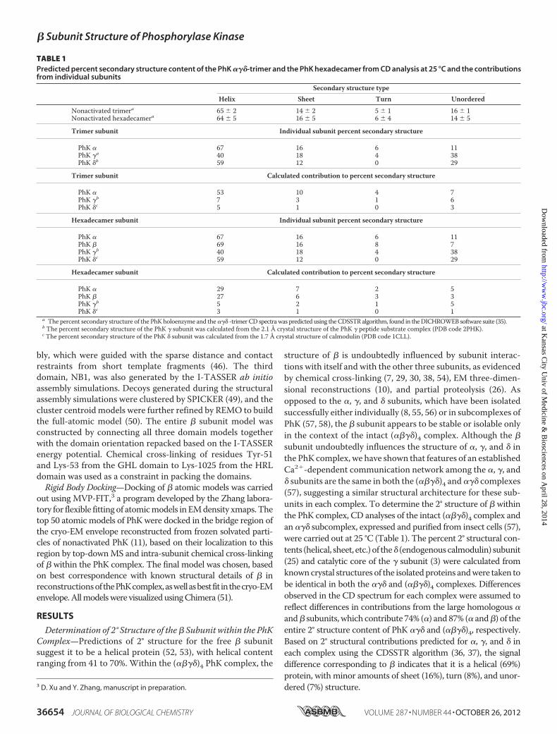
Determination of 2° Structure of the� Subunit within the PhKComplex—Predictions of 2° structure for the free � subunitsuggest it to be a helical protein (52, 53), with helical contentranging from 41 to 70%. Within the (����)4 PhK complex, the
structure of � is undoubtedly influenced by subunit interac-tions with itself and with the other three subunits, as evidencedby chemical cross-linking (7, 29, 30, 38, 54), EM three-dimen-sional reconstructions (10), and partial proteolysis (26). Asopposed to the �, �, and � subunits, which have been isolatedsuccessfully either individually (8, 55, 56) or in subcomplexes ofPhK (57, 58), the � subunit appears to be stable or isolable onlyin the context of the intact (����)4 complex. Although the �
subunit undoubtedly influences the structure of �, �, and � inthe PhK complex, we have shown that features of an establishedCa2�-dependent communication network among the �, �, and� subunits are the same in both the (����)4 and��� complexes(57), suggesting a similar structural architecture for these sub-units in each complex. To determine the 2° structure of � withinthe PhK complex, CD analyses of the intact (����)4 complex andan ��� subcomplex, expressed and purified from insect cells (57),were carried out at 25 °C (Table 1). The percent 2° structural con-tents (helical, sheet, etc.)of the� (endogenouscalmodulin) subunit(25) and catalytic core of the � subunit (3) were calculated fromknowncrystal structures of the isolatedproteins andwere taken tobe identical in both the ��� and (����)4 complexes. Differencesobserved in the CD spectrum for each complex were assumed toreflect differences in contributions from the large homologous �
and� subunits,which contribute 74% (�) and87% (� and�) of theentire 2° structure content of PhK ��� and (����)4, respectively.Based on 2° structural contributions predicted for �, �, and � ineach complex using the CDSSTR algorithm (36, 37), the signaldifference corresponding to � indicates that it is a helical (69%)protein, withminor amounts of sheet (16%), turn (8%), and unor-dered (7%) structure.3 D. Xu and Y. Zhang, manuscript in preparation.
TABLE 1Predicted percent secondary structure content of the PhK ���-trimer and the PhK hexadecamer from CD analysis at 25 °C and the contributionsfrom individual subunits
Secondary structure typeHelix Sheet Turn Unordered
Nonactivated trimera 65 � 2 14 � 2 5 � 1 16 � 1Nonactivated hexadecamera 64 � 5 16 � 5 6 � 4 14 � 5
Trimer subunit Individual subunit percent secondary structure
PhK � 67 16 6 11PhK �a 40 18 4 38PhK �b 59 12 0 29
Trimer subunit Calculated contribution to percent secondary structure
PhK � 53 10 4 7PhK �b 7 3 1 6PhK �c 5 1 0 3
Hexadecamer subunit Individual subunit percent secondary structure
PhK � 67 16 6 11PhK � 69 16 8 7PhK �b 40 18 4 38PhK �c 59 12 0 29
Hexadecamer subunit Calculated contribution to percent secondary structure
PhK � 29 7 2 5PhK � 27 6 3 3PhK �b 5 2 1 5PhK �c 3 1 0 1
a The percent secondary structure of the PhK holoenzyme and the ��� -trimerCD spectrawas predicted using theCDSSTR algorithm, found in theDICHROWEB software suite (35).b The percent secondary structure of the PhK � subunit was calculated from the 2.1 Šcrystal structure of the PhK � peptide substrate complex (PDB code 2PHK).c The percent secondary structure of the PhK � subunit was calculated from the 1.7 Šcrystal structure of calmodulin (PDB code 1CLL).
� Subunit Structure of Phosphorylase Kinase
36654 JOURNAL OF BIOLOGICAL CHEMISTRY VOLUME 287 • NUMBER 44 • OCTOBER 26, 2012
at Kansas C
ity Univ of M
edicine & B
iosciences on April 28, 2014
http://ww
w.jbc.org/
Dow
nloaded from
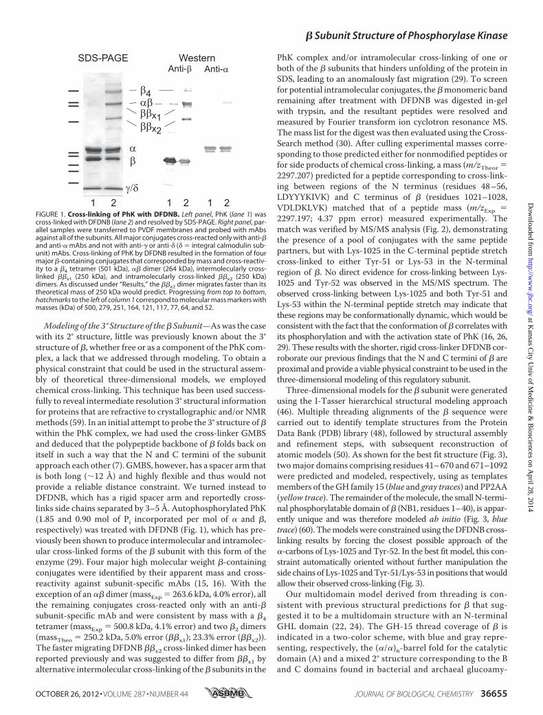
Modeling of the 3° Structure of the� Subunit—Aswas the casewith its 2° structure, little was previously known about the 3°structure of �, whether free or as a component of the PhK com-plex, a lack that we addressed through modeling. To obtain aphysical constraint that could be used in the structural assem-bly of theoretical three-dimensional models, we employedchemical cross-linking. This technique has been used success-fully to reveal intermediate resolution 3° structural informationfor proteins that are refractive to crystallographic and/or NMRmethods (59). In an initial attempt to probe the 3° structure of�within the PhK complex, we had used the cross-linker GMBSand deduced that the polypeptide backbone of � folds back onitself in such a way that the N and C termini of the subunitapproach each other (7). GMBS, however, has a spacer arm thatis both long (�12 Å) and highly flexible and thus would notprovide a reliable distance constraint. We turned instead toDFDNB, which has a rigid spacer arm and reportedly cross-links side chains separated by 3–5 Å. Autophosphorylated PhK(1.85 and 0.90 mol of Pi incorporated per mol of � and �,respectively) was treated with DFDNB (Fig. 1), which has pre-viously been shown to produce intermolecular and intramolec-ular cross-linked forms of the � subunit with this form of theenzyme (29). Four major high molecular weight �-containingconjugates were identified by their apparent mass and cross-reactivity against subunit-specific mAbs (15, 16). With theexception of an �� dimer (massExp � 263.6 kDa, 4.0% error), allthe remaining conjugates cross-reacted only with an anti-�subunit-specific mAb and were consistent by mass with a �4tetramer (massExp � 500.8 kDa, 4.1% error) and two �2 dimers(massTheo � 250.2 kDa, 5.0% error (��x1); 23.3% error (��x2)).The faster migrating DFDNB ��x2 cross-linked dimer has beenreported previously and was suggested to differ from ��x1 byalternative intermolecular cross-linking of the� subunits in the
PhK complex and/or intramolecular cross-linking of one orboth of the � subunits that hinders unfolding of the protein inSDS, leading to an anomalously fast migration (29). To screenfor potential intramolecular conjugates, the�monomeric bandremaining after treatment with DFDNB was digested in-gelwith trypsin, and the resultant peptides were resolved andmeasured by Fourier transform ion cyclotron resonance MS.Themass list for the digest was then evaluated using the Cross-Search method (30). After culling experimental masses corre-sponding to those predicted either for nonmodified peptides orfor side products of chemical cross-linking, a mass (m/zTheor �2297.207) predicted for a peptide corresponding to cross-link-ing between regions of the N terminus (residues 48–56,LDYYYKIVK) and C terminus of � (residues 1021–1028,VDLDKLVK) matched that of a peptide mass (m/zExp �2297.197; 4.37 ppm error) measured experimentally. Thematch was verified by MS/MS analysis (Fig. 2), demonstratingthe presence of a pool of conjugates with the same peptidepartners, but with Lys-1025 in the C-terminal peptide stretchcross-linked to either Tyr-51 or Lys-53 in the N-terminalregion of �. No direct evidence for cross-linking between Lys-1025 and Tyr-52 was observed in the MS/MS spectrum. Theobserved cross-linking between Lys-1025 and both Tyr-51 andLys-53 within the N-terminal peptide stretch may indicate thatthese regions may be conformationally dynamic, which would beconsistentwith the fact that the conformation of� correlates withits phosphorylation and with the activation state of PhK (16, 26,29). These results with the shorter, rigid cross-linkerDFDNB cor-roborate our previous findings that the N and C termini of � areproximal and provide a viable physical constraint to be used in thethree-dimensional modeling of this regulatory subunit.Three-dimensional models for the � subunit were generated
using the I-Tasser hierarchical structural modeling approach(46). Multiple threading alignments of the � sequence werecarried out to identify template structures from the ProteinData Bank (PDB) library (48), followed by structural assemblyand refinement steps, with subsequent reconstruction ofatomic models (50). As shown for the best fit structure (Fig. 3),twomajor domains comprising residues 41–670 and 671–1092were predicted and modeled, respectively, using as templatesmembers of theGH family 15 (blue and gray traces) and PP2AA(yellow trace). The remainder of themolecule, the smallN-termi-nal phosphorylatable domain of� (NB1, residues 1–40), is appar-ently unique and was therefore modeled ab initio (Fig. 3, bluetrace) (60). Themodelswere constrained using theDFDNBcross-linking results by forcing the closest possible approach of the�-carbons of Lys-1025 and Tyr-52. In the best fit model, this con-straint automatically oriented without further manipulation theside chains of Lys-1025 andTyr-51/Lys-53 in positions thatwouldallow their observed cross-linking (Fig. 3).Our multidomain model derived from threading is con-
sistent with previous structural predictions for � that sug-gested it to be a multidomain structure with an N-terminalGHL domain (22, 24). The GH-15 thread coverage of � isindicated in a two-color scheme, with blue and gray repre-senting, respectively, the (�/�)6-barrel fold for the catalyticdomain (A) and a mixed 2° structure corresponding to the Band C domains found in bacterial and archaeal glucoamy-
FIGURE 1. Cross-linking of PhK with DFDNB. Left panel, PhK (lane 1) wascross-linked with DFDNB (lane 2) and resolved by SDS-PAGE. Right panel, par-allel samples were transferred to PVDF membranes and probed with mAbsagainst all of the subunits. All major conjugates cross-reacted only with anti-�and anti-� mAbs and not with anti-� or anti-� (� � integral calmodulin sub-unit) mAbs. Cross-linking of PhK by DFDNB resulted in the formation of fourmajor �-containing conjugates that corresponded by mass and cross-reactiv-ity to a �4 tetramer (501 kDa), �� dimer (264 kDa), intermolecularly cross-linked ��x1 (250 kDa), and intramolecularly cross-linked ��x2 (250 kDa)dimers. As discussed under “Results,” the ��x2 dimer migrates faster than itstheoretical mass of 250 kDa would predict. Progressing from top to bottom,hatchmarks to the left of column 1 correspond to molecular mass markers withmasses (kDa) of 500, 279, 251, 164, 121, 117, 77, 64, and 52.
� Subunit Structure of Phosphorylase Kinase
OCTOBER 26, 2012 • VOLUME 287 • NUMBER 44 JOURNAL OF BIOLOGICAL CHEMISTRY 36655
at Kansas C
ity Univ of M
edicine & B
iosciences on April 28, 2014
http://ww
w.jbc.org/
Dow
nloaded from

lases and glucodextranases (61). The protein structurallyclosest to the best model of this region is Anthrobacter glo-biformus glucodextranase (PDB code 1ULV) (61), with atemplate modeling score of 0.8462 and root mean squaredeviation of 1.92 Å, indicating a good topographical match(62). The crystal structure of the glucodextranase templaterevealed the protein in complex with the pseudo tetrasac-charide acarbose, a potent transition state inhibitor of glu-coamylases (63). Correspondingly, we recently showed thatPhK binds acarbose, which in turn promotes a conforma-tional change in the � subunit (64). The large C-terminal
domain of the � model was a relatively good topographicalmatch (template modeling score � 0.6272; root mean squaredeviation � 5.41) with PP2AA (PDB code 1B3U), and thepredominantly helical HEAT repeat structure correspondedwell with the helical content estimated for � by CDmeasure-ments (65), revealing an overall helical topology for �.Determination of the 4° Structure of the � Subunits within the
PhK Complex—Having analyzed the 2° and 3° structures ofPhK�, we set out to determine the arrangement of these sub-units in the 4° structure of PhK. Both cryo-EM and SAXS anal-yses of the native (����)4 PhK complex show it to be a large
FIGURE 2. MS/MS analysis of the signal at m/z 1149.102 identifying a conjugate comprising residues 48 –56 and 1021–1028 of the regulatory � subunitof PhK. The composition of the ions identifying the cross-linked peptide and chemical structure of the cross-link are shown. Lowercase letters denote ionsarising from amide cleavages of the peptide backbone and are color-coded for each peptide in the conjugate pair (black for residues 48 –56 and green for1021–1028). Intact covalent links formed between peptide fragments are indicated by a forward slash (/). For singly charged ions, it should be noted that oneof the two peptide ions covalently attached to either position of the ring is a neutral product of the indicated backbone amide cleavage. Lines bisecting thebonds between the ring carbons and NO2 nitrogens in the cross-link structure represent the loss of nitro groups, indicated in several fragment ions (40, 41).Heavy black bars between each peptide indicate residues cross-linked and are consistent with a pool of peptides, containing either Lys-1025/Tyr-51 orLys-1025/Lys-53 cross-linked side chains.
FIGURE 3. Theoretical three-dimensional model of the PhK � subunit. Hierarchical protein structural modeling of the � subunit was carried out usingI-TASSER (46). X-ray crystal structures of Aspergillus awamori glucoamylase (blue and gray ribbon traces) and human PP2A PR65/A subunit (yellow trace) wereused to thread, respectively, residues 41– 670 and 671–1092 of the multidomain � subunit primary sequence (61, 65). The remaining N-terminal residues (1– 40,blue trace) were modeled ab initio using QUARK (60). Models were constrained using the DFDNB cross-linking results, forcing the approach of the Lys-1025 andTyr-52 �-carbons. Side chains of the DFDNB cross-linked residues (red) and the distances between the Lys-1025/Lys-51 and Lys-1025/Lys-53 cross-linked pairs(arrows) are indicated in a magnified view of the cross-link site linking the N and C termini of �.
� Subunit Structure of Phosphorylase Kinase
36656 JOURNAL OF BIOLOGICAL CHEMISTRY VOLUME 287 • NUMBER 44 • OCTOBER 26, 2012
at Kansas C
ity Univ of M
edicine & B
iosciences on April 28, 2014
http://ww
w.jbc.org/
Dow
nloaded from
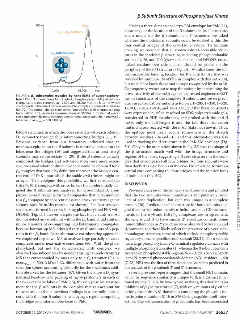
bilobal structure, inwhich the lobes associatewith each other inD2 symmetry through four interconnecting bridges (11, 19).Previous evidence from our laboratory indicated that anunknown epitope on the � subunit is centrally located on thelobes near the bridges (16) and suggested that at least two �subunits may self-associate (7, 29). If the � subunits actuallycomposed the bridges and self-association were more exten-sive, we asked whether direct evidence could be obtained for a�4 complex that would by definition represent the bridged cen-tral core of PhK upon which the stable ��� trimers might bearrayed. To investigate this possibility, we first screened the(����)4 PhK complex with cross-linkers that preferentially tar-geted the � subunits and analyzed for cross-linked �4 com-plexes. Several reagents formed conjugates that correspondedto a �4 conjugate by apparent mass and cross-reactivity againstsubunit-specific mAbs (results not shown). The best resolvedspecies was formed by cross-linking phosphorylated PhK withDFDNB (Fig. 1); however, despite the fact that an anti-� mAbdid not detect any � subunit within the �4 band, it did containminor amounts of co-migrating �/� heteromeric conjugates,because bottom-upMS indicated very small amounts of � pep-tides in the �4 band. As an alternative corroborating approach,we employed top-down MS to analyze large partially solvatedcomplexes under near-native conditions (66). With the phos-phorylated, but not the nonactivated, PhK complex, weobserved one subcomplex by nondenaturing nano-electrosprayMS that corresponded by mass only to a �4 tetramer (Fig. 4;massTheo � 500. 3 kDa; 1.3% difference), with water from thesolvation sphere accounting primarily for the small mass addi-tion observed for the tetramer (67). Given the known D2 sym-metrical head-to-head packing of ���� protomers in each ofthe two octameric lobes of PhK (15), the only possible arrange-ment for the � subunits in the complex that can account forthese results and our previous findings is a centrally locatedcore, with the four � subunits occupying a region comprisingthe bridges and internal lobe-faces of PhK.
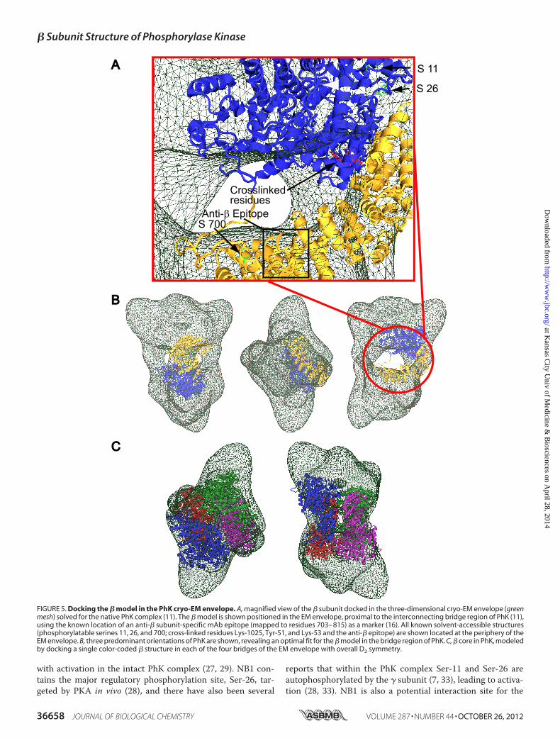
Having a three-dimensional cryo-EM envelope for PhK (11),knowledge of the location of the � subunits in its 4° structure,and a model for the � subunit in it 3° structure, we askedwhether the modeled � subunits could be docked within thefour central bridges of the cryo-EM envelope. To facilitatedocking, we reasoned that all known solvent-accessible struc-tures in the modeled � structure, including phosphorylatableserines 11, 26, and 700 (green side chains) and DFDNB-cross-linked residues (red side chains), should be placed on theperiphery of the EM structure (Fig. 5A). We also knew the sol-vent-accessible binding location for the anti-� mAb that wasrevealed by immuno-EMof PhK in complexwith thismAb (16),but we did not know the actual epitope recognized by themAb.Consequently, we set out tomap the epitope by determining thecross-reactivity of the mAb against expressed engineered GSTfusion constructs of the complete � subunit and seven previ-ously used truncationmutants as follows: 1–285, 1–350, 1–520,1–703, 1–815, 1–916, and 33–1092 (7). After these constructswere expressed, purified, resolved on SDS-polyacrylamide gels,transferred to PDF membranes, and probed with the anti-�mAb, only the full-length � and the last three truncationmutants cross-reacted with the mAb (data not shown). Thus,the epitope most likely occurs somewhere in the stretchbetween residues 704 and 815, and this information was alsoused in docking the � structure in the PhK EM envelope (Fig.5A). Only in the orientation shown in Fig. 5B does the shape ofthe � structure match well with the bridge structure andregions of the lobes, suggesting a � core structure in the com-plex that encompasses all four bridges. All four subunits werethen docked as rigid bodies in the cryo-EM envelope, forming acentral core comprising the four bridges and the interior facesof both lobes (Fig. 5C).
DISCUSSION
Previous analyses of the primary structures of � and � notedthat the two subunits were homologous and putatively prod-ucts of gene duplication, but each was unique as a completeprotein (20). Predictions of 2° structure for both subunits sug-gest them to be predominantly �-helical, and our CDmeasure-ments of the ��� and (����)4 complexes are in agreement,showing � and � to have similar 2° structure content. Someminor differences in 2° structure were observed between � and�, however, and these likely reflect the presence of several non-homologous stretches, some of which include phosphorylatableregulatorydomains specific toeachsubunit (20, 21).The� subunithas a large phosphorylatable C-terminal regulatory domain withmultiple phosphorylation sites (1), whereas the� subunit containstwo known phosphorylatable regions, Ser-700 plus Ser-11/Ser-26in theN-terminal phosphorylatable domain (NB1, residues 1–40)(7, 28). NB1 was the first of three functional domains predicted inour analysis of the � subunit 2° and 3° structures.
Several previous reports suggest that the small NB1 domain,which by sequence similarity is unique to �, is a distinct func-tional moiety (7, 20). By two-hybrid analyses, this domain is aninhibitor of �-� dimerization (7), with only mutants of � eitherlacking the entire NB1 domain or containing the phospho-mi-metic pointmutations S11E or S26E being capable of self-inter-action. The self-association of � subunits has been associated
FIGURE 4. �4 subcomplex revealed by nano-ESMS of autophosphory-lated PhK. Nondenaturing MS of intact phosphorylated PhK yielded twocharge state series centered at 12,500 and 16,000 m/z, the latter of whichcorresponds to the intact hexadecameric PhK complex (discussed in detail inRef. 72). The former charge state series (blue circles), with charges rangingfrom �46 to �50, yielded a measured mass of 507,062 � 55 Da that was inclose agreement by mass with only one combination of subunits, namely a �4tetramer (massTheo � 500,338 Da).
� Subunit Structure of Phosphorylase Kinase
OCTOBER 26, 2012 • VOLUME 287 • NUMBER 44 JOURNAL OF BIOLOGICAL CHEMISTRY 36657
at Kansas C
ity Univ of M
edicine & B
iosciences on April 28, 2014
http://ww
w.jbc.org/
Dow
nloaded from
with activation in the intact PhK complex (27, 29). NB1 con-tains the major regulatory phosphorylation site, Ser-26, tar-geted by PKA in vivo (28), and there have also been several
reports that within the PhK complex Ser-11 and Ser-26 areautophosphorylated by the � subunit (7, 33), leading to activa-tion (28, 33). NB1 is also a potential interaction site for the
FIGURE 5. Docking the � model in the PhK cryo-EM envelope. A, magnified view of the � subunit docked in the three-dimensional cryo-EM envelope (greenmesh) solved for the native PhK complex (11). The � model is shown positioned in the EM envelope, proximal to the interconnecting bridge region of PhK (11),using the known location of an anti-� subunit-specific mAb epitope (mapped to residues 703– 815) as a marker (16). All known solvent-accessible structures(phosphorylatable serines 11, 26, and 700; cross-linked residues Lys-1025, Tyr-51, and Lys-53 and the anti-� epitope) are shown located at the periphery of theEM envelope. B, three predominant orientations of PhK are shown, revealing an optimal fit for the � model in the bridge region of PhK. C, � core in PhK, modeledby docking a single color-coded � structure in each of the four bridges of the EM envelope with overall D2 symmetry.
� Subunit Structure of Phosphorylase Kinase
36658 JOURNAL OF BIOLOGICAL CHEMISTRY VOLUME 287 • NUMBER 44 • OCTOBER 26, 2012
at Kansas C
ity Univ of M
edicine & B
iosciences on April 28, 2014
http://ww
w.jbc.org/
Dow
nloaded from
intrinsic calmodulin (�) subunit, as a synthetic peptide corre-sponding to residues 5–28 of NB1 inhibited active forms of thePhK complex and had nanomolar affinity for free calmodulin(68). We also previously demonstrated by cross-linking thatArg-18 of NB1 is proximal (�12 Å) to the �CRD (7), whichbinds the � subunit within the PhK complex (4, 5). Based on itshigh affinity for calmodulin, this region of NB1 (residues 5–28)was suggested to have the characteristic amphipathic �-helicalstructure of many known calmodulin targets (68), an architec-ture that is also observed in our current ab initiomodel of thisdomain. The proximity ofNB1 to� in the PhK complex indicatedby cross-linking and by its phosphorylation mandated that thisregion be surface-accessible and proximal to the position of � intheEMstructureof the (����)4 complex.The localizationof�wasachievedbymapping two-dimensional EMimages of PhK in com-plex with an anti-�mAb directed against the C-terminal lobe of �(16). An optimal fit of the � subunit model in the bridge regionoccurred when satisfying the above conditions.Aside from the small NB1 domain, the remaining 93% of the
1092-residue � subunit structure was covered by templatesfrom two distinct protein families. The first of these, theGH-15family of glycosyl hydrolases, provided several good templatematches that extended from residues 41 to 670. These resultsdiffered from two previous reports that predicted only theGH-15 (�/�)6 catalytic core to be present in � (22, 24), extend-ing approximately through its first 480 residues (22). Just C-ter-minal to the GH-15 catalytic core, Carriere et al. (23) predicteda small loop followed by an additional domain (termed B),which extended from residues 492 to 676; however, they wereunable to achieve fold recognition for this domain.OurGH foldrecognition extended throughout this region and correspondedto the � GH-15 template archaeal glucodextranase B and Cdomains, which show homology with proteins containing theimmunoglobulin folds and carbohydrate-binding domains ofseveral glycosidases, respectively (61). An independent analysisof the primary structure of � using the Pfam database also pre-dicted a GHL fold for this region of the subunit (69). Our pre-dicted domain structures for residues 40–670 of the � subunitmirrored that of the shorter domain predicted by Carriere et al.(23) and are topologically similar to several domains observedin the template glucodextranase crystal structure (61), com-prising an (�/�)6 catalytic core connected to the B and Cdomains by a short loop (61).In addition to threading, we used the constraint from intra-
molecular cross-linkingwithDFDNB to influence the structureof the � model by positioning the GHL domain proximal to the� C terminus (Fig. 3). In the absence of this constraint, the top100 models generated for the � subunit did not complementwell the shape of the PhK cryo-EMenvelopewhenpositioned inall possible orientations in the central bridge region of the mol-ecule. We assume that cross-linking of the � subunit in theintact complex captures a conformation of this subunit that isinduced by its interactions with all subunits, including itself, inthe 4° structure of PhK. In further support of the use of thiscross-linking constraint, we recently demonstrated that PhKbinds the glucoamylase inhibitor acarbose and that this bindingperturbs intramolecular cross-linking of the � subunit byGMBS (64), which cross-links the GHL domain to the C termi-
nus of the � subunit (30). The direct binding of acarbose by the� GHL domains would allow a potential mechanism for induc-ing a conformational change in this subunit through alteringpotential interactions between the predicted � GHL and theHRL domains, consistent with results from the intramolecularcross-linking of� byDFDNB.Acarbose also stimulates the pro-tein kinase activity of PhK � (64), and functionally it mimicsglycogen, which also binds to PhK, activating the catalytic �subunit (9). It must be cautioned, however, that because thehomologous � and � subunits are both predicted to containGHL domains, we cannot rule out the possibility that effects ofacarbose and glycogen on the PhK � and � subunits are medi-ated by the binding of these ligands to the � subunit.Other potential functions for the � subunit have been sug-
gested frommodels of its C terminus from residues 671 to 1092.From comparing sequence alignments of the � and � subunitsfrom diverse species, Carriere et al. (23) predicted for thisregion of � (as well as the homologous �) two structurallyrelated�-helical domains as follows: C (residues 711–915) withno fold recognition, andD (residues 915–1092)with a calcineu-rin B-like (CBL) fold. The CBL fold, identified by hydrophobiccluster analysis (23), is typical of Ca2�-binding proteins con-taining two globular EF-handmotifs (two perpendicular �-hel-ices tethered by a loop) that are connected by a flexible linker(70). Based on observed chemical cross-linking between the �subunit CBL domain and the catalytic � subunit (6, 38), thelatter of which also binds the EF-hand endogenous CaM (�)subunit (5), it was proposed that theCBLdomain targets knownCaM-binding sites on � and/or those predicted for the � and �subunits (23). Although we did not observe a CBL domain on �by threading, the predicted structural relatedness of domains Cand D and their predominantly �-helical structure are consist-ent with our modeling of this region of the subunit as a singledomain using the template PP2AA subunit. Crystal structureanalysis of PP2AA revealed a molecule in which 15 tandemHEAT repeats (motifs comprising a pair of anti-parallel �-hel-ices) formed a left-handed superhelical structure (65). HEAT-repeat domains have been shown to mediate protein-proteininteractions in several proteins, including the PP2AA subunit(65). PP2AA tightly associates with the catalytic PP2A subunitto form a heterodimeric scaffold for binding diverse regulatorysubunits that direct the functional phosphatase to specific sub-cellular sites (71). If present in the � subunit, the HRL domainmay function similarly in the context of the PhK complex,forming a scaffold with the GHL domain that promotes inter-actions with either the catalytic � subunit or other regulatory �subunits. Such interactions are consistentwith the formation of�� and �� dimers by cross-linking PhK with DFDNB andGMBS (29, 30), with the observed structural coupling between� and � in the intact PhK complex (16), and with two-hybridanalyses of � subunit truncation mutants, which demonstratedthat the C-terminal half of the HRL domain is required forself-association of the � subunits (7). In addition to a potentialcorrespondence in function, the predominantly helical HEAT-repeat structure agreed well with the 2° structural content esti-mated for the � subunit and complemented the shape of thebridge structure and interior lobe face after docking � into thePhK cryo-EM envelope.
� Subunit Structure of Phosphorylase Kinase
OCTOBER 26, 2012 • VOLUME 287 • NUMBER 44 JOURNAL OF BIOLOGICAL CHEMISTRY 36659
at Kansas C
ity Univ of M
edicine & B
iosciences on April 28, 2014
http://ww
w.jbc.org/
Dow
nloaded from
The first direct evidence for a �4 subcomplex in the (����)4PhK complex revealed herein by top-down MS and inter-sub-unit cross-linking of the � subunits by DFDNB provided therationale for docking these subunits in the central connectingbridge region of the PhK cryo-EM structure. This arrangementof the � subunits is consistent with the observed head-to-headpacking of ���� protomers in D2 symmetry (15), whichrequires any homotetrameric association of subunits to occurat known points of contact between the lobes, i.e. centrally inthe complex (Fig. 5). Additionally, our direct results correspondto several reports indirectly linking the � subunits to a centrallocation of the complex by partial proteolysis and EM immu-nolocalization (16, 17). Taken together, the results herein indi-cate the presence of a �4 core in PhK and suggest a structuralrole for these subunits as a scaffold upon which the �, �, and �subunits are arrayed. It is likely that the � subunits position thelobes, and thus the catalytic � subunits, differently with respectto one another in nonactivated and activated conformers of PhK.For example, we have demonstrated that the conformation of the� subunits correlates with activation of PhK by several mecha-nisms (10, 26, 29), that the � and � subunits are structurally cou-pled to one another during enzyme activation (16), and thatchanges in the dihedral angles between the lobes and alteredbridge structures are readily observed between nonactivated andCa2�-activated forms of PhK by EM and SAXS analyses (10, 19).
REFERENCES1. Brushia, R. J., and Walsh, D. A. (1999) Phosphorylase kinase. The com-
plexity of its regulation is reflected in the complexity of its structure. Front.Biosci. 4, D618–641
2. Lowe, E. D., Noble, M. E., Skamnaki, V. T., Oikonomakos, N. G., Owen,D. J., and Johnson, L. N. (1997) The crystal structure of a phosphorylasekinase peptide substrate complex. Kinase substrate recognition. EMBO J.16, 6646–6658
3. Owen,D. J., Noble,M. E., Garman, E. F., Papageorgiou, A. C., and Johnson,L. N. (1995) Two structures of the catalytic domain of phosphorylasekinase. An active protein kinase complexed with substrate analogue andproduct. Structure 3, 467–482
4. Dasgupta, M., Honeycutt, T., and Blumenthal, D. K. (1989) The �-subunitof skeletal muscle phosphorylase kinase contains two noncontiguousdomains that act in concert to bind calmodulin. J. Biol. Chem. 264,17156–17163
5. Jeyasingham, M. D., Artigues, A., Nadeau, O. W., and Carlson, G. M.(2008) Structural evidence for co-evolution of the regulation of contrac-tion and energy production in skeletal muscle. J. Mol. Biol. 377, 623–629
6. Rice, N. A., Nadeau, O. W., Yang, Q., and Carlson, G. M. (2002) Thecalmodulin-binding domain of the catalytic � subunit of phosphorylasekinase interacts with its inhibitory� subunit. Evidence for aCa2� sensitivenetwork of quaternary interactions. J. Biol. Chem. 277, 14681–14687
7. Nadeau, O. W., Anderson, D. W., Yang, Q., Artigues, A., Paschall, J. E.,Wyckoff, G. J., McClintock, J. L., and Carlson, G. M. (2007) Evidence forthe location of the allosteric activation switch in the multisubunit phos-phorylase kinase complex from mass spectrometric identification ofchemically cross-linked peptides. J. Mol. Biol. 365, 1429–1445
8. Paudel, H. K., and Carlson, G. M. (1987) Inhibition of the catalyticsubunit of phosphorylase kinase by its �/� subunits. J. Biol. Chem. 262,11912–11915
9. Krebs, E. G., Love, D. S., Bratvold, G. E., Trayser, K. A., Meyer, W. L., andFischer, E. H. (1964) Purification and properties of rabbit skeletal musclephosphorylase B kinase. Biochemistry 3, 1022–1033
10. Nadeau, O.W., Carlson, G.M., andGogol, E. P. (2002) ACa2�-dependentglobal conformational change in the three-dimensional structure of phos-phorylase kinase obtained from electronmicroscopy. Structure 10, 23–32
11. Nadeau, O. W., Gogol, E. P., and Carlson, G. M. (2005) Cryoelectronmicroscopy reveals new features in the three-dimensional structure ofphosphorylase kinase. Protein Sci. 14, 914–920
12. Venien-Bryan, C., Lowe, E. M., Boisset, N., Traxler, K. W., Johnson, L. N.,and Carlson, G. M. (2002) Three-dimensional structure of phosphorylasekinase at 22-Å resolution and its complex with glycogen phosphorylase b.Structure 10, 33–41
13. Norcum, M. T., Wilkinson, D. A., Carlson, M. C., Hainfeld, J. F., andCarlson, G. M. (1994) Structure of phosphorylase kinase. A three-dimen-sional model derived from stained and unstained electron micrographs. J.Mol. Biol. 241, 94–102
14. Traxler, K. W., Norcum, M. T., Hainfeld, J. F., and Carlson, G. M. (2001)Direct visualization of the calmodulin subunit of phosphorylase kinase viaelectron microscopy following subunit exchange. J. Struct. Biol. 135,231–238
15. Wilkinson, D. A., Marion, T. N., Tillman, D.M., Norcum,M. T., Hainfeld,J. F., Seyer, J. M., and Carlson, G. M. (1994) An epitope proximal to thecarboxyl terminus of the �-subunit is located near the lobe tips of thephosphorylase kinase hexadecamer. J. Mol. Biol. 235, 974–982
16. Wilkinson, D. A., Norcum, M. T., Fizgerald, T. J., Marion, T. N., Tillman,D. M., and Carlson, G. M. (1997) Proximal regions of the catalytic � andregulatory � subunits on the interior lobe face of phosphorylase kinase arestructurally coupled to each other and with enzyme activation. J. Mol. Biol.265, 319–329
17. Trempe, M. R., Carlson, G. M., Hainfeld, J. F., Furcinitti, P. S., and Wall,J. S. (1986) Analyses of phosphorylase kinase by transmission and scan-ning transmission electron microscopy. J. Biol. Chem. 261, 2882–2889
18. Venien-Bryan, C., Jonic, S., Skamnaki, V., Brown,N., Bischler, N., Oikono-makos, N. G., Boisset, N., and Johnson, L. N. (2009) The structure ofphosphorylase kinase holoenzyme at 9.9 angstroms resolution and loca-tion of the catalytic subunit and the substrate glycogen phosphorylase.Structure 17, 117–127
19. Priddy, T. S., MacDonald, B. A., Heller, W. T., Nadeau, O. W., Trewhella, J.,andCarlson,G.M. (2005)Ca2�-induced structural changes inphosphorylasekinase detected by small angle x-ray scattering. Protein Sci. 14, 1039–1048
20. Kilimann, M. W., Zander, N. F., Kuhn, C. C., Crabb, J. W., Meyer, H. E.,and Heilmeyer, L. M., Jr. (1988) The � and � subunits of phosphorylasekinase are homologous. cDNA cloning and primary structure of the �
subunit. Proc. Natl. Acad. Sci. U.S.A. 85, 9381–938521. Zander, N. F., Meyer, H. E., Hoffmann-Posorske, E., Crabb, J. W., Hei-
lmeyer, L. M., Jr., and Kilimann, M. W. (1988) cDNA cloning and com-plete primary structure of skeletal muscle phosphorylase kinase (� sub-unit). Proc. Natl. Acad. Sci. U.S.A. 85, 2929–2933
22. Carriere, C., Jonic, S.,Mornon, J. P., andCallebaut, I. (2008) Three-dimen-sional mapping of glycogenosis-causing mutations in the large regulatory� subunit of phosphorylase kinase.Biochim. Biophys. Acta1782, 664–670
23. Carriere, C., Mornon, J. P., Venien-Bryan, C., Boisset, N., and Callebaut, I.(2008) Calcineurin B-like domains in the large regulatory �/� subunits ofphosphorylase kinase. Proteins 71, 1597–1606
24. Pallen,M. J. (2003)Glucoamylase-like domains in the�- and�-subunits ofphosphorylase kinase. Protein Sci. 12, 1804–1807
25. Chattopadhyaya, R., Meador, W. E., Means, A. R., and Quiocho, F. A.(1992) Calmodulin structure refined at 1.7 Å resolution. J. Mol. Biol. 228,1177–1192
26. Trempe,M. R., and Carlson, G.M. (1987) Phosphorylase kinase conform-ers. Detection by proteases. J. Biol. Chem. 262, 4333–4340
27. Cheng, A., Fitzgerald, T. J., and Carlson, G. M. (1985) Adenosine 5�-diphosphate as an allosteric effector of phosphorylase kinase from rabbitskeletal muscle. J. Biol. Chem. 260, 2535–2542
28. Cohen, P., Watson, D. C., and Dixon, G. H. (1975) The hormonal controlof activity of skeletal muscle phosphorylase kinase. Amino acid sequencesat the two sites of action of adenosine-3�:5�-monophosphate-dependentprotein kinase. Eur. J. Biochem. 51, 79–92
29. Fitzgerald, T. J., and Carlson, G. M. (1984) Activated states of phos-phorylase kinase as detected by the chemical cross-linker 1,5-difluoro-2,4-dinitrobenzene. J. Biol. Chem. 259, 3266–3274
30. Nadeau, O. W., Wyckoff, G. J., Paschall, J. E., Artigues, A., Sage, J., Villar,M. T., and Carlson, G. M. (2008) CrossSearch, a user-friendly search en-
� Subunit Structure of Phosphorylase Kinase
36660 JOURNAL OF BIOLOGICAL CHEMISTRY VOLUME 287 • NUMBER 44 • OCTOBER 26, 2012
at Kansas C
ity Univ of M
edicine & B
iosciences on April 28, 2014
http://ww
w.jbc.org/
Dow
nloaded from
gine for detecting chemically cross-linked peptides in conjugated proteins.Mol. Cell. Proteomics 7, 739–749
31. King,M.M., and Carlson, G.M. (1981) Synergistic activation by Ca2� andMg2� as the primary cause for hysteresis in the phosphorylase kinasereactions. J. Biol. Chem. 256, 11058–11064
32. Cohen, P. (1973) The subunit structure of rabbit skeletal muscle phospho-rylase kinase, and the molecular basis of its activation reactions. Eur.J. Biochem. 34, 1–14
33. King, M. M., Fitzgerald, T. J., and Carlson, G. M. (1983) Characterizationof initial autophosphorylation events in rabbit skeletal muscle phospho-rylase kinase. J. Biol. Chem. 258, 9925–9930
34. Priddy, T. S., Middaugh, C. R., and Carlson, G. M. (2007) Electrostaticchanges in phosphorylase kinase induced by its obligatory allosteric acti-vator Ca2�. Protein Sci. 16, 517–527
35. Lobley, A., Whitmore, L., and Wallace, B. A. (2002) DICHROWEB. Aninteractive website for the analysis of protein secondary structure fromcircular dichroism spectra. Bioinformatics 18, 211–212
36. Sreerama, N., andWoody, R.W. (1994) Protein secondary structure fromcircular dichroism spectroscopy. Combining variable selection principleand cluster analysis with neural network, ridge regression, and self-con-sistent methods. J. Mol. Biol. 242, 497–507
37. Sreerama, N., and Woody, R. W. (2000) Estimation of protein secondarystructure from circular dichroism spectra. Comparison of CONTIN, SEL-CON, and CDSSTR methods with an expanded reference set. Anal.Biochem. 287, 252–260
38. Nadeau, O. W., Traxler, K. W., Fee, L. R., Baldwin, B. A., and Carlson,G. M. (1999) Activators of phosphorylase kinase alter the cross-linking ofits catalytic subunit to theC-terminal one-sixth of its regulatory� subunit.Biochemistry 38, 2551–2559
39. Schilling, B., Row, R. H., Gibson, B. W., Guo, X., and Young, M. M. (2003)MS2Assign, automated assignment and nomenclature of tandem massspectra of chemically cross-linked peptides. J. Am. Soc.Mass Spectrom. 14,834–850
40. Brill, T. B., James, K. J., Chawla, R., Nicol, G. Shukla, A., and Futrell, J. H.(1999) Influence of the substituent on the major decomposition channelsof the NO2 group in para-substituted nitrobenzenes. A tandem massspectrometric study. J. Phys. Organic Chem. 12, 819–826
41. Sigman,M. E., and Clark, C. D. (2005) Two-dimensional correlation spec-troscopy techniques applied to ion trap tandemmass spectrometric anal-ysis. Nitroaromatics. Rapid Commun. Mass Spectrom. 19, 3731–3736
42. Chalkley, R. J., Hansen, K. C., and Baldwin, M. A. (2005) Bioinformaticmethods to exploit mass spectrometric data for proteomic applications.Methods Enzymol. 402, 289–312
43. Sobott, F., Hernandez, H., McCammon,M. G., Tito, M. A., and Robinson,C. V. (2002) A tandemmass spectrometer for improved transmission andanalysis of large macromolecular assemblies. Anal. Chem. 74, 1402–1407
44. Hernandez,H., andRobinson, C.V. (2007)Determining the stoichiometryand interactions of macromolecular assemblies from mass spectrometry.Nat. Protoc. 2, 715–726
45. Taverner, T., Hernandez, H., Sharon, M., Ruotolo, B. T., Matak-Vinkovic,D., Devos, D., Russell, R. B., and Robinson, C. V. (2008) Subunit architec-ture of intact protein complexes from mass spectrometry and homologymodeling. Acc. Chem. Res. 41, 617–627
46. Zhang, Y. (2008) I-TASSER server for protein three-dimensional struc-ture prediction. BMC Bioinformatics 9, 40
47. Wu, S., Skolnick, J., and Zhang, Y. (2007) Ab initio modeling of smallproteins by iterative TASSER simulations. BMC Biol. 5, 17
48. Wu, S., and Zhang, Y. (2007) LOMETS. A local meta-threading server forprotein structure prediction. Nucleic Acids Res. 35, 3375–3382
49. Zhang, Y., and Skolnick, J. (2004) SPICKER. A clustering approach toidentify near-native protein folds. J. Comput. Chem. 25, 865–871
50. Li, Y., and Zhang, Y. (2009) REMO. A new protocol to refine full atomicprotein models from C-� traces by optimizing hydrogen-bonding net-works. Proteins 76, 665–676
51. Pettersen, E. F.,Goddard,T.D.,Huang,C.C., Couch,G. S.,Greenblatt,D.M.,Meng, E. C., and Ferrin, T. E. (2004) UCSF Chimera. A visualization systemfor exploratory research and analysis. J. Comput. Chem. 25, 1605–1612
52. Jones, D. T. (1999) Protein secondary structure prediction based on posi-tion-specific scoring matrices. J. Mol. Biol. 292, 195–202
53. Kneller, D. G., Cohen, F. E., and Langridge, R. (1990) Improvements inprotein secondary structure prediction by an enhanced neural network. J.Mol. Biol. 214, 171–182
54. Nadeau, O. W., and Carlson, G. M. (1994) Zero length conformation-dependent cross-linking of phosphorylase kinase subunits by transglu-taminase. J. Biol. Chem. 269, 29670–29676
55. Teo, T. S.,Wang, T. H., andWang, J. H. (1973) Purification and propertiesof the protein activator of bovine heart cyclic adenosine 3�,5�-monophos-phate phosphodiesterase. J. Biol. Chem. 248, 588–595
56. Kee, S. M., and Graves, D. J. (1986) Isolation and properties of the active �subunit of phosphorylase kinase. J. Biol. Chem. 261, 4732–4737
57. Boulatnikov, I. G., Peters, J. L., Nadeau, O. W., Sage, J. M., Daniels, P. J.,Kumar, P., Walsh, D. A., and Carlson, G. M. (2009) Expressed phosphor-ylase b kinase and its ��� subcomplex as regulatory models for the rabbitskeletal muscle holoenzyme. Biochemistry 48, 10183–10191
58. Chan, K. F., and Graves, D. J. (1982) Isolation and physicochemical prop-erties of active complexes of rabbit muscle phosphorylase kinase. J. Biol.Chem. 257, 5939–5947
59. Nadeau, O.W., and Carlson, G. M. (2005) in Protein-Protein Interactions: AMolecular CloningManual (Golemis, E., and Adams, P. D., eds) 2nd Ed., pp.105–127, Cold Spring Harbor Laboratory Press, Cold Spring Harbor, NY
60. Xu, D., Zhang, J., Roy, A., and Zhang, Y. (2011) Automated protein struc-ture modeling in CASP9 by I-TASSER pipeline combined with QUARK-based ab initio folding and FG-MD-based structure refinement. Proteins79, Suppl. 10, 147–160
61. Mizuno, M., Tonozuka, T., Suzuki, S., Uotsu-Tomita, R., Kamitori, S., Ni-shikawa,A., andSakano,Y. (2004)Structural insights into substrate specificityand function of glucodextranase. J. Biol. Chem. 279, 10575–10583
62. Xu, J., and Zhang, Y. (2010) How significant is a protein structure similar-ity with TM-score � 0.5? Bioinformatics 26, 889–895
63. Svensson, B., and Sierks, M. R. (1992) Roles of the aromatic side chains inthe binding of substrates, inhibitors, and cyclomalto-oligosaccharides tothe glucoamylase fromAspergillus nigerprobed by perturbation differencespectroscopy, chemical modification, and mutagenesis. Carbohydr. Res.227, 29–44
64. Nadeau, O. W., Liu, W., Boulatnikov, I. G., Sage, J. M., Peters, J. L., andCarlson, G. M. (2010) The glucoamylase inhibitor acarbose is a directactivator of phosphorylase kinase. Biochemistry 49, 6505–6507
65. Groves,M. R., Hanlon, N., Turowski, P., Hemmings, B. A., and Barford, D.(1999) The structure of the protein phosphatase 2A PR65/A subunit re-veals the conformation of its 15 tandemly repeated HEATmotifs. Cell 96,99–110
66. Benesch, J. L., and Robinson, C. V. (2006) Mass spectrometry of macro-molecular assemblies. Preservation and dissociation. Curr. Opin. Struct.Biol. 16, 245–251
67. Benesch, J. L., Ruotolo, B. T., Simmons, D. A., and Robinson, C. V. (2007)Protein complexes in the gas phase. Technology for structural genomicsand proteomics. Chem. Rev. 107, 3544–3567
68. Newsholme, P., Angelos, K. L., and Walsh, D. A. (1992) High and inter-mediate affinity calmodulin binding domains of the � and � subunits ofphosphorylase kinase and their potential role in phosphorylation-depen-dent activation of the holoenzyme. J. Biol. Chem. 267, 810–818
69. Punta, M., Coggill, P. C., Eberhardt, R. Y., Mistry, J., Tate, J., Boursnell, C.,Pang, N., Forslund, K., Ceric, G., Clements, J., Heger, A., Holm, L., Sonn-hammer, E. L., Eddy, S. R., Bateman, A., and Finn, R. D. (2012) The Pfamprotein families database. Nucleic Acids Res. 40, D290–D301
70. Nelson, M. R., and Chazin, W. J. (1998) Structures of EF-hand Ca2�-binding proteins. Diversity in the organization, packing and response toCa2� binding. Biometals 11, 297–318
71. Shi, Y. (2009) Serine/threonine phosphatases. Mechanism through struc-ture. Cell 139, 468–484
72. Lane, L. A., Nadeau, O. W., Carlson, G.M., and Robinson, C. V. (2012)Mass spectrometry reveals differences in stability and subunit interactionsbetween activated and nonactivated conformers of the (����)4 phosphor-ylase kinase complex.Mol. Cell. Proteomics, in press
� Subunit Structure of Phosphorylase Kinase
OCTOBER 26, 2012 • VOLUME 287 • NUMBER 44 JOURNAL OF BIOLOGICAL CHEMISTRY 36661
at Kansas C
ity Univ of M
edicine & B
iosciences on April 28, 2014
http://ww
w.jbc.org/
Dow
nloaded from
Copyright © 2022 FDOKUMEN