Protein deposition on contact lenses: The past, the present, and the future
12
Author's personal copy Contact Lens & Anterior Eye 35 (2012) 53–64 Contents lists available at SciVerse ScienceDirect Contact Lens & Anterior Eye jou rn al h om epa ge: www.elsevier.com/locate/clae Review Protein deposition on contact lenses: The past, the present, and the future Doerte Luensmann ∗ , Lyndon Jones Centre for Contact Lens Research, School of Optometry, University of Waterloo, Waterloo, Ontario, N2L 3G1, Canada a r t i c l e i n f o Keywords: Protein deposition Contact lens Hydrogel PolyHEMA Silicone hydrogel Biocompatibility a b s t r a c t Proteins are a key component in body fluids and adhere to most biomaterials within seconds of their exposure. The tear film consists of more than 400 different proteins, ranging in size from 10 to 2360 kDa, with a net charge of pH 1–11. Protein deposition rates on poly-2-hydroxyethyl methacrylate (pHEMA) and silicone hydrogel soft contact lenses have been determined using a number of ex vivo and in vitro experiments. Ionic, high water pHEMA-based lenses attract the highest amount of tear film protein (1300 g/lens), due to an electrostatic attraction between the material and positively charged lysozyme. All other types of pHEMA-based lenses deposit typically less than 100 g/lens. Silicone hydrogel lenses attract less protein than pHEMA-based materials, with <10 g/lens for non-ionic and up to 34 g/lens for ionic materials. Despite the low protein rates on silicone hydrogel lenses, the percentage of dena- tured protein is typically higher than that seen on pHEMA-based lenses. Newer approaches incorporating phosphorylcholine, polyethers or hyaluronic acid into potential contact lens materials result in reduced protein deposition rates compared to current lens materials. © 2012 British Contact Lens Association. Published by Elsevier Ltd. All rights reserved. 1. Introduction Contact lenses represent a biomaterial that is widely used and relatively easy to study, due to its ease of removal from the ocular surface. Immediately after being placed on the eye, contact lenses are coated with a protein layer and most proteins attach strongly to the material, with typically less than 50% being removed by con- ventional care regimens [1–3]. The deposition of certain proteins to contact lenses has shown to increase the risk of microbial cell attachment to the lens material [4–6], and is also associated with inflammatory complications such as giant papillary conjunctivitis [7]. 2. Proteins in the tear film Protein deposition on contact lenses is substantially impacted by the lens material, and also by the protein concentration, protein structure and charge of the proteins within the tear film. Proteins are a major component of the human tear film and perform a vari- ety of important tasks, which include protecting the ocular surface from microorganisms, cell membrane transport/metabolism, regulating immune responses, protein folding, antioxidation, and act as protease inhibitors [8]. de Souza [9] identified 491 different ∗ Corresponding author at: Centre for Contact Lens Research, University of Water- loo, 200 University Ave West, Waterloo, N2l 3G1, ON, Canada. Tel.: +1 519 888 4567x37312; fax: +1 519 884 8769. E-mail address: [email protected] (D. Luensmann). proteins and mucins in the tear film, ranging in size from 10 kDa to 2360 kDa [9]. Approximately 80% of the proteins have a size of <100 kDa [9] and they range in charge from isoelectric points (pI) of pH 1 to 11 [10]. Examples of proteins that have received the most attention in contact lens research include lysozyme (14.3 kDa, pI pH 11.4), lactoferrin (80 kDa, pI pH 8.7) and albumin (66 kDa, pI pH 5.2). The average pH of the tear film is 7.4, which results in lysozyme and lactoferrin being positively charged and albumin being nega- tively charged. Most proteins have a pI significantly above or below pH 7.4, which helps their solubility in the tear film, as proteins are least soluble if the environment is close to the protein’s pI, which would lead to increased aggregation and deposition rates [11,12]. The total protein concentration in the tear film ranges between 6.5 and 9.0 mg/mL and varies between individuals [13]. A variety of factors are known to influence the protein concentration in the tear film, including: time of the day [14,15], contact lens wear [16], age [17] and eye diseases such as Sjögrens syndrome [18]. Signif- icant differences in concentration are also seen when tearing is stimulated, compared to that seen with unstimulated tears [19]. 3. The principals of protein sorption Proteins adsorb to most surfaces, and while hydrophobic (non- polar) amino acids are typically protected inside the protein molecule, the hydrophilic (polar) amino acids, with and without charged side chains, interact freely with their environment [20]. If the charged side chains come into contact with an oppositely charged surface, the adsorption process is further reinforced. Each protein is typically folded in a three-dimensional structure, which 1367-0484/$ – see front matter © 2012 British Contact Lens Association. Published by Elsevier Ltd. All rights reserved. doi:10.1016/j.clae.2011.12.005
Transcript of Protein deposition on contact lenses: The past, the present, and the future

Author's personal copy
Contact Lens & Anterior Eye 35 (2012) 53– 64
Contents lists available at SciVerse ScienceDirect
Contact Lens & Anterior Eye
jou rn al h om epa ge: www.elsev ier .com/ locate /c lae
Review
Protein deposition on contact lenses: The past, the present, and the future
Doerte Luensmann ∗, Lyndon JonesCentre for Contact Lens Research, School of Optometry, University of Waterloo, Waterloo, Ontario, N2L 3G1, Canada
a r t i c l e i n f o
Keywords:Protein depositionContact lensHydrogelPolyHEMASilicone hydrogelBiocompatibility
a b s t r a c t
Proteins are a key component in body fluids and adhere to most biomaterials within seconds of theirexposure. The tear film consists of more than 400 different proteins, ranging in size from 10 to 2360 kDa,with a net charge of pH 1–11. Protein deposition rates on poly-2-hydroxyethyl methacrylate (pHEMA)and silicone hydrogel soft contact lenses have been determined using a number of ex vivo and in vitroexperiments. Ionic, high water pHEMA-based lenses attract the highest amount of tear film protein(1300 �g/lens), due to an electrostatic attraction between the material and positively charged lysozyme.All other types of pHEMA-based lenses deposit typically less than 100 �g/lens. Silicone hydrogel lensesattract less protein than pHEMA-based materials, with <10 �g/lens for non-ionic and up to 34 �g/lensfor ionic materials. Despite the low protein rates on silicone hydrogel lenses, the percentage of dena-tured protein is typically higher than that seen on pHEMA-based lenses. Newer approaches incorporatingphosphorylcholine, polyethers or hyaluronic acid into potential contact lens materials result in reducedprotein deposition rates compared to current lens materials.
© 2012 British Contact Lens Association. Published by Elsevier Ltd. All rights reserved.
1. Introduction
Contact lenses represent a biomaterial that is widely used andrelatively easy to study, due to its ease of removal from the ocularsurface. Immediately after being placed on the eye, contact lensesare coated with a protein layer and most proteins attach stronglyto the material, with typically less than 50% being removed by con-ventional care regimens [1–3]. The deposition of certain proteinsto contact lenses has shown to increase the risk of microbial cellattachment to the lens material [4–6], and is also associated withinflammatory complications such as giant papillary conjunctivitis[7].
2. Proteins in the tear film
Protein deposition on contact lenses is substantially impactedby the lens material, and also by the protein concentration, proteinstructure and charge of the proteins within the tear film. Proteinsare a major component of the human tear film and perform a vari-ety of important tasks, which include protecting the ocular surfacefrom microorganisms, cell membrane transport/metabolism,regulating immune responses, protein folding, antioxidation, andact as protease inhibitors [8]. de Souza [9] identified 491 different
∗ Corresponding author at: Centre for Contact Lens Research, University of Water-loo, 200 University Ave West, Waterloo, N2l 3G1, ON, Canada.Tel.: +1 519 888 4567x37312; fax: +1 519 884 8769.
E-mail address: [email protected] (D. Luensmann).
proteins and mucins in the tear film, ranging in size from 10 kDato 2360 kDa [9]. Approximately 80% of the proteins have a size of<100 kDa [9] and they range in charge from isoelectric points (pI) ofpH 1 to 11 [10]. Examples of proteins that have received the mostattention in contact lens research include lysozyme (14.3 kDa, pIpH 11.4), lactoferrin (80 kDa, pI pH 8.7) and albumin (66 kDa, pI pH5.2). The average pH of the tear film is 7.4, which results in lysozymeand lactoferrin being positively charged and albumin being nega-tively charged. Most proteins have a pI significantly above or belowpH 7.4, which helps their solubility in the tear film, as proteins areleast soluble if the environment is close to the protein’s pI, whichwould lead to increased aggregation and deposition rates [11,12].
The total protein concentration in the tear film ranges between6.5 and 9.0 mg/mL and varies between individuals [13]. A varietyof factors are known to influence the protein concentration in thetear film, including: time of the day [14,15], contact lens wear [16],age [17] and eye diseases such as Sjögrens syndrome [18]. Signif-icant differences in concentration are also seen when tearing isstimulated, compared to that seen with unstimulated tears [19].
3. The principals of protein sorption
Proteins adsorb to most surfaces, and while hydrophobic (non-polar) amino acids are typically protected inside the proteinmolecule, the hydrophilic (polar) amino acids, with and withoutcharged side chains, interact freely with their environment [20].If the charged side chains come into contact with an oppositelycharged surface, the adsorption process is further reinforced. Eachprotein is typically folded in a three-dimensional structure, which
1367-0484/$ – see front matter © 2012 British Contact Lens Association. Published by Elsevier Ltd. All rights reserved.doi:10.1016/j.clae.2011.12.005

Author's personal copy
54 D. Luensmann, L. Jones / Contact Lens & Anterior Eye 35 (2012) 53– 64
is held together by hydrophobic forces, hydrogen bonds and vander Waals forces [21]. However, the structure of most proteins ismetastable and the exposure to water-based solutions is energet-ically unfavorable due to an increase in Gibbs energy [22]. Whennow exposed to a solid surface – particularly hydrophobic surfaces– proteins tend to rearrange their structure and favor adsorptionto the surface in order to once again lower the Gibbs energy [23].Once proteins are unfolded and have coiled in a random manner,they are unable to perform their natural tasks, but instead mayinteract with other proteins/cells in an undesired manner. Theseunfolded, or “denatured” proteins, may cause aggregation or cantrigger immune reactions [24,25].
4. Analyzing protein deposits on contact lenses
A number of qualitative and quantitative techniques have beenapplied to analyze protein deposition on contact lens materials[26]. These can be broadly categorized into clinical assessment,biochemical and imaging techniques, each of which provides veryspecific information about the deposition on the lens material. Abrief overview is outlined in this section, describing some com-monly used techniques.
Subjective clinical grading provides a fast, non-destructivemethod of assessing visible deposition on contact lenses. Rudkoand Proby [27] described the degree of visible deposition on softcontact lenses using a slit lamp, by categorizing the deposits basedupon the level of magnification required to visualize the deposits,on a dry or wet lens. Despite some modifications of this classifi-cation system over the years, research has shown that there is alack of correlation between these so-called “Rudko scores” and thetotal amount of protein deposited on lenses, as determined usingquantitative techniques [28]. Further, it is difficult to differenti-ate between types of deposition (for example whether the visibledeposits are protein or other tear film components) [28,29] usingsubjective clinical grading.
Biochemical laboratory-based techniques provide detailed infor-mation on the quantity and/or type of deposition present. Proteinassays typically require extraction of the proteins from the contactlens before they can be analyzed, and focus on the identificationand/or quantification of the protein content. Chemical reagents thatare typically used to undertake the extraction include urea, guani-dine hydrochloride, potassium thiocyanate, potassium perchlorate,hydroxylamine, ethylene dithretyl acetamide, sodium dodecyl sul-fate (SDS), dithiothreitol (DTT), and acetonitrile/trifluoroacetic acid(ACN/TFA) [30,31]. The efficiency of these reagents with respectto removing the protein depends on the type of protein and com-position of the lens material, and may remove as little as 25% ofthe deposited substance [32,33]. Once extracted, general proteinassays such as the bicinchoninic acid (BCA) assay can quantifythe total amount of protein [34–36], while amino acid analysis[32] or sodium dodecyl sulfate polyacrylamide gel electrophore-sis (SDS-PAGE) have been used to quantify and identify depositedproteins. Another sensitive technique for identifying different typesof proteins is high performance liquid chromatography (HPLC),which provides semi-quantitative results [31,37]. The conforma-tional state of deposited lysozyme has further been determinedusing the micrococcal activity assay [38–40].
Imaging techniques provide primarily qualitative results, withsome techniques providing quantitative information [41,42]. Anumber of microscopy techniques, such as light and dark fieldmicroscopy, phase contrast and interference microscopy havebeen used to examine gross and fine morphological aspects ofdeposition [41,43]. For higher resolution imaging and elemen-tal analysis, scanning electron (SEM) and transmission electronmicroscopy (TEM) have successfully been adopted [44–46]. Atomic
force microscopy (AFM) provides details at the nanometer rangeand is therefore even more advanced compared to conventionalscanning microscopy techniques [47]. In contact lens research, AFMhas been used to image the interaction between surface rough-ness and tear film deposition [5,48–50]. Confocal laser scanningmicroscopy (CLSM) is a unique technique that allows the examinerto scan directly into the contact lens matrix, making it possible todetermine the penetration depth of proteins. [51,52]. In contrastto microscopy, spectroscopic techniques typically measure theenergy that is either absorbed or emitted by the deposited speciesand can, for example, identify proteins, carbohydrates or lipids byanalyzing specific absorption bands. Ultraviolet (UV) and fluores-cence spectroscopy [42], attenuated total reflectance (ATR) [53],electron spectroscopy for chemical analysis (ESCA) [54], surfacematrix assisted laser desorption/ionisation (MALDI) mass spec-trometry [55,56] and radiolabeling [57,58] are just some examplesof spectroscopic techniques used to analyze protein deposition.The conformational state of deposited proteins has further beeninvestigated using electron spin resonance (ESR) spectroscopy [59]or Fourier transform infrared-attenuated total reflectance spec-troscopy (FTIR-ATR) [53].
A number of factors need to be considered when evaluating thelevel of protein deposition on contact lenses. These include thelength of time the lenses were worn for, the use of specific con-tact lens care regimens, the extraction and quantification methodemployed, inter-subject differences and finally an acceptance thatin vitro experiments may not necessarily mimic ex vivo situations.
This review provides an overview of quantitative protein sorp-tion to soft contact lens materials that have been used in the past,materials presently in use and materials that could potentially beused in the future. Most information is currently available on thetear film proteins lysozyme, lactoferrin and albumin, therefore spe-cific information on these proteins are included in this review,where available.
5. Hydrogel contact lenses – the past
Poly-2-hydroxyethyl methacrylate (pHEMA) was invented byOtto Wichterle more than 50 years ago and has shown such accept-able biocompatibility that even today it is still used in manybiomedical fields, for blood-contacting implants, artificial organs,drug delivery devices, intraocular lenses (IOL) and contact lenses[60–62]. Hydrated pHEMA has a water content of 38%, but variousmonomers or polymers are frequently incorporated for use in con-tact lens materials, to enhance material strength and to increaseequilibrium of water content and hence oxygen permeability [63].Currently, more than 150 different types of soft contact lenses areavailable, most of which are still based on pHEMA compositions[64].
5.1. 2-Hydroxyethyl methacrylate
Contact lens materials that primarily consist of pHEMA are cat-egorized by the Food and Drug Administration (FDA) as Group I(non ionic, <50% water) materials and account today for approx-imately 10% of all newly fitted soft contact lenses worldwide[65]. The literature to-date unanimously agrees that contact lensesmade of pHEMA deposit less protein than pHEMA materials com-bined with other monomers, particularly methacrylic acid orN-vinylpyrrolidone [31,66].
Despite the low deposition level of this material, proteins can bedetected after as little as 1 min of lens wear [67,68]. Highly abun-dant proteins in the tear film (e.g. lysozyme; lactoferrin) depositonto pHEMA lenses at quantities that are similar or even lowerthan less abundant proteins such as albumin or Immunoglobulin
Author's personal copy
D. Luensmann, L. Jones / Contact Lens & Anterior Eye 35 (2012) 53– 64 55
G [58,66]. The amount of protein deposition is further impactedby lens manufacturing technique as suggested in a previous study.These data showed that lysozyme and albumin levels on EGDMA-crosslinked pHEMA materials are 1.5–2× higher on lenses that weremanufactured using a lathe-cut technique as compared to thosethat were spun cast [69,70].
The total amount of protein depositing on worn pHEMA lensesranges between 4 and 75 �g/lens [28,30,32,34,71], while in vitroexperiments have detected 16–23 �g of lysozyme [72] and 4.5 �g[73] to 41 �g [4] of albumin per lens. The overall protein sur-face coating on pHEMA lenses worn for 2 h is approximately10–30 ng/cm2, as determined by X-ray photoelectron spectroscopy(XPS) [56].
5.2. Methacrylic acid
Methacrylic acid (MAA) is the most commonly employedhydrophilic monomer that is used in combination with pHEMAmaterials. It is found in some FDA Group III materials (ionic,<50% water) and is almost always present in FDA Group IV (ionic,>50% water) lens materials [63]. Copolymerization with this highlynegatively charged (anionic) monomer increases the water con-tent of pHEMA, which results in higher oxygen permeability.However, the negatively charged carboxyl groups of MAA attractpositively charged proteins such as lysozyme, and both ex vivoand in vitro studies have shown that with increasing amountsof MAA the amount of lysozyme accumulation increases [58,74],while negatively charged proteins such as albumin decrease[58].
Although several contact lens materials are categorized withinFDA Group IV, the amount of MAA varies greatly. The impactof this is shown by the fact that materials containing “low”amounts of MAA such as vifilcon A [58] (which also includevinyl pyrrolidone), typically deposit less than half the amountof total protein compared to etafilcon A [32,74], which containsmore MAA. Ex vivo results have determined 82–488 �g totalprotein on vifilcon A lenses, which is similar to in vitro experi-ments investigating lysozyme alone [72]. The amount of lysozymedepositing on worn etafilcon A (Group IV) lenses accounts for85% [75] to 92% [31] of the total protein. The average amount ofdeposited lysozyme is approximately 1300 �g/lens, ranging from209 to 3700 �g/lens [28,34,37,38,71,76,77] as determined by var-ious extraction/quantification methods. Slightly lower amounts oftotal protein are typically found when using spectroscopy methods[75,78]. In vitro studies are in reasonable agreement with ex vivoresults, reporting on 428–2200 �g of lysozyme [31,35,39,72,73],6–11 �g of lactoferrin [79] and <1–4 �g of albumin [3,73] per lens.
In vitro CLSM experiments have determined that etafilcon Adeposits both lysozyme and albumin throughout the lens material,when exposed for >24 h [3,52]. However, vifilcon A lenses accumu-late lysozyme at slightly higher concentrations in the outer surfaceregion, but significant amounts are still detected within the lensmatrix [80]. After 2 h of lens wear, the protein surface coating onetafilcon A lenses is approximately 70–220 ng/cm2, as determinedby XPS analysis [56].
Comparisons between pHEMA-MAA and crosslinked pHEMAlenses have determined that albumin deposited on pHEMA exhibitsconformational changes earlier than pHEMA-MAA lenses [69]. ESRspectroscopy has further shown that albumin binds irreversiblyand denatures within 1 h of exposure to vifilcon A, which is fasterthan that measured on etafilcon A [59]. These findings suggest thatthe amount of MAA in the material impacts the stability of thedeposited protein. Finally, the amount of active lysozyme on etafil-con A lenses is typically >75%, as determined by the micrococcalactivity assay [39,76].
5.3. N-vinylpyrrolidone
N-vinylpyrrolidone (NVP) is another hydrophilic monomer thatis used to increase the water content of either pHEMA or poly-methyl methacrylate (pMMA) [63]. NVP or PVP, which is thepolymer of NVP [11], can either be incorporated into the bulk mate-rial or is grafted onto the material’s surface. NVP is present in mostFDA Group II lenses (non ionic, >50% water), but is also seen in com-bination with MAA in certain FDA Group IV materials (e.g. vifilconA). PVP is a component that is often incorporated in biomaterialsto increase surface hydrophilicity and to reduce protein deposition[81].
The incorporation of NVP into pHEMA materials impacts theoverall charge of the material, to a small but noticeable degree:with an increasing NVP content, less positively charged lysozymeand more negatively charged albumin deposits on HEMA-basedlenses [58,66]. A study using synthesized hydrogels containing 80%HEMA, 1% MAA and 19% NVP found similar amounts of albumin andlysozyme on these lenses, but surprisingly more than twice as muchof the larger protein lactoferrin, after incubation in an artificial tearsolution (ATS) containing various proteins [82].
The total amount of protein detected on patient-worn lensesranges from 7 to 87 �g/lens [28,32,42,75,78,83], with most stud-ies reporting 30–40 �g/lens [28,42,78,83]. In vitro studies thatdetermined the amount of individual proteins on NVP-containinghydrogels have reported on 35–68 �g/lens for lysozyme [39,72],4–7 �g/lens for lactoferrin [79] and approximately 2–7 �g/lens foralbumin [73,84].
Similar to other pHEMA-based materials, lysozyme can bedetected throughout the NVP-containing lens material alphafilconA after only 1 day of exposure to a single protein solution, as deter-mined by CLSM using fluorescently conjugated lysozyme [80].
Clearly, MAA and NVP have a strong impact on protein sorptionto pHEMA-based materials. However, other principal componentsnot reviewed in this paper may also play important roles in mod-ifying or controlling protein deposition. Some examples includemethyl methacrylate and other di- or trifunctional methacry-lates (which are mainly hydrophobic), allyl methacrylate, divinylbenzene diacetone acrylamide, isobutyl methacrylate, vinylacetate, hydrophilic 2,3-dihydroxypropyl methacrylate, diacetoneacrylamide (which is ionic), phosphorylcholine, the crosslinkerEGDMA, polyvinyl alcohol (which is nonionic) and others [63].
In summary, hydrogel lenses are made of a number of differ-ent hydrophobic or hydrophilic, negatively or positively chargedmonomers, which have a significant impact on the amount ofprotein deposition. Group I (non-ionic; <50% water) lenses typ-ically deposit the lowest amount of protein, followed by similaramounts on Group II (non-ionic; >50% water) and Group III (ionic;<50% water), with the most being found on Group IV (ionic; >50%water) lenses, which deposit approximately 10 times more pro-tein than Group I lenses (Table 1a). The amount of protein foundon worn Group I, II and III lenses is typically less than 100 �g/lens,while most Group IV lenses deposit between 400 and 2000 �g/lens,depending on the quantification method and degree of ionicity ofthe material. Comparisons between hydrogel materials have shownthat the percentage of active lysozyme is typically >2× higher onionic materials, compared to non-ionic materials, as shown in bothin vitro [39] and ex vivo studies [85].
6. Silicone hydrogel contact lenses – the present
Pure silicone is a highly gas permeable material, but due to itshydrophobic character silicone-based contact lenses are poorlywettable [86] and show high rates of lipid deposition [76,87].

Author's personal copy
56 D. Luensmann, L. Jones / Contact Lens & Anterior Eye 35 (2012) 53– 64
Tab
le
1aQ
uan
tita
tive
dat
a
on
pro
tein
dep
osit
ion
to
pH
EMA
-bas
ed
con
tact
len
s
mat
eria
ls.
Mat
eria
l
Tota
l pro
tein
(�g/
len
s)Ly
sozy
me
(�g/
len
s)La
ctof
erri
n(�
g/le
ns)
Alb
um
in(�
g/le
ns)
In
vitr
o/ex
vivo
Sol t
ype/
extr
acti
on
met
hod
/qu
anti
fica
tion
met
hod
Exp
osu
reti
me
(day
s)
Gro
up
I[2
8]A
ll13
.6
±
16.8
Ex
vivo
Nin
hyd
rin
assa
y/sp
ectr
oph
otom
etry
Op
en[3
4]
N/A
74.5
±
5.7
Ex
vivo
Alc
ohol
, ure
a,
acet
ic
acid
/Bio
-Rad
assa
y
Op
en[3
4]N
/A86
.9
±
12
In
vitr
oA
TS/A
lcoh
ol, u
rea,
acet
ic
acid
/Bio
-Rad
assa
y1
[73]
N/A
4.5
±
2.0
In
vitr
o
Sin
gle
sol/
AC
N-T
FA
extr
acti
on/C
oom
assi
ebr
illi
ant
blu
e1
[71]
Poly
mac
on
30
±
60
Ex
vivo
AC
N-T
FA
extr
acti
on/B
CA
anal
ysis
14[3
0]
Poly
mac
on
3.9
±
2.2
Ex
vivo
Hea
t,
SDS/
Low
ry
colo
rim
etri
c
test
Op
en[7
2]Po
lym
acon
16
±
8
In
vitr
o
Sin
gle
sol/
rad
iola
beli
ng
14[7
2]
Poly
mac
on
23.2
±
9
In
vitr
o
Sin
gle
sol/
rad
iola
beli
ng
28[3
1]Po
lym
acon
3.2
±
0.7
In
vitr
oA
TS/A
CN
-TFA
extr
acti
on/H
PLC
, BC
A
anal
ysis
,SD
S-PA
GE
14
[4]
Poly
mac
on41
.1
In
vitr
oU
rea,
acet
ic
acid
/Bra
dfo
rd
assa
y2
[32]
Tefi
lcon
A
20.4
±
17
Ex
vivo
SDS
extr
acti
on/a
min
o ac
id
anal
ysis
Op
en
Gro
up
II[2
8]A
ll37
.7
±
135
Ex
vivo
Nin
hyd
rin
assa
y/sp
ectr
oph
otom
eter
Op
en[3
4]N
/A23
4.3
±
19
In
vitr
o
ATS
Alc
ohol
+ u
rea
+
acet
ic
acid
/Bio
-Rad
assa
y
1[7
3]N
/A
6.8
±
4.1
In
vitr
o
Sin
gle
sol/
AC
N-T
FA
extr
acti
on/C
oom
assi
ebr
illi
ant
blu
e1
[78]
Alp
hafi
lcon
A
26
±
7
Ex
vivo
UV
spec
trop
hot
omet
ry
14[7
8]
Alp
hafi
lcon
A
40
±
7
Ex
vivo
UV
spec
trop
hot
omet
ry
28[7
2]A
lph
afilc
on
A44
.5
±
13In
vitr
oSi
ngl
e
sol/
rad
iola
beli
ng
14[7
2]
Alp
hafi
lcon
A
53.3
±
11
In
vitr
o
Sin
gle
sol/
rad
iola
beli
ng
28[3
2]
Atl
afilc
on
A
6.8
±
5.6
Ex
vivo
SDS
extr
acti
on/a
min
o
acid
anal
ysis
Op
en[3
1]
Lid
ofilc
on
A
14.8
±
1.4
In
vitr
o
ATS
/AC
N-T
FA
extr
acti
on/H
PLC
, BC
A
anal
ysis
,SD
S-PA
GE
14
[84]
Lid
ofilc
on
A
2.2
±
0.1
In
vitr
o Si
ngl
e
sol/
rad
iola
beli
ng
3[7
5]
Net
rafi
lcon
A
87
Ex
vivo
UV
spec
trop
hot
omet
ry
90[7
2]O
mafi
lcon
A35
.3
±
8In
vitr
oSi
ngl
e
sol/
rad
iola
beli
ng
14[7
2]
Om
afilc
on
A
43.8
±
13
In
vitr
o
Sin
gle
sol/
rad
iola
beli
ng
28[7
9]
Om
afilc
on
A
4.1
±
1.1
In
vitr
o
Sin
gle
sol/
rad
iola
beli
ng
14[7
9]
Om
afilc
on
A
6.8
±
2.0
In
vitr
o
Sin
gle
sol/
rad
iola
beli
ng
28[3
9]O
mafi
lcon
A68
±
28In
vitr
oSi
ngl
e
sol/
AC
N-T
FA
extr
acti
on/W
este
rnbl
otti
ng
17
[83]
Vas
urfi
lcon
A
42
±
7
Ex
vivo
SDS
extr
acti
on/s
pec
trop
hot
omet
ry
30[4
2]V
asu
rfilc
on
A28
±
20Ex
vivo
SDS
extr
acti
on/s
pec
trop
hot
omet
ry
30
Gro
up
III
[28]
All
33.2
±
73.6
Ex
vivo
Nin
hyd
rin
assa
y/sp
ectr
oph
otom
eter
Op
en[3
4]N
/A78
.5
±
2.1
2
sam
ple
son
ly!!
!Ex
vivo
ATS
/Alc
ohol
, ure
a,
acet
ic
acid
/Bio
-Rad
assa
y
N/A
[34]
N/A
151.
4
±
10.6
In
vitr
o
ATS
/Alc
ohol
, ure
a,
acet
ic
acid
/Bio
-Rad
assa
y
1[7
3]N
/A
5.8
±
0.8
In
vitr
o
Sin
gle
sol/
AC
N-T
FA
extr
acti
on/C
oom
assi
ebr
illi
ant
blu
e1
[31]
Phem
filc
on
A
12.2
±
2.3
In
vitr
o
ATS
/AC
N-T
FA
extr
acti
on/H
PLC
, BC
A
anal
ysis
,SD
S-PA
GE
14

Author's personal copy
D. Luensmann, L. Jones / Contact Lens & Anterior Eye 35 (2012) 53– 64 57
Tabl
e
1a
(Con
tinu
ed)
Mat
eria
l
Tota
l pro
tein
(�g/
len
s)Ly
sozy
me
(�g/
len
s)La
ctof
erri
n(�
g/le
ns)
Alb
um
in(�
g/le
ns)
In
vitr
o/ex
vivo
Sol t
ype/
extr
acti
on
met
hod
/qu
anti
fica
tion
met
hod
Exp
osu
reti
me
(day
s)
Gro
up
IV[2
8]
All
991.
2
±
472.
7
Ex
vivo
Nin
hyd
rin
assa
y/sp
ectr
oph
otom
eter
Op
en[3
4]
N/A
208.
7
±
137.
8
Ex
vivo
Alc
ohol
, ure
a,
acet
ic
acid
/Bio
-Rad
assa
y
Op
en[3
4]
N/A
322.
6
±
10.3
In
vitr
o
ATS
/Alc
ohol
, ure
a,
acet
ic
acid
/Bio
-Rad
assa
y
1[7
3]
N/A
4.0
±
1.8
In
vitr
o
Sin
gle
sol/
AC
N-T
FA
extr
acti
on/C
oom
assi
ebr
illi
ant
blu
e1
[78]
Etafi
lcon
A
482
±
67
Ex
vivo
UV
spec
trop
hot
omet
ry
14
[78]
Etafi
lcon
A
493
±
101
Ex
vivo
UV
spec
trop
hot
omet
ry
28
[77]
Etafi
lcon
A
1341
.7
±
175.
5
Ex
vivo
AC
N-T
FA/L
owry
met
hod
, sp
ectr
oph
otom
etry
14[7
6]Et
afilc
on
A
985
±
241
Ex
vivo
AC
N-T
FA
extr
acti
on/S
DS-
PAG
E,
Wes
tern
blot
tin
g14
[75]
Etafi
lcon
A
707
Ex
vivo
UV
spec
trop
hot
omet
ry
14
[37]
Etafi
lcon
A
600
Ex
vivo
AC
N-T
FA
extr
acti
on/H
PLC
1[3
7]
Etafi
lcon
A
1300
Ex
vivo
AC
N-T
FA
extr
acti
on/H
PLC
11[7
1]
Etafi
lcon
A
3700
±
700
Ex
vivo
AC
N-T
FA
extr
acti
on/B
CA
14
[38]
Etafi
lcon
A
1413
±
247
2005
±
252
935
±
271
OFE
1551
±
371
Ren
u
MP
Ex
vivo
AC
N-T
FA
extr
acti
on/W
este
rn
blot
tin
g
30
[72]
Etafi
lcon
A
1433
.5
±
76
In
vitr
o
Sin
gle
sol/
rad
iola
beli
ng
14[7
2]
Etafi
lcon
A
1434
.5
±
56
In
vitr
o
Sin
gle
sol/
rad
iola
beli
ng
28[7
9]
Etafi
lcon
A
5.7
±
0.9
In
vitr
o
Sin
gle
sol/
rad
iola
beli
ng
14[7
9]
Etafi
lcon
A
11.3
±
1.9
In
vitr
o Si
ngl
e
sol/
rad
iola
beli
ng
28[3
5]
Etafi
lcon
A
427.
5
±
6.4
In
vitr
o Si
ngl
e
sol/
AC
N-T
FA
extr
acti
on/B
CA
assa
y
1[4
]
Etafi
lcon
A
30.1
In
vitr
o
Sin
gle
sol/
Ure
a
+
acet
ic
acid
/Bra
dfo
rd
assa
y
2[3
9]
Etafi
lcon
A
1800
±
600
In
vitr
o
Sin
gle
sol/
AC
N-T
FA
extr
acti
on/W
este
rnbl
otti
ng
17
[3]
Etafi
lcon
A
2200
.3
±
15.6
0.2
±
0.04
In
vitr
o
Sin
gle
sol/
rad
iola
beli
ng
14[3
1]
Etafi
lcon
A
554.
9
±
18.3
In
vitr
o
ATS
/AC
N-T
FA
extr
acti
on/H
PLC
, BC
A
anal
ysis
,SD
S-PA
GE
14
[75]
Vifi
lcon
A
82
Ex
vivo
UV
spec
trop
hot
omet
ry
30
[32]
Vifi
lcon
A
376
±
216
Ex
vivo
SDS
extr
acti
on/a
min
o
acid
anal
ysis
Op
en
[42]
Vifi
lcon
A
488
±
40
Ex
vivo
SDS
extr
acti
on/s
pec
trop
hot
omet
ry
30
[72]
Vifi
lcon
A
356
±
48
In
vitr
o
Sin
gle
sol/
rad
iola
beli
ng
14[7
2]
Vifi
lcon
A
512.
3
±
51
In
vitr
o
Sin
gle
sol/
rad
iola
beli
ng
28[1
28]
Vifi
lcon
A
598
±
184
In
vitr
o
ATS
/Nin
hyd
rin
assa
y/sp
ectr
oph
otom
eter
16
h
[84]
Perfi
lico
n
A
3.0
±
0.05
In
vitr
o
Sin
gle
sol/
rad
iola
beli
ng
3[8
4]
Bu
filc
on
A
0.2
±
0.02
In
vitr
o
Sin
gle
sol/
rad
iola
beli
ng
3[1
29]
Phem
filc
on
A
905
OFE
1025
Ren
uEx
vivo
AC
N-T
FA
extr
acti
on/H
PLC
90
AC
N-T
FA:
acet
onit
rile
/tri
flu
oroa
ceti
c
acid
, ATS
:
arti
fici
al
tear
solu
tion
, BC
A:
bici
nch
onin
ic
acid
, DTT
:
dit
hio
thre
itol
, FTI
R:
Fou
rier
tran
sfor
m
infr
ared
, HPL
C:
hig
h
per
form
ance
liqu
id
chro
mat
ogra
ph
y,
OFE
:
Op
tifr
ee
Exp
ress
, OFR
:O
pti
free
Rep
len
ish
, SD
S:
sod
ium
dod
ecyl
sup
hat
e,
SDS-
PAG
E:
sod
ium
dod
ecyl
sulf
ate
pol
yacr
ylam
ide
gel e
lect
rop
hor
esis
, sol
:
solu
tion
, UV
:
ult
ravi
olet
.

Author's personal copy
58 D. Luensmann, L. Jones / Contact Lens & Anterior Eye 35 (2012) 53– 64
Silicone hydrogel lenses, which became commercially availablein 1999 combine the benefits of the hydrophilic, ion-transportingproperty of pHEMA and the high oxygen permeability of siloxane-groups [88,89]. To-date, a dozen silicone hydrogel materials aremarketed worldwide, with oxygen transmissibilities (Dk/t) rangingfrom 65 to 175 × 10−9 units. These lenses account for approxi-mately 39% of all newly fitted soft contact lenses worldwide [65].
Most silicone hydrogel lens materials require surface mod-ification to overcome the hydrophobic nature of the siliconecomponents, which impacts the distribution of the protein on thesurface and within the lens matrix [80]. Various plasma treatmentshave been used to improve the surface wettability of certain sili-cone hydrogel lenses [64,90].
Silicone hydrogel lenses have a more complex monomer compo-sition than pHEMA-based materials [63,64]. Components that arecommonly seen are DMA (N,N-dimethylacrylamide), PDMS (poly-dimethylsiloxane), TPVC (tris-(trimethylsiloxysilyl) propylvinylcarbamate), TRIS (trimethylsiloxy silane), proplvinyl carbamate,PVP and other siloxane macromers [64]. Due to the complex-ity of these materials, the following protein sorption profiles arereviewed by lens type rather than material groups.
6.1. Balafilcon A
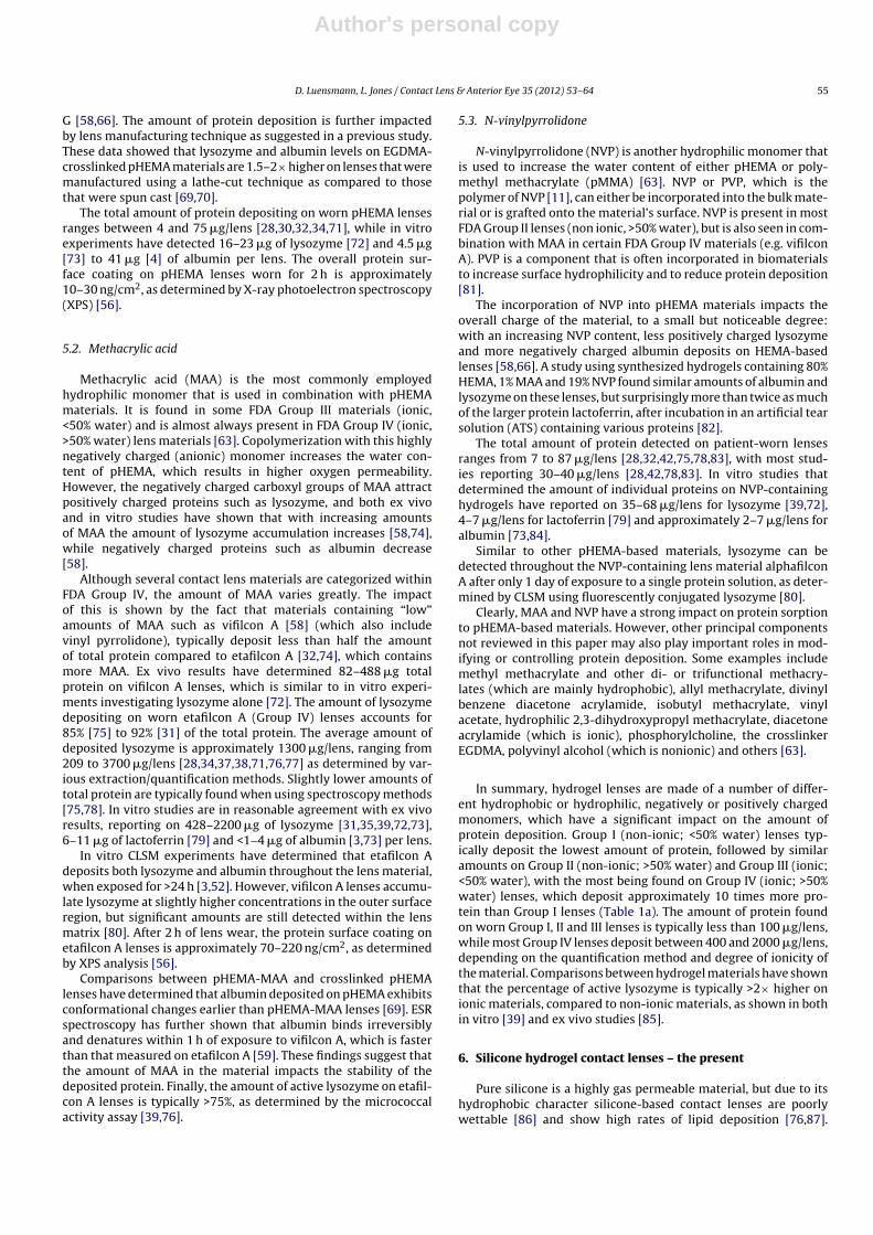
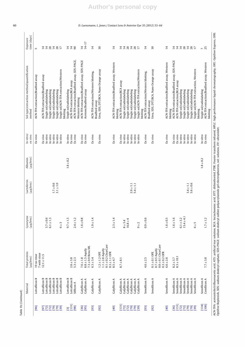
A reactive gas plasma process transforms the hydrophobicsiloxane components on the surface of balafilcon A lenses intohydrophilic silicate compounds (‘glassy islands’) [64,91]. However,this surface modification is no barrier for lysozyme and albuminto penetrate into the matrix, as demonstrated by CLSM [3,80]. Bal-afilcon A is the only silicone hydrogel material considered ionic(FDA Group III) due to the incorporation of an ionic group (N-vinylaminobutyric acid), and it attracts more protein than all other sil-icone hydrogel lenses currently available [39,72,92]. Patient wornlenses deposit 5–34 �g of total protein [36,92,93], while lysozymeaccounts for approximately 32% [36] to 50% [93] of the totalamount of deposited protein. The amounts of individual proteinsdepositing on balafilcon A have been determined using in vitroexperiments, and indicate 10–50 �g of lysozyme [3,35,39,72,76],6–17 �g of lactoferrin [35,79] and less than 2 �g of albumin [3]per lens. Lysozyme activity on worn lenses and in vitro modelsis approximately 50% [38,39,76,93]. Choice of care regimen mayalso impact protein activity on these lenses: higher levels of dena-tured lysozyme have been found when lenses were cleaned with apolyhexanide-based system compared to a polyquaternium-basedsystem [38,76].
6.2. Lotrafilcon A, lotrafilcon B and sifilcon A
The contact lens materials lotrafilcon A and B are permanentlymodified by a gas plasma treatment using a mixture of trimethylsi-lane, oxygen and methane to form a 25 nm thin hydrophilic coatingover the surface [63,88,94]. This lens surface minimizes albuminand lysozyme penetration into the materials, with higher accumu-lations seen on the surface as determined by CLSM [3,52]. Despitethe similarity between both materials, lotrafilcon B, which has alower Dk/t and a higher water content compared to lotrafilcon A,seems to deposit slightly more protein. Quantitative analysis onworn lenses detected 5–7 �g/lens of total protein on lotrafilcon A[36,93], while <1–19 �g/lens has been reported for lotrafilcon B,with the majority of studies reporting >7 �g per lens [92,93,95–97].Ex vivo studies have further determined that lysozyme accountsfor <25% of the total protein deposited on worn lotrafilcon A and B[36,93].
In vitro studies on both lotrafilcon materials have confirmed theresults from ex vivo lenses, showing that slightly higher amountsof deposited protein are typically found on lotrafilcon B compared
to lotrafilcon A lenses: lotrafilcon A deposited 2–4 �g lysozyme[39,72,76] and 1–2 �g lactoferrin [79], while lotrafilcon B accu-mulated 4–10 �g of lysozyme [3,39,72] and 2–3 �g of lactoferrin[79]. Lysozyme activity on both lens types have been determinedto be ≤25%, while results typically show marginally, but statisti-cally insignificant, higher levels of active lysozyme on lotrafilcon Blenses [38,39,76,93].
Sifilcon A is manufactured using a lathe-cutting process and rep-resents the newest member of this “family” of materials. Ex vivoresults confirmed that the amount of protein depositing on thislens is similar to lotrafilcon A and lotrafilcon B, with 5 �g of totalprotein and 2 �g of lysozyme/lens being found after 90 days of wear[98]. The level of protein denaturation is also similar to the othertwo lens types, with 20% lysozyme activity for patient-worn lenses[98].
6.3. Asmofilcon A
The surface of asmofilcon A lenses is modified based on “Nano-glass” technology using a new plasma treatment, which combinesplasma coating and surface oxidation [99]. Although these lenseshave been available since 2007, no data on protein sorption havebeen reported as of to date.
6.4. Galyfilcon A and senofilcon A
High molecular weight chains of PVP are incorporated into galy-filcon A and senofilcon A lenses to enhance surface wettability ofthe lens on eye [100–104]. Both lens types are manufactured usingsimilar material components.
In vitro experiments have shown that lysozyme can penetrateinto the matrix of both lens materials after 24 h of incubationwith fluorescently conjugated protein [80]. Both lens types tendto deposit similar amounts of tear film proteins after wear. Thevalues reported range from <1 to 9 �g/lens, while slightly higheramounts are typically found on galyfilcon A lenses [36,40,92,93].The majority of studies report approximately 7 �g/lens of total pro-tein, of which lysozyme contributes <2 �g (∼25%) [36,93]. In vitrostudies have also confirmed that galyfilcon A, which has a lowerDk/t and a higher water content compared to senofilcon A, accumu-lates slightly more protein than senofilcon A [39,72,79]. Incubationin single protein solutions generally resulted in a higher proteinuptake in comparison to ex vivo studies, which can be explainedby the missing tear film components, which typically competewith proteins during the sorption process. In vitro studies of galy-filcon A and senofilcon A determined 8–17 �g and 6–13 �g oflysozyme/lens [39,72], respectively, while lactoferrin was found insimilar quantities on both lens types, at 3–5 �g/lens [79]. The activ-ity of deposited lysozyme on galyfilcon A lenses was 42–60%, whilea slightly lower percentage of 28–51% has been found for senofil-con A lenses [36,39,40,93]. However, so far none of these ex vivoand in vitro studies have shown a statistically significant differencebetween the two materials [36,39,40,93].
6.5. Narafilcon A and narafilcon B
These materials are modified using an internal PVP-based wet-ting agent (Hydraclear 1) and are used as single-use daily disposablelenses. Narafilcon A and B have been introduced fairly recently andas a result, no scientific data on protein deposition are available todate.
6.6. Comfilcon A and enfilcon A
These materials incorporate silicone which is based on siloxy-macromers instead of the commonly used TRIS-derivates [105].

Author's personal copy
D. Luensmann, L. Jones / Contact Lens & Anterior Eye 35 (2012) 53– 64 59
Tab
le
1bQ
uan
tita
tive
dat
a
on
pro
tein
dep
osit
ion
to
sili
con
e
hyd
roge
l con
tact
len
s
mat
eria
ls.
Mat
eria
l
Tota
l pro
tein
(�g/
len
s)Ly
sozy
me
(�g/
len
s)La
ctof
erri
n(�
g/le
ns)
Alb
um
in(�
g/le
ns)
In
vitr
o/ex
vivo
Sol t
ype/
extr
acti
on
met
hod
/qu
anti
fica
tion
met
hod
Exp
osu
reti
me
(day
s)
SiH
y[1
30]
All
5.2
±
10
Ex
vivo
Ure
a,
SDS,
DTT
/BC
A, N
ano
Ora
nge
assa
y
14
and
30[3
6]
Bal
afilc
on
A
33.5
±
6.1
10.9
±
2.9
Ex
vivo
AC
N-T
FA
extr
acti
on/B
rad
ford
assa
y,
SDS-
PAG
E 14
[76]
Bal
afilc
on
A
10
±
3
Ex
vivo
AC
N-T
FA
extr
acti
on/S
DS-
PAG
E,
Wes
tern
blot
tin
g30
[38]
Bal
afilc
on
A
10
±
5.0
OFE
10
±
3.5
Ren
u
MP
Ex
vivo
AC
N-T
FA
extr
acti
on/W
este
rn
blot
tin
g 30
[93]
Bal
afilc
on
A
26.9
±
2.2
13.3
±
9.0
Ex
vivo
AC
N-T
FA
extr
acti
on/W
este
rn
blot
tin
g,B
rad
ford
assa
y14
[131
]
Bal
afilc
on
A
110.
1
±
26.1
Ex
vivo
AC
N-T
FA
extr
acti
on/B
CA
assa
y
14[9
2]
Bal
afilc
on
A
23.1
±
5.8
OFE
5.4
±
6.7
Aqu
ify
23.2
±
10.7
Cle
arC
are
17.6
±
6.1
OFR
Ex
vivo
Ure
a,
SDS,
DTT
/BC
A, N
ano
Ora
nge
assa
y
30
[35]
Bal
afilc
on
A
17
±
1.4
In
vitr
o
Sin
gle
sol/
AC
N-T
FA
extr
acti
on/B
CA
assa
y
4
h[7
2]
Bal
afilc
on
A
10.6
±
1.6
In
vitr
o
Sin
gle
sol/
rad
iola
beli
ng
14[7
2]
Bal
afilc
on
A
19.4
±
2.9
In
vitr
o
Sin
gle
sol/
rad
iola
beli
ng
28[7
9]B
alafi
lcon
A
6.3
±
1.1
In
vitr
o
Sin
gle
sol/
rad
iola
beli
ng
14[7
9]B
alafi
lcon
A
11.8
±
2.9
In
vitr
o
Sin
gle
sol/
rad
iola
beli
ng
28[3
5]B
alafi
lcon
A
17
±
1.4
In
vitr
o
Sin
gle
sol/
AC
N-T
FA
extr
acti
on/B
CA
assa
y
1[3
9]
Bal
afilc
on
A
44
±
10
In
vitr
o
Sin
gle
sol/
AC
N-T
FA
extr
acti
on, W
este
rnbl
otti
ng
17
[3]
Bal
afilc
on
A
50
±
0.1
1.9
±
0.4
In
vitr
o Si
ngl
e
sol/
rad
iola
beli
ng
14[3
6]
Lotr
afilc
on
A
6.7
±
2.7
0.7
±
0.5
Ex
vivo
AC
N-T
FA
extr
acti
on/B
rad
ford
assa
y,
SDS-
PAG
E
14[1
32]
Lotr
afilc
on
A
0.07
0.21
0.17
Ex
vivo
ELIS
A/p
erox
idas
e
con
juga
ted
anti
bod
ies
30[7
6]
Lotr
afilc
on
A
3
±
1
Ex
vivo
AC
N-T
FA
extr
acti
on/S
DS-
PAG
E,
Wes
tern
blot
tin
g30
[93]
Lotr
afilc
on
A
5.2
±
2.2
1.1
±
0.8
Ex
vivo
AC
N-T
FA
extr
acti
on/W
este
rn
blot
tin
g,B
rad
ford
assa
y14
[133
]
Lotr
afilc
on
A
0.7
rub
Com
ple
te2.
0
no
rub
OFE
Ex
vivo
Tert
-bu
tyl-
met
hyl
eth
er/F
TIR
30
[72]
Lotr
afilc
on
A
2.7
±
0.7
In
vitr
o
Sin
gle
sol/
rad
iola
beli
ng
14[7
2]
Lotr
afilc
on
A
4.2
±
0.9
In
vitr
o
Sin
gle
sol/
rad
iola
beli
ng
28[7
9]
Lotr
afilc
on
A
0.7
±
0.7
In
vitr
o
Sin
gle
sol/
rad
iola
beli
ng
14[7
9]
Lotr
afilc
on
A
2.1
±
0.9
In
vitr
o
Sin
gle
sol/
rad
iola
beli
ng
28[3
9]
Lotr
afilc
on
A
2
±
1
In
vitr
o
Sin
gle
sol/
AC
N-T
FA
extr
acti
on/W
este
rnbl
otti
ng
17
[95]
Lotr
afilc
on
B
9.8
±
1.4
Aqu
ify
9.8
±
1.0
Ren
u
ML
Ex
vivo
Ace
ton
e/B
rad
ford
assa
y
14–1
7
[93]
Lotr
afilc
on
B
6.6
±
3.4
1.4
±
1.1
Ex
vivo
AC
N-T
FA
extr
acti
on/W
este
rn
blot
tin
g,B
rad
ford
assa
y14
[92]
Lotr
afilc
on
B
3.6
±
1.0
OFE
0.3
±
0.9
Aqu
ify
0.5
±
0.4
Cle
arC
are
1.7
±
2.3
OFR
Ex
vivo
Ure
a,
SDS,
DTT
/BC
A, N
ano
Ora
nge
assa
y
30
Author's personal copy
60 D. Luensmann, L. Jones / Contact Lens & Anterior Eye 35 (2012) 53– 64
Tabl
e
1b
(Con
tinu
ed)
Mat
eria
l
Tota
l pro
tein
(�g/
len
s)Ly
sozy
me
(�g/
len
s)La
ctof
erri
n(�
g/le
ns)
Alb
um
in(�
g/le
ns)
In
vitr
o/ex
vivo
Sol t
ype/
extr
acti
on
met
hod
/qu
anti
fica
tion
met
hod
Exp
osu
reti
me
(day
s)
[96]
Lotr
afilc
on
B
19
no
rin
se7
wit
h
rin
seEx
vivo
AC
N-T
FA
extr
acti
on/B
rad
ford
assa
y
5
[97]
Lotr
afilc
on
B
12.1
±
11.5
Ex
vivo
AC
N-T
FA
extr
acti
on/B
rad
ford
assa
y
14[7
2]
Lotr
afilc
on
B
3.7
±
0.6
In
vitr
o
Sin
gle
sol/
rad
iola
beli
ng
14[7
2]Lo
trafi
lcon
B6.
1
±
1.3
In
vitr
o
Sin
gle
sol/
rad
iola
beli
ng
28[7
9]
Lotr
afilc
on
B
1.7
±
0.6
In
vitr
o
Sin
gle
sol/
rad
iola
beli
ng
14[7
9]Lo
trafi
lcon
B3.
1
±
1.0
In
vitr
oSi
ngl
e
sol/
rad
iola
beli
ng
28[3
9]Lo
trafi
lcon
B
6
±
3
In
vitr
o
Sin
gle
sol/
AC
N-T
FA
extr
acti
on/W
este
rnbl
otti
ng
17
[3]
Lotr
afilc
on
B
9.7
±
1.5
1.8
±
0.2
In
vitr
o
Sin
gle
sol/
rad
iola
beli
ng
14[1
31]
Lotr
afilc
on
B
2.6
±
3.8
Ex
vivo
AC
N-T
FA
extr
acti
on/B
CA
assa
y
14[9
8]Si
filc
on
A5.
3
±
2.3
2.4
±
1.2
Ex
vivo
AC
N-T
FA
extr
acti
on/B
rad
ford
assa
y,
SDS-
PAG
E,W
este
rn
blot
tin
g90
[36]
Gal
yfilc
on
A7.
6
±
1.8
1.6
±
0.8
Ex
vivo
AC
N-T
FA
extr
acti
on/B
rad
ford
assa
y,
SDS-
PAG
E14
[95]
Gal
yfilc
on
A
8.8
±
1.5
Aqu
ify
7.3
±
0.9
Ren
u
ML
Ex
vivo
Ace
ton
e/B
rad
ford
assa
y 14
–17
[93]
Gal
yfilc
on
A
6.3
±
3.4
1.9
±
1.4
Ex
vivo
AC
N-T
FA
extr
acti
on/W
este
rn
blot
tin
g,B
rad
ford
assa
y14
[92]
Gal
yfilc
on
A1.
2
±
1.2
OFE
1.1
±
2.8
Aqu
ify
0.1
±
0.2
Cle
arC
are
0.3
±
1.1
OFR
Ex
vivo
Ure
a,
SDS,
DTT
/BC
A, N
ano
Ora
nge
assa
y
30
[40]
Gal
yfilc
on
A
8.5
±
4.7
2.3
±
1.4
Ex
vivo
AC
N-T
FA
extr
acti
on/B
rad
ford
assa
y,
Wes
tern
blot
tin
g14
[131
]G
alyfi
lcon
A
8.7
±
8.1
Ex
vivo
AC
N-T
FA
extr
acti
on/B
CA
assa
y
14[7
2]G
alyfi
lcon
A8
±
3.4
In
vitr
oSi
ngl
e
sol/
rad
iola
beli
ng
14[7
2]
Gal
yfilc
on
A
16.8
±
4
In
vitr
o
Sin
gle
sol/
rad
iola
beli
ng
28[7
9]
Gal
yfilc
on
A
2.9
±
1.1
In
vitr
o Si
ngl
e
sol/
rad
iola
beli
ng
14[7
9]G
alyfi
lcon
A5.
4
±
1.1
In
vitr
o Si
ngl
e
sol/
rad
iola
beli
ng
28[3
9]
Gal
yfilc
on
A
9
±
2
In
vitr
o
Sin
gle
sol/
AC
N-T
FA
extr
acti
on/W
este
rnbl
otti
ng
17
[93]
Sen
ofilc
on
A
4.6
±
2.5
0.9
±
0.6
Ex
vivo
AC
N-T
FA
extr
acti
on/W
este
rn
blot
tin
g,B
rad
ford
assa
y14
[92]
Sen
ofilc
on
A
0.1
±
0.1
OFE
0.7
±
0.5
Aqu
ify
0.0
±
0.1
Cle
arC
are
0.3
±
0.2
OFR
Ex
vivo
Ure
a,
SDS,
DTT
/BC
A, N
ano
Ora
nge
assa
y
30
[40]
Sen
ofilc
on
A
6.6
±
2.6
1.6
±
0.5
Ex
vivo
AC
N-T
FA
extr
acti
on/B
rad
ford
assa
y,
Wes
tern
blot
tin
g14
[36]
Sen
ofilc
on
A8.
2
±
3.7
1.6
±
1.6
Ex
vivo
AC
N-T
FA
extr
acti
on/B
rad
ford
assa
y,
SDS-
PAG
E14
[131
]
Sen
ofilc
on
A
8.3
±
10.1
Ex
vivo
AC
N-T
FA
extr
acti
on/B
CA
assa
y
14[7
2]
Sen
ofilc
on
A
6.1
±
3.2
In
vitr
o
Sin
gle
sol/
rad
iola
beli
ng
14[7
2]Se
nofi
lcon
A
13.4
±
4.1
In
vitr
o
Sin
gle
sol/
rad
iola
beli
ng
28[7
9]
Sen
ofilc
on
A
3.4
± 1.
1
In
vitr
o
Sin
gle
sol/
rad
iola
beli
ng
14[7
9]
Sen
ofilc
on
A
5.6
± 0.
6
In
vitr
o
Sin
gle
sol/
rad
iola
beli
ng
28[3
9]
Sen
ofilc
on
A
6
±
5
In
vitr
o
Sin
gle
sol/
AC
N-T
FA
extr
acti
on, W
este
rnbl
otti
ng
17
[134
]
Sen
ofilc
on
A
1.8
±
0.2
In
vitr
o
Sin
gle
sol/
rad
iola
beli
ng
7[1
06]
Com
filc
on
A
7.7
±
3.8
1.7
±
1.2
Ex
vivo
AC
N-T
FA
extr
acti
on/B
rad
ford
assa
y,
Wes
tern
blot
tin
g25
AC
N-T
FA:
acet
onit
rile
/tri
flu
oroa
ceti
c
acid
, ATS
:
arti
fici
al
tear
solu
tion
, BC
A:
bici
nch
onin
ic
acid
, DTT
:
dit
hio
thre
itol
, FTI
R:
Fou
rier
tran
sfor
m
infr
ared
, HPL
C:
hig
h
per
form
ance
liqu
id
chro
mat
ogra
ph
y,
OFE
:
Op
tifr
ee
Exp
ress
, OFR
:O
pti
free
Rep
len
ish
, SD
S:
sod
ium
dod
ecyl
sup
hat
e,
SDS-
PAG
E:
sod
ium
dod
ecyl
sulf
ate
pol
yacr
ylam
ide
gel e
lect
rop
hor
esis
, sol
:
solu
tion
, UV
:
ult
ravi
olet
.

Author's personal copy
D. Luensmann, L. Jones / Contact Lens & Anterior Eye 35 (2012) 53– 64 61
Specific surface treatments or internal wetting agents are not usedin these lens types [105]. Since their launch in 2007, only one studyhas presented data on protein deposition with comfilcon A. In thisex vivo study a total of 8 �g protein/lens was determined, whichincluded <2 �g of lysozyme/lens after 25 days of wear [106].
6.7. Filcon II 3
Little is known about filcon II 3, which is currently only availablein Europe. It incorporates a surface wetting technology namedAquaGenTM, but no data on protein deposition rates have beenpublished to-date.
In summary, bulk material composition and surface modifi-cation methods vary greatly between silicone hydrogel lenses.However, the amount of protein deposition on these lens types isfairly similar and typically <10 �g/lens, with the exception of bal-afilcon A, which accumulates approximately three times as much(Table 1b).
7. Contact lens materials and surface modifications – thefuture
Silicone hydrogel materials have solved hypoxia-related com-plications and show low rates of protein deposition, but the relativeamount of denatured protein detected on these materials is typi-cally higher than that measured on pHEMA-based lenses [39,107].Furthermore, corneal inflammatory events tend to be equal orhigher with silicone hydrogel lenses compared to hydrogel lenses[108], which raises the question of whether surface engineeringtechniques or other material components can improve the biocom-patibility of these latest contact lens materials.
A number of different approaches to passivate synthetic ornatural biomaterials have been developed for blood contactingimplants, and some of these have been evaluated for contact lensmaterials [109].
Surface coatings containing phosphorylcholine (PC) havebeen used in various biomedical applications, as they showacceptable level of biocompatibility [110,111] and attract onlysmall amounts of protein [39,72,79,112,113]. Novel materialcompositions with 2-methacryloyloxyethyl phosphorylcholine(MPC) confirmed a decrease in protein deposition rates withincreasing amounts of PC [114,115]. The combination of MPCwith bis(trimethylsilyloxy)methylsilylpropyl glycerol methacry-late (SiMA) has recently been suggested as a potential sili-cone hydrogel contact lens material, as it reaches an oxygentransmissibility of >125 units [116]. An in vitro experi-ment with this material confirmed the relationship betweenincreased MPC content and reduced protein deposition rates,and these materials deposited less protein than senofilconA lenses [116]. In another study, a hydrogel lens materialwas synthesized using MPC with 2-(methacryloyloxy)ethyl-N-(2-methacryloyloxy)ethyl]phosphorylcholine (MMPC). This materialexhibited an oxygen permeability of 64 units, which is significantlyhigher than pHEMA, and the highly wettable surface depositedapproximately three times less protein than omafilcon A lenses[117].
Polyethers such as poly(ethylene glycol) (PEG) or poly(ethyleneoxide) (PEO) are currently used in various biomaterial applications,due to their hydrophilicity, bioinertness and resistance to proteinand cell adsorption [118,119]. In a recent clinical study, lotrafilconA lenses were coated with PEO using an allylamine plasma poly-mer interlayer and their in vivo performance was evaluated [120].The researchers confirmed good biocompatibility after 6 h of lenswear and reported that modified lenses attracted less than half the
amount of protein compared to non-modified lotrafilcon A lenses[120].
Crosslinking hyaluronic acid (HA) into pHEMA, siliconehydrogel-like materials or PMMA also reduces protein deposi-tion rates compared to non-treated materials [121–123]. PHEMAcrosslinked to HA of differing molecular weights accumulated sig-nificantly lower amounts of albumin and IgG compared to nelfilconA (FDA Group II) and the silicone hydrogel lens material senofil-con A [121]. Lysozyme was found in lower amounts on nelfilconA, but no difference between the HA-crosslinked materials andsenofilcon A was seen [121]. A strong lysozyme repelling effectwas also seen when HA was crosslinked to methacryloxy propyltris(trimethylsiloxy) silane (Tris)-pHEMA hydrogels, independentof the ratio between Tris:pHEMA [122].
Finally, other approaches for ophthalmic material/surfacedesigns use combinations of frequently used materials withperfluoropolyethers [124], polyvinyl alcohol [125] and surfacecoatings with pyrolytic carbon [126], albumin, elastin [109] orcationic peptide [127] to reduce protein accumulation and micro-bial adherence.
In conclusion, improvement in contact lens biocompatibility isan ongoing process and controlling the amount and conformationalstate of deposited protein remains one of the major challenges.The ocular surface environment is very complex, and tear filmcomposition varies greatly between individuals [14]. While thisreview has only dealt with protein deposition, it is known that sili-cone hydrogel lenses attract greater amounts of lipid compared tomany polyHEMA-based lenses [87], which is the opposite trendto that seen with protein accumulation. Increased lipid deposi-tion may impact clarity of vision, wettability and comfort, buthas not been associated directly with ocular surface inflamma-tory responses. Although silicone hydrogel lenses have significantlyreduced complications related to hypoxia [107] they have notreduced the prevalence of ocular complications such as inflamma-tion and microbial keratitis, and the role of material deposition tothese complications remains unclear [108].
Optimal biocompatibility with lens materials may not requirethe material to be resistant to all tear film components. Rather,it is important that the material is accepted by the ocular envi-ronment and this may require the deposition of selected tear filmcomponents. In respect to proteins, the ideal contact lens mate-rial should bind proteins only loosely, allowing the protein to bereadily removed when exposed to contact lens care regimens andmaintaining the native state of any bound proteins. New contactlens material designs and surface modifications are currently underinvestigation, but only the future will show whether or not they canprovide enhanced on eye biocompatibility.
References
[1] Franklin VJ. Cleaning efficacy of single-purpose surfactant cleaners and multi-purpose solutions. Cont Lens Anterior Eye 1997;20:63–8.
[2] Jung J, Rapp J. The efficacy of hydrophilic contact lens cleaning systems inremoving protein deposits. CLAO J 1993;19:47–9.
[3] Luensmann D, Heynen M, Liu L, Sheardown H, Jones L. The efficiency of contactlens care regimens on protein removal from hydrogel and silicone hydrogellenses. Mol Vis 2010;16:79–92.
[4] Taylor RL, Willcox MD, Williams TJ, Verran J. Modulation of bacterial adhesionto hydrogel contact lenses by albumin. Optom Vis Sci 1998;75:23–9.
[5] Santos L, Rodrigues D, Lira M, Real Oliveira ME, Oliveira R, Vilar EY, et al.Bacterial adhesion to worn silicone hydrogel contact lenses. Optom Vis Sci2008;85:520–5.
[6] Miller MJ, Wilson LA, Ahearn DG. Effects of protein, mucin, and human tearson adherence of Pseudomonas aeruginosa to hydrophilic contact lenses. J ClinMicrobiol 1988;26:513–7.
[7] Tan ME, Demirci G, Pearce D, Jalbert I, Sankaridurg P, Willcox MD. Con-tact lens-induced papillary conjunctivitis is associated with increasedalbumin deposits on extended wear hydrogel lenses. Adv Exp Med Biol2002;506:951–5.

Author's personal copy
62 D. Luensmann, L. Jones / Contact Lens & Anterior Eye 35 (2012) 53– 64
[8] Green-Church KB, Nichols KK, Kleinholz NM, Zhang L, Nichols JJ. Investigationof the human tear film proteome using multiple proteomic approaches. MolVis 2008;14:456–70.
[9] de Souza GA, Godoy LM, Mann M. Identification of 491 proteins in the tearfluid proteome reveals a large number of proteases and protease inhibitors.Genome Biol 2006;7:R72.
[10] Zydney AL. In: Zeman LJ, Zydney AL, editors. Microfiltration and ultra-filtration: principles and applications. New York: Marcel Dekker; 1996.p. 618.
[11] Nuyken O, Billig-Peters W, Frasch T. Polystyrenes and other aromaticpoly(vinyl compound)s. In: Kricheldorf HR, Nuyken O, Swift G, editors. Hand-book of polymer synthesis. 2nd ed. New York: Marcel Dekker; 2005. p.73–150.
[12] Bajpai AK, Mishra DD. Adsorption of a blood protein on to hydrophilicsponges based on poly(2-hydroxyethyl methacrylate). J Mater Sci Mater Med2004;15:583–92.
[13] Bright AM, Tighe BJ. The composition and interfacial properties of tears, tearsubstitutes and tear models. J Br Contact Lens Assoc 1993;16:57–66.
[14] Ng V, Cho P, Mak S, Lee A. Variability of tear protein levels in normalyoung adults: between-day variation. Graefes Arch Clin Exp Ophthalmol2000;238:892–9.
[15] Sack RA, Tan KO, Tan A. Diurnal tear cycle: evidence for a nocturnal inflam-matory constitutive tear fluid. Invest Ophthalmol Vis Sci 1992;33:626–40.
[16] Farris RL. Tear analysis in contact lens wearers. Trans Am Ophthalmol Soc1985;83:501–45.
[17] McGill JI, Liakos GM, Goulding N, Seal DV. Normal tear protein profiles andage-related changes. Br J Ophthalmol 1984;68:316–20.
[18] Mandel ID, Stuchell RN. The lacrimal-salivary axis in health and disease. In:Holly FJ, Lamberts DW, MacKeen DL, editors. The preocular tear film in health,disease, and contact lens wear. Texas: Lubbock, Dry Eye Institute, Inc.; 1986.p. 852–6.
[19] Fullard RJ, Tucker DL. Changes in human tear protein levels with progressivelyincreasing stimulus. Invest Ophthalmol Vis Sci 1991;32:2290–301.
[20] Vadgama P. Surfaces and interfaces for biomaterials. Boca Raton, FL: CRCPress/Woodhead; 2005.
[21] Voet D, Voet JG. Three-dimensional structures of proteins. Biochemistry. 3rded. Hoboken, NJ: Wiley; 2004. p. 219–75.
[22] Norde W. Energy and entropy of protein adsorption. J Dispersion Sci Technol1992;13:363–77.
[23] Roach P, Farrar D, Perry CC. Interpretation of protein adsorption: surface-induced conformational changes. J Am Chem Soc 2005;127:8168–73.
[24] Lindgren M, Sorgjerd K, Hammarstrom P. Detection and characterization ofaggregates, prefibrillar amyloidogenic oligomers, and protofibrils using fluo-rescence spectroscopy. Biophys J 2005;88:4200–12.
[25] Allansmith MR, Korb DR, Greiner JV, Henriquez AS, Simon MA, FinnemoreVM. Giant papillary conjunctivitis in contact lens wearers. Am J Ophthalmol1977;83:697–708.
[26] Brennan NA, Coles ML. Deposits and symptomatology with soft contact lenswear. Int Contact Lens Clin 2000;27:75–100.
[27] Rudko P, Proby JA. A method for classifying and describing protein depositionon hydrophilic lenses. Allergan pharmaceutical report; 1974, Series 94.
[28] Minno GE, Eckel L, Groemminger S, Minno B, Wrzosek T. Quantitative analysisof protein deposits on hydrophilic soft contact lenses. I. Comparison to visualmethods of analysis. II. Deposit variation among FDA lens material groups.Optom Vis Sci 1991;68:865–72.
[29] Myers RI, Larsen DW, Tsao M, Castellano C, Becherer LD, Fontana F, et al.Quantity of protein deposited on hydrogel contact lenses and its relation tovisible protein deposits. Optom Vis Sci 1991;68:776–82.
[30] Wedler FC. Analysis of biomaterials deposited on soft contact lenses. J BiomedMater Res 1977;11:525–35.
[31] Keith D, Hong B, Christensen M. A novel procedure for the extraction of pro-tein deposits from soft hydrophilic contact lenses for analysis. Curr Eye Res1997;16:503–10.
[32] Yan G, Nyquist G, Caldwell KD, Payor R, McCraw EC. Quantitation of totalprotein deposits on contact lenses by means of amino acid analysis. InvestOphthalmol Vis Sci 1993;34:1804–13.
[33] Subbaraman LN, Glasier MA, Sheardown H, Jones L. Efficacy of an extrac-tion solvent used to quantify albumin deposition on hydrogel contact lensmaterials. Eye Contact Lens 2009;35:76–80.
[34] Minarik L, Rapp J. Protein deposits on individual hydrophilic contact lenses:effects of water and ionicity. CLAO J 1989;15:185–8.
[35] Zhang S, Borazjani RN, Salamone JC, Ahearn DG, Crow Jr SA, Pierce GE.In vitro deposition of lysozyme on etafilcon A and balafilcon A hydrogel con-tact lenses: effects on adhesion and survival of Pseudomonas aeruginosa andStaphylococcus aureus. Cont Lens Anterior Eye 2005;28:113–9.
[36] Boone A, Heynen M, Joyce E, Varikooty J, Jones L. Ex vivo protein deposition onbi-weekly silicone hydrogel contact lenses. Optom Vis Sci 2009;86:1241–9.
[37] Keith DJ, Christensen MT, Barry JR, Stein JM. Determination of the lysozymedeposit curve in soft contact lenses. Eye Contact Lens 2003;29:79–82.
[38] Senchyna M, Jones L, Louie D, May C, Forbes I, Glasier MA. Quantitative andconformational characterization of lysozyme deposited on balafilcon andetafilcon contact lens materials. Curr Eye Res 2004;28:25–36.
[39] Suwala M, Glasier MA, Subbaraman LN, Jones L. Quantity and conformationof lysozyme deposited on conventional and silicone hydrogel contact lensmaterials using an in vitro model. Eye Contact Lens 2007;33:138–43.
[40] Glasier MA, Keech A, Sheardown H, Subbaraman LN, Jones L. Conformationaland quantitative characterization of lysozyme extracted from galyfilcon andsenofilcon silicone hydrogel contact lenses. Curr Eye Res 2008;33:1–11.
[41] Ratner BD, Horbett TA, Mateo NB. Contact lens spoilation. Part 1. Biochemicalaspect of lens spoilation. In: Ruben M, Guillon M, editors. Contact lens practice.1st ed. London: Chapman & Hall; 1994. p. 1083–98.
[42] Jones L, Evans K, Sariri R, Franklin V, Tighe B. Lipid and protein deposition ofN-vinyl pyrrolidone-containing Group II and Group IV frequent replacementcontact lenses. CLAO J 1997;23:122–6.
[43] Miller B. Observations of deposits on soft contact lenses by different methodsof light microscopy, scanning microscopy, and electron microprobe analysis.Int Contact Lens Clin 1980;3–4:22–35.
[44] Tripathi RC, Tripathi BJ, Ruben M. The pathology of soft contact lens spoilage.Ophthalmology 1980;87:365–80.
[45] Bilbaut T, Gachon AM, Dastugue B. Deposits on soft contact lenses. Elec-trophoresis and scanning electron microscopic examinations. Exp Eye Res1986;43:153–65.
[46] Versura P, Maltarello MC, Roomans GM, Caramazza R, Laschi R. Scanning elec-tron microscopy, X-ray microanalysis and immunohistochemistry on wornsoft contact lenses. Scanning Microsc 1988;2:397–410.
[47] Braga PC, Ricci D. Atomic force microscopy: biomedical methods and appli-cations. Totowa: Humana Press; 2004.
[48] Teichroeb JH, Forrest JA, Ngai V, Martin JW, Jones L, Medley J. Imaging proteindeposits on contact lens materials. Optom Vis Sci 2008;85:1151–64.
[49] Lira M, Santos L, Azeredo J, Yebra-Pimentel E, Oliveira ME. Comparativestudy of silicone–hydrogel contact lenses surfaces before and after wearusing atomic force microscopy. J Biomed Mater Res B: Appl Biomater2008;85:361–7.
[50] Rebeix V, Sommer F, Marchin B, Baude D, Tran MD. Artificial tear adsorp-tion on soft contact lenses: methods to test surfactant efficacy. Biomaterials2000;21:1197–205.
[51] Meadows DL, Paugh JR. Use of confocal microscopy to determine matrixand surface protein deposition profiles in hydrogel contact lenses. CLAO J1994;20:237–41.
[52] Luensmann D, Glasier MA, Zhang F, Bantseev V, Simpson T, Jones L. Confo-cal microscopy and albumin penetration into contact lenses. Optom Vis Sci2007;84:839–47.
[53] Castillo EJ, Koenig JL, Anderson JM. Characterization of protein adsorptionon soft contact lenses. IV. Comparison of in vivo spoilage with the in vitroadsorption of tear proteins. Biomaterials 1986;7:89–96.
[54] Hart DE, DePaolis M, Ratner BD, Mateo NB. Surface analysis of hydrogel con-tact lenses by ESCA. CLAO J 1993;19:169–73.
[55] Zhao Z, Wei X, Aliwarga Y, Carnt NA, Garrett Q, Willcox MD. Proteomic analysisof protein deposits on worn daily wear silicone hydrogel contact lenses. MolVis 2008;14:2016–24.
[56] Kingshott P, St John HA, Chatelier RC, Griesser HJ. Matrix-assisted laser des-orption ionization mass spectrometry detection of proteins adsorbed in vivoonto contact lenses. J Biomed Mater Res 2000;49:36–42.
[57] Holmberg M, Hou X. Competitive protein adsorption – multilayer adsorptionand surface induced protein aggregation. Langmuir 2009;25:2081–9.
[58] Garrett Q, Laycock B, Garrett RW. Hydrogel lens monomer constituents mod-ulate protein sorption. Invest Ophthalmol Vis Sci 2000;41:1687–95.
[59] Garrett Q, Griesser HJ, Milthorpe BK, Garrett RW. Irreversible adsorption ofhuman serum albumin to hydrogel contact lenses: a study using electron spinresonance spectroscopy. Biomaterials 1999;20:1345–56.
[60] Hoffman AS. Hydrogels for biomedical applications. Adv Drug Deliv Rev2002;54:3–12.
[61] Peppas NA, Huang Y, Torres-Lugo M, Ward JH, Zhang J. Physicochemical foun-dations and structural design of hydrogels in medicine and biology. Annu RevBiomed Eng 2000;2:9–29.
[62] Kopecek J. Hydrogels from soft contact lenses and implants to self-assemblednanomaterials. J Polym Sci A: Polym Chem 2009;47:5929–46.
[63] Tighe B. Contact lens materials. In: Phillips A, Speedwell L, editors. Contactlenses. 5th ed. Edinburgh: Butterworth-Heinemann; 2007. p. 59–78.
[64] Nicolson PC, Vogt J. Soft contact lens polymers: an evolution. Biomaterials2001;22:3273–83.
[65] Morgan PB, Woods C, Tranoudis IG, Helland M, Efron N, Knajian R, et al.International contact lens prescribing in 2009. Contact Lens Spectrum2010;25:30–53.
[66] Bohnert JL, Horbett TA, Ratner BD, Royce FH. Adsorption of proteins fromartificial tear solutions to contact lens materials. Invest Ophthalmol Vis Sci1988;29:362–73.
[67] Leahy CD, Mandell RB, Lin ST. Initial in vivo tear protein deposition on indi-vidual hydrogel contact lenses. Optom Vis Sci 1990;67:504–11.
[68] Lin ST, Mandell RB, Leahy CD, Newell JO. Protein accumulation on disposableextended wear lenses. CLAO J 1991;17:44–50.
[69] Castillo EJ, Koenig JL, Anderson JM, Lo J. Characterization of protein adsorptionon soft contact lenses. I. Conformational changes of adsorbed human serumalbumin. Biomaterials 1984;5:319–25.
[70] Castillo EJ, Koenig JL, Anderson JM, Lo J. Protein adsorption on hydrogels. II.Reversible and irreversible interactions between lysozyme and soft contactlens surfaces. Biomaterials 1985;6:338–45.
[71] Mochizuki H, Yamada M, Hatou S, Kawashima M, Hata S. Deposition of lipid,protein, and secretory phospholipase A2 on hydrophilic contact lenses. EyeContact Lens 2008;34:46–9.

Author's personal copy
D. Luensmann, L. Jones / Contact Lens & Anterior Eye 35 (2012) 53– 64 63
[72] Subbaraman LN, Glasier MA, Senchyna M, Sheardown H, Jones L. Kinet-ics of in vitro lysozyme deposition on silicone hydrogel, PMMA, andFDA groups I, II, and IV contact lens materials. Curr Eye Res 2006;31:787–96.
[73] Okada E, Matsuda T, Yokoyama T, Okuda K. Lysozyme penetration in GroupIV soft contact lenses. Eye Contact Lens 2006;32:174–7.
[74] Garrett Q, Garrett RW, Milthorpe BK. Lysozyme sorption in hydrogel contactlenses. Invest Ophthalmol Vis Sci 1999;40:897–903.
[75] Maissa C, Franklin V, Guillon M, Tighe B. Influence of contact lens mate-rial surface characteristics and replacement frequency on protein and lipiddeposition. Optom Vis Sci 1998;75:697–705.
[76] Jones L, Senchyna M, Glasier MA, Schickler J, Forbes I, Louie D, et al. Lysozymeand lipid deposition on silicone hydrogel contact lens materials. Eye ContactLens 2003;29:75–9.
[77] Caron P, St-Jacques J, Michaud L. Clinical discussion on the relative efficacy of 2surfactant-containing lubricating agents in removing proteins during contactlens wear. Optometry 2007;78:23–9.
[78] Jones L, Mann A, Evans K, Franklin V, Tighe B. An in vivo comparison of thekinetics of protein and lipid deposition on Group II and Group IV frequent-replacement contact lenses. Optom Vis Sci 2000;77:503–10.
[79] Chow LM, Subbaraman LN, Sheardown H, Jones L. Kinetics of in vitrolactoferrin deposition on silicone hydrogel and FDA Group II and GroupIV hydrogel contact lens materials. J Biomater Sci Polym Ed 2009;20:71–82.
[80] Luensmann D, Zhang F, Subbaraman LN, Sheardown H, Jones L. Localization oflysozyme sorption to conventional and silicone hydrogel contact lenses usingconfocal microscopy. Curr Eye Res 2009;34:683–97.
[81] Rovira-Bru M, Giralt F, Cohen Y. Protein adsorption onto zirconia mod-ified with terminally grafted polyvinylpyrrolidone. J Colloid Interface Sci2001;235:70–9.
[82] Lord MS, Stenzel MH, Simmons A, Milthorpe BK. The effect of chargedgroups on protein interactions with poly(HEMA) hydrogels. Biomaterials2006;27:567–75.
[83] Jones L, Franklin V, Evans K, Sariri R, Tighe B. Spoilation and clinical per-formance of monthly vs. three monthly Group II disposable contact lenses.Optom Vis Sci 1996;73:16–21.
[84] Baines MG, Cai F, Backman HA. Adsorption and removal of protein bound tohydrogel contact lenses. Optom Vis Sci 1990;67:807–10.
[85] Sack RA, Jones B, Antignani A, Libow R, Harvey H. Specificity and biologi-cal activity of the protein deposited on the hydrogel surface. Relationship ofpolymer structure to biofilm formation. Invest Ophthalmol Vis Sci 1987;28:842–9.
[86] Rogers R. In vitro and ex vivo wettability of hydrogel contact lenses. Waterloo,Ont.: University of Waterloo; 2006.
[87] Lorentz H, Jones L. Lipid deposition on hydrogel contact lenses: how historycan help us today. Optom Vis Sci 2007;84:286–95.
[88] Nicolson PC, Baron RC, Chabrecek P, Court J, Domschke A, Griesser HJ, et al.Extended wear ophthalmic lens. US Patent No. 5760100; 1998.
[89] Grobe GL, Kunzler J, Seelye D, Salamone J. Silicone hydrogels for contact lensapplications. Polym Mater Sci Eng 1999;80:108–9.
[90] Yasuda H. Biocompatibility of nanofilm-encapsulated silicone and silicone-hydrogel contact lenses. Macromol Biosci 2006;6:121–38.
[91] Weikart CM, Matsuzawa Y, Winterton L, Yasuda HK. Evaluation of plasmapolymer-coated contact lenses by electrochemical impedance spectroscopy.J Biomed Mater Res 2001;54:597–607.
[92] Zhao Z, Carnt NA, Aliwarga Y, Wei X, Naduvilath T, Garrett Q, et al. Care regi-men and lens material influence on silicone hydrogel contact lens deposition.Optom Vis Sci 2009;86:251–9.
[93] Subbaraman L, Glasier MA, Dumbleton K, Jones L. Quantification of pro-tein deposition on five commercially available silicone hydrogel contact lensmaterials. Optom Vis Sci 2007;84. E-abstract 070031.
[94] Lopez-Alemany A, Compan V, Refojo MF. Porous structure of Purevision versusFocus Night&Day and conventional hydrogel contact lenses. J Biomed MaterRes 2002;63:319–25.
[95] Green-Church KB, Nichols JJ. Mass spectrometry-based proteomic analyses ofcontact lens deposition. Mol Vis 2008;14:291–7.
[96] Pucker AD, Nichols JJ. Impact of a rinse step on protein removal from siliconehydrogel contact lenses. Optom Vis Sci 2009;86:943–7.
[97] Powell DR, Thangavelu M, Nichols JJ. Comparison of pooled and non-pooledextracted tear protein profiles from silicone hydrogel lenses. ARVO. Fort Laud-erdale, FL: Invest Ophthalmol Vis Sci; 2010.
[98] Subbaraman LN, Woods J, Teichroeb JH, Jones L. Protein deposition on alathe-cut silicone hydrogel contact lens material. Optom Vis Sci 2009;86:244–50.
[99] Jones L. Contact lens materials: A new silicone hydrogel comes to market.Contact Lens Spectrum 2007;22:23.
[100] Steffen R, McCabe K. Finding the comfort zone. Contact Lens Spectrum2004;13:3.
[101] Steffen R, Schnider C. A next generation silicone hydrogel lens for daily wear.Part 1 – Material properties. Optician 2004;227:23–5.
[102] McCabe KP, Molock FF, Hill GA, Alli A, Steffen RB, Vanderlaan DG, et al. Biomed-ical devices containing internal wetting agents; 2005.
[103] Riley C, Young G, Chalmers R. Prevalence of ocular surface symptoms, signs,and uncomfortable hours of wear in contact lens wearers: the effect of refit-ting with daily-wear silicone hydrogel lenses (senofilcon a). Eye Contact Lens2006;32:281–6.
[104] Brennan NA, Coles ML, Ang JH. An evaluation of silicone-hydrogel lenses wornon a daily wear basis. Clin Exp Optom 2006;89:18–25.
[105] Tighe B. Trends Dev Silicone Hydrogel Mater 2006, http://www.siliconehydrogels.com.
[106] Jones L, Glasier MA, Boone A, Keir N, Dumbleton K. Protein deposition oncontinuous wear surface modified (balafilcon A) and non-surface modified(comfilcon A) silicone hydrogel contact lens materials. AAO: Opt Vis Sci2007;84. E-abstract 075139.
[107] Fonn D, Bruce AS. A review of the Holden-Mertz criteria for critical oxygentransmission. Eye Contact Lens 2005;31:247–51.
[108] Szczotka-Flynn L, Diaz M. Risk of corneal inflammatory events with siliconehydrogel and low dk hydrogel extended contact lens wear: a meta-analysis.Optom Vis Sci 2007;84:247–56.
[109] Jordan SW, Chaikof EL. Novel thromboresistant materials. J Vasc Surg2007;45(Suppl A):A104–15.
[110] Hayward JA, Chapman D. Biomembrane surfaces as models for poly-mer design: the potential for haemocompatibility. Biomaterials 1984;5:135–42.
[111] Rose SF, Lewis AL, Hanlon GW, Lloyd AW. Biological responses to cation-ically charged phosphorylcholine-based materials in vitro. Biomaterials2004;25:5125–35.
[112] Ishihara K, Nomura H, Mihara T, Kurita K, Iwasaki Y, Nakabayashi N. Whydo phospholipid polymers reduce protein adsorption. J Biomed Mater Res1998;39:323–30.
[113] Keith EO, Chandrasekaran NS, Sjolanki DH, SDesai SS, Janoff AM. Adhe-sion of lysozyme and transferrin to omafilcon A contact lenses. In: AnnualMeeting of the Federation of American Societies for Experimental Biology.2009.
[114] Iwasaki Y, Ishihara K. Phosphorylcholine-containing polymers for biomedicalapplications. Anal Bioanal Chem 2005;381:534–46.
[115] Abraham S, Brahim S, Ishihara K, Guiseppi-Elie A. Molecularly engineeredp(HEMA)-based hydrogels for implant biochip biocompatibility. Biomaterials2005;26:4767–78.
[116] Shimizu T, Goda T, Minoura N, Takai M, Ishihara K. Super-hydrophilic siliconehydrogels with interpenetrating poly(2-methacryloyloxyethyl phosphoryl-choline) networks. Biomaterials 2010;31:3274–80.
[117] Goda T, Matsuno R, Konno T, Takai M, Ishihara K. Protein adsorption resistanceand oxygen permeability of chemically crosslinked phospholipid polymerhydrogel for ophthalmologic biomaterials. J Biomed Mater Res B: Appl Bio-mater 2009;89:184–90.
[118] Atala A, Lanza R, Thomson J, Nerem R. Principles of regenerativemedicine. 1st ed. Amsterdam/Boston: Elsevier/Academic Press; 2008. p. 579–742.
[119] Scott EA, Nichols MD, Cordova LH, George BJ, Jun YS, Elbert DL. Pro-tein adsorption and cell adhesion on nanoscale bioactive coatings formedfrom poly(ethylene glycol) and albumin microgels. Biomaterials 2008;29:4481–93.
[120] Thissen H, Gengenbach T, du Toit R, Sweeney DF, Kingshott P, Griesser HJ, et al.Clinical observations of biofouling on PEO coated silicone hydrogel contactlenses. Biomaterials 2010;31:5510–9.
[121] Van Beek M, Jones L, Sheardown H. Hyaluronic acid containing hydrogels forthe reduction of protein adsorption. Biomaterials 2008;29:780–9.
[122] van Beek M, Weeks A, Jones L, Sheardown H. Immobilized hyaluronic acidcontaining model silicone hydrogels reduce protein adsorption. J BiomaterSci Polym Ed 2008;19:1425–36.
[123] Cassinelli C, Morra M, Pavesio A, Renier D. Evaluation of interfacial prop-erties of hyaluronan coated poly(methylmethacrylate) intraocular lenses. JBiomater Sci Polym Ed 2000;11:961–77.
[124] Chan GY, Hughes TC, McLean KM, McFarland GA, Nguyen X, Wilkie JS, et al.Approaches to improving the biocompatibility of porous perfluoropolyethersfor ophthalmic applications. Biomaterials 2006;27:1287–95.
[125] Kita M, Ogura Y, Honda Y, Hyon SH, Cha II W, Ikada Y. Evaluation of polyvinylalcohol hydrogel as a soft contact lens material. Graefes Arch Clin Exp Oph-thalmol 1990;228:533–7.
[126] Sick PB, Gelbrich G, Kalnins U, Erglis A, Bonan R, Aengevaeren W, et al.Comparison of early and late results of a carbofilm-coated stent versus apure high-grade stainless steel stent (the Carbostent-Trial). Am J Cardiol2004;93:1351–6. A5.
[127] Willcox MD, Hume EB, Aliwarga Y, Kumar N, Cole N. A novel cationic-peptidecoating for the prevention of microbial colonization on contact lenses. J ApplMicrobiol 2008;105:1817–25.
[128] Zhang J, Hodge WG. Contact lens integrated with a biosensor for the detec-tion of glucose and other components in tears. US Patent No. 20100113901;2010.
[129] Christensen B, Lebow K, White EM, Cedrone R, Bevington R. Effectiveness ofcitrate-containing cens care regimens: A controlled clinical comparison. ICLC1998;25:50–8.
[130] Zhao Z, Naduvilath T, Flanagan JL, Carnt NA, Wei X, Diec J, et al. Contact lensdeposits, adverse responses, and clinical ocular surface parameters. OptomVis Sci 2010;87:669–74.
[131] Nash WL, Amos CF, Carney FP. Factors that influence the adsorption of proteinand lipid to silicone hydrogel lenses after 2 weeks of daily wear. ARVO, FortLauderdale. Invest Ophthalmol Vis Sci 2007.
[132] Pearce D, Tan ME, Demirci G, Willcox MD. Surface protein profile ofextended-wear silicon hydrogel lenses. Adv Exp Med Biol 2002;506:957–60.

Author's personal copy
64 D. Luensmann, L. Jones / Contact Lens & Anterior Eye 35 (2012) 53– 64
[133] Powell CH, Hoong LD, Huth SW. Lipid and protein removal from a siliconehydrogel lens (lotrafilcon A) by a rub versus a no-rub multipurpose solu-tionusing infrared analysis of clinically worn lenses. ARVO, Fort Lauderdale,FL. Invest Ophthalmol Vis Sci 2009.
[134] Luensmann D, Jones L. Impact of fluorescent probes on albumin sorptionprofiles to ophthalmic biomaterials. J Biomed Mater Res B Appl Biomater2010;94:327–36.