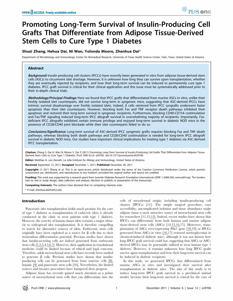
Promoting Long-Term Survival of Insulin-Producing Cell Grafts That Differentiate from Adipose...
9
Promoting Long-Term Survival of Insulin-Producing Cell Grafts That Differentiate from Adipose Tissue-Derived Stem Cells to Cure Type 1 Diabetes Shuzi Zhang, Hehua Dai, Ni Wan, Yolonda Moore, Zhenhua Dai* Department of Microbiology and Immunology, Center for Biomedical Research, University of Texas Health Science Center, Tyler, Texas, United States of America Abstract Background: Insulin-producing cell clusters (IPCCs) have recently been generated in vitro from adipose tissue-derived stem cells (ASCs) to circumvent islet shortage. However, it is unknown how long they can survive upon transplantation, whether they are eventually rejected by recipients, and how their long-term survival can be induced to permanently cure type 1 diabetes. IPCC graft survival is critical for their clinical application and this issue must be systematically addressed prior to their in-depth clinical trials. Methodology/Principal Findings: Here we found that IPCC grafts that differentiated from murine ASCs in vitro, unlike their freshly isolated islet counterparts, did not survive long-term in syngeneic mice, suggesting that ASC-derived IPCCs have intrinsic survival disadvantage over freshly isolated islets. Indeed, b cells retrieved from IPCC syngrafts underwent faster apoptosis than their islet counterparts. However, blocking both Fas and TNF receptor death pathways inhibited their apoptosis and restored their long-term survival in syngeneic recipients. Furthermore, blocking CD40-CD154 costimulation and Fas/TNF signaling induced long-term IPCC allograft survival in overwhelming majority of recipients. Importantly, Fas- deficient IPCC allografts exhibited certain immune privilege and enjoyed long-term survival in diabetic NOD mice in the presence of CD28/CD40 joint blockade while their islet counterparts failed to do so. Conclusions/Significance: Long-term survival of ASC-derived IPCC syngeneic grafts requires blocking Fas and TNF death pathways, whereas blocking both death pathways and CD28/CD40 costimulation is needed for long-term IPCC allograft survival in diabetic NOD mice. Our studies have important clinical implications for treating type 1 diabetes via ASC-derived IPCC transplantation. Citation: Zhang S, Dai H, Wan N, Moore Y, Dai Z (2011) Promoting Long-Term Survival of Insulin-Producing Cell Grafts That Differentiate from Adipose Tissue- Derived Stem Cells to Cure Type 1 Diabetes. PLoS ONE 6(12): e29706. doi:10.1371/journal.pone.0029706 Editor: Matthias G. von Herrath, La Jolla Institute for Allergy and Immunology, United States of America Received September 12, 2011; Accepted December 1, 2011; Published December 28, 2011 Copyright: ß 2011 Zhang et al. This is an open-access article distributed under the terms of the Creative Commons Attribution License, which permits unrestricted use, distribution, and reproduction in any medium, provided the original author and source are credited. Funding: This work was supported by a research grant from Juvenile Diabetes Research Foundation International (JDRF 1-2008-583, www.jdrf.org). The funders had no role in study design, data collection and analysis, decision to publish, or preparation of the manuscript. Competing Interests: The authors have declared that no competing interests exist. * E-mail: [email protected] Introduction Pancreatic islet transplantation holds much promise for the cure of type 1 diabetes as transplantation of cadaveric islets is already conducted in the clinic to treat patients with type 1 diabetes. However, the scarcity of human donor islets remains a major obsta- cle to widespread islet transplantation. It is therefore compelling to search for alternative sources of islets. Embryonic stem cells originally have been exploited as a source for b cells due to their tremendous differentiation potential. Previous studies have shown that insulin-secreting cells are indeed generated from embryonic stem cells [1,2,3,4,5,6,7]. However, their application in translational medicine could be limited because of ethical and legal concerns. Therefore, adult mesenchymal stem cells have recently been studied to generate b cells. Previous studies have shown that insulin- producing cells can be generated from bone marrow cells [8], hepatic [9] and pancreatic stem cells [10]. Nevertheless, the limited sources and invasive procedures have hampered their progress. Adipose tissue has recently gained much attention as a prime source of mesenchymal stem cells that can differentiate into the cells of mesodermal origin, including insulin-producing cell clusters (IPCCs) [11]. The simple surgical procedure, easy accessibility, uncomplicated isolation and tissue abundance make adipose tissue a most attractive source of mesenchymal stem cells for researchers [11,12,13]. Indeed, recent studies have shown that IPCCs can differentiate from both human and murine adipose tissue-derived stem cells (ASCs) [14,15,16,17]. Moreover, trans- plantation of ASCs over-expressing Pdx1 gene [18,19] or IPCCs generated from ASCs in vitro [16,17] restored normoglycemia in chemical-induced diabetic mice, although it was not known how long IPCC graft survival could last, suggesting that ASCs or ASC- derived IPCCs may be potentially utilized to treat human type 1 diabetes. However, it remains unknown how long IPCC grafts survive upon transplantation and how their long-term survival can be induced in diabetic recipients. In this study, we generated IPCCs that differentiated from murine ASCs in vitro and investigated their survival after transplantation in diabetic mice. The aim of this study is to induce long-term IPCC graft survival in a preclinical animal model, because their long-term survival is critical for the cure of PLoS ONE | www.plosone.org 1 December 2011 | Volume 6 | Issue 12 | e29706
-
Upload
apiitmalaysia -
Category
Documents
-
view
3 -
download
0
Transcript of Promoting Long-Term Survival of Insulin-Producing Cell Grafts That Differentiate from Adipose...
Promoting Long-Term Survival of Insulin-Producing CellGrafts That Differentiate from Adipose Tissue-DerivedStem Cells to Cure Type 1 DiabetesShuzi Zhang, Hehua Dai, Ni Wan, Yolonda Moore, Zhenhua Dai*
Department of Microbiology and Immunology, Center for Biomedical Research, University of Texas Health Science Center, Tyler, Texas, United States of America
Abstract
Background: Insulin-producing cell clusters (IPCCs) have recently been generated in vitro from adipose tissue-derived stemcells (ASCs) to circumvent islet shortage. However, it is unknown how long they can survive upon transplantation, whetherthey are eventually rejected by recipients, and how their long-term survival can be induced to permanently cure type 1diabetes. IPCC graft survival is critical for their clinical application and this issue must be systematically addressed prior totheir in-depth clinical trials.
Methodology/Principal Findings: Here we found that IPCC grafts that differentiated from murine ASCs in vitro, unlike theirfreshly isolated islet counterparts, did not survive long-term in syngeneic mice, suggesting that ASC-derived IPCCs haveintrinsic survival disadvantage over freshly isolated islets. Indeed, b cells retrieved from IPCC syngrafts underwent fasterapoptosis than their islet counterparts. However, blocking both Fas and TNF receptor death pathways inhibited theirapoptosis and restored their long-term survival in syngeneic recipients. Furthermore, blocking CD40-CD154 costimulationand Fas/TNF signaling induced long-term IPCC allograft survival in overwhelming majority of recipients. Importantly, Fas-deficient IPCC allografts exhibited certain immune privilege and enjoyed long-term survival in diabetic NOD mice in thepresence of CD28/CD40 joint blockade while their islet counterparts failed to do so.
Conclusions/Significance: Long-term survival of ASC-derived IPCC syngeneic grafts requires blocking Fas and TNF deathpathways, whereas blocking both death pathways and CD28/CD40 costimulation is needed for long-term IPCC allograftsurvival in diabetic NOD mice. Our studies have important clinical implications for treating type 1 diabetes via ASC-derivedIPCC transplantation.
Citation: Zhang S, Dai H, Wan N, Moore Y, Dai Z (2011) Promoting Long-Term Survival of Insulin-Producing Cell Grafts That Differentiate from Adipose Tissue-Derived Stem Cells to Cure Type 1 Diabetes. PLoS ONE 6(12): e29706. doi:10.1371/journal.pone.0029706
Editor: Matthias G. von Herrath, La Jolla Institute for Allergy and Immunology, United States of America
Received September 12, 2011; Accepted December 1, 2011; Published December 28, 2011
Copyright: � 2011 Zhang et al. This is an open-access article distributed under the terms of the Creative Commons Attribution License, which permitsunrestricted use, distribution, and reproduction in any medium, provided the original author and source are credited.
Funding: This work was supported by a research grant from Juvenile Diabetes Research Foundation International (JDRF 1-2008-583, www.jdrf.org). The fundershad no role in study design, data collection and analysis, decision to publish, or preparation of the manuscript.
Competing Interests: The authors have declared that no competing interests exist.
* E-mail: [email protected]
Introduction
Pancreatic islet transplantation holds much promise for the cure
of type 1 diabetes as transplantation of cadaveric islets is already
conducted in the clinic to treat patients with type 1 diabetes.
However, the scarcity of human donor islets remains a major obsta-
cle to widespread islet transplantation. It is therefore compelling
to search for alternative sources of islets. Embryonic stem cells
originally have been exploited as a source for b cells due to their
tremendous differentiation potential. Previous studies have shown
that insulin-secreting cells are indeed generated from embryonic
stem cells [1,2,3,4,5,6,7]. However, their application in translational
medicine could be limited because of ethical and legal concerns.
Therefore, adult mesenchymal stem cells have recently been studied
to generate b cells. Previous studies have shown that insulin-
producing cells can be generated from bone marrow cells [8],
hepatic [9] and pancreatic stem cells [10]. Nevertheless, the limited
sources and invasive procedures have hampered their progress.
Adipose tissue has recently gained much attention as a prime
source of mesenchymal stem cells that can differentiate into the
cells of mesodermal origin, including insulin-producing cell
clusters (IPCCs) [11]. The simple surgical procedure, easy
accessibility, uncomplicated isolation and tissue abundance make
adipose tissue a most attractive source of mesenchymal stem cells
for researchers [11,12,13]. Indeed, recent studies have shown that
IPCCs can differentiate from both human and murine adipose
tissue-derived stem cells (ASCs) [14,15,16,17]. Moreover, trans-
plantation of ASCs over-expressing Pdx1 gene [18,19] or IPCCs
generated from ASCs in vitro [16,17] restored normoglycemia in
chemical-induced diabetic mice, although it was not known how
long IPCC graft survival could last, suggesting that ASCs or ASC-
derived IPCCs may be potentially utilized to treat human type 1
diabetes. However, it remains unknown how long IPCC grafts
survive upon transplantation and how their long-term survival can
be induced in diabetic recipients.
In this study, we generated IPCCs that differentiated from
murine ASCs in vitro and investigated their survival after
transplantation in diabetic mice. The aim of this study is to
induce long-term IPCC graft survival in a preclinical animal
model, because their long-term survival is critical for the cure of
PLoS ONE | www.plosone.org 1 December 2011 | Volume 6 | Issue 12 | e29706
type 1 diabetes. Previous studies have focused on other important
issues at the early stage of IPCC studies, including the generation
of IPCCs in vitro, their temporary functions in vivo, and their
immunogenicity. The novelty of this study lies in the first induction
of long-term IPCC graft survival in diabetic mice, including
autoimmune-prone NOD mice, by blocking both IPCC cell death
and T cell costimulation.
Results
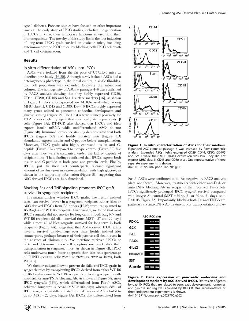
In vitro differentiation of ASCs into IPCCsASCs were isolated from the fat pads of C57BL/6 mice as
described previously [16,20]. Although newly isolated ASCs had a
heterogeneous phenotype in the initial culture, a single fibroblas-
toid cell population was expanded following the subsequent
cultures. The homogeneity of ASCs at passages 4–6 was confirmed
by FACS analysis showing that they highly expressed CD29,
CD44, CD90, CD105 and Sca-1 surface markers [16], as shown
in Figure 1. They also expressed low MHC-class-I while lacking
MHC-class-II, CD45 and CD80. Day-10 IPCCs highly expressed
many genes related to pancreatic endocrine development and
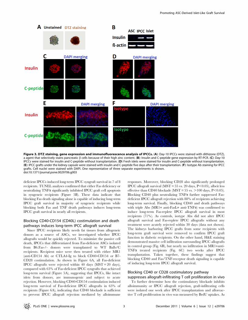
glucose sensing (Figure 2). The IPCCs were stained positively for
DTZ, a zinc-chelating agent that specifically stains pancreatic bcells (Figure 3A). RT-PCR also showed that IPCCs and islets
express insulin mRNA while undifferentiated ASCs do not
(Figure 3B). Immunofluorescence staining demonstrated that both
IPCCs (Figure 3C) and freshly isolated islets (Figure 3D)
abundantly express insulin and C-peptide before transplantation.
Moreover, IPCC grafts also highly expressed insulin and C-
peptide (Figure 3E) compared to isotype control (Figure 3F) five
days after they were transplanted under the kidney capsule of
recipient mice. These findings confirmed that IPCCs express both
insulin and C-peptide at both gene and protein levels. Finally,
IPCCs, just like their islet counterparts, released significant
amount of insulin upon in vitro-stimulation with high glucose, as
shown in the supporting information (Figure S1), suggesting that
ASC-derived IPCCs are fully functional.
Blocking Fas and TNF signaling promotes IPCC graftsurvival in syngeneic recipients
It remains unclear whether IPCC grafts, like freshly isolated
islets, can survive forever in a syngeneic recipient. Either islets or
ASC-derived IPCCs from B6 donors (H-2b) were transplanted to
B6.Rag1-/- or WT B6 recipients. Surprisingly, we found that most
IPCC syngrafts did not survive for long-term in both Rag1-/- and
WT B6 recipients (Median survival time, MST = 37 and 22 days)
while almost all of islet syngrafts survived for long-term in both
recipients (Figure 4A), suggesting that ASC-derived IPCC grafts
have a survival disadvantage over their freshly isolated islet
counterparts, perhaps because of their passive cell death even in
the absence of alloimmunity. We therefore retrieved IPCCs or
islets and determined their cell apoptosis one week after their
transplantation in syngeneic mice. As shown in Figure 4B, IPCC
cells underwent much faster apoptosis than islet cells (percentage
of TUNEL-positive cells: 2563 or 2664 vs. 962 or 1063, both
P,0.05).
We then investigated how to prevent the failure of IPCC grafts in
syngeneic mice by transplanting IPCCs derived from either WT B6
or B6.Fas-/- donors to WT B6 recipients or treating recipients with
anti-FasL or anti-TNFa blocking Ab. As shown in Figure 5A, most
IPCC syngrafts (63%), which differentiated from Fas-/- ASCs,
achieved long-term survival (MST.100 days) whereas 88% of
IPCC syngrafts that differentiated from WT-derived ASCs failed to
do so (MST = 22 days, Figure 4A). IPCCs that differentiated from
Fas-/- ASCs were confirmed to be Fas-negative by FACS analysis
(data not shown). Moreover, treatments with either anti-FasL or
anti-TNFa blocking Ab in recipients that received Fas-replete
IPCCs significantly prolonged IPCC syngraft survival compared
with isotype Ab control (MST = 79 vs. 21 or 60 vs. 21 days, both
P,0.05, Figure 5A). Importantly, blocking both Fas and TNF death
pathways via anti-TNFa Ab treatment plus transplantation of Fas-
Figure 1. In vitro characterization of ASCs for their markers.Expanded ASC clone at passage 4 was assessed by flow cytometricanalysis. Expanded ASCs highly expressed CD29, CD44, CD90, CD105and Sca-1 while their MHC class-I expression was low. They did notexpress MHC class-II, CD45 and CD80 at all. One representative of threeseparate experiments is shown.doi:10.1371/journal.pone.0029706.g001
Figure 2. Gene expression of pancreatic endocrine anddevelopment markers by ASC-derived IPCCs. Expression of genesby day-10 IPCCs that are related to pancreatic development, hormonesand glucose sensing was analyzed by RT-PCR. One representative ofthree independent experiments is shown.doi:10.1371/journal.pone.0029706.g002
Promoting ASC-Derived Islet-Like Graft Survival
PLoS ONE | www.plosone.org 2 December 2011 | Volume 6 | Issue 12 | e29706
deficient IPCCs induced long-term IPCC syngraft survival in 7 of 8
recipients. TUNEL analyses confirmed that either Fas deficiency or
neutralizing TNFa significantly inhibited IPCC graft cell apoptosis
in syngeneic recipients (Figure 5B). These data indicate that
blocking Fas death signaling alone is capable of inducing long-term
IPCC graft survival in majority of syngeneic recipients while
blocking both Fas and TNF death pathways induces long-term
IPCC graft survival in nearly all recipients.
Blocking CD40-CD154 (CD40L) costimulation and deathpathways induces long-term IPCC allograft survival
Since IPCC recipients likely needs fat tissues from allogeneic
donors as a source of ASCs, we investigated whether IPCC
allografts would be quickly rejected. To minimize the passive cell
death, IPCCs that differentiated from Fas-deficient ASCs isolated
from B6.Fas-/- donors were transplanted to WT Balb/C
recipients. Recipient mice were then treated with either MR1
(anti-CD154 Ab) or CTLA4-Ig to block CD40-CD154 or B7-
CD28 costimulation. As shown in Figure 6A, all Fas-deficient
IPCC allografts were rejected within 40 days (MST = 20 days),
compared with 63% of Fas-deficient IPCC syngrafts that achieved
long-term survival (Figure 5A), suggesting that IPCCs, like intact
islets from donors, are immunogenic and subject to acute
rejection. However, blocking CD40-CD154 costimulation induced
long-term survival of Fas-deficient IPCC allografts in 63% of
recipients (Figure 6A), indicating that CD40 blockade is sufficient
to prevent IPCC allograft rejection mediated by alloimmune
responses. Moreover, blocking CD28 also significantly prolonged
IPCC allograft survival (MST = 55 vs. 20 days, P,0.05), albeit less
effective than CD40 blockade (MST = 55 vs. .100 days, P,0.05).
Blocking CD40 plus neutralizing TNFa further suppressed Fas-
deficient IPCC allograft rejection with 88% of recipients achieving
long-term survival. Finally, blocking CD40 and death pathways
with triple Abs (MR1+ anti-FasL+ anti-TNFa) was confirmed to
induce long-term Fas-replete IPCC allograft survival in most
recipients (71%). As controls, isotype Abs did not alter IPCC
allograft survival and Fas-replete IPCC allografts without any
treatment were acutely rejected within 30 days (data not shown).
The kidneys harboring IPCC grafts from some recipients with
long-term graft survival were removed to confirm IPCC graft
function in diabetic recipients. On the other hand, H&E staining
demonstrated massive cell infiltration surrounding IPCC allografts
in control group (Fig. 6B), but nearly no infiltration in MR1+anti-
TNFa treated recipients (Fig. 6C) two weeks after IPCC
transplantation. Taken together, these findings suggest that
blocking CD40 and Fas/TNF-receptor death signaling is capable
of inducing long-term IPCC allograft survival.
Blocking CD40 or CD28 costimulatory pathwaysuppresses allograft-infiltrating T cell proliferation in vivo
To further determine how the costimulatory blockade inhibits
alloimmunity or IPCC allograft rejection, graft-infiltrating cells
were isolated one week after IPCC transplantation and alloreac-
tive T cell proliferation in vivo was measured by BrdU uptakes. As
Figure 3. DTZ staining, gene expression and immunofluorescence analysis of IPCCs. (A): Day-10 IPCCs were stained with dithizone (DTZ),a agent that selectively stains pancreatic b cells because of their high zinc content. (B): Insulin and C-peptide gene expression by RT-PCR. (C): Day-10IPCCs were stained for insulin and C-peptide without transplantation. (D) Fresh islets were stained for insulin and C-peptide without transplantation.(E): IPCC grafts under the kidney capsule were stained with insulin and C-peptide five days after their transplantation. (F): Isotype Ab staining for IPCCgrafts. Cell nuclei were stained with DAPI. One representative of three separate experiments is shown.doi:10.1371/journal.pone.0029706.g003
Promoting ASC-Derived Islet-Like Graft Survival
PLoS ONE | www.plosone.org 3 December 2011 | Volume 6 | Issue 12 | e29706
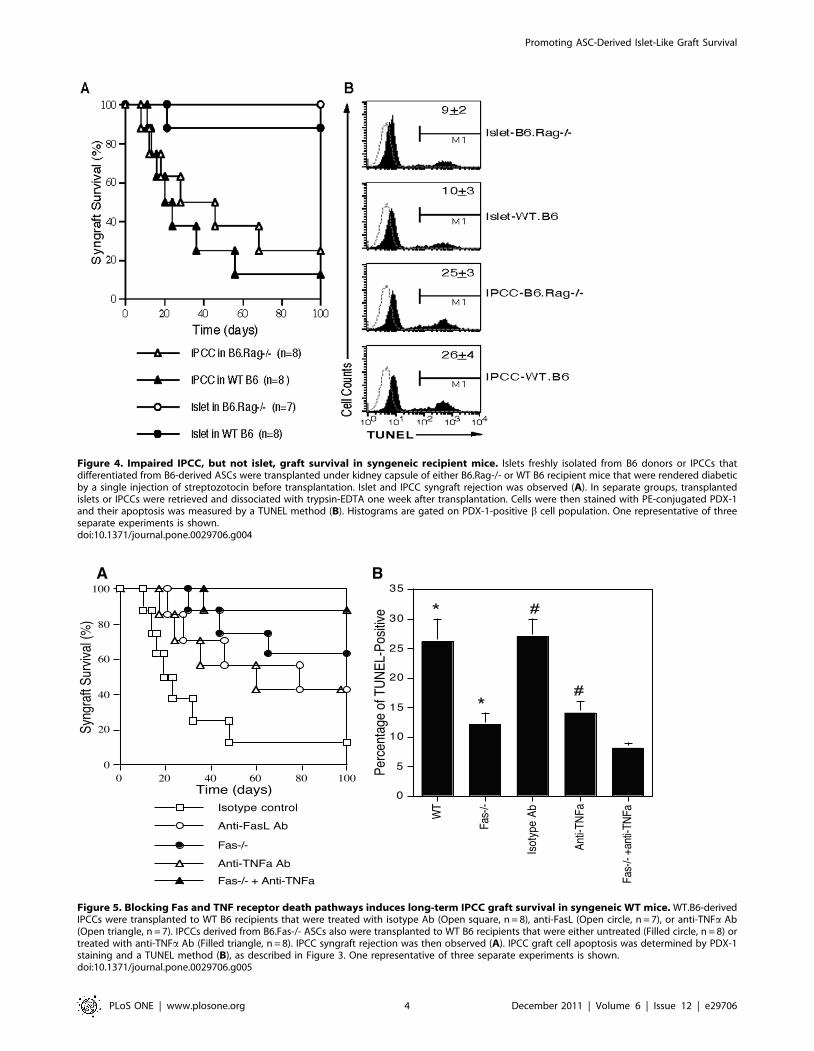
Figure 4. Impaired IPCC, but not islet, graft survival in syngeneic recipient mice. Islets freshly isolated from B6 donors or IPCCs thatdifferentiated from B6-derived ASCs were transplanted under kidney capsule of either B6.Rag-/- or WT B6 recipient mice that were rendered diabeticby a single injection of streptozotocin before transplantation. Islet and IPCC syngraft rejection was observed (A). In separate groups, transplantedislets or IPCCs were retrieved and dissociated with trypsin-EDTA one week after transplantation. Cells were then stained with PE-conjugated PDX-1and their apoptosis was measured by a TUNEL method (B). Histograms are gated on PDX-1-positive b cell population. One representative of threeseparate experiments is shown.doi:10.1371/journal.pone.0029706.g004
Figure 5. Blocking Fas and TNF receptor death pathways induces long-term IPCC graft survival in syngeneic WT mice. WT.B6-derivedIPCCs were transplanted to WT B6 recipients that were treated with isotype Ab (Open square, n = 8), anti-FasL (Open circle, n = 7), or anti-TNFa Ab(Open triangle, n = 7). IPCCs derived from B6.Fas-/- ASCs also were transplanted to WT B6 recipients that were either untreated (Filled circle, n = 8) ortreated with anti-TNFa Ab (Filled triangle, n = 8). IPCC syngraft rejection was then observed (A). IPCC graft cell apoptosis was determined by PDX-1staining and a TUNEL method (B), as described in Figure 3. One representative of three separate experiments is shown.doi:10.1371/journal.pone.0029706.g005
Promoting ASC-Derived Islet-Like Graft Survival
PLoS ONE | www.plosone.org 4 December 2011 | Volume 6 | Issue 12 | e29706
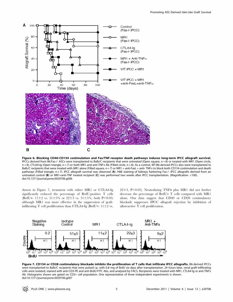
shown in Figure 7, treatment with either MR1 or CTLA4-Ig
significantly reduced the percentage of BrdU-positive T cells
(BrdU+: 1162 vs. 5165% or 2263 vs. 5165%, both P,0.05)
although MR1 was more effective in the suppression of graft-
infiltrating T cell proliferation than CTLA4-Ig (BrdU+: 1162 vs.
2263, P,0.05). Neutralizing TNFa plus MR1 did not further
decrease the percentage of BrdU+ T cells compared with MR1
alone. Our data suggest that CD40 or CD28 costimulatory
blockade suppresses IPCC allograft rejection by inhibition of
alloreactive T cell proliferation.
Figure 6. Blocking CD40-CD154 costimulation and Fas/TNF-receptor death pathways induces long-term IPCC allograft survival.IPCCs derived from B6.Fas-/- ASCs were transplanted to Balb/C recipients that were untreated (Open square, n = 8) or treated with MR1 (Open circle,n = 8), CTLA4-Ig (Open triangle, n = 7) or both MR1 and anti-TNFa Ab (Filled circle, n = 8). As a control, WT.B6-derived IPCCs also were transplanted toBalb/C recipients that were treated with MR1 alone (Filled square, n = 7) or MR1 + anti-FasL + anti- TNFa to block both CD154 costimulation and deathpathways (Filled triangle, n = 7). IPCC allograft survival was observed (A). H&E staining of kidneys harboring Fas-/- IPCC allografts derived from anuntreated control (B) or MR1+anti-TNF treated recipient (C) was performed two weeks after IPCC transplantation. (Magnification 6100).doi:10.1371/journal.pone.0029706.g006
Figure 7. CD154 or CD28 costimulatory blockade inhibits the proliferation of T cells that infiltrate IPCC allografts. B6-derived IPCCswere transplanted to Balb/C recipients that were pulsed i.p. with 0.8 mg of BrdU six days after transplantation. 24 hours later, renal graft-infiltratingcells were isolated, stained with anti-CD3-PE and anti-BrdU-FITC Abs, and analyzed by FACS. Recipients were treated with MR1, CTLA4-Ig or anti-TNFaAb. Histograms shown are gated on CD3+ cell population. One representative of three independent experiments is shown.doi:10.1371/journal.pone.0029706.g007
Promoting ASC-Derived Islet-Like Graft Survival
PLoS ONE | www.plosone.org 5 December 2011 | Volume 6 | Issue 12 | e29706
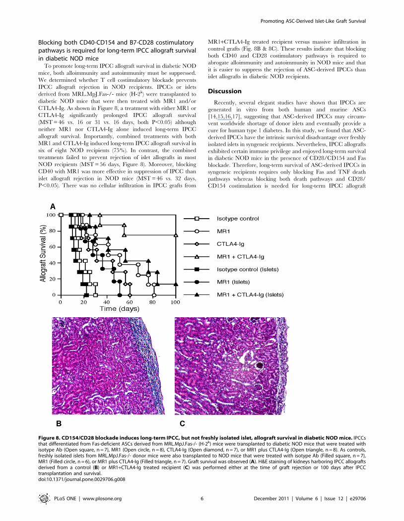
Blocking both CD40-CD154 and B7-CD28 costimulatorypathways is required for long-term IPCC allograft survivalin diabetic NOD mice
To promote long-term IPCC allograft survival in diabetic NOD
mice, both alloimmunity and autoimmunity must be suppressed.
We determined whether T cell costimulatory blockade prevents
IPCC allograft rejection in NOD recipients. IPCCs or islets
derived from MRL.MpJ.Fas-/- mice (H-2k) were transplanted to
diabetic NOD mice that were then treated with MR1 and/or
CTLA4-Ig. As shown in Figure 8, a treatment with either MR1 or
CTLA4-Ig significantly prolonged IPCC allograft survival
(MST = 46 vs. 16 or 31 vs. 16 days, both P,0.05) although
neither MR1 nor CTLA4-Ig alone induced long-term IPCC
allograft survival. Importantly, combined treatments with both
MR1 and CTLA4-Ig induced long-term IPCC allograft survival in
six of eight NOD recipients (75%). In contrast, the combined
treatments failed to prevent rejection of islet allografts in most
NOD recipients (MST = 56 days, Figure 8). Moreover, blocking
CD40 with MR1 was more effective in suppression of IPCC than
islet allograft rejection in NOD mice (MST = 46 vs. 32 days,
P,0.05). There was no cellular infiltration in IPCC grafts from
MR1+CTLA4-Ig treated recipient versus massive infiltration in
control grafts (Fig. 8B & 8C). These results indicate that blocking
both CD40 and CD28 costimulatory pathways is required to
abrogate alloimmunity and autoimmunity in NOD mice and that
it is easier to suppress the rejection of ASC-derived IPCCs than
islet allografts in diabetic NOD recipients.
Discussion
Recently, several elegant studies have shown that IPCCs are
generated in vitro from both human and murine ASCs
[14,15,16,17], suggesting that ASC-derived IPCCs may circum-
vent worldwide shortage of donor islets and eventually provide a
cure for human type 1 diabetes. In this study, we found that ASC-
derived IPCCs have the intrinsic survival disadvantage over freshly
isolated islets in syngeneic recipients. Nevertheless, IPCC allografts
exhibited certain immune privilege and enjoyed long-term survival
in diabetic NOD mice in the presence of CD28/CD154 and Fas
blockade. Therefore, long-term survival of ASC-derived IPCCs in
syngeneic recipients requires only blocking Fas and TNF death
pathways whereas blocking both death pathways and CD28/
CD154 costimulation is needed for long-term IPCC allograft
Figure 8. CD154/CD28 blockade induces long-term IPCC, but not freshly isolated islet, allograft survival in diabetic NOD mice. IPCCsthat differentiated from Fas-deficient ASCs derived from MRL.MpJ.Fas-/- (H-2k) mice were transplanted to diabetic NOD mice that were treated withisotype Ab (Open square, n = 7), MR1 (Open circle, n = 8), CTLA4-Ig (Open diamond, n = 7), or MR1 plus CTLA4-Ig (Open triangle, n = 8). As controls,freshly isolated islets from MRL.MpJ.Fas-/- donor mice were also transplanted to NOD mice that were treated with isotype Ab (Filled square, n = 7),MR1 (Filled circle, n = 6), or MR1 plus CTLA4-Ig (Filled triangle, n = 7). Graft survival was observed (A). H&E staining of kidneys harboring IPCC allograftsderived from a control (B) or MR1+CTLA4-Ig treated recipient (C) was performed either at the time of graft rejection or 100 days after IPCCtransplantation and survival.doi:10.1371/journal.pone.0029706.g008
Promoting ASC-Derived Islet-Like Graft Survival
PLoS ONE | www.plosone.org 6 December 2011 | Volume 6 | Issue 12 | e29706
survival in diabetic NOD mice. Thus, our studies suggest that
ASC-derived IPCCs may provide an effective cell replacement
therapy in human type 1 diabetes.
Using IPCCs that differentiated in vitro from murine ASCs
based on a recent protocol [16], we first compared their survival to
that of freshly-isolated intact islets in syngeneic recipients.
Surprisingly, we found that overwhelming majority of IPCC grafts
did not survive for long-term in syngeneic WT or Rag1-/- mice
whereas nearly all of syngeneic islet grafts survived for long-term
(.100 days). Our findings suggest that freshly isolated islets have a
survival advantage over in vitro-generated IPCCs upon trans-
plantation. Therefore, IPCC syngrafts die out in vivo even in the
absence of alloimmune responses, although exact mechanisms
responsible for their shortened survival in syngeneic hosts remain
to be defined. Our results are consistent with a previous study
showing that IPCC grafts, derived from murine embryonic stem
cells, have survived for only a average of two to three weeks in
syngeneic Rag-/- hosts [7], implying that IPCC grafts generated
from stem cells are incapable of surviving for long-term even in
the absence of alloimmune-based rejection. However, our find-
ings differ from a recent study showing that human ASC-derived
IPCC grafts have survived beyond 60 days in 50% of xenogeneic
recipient mice without immunosuppression [17]. It is unclear
how IPCC xenografts survived for such a long time without
immunosuppression.
We then investigated how to prevent IPCC graft dysfunction in
the absence of alloimmunity or any immune-based rejection,
which is an important step prior to in-depth IPCC allotransplan-
tation. Since Fas or TNF signaling mediates apoptosis in most cell
types, including b cells [21,22,23,24], we asked whether blocking
their signaling pathways would enhance IPCC graft survival or
restore their function in syngeneic recipient mice. Indeed, blocking
either Fas or TNF signaling significantly prolonged IPCC graft
survival and inhibited IPCC cell apoptosis in syngeneic recipients
while blocking both pathways induced long-term IPCC syngraft
survival in overwhelming majority, if not all, of recipient mice.
Our findings suggest that IPCC graft dysfunction due to passive
cell death, which is mediated by Fas and TNF signal pathways
but independent of alloimmune-based rejection, must also be
overcome upon IPCC allotransplantation.
We next examined IPCC allograft survival in alloimmune-
competent WT recipient mice. We found that IPCC allografts
derived from both Fas-replete and Fas-deficient ASCs were acutely
rejected in allogeneic WT recipient mice, suggesting that ASC-
derived IPCCs are highly immunogenic and can evoke acute
allograft rejection. Blocking CD40-CD154 costimulation induced
Fas-deficient IPCC allograft survival for long-term in 63% of
recipient mice. Given that the same percentage of Fas-deficient
IPCC grafts survived in syngeneic recipients without CD154
costimulatory blockade, we conclude that blocking CD40-CD154
costimulation is sufficient to suppress IPCC allograft rejection
mediated by alloimmune responses. Furthermore, simultaneously
blocking CD154, Fas and TNF-receptor signaling induced long-
term IPCC allograft survival in overwhelming majority, if not all,
of recipient mice. Taken together, both death signaling of b cells
and alloreactivity of recipients need to be suppressed in order to
achieve long-term IPCC allograft survival.
In this study, we determined whether costimulatory blockade
promotes long-term IPCC allograft survival in NOD mice that are
closely relevant to human type 1 diabetes. Costimulatory blockade
has been shown to suppress alloimmune responses and induce
long-term allograft survival [25,26,27]. Previous studies have also
shown that blocking both CD28 and CD154 costimulatory
pathways prevents autoimmune diabetes induced by transfer of
transgenic T cells in NOD.SCID mice [28] but fails to induce
long-term islet allograft survival in NOD recipients [29],
suggesting that CD28/CD154 blockade is sufficient to suppress
autoimmunity but insufficient to simultaneously suppress both
alloimmunity and autoimmunity in NOD recipients. In contrast,
another study has shown that ICOS/CD154 blockade is capable
of inducing islet allograft tolerance and preventing autoimmune
diabetes in NOD mice [30]. Here we found that CD28/CD154
blockade induces long-term Fas-/- IPCC, but not Fas-/- islet,
allograft survival in diabetic NOD recipients. We utilized Fas-
deficient IPCCs because Fas-replete IPCC grafts died out over
time even in syngeneic recipients due to Fas-mediated cell death.
Our data have further confirmed that CD28/CD154 blockade
does not induce long-term islet allograft survival in diabetic NOD
recipients. On the other hand, we have demonstrated for the first
time that blocking CD28 and CD154 costimulatory signaling is
capable of inducing long-term survival of ASC-derived IPCC
allografts in diabetic NOD mice, although a recent study has
shown that blocking CD28/CD154/LFA-1 enhances engraftment
of allogeneic ESC-derived endothelial cells in non-diabetic
environment [31].
It is unclear whether CD154 costimulatory blockade can be
translated to the clinic in the future, although it prolongs IPCC
allograft survival. Clinical trials using anti-CD154 Ab were halted
due to its thromboembolic side effects [32,33]. The progress in its
clinical application, hence, has been hampered. Recently, Abs
against its counter receptor, CD40, have been sought as an
alternative to blocking CD40/CD154 costimulatory pathway.
These promising Abs have been shown to potently suppress
allograft rejection in both mice and non-human primates [34,35].
However, it remains to be defined whether they will also cause the
same side effects in the future clinical trials.
Our finding indicates that it is easier to suppress the rejection of
ASC-derived IPCC allografts than freshly isolated islet allografts in
NOD recipient mice once passive cell death is blocked. This is
consistent with previous studies showing that ESC-derived IPCCs
or tissues exhibit a certain degree of immune privilege [6,7,36].
Therefore, ASC-derived IPCCs enjoy at least two advantages over
freshly isolated islets: relatively immune privilege and unlimited
sources while displaying an intrinsic survival disadvantage that is
unrelated to alloimmune-based rejection. A recent study has
demonstrated that transplantation of IPCCs derived from human
umbilical cord mesenchymal stem cells alleviates hyperglycemia in
diabetic NOD mice without any immunosuppressive treatment
[37], although it was not observed how long IPCC xenografts
survived and how their rejection could be suppressed. Taken
together, previous and our current studies indicate that clinical
application of ASC-derived IPCCs for treating type 1 diabetes is
no longer elusive.
Materials and Methods
Ethics StatementAll animal experiments were approved by the Animal Care and
Use Committee of the University of Texas Health Science Center
(animal approval ID: 463B), Tyler, TX.
MiceWild-type BALB/c (H-2d) and C57BL/6 (H-2b) mice were
purchased from National Cancer Institute (NIH, Bethesda, MD,
USA). B6.Rag1-/-, NOD, B6.MRL.Fas-/- (H-2b) and MRL/
MpJ.Fas-/- (H-2k) were purchased from the Jackson Laboratory.
All mice were aged 6–8 weeks when experiments were initiated.
They were housed in a specific pathogen-free environment.
Promoting ASC-Derived Islet-Like Graft Survival
PLoS ONE | www.plosone.org 7 December 2011 | Volume 6 | Issue 12 | e29706
Adipose-derived stem cell isolation and cultureAdipose tissues from epididymal fat and subcutaneous fat of
anterior abdominal wall of donor mice were minced and digested
with 0.1% type II collagenase (Invitrogen) in phosphate-buffered
saline (PBS) for 30 minutes at 37uC with gentle stirring. Cell
suspensions were cultured in Dulbecco’s modified Eagle’s medium
(DMEM)/Ham’s F-12 with 10% FBS and 100 U/ml penicillin,
0.1 mg/ml streptomycin (Invitrogen) at 5% CO2, 37uC. Cells
were passaged under maintenance culture conditions. All
experiments were performed using ASCs at 4–6 passages.
In vitro differentiation of ASCs into IPCCsASCs at p4-p6 were suspended in medium M (DMEM/F12
at 1:1) with 17.5 mM glucose, 1% BSA and 5 mg/l insulin-
transferrin-selenium, plated on ultralow attachment tissue culture
plates (Fisher Scientific) at 16106 cells per well, and then cultured
in medium MA (M with 4 nM activin A, R&D System) for two
days, medium MB (M with 0.3 mM taurine) for two days, and
medium MC (M with 100 nM glucagon-like peptide-1, 1 mM
nicotinamide and 16 nonessential amino acids) for five days as
described previously [16]. All chemicals and supplements were
purchased from Sigma Aldrich unless otherwise indicated.
Flow cytometric analysis for phenotypingImmunophenotyping of ASCs were performed using fluores-
cent-conjugated antibodies against mouse antigens MHC I (H-
2Kb), MHC II (I-Ab), CD44, CD45, CD29, CD80, CD90, CD105
and Sca-1 (Biolegend). ASCs at the fourth passage were released
by 0.25% trypsin-EDTA. A total of 16106 cells were incubated
with fluorescent-conjugated Abs for 30 min at room temperature.
Cells finally were analyzed using FACSCalibur.
RT-PCR detection of pancreatic gene expression by IPCCsTotal RNA was isolated from undifferentiated ASCs, IPCCs
and islets using TRIzol extraction method (TRIzol Reagent,
Invitrogen). The extracted RNA was subject to cDNA synthesis
using the Superscript III First-Strand Synthesis system for RT-
PCR (Invitrogen). The PCR cycling conditions varied depending
on different pancreatic genes and primers, which were listed in the
supporting information (Table S1).
Transplantation of islets or IPCCs,400 donor islets or 800 IPCCs that differentiated from ASCs
were transplanted under the kidney capsule of recipient mice as
described in our previous studies [38,39]. Previous studies and our
preliminary data have determined that at least 800–900 IPCCs are
required to reverse hyperglycemia. Recipient mice were rendered
diabetic by a single injection of streptozotocin (Sigma) (180 mg/kg
for WT mice and 200 mg/kg for Rag-/- recipients) 8–10 days
before transplantation. Primary graft function was defined as
blood glucose under 200 mg/dl for 48 hours after transplantation.
Graft rejection was defined as a rise in blood glucose to .300 mg/
dl for three consecutive days after primary function. Rejection was
also confirmed by histology showing cellular infiltration. In some
recipients that survived for long-term, the kidney harboring IPCC
grafts was removed to confirm that normalizing hyperglycemia in
diabetic mice was caused by IPCC transplantation.
Treatments of recipient mice with AbsRecipient mice were treated with MR1 (anti-CD40L), CTLA4-Ig,
or anti-TNFaAb (Bio-Express Inc, West Lebanon, NH) at 0.25 mg on
days 0, 2, 4, and 6 while anti-FasL blocking Ab (BD Biosciences) was
administered at 0.1 mg on days 0, 2, 4, and 6 after transplantation.
Histology and immunofluorescence stainingKidney samples containing IPCC grafts from transplanted mice
were harvested, fixed in 10% formalin, embedded in paraffin,
sectioned with a microtome at 4 mm thickness, stained with
haematoxylin/eosin (H&E), and observed for cell infiltration under
the light microscope. Moreover, both IPCCs and kidneys harboring
IPCC grafts were fixed, embedded and sectioned as described above.
Sections were used for immunofluorescence staining of murine
insulin and c-peptide. Briefly, slides were deparaffinized in xylene,
rehydrated in descending grades of alcohol, immersed in citrate
buffer in a microwave oven with heating for 20 min at 95uC, and
treated with 0.3% hydrogen peroxide in methanol for 10 min.
Sections were then placed in diluted (10%) normal serum for 20 min,
covered with primary antibody (Cell Signaling) overnight at 4uC and
incubated with 1:500-diluted Alexa Fluor 488- or 568-conjugated
secondary Ab solution (Invitrogen) at room temperature for 1 hour.
They were finally visualized under a fluorescence microscope.
Isolation of tissue-infiltrating cellsTissue-infiltrating cells were isolated as described in our
previous publications [40,41]. Briefly, the kidneys harboring islet
grafts were perfused in situ with heparinized 0.9% saline. They
were then minced and digested at 37uC for 30 min in 20 ml
RPMI-1640 medium containing 5% FCS and 350 u/ml collage-
nase (Sigma, St. Louis, MO). To clear the debris, cell suspen-
sions were rapidly passed down 70 mm cell strainer, then mixed
with Percoll solution (Sigma) to a concentration of 30%, and
centrifuged at 2000 rpm for 15 minutes at room temperature. The
pellet was re-suspended and stained before analysis.
Analysis of T cell proliferation in vivo by 5-Bromo-29-Deoxyuridine (BrdU) labeling
Recipient mice were pulsed i.p. with 0.8 mg of BrdU (Sigma) six
days after transplantation. 24 hours later, renal graft-infiltrating
cells were isolated and first stained with anti-CD3-PE Ab. Cells
were then fixed in 70% ethanol followed by 1% paraformaldehyde
and incubated with 50 Units/ml of DNase I (Sigma). Cells were
finally stained with anti-BrdU-FITC (BD Biosciences) and
analyzed by a FACSCalibur as described previously [40,41].
Analysis of insulin-producing cell apoptosis in vivo by aTUNEL method
IPCCs or islets were retrieved and dissociated with 0.25% trypsin-
EDTA (Sigma-Aldrich) at 37uC for 10 minutes. To detect cell
apoptosis, cells were fixed in 2% paraformaldehyde, permeabilized
with 0.1% Triton X-100 solution, and labeled with fluorescein-
tagged deoxyuridine triphosphate (dUTP) by the terminal deox-
ynucleotidyl transferase-mediated dUTP nick-end labeling (TU-
NEL) method according to the manufacture’s instructions (Roche
Applied Science, Mannheim, Germany) [40,42].
Statistical AnalysisData are presented as mean 6 SEM and represent an average
of at least three independent experiments. Comparisons of the
mean among different groups were done using the Student t test
and ANOVA. The analysis of graft survival data was performed
using the Kaplan-Meier method (log-rank test). A value of P,0.05
was considered statistically significant.
Supporting Information
Figure S1 Insulin release by IPCCs and islets in vitro.Day-10 IPCCs or freshly isolated islets were cultured in DMEM-LG
Promoting ASC-Derived Islet-Like Graft Survival
PLoS ONE | www.plosone.org 8 December 2011 | Volume 6 | Issue 12 | e29706
(low glucose) containing 0.5% BSA for 10 hours, washed and then
stimulated or incubated in DMEM-HG (high glucose) media at
37uC for two hours. Insulin released into the media was measured
using ELISA kit (Mercodia, Winston Salem, NC) according to the
manufacturer’s instructions. Undifferentiated ASCs were also
utilized as a control. Both IPCCs and islets produced significant
amount of insulin in high-glucose condition in vitro while
undifferentiated ASCs did not. One of three separate experiments
is shown.
(PDF)
Table S1 Primers for RT-PCR. The primer sequences,
including the sense and anti-sense, for RT-PCR for pancreatic
gene expression are listed.
(PDF)
Author Contributions
Conceived and designed the experiments: SZ ZD. Performed the
experiments: SZ HD. Analyzed the data: SZ ZD. Contributed reagents/
materials/analysis tools: NW YM. Wrote the paper: SZ ZD.
References
1. Assady S, Maor G, Amit M, Itskovitz-Eldor J, Skorecki KL, et al. (2001) Insulin
production by human embryonic stem cells. Diabetes 50: 1691–1697.2. Lumelsky N, Blondel O, Laeng P, Velasco I, Ravin R, et al. (2001)
Differentiation of embryonic stem cells to insulin-secreting structures similar to
pancreatic islets. Science 292: 1389–1394.3. Soria B, Roche E, Berna G, Leon-Quinto T, Reig JA, et al. (2000) Insulin-
secreting cells derived from embryonic stem cells normalize glycemia instreptozotocin-induced diabetic mice. Diabetes 49: 157–162.
4. D’Amour KA, Bang AG, Eliazer S, Kelly OG, Agulnick AD, et al. (2006)Production of pancreatic hormone-expressing endocrine cells from human
embryonic stem cells. Nat Biotechnol 24: 1392–1401.
5. Jiang J, Au M, Lu K, Eshpeter A, Korbutt G, et al. (2007) Generation of insulin-producing islet-like clusters from human embryonic stem cells. Stem Cells 25:
1940–1953.6. Boyd AS, Wood KJ (2010) Characteristics of the early immune response
following transplantation of mouse ES cell derived insulin-producing cell
clusters. PLoS One 5: e10965.7. Wu DC, Boyd AS, Wood KJ (2008) Embryonic stem cells and their
differentiated derivatives have a fragile immune privilege but still representnovel targets of immune attack. Stem Cells 26: 1939–1950.
8. Tang DQ, Cao LZ, Burkhardt BR, Xia CQ, Litherland SA, et al. (2004) In vivo
and in vitro characterization of insulin-producing cells obtained from murinebone marrow. Diabetes 53: 1721–1732.
9. Yang L, Li S, Hatch H, Ahrens K, Cornelius JG, et al. (2002) In vitro trans-differentiation of adult hepatic stem cells into pancreatic endocrine hormone-
producing cells. Proc Natl Acad Sci U S A 99: 8078–8083.10. Seaberg RM, Smukler SR, Kieffer TJ, Enikolopov G, Asghar Z, et al. (2004)
Clonal identification of multipotent precursors from adult mouse pancreas that
generate neural and pancreatic lineages. Nat Biotechnol 22: 1115–1124.11. Schaffler A, Buchler C (2007) Concise review: adipose tissue-derived stromal
cells–basic and clinical implications for novel cell-based therapies. Stem Cells 25:818–827.
12. Zuk PA, Zhu M, Ashjian P, De Ugarte DA, Huang JI, et al. (2002) Human
adipose tissue is a source of multipotent stem cells. Mol Biol Cell 13: 4279–4295.13. Kim SC, Han DJ, Lee JY (2010) Adipose tissue derived stem cells for
regeneration and differentiation into insulin-producing cells. Curr Stem Cell ResTher 5: 190–194.
14. Timper K, Seboek D, Eberhardt M, Linscheid P, Christ-Crain M, et al. (2006)Human adipose tissue-derived mesenchymal stem cells differentiate into insulin,
somatostatin, and glucagon expressing cells. Biochem Biophys Res Commun
341: 1135–1140.15. Lee J, Han DJ, Kim SC (2008) In vitro differentiation of human adipose tissue-
derived stem cells into cells with pancreatic phenotype by regenerating pancreasextract. Biochem Biophys Res Commun 375: 547–551.
16. Chandra VGS, Phadnis S, Nair PD, Bhonde RR (2009) Generation of
pancreatic hormone-expressing islet-like cell aggregates from murine adiposetissue-derived stem cells. Stem Cells 27: 1941–1953.
17. Kang HM, Kim J, Park S, Kim H, Kim KS, et al. (2009) Insulin-secreting cellsfrom human eyelid-derived stem cells alleviate type I diabetes in immunocom-
petent mice. Stem Cells 27: 1999–2008.18. Lin G, Wang G, Liu G, Yang LJ, Chang LJ, et al. (2009) Treatment of type 1
diabetes with adipose tissue-derived stem cells expressing pancreatic duodenal
homeobox 1. Stem Cells Dev 18: 1399–1406.19. Kajiyama H, Hamazaki TS, Tokuhara M, Masui S, Okabayashi K, et al. (2010)
Pdx1-transfected adipose tissue-derived stem cells differentiate into insulin-producing cells in vivo and reduce hyperglycemia in diabetic mice. Int J Dev Biol
54: 699–705.
20. Di Rocco G, Iachininoto MG, Tritarelli A, Straino S, Zacheo A, et al. (2006)Myogenic potential of adipose-tissue-derived cells. J Cell Sci 119: 2945–2952.
21. Pearl-Yafe M, Yolcu ES, Yaniv I, Stein J, Shirwan H, et al. (2006) The dual roleof Fas-ligand as an injury effector and defense strategy in diabetes and islet
transplantation. Bioessays 28: 211–222.22. Giannoukakis N, Rudert WA, Ghivizzani SC, Gambotto A, Ricordi C, et al.
(1999) Adenoviral gene transfer of the interleukin-1 receptor antagonist protein
to human islets prevents IL-1beta-induced beta-cell impairment and activationof islet cell apoptosis in vitro. Diabetes 48: 1730–1736.
23. Silva DG, Petrovsky N, Socha L, Slattery R, Gatenby P, et al. (2003)
Mechanisms of accelerated immune-mediated diabetes resulting from islet beta
cell expression of a Fas ligand transgene. J Immunol 170: 4996–5002.
24. Judge TA, Desai NM, Yang Z, Rostami S, Alonso L, et al. (1998) Utility of
adenoviral-mediated Fas ligand gene transfer to modulate islet allograft survival.
Transplantation 66: 426–434.
25. Larsen C, Elwood E, Alexander D, Ritchie S, Hendrix R, et al. (1996) Long-
term acceptance of skin and cardiac allografts after blocking CD40 and CD28
pathways. Nature 381: 434–438.
26. Wekerle T, Kurtz J, Bigenzahn S, Takeuchi Y, Sykes M (2002) Mechanisms of
transplant tolerance induction using costimulatory blockade. Curr Opin
Immunol 14: 592–600.
27. Li XC, Rothstein DM, Sayegh MH (2009) Costimulatory pathways in
transplantation: challenges and new developments. Immunol Rev 229: 271–293.
28. Rigby MR, Trexler AM, Pearson TC, Larsen CP (2008) CD28/CD154
blockade prevents autoimmune diabetes by inducing nondeletional tolerance
after effector t-cell inhibition and regulatory T-cell expansion. Diabetes 57:
2672–2683.
29. Demirci G, Strom TB, Li XC (2003) Islet allograft rejection in nonobese diabetic
mice involves the common gamma-chain and CD28/CD154-dependent and -
independent mechanisms. J Immunol 171: 3878–3885.
30. Nanji SA, Hancock WW, Luo B, Schur CD, Pawlick RL, et al. (2006)
Costimulation blockade of both inducible costimulator and CD40 ligand induces
dominant tolerance to islet allografts and prevents spontaneous autoimmune
diabetes in the NOD mouse. Diabetes 55: 27–33.
31. Pearl JI, Lee AS, Leveson-Gower DB, Sun N, Ghosh Z, et al. (2011) Short-term
immunosuppression promotes engraftment of embryonic and induced pluripo-
tent stem cells. Cell Stem Cell 8: 309–317.
32. Kanmaz T, Fechner JJ, Jr., Torrealba J, Kim HT, Dong Y, et al. (2004)
Monotherapy with the novel human anti-CD154 monoclonal antibody ABI793
in rhesus monkey renal transplantation model. Transplantation 77: 914–920.
33. Kawai T, Andrews D, Colvin RB, Sachs DH, Cosimi AB (2000) Thromboem-
bolic complications after treatment with monoclonal antibody against CD40
ligand. Nature medicine 6: 114.
34. Gilson CR, Milas Z, Gangappa S, Hollenbaugh D, Pearson TC, et al. (2009)
Anti-CD40 monoclonal antibody synergizes with CTLA4-Ig in promoting long-
term graft survival in murine models of transplantation. Journal of immunology
183: 1625–1635.
35. Aoyagi T, Yamashita K, Suzuki T, Uno M, Goto R, et al. (2009) A human anti-
CD40 monoclonal antibody, 4D11, for kidney transplantation in cynomolgus
monkeys: induction and maintenance therapy. American journal of transplan-
tation : official journal of the American Society of Transplantation and the
American Society of Transplant Surgeons 9: 1732–1741.
36. Lui KO, Boyd AS, Cobbold SP, Waldmann H, Fairchild PJ (2010) A role for
regulatory T cells in acceptance of ESC-derived tissues transplanted across an
major histocompatibility complex barrier. Stem Cells 28: 1905–1914.
37. Wang HS, Shyu JF, Shen WS, Hsu HC, Chi TC, et al. (2011) Transplantation
of Insulin-Producing Cells Derived From Umbilical Cord Stromal Mesenchymal
Stem Cells to Treat NOD Mice. Cell Transplant 20: 455–466.
38. Liu ZW, Dai H, Wan N, Wang T, Bertera S, et al. (2007) Suppression of
memory CD8 T cell generation and function by tryptophan catabolism.
J Immunol 178: 4260–4266.
39. Wan N, Dai H, Wang T, Moore Y, Zheng XX, et al. (2008) Bystander central
memory but not effector memory CD8+ T cells suppress allograft rejection.
J Immunol 180: 113–121.
40. Dai Z, Li Q, Wang Y, Gao G, Diggs LS, et al. (2004) CD4+CD25+ regulatory T
cells suppress allograft rejection mediated by memory CD8+ T cells via a CD30-
dependent mechanism. J Clin Invest 113: 310–317.
41. Wang Y, Dai H, Liu Z, Cheng X, Tellides G, et al. (2006) Neutralizing IL-7
promotes long-term allograft survival induced by CD40/CD40L costimulatory
blockade. Am J Transplant 6: 2851–2860.
42. Wang T, Dai H, Wan N, Moore Y, Dai Z (2008) The role for monocyte
chemoattractant protein-1 in the generation and function of memory CD8+ T
cells. J Immunol 180: 2886–2893.
Promoting ASC-Derived Islet-Like Graft Survival
PLoS ONE | www.plosone.org 9 December 2011 | Volume 6 | Issue 12 | e29706