Prodrugs for Skin Delivery of Menahydroquinone-4, an Active ...
13
International Journal of Molecular Sciences Article Prodrugs for Skin Delivery of Menahydroquinone-4, an Active Form of Vitamin K 2(20) , Could Overcome the Photoinstability and Phototoxicity of Vitamin K 2(20) Shotaro Goto † , Shuichi Setoguchi † , Hirofumi Yamakawa, Daisuke Watase, Kazuki Terada, Kazuhisa Matsunaga *, Yoshiharu Karube and Jiro Takata * Faculty of Pharmaceutical Sciences, Fukuoka University, Nanakuma, Jonan-ku, Fukuoka 814-0180, Japan; [email protected] (S.G.); [email protected] (S.S.); [email protected] (H.Y.); [email protected] (D.W.); [email protected] (K.T.); [email protected] (Y.K.) * Correspondence: [email protected] (K.M.); [email protected] (J.T.); Tel.: +81-92-871-6631 (ext. 6672) (K.M.); +81-92-871-6631 (ext. 6662) (J.T.) † Both of the authors are listed as co-first authors and contributed equally to this work. Received: 27 March 2019; Accepted: 22 May 2019; Published: 24 May 2019 Abstract: The effective delivery of menahydroquinone-4 (MKH), an active form of menaquinone-4 (MK-4, vitamin K 2(20) ), to the skin is beneficial in the treatment of various skin pathologies. However, its delivery through the application of MK-4 to the skin is hampered due to the photoinstability and phototoxicity of MK-4. This study aimed to evaluate the potential of ester prodrugs of MKH for its delivery into the skin to avoid the abovementioned issues. The ester prodrugs, MKH 1,4-bis-N,N-dimethylglycinate hydrochloride (MKH-DMG) and MKH 1,4-bis-hemisuccinate (MKH-SUC), were prepared using our previously reported methods. Photostability was determined under artificial sunlight and multi-wavelength light irradiation, phototoxicity was determined by intracellular ROS formation and cell viability of UVA-irradiated human epidermal keratinocyte cells (HaCaT), and delivery of MKH into HaCaT cells was assessed by measuring menaquinone-4 epoxide (MKO) levels. MKH prodrugs showed higher photostability than MK-4. Although MK-4 induced cellular ROS and reduced cell viability after UVA irradiation, MKH prodrugs did not affect either ROS generation or cell viability. MKH prodrugs enhanced intracellular MKO, indicating effective delivery of MKH and subsequent carboxylation activity. In conclusion, these MKH prodrugs show potential for the delivery of MKH into the skin without photoinstability and phototoxicity. Keywords: menaquinone-4; menahydroquinone-4; prodrug; drug delivery system; phototoxicity; photostability; photodegradation; skin application; vitamin K 1. Introduction Menaquinone-4 (MK-4) or vitamin K 2(20) is an important vitamin K compound used clinically. Menahydroquinone-4 (MKH), the fully reduced and active form of MK-4, is a cofactor for γ-glutamyl carboxylase (GGCX), which catalyzes the post-transcriptional carboxylation of vitamin K-dependent proteins. MKH is produced by the enzymatic conversion of vitamin K 1 and K 3 in the body [1]. Since MKH is readily oxidized to MK-4, MK-4 is available for clinical use. Skin application of vitamin K exhibits several beneficial effects such as suppressing pigmentation and resolving bruising [2–4], prophylactically limiting the occurrence of acneiform side effects in patients receiving the monoclonal antibody cetuximab [5–8], and promoting wound healing [9]. Because vitamin K is highly unstable in light, it should be strictly protected from light exposure during manufacturing, preparation, storage, and treatment [10–12]. Furthermore, vitamin K is phototoxic against cultured epidermis upon ultraviolet A (UVA) irradiation (dose 6 J/cm 2 )[13]. Given Int. J. Mol. Sci. 2019, 20, 2548; doi:10.3390/ijms20102548 www.mdpi.com/journal/ijms
-
Upload
khangminh22 -
Category
Documents
-
view
2 -
download
0
Transcript of Prodrugs for Skin Delivery of Menahydroquinone-4, an Active ...
International Journal of
Molecular Sciences
Article
Prodrugs for Skin Delivery of Menahydroquinone-4,an Active Form of Vitamin K2(20), Could Overcome thePhotoinstability and Phototoxicity of Vitamin K2(20)
Shotaro Goto †, Shuichi Setoguchi †, Hirofumi Yamakawa, Daisuke Watase, Kazuki Terada,Kazuhisa Matsunaga *, Yoshiharu Karube and Jiro Takata *
Faculty of Pharmaceutical Sciences, Fukuoka University, Nanakuma, Jonan-ku, Fukuoka 814-0180, Japan;[email protected] (S.G.); [email protected] (S.S.); [email protected] (H.Y.);[email protected] (D.W.); [email protected] (K.T.); [email protected] (Y.K.)* Correspondence: [email protected] (K.M.); [email protected] (J.T.);
Tel.: +81-92-871-6631 (ext. 6672) (K.M.); +81-92-871-6631 (ext. 6662) (J.T.)† Both of the authors are listed as co-first authors and contributed equally to this work.
Received: 27 March 2019; Accepted: 22 May 2019; Published: 24 May 2019�����������������
Abstract: The effective delivery of menahydroquinone-4 (MKH), an active form of menaquinone-4(MK-4, vitamin K2(20)), to the skin is beneficial in the treatment of various skin pathologies. However,its delivery through the application of MK-4 to the skin is hampered due to the photoinstabilityand phototoxicity of MK-4. This study aimed to evaluate the potential of ester prodrugs ofMKH for its delivery into the skin to avoid the abovementioned issues. The ester prodrugs,MKH 1,4-bis-N,N-dimethylglycinate hydrochloride (MKH-DMG) and MKH 1,4-bis-hemisuccinate(MKH-SUC), were prepared using our previously reported methods. Photostability was determinedunder artificial sunlight and multi-wavelength light irradiation, phototoxicity was determined byintracellular ROS formation and cell viability of UVA-irradiated human epidermal keratinocyte cells(HaCaT), and delivery of MKH into HaCaT cells was assessed by measuring menaquinone-4 epoxide(MKO) levels. MKH prodrugs showed higher photostability than MK-4. Although MK-4 inducedcellular ROS and reduced cell viability after UVA irradiation, MKH prodrugs did not affect eitherROS generation or cell viability. MKH prodrugs enhanced intracellular MKO, indicating effectivedelivery of MKH and subsequent carboxylation activity. In conclusion, these MKH prodrugs showpotential for the delivery of MKH into the skin without photoinstability and phototoxicity.
Keywords: menaquinone-4; menahydroquinone-4; prodrug; drug delivery system; phototoxicity;photostability; photodegradation; skin application; vitamin K
1. Introduction
Menaquinone-4 (MK-4) or vitamin K2(20) is an important vitamin K compound used clinically.Menahydroquinone-4 (MKH), the fully reduced and active form of MK-4, is a cofactor for γ-glutamylcarboxylase (GGCX), which catalyzes the post-transcriptional carboxylation of vitamin K-dependentproteins. MKH is produced by the enzymatic conversion of vitamin K1 and K3 in the body [1]. SinceMKH is readily oxidized to MK-4, MK-4 is available for clinical use.
Skin application of vitamin K exhibits several beneficial effects such as suppressing pigmentationand resolving bruising [2–4], prophylactically limiting the occurrence of acneiform side effects inpatients receiving the monoclonal antibody cetuximab [5–8], and promoting wound healing [9].
Because vitamin K is highly unstable in light, it should be strictly protected from light exposureduring manufacturing, preparation, storage, and treatment [10–12]. Furthermore, vitamin K isphototoxic against cultured epidermis upon ultraviolet A (UVA) irradiation (dose 6 J/cm2) [13]. Given
Int. J. Mol. Sci. 2019, 20, 2548; doi:10.3390/ijms20102548 www.mdpi.com/journal/ijms

Int. J. Mol. Sci. 2019, 20, 2548 2 of 13
that when vitamin K is applied to exposed skin, it is difficult to protect it from light exposure, and thesephysicochemical properties of MK-4 limit its use as a skin application and hamper its beneficial effects.
It has been shown that the main photodegradation product of MK-4 is MK-4 chromenol (MKC),which is produced from the naphthoquinone structure and isoprenyl side chain of MK-4. We havepreviously reported that ester-type prodrugs of MKH, such as the 1,4-bis-N,N-dimethylglycinate(MKH-DMG) and hemi-succinate (MKH-SUC) derivatives, can act as delivery systems for MKHwithout a reductive activation step [14–17]. Since these compounds do not contain the naphthoquinonestructure, these prodrugs may overcome the photoinstability and phototoxicity problems. It hasbeen reported that the partition coefficient (log P) of MKH-DMG at physiological pH 7.4 is 3.66 [14];this value indicates that MKH-DMG has good lipophilicity for membrane permeation. In addition,MKH-DMG and MKH-SUC could deliver MKH into hepatocellular carcinoma (HCC) cells moreeffectively than MK-4 [17]. Thus, MKH-DMG and MKH-SUC were considered to be good candidatecompounds for skin application. In the current study, we examined their effectiveness as photostableand low-phototoxicity prodrugs of MKH in order to further development of a vitamin K skin application.The concept underlying the dermal delivery system of MKH is shown in Figure 1.
Int. J. Mol. Sci. 2019, 20 FOR PEER REVIEW 2
that when vitamin K is applied to exposed skin, it is difficult to protect it from light exposure, and these physicochemical properties of MK-4 limit its use as a skin application and hamper its beneficial effects.
It has been shown that the main photodegradation product of MK-4 is MK-4 chromenol (MKC), which is produced from the naphthoquinone structure and isoprenyl side chain of MK-4. We have previously reported that ester-type prodrugs of MKH, such as the 1,4-bis-N,N-dimethylglycinate (MKH-DMG) and hemi-succinate (MKH-SUC) derivatives, can act as delivery systems for MKH without a reductive activation step [14–17]. Since these compounds do not contain the naphthoquinone structure, these prodrugs may overcome the photoinstability and phototoxicity problems. It has been reported that the partition coefficient (log P) of MKH-DMG at physiological pH 7.4 is 3.66 [14]; this value indicates that MKH-DMG has good lipophilicity for membrane permeation. In addition, MKH-DMG and MKH-SUC could deliver MKH into hepatocellular carcinoma (HCC) cells more effectively than MK-4 [17]. Thus, MKH-DMG and MKH-SUC were considered to be good candidate compounds for skin application. In the current study, we examined their effectiveness as photostable and low-phototoxicity prodrugs of MKH in order to further development of a vitamin K skin application. The concept underlying the dermal delivery system of MKH is shown in Figure 1.
Figure 1. Concept of dermal delivery of MKH that avoids photoinstability and phototoxicity using a prodrug approach. Solid line: the advocated or expected processes based on the literatures and obtained results in this study. Dashed line: the possible process during the drug measurement.
2. Results
Figure 1. Concept of dermal delivery of MKH that avoids photoinstability and phototoxicity usinga prodrug approach. Solid line: the advocated or expected processes based on the literatures andobtained results in this study. Dashed line: the possible process during the drug measurement.
Int. J. Mol. Sci. 2019, 20, 2548 3 of 13
2. Results
2.1. Photostability of MKH-Ester Derivatives in Artificial Sunlight
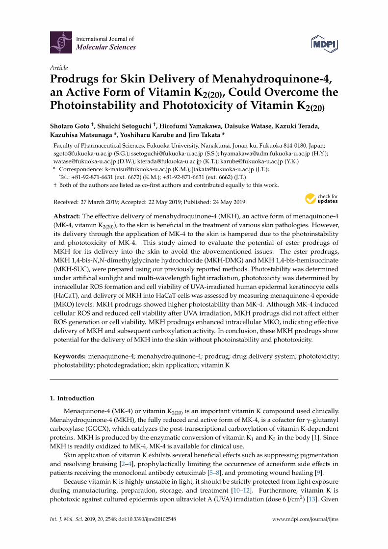
To evaluate the photostability of the MK-4 and MKH derivatives in skin application, 1 µM solutions ofMK-4, MKH-DMG, or MKH-SUC in ethanol were irradiated with artificial sunlight (12,000 lx) at 25 ◦C withand without shading, and the final amounts of MKH derivatives and their monoesters, MK-4 and MK-4chromenol, were determined by LC-MS/MS. The relative irradiance of artificial sunlight at wavelengthsof 300 to 800 nm is almost equal to that of natural sunlight, according to the manufacturer. MK-4 andMKH-DMG concentrations decreased in sunlight irradiation according to an apparent first order rate(Figure 2A), but were unchanged under shading. MKH-SUC concentration decreased according to anapparent first order rate both with and without shading, although the rate of decrease was acceleratedwithout shading. Apparent first order constants and half-lives are listed in Table 1. MKH-DMG andMKH-SUC were more photostable than MK-4 by ca. 50- and 3-fold, respectively. These results indicate thatthe use of MKH ester derivatives is a valid method to overcome the photolability of MK-4. In particular,MKH-DMG exhibits excellent photostability in potential skin application.
Following sunlight irradiation, MK-4 in ethanol was mainly converted to MK-4 chromenol.This photochemical result is consistent with previously reported results in polar solvents [11,12].In contrast, MKH-DMG was mainly hydrolyzed to the corresponding monoester (MKH-mono-DMG).MKH-SUC was hydrolyzed to MKH-mono-succinate (MKH-mono-SUC) under shading, with thishydrolytic conversion being accelerated by unshaded sunlight irradiation (Figure 2B) (Chromatogramsof MK-4 chromenol, MKH-mono-DMG, and MKH-mono-SUC are shown in Supplemental Figure S1).These results suggest that the difference in the degree of hydrolysis of MKH-DMG and MKH-SUC atshading is probably due to the instability of the ester bonds.
Int. J. Mol. Sci. 2019, 20 FOR PEER REVIEW 3
2.1. Photostability of MKH-Ester Derivatives in Artificial Sunlight
To evaluate the photostability of the MK-4 and MKH derivatives in skin application, 1 μM solutions of MK-4, MKH-DMG, or MKH-SUC in ethanol were irradiated with artificial sunlight (12,000 lx) at 25 °C with and without shading, and the final amounts of MKH derivatives and their monoesters, MK-4 and MK-4 chromenol, were determined by LC-MS/MS. The relative irradiance of artificial sunlight at wavelengths of 300 to 800 nm is almost equal to that of natural sunlight, according to the manufacturer. MK-4 and MKH-DMG concentrations decreased in sunlight irradiation according to an apparent first order rate (Figure 2A), but were unchanged under shading. MKH-SUC concentration decreased according to an apparent first order rate both with and without shading, although the rate of decrease was accelerated without shading. Apparent first order constants and half-lives are listed in Table 1. MKH-DMG and MKH-SUC were more photostable than MK-4 by ca. 50- and 3-fold, respectively. These results indicate that the use of MKH ester derivatives is a valid method to overcome the photolability of MK-4. In particular, MKH-DMG exhibits excellent photostability in potential skin application.
Following sunlight irradiation, MK-4 in ethanol was mainly converted to MK-4 chromenol. This photochemical result is consistent with previously reported results in polar solvents [11,12]. In contrast, MKH-DMG was mainly hydrolyzed to the corresponding monoester (MKH-mono-DMG). MKH-SUC was hydrolyzed to MKH-mono-succinate (MKH-mono-SUC) under shading, with this hydrolytic conversion being accelerated by unshaded sunlight irradiation (Figure 2B) (Chromatograms of MK-4 chromenol, MKH-mono-DMG, and MKH-mono-SUC are shown in Supplemental Figure S1). These results suggest that the difference in the degree of hydrolysis of MKH-DMG and MKH-SUC at shading is probably due to the instability of the ester bonds.
Figure 2. Photostability of MK-4 and MKH derivatives (1 μM in ethanol) in artificial sunlight (12,000 lx) at 25 °C. (A) ○: MK-4 and ●: MKH-DMG; (B) MKH-SUC with (◆) and without (◇) shading. Remaining concentration was calculated as a percentage of the initial concentration of each respective compound.
Table 1. The apparent first order rate constants (k) and half-lives (t1/2) of degradation of MK-4 and MKH derivatives in ethanol under irradiation with artificial sunlight (12,000 lx) at 25 °C.
Compound a Irradiation Conditions k (h−1) t1/2 (h) MK-4 Sunlight 8.239 0.084
MKH-DMG Sunlight 0.167 4.150
MKH-SUC Sunlight 2.796 0.248 Shading b 0.883 0.785
a Initial concentrations were 1 μM in ethanol. b During irradiation, compound was covered with aluminum foil for shading.
Figure 2. Photostability of MK-4 and MKH derivatives (1 µM in ethanol) in artificial sunlight (12,000 lx)at 25 ◦C. (A) #: MK-4 and �: MKH-DMG; (B) MKH-SUC with (� ) and without (3) shading. Remainingconcentration was calculated as a percentage of the initial concentration of each respective compound.
Table 1. The apparent first order rate constants (k) and half-lives (t1/2) of degradation of MK-4 andMKH derivatives in ethanol under irradiation with artificial sunlight (12,000 lx) at 25 ◦C.
Compound a Irradiation Conditions k (h−1) t1/2 (h)
MK-4 Sunlight 8.239 0.084MKH-DMG Sunlight 0.167 4.150
MKH-SUCSunlight 2.796 0.248
Shading b 0.883 0.785a Initial concentrations were 1 µM in ethanol. b During irradiation, compound was covered with aluminum foilfor shading.
To evaluate the effect of irradiation wavelength on photostability of MK-4 and MKH-esterderivatives, 1 µM solutions of MK-4, MKH-DMG, and MKH-SUC in ethanol were irradiated with axenon light source monochromatic light at 279, 341, 373, 404, or 435 nm, and the concentration of each
Int. J. Mol. Sci. 2019, 20, 2548 4 of 13
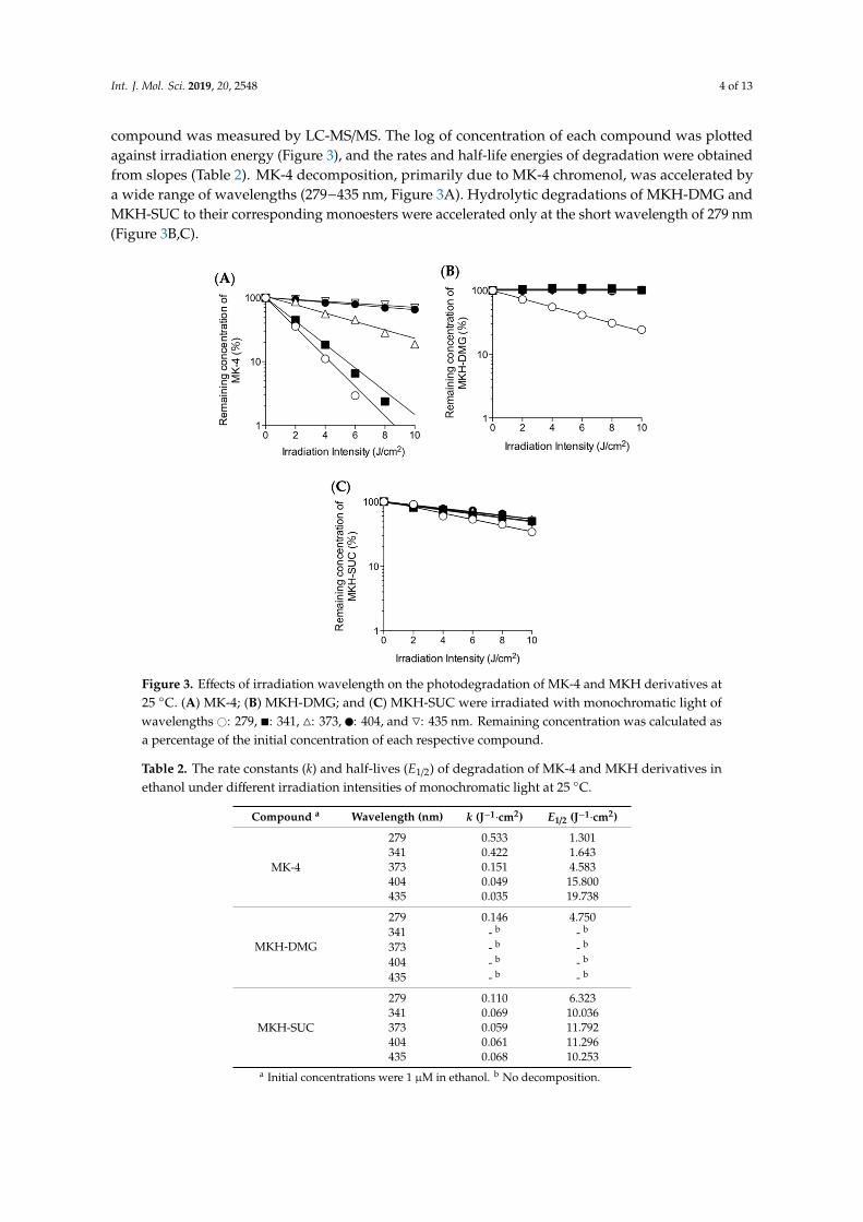
compound was measured by LC-MS/MS. The log of concentration of each compound was plottedagainst irradiation energy (Figure 3), and the rates and half-life energies of degradation were obtainedfrom slopes (Table 2). MK-4 decomposition, primarily due to MK-4 chromenol, was accelerated bya wide range of wavelengths (279−435 nm, Figure 3A). Hydrolytic degradations of MKH-DMG andMKH-SUC to their corresponding monoesters were accelerated only at the short wavelength of 279 nm(Figure 3B,C).
Int. J. Mol. Sci. 2019, 20 FOR PEER REVIEW 4
To evaluate the effect of irradiation wavelength on photostability of MK-4 and MKH-ester derivatives, 1 μM solutions of MK-4, MKH-DMG, and MKH-SUC in ethanol were irradiated with a xenon light source monochromatic light at 279, 341, 373, 404, or 435 nm, and the concentration of each compound was measured by LC-MS/MS. The log of concentration of each compound was plotted against irradiation energy (Figure 3), and the rates and half-life energies of degradation were obtained from slopes (Table 2). MK-4 decomposition, primarily due to MK-4 chromenol, was accelerated by a wide range of wavelengths (279−435 nm, Figure 3A). Hydrolytic degradations of MKH-DMG and MKH-SUC to their corresponding monoesters were accelerated only at the short wavelength of 279 nm (Figure 3B,C).
Figure 3. Effects of irradiation wavelength on the photodegradation of MK-4 and MKH derivatives at 25 °C. (A) MK-4; (B) MKH-DMG; and (C) MKH-SUC were irradiated with monochromatic light of wavelengths ○ : 279, ■ : 341, △ : 373, ● : 404, and ▽ : 435 nm. Remaining concentration was calculated as a percentage of the initial concentration of each respective compound.
Figure 3. Effects of irradiation wavelength on the photodegradation of MK-4 and MKH derivatives at25 ◦C. (A) MK-4; (B) MKH-DMG; and (C) MKH-SUC were irradiated with monochromatic light ofwavelengths #: 279, �: 341, 4: 373, �: 404, and 5: 435 nm. Remaining concentration was calculated asa percentage of the initial concentration of each respective compound.
Table 2. The rate constants (k) and half-lives (E1/2) of degradation of MK-4 and MKH derivatives inethanol under different irradiation intensities of monochromatic light at 25 ◦C.
Compound a Wavelength (nm) k (J−1·cm2) E1/2 (J−1·cm2)
MK-4
279 0.533 1.301341 0.422 1.643373 0.151 4.583404 0.049 15.800435 0.035 19.738
MKH-DMG
279 0.146 4.750341 - b - b
373 - b - b
404 - b - b
435 - b - b
MKH-SUC
279 0.110 6.323341 0.069 10.036373 0.059 11.792404 0.061 11.296435 0.068 10.253
a Initial concentrations were 1 µM in ethanol. b No decomposition.
Int. J. Mol. Sci. 2019, 20, 2548 5 of 13
2.2. Phototoxicity of MK-4 and MKH-Ester Derivatives
Some pharmaceutical substances are photosensitizers that can lead to the generation of reactiveoxygen species (ROS) that cause oxidative damage to skin cells. It has been also shown that vitamin K1
exhibits phototoxicity against cultured epidermis upon UVA irradiation [13]. Therefore, we evaluatedthe generation of ROS from MK-4 and MKH derivatives by UVA (320–400 nm) irradiation, which isstrongly related to phototoxicity and accounts for approximately 95% of total ultraviolet radiation insunlight that reaches the surface of the earth.
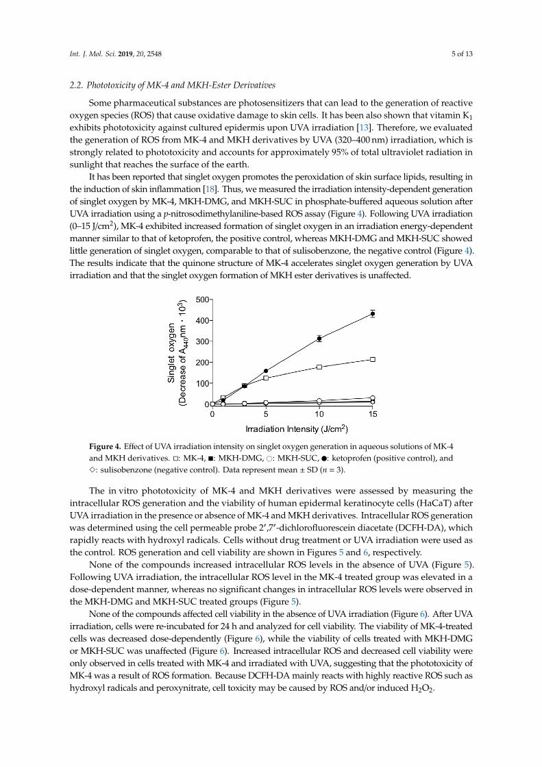
It has been reported that singlet oxygen promotes the peroxidation of skin surface lipids, resulting inthe induction of skin inflammation [18]. Thus, we measured the irradiation intensity-dependent generationof singlet oxygen by MK-4, MKH-DMG, and MKH-SUC in phosphate-buffered aqueous solution afterUVA irradiation using a p-nitrosodimethylaniline-based ROS assay (Figure 4). Following UVA irradiation(0–15 J/cm2), MK-4 exhibited increased formation of singlet oxygen in an irradiation energy-dependentmanner similar to that of ketoprofen, the positive control, whereas MKH-DMG and MKH-SUC showedlittle generation of singlet oxygen, comparable to that of sulisobenzone, the negative control (Figure 4).The results indicate that the quinone structure of MK-4 accelerates singlet oxygen generation by UVAirradiation and that the singlet oxygen formation of MKH ester derivatives is unaffected.Int. J. Mol. Sci. 2019, 20 FOR PEER REVIEW 6
Figure 4. Effect of UVA irradiation intensity on singlet oxygen generation in aqueous solutions of MK-4 and MKH derivatives. □: MK-4, ■: MKH-DMG, ○: MKH-SUC, ●: ketoprofen (positive control), and ◇: sulisobenzone (negative control). Data represent mean ± SD (n = 3).
The in vitro phototoxicity of MK-4 and MKH derivatives were assessed by measuring the intracellular ROS generation and the viability of human epidermal keratinocyte cells (HaCaT) after UVA irradiation in the presence or absence of MK-4 and MKH derivatives. Intracellular ROS generation was determined using the cell permeable probe 2′,7′-dichlorofluorescein diacetate (DCFH-DA), which rapidly reacts with hydroxyl radicals. Cells without drug treatment or UVA irradiation were used as the control. ROS generation and cell viability are shown in Figures 5 and 6, respectively.
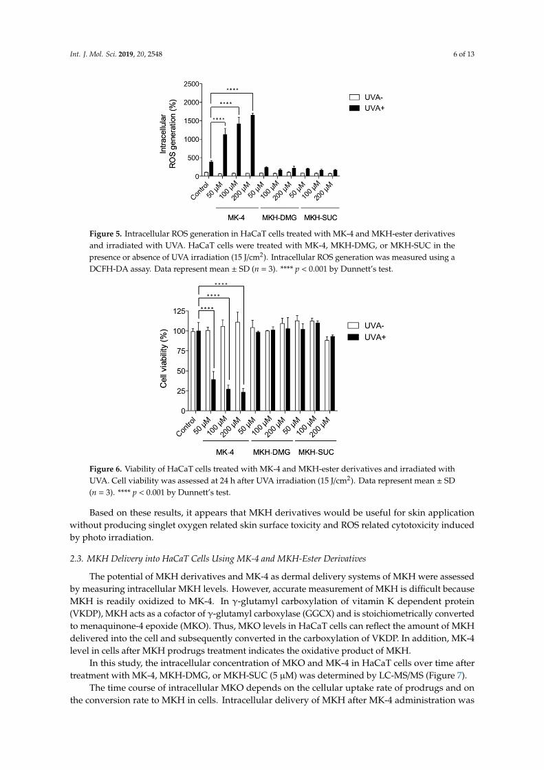
None of the compounds increased intracellular ROS levels in the absence of UVA (Figure 5). Following UVA irradiation, the intracellular ROS level in the MK-4 treated group was elevated in a dose-dependent manner, whereas no significant changes in intracellular ROS levels were observed in the MKH-DMG and MKH-SUC treated groups (Figure 5).
Figure 5. Intracellular ROS generation in HaCaT cells treated with MK-4 and MKH-ester derivatives and irradiated with UVA. HaCaT cells were treated with MK-4, MKH-DMG, or MKH-SUC in the presence or absence of UVA irradiation (15 J/cm2). Intracellular ROS generation was measured using a DCFH-DA assay. Data represent mean ± SD (n = 3). **** p < 0.001 by Dunnett’s test.
None of the compounds affected cell viability in the absence of UVA irradiation (Figure 6). After UVA irradiation, cells were re-incubated for 24 h and analyzed for cell viability. The viability of MK-
Figure 4. Effect of UVA irradiation intensity on singlet oxygen generation in aqueous solutions of MK-4and MKH derivatives. �: MK-4, �: MKH-DMG, #: MKH-SUC, �: ketoprofen (positive control), and3: sulisobenzone (negative control). Data represent mean ± SD (n = 3).
The in vitro phototoxicity of MK-4 and MKH derivatives were assessed by measuring theintracellular ROS generation and the viability of human epidermal keratinocyte cells (HaCaT) afterUVA irradiation in the presence or absence of MK-4 and MKH derivatives. Intracellular ROS generationwas determined using the cell permeable probe 2′,7′-dichlorofluorescein diacetate (DCFH-DA), whichrapidly reacts with hydroxyl radicals. Cells without drug treatment or UVA irradiation were used asthe control. ROS generation and cell viability are shown in Figures 5 and 6, respectively.
None of the compounds increased intracellular ROS levels in the absence of UVA (Figure 5).Following UVA irradiation, the intracellular ROS level in the MK-4 treated group was elevated in adose-dependent manner, whereas no significant changes in intracellular ROS levels were observed inthe MKH-DMG and MKH-SUC treated groups (Figure 5).
None of the compounds affected cell viability in the absence of UVA irradiation (Figure 6). After UVAirradiation, cells were re-incubated for 24 h and analyzed for cell viability. The viability of MK-4-treatedcells was decreased dose-dependently (Figure 6), while the viability of cells treated with MKH-DMGor MKH-SUC was unaffected (Figure 6). Increased intracellular ROS and decreased cell viability wereonly observed in cells treated with MK-4 and irradiated with UVA, suggesting that the phototoxicity ofMK-4 was a result of ROS formation. Because DCFH-DA mainly reacts with highly reactive ROS such ashydroxyl radicals and peroxynitrate, cell toxicity may be caused by ROS and/or induced H2O2.
Int. J. Mol. Sci. 2019, 20, 2548 6 of 13
Int. J. Mol. Sci. 2019, 20 FOR PEER REVIEW 6
Figure 4. Effect of UVA irradiation intensity on singlet oxygen generation in aqueous solutions of MK-4 and MKH derivatives. □: MK-4, ■: MKH-DMG, ○: MKH-SUC, ●: ketoprofen (positive control), and ◇: sulisobenzone (negative control). Data represent mean ± SD (n = 3).
The in vitro phototoxicity of MK-4 and MKH derivatives were assessed by measuring the intracellular ROS generation and the viability of human epidermal keratinocyte cells (HaCaT) after UVA irradiation in the presence or absence of MK-4 and MKH derivatives. Intracellular ROS generation was determined using the cell permeable probe 2′,7′-dichlorofluorescein diacetate (DCFH-DA), which rapidly reacts with hydroxyl radicals. Cells without drug treatment or UVA irradiation were used as the control. ROS generation and cell viability are shown in Figures 5 and 6, respectively.
None of the compounds increased intracellular ROS levels in the absence of UVA (Figure 5). Following UVA irradiation, the intracellular ROS level in the MK-4 treated group was elevated in a dose-dependent manner, whereas no significant changes in intracellular ROS levels were observed in the MKH-DMG and MKH-SUC treated groups (Figure 5).
Figure 5. Intracellular ROS generation in HaCaT cells treated with MK-4 and MKH-ester derivatives and irradiated with UVA. HaCaT cells were treated with MK-4, MKH-DMG, or MKH-SUC in the presence or absence of UVA irradiation (15 J/cm2). Intracellular ROS generation was measured using a DCFH-DA assay. Data represent mean ± SD (n = 3). **** p < 0.001 by Dunnett’s test.
None of the compounds affected cell viability in the absence of UVA irradiation (Figure 6). After UVA irradiation, cells were re-incubated for 24 h and analyzed for cell viability. The viability of MK-
Figure 5. Intracellular ROS generation in HaCaT cells treated with MK-4 and MKH-ester derivativesand irradiated with UVA. HaCaT cells were treated with MK-4, MKH-DMG, or MKH-SUC in thepresence or absence of UVA irradiation (15 J/cm2). Intracellular ROS generation was measured using aDCFH-DA assay. Data represent mean ± SD (n = 3). **** p < 0.001 by Dunnett’s test.
Int. J. Mol. Sci. 2019, 20 FOR PEER REVIEW 7
4-treated cells was decreased dose-dependently (Figure 6), while the viability of cells treated with MKH-DMG or MKH-SUC was unaffected (Figure 6). Increased intracellular ROS and decreased cell viability were only observed in cells treated with MK-4 and irradiated with UVA, suggesting that the phototoxicity of MK-4 was a result of ROS formation. Because DCFH-DA mainly reacts with highly reactive ROS such as hydroxyl radicals and peroxynitrate, cell toxicity may be caused by ROS and/or induced H2O2.
Figure 6. Viability of HaCaT cells treated with MK-4 and MKH-ester derivatives and irradiated with UVA. Cell viability was assessed at 24 h after UVA irradiation (15 J/cm2). Data represent mean ± SD (n = 3). **** p < 0.001 by Dunnett’s test.
Based on these results, it appears that MKH derivatives would be useful for skin application without producing singlet oxygen related skin surface toxicity and ROS related cytotoxicity induced by photo irradiation.
2.3. MKH Delivery into HaCaT Cells Using MK-4 and MKH-Ester Derivatives
The potential of MKH derivatives and MK-4 as dermal delivery systems of MKH were assessed by measuring intracellular MKH levels. However, accurate measurement of MKH is difficult because MKH is readily oxidized to MK-4. In γ-glutamyl carboxylation of vitamin K dependent protein (VKDP), MKH acts as a cofactor of γ-glutamyl carboxylase (GGCX) and is stoichiometrically converted to menaquinone-4 epoxide (MKO). Thus, MKO levels in HaCaT cells can reflect the amount of MKH delivered into the cell and subsequently converted in the carboxylation of VKDP. In addition, MK-4 level in cells after MKH prodrugs treatment indicates the oxidative product of MKH.
In this study, the intracellular concentration of MKO and MK-4 in HaCaT cells over time after treatment with MK-4, MKH-DMG, or MKH-SUC (5 μM) was determined by LC-MS/MS (Figure 7).
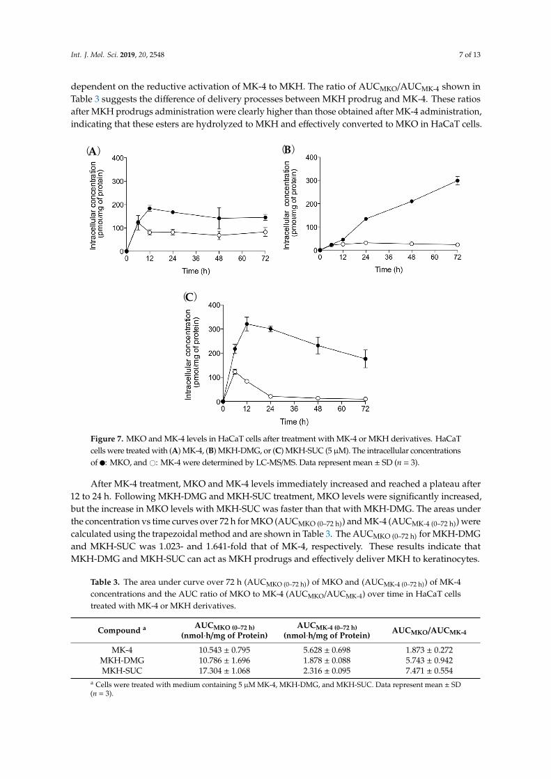
After MK-4 treatment, MKO and MK-4 levels immediately increased and reached a plateau after 12 to 24 h. Following MKH-DMG and MKH-SUC treatment, MKO levels were significantly increased, but the increase in MKO levels with MKH-SUC was faster than that with MKH-DMG. The areas under the concentration vs time curves over 72 h for MKO (AUCMKO (0–72 h)) and MK-4 (AUCMK-
4 (0–72 h)) were calculated using the trapezoidal method and are shown in Table 3. The AUCMKO (0–72 h) for MKH-DMG and MKH-SUC was 1.023- and 1.641-fold that of MK-4, respectively. These results indicate that MKH-DMG and MKH-SUC can act as MKH prodrugs and effectively deliver MKH to keratinocytes.
The time course of intracellular MKO depends on the cellular uptake rate of prodrugs and on the conversion rate to MKH in cells. Intracellular delivery of MKH after MK-4 administration was
Figure 6. Viability of HaCaT cells treated with MK-4 and MKH-ester derivatives and irradiated withUVA. Cell viability was assessed at 24 h after UVA irradiation (15 J/cm2). Data represent mean ± SD(n = 3). **** p < 0.001 by Dunnett’s test.
Based on these results, it appears that MKH derivatives would be useful for skin applicationwithout producing singlet oxygen related skin surface toxicity and ROS related cytotoxicity inducedby photo irradiation.
2.3. MKH Delivery into HaCaT Cells Using MK-4 and MKH-Ester Derivatives
The potential of MKH derivatives and MK-4 as dermal delivery systems of MKH were assessedby measuring intracellular MKH levels. However, accurate measurement of MKH is difficult becauseMKH is readily oxidized to MK-4. In γ-glutamyl carboxylation of vitamin K dependent protein(VKDP), MKH acts as a cofactor of γ-glutamyl carboxylase (GGCX) and is stoichiometrically convertedto menaquinone-4 epoxide (MKO). Thus, MKO levels in HaCaT cells can reflect the amount of MKHdelivered into the cell and subsequently converted in the carboxylation of VKDP. In addition, MK-4level in cells after MKH prodrugs treatment indicates the oxidative product of MKH.
In this study, the intracellular concentration of MKO and MK-4 in HaCaT cells over time aftertreatment with MK-4, MKH-DMG, or MKH-SUC (5 µM) was determined by LC-MS/MS (Figure 7).
The time course of intracellular MKO depends on the cellular uptake rate of prodrugs and onthe conversion rate to MKH in cells. Intracellular delivery of MKH after MK-4 administration was
Int. J. Mol. Sci. 2019, 20, 2548 7 of 13
dependent on the reductive activation of MK-4 to MKH. The ratio of AUCMKO/AUCMK-4 shown inTable 3 suggests the difference of delivery processes between MKH prodrug and MK-4. These ratiosafter MKH prodrugs administration were clearly higher than those obtained after MK-4 administration,indicating that these esters are hydrolyzed to MKH and effectively converted to MKO in HaCaT cells.
Int. J. Mol. Sci. 2019, 20 FOR PEER REVIEW 8
dependent on the reductive activation of MK-4 to MKH. The ratio of AUCMKO/AUCMK-4 shown in Table 3 suggests the difference of delivery processes between MKH prodrug and MK-4. These ratios after MKH prodrugs administration were clearly higher than those obtained after MK-4 administration, indicating that these esters are hydrolyzed to MKH and effectively converted to MKO in HaCaT cells.
Figure 7. MKO and MK-4 levels in HaCaT cells after treatment with MK-4 or MKH derivatives. HaCaT cells were treated with (A) MK-4, (B) MKH-DMG, or (C) MKH-SUC (5 μM). The intracellular concentrations of ●: MKO, and ○: MK-4 were determined by LC-MS/MS. Data represent mean ± SD (n = 3).
Table 3. The area under curve over 72 h (AUCMKO (0-72 h)) of MKO and (AUCMK-4 (0-72 h)) of MK-4 concentrations and the AUC ratio of MKO to MK-4 (AUCMKO/AUCMK-4) over time in HaCaT cells treated with MK-4 or MKH derivatives.
Compound a AUCMKO (0−72 h)
(nmol·h/mg of protein) AUCMK-4 (0−72 h)
(nmol·h/mg of protein) AUCMKO/AUCMK-4
MK-4 10.543 ± 0.795 5.628 ± 0.698 1.873 ± 0.272 MKH-DMG 10.786 ± 1.696 1.878 ± 0.088 5.743 ± 0.942 MKH-SUC 17.304 ± 1.068 2.316 ± 0.095 7.471 ± 0.554
a Cells were treated with medium containing 5 μM MK-4, MKH-DMG, and MKH-SUC. Data represent mean ± SD (n = 3).
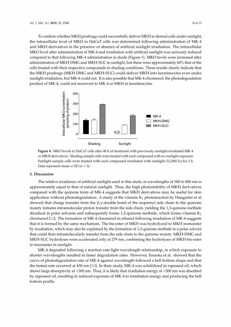
To confirm whether MKH prodrugs could successfully deliver MKH to dermal cells under sunlight, the intracellular level of MKO in HaCaT cells was determined following administration of MK-4 and MKH derivatives in the presence or absence of artificial sunlight irradiation. The intracellular MKO level after administration of MK-4 and irradiation with artificial sunlight was seriously reduced compared to that following MK-4 administration in shade (Figure 8). MKO levels were increased after administration of MKH-DMG and MKH-SUC in sunlight, but these were approximately 60% that of the cells treated with their respective compounds in shading conditions.
(A) (B)
(C)
Figure 7. MKO and MK-4 levels in HaCaT cells after treatment with MK-4 or MKH derivatives. HaCaTcells were treated with (A) MK-4, (B) MKH-DMG, or (C) MKH-SUC (5µM). The intracellular concentrationsof �: MKO, and #: MK-4 were determined by LC-MS/MS. Data represent mean ± SD (n = 3).
After MK-4 treatment, MKO and MK-4 levels immediately increased and reached a plateau after12 to 24 h. Following MKH-DMG and MKH-SUC treatment, MKO levels were significantly increased,but the increase in MKO levels with MKH-SUC was faster than that with MKH-DMG. The areas underthe concentration vs time curves over 72 h for MKO (AUCMKO (0–72 h)) and MK-4 (AUCMK-4 (0–72 h)) werecalculated using the trapezoidal method and are shown in Table 3. The AUCMKO (0–72 h) for MKH-DMGand MKH-SUC was 1.023- and 1.641-fold that of MK-4, respectively. These results indicate thatMKH-DMG and MKH-SUC can act as MKH prodrugs and effectively deliver MKH to keratinocytes.
Table 3. The area under curve over 72 h (AUCMKO (0–72 h)) of MKO and (AUCMK-4 (0–72 h)) of MK-4concentrations and the AUC ratio of MKO to MK-4 (AUCMKO/AUCMK-4) over time in HaCaT cellstreated with MK-4 or MKH derivatives.
Compound a AUCMKO (0–72 h)(nmol·h/mg of Protein)
AUCMK-4 (0–72 h)(nmol·h/mg of Protein) AUCMKO/AUCMK-4
MK-4 10.543 ± 0.795 5.628 ± 0.698 1.873 ± 0.272MKH-DMG 10.786 ± 1.696 1.878 ± 0.088 5.743 ± 0.942MKH-SUC 17.304 ± 1.068 2.316 ± 0.095 7.471 ± 0.554
a Cells were treated with medium containing 5 µM MK-4, MKH-DMG, and MKH-SUC. Data represent mean ± SD(n = 3).
Int. J. Mol. Sci. 2019, 20, 2548 8 of 13
To confirm whether MKH prodrugs could successfully deliver MKH to dermal cells under sunlight,the intracellular level of MKO in HaCaT cells was determined following administration of MK-4and MKH derivatives in the presence or absence of artificial sunlight irradiation. The intracellularMKO level after administration of MK-4 and irradiation with artificial sunlight was seriously reducedcompared to that following MK-4 administration in shade (Figure 8). MKO levels were increased afteradministration of MKH-DMG and MKH-SUC in sunlight, but these were approximately 60% that of thecells treated with their respective compounds in shading conditions. These results clearly indicate thatthe MKH prodrugs (MKH-DMG and MKH-SUC) could deliver MKH into keratinocytes even undersunlight irradiation, but MK-4 could not. It is also possible that MK-4 chromenol, the photodegradationproduct of MK-4, could not reconvert to MK-4 or MKH in keratinocytes.
Int. J. Mol. Sci. 2019, 20 FOR PEER REVIEW 9
These results clearly indicate that the MKH prodrugs (MKH-DMG and MKH-SUC) could deliver MKH into keratinocytes even under sunlight irradiation, but MK-4 could not. It is also possible that MK-4 chromenol, the photodegradation product of MK-4, could not reconvert to MK-4 or MKH in keratinocytes.
Figure 8. MKO levels in HaCaT cells after 48 h of treatment with previously sunlight-irradiated MK-4 or MKH derivatives. Shading sample cells were treated with each compound with no sunlight exposure. Sunlight sample cells were treated with each compound irradiated with sunlight (12,000 lx) for 3 h. Data represent mean ± SD (n = 3).
3. Discussion
The relative irradiance of artificial sunlight used in this study at wavelengths of 300 to 800 nm is approximately equal to that of natural sunlight. Thus, the high photostability of MKH derivatives compared with the quinone form of MK-4 suggests that MKH derivatives may be useful for skin application without photodegradation. A study of the vitamin K1 photoreaction by Hangarter et al. showed that charge transfer from the β,γ-double bond of the isoprenyl side chain to the quinone moiety initiates intramolecular proton transfer from the side chain, yielding the 1,3-quinone methide diradical in polar solvents and subsequently forms 1,2-quinone methide, which forms vitamin K1 chromenol [12]. The formation of MK-4 chromenol in ethanol following irradiation of MK-4 suggests that it is formed by the same mechanism. The bis-ester of MKH was hydrolyzed to MKH monoester by irradiation, which may also be explained by the formation of 1,3-quinone methide in a polar solvent that could then intramolecularly transfer from the side chain to the quinone moiety. MKH-DMG and MKH-SUC hydrolyses were accelerated only at 279 nm, confirming the hydrolyses of MKH-bis-ester to monoester in sunlight.
MK-4 degraded following a reaction rate-light wavelength relationship, in which exposure to shorter wavelengths resulted in faster degradation rates. However, Teraoka et al. showed that the curve of photodegradation rate of MK-4 against wavelength followed a bell bottom shape and that the fastest rate occurred at 430 nm [10]. In their study, MK-4 was solubilized in rapeseed oil, which shows large absorptivity at <300 nm. Thus, it is likely that irradiation energy at <300 nm was absorbed by rapeseed oil, resulting in reduced exposure of MK-4 to irradiation energy and producing the bell bottom profile.
Our results indicate that UVA irradiation of MK-4 accelerates singlet oxygen generation in aqueous solution, and increases intracellular ROS generation and cell toxicity in the HaCaT keratinocyte cell line. These results may be related to 1,3-quinone methide diradical formation by UVA irradiation of MK-4. Contrastingly, MKH derivatives avoided ROS formation and phototoxicity under UVA irradiation. Therefore, MKH derivatives would be candidates for effective, non-phototoxic use in skin application.
In order to act as an MKH prodrug, MKH derivatives must be converted to the parent drug in dermal cells after skin application. Our results clearly show that MKH derivatives and MK-4 increase intracellular MKO levels in the keratinocyte cell line we used, indicating that they could be used to
Figure 8. MKO levels in HaCaT cells after 48 h of treatment with previously sunlight-irradiated MK-4or MKH derivatives. Shading sample cells were treated with each compound with no sunlight exposure.Sunlight sample cells were treated with each compound irradiated with sunlight (12,000 lx) for 3 h.Data represent mean ± SD (n = 3).
3. Discussion
The relative irradiance of artificial sunlight used in this study at wavelengths of 300 to 800 nm isapproximately equal to that of natural sunlight. Thus, the high photostability of MKH derivativescompared with the quinone form of MK-4 suggests that MKH derivatives may be useful for skinapplication without photodegradation. A study of the vitamin K1 photoreaction by Hangarter et al.showed that charge transfer from the β,γ-double bond of the isoprenyl side chain to the quinonemoiety initiates intramolecular proton transfer from the side chain, yielding the 1,3-quinone methidediradical in polar solvents and subsequently forms 1,2-quinone methide, which forms vitamin K1
chromenol [12]. The formation of MK-4 chromenol in ethanol following irradiation of MK-4 suggeststhat it is formed by the same mechanism. The bis-ester of MKH was hydrolyzed to MKH monoesterby irradiation, which may also be explained by the formation of 1,3-quinone methide in a polar solventthat could then intramolecularly transfer from the side chain to the quinone moiety. MKH-DMG andMKH-SUC hydrolyses were accelerated only at 279 nm, confirming the hydrolyses of MKH-bis-esterto monoester in sunlight.
MK-4 degraded following a reaction rate-light wavelength relationship, in which exposure toshorter wavelengths resulted in faster degradation rates. However, Teraoka et al. showed that thecurve of photodegradation rate of MK-4 against wavelength followed a bell bottom shape and thatthe fastest rate occurred at 430 nm [10]. In their study, MK-4 was solubilized in rapeseed oil, whichshows large absorptivity at <300 nm. Thus, it is likely that irradiation energy at <300 nm was absorbedby rapeseed oil, resulting in reduced exposure of MK-4 to irradiation energy and producing the bellbottom profile.
Int. J. Mol. Sci. 2019, 20, 2548 9 of 13
Our results indicate that UVA irradiation of MK-4 accelerates singlet oxygen generation in aqueoussolution, and increases intracellular ROS generation and cell toxicity in the HaCaT keratinocyte cell line.These results may be related to 1,3-quinone methide diradical formation by UVA irradiation of MK-4.Contrastingly, MKH derivatives avoided ROS formation and phototoxicity under UVA irradiation.Therefore, MKH derivatives would be candidates for effective, non-phototoxic use in skin application.
In order to act as an MKH prodrug, MKH derivatives must be converted to the parent drug indermal cells after skin application. Our results clearly show that MKH derivatives and MK-4 increaseintracellular MKO levels in the keratinocyte cell line we used, indicating that they could be used todeliver MKH into keratinocytes, where it then acts as a cofactor for GGCX. We previously showed thatMKH-DMG and MKH-SUC reconvert to MKH in a reaction catalyzed by carboxyl esterase [17], whichis present in dermal cells [19–21], so accelerated intracellular reconversion to a parent drug is possible.Although UVA-irradiated MK-4 did not increase intracellular MKO, UVA-irradiated MKH derivativeswere able to.
MKH delivery with MKH-SUC into HaCaT cells is faster than that with MKH-DMG. A similarresult was obtained while using hepatocellular carcinoma (HCC) cells in a previous study—the uptakeand reconversion rates of MKH-SUC in HCC cells were faster than those of MKH-DMG [17]. Therefore,it can be presumed that the rapid and increased MKH delivery with MKH-SUC in HaCaT cells is dueto the rapid uptake and fast reconversion rates.
In conclusion, MKH-DMG and MKH-SUC could act as MKH prodrugs for skin applicationwithout photoinstability and phototoxicity. This ideal strategy is a safer and more efficient way todeliver MKH than using the quinone form MK-4. Therefore, MKH prodrugs have the potential to beused for skin diseases which require the activity of vitamin K dependent proteins for treatment, evenunder direct sunlight irradiation. Further studies that can reflect the actual skin environment usinghuman origin skin models such as 3-dimensional skin models or flesh skin tissue samples are needed.
4. Materials and Methods
4.1. Chemicals
MK-4 was purchased from Seebio Biotech, Inc. (Shanghai, China). Menaquinone-4 epoxide (MKO)was kindly provided by Eisai Co., Ltd. (Tokyo, Japan). Menahydroquinone-4 1,4-bis-N,N-dimethylglycinatehydrochloride (MKH-DMG) and menahydroquinone-4 1,4-bis-hemisuccinate (MKH-SUC) weresynthesized in our laboratory using previously reported methods [17]. Other chemicals were purchasedfrom FUJIFILM Wako Pure Chemical Corporation (Osaka, Japan).
4.2. Cell Culture
Human epidermal keratinocyte cell line HaCaT was obtained from CLS Cell Lines ServiceGmbH (Eppelheim, Germany). Cells were maintained in DMEM (high glucose, CLS) with 10% FBS(Life Technologies, Carlsbad, CA, USA) and 1% penicillin/streptomycin (Life Technologies) at 37 ◦Cunder an atmosphere of 5% CO2.
4.3. Photostability
Solutions of MK-4 and MKH derivatives in ethanol (1 µM) were irradiated with artificial sunlight(SOLAX 100 W XC-100 B, Seric Ltd., Tokyo, Japan) at 12,000 lx in quartz cells. Illuminance wasmeasured using a digital luminometer (LX-1108, Mother Tool). Irradiation samples (1 µM in ethanol)were irradiated with artificial sunlight at 12,000 lx, and with monochromatic light (279, 341, 373,404, and 435 nm) using a multi-wavelength irradiation spectrometer (MM 3, Bunkoukeiki Co., Ltd.,Tokyo, Japan). Irradiation intensity was measured using an irradiation energy measurement powermeter (MM 3, Bunkoukeiki Co., Ltd.). Concentrations of MK-4, MK-4 chromenol, MKH-DMG,MKH-mono-NN-dimethyl glycinate (MKH-mono-DMG), MKH-SUC, and MKH-mono-succinate(MKH-mono-SUC) in each solution were analyzed by LC-MS/MS as described below (Section 4.9).
Int. J. Mol. Sci. 2019, 20, 2548 10 of 13
4.4. Singlet Oxygen Generation Assay
MK-4 and MKH derivatives were dissolved in a phosphate buffer (NaPB, pH 7.4) containing0.2% polyoxyethylene hydrogenated castor oil 40 (HCO 40, obtained as a gift sample from NikkoChemicals Co., Ltd., Osaka, Japan), 2% dimethyl sulfoxide, and 0.1% glycerol. The solutions weretreated according to the singlet oxygen assay method (The Japanese Center for the Validation ofAlternative Methods, JaCVAM) [22] and irradiated with UVA light from a CL-1000L UV Crosslinker(UVP, Upland, CA, USA). Absorbance was measured using an Infinite M200 PRO (Tecan Life Sciences,Zurich, Switzerland).
4.5. Intracellular ROS Generation Assay
Intracellular ROS generation was measured using 2′,7′-dichlorofluorescein diacetate (DCFH-DA;Invitrogen, Carlsbad, CA, USA) as a fluorescent probe. HaCaT cells were seeded at 1.0 × 10⁵ cells/wellin 96-well plates and cultured overnight. Thereafter, cells were washed with PBS and incubated with10 µM DCFH-DA solution in PBS at 37 ◦C for 1 h. Following incubation, test compound solutions inPBS described above were added to each well and re-incubated for 1 h. After incubation, cells wereirradiated with UVA at an irradiation intensity of 15 J/cm2 and the fluorescence intensity (Ex: 485 nm,Em: 530 nm) was measured using an Infinite M200 PRO (Tecan Life Sciences).
4.6. Cell Viability Assay
HaCaT cells were seeded at 1.0× 10⁵ cells/well in 96-well plates and cultured overnight. Thereafter,cells were washed with PBS, and test compound solutions in PBS described above were added toeach well and re-incubated for 1 h. After incubation, cells were irradiated with UVA at an irradiationintensity of 15 J/cm2. After irradiation, cells were washed twice with PBS and replaced in culturemedium. After 24 h incubation, cell viability was measured using a CellTiter-Glo® Luminescent CellViability Assay (Promega, Madison, WI, USA).
4.7. Determination of Intracellular MKO Levels after Treatment With MK-4 and MKH Derivatives
HaCaT cells were seeded at 5.0 × 10⁴ cells/well in 24-well plates and allowed to attach for 48 h.Cells were cultured in medium containing 5 µM MK-4, MKH-DMG, or MKH-SUC, then medium wasremoved and cells were washed twice with PBS. Cells were collected in 500 µL of PBS and sonicated.Cell homogenates were combined with an equal volume of methanol and three times volume ofn-hexane, vortexed for 2 min, and centrifuged at 1750× g for 10 min. The organic layer was evaporatedunder N2 gas. The residue was reconstituted with 20 µL of methanol, and subjected to LC-MS/MS,as described below. The protein concentration of the cell homogenate was determined using a BCAprotein assay kit (Thermo Fisher Scientific, Waltham, MA, USA).
4.8. Determination of Intracellular MKO Levels After Treatment With Sunlight-Irradiated MK-4 andMKH Derivatives
Solutions of 200 µM MK-4, MKH-DMG, or MKH-SUC in ethanol were irradiated with artificialsunlight for 3 h at 12,000 lx in quartz cells. After irradiation, ethanol was evaporated under N2 gas.Residues were re-dissolved in a volume of medium equal to the evaporated ethanol, diluted 40-fold,and added to the cells for up to 48 h. Measurement of intracellular MKO was carried out using thesame method as described above (Section 4.7).
Int. J. Mol. Sci. 2019, 20, 2548 11 of 13
4.9. LC-MS/MS
LC-MS/MS was performed using an LCMS-8050 Liquid Chromatograph Mass Spectrometer(Shimadzu, Kyoto, Japan) and Shimadzu HPLC System (system controller (CBM-20A), pump(LD-20AD), degasser (DGU-20As), UV detector (SPD-20A), and auto injector (SIL-20AC HT)).Separations were performed on a CAPCELL PAK C18 UG120 (3 µm, 2.0 mm × 100 mm,Shiseido Co., Ltd., Tokyo, Japan) using a mobile phase of 10 mmol/L ammonium acetate and 0.1%acetic acid in methanol and water (97:3) at a flow rate of 0.4 mL/min. Column temperature wasmaintained at 40 ◦C. The mass spectrometer was equipped with an electrospray ionization and wasrun in positive ion mode. Identification and quantitation were based on MS/MS-multiple reactionmonitoring mode using transition ions as follows: m/z 445→ 187 for the [M+H]+ MK-4 adduct, m/z445→ 187 for the [M + H]+ chromenol adduct, 619→ 58 for the [M + H]+ MKH-DMG adduct, m/z532→ 58 for the [M + H]+ MKH-mono-DMG adduct, m/z 664→ 187 for the [M + H]+ MKH-SUCadduct, m/z 564→ 187 for the [M + H]+ MKH-mono-SUC adduct, and m/z 461→ 81 for the [M + H]+
MKO adduct. Retention times were: MK-4, 3.3 min; MK-4 chromenol, 2.0 min; MKH-DMG, 1.7 min;MKH-mono-DMG, 1.6 min; MKH-SUC, 1.1 min; MKH-mono-SUC, 1.2 min; and MKO, 2.5 min.
4.10. Statistical Analysis
Statistical significance was determined using Dunnett’s test. p < 0.05 was considered statisticallysignificant. Data were analyzed using GraphPad Prism 6 (GraphPad Software).
Supplementary Materials: Supplementary materials can be found at http://www.mdpi.com/1422-0067/20/10/2548/s1.
Author Contributions: Conceptualization, S.G., S.S. and J.T.; Methodology, S.G., S.S.; Validation, S.G., S.S.; FormalAnalysis, S.S., K.M.; Investigation, S.G., S.S., H.Y., D.W. and K.T.; Resources, S.S., J.T.; Data Curation, S.G., S.S.;Writing—Original Draft Preparation, S.G., S.S. and J.T.; Writing—Review & Editing, S.G., S.S. and J.T.; Visualization,S.G., S.S.; Supervision, K.M., J.T.; Project Administration, K.M., J.T.; Funding acquisition, Y.K., J.T.
Acknowledgments: We thank Noriko Sakamoto, Ryosuke Noguchi, and Akiko Imamura who were collegestudents in our laboratory that supported the experimental execution of this study.
Conflicts of Interest: The authors declare no conflict of interest.
Abbreviations
MKH menahydroquinone-4MK-4 menaquinone-4MKC menaquinone-4 chromenolMKH-DMG menahydroquinone-4 1,4-bis-N,N-dimethylglycinate hydrochloride
MKH-mono-DMGmenahydroquinone-4 1-mono N,N-dimethylglycinate hydrochloridemenahydroquinone-4 4-mono N,N-dimethylglycinate hydrochloride
MKH-SUC menahydroquinone-4 1,4-bis-hemisuccinate
MKH-mono-SUCmenahydroquinone-4 1-mono hemisuccinatemenahydroquinone-4 4-mono hemisuccinate
MKO menaquinone-4 epoxideGGCX γ-glutamyl carboxylaseVKORC1 vitamin K epoxide reductase complex subunit 1VKORC1L1 vitamin K epoxide reductase complex subunit 1 like 1ROS reactive oxygen speciesDCFH-DA 2′,7′-dichlorofluorescein diacetateAUC area under the concentration versus time curveMRM MS/MS-multiple reaction monitoring mode
Int. J. Mol. Sci. 2019, 20, 2548 12 of 13
References
1. Nakagawa, K.; Hirota, Y.; Sawada, N.; Yuge, N.; Watanabe, M.; Uchino, Y.; Okuda, N.; Shimomura, Y.;Suhara, Y.; Okano, T. Identification of UBIAD1 as a novel human menaquinone-4 biosynthetic enzyme.Nature 2010, 468, 117. [CrossRef] [PubMed]
2. Lou, W.W.; Quintana, A.T.; Geronemus, R.G.; Grossman, M.C. Effects of topical vitamin K and retinol onlaser-induced purpura on nonlesional skin. Dermatol. Surg. 1999, 25, 942–944. [CrossRef]
3. Lopes, L.B.; Speretta, F.F.F.; Vitoria, M.; Bentley, L.B. Enhancement of skin penetration of vitamin K usingmonoolein-based liquid crystalline systems. Eur. J. Pharm. Sci. 2007, 32, 209–215. [CrossRef] [PubMed]
4. Shah, N.S.; Lazarus, M.C.; Bugdodel, R.; Hsia, S.L.; He, J.; Duncan, R.; Baumann, L. The effects of topicalvitamin K on bruising after laser treatment. J. Am. Acad. Dermatol. 2002, 47, 241–244. [CrossRef] [PubMed]
5. Ocvirk, J. Management of cetuximab-induced skin toxicity with the prophylactic use of topical vitamin K1cream. Radiol. Oncol. 2010, 44, 265–266. [CrossRef] [PubMed]
6. Pinta, F.; Ponzetti, A.; Spadi, R.; Fanchini, L.; Zanini, M.; Mecca, C.; Sonetto, C.; Ciuffreda, L.; Racca, P.Pilot Clinical Trial on the Efficacy of Prophylactic Use of Vitamin K-1-Based Cream (Vigorskin) to PreventCetuximab-Induced Skin Rash in Patients With Metastatic Colorectal Cancer. Clin. Colorectal Cancer 2014, 13,62–67. [CrossRef] [PubMed]
7. Tan, E.H.; Chan, A. Evidence-Based Treatment Options for the Management of Skin Toxicities Associatedwith Epidermal Growth Factor Receptor Inhibitors. Ann. Pharm. 2009, 43, 1658–1666. [CrossRef] [PubMed]
8. Li, T.H.; Perez-Soler, R. Skin toxicities associated with epidermal growth factor receptor inhibitors.Targeted Oncol. 2009, 4, 107–119. [CrossRef] [PubMed]
9. Hemmati, A.A.; Houshmand, G.; Ghorbanzadeh, B.; Nemati, M.; Behmanesh, M.A. Topical vitamin K-1promotes repair of full thickness wound in rat. Indian J. Pharmacol. 2014, 46, 409–412. [CrossRef] [PubMed]
10. Teraoka, R.; Matsuda, Y. Stabilization-oriented preformulation study of photolabile menatetrenone(vitamin K2). Int. J. Pharm. 1993, 93, 85–90. [CrossRef]
11. Fujisawa, S.; Kawabata, S.; Yamamoto, R. Photo-degradation and stabilization of vitamin K1. I. Degradationproduct in ethanol and in non-ionic surfactant solution. Yakugaku Zasshi 1967, 87, 1451–1456. [CrossRef][PubMed]
12. Hangarter, M.A.; Hormann, A.; Kamdzhilov, Y.; Wirz, J. Primary photoreactions of phylloquinone (vitaminK-1) and plastoquinone-1 in solution. Photochem. Photobiol. Sci. 2003, 2, 524–535. [CrossRef] [PubMed]
13. Scientific Committees on Consumer Safety of the European Comission. Opinion on Vitamin K1 (Phytonadione).23 March 2010. Available online: https://ec.europa.eu/health/scientific_committees/consumer_safety/docs/sccs_o_014.pdf (accessed on 23 May 2019). [CrossRef]
14. Takata, J.; Karube, Y.; Hanada, M.; Matsunaga, K.; Matsushima, Y.; Sendo, T.; Aoyama, T. Vitamin K prodrugs:1. Synthesis of amino acid esters of menahydroquinone-4 and enzymatic reconversion to an active form.Pharm. Res. 1995, 12, 18–23. [CrossRef] [PubMed]
15. Takata, J.; Karube, Y.; Hanada, M.; Matsunaga, K.; Matsushima, Y.; Sendo, T.; Oishi, R. Vitamin K prodrugs:2. Water-soluble prodrugs of menahydroquinone-4 for systemic site-specific delivery. Pharm. Res. 1995, 12,1973–1979. [CrossRef] [PubMed]
16. Takata, J.; Karube, Y.; Hanada, M.; Matsunaga, K.; Iwasaki, H. Prodrug for bioreductive activation-independentdelivery of menahydroquinone-4: Human liver enzymatic activation and its action in warfarin-poisoned humanliver. Biol. Pharm. Bull. 1999, 22, 172–178. [CrossRef] [PubMed]
17. Setoguchi, S.; Watase, D.; Matsunaga, K.; Yamakawa, H.; Goto, S.; Terada, K.; Ohe, K.; Enjoji, M.; Karube, Y.;Takata, J. Antitumor Effects and Delivery Profiles of Menahydroquinone-4 Prodrugs with Ionic or NonionicPromoiety to Hepatocellular Carcinoma Cells. Molecules 2018, 23, 1738. [CrossRef] [PubMed]
18. Ryu, A.; Arakane, K.; Koide, C.; Arai, H.; Nagano, T. Squalene as a Target Molecule in Skin HyperpigmentationCaused by Singlet Oxygen. Biol. Pharm. Bull. 2009, 32, 1504–1509. [CrossRef] [PubMed]
19. Prusakiewicz, J.J.; Ackermann, C.; Voorman, R. Comparison of skin esterase activities from different species.Pharm. Res. 2006, 23, 1517–1524. [CrossRef] [PubMed]
20. Batz, F.M.; Klipper, W.; Korting, H.C.; Henkler, F.; Landsiedel, R.; Luch, A.; von Fritschen, U.; Weindl, G.;Schafer-Korting, M. Esterase activity in excised and reconstructed human skin—Biotransformation ofprednicarbate and the model dye fluorescein diacetate. Eur. J. Pharm. Biopharm. 2013, 84, 374–385. [CrossRef][PubMed]

Int. J. Mol. Sci. 2019, 20, 2548 13 of 13
21. Fu, J.; Sadgrove, M.; Marson, L.; Jay, M. Biotransformation Capacity of Carboxylesterase in Skin andKeratinocytes for the Penta-Ethyl Ester Prodrug of DTPA. Drug Metab. Dispos. 2016, 44, 1313–1318.[CrossRef] [PubMed]
22. ROS Assay Validation Management Team of Japanese Center for the Validation of Alternative Methods (JaCVAM).Reactive Oxygen Species (Ros) Assay to Examine Photoreactivity of Chemicals. Version 3.2. 28 November 2014.Available online: http://www.jacvam.jp/files/doc/02_03/02_03_E3.pdf (accessed on 23 May 2019).
© 2019 by the authors. Licensee MDPI, Basel, Switzerland. This article is an open accessarticle distributed under the terms and conditions of the Creative Commons Attribution(CC BY) license (http://creativecommons.org/licenses/by/4.0/).


![Nanocarriers for skin delivery of cosmetic antioxidants. [Nanovehículos para la liberación en piel de cosméticos antioxidantes]](https://static.fdokumen.com/doc/165x107/631cc2205a0be56b6e0e5bcd/nanocarriers-for-skin-delivery-of-cosmetic-antioxidants-nanovehiculos-para-la.jpg)