Proapoptotic and Antimetastatic Properties of Supercritical CO2 Extract of Nigella sativa Linn....
10
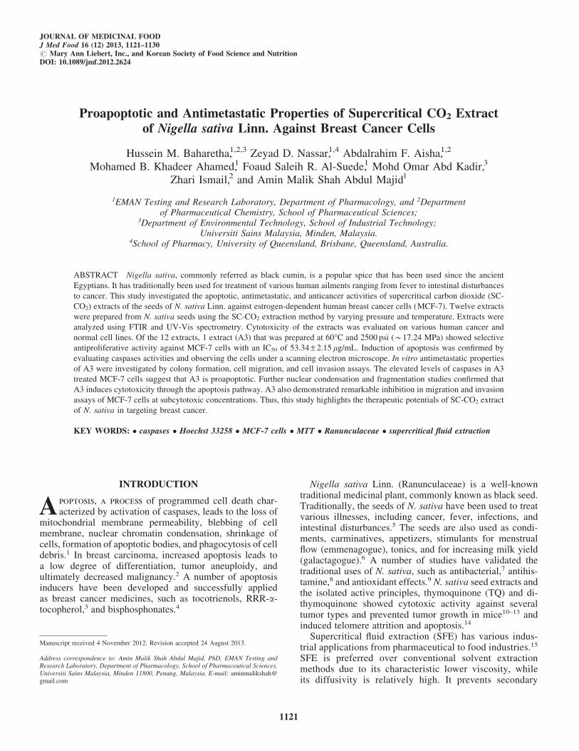
Proapoptotic and Antimetastatic Properties of Supercritical CO 2 Extract of Nigella sativa Linn. Against Breast Cancer Cells Hussein M. Baharetha, 1,2,3 Zeyad D. Nassar, 1,4 Abdalrahim F. Aisha, 1,2 Mohamed B. Khadeer Ahamed, 1 Foaud Saleih R. Al-Suede, 1 Mohd Omar Abd Kadir, 3 Zhari Ismail, 2 and Amin Malik Shah Abdul Majid 1 1 EMAN Testing and Research Laboratory, Department of Pharmacology, and 2 Department of Pharmaceutical Chemistry, School of Pharmaceutical Sciences; 3 Department of Environmental Technology, School of Industrial Technology; Universiti Sains Malaysia, Minden, Malaysia. 4 School of Pharmacy, University of Queensland, Brisbane, Queensland, Australia. ABSTRACT Nigella sativa, commonly referred as black cumin, is a popular spice that has been used since the ancient Egyptians. It has traditionally been used for treatment of various human ailments ranging from fever to intestinal disturbances to cancer. This study investigated the apoptotic, antimetastatic, and anticancer activities of supercritical carbon dioxide (SC- CO2) extracts of the seeds of N. sativa Linn. against estrogen-dependent human breast cancer cells (MCF-7). Twelve extracts were prepared from N. sativa seeds using the SC-CO2 extraction method by varying pressure and temperature. Extracts were analyzed using FTIR and UV-Vis spectrometry. Cytotoxicity of the extracts was evaluated on various human cancer and normal cell lines. Of the 12 extracts, 1 extract (A3) that was prepared at 60°C and 2500 psi (*17.24 MPa) showed selective antiproliferative activity against MCF-7 cells with an IC50 of 53.34 – 2.15 lg/mL. Induction of apoptosis was confirmed by evaluating caspases activities and observing the cells under a scanning electron microscope. In vitro antimetastatic properties of A3 were investigated by colony formation, cell migration, and cell invasion assays. The elevated levels of caspases in A3 treated MCF-7 cells suggest that A3 is proapoptotic. Further nuclear condensation and fragmentation studies confirmed that A3 induces cytotoxicity through the apoptosis pathway. A3 also demonstrated remarkable inhibition in migration and invasion assays of MCF-7 cells at subcytotoxic concentrations. Thus, this study highlights the therapeutic potentials of SC-CO2 extract of N. sativa in targeting breast cancer. KEY WORDS: caspases Hoechst 33258 MCF-7 cells MTT Ranunculaceae supercritical fluid extraction INTRODUCTION A poptosis, a process of programmed cell death char- acterized by activation of caspases, leads to the loss of mitochondrial membrane permeability, blebbing of cell membrane, nuclear chromatin condensation, shrinkage of cells, formation of apoptotic bodies, and phagocytosis of cell debris. 1 In breast carcinoma, increased apoptosis leads to a low degree of differentiation, tumor aneuploidy, and ultimately decreased malignancy. 2 A number of apoptosis inducers have been developed and successfully applied as breast cancer medicines, such as tocotrienols, RRR-a- tocopherol, 3 and bisphosphonates. 4 Nigella sativa Linn. (Ranunculaceae) is a well-known traditional medicinal plant, commonly known as black seed. Traditionally, the seeds of N. sativa have been used to treat various illnesses, including cancer, fever, infections, and intestinal disturbances. 5 The seeds are also used as condi- ments, carminatives, appetizers, stimulants for menstrual flow (emmenagogue), tonics, and for increasing milk yield (galactagogue). 6 A number of studies have validated the traditional uses of N. sativa, such as antibacterial, 7 antihis- tamine, 8 and antioxidant effects. 9 N. sativa seed extracts and the isolated active principles, thymoquinone (TQ) and di- thymoquinone showed cytotoxic activity against several tumor types and prevented tumor growth in mice 10–13 and induced telomere attrition and apoptosis. 14 Supercritical fluid extraction (SFE) has various indus- trial applications from pharmaceutical to food industries. 15 SFE is preferred over conventional solvent extraction methods due to its characteristic lower viscosity, while its diffusivity is relatively high. It prevents secondary Manuscript received 4 November 2012. Revision accepted 24 August 2013. Address correspondence to: Amin Malik Shah Abdul Majid, PhD, EMAN Testing and Research Laboratory, Department of Pharmacology, School of Pharmaceutical Sciences, Universiti Sains Malaysia, Minden 11800, Penang, Malaysia, E-mail: aminmalikshah@ gmail.com JOURNAL OF MEDICINAL FOOD J Med Food 16 (12) 2013, 1121–1130 # Mary Ann Liebert, Inc., and Korean Society of Food Science and Nutrition DOI: 10.1089/jmf.2012.2624 1121
Transcript of Proapoptotic and Antimetastatic Properties of Supercritical CO2 Extract of Nigella sativa Linn....
Proapoptotic and Antimetastatic Properties of Supercritical CO2 Extractof Nigella sativa Linn. Against Breast Cancer Cells
Hussein M. Baharetha,1,2,3 Zeyad D. Nassar,1,4 Abdalrahim F. Aisha,1,2
Mohamed B. Khadeer Ahamed,1 Foaud Saleih R. Al-Suede,1 Mohd Omar Abd Kadir,3
Zhari Ismail,2 and Amin Malik Shah Abdul Majid1
1EMAN Testing and Research Laboratory, Department of Pharmacology, and 2Departmentof Pharmaceutical Chemistry, School of Pharmaceutical Sciences;
3Department of Environmental Technology, School of Industrial Technology;Universiti Sains Malaysia, Minden, Malaysia.
4School of Pharmacy, University of Queensland, Brisbane, Queensland, Australia.
ABSTRACT Nigella sativa, commonly referred as black cumin, is a popular spice that has been used since the ancient
Egyptians. It has traditionally been used for treatment of various human ailments ranging from fever to intestinal disturbances
to cancer. This study investigated the apoptotic, antimetastatic, and anticancer activities of supercritical carbon dioxide (SC-
CO2) extracts of the seeds of N. sativa Linn. against estrogen-dependent human breast cancer cells (MCF-7). Twelve extracts
were prepared from N. sativa seeds using the SC-CO2 extraction method by varying pressure and temperature. Extracts were
analyzed using FTIR and UV-Vis spectrometry. Cytotoxicity of the extracts was evaluated on various human cancer and
normal cell lines. Of the 12 extracts, 1 extract (A3) that was prepared at 60�C and 2500 psi (*17.24 MPa) showed selective
antiproliferative activity against MCF-7 cells with an IC50 of 53.34 – 2.15 lg/mL. Induction of apoptosis was confirmed by
evaluating caspases activities and observing the cells under a scanning electron microscope. In vitro antimetastatic properties
of A3 were investigated by colony formation, cell migration, and cell invasion assays. The elevated levels of caspases in A3
treated MCF-7 cells suggest that A3 is proapoptotic. Further nuclear condensation and fragmentation studies confirmed that
A3 induces cytotoxicity through the apoptosis pathway. A3 also demonstrated remarkable inhibition in migration and invasion
assays of MCF-7 cells at subcytotoxic concentrations. Thus, this study highlights the therapeutic potentials of SC-CO2 extract
of N. sativa in targeting breast cancer.
KEY WORDS: � caspases � Hoechst 33258 � MCF-7 cells � MTT � Ranunculaceae � supercritical fluid extraction
INTRODUCTION
Apoptosis, a process of programmed cell death char-acterized by activation of caspases, leads to the loss of
mitochondrial membrane permeability, blebbing of cellmembrane, nuclear chromatin condensation, shrinkage ofcells, formation of apoptotic bodies, and phagocytosis of celldebris.1 In breast carcinoma, increased apoptosis leads toa low degree of differentiation, tumor aneuploidy, andultimately decreased malignancy.2 A number of apoptosisinducers have been developed and successfully appliedas breast cancer medicines, such as tocotrienols, RRR-a-tocopherol,3 and bisphosphonates.4
Nigella sativa Linn. (Ranunculaceae) is a well-knowntraditional medicinal plant, commonly known as black seed.Traditionally, the seeds of N. sativa have been used to treatvarious illnesses, including cancer, fever, infections, andintestinal disturbances.5 The seeds are also used as condi-ments, carminatives, appetizers, stimulants for menstrualflow (emmenagogue), tonics, and for increasing milk yield(galactagogue).6 A number of studies have validated thetraditional uses of N. sativa, such as antibacterial,7 antihis-tamine,8 and antioxidant effects.9 N. sativa seed extracts andthe isolated active principles, thymoquinone (TQ) and di-thymoquinone showed cytotoxic activity against severaltumor types and prevented tumor growth in mice10–13 andinduced telomere attrition and apoptosis.14
Supercritical fluid extraction (SFE) has various indus-trial applications from pharmaceutical to food industries.15
SFE is preferred over conventional solvent extractionmethods due to its characteristic lower viscosity, whileits diffusivity is relatively high. It prevents secondary
Manuscript received 4 November 2012. Revision accepted 24 August 2013.
Address correspondence to: Amin Malik Shah Abdul Majid, PhD, EMAN Testing andResearch Laboratory, Department of Pharmacology, School of Pharmaceutical Sciences,Universiti Sains Malaysia, Minden 11800, Penang, Malaysia, E-mail: [email protected]
JOURNAL OF MEDICINAL FOODJ Med Food 16 (12) 2013, 1121–1130# Mary Ann Liebert, Inc., and Korean Society of Food Science and NutritionDOI: 10.1089/jmf.2012.2624
1121
reactions in the extract that tend to occur during solventextraction such as oxidation and hydrolysis.15 Supercriticalcarbon dioxide (SC-CO2) extraction is the most popularSFE method. It has many advantages, since it is efficient,fast and environmentally safe, nontoxic, nonexplosive,nonflammable, and inert to solutes. CO2 is gaseous atroom temperature and pressure and this leads to a verysimple recovery of the extract and results in solvent-freeextracts.16 In the present study, the proapoptotic and anti-metastatic activities of SC-CO2 extracts of N. sativa ob-tained from various extraction parameters (varying pressuresand temperatures) were evaluated in a panel of humancancer cell lines.
MATERIALS AND METHODS
Materials
The SC-CO2 extractor (SFX-220 SFE system) was ob-tained from ISCO. Dimethyl sulfoxide (DMSO), Folin-Ciocalteau reagent, TQ, tamoxifen, betulinic acid, Hoechst33258 stain, and 3-(4,5-dimethylthiazol-2-yl)-2,5-diphenyltetrazolium bromide (MTT) were purchased from Sigma-Aldrich. Caspases 3/7, 8, and 9 were purchased from Pro-mega and Matrigel� (10 mg/mL) was obtained from BDBioscience.
Plant material
N. sativa seeds were purchased from a local market inSeiyun, Yemen. The plant was authenticated at the Her-barium Department, School of Biological Sciences, Uni-versiti Sains Malaysia (USM) with a voucher number(11221—N. sativa—22/3/2011). The seeds were washed,dried, pulverized, and sieved into particles of 0.5 mm.
Cell culture and cell lines
Human cell lines, HCT 116 (colorectal carcinoma),MCF-7 (hormone sensitive and invasive breast cancer),MDA-MB-231 (hormone resistant breast cancer), Hep G2(hepatocellular carcinoma), PC-3 (prostate carcinoma), andCCD-18Co (normal colonic fibroblasts) were obtained fromATCC. Cells were cultured at 5% CO2 humidified atmo-sphere at 37�C in a growth medium supplemented with 10%heat inactivated fetal bovine serum and 1% penicillin/streptomycin. HCT 116 and MCF-7 cells were cultured inRPMI-1640 and MEM media, respectively. PC-3 cells werecultured in F-12K, while MDA-MB-231, Hep G2, and CCD-18Co cells were grown in the DMEM.
SC-CO2 extraction
SC-CO2 extraction was employed using SFX-220, SFEsystem to obtain 12 extracts from N. sativa seeds (A1, A2,A3, B1, B2, B3, C1, C2, C3, D1, D2, and D3). Briefly, 1.2 gpowder was extracted for 60 min with liquefied CO2 atvarious pressures (2500, 3000, 4500, and 6000 psi; *17.24,20.68, 31.03, and 41.37 MPa) and temperatures (32�C,45�C, and 60�C) at a CO2 flow rate of 2 mL/min.
Characterization and phytochemical analysis
FTIR. FTIR spectra were recorded at a wavelengthranging from 4000 to 400 cm - 1 using an FTIR spectrometer(Thermo. Nicolet Nexus 670; Thermo Scientific) equippedwith OMNIC application software (Thermo; ElectronCorporation).
UV-Vis spectrophotometry. UV-Vis spectrophotometrywas carried out using a Lambda25 UV/Vis spectropho-tometer system operated with UV WinLab V2.85 software(Perkin Elmer). Samples were prepared in methanol at100 lg/mL, and were scanned at the wavelength range from500 to 200 nm.
Total phenolic and flavonoid contents
Total phenolics were determined using the Folin-Ciocalteau reagent with gallic acid as a standard and theresult was expressed as mg of gallic acid equivalent.17 Totalflavonoids were determined using the AlCl3 colorimetricmethod with quercetin as standard and the result was ex-pressed as mg of quercetin equivalent.18
Cell viability
MTT assay19 was performed to assess the cytotoxicity ofthe extracts on various cancer cell lines (HCT 116, MCF-7,PC-3, MDA-MB-231, and Hep G2). CCD-18Co was used asthe model cell line for normal cells. The assay plates wereread using a microtiter plate reader (Hitachi U-2000) at570 nm absorbance. DMSO (1%) was used as a negativecontrol.
In vitro apoptotic and antitumorigenic activityof A3 on MCF-7 cells
Effect of A3 on caspases 3/7, 8, and 9. The assays werecarried out according to the manufacturer’s protocol(Promega). MCF-7 cells were treated with various con-centrations of A3 (60–120 lg/mL) for different time in-tervals (3, 6, and 9 h). Tamoxifen (10 lg/mL) was used asa positive control, and DMSO (1%) as a negative control.Subsequently, an equal volume of freshly prepared cas-pase 3/7, 8, or 9 substrates were added, incubated at roomtemperature for 30 min, and luminescence was measuredusing the Infinite M200 PRO microplate reader (TecanGroup Ltd.). The results were expressed as the foldchanges in the caspase activity relative to the negativecontrol.
Nuclear chromatin condensation of MCF-7 cells. Theeffect of A3 on nuclear chromatin condensation in MCF-7cells was quantified by fluorescence microscopy usingHoechst 33258 stain.20 The cells treated with A3 (20, 40,and 60 lg/mL) or 1% DMSO for 24 h, were fixed, stained,and photographed at 20 · magnification, using a digitalmicroscope (EVOS fl; Advanced Microscopy Group). Thepercentage of apoptotic index was presented as mean ofexperiments performed in triplicates.
1122 BAHARETHA ET AL.

Transmission electron microscope
MCF-7 cells were treated for 24 h with A3 (40 and 80 lg/mL) or 1% DMSO. Cells were fixed with 0.1 M McDowell–Trump and stained with 1% osmium tetroxide. The cellswere then solidified in 2% agar, cut into small slides, anddehydrated in ethanol followed by acetone. The slides wereembedded in resin and infiltrated five times in Suprr’smixture at 60�C for overnight. Subsequently, cells weremolded in resin, sliced into semithin (1 lm) and ultrathinsections (0.1 lm). The semithin sections were stained withtoluidine blue, whereas the ultrathin sections were collectedin copper grids and stained with uranyl acetate and leadcitrate. Finally, the cells were photographed using transmis-sion electron microscope (TEM) at 1600 · magnification.
Colony formation
The effect of A3 on clonogenicity of MCF-7 cells wasinvestigated by the colony formation assay.21 Briefly, MCF-7 cells (500 cells/mL) were seeded in six-well plates andincubated for 12 h. Subsequently, cells were treated for 48 hwith A3 (10, 20, 40, and 60 lg/mL) or tamoxifen (10 lg/mL) or 1% DMSO. The cells were maintained for 10 daysuntil sufficiently large colonies ( ‡ 50 cells) were produced.The colonies were fixed, stained with 0.2% crystal violet,and counted under stereomicroscope. The percentage ofplating efficiency (PE%) and percentage of surviving frac-tion were calculated.
Cell migration
Cell migration assay was conducted as described byLiang et al.22 MCF-7 cells were seeded in 24-well plates andincubated for 48 h to achieve an almost 100% confluentmonolayer. A straight scratch was created using a 200-lLmicropipette tip, and the cells were treated immediately withA3 (20 and 40 lg/mL) or 0.5% DMSO. The wound wasphotographed at 0, 12, and 24 h. The distance of the cell-free
area was measured using Leica Quin software, and the re-sults are presented as percentage of inhibition of migrationin comparison to the negative control.
Cell invasion
Invasion assay was performed using Matrigel as an arti-ficial basement membrane matrix.23 Matrigel in MEM (1:1)was pipetted and incubated for 45 min to solidify. MCF-7 cells (5 · 103 per well in 96-well plates) were treated for24 h with 0.5% DMSO or A3 (concentrations of 20 and40 lg/mL). Subsequently, the upper media were carefullyaspirated and noninvading cells were washed off gently, andcells were photographed microscopically. Invading cellswere counted and the result was reported as percentage in-hibition of invasion relative to untreated cells.
Statistical analysis
Results were calculated as mean – SD of triplicates ofthree independent experiments and analyzed using SPSS16.0 package. Differences were tested by one-way ANOVAfollowed by post hoc—Dunnett’s or Tukey’s—tests. Cor-relations were calculated by the bivariate, two-tailed Pear-son’s test; while R2 values were analyzed by the linearregression test. Significance was considered at P < .05.
RESULTS
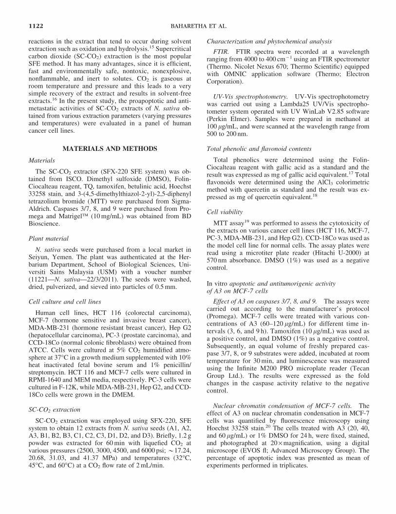
Yield of SC-CO2 extracts of N. sativa seeds
Cumulative percentage yields of the N. sativa seed ex-tracts were calculated at intervals of 15 min. The percentageyield increased significantly with increasing extraction time.The results indicate that 60 min is the optimum extractiontime of N. sativa seeds by SC-CO2 (Table 1). The extractionpressure showed a significant positive correlation with thepercentage yield at 32�C, 45�C, and 60�C with R2 = 0.89,0.94, and 0.96, respectively (Fig. 1a). On the contrary, the
Table 1. Percentage Yield of Supercritical Carbon Dioxide Extracts of Nigella sativa Seeds
in Relation to the Extraction Time Interval
Percentage yield (g/100 g) in the extraction time intervals
Extract code Pressure (psi) Temperature (�C) 15 min 30 min 45 min 60 min Correlation R2 value
A1 2500 32 02.58 – 0.24 06.57 – 0.02 09.04 – 0.11 10.81 – 1.18 0.95A2 2500 45 01.88 – 0.22 03.01 – 0.18 04.17 – 0.26 06.88 – 1.63 0.82A3 2500 60 01.43 – 0.06 02.60 – 0.06 02.65 – 0.08 03.06 – 0.85 0.59B1 3000 32 04.81 – 0.49 11.42 – 0.16 15.10 – 0.57 17.77 – 0.98 0.91B2 3000 45 03.72 – 0.27 07.46 – 0.47 11.08 – 0.94 13.89 – 0.33 0.97B3 3000 60 02.32 – 0.18 05.78 – 0.30 09.66 – 0.55 09.48 – 0.59 0.96C1 4500 32 05.72 – 0.23 18.49 – 0.37 25.34 – 0.23 30.41 – 1.09 0.95C2 4500 45 14.50 – 0.28 23.52 – 0.11 27.62 – 0.67 27.94 – 1.37 0.83C3 4500 60 17.34 – 0.04 24.34 – 0.11 25.44 – 0.69 26.86 – 0.15 0.82D1 6000 32 19.65 – 0.17 28.79 – 0.13 29.93 – 0.04 30.41 – 0.17 0.72D2 6000 45 15.17 – 0.11 26.75 – 0.28 29.85 – 0.06 30.27 – 0.41 0.78D3 6000 60 23.57 – 0.06 27.21 – 0.47 29.77 – 0.08 29.85 – 0.20 0.86
Results are represented as mean – SD (n = 3), P < .05.
SC-CO2, supercritical carbon dioxide.
ANTI-NEOPLASTICITY OF NIGELLA SATIVA 1123
extraction temperature showed a negative correlation withthe percentage yield at 2500 psi (R2 = 0.90) and 3000 psi(R2 = 0.98), whereas a lower effect of temperature on per-centage yield was observed at 4500 psi (R2 = 0.64) and6000 psi (R2 = 0.53) (Fig. 1b).
Phytochemical study of the extracts
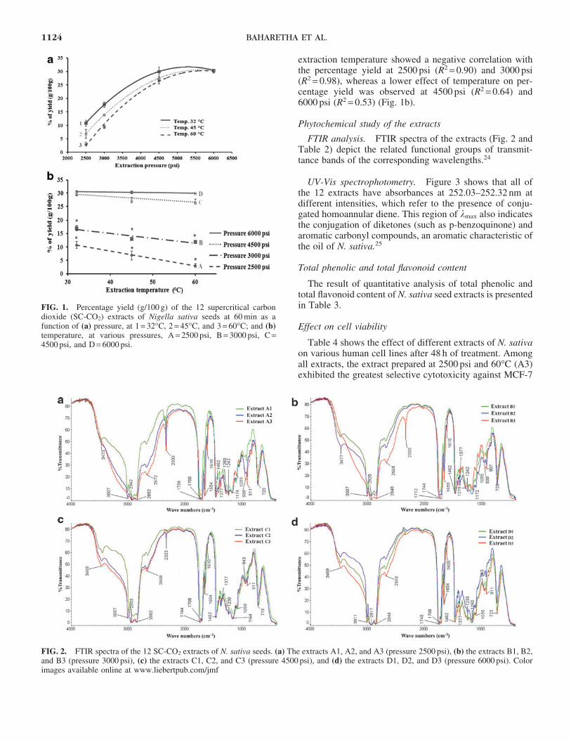
FTIR analysis. FTIR spectra of the extracts (Fig. 2 andTable 2) depict the related functional groups of transmit-tance bands of the corresponding wavelengths.24
UV-Vis spectrophotometry. Figure 3 shows that all ofthe 12 extracts have absorbances at 252.03–252.32 nm atdifferent intensities, which refer to the presence of conju-gated homoannular diene. This region of kmax also indicatesthe conjugation of diketones (such as p-benzoquinone) andaromatic carbonyl compounds, an aromatic characteristic ofthe oil of N. sativa.25
Total phenolic and total flavonoid content
The result of quantitative analysis of total phenolic andtotal flavonoid content of N. sativa seed extracts is presentedin Table 3.
Effect on cell viability
Table 4 shows the effect of different extracts of N. sativaon various human cell lines after 48 h of treatment. Amongall extracts, the extract prepared at 2500 psi and 60�C (A3)exhibited the greatest selective cytotoxicity against MCF-7
FIG. 1. Percentage yield (g/100 g) of the 12 supercritical carbondioxide (SC-CO2) extracts of Nigella sativa seeds at 60 min as afunction of (a) pressure, at 1 = 32�C, 2 = 45�C, and 3 = 60�C; and (b)temperature, at various pressures, A = 2500 psi, B = 3000 psi, C =4500 psi, and D = 6000 psi.
FIG. 2. FTIR spectra of the 12 SC-CO2 extracts of N. sativa seeds. (a) The extracts A1, A2, and A3 (pressure 2500 psi), (b) the extracts B1, B2,and B3 (pressure 3000 psi), (c) the extracts C1, C2, and C3 (pressure 4500 psi), and (d) the extracts D1, D2, and D3 (pressure 6000 psi). Colorimages available online at www.liebertpub.com/jmf
1124 BAHARETHA ET AL.

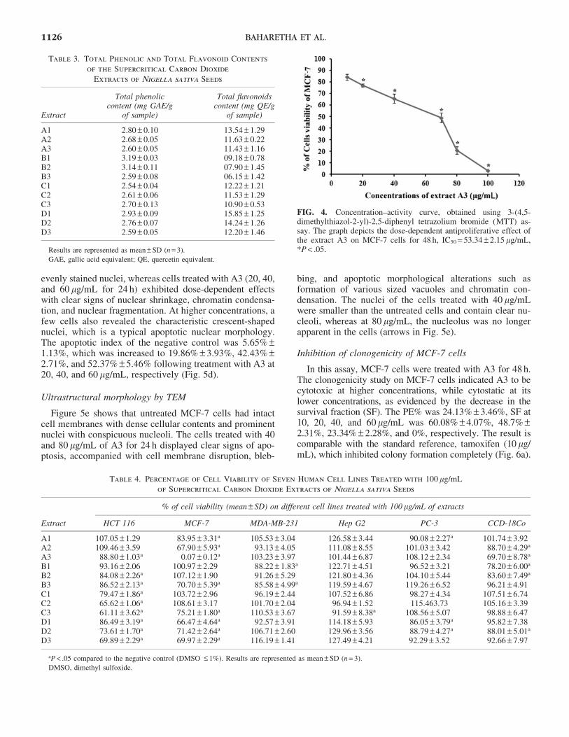
cells with the cell viability of 0.07% – 0.12% at 100 lg/mL.A3 exhibited dose-dependent cytotoxicity toward MCF-7 cells with IC50 53.34 – 2.15 lg/mL, whereas A3 showedpoor antiproliferative activity toward other tested cell lines.IC50 values of TQ and tamoxifen were 5.33 – 0.29 and10.51 – 0.38 lg/mL, respectively (Fig. 4).
In vitro proapoptotic activity of A3 on MCF-7
Activation of caspases 3/7, 8, and 9. The cellular levelsof apoptotic markers, caspases, were estimated to explorethe mechanism of action of A3 by which the extract inducedtoxicity in MCF-7 cells. The effect of A3 was studied atthree different time intervals (3, 6, and 9 h). Treatment ofMCF-7 cells with A3 for either 3 or 6 h did not elevatecaspase levels, whereas the treatment for 9 h caused sig-nificant activation of caspases 3/7 at 100 and 80 lg/mL by2- and 1.5-fold, respectively. At 60 lg/mL, there was in-significant elevation of the level of caspases when comparedto the negative control (Fig. 5a). A3 also activated caspase 8at 100 and 120 lg/mL by two- and sevenfold. On the otherhand, A3 did not show significant elevation in the level ofcaspase 9 (Fig. 5b).
Nuclear morphology and chromatin condensation. Theeffect of A3 on nuclear morphology of MCF-7 cells wasinvestigated by staining the nucleus with Hoechst 33258stain. Figure 5c illustrates that the untreated cells displayed
Table 2. Infrared Transmittance Bands Correlated
to the Corresponding Wavenumbers Representing
the Functional Organic Groups Present
in the Supercritical Carbon Dioxide
Extracts of Nigella sativa seeds
Vibrationalfrequency (cm - 1) Extract
Correspondingorganic groups
3477–3469 (broad) A1, B1, B2, C1, C2,C3, D1, D2, and D3
O-H stretching
3015–2844 All 12 extracts C-H stretching1748–1708 (strong) All 12 extracts C = O stretching1659–1654 (weak) All 12 extracts C = C stretching1610–1605 All 12 extracts NO2 - stretching1471–1462 All 12 extracts C-H bending
(deformation)1381–1373 All 12 extracts C-C, C-H, O-H,
and C-O bending1242–1160 All 12 extracts C-C, C-O, C-N
stretching1099–1094 All 12 extracts C-C, C-O, C-N
stretching1050–1046 A2, A3, B3 C-C, C-O, C-N
stretching960–907 All 12 extracts C-H bending723–719 All 12 extracts C-H bending
FIG. 3. UV-Vis absorption spectra of the 12 SC-CO2 extracts N. sativa seeds. (a) The extracts A1, A2, and A3 (pressure 2500 psi), (b) theextracts B1, B2, and B3 (pressure 3000 psi), (c) the extracts C1, C2, and C3 (pressure 4500 psi), and (d) the extracts D1, D2, and D3 (pressure6000 psi). Color images available online at www.liebertpub.com/jmf
ANTI-NEOPLASTICITY OF NIGELLA SATIVA 1125
evenly stained nuclei, whereas cells treated with A3 (20, 40,and 60 lg/mL for 24 h) exhibited dose-dependent effectswith clear signs of nuclear shrinkage, chromatin condensa-tion, and nuclear fragmentation. At higher concentrations, afew cells also revealed the characteristic crescent-shapednuclei, which is a typical apoptotic nuclear morphology.The apoptotic index of the negative control was 5.65% –1.13%, which was increased to 19.86% – 3.93%, 42.43% –2.71%, and 52.37% – 5.46% following treatment with A3 at20, 40, and 60 lg/mL, respectively (Fig. 5d).
Ultrastructural morphology by TEM
Figure 5e shows that untreated MCF-7 cells had intactcell membranes with dense cellular contents and prominentnuclei with conspicuous nucleoli. The cells treated with 40and 80 lg/mL of A3 for 24 h displayed clear signs of apo-ptosis, accompanied with cell membrane disruption, bleb-
bing, and apoptotic morphological alterations such asformation of various sized vacuoles and chromatin con-densation. The nuclei of the cells treated with 40 lg/mLwere smaller than the untreated cells and contain clear nu-cleoli, whereas at 80 lg/mL, the nucleolus was no longerapparent in the cells (arrows in Fig. 5e).
Inhibition of clonogenicity of MCF-7 cells
In this assay, MCF-7 cells were treated with A3 for 48 h.The clonogenicity study on MCF-7 cells indicated A3 to becytotoxic at higher concentrations, while cytostatic at itslower concentrations, as evidenced by the decrease in thesurvival fraction (SF). The PE% was 24.13% – 3.46%, SF at10, 20, 40, and 60 lg/mL was 60.08% – 4.07%, 48.7% –2.31%, 23.34% – 2.28%, and 0%, respectively. The result iscomparable with the standard reference, tamoxifen (10 lg/mL), which inhibited colony formation completely (Fig. 6a).
Table 4. Percentage of Cell Viability of Seven Human Cell Lines Treated with 100 lg/mL
of Supercritical Carbon Dioxide Extracts of Nigella sativa Seeds
% of cell viability (mean – SD) on different cell lines treated with 100 lg/mL of extracts
Extract HCT 116 MCF-7 MDA-MB-231 Hep G2 PC-3 CCD-18Co
A1 107.05 – 1.29 83.95 – 3.31a 105.53 – 3.04 126.58 – 3.44 90.08 – 2.27a 101.74 – 3.92A2 109.46 – 3.59 67.90 – 5.93a 93.13 – 4.05 111.08 – 8.55 101.03 – 3.42 88.70 – 4.29a
A3 88.80 – 1.03a 0.07 – 0.12a 103.23 – 3.97 101.44 – 6.87 108.12 – 2.34 69.70 – 8.78a
B1 93.16 – 2.06 100.97 – 2.29 88.22 – 1.83a 122.71 – 4.51 96.52 – 3.21 78.20 – 6.00a
B2 84.08 – 2.26a 107.12 – 1.90 91.26 – 5.29 121.80 – 4.36 104.10 – 5.44 83.60 – 7.49a
B3 86.52 – 2.13a 70.70 – 5.39a 85.58 – 4.99a 119.59 – 4.67 119.26 – 6.52 96.21 – 4.91C1 79.47 – 1.86a 103.72 – 2.96 96.19 – 2.44 107.52 – 6.86 98.27 – 4.34 107.51 – 6.74C2 65.62 – 1.06a 108.61 – 3.17 101.70 – 2.04 96.94 – 1.52 115.463.73 105.16 – 3.39C3 61.11 – 3.62a 75.21 – 1.80a 110.53 – 3.67 91.59 – 8.38a 108.56 – 5.07 98.88 – 6.47D1 86.49 – 3.19a 66.47 – 4.64a 92.57 – 3.91 114.18 – 5.93 86.05 – 3.79a 95.82 – 7.38D2 73.61 – 1.70a 71.42 – 2.64a 106.71 – 2.60 129.96 – 3.56 88.79 – 4.27a 88.01 – 5.01a
D3 69.89 – 2.29a 69.97 – 2.29a 116.19 – 1.41 127.49 – 4.21 92.29 – 3.52 92.66 – 7.97
aP < .05 compared to the negative control (DMSO £ 1%). Results are represented as mean – SD (n = 3).
DMSO, dimethyl sulfoxide.
Table 3. Total Phenolic and Total Flavonoid Contents
of the Supercritical Carbon Dioxide
Extracts of Nigella sativa Seeds
Extract
Total phenoliccontent (mg GAE/g
of sample)
Total flavonoidscontent (mg QE/g
of sample)
A1 2.80 – 0.10 13.54 – 1.29A2 2.68 – 0.05 11.63 – 0.22A3 2.60 – 0.05 11.43 – 1.16B1 3.19 – 0.03 09.18 – 0.78B2 3.14 – 0.11 07.90 – 1.45B3 2.59 – 0.08 06.15 – 1.42C1 2.54 – 0.04 12.22 – 1.21C2 2.61 – 0.06 11.53 – 1.29C3 2.70 – 0.13 10.90 – 0.53D1 2.93 – 0.09 15.85 – 1.25D2 2.76 – 0.07 14.24 – 1.26D3 2.59 – 0.05 12.20 – 1.46
Results are represented as mean – SD (n = 3).
GAE, gallic acid equivalent; QE, quercetin equivalent.
FIG. 4. Concentration–activity curve, obtained using 3-(4,5-dimethylthiazol-2-yl)-2,5-diphenyl tetrazolium bromide (MTT) as-say. The graph depicts the dose-dependent antiproliferative effect ofthe extract A3 on MCF-7 cells for 48 h, IC50 = 53.34 – 2.15 lg/mL,*P < .05.
1126 BAHARETHA ET AL.

FIG. 5. Apoptotic effect of A3 in MCF-7 cells. The level of caspases in cells was measured at 3, 6, and 9 h after the treatment. (a) The dose-dependent stimulatory effect of A3 on the caspase 3/7 level after 9 h of treatment. (b) After 9 h of treatment, A3 increased the level of caspases 8,whereas the level of caspase 9 was unaltered. (c) MCF-7 cells with DNA Hoechst 33258 stain. The picture reveals the proapoptotic properties ofA3, as there is evidence of chromatin condensation in treated (for 24 h) cells, which is considerably significant when compared to the untreatedcells (P < .05). It can be seen clearly that higher concentrations of A3 rendered the cells with crescent shaped nuclei, which is a characteristicfeatures of apoptosis. (d) The percentage of apoptotic indices of chromatin condensation assay. (e) Ultrastructural micrographs using thetransmission electron microscope (TEM) reveal further supportive information of the apoptotic property of A3. The pictures at magnification1600 · showed all characteristic features of the apoptosis in MCF-7 cells treated with A3 for 24 h. *P < .05; **P < .01. Color images availableonline at www.liebertpub.com/jmf
ANTI-NEOPLASTICITY OF NIGELLA SATIVA 1127
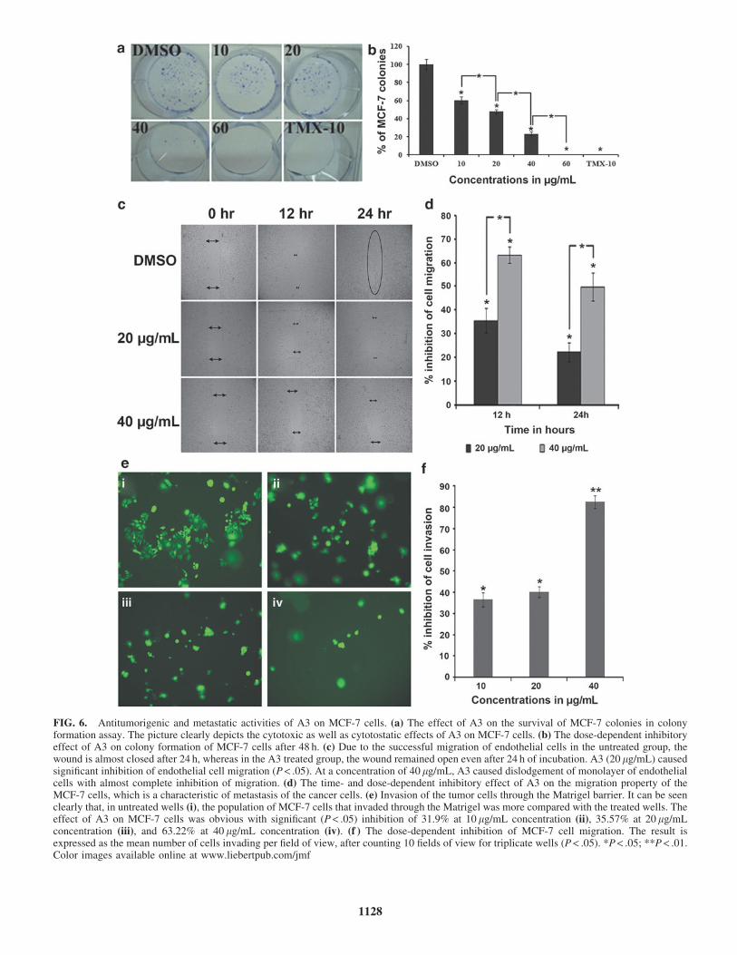
FIG. 6. Antitumorigenic and metastatic activities of A3 on MCF-7 cells. (a) The effect of A3 on the survival of MCF-7 colonies in colonyformation assay. The picture clearly depicts the cytotoxic as well as cytotostatic effects of A3 on MCF-7 cells. (b) The dose-dependent inhibitoryeffect of A3 on colony formation of MCF-7 cells after 48 h. (c) Due to the successful migration of endothelial cells in the untreated group, thewound is almost closed after 24 h, whereas in the A3 treated group, the wound remained open even after 24 h of incubation. A3 (20 lg/mL) causedsignificant inhibition of endothelial cell migration (P < .05). At a concentration of 40 lg/mL, A3 caused dislodgement of monolayer of endothelialcells with almost complete inhibition of migration. (d) The time- and dose-dependent inhibitory effect of A3 on the migration property of theMCF-7 cells, which is a characteristic of metastasis of the cancer cells. (e) Invasion of the tumor cells through the Matrigel barrier. It can be seenclearly that, in untreated wells (i), the population of MCF-7 cells that invaded through the Matrigel was more compared with the treated wells. Theeffect of A3 on MCF-7 cells was obvious with significant (P < .05) inhibition of 31.9% at 10 lg/mL concentration (ii), 35.57% at 20 lg/mLconcentration (iii), and 63.22% at 40 lg/mL concentration (iv). (f ) The dose-dependent inhibition of MCF-7 cell migration. The result isexpressed as the mean number of cells invading per field of view, after counting 10 fields of view for triplicate wells (P < .05). *P < .05; **P < .01.Color images available online at www.liebertpub.com/jmf
1128

Inhibition of cell migration
Cell migration assay represents an important step in themetastasis of cancer cells. The results are presented aspercentage inhibition of migrating cells relative to untreatedcells (Fig. 6b, c). Significant reduction in MCF-7 cell mo-tility was achieved at subcytotoxic concentrations of 20and 40 lg/mL with 35.57% – 5.11% and 63.22% – 3.47%inhibition, respectively, at 12 h of treatment. At 24 h, thepercentage inhibition was 22.33% – 4.09% and 49.80% –5.89% at 20 and 40 lg/mL, respectively.
Inhibition of cell invasion
Cell invasion assay was performed on the Matrigel matrixand the results are shown as a percentage inhibition of in-vading cells. After a 24-h treatment period, the percentageinhibition was 79.29% – 2.09% and 43.04% – 2.25% fol-lowing treatment with A3 at 40 and 20 lg/mL, respectively(Fig. 6d).
DISCUSSION
The findings of the present study showed that, out of the12 extracts, A3 (prepared at 2500 psi and 60�C) showedconsiderable antiproliferative activity against MCF-7 cells,whereas no cytotoxic effect was observed on other testedcancer cells, including the normal CCD-18Co cells. Thisindicates that A3 has potential selective cytotoxicity againstMCF-7 cells. Apoptotic effects of A3 were investigated tofurther elucidate its mode of action. Caspases are a family ofenzymes that have important roles in cell apoptosis cas-cades. Their activation occurs at the early steps of apopto-sis.26 This subsequently activates other apoptosis triggeringsignals and leads to cell shrinkage, chromatin condensation,and fragmentation. Finally, the cascade culminates in theformation of apoptotic bodies.27 A3 significantly stimulatesthe apoptosis activator (caspase 8) in MCF-7 cells, which isconsequently coincided by the activation of apoptosis exe-cutioner (caspases 3/7). This results in a significant induc-tion of apoptosis and survival suppression of MCF-7 cells,which further contributes to counteracting the breast cancerdevelopment.28 The activation of caspases leads to nuclearchromatin condensation and DNA degradation. The seg-mentation of chromatin and formation of apoptotic bodiesare indicators of apoptosis in the cells. The ultrastructuralalterations of the A3 treated breast cancer cells were dem-onstrated using TEM, which showed obvious apoptotic signs.The shrinkage of nucleus and disappearance of nucleoli at80 lg/mL of A3 indicated an advanced stage of apoptosis.29
The induction of apoptosis controls various regulatorsthat abrogate tumorigenesis and metastasis.30 Tumor cellsare shed daily as part of their movement in blood circulation,and the cells continue their extravasations and proliferationinto the secondary sites, despite the daily hemodynamicstresses and body immunity. This is based on the cell mi-gration and invasion cascades.31 A3 suppressed colonizationof MCF-7 cells in a dose-dependent manner. It is cytotoxicagainst MCF-7 cells at 60 lg/mL, where it irreversibly
damaged the cell integrity, whereas at 10 and 20 lg/mL, itshowed cytostatic effects. Cell migration and invasion arecrucial processes that control tumorigenesis and metastasis.Inhibition of MCF-7 cell migration in vitro by A3 indicatesthat the extract could probably inhibit cell metastasis.32
Treatment of MCF-7 cells with A3 resulted in a potent inhi-bition of cell migration toward the closure of the woundscratch, even after 12 and 18 h, indicating that the extract has acapability to suppress the motility of the breast cancer cells,whereas the wound was totally closed after 24 h in untreatedcells. Cell invasion is one of the main hallmarks of metastaticcascade.33 Although induction of caspase 8 activity in the earlysteps of apoptosis alters survival, it also inhibits cell invasioninto the basement membranes.33 The extract A3 activatedcaspase 8 in the MCF-7 cell line; thus it can be postulated thatthis leads to its inhibitory effect on invasion of MCF-7 cells.A3 significantly restricted invasion of MCF-7 cells into theMatrigel basement. This further technically supports the an-timetastatic potential of A3 against breast cancer.
FTIR and UV-Vis spectrometric analysis of the extractsof N. sativa seeds resulted in obvious differences in theirchemical constituents, which were related to the differentbioactivities observed for the extracts. The antiproliferative,proapoptotic, and antimetastatic effects of A3 may be due tothe collective contribution of its antioxidant-rich polyphe-nolic contents, particularly, TQ, thymohydroquinone, andalpha-hederin. 10,13 Phenolic compounds counteract cancereither by means of antioxidant effect or by inhibiting theformation of carcinogenic metabolites that damage the vitalbiomolecules.33,34 Studies have shown that flavonoids havepotent cytotoxic activities by induction of the apoptosispathway.35 In this study, the A3 extract was rich in totalflavonoids and polyphenols, which might have contributedtoward its overall anticancer effects observed in MCF-7 cells.
CONCLUSION
In conclusion, the present work provides good support-ing evidence that the SC-CO2 extract A3, prepared using60�C and 2500 psi, interferes with the several physio-logical properties of MCF-7 breast cancer cells. The anti-tumorigenic effect of A3 may be due to the collectivecontributions of phytochemicals, particularly, TQ and thy-mohydroquinone. The findings reveal that SC-CO2 extrac-tion can be useful to produce potent extracts from N. sativaseeds that can induce apoptosis. It was found that theanticancer potency of this extract is highly dependent onextraction temperature and pressure. Higher extractiontemperature (60�C) and lower extraction pressure (2500 and3000 psi) produced the most potent extracts. The MCF-7 cellline was the most sensitive. A3 halted the proliferation andinvasion of MCF-7 cells and caused significant apoptosis inthe cell line by activating caspase 3/7 and 8.
ACKNOWLEDGMENTS
This study was supported by Natureceuticals Sdn Bhd,Malaysia and the research grant (Ref. no. 1001/PFARMASI/
ANTI-NEOPLASTICITY OF NIGELLA SATIVA 1129

834052) at Universiti Sains Malaysia. It was also fundedfrom the Institute of Health Sciences in Seiyun–Hadramautand Ministry of Health, Yemen.
AUTHOR DISCLOSURE STATEMENT
No competing financial interests exist for any of theauthors.
REFERENCES
1. O’Driscoll L, O’Connor R, Clynes M: Methods in apoptosis. In:
Cell Biology, 3rd edition. (Julio EC, ed.) Academic Press, Bur-
lington, MA, USA, 2006, pp. 335–342.
2. Lipponen P, Aaltomaa S, Kosma VM, Syrjanen K: Apoptosis in
breast cancer as related to histopathological characteristics and
prognosis. Eur J Cancer 1994;30A:2068–2073.
3. Yu W, Simmons MM, Gapor A, Sanders BG, Kline K: Induction
of apoptosis in human breast cancer cells by tocopherols and
tocotrienols. Nutr Cancer 1999;33:26–32.
4. Senaratne SG, Pirianov G, Mansi JL, Arnett TR, Colston KW:
Bisphosphonates induce apoptosis in human breast cancer cell
lines. Br J Cancer 2000;82:1459–1468.
5. Khan MA, Chen H, Tania M, Zhang D: Anticancer activities of
Nigella sativa (Black cumin). Afr J Tradit Complement Altern
Med 2011;8:226–232.
6. Nadkarni KM: Indian Materia Medica, Vol. 2, 3rd edition.
Popular Prakashan Pvt. Ltd, Mumbai, 1976, pp. 956.
7. Ferdous AJ, Islam SN, Ahsan M, Hasan CM, Ahmed ZU: In vitro
antibacterial activity of the volatile oil of Nigella sativa seeds
against multiple drug-resistant isolates of Shigella spp. and iso-
lates of Vibrio cholerae and Escherichia coli. Phytother Res
1992;6:137–140.
8. Chakravarty N: Inhibition of histamine release from mast cells by
nigellone. Ann Allergy Asthma Immunol 1993;70:237–242.
9. Burits M, Bucar F: Antioxidant activity of Nigella sativa es-
sential oil. Phytother Res 2000;14:323–328.
10. Worthen DR, Ghosheh OA, Crooks PA: The in vitro anti-tumor
activity of some crude and purified components of blackseed,
Nigella sativa L. Anticancer Res 1998;18:1527–1532.
11. Kumara SS, Huat BT: Extraction, isolation and characterisation
of antitumor principle, alpha-hederin, from the seeds of Nigella
sativa. Planta Med 2001;67:29–32.
12. Ivankovic S, Stojkovic R, Jukic M, Milos M, Jurin M: The an-
titumor activity of thymoquinone and thymohydroquinone in
vitro and in vivo. Exp Oncol 2006;28:220–224.
13. Salomi NJ, Nair SC, Jayawardhanan KK, Varghese CD, Panikkar
KR: Antitumour principles from Nigella sativa seeds. Cancer
Lett 1992;63:41–46.
14. Gurung RL, Lim SN, Khaw AK, Soon JFF, Shenoy K, Ali SM,
Jayapal M, Sethu S, Baskar R, Hande MP: Thymoquinone in-
duces telomere shortening, DNA damage and apoptosis in human
glioblastoma cells. PLoS One 2010;5:e12124.
15. Sahena F, Zaidul ISM, Jinap S, Karim AA, Abbas KA, Norulaini
NAN, Omar, AKM: Application of supercritical CO2 in lipid
extraction—a review. J Food Eng 2009;95:240–253.
16. Brunner G: Supercritical fluids: technology and application to
food processing. J Food Eng 2005;67:21–33.
17. Lizcano LJ, Bakkali F, Ruiz-Larrea MB, Ruiz-Sanz JI: Anti-
oxidant activity and polyphenol content of aqueous extracts from
Colombian Amazonian plants with medicinal use. Food Chem
2010;119:1566–1570.
18. Chang C, Yang M, Wen H, Chern J: Estimation of total flavonoid
content in propolis by two complementary colorimetric methods.
J Food Drug Anal 2002;10:178–182.
19. Mosmann T: Rapid colorimetric assay for cellular growth and
survival: application to proliferation and cytotoxicity assays.
J Immunol Methods 1983;65:55–63.
20. Cheah YH, Azimahtol HL, Abdullah NR: Xanthorrhizol exhibits
antiproliferative activity on MCF-7 breast cancer cells via apo-
ptosis induction. Anticancer Res 2006;26:4527–4534.
21. Franken NAP, Rodermond HM, Stap J, Haveman J, van Bree C:
Clonogenic assay of cells in vitro. Nat Protoc 2006;1:2315–2319.
22. Liang CC, Park AY, Guan JL: In vitro scratch assay: a conve-
nient and inexpensive method for analysis of cell migration in
vitro. Nat Protoc 2007;2:329–333.
23. Shaw LM: Tumor cell invasion assays. Meth Mol Biol 2005;
294:97–105.
24. Pavia DL, Lampman GM, Kriz GS, Engel RG: A Small Scale
Approach to Organic Laboratory Techniques, 3rd edition.
Brooks/Cole, Belmont, CA, USA, 2009.
25. Mistry BD: A Handbook of Spectroscopic Data Chemistry. Ox-
ford Book Company, Gujrat, India, 2009.
26. Eguchi K: Apoptosis in autoimmune diseases. Intern Med 2001;
40:275–284.
27. Kass GE, Eriksson JE, Weis M, Orrenius S, Chow SC: Chro-
matin condensation during apoptosis requires ATP. Biochem
J 1996;318:749–752.
28. Fan TJ, Han LH, Cong RS, Liang J: Caspase family proteases
and apoptosis. Acta Biochim Biophys Sin (Shanghai) 2005;37:
719–727.
29. John FRK: History of the events leading to the formulation of the
apoptosis concept. Toxicology 2002;181–182:471–474.
30. Zhang YA, Nemunaitis J, Scanlon KJ, Tong AW: Anti-tumori-
genic effect of a K-ras ribozyme against human lung cancer cell
line heterotransplants in nude mice. Gene Ther 2000;7:2041–
2050.
31. Bockhorn M, Jain RK, Munn LL: Active versus passive mech-
anisms in metastasis: do cancer cells crawl into vessels, or are
they pushed? Lancet Oncol 2007;8:444–448.
32. Valster A, Tran NL, Nakada M, Berens ME, Chan AY, Symons
M: Cell migration and invasion assays. Methods 2005;37:208–215.
33. Hocman G: Chemoprevention of cancer: phenolic antioxidants
(BHT, BHA). Int J Biochem 1988;20:639–651.
34. Ahamed MB, Aisha AF, Nassar ZD, Siddiqui JM, Ismail Z,
Omari SM, Parish CR, Majid AM: Cat’s whiskers tea (Orthosi-
phon stamineus) extract inhibits growth of colon tumor in nude
mice and angiogenesis in endothelial cells via suppressing
VEGFR phosphorylation. Nutr Cancer 2012;64:89–99.
35. Ramos S: Effects of dietary flavonoids on apoptotic pathways
related to cancer chemoprevention. J Nutr Biochem 2007;18:
427–442.
1130 BAHARETHA ET AL.