Prevention of radiation-induced bystander effects by agents ...
16
Kirolikar et al. Cell Death and Disease (2018)9:1142 DOI 10.1038/s41419-018-1181-x Cell Death & Disease ARTICLE Open Access Prevention of radiation-induced bystander effects by agents that inactivate cell-free chromatin released from irradiated dying cells Saurabh Kirolikar 1 , Preeti Prasannan 1 , Gorantla V. Raghuram 1 , Namrata Pancholi 1 , Tannishtha Saha 1 , Pritishkumar Tidke 1 , Pradip Chaudhari 2 , Al fina Shaikh 1 , Bhagyeshri Rane 1 , Richa Pandey 1 , Harshada Wani 1 , Naveen K. Khare 1 , Sophiya Siddiqui 1 , Jenevieve D’ souza 1 , Ratnam Prasad 1 , Sushma Shinde 1 , Sailee Parab 1 , Naveen K. Nair 1 , Kavita Pal 1 and Indraneel Mittra 1 Abstract Radiation-induced bystander effect (RIBE) is a poorly understood phenomenon wherein non-targeted cells exhibit effects of radiation. We have reported that cell-free chromatin (cfCh) particles that are released from dying cells can integrate into genomes of surrounding healthy cells to induce DNA damage and inflammation. This raised the possibility that RIBE might be induced by cfCh released from irradiated dying cells. When conditioned media from BrdU-labeled irradiated cells were passed through filters of pore size 0.22 μm and incubated with unexposed cells, BrdU-labeled cfCh particles could be seen to readily enter their nuclei to activate H2AX, active Caspase-3, NFκB, and IL-6. A direct relationship was observed with respect to activation of RIBE biomarkers and radiation dose in the range of 0.1–50 Gy. We confirmed by FISH and cytogenetic analysis that cfCh had stably integrated into chromosomes of bystander cells and had led to extensive chromosomal instability. The above RIBE effects could be abrogated when conditioned media were pre-treated with agents that inactivate cfCh, namely, anti-histone antibody complexed nanoparticles (CNPs), DNase I and a novel DNA degrading agent Resveratrol-copper (R-Cu). Lower hemi-body irradiation with γ-rays (0.1–50 Gy) led to activation of H2AX, active Caspase-3, NFκB, and IL-6 in brain cells in a dose- dependent manner. Activation of these RIBE biomarkers could be abrogated by concurrent treatment with CNPs, DNase I and R-Cu indicating that activation of RIBE was not due to radiation scatter to the brain. RIBE activation was seen even when mini-beam radiation was delivered to the umbilical region of mice wherein radiation scatter to brain was negligible and could be abrogated by cfCh inactivating agents. These results indicate that cfCh released from radiation-induced dying cells are activators of RIBE and that it can be prevented by treatment with appropriate cfCh inactivating agents. Introduction Radiation-induced bystander effect (RIBE) is a phe- nomenon wherein cells not directly exposed to ionizing radiation show heritable changes that include DNA damage, mutations, chromosomal aberrations, chromo- somal instability, senescence, apoptosis, and oncogenic transformations 1,2 . Although RIBE has been well docu- mented in a variety of biological systems, the mechanism © The Author(s) 2018 Open Access This article is licensed under a Creative Commons Attribution 4.0 International License, which permits use, sharing, adaptation, distribution and reproduction in any medium or format, as long as you give appropriate credit to the original author(s) and the source, provide a link to the Creative Commons license, and indicate if changes were made. The images or other third party material in this article are included in the article’ s Creative Commons license, unless indicated otherwise in a credit line to the material. If material is not included in the article’s Creative Commons license and your intended use is not permitted by statutory regulation or exceeds the permitted use, you will need to obtain permission directly from the copyright holder. To view a copy of this license, visit http://creativecommons.org/licenses/by/4.0/. Correspondence: Indraneel Mittra ([email protected]) 1 Translational Research Laboratory, Advanced Centre for Treatment, Research and Education in Cancer, Tata Memorial Centre, Navi-Mumbai 410210, India 2 Comparative Oncology Program and Small Animal Imaging Facility, Advanced Centre for Treatment, Research and Education in Cancer, Tata Memorial Centre, Navi-Mumbai 410210, India These authors contributed equally: Saurabh Kirolikar, Preeti Prasannan Edited by Y. Haupt Official journal of the Cell Death Differentiation Association 1234567890():,; 1234567890():,; 1234567890():,; 1234567890():,;
-
Upload
khangminh22 -
Category
Documents
-
view
3 -
download
0
Transcript of Prevention of radiation-induced bystander effects by agents ...
Kirolikar et al. Cell Death and Disease (2018) 9:1142
DOI 10.1038/s41419-018-1181-x Cell Death & Disease
ART ICLE Open Ac ce s s
Prevention of radiation-induced bystandereffects by agents that inactivate cell-freechromatin released from irradiated dyingcellsSaurabh Kirolikar1, Preeti Prasannan1, Gorantla V. Raghuram1, Namrata Pancholi1, Tannishtha Saha1, Pritishkumar Tidke1,Pradip Chaudhari2, Alfina Shaikh1, Bhagyeshri Rane1, Richa Pandey1, Harshada Wani1, Naveen K. Khare1,Sophiya Siddiqui1, Jenevieve D’souza1, Ratnam Prasad1, Sushma Shinde1, Sailee Parab1, Naveen K. Nair1, Kavita Pal1 andIndraneel Mittra1
AbstractRadiation-induced bystander effect (RIBE) is a poorly understood phenomenon wherein non-targeted cells exhibiteffects of radiation. We have reported that cell-free chromatin (cfCh) particles that are released from dying cells canintegrate into genomes of surrounding healthy cells to induce DNA damage and inflammation. This raised thepossibility that RIBE might be induced by cfCh released from irradiated dying cells. When conditioned media fromBrdU-labeled irradiated cells were passed through filters of pore size 0.22 µm and incubated with unexposed cells,BrdU-labeled cfCh particles could be seen to readily enter their nuclei to activate H2AX, active Caspase-3, NFκB, andIL-6. A direct relationship was observed with respect to activation of RIBE biomarkers and radiation dose in the rangeof 0.1–50 Gy. We confirmed by FISH and cytogenetic analysis that cfCh had stably integrated into chromosomes ofbystander cells and had led to extensive chromosomal instability. The above RIBE effects could be abrogated whenconditioned media were pre-treated with agents that inactivate cfCh, namely, anti-histone antibody complexednanoparticles (CNPs), DNase I and a novel DNA degrading agent Resveratrol-copper (R-Cu). Lower hemi-bodyirradiation with γ-rays (0.1–50 Gy) led to activation of H2AX, active Caspase-3, NFκB, and IL-6 in brain cells in a dose-dependent manner. Activation of these RIBE biomarkers could be abrogated by concurrent treatment with CNPs,DNase I and R-Cu indicating that activation of RIBE was not due to radiation scatter to the brain. RIBE activation wasseen even when mini-beam radiation was delivered to the umbilical region of mice wherein radiation scatter to brainwas negligible and could be abrogated by cfCh inactivating agents. These results indicate that cfCh released fromradiation-induced dying cells are activators of RIBE and that it can be prevented by treatment with appropriate cfChinactivating agents.
IntroductionRadiation-induced bystander effect (RIBE) is a phe-
nomenon wherein cells not directly exposed to ionizingradiation show heritable changes that include DNAdamage, mutations, chromosomal aberrations, chromo-somal instability, senescence, apoptosis, and oncogenictransformations1,2. Although RIBE has been well docu-mented in a variety of biological systems, the mechanism
© The Author(s) 2018OpenAccessThis article is licensedunder aCreativeCommonsAttribution 4.0 International License,whichpermits use, sharing, adaptation, distribution and reproductionin any medium or format, as long as you give appropriate credit to the original author(s) and the source, provide a link to the Creative Commons license, and indicate if
changesweremade. The images or other third partymaterial in this article are included in the article’s Creative Commons license, unless indicated otherwise in a credit line to thematerial. Ifmaterial is not included in the article’s Creative Commons license and your intended use is not permitted by statutory regulation or exceeds the permitted use, you will need to obtainpermission directly from the copyright holder. To view a copy of this license, visit http://creativecommons.org/licenses/by/4.0/.
Correspondence: Indraneel Mittra ([email protected])1Translational Research Laboratory, Advanced Centre for Treatment, Researchand Education in Cancer, Tata Memorial Centre, Navi-Mumbai 410210, India2Comparative Oncology Program and Small Animal Imaging Facility, AdvancedCentre for Treatment, Research and Education in Cancer, Tata Memorial Centre,Navi-Mumbai 410210, IndiaThese authors contributed equally: Saurabh Kirolikar, Preeti PrasannanEdited by Y. Haupt
Official journal of the Cell Death Differentiation Association
1234
5678
90():,;
1234
5678
90():,;
1234567890():,;
1234
5678
90():,;
(s) by which RIBE is activated is not well understood. It isthought that multiple pathways are involved in thebystander phenomenon, and different cell types responddifferently to bystander signaling1,2.Inter-cellular gap-junctional communication or soluble
factors released from irradiated cells have been implicatedin RIBE3,4. Experiments in vitro have shown thatfiltered conditioned media from irradiated cells induceRIBE when added to un-irradiated cells5. Reactiveoxygen species (ROS)6 and secondary messengers, suchas nitric oxide (NO)7, protein kinase8 as well as cytokines,such as TGF-β9 and TNF-α10 have also been consideredto be involved in RIBE. Bystander effects have beenreported using synchrotron—generated micro—beamirradiation11,12, and targeted cytoplasmic irradiation hasbeen shown to induce bystander responses13, challengingthe belief that direct damage to DNA is a prerequisitefor RIBE. In addition to DNA damage and apoptosis,high dose micro-beam irradiation has been reported togenerate local and systemic immune responses12. Recentreports suggest that miRNAs play an important rolein inter-cellular signaling between irradiated and bystan-der cells14,15. Serum from patients who have receivedfocal radiation therapy have been shown to have RIBE-inducing properties, and out-of-field RIBE has beenreported in distant organs16. Evidence of RIBE wasdemonstrated in non-small cell lung cancer patientsexposed to focal irradiation wherein DNA damage wasobserved in both irradiated and out-of-field normalcells17. Cranial X-irradiation of mice has been reportedto lead to elevated DNA damage, altered cellular pro-liferation, apoptosis, and increased p53 levels in theshielded spleen18. Development of brain tumors in sus-ceptible strains of mice exposed to trunk irradiationis another example of RIBE induced in distant organs19.Evidence of RIBE in the form of clastogenic effectsand elevated levels of micronuclei, signifying DNAdamage, was observed when cells were exposed tosera from victims of Chernobyl disaster long afterexposure to ionizing radiation20. However, in spite ofextensive research demonstrating the phenomenon ofRIBE in various biological systems and identificationof multiple agents involved in inter-cellular signaling,the mechanism(s) responsible for RIBE are still not fullyunderstood1,2.Apoptotic cell death with release of nucleosomes is
one of the hallmarks of cell death following ionizingradiation21,22. We have recently reported that cfCh par-ticles (nucleosomes) that are released from dying cellscan integrate into surrounding healthy cells to induceDNA damage and inflammation23. We have also reportedthat cfCh derived from dying cells that circulate inblood can have systemic damaging effects on cells ofthe host24,25. They can incorporate themselves into
host cell genomes and induce dsDNA breaks and apop-tosis of healthy cells23–25. These findings led us to hypo-thesize that RIBE may be activated by cfCh that arereleased from dying cells exposed to ionizing radiationby integrating themselves into genomes of neighboringun-irradiated cells, or those of distant organs, to damagetheir DNA.
Materials and methodsCell linesNIH3T3 (mouse fibroblast), Jurkat (human lympho-
blastic leukemia), and MDA-MB-231 (human breastcancer) cells were obtained from the American TypeCulture Collection (ATCC), USA. NIH3T3 and Jurkatcells were grown in Dulbecco’s Modified Eagle’s Medium(DMEM) (Gibco, catalog#12800-017) containing 10%bovine calf serum (HyClone, catalog#SH30073), whereasMDA-MB-231 cells were cultured in DMEM containing10% fetal bovine serum (Gibco, catalog#26140079).Cells were maintained in an incubator at 37 °C in anatmosphere of 5% CO2 and air.
Experiments to investigate bystander uptake offluorescently dual-labeled cfChJurkat cells were plated at a density of 6 × 10⁴ cells and
after overnight culture (cell count 1 × 10⁵) were duallylabeled in their histones (H2B) and DNA. Histone H2Blabelling was done for 36 h with CellLight® Histone 2B-GFP (Thermo Fisher Scientific, MA, USA, CatalogueNo. C10594) and DNA labeling for 24 h using BrdU(10 μM Sigma Chemicals, MO, USA, Catalogue No.B5002). Dually labeled Jurkat cells were plated at a densityof 6 × 10⁴, and after overnight culture (~16 h) when thecell density was ~1 × 10⁵, they were irradiated usinga telecobalt machine (Bhabatron-II)© delivering ~2.26 Gy/min to a total dose of 15 Gy. Irradiated Jurkat cellswere co-cultivated with NIH3T3 cells in a ratio of 1:1in 35 mm3 Petri dishes for 24 h. Cells were thoroughlywashed and the adherent bystander NIH3T3 cells wereprocessed for confocal microscopy.
Experiments to investigate RIBE using BrdU-labeledirradiated cellsWe employed the experimental system using filtered
conditioned medium to investigate whether cfCh wereinvolved in RIBE3,26 NIH3T3 cells pre-labeled for 24 hwith 10 μM BrdU were seeded in 35mm plates (6 × 10⁴),and after overnight culture (~16 h) when the cell densitywas ~1 × 10⁵, they were irradiated using a tele Cobaltmachine (Bhabatron-II)© delivering ~2.26 Gy/min toa total dose of 10 Gy. Cells were returned to theincubator and after 6 h, conditioned media from donorcells were collected and passed through syringe filterscomprising of pore sizes of 0.22 µm and 0.1 µm (Pall
Kirolikar et al. Cell Death and Disease (2018) 9:1142 Page 2 of 16
Official journal of the Cell Death Differentiation Association

Corporation, Catalog # PN4612 and PN4611, respec-tively). The filtered conditioned media were applied to un-irradiated NIH3T3 cells plated on cover slips in 35mmculture dishes (1 × 10⁵ cells). After 12 h of incubation, therecipient cells were washed, fixed, and processed forimmunofluorescence to detect presence of fluorescentBrdU particles in the recipients and activation of RIBE inthe form of expression of γ-H2AX, active Caspase-3,NFκB, and IL-6 (see later for immunofluorescencemethodology).
Experiments to investigate RIBE in different cellularcombinationsThe same experimental protocol described above was
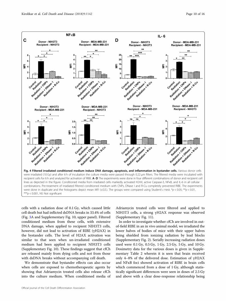
used except that the irradiated cells were not labeled withBrdU and the filtered culture medium was applied torecipient bystander cells for 6 h. The various cellularcombinations used comprising (1) NIH3T3 (mouse nor-mal donor) and NIH3T3 (mouse normal recipient); (2)MDA-MB-231 (human tumor donor) and NIH3T3(mouse normal recipient); (3) MDA-MB-231 (humantumor donor) and MDA-MB-231 (human tumor reci-pient); (4) NIH3T3 (mouse normal donor) and MDAMB-231 (human tumor recipient). Various RIBE parametersnamely γH2AX, active caspase-3, NFκB, and IL-6 wereanalyzed by immuno-flourescence.
Electron microscopy to detect cfCh in conditioned mediaThe filtered conditioned medium (~1.5 ml) from cells of
six irradiated MDA-MB-231 culture dishes were pooledand centrifuged at 700,000×g for 16 h, and the pelletobtained was washed in PBS by another centrifugationat 700,000×g for 16 h. Un-filtered cultured media weresimilarly processed and used as controls. The resultingpellets were re-suspended in 50 µl PBS and 10 µl aliquotswere processed for negative staining technique asdescribed by Horne, 196527. Grids layered with the sam-ples were examined under JEM1400 transmission electronmicroscope and imaged at × 60,000 magnification28.
Dosimetry for in vitro studiesCells were plated in six-well plates and exposed to a
radiation dose of 10 Gy (Supplementary Fig. 1). The fieldarea was 25 × 25 cm2 covering the entire plate (12.5 × 8.5cm). The depth of irradiation was kept at 0.5 cm and theexposure was given for 5.89 min to deliver 10 Gy. Theplate was placed exactly at the center of the field (awayfrom the prenumbra) so as to obtain uniform radiationexposure.
Estimation of cell deathEstimation of cell death was performed using the
acridine orange (AO)/propidium iodide (PI) stainingmethod by fluorescence microscopy. AO is a membrane-
permeable dye that stains DNA of healthy cells whilePI stains DNA of dead or dying cells. Following radiationat different doses (0.1 Gy, 0.25 Gy, 0.5 Gy, 0.75 Gy, 1Gy, 2.5 Gy, 5 Gy, 10 Gy, and 50 Gy), MDA-MB-231 cellswere incubated for 6 h and were stained at 1:1 ratio ofAO and PI and mounted with VectaShield mountingmedium with DAPI (Vector Laboratories, Catalog#H-1200). Images were acquired immediately and ana-lyzed on Applied Spectral Imaging system. A total of 1000nuclei staining with either dye were counted and pro-portion of nuclei that stained with PI were scored as dead.PI was sourced from Sigma-Aldrich Merck, Catalog #P4170 while AO was a kind gift from Dr. S Chiplunkar,ACTREC.
Apoptosis or necrosisTo determine what proportion of cell death was due to
apoptosis and/or necrosis, irradiated cells (10 Gy) wereincubated for 6 h post irradiation and were concurrentlystained with PI and active caspase-3 (a marker of apop-tosis) or PI and cyclophilin-A (a marker of necrosis).Proportion of nuclei that stained with active caspase-3or cyclophilin-A that co-localized with PI stained nucleiwere counted (Supplementary Fig. 4).
Chemotherapy-induced bystander effectMDA-MB-231 cells were treated with Adriamycin
(5 µg/ml for 24 h). Cells were trypsinised and extensivelywashed in PBS and incubated in fresh medium for 12 h.The conditioned medium was passed through 0.22 µmfilter and applied to NIH3T3 cells for 6 h. Activation ofH2AX was estimated by immuno-flourescence.
Isolation of cfCh from irradiated conditioned mediumcfCh from filtered conditioned media of irradiated
(10 Gy) MDA-MB-231 cells that had been pre-labeledin their DNA with BrdU and histones with CellLight®Histone 2B-GFP were isolated by a method describedus in detail elsewhere24. The purification method pri-marily comprises of ultra-centrifugation, lysis andaffinity chromatography24. The final isolate was sus-pended 500 µl of PBS, and 100 µl from the suspension wascytospun on a slide and examined by confocal micro-scopy. In a parallel set of experiments, cfCh were similarlyisolated from irradiated filtered conditioned media thathad not been fluorescently dually labeled and weresimilarly applied on a slide and stained with exosomecomponent marker (Hsp 70) to confirm that cfCh isolateswere devoid of exosomes.
Experiments using cfCh inactivating agentsIn order to further confirm that cfCh are involved in
RIBE, we treated the irradiated filtered conditionedmedium with cfCh neutralizing/degrading agents
Kirolikar et al. Cell Death and Disease (2018) 9:1142 Page 3 of 16
Official journal of the Cell Death Differentiation Association
(described below) prior to applying them to un-irradiatedbystander cells. We used four different combinationsof cells as mentioned earlier. The cfCh inactivating agentsused were: (1) anti-histone antibody complexed nano-particles (CNPs) (25 µg/1.5 ml)29, DNase I (0.0005 U/ml,Sigma, Catalog#DN25) or a novel DNA degradingagent resveratrol-copper (R-Cu) (molar ratio of 1 mMR: 0.0001mM Cu) (see later)30.
ImmunofluorescencePrimary and secondary antibodies used in the immu-
nofluorescence experiments are listed in SupplementaryTable 1.
In vitro experimentsImmunofluorescence to detect presence of BrdU and
activation of H2AX, active Caspase-3, NFκB, and IL-6 wasperformed by standard procedure described by us ear-lier23,24. Slides were mounted with vectashield mountingmedium with DAPI (Vector Laboratories, Catalog#H-1200) and analyzed on Applied Spectral Imaging system.All experiments were done in duplicate; 200 cells wereanalyzed for each cellular combination and mean fluor-escence intensity (MFI) was calculated and comparisonbetween groups was performed using Student’s t-test. Incase of γ-H2AX and NFκB, nuclear fluorescence was usedto calculate MFI.
In vivo experimentsThe in vivo experiments consisted of delivering lower
hemi-body irradiation (HBI) or focused mini - beamirradiation details of which are described later. Cryo-sections of brains of control and irradiated animals wereimmuno-stained for detection of γH2AX, active Caspase-3, NFκB, and IL-6 using primary and secondary antibodiesdescribed above. One thousand cells were analyzed fromeach brain and results were expressed as mean MFI(±S.E.). Comparison between groups was performed usingStudent’s t-test. Activation of the above RIBE markersappeared as fluorescent foci. We ascertained at the outsetthat fluorescent foci count strictly correlated with MFIvalues and expressed the results as such. To confirm thatDNA damage (γH2AX) was confined to neuronal cells(astrocytes), antibody against Glial Fibrillary Acidic Pro-tein (GFAP) was used, which were applied on 3 µm par-affin sections. The secondary antibody was donkey anti-rabbit TRITC (Supplementary Fig. 12).
Fluorescence in situ hybridization (FISH)Metaphase spreads were prepared from bystander
NIH3T3 cells (at 10th passage) treated with conditionedmedium of irradiated and un-irradiated MDA-MB-231cells. Metaphases were hybridized with Cyanine-3 orange
labeled human whole genomic probe (Chrom-BiosGmbH, Germany. Catalogue No. HGDNAOR10). Thirtymetaphases were imaged in each case and examined forthe presence of human DNA signals using FISHviewsoftware 5.0 (Applied Spectral Imaging, Israel).
Analysis of chromosomal aberrationsMetaphase spreads were prepared at 10th passage from
recipient NIH3T3 cells that had been treated with con-ditioned medium from irradiated or un-irradiated MDA-MB-231 cells and stained through conventional Giemsastaining. Fifty metaphases were analyzed in each case anddistribution and frequency of chromosomal aberrationswere analyzed using the Bandview software 5.0 on Spec-tral Bio-Imaging System (Applied spectral imaging,Israel).
Hemi-body irradiation to investigate RIBE in vivoBALB/c mice, aged 6–8 weeks, were subjected to
lower HBI at different doses viz, 0.1 Gy, 0.5 Gy, 1 Gy,2.5 Gy, 5 Gy, and 10 Gy wherein the upper half ofthe body was covered with lead shield. Each radiationgroup comprised of five animals. Lower HBI wasdelivered by gamma radiation using a Cobalt-60 sourceby single anterior portal of 20 cm × 5 cm field size. Theradiation doses were normalized at 1.5 cm depth andat a dose rate of 175 cGy/minute. The HBI setupis depicted in Supplementary Fig. 2. The animals werekilled after 48 h and their brains were removed, snap-frozen, and subsequently processed for cryo-sectioningand immune-florescence staining. Studies using cfChinhibitors were undertaken in animals receiving 10 Gyradiation dose. Twenty-five animals were divided into5 groups of five animals each as follows: (1) control,(mice not exposed to radiation); (2) 10 Gy lower HBIalone; (3) 10 Gy lower HBI+CNPs (50 µg anti-H4 con-jugated nanoparticles once daily i.p.); (4) 10 Gy lowerHBI+DNase I (15 mg/kg twice daily i.p.); (5) 10 Gy lowerHBI+R-Cu (R= 1mg/kg and Cu= 10−4mg/kg twicedaily by oral gavage). The first dose of CNPs, DNase Iand R–Cu were administered 4 h prior to deliveryof lower HBI; subsequent doses and frequency ofadministration of cfCh inhibitors are given in appropriatesections below.
Dosimetry of hemi-body irradiation experimentsThermo-luminescent dosimeters (TLDs) were prepared
in-house using 40mg of freshly annealed TLD-100(LiF: Mg,Ti) powder (Harshaw Chemical Co. Solon,Ohio USA) and packed in square polyethylene pouches(1 cm × 1 cm). BALB/c mice were killed under CO2
atmosphere and TLD pouches were surgically placedclose to various organs/tissue namely thigh muscle,
Kirolikar et al. Cell Death and Disease (2018) 9:1142 Page 4 of 16
Official journal of the Cell Death Differentiation Association
liver, lung, heart, and brain. Nylon sutures were used forclosure of incisions. Mice (n= 2 per dose) were exposedto various radiation doses as described earlier. TLDswere collected post irradiation for dosimetry analysis.The thermoluminescent output was recorded usingcommercial TLD-reader (REXON UL-320) and expressedas output per unit weight (nC/mg). The uncertainty factorin TLD-100 powder measurements is ±2%. Dosimterydata are given in Supplementary Table 2.
Mini beam irradiation using Varian/BrainLAB Novalis Tx™radiotherapy platformClinical Varian Novalis Tx™ 6 MV accelerator powered
by TrueBeam STx was used for focused beam irradiation.This platform has capabilities for targeted StereotacticBody Radiation Therapy and includes Brainlab iPlan®treatment planning and Varian’s HD120 MLC multileafcollimator for high resolution beam shaping. BalbC mice,aged 6–8 weeks, were subjected for focused mini beamradiation of 1 cm diameter at 2000 cGy dose over theumbilical region under general anesthesia (SupplementaryFig. 3). Target to surface distance (TSD) techniquewas used with TSD at surface 100 cm for dose delivery.Animals were placed in supine position and dose wasdelivered with 1.5 cm depth of prescription. Dose expo-sure plan was generated using Varian Eclipse planningsystem (v 13). Varians HD MLC were used to shapethe treatment diameter of 10 mm and planned to delivera 20 Gy of dose to the target. A wax bolus of 8 mmthickness was used to create sufficient dose build-up. Thescatter radiation dose for target to non-target regionsis depicted in Supplementary Fig. 3.
Dosimetry for focused mini-beam irradiation experimentsThe calibrated chambers and radiochromic film were
used for scattered dose calculations in non-target region.Solid water slab phantom was used to check the accuracyof the Plan generated by the treatment planning system.Plan-specific quality assurance were performed beforethe actual delivery of dose. Focused mini-beam radiationof 1 cm diameter at 2000 cGy dose over the umbilicalregion produce a scatter to the brain which was equivalentto that in ambient atmosphere (0.01216 Gy) (Supple-mentary Fig. 3). Forty-eight hr after radiation, mice werekilled under anesthesia and brain tissue were collectedand cryopreserved. The cryosections of brain tissue wereanalyzed for activation of H2AX and NFkB expressionby Immuno-flurescence.
Agents used to inactivate cfChAnti-histone antibody complexed nanopaticles (CNPs)Synthesis of pullulan-histone antibody nanoconjugates
was performed as described by us earlier using H4IgG23,24,29. For in vitro studies, 25 µg of CNPs in 100 µl of
buffer was added to 1.5 ml of culture medium. For in vivostudies, 50 µg of CNPs in 100 µl of buffer was adminis-tered i.p. once a day for a duration of 48 h when theanimals were killed (total number of doses received= 3).The first dose of CNPs was given 4 hr prior to irradiation.
Resveratrol-copper (R-Cu)Resveratrol (R) is a plant polyphenol with antioxidant
properties31. However, R acts as a pro-oxidant in thepresence of copper (Cu) by reducing Cu (II) to Cu (I)thereby generating a free radical32. R-Cu can cleaveplasmid DNA by this pro-oxidant activity33. We haveshown that R-Cu can also degrade genomic DNA30, andthat the pro-oxidant property of R-Cu with respect todegradation of DNA is maintained even when the molarconcentration of Cu is substantially reduced with respectto that of R30.
In vitro experimentsThe R-Cu molar combination used in these experiments
was R (1 mM): Cu (0.0001 mM). R (2.3 mg) (Sigma, Cat-alog#R5010) was dissolved in 5ml of 30% ethanol (2 mM)(solution A). Copper sulphate (4.98 mg) (MP Biomedicals,Catalog#191415) was dissolved in 1ml distilled water(20 mM) and then serially diluted to 0.0002 µM con-centration (solution B). Solutions A and B were mixed(50% v/v) to obtain a mixture containing 1mM R and0.0001mM Cu. One hundred microlitres of this mixturewas added to 1.5 ml of culture medium.
In vivo experimentsWe used resveratrol (Trade name—TransMaxTR,
Biotivia LLC, USA) and copper (Trade name—ChelatedCopper, J.R. Carlson Laboratories Inc. USA) for in vivoexperiments, both of which are approved for humanconsumption as dietary supplements. The contents of500mg capsules of R were dissolved in sterile distilledwater (concentration= 0.4 mg/ml). Five milligram tabletsof copper were crushed into fine powder and dissolvedin distilled water (concentration= 0.04 µg/ml). Bothsolutions were administered (50 µl each) by oral gavageone followed by the other twice daily for a duration of48 h when they were killed (total number of dosesreceived= 5). The final concentration of R was 1 mg/kgand Cu was 0.1 µg/kg at a final ratio of 1:10−4. Thefirst dose of R-Cu was given 4 hr prior to irradiation.
DNase IIn vitro experiments Bovine pancreatic DNase I(0.005 U; Sigma-Aldrich; Catalogue No- DN25-1G) wasused per 1.5 ml of culture media in all experiments.
In vivo experiments Bovine pancreatic DNase I (Sigma-Aldrich; Catalogue No- DN25-1G) was injected i.p. at
Kirolikar et al. Cell Death and Disease (2018) 9:1142 Page 5 of 16
Official journal of the Cell Death Differentiation Association
15mg/kg twice daily for a duration of 48 h when theywere killed (total number of doses received= 5). The firstdose of DNase I was given 4 hr prior to irradiation.
Estimation of serum cfChMice were irradiated with lower HBI (10 Gy) and serial
blood samples were collected at 12 h, 24 h, 36 h, 48 h, and60 h. Serum was separated and serum cfCh was estimatedby Cell Death Detection ELISAplus kit (Roche DiagnosticsGmbH, Germany) as described by us earlier24. Results areexpressed in terms of absorbance kinetics at 405 nm.
Animal ethics approvalThe protocol for animal experiments were approved by
the Institutional Animal Ethics Committee (IAEC) ofthe Institute in compliance to ARRIVE guidelines. C57Bl6and BALB/c mice (6–8 weeks old weighing ~20 g) wereobtained from and housed in the Institute Animal HouseFacility.
Statistical analysisGraphPad Prism 5 (GraphPad Software, Inc., USA.
Version 5.0) was used to perform statistical analysis.Results were compared using Student’s t-test. Graphswere presented as mean ± standard error of mean (SEM).Differences between groups were considered significantwhen P-value was <0.05 (two-tailed).
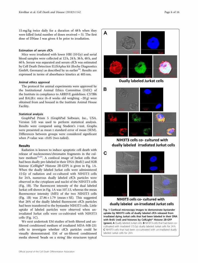
ResultsRadiation is known to induce apoptotic cell death with
release of nucleosomes/chromatin fragments in the cul-ture medium21,22. A confocal image of Jurkat cells thathad been dually pre-labeled in their DNA (BrdU) and H2Bhistone (Celllight® Histone 2B-GFP) is given in Fig. 1A.When the dually labeled Jurkat cells were administered15 Gy of radiation and co-cultured with NIH3T3 cellsfor 24 h, numerous dually labeled cfCh particles wereobserved in the cytoplasm and nuclei of the NIH3T3 cells(Fig. 1B). The fluorescent intensity of the dual labeledJurkat cell shown in Fig. 1A was 107.13, whereas the meanfluorescence intensity (MFI) of the two NIH3T3 cells(Fig. 1B) was 27.86 ± 1.79 (mean ± SE). This suggestedthat 26% of the dually labeled fluorescent cfCh particleshad been transferred to the bystander NIH3T3 cells. Littleuptake of labeled particles were observed when un-irradiated Jurkat cells were co-cultivated with NIH3T3cells (Fig. 1C).We next undertook EM studies of both filtered and un-
filtered conditioned medium of irradiated MDA-MB-231cells to investigate whether cfCh particles could bevisually demonstrated. EM of un-filtered conditionedmedia showed ‘beads on a string’ like structures typical
Fig. 1 Confocal microscopy images to demonstrate bystanderuptake by NIH3T3 cells of dually labeled cfCh released fromirradiated dying Jurkat cells that had been labeled in their DNAwith BrdU (red) and histones by CellLight® Histone 2B-GFP(green). A Dually labeled Jurkat cells. B NIH3T3 cells that had been co-cultivated with irradiated (15 Gy) dually labeled Jurkat cells for 24 h.C NIH3T3 cells that had been co-cultivated with un-irradiated duallylabeled Jurkat cells for 24 h
Kirolikar et al. Cell Death and Disease (2018) 9:1142 Page 6 of 16
Official journal of the Cell Death Differentiation Association
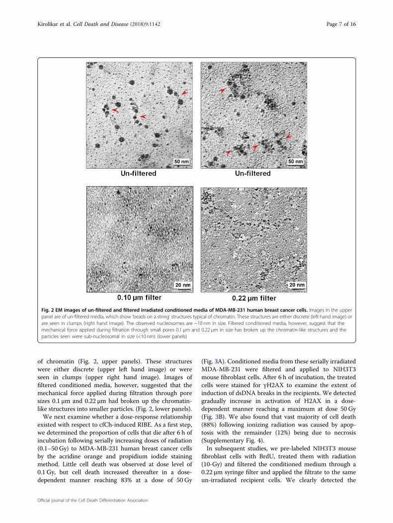
of chromatin (Fig. 2, upper panels). These structureswere either discrete (upper left hand image) or wereseen in clumps (upper right hand image). Images offiltered conditioned media, however, suggested that themechanical force applied during filtration through poresizes 0.1 µm and 0.22 µm had broken up the chromatin-like structures into smaller particles. (Fig. 2, lower panels).We next examine whether a dose-response relationship
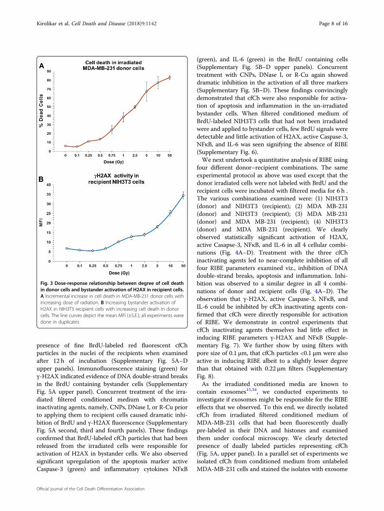
existed with respect to cfCh-induced RIBE. As a first step,we determined the proportion of cells that die after 6 h ofincubation following serially increasing doses of radiation(0.1–50 Gy) to MDA-MB-231 human breast cancer cellsby the acridine orange and propidium iodide stainingmethod. Little cell death was observed at dose level of0.1 Gy, but cell death increased thereafter in a dose-dependent manner reaching 83% at a dose of 50 Gy
(Fig. 3A). Conditioned media from these serially irradiatedMDA-MB-231 were filtered and applied to NIH3T3mouse fibroblast cells. After 6 h of incubation, the treatedcells were stained for γH2AX to examine the extent ofinduction of dsDNA breaks in the recipients. We detectedgradually increase in activation of H2AX in a dose-dependent manner reaching a maximum at dose 50 Gy(Fig. 3B). We also found that vast majority of cell death(88%) following ionizing radiation was caused by apop-tosis with the remainder (12%) being due to necrosis(Supplementary Fig. 4).In subsequent studies, we pre-labeled NIH3T3 mouse
fibroblast cells with BrdU, treated them with radiation(10-Gy) and filtered the conditioned medium through a0.22 µm syringe filter and applied the filtrate to the sameun-irradiated recipient cells. We clearly detected the
Fig. 2 EM images of un-filtered and filtered irradiated conditioned media of MDA-MB-231 human breast cancer cells. Images in the upperpanel are of un-filtered media, which show ‘beads on a string’ structures typical of chromatin. These structures are either discrete (left hand image) orare seen in clumps (right hand image). The observed nucleosomes are ~10 nm in size. Filtered conditioned media, however, suggest that themechanical force applied during filtration through small pores 0.1 µm and 0.22 µm in size has broken up the chromatin-like structures and theparticles seen were sub-nucleosomal in size (<10 nm) (lower panels)
Kirolikar et al. Cell Death and Disease (2018) 9:1142 Page 7 of 16
Official journal of the Cell Death Differentiation Association
presence of fine BrdU-labeled red fluorescent cfChparticles in the nuclei of the recipients when examinedafter 12 h of incubation (Supplementary Fig. 5A–Dupper panels). Immunofluorescence staining (green) forγ-H2AX indicated evidence of DNA double-strand breaksin the BrdU containing bystander cells (SupplementaryFig. 5A upper panel). Concurrent treatment of the irra-diated filtered conditioned medium with chromatininactivating agents, namely, CNPs, DNase I, or R-Cu priorto applying them to recipient cells caused dramatic inhi-bition of BrdU and γ-H2AX fluorescence (SupplementaryFig. 5A second, third and fourth panels). These findingsconfirmed that BrdU-labeled cfCh particles that had beenreleased from the irradiated cells were responsible foractivation of H2AX in bystander cells. We also observedsignificant upregulation of the apoptosis marker activeCaspase-3 (green) and inflammatory cytokines NFκB
(green), and IL-6 (green) in the BrdU containing cells(Supplementary Fig. 5B–D upper panels). Concurrenttreatment with CNPs, DNase I, or R-Cu again showeddramatic inhibition in the activation of all three markers(Supplementary Fig. 5B–D). These findings convincinglydemonstrated that cfCh were also responsible for activa-tion of apoptosis and inflammation in the un-irradiatedbystander cells. When filtered conditioned medium ofBrdU-labeled NIH3T3 cells that had not been irradiatedwere and applied to bystander cells, few BrdU signals weredetectable and little activation of H2AX, active Caspase-3,NFκB, and IL-6 was seen signifying the absence of RIBE(Supplementary Fig. 6).We next undertook a quantitative analysis of RIBE using
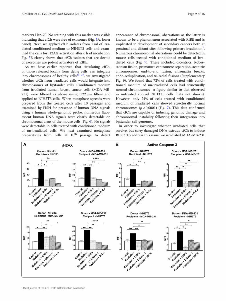
four different donor–recipient combinations. The sameexperimental protocol as above was used except that thedonor irradiated cells were not labeled with BrdU and therecipient cells were incubated with filtered media for 6 h .The various combinations examined were: (1) NIH3T3(donor) and NIH3T3 (recipient); (2) MDA MB-231(donor) and NIH3T3 (recipient); (3) MDA MB-231(donor) and MDA MB-231 (recipient); (4) NIH3T3(donor) and MDA MB-231 (recipient). We clearlyobserved statistically significant activation of H2AX,active Casapse-3, NFκB, and IL-6 in all 4 cellular combi-nations (Fig. 4A–D). Treatment with the three cfChinactivating agents led to near-complete inhibition of allfour RIBE parameters examined viz., inhibition of DNAdouble-strand breaks, apoptosis and inflammation. Inhi-bition was observed to a similar degree in all 4 combi-nations of donor and recipient cells (Fig. 4A–D). Theobservation that γ-H2AX, active Caspase-3, NFκB, andIL-6 could be inhibited by cfCh inactivating agents con-firmed that cfCh were directly responsible for activationof RIBE. We demonstrate in control experiments thatcfCh inactivating agents themselves had little effect ininducing RIBE parameters γ-H2AX and NFκB (Supple-mentary Fig. 7). We further show by using filters withpore size of 0.1 µm, that cfCh particles <0.1 µm were alsoactive in inducing RIBE albeit to a slightly lesser degreethan that obtained with 0.22 µm filters (SupplementaryFig. 8).As the irradiated conditioned media are known to
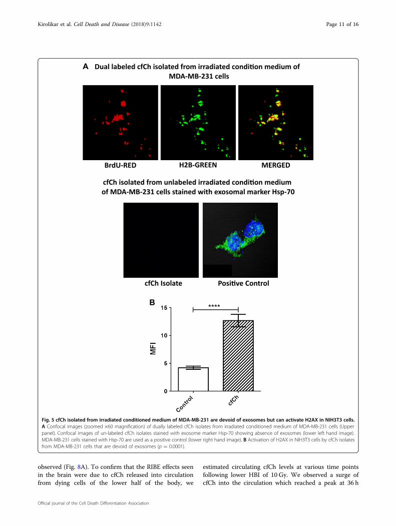
contain exosomes15,34, we conducted experiments toinvestigate if exosomes might be responsible for the RIBEeffects that we observed. To this end, we directly isolatedcfCh from irradiated filtered conditioned medium ofMDA-MB-231 cells that had been fluorescently duallypre-labeled in their DNA and histones and examinedthem under confocal microscopy. We clearly detectedpresence of dually labeled particles representing cfCh(Fig. 5A, upper panel). In a parallel set of experiments weisolated cfCh from conditioned medium from unlabeledMDA-MB-231 cells and stained the isolates with exosome
Fig. 3 Dose-response relationship between degree of cell deathin donor cells and bystander activation of H2AX in recipient cells.A Incremental increase in cell death in MDA-MB-231 donor cells withincreasing dose of radiation. B Increasing bystander activation ofH2AX in NIH3T3 recipient cells with increasing cell death in donorcells. The line curves depict the mean MFI (±S.E.); all experiments weredone in duplicates
Kirolikar et al. Cell Death and Disease (2018) 9:1142 Page 8 of 16
Official journal of the Cell Death Differentiation Association
markers Hsp-70. No staining with this marker was visibleindicating that cfCh were free of exosomes (Fig. 5A, lowerpanel). Next, we applied cfCh isolates from 1ml of irra-diated conditioned medium to NIH3T3 cells and exam-ined the cells for H2AX activation after 6 h of incubation.Fig. 5B clearly shows that cfCh isolates that are devoidof exosomes are potent activators of RIBE.As we have earlier reported that circulating cfCh,
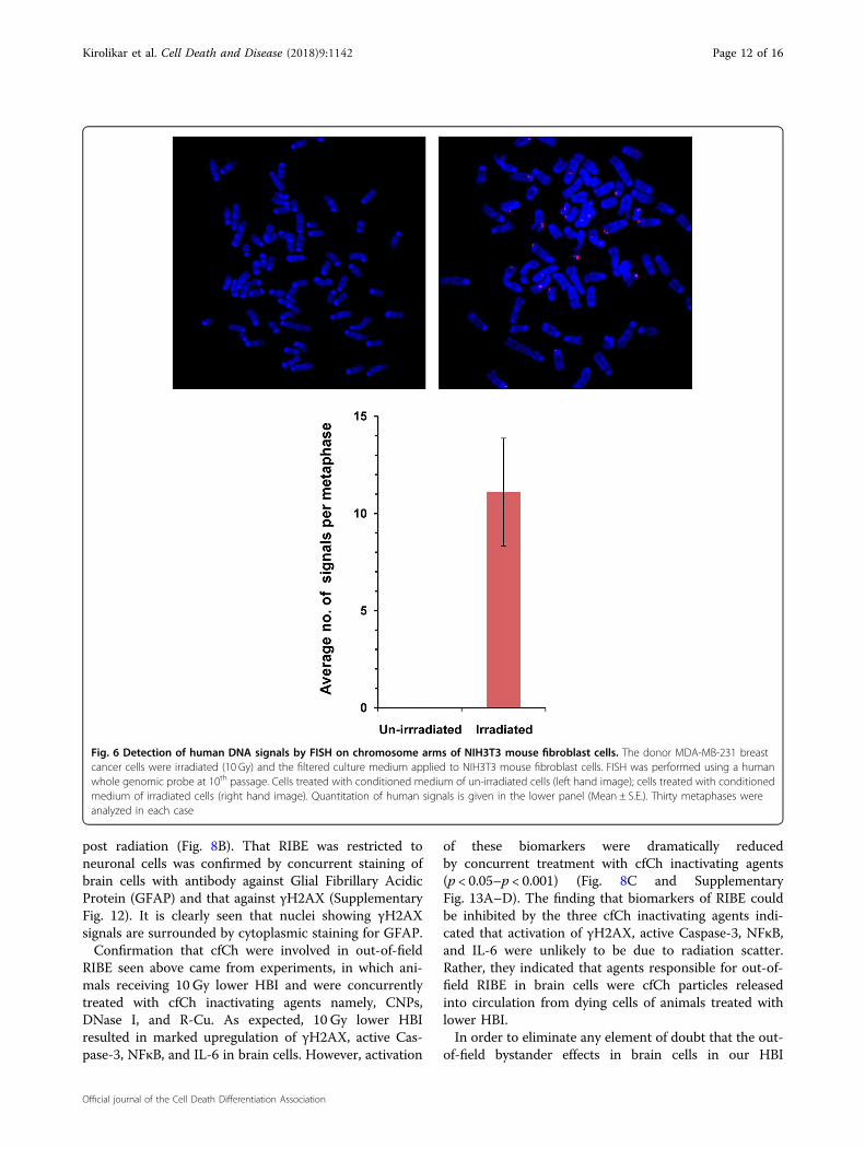
or those released locally from dying cells, can integrateinto chromosomes of healthy cells23–25, we investigatedwhether cfCh from irradiated cells would integrate intochromosomes of bystander cells. Conditioned mediumfrom irradiated human breast cancer cells (MDA-MB-231) were filtered as above using 0.22 µm filters andapplied to NIH3T3 cells. When metaphase spreads wereprepared from the treated cells after 10 passages andexamined by FISH for presence of human DNA signalsusing a human whole-genomic probe, numerous fluor-escent human DNA signals were clearly detectable onchromosomal arms of the mouse cells (Fig. 6). No signalswere detectable in cells treated with conditioned mediumof un-irradiated cells. We next examined metaphasepreparations from cells at 10th passage to detect
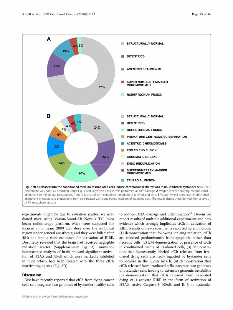
appearance of chromosomal aberrations as the latter isknown to be a phenomenon associated with RIBE and isimplicated in development of secondary cancers both atproximal and distant sites following primary irradiation1.Numerous chromosomal aberrations could be detected inmouse cells treated with conditioned medium of irra-diated cells (Fig. 7). These included dicentrics, Rober-stonian fusion, premature centromere separation, acentricchromosomes, end-to-end fusion, chromatin breaks,endo-reduplication, and tri-radial fusions (SupplementaryFig. 9). We found that 72% of cells treated with condi-tioned medium of un-irradiated cells had structurallynormal chromosomes—a figure similar to that observedin untreated control NIH3T3 cells (data not shown).However, only 24% of cells treated with conditionedmedium of irradiated cells showed structurally normalchromosomes (p < 0.0001) (Fig. 7). This data confirmedthat cfCh are capable of inducing genomic damage andchromosomal instability following their integration intobystander cell genomes.In order to investigate whether irradiated cells that
survive, but carry damaged DNA extrude cfCh to induceRIBE? To address this issue, we irradiated MDA-MB-231
Kirolikar et al. Cell Death and Disease (2018) 9:1142 Page 9 of 16
Official journal of the Cell Death Differentiation Association
cells with a radiation dose of 0.1 Gy, which caused littlecell death but had inflicted dsDNA breaks in 33.4% of cells(Fig. 3A and Supplementary Fig. 10, upper panel). Filteredconditioned medium from these cells, with extensiveDNA damage, when applied to recipient NIH3T3 cells,however, did not lead to activation of RIBE (γH2AX) inthe bystander cells. The level of H2AX activation wassimilar to that seen when un-irradiated conditionedmedium had been applied to recipient NIH3T3 cells(Supplementary Fig. 10). These findings suggest that cfChare released mainly from dying cells and not from thosewith dsDNA breaks without accompanying cell death.We demonstrate that bystander effects can also occur
when cells are exposed to chemotherapeutic agents byshowing that Adriamycin treated cells also release cfChinto the culture medium. When conditioned media of
Adriamycin treated cells were filtered and applied toNIH3T3 cells, a strong γH2AX response was observed(Supplementary Fig. 11).In order to investigate whether cfCh are involved in out-
of-field RIBE in an in vivo animal model, we irradiated thelower halves of bodies of mice with their upper halvesbeing shielded from ionizing radiation by lead blocks(Supplementary Fig. 2). Serially increasing radiation dosesused were 0.1 Gy, 0.5 Gy, 1 Gy, 2.5 Gy, 5 Gy, and 10 Gy.Dosimetry data for the various doses is given in Supple-mentary Table 2 wherein it is seen that brain receivedonly 4–8% of the delivered dose. Estimation of γH2AXand NFκB foci showed activation of RIBE in brain cellswhich commenced from a dose of 1 Gy, although statis-tically significant differences were seen in doses of 2.5 Gyand above with a clear dose-response relationship being
Fig. 4 Filtered irradiated conditioned medium induce DNA damage, apoptosis, and inflammation in bystander cells. Various donor cellswere irradiated (10 Gy) and after 6 h of incubation the culture media were passed through 0.22 µm filters. The filtered media were incubated withrecipient cells for 6 h and analyzed for activation of RIBE. A–D The experiments were done in four different combinations of donor and recipient celllines as depicted in the figure. Conditioned media from irradiated cells markedly activated H2AX, active Caspase-3, NFκB, and IL-6 in all cellularcombinations. Pre-treatment of irradiated filtered conditioned medium with CNPs, DNase I and R-Cu completely prevented RIBE. The experimentswere done in duplicate and the histograms depict mean MFI (±S.E.). The groups were compared using Student’s t-test. *p < 0.05; **p < 0.01,***p < 0.001, NS Not significant
Kirolikar et al. Cell Death and Disease (2018) 9:1142 Page 10 of 16
Official journal of the Cell Death Differentiation Association
observed (Fig. 8A). To confirm that the RIBE effects seenin the brain were due to cfCh released into circulationfrom dying cells of the lower half of the body, we
estimated circulating cfCh levels at various time pointsfollowing lower HBI of 10 Gy. We observed a surge ofcfCh into the circulation which reached a peak at 36 h
BrdU-RED
Dual labeled cfCh isolated from irradiated condi�on medium of MDA-MB-231 cells
H2B-GREEN MERGED
A
Posi�ve Control cfCh Isolate
cfCh isolated from unlabeled irradiated condi�on medium of MDA-MB-231 cells stained with exosomal marker Hsp-70
B
Fig. 5 cfCh isolated from irradiated conditioned medium of MDA-MB-231 are devoid of exosomes but can activate H2AX in NIH3T3 cells.A Confocal images (zoomed ×60 magnification) of dually labeled cfCh isolates from irradiated conditioned medium of MDA-MB-231 cells (Upperpanel). Confocal images of un-labeled cfCh isolates stained with exosome marker Hsp-70 showing absence of exosomes (lower left hand image).MDA-MB-231 cells stained with Hsp-70 are used as a positive control (lower right hand image). B Activation of H2AX in NIH3T3 cells by cfCh isolatesfrom MDA-MB-231 cells that are devoid of exosomes (p = 0.0001).
Kirolikar et al. Cell Death and Disease (2018) 9:1142 Page 11 of 16
Official journal of the Cell Death Differentiation Association
post radiation (Fig. 8B). That RIBE was restricted toneuronal cells was confirmed by concurrent staining ofbrain cells with antibody against Glial Fibrillary AcidicProtein (GFAP) and that against γH2AX (SupplementaryFig. 12). It is clearly seen that nuclei showing γH2AXsignals are surrounded by cytoplasmic staining for GFAP.Confirmation that cfCh were involved in out-of-field
RIBE seen above came from experiments, in which ani-mals receiving 10 Gy lower HBI and were concurrentlytreated with cfCh inactivating agents namely, CNPs,DNase I, and R-Cu. As expected, 10 Gy lower HBIresulted in marked upregulation of γH2AX, active Cas-pase-3, NFκB, and IL-6 in brain cells. However, activation
of these biomarkers were dramatically reducedby concurrent treatment with cfCh inactivating agents(p < 0.05–p < 0.001) (Fig. 8C and SupplementaryFig. 13A–D). The finding that biomarkers of RIBE couldbe inhibited by the three cfCh inactivating agents indi-cated that activation of γH2AX, active Caspase-3, NFκB,and IL-6 were unlikely to be due to radiation scatter.Rather, they indicated that agents responsible for out-of-field RIBE in brain cells were cfCh particles releasedinto circulation from dying cells of animals treated withlower HBI.In order to eliminate any element of doubt that the out-
of-field bystander effects in brain cells in our HBI
Fig. 6 Detection of human DNA signals by FISH on chromosome arms of NIH3T3 mouse fibroblast cells. The donor MDA-MB-231 breastcancer cells were irradiated (10 Gy) and the filtered culture medium applied to NIH3T3 mouse fibroblast cells. FISH was performed using a humanwhole genomic probe at 10th passage. Cells treated with conditioned medium of un-irradiated cells (left hand image); cells treated with conditionedmedium of irradiated cells (right hand image). Quantitation of human signals is given in the lower panel (Mean ± S.E.). Thirty metaphases wereanalyzed in each case
Kirolikar et al. Cell Death and Disease (2018) 9:1142 Page 12 of 16
Official journal of the Cell Death Differentiation Association
experiments might be due to radiation scatter, we irra-diated mice using Varian/BrainLAB Novalis Tx™ minibeam radiotherapy platform. Mice were subjected forfocused mini beam 2000 cGy dose over the umbilicalregion under general anesthesia; and they were killed after48 h and brains were examined for activation of RIBE.Dosimetry revealed that the brain had received negligibleradiation scatter (Supplementary Fig. 3). Immuno-flourescence analysis of brain showed significant activa-tion of H2AX and NFκB which were markedly inhibitedin mice which had been treated with the three cfChinactivating agents (Fig. 8D).
DiscussionWe have recently reported that cfCh from dying cancer
cells can integrate into genomes of bystander healthy cells
to induce DNA damage and inflammation23. Herein wereport results of multiple additional experiments and newevidence which strongly implicates cfCh in activation ofRIBE. Results of new experiments reported herein include;(1) demonstration that, following ionizing radiation, cfChare released predominantly from apoptotic rather thannecrotic cells; (2) EM demonstration of presence of cfChin conditioned media of irradiated cells; (3) demonstra-tion that fluorescently labeled cfCh released from irra-diated dying cells are freely ingested by bystander cellsto localize in the nuclei by 6 h; (4) demonstration thatcfCh released from irradiated cells integrate into genomesof bystander cells leading to extensive genomic instability;(5) demonstration that cfCh released from irradiateddying cells activate RIBE in the form of activation ofH2AX, active Caspase-3, NFκB, and IL-6 in bystander
Fig. 7 cfCh released into the conditioned medium of irradiated cells induce chromosomal aberrations in un-irradiated bystander cells. Theexperiment was done as described under Fig. 3 and karyotype analysis was performed at 10th passage. A Wagon wheel depicting chromosomalaberrations in metaphase preparations from cells treated with conditioned medium of un-irradiated cells. B Wagon wheel depicting chromosomalaberrations in metaphase preparations from cells treated with conditioned medium of irradiated cells. The results depict those derived from analysisof 50 metaphase spreads
Kirolikar et al. Cell Death and Disease (2018) 9:1142 Page 13 of 16
Official journal of the Cell Death Differentiation Association

cells; (6) demonstration that the above RIBE parameterscan be abrogated by cfCh inactivating agents namelyCNPs, DNase I and R-Cu; (7) demonstration of activationof RIBE parameters using multiple cellular combinationsthat include tumor to non-tumor and human to mousecell lines; (8) demonstration of a direct relationshipbetween radiation dose and cfCh-induced RIBE; (9)demonstration that cfCh-induced RIBE parameters canalso be activated following chemotherapy; (10) isolationof cfCh from irradiated conditioned medium anddemonstration that cfCh thus isolated can directly acti-vate RIBE; (11) demonstration that cfCh-induced RIBE isnot due to contaminating exosomes; (12) demonstrationthat cfCh can induce out-of-field RIBE in brain cells ofmice following lower hemi-body irradiation; (13)demonstration that RIBE induced in brain cells is medi-ated via release of cfCh into the blood stream from irra-diated dying cells; (14) demonstration of a dose-responserelationship between radiation dose and activation ofRIBE in brain cells; (15) demonstration that focused mini-beam radiation delivered to umbilical region of mice, thatproduces little scatter to the brain, can induce RIBE;(16) demonstration that RIBE in brain cells induced byboth lower HBI and focused mini-beam irradiation canbe prevented by cfCh inactivating agents.It is generally assumed that as RIBE occurs when the
conditioned medium from irradiated cells are filteredthrough 0.22 µm filters that the putative RIBE inducingdamaging signals are soluble factors3,26. We demonstrateby EM that RIBE-inducing signaling agents are particulatein nature in the form of cfCh (Fig. 2). The latter canbe isolated from conditioned medium of irradiated cellsby ultra-centrifugation and affinity chromatography, andwhen they are examined by confocal microscopy, appear
Fig. 8 Activation of DNA damage and inflammation in brain cellsof mice delivered lower hemi-body and focused mini-beamradiations and their abrogation by concurrent treatment withcfCh inactivating agents. A Dose-response effect of out-of-field RIBEfollowing lower HBI in brain cells with respect to γH2AX and NFκB.B Graph showing surge of cfCh in circulation at 36 h following lowerHBI measured by Cell Death Detection ELISAplus kit wherein results areexpressed in terms of absorbance kinetics at 405 nm. C Inhibition ofout-of-field RIBE with respect to γH2AX, active Caspase -3, NFκB, andIL-6 by CNPs, DNase I and R-Cu. All animals except the control groupreceived lower HBI (10 Gy) with and without CNPs, DNase I, and R-Cu.D Inhibition of out-of-field RIBE with respect to γH2AX and NFκB.All animals except the control group received focused min-beamradiation (20 Gy) with and without CNPs, DNase I, and R-Cu. Thecontrol and irradiated groups comprising five animals each whilethose receiving radiation plus CNPs, DNase I and R-Cu comprisedof three animals each. All histograms depict mean MFI (±S.E.). Thegroups were compared using Student’s t-test. *p < 0.05; **p < 0.01,***p < 0.001
Kirolikar et al. Cell Death and Disease (2018) 9:1142 Page 14 of 16
Official journal of the Cell Death Differentiation Association
as discreet particles (Fig. 5A). Furthermore that the iso-lated cfCh particles when applied to bystander cells evokea strong γH2AX signal (Fig. 5B). Although, inflammatorycytokines have been associated with RIBE, they have beenconsidered to be secreted by macrophages, which areattracted by radiation-induced dead or damaged cells35.We show the direct involvement of cfCh, in the absenceof macrophages, in activation of H2AX by bystandercells (Fig. 5B).We demonstrate that radiation-induced cell death is
predominantly due to apoptosis (88%) rather than bynecrosis (12%) (Supplementary Fig. 4). By undertakingradiation dose-response experiments we show a linearincrease in cell death in the donor MDA-MB-231 breastcancer cells with increasing dosage of ionizing radiationbetween a range of 0.25 and 50 Gy with the lowest dose(0.1 Gy) showing little cell death (Fig. 3, upper panel).When filtered conditioned medium from the above seri-ally irradiated cells were applied to NIH3T3 mousefibroblast cells, RIBE in the form of H2AX activation alsoshowed a dose-response relationship with respect toincreasing radiation dose beyond a dose level of 0.5 Gy(Fig. 3, lower panel). This dose-response relationship inbystander cells is not consistent with the report by Smi-lenov et al. who employed single-cell irradiation usingalpha particles and reported a lack of dose-responseeffect36. Differences in experimental design and type ofradiation used may explain the discrepancy with respectto our findings.We demonstrate by FISH that radiation-induced cfCh
that are taken up by bystander cells get integrated intotheir chromosomes (Fig. 6). Metaphase preparations ofNIH3T3 cells that had been co-cultured with irradiatedJurkat cells showed multiple fluorescent signals of humanDNA in the mouse cell chromosomes. The fact thatthey were detectable even after the bystander cells hadgone through 10 passages confirmed that cfCh fromirradiated cells had stably integrated into bystandercellular genomes.By conventional cytogenetic analysis, we demonstrate
that genomic integration of cfCh had induced extensivechromosomal aberrations in successive generations ofbystander cells (Fig. 7 and Supplementary Fig. 9).We demonstrate that cfCh-induced RIBE is not unique
to ionizing radiation but can be activated with cfChrelease by cells treated with chemotherapy (Supplemen-tary Fig. 11). In this context, we have recently reportedactivation of chemotherapy-induced bystander phenom-enon in vivo in mice37. We showed that toxicity of che-motherapy is primarily due to release of cfCh from theinitial round of drug-induced cell death, and that thistriggered a cascading effect causing exaggerated DNAdamage, apoptotic and inflammatory responses inbystander healthy cells37.
We demonstrate that cfCh are also involved in out-of-field RIBE in vivo. Lower HBI in mice led to activation ofDNA damage, apoptosis and inflammation in cells of thebrain (Fig. 8C and Supplementary Fig. 13). The invol-vement of cfCh was substantiated by the fact that RIBEactivation could be dramatically reduced to near base-line levels by concurrent treatment with the three cfChinactivating agents (Fig. 8C and Supplementary Fig. 13).We show that lower HBI had resulted in a surge of cfChin circulation suggesting that cfCh particles from dyingcells of the lower hemi-body had reached the brain viathe blood stream to induce RIBE (Fig. 8B). We observeda dose-response effect with respect to out-of-field RIBEin brain cells which increased progressively and wassignificantly higher than the baseline RIBE parameters(γH2AX and NFκB) beyond the dose level of 1.0 Gy(Fig. 8a). Lower dose levels (0.1 Gy, 0.5 Gy, and 1 Gy) didnot significantly activate out-of-field RIBE. Our findingsof a dose-response relationship is somewhat at variancewith that reported by Ventura et al.12 who delivered highdose (~50 Gy) localized synchrotron-generated radia-tion to mouse skin and observed persistent systemicgenotoxic and immune responses which they attributedto a scatter effect. Differences from our results can beattributed to this scatter radiation effect, which couldhave induced an adaptive response thereby influencing/modifying any bystander damage effect. The differencesmay also be attributed to differences in experimentaldesign between the two studies.In order to dispel any element of doubt that the out-of-
field bystander effects in brain cells in our HBI experi-ments was due to radiation scatter, we irradiated miceusing Varian/BrainLAB Novalis Tx™ mini-beam radio-therapy platform. Mice were subjected to focused mini-beam 2000 cGy radiation of 1 cm diameter over theumbilical region. Dosimetry revealed that radiation scatterto the brain was equivalent to background levels (Sup-plementary Fig. 3). Immuno-flourescence examinationshown activation of H2AX and NFκB which were mark-edly inhibited in mice which had received the cfChinactivating agents (Fig. 8d).In conclusion, our results demonstrate that cfCh can
induce RIBE both in vitro and in vivo. The fact that cfChinactivating agents namely CNPs, DNase I, and R-Cucould virtually abolish RIBE suggests that cfCh areimportant inducers of RIBE. Prevention of RIBE hasclinical relevance as RIBE has been implicated in induc-tion of morbid systemic side-effects2 secondary tissuedamage38 and secondary malignancies39 following radio-therapy for cancer.
AcknowledgementsThis study was supported by the Department of Atomic Energy, Governmentof India, through its grant CTCTMC to Tata Memorial Centre awarded to I.M.We wish to thank Mr. Libin Scaria and Mr. Chandrashekhar M Tambe of
Kirolikar et al. Cell Death and Disease (2018) 9:1142 Page 15 of 16
Official journal of the Cell Death Differentiation Association
Department of Radiation Oncology and Medical Physics, Tata MemorialHospital for help with focused mini-beam radiation experiment and preparingthe micro dosimeters and recording the dosimetry measurements respectively.Thanks are also due to Ms. Sharda Sawant for her help with EM experimentsand to Dr. Jayant Sastri Goda for his helpful comments on the manuscript.We thank Harshali Tandel and Vishal Jadhav for technical assistance.
Conflict of interestThe authors declare that they have no conflict of interest.
Publisher’s noteSpringer Nature remains neutral with regard to jurisdictional claims inpublished maps and institutional affiliations.
Supplementary Information accompanies this paper at (https://doi.org/10.1038/s41419-018-1181-x).
Received: 24 April 2018 Revised: 20 October 2018 Accepted: 22 October2018
References1. Morgan, W. F. Non-targeted and delayed effects of exposure to
ionizing radiation: I. Radiation-induced genomic instability and bystandereffects in vitro. Radiat. Res. https://doi.org/10.1016/j.canlet.2013.09.009(2003).
2. Widel, M. Radiation induced bystander effect: from in vitro studies to clinicalapplication. Int. J. Med. Phys. Clin. Eng. Radiat. Oncol. https://doi.org/10.4236/ijmpcero.2016.51001 (2016).
3. Ryan, L. A., Smith, R. W., Seymour, C. B. & Mothersill, C. E. Dilution of irradiatedcell conditioned medium and the bystander effect. Radiat. Res. https://doi.org/10.1667/RR1141.1 (2008).
4. Azzam, E. I., De Toledo, S. M. & Little, J. B. Oxidative metabolism, gap junctionsand the ionizing radiation-induced bystander effect. Oncogene https://doi.org/10.1038/sj.onc.1206961 (2003).
5. Mothersill, C. & Seymour, C. B. Radiation-induced bystandereffects–implications for cancer. Nat. Rev. Cancer https://doi.org/10.1038/nrc1277 (2004).
6. Zhang, D. et al. Reactive oxygen species formation and bystander effects ingradient irradiation on human breast cancer cells. Oncotarget https://doi.org/10.18632/oncotarget.9517 (2016).
7. Shao, C., Stewart, V., Folkard, M., Michael, B. D., Prise, K. M. Nitric oxide-mediatedsignaling in the bystander response of individually targeted glioma cells.Cancer Res. 63, 8437–8442 (2003).
8. Lyng, F. M., Maguire, P., McClean, B., Seymour, C. & Mothersill, C. The invol-vement of calcium and MAP kinase signaling pathways in the production ofradiation-induced bystander effects. Radiat. Res. https://doi.org/10.1667/RR3527.1 (2006).
9. Hu, W. et al. MiR-663 inhibits radiation-induced bystander effects by targetingTGFB1 in a feedback mode. RNA Biol. https://doi.org/10.4161/rna.34345 (2014)..
10. Shareef, M. M. et al. Role of tumor necrosis factor-α and TRAIL in high-doseradiation-induced bystander signaling in lung adenocarcinoma. Cancer Res.https://doi.org/10.1158/0008-5472.CAN-07-0722 (2007).
11. Buonanno, M. et al. Ear model for bystander studies induced by microbeamirradiation. Radiat. Res. https://doi.org/10.1667/RR14057.1 (2015).
12. Ventura, J. et al. Localized synchrotron irradiation of mouse skin inducespersistent systemic genotoxic and immune responses. Cancer Res. https://doi.org/10.1158/0008-5472.CAN-17-1066 (2017).
13. Shao, C., Folkard, M., Michael, B. D. & Prise, K. M. Targeted cytoplasmic irra-diation induces bystander responses. Proc. Natl Acad. Sci. USA https://doi.org/10.1073/pnas.0404930101 (2004).
14. Dickey, J. S., Zemp, F. J., Martin, O. A. & Kovalchuk, O. The role of miRNA in thedirect and indirect effects of ionizing radiation. Radiat. Environ. Biophys. https://doi.org/10.1007/s00411-011-0386-5 (2011).
15. Xu, S. et al. Exosome-mediated microRNA transfer plays a role inradiation-induced bystander effect. RNA Biol. https://doi.org/10.1080/15476286.2015.1100795 (2015).
16. Prise, K. M., . & O’Sullivan, J. M. Radiation-induced bystander signalling in cancertherapy. Nat. Rev. Cancer 9, 351–360 (2009).
17. Siva, S. et al. Radiotherapy for non-small cell lung cancer induces DNAdamage response in both irradiated and out-of-field normal tissues. Clin.Cancer Res. https://doi.org/10.1158/1078-0432.CCR-16-0138 (2016).
18. Koturbash, I. et al. In vivo bystander effect: cranial X-irradiation leads to ele-vated DNA damage, altered cellular proliferation and apoptosis, and increasedp53 levels in shielded spleen. Int J. Radiat. Oncol. Biol. Phys. https://doi.org/10.1016/j.ijrobp.2007.09.039 (2008).
19. Mancuso, M. et al. Oncogenic bystander radiation effects in Patched hetero-zygous mouse cerebellum. Proc. Natl Acad. Sci. USA https://doi.org/10.1073/pnas.0804186105 (2008).
20. Geras’kin, S. A., Fesenko, S. V. & Alexakhin, R. M. Effects of non-human speciesirradiation after the Chernobyl NPP accident. Environ. Int. https://doi.org/10.1016/j.envint.2007.12.012 (2008).
21. Dewey, W. C., Ling, C. C. & Meyn, R. E. Radiation-induced apoptosis: relevanceto radiotherapy. Int J. Radiat. Oncol. Biol. Phys. https://doi.org/10.1016/0360-3016(95)00214-8 (1995).
22. Van Nieuwenhuijze, A. E. M., Van Lopik, T., Smeenk, R. J. T. & Aarden, L. A. Timebetween onset of apoptosis and release of nucleosomes from apoptotic cells:putative implications for systemic lupus erythematosus. Ann. Rheum. Dis.https://doi.org/10.1136/ard.62.1.10 (2003).
23. Mittra, I. et al. Cell-free chromatin from dying cancer cells integrateinto genomes of bystander healthy cells to induce DNA damage andinflammation. Cell Death Discov. 3 https://doi.org/10.1038/cddiscovery.2017.15(2017).
24. Mittra, I. et al. Circulating nucleic acids damage DNA of healthy cells byintegrating into their genomes. J. Biosci. 40, 91–111 (2015).
25. Mittra, I. Circulating nucleic acids: a new class of physiological mobile geneticelements. F1000Research 4, 924 (2015).
26. Mothershill, C. & Seymour, C. Medium from irradiated human epithelialcells but not human fibroblasts reduces the clonogenic survival of uni-rradiated cells. Int. J. Radiat. Biol. https://doi.org/10.1080/095530097144030(1997).
27. Horne, R. In Techniques for Electron Microscopy 2nd edn, (ed KAT D. H.)311–355 (Blackwell Scientific Publications, Oxford, 1965).
28. Hayat, M. A. & Miller, S. E. Negative Staining. 253 (McGraw Hill PublishingCompany, NY, 1990).
29. Rekha, M. R. et al. Pullulan-histone antibody nanoconjugates for the removalof chromatin fragments from systemic circulation. Biomaterials 34, 6328–6338(2013).
30. Subramaniam, S., Vohra, I., Iyer, A., Nair, N. K. & Mittra, I. A paradoxical synergismbetween Resveratrol and copper (II) with respect to degradation of DNA andRNA. F1000Research 4, 1145 (2015).
31. Gülçin, I. Antioxidant properties of resveratrol: a structure-activity insight. Innov.Food Sci. Emerg. Technol. https://doi.org/10.1016/j.ifset.2009.07.002 (2010).
32. de la Lastra, C. A. & Villegas, I. Resveratrol as an antioxidant and pro-oxidantagent: mechanisms and clinical implications. Biochem. Soc. Trans., https://doi.org/10.1042/BST0351156 (2007).
33. Fukuhara, K. & Miyata, N. Resveratrol as a new type of DNA-cleaving agent.Bioorg. Med Chem. Lett. https://doi.org/10.1016/S0960-894X(98)00585-X (1998).
34. Le, M. et al. Exosomes are released by bystander cells exposed to radiation-induced biophoton signals: Reconciling the mechanisms mediating thebystander effect. PLoS ONE https://doi.org/10.1371/journal.pone.0173685(2017).
35. Coates, P. J., Robinson, J. I., Lorimore, S. A. & Wright, E. G. Ongoing activation ofp53 pathway responses is a long-term consequence of radiation exposurein vivo and associates with altered macrophage activities. J. Pathol. https://doi.org/10.1002/path.2321 (2008).
36. Smilenov, L. B., Hall, E. J., Bonner, W. M. & Sedelnikova, O. A. A microbeamstudy of DNA double-strand breaks in bystander primary human fibroblasts.Radiat. Prot. Dosim. https://doi.org/10.1093/rpd/ncl461 (2006).
37. Mittra, I. et al. Prevention of chemotherapy toxicity by agents that neutralize ordegrade cell-free chromatin. Ann. Oncol. https://doi.org/10.1093/annonc/mdx318 (2017).
38. Belyakov, O.V. et al. Biological effects in unirradiated human tissue inducedby radiation damage up to 1mm away. Proc Natl Acad Sci USA 102,14203–14208 (2005).
39. Brenner, D. J., Curtis, R. E., Hall, E. J. & Ron, E. Second malignancies in prostatecarcinoma patients after radiotherapy compared with surgery. Cancer 88,398–406 (2000).
Kirolikar et al. Cell Death and Disease (2018) 9:1142 Page 16 of 16
Official journal of the Cell Death Differentiation Association