Preparation of Caco-2 cell sheets using plasma polymerised acrylic acid as a weak boundary layer
8
Preparation of Caco-2 cell sheets using plasma polymerised acrylic acid as a weak boundary layer Ruby Majani a, b , Mischa Zelzer a,1 , Nikolaj Gadegaard c , Felicity R. Rose b , Morgan R. Alexander a, * a Laboratory of Biophysics and Surface Analysis, School of Pharmacy, The University of Nottingham, University Park, Nottingham, NG7 2RD, UK b Division of Drug Delivery and Tissue Engineering, School of Pharmacy, Centre for Biomolecular Sciences, The University of Nottingham, University Park, Nottingham, NG7 2RD, UK c Department of Electronics & Electrical Engineering, Rankine Building, University of Glasgow, Glasgow G12 8LT, UK article info Article history: Received 24 March 2010 Accepted 19 May 2010 Available online 18 June 2010 Keywords: Plasma polymerisation Cell viability Surface analysis Electron beam lithography Intestine Poly(lactic-co-glycolic acid) (PLGA) abstract The use of cell sheets for tissue engineering applications has considerable advantages over single cell seeding techniques. So far, only thermoresponsive surfaces have been used to manufacture cell sheets without chemically disrupting the cell-surface interactions. Here, we present a new and facile technique to prepare sheets of epithelial cells using plasma polymerised acrylic acid films. The cell sheets are harvested by gentle agitation of the media without the need of any additional external stimulus. We demonstrate that the plasma polymer deposition conditions affect the viability and metabolic activity of the cells in the sheet and relate these effects to the different surface properties of the plasma poly- merised acrylic acid films. Based on surface analysis data, a first attempt is made to explain the mech- anism behind the cell sheet formation. The advantage of the epithelial cell sheets generated here over single cell suspensions to seed a PLGA scaffold is presented. The scaffold itself, prepared using a mould fabricated via photolithography, exhibits a unique architecture that mimics closely the dimensions of the native tissue (mouse intestine). Ó 2010 Elsevier Ltd. All rights reserved. 1. Introduction The desire to regenerate damaged tissue has stimulated new research into various techniques that allow tissue engineers to use cells to create new tissue in situ or design more complex 3D tissue structures in vitro. The two most widely employed concepts are the injection of single cell suspensions to deliver cells in vivo and the use of degradable scaffolds as support for the creation of tissue with more complex architecture and functionality [1]. A more recent addition to these techniques is the preparation of cell sheets [2,3]. Cells grown in coherent sheets are required for certain applications such as skin and cornea tissue engineering where the native tissue is organized in layers of cells that can be grafted onto the under- lying tissue [4,5]. In addition, a layer-by-layer assembly of cell sheets provides the possibility to assemble more complex, thicker structures such as myocardial tissue without the need for a sup- porting scaffold [6] and most recently this approach was used to form pre-vascular networks from human endothelial cells [7]. The concept of cell sheets was pioneered by researchers led by Okano who developed a technique that allows the harvesting of cells as a coherent layer from in vitro cell culture [2,3,8]. This was achieved by grafting poly(N-isopropylacrylamide) (pNIPAM), a thermoresponsive material, to the surface of tissue culture plates. Under standard cell culture conditions (37 C), pNIPAM is hydrophobic and promotes cell attachment and growth. When the cells reached confluency, the temperature was lowered to 32 C upon which pNIPAM becomes hydrophilic and swells. Conse- quently, the cells are lifted off the surface as a coherent sheet. This elegant method allows the harvesting of cells without the need for proteolysing agents such as trypsin and therefore leaves the extracellular matrix (ECM) intact [1]. Compared to single cell suspensions, the cells can be delivered in vivo in a coherent structure together with their ECM without the need of a sup- porting scaffold [9]. To date, the techniques available to prepare cell sheets are limited to thermoresponsive materials, most commonly pNIPAM [10e12]. Although they have been successfully used on many cell types such as skin [5], corneal epithelial [4,13], myocardial [8,14] and mesenchymal stem cells [15] and efforts have been made to reduce the time it takes until cell detachment is complete [2], only cell lines that grow on pNIPAM and that are largely insen- sitive to temperatures below 37 C can be used. To extend the * Corresponding author. Tel.: þ44 (0) 115 951 5119; fax: þ44 (0) 115 951 5102. E-mail address: [email protected] (M.R. Alexander). 1 Present address: University of Strathclyde, Department of Pure and Applied Chemistry, 295 Cathedral Street, Glasgow, G1 1XL, UK. Contents lists available at ScienceDirect Biomaterials journal homepage: www.elsevier.com/locate/biomaterials 0142-9612/$ e see front matter Ó 2010 Elsevier Ltd. All rights reserved. doi:10.1016/j.biomaterials.2010.05.049 Biomaterials 31 (2010) 6764e6771
-
Upload
independent -
Category
Documents
-
view
0 -
download
0
Transcript of Preparation of Caco-2 cell sheets using plasma polymerised acrylic acid as a weak boundary layer

lable at ScienceDirect
Biomaterials 31 (2010) 6764e6771
Contents lists avai
Biomaterials
journal homepage: www.elsevier .com/locate/biomateria ls
Preparation of Caco-2 cell sheets using plasma polymerised acrylic acidas a weak boundary layer
Ruby Majani a,b, Mischa Zelzer a,1, Nikolaj Gadegaard c, Felicity R. Rose b, Morgan R. Alexander a,*a Laboratory of Biophysics and Surface Analysis, School of Pharmacy, The University of Nottingham, University Park, Nottingham, NG7 2RD, UKbDivision of Drug Delivery and Tissue Engineering, School of Pharmacy, Centre for Biomolecular Sciences, The University of Nottingham, University Park, Nottingham, NG7 2RD, UKcDepartment of Electronics & Electrical Engineering, Rankine Building, University of Glasgow, Glasgow G12 8LT, UK
a r t i c l e i n f o
Article history:Received 24 March 2010Accepted 19 May 2010Available online 18 June 2010
Keywords:Plasma polymerisationCell viabilitySurface analysisElectron beam lithographyIntestinePoly(lactic-co-glycolic acid) (PLGA)
* Corresponding author. Tel.: þ44 (0) 115 951 5119E-mail address: [email protected]
1 Present address: University of Strathclyde, DepaChemistry, 295 Cathedral Street, Glasgow, G1 1XL, UK
0142-9612/$ e see front matter � 2010 Elsevier Ltd.doi:10.1016/j.biomaterials.2010.05.049
a b s t r a c t
The use of cell sheets for tissue engineering applications has considerable advantages over single cellseeding techniques. So far, only thermoresponsive surfaces have been used to manufacture cell sheetswithout chemically disrupting the cell-surface interactions. Here, we present a new and facile techniqueto prepare sheets of epithelial cells using plasma polymerised acrylic acid films. The cell sheets areharvested by gentle agitation of the media without the need of any additional external stimulus. Wedemonstrate that the plasma polymer deposition conditions affect the viability and metabolic activity ofthe cells in the sheet and relate these effects to the different surface properties of the plasma poly-merised acrylic acid films. Based on surface analysis data, a first attempt is made to explain the mech-anism behind the cell sheet formation. The advantage of the epithelial cell sheets generated here oversingle cell suspensions to seed a PLGA scaffold is presented. The scaffold itself, prepared using a mouldfabricated via photolithography, exhibits a unique architecture that mimics closely the dimensions of thenative tissue (mouse intestine).
� 2010 Elsevier Ltd. All rights reserved.
1. Introduction
The desire to regenerate damaged tissue has stimulated newresearch into various techniques that allow tissue engineers to usecells to create new tissue in situ or design more complex 3D tissuestructures in vitro. The two most widely employed concepts are theinjection of single cell suspensions to deliver cells in vivo and theuse of degradable scaffolds as support for the creation of tissuewithmore complex architecture and functionality [1]. A more recentaddition to these techniques is the preparation of cell sheets [2,3].Cells grown in coherent sheets are required for certain applicationssuch as skin and cornea tissue engineering where the native tissueis organized in layers of cells that can be grafted onto the under-lying tissue [4,5]. In addition, a layer-by-layer assembly of cellsheets provides the possibility to assemble more complex, thickerstructures such as myocardial tissue without the need for a sup-porting scaffold [6] and most recently this approach was used toform pre-vascular networks from human endothelial cells [7].
; fax: þ44 (0) 115 951 5102..uk (M.R. Alexander).rtment of Pure and Applied.
All rights reserved.
The concept of cell sheets was pioneered by researchers led byOkano who developed a technique that allows the harvesting ofcells as a coherent layer from in vitro cell culture [2,3,8]. This wasachieved by grafting poly(N-isopropylacrylamide) (pNIPAM),a thermoresponsive material, to the surface of tissue cultureplates. Under standard cell culture conditions (37 �C), pNIPAM ishydrophobic and promotes cell attachment and growth. When thecells reached confluency, the temperature was lowered to 32 �Cupon which pNIPAM becomes hydrophilic and swells. Conse-quently, the cells are lifted off the surface as a coherent sheet. Thiselegant method allows the harvesting of cells without the needfor proteolysing agents such as trypsin and therefore leaves theextracellular matrix (ECM) intact [1]. Compared to single cellsuspensions, the cells can be delivered in vivo in a coherentstructure together with their ECM without the need of a sup-porting scaffold [9].
To date, the techniques available to prepare cell sheets arelimited to thermoresponsive materials, most commonly pNIPAM[10e12]. Although they have been successfully used on many celltypes such as skin [5], corneal epithelial [4,13], myocardial [8,14]and mesenchymal stem cells [15] and efforts have been made toreduce the time it takes until cell detachment is complete [2],only cell lines that grow on pNIPAM and that are largely insen-sitive to temperatures below 37 �C can be used. To extend the
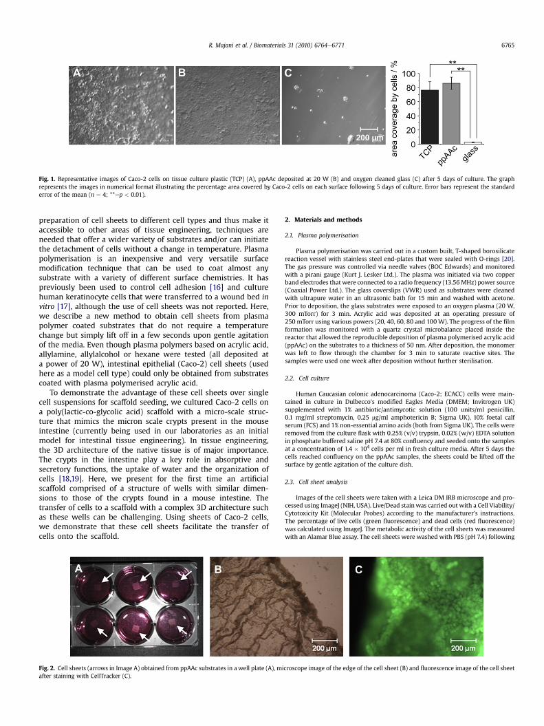
Fig. 1. Representative images of Caco-2 cells on tissue culture plastic (TCP) (A), ppAAc deposited at 20 W (B) and oxygen cleaned glass (C) after 5 days of culture. The graphrepresents the images in numerical format illustrating the percentage area covered by Caco-2 cells on each surface following 5 days of culture. Error bars represent the standarderror of the mean (n ¼ 4; **¼p < 0.01).
R. Majani et al. / Biomaterials 31 (2010) 6764e6771 6765
preparation of cell sheets to different cell types and thus make itaccessible to other areas of tissue engineering, techniques areneeded that offer a wider variety of substrates and/or can initiatethe detachment of cells without a change in temperature. Plasmapolymerisation is an inexpensive and very versatile surfacemodification technique that can be used to coat almost anysubstrate with a variety of different surface chemistries. It haspreviously been used to control cell adhesion [16] and culturehuman keratinocyte cells that were transferred to a wound bed invitro [17], although the use of cell sheets was not reported. Here,we describe a new method to obtain cell sheets from plasmapolymer coated substrates that do not require a temperaturechange but simply lift off in a few seconds upon gentle agitationof the media. Even though plasma polymers based on acrylic acid,allylamine, allylalcohol or hexane were tested (all deposited ata power of 20 W), intestinal epithelial (Caco-2) cell sheets (usedhere as a model cell type) could only be obtained from substratescoated with plasma polymerised acrylic acid.
To demonstrate the advantage of these cell sheets over singlecell suspensions for scaffold seeding, we cultured Caco-2 cells ona poly(lactic-co-glycolic acid) scaffold with a micro-scale struc-ture that mimics the micron scale crypts present in the mouseintestine (currently being used in our laboratories as an initialmodel for intestinal tissue engineering). In tissue engineering,the 3D architecture of the native tissue is of major importance.The crypts in the intestine play a key role in absorptive andsecretory functions, the uptake of water and the organization ofcells [18,19]. Here, we present for the first time an artificialscaffold comprised of a structure of wells with similar dimen-sions to those of the crypts found in a mouse intestine. Thetransfer of cells to a scaffold with a complex 3D architecture suchas these wells can be challenging. Using sheets of Caco-2 cells,we demonstrate that these cell sheets facilitate the transfer ofcells onto the scaffold.
Fig. 2. Cell sheets (arrows in Image A) obtained from ppAAc substrates in a well plate (A), miafter staining with CellTracker (C).
2. Materials and methods
2.1. Plasma polymerisation
Plasma polymerisation was carried out in a custom built, T-shaped borosilicatereaction vessel with stainless steel end-plates that were sealed with O-rings [20].The gas pressure was controlled via needle valves (BOC Edwards) and monitoredwith a pirani gauge (Kurt J. Lesker Ltd.). The plasma was initiated via two copperband electrodes that were connected to a radio frequency (13.56 MHz) power source(Coaxial Power Ltd.). The glass coverslips (VWR) used as substrates were cleanedwith ultrapure water in an ultrasonic bath for 15 min and washed with acetone.Prior to deposition, the glass substrates were exposed to an oxygen plasma (20 W,300 mTorr) for 3 min. Acrylic acid was deposited at an operating pressure of250 mTorr using various powers (20, 40, 60, 80 and 100W). The progress of the filmformation was monitored with a quartz crystal microbalance placed inside thereactor that allowed the reproducible deposition of plasma polymerised acrylic acid(ppAAc) on the substrates to a thickness of 50 nm. After deposition, the monomerwas left to flow through the chamber for 3 min to saturate reactive sites. Thesamples were used one week after deposition without further sterilisation.
2.2. Cell culture
Human Caucasian colonic adenocarcinoma (Caco-2; ECACC) cells were main-tained in culture in Dulbecco’s modified Eagles Media (DMEM; Invitrogen UK)supplemented with 1% antibiotic/antimycotic solution (100 units/ml penicillin,0.1 mg/ml streptomycin, 0.25 mg/ml amphotericin B; Sigma UK), 10% foetal calfserum (FCS) and 1% non-essential amino acids (both from Sigma UK). The cells wereremoved from the culture flask with 0.25% (v/v) trypsin, 0.02% (w/v) EDTA solutionin phosphate buffered saline pH 7.4 at 80% confluency and seeded onto the samplesat a concentration of 1.4 � 104 cells per ml in fresh culture media. After 5 days thecells reached confluency on the ppAAc samples, the sheets could be lifted off thesurface by gentle agitation of the culture dish.
2.3. Cell sheet analysis
Images of the cell sheets were taken with a Leica DM IRB microscope and pro-cessed using ImageJ (NIH, USA). Live/Dead stainwas carried out with a Cell Viability/Cytotoxicity Kit (Molecular Probes) according to the manufacturer’s instructions.The percentage of live cells (green fluorescence) and dead cells (red fluorescence)was calculated using ImageJ. The metabolic activity of the cell sheets was measuredwith an Alamar Blue assay. The cell sheets were washed with PBS (pH 7.4) following
croscope image of the edge of the cell sheet (B) and fluorescence image of the cell sheet

Fig. 3. Representative images illustrating a comparison of the viability of Caco-2 cell sheets obtained from ppAAc deposited at either 20 W (A) or 60 W (B) (green: live cells; red:dead cells).
R. Majani et al. / Biomaterials 31 (2010) 6764e67716766
removal of the cell culture media. They were then incubated in 1 ml of workingAlamar Blue solution (1 ml Alamar Blue stock solution (SeroTec) diluted in 9 mlHanks Balanced Salts Solution without Phenol Red (HBSS)) and incubated for90 min. The fluorescence of 100 ml of the resulting Alamar Blue solution wasmeasured with an MFX fluorometer (Dynex technologies) with lEx ¼ 355 nm andlEm ¼ 460 nm. A cell count prior to the Alamar Blue assay acertained the cell sheetshad comparable cell numbers. Statistical analysis was performed using Graph PadPrism (Graph Pad Software Inc., San Diego, California). Group comparisons wereassessed by analysis of variance (ANOVA) using the repeated measures test followedby Tukey’s multiple comparison post test. The statistical significance was set atp < 0.05.
2.4. Surface analysis
Static water contact angles (WCA) were recorded with a Cam200 (KSV Instru-ments Ltd.). After placing the drop on the sample, images were recorded in 1 sintervals over 20 s. For each image, the contact angle was determined using a YoungLaplace fit. After discarding the first value, the WCA at t ¼ 0 was determined viaa linear regression through the remaining data points.
XPS analysis was carried out on a Kratos Axis Ultra equipped with a mono-chromated Al ka X-ray source (1486.6 eV). It was operated with an emission currentof 15 mA and an anode potential of 10 kV. The instrument was set to fixed trans-mission mode and the take-off angle for the photoelectron analyzer was 90� . Chargeneutralisation was used for all samples. Data analysis was carried out with CasaXPSusing empirical sensitivity factors provided by the manufacturer.
Images of the surface topography were taken on a Dimension 3000 atomic forcemicroscope (Digital Instruments, Veeco) in tapping mode. The instrument wascontrolled with NanoScope (V5.30, 2005). The images were acquired at a 10� 10 mmscale. The rootmean square roughnesswas calculated from the images after linewiseplane corrections and is an average of 3 measurements.
2.5. PLGA scaffold manufacture
A silicon mould containing an array of cylindrically shaped wells with a diam-eter of 62 mm, a depth of 50 mm and a pitch of 70 mm was prepared by photoli-thography. A silicon substrate was coated with a photoresist (AZ4562) and softbaked at 90 �C for 30 min before UV exposure in a Suss MA6 mask aligner. After
20 30 40 50 600
20
40
60
80
100
cell
viab
ility
/ % li
ve c
ells
deposition power / W
a
Fig. 4. Viability (a) and metabolic activity (b) of Caco-2 cell sheets generated on ppAAc surfmean (n ¼ 4); ANOVA: ***¼p < 0.001, **¼p < 0.01.
development, the sample was etched in an Oxford STS-ICP deep silicon etch tool toa final depth of 50 mm. From this silicon mould, a negative template was prepared byhot embossing onto a polyolefin plastomer (POP) block which was prepared fromAffinity� POP pellets (Dow Europe GmbH).
Cell culture scaffolds were prepared from the POP template using poly(lactic-co-glycolic acid) (PLGA) particles. To obtain PLGA particles, 4 ml of a 3% (w/w) solutionof PLGA 85/15 (MW 35 000, Lakeshore Biomaterials) in CH2Cl2 and 4 ml of a 10%solution of polyvinylalcohol (PVA) in water were mixed and homogenized at12,400 rpm for 5 min [21]. The emulsion was poured into 1 L of 0.3% (w/v) PVA inwater and stirred overnight. The formed particles were separated by centrifugationand freeze dried for 3 days. Their size was determined with dynamic light scattering(DLS) (Malvern Zetasizer Nano 4700). The particles were then suspended in a 0.01%(v/v) solution of Tween in phosphate buffered saline (PBS). The resulting slurry wasplaced in the POP mould and allowed to air dry for 2 h. The particles were thensintered for 1.5 h at 60 �C. The PLGA scaffold was separated from the POPmould aftercooling overnight. Images of the scaffold were taken with a JEOL JSM-6060LVscanning electron microscope (SEM) operated at 10e12 kV.
2.6. Cell seeding on the scaffold
The scaffold was sterilised in a 5% (v/v) antibiotic/antimycotic solution in PBS for1 h in the incubator. The scaffold was then rinsed and conditioned with Caco-2media supplemented as described previously. Cell sheets were prepared asdescribed above, lifted from 20 W ppAAc coverslips and transferred onto the scaf-fold. The cell sheets were temporarily held in place with a clean glass coverslip for3 h. The coverslip was then lifted off and the cells were cultured on the scaffold for 5days in the incubator as described previously. For the single cell suspension, the cellswere cultured and passaged as described above and seeded onto the scaffold ata density of 2 � 105 cells per cm2. Cells were stained with CellTracker (MolecularProbes) to identify live cells after 5 days. Imaging of the samples was performed asdescribed previously.
3. Results
The glass substrates for cell culture were prepared by depositingacrylic acid from an RF plasma on a glass slide to form a thin layer of
20 30 40 50 600.0
0.2
0.4
0.6
0.8
1.0
b
**
rela
tive
met
abol
ic a
ctiv
ity
deposition power / W
***
aces using a range of deposition powers. Error bars represent the standard error of the

a
b
0
2
4
6
8
10
12
14
20 40 60 80 10055
60
65
70
75
80
85
rela
tive
amou
nt o
f C-H
/C-C
/ %
deposition power / W
rela
tive
amou
nt o
f CO
OX
/ %
c
0 20 40 60 80 1000.00
0.04
0.08
0.12
0.16
0.20
rms
roug
hnes
s / n
m
deposition power / W
***
***
0 20 40 60 8030
40
50
60
70
80
WC
A / d
egre
e
deposition power / W
glass 20 W 40 W 60 W 80 W
Fig. 5. a) Effect of the deposition power on the water contact angle (WCA) of ppAAc surfaces. Error bars represent standard deviations. The images show typical water drops on thesurface of each sample. The sample at 0 W represents the untreated glass substrate. b) Change in the amount of hydrocarbon (CeC/CeH, -) and carboxyl (COOX, 6) functionalitieson the ppAAc surface with deposition power. The data was obtained from curve fits to the C1s peaks in the XPS spectra. c) Dependence of the root mean square (rms) roughness ofppAAc on the deposition power. Error bars represent standard deviations (n ¼ 3); ANOVA: *¼p < 0.05, **¼p < 0.01.
Table 1Elemental composition of ppAAc deposited at various deposition powers.
Deposition Power/W 20 40 60 80 100
Carbon 77 80 82 85 89Oxygen 23 20 18 15 11
R. Majani et al. / Biomaterials 31 (2010) 6764e6771 6767
plasma polymerised acrylic acid (ppAAc). The suitability of thissurface as a support for the culture of Caco-2 cells was assessed bycomparison with tissue culture plastic and glass cleaned in anoxygen plasma after 5 days of culture (Fig. 1aec). The cells adheredwell to the tissue culture plastic (TCP) and ppAAc modified glasscoverslips; the percentage area covered with cells on these surfaceswas above 75% and no significant difference in the percentage areacovered by Caco-2 cells was found between these two samples(Fig.1; p< 0.01). In contrast, the area coverage on glass following anoxygen plasma etch was very low and statistically significantlydifferent compared with both TCP and ppAAc modified glasscoverslips (2.4%; p < 0.01), demonstrating that the deposition ofppAAc on glass greatly improved cell adhesion.
The cell sheets were obtained from the ppAAc coated samplesby gentle agitation of the media after the cells reached confluencyon the fifth day of culture (Fig. 2a). The cells lifted off as a coherentsheet as shown in Fig. 2b and staining with CellTracker showed thatthe sheet consisted largely of live cells (green staining; Fig. 2c).
For cell sheets to be of practical use in subsequent tissue cultureprocesses, it is important to maximize the viability of cells within
the cell sheet. The power used during the deposition of a plasmapolymer onto a surface is known to affect the composition of theplasma polymer film [22] and can subsequently influence theactivity of cells maintained in culture on this surface [23]. We useda Live/Dead stain and an Alamar Blue assay to determine theviability and metabolic activity of Caco-2 cell sheets obtained fromppAAc films deposited at 20, 30, 40, 50 and 60 W. When the cellswere cultured on ppAAc deposited at 80 or 100 W they did not liftoff the surface as sheets even though they had reached confluence.Fig. 3 shows the images of the Live/Dead stain on 20 and 60 WppAAc. While almost all cells in the cell sheet generated on the20WppAAc surfaces were viable, a large number of dead cells wereobserved in the cell sheet obtained from the 60 W ppAAc surface.

Fig. 6. Representative SEM images of the PLGA scaffold showing a side (A), a top (B) and an angled view (C) of the well structure.
R. Majani et al. / Biomaterials 31 (2010) 6764e67716768
The graph in Fig. 4a shows that the viability of cell sheets (asdetermined by the Live/Dead assay) is similar for those generatedon 20 W and 30 W ppAAc surfaces (95% and 97%, respectively).However, the viability of the cell sheets decreases continuouslythereafter to 53% in sheets obtained from 60 W ppAAc. The meta-bolic activity is highest in sheets obtained from 20 W ppAAc(Fig. 4b) but drops significantly for all other surfaces (p < 0.001against all other deposition powers). From 40 W ppAAc to 60 WppAAc the metabolic activity decreases continuously again(p < 0.01).
To explain these observations and investigate which materialproperties affect the viability and metabolism of cells in the cellsheets we studied the surface chemistry and topography of theplasma polymer deposits. The deposition of ppAAc on the glasssubstrate resulted in an increase in the water contact angle relativeto oxygen cleaned glass. The WCA of the ppAAc films increased asthe deposition power increased to 60 W but dropped again forppAAc deposited at 80 W (Fig. 5a). XPS analysis showed that only
100 µm
A B
Fig. 7. Seeding of the PLGA scaffold from a suspension of individual cells (A) and from a cecompare the extent of cell seeding using each method (n ¼ 5; p ¼ 0.0072).
carbon and oxygen were present on the surface (SupplementaryFig. 1). The relative amounts of C and O for various depositionpowers is shown in Table 1. The amount of C increased withincreasing deposition power whereas the amount of O decreased.Peak fits to the C 1s peak allowed the determination of the relativeamount of functional groups on the surface (Supplementary Fig. 2).With increasing deposition power, the amount of hydrocarbongroups increased on the surfacewhile the concentration of carboxylgroups decreased (Fig. 5b). The AFM images showed that under allconditions the deposited ppAAc filmwas very flat and displayed notopographical features (Supplementary Fig. 3). The root meansquare (rms) roughness of all samples is shown in Fig. 5c. Theroughness decreased from 0.16 nm on 10 W ppAAc to about 0.1 nmon 50W ppAAc. At depositing powers from 50 to 100W the surfaceroughness of ppAAc stayed close to 0.1 nm.
To test the performance of Caco-2 cell sheets, we compared theefficiency of seeding a scaffold using the cell sheets and single cellsuspensions. Since the structure of a functional intestinal tissue
100 µm
0
50
100
150
200
250
300
cellsheets
cell
dens
ity /
cells
/ m
m2
individualcells
**C
ll sheet (B) was compared; CellTracker staining of the cells was used to estimate and
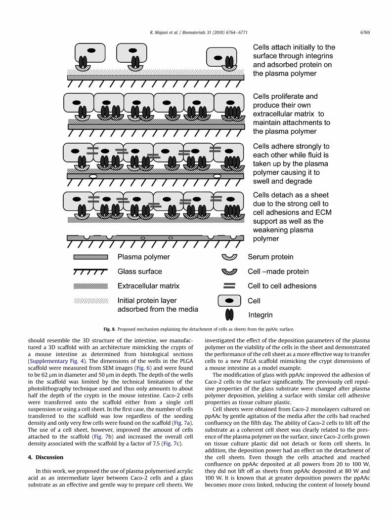
Fig. 8. Proposed mechanism explaining the detachment of cells as sheets from the ppAAc surface.
R. Majani et al. / Biomaterials 31 (2010) 6764e6771 6769
should resemble the 3D structure of the intestine, we manufac-tured a 3D scaffold with an architecture mimicking the crypts ofa mouse intestine as determined from histological sections(Supplementary Fig. 4). The dimensions of the wells in the PLGAscaffold were measured from SEM images (Fig. 6) and were foundto be 62 mm in diameter and 50 mm in depth. The depth of the wellsin the scaffold was limited by the technical limitations of thephotolithography technique used and thus only amounts to abouthalf the depth of the crypts in the mouse intestine. Caco-2 cellswere transferred onto the scaffold either from a single cellsuspension or using a cell sheet. In the first case, the number of cellstransferred to the scaffold was low regardless of the seedingdensity and only very few cells were found on the scaffold (Fig. 7a).The use of a cell sheet, however, improved the amount of cellsattached to the scaffold (Fig. 7b) and increased the overall celldensity associated with the scaffold by a factor of 7.5 (Fig. 7c).
4. Discussion
In this work, we proposed the use of plasma polymerised acrylicacid as an intermediate layer between Caco-2 cells and a glasssubstrate as an effective and gentle way to prepare cell sheets. We
investigated the effect of the deposition parameters of the plasmapolymer on the viability of the cells in the sheet and demonstratedthe performance of the cell sheet as amore effectiveway to transfercells to a new PLGA scaffold mimicking the crypt dimensions ofa mouse intestine as a model example.
The modification of glass with ppAAc improved the adhesion ofCaco-2 cells to the surface significantly. The previously cell repul-sive properties of the glass substrate were changed after plasmapolymer deposition, yielding a surface with similar cell adhesiveproperties as tissue culture plastic.
Cell sheets were obtained from Caco-2 monolayers cultured onppAAc by gentle agitation of the media after the cells had reachedconfluency on the fifth day. The ability of Caco-2 cells to lift off thesubstrate as a coherent cell sheet was clearly related to the pres-ence of the plasma polymer on the surface, since Caco-2 cells grownon tissue culture plastic did not detach or form cell sheets. Inaddition, the deposition power had an effect on the detachment ofthe cell sheets. Even though the cells attached and reachedconfluence on ppAAc deposited at all powers from 20 to 100 W,they did not lift off as sheets from ppAAc deposited at 80 W and100 W. It is known that at greater deposition powers the ppAAcbecomes more cross linked, reducing the content of loosely bound

R. Majani et al. / Biomaterials 31 (2010) 6764e67716770
highly soluble oligomeric material [20]. This is consistent with thechange in the functional composition observed from the XPSanalysis in Fig. 5b where carboxyl groups are increasingly elimi-nated from the deposit as the deposition power increases. It isproposed that it is this weak boundary layer of ppAAc that ismissing in the ppAAc deposited at 80Wand 100Wwhich preventsthe cells from lifting off.
Based on the observations of cell sheet formation and thesurface analysis data, a model describing the contributing factors incell sheet formation is proposed (Fig. 8). First a layer of proteinsadsorb to the surface from the serum containing media to whichcells attach based on integrin mediated signaling after contact withthe surface. Over time, the cells proliferate and form their ownextracellular matrix (ECM). The hydrophilic functional groups ofppAAc can easily interact with the aqueous surrounding, causingchanges in the composition of the plasma polymer over time.Changes in the surface chemistry and topography of the hydrophilicplasma polymers in water over time have been reported before[24e26]. In particular, it has been shown that the solubility ofppAAc inwater depends on the deposition power, withmore ppAAcbeing dissolved at lower powers [27]. As a result, the cellesurfaceinteraction is expected to weaken due to a loss in the integrity ofthe plasma polymer film, whereas the ECM network becomesstronger over time. Consequently, when the media is agitated thecells readily lift off from the surface due to a loss of the adhesion ofthe ECM to the substrate. The cells are kept together as a coherentsheet by cellecell interactions and adhesion to the ECM. This isconsistent with the observation that cell sheets are more readilyobtained from ppAAc surfaces deposited at lower powers wherethe higher retention of functional groups facilitates the interactionof the filmwith water. The ppAAc film can thus be considered to bea weak boundary layer between the glass substrate and the ECMthat supports the cell sheet.
Previously, MacNeil and coworkers used ppAAc surfaces totransfer keratinocytes from the plasma polymer surface on whichthey had been cultured to a wound bed [17]. Although this workpresumably exploited the facile detachment of cells from ppAAcsurfaces that is used here to prepare cell sheets, these researchersdid not lift the cells off as whole sheets. Instead the cells weretransferred to another substrate by a transfer-on-contact technique.The cells cultured on ppAAc coated substrates were pressed directlyonto the new surface and lifting the ppAAc coated substrate offthereafter. Thus, the present work is the first account that explicitlydescribes the use of a polymer film to obtain cell sheets withoutapplying chemicals or a change in temperature.
The viability of the Caco-2 cells on the cell sheets depended onthe power used to deposit the ppAAc. While lower powers (20 and30W) resulted in almost 100% viability, increasing the power above30W resulted in increasingly more dead cells within the cell sheets(Fig. 4). The high cell viability on 20 W ppAAc also corresponded tohigher metabolic activity. However, on 30 W ppAAc the metabolicactivity decreased dramatically, in contrast to the high viability ofthe cells on this surface (indicated using CellTracker staining) andindicates that a high viability does not necessarily result in a highmetabolic activity.
The surface roughness systematically varied with depositionpower, exhibiting a minimum at 50 W (Fig. 5c). It has been shownbefore that topographical features on the nano-scale are importantfor cell-surface interactions [28,29], although given the othersurface variables that change with power, e.g., chemical composi-tion and WCA, it is thought unlikely that the roughness playsa dominant role in the cell response to the surface in theseexperiments.
Research within our laboratories has been investigating theability to tissue engineer intestinal structures. For this purpose, we
have been researching methods in which we can prepare a 3Dscaffold that mimics the architecture of the crypts in the intestine.In a proof-of-concept experiment, using the dimensions of cryptstructures in a mouse intestine as a template, we generated a 3Dscaffold based on PLGA microparticles, using a mould fabricated byphotolithography, which contained an array of wells with dimen-sions similar to the native tissue. Themicro-structure of the scaffolddid not lend itself well to cell seeding with a single cell suspensionpossibly due to surface tension when the liquid was applied and sowe used the cell sheets to determine whether this would providea more effective seeding method. Significantly more cells attachedto the complex shape of the crypt-containing PLGA scaffold whenthe cells were transferred using a cell sheet. In contrast, very fewCaco-2 cells attached to the surface from a single cell suspension.The obtained cell sheets were therefore useful to considerablyimprove the amount of cells that adhered and survived whentransferred to the tissue engineering scaffold.
5. Conclusions
We have developed a new method that allows the facile prep-aration of mammalian cell sheets on thin films of plasma poly-merised acrylic acid. The viability and metabolic activity of the cellsin the sheet was greatest when low deposition powers were used.This was related to the surface properties of the film, in particulara higher retention of carboxylic acid groups and a higher surfaceroughness. To explain the mechanism of intact cell sheet formationwe proposed that the low power acrylic acid plasma polymer filmsact as a weak boundary layer between the ECM and the substratewhich allows initial cell attachment but subsequently loses itsstructural integrity releasing an intact cell sheet from the surface. Ascaffold with architecture similar to the crypts in a mouse intestinewas prepared from PLGA particles and a mould fabricated usingphotolithography. We illustrated that considerably more cells couldbe transferred onto the scaffold from a cell sheet compared toa single cell suspension.
Acknowledgements
Funding for RM by the University of Nottingham and MZ by theEPSRC (Grant GR/S55026/0) is gratefully acknowledged. M Rob-ertson and S McFarlane provided technical assistance for siliconmould fabrication with the James Watt Nanofabrication Centre atUniversity of Glasgow.
Appendix. Supplementary data
The supplementary data associated with this article can befound in the on-line version at doi:10.1016/j.biomaterials.2010.05.049.
Appendix
Figures with essential colour discrimination. Figs. 2,3 and 7 inthis article may be difficult to interpret in black and white. The fullcolour images can be found in the on-line version, at doi:10.1016/j.biomaterials.2010.05.049.
References
[1] Yang J, Yamato M, Kohno C, Nishimoto A, Sekine H, Fukai F, et al. Cell sheetengineering: recreating tissues without biodegradable scaffolds. Biomaterials2005;26(33):6415e22.
[2] Kwon OH, Kikuchi A, Yamato M, Sakurai Y, Okano T. Rapid cell sheetdetachment from poly(N-isopropylacrylamide)-grafted porous cell culturemembranes. J Biomed Mater Res 2000;50(1):82e9.
R. Majani et al. / Biomaterials 31 (2010) 6764e6771 6771
[3] Hirose M, Kwon OH, Yamato M, Kikuchi A, Okano T. Creation of designedshape cell sheets that are noninvasively harvested and moved onto anothersurface. Biomacromolecules 2000;1(3):377e81.
[4] Nishida K, Yamato M, Hayashida Y, Watanabe K, Maeda N, Watanabe H, et al.Functional bioengineered corneal epithelial sheet grafts from corneal stemcells expanded ex vivo on a temperature-responsive cell culture surface.Transplantation 2004;77(3):379e85.
[5] Yamato M, Utsumi M, Kushida A, Konno C, Kikuchi A, Okano T. Thermo-responsive culture dishes allow the intact harvest of multilayered keratino-cyte sheets without dispase by reducing temperature. Tissue Eng 2001;7(4):473e80.
[6] Shimizu T, Yamato M, Isoi Y, Akutsu T, Setomaru T, Abe K, et al. Fabrication ofpulsatile cardiac tissue grafts using a novel 3-dimensional cell sheet manip-ulation technique and temperature-responsive cell culture surfaces. Circ Res2002;90(3):E40e8.
[7] Asakawa N, Shimizu T, Tsuda Y, Sekiya S, Sasagawa T, Yamato M, et al. Pre-vascularization of in vitro three-dimensional tissues created by cell sheetengineering. Biomaterials 2010;31(14):3903e9.
[8] Shimizu T, Yamato M, Kikuchi A, Okano T. Cell sheet engineering formyocardial tissue reconstruction. Biomaterials 2003;24(13):2309e16.
[9] Yamato M, Akiyama Y, Kobayashi J, Yang J, Kikuchi A, Okano T. Temperature-responsive cell culture surfaces for regenerative medicine with cell sheetengineering. Prog Polym Sci 2007;32:1123e33.
[10] Matsuda N, Shimizu T, Yamato M, Okano T. Tissue engineering based on cellsheet technology. Adv Mater 2007;19(20):3089e99.
[11] Canavan HE, Graham DJ, Cheng XH, Ratner BD, Castner DG. Comparison ofnative extracellular matrix with adsorbed protein films using secondary ionmass spectrometry. Langmuir 2007;23(1):50e6.
[12] da Silva RMP, Lopez-Perez PM, Elvira C, Mano JF, Roman JS, Reis RL. Poly(N-isopropylacrylamide) surface-grafted chitosan membranes as a new substratefor cell sheet engineering and manipulation. Biotechnol Bioeng 2008;101(6):1321e31.
[13] Nishida K, Yamato M, Hayashida Y, Watanabe K, Yamamoto K, Adachi E, et al.Corneal reconstruction with tissue-engineered cell sheets composed ofautologous oral mucosal epithelium. N Engl J Med 2004;351(12):1187e96.
[14] Shimizu T, Sekine H, Isoi Y, Yamato M, Kikuchi A, Okano T. Long-term survivaland growth of pulsatile myocardial tissue grafts engineered by the layering ofcardiomyocyte sheets. Tissue Eng 2006;12(3):499e507.
[15] Miyahara Y, Nagaya N, Kataoka M, Yanagawa B, Tanaka K, Hao H, et al.Monolayered mesenchymal stem cells repair scarred myocardium aftermyocardial infarction. Nat Med 2006;12(4):459e65.
[16] Zelzer M, Majani R, Bradley JW, Rose F, Davies MC, Alexander MR. Investi-gation of cell-surface interactions using chemical gradients formed fromplasma polymers. Biomaterials 2008;29:172e84.
[17] Haddow DB, Steele DA, Short RD, Dawson RA, MacNeil S. Plasma-polymerizedsurfaces for culture of human keratinocytes and transfer of cells to an in vitrowound-bed model. J Biomed Mater Res A 2003;64A(1):80e7.
[18] Geibel JP. Secretion and absorption by colonic crypts. Annu Rev Physiol2005;67:471e90.
[19] Peifer M. Developmental biology e colon construction. Nature 2002;420(6913):274e7.
[20] Alexander MR, Duc TM. The chemistry of deposits formed from acrylic acidplasmas. J Mater Chem 1998;8(4):937e43.
[21] Rafati H, Coombes AGA, Adler J, Holland J, Davis SS. Protein-loaded poly(DL-lactide-co-glycolide) microparticles for oral administration: formulation,structural and release characteristics. J Control Release 1997;43(1):89e102.
[22] Yasuda H. Plasma polymerization. New York: Academic Press; 1985.[23] Hamerli P, Weigel T, Groth T, Paul D. Surface properties of and cell adhesion
onto allylamine-plasma-coated polyethylenterephtalat membranes. Bioma-terials 2003;24:3989e99.
[24] Tarasova A, Hamilton-Brown P, Gengenbach T, Griesser HJ, Meagher L. Colloidprobe AFM and XPS study of time-dependent aging of amine plasma polymercoatings in aqueous media. Plasma Process Polym 2008;5(2):175e85.
[25] Vasilev K, Britcher L, Casanal A, Griesser HJ. Solvent-induced porosity in ultra-thin amine plasma polymer coatings. J Phys Chem B 2008;112(35):10915e21.
[26] Zelzer M, Alexander MR. Nanopores in single- and double-layer plasmapolymers used for cell guidance in water and protein containing buffersolutions. J Phys Chem B 2009;114(1):569e76.
[27] Alexander MR, Duc TM. A study of the interaction of acrylic acid/1,7-octadieneplasma deposits with water and other solvents. Polymer 1999;40:5479e88.
[28] Dalby MJ, Gadegaard N, Wilkinson CDW. The response of fibroblasts tohexagonal nanotopography fabricated by electron beam lithography. J BiomedMater Res A 2008;84A(4):973e9.
[29] Biggs MJP, Richards RG, Gadegaard N, Wilkinson CDW, Dalby MJ. The effects ofnanoscale pits on primary human osteoblast adhesion formation and cellularspreading. J Mater Sci Mater Med 2007;18(2):399e404.