Three-step preoperative sequential planning for pulmonary ...
Upload
independentCategory
view
1download
0
Preoperative and Long-term CardiacRisk AssessmentPredictive Value of23 Clinical Descriptors, 7 Multivariate Scoring Systems, andQuantitative Dipyridamole Imaging in 360 Patients
JEAN LETTE, M.D.*,t DAVID WATERS, M.D.,t HELENE BERNIER,* PATRICK CHAMPAGNE, B.Sc.,*JEAN LASSONDE, M.D.,* MICHEL PICARD, M.D.,4 MICHEL CERINO, M.D.,* STANLEY NATTEL, M.D.,tYVAN BOUCHER, M.D.,* FRANCOISE HEYEN, M.D.,* and SERGE DUBE, M.D.*
A total of 360 patients underwent preoperative cardiac risk as-sessment using 23 clinical parameters, seven multivariate clinicalscoring systems, and quantitative dipyridamole-thallium imagingto predict postoperative and long-term myocardial infarction andcardiac death after noncardiac surgery. There were 30 postop-erative and an additional 13 cumulative long-term cardiac eventsafter an average follow-up of 15 months. Clinical descriptorswere not useful in predicting the outcome of individual patients.The postoperative and long-term cardiac event rates were 1%and 3.5%, respectively, in patients with normal scans or fixedperfusion defects, and 17.5% and 22% in patients with reversibledefects. Using quantitative indices reflecting the amount ofjeop-ardized myocardium, patients could be stratified by dipyridamoleimaging into multiple scintigraphic subsets, with correspondingpostoperative and 1-year coronary morbidity and mortality ratesranging from 0.5% to 100% (p = 0.0001). Thus, postoperativeand long-term cardiac events cannot be predicted clinically,whereas quantitative dipyridamole imaging accurately identifieshigh-risk patients who require preoperative coronary angio-graphy.
MW j ' YOCARDIAL INFARCTION AND sudden deathare the most common causes of complicationsand death in coronary patients after major gen-
eral and vascular surgery.'" Ifa patient has a good exercisetolerance, and the standard treadmill test is clinically andelectrically negative at 85% ofthe maximal predicted heartrate, the postoperative and long-term outlooks are good."Dipyridamole-thallium imaging is routinely used for pre-operative cardiac risk assessment in patients who cannotincrease their heart rate sufficiently on the treadmill becauseofphysical limitations or beta-blocker drugs. Patients withnormal scans can undergo surgery safely.7 Most authorsrecommend preoperative coronary angiography in patientswith reversible defect or defects.8'8
From the Departments of Medicine, Nuclear Medicine andSurgery, Maisonneuve Hospital*; The Montreal Heart
Institutet; and Saint Luc Hospitalf; Montreal,Quebec, Canada
The current study was designed to compare the accu-racy of 23 individual clinical descriptors of seven multi-variate clinical scoring systems and ofdipyridamole myo-cardial perfusion imaging to predict postoperative andlong-term outcome after vascular and major general sur-gery. Quantitative scintigraphic indices were developedto assess the amount ofjeopardized myocardium and torestrict the requirement of preoperative coronary angiog-raphy to a small subset of high-risk coronary patients.
Materials and Methods
Patient Population
The study population consisted of 360 consecutive pa-tients referred for dipyridamole-thallium imaging beforevascular or major general surgery. The surgical proceduresare listed in Table 1. An adequate level of stress on thestandard treadmill test could not be achieved either be-cause of physical limitations or because of beta-blockerusage.
Clinical evaluation. Medical charts were reviewed andDripps-American Surgical Association score,'9 GoldmanCardiac Risk Index score and class,20 Detsky ModifiedRisk Index score and class,2' Eagle's low-risk criteria,22Yeager's low-risk criteria,23 Cooperman event probabil-ity,24 and the statistical significance of the parameters ofthe Eagle equation25 were determined for each patient.The scoring systems are described in Table 2.
Dipyridamole infusion protocol. The dipyridamole in-fusion protocol is described in detail in a previous report.26Briefly, patients were studied in the fasting state, having
192
Address reprint requests to Jean Lette, M.D., Maisonneuve Hospital,5415 Assomption Boulevard, Montreal, Canada HIT 2M4.
Accepted for publication December 20, 1991.

PREOPERATIVE AND LONG-TERM CARDIAC RISK ASSESSMENT 193TABLE 1. Surgical Procedures
Vascular surgery: 66% (236/360)Elective aortic replacement for aortoiliac disease (73), resection of
an abdominal (71) or thoracoabdominal (5) aortic aneurysm,ileofemoral and infrainguinal vascular reconstruction (63), aorticendarterectomy (1), aortorenal bypass (12), carotid surgery (9),splenorenal bypass (1)
Major general surgery: 15% (54/360)Esophageal, gastric, and bowel surgery and various intra-abdominal
procedures (37), renal transplantation (7), neck dissection (9),mastectomy (1)
Urology: 3.6% (13/360)Nephrectomy (5), bladder surgery (4), radical prostatectomy (2),
ureteral reconstruction (1)Orthopedic surgery: 9% (32/360)
Total hip (12) or knee (9) prosthesis, amputation (3), discoidectomy(7), hip arthroplasty (1)
Lung surgery: 3.9% (14/360)Gynecologic surgery (hysterectomy or major vaginal surgery) 2.8%
(10/360)Neurosurgery for brain tumor: 0.3% (1/360)
avoided coffee, tea, soft drinks, and chocolate for 24 hours,and all theophylline derivatives were discontinued for 48hours before the test. A 20-gauge cannula was installedin a large antecubital vein, and the cardiac rhythm wascontinuously monitored with a II lead. Baseline and once-a-minute heart rate and blood pressure measurementswere recorded. Dipyridamole was infused at a rate of 0. 14mg/kg/minute over 4 minutes. After the infusion, the pa-tient stood up and walked in place for 2 minutes. At thatpoint, 3.0 mCi thallium-201 were injected as a compactbolus into the cannula, rapidly followed by a 10-mL bolusofnormal saline solution. The patient continued walkingon the spot for 2 minutes, then lay under the scintillationcamera, and imaging was begun. During each study, am-inophylline 125 mg was available to reverse any seriousadverse effects of dipyridamole. Patients who developedsymptomatic myocardial ischemia responded quickly tointravenous aminophylline. The mean variations in heartrate and systolic blood pressure after dipyridamole infu-sion were 11 ± 16 beats/minute and 18 ± 14 mmHg,respectively.
Thallium-201 myocardial imaging. The thallium im-aging protocol, our definition of a fixed defect, and thequantification method are described in detail in a previousreport.27 The first image was acquired in the best septalview, followed by the left anterior oblique 70 degrees (withbreast support in female patients), and the anterior view.Delayed images were obtained 4 hours after thallium in-jection, and the patient was instructed to eat lightly duringthat time. Preset 8-minute images were acquired in eachofthe three views (initial and delayed images) with a pho-topeak set at 80 keV with a 20% window. Care was takento position the patients identically for the initial and re-distribution studies. For each scintigraphic study, the fol-lowing images were displayed: analog images, interpolated
background-subtracted images, circumferential profiles,and washout rate analysis. All myocardial scintigraphicimages were interpreted by two experienced observerswithout prior knowledge of patient history, coronaryanatomy, or postoperative or long-term outcome.
Definition ofafixed defect. Because ofthe high reportedincidence of viable but "stunned" myocardium in fixeddefects,28 we injected a supplementary 1-mCi dose ofthallium before the redistribution images, or acquired ei-ther delayed 24-hour resting thallium images or a radio-nuclide gated equilibrium ventriculogram (MUGA scan)under continuous intravenous nitroglycerin infusion.
Infrequent disagreements in interpretation were re-solved by consensus.
Quantification of the severity and extent of reversibleperfusion defects. Scans were divided into seven segments,which were reduced to five myocardial perfusion regions,taking into account anatomic overlap (Fig. 1). All regionalperfusion defects were graded according to a three-pointscale: grade 0/3 being normal and grade 3/3 the mostsevere. Both immediate postdipyridamole and delayed (4hours) images were evaluated. The quantitative indicesused to assess the extent and severity of reversible defectsand the amount ofjeopardized and infarcted myocardiumare described in Table 3.
Left Ventricular Cavitary Dilation
The left ventricular (LV) cavity was measured in thebest septal view on the immediate postdipyridamole andredistribution analog images. A patient was considered toshow transient cavitary dilation when the cavity size onthe early images was > 15% larger than on the delayedimages.
Study end points: postoperative outcome. Only cardiacdeath (acute myocardial infarction or sudden unexpecteddeath) and acute myocardial infarction before dischargefrom the hospital were accepted as end points. Acutemyocardial infarction was diagnosed when at least two ofthe following three criteria were met: (1) a recent episodeof characteristic chest pain that lasted longer than 30minutes; (2) a transient increase above the upper limit ofnormal oftotal serum creatinine kinase and its myocardialisoenzyme subfraction, related temporally to the episodeof chest pain; and (3) Minnesota code criteria for definiteor probable myocardial infarction accompanied byevolving ST and T wave changes.29'30 The results of thal-lium studies were made available to referring physicians,and the decision to refer the patient for coronary angiog-raphy or directly to surgery was left up to them. Patientswith redistribution were usually monitored with a Swan-Ganz catheter during surgery, transferred to the intensivecare unit after operation, and followed by the consultingcardiologist after operation.
Vol. 216 * No. 2

LETTE AND OTHERS
TABLE 2. Clinical Scoring Systems
No. ofTest Points Test
Goldman Cardiac Operative Risk Index8HistoryAge >70 yrMyocardial infarction <6 moAortic stenosis
Physical examinationThird heart sound, S3 gallop, or signs of
congestive heart failureElectrocardiogramAny rhythm other than sinus>5 premature ventricular contractions/min
Poor general medical conditionPo, <60Pco2 >50K+ <3BUN >50Creatinine >3 (>260 mmol/L)Bedridden
OperationEmergencyIntrathoracic or intra-abdominal or aortic
Total points = 0-53Goldman classification
I234
Detsky Modified Multifactorial Risk Index9Coronary artery disease
Myocardial infarction <6 moMyocardial infarction >6 mo
Canadian Cardiovascular Society anginaClass IIIClass IVUnstable angina <6 mo
Alveolar pulmonary edemaWithin I wkEver
Valvular diseaseSuspected critical aortic stenosis
ArrhythmiasRhythm other than sinus (may have APB)>5 PVC before surgery
Poor general medical statusAge >70 yrEmergency operation
5
103
Total points0-56-1213-25>25
Total score: 0-120Detsky classification
I23
Total points0-1516-30>30
Eagle vascular surgery low-risk clinical markers'0The patient is considered to be at low risk for surgery if the
I1 following five clinical markers are absent: history of angina,clinical or electrocardiographic evidence of prior myocardial
7 infarction, diabetes, and congestive heart failure.7 Cooperman equation"3 P = antilog2 [C,*XI + (C2*X2) +. ......... + C]
XI = angina (C, = 0.46)X2 = congestive heart failure (C2 = 1.02)X3 = arrhythmia (C3 = 0.62)X4 = previous myocardial infarction (C4 = 0.64)Xs = cerebrovascular accident (C6 = 1.15)X6 = abnormal electrocardiogram (C5 = 1.25)
Dripps-American Surgical Association Physical Status Score74 1 = normal healthy patient for elective operation3 2 = mild systemic disease
3 = severe systemic disease with limited activity but notincapacitated
4 = incapacitating systemic disease that is a constant threat to life5 = moribund patient not expected to survive 24 hr with or without
operationEagle equation
10
ereProbability of postoperative event =I + el'
5 Calculation of score0.077 X age - 10
10 + I if history of angina20 + 1.4 if Q-wave on electrocardiogram10 + 1.2 if history of ventricular ectopic activity
+ 1.0 if diabetes10 + 1.3 if ischemic electrocardiographic changes during5 dipyridamole infusion
+ 2.3 if redistribution of thallium20 Total = score
Yeager clinical markers of low cardiac risk5 The patient is considered to be at low risk if the following three5 preoperative clinical markers are absent5 History of angina5 History of myocardial infarction10 History of congestive heart failure
Study End Points: Outpatient Follow-up
All patients were contacted by phone by a trained re-
search assistant unless it was clearly recorded in the hos-pital chart that the patient had been referred for coronaryrevascularization or had sustained a cardiac event. Allreported cardiac events were verified by the main inves-tigator (J.L.) before being entered into the database.
Statistical Analysis
Correlation between clinical parameters and scoringsystems, results of dipyridamole-thallium imaging, andcardiac events was done using chi square and analysis of
variance. All values are mean ± standard deviation. Mul-tivariate stepwise logistic regression analysis was carriedout by the maximal likelihood ratio method.
Results
Clinical Features and Postoperative Outcome
The clinical features of the 360 patients are listed inTable 4. There were 30 postoperative cardiac events: 19coronary deaths ( 13 fatal myocardial infarctions and sixsudden deaths) and nonfatal myocardial infarctions.The events occurred an average of 4 days after surgery.
194 Ann. Surg. - August 1992

PREOPERATIVE AND LONG-TERM CARDIAC RISK ASSESSMENT 195
FIG. 1. Division of myocardium intofive regions. Scans were divided intoseven segments, which were reducedto five myocardial perfusion regions,taking into account anatomic over-lap. When scintigraphic indices foroverlapping segments differed inmagnitude, the segment with themost severe and reversible perfusiondefect was considered as represen-tative of the region.
SE
BEST SEPTALVIEW
Postoperative Outcome: Predictive Value of Clinical Pa-rametersThe correlation between individual clinical parameters,
multivariate scoring systems, quantitative perfusion in-dices, and postoperative outcome are shown in Tables 4,5, and 6, respectively. Age (p = 0.015), history ofcoronaryartery disease (p = 0.016), and diabetes (p = 0.001) at-tained statistical significance by univariate analysis. In-terestingly, the Goldman (p = 0.0008) and Detsky (p =0.005) "scores" correlated with the occurrence of post-operative cardiac complications, but of the patients whosustained cardiac events after operation, most had beenclassified as low risk according to the Goldman and Detsky"classes" that are used for risk stratification in clinicalpractice. The Cooperman equation correlated with post-operative outcome (p = 0.04) for the population as awhole, but there was too much individual variation to beof any practical use.
Postoperative Outcome: Predictive Value ofDipyridamole-Thallium Imaging Interpreted as Either "Positive" (Re-versible Defect) or "Negative" (Normal Scan or Fixed De-fect)
There was one fatal myocardial infarction (with normalcoronary arteries at autopsy) and one nonfatal myocardial
TABLE 3. Definition ofIndices for Severity and Extent
LEFT ANTERIOR ANTERIOROBLIQUE 700 VIEW VIEW
infarction in the 200 patients with either normal scans orfixed perfusion defects as defined in this study. The cardiacevent rate was 17.5% in 160 patients with reversible de-fects.
Postoperative Outcome: Predictive Value ofDipyridamole-Thallium Imaging Based on the Severity and Extent ofReversible Perfusion Defects
There was a strong correlation between the scintigraphicindices of severity and extent of reversible defects andpostoperative cardiac events (p = 0.0001), as shown inTable 6. There is a clear demarcation in severity and extentof thallium redistribution between patients with andwithout postoperative events, as illustrated in Figures 2,3A, and 3B. Reversible left ventricular cavitary dilatationwas observed in 53% (10/19) of patients who sustained apostoperative cardiac event.
Postoperative Outcome: Stepwise Logistic RegressionMultivariate Analysis
Only two variables predicted postoperative outcomeby stepwise logistic regression analysis: age and thesummed reversibility index (a reflection ofthe amount ofjeopardized myocardium). The regression coefficients anda simple predictive index derived from our model areshown in Table 7. A three-dimensional graphic represen-tation of the statistical model is depicted in Figure 4A.
1. Extent: Reversible extent (RE): the myocardium is divided into fiveanatomic regions. The extent refers to the number of regions thatdisplay a given characteristic (range 1-5).
2. Reversibility score: the difference in the severity of the perfusiondefect in a given anatomic region between the immediatepostdipyridamole and the delayed images (range 0-3; measured foreach of the five regions).Summed reversibility index (SR): the sum of the reversibility
scores measured for each of the five regions (range 0-15).Maximal reversibility index (MR): the highest reversibility scoreamong the five regions (range 0-3).
3. Amount of infarcted myocardiumInfarcted myocardium index (IM): the amount of nonischemic,
nonjeopardized myocardium calculated as the sum of severityscores in each anatomic region on the redistribution images.
Outpatient Follow-up
Patient outcomes. A follow-up was obtained in 98.6%(355/360) of patients. The average follow-up was 15months. There were 278 uneventful outcomes, 19 nonfatalmyocardial infarctions, 24 coronary deaths, 25 noncardiacdeaths, and nine patients underwent coronary revascu-larization after the noncardiac surgery. The clinical fea-tures of patients in the different categories are depictedin Table 8. Causes for cardiac deaths include cancer (10),cerebrovascular accidents (five), terminal valvular heartdisease (one), chronic renal failure (two), sepsis (four),
Vol. 216 * No. 2
LETTE AND OTHERS Ann. Surg. * August 1992
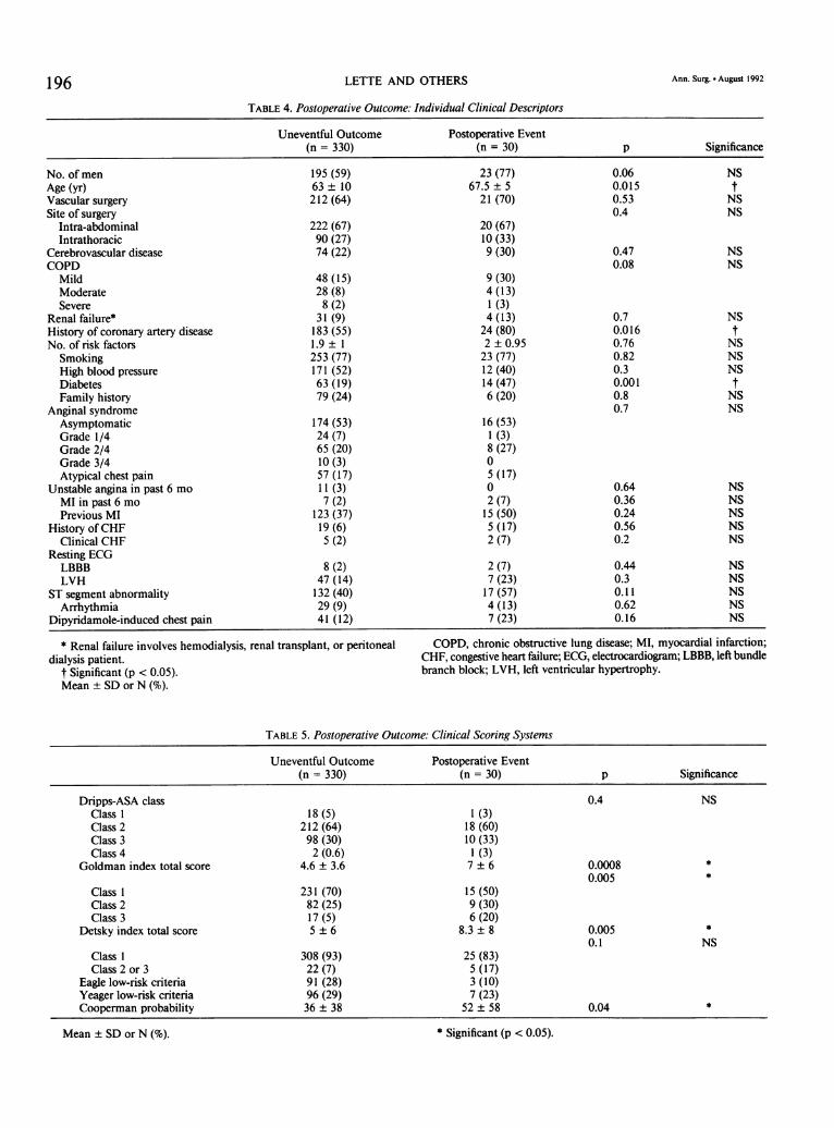
TABLE 4. Postoperative Outcome: Individual Clinical Descriptors
Uneventful Outcome Postoperative Event(n = 330) (n = 30) p Significance
No. of men 195 (59) 23 (77) 0.06 NSAge (yr) 63 ± 10 67.5 ± 5 0.015 tVascular surgery 212 (64) 21 (70) 0.53 NSSite of surgery 0.4 NS
Intra-abdominal 222 (67) 20 (67)Intrathoracic 90 (27) 10 (33)
Cerebrovascular disease 74 (22) 9 (30) 0.47 NSCOPD 0.08 NS
Mild 48 (15) 9 (30)Moderate 28 (8) 4 (13)Severe 8 (2) 1 (3)
Renal failure* 31 (9) 4 (13) 0.7 NSHistory of coronary artery disease 183 (55) 24 (80) 0.016 tNo. of risk factors 1.9 ± 1 2 ± 0.95 0.76 NSSmoking 253 (77) 23 (77) 0.82 NSHigh blood pressure 171 (52) 12 (40) 0.3 NSDiabetes 63 (19) 14 (47) 0.001 tFamily history 79 (24) 6 (20) 0.8 NS
Anginal syndrome 0.7 NSAsymptomatic 174 (53) 16 (53)Grade 1/4 24 (7) 1 (3)Grade 2/4 65 (20) 8 (27)Grade 3/4 10(3) 0Atypical chest pain 57 (17) 5 (17)
Unstable angina in past 6 mo 11 (3) 0 0.64 NSMI in past 6 mo 7 (2) 2 (7) 0.36 NSPrevious MI 123 (37) 15 (50) 0.24 NS
History ofCHF 19 (6) 5 (17) 0.56 NSClinical CHF 5 (2) 2 (7) 0.2 NS
Resting ECGLBBB 8 (2) 2 (7) 0.44 NSLVH 47 (14) 7 (23) 0.3 NS
ST segment abnormality 132 (40) 17 (57) 0.11 NSArrhythmia 29 (9) 4 (13) 0.62 NS
Dipyridamole-induced chest pain 41 (12) 7 (23) 0.16 NS
* Renal failure involves hemodialysis, renal transplant, or peritoneal COPD, chronic obstructive lung disease; MI, myocardial infarction;dialysis patient. CHF, congestive heart failure; ECG, electrocardiogram; LBBB, left bundle
t Significant (p < 0.05). branch block; LVH, left ventricular hypertrophy.Mean ± SD or N (%).
TABLE 5. Postoperative Outcome: Clinical Scoring Systems
Uneventful Outcome Postoperative Event(n = 330) (n = 30) p Significance
Dripps-ASA class 0.4 NSClass 1 18 (5) 1 (3)Class 2 212 (64) 18 (60)Class 3 98 (30) 10 (33)Class 4 2 (0.6) 1 (3)
Goldman index total score 4.6 ± 3.6 7 ± 6 0.0008 *0.005 *
Class 1 231 (70) 15 (50)Class 2 82 (25) 9 (30)Class 3 17 (5) 6 (20)
Detsky index total score 5 ± 6 8.3 ± 8 0.005 *0.1 NS
Class 1 308 (93) 25 (83)Class 2 or 3 22 (7) 5 (17)
Eagle low-risk criteria 91 (28) 3 (10)Yeager low-risk criteria 96 (29) 7 (23)Cooperman probability 36 ± 38 52 ± 58 0.04 *
196
Mean ± SD or N (%). * Significant (p < 0.05).
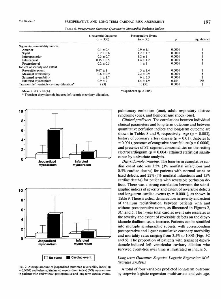
PREOPERATIVE AND LONG-TERM CARDIAC RISK ASSESSMENT 197TABLE 6. Postoperative Outcome: Quantitative Myocardial Perfusion Indices
Uneventful Outcome Postoperative Event(n = 330) (n = 30) p Significance
Segmental reversibility indicesAnterior 0.1 ± 0.4 0.9 1.1 0.0001 tSeptal 0.2 ± 0.6 1.2 ± 1.7 0.0001 tInferoposterior 0.3 ± 0.7 1.2 1 0.0001 tInferoapical 0.15 ± 0.5 1.4 1.2 0.0001 tPosterolateral 0.2 ± 0.5 1 1 0.0001 t
Indices of severity and extentReversible extent 0.67 ± 1 3 ± 1.4 0.0001 tMaximal reversibility 0.6 ± 0.9 2.2 ± 0.9 0.0001 tSummed reversibility 1 ± 1.7 6 ± 3.3 0.0001 tInfarcted myocardium 0.9 ± 2 1.5 ± 1.9 0.154 NS
Transient left ventricle cavitary dilatation* 9 (3) 10 (33) 0.0001 t
Mean ± SD or N (%). t Significant (p < 0.05).* Transient dipyridamole-induced left ventricle cavitary dilatation.
10- pulmonary embolism (one), adult respiratory distresssyndrome (one), and hemorrhagic shock (one).
8- Clinical predictors. The correlations between individualclinical parameters and long-term outcome and between
6- quantitative perfusion indices and long-term outcome areshown in Tables 8 and 9, respectively. Age (p = 0.003),
4 history of coronary artery disease (p = 0.01), diabetes (p= 0.001), presence of congestive heart failure (p = 0.0006),
2 ] |TIT | E and presence of ST segment abnormalities on the restingJI tLI. . L1 I electrocardiogram (p = 0.004) attained statistical signifi-
O =71 = q _J H _cance by univariate analysis.Jeopardized Infarcted Dipyridamole imaging. The long-term cumulative car-myocardium myocardium diac event rate was 3.5% (3% nonfatal infarctions and
0.5% cardiac deaths) for patients with normal scans orfixed defects, and 22% (7% nonfatal infarctions and 15%cardiac deaths) for patients with reversible perfusion de-fects. There was a strong correlation between the scinti-
10 graphic indices of severity and extent of reversible defectsand long-term cardiac events (p = 0.0001), as shown in
8 Table 9. There is a clear demarcation in severity and extentof thallium redistribution between patients with and
6 without postoperative events, as illustrated in Figures 2,3C, and 5. The 1-year total cardiac event rate escalates as
4 2 | l |the severity and extent of reversible defects on the dipyr-4 i r i idamole-thallium scans increase. Patients can be stratified
2| T|T | | into multiple scintigraphic subsets, with corresponding
2 | | ~ >~ | - 1 | postoperative and 1-year cumulative coronary morbidity
0 _ , 4 _ , and mortality rates ranging from 3.5% to 100% (Figs. 3C0 Jeopardized Infarcted and 5). The proportion of patients with transient dipyri-
myocardium myocardium damole-induced left ventricular cavitary dilation whosurvived event-free over time is illustrated in Figure 5.
El No event * Cardiac event
FIG. 2. Average amount ofjeopardized (summed reversibility index) (p= 0.0001) and infarcted (infarcted myocardium index) (NS) myocardiumin patients with and without postoperative and long-term cardiac events.
Long-term Outcome: Stepwise Logistic Regression Mul-tivariate Analysis
A total of four variables predicted long-term outcomeby stepwise logistic regression multivariate analysis: age,
Vol. 216 * No. 2

198
APERCENT OF TOTAL PATIENTS
LETTE AND OTHERS
BCARDIAC EVENT RATE
0O EXTENT
i Non fatal infarction: 0.5%Cardiac death: 0.5%
FIG. 3. (A) Distribution of patients in scintigraphic subsets according tothe severity and extent ofdipyridamole-induced reversible perfusion de-fects. (B) Postoperative and (C) long-term cardiac event rates in the variousscintigraphic subsets. It is apparent that a relatively small number ofpatients with multiple and severe reversible perfusion defects (high-riskscintigraphic subsets) account for most postoperative cardiac events.
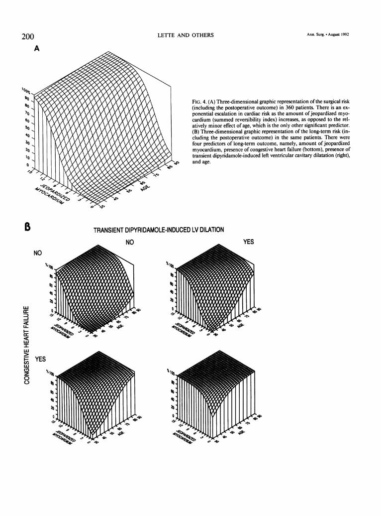
presence of congestive heart failure, presence of dipyri-damole-induced left ventricular cavitary dilation, and thesummed reversibility index (a reflection ofthe amount ofjeopardized myocardium). The regression coefficients anda simple predictive index derived are shown in Table 10.A three-dimensional graphic representation of the long-term statistical model is depicted in Figure 4B.
Discussion
Why Assess Cardiac Risk Before Operation?
Preoperative cardiac risk assessment should identifythree patient subgroups: those without significant coronaryartery disease, those with coronary stenoses who can safelyundergo noncardiac surgery with anti-ischemic medica-
Ann. Surg. * August 1992
CCUMULATIVE
CARDIAC EVENT RATE
100 '
:3.0 %0.5%

PREOPERATIVE AND LONG-TERM CARDIAC RISK ASSESSMENT
TABLE 7. Postoperative Outcome
Logistic Regression Analysis: Clinical and Scintigraphic Predictors
95%Standard Odds Confidence
Variable Coefficients Error PI Ratio Interval
Age 0.07 0.03 0.022 1.07 1.006-1.14SR 0.649 0.09 0.00001 1.914 1.58-2.32
PI, predictive index (probability of a postoperative cardiac event)
eu= 1 + eu
where e = base of natural logarithmu =-8.9435 + (0.6491* SR) + (0.07* age)
Operating characteristics of the predictive modelSensitivity = 80%Specificity = 83.3%* SR, summed reversibility index (amount of jeopardized myocar-
dium).
tion, and the small group of high-risk coronary patientswho require preoperative coronary angiography and fre-quently revascularization. Therefore, risk assessment isdistinct from diagnostic screening for coronary artery dis-ease, which pigeonholes patients into two categories: pos-itive or negative. In addition to the perioperative andpostoperative course, preoperative cardiac risk assessmentobviously should be concerned with long-term cardiacmortality rate to insure that those who undergo majornoncardiac surgery live long enough to benefit from theprocedure.
Pitfalls of Basing Individual Patient Management onMultivariate Clinical Models
Of23 separate clinical descriptors studied, only historyof coronary artery disease (p = 0.016), age (p = 0.015),and diabetes (p = 0.001) correlated with the occurrenceof postoperative cardiac events. This is of little practicaluse, however: the expression "history of coronary arterydisease" is too imprecise, and, of the patients who sus-tained cardiac complications, 56% were younger than age70, and 53% did not have diabetes. Remarkably, the pres-ence and severity of angina pectoris had no bearing oneither the postoperative or the long-term outcome, whichunderscores the difficulty in evaluating clinically patientswith a low exercise tolerance.3'The area of preoperative cardiac risk assessment pro-
vides a unique opportunity to examine the perils ofbasingindividual patient management on statistically derivedclinical scoring systems. Although the "clinical scores"derived from multivariate models correlated strongly withpostoperative events, most patients who sustained cardiacevents had in fact been categorized as low risk (Table 5).
For example, the Goldman et al.20 (p = 0.0008) and Det-sky et al.2' (p = 0.005) scores both correlated stronglywith postoperative outcome, but mislabeled as "low risk"80% (Goldman class 1 or 2) and 83% (Detsky class 1) ofpatients who sustained postoperative events. On closerexamination, these multivariate models appear to add lit-tle to basic clinical common sense: the sicker a populationof patients, the higher the incidence of postoperativecomplications, or, in candid terms, the sicker the patient,the higher the risk. In addition, clinical scoring systemsbased on multivariate analysis are not transportable be-tween institutions'2"13"8 and are not reproducible withinthe same institution.32
Risk Assessment Based on the Resting Preoperative Ejec-tion Fraction
There is a high reported incidence ofpostoperative andlong-term cardiac complications in patients with a lowleft ventricular ejection fraction (35% or less), leadingsome authors to recommend preoperative radionuclideventriculography as a tool for identifying high-risk pa-tients.33-37 Such a quick and simple approach is appealingbut ignores a basic precept of clinical cardiology: patientswith severe coronary disease, a large ischemic burden,and a normal ejection fraction at rest are at high risk.38'39For example, who would suggest that a patient with a 90%left main coronary artery stenosis and normal ventricu-logram is at low risk? Conversely, it is misleading to lumpall patients with a low ejection fraction into a high-riskcategory, irrespective ofthe amount of residual ischemia:a substantial number of these patients have a good long-term outlook and should not be denied surgery solely onthe basis of a low ejection fraction.40 In our study, theamount of jeopardized myocardium correlated stronglywith postoperative outcome, whereas the amount of in-farcted myocardium did not (Tables 6 and 9; Fig. 2). Weare developing objective criteria combining resting ejec-tion fraction and stress myocardial perfusion data tostratify patients with a low ejection fraction into high-and low-risk subgroups.
The Conventional Interpretation ofDipyridamole Imagesand its Limitations
The introduction ofdipyridamole imaging was greetedwith enthusiasm because ofthe low event rate in patientswith normal studies or fixed defects.'17 In our study, anegative dipyridamole-thallium test was reported in 195patients and indicated a very good prognosis: after op-eration, 0.5% (1/195) sustained a nonfatal myocardial in-farction and 0.5% (1/195) sustained a fatal myocardialinfarction (with normal coronary arteries at autopsy). The1-year outlook was also good: of the 194 patients with
199VOl. 216.- NO. 2
LETTE AND OTHERS
FIG. 4. (A) Three-dimensional graphic representation of the surgical risk(including the postoperative outcome) in 360 patients. There is an ex-ponential escalation in cardiac risk as the amount of jeopardized myo-cardium (summed reversibility index) increases, as opposed to the rel-atively minor effect of age, which is the only other significant predictor.(B) Three-dimensional graphic representation of the long-term risk (in-cluding the postoperative outcome) in the same patients. There werefour predictors of long-term outcome, namely, amount of jeopardizedmyocardium, presence of congestive heart failure (bottom), presence oftransient dipyridamole-induced left ventricular cavitary dilatation (right),
TRANIN Dand age.
TRANSIENT DIPYRIDAMOLE-INDUCED LV DILATION
14~~~~~~~~~4UP4.~~~~~~~~, q
%Igo
to
-la
YES
I~~~~~~~
.9bP ,
14
I.6
F,
qgplo.l
47 AP
200A
I o,!'
N)r
10
30
B
NO
to
to
-JLLI
I-if
X-
C'3w00
YES
Ann. Surg. * August 1992
00to40200"a
4
kl:.

PREOPERATIVE AND LONG-TERM CARDIAC RISK ASSESSMENT201
TABLE 8. Long-term Outcome: Clinical Descriptors
CoronaryNo Cardiac Event Cardiac Event Revascularization
(n = 303) (n = 43) (n = 9) p Significance
No. of men 177 31 6 0.12 NSAge (yr) 63 ± 10 68 ± 7 60 10 0.003 tCerebrovascular disease 69 (23) 11 (26) 3 (33) 0.82 NSCOPD 0.1 NS
Mild 43 (14) 12 (28) 1 (11)Moderate 26(9) 5(12) 1 (11)Severe 8 (3) 1 (2) 0
Renal failure* 15 (5) 2 (5) 2 (22) 0.77 NSHigh blood pressure 158 (52) 17 (40) 6 (67) 0.16 NSDiabetes 57 (19) 18 (42) 2 (22) 0.001 tHistory of coronary artery disease 167 (55) 33 (77) 5 (56) 0.01 tPrevious MI 112 (37) 21 (49) 2(22) 0.18 NSCHF 2 (1) 4 (9) 1 (11) 0.0006 tAnginal syndrome 0.95 NS
Asymptomatic 161 (53) 21 (49) 6 (67)Grade 1/4 22(7) 3(7) 0Grade2/4 61 (20) 11 (26) 0Grade 3/4 8 (3) 1 (2) 1 (11)Atypical chest pain 51 (17) 7 (16) 2 (22)
ElectrocardiogramLBBB 8(3) 2 (5) 0 0.8 NSLVH 42 (14) 10 (23) 1 (11) 0.17 NS
ST segment abnormality 117 (39) 27 (63) 4 (44) 0.004 tDipyridamole-induced chest pain 36 (12) 9 (21) 2 (22) 0.16 NS
Values are mean ± SD or N (%). COPD, chronic obstructive lung disease; MI, myocardial infarction;* Renal failure involves hemodialysis, renal transplant, or peritoneal LBBB, left bundle branch block; LVH, left ventricular hypertrophy; NS,
dialysis patient. not significant.t Significant at p < 0.05.
normal tests who were discharged from hospital after sur- angiography performed and coronary revascularizationgery, none died and 2.6% (5/194) sustained a nonfatal considered in approximately 85% ofcoronary patients re-myocardial infarction. The low mortality rate is compa- ferred for noncardiac surgery. This approach is not sup-rable to that previously reported in patients with mild ported by a previous large multicenter study that showednonobstructive (less than 50%) coronary stenoses.41'42 a low overall surgical risk in coronary patients undergoingMost investigators lump all patients with reversible de- noncardiac surgery,43 and underscores the confusion cre-
fects into a high-risk category and recommend coronary ated by taking a diagnostic procedure designed to screenangiography.8-'8 Because the test was initially designed to for coronary artery disease and applying it blindly to riskscreen for coronary artery disease, this would result in stratification.
TABLE 9. Long-term Outcome: Quantitative Myocardial Perfusion Indices
CoronaryNo Cardiac Event Cardiac Event Revascularization
(n = 303) (n = 43) (n = 9) p Significance
Segmental reversibility indicesAnterior 0.1 ± 0.4 0.8 ± 1 0.2 ± 0.7 0.0001 *Septal 0.2 ±0.6 1 1 0.8 1 0.0001 *Inferoposterior 0.3 ± 0.7 1.1 ± 1 0.9 ± 1 0.0001 *Inferoapical 0.1 ± 0.5 1.2 1.2 0.6± 1 0.0001 *Posterolateral 0.2 ± 0.5 0.9 ± 1 0.1 ± 0.3 0.0001 *
Indices of severity and extentReversible extent 0.6 ± 0.9 2.5 ± 1.6 1.5 ± 1.2 0.0001 *Maximal reversibility 0.6 ± 0.9 1 ± 0.2 1 ± 0.4 0.0001 *Summed reversibility 0.9 ± 1.6 5 ± 3.6 3 ± 1 0.0001 *Infarcted myocardium 1 ± 2 1.4 ± 2.4 0.7 ± 1.9 0.5 NS
Transient left ventricle cavitary dilatation 3 (1) 14 (33) 2 (22) 0.0001 *
Vol. 216 - No. 2
Mean ± SD or N (%). * Significant (p < 0.05).

LETTE AND OTHERS
100
80
60
40
20
0 2
Postop events(first 2 weeks)
4 6 8 10 12 14 16 18
FU (months)
FIG. 5. Proportion of patients who remained cardiac event-free withnormal scans or fixed defects (upper curve), reversible perfusion defect(s)(middle curve), and transient dipyridamole-induced left ventricular cav-itary dilatation (lower curve). Patients with normal scans or fixed defectshave an excellent prognosis; patients with reversible defect(s) are at an
increased risk but clearly cannot be labeled as high risk. For patientswith transient dipyridamole-induced left ventricular cavitary dilatation,the postoperative cardiac event rate is high, and most patients who survivethe surgery eventually sustained a cardiac event on long-term follow-up,usually within the first 3 months after the surgery.
Thus, whereas clinical models failed to identify high-risk patients, dipyridamole-thallium testing displaced theproblem, exposing large numbers of coronary patients tothe risk of systematic preoperative coronary angiographyand prophylactic revascularization: in this study, only17.5% of patients with reversible defects developed post-operative cardiac complications; 22.5% sustained long-term cardiac events, including 14.4% who died ofcoronary
disease. Furthermore, as preoperative dipyridamole-thal-lium imaging is being routinely performed, its predictiveaccuracy is gradually declining. This early referral bias infavor of more severe coronary patients is well illustratedby our own previous account of a 43% postoperative car-diac event rate in patients with reversible defects.27
The "Risk" ofPreoperative Cardiac Risk Assessment
By "risk" of cardiac risk assessment, we refer to thesignificant increase in total morbidity and mortality ratesfrom widespread preoperative coronary arteriography andprophylactic revascularization. Blombery et al.44 report amortality rate as high as 2.4% from coronary arteriographyalone in multilevel vascular patients, and Hertzer et al.45report an operative mortality rate in excess of 5% fromprophylactic coronary revascularization before vascularsurgery. In addition, the delay in performing the noncar-diac surgery increases the total risk to the patient: we haveobserved a few instances of critical lower limb ischemiadue to the delay in the initially planned vascular surgerywhile patients undergo extensive cardiac investigation.Some patients are simply overwhelmed by the thought ofcoronary revascularization followed by major noncardiacsurgery and refuse all procedures en bloc.
Only myocardial infarction and sudden coronary deathwere accepted as end points in our study. Unstable angina,ischemic pulmonary edema, cardiac arrhythmias, andother reversible conditions that uniformly respond tomedical therapy initially do not justify the added mor-bidity and mortality from preoperative coronary revas-cularization.
Risk Stratification Based on the Severity and Extent ofDipyridamole-induced Perfusion DefectsThe quantitative perfusion indices, which reflect the
amount of potentially ischemic myocardium, correlatedstrongly with both the postoperative and long-term out-come (Tables 5, 6, and 9; Figs. 2, 3, and 4). Hence, using
TABLE 10. Long-term Outcome
Logistic Regression Analysis: Clinical and Scintigraphic Predictors
Standard 95% ConfidenceVariable Coefficients Error PI Odds Ratio Interval
Age 0.0809 0.0283 0.0026 1.084 1.02-1.14CHF 1.7061 0.5433 0.0044 5.507 1.85-16.32LVdil 1.088 0.4166 0.0073 2.968 1.29-6.82SR* 0.5463 0.0885 0.00001 1.727 1.44-2.06
PI, predictive index (probability of a long-term cardiac event)
eu
+ eu
where e = base of natural logarithmu = (-6.1994) + (0.0897* Age) + (1.7061 * CHF) + (1.088* LVdil)
+ (0.5463* SR)
CHF and LVdil are equal to 0 or 1.Operating characteristics of the predictive model
Sensitivity = 81%Specificity = 80.5%* SR = summed reversibility index = amount of jeopardized myo-
cardium.CHF, congestive heart failure; LVdil, transient dipyridamole-induced
left ventricular cavitary dilatation.
202
0-
0I-E
2a
Ann. Surg. * August 1992

PREOPERATIVE AND LONG-TERM CARDIAC RISK ASSESSMENT
quantitative indices, it is possible to convert a test initiallydesigned to screen for coronary artery disease into a truesimple and noninvasive risk stratification procedure. Pa-tients can be stratified into multiple scintigraphicsubgroups with differing amounts of jeopardized myo-cardium and corresponding postoperative and long-termcoronary morbidity and mortality rates ranging from 0.5%to 100% (Fig. 3). This allows both the consulting car-
diologist and the surgeon to tailor the decision either toproceed with surgery or to consider preoperative coronaryangiography and revascularization to individual patient'sage, general health, and the overall risk profile (operativerisk, 1-year myocardial infarction, and coronary deathrates). In fact, using quantification, only 13% (45/360) ofpatients are truly at high risk (versus 194/360 = 45% ofpatients labeled as "high risk" when the test is interpretedconventionally).
Obviously, no single test can substitute for good clinicaljudgment, but it may facilitate the clinician's task. Forexample, a dipyridamole-thallium scintigraphic patternpredicting an 8% postoperative cardiac event rate and a20% 1-year cardiac mortality rate would be unacceptablein a young otherwise healthy patient undergoing electivesurgery, but might be acceptable in an elderly patient withmultisystem disease or multilevel arteriosclerosis, and a
large aortic aneurysm. If, however, the latter patient hadmultiple severe reversible defects and predicted postop-erative and 1-year cardiac mortality rates on the order of60% and 90%, respectively, heroic measures including se-
quential or combined cardiac and vascular surgery maywell be justified.
Example ofa High-risk Scintigraphic Subset
When the amount ofjeopardized myocardium is takeninto account during risk stratification, it is clear that mostpostoperative and long-term cardiac events occur in smallscintigraphic high-risk subgroups (Figs. 3, 6). For example,in a previous report, we described the occurrence of tran-sient dipyridamole-induced ischemic left ventricular cav-itary dilation, and showed that it is simple to recognize,is associated with a large amount ofjeopardized myocar-dium and severe underlying coronary artery disease, anddenotes a grim prognosis. In the current study, transientischemic dilation was observed in only 4.5% (16/360) ofthe total patient population. This small subset of patientscould not be identified clinically: 69% (11/16) denied anyhistory ofangina pectoris. They sustained a dreadful 63%(10/16) postoperative cardiac event rate, however, a 79%1-year cardiac event rate, and they accounted for 33%(10/30) of all postoperative cardiac events.
Future Prospects
There is ongoing research at our institution to furthersimplify risk stratification based on dipyridamole imaging.
203Preliminary results suggest that postoperative and long-term outcome are determined primarily by perfusion inonly three myocardial segments (anterior, posterolateral,and inferoposterior).
AcknowledgmentThe authors thank Ms. Christiane Lussier, Ms. Mary Morello, Mrs.
Nicole Choiniere, Mr. Mao, Mrs. Maryrose Fontana, and Ms. CheetahPeltz-Fraser for their assistance in preparing the manuscript, and Mr.Martin Dufresne for conceiving the three-dimensional graphic represen-tation of the multivariate statistical models.
References1. Calvin JE, Kieser TM, Walley VM, et al. Cardiac mortality and
morbidity after vascular surgery. Can J Surg 1986; 29:93-97.2. Deron SJ, Kotler MN. Noncardiac surgery in the cardiac patient.
Am Heart J 1988; 116:831-838.3. Hertzer NR, Beven EG, Young JR, et al. Fatal myocardial infarction
following peripheral vascular operations: a study of 951 patientsfollowed 6 to 11 years postoperatively. Cleve Clin Q 1982; 49:1-11.
4. Gage AA, Bhayana JN, Balu V, et al. Assessment of cardiac risk insurgical patients. Ann Surg 1977; 112:1488-1492.
5. Arous EJ, Baum PL, Cutler BS. The ischemic exercise test in patientswith peripheral vascular disease. Implications for management.Arch Surg 1984; 119:780-783.
6. McCabe CJ, Reidy NC, Abbott WM, et al. The value of electrocar-diogram monitoring during treadmill testing for peripheral vas-cular disease. Surgery 1981; 89:183-186.
7. Iskandrian AS, Heo J, Askenase A, et al. Dipyridamole cardiac im-aging. Am Heart J 1988; 115:432-443.
8. Eagle KA, Strauss HW, Boucher CA. Dipyridamole myocardial per-fusion imaging for coronary heart disease. Am J Cardiac Imaging1988; 2:292-303.
9. Eagle KA, Singer DE, Brewster DC, et al. Dipyridamole-thalliumscanning in patients undergoing vascular surgery. JAMA 1987;257:2185-2189.
10. Sachs RN, Tellier P, Larmignat P, et al. Assessment by dipyridamole-thallium-201 myocardial scintigraphy ofcoronary risk before pe-ripheral vascular surgery. Surgery 1988; 5:584-587.
11. Cutler BS, Leppo JA. Dipyridamole thallium-201 scintigraphy todetect coronary artery disease before abdominal aortic surgery.J Vasc Surg 1987; 5:91-100.
12. Boucher CA, Brewster DC, Darling RC, et al. Determination ofcardiac risk by dipyridamole-thallium imaging before peripheralvascular disease. N Engl J Med 1985; 312:389-394.
13. Fletcher JP, Antico VF, Gruenewald S, Kershaw LZ. Dipyridamole-thallium imaging for screening of coronary artery disease priorto vascular surgery. J Cardiovasc Surg 1988; 29:666-669.
14. Leppo J, Boucher CA, Okada RD, et al. Serial thallium-201 myo-cardial imaging after dipyridamole infusion: diagnostic utility indetecting coronary stenoses and relationship to regional wall mo-tion. Circulation 1982; 3:649-657.
15. Younis LT, Aguirre F, Byers S, et al. Perioperative and long-termprognostic value ofintravenous dipyridamole scintigraphy in pa-tients with peripheral vascular disease. Am Heart J 1990; 119:1287-1292.
16. Camp AD, Garvin PJ, Hoff J, et al. Prognostic value of intravenousdipyridamole-thallium imaging in patients with diabetes mellitusconsidered for renal transplantation. Am J Cardiol 1990; 65:1459-1463.
17. Brewster DC, Okada RD, Strauss W, et al. Selection of patients forpreoperative coronary angiography: use of dipyridamole-stress-thallium myocardial imaging. J Vasc Surg 1985; 2:504-510.
18. Gillet T, LecorffG, Scemama J, et al. Approche diagnostique de lacoronarographie en chirurgie vasculaire. J Mal Vasc 1990; 15:55-62.
19. Dripps RD, Lamont A, Eckenhoff JE. The role of anaesthesia insurgical mortality. JAMA 1961; 178:261-266.
Vol. 216 * No. 2

204 LETTE AND OTHERS
20. Goldman L, Caldera DL, Nussbaum SR, et al. Multifactorial indexof cardiac risk in noncardiac surgical procedures. N Engl J Med1977; 297:845-850.
21. Detsky AS, Abrams HB, McLaughlin JR. Predicting cardiac com-plications in patients undergoing noncardiac surgery. J Gen InternMed 1986; 1:211-219.
22. Eagle KA, Singer DE, Brewster DC, et al. Dipyridamole-thalliumscanning in patients undergoing vascular surgery. JAMA 1987;257:2185-2189.
23. Yeager RA, Weigel RM, Murphy ES, et al. Application of clinicallyvalid cardiac risk factors to aortic aneurysm surgery. Arch Surg1986; 121:278-281.
24. Cooperman M, Pflug B, Martin EW, Evans WE. Cardiovascular riskfactors in patients with peripheral vascular disease. Surgery 1978;84:505-509.
25. Eagle KA, Coley CM, Newell JB, et al. Combining clinical and thal-lium data optimizes preoperative assessment ofcardiac risk beforemajor vascular surgery. Ann Intern Med 1989; 110:859-866.
26. Taillefer R, Lette J, Phaneuf DC, et al. Thallium-201 myocardialimaging during pharmacologic coronary vasodilation: comparisonof oral and intravenous administration of dipyridamole. J AmColl Cardiol 1986; 8:76-83.
27. Lette J, Waters D, Lapointe J, et al. Usefulness of the severity andextent of reversible defects during thallium dipyridamole imagingfor cardiac risk assessment before non cardiac surgery. Am JCardiol 1989; 64:276-281.
28. Braunwald E, Kloner RA. The stunned myocardium: prolonged,postischemic ventricular dysfunction. Circulation 1982; 66:1146-1149.
29. Blackburn H, Keys A, Simonson E, et al. The electrocardiogram inpopulation studies: a classification system. Circulation 1960; 21:1160-1175.
30. Rose GA, Blackburn H. Cardiovascular survey methods. Geneva:World Health Organization Monograph, 1968, p 56.
31. Goldman L, Cook F, Mitchell N, et al. Pitfalls in the serial assessmentofcardiac functional status: how a reduction in "ordinary" activitymay reduce the apparent degree of cardiac compromise and givea misleading impression of improvement. J Chronic Dis 1982;35:763-771.
32. Lette J, Waters DD, Lassonde J, et al. Postoperative myocardialinfarction and cardiac death: predictive value of dipyridamole-thallium imaging and five clinical scoring systems based on mul-tifactorial analysis. Ann Surg 1990; 211:84-90.
Ann. Surg. * August 1992
33. Pasternak PF, Imparato AM, Bear G, et al. The value ofradionuclideangiography as a predictor of perioperative myocardial infarctionin patients undergoing abdominal aortic resection. J Vasc Surg1984; 1:320-325.
34. Mosley JG, Clarke JMF, Ell PJ, Marston A. Assessment of myo-cardial function before aortic surgery by radionuclide angiocar-diography. Br J Surg 1985; 72:886-887.
35. Fletcher JP, Antico VF, Gruenewald S, Kershaw LZ. Risk of aorticaneurysm surgery as assessed by preoperative gated heart poolscan. Br J Surg 1989; 76:26-28.
36. Kazmers A, Cerqueira MD, Zierler RE. Perioperative and late out-come in patients with left ventricular ejection fraction of 35% orless who require major vascular surgery. J Vasc Surg 1988; 8:307-315.
37. Kazmers A, Moneta GL, Cerqueira MD, et al. The role of preop-erative radionuclide ventriculography in defining outcome afterrevascularization of the extremity. Surg Gynecol Obstet 1990;171:481-488.
38. Severi S, Michelassi C. Prognostic impact of stress testing in coronaryartery disease. Circulation 1991; 83(5 Suppl III):82-88.
39. Brown KA. Prognostic value of thallium-201 myocardial perfusionimaging: a diagnostic tool comes of age. Circulation 1991; 83:363-381.
40. Pilote L, Silberberg J, Lisbona R, Sniderman A. Prognosis in patientswith low left ventricular ejection fraction after myocardial in-farction. Circulation 1989; 80:1636-1641.
41. Proudfit WL, Bruschke AVG, Sones FM. Clinical course ofpatientswith normal or slightly abnormal coronary arteriograms: 10 yearsfollow-up of 521 patients. Circulation 1980; 62:712-717.
42. Kemp HG, Kronmal RA, Vlietstra RE, et al. Seven year survivalof patients with normal or near normal coronary arteriograms:a CASS registry study. J Am Coll Cardiol 1986; 7:479-483.
43. Foster ED, Davis KB, Carpenter JA, et al. Risk of noncardiac op-eration in patients with defined coronary disease: the CoronaryArtery Surgery Study (CASS) Registry experience. Ann ThoracSurg 1986; 41:42-50.
44. Blombery PA, Ferguson IA, Rosengarten DS, et al. The role of cor-onary artery surgery in complications of abdominal aortic aneu-rysm surgery. Surgery 1987; 101:150-155.
45. Hertzer NR, Beven EG, Young JR, et al. Coronary artery diseasein peripheral vascular patients: a classification of 1000 coronaryangiograms and results of surgical management. Ann Surg 1984;199:223-233.
Copyright © 2022 FDOKUMEN