Postharvest Biology and Technology 95 (2014) 1–6 Contents lists available at ScienceDirect
6
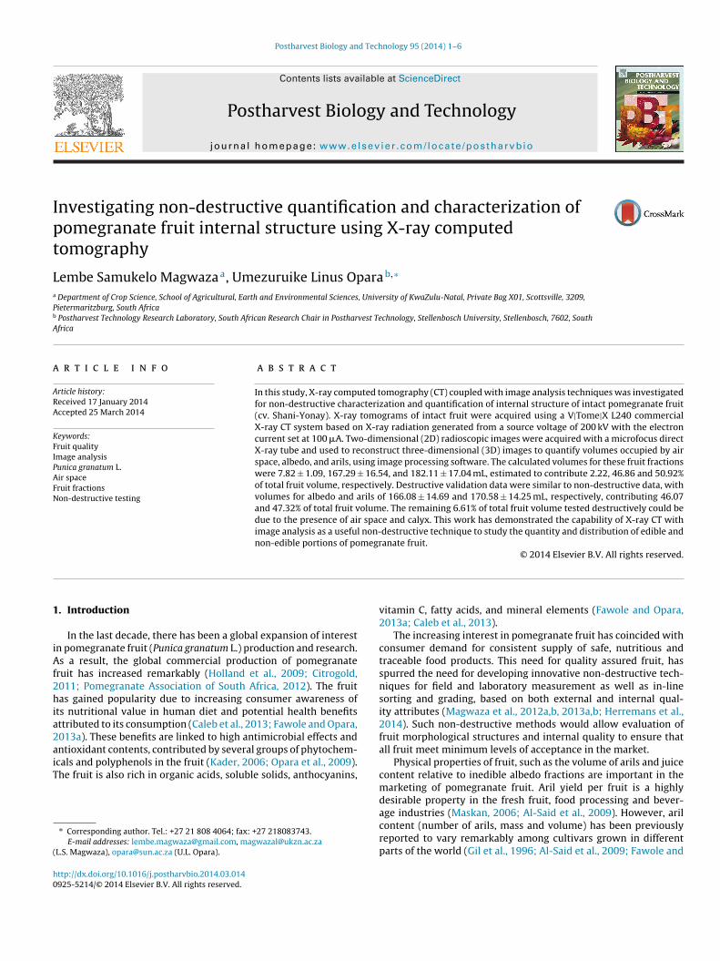
Postharvest Biology and Technology 95 (2014) 1–6 Contents lists available at ScienceDirect Postharvest Biology and Technology jou rn al h om epage: www.elsevier.com/locate/postharvbio Investigating non-destructive quantification and characterization of pomegranate fruit internal structure using X-ray computed tomography Lembe Samukelo Magwaza a , Umezuruike Linus Opara b,∗ a Department of Crop Science, School of Agricultural, Earth and Environmental Sciences, University of KwaZulu-Natal, Private Bag X01, Scottsville, 3209, Pietermaritzburg, South Africa b Postharvest Technology Research Laboratory, South African Research Chair in Postharvest Technology, Stellenbosch University, Stellenbosch, 7602, South Africa a r t i c l e i n f o Article history: Received 17 January 2014 Accepted 25 March 2014 Keywords: Fruit quality Image analysis Punica granatum L. Air space Fruit fractions Non-destructive testing a b s t r a c t In this study, X-ray computed tomography (CT) coupled with image analysis techniques was investigated for non-destructive characterization and quantification of internal structure of intact pomegranate fruit (cv. Shani-Yonay). X-ray tomograms of intact fruit were acquired using a V|Tome|X L240 commercial X-ray CT system based on X-ray radiation generated from a source voltage of 200 kV with the electron current set at 100 A. Two-dimensional (2D) radioscopic images were acquired with a microfocus direct X-ray tube and used to reconstruct three-dimensional (3D) images to quantify volumes occupied by air space, albedo, and arils, using image processing software. The calculated volumes for these fruit fractions were 7.82 ± 1.09, 167.29 ± 16.54, and 182.11 ± 17.04 mL, estimated to contribute 2.22, 46.86 and 50.92% of total fruit volume, respectively. Destructive validation data were similar to non-destructive data, with volumes for albedo and arils of 166.08 ± 14.69 and 170.58 ± 14.25 mL, respectively, contributing 46.07 and 47.32% of total fruit volume. The remaining 6.61% of total fruit volume tested destructively could be due to the presence of air space and calyx. This work has demonstrated the capability of X-ray CT with image analysis as a useful non-destructive technique to study the quantity and distribution of edible and non-edible portions of pomegranate fruit. © 2014 Elsevier B.V. All rights reserved. 1. Introduction In the last decade, there has been a global expansion of interest in pomegranate fruit (Punica granatum L.) production and research. As a result, the global commercial production of pomegranate fruit has increased remarkably (Holland et al., 2009; Citrogold, 2011; Pomegranate Association of South Africa, 2012). The fruit has gained popularity due to increasing consumer awareness of its nutritional value in human diet and potential health benefits attributed to its consumption (Caleb et al., 2013; Fawole and Opara, 2013a). These benefits are linked to high antimicrobial effects and antioxidant contents, contributed by several groups of phytochem- icals and polyphenols in the fruit (Kader, 2006; Opara et al., 2009). The fruit is also rich in organic acids, soluble solids, anthocyanins, ∗ Corresponding author. Tel.: +27 21 808 4064; fax: +27 218083743. E-mail addresses: [email protected], [email protected] (L.S. Magwaza), [email protected] (U.L. Opara). vitamin C, fatty acids, and mineral elements (Fawole and Opara, 2013a; Caleb et al., 2013). The increasing interest in pomegranate fruit has coincided with consumer demand for consistent supply of safe, nutritious and traceable food products. This need for quality assured fruit, has spurred the need for developing innovative non-destructive tech- niques for field and laboratory measurement as well as in-line sorting and grading, based on both external and internal qual- ity attributes (Magwaza et al., 2012a,b, 2013a,b; Herremans et al., 2014). Such non-destructive methods would allow evaluation of fruit morphological structures and internal quality to ensure that all fruit meet minimum levels of acceptance in the market. Physical properties of fruit, such as the volume of arils and juice content relative to inedible albedo fractions are important in the marketing of pomegranate fruit. Aril yield per fruit is a highly desirable property in the fresh fruit, food processing and bever- age industries (Maskan, 2006; Al-Said et al., 2009). However, aril content (number of arils, mass and volume) has been previously reported to vary remarkably among cultivars grown in different parts of the world (Gil et al., 1996; Al-Said et al., 2009; Fawole and http://dx.doi.org/10.1016/j.postharvbio.2014.03.014 0925-5214/© 2014 Elsevier B.V. All rights reserved.
Transcript of Postharvest Biology and Technology 95 (2014) 1–6 Contents lists available at ScienceDirect
Ipt
La
Pb
A
a
ARA
KFIPAFN
1
iAf2hia2aiT
(
h0
Postharvest Biology and Technology 95 (2014) 1–6
Contents lists available at ScienceDirect
Postharvest Biology and Technology
jou rn al h om epage: www.elsev ier .com/ locate /postharvbio
nvestigating non-destructive quantification and characterization ofomegranate fruit internal structure using X-ray computedomography
embe Samukelo Magwazaa, Umezuruike Linus Oparab,∗
Department of Crop Science, School of Agricultural, Earth and Environmental Sciences, University of KwaZulu-Natal, Private Bag X01, Scottsville, 3209,ietermaritzburg, South AfricaPostharvest Technology Research Laboratory, South African Research Chair in Postharvest Technology, Stellenbosch University, Stellenbosch, 7602, Southfrica
r t i c l e i n f o
rticle history:eceived 17 January 2014ccepted 25 March 2014
eywords:ruit qualitymage analysisunica granatum L.ir spaceruit fractions
a b s t r a c t
In this study, X-ray computed tomography (CT) coupled with image analysis techniques was investigatedfor non-destructive characterization and quantification of internal structure of intact pomegranate fruit(cv. Shani-Yonay). X-ray tomograms of intact fruit were acquired using a V|Tome|X L240 commercialX-ray CT system based on X-ray radiation generated from a source voltage of 200 kV with the electroncurrent set at 100 �A. Two-dimensional (2D) radioscopic images were acquired with a microfocus directX-ray tube and used to reconstruct three-dimensional (3D) images to quantify volumes occupied by airspace, albedo, and arils, using image processing software. The calculated volumes for these fruit fractionswere 7.82 ± 1.09, 167.29 ± 16.54, and 182.11 ± 17.04 mL, estimated to contribute 2.22, 46.86 and 50.92%of total fruit volume, respectively. Destructive validation data were similar to non-destructive data, with
on-destructive testing volumes for albedo and arils of 166.08 ± 14.69 and 170.58 ± 14.25 mL, respectively, contributing 46.07and 47.32% of total fruit volume. The remaining 6.61% of total fruit volume tested destructively could bedue to the presence of air space and calyx. This work has demonstrated the capability of X-ray CT withimage analysis as a useful non-destructive technique to study the quantity and distribution of edible andnon-edible portions of pomegranate fruit.
© 2014 Elsevier B.V. All rights reserved.
. Introduction
In the last decade, there has been a global expansion of interestn pomegranate fruit (Punica granatum L.) production and research.s a result, the global commercial production of pomegranate
ruit has increased remarkably (Holland et al., 2009; Citrogold,011; Pomegranate Association of South Africa, 2012). The fruitas gained popularity due to increasing consumer awareness of
ts nutritional value in human diet and potential health benefitsttributed to its consumption (Caleb et al., 2013; Fawole and Opara,013a). These benefits are linked to high antimicrobial effects andntioxidant contents, contributed by several groups of phytochem-
cals and polyphenols in the fruit (Kader, 2006; Opara et al., 2009).he fruit is also rich in organic acids, soluble solids, anthocyanins,∗ Corresponding author. Tel.: +27 21 808 4064; fax: +27 218083743.E-mail addresses: [email protected], [email protected]
L.S. Magwaza), [email protected] (U.L. Opara).
ttp://dx.doi.org/10.1016/j.postharvbio.2014.03.014925-5214/© 2014 Elsevier B.V. All rights reserved.
vitamin C, fatty acids, and mineral elements (Fawole and Opara,2013a; Caleb et al., 2013).
The increasing interest in pomegranate fruit has coincided withconsumer demand for consistent supply of safe, nutritious andtraceable food products. This need for quality assured fruit, hasspurred the need for developing innovative non-destructive tech-niques for field and laboratory measurement as well as in-linesorting and grading, based on both external and internal qual-ity attributes (Magwaza et al., 2012a,b, 2013a,b; Herremans et al.,2014). Such non-destructive methods would allow evaluation offruit morphological structures and internal quality to ensure thatall fruit meet minimum levels of acceptance in the market.
Physical properties of fruit, such as the volume of arils and juicecontent relative to inedible albedo fractions are important in themarketing of pomegranate fruit. Aril yield per fruit is a highlydesirable property in the fresh fruit, food processing and bever-
age industries (Maskan, 2006; Al-Said et al., 2009). However, arilcontent (number of arils, mass and volume) has been previouslyreported to vary remarkably among cultivars grown in differentparts of the world (Gil et al., 1996; Al-Said et al., 2009; Fawole and
2 est Bio
Otori2ftq
ibl2r22et(ceiwpa
sct2raccdXopts(2
WCscXppTdif
iifagdatCt
L.S. Magwaza, U.L. Opara / Postharv
para, 2013b). Gil et al. (1996) reported that for the ‘Mollar’ cul-ivar grown in Spain, arils constituted 57–66%, while aril contentf ‘Jabal 1’, ‘Jabal 2’, Jabal 3’ and a wild cultivar grown in Omananged from 50 to 67% (Al-Said et al., 2009). In six cultivars grownn Morocco, aril yield ranged between 53 and 61% (Martínez et al.,012). High variability in aril yield between different cultivars andruit of the same cultivar has prompted the need for the indus-ry to develop techniques that can non-destructively visualize anduantify internal structures of the fruit.
A wide range of objective instruments and techniques for qual-ty assessment of external and internal quality of fresh produce haseen the subject of numerous reviews and research articles in the
iterature (Nicolaï et al., 2007; Lorente et al., 2012; Magwaza et al.,012a, 2014a,b; Alfatni et al., 2013). Techniques such as magneticesonance imaging (MRI, Lammertyn et al., 2003a; Defraeye et al.,013), nuclear magnetic resonance (NMR, Zhang and McCarthy,013), visible to near infrared spectroscopy (Vis/NIRS, Magwazat al., 2012b, 2014a), Vis/NIRS-based systems such as hyperspec-ral imaging (Haff et al., 2013) and optical coherence tomographyOCT, Magwaza et al., 2013a; Verboven et al., 2013) as well as X-rayomputed tomography (CT, Kotwaliwale et al., 2014; Herremanst al., 2013) have been explored for non-destructive internal qual-ty evaluation of different horticultural products. In conjunction
ith image analysis techniques, these imaging techniques haveresented many potential avenues for non-destructive qualityssessment of fresh fruit and vegetables.
X-ray CT has become one of the well-established research toolsuitable for non-destructive analysis of internal morphologicalharacteristics and detect internal defects of fruit and other hor-icultural products (Lammertyn et al., 2003a,b; Verboven et al.,008; Herremans et al., 2013). Schatzki et al. (1997) applied X-ay CT to detect internal defects in apples. X-ray CT imaging haslso been used to non-destructively characterize fruit physiologi-al disorders such as translucency in pineapples (Haff et al., 2006),ore breakdown in pears (Lammertyn et al., 2003a,b), watercoreisorder of apples (Herremans et al., 2014). The main advantage of-ray CT lies in its non-destructive characteristics and its large fieldf view, which allows scanning the entire sample without sam-le preparation (Léonard et al., 2008). Several studies comparinghe performances of X-ray CT against other imaging techniquesuch MRI showed that X-ray is more convenient and less costlyLammertyn et al., 2003a; Yacob et al., 2005; Herremans et al.,014).
X-ray CT produces a stack of two-dimensional (2D) images.hen coupled with different image analysis techniques, X-ray
T allows reconstruction of three-dimensional (3D) images fromtacked series of image data allowing characterization of physi-al and physiological structures of biological materials. Although-ray CT has recently gained significant attention and providedromising results for determining internal quality of other freshroduce, this technique has not been used on pomegranate fruit.he ability of X-ray CT to differentiate between structures withifferent density is envisioned to enable visualization, character-
zation and quantification of internal structures in pomegranateruit.
The capability of X-ray CT technology together with associatedmage analysis could open a new avenue for using these techniquesn pomegranate breeding programmes to assess cultivars with dif-erent proportions of arils to albedo. Considering that aril yieldnd postharvest storage performance are of particular interest torowers, breeders and postharvest technologists, using this non-estructive approach would allow internal quality evaluation and
ssessment of postharvest storability of the same fruit. The objec-ives of the study were (1) to investigate the feasibility of X-rayT as a non-destructive technique for determining internal struc-ures of pomegranate fruit, and (2) to demonstrate the capabilitylogy and Technology 95 (2014) 1–6
of imaging analysis to characterize internal structures and quantifyvolumes of edible and non-edible portions.
2. Materials and methods
2.1. Pomegranate fruit supply
The study was conducted using 12 pomegranate fruit (P.granatum L. cv. Shani-Yonay) of uniform size (322.35 ± 17.52 g),purchased from a retail supermarket in Stellenbosch, Western CapeProvince, South Africa. Based on information provided by the sup-plier (Freshmark (Pty.) Ltd., Brackenfell, South Africa), the fruit wereimported from Israel. All fruit used for analysis of internal structurewere free from external defects.
2.2. X-ray computed tomography scanning
X-ray CT images of intact fruit were acquired using a commercialX-ray computed tomography (CT) system (V|Tome|X L240, GeneralElectric Sensing & Inspection Technologies GmbH, Phoenix, Wun-storf, Germany) in the Central Analytical Facility at the Universityof Stellenbosch, South Africa. Various system settings were testedto optimize the scan quality. Optimal CT scans (tomograms) wereobtained with an isotropic voxel size of 71.4 �m based on X-rayradiation generated from a source voltage of 200 kV and the elec-tron current was set at 100 �A. Radioscopic images were acquiredwith a microfocus direct X-ray tube resulting in better signal tonoise ratio and less reconstruction artefacts around the edges. Thesystem was equipped with copper filter to cut out low energy X-rays. Pomegranate samples were mounted on a translation stagewhich was at a fixed physical distance of 250 mm from the X-raysource and 700 mm from the detector. Based on these system sett-ings, the scan resolution was 71.4 �m to accommodate the size andvolume of pomegranate samples.
A series of 2D X-ray images were obtained as the fruit wasrotated 360 degrees, with 500 milliseconds of exposure time perimage, without averaging or skipping of images, hence, recording2500–3000 images in one rotation, depending on the size of sam-ple and resolution. These image slices, covering the entire samplewere acquired using a fully automated data acquisition system andsaved onto a processing workstation, operated by system-suppliedreconstruction software (Datos|x® 2.1, General Electric Sensing &Inspection Technologies GmbH, Phoenix, Wunstorf, Germany). Thetotal scan time for each sample was approximately 1 h.
2.3. Image processing
X-ray image slices were used to reconstruct 3D images for quan-tification purposes using volume graphics software (VG StudioMax 2.1. and 2.2, Volume Graphics GmbH, Heidelberg, Germany).The X-ray data was first smoothed to remove random noise usingGuass (5 × 5) filtering method. Before computing volumes, beam-hardening correction was applied to the dataset to suppress beamhardening artefacts. Beam hardening correction was only appliedat 7.5 units due to the use of a copper filter during acquisition. Cop-per filter suppressed low energy X-rays from the source, reducingbeam hardening artefacts.
A representative slice from one of the 12 fruit was selected fromthe data set to obtain an average grey value for each of the fruitportions (albedo, arils and air spaces). Surface fitting of the datawas performed using interactive thresholding of grey values andobserving the fit line on a slice view. A typical graph showing grey
values for different fruit parts is presented in Fig. 1. The average greyvalues were from 4,340 to 23,845 units for air spaces, from 23,846to 44,660 units for albedo and from 44,661 to 56,200 for arils. Afterthresholding, a surface extraction procedure to non-destructively
L.S. Magwaza, U.L. Opara / Postharvest Bio
Fig. 1. A typical graph showing grey values for different fruit parts. The figuredt
qncsr
2
am(romldisT
2
(ddat(
3
3
aoinwacaor
emonstrated that fruit parts can be separated based on grey level frequency dis-ribution values in the histogram.
uantify volumes of air spaces, albedo and arils was executed. Toon-destructively quantify volumes of arils and albedo, the area ofentre most floating stack of slices arils were selected from eachample. By stacking slices in 3D across the, the volume of eachegion was automatically calculated.
.4. Validation of X-ray CT results
In order to validate results computed from X-ray CT imagenalysis, fruit samples used for the scanning experiments wereanually cut open and carefully separated into the fractions
arils and albedo). Volumetric measurements were determined atoom temperature using the water displacement method basedn Archimedes’ Principle (Lang and Thorpe, 1989). The volumetriceasurements made allowed us to confirm if measured morpho-
ogical features of the fruit corresponded with X-ray CT estimatedata. X-ray CT radiographs were also compared to digital colour
mages acquired using a full high definition digital camera (Sam-ung WB850F, 21X Optical Zoom, Samsung Electronics Co. Ltd.,okyo, Japan), after dissecting fruit either vertically or horizontally.
.5. Statistical analysis
Statistical analyses were carried out using software STATISTICAVers. 11.0, StatSoft Inc., USA). To assess the accuracy of non-estructive prediction, data calculated by X-ray CT and measuredestructively were subjected to linear regression analysis (p = 0.05)nd paired two-tailed Fischer’s t-test (p = 0.05). Graphical presen-ations were made using GraphPad Prism software version 4.03GraphPad Software, Inc., San Diego, USA).
. Results and discussion
.1. Image analysis
For optimum speed and efficiency of sample measurement, anlgorithm for image processing was created using data set fromne representative sample. Firstly, the selected representative 2Dmage slice (Fig. 2a) was segmented to visually separate arils fromon-edible albedo and air spaces. Arils were assigned red colour,hile non-edible albedo material and air spaces were, respectively,
ssigned grey and black colours, based on their densities (Fig. 2b and
). The next step was to extract the area occupied by arils (Fig. 2d),nd to apply morphological opening. After which, the perimeterf albedo was eroded and bright regions then re-dilated, therebyemoving small specks of noise observed in (Fig. 2d and e).logy and Technology 95 (2014) 1–6 3
A 3D model (Fig. 2f) was reconstructed by appropriately stackingfloating image slices of extracted arils areas with appropriate scal-ing such that equal distances along all three axes represent equaldistances in physical space. Once developed for one dataset, thealgorithm was used to automatically extract parameter for the restof the samples. The algorithm for automatic image analysis devel-oped in this study is a tool for non-destructive identification andquantification of known morphological features of a pomegranatefruit. The combined discriminative power of X-ray CT and imageanalysis technique to differentiate tissues of different densities canalso be used to identify unknown features such as pest infestation,disease infection and the presence of internal defects associatedwith physiological disorders.
3.2. Volume analysis of different fruit fractions
After image analysis, it was possible to determine volumesof pomegranate morphological structures. A histogram showingvolumes occupied by air space, albedo and arils is presentedin Table 1. The results obtained using X-ray CT coupled withimage analysis technique showed that on average, arils occupied182.11 ± 17.04 mL, estimated to 50.98% of the total pomegranatefruit volume (357.22 ± 29.44). The calculated volumes for air spacesand albedo fractions were 7.82 ± 1.09 and 167.29 ± 16.54 mL, esti-mated to respectively contribute 2.19 and 46.83% of the total fruitvolume.
Destructive validation results were similar to X-ray CT non-destructive data, showing volumes for albedo and arils of166.08 ± 14.69 and 170.58 ± 14.25 mL, respectively, correspond-ing to 46.07 and 47.32% of the total volume (360.49 ± 27.66 mL)(Table 1). The remaining 6.61% (23.83 ± 1.50 mL) of the total fruitvolume in destructive results could be due to air spaces. The resultson volume of different fruit fractions obtained in this study are sim-ilar to those reported by Shulman et al. (1984) for ‘Mule’s Head’and ‘Wonderful’ cultivars, where fruit arils constituted 50% of thetotal fruit mass. Similar results were also reported by Fawole andOpara (2013c) for ‘Bhagwa’ and ‘Ruby’ cultivars, where aril yieldwas below 50% at commercial maturity. According to the authors’knowledge, there is no study that has attempted to quantify airspaces of pomegranate fruit.
The results in Table 1 also showed that the total volume of airspaces was overestimated by an average of 67.18%, while arils andalbedo were underestimated by 6.76% and 0.73%, respectively. Theresults showed that the difference between X-ray calculated anddestructively measured mean volume for total air spaces was signif-icantly (p < 0.001) overestimated. Fischer’s t-test analysis indicatedthat the total volume of air spaces obtained non-destructively weresignificantly (p < 0.001) different from those measured destruc-tively. However, for albedo and arils, the differences between X-rayCT predicted values and actual measurements were not signif-icantly different. These results demonstrated that X-ray CT andassociated image analysis were significantly accurate in predictingarils and albedo but less accurate with air spaces.
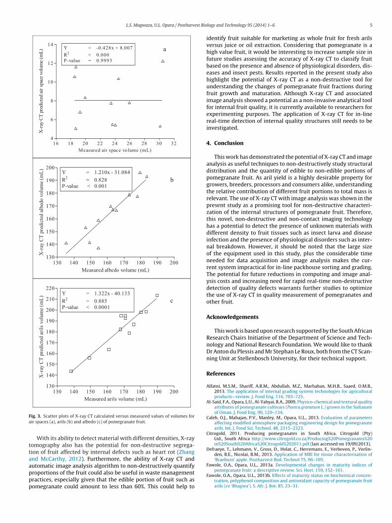
The results of X-ray CT predicted and destructively measureddata on amount of fruit fractions were further subjected to linearregression analysis. In Fig. 3, the volume of air space, albedo andarils predicted with X-ray CT are plotted against the destructivelymeasured values. The significant difference between X-ray CT pre-dicted and measured volume for air spaces was further confirmedby very low regression statistics (R2 < 0.000 and p = 0.999) whenthe two were plotted in the same axis (Fig. 3a). This differencecould partly be attributed to the contribution of the air space in the
fruit calyx area during destructive measurements. Linear regres-sion models of predicted versus destructively measured volumesof albedo (Fig. 3b) and arils (Fig. 3c) showed significantly (p < 0.001)high accuracy, with R2 values of 0.83 and 0.89, respectively.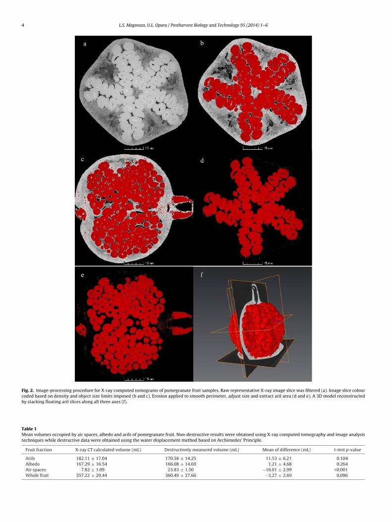
4 L.S. Magwaza, U.L. Opara / Postharvest Biology and Technology 95 (2014) 1–6
Fig. 2. Image-processing procedure for X-ray computed tomograms of pomegranate fruit samples. Raw representative X-ray image slice was filtered (a). Image slice colourcoded based on density and object size limits imposed (b and c). Erosion applied to smooth perimeter, adjust size and extract aril area (d and e). A 3D model reconstructedby stacking floating aril slices along all three axes (f).
Table 1Mean volumes occupied by air spaces, albedo and arils of pomegranate fruit. Non-destructive results were obtained using X-ray computed tomography and image analysistechniques while destructive data were obtained using the water displacement method based on Archimedes’ Principle.
Fruit fraction X-ray CT calculated volume (mL) Destructively measured volume (mL) Mean of difference (mL) t-test p-value
Arils 182.11 ± 17.04 170.58 ± 14.25 11.53 ± 6.21 0.104Albedo 167.29 ± 16.54 166.08 ± 14.69 1.21 ± 4.68 0.264Air spaces 7.82 ± 1.09 23.83 ± 1.50 −16.01 ± 2.99 <0.001Whole fruit 357.22 ± 29.44 360.49 ± 27.66 −3.27 ± 2.69 0.096
L.S. Magwaza, U.L. Opara / Postharvest Bio
16 18 20 22 24 26 28 30 324
6
8
10
12
14Y = -0.428 x + 8.00 7
R2 < 0.00 0
P-value = 0.999 3
Measured air space volume (mL)
X-r
ay C
T p
redic
ted a
ir s
pac
e volu
me
(mL
)
130 14 0 15 0 160 170 18 0 19 0 200130
140
150
160
170
180
190
200Y = 1.210 x - 31 .08 4
R2 = 0.828
P-value < 0.00 1
Measured albedo volume (mL)
X-r
ay C
T p
redic
ted a
lbed
o v
olu
me
(mL
)
130 14 0 15 0 160 170 18 0 19 0 200130
140
150
160
170
180
190
200
210
220Y = 1.322 x - 40 .13 3
R2 = 0.885
P-value < 0.000 1
Measured aril s volume (mL)
X-r
ay C
T p
redic
ted a
rils
volu
me
(mL
)
a
b
c
Fa
ttaappp
ig. 3. Scatter plots of X-ray CT calculated versus measured values of volumes forir spaces (a), arils (b) and albedo (c) of pomegranate fruit.
With its ability to detect material with different densities, X-rayomography also has the potential for non-destructive segrega-ion of fruit affected by internal defects such as heart rot (Zhangnd McCarthy, 2012). Furthermore, the ability of X-ray CT and
utomatic image analysis algorithm to non-destructively quantifyroportions of the fruit could also be useful in waste managementractices, especially given that the edible portion of fruit such asomegranate could amount to less than 60%. This could help tology and Technology 95 (2014) 1–6 5
identify fruit suitable for marketing as whole fruit for fresh arilsversus juice or oil extraction. Considering that pomegranate is ahigh value fruit, it would be interesting to increase sample size infuture studies assessing the accuracy of X-ray CT to classify fruitbased on the presence and absence of physiological disorders, dis-eases and insect pests. Results reported in the present study alsohighlight the potential of X-ray CT as a non-destructive tool forunderstanding the changes of pomegranate fruit fractions duringfruit growth and maturation. Although X-ray CT and associatedimage analysis showed a potential as a non-invasive analytical toolfor internal fruit quality, it is currently available to researchers forexperimenting purposes. The application of X-ray CT for in-linereal-time detection of internal quality structures still needs to beinvestigated.
4. Conclusion
This work has demonstrated the potential of X-ray CT and imageanalysis as useful techniques to non-destructively study structuraldistribution and the quantity of edible to non-edible portions ofpomegranate fruit. As aril yield is a highly desirable property forgrowers, breeders, processors and consumers alike, understandingthe relative contribution of different fruit portions to total mass isrelevant. The use of X-ray CT with image analysis was shown in thepresent study as a promising tool for non-destructive characteri-zation of the internal structures of pomegranate fruit. Therefore,this novel, non-destructive and non-contact imaging technologyhas a potential to detect the presence of unknown materials withdifferent density to fruit tissues such as insect larva and diseaseinfection and the presence of physiological disorders such as inter-nal breakdown. However, it should be noted that the large sizeof the equipment used in this study, plus the considerable timeneeded for data acquisition and image analysis makes the cur-rent system impractical for in-line packhouse sorting and grading.The potential for future reductions in computing and image anal-ysis costs and increasing need for rapid real-time non-destructivedetection of quality defects warrants further studies to optimizethe use of X-ray CT in quality measurement of pomegranates andother fruit.
Acknowledgements
This work is based upon research supported by the South AfricanResearch Chairs Initiative of the Department of Science and Tech-nology and National Research Foundation. We would like to thankDr Anton du Plessis and Mr Stephan Le Roux, both from the CT Scan-ning Unit at Stellenbosch University, for their technical support.
References
Alfatni, M.S.M., Shariff, A.R.M., Abdullah, M.Z., Marhaban, M.H.B., Saaed, O.M.B.,2013. The application of internal grading system technologies for agriculturalproducts—review. J. Food Eng. 116, 703–725.
Al-Said, F.A., Opara, L.U., Al-Yahyai, R.A., 2009. Physico-chemical and textural qualityattributes of pomegranate cultivars (Punica granatum L.) grown in the Sultanateof Oman. J. Food Eng. 90, 129–134.
Caleb, O.J., Mahajan, P.V., Manley, M., Opara, U.L., 2013. Evaluation of parametersaffecting modified atmosphere packaging engineering design for pomegranatearils. Int. J. Food Sci. Technol. 48, 2315–2323.
Citrogold, 2011. Producing pomegranates in South Africa. Citrogold (Pty)Ltd., South Africa http://www.citrogold.co.za/Producing%20Pomegranates%20in%20South%20Africa%20Citrogold%202011.pdf (last accessed on 19/09/2013).
Defraeye, T., Lehmann, V., Gross, D., Holat, C., Herremans, E., Verboven, P., Verlin-den, B.E., Nicolai, B.M., 2013. Application of MRI for tissue characterisation of‘Braeburn’ apple. Postharvest Biol. Technol 75, 96–105.
Fawole, O.A., Opara, U.L., 2013a. Developmental changes in maturity indices ofpomegranate fruit: a descriptive review. Sci. Hort. 159, 152–161.
Fawole, O.A., Opara, U.L., 2013b. Effects of maturity status on biochemical concen-tration, polyphenol composition and antioxidant capacity of pomegranate fruitarils (cv ‘Bhagwa’). S. Afr. J. Bot. 85, 23–31.

6 est Bio
F
G
H
H
H
H
H
K
K
L
L
L
L
L
M
M
M
L.S. Magwaza, U.L. Opara / Postharv
awole, O.A., Opara, U.L., 2013c. Fruit growth dynamics, respiration rate and physico-textural properties during pomegranate development and ripening. Sci. Hort.157, 90–98.
il, M.I., Martinez, J.A., Artes, F., 1996. Minimally processed pomegranate seeds.LWT-Food Sci. Technol. 29, 708–713.
aff, R.P., Sarawong, S., Thanapase, W., Janhiran, A., Kasemsumran, S., Kawano, S.,2013. Automatic image analysis and spot classification for detection of fruit flyinfestation in hyperspectral images of mangoes. Postharvest Biol. Technol. 86,23–38.
aff, R.P., Slaughter, D.C., Sarig, Y., Kader, A., 2006. X-ray assessment of translucencyin pineapple. J. Food Process. Preserv. 30, 527–533.
erremans, E., Verboven, P., Bongaers, E., Estrade, P., Verlinden, B.E., Wevers, M.,Hertog, M.L.A.T.M., Nicolaï, B.M., 2013. Characterisation of ‘Braeburn’ browningdisorder by means of X-ray micro-CT. Postharvest Biol. Technol. 75, 114–124.
erremans, E., Melado-Herreros, A., Defraeye, T., Verlinden, B., Hertog, M., Verboven,P., Val, J., Fernández-Valle, M.E., Bongaers, E., Estrade, P., Wevers, M., Barreiro,P., Nicolaï, B.M., 2014. Comparison of X-ray CT and MRI of watercore disorder ofdifferent apple cultivars. Postharvest Biol. Technol. 87, 42–50.
olland, D., Hatib, K., Bar-Ya’akov, I., 2009. Pomegranate: botany, horticulture,breeding. Hort. Rev. 35, 127–191.
ader, A.A., 2006. Postharvest biology and technology of pomegranates. In: Seeram,N.P., Schulman, R.N., Heber, D. (Eds.), Pomegranates: Ancient Roots to the Mod-ern Medicine. CRC Press Taylor and Francis Group, Boca Raton, FL, United States,pp. 211–220.
otwaliwale, N., Singh, K., Kalne, A., Jha, S.N., Seth, N., Kar, A., 2014. X-ray imag-ing methods for internal quality evaluation of agricultural produce. J. Food Sci.Technol. 51, 1–15.
ammertyn, J., Dresselaers, T., Van Hecke, P., Jancsók, P., Wevers, M., Nicolaï, B.M.,2003a. MRI and X-ray CT study of spatial distribution of core breakdown in‘Conference’ pears. Magn. Reson. Imaging 21, 805–815.
ammertyn, J., Dresselaers, T., Van Hecke, P., Jancsók, P., Wevers, M., Nicolaï, B.M.,2003b. Analysis of the time course of core breakdown in ‘Conference’ pears bymeans of MRI and X-ray CT. Postharvest Biol. Technol. 29, 19–28.
ang, A., Thorpe, M.R., 1989. Xylem, phloem and transpiration flows in a grape:application of a technique for measuring the volume of attached fruits to highresolution using Archimedes’ Principle. J. Exp. Bot. 40, 1069–1078.
éonard, A., Blacher, S., Nimmol, C., Devahastin, S., 2008. Effect of far-infrared radi-ation assisted drying on microstructure of banana slices: an illustrative use ofX-ray microtomography in microstructural evaluation of a food product. J. FoodEng. 85, 154–162.
orente, D., Aleixos, N., Gómez-Sanchis, J., Cubero, S., García-Navarrete, O.L., Blasco,J., 2012. Recent advances and applications of hyperspectral imaging for fruit andvegetable quality assessment. Food Bioprocess Technol. 5, 1121–1142.
agwaza, L.S., Opara, U.L., Nieuwoudt, H., Cronje, P., Saeys, W., Nicolaï, B., 2012a.NIR spectroscopy applications for internal and external quality analysis of citrusfruit—a review. Food Bioprocess Technol. 5, 425–444.
agwaza, L.S., Opara, U.L., Terry, L.A., Landahl, S., Cronje, P.J.R., Nieuwoudt, H.H.,
Mouazen, A.M., Saeys, W., Nicolaï, B.M., 2012b. Prediction of ‘Nules Clementine’mandarin susceptibility to rind breakdown disorder using Vis/NIR spectroscopy.Postharvest Biol. Technol. 74, 1–10.agwaza, L.S., Ford, H.D., Cronje, P.J.R., Opara, U.L., Landahl, S., Tatam,R.P., Terry, L.A., 2013a. Application of optical coherence tomography to
logy and Technology 95 (2014) 1–6
non-destructively characterise rind breakdown disorder of ‘Nules Clementine’mandarins. Postharvest Biol. Technol. 84, 16–21.
Magwaza, L.S., Opara, U.L., Terry, L.A., Landahl, S., Cronje, P.J.R., Nieuwoudt, H.H.,Hanssens, A., Saeys, W., Nicolaï, B.M., 2013b. Evaluation of Fourier transform-NIR spectroscopy for integrated external and internal quality assessment of‘Valencia’ oranges. J. Food Comp. Anal. 31, 144–154.
Magwaza, L.S., Opara, U.L., Cronje, P.J.R., Landahl, S., Nieuwoudt, H.H., Mouazen, A.M.,Nicolaï, B.M., Terry, L.A., 2014a. Assessment of rind quality of ‘Nules Clemen-tine’ mandarin fruit during postharvest storage: 1. Vis/NIRS PCA models andrelationship with canopy position. Sci. Hort. 165, 410–420.
Magwaza, L.S., Opara, U.L., Cronje, P.J.R., Landahl, S., Nieuwoudt, H.H., Mouazen,A.M., Nicolaï, B.M., Terry, L.A., 2014b. Assessment of rind quality of ‘NulesClementine’ mandarin fruit during postharvest storage: 2. Robust Vis/NIRSPLS models for prediction of physico-chemical attributes. Sci. Hort. 165,421–432.
Martínez, J.J., Hernández, F., Abdelmajid, H., Legua, P., Martínez, R., Amine, A.E.,Melgarejo, P., 2012. Physico-chemical characterization of six pomegranate cul-tivars from Morocco: processing and fresh market aptitudes. Sci. Hort. 140,100–106.
Maskan, M., 2006. Production of pomegranate (Punica granatum L.) juice concen-trate by various heating methods: colour degradation kinetics. J. Food Eng. 72,218–224.
Nicolaï, B.M., Beullens, K., Bobelyn, E., Peirs, A., Saeys, W., Theron, I.K., Lammertyn,J., 2007. Non-destructive measurement of fruit and vegetable quality by meansof NIR spectroscopy: a review. Postharvest Biol. Technol. 46, 99–118.
Opara, L.U., Al-Ani, M.R., Al-Shuaibi, Y.S., 2009. Physico-chemical properties, vitaminC content, and antimicrobial properties of pomegranate fruit (Punica granatumL.). Food Bioprocess Technol. 2, 315–321.
Pomegranate Association of South Africa (POMASA), 2012. Pomegranateindustry statistics. Paarl, South Africa http://www.hortgro.co.za/portfolio/pomegranates/ (last accessed on 19.09.13).
Schatzki, T.F., Haff, R.P., Young, R., Can, I., Le, L.C., Toyofuku, N., 1997. Defect detectionin apples by means of X-ray imaging. Trans. ASAE 40, 1407–1415.
Shulman, Y., Fainberstein, L., Lavee, S., 1984. Pomegranate fruit development andmaturation. J. Hortic. Sci. Biotechnol. 59, 265–274.
Verboven, P., Kerckhofs, G., Mebatsion, H.K., Ho, Q., Temst, K., Wevers, M., Cloetens,P., Nicolaï, B.M., 2008. Three-dimensional gas exchange pathways in pome fruitcharacterized by synchrotron X-ray computed tomography. Plant Physiol. 147,518–527.
Verboven, P., Nemeth, A., Abera, M.K., Bongaers, E., Daelemans, D., Estrade, P., Herre-mans, E., Hertog, M., Saeys, W., Vanstreels, E., Verlinden, E., Leitner, M., Nicolaï,B., 2013. Optical coherence tomography visualizes microstructure of apple peel.Postharvest Biol. Technol. 78, 123–132.
Yacob, Y., Ahmad, H., Saad, P., Raof, R.A.A., Ismail, S., 2005. A comparison between X-ray and MRI in postharvest non-destructive detection method. In: Proceedingsof the International Conference on Information Technology and Multimedia,Uniten, Malaysia.
Zhang, L., McCarthy, M.J., 2012. Black heart characterization and detection inpomegranate using NMR relaxometry and MR imaging. Postharvest Biol. Tech-nol. 67, 96–101.
Zhang, L., McCarthy, M.J., 2013. Assessment of pomegranate postharvest qualityusing nuclear magnetic resonance. Postharvest Biol. Technol. 77, 59–68.