Pompe disease in a Brazilian series: clinical and molecular analyses with identification of nine new...
10
ORIGINAL COMMUNICATION Pompe disease in a Brazilian series: clinical and molecular analyses with identification of nine new mutations Sueli M. Oba-Shinjo Roseli da Silva Fernanda G. Andrade Rachel E. Palmer Robert J. Pomponio Kristina M. Ciociola Mary S. Carvalho Paulo S. Gutierrez Gilda Porta Carlo D. Marrone Vero ˆnica Munoz Anderson K. Grzesiuk Juan C. Llerena Jr. Ce ´lia R. Berditchevsky Claudia Sobreira Dafne Horovitz Thamine P. Hatem Elizabeth R. C. Frota Rogerio Pecchini Joa ˜o Aris Kouyoumdjian Lineu Werneck Veronica M. Amado Jose ´ S. Camelo Jr. Robert J. Mattaliano Suely K. N. Marie Received: 26 August 2008 / Revised: 1 April 2009 / Accepted: 11 June 2009 Ó Springer-Verlag 2009 Abstract Pompe disease (glycogen storage disease type II or acid maltase deficiency) is an inherited autosomal recessive deficiency of acid a-glucosidase (GAA), with predominant manifestations of skeletal muscle weakness. A broad range of studies have been published focusing on Pompe patients from different countries, but none from Brazil. We investigated 41 patients with either infantile- onset (21 cases) or late-onset (20 cases) disease by muscle pathology, enzyme activity and GAA gene mutation screening. Molecular analyses identified 71 mutant alleles S. M. Oba-Shinjo R. da Silva F. G. Andrade M. S. Carvalho S. K. N. Marie (&) Myopathies and Molecular Biology Group, Department of Neurology, School of Medicine, University of Sa ˜o Paulo, Av Dr Arnaldo, 455, 4th Floor, Room 4110, Sa ˜o Paulo, SP 01246-903, Brazil e-mail: [email protected] P. S. Gutierrez Heart Institute, University of Sa ˜o Paulo, Sa ˜o Paulo, Brazil G. Porta Department of Pediatrics, School of Medicine, University of Sa ˜o Paulo, Sa ˜o Paulo, Brazil R. E. Palmer R. J. Pomponio K. M. Ciociola Clinical Laboratory Science, Molecular Genetic Analysis Group, Genzyme Corporation, Framingham, MA, USA C. D. Marrone Division of Pathology Anatomy, Clı ´nica Marrone, Porto Alegre, Rio Grande do Sul, Brazil V. Munoz Hospital das Clı ´nicas de Porto Alegre, Porto Alegre, Rio Grande do Sul, Brazil A. K. Grzesiuk Instituto Neurolo ´gico e da Coluna Vertebral, Cuiaba ´, Mato Grosso, Brazil J. C. Llerena Jr. Instituto Fernandes Figueira, Fundac ¸a ˜o Oswaldo Cruz, Rio de Janeiro, Brazil C. R. Berditchevsky Hospital Servidores do Estado do Rio de Janeiro, Rio de Janeiro, Brazil C. Sobreira Department of Neurology, Psychiatry and Medical Psychology, University of Sa ˜o Paulo, Ribeira ˜o Preto, Sa ˜o Paulo, Brazil D. Horovitz J. S. Camelo Jr. Department of Puericulture and Pediatrics, Ribeira ˜o Preto School of Medicine, University of Sa ˜o Paulo, Ribeira ˜o Preto, Sa ˜o Paulo, Brazil T. P. Hatem Unidade de Cardiologia e Medicina Fetal, Recife, Pernambuco, Brazil E. R. C. Frota Hospital das Clı ´nicas Federal University of Minas Gerais, Belo Horizonte, Minas Gerais, Brazil R. Pecchini Santa Casa de Miserico ´rdia Medical School, Sa ˜o Paulo, Brazil J. A. Kouyoumdjian Department of Neurological Sciences, School of Medicine of Sa ˜o Jose ´ do Rio Preto, Sa ˜o Paulo, Brazil L. Werneck Hospital das Clı ´nicas do Parana ´, University of Parana ´, Curitiba, Parana ´, Brazil 123 J Neurol DOI 10.1007/s00415-009-5219-y
-
Upload
independent -
Category
Documents
-
view
0 -
download
0
Transcript of Pompe disease in a Brazilian series: clinical and molecular analyses with identification of nine new...
ORIGINAL COMMUNICATION
Pompe disease in a Brazilian series: clinical and molecularanalyses with identification of nine new mutations
Sueli M. Oba-Shinjo Æ Roseli da Silva Æ Fernanda G. Andrade Æ Rachel E. Palmer ÆRobert J. Pomponio Æ Kristina M. Ciociola Æ Mary S. Carvalho Æ Paulo S. Gutierrez ÆGilda Porta Æ Carlo D. Marrone Æ Veronica Munoz Æ Anderson K. Grzesiuk Æ Juan C. Llerena Jr. ÆCelia R. Berditchevsky Æ Claudia Sobreira Æ Dafne Horovitz Æ Thamine P. Hatem ÆElizabeth R. C. Frota Æ Rogerio Pecchini Æ Joao Aris Kouyoumdjian Æ Lineu Werneck ÆVeronica M. Amado Æ Jose S. Camelo Jr. Æ Robert J. Mattaliano Æ Suely K. N. Marie
Received: 26 August 2008 / Revised: 1 April 2009 / Accepted: 11 June 2009
� Springer-Verlag 2009
Abstract Pompe disease (glycogen storage disease type
II or acid maltase deficiency) is an inherited autosomal
recessive deficiency of acid a-glucosidase (GAA), with
predominant manifestations of skeletal muscle weakness.
A broad range of studies have been published focusing on
Pompe patients from different countries, but none from
Brazil. We investigated 41 patients with either infantile-
onset (21 cases) or late-onset (20 cases) disease by muscle
pathology, enzyme activity and GAA gene mutation
screening. Molecular analyses identified 71 mutant alleles
S. M. Oba-Shinjo � R. da Silva � F. G. Andrade �M. S. Carvalho � S. K. N. Marie (&)
Myopathies and Molecular Biology Group,
Department of Neurology, School of Medicine,
University of Sao Paulo, Av Dr Arnaldo, 455,
4th Floor, Room 4110, Sao Paulo, SP 01246-903, Brazil
e-mail: [email protected]
P. S. Gutierrez
Heart Institute, University of Sao Paulo, Sao Paulo, Brazil
G. Porta
Department of Pediatrics, School of Medicine,
University of Sao Paulo, Sao Paulo, Brazil
R. E. Palmer � R. J. Pomponio � K. M. Ciociola
Clinical Laboratory Science, Molecular Genetic Analysis Group,
Genzyme Corporation, Framingham, MA, USA
C. D. Marrone
Division of Pathology Anatomy, Clınica Marrone,
Porto Alegre, Rio Grande do Sul, Brazil
V. Munoz
Hospital das Clınicas de Porto Alegre,
Porto Alegre, Rio Grande do Sul, Brazil
A. K. Grzesiuk
Instituto Neurologico e da Coluna Vertebral,
Cuiaba, Mato Grosso, Brazil
J. C. Llerena Jr.
Instituto Fernandes Figueira,
Fundacao Oswaldo Cruz, Rio de Janeiro, Brazil
C. R. Berditchevsky
Hospital Servidores do Estado do Rio de Janeiro,
Rio de Janeiro, Brazil
C. Sobreira
Department of Neurology, Psychiatry and Medical Psychology,
University of Sao Paulo, Ribeirao Preto, Sao Paulo, Brazil
D. Horovitz � J. S. Camelo Jr.
Department of Puericulture and Pediatrics,
Ribeirao Preto School of Medicine, University of Sao Paulo,
Ribeirao Preto, Sao Paulo, Brazil
T. P. Hatem
Unidade de Cardiologia e Medicina Fetal,
Recife, Pernambuco, Brazil
E. R. C. Frota
Hospital das Clınicas Federal University of Minas Gerais,
Belo Horizonte, Minas Gerais, Brazil
R. Pecchini
Santa Casa de Misericordia Medical School, Sao Paulo, Brazil
J. A. Kouyoumdjian
Department of Neurological Sciences,
School of Medicine of Sao Jose do Rio Preto, Sao Paulo, Brazil
L. Werneck
Hospital das Clınicas do Parana,
University of Parana, Curitiba, Parana, Brazil
123
J Neurol
DOI 10.1007/s00415-009-5219-y
from the probands, nine of which are novel (five missense
mutations c.136T [ G, c.650C [ T, c.1456G [ C,
c.1834C [ T, and c.1905C [ A, a splice-site mutation
c.1195-2A [ G, two deletions c.18_25del and c.2185delC,
and one nonsense mutation c.643G [ T). Interestingly, the
c.1905C [ A variant was detected in four unrelated
patients and may represent a common Brazilian Pompe
mutation. The c.2560C [ T severe mutation was frequent
in our population suggesting a high prevalence in Brazil.
Also, eight out of the 21 infantile-onset patients have two
truncating mutations predicted to abrogate protein expres-
sion. Of the ten late-onset patients who do not carry the
common late-onset intronic mutation c.-32-13T [ G, five
(from three separate families) carry the recently described
intronic mutation, c.-32-3C [ A, and one sibpair carries
the novel missense mutation c.1781G [ C in combination
with known severe mutation c.1941C [ G. The association
of these variants (c.1781G [ C and c.-32-3C [ A) with
late-onset disease suggests that they allow for some
residual activity in these patients. Our findings help to
characterize Pompe disease in Brazil and support the need
for additional studies to define the wide clinical and path-
ological spectrum observed in this disease.
Keywords Acid a-glucosidase � Pompe disease �Glycogen storage disease type II �Acid maltase deficiency � Mutation analysis �Novel mutation
Introduction
Pompe disease, also known as acid maltase deficiency, or
glycogen storage disease type II (GSDII), is an inherited
autosomal recessive disease of glycogen metabolism
resulting from a deficiency of the lysosomal enzyme acid
1–4 a-glucosidase (GAA) (EC.3.2.1.20). Deficiency of
this enzyme occurs in all cell types, with the cardiac,
skeletal and smooth muscle cells being the most markedly
affected. The resulting accumulation of glycogen disrupts
cellular architecture and contributes to progressive tissue
damage [9]. The disease manifests as a clinical spectrum
of severity ranging from severe infantile-onset disease to
the milder late-onset disease. This spectrum comprises
different ages of onset, rates of progression and extent of
tissue involvement. The infantile form, with onset in the
first few months of life, is characterized by severe
hypotonia, progressive weakness, massive cardiomegaly,
with variable hepatomegaly, and macroglossia. The
infantile-onset disease is typically fatal before the age of
2 years due to cardiac failure from massive glycogen
storage. Conversely, the late-onset forms, classically
referred to as juvenile and adult onset, have symptoms
that are generally limited to skeletal muscle, with a
slowly progressive proximal myopathy and marked clin-
ical involvement of respiratory muscles [9].
The human structural gene encoding GAA is located at
chromosome 17q25.2–q25.3 and contains 20 exons, the
first of which is non-coding [9, 10, 18, 26]. The GAA
cDNA is over 3.6 kb in length, with 2,856 nucleotides of
coding sequence, predicting a protein of 952 amino acids
with a calculated molecular mass of 105 kDa for the non-
glycosylated protein [11, 12, 19, 20]. The enzyme is syn-
thesized as a 110-kDa glycoprotein precursor that matures
into a multi-subunit complex through multiple proteolytic
and carbohydrate moiety modifications [8, 21, 30]. To date,
more than 289 different variations are listed in the Pompe
disease mutation database (www.pompecenter.nl). Of
these, 197 have been demonstrated to be pathogenic [17].
The most frequent mutation among late-onset GSDII
patients is the leaky c.-32-13T [ G, which gives rise to
alternatively spliced transcripts, including a deletion of the
first coding exon, but still allows for the production of a
low amount of normally processed mRNA [12, 15, 27].
Pompe disease has been observed and reported in a
number of different populations but not from Brazil. In
order to characterize this population and compare it to
others, we have performed a molecular analysis of 41
Brazilian patients with a deficiency of GAA activity with
varying ages of disease onset. A total of 29 distinct
mutations from these patients were identified, nine of
which were novel.
Materials and methods
Patients and skeletal muscle biopsy
Forty-one patients with clinical features consistent with
Pompe disease were included in this analysis. This study
was approved by the local ethics committee, and informed
consent was given by each patient or legal guardian.
Twenty-one patients presented with the infantile-onset
form (onset at birth or in the first 3 months of life, car-
diomegaly, respiratory insufficiency), and 20 patients
presented with the late-onset form (onset after 1 year of
age, less severe or absence of cardiac involvement and
slower progression) [4] (Table 1). Diagnoses were based
on neurological examination, muscular biopsy findings
from biceps brachialis performed in 32 patients, autopsy
V. M. Amado
School of Medicine, University of Brasilia, Brasilia, Brazil
R. J. Mattaliano
Genzyme Corporation, Framingham, MA, USA
J Neurol
123
findings (two patients), paraffin-embedded cardiac biopsy
(one patient) and dry-blood spot analysis (eight patients).
The analysis of muscle biopsies by light microscopy was
performed after H&E, modified Gomori, periodic acid
Schiff (PAS) and acid phosphatase staining. Parents were
genotyped when blood samples were available.
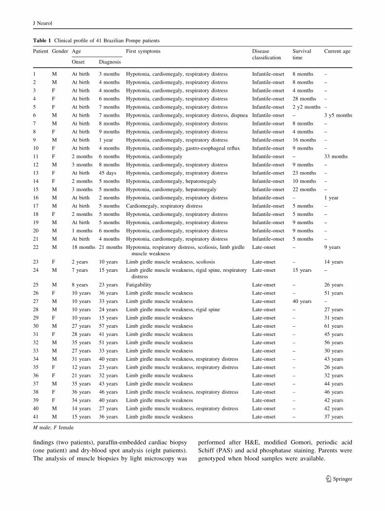
Table 1 Clinical profile of 41 Brazilian Pompe patients
Patient Gender Age First symptoms Disease
classification
Survival
time
Current age
Onset Diagnosis
1 M At birth 3 months Hypotonia, cardiomegaly, respiratory distress Infantile-onset 8 months –
2 M At birth 4 months Hypotonia, cardiomegaly, respiratory distress Infantile-onset 8 months –
3 F At birth 4 months Hypotonia, cardiomegaly, respiratory distress Infantile-onset 4 months –
4 F At birth 6 months Hypotonia, cardiomegaly, respiratory distress Infantile-onset 28 months –
5 F At birth 7 months Hypotonia, cardiomegaly, respiratory distress Infantile-onset 2 y2 months –
6 M At birth 7 months Hypotonia, cardiomegaly, respiratory distress, dispnea Infantile-onset – 3 y5 months
7 M At birth 8 months Hypotonia, cardiomegaly, respiratory distress Infantile-onset 8 months –
8 F At birth 9 months Hypotonia, cardiomegaly, respiratory distress Infantile-onset 4 months –
9 M At birth 1 year Hypotonia, cardiomegaly, respiratory distress Infantile-onset 16 months –
10 F At birth 4 months Hypotonia, cardiomegaly, gastro-esophageal reflux Infantile-onset 9 months –
11 F 2 months 6 months Hypotonia, cardiomegaly Infantile-onset – 33 months
12 M 3 months 8 months Hypotonia, cardiomegaly, respiratory distress Infantile-onset 9 months –
13 F At birth 45 days Hypotonia, cardiomegaly, respiratory distress Infantile-onset 23 months –
14 F 2 months 5 months Hypotonia, cardiomegaly, hepatomegaly Infantile-onset 10 months –
15 M 3 months 5 months Hypotonia, cardiomegaly, hepatomegaly Infantile-onset 22 months –
16 M At birth 2 months Hypotonia, cardiomegaly, respiratory distress Infantile-onset – 1 year
17 M At birth 5 months Cardiomegaly, respiratory distress Infantile-onset 5 months –
18 F 2 months 5 months Hypotonia, cardiomegaly, respiratory distress Infantile-onset 5 months –
19 M At birth 5 months Hypotonia, cardiomegaly, respiratory distress Infantile-onset 9 months –
20 M 1 months 6 months Hypotonia, cardiomegaly, respiratory distress Infantile-onset 9 months –
21 M At birth 4 months Hypotonia, cardiomegaly, respiratory distress Infantile-onset 5 months –
22 M 18 months 21 months Hypotonia, respiratory distress, scoliosis, limb girdle
muscle weakness
Late-onset – 9 years
23 F 2 years 10 years Limb girdle muscle weakness, scoliosis Late-onset – 14 years
24 M 7 years 15 years Limb girdle muscle weakness, rigid spine, respiratory
distress
Late-onset 15 years –
25 M 8 years 23 years Fatigability Late-onset – 26 years
26 F 10 years 36 years Limb girdle muscle weakness Late-onset – 51 years
27 M 10 years 33 years Limb girdle muscle weakness Late-onset 40 years –
28 M 10 years 24 years Limb girdle muscle weakness, rigid spine Late-onset – 27 years
29 F 10 years 15 years Limb girdle muscle weakness Late-onset – 31 years
30 M 27 years 57 years Limb girdle muscle weakness Late-onset – 61 years
31 F 28 years 41 years Limb girdle muscle weakness Late-onset – 45 years
32 M 35 years 51 years Limb girdle muscle weakness Late-onset – 56 years
33 M 27 years 33 years Limb girdle muscle weakness Late-onset – 30 years
34 M 31 years 40 years Limb girdle muscle weakness, respiratory distress Late-onset – 43 years
35 F 12 years 23 years Limb girdle muscle weakness, respiratory distress Late-onset – 26 years
36 F 21 years 32 years Limb girdle muscle weakness Late-onset – 32 years
37 M 35 years 43 years Limb girdle muscle weakness Late-onset – 44 years
38 F 36 years 46 years Limb girdle muscle weakness, respiratory distress Late-onset – 46 years
39 F 34 years 40 years Limb girdle muscle weakness Late-onset – 42 years
40 M 14 years 27 years Limb girdle muscle weakness, respiratory distress Late-onset – 42 years
41 M 15 years 36 years Limb girdle muscle weakness Late-onset – 37 years
M male; F female
J Neurol
123
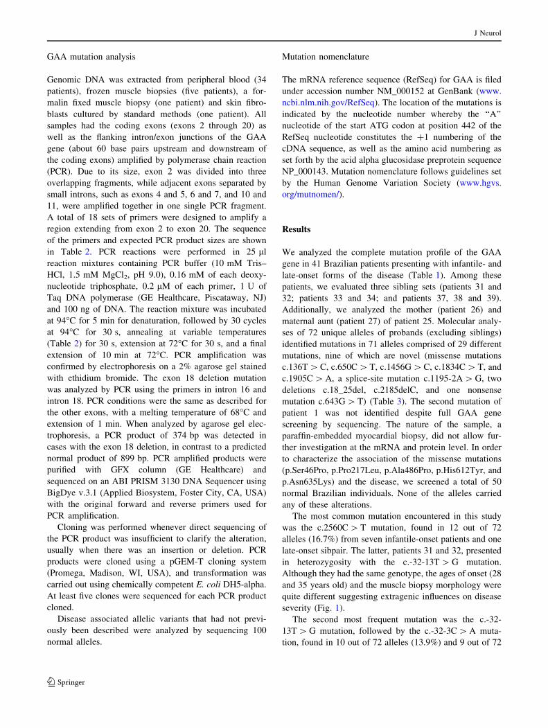
GAA mutation analysis
Genomic DNA was extracted from peripheral blood (34
patients), frozen muscle biopsies (five patients), a for-
malin fixed muscle biopsy (one patient) and skin fibro-
blasts cultured by standard methods (one patient). All
samples had the coding exons (exons 2 through 20) as
well as the flanking intron/exon junctions of the GAA
gene (about 60 base pairs upstream and downstream of
the coding exons) amplified by polymerase chain reaction
(PCR). Due to its size, exon 2 was divided into three
overlapping fragments, while adjacent exons separated by
small introns, such as exons 4 and 5, 6 and 7, and 10 and
11, were amplified together in one single PCR fragment.
A total of 18 sets of primers were designed to amplify a
region extending from exon 2 to exon 20. The sequence
of the primers and expected PCR product sizes are shown
in Table 2. PCR reactions were performed in 25 ll
reaction mixtures containing PCR buffer (10 mM Tris–
HCl, 1.5 mM MgCl2, pH 9.0), 0.16 mM of each deoxy-
nucleotide triphosphate, 0.2 lM of each primer, 1 U of
Taq DNA polymerase (GE Healthcare, Piscataway, NJ)
and 100 ng of DNA. The reaction mixture was incubated
at 94�C for 5 min for denaturation, followed by 30 cycles
at 94�C for 30 s, annealing at variable temperatures
(Table 2) for 30 s, extension at 72�C for 30 s, and a final
extension of 10 min at 72�C. PCR amplification was
confirmed by electrophoresis on a 2% agarose gel stained
with ethidium bromide. The exon 18 deletion mutation
was analyzed by PCR using the primers in intron 16 and
intron 18. PCR conditions were the same as described for
the other exons, with a melting temperature of 68�C and
extension of 1 min. When analyzed by agarose gel elec-
trophoresis, a PCR product of 374 bp was detected in
cases with the exon 18 deletion, in contrast to a predicted
normal product of 899 bp. PCR amplified products were
purified with GFX column (GE Healthcare) and
sequenced on an ABI PRISM 3130 DNA Sequencer using
BigDye v.3.1 (Applied Biosystem, Foster City, CA, USA)
with the original forward and reverse primers used for
PCR amplification.
Cloning was performed whenever direct sequencing of
the PCR product was insufficient to clarify the alteration,
usually when there was an insertion or deletion. PCR
products were cloned using a pGEM-T cloning system
(Promega, Madison, WI, USA), and transformation was
carried out using chemically competent E. coli DH5-alpha.
At least five clones were sequenced for each PCR product
cloned.
Disease associated allelic variants that had not previ-
ously been described were analyzed by sequencing 100
normal alleles.
Mutation nomenclature
The mRNA reference sequence (RefSeq) for GAA is filed
under accession number NM_000152 at GenBank (www.
ncbi.nlm.nih.gov/RefSeq). The location of the mutations is
indicated by the nucleotide number whereby the ‘‘A’’
nucleotide of the start ATG codon at position 442 of the
RefSeq nucleotide constitutes the ?1 numbering of the
cDNA sequence, as well as the amino acid numbering as
set forth by the acid alpha glucosidase preprotein sequence
NP_000143. Mutation nomenclature follows guidelines set
by the Human Genome Variation Society (www.hgvs.
org/mutnomen/).
Results
We analyzed the complete mutation profile of the GAA
gene in 41 Brazilian patients presenting with infantile- and
late-onset forms of the disease (Table 1). Among these
patients, we evaluated three sibling sets (patients 31 and
32; patients 33 and 34; and patients 37, 38 and 39).
Additionally, we analyzed the mother (patient 26) and
maternal aunt (patient 27) of patient 25. Molecular analy-
ses of 72 unique alleles of probands (excluding siblings)
identified mutations in 71 alleles comprised of 29 different
mutations, nine of which are novel (missense mutations
c.136T [ C, c.650C [ T, c.1456G [ C, c.1834C [ T, and
c.1905C [ A, a splice-site mutation c.1195-2A [ G, two
deletions c.18_25del, c.2185delC, and one nonsense
mutation c.643G [ T) (Table 3). The second mutation of
patient 1 was not identified despite full GAA gene
screening by sequencing. The nature of the sample, a
paraffin-embedded myocardial biopsy, did not allow fur-
ther investigation at the mRNA and protein level. In order
to characterize the association of the missense mutations
(p.Ser46Pro, p.Pro217Leu, p.Ala486Pro, p.His612Tyr, and
p.Asn635Lys) and the disease, we screened a total of 50
normal Brazilian individuals. None of the alleles carried
any of these alterations.
The most common mutation encountered in this study
was the c.2560C [ T mutation, found in 12 out of 72
alleles (16.7%) from seven infantile-onset patients and one
late-onset sibpair. The latter, patients 31 and 32, presented
in heterozygosity with the c.-32-13T [ G mutation.
Although they had the same genotype, the ages of onset (28
and 35 years old) and the muscle biopsy morphology were
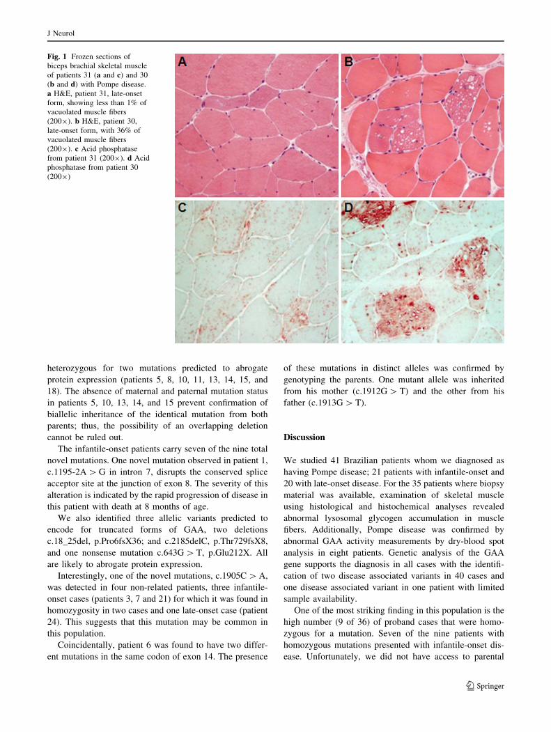
quite different suggesting extragenic influences on disease
severity (Fig. 1).
The second most frequent mutation was the c.-32-
13T [ G mutation, followed by the c.-32-3C [ A muta-
tion, found in 10 out of 72 alleles (13.9%) and 9 out of 72
J Neurol
123
alleles (12.5%), respectively; all of them from late-onset
patients. The c.-32-13T [ G [IVS1] mutation is commonly
present in heterozygosity in late-onset patients. Patient 25
inherited the -32-13T [ G mutation from his father (con-
firmed by paternal genotype) and the c.1927G [ A muta-
tion from his mother. Genotype analysis of the mother
(patient 26) and her brother (patient 27) revealed that
they are also heterozygous for the c.-32-13T [ G and
c.1927G [ A mutations.
The c.-32-3C [ A mutation was found in three probands
who did not carry the c.-32-13T [ G mutation. Three
affected siblings (patients 37, 38 and 39) were homozygous
for the c.-32-3C [ A mutation and had the late-onset pre-
sentation of the disease. Both parents were heterozygous
for the mutation. They are second cousins suggesting that
the two alleles inherited by patients 37, 38, and 39 are
identical by descent. Two late-onset patients (sibpair
patients 33 and 34) are heterozygous for the missense
Table 2 Nucleotide sequences of the primers used for PCR amplification of GAA gene, location and PCR product size and annealing
temperature
Exon Location Orientation Primer sequence (50-30) PCR
product (bp)
Annealing
temperature (�C)
Intron 1 Sense TTT GAG AGC CCC GTG AGT GC 273 65
Exon 2 Antisense TCC CTG CTG GTG AGC TGG GT
2 Exon 2 Sense CGA GAG CTG AGT GGC TCC TC 270 68
Exon2 Antisense GAA GAA GCA CCA GGG CTG CC
Exon 2 Sense CCT GCA AAG CAG GGG CTG CA 267 64
Intron 2 Antisense ATG TCC ACG GGC ACC CTC TG
3 Intron 2 Sense GAC CTG ACC TGT CCT TGG CG 271 70
Intron 3 Antisense TCG CCC TCC CCA TCA TGC TG
4 and 5 Intron 3 Sense GTG CTC TCA GGC TCG TGT GG 464 66
Intron 5 Antisense GTC TCC AGG GCA GGC AGC AC
6 and 7 Intron 5 Sense GGT GCA GAG CCC TCC AAG TG 445 68
Intron 7 Antisense TCT GCT GGG GCC TGA GGA GA
8 Intron 7 Sense GTG AGT TGG GGT GGT GGC AG 285 68
Intron 8 Antisense GAG AAG GAG CCA CTG GGC AC
9 Intron 8 Sense CTC AGT TTT CCC CGT GGC TG 230 68
Intron 9 Antisense GCT GGA GGC CTC TGC TTT CT
10 and 11 Intron 9 Sense GCT CAG TGG GGC TTC CAT GC 449 68
Intron 11 Antisense TGA GGG TGC TAA GTC TCC CA
12 Intron 11 Sense GAG GAA GCT CCC TGG AAA CC 210 62
Intron 12 Antisense CTT GTA GGA CAG GCT GTG AG
13 Intron 12 Sense TGA CAG GGT TCC CGA GTG AC 255 64
Intron 13 Antisense GCC TCC CAT AGA GGC CCC CG
14 Intron 13 Sense CTG GCT CTG CTG CAG CAG CC 295 68
Intron 14 Antisense GCA TGG GGT GCT TCT CCA GC
15 Intron 14 Sense TGA GAA GTG CAG CTC TCC CG 309 68
Intron 15 Antisense AGG GCT GCC TGG CAG TTA CG
16 Intron 15 Sense GGG TGG GCA TAT GAG CCA GC 265 68
Intron 16 Antisense TGG GAG GGC TGC TCT GGT CT
17 Intron 16 Sense AGC GTG GTT CCT GAG GAC AG 249 68
Intron 17 Antisense CTG CAG TGT GCT GTC CAC AC
18 Intron 17 Sense AGG CCT CCACCT CCA CCA GG 293 68
Intron 18 Antisense CCA GGT CCC CTC ACC CCT TC
19 Intron 18 Sense AGC TGT CTG CTG ACA CCT CC 319 64
Intron 19 Antisense CCC AGC TAC CTC TGT TCC TG
20 Intron 19 Sense CTG GGG TCT CAC TGC TGC TG 175 68
Intron 20 Antisense CTG CTT CCC TGG GGA ACC AG
J Neurol
123
mutation c.1781G [ C in combination with the severe
mutation c.1941C [ G. The two remaining late-onset
patients (22 and 23) presented with disease at a very young
age (1.5 and 2 years, respectively). Patient 22 is
homozygous for c.1655T [ C, and patient 23 is heterozy-
gous for the c.377G [ A and the c.1655T [ C mutations.
The infantile-onset patients carry a range of different
mutations with a large proportion homozygous or
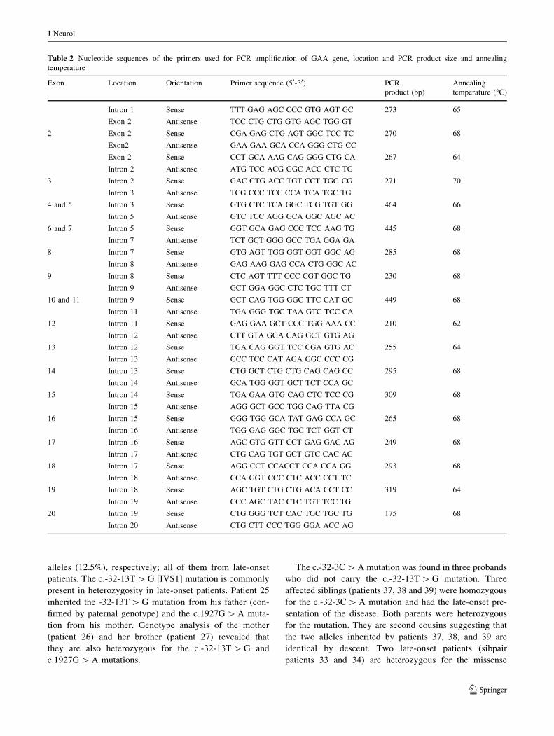
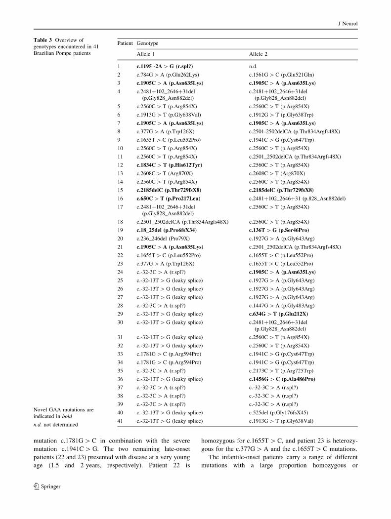
Table 3 Overview of
genotypes encountered in 41
Brazilian Pompe patients
Novel GAA mutations are
indicated in bold
n.d. not determined
Patient Genotype
Allele 1 Allele 2
1 c.1195 -2A [ G (r.spl?) n.d.
2 c.784G [ A (p.Glu262Lys) c.1561G [ C (p.Glu521Gln)
3 c.1905C [ A (p.Asn635Lys) c.1905C [ A (p.Asn635Lys)
4 c.2481?102_2646?31del
(p.Gly828_Asn882del)
c.2481?102_2646?31del
(p.Gly828_Asn882del)
5 c.2560C [ T (p.Arg854X) c.2560C [ T (p.Arg854X)
6 c.1913G [ T (p.Gly638Val) c.1912G [ T (p.Gly638Trp)
7 c.1905C [ A (p.Asn635Lys) c.1905C [ A (p.Asn635Lys)
8 c.377G [ A (p.Trp126X) c.2501-2502delCA (p.Thr834Argfs48X)
9 c.1655T [ C (p.Leu552Pro) c.1941C [ G (p.Cys647Trp)
10 c.2560C [ T (p.Arg854X) c.2560C [ T (p.Arg854X)
11 c.2560C [ T (p.Arg854X) c.2501_2502delCA (p.Thr834Argfs48X)
12 c.1834C [ T (p.His612Tyr) c.2560C [ T (p.Arg854X)
13 c.2608C [ T (Arg870X) c.2608C [ T (Arg870X)
14 c.2560C [ T (p.Arg854X) c.2560C [ T (p.Arg854X)
15 c.2185delC (p.Thr729fsX8) c.2185delC (p.Thr729fsX8)
16 c.650C [ T (p.Pro217Leu) c.2481?102_2646?31 (p.828_Asn882del)
17 c.2481?102_2646?31del
(p.Gly828_Asn882del)
c.2560C [ T (p.Arg854X)
18 c.2501_2502delCA (p.Thr834Argfs48X) c.2560C [ T (p.Arg854X)
19 c.18_25del (p.Pro6fsX34) c.136T [ G (p.Ser46Pro)
20 c.236_246del (Pro79X) c.1927G [ A (p.Gly643Arg)
21 c.1905C [ A (p.Asn635Lys) c.2501_2502delCA (p.Thr834Argfs48X)
22 c.1655T [ C (p.Leu552Pro) c.1655T [ C (p.Leu552Pro)
23 c.377G [ A (p.Trp126X) c.1655T [ C (p.Leu552Pro)
24 c.-32-3C [ A (r.spl?) c.1905C [ A (p.Asn635Lys)
25 c.-32-13T [ G (leaky splice) c.1927G [ A (p.Gly643Arg)
26 c.-32-13T [ G (leaky splice) c.1927G [ A (p.Gly643Arg)
27 c.-32-13T [ G (leaky splice) c.1927G [ A (p.Gly643Arg)
28 c.-32-3C [ A (r.spl?) c.1447G [ A (p.Gly483Arg)
29 c.-32-13T [ G (leaky splice) c.634G [ T (p.Glu212X)
30 c.-32-13T [ G (leaky splice) c.2481?102_2646?31del
(p.Gly828_Asn882del)
31 c.-32-13T [ G (leaky splice) c.2560C [ T (p.Arg854X)
32 c.-32-13T [ G (leaky splice) c.2560C [ T (p.Arg854X)
33 c.1781G [ C (p.Arg594Pro) c.1941C [ G (p.Cys647Trp)
34 c.1781G [ C (p.Arg594Pro) c.1941C [ G (p.Cys647Trp)
35 c.-32-3C [ A (r.spl?) c.2173C [ T (p.Arg725Trp)
36 c.-32-13T [ G (leaky splice) c.1456G [ C (p.Ala486Pro)
37 c.-32-3C [ A (r.spl?) c.-32-3C [ A (r.spl?)
38 c.-32-3C [ A (r.spl?) c.-32-3C [ A (r.spl?)
39 c.-32-3C [ A (r.spl?) c.-32-3C [ A (r.spl?)
40 c.-32-13T [ G (leaky splice) c.525del (p.Gly176fsX45)
41 c.-32-13T [ G (leaky splice) c.1913G [ T (p.Gly638Val)
J Neurol
123
heterozygous for two mutations predicted to abrogate
protein expression (patients 5, 8, 10, 11, 13, 14, 15, and
18). The absence of maternal and paternal mutation status
in patients 5, 10, 13, 14, and 15 prevent confirmation of
biallelic inheritance of the identical mutation from both
parents; thus, the possibility of an overlapping deletion
cannot be ruled out.
The infantile-onset patients carry seven of the nine total
novel mutations. One novel mutation observed in patient 1,
c.1195-2A [ G in intron 7, disrupts the conserved splice
acceptor site at the junction of exon 8. The severity of this
alteration is indicated by the rapid progression of disease in
this patient with death at 8 months of age.
We also identified three allelic variants predicted to
encode for truncated forms of GAA, two deletions
c.18_25del, p.Pro6fsX36; and c.2185delC, p.Thr729fsX8,
and one nonsense mutation c.643G [ T, p.Glu212X. All
are likely to abrogate protein expression.
Interestingly, one of the novel mutations, c.1905C [ A,
was detected in four non-related patients, three infantile-
onset cases (patients 3, 7 and 21) for which it was found in
homozygosity in two cases and one late-onset case (patient
24). This suggests that this mutation may be common in
this population.
Coincidentally, patient 6 was found to have two differ-
ent mutations in the same codon of exon 14. The presence
of these mutations in distinct alleles was confirmed by
genotyping the parents. One mutant allele was inherited
from his mother (c.1912G [ T) and the other from his
father (c.1913G [ T).
Discussion
We studied 41 Brazilian patients whom we diagnosed as
having Pompe disease; 21 patients with infantile-onset and
20 with late-onset disease. For the 35 patients where biopsy
material was available, examination of skeletal muscle
using histological and histochemical analyses revealed
abnormal lysosomal glycogen accumulation in muscle
fibers. Additionally, Pompe disease was confirmed by
abnormal GAA activity measurements by dry-blood spot
analysis in eight patients. Genetic analysis of the GAA
gene supports the diagnosis in all cases with the identifi-
cation of two disease associated variants in 40 cases and
one disease associated variant in one patient with limited
sample availability.
One of the most striking finding in this population is the
high number (9 of 36) of proband cases that were homo-
zygous for a mutation. Seven of the nine patients with
homozygous mutations presented with infantile-onset dis-
ease. Unfortunately, we did not have access to parental
Fig. 1 Frozen sections of
biceps brachial skeletal muscle
of patients 31 (a and c) and 30
(b and d) with Pompe disease.
a H&E, patient 31, late-onset
form, showing less than 1% of
vacuolated muscle fibers
(2009). b H&E, patient 30,
late-onset form, with 36% of
vacuolated muscle fibers
(2009). c Acid phosphatase
from patient 31 (2009). d Acid
phosphatase from patient 30
(2009)
J Neurol
123
DNA to confirm biallelic inheritance. Therefore, in order to
confirm homozygosity and rule out intragenic deletions, we
analyzed a series of polymorphisms across the GAA gene
(data not shown). Only two of the patients (cases 5 and 13)
were heterozygous for polymorphisms near the region of
the detected mutation suggesting that an overlapping
deletion on one allele was unlikely and that the two alleles
in these patients were not identical by descent.
Patient 6 has two different missense mutations in the
same codon of exon 14, confirmed to be in distinct alleles
by inheritance from his mother (c.1912G [ T) and from
his father (c.1913G [ T). Both mutations are predicted to
be responsible for severe phenotypes by the replacement of
glycine at codon 638 with valine and tryptophan, an amino
acid residue that is highly conserved [29]. An homozygous
patient for the c.1912G [ T mutation presented with
clinical onset at 2 months of age [25]. Functional analysis
for the c.1912G [ T mutation by Western blot and the
protein expression in COS cells demonstrated low levels
GAA synthesis and degradation of the enzyme precursor
[28]. Despite unusual survival of patient 6, who is under
enzyme replacement therapy (ERT) for 3 years, he had
some improvement of cardiac function but no improvement
of motor function, inability to move any segment of upper
or lower limbs, dependence of BIPA 24 h/day and feeding
by gastrectomy.
The novel mutation c.1905C [ A [p.Asn635Lys] was
found in homozygosity in two unrelated children with
infantile-onset disease (patients 3 and 7) who survived for
only 4 and 8 months, respectively, suggesting a severe
phenotype due to this mutation. In addition, another unre-
lated child (patient 21), heterozygous for the c.1905C [ A
mutation and a deletion (c.2501_2502del), survived for
5 months. The c.1905C [ A mutation alters amino acid
635 and occurs in a highly conserved region of the protein
[14], strongly suggesting a deleterious effect on enzyme
expression. Patient 4 was found to be homozygous for the
common c.2481 ? 102_2646 ? 31del [p.Gly828_
Asn882del, Del exon 18] mutation [13]. Homozygosity for
this mutation is uncommon and may represent consan-
guinity; however, a comprehensive family history was not
taken in this case nor for the other infantile-onset patients.
The mutation c.2560G [ T [p.Arg854X] in exon 18,
was observed in 16.7% (12 of 72) of the alleles studied.
This mutation was detected in homozygosity in three
infantile cases (patients 5, 10 and 14), and in heterozy-
gosity in four other infantile cases (patients 11, 12, 17 and
18), and two affected adult siblings (patients 31 and 32).
For the two adults, this mutation was observed in combi-
nation with the common intronic 1 mutation (c.-32-
13T [ G). All patients are of African descent, and it is
most likely this is the origin of this mutation in Brazil. This
is consistent with previous reports of this mutation having a
higher incidence amongst Pompe patients of African origin
[1, 2]. While this mutation has been described in a Spanish
juvenile onset patient who inherited the mutation from his
Dominican mother [5], it has not been reported with such a
high frequency by others in Spain or South America [6, 7].
Despite the fact that patient 11 has two different alterations
leading to protein truncation, c.2560G [ T [p.Arg854X]
and c.2501_2502del [p.Thr834Argfs48X] and first symp-
toms onset at 2 months of age, she is still alive, under ERT,
being able to walk with support.
By way of comparison to other Pompe patient popula-
tions, the c.377G [ A [p.Trp126X] mutation which has
been observed as very common in the Argentinean Pompe
patients of Italian origin [23], was only observed in two
individuals. Another mutation, c.1655T [ C [p.Leu552-
Pro], which was initially classified as a severe mutation in a
7-year-old male with ‘‘late infantile’’ Pompe disease and
was found to have very low residual activity after expres-
sion in COS cells [3], was observed in three individuals
(patients 9, 22, and 23), one being homozygous (patient 22)
for this mutation. All three of the current patients in our
study presented with an age of onset ranging from at birth
to 2 years of age (Table 1).
Of particular interest is the survival of patients 22 and
23, both of whom presented with disease early in life and
are still alive, compared to patient 9 who died at 16 months
of age. The c.1655T [ C mutation has previously been
described in Italian and Spanish Pompe patients [7, 25].
Pittis et al. [25] described the c.1655T [ C as the second
most frequent among Italian Pompe cases, and in vitro
analysis demonstrated a response to chaperones [24], sug-
gesting that this mutation may be associated with late-onset
Pompe patients with onset in the first years of age. It is
possible that other genetic or environmental factors mod-
ulate the severity and outcome in untreated patients with
this mutation.
We also identified three allelic variants predicted to
encode for truncated forms of GAA, two deletions
c.18_25del, p.Pro6fsX36; and c.2185delC, p.Thr729fsX8,
and one nonsense mutation c.643G [ T, p.Glu212X. The
mutant transcripts are likely to be targeted by nonsense-
mediated decay based on their location within the gene
[22]. Even the mutation c.2185delC, p.Thr729fsX8, found
in patient 15, is likely to abrogate the expression as the
patient presented with severe infantile-onset disease at
2 months of age and survived less than 2 years.
Five novel allelic variants described here encoded for
missense mutations: c.136T [ C, [p.Ser46Pro]; c.650C [T, [p.Pro217Leu]; c.1456G [ C, [p.Ala486Pro]; c.1834C
[ T, [p.His612Tyr]; and c.1905C [ A, [p.Asn635Lys]. As
the effect of these variants on GAA activity and expression
has not been studied, their significance to disease devel-
opment remains unknown. Sequence analysis of 100
J Neurol
123
normal Brazilian GAA alleles did not reveal these muta-
tions to be present in the normal individuals. This suggests
that they are not common normal polymorphisms among
our patient population.
Of the late-onset patients, 8 of 15 probands (53%) car-
ried the common late-onset c.-32-13T [ G (IVS1) muta-
tion, whereas the remaining seven had combinations that
lacked this mutation. The c.-32-13T [ G mutation was
originally identified in 68% of adult patients with ages at
diagnosis ranging from 17 to 73 years [12]. When Kroos
et al. [16] analyzed 98 adult compound heterozygotes with
the c.-32-13T [ G and another fully deleterious mutation,
they observed a broad spectrum of age at onset of first
symptoms manifestations, ranging from \1 to 78 years.
The range of age of onset from 8 to 35 years among our
patients is consistent with these findings and lends to the
speculation that other genetic factors may play a role in the
variability of onset and severity in cases carrying the c.-32-
13T [ G mutation [16].
We and others recently described the c.-32-3C [ A
mutation in intron 1 and c.1781G [ C [p.Arg594Pro]
mutation in exon 13 [17, 25]. Of interest, six of 20 late-
onset cases (patients 24, 28 and 35, and siblings 37, 38 and
39) carry the c.-32-3C [ A mutation. Patients 24, 28 and
35 presented as juvenile patients (ages 7, 10 and 12 years,
respectively) while the three siblings who are homozygous
for the mutation present with disease in their 30s. Although
the ages of onset for the siblings are similar, the evolution
and clinical symptoms need a longer follow-up, since only
the older sister has presented with respiratory distress in
addition to the limb girdle muscle weakness suggesting that
the disease course may diverge. The diminished severity in
the homozygous siblings as compared to patients 24 and 28
support the possibility of low residual activity from the
mutant allele such that the presence of two copies may
delay disease onset and rate of progression. It is also pos-
sible, however, that other factors within the mutant GAA
allele or other modifier genes may contribute to the dif-
ferences in age of onset. Interestingly, Pittis et al. [25]
observed the c.-32-3C [ A variant in an infantile-onset
patient. They determined that the mutant allele leads to a
deletion of 579 bp which would result in the loss of the first
182 amino acids of the protein (p.M1_T182). Thus, factors
which control splice-site selection may contribute to the
relative level of normal transcript the mutant allele is able
to generate.
Analysis of our Brazilian Pompe patients showed that
sometimes the nature of the mutation matched the phe-
notype within this group. When compared to other pop-
ulations this was not always consistent. In many, the age
of onset and the disease course were different in patients
with the same genotype, as in the cases of relatives
studied here.
In conclusion, this first study of Brazilian Pompe
patients has been helpful in furthering the characterization
of the mutations present in this population and has allowed
us to compare our patients, their clinical presentation and
severity to others with this disease. The remarkable
heterogeneity in the mutational spectrum of Brazilian
Pompe patients may reflect the ethnic diversity of our
population and serve to aid in determining what other
factors influence disease progression and outcome.
Acknowledgments This work was supported by Fundacao de
Amparo a Pesquisa do Estado de Sao Paulo (FAPESP), grant number
2001/00422-5, and Genzyme Corporation.
References
1. Adams EM, Becker JA, Griffith L, Segal A, Plotz PH, Raben N
(1997) Glycogenosis type II: a juvenile specific mutation with an
unusual splicing pattern and a shared mutation in African
Americans. Hum Mutat 10:128–134
2. Becker JA, Vlach J, Raben N, Nagaraju K, Adams EM, Hermans
MM, Reuser AJJ, Brooks SS, Tifft CJ, Hirschhorn R, Huie ML,
Nicolino M, Plotz PH (1998) The African origin of the common
mutation in African American patients with glycogen-storage
disease type II. Am J Hum Genet 62:991–994
3. Bodamer OA, Haas D, Hermans MM, Reuser AJ, Hoffmann GF
(2002) L-alanine supplementation in late infantile glycogen
storage disease type II. Pediatr Neurol 27:145–146
4. Case LE, Kishnani PS (2006) Physical therapy management of
Pompe disease. Genet Med 8:318–327
5. Castro-Gago M, Eirıs-Punal J, Rodrıguez-Nunez Pintos-Martınez
E, Benlloch-Marın Barros-Angueira (1999) Forma grave de
glucogenosis tipo II juvenile en un nino heterocigoto compuesto
(Tyr-292 [ Cys/Arg-854 [ Stop). Rev Neurol 29(1):46–49
6. Fernandez-Hojas R, Huie ML, Navarro C, Dominguez C, Roig
M, Lopez-Coronas D, Teijeira S, Anyane-Yeboa K, Hirschhorn R
(2002) Identification of six novel mutations in the acid alpha-
glucosidase gene in three Spanish patients with infantile onset
glycogen storage disease type II (Pompe disease). Neuromuscul
Disord 12:159–166
7. Gort L, Coll MJ, Chabas A (2007) Glycogen storage disease type
II in Spanish patients: high frequency of c.1076–1G [ C muta-
tion. Mol Genet Metab 92:183–187
8. Hermans MM, Wisselaar HA, Kroos MA, Oostra BA, Reuser AJ
(1993) Human lysosomal alpha-glucosidase: functional charac-
terization of the glycosylation sites. Biochem J 289:681–686
9. Hirschhorn R (2001) Reuser AJJ (2001) Glycogen storage disease
type II (GSDII). In: Scriver CR, Beaudet AL, Sly WS, Valle D
(eds) The metabolic and molecular bases of inherited disease.
McGraw-Hill, New York, pp 3389–3420
10. Hoefsloot LH, Hoogeveen-Westerveld M, Reuser AJ, Oostra BA
(1990) Characterization of the human lysosomal alpha-glucosi-
dase gene. Biochem J 272:493–497
11. Hoefsloot LH, Willemsen R, Kroos MA, Hoogeveen-Westerveld
M, Hermans MM, Van der Ploeg AT, Oostra BA, Reuser AJ
(1990) Expression and routeing of human lysosomal alpha-glu-
cosidase in transiently transfected mammalian cells. Biochem J
272:485–492
12. Huie ML, Chen AS, Tsujino S, Shanske S, DiMauro S, Engel
AG, Hirschhorn R (1994) Aberrant splicing in adult onset gly-
cogen storage disease type II (GSDII): molecular identification of
an IVS1 (-13T- [ G) mutation in a majority of patients and a
J Neurol
123
novel IVS10 (?1GT- [ CT) mutation. Hum Mol Genet 3:2231–
2236
13. Huie ML, Chen AS, Brooks SS, Grix A, Hirschhorn R (1994) A
de novo 13 nt deletion, a newly identified C647 W missense
mutation and a deletion of exon 18 in infantile onset glycogen
storage disease type II (GSDII). Hum Mol Genet 3:1081–1087
14. Huie ML, Tsujino S, Sklower Brooks S, Engel A, Elias E,
Bonthron DT, Bessley C, Shanske S, DiMauro S, Goto YI, Hir-
schhorn R (1998) Glycogen storage disease type II: identification
of four novel missense mutations (D645N, G648S, R672W,
R672Q) and two insertions/deletions in the acid alpha-glucosi-
dase locus of patients of differing phenotype. Biochem Biophys
Res Commun 244:921–927
15. Kroos MA, Van der Kraan M, Van Diggelen OP, Kleijer WJ,
Reuser AJ, Van den Boogaard MJ, Ausems MG, Ploos van
Amstel HK, Poenaru L, Nicolino M, Cochin C, Wevers R (1995)
Glycogen storage disease type II: frequency of three common
mutant alleles and their associated clinical phenotypes studied in
121 patients. J Med Genet 32:836–837
16. Kroos MA, Pomponio RJ, Hagemans ML, Keulemans JL, Phipps
M, DeRiso M, Palmer RE, Ausems MG, Van der Beek NA, Van
Diggelen OP, Halley DJ, Van der Ploeg AT, Reuser AJ (2007)
Broad spectrum of Pompe disease in patients with the same
c.-32–13T- [ G haplotype. Neurology 68:110–115
17. Kroos M, Pomponio RJ, van Vliet L, Palmer RE, Phipps M, Van
der Helm R, Halley D, Reuser A (2008) GAA database consor-
tium. Update of the Pompe disease mutation database with 107
sequence variants and a format for severity rating. Hum Mutat
29:E13–E26
18. Kuo WL, Hirschhorn R, Huie ML, Hirschhorn K (1996) Local-
ization and ordering of acid alpha-glucosidase (GAA) and thy-
midine kinase (TK1) by fluorescence in situ hybridization. Hum
Genet 97:404–406
19. Martiniuk F, Bodkin M, Tzall S, Hirschhorn R (1991) Isolation
and partial characterization of the structural gene for human acid
alpha glucosidase. DNA Cell Biol 10:283–292
20. Martiniuk F, Mehler M, Tzall S, Meredith G, Hirschhorn R
(1990) Sequence of the cDNA and 50-flanking region for human
acid alpha-glucosidase, detection of an intron in the 50 untrans-
lated leader sequence, definition of 18-bp polymorphisms, and
differences with previous cDNA and amino acid sequences. DNA
Cell Biol 9:85–94
21. Moreland RJ, Jin X, Zhang XK, Decker RW, Albee KL, Lee KL,
Cauthron RD, Brewer K, Edmunds T, Canfield WM (2005)
Lysosomal acid alpha-glucosidase consists of four different
peptides processed from a single chain precursor. J Biol Chem
280:6780–6791
22. Nagy E, Maquat LE (1988) A rule for termination-codon position
within intron-containing genes: when nonsense affects RNA
abundance. Trends Biochem Sci 23:198–199
23. Palmer RE, Amartino HM, Niizawa G, Blanco M, Pomponio RJ,
Chamoles NA (2007) Pompe disease (glycogen storage disease
type II) in Argentineans: clinical manifestations and identification
of nine novel mutations. Neuromuscul Disord 17:16–22
24. Parenti G, Zuppaldi A, Gabriela Pittis M, Rosaria Tuzzi M,
Annunziata I, Meroni G, Porto C, Donaudy F, Rossi B, Rossi M,
Filocamo M, Donati A, Bembi B, Ballabio A, Andria G (2007)
Pharmacological enhancement of mutated alpha-glucosidase
activity in fibroblasts from patients with Pompe disease. Mol
Ther 15:508–514
25. Pittis MG, Donnarumma M, Montalvo AL, Dominissini S, Kroos
M, Rosano C, Stroppiano M, Bianco MG, Donati MA, Parenti G,
D’Amico A, Ciana G, Di Rocco M, Reuser A, Bembi B, Filo-
camo M (2008) Molecular and functional characterization of
eight novel GAA mutations in Italian infants with Pompe disease.
Hum Mutat 29:E27–E36
26. Raben N, Nichols RC, Boerkoel C, Plotz P (1995) Genetic
defects in patients with glycogenosis type II (acid maltase defi-
ciency). Muscle Nerve 3:S70–S74
27. Raben N, Nichols RC, Martiniuk F, Plotz PH (1996) A model of
mRNA splicing in adult lysosomal storage disease (glycogenosis
type II). Hum Mol Genet 5:995–1000
28. Van den Hout JM, Kamphoven JH, Winkel LP, Arts WF, De
Klerk JB, Loonen MC, Vulto AG, Cromme-Dijkhuis A, Weis-
glas-Kuperus N, Hop W, Van Hirtum H, Van Diggelen OP, Boer
M, Kroos MA, Van Doorn PA, Van der Voort E, Sibbles B, Van
Corven EJ, Brakenhoff JP, Van Hove J, Smeitink JA, de Jong G,
Reuser AJ, Van der Ploeg AT (2004) Long-term intravenous
treatment of Pompe disease with recombinant human alpha-glu-
cosidase from milk. Pediatrics 113:e448–e457
29. Vorgerd M, Burwinkel B, Reichmann H, Malin JP, Kilimann
MW (1998) Adult-onset glycogen storage disease type II: phe-
notypic and allelic heterogeneity in German patients. Neuroge-
netics 1:205–211
30. Wisselaar HA, Kroos MA, Hermans MM, van Beeumen J, Reuser
AJ (1993) Structural and functional changes of lysosomal acid
alpha-glucosidase during intracellular transport and maturation.
J Biol Chem 268:2223–2231
J Neurol
123