Poly(lactic acid)-poly(ethylene glycol) nanoparticles as new carriers for the delivery of plasmid...
14
Journal of Controlled Release 75 (2001) 211–224 www.elsevier.com / locate / jconrel Poly(lactic acid)-poly(ethylene glycol) nanoparticles as new carriers for the delivery of plasmid DNA a a b b b a, * C. Perez , A. Sanchez , D. Putnam , D. Ting , R. Langer , M.J. Alonso a Department of Pharmacy and Pharmaceutical Technology, University of Santiago de Compostela, Campus Sur, 15705 Santiago de Compostela, Spain b MIT, Blg. E 25, Rm. 342, 77 Massachusetts Ave., Cambridge, MA 02139, USA Received 2 January 2001; accepted 16 May 2001 Abstract The purpose of the present work was to produce and characterize poly(lactic acid)-poly(ethylene glycol) (PLA-PEG) nanoparticles (size lower than 300 nm) containing a high loading of plasmid DNA in a free form or co-encapsulated with either poly(vinyl alcohol) (PVA) or poly(vinylpyrrolidone) (PVP). The plasmid alone or with PVA or PVP was encapsulated by two different techniques: an optimized w / o / w emulsion-solvent evaporation technique as well as by a new w / o emulsion-solvent diffusion technique. Particle size, z potential, plasmid DNA loading and in vitro release were determined for the three plasmid-loaded formulations. The influence of the initial plasmid loadings (5, 10, 20 mg plasmid DNA / mg PLA-PEG) on those parameters was also investigated. The plasmid loaded into the nanoparticles and released in vitro was quantified by fluorimetry and the different molecular forms were identified by gel electrophoresis. PLA-PEG nanoparticles containing plasmid DNA in a free form or co-encapsulated with PVA or PVP were obtained in the range size of 150–300 nm and with a negative z potential, both parameters being affected by the preparation technique. Encapsulation efficiencies were high irrespective of the presence of PVA or PVP (60–90%) and were slightly affected by the preparation technique and by the initial loading. The final plasmid DNA loading in the nanoparticles was up to 10–12 mg plasmid DNA / mg polymer. Plasmid DNA release kinetics varied depending on the plasmid incorporation technique: nanoparticles prepared by the w / o diffusion technique released their content rapidly whereas those obtained by the w / o / w showed an initial burst followed by a slow release for at least 28 days. No significant influence of the plasmid DNA loading and of the co-encapsulation of PVP or PVA on the in vitro release rate was observed. In all cases the conversion of the supercoiled form to the open circular and linear forms was detected. In conclusion, plasmid DNA can be very efficiently encapsulated, either in a free form or in combination with PVP and PVA, into PLA-PEG nanoparticles. Additionally, depending on the processing conditions, these nanoparticles release plasmid DNA either very rapidly or in a controlled manner. 2001 Published by Elsevier Science B.V. Keywords: DNA; PLA-PEG nanoparticles; Controlled release; Nanoencapsulation 1. Introduction *Corresponding author. Tel.: 134-981-594-627; fax: 134-981- At present, it is broadly accepted that plasmid 547-148. E-mail address: [email protected] (M.J. Alonso). DNA has potential not only as a therapeutic agent 0168-3659 / 01 / $ – see front matter 2001 Published by Elsevier Science B.V. PII: S0168-3659(01)00397-2
-
Upload
independent -
Category
Documents
-
view
0 -
download
0
Transcript of Poly(lactic acid)-poly(ethylene glycol) nanoparticles as new carriers for the delivery of plasmid...
Journal of Controlled Release 75 (2001) 211–224www.elsevier.com/ locate / jconrel
Poly(lactic acid)-poly(ethylene glycol) nanoparticles as newcarriers for the delivery of plasmid DNA
a a b b b a ,*C. Perez , A. Sanchez , D. Putnam , D. Ting , R. Langer , M.J. AlonsoaDepartment of Pharmacy and Pharmaceutical Technology, University of Santiago de Compostela, Campus Sur,
15705 Santiago de Compostela, SpainbMIT, Blg. E 25, Rm. 342, 77 Massachusetts Ave., Cambridge, MA 02139, USA
Received 2 January 2001; accepted 16 May 2001
Abstract
The purpose of the present work was to produce and characterize poly(lactic acid)-poly(ethylene glycol) (PLA-PEG)nanoparticles (size lower than 300 nm) containing a high loading of plasmid DNA in a free form or co-encapsulated witheither poly(vinyl alcohol) (PVA) or poly(vinylpyrrolidone) (PVP). The plasmid alone or with PVA or PVP was encapsulatedby two different techniques: an optimized w/o/w emulsion-solvent evaporation technique as well as by a new w/oemulsion-solvent diffusion technique. Particle size, z potential, plasmid DNA loading and in vitro release were determinedfor the three plasmid-loaded formulations. The influence of the initial plasmid loadings (5, 10, 20 mg plasmid DNA/mgPLA-PEG) on those parameters was also investigated. The plasmid loaded into the nanoparticles and released in vitro wasquantified by fluorimetry and the different molecular forms were identified by gel electrophoresis. PLA-PEG nanoparticlescontaining plasmid DNA in a free form or co-encapsulated with PVA or PVP were obtained in the range size of 150–300 nmand with a negative z potential, both parameters being affected by the preparation technique. Encapsulation efficiencies werehigh irrespective of the presence of PVA or PVP (60–90%) and were slightly affected by the preparation technique and bythe initial loading. The final plasmid DNA loading in the nanoparticles was up to 10–12 mg plasmid DNA/mg polymer.Plasmid DNA release kinetics varied depending on the plasmid incorporation technique: nanoparticles prepared by the w/odiffusion technique released their content rapidly whereas those obtained by the w/o/w showed an initial burst followed by aslow release for at least 28 days. No significant influence of the plasmid DNA loading and of the co-encapsulation of PVP orPVA on the in vitro release rate was observed. In all cases the conversion of the supercoiled form to the open circular andlinear forms was detected. In conclusion, plasmid DNA can be very efficiently encapsulated, either in a free form or incombination with PVP and PVA, into PLA-PEG nanoparticles. Additionally, depending on the processing conditions, thesenanoparticles release plasmid DNA either very rapidly or in a controlled manner. 2001 Published by Elsevier ScienceB.V.
Keywords: DNA; PLA-PEG nanoparticles; Controlled release; Nanoencapsulation
1. Introduction
*Corresponding author. Tel.: 134-981-594-627; fax: 134-981-At present, it is broadly accepted that plasmid547-148.
E-mail address: [email protected] (M.J. Alonso). DNA has potential not only as a therapeutic agent
0168-3659/01/$ – see front matter 2001 Published by Elsevier Science B.V.PI I : S0168-3659( 01 )00397-2
212 C. Perez et al. / Journal of Controlled Release 75 (2001) 211 –224
but also as a new vaccination approach [1]. How- character, the encapsulation of plasmid DNA mole-ever, as research in this field advances, it becomes cules within hydrophobic PLGA microspheres ap-evident that the use of plasmid DNA-based pharma- pears to be a challenge for researchers interested inceuticals as drugs and vaccines will largely depend the formulation of delicate macromolecules. One ofon the development of efficient delivery systems [2]. the approaches to associate plasmid DNA to PLA/Naked plasmid DNA is highly susceptible to nu- PLGA nano/microparticles has focused on the use ofclease degradation and, therefore, is rapidly cleared PLA grafted with cationic polysaccharides or cat-from the bloodstream when injected intravenously ionic surfactants which provide a positive charge to[3]. Additionally, because of its large size and the particles, thereby facilitating the adsorption ofnegative charge plasmid DNA is not able to enter the negatively charged plasmid DNA moleculescells and, hence, not able to cross epithelia. An [10,11]. Nevertheless, in order to achieve a betterexception is represented by muscle and epidermal protection of the plasmid and a more precise controlcells, which have been shown to produce detectable of the release process, attention has been speciallylevels of gene expression following intramuscular addressed to the encapsulation of the plasmid withininjection or by gene gun delivery [4]. Nevertheless, PLGA microspheres. Using three model plasmids,the access of plasmids to other population of cells is Luo et al. showed that prolonged plasmid DNAhighly restricted. For example, in the case of a DNA release could be achieved by adjusting the polymervaccine, it would be critical to target the plasmid to molecular weight and composition of PLA/PLGAthe antigen presenting cells (APCs) in order to microspheres [12]. A limitation of some of thesegenerate a T-cell response. These cells are in the microspheres with respect to their utility for vaccineblood lymphocyte pool and particularly concentrated delivery is their large size (100 mm). In fact, is hasin the sub-mucosal tissues. Therefore, the design of a been generally accepted that only particles of a sizecarrier appropriately tailored for delivering plasmid smaller than 10 mm are able to cross mucosalDNA to the APCs, following mucosal administra- surfaces and enter the APCs, significantly. Success-tion, appears to be a very promising but challenging ful approaches for encapsulating plasmid DNA with-approach towards improving DNA vaccination. In in PLGA microspheres have been the spray-dryingaddition, the possibility to provide a long-term [13] and the water–oil–water solvent evaporationsustained delivery of plasmid DNA would represent techniques [12,14]. However, what these authorsa major advance for the use of DNA vaccines. discovered while developing these microspheres is
One strategy to achieve targeted and controlled that the encapsulated plasmid is degraded not onlydelivery of DNA vaccines has been based on the use during the encapsulation process but mainly in theof biodegradable polymer particles. Interesting re- course of the polymer degradation [13,14]. Thesults were found following oral administration of inactivation of the plasmid during encapsulation wasmucoadhesive particles such as polyanhydride micro- addressed by Ando et al. [15] who proposed aspheres [5] and chitosan nanoparticles [6]. In both cryopreparation technique in order to prevent thecases, the model plasmids associated to the particles exposure of the plasmid to shear forces createdelicited significant levels of transfection. However, during the homogenization process. Nevertheless, theprobably because of their safety profile, micro- stabilization of the plasmid in a long-term releasespheres made of PLGA are those which have re- process requires further investigation. A possibilityceived the greatest deal of attention over the last few could rely on the encapsulation of plasmids previous-years [7]. Research in this area has also been ly complexed with other hydrophilic polymers. Forencouraged by the protective humoral and mucosal example, Capan et al. observed that the complexationimmune responses obtained following oral adminis- of the plasmid with poly(L-lysine) (PLL) improvedtration of plasmid DNA encapsulated in PLGA its encapsulation and release in the supercoiled formmicrospheres [8] and also by the evidence of the [16,17]. However, a limitation of these small micro-ability of these plasmid containing particles to elicit spheres was their low encapsulation efficiency and,T-cell responses [9,10]. hence, their low final loading.
Because of their extremely large size and polar In the present work, taking advantage of our
C. Perez et al. / Journal of Controlled Release 75 (2001) 211 –224 213
experience on the encapsulation and stabilization of 2.2. Encapsulation of the plasmid within PLA-PEGproteins in PLGA and PLA-PEG micro /nanoparti- nanoparticlescles, we attempt to explore the potential of PLA-PEG nanoparticles as carriers for the controlled The plasmid DNA was encapsulated into PLA-delivery of plasmid DNA. The selection of this PEG nanoparticles using two different techniques:carrier was based on its capacity to encapsulate the water-in-oil-in-water solvent evaporation tech-antigens efficiently, i.e., tetanus toxoid, and to trans- nique and a newly developed water-in-oil solventport them across mucosal surfaces, after either nasal diffusion technique. Prior to encapsulation into the[18] or oral administration [19]. Furthermore, recent PLA-PEG nanoparticles, some samples of plasmidstudies have revealed their ability to elicit long DNA (3.08 mg/ml) were incubated with PVP (10%,lasting humoral and mucosal immune responses w/w) or PVA (4%, w/w) at room temperature for 30following nasal immunization [20]. Based on this min (concentration of plasmid DNA in the PVP andinformation, the goal of this study is to evaluate the PVA solution: 1.206 mg/ml). The w:w plasmidpotential of these nanoparticles as carriers for the DNA:PVP ratio (2:100) was selected taking intocontrolled release of plasmid DNA, using pCMVLuc account previous transfection studies [21,22] and theas a model plasmid. From a practical standpoint, the plasmid DNA:PVA ratio (5:100) was chosen toobjectives are: (i) to encapsulate a high amount of provide a viscosity to the plasmid solutionplasmid DNA in a free form or in combination with (2.3560.04 mPa) similar to the one of the solutionPVA or PVP within very small PEG-coated PLA containing plasmid DNA:PVP (2.4460.04 mPa).nanoparticles (ii) to evaluate the controlled release Viscosity measurements were performed at 258Cproperties of this new plasmid DNA delivery device. using a capillary viscosimeter (Cannon-Fenske vis-
cosimeter) (Afora, Spain).
2.3. Water-in-oil-in-water solvent evaporation2. Materials and methods
technique
2.1. Materials The technique was a modification of the previous-ly described for the encapsulation of proteins within
Methoxy PEG-(D,L-lactide) (PLA-PEG) with a PLGA nanoparticles [23]. A volume of 166 ml of thePLA and PEG molecular weight (M ) of 46 and 5 plasmid solution (free or with PVP and PVA) wasw
kDa, respectively, was purchased from Alkermes emulsified in 0.5 ml of an organic solution consisting(Cincinnati, OH). Luciferase reporter plasmid (pBK- of 40 mg of PLA-PEG dissolved in a 1:1 mixture ofLuc) driven by the CMV promoter was a kind gift ethyl acetate: methylene chloride by sonicationfrom Zycos, (Cambridge, MA). Plasmid was har- (Sonifier 250, Branson, Barcelona, Spain) for 5 svested from STBL cells by ion-exchange chromatog- (output510 units). Then, 2 ml of an aqueous PVAraphy (Qiagen, Chatsworth, CA). One-kb DNA solution (1%, w/v) were added to this emulsion. Theladder was obtained from Life Technologies (Bar- resulting (w/o /w) emulsion was sonicated again for
celona, Spain). PicoGreen quantitation kit was 5 s (output510) and poured into 50 ml of a 0.3%acquired from Molecular Probes (Eugene, OR). (w/v) PVA aqueous solution under moderate mag-Special grade poly(vinylpyrrolidone) (PVP) (M 540 netic stirring. The magnetic stirring was maintainedw
kDa) was purchased from Ega-Chemie (Albuch, for 20 min to allow solidification of the nanodrop-Germany). Poly(vinyl alcohol) (PVA) (average M 5 lets, and the organic solvents were eliminated byw
¨30–70 kDa) (88% hydrolysis), Trizma , agarose, evaporation under vacuum (Rotavapor R-114, Buchi,xylene cyanole, bromophenol blue, ethidium bromide Switzerland) at 378C. The nanoparticles were finally(purity 95%), and glycerol were all obtained from collected by centrifugation at 10 0003g for 2 hSigma–Aldrich (Madrid, Spain). All other chemicals (Avanti 30, Beckman, Barcelona, Spain). The super-used were of the highest grade commercially avail- natant samples were collected for the determinationable. of the non encapsulated plasmid and the sediments
214 C. Perez et al. / Journal of Controlled Release 75 (2001) 211 –224
were either lyophilized for the determination of the ing their resuspension in KCl 0.1 mM, using aprocess yield or resuspended in 10 mM Tris–HCl, 1 Zetasizer 300 HS (Malvern Instruments, UK). ThreemM EDTA, pH 7.5 (TE) buffer for further analysis determinations of two to four different batches ofof the in vitro release behavior. These centrifugation each nanoparticle formulations were performed.conditions were carefully selected following a Thus, the mean of the particle size distribution asscreening of conditions investigated in order to well as the mean of the z potential distributionassess the sedimentation of a maximum amount of values are the average of six to 12 determinations.nanoparticles while preventing the sedimentation ofthe non encapsulated plasmid DNA in the free form 2.6. Determination of encapsulation efficiencyor associated with PVA or PVP.
The efficiency of the encapsulation process was The encapsulation efficiency, which refers to thetested for three different initial theoretical loadings: amount of plasmid encapsulated into the nanoparti-5, 10 and 20 mg of plasmid per mg of PLA-PEG. cles as compared to the amount used in the encapsu-
lation process, was determined by measuring the2.4. Water-in-oil solvent diffusion technique amount of plasmid that was not encapsulated and,
therefore, remained in the supernatant upon centrifu-This technique combines the formation of an gation of the nanoparticle suspension. The amount of
emulsion with the immediate precipitation of the plasmid was calculated from the fluorescence (LSPLA-PEG, caused by the diffusion of the polymer 50B luminescence spectrometer, Perkin-Elmer, Nor-
solvent to an external organic phase. Briefly, a walk, CT) developed by the PicoGreen reagentvolume of 166 ml of the plasmid aqueous solution using the method described by the manufacturer(free or associated to PVP and PVA) was emulsified (Molecular Probes, Eugene, OR). The number ofin 1.5 ml of an organic polymer solution (40 mg of replicates was four.PLA-PEG dissolved in methylene chloride) by soni- The lack of interference of PVP and PVA in the
cation for 5 s (output510 units). The nanoemulsion fluorescence developed by the PicoGreen -DNA wasformed was poured into 25 ml of EtOH under assessed by comparing the calibration curves per-moderate stirring, thereby leading to the immediate formed with and without each of these agents.polymer precipitation. Then, in order to facilitate the Likewise, the lack of interference of some nanoparti-collection of the particles, 25 ml of ultrapure water cles, which could remain in the supernatant follow-were added to the nanoparticles suspended in the ing the centrifugation process, was also assessed.organic phase (ethanol plus methylene chloride), and A loading optimization study was also performed.the stirring was maintained for 10 min. Finally, the For this purpose, we selected the formulation de-organic solvents were eliminated by evaporation veloped using the double emulsion technique andunder vacuum at 378C. The nanoparticles were containing PVP. Three formulations with increasingcollected by centrifugation as described above and nominal loadings from 5 to 20 mg of plasmid DNAdirectly resuspended in TE buffer to perform the in per mg of PLA-PEG were prepared (four batches pervitro release studies or lyophilized to determine the group) and assayed for their final loadings as de-process yield. scribed above.
The efficiency of the encapsulation process wastested for a nominal loading: 5 mg of plasmid per mg 2.7. In vitro interaction of plasmid DNA withof PLA-PEG. PLA-PEG nanoparticles
2.5. Physicochemical and morphological The interaction of plasmid DNA with blank PLA-characterization of nanoparticles PEG nanoparticles was assessed by a control experi-
ment consisting on incubating the plasmid DNA withThe particle size and z potential of nanoparticles blank PLA-PEG nanoparticles (100 mg of plasmid /
were determined by photon correlation spectroscopy 20 mg nanoparticles) in ultrapure water (0.5 ml) at(PCS) and laser Doppler anemometry (LDA) follow- room temperature under horizontal shaking (60–80
C. Perez et al. / Journal of Controlled Release 75 (2001) 211 –224 215
21cycles min ). After 1 h of incubation the suspen- periment was performed to show that the co-en-sion of nanoparticles was centrifuged (10 0003g for capsulation of PVP and PVA with DNA does not2 h) (Avanti 30, Beckman) and the supernatant affect the plasmid DNA migration pattern.collected for plasmid DNA quantification by the
PicoGreen assay, as previously described. The 2.10. Statistical analysispercentage of plasmid DNA which interacted andremains associated to the PLA-PEG nanoparticles The influence of formulation parameters (methodwas determined by difference between the theoretical of nanoparticle preparation, DNA encapsulation,and the final plasmid DNA concentrations in the PVP or PVA co-encapsulation) on nanoparticle z
supernatant. potential was assessed by one-way analysis of vari-ance (ANOVA) with the least significant difference
2.8. In vitro release of plasmid DNA from PLA- (LSD) test for multiple comparisons (SigmaStatPEG nanoparticles program, Version 1.0, USA). Differences were con-
sidered to be significant at a level of a,0.01. AllThe release of plasmid DNA was studied by pairwise multiple comparison between treatments
incubating the nanoparticles (12 mg) in 1 ml of TE was carried out by the Student–Newman–Kelusbuffer (pH 7.5) at 378C under horizontal shaking method using the same program.
21(60–80 cycles min ). At each time interval (2 h,and 1, 3, 7, 14, 21 and 28 days for nanoparticlesprepared by the double emulsion technique and 15, 3. Results and discussion30 and 60 min for nanoparticles prepared by theemulsion-diffusion technique), the suspension of Several articles published recently have shown thenanoparticles was centrifuged at 10 0003g for 2 h potential value of PLGA microspheres as plasmid(Avanti 30, Beckman) and the supernatant collected DNA controlled release systems [7,12,13,16,17,24].for further plasmid DNA analysis. The nanoparticles Interestingly, our previous attempts to create newwere resuspended in the same volume or fresh mucosal vaccine carriers led us to consider thatmedium and incubated again under the same con- nanoparticles may be preferable to microparticles,ditions. The number of replicates was between 2 and and also that a PEG coating around the nanoparticles4. The selection of the time intervals of the release has a positive effect on their interaction with mucos-process was based on the data collected in a pre- al surfaces [18,19]. Besides the importance of theliminary in vitro release analysis. particle size and the PEG coating on the in vivo
The amount of plasmid released in each time behavior of PLA-PEG nanoparticles, the specificinterval was determined by the PicoGreen assay. characteristics of these particles may have a role in
The interference of the PLA-PEG degradation prod- preserving the stability of the encapsulated com-ucts was assessed by comparing the calibration pound. Previous work on the encapsulation of pro-curves of the plasmid in the buffered release medium teins into PLGA microspheres led to the conclusionand in the release medium containing blank that proteins suffer an inactivation process in thenanoparticles. course of the polymer degradation. This process is
mainly caused by the acidic microenvironment2.9. Stability of the plasmid released from the created by the accumulation of oligomers inside thenanoparticles microspheres [25,26]. The oligomers accumulation
may be affected by the size of the polymer device:The structure of the plasmid released from the the larger the device is the greater is the difficulty for
nanoparticles was analyzed by gel electrophoresis the oligomers to diffuse out of the device. Conse-(1% agarose containing ethidium bromide, 110 V, 90 quently, under this premise, nanoparticles may havemin) and compared to untreated plasmid DNA. Prior potential advantages as delivery devices as comparedto the analysis, the plasmid DNA released was to microspheres in terms of preserving the stabilityconcentrated by EtOH precipitation. A control ex- of the encapsulated compounds. Even though the pH
216 C. Perez et al. / Journal of Controlled Release 75 (2001) 211 –224
inside the nanoparticles has not been yet determined, and a minimum exposure to sonication (two cycleswe have verified that the activity of tetanus toxoid of 5 s). On the other hand, we developed another[20] as well as other proteins (non-published results) technique that combines the formation of an emul-encapsulated in PLA-PEG nanoparticles is preserved. sion of the plasmid aqueous solution into the organicPlasmids are also unstable under acidic conditions polymer solution with the spontaneous diffusion of[27] and, therefore, their degradation is expected to the polymer solvent into a non-polymer solvent, thusoccur inside the microspheres as the polymer de- allowing the precipitation of the polymer to occurgrades. This assumption was recently confirmed in very rapidly. Theoretically, this technique could bestudies examining the integrity and activity of plas- advantageous for increasing the plasmid DNA load-mid DNA released from 1 to 10 mm PLGA micro- ing since the diffusion of the plasmid to an externalspheres [13,24]. These authors found that plasmid organic medium is not expected to occur. Additional-DNA released at late times (after a few days) was ly, it reduces the sonication exposure to one, singlegreatly damaged. 5-s sonication step.
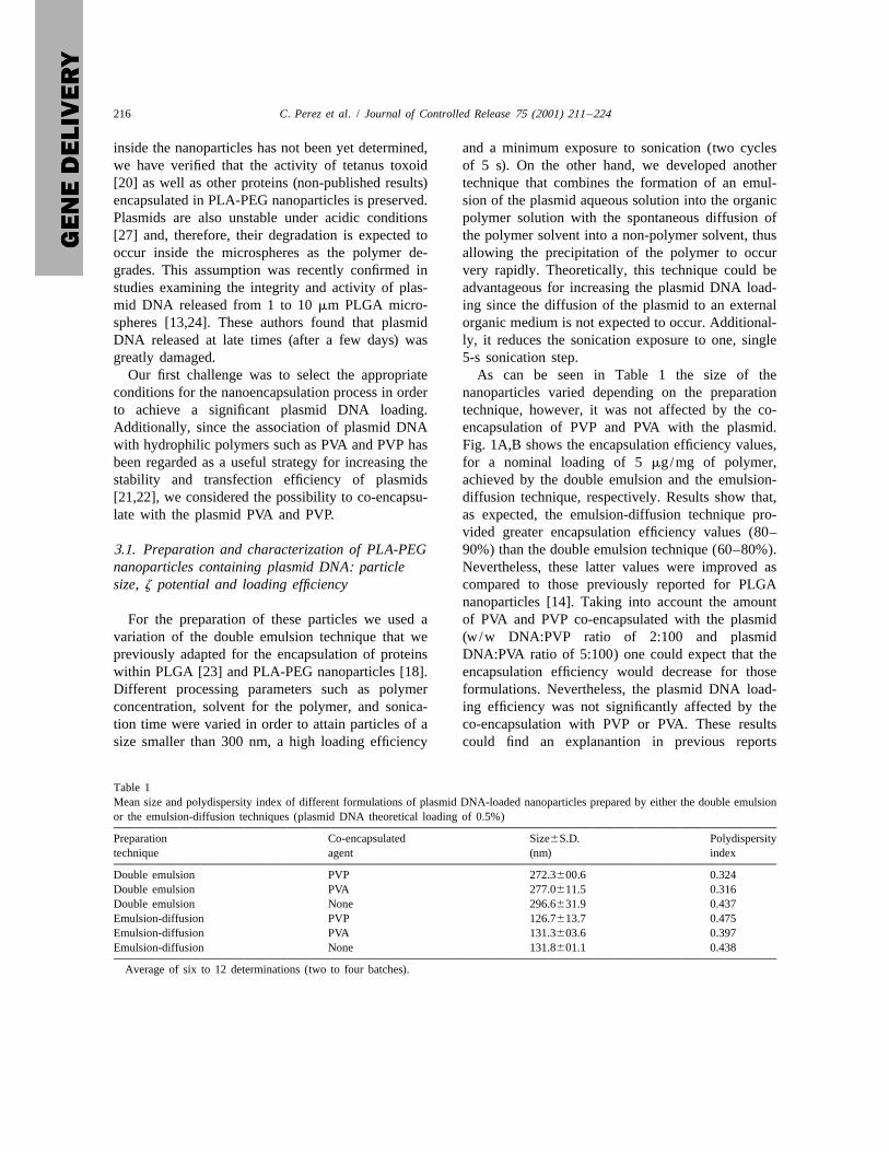
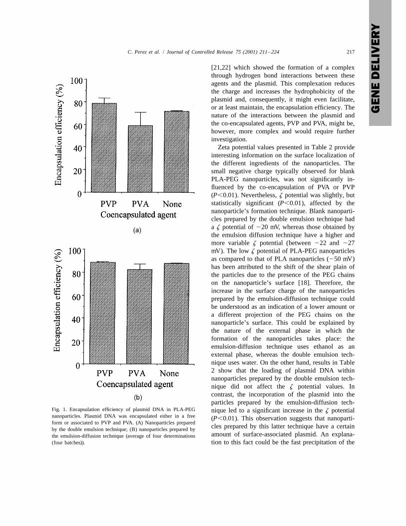
Our first challenge was to select the appropriate As can be seen in Table 1 the size of theconditions for the nanoencapsulation process in order nanoparticles varied depending on the preparationto achieve a significant plasmid DNA loading. technique, however, it was not affected by the co-Additionally, since the association of plasmid DNA encapsulation of PVP and PVA with the plasmid.with hydrophilic polymers such as PVA and PVP has Fig. 1A,B shows the encapsulation efficiency values,been regarded as a useful strategy for increasing the for a nominal loading of 5 mg/mg of polymer,stability and transfection efficiency of plasmids achieved by the double emulsion and the emulsion-[21,22], we considered the possibility to co-encapsu- diffusion technique, respectively. Results show that,late with the plasmid PVA and PVP. as expected, the emulsion-diffusion technique pro-
vided greater encapsulation efficiency values (80–3.1. Preparation and characterization of PLA-PEG 90%) than the double emulsion technique (60–80%).nanoparticles containing plasmid DNA: particle Nevertheless, these latter values were improved assize, z potential and loading efficiency compared to those previously reported for PLGA
nanoparticles [14]. Taking into account the amountFor the preparation of these particles we used a of PVA and PVP co-encapsulated with the plasmid
variation of the double emulsion technique that we (w/w DNA:PVP ratio of 2:100 and plasmidpreviously adapted for the encapsulation of proteins DNA:PVA ratio of 5:100) one could expect that thewithin PLGA [23] and PLA-PEG nanoparticles [18]. encapsulation efficiency would decrease for thoseDifferent processing parameters such as polymer formulations. Nevertheless, the plasmid DNA load-concentration, solvent for the polymer, and sonica- ing efficiency was not significantly affected by thetion time were varied in order to attain particles of a co-encapsulation with PVP or PVA. These resultssize smaller than 300 nm, a high loading efficiency could find an explanantion in previous reports
Table 1Mean size and polydispersity index of different formulations of plasmid DNA-loaded nanoparticles prepared by either the double emulsionor the emulsion-diffusion techniques (plasmid DNA theoretical loading of 0.5%)
Preparation Co-encapsulated Size6S.D. Polydispersitytechnique agent (nm) index
Double emulsion PVP 272.3600.6 0.324Double emulsion PVA 277.0611.5 0.316Double emulsion None 296.6631.9 0.437Emulsion-diffusion PVP 126.7613.7 0.475Emulsion-diffusion PVA 131.3603.6 0.397Emulsion-diffusion None 131.8601.1 0.438
Average of six to 12 determinations (two to four batches).
C. Perez et al. / Journal of Controlled Release 75 (2001) 211 –224 217
[21,22] which showed the formation of a complexthrough hydrogen bond interactions between theseagents and the plasmid. This complexation reducesthe charge and increases the hydrophobicity of theplasmid and, consequently, it might even facilitate,or at least maintain, the encapsulation efficiency. Thenature of the interactions between the plasmid andthe co-encapsulated agents, PVP and PVA, might be,however, more complex and would require furtherinvestigation.
Zeta potential values presented in Table 2 provideinteresting information on the surface localization ofthe different ingredients of the nanoparticles. Thesmall negative charge typically observed for blankPLA-PEG nanoparticles, was not significantly in-fluenced by the co-encapsulation of PVA or PVP(P,0.01). Nevertheless, z potential was slightly, butstatistically significant (P,0.01), affected by thenanoparticle’s formation technique. Blank nanoparti-cles prepared by the double emulsion technique hada z potential of 220 mV, whereas those obtained bythe emulsion diffusion technique have a higher andmore variable z potential (between 222 and 227mV). The low z potential of PLA-PEG nanoparticlesas compared to that of PLA nanoparticles (250 mV)has been attributed to the shift of the shear plain ofthe particles due to the presence of the PEG chainson the nanoparticle’s surface [18]. Therefore, theincrease in the surface charge of the nanoparticlesprepared by the emulsion-diffusion technique couldbe understood as an indication of a lower amount ora different projection of the PEG chains on thenanoparticle’s surface. This could be explained bythe nature of the external phase in which theformation of the nanoparticles takes place: theemulsion-diffusion technique uses ethanol as anexternal phase, whereas the double emulsion tech-nique uses water. On the other hand, results in Table2 show that the loading of plasmid DNA withinnanoparticles prepared by the double emulsion tech-nique did not affect the z potential values. Incontrast, the incorporation of the plasmid into theparticles prepared by the emulsion-diffusion tech-
Fig. 1. Encapsulation efficiency of plasmid DNA in PLA-PEG nique led to a significant increase in the z potentialnanoparticles. Plasmid DNA was encapsulated either in a free (P,0.01). This observation suggests that nanoparti-form or associated to PVP and PVA. (A) Nanoparticles prepared
cles prepared by this latter technique have a certainby the double emulsion technique; (B) nanoparticles prepared byamount of surface-associated plasmid. An explana-the emulsion-diffusion technique (average of four determinations
(four batches)). tion to this fact could be the fast precipitation of the
218 C. Perez et al. / Journal of Controlled Release 75 (2001) 211 –224
Table 2Mean z potential values of different formulations of plasmid-loaded nanoparticles and blank nanoparticles (no plasmid encapsulated)prepared by either the double emulsion or the emulsion-diffusion techniques (plasmid DNA theoretical loading of 0.5%)
Preparation Plasmid Co-encapsulated z potential6S.D.technique encapsulated agent (mV)
Double emulsion Yes PVP 219.162.1Double emulsion No PVP 220.262.9Double emulsion Yes PVA 220.362.5Double emulsion No PVA 219.060.4Double emulsion Yes None 218.661.8Double emulsion No None 219.160.4Emulsion-diffusion Yes PVP 233.561.8Emulsion-diffusion No PVP 226.162.4Emulsion-diffusion Yes PVA 230.362.8Emulsion-diffusion No PVA 227.461.7Emulsion-diffusion Yes None 228.962.5Emulsion-diffusion No None 222.262.8
Average of six to 12 determinations (two to four batches).
polymer caused by the diffusion of the organic chain of the polymer. In fact, the encapsulationsolvent towards the external ethanolic phase. This efficiency of PLA nanoparticles prepared by theobservation was further confirmed by the fact that double emulsion technique, under the conditions hereextensive washing of the nanoparticles led to a specified, went down to 2560.4%. This hypothesis2767% loss of the associated plasmid. Consequent- was corroborated by the observed association of thely, these data indicate that the localization of the plasmid to previously formed PLA-PEG blankplasmid may vary depending on the encapsulationtechnique. Nevertheless, both techniques can beconsidered efficient for achieving high loadings ofplasmid DNA within PLA-PEG nanoparticles.
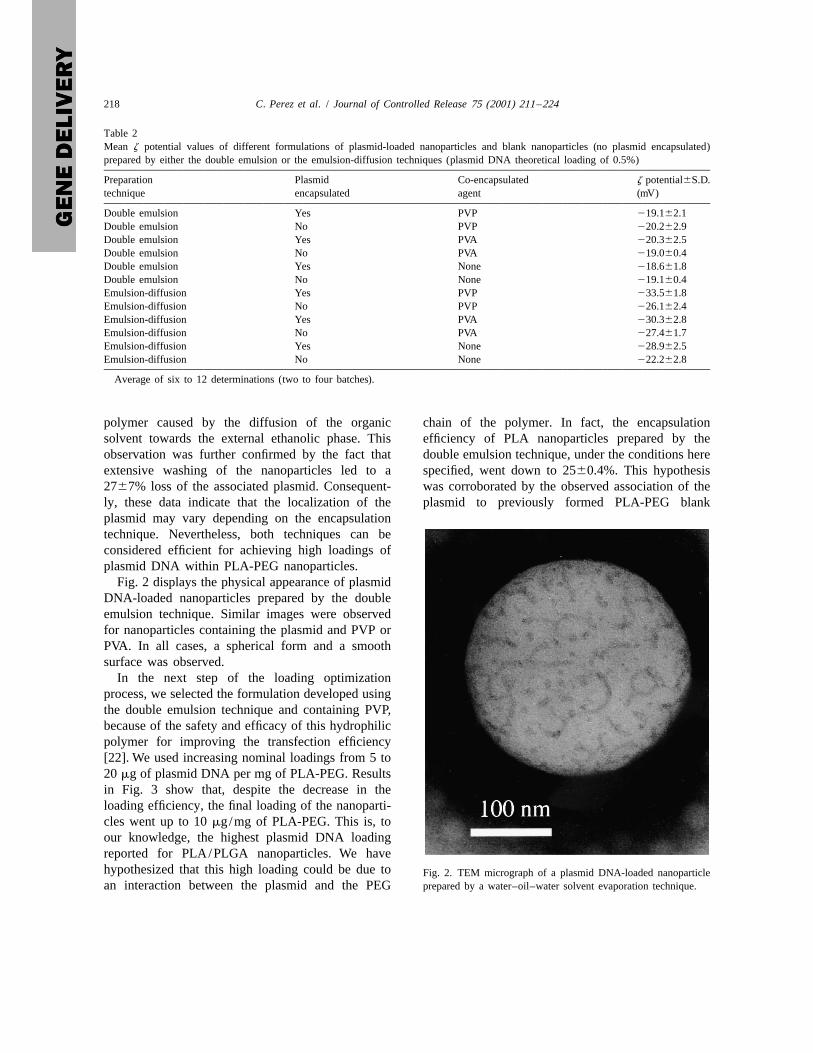
Fig. 2 displays the physical appearance of plasmidDNA-loaded nanoparticles prepared by the doubleemulsion technique. Similar images were observedfor nanoparticles containing the plasmid and PVP orPVA. In all cases, a spherical form and a smoothsurface was observed.
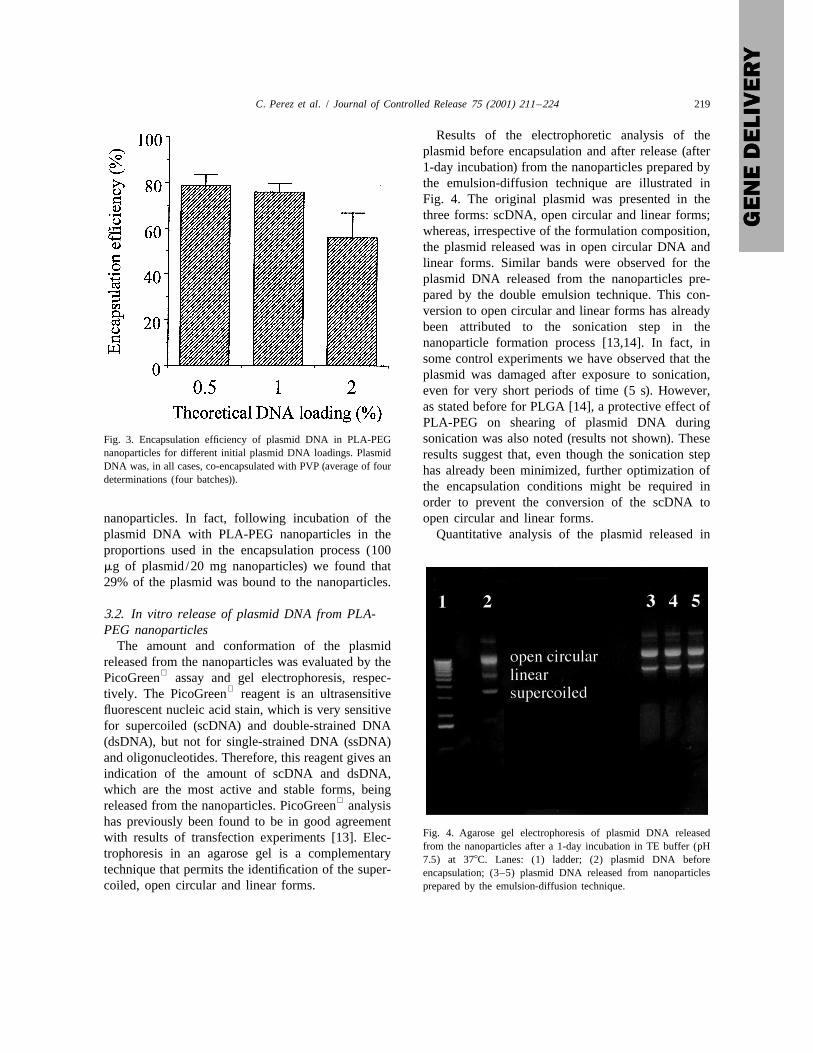
In the next step of the loading optimizationprocess, we selected the formulation developed usingthe double emulsion technique and containing PVP,because of the safety and efficacy of this hydrophilicpolymer for improving the transfection efficiency[22]. We used increasing nominal loadings from 5 to20 mg of plasmid DNA per mg of PLA-PEG. Resultsin Fig. 3 show that, despite the decrease in theloading efficiency, the final loading of the nanoparti-cles went up to 10 mg/mg of PLA-PEG. This is, toour knowledge, the highest plasmid DNA loadingreported for PLA/PLGA nanoparticles. We havehypothesized that this high loading could be due to Fig. 2. TEM micrograph of a plasmid DNA-loaded nanoparticlean interaction between the plasmid and the PEG prepared by a water–oil–water solvent evaporation technique.
C. Perez et al. / Journal of Controlled Release 75 (2001) 211 –224 219
Results of the electrophoretic analysis of theplasmid before encapsulation and after release (after1-day incubation) from the nanoparticles prepared bythe emulsion-diffusion technique are illustrated inFig. 4. The original plasmid was presented in thethree forms: scDNA, open circular and linear forms;whereas, irrespective of the formulation composition,the plasmid released was in open circular DNA andlinear forms. Similar bands were observed for theplasmid DNA released from the nanoparticles pre-pared by the double emulsion technique. This con-version to open circular and linear forms has alreadybeen attributed to the sonication step in thenanoparticle formation process [13,14]. In fact, insome control experiments we have observed that theplasmid was damaged after exposure to sonication,even for very short periods of time (5 s). However,as stated before for PLGA [14], a protective effect ofPLA-PEG on shearing of plasmid DNA duringsonication was also noted (results not shown). TheseFig. 3. Encapsulation efficiency of plasmid DNA in PLA-PEG
nanoparticles for different initial plasmid DNA loadings. Plasmid results suggest that, even though the sonication stepDNA was, in all cases, co-encapsulated with PVP (average of four has already been minimized, further optimization ofdeterminations (four batches)).
the encapsulation conditions might be required inorder to prevent the conversion of the scDNA to
nanoparticles. In fact, following incubation of the open circular and linear forms.plasmid DNA with PLA-PEG nanoparticles in the Quantitative analysis of the plasmid released inproportions used in the encapsulation process (100mg of plasmid/20 mg nanoparticles) we found that29% of the plasmid was bound to the nanoparticles.
3.2. In vitro release of plasmid DNA from PLA-PEG nanoparticles
The amount and conformation of the plasmidreleased from the nanoparticles was evaluated by the
PicoGreen assay and gel electrophoresis, respec-tively. The PicoGreen reagent is an ultrasensitive
fluorescent nucleic acid stain, which is very sensitivefor supercoiled (scDNA) and double-strained DNA(dsDNA), but not for single-strained DNA (ssDNA)and oligonucleotides. Therefore, this reagent gives anindication of the amount of scDNA and dsDNA,which are the most active and stable forms, being
released from the nanoparticles. PicoGreen analysishas previously been found to be in good agreement
Fig. 4. Agarose gel electrophoresis of plasmid DNA releasedwith results of transfection experiments [13]. Elec-from the nanoparticles after a 1-day incubation in TE buffer (pH
trophoresis in an agarose gel is a complementary 7.5) at 378C. Lanes: (1) ladder; (2) plasmid DNA beforetechnique that permits the identification of the super- encapsulation; (3–5) plasmid DNA released from nanoparticlescoiled, open circular and linear forms. prepared by the emulsion-diffusion technique.
220 C. Perez et al. / Journal of Controlled Release 75 (2001) 211 –224
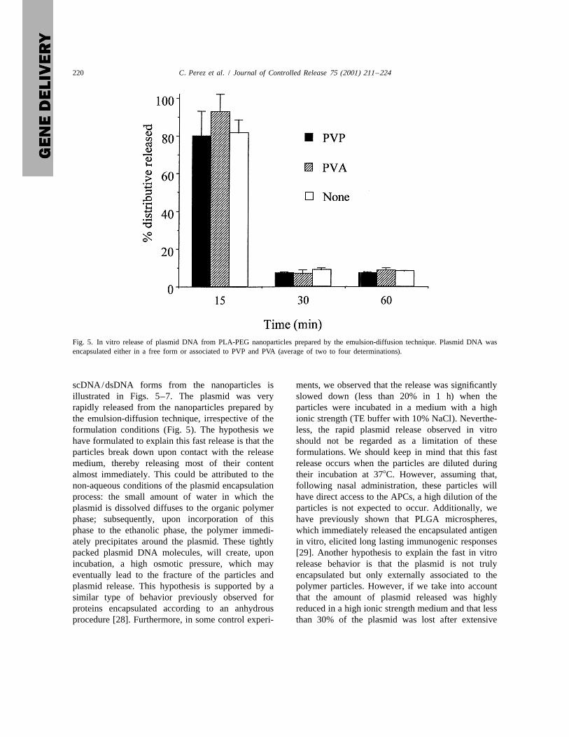
Fig. 5. In vitro release of plasmid DNA from PLA-PEG nanoparticles prepared by the emulsion-diffusion technique. Plasmid DNA wasencapsulated either in a free form or associated to PVP and PVA (average of two to four determinations).
scDNA/dsDNA forms from the nanoparticles is ments, we observed that the release was significantlyillustrated in Figs. 5–7. The plasmid was very slowed down (less than 20% in 1 h) when therapidly released from the nanoparticles prepared by particles were incubated in a medium with a highthe emulsion-diffusion technique, irrespective of the ionic strength (TE buffer with 10% NaCl). Neverthe-formulation conditions (Fig. 5). The hypothesis we less, the rapid plasmid release observed in vitrohave formulated to explain this fast release is that the should not be regarded as a limitation of theseparticles break down upon contact with the release formulations. We should keep in mind that this fastmedium, thereby releasing most of their content release occurs when the particles are diluted duringalmost immediately. This could be attributed to the their incubation at 378C. However, assuming that,non-aqueous conditions of the plasmid encapsulation following nasal administration, these particles willprocess: the small amount of water in which the have direct access to the APCs, a high dilution of theplasmid is dissolved diffuses to the organic polymer particles is not expected to occur. Additionally, wephase; subsequently, upon incorporation of this have previously shown that PLGA microspheres,phase to the ethanolic phase, the polymer immedi- which immediately released the encapsulated antigenately precipitates around the plasmid. These tightly in vitro, elicited long lasting immunogenic responsespacked plasmid DNA molecules, will create, upon [29]. Another hypothesis to explain the fast in vitroincubation, a high osmotic pressure, which may release behavior is that the plasmid is not trulyeventually lead to the fracture of the particles and encapsulated but only externally associated to theplasmid release. This hypothesis is supported by a polymer particles. However, if we take into accountsimilar type of behavior previously observed for that the amount of plasmid released was highlyproteins encapsulated according to an anhydrous reduced in a high ionic strength medium and that lessprocedure [28]. Furthermore, in some control experi- than 30% of the plasmid was lost after extensive
C. Perez et al. / Journal of Controlled Release 75 (2001) 211 –224 221
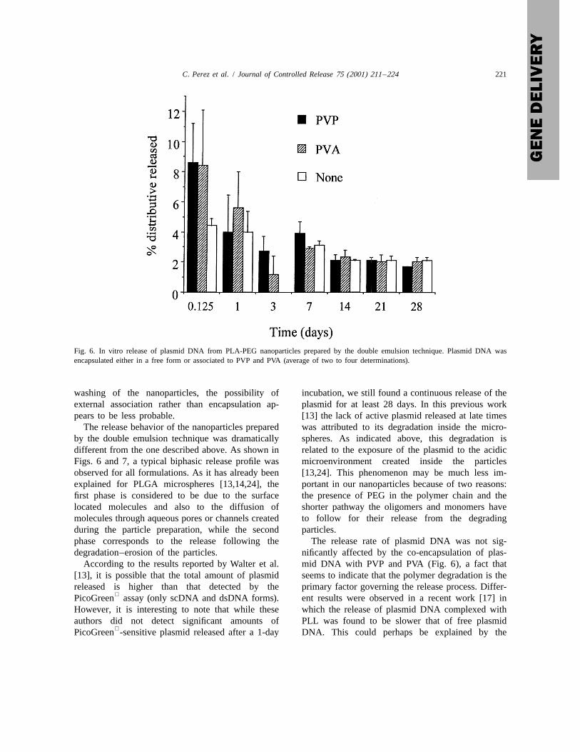
Fig. 6. In vitro release of plasmid DNA from PLA-PEG nanoparticles prepared by the double emulsion technique. Plasmid DNA wasencapsulated either in a free form or associated to PVP and PVA (average of two to four determinations).
washing of the nanoparticles, the possibility of incubation, we still found a continuous release of theexternal association rather than encapsulation ap- plasmid for at least 28 days. In this previous workpears to be less probable. [13] the lack of active plasmid released at late times
The release behavior of the nanoparticles prepared was attributed to its degradation inside the micro-by the double emulsion technique was dramatically spheres. As indicated above, this degradation isdifferent from the one described above. As shown in related to the exposure of the plasmid to the acidicFigs. 6 and 7, a typical biphasic release profile was microenvironment created inside the particlesobserved for all formulations. As it has already been [13,24]. This phenomenon may be much less im-explained for PLGA microspheres [13,14,24], the portant in our nanoparticles because of two reasons:first phase is considered to be due to the surface the presence of PEG in the polymer chain and thelocated molecules and also to the diffusion of shorter pathway the oligomers and monomers havemolecules through aqueous pores or channels created to follow for their release from the degradingduring the particle preparation, while the second particles.phase corresponds to the release following the The release rate of plasmid DNA was not sig-degradation–erosion of the particles. nificantly affected by the co-encapsulation of plas-
According to the results reported by Walter et al. mid DNA with PVP and PVA (Fig. 6), a fact that[13], it is possible that the total amount of plasmid seems to indicate that the polymer degradation is thereleased is higher than that detected by the primary factor governing the release process. Differ-
PicoGreen assay (only scDNA and dsDNA forms). ent results were observed in a recent work [17] inHowever, it is interesting to note that while these which the release of plasmid DNA complexed withauthors did not detect significant amounts of PLL was found to be slower that of free plasmid
PicoGreen -sensitive plasmid released after a 1-day DNA. This could perhaps be explained by the
222 C. Perez et al. / Journal of Controlled Release 75 (2001) 211 –224
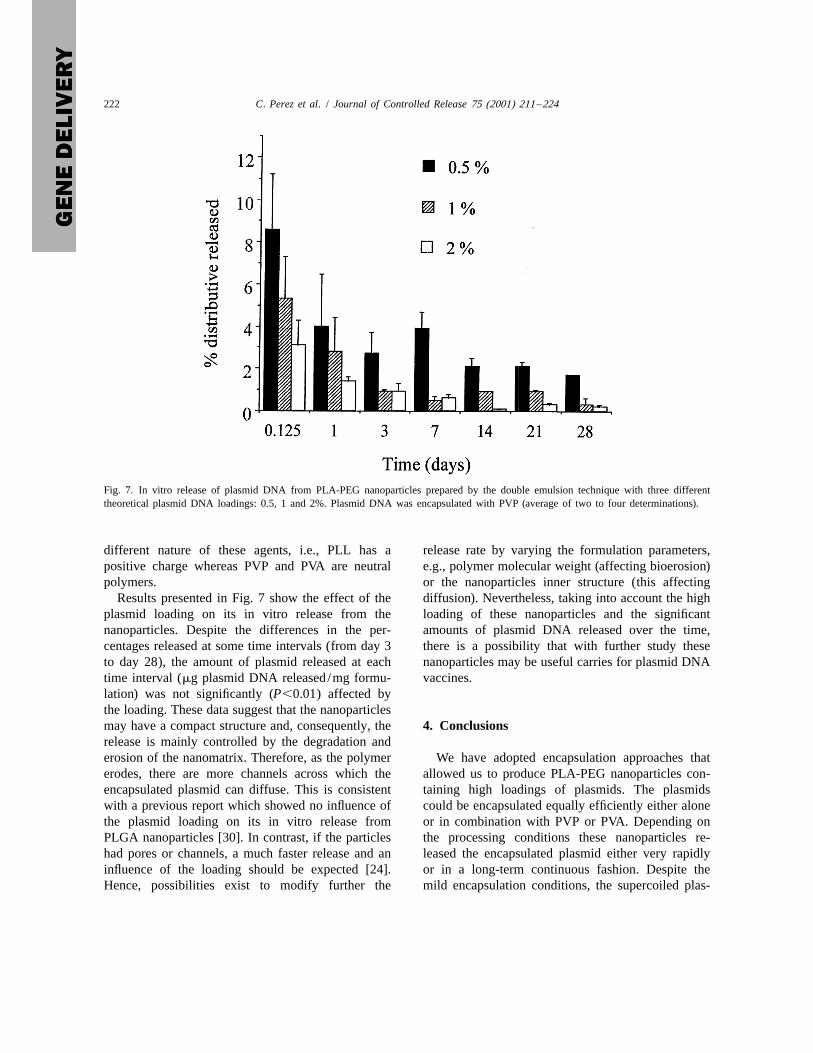
Fig. 7. In vitro release of plasmid DNA from PLA-PEG nanoparticles prepared by the double emulsion technique with three differenttheoretical plasmid DNA loadings: 0.5, 1 and 2%. Plasmid DNA was encapsulated with PVP (average of two to four determinations).
different nature of these agents, i.e., PLL has a release rate by varying the formulation parameters,positive charge whereas PVP and PVA are neutral e.g., polymer molecular weight (affecting bioerosion)polymers. or the nanoparticles inner structure (this affecting
Results presented in Fig. 7 show the effect of the diffusion). Nevertheless, taking into account the highplasmid loading on its in vitro release from the loading of these nanoparticles and the significantnanoparticles. Despite the differences in the per- amounts of plasmid DNA released over the time,centages released at some time intervals (from day 3 there is a possibility that with further study theseto day 28), the amount of plasmid released at each nanoparticles may be useful carries for plasmid DNAtime interval (mg plasmid DNA released /mg formu- vaccines.lation) was not significantly (P,0.01) affected bythe loading. These data suggest that the nanoparticlesmay have a compact structure and, consequently, the 4. Conclusionsrelease is mainly controlled by the degradation anderosion of the nanomatrix. Therefore, as the polymer We have adopted encapsulation approaches thaterodes, there are more channels across which the allowed us to produce PLA-PEG nanoparticles con-encapsulated plasmid can diffuse. This is consistent taining high loadings of plasmids. The plasmidswith a previous report which showed no influence of could be encapsulated equally efficiently either alonethe plasmid loading on its in vitro release from or in combination with PVP or PVA. Depending onPLGA nanoparticles [30]. In contrast, if the particles the processing conditions these nanoparticles re-had pores or channels, a much faster release and an leased the encapsulated plasmid either very rapidlyinfluence of the loading should be expected [24]. or in a long-term continuous fashion. Despite theHence, possibilities exist to modify further the mild encapsulation conditions, the supercoiled plas-
C. Perez et al. / Journal of Controlled Release 75 (2001) 211 –224 223
saccharide copolymer and poly(D,L-lactic acid), Bioconjug.mid was converted into open circular and linearChem. 8 (1997) 735–742.forms. Therefore, the next step in our work will be to
[12] D. Luo, K. Woodrow-Mumford, N. Belcheva, W.M.optimize the stability of the encapsulated plasmid Saltzman, Controlled DNA delivery systems, Pharm. Res. 16and to evaluate the potential of these new plasmid (1999) 1300–1308.
[13] E. Walter, K. Moelling, J. Pavlovic, H.P. Merkle, Microen-DNA delivery systems for eliciting an immunecapsulation of DNA using poly(DL-lactide-co-glycolide):response following intranasal administration.stability issues and release characteristics, J. Control. Release61 (1999) 361–374.
[14] D. Wang, D.R. Robinson, G.S. Kwon, J. Samuel, Encapsula-tion of plasmid DNA in biodegradable poly(D,L-lactic-co-Acknowledgementsglycolic acid) microspheres as a novel approach for immuno-gene delivery, J. Control. Release 57 (1999) 9–18.
This work has been supported by grants from the [15] S. Ando, D. Putnam, D.W. Pack, R. Langer, PLGA micro-spheres containing plasmid DNA: preservation of super-Fullbright Commission (Ref. 99112) and the Xuntacoiled DNA via cryopreparation and carbohydrate stabiliza-de Galicia (Ref. PGIDT00BIO20301PR) (Spain).tion, J. Pharm. Sci. 88 (1999) 126–130.
[16] Y. Capan, B.H. Woo, S. Gebrekidan, S. Ahmed, P.P. DeLuca, Influence of formulation parameters on the characteris-tics of poly(D,Llactide-co-glycolide) microspheres containingReferencespoly(L-lysine) complexed plasmid DNA, J. Control. Release60 (1999) 279–286.
[1] K.E. Shroff, L.R. Smith, Y. Baine, T.J. Higgins, Potential for [17] Y. Capan, B.H. Woo, S. Gebrekidan, S. Ahmed, P.P. Deplasmid DNAs as vaccines for the new millenium, Pharm. Luca, Preparation and characterization of poly(D,Llactide-co-Sci. Tech. Today 5 (1999) 205–212. glycolide) microspheres for controlled release of poly(L-
[2] T. Friedman, Overcoming obstacles in gene therapy, Sci. lysine) complexed plasmid DNA, Pharm. Res. 16 (1999)Am. 6 (1997) 96–101. 509–513.
[3] K. Kawabata, Y. Takkura, M. Hashida, The fate of plasmid ´[18] M. Tobio, R. Gref, A. Sanchez, R. Langer, M.J. Alonso,DNA after intravenous injection in mice: involvement of Stealth PLA-PEG nanoparticles as protein carriers for nasalscavenger receptors in its hepatic uptake, Pharm. Res. 12 administration, Pharm. Res. 15 (1998) 270–275.(1995) 825–839. [19] M. Tobio, A. Sanchez, A.Vila, I. Soriano, C. Evora, J.L.Vila
[4] J.A. Wolff, R.W. Malone, P. Williams, W. Chong, G. Aesadi, Jato, M.J. Alonso, The role of PEG on the stability inA. Jani, P.L. Felgner, Direct gene transfer into mouse muscle digestive fluids and in vivo fate of PEG-PLA nanoparticlesin vivo, Science 247 (1990) 1465–1468. following oral administration, Colloids Surfaces B. Biointer-
[5] E. Mathiowitz, J.S. Jacob, Y.S. Jong, G.P. Carino, D.E. faces 18 (2000) 315–323.Chickering, P. Chaturvedi, C.A. Santos, K. Vijayaraghavan, [20] A.Vila, A. Sanchez, M. Tobio, I. Soriano, C. Evora, J.L.VilaS. Montgomery, M. Bassett, C. Morell, Biologically erodable Jato, M.J. Alonso, PLA-PEG nanoparticles as new vaccinemicrospheres as potential oral drug delivery systems, Nature delivery vehicles for nasal administration: systemic and386 (1997) 410–414. mucosal immune responses to tetanus toxoid (in prepara-
[6] K. Roy, H.Q. Mao, S.K. Huang, K.W. Leong, Oral gene tion).delivery with chitosan-DNA nanoparticles generates im- [21] R.J. Mumper, J.D. Duguid, K. Anwer, M.K. Barron, H.munologic protection in a murine model of peanut allergy, Nitta, A.P. Rolland, Polyvinyl derivatives as novel interactiveNat. Med. 5 (1999) 387–391. polymers for controlled gene delivery to muscle, Pharm. Res.
[7] D. Luo, M. Saltzman, Synthetic DNA delivery systems, Nat. 13 (1996) 701–709.Biotech. 18 (2000) 33–37. [22] J.R. Mumper, J. Wang, S.L. Klakamp, H. Nitta, K. Anwer, F.
[8] D.H. Jones, S. Corris, S. McDonald, J.C.S. Clegg, G.H. Tagliaferry, A.P. Rolland, Protective interactive noncondens-Farrar, Poly(DL-lactide-coglycolide)-encapsulated plasmid ing (PINC) polymers for enhanced plasmid distribution andDNA elicits systemic and mucosal antibody responses to expression in rat skeletal muscle, J. Control. Release. 52encoded protein after oral administration, Vaccine 15 (1997) (1998) 191–203.814–817. [23] D. Blanco, M.J. Alonso, Development and characterization
[9] M.L. Hedley, J.C. Curley, R. Urba, Microspheres containing of protein-loaded poly(lactide-co-glycolide) nanoparticles,plasmid-encoded antigens elicit cytotoxic T-cell responses, Eur. J. Pharm. Biopharm. 43 (1997) 285–294.Nat. Med. 14 (1998) 365–368. [24] A.M. Tinsley-Brwon, R. Fretwell, S.L. Davis, G.H. Farrar,
[10] M. Singh, M. Brioness, G. Ott, D. O’Hagan, Cationic Formulation of poly(D,L-lactic-co-glycolic acid) microparti-microparticles: a potent delivery system for DNA vaccines, cles for rapid plasmid DNA delivery, J. Control. Release 66Proc. Natl. Acad. Sci. USA 97 (2000) 811–816. (2000) 229–241.
[11] A. Maruyama, T. Ishihara, J. S Kim, S.W. Kim, T. Akaike, [25] M. Tobio, M.J. Alonso, Study of the inactivation process ofNanoparticle DNA carrier with poly(L-lysine) grated poly- the tetanus toxoid in contact with poly(lactic acid /glycolic
224 C. Perez et al. / Journal of Controlled Release 75 (2001) 211 –224
acid) degrading microspheres, STP Pharma Sci. 8 (1998) [28] S. Shwendeman, M. Tobio, M. Joworowicz, M.J. Alonso, R.303–310. Langer, New strategies for the microencapsulation of tetanus
[26] K. Fu, D.W. Pack, A.M. Klivanov, R. Langer, Visual vaccine, J. Microencap. 15 (1998) 299–318.evidence of acidic environment within degrading poly(lactic- [29] M. Tobio, S.P. Schendeman, Y. Guo, J. McIver, R. Langer,co-glycolic acid) (PLGA) microspheres, Pharm. Res. 17 M.J. Alonso, Improved immunogenicity of a core-coated(2000) 100–106. tetanus toxoid delivery system, Vaccine 18 (2000) 618–622.
[27] D.E. Berstrom, P. Zhang, N. Paul, dsDNA stability depen- [30] H. Cohen, G. Golomb, R.J. Levy, Gene delivery by polymer-dence on pH and salt concentration, Biotechniques 24 (1998) encapsulated DNA, Proc. Int. Symp. Control. Release Bioact.992–994. Mater. 26 (1999) 1138–1139.