Biomimetic poly(glycerol sebacate) (PGS) membranes for cardiac patch application

Encyclopedia of Biomedical Polymers and Polymeric Biomaterials DOI: 10.1081/E-EBPP-120051058Copyright © 2014 by Taylor & Francis. All rights reserved. 1
Poly(glycerol sebacate) Patch: Cardiac Embryonic Stem Cell Delivery
Qizhi ChenDepartment of Materials Engineering, Monash University, Clayton, Australia
AbstractHeart patch, a tissue engineering strategy, combines isolated functional cardiomyocytes and a biodegradable biomaterial to repair diseased heart muscle. Nowadays, there are increasing research efforts in the development of elastomeric biomaterials for use in heart muscle engineering. Poly(glycerol sebacate) (PGS) is one of most promising candidates. This entry provides a comprehensive update on the development of PGS, including synthesis, properties, bioevaluation, and in vivo application of this elastomer. This entry is organized as follows: A medical background of the heat patch strategy is introduced fi rst (Introduction section); followed by a brief review on biomaterials used in myocardial tissue engineering (Biomaterials in myocardial tissue engineering section); the Poly(glycerol sebacate) section presents a systematic review on PGS – this section starts with polymerization and fabrication of PGS elastomer and goes on reviews on the biocompatibility, biodegradability, and mechanical properties of this polymer; and then the in vivo applications of PGS heart patch. At the end of this entry, the major achievements and remaining challenges in the development of PGS heart patch are summarized (Applications of PGS as heart patch section).
INTRODUCTION
Heart disease is the leading cause of death and disability worldwide, accounting for nearly 40% of human mortality.[1] Heart tissue engineering, however, is one of the least explored areas in the rapidly evolving fi eld of tissue engineering. Myocardial tissue engineering for regeneration represents one of the most exciting areas of development, but it is also an undertaking involving multiple challenges.
Heart failure is caused by a variety of underlying dis-eases. The single most common cause of left-sided car-diac failure is ischemic heart disease following acute myocardial infarct (MI, also known as a heart attack in common parlance). MI typically results in massive and permanent cell loss. The weakening of the collagen extra-cellular matrix results in myocardial thinning and ven-tricular dilation. The enlargement in ventricular volume leads to progressive structural and functional changes (called ventricular remodeling). Ventricular remodeling is initially compensatory, but subsequently adds further ineffi ciency to the compromised mechanical pumping of the ventricular muscle. Such ineffi ciency predisposes the subject to end-stage heart failure, a condition in which the heart cannot pump suffi cient amounts of blood to meet the metabolic requirements of the body.[2] In short, massive, permanent cell death in heart muscle and the subsequent (negative) remodeling of the myocardium are key pathological mechanisms leading to heart failure after MI.
Pharmacological therapy represents the current standard treatment for patients with heart disease. Interventional therapies, such as bypass surgery or implantation of pacing devices, are receiving widespread application for patients with marked symptoms despite pharmacological therapy.[3] However, both drug and interventional therapies cannot adequately prevent disease progression to end stage.[4] Eventually, heart transplantation is currently the fi nal treat-ment option for end-stage heart failure. Owing to the lack of organ donors and complications associated with immuno-suppressive treatments, new strategies to repair the injured heart are needed.[5] Currently two tissue engineering strate-gies are under intensive investigation: cell-based therapy (Fig. 1A) and passive left ventricular restraint (Fig. 1B). These strategies aim to address, respectively, above- mentioned two pathological consequences of MI, i.e., the massive cell loss and negative remodeling associated with disease progression.
However, cell therapy and ventricular restraint strate-gies both have limitations. Left ventricular restraint is a nonradical treatment, as a mechanical support cannot com-pensate cell loss at the injured part of the heart wall. Indeed, the primary aim of left ventricular restraint is to halt or slow down the progress of heart failure, thus offering a bridge to heart transplantation.[6] Cell-based therapy, on the other hand, represents a radical procedure. In this approach, healthy, functional cells are injected into the infarcted region via the epicardium, coronary arteries, coronary veins, or endocardium (Fig. 1A). Studies have shown that
2 Poly(glycerol sebacate) Patch: Cardiac Embryonic Stem Cell Delivery
transplantation of fetal cardiomyocytes can result in the replacement of diseased myocardium with healthy myo-cardium.[7] However, effi ciency is very low using current methods due to a major loss of cells from the heart follow-ing delivery. Moreover, due to the elevated fi lling pressures in the beating heart, cardiac muscle slippage and thus nega-tive remodeling of the infarct region occurs if a mechanical support is not used.
The limitations of cell therapy and left ventricular restraint prompt a hypothesis of heart patch (Fig. 2). In this approach, a heart patch, which is made of a poly-meric material and sized to fi t the infarct area, is surgi-cally implanted. In conjunction, temporary bypass surgery is applied to transiently relieve pressure on the healing site. The heart patch serves the dual purpose of both cell-delivery vehicle and mechanical support. Important benefi ts of the proposed heart patch approach include the following:
1. The promotion of healing of the infarcted myocardium by the implanted cells.
2. The effi ciency of cell retention is signifi cantly improved compared with currently applied injection methods via the epicardium, coronary arteries, coro-nary veins, or endocardium.
3. Heart wall support is increased and stress is reduced, thus preventing further left ventricular dilation, one of the more important pathophysiological mechanisms underlying the clinical cause of progressive heart fail-ure. According to previous work, the addition of an endoventricular patch to the border-zone decreases heart wall stress and improves cardiac remodeling,[8] but cell delivery has been not applied in these studies.
4. Using a degradable heart patch, long-term detrimental effects associated with persisting foreign substances within the body and the need for a second operation to remove a nondegradable device can be obviated.
BIOMATERIALS IN MYOCARDIAL TISSUE ENGINEERING
The polymeric biomaterials used in the fi eld of myocardial tissue engineering include natural polymers (e.g., collagen) and synthetic thermoplastics [e.g., polyester-based poly(glycolide) and poly(L-lactic acid) (PLA)]. Major drawbacks associated with the use of naturally occurring polymers, however, are their poor mechanical performance and concerns regarding immunogenicity. To engineer con-stantly and cyclically beating myocardial tissue, the bio-material used should show long-term elasticity. These mechanical properties cannot be reliably mimicked at pres-ent using nonporous thermoplastic polymers (except for knitted networks, which are inappropriate because they cannot effi ciently deliver cells) because they undergo deformation and failure when exposed to long-term cyclic strain. The limitations of these early applied polymers have
Fig. 1 (A) Cell-based therapy, (B) cardiac support device by Acorn CorCap™. (http://www.sciencedaily.com/releases/)
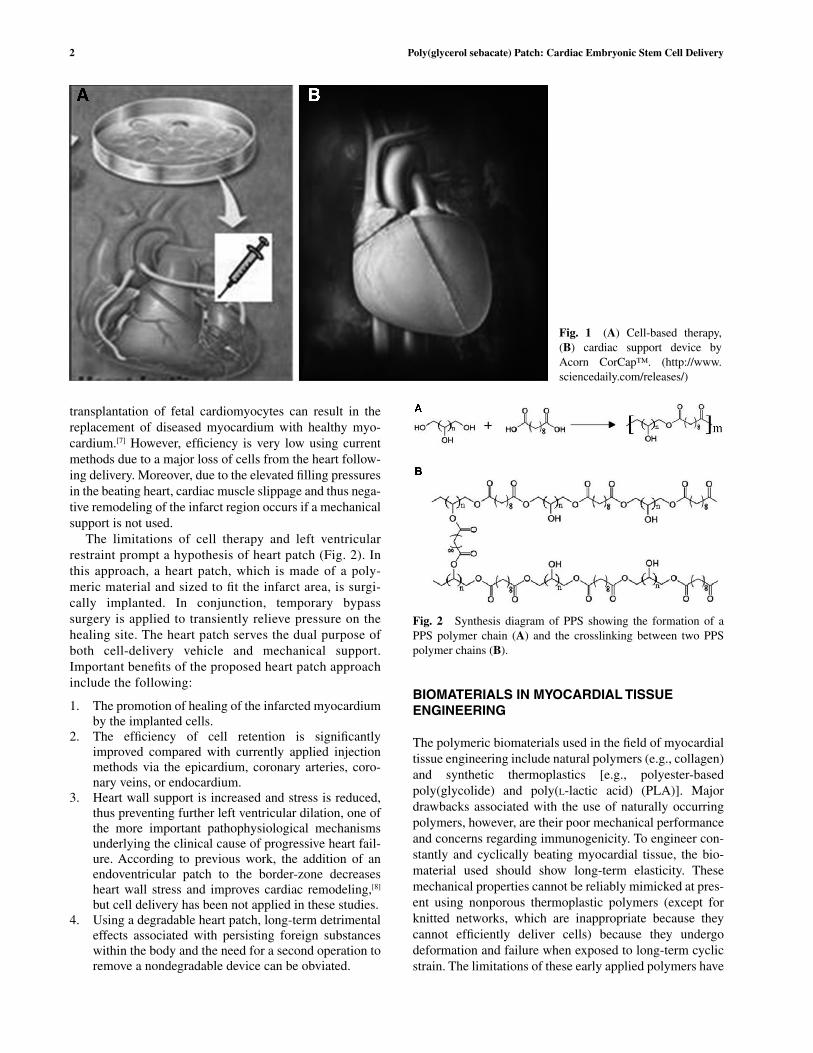
Fig. 2 Synthesis diagram of PPS showing the formation of a PPS polymer chain (A) and the crosslinking between two PPS polymer chains (B).

Poly(glycerol sebacate) Patch: Cardiac Embryonic Stem Cell Delivery 3
prompted researchers to turn to elastomers, one of which is poly(glycerol sebacate) (PGS), a member of a family of crosslinked polyester elastomers poly(polyol sebacate) (PPS), developed for medical applications by Langer’s group at MIT.[9,10]
POLY(GLYCEROL SEBACATE)
Synthesis and Processing
PGS is synthesized from a glycerol (an alcohol containing three hydroxyl groups) and sebacic acid, a dicarboxylic acid with the structure (HCOO)(CH
2)
8(COOH) (Fig. 2A).
Usually the synthesis of PGS involves polycondensation (esterifi cation) between carboxylic acids and alcohol groups (Fig. 2B). The primary –OH groups at the two ends of polyol monomers react fi rst with the carboxylic acid groups, forming polymer chains at the early stage of polymerization (Fig. 2A). At later stages there is a defi -ciency of primary alcohol groups in the reacting system so that the secondary –OH groups (toward the middle of the polyol monomers) then react with the –COOH groups to form ester links and eventually crosslinks between the polymer chains (Fig. 2B).
The synthesis of PGS is typically conducted in two steps:[11,12] initially prepolymerization and then gelation and further crosslinking. The prepolymer is formed by heating a mixture of glycerol and sebacic acid under a purge of inert gas through the reaction chamber to prevent reactant oxida-tion, performed at atmospheric pressure to minimize evapo-ration of the volatile polyol monomers and their azeotrope with water. The prepolymerization conditions vary from 120°C to 130°C for 1–24 hr, depending on the polyol mono-mer. The combination of temperature and time of prepoly-merization is such that the esterifi cation reaction is preferentially between the primary hydroxyl groups and the carboxylic acids, thus forming polymer chains (Fig. 2A), with few secondary –OH groups forming branching sites. As such, the non-crosslinked prepolymer can fl ow freely at approximately 50°C or dissolve into solvents, such as meth-anol, acetone, and tetrahydrofuran (THF).
The prepolymerization is then followed by further cur-ing for several days at the same temperature as the fi rst step but under vacuum. During this second step, the now more numerous secondary alcohol groups react with the carbox-ylic acids, resulting in crosslinking between polymer chains (Fig. 2B). Like other chemically crosslinked poly-mers, the crosslinked PGS is neither soluble nor meltable, however, the prepolymer of PGS (polymerized but non-crosslinked) can be processed into various shapes by melt-ing approximately at 50°C or dissolved in organic solvents such as 1,3-dioxolane, THF, N,N-dimethylformamide, iso-propanol, or ethanol prior to its gelation.[9]
The processing of chemically crosslinked elastomers is challenging. A major technical hurdle is that these
polymers cannot dissolve into any solvents for electrospin-ning, e.g., once crosslinked, and any fi bers spun from non-crosslinked prepolymers would be melted in subsequent crosslink treatment. Chen and co-workers addressed this issue using a novel nanofi ber fabrication technique: core/shell electrospinning (Fig. 3). In this technique, the core is fed with a PGS non-crosslinked prepolymer, and the shell is fed with a thermoplastic. During the crosslinking pro-cess, the solid thermoplastic shell maintains the tubular shape, whereas the melted PPS prepolymer inside the tubes undergoes a crosslink reaction. So far, PGS has been syn-thesized into sheets with a printed microstructure (by lithography),[13] formed into 3D porous scaffolds (by a salt leaching technique)[14,15] and spun into fi bers.[16] Most recently, researchers successfully created a muscle-like fi brous sheet with PGS using a core/shell electrospinning (Fig. 4).[16] This new process has produced a porous,
Fig. 3 Core/shell spinning.
Fig. 4 Core/shell electrospun PGS/PLLA fi brous mesh (inset: the core/shell structure of three fi bers).Source: Reprinted from Xu et al.,[17] Copyright 2013, with permission from Elsevier.

4 Poly(glycerol sebacate) Patch: Cardiac Embryonic Stem Cell Delivery
elastomeric 3D scaffold that mechanically matches the stiffness of heart tissue.[17]
Biocompatibility of PGS
Glycerol has been used in the food industry. Sebacic acid has been used as plasticizers, aromatics, antiseptics, cos-metics, and drug coatings. Glycerol and sebacic acid are also both endogenous monomers found in human metabo-lites.[18–20] Hence, PGS generally has little toxicity to the body tissues,[21] with the highly crosslinked PGS showing excellent cytocompatibility in vitro (Fig. 5).
Although the majority of in vitro data demonstrate that PGS has a good compatibility, there are some reports on the toxic effects of PGS on cells.[11] Apparently, the degree of cytotoxicity is associated with the crosslink density. For instance, soft PGS prepared from equimolar monomers and crosslinked for a short time was shown to be toxic to mouse fi broblasts, whereas the PGS of the same molar ratio but nearly fully crosslinked demonstrated excellent cytocompatibility.[22–24] Under the same synthesis condi-tions, a high crosslink density can also be achieved by modifi cation of molar ratio of the glycerol and sebacic acid, such as in PGS (2:3).[25] It is possible that a highly crosslinked network degrades via hydrolysis of ester bonds more slowly, and thus the concentration of potentially toxic degradation products leached into the medium environ-ment remains suffi ciently low to prevent signifi cant cell death.
Highly crosslinked PGS tend to have compromised mechanical properties, such as brittleness. Hence there is a
necessity to produce fl exible PGS of low crosslink density; however, a balance between good cytocompatibility and compliance simultaneously in a pure PGS polymer is tech-nically diffi cult. To improve cytocompatibility of soft PGS, as well as other properties such as the rapid degradation rate and suitable mechanical strength, PGS has been copo-lymerized with other polyesters,[26,27] or made as a compos-ite with bioceramics. [23,24]
Biodegradation of PGS
Polymers containing hydrolysable bonds (e.g., ester bonds) can be degraded by hydrolysis, a reaction that can be cata-lyzed by biological enzymes. PGS is one such degradable polyester system. In an in vitro study,[9] PGS was reported to degrade up to 11–23% (in terms of weight loss) after 60 days agitation in PBS at 37°C. Similar degradation kinet-ics was also reported by Liang and co-workers, who found that the weight loss of PGS and PGS/Bioglass® composites was approximately 10–25% after 60 days incubation in a standard tissue culture medium.[24] However, the above in vitro degradation rates of PGS-based materials are inconsis-tent with the degradation kinetics of PGS in vivo. Wang and co-workers have reported that PGS is completely resorbed after 60 days implantation in rats.[21] This comparatively faster degradation rate of PGS in vivo was also reported by a research group from Imperial College London, who used PGS sheets as a pericardial heart patch.[28] They found that the PGS patch was completely resorbed after 6 weeks. These examples of in vivo degradation indicate that aque-ous enzymatic action, combined with dynamic tissue
Fig. 5 Cytotoxicity of the PGS and poly(xylitol sebacate) (PXS), determined by measuring the release of lactate dehydrogenase (LDH) after 2 days culture. The positive control group was PDLLA. The percentages of dead cells were signifi cantly higher in the cultures con-taining the extracts of PGS and PXS cured for 2 days than those of other fi ve cultures (p < 0.001), whereas the PGS and PXS specimens cured for longer time (4 or 7 days, respectively) did not show signifi cant differences from either negative or positive control (PDLLA) group (p > 0.05).Source: Reprinted from Li et al.,[11] Copyright 2013, with permission from John Wiley and Sons.

Poly(glycerol sebacate) Patch: Cardiac Embryonic Stem Cell Delivery 5
movements and vascular perfusion, might enhance the enzymatic breakdown of ester bonds in PGS and thus facili-tate the hydrolytic weakening of this material in vivo.
Because enzymatic complexes may have limited diffu-sion into a crosslinked polymer network, it is likely that enzyme-assisted degradation of PPSs in vivo occurs pre-dominantly through surface erosion. This is indicated by steady and linear mass loss, a correlation between percent size reduction and the percent weight loss,[29,30] and a pres-ervation of implant geometry.[21] Hence, the degradation rate could be infl uenced by the surface area per mass of the medical device. Another important factor could be the dif-ferences in degradation depending on the anatomic posi-tion of implantation in these two studies, which might refl ect differences in tissue turnover and enzyme activities. In the work of Wang and colleagues, the samples of PGS of 3 mm in thickness were implanted subcutaneously,[21] whereas 0.3 mm patches were grafted pericardially in the study by Stuckey and colleagues,[28] yet degradation rate in the latter study was not dramatically lower, despite cardiac tissue being more metabolically active than skin.
Most an in vitro enzymatic degradation protocol was reported to be able to simulate and quantitatively capture the features of in vivo degradation of PGS-based materi-als.[29,30] In this study, PGS and PGS/Bioglass composites were subject to enzymatic degradation in tissue culture medium or a buffered solution at the pH optima, in the presence of defi ned concentrations of an esterase. The in vitro enzymatic degradation rates of the PGS-based mate-rials were markedly higher in the tissue culture medium than in the buffered solution at the optimal pH 8. The in vitro enzymatic degradation rate of PGS-based biomaterials crosslinked at 125°C for 2 days was approxi-mately 0.5–0.8 mm/month in tissue culture medium, which falls within the range of in vivo degradation rates (0.2–1.5 mm/mo) for PGS crosslinked at similar condi-tions. Enzymatic degradation was also further enhanced in relation to cyclic mechanical deformation.[29,30]
In a previous study,[31] it was reported that the degrada-tion rate of PGS materials is apparently not a function of the crosslink density, in contrast to fi ndings from our group. This conclusion is arguable, because the PGS mate-rials (crosslinked at 120°C for 42, 66, 90, 114 hr following prepolymerization) used in that work seemed to have been fully crosslinked, except for the 42 hr group. In our labora-tory, PGS prepolymerized at 120°C for 24 hr (with 70–80% water being condensed out) and cured for another 3 days or longer exhibit similar mechanical properties, indicating a saturated crosslink density. Secondly, the in vivo data reported in the same study[31] showed reduced degradation rates in the groups cured for 66, 90, and 114 hr, compared with the group cured for 42 hr,[31] and it is not surprising that there were no signifi cant differences between the three groups cured for 66, 90, and 114 hr. Finally, lipase was used in the in vitro investigation in the same study,[31] which is not the primary enzyme responsible for the degradation
of polymeric implants in vivo. Rather, esterase has been reported to play a key role in the degradation process of polymers in vivo.[32–34]
In brief, PGS and the related PPS family are rapidly degrading polymers, over several weeks in duration. The rapid degradation is believed to limit their application as a cell delivery material in engineering tissues that have a healing rate of several months or years (e.g., cardiac muscle). Hence, alternative chemistry approaches are needed to decrease the enzymatic hydrolysis rate of the ester bonds in PGS polymers.[31] The rapid kinetics do, however, match the healing rate of some tissues (e.g., bone), with complete healing rates of 6–12 weeks.[35]
Mechanical Properties of PGS
The mechanical properties of PGS are directly related to the crosslink density in the network. In general, the Young’s modulus and ultimate tensile strength (UTS) val-ues of PPS network increase with increasing strand den-sity, whereas elongation at break decreases (Fig. 6). The Young’s modulus of PGS is in the range of 0.056–1.5 MPa, and its elongation at break ranges from 40% to 450% depending on the synthesis conditions.[36] The above val-ues of the Young’s modulus of PGS cover those of many soft tissues, such as muscle (0.01–0.5 MPa),[37,38] skin (0.7–16 MPa),[37,38] and ligament (0.5–1.5 MPa).[39–41] In short, PGSs has soft and fl exible mechanical properties that make them suitable for soft tissue in a dynamic envi-ronment, such as heart or lung.[42]
It must be noted that the stress–strain curves of syn-thetic elastomers are approximately linear at a low strain level (15% is the maximal strain of living tissues), whereas the stress–strain curves of biological tissues are nonlinear, showing an approximate J-shaped profi le at the same level (Fig. 7). Hence, the deformation profi le of synthetic
Fig. 6 Typical stress–strain curves of PGS crosslinked at 125°C for 1, 2, and 4 days, following prepolymerization at 125°C for 1 day.Source: Reprinted from Li et al.,[11] Copyright 2013, with permis-sion from John Wiley and Sons.

6 Poly(glycerol sebacate) Patch: Cardiac Embryonic Stem Cell Delivery
elastomers is not well matched with the stress–strain rela-tionship for biological tissues. The structural mechanism behind these two different elastic behaviors is that the polymer chains are randomly tangled in a synthetic elasto-mer (Fig. 8A), whereas protein nanofi bers are aligned in the muscular tissue (Fig. 8B). Hence, producing aligned nanofi brous structure is critical to achieving nonlinear elas-ticity in a synthetic polymer as in a biological soft tissue.
As mentioned above, PGS fi bers have been successfully produced using the core/shell technique (Fig. 4).[17] The spun fi brous materials, containing a PGS core and PLLA shell, demonstrated J-shaped stress–strain curves, having ultimate tensile strength, rupture elongation, and stiffness constants of 1 ± 0.2 MPa, 25% ± 3% and 12 ± 2,
respectively (Table 1 and Fig. 9), which are comparable to muscle tissue properties.
APPLICATIONS OF PGS AS HEART PATCH
The major reason for the interest in tissue engineering and regeneration applications is that PPS polymers degrade by simple hydrolysis into metabolizable products, and have been reported to be nontoxic based on in vitro cell-based outcomes[13] and in vivo fi ndings in animal models.[21,28] Although PGS has been found to produce leachates with sig-nifi cant cellular toxicity in vitro,[22] the toxicity was not man-ifested in vivo. Acidic degradation products are not unique to PGS polymers; biodegradable thermoplastic polyesters have also been documented to behave in a similar manner, limiting their ability as a vehicle for transplantable cells in many organ systems.[25,46] PGS is the most well-studied PPS elastomer, with specifi c benefi ts shown in cardiovascular tis-sue engineering alone or as a polymer/ceramic compos-ite[22,47] and small diameter nerve grafting.[48] This section is devoted to review of the application of PGS as heart patch.
Fig. 8 Schematic illustrations of (A) randomly tangled polymer chains and (B) aligned nanofi bers in muscle.
Fig. 7 Linear stress–strain curves of synthetic polymer and J-shaped stress–strain curves of muscle.Source: Reprinted from Chen et al.,[36] Copyright 2008, with permission from Elsevier.
Table 1 Mechanical properties of PGS/PLLA blends heat treated for PGS crosslinking in vacuum at 130°C for 3 days
Nominal percentage of PLLA in blends Young’s modulus (MPa) UTS (MPa) Elongation at break (%) Resilience
0 1.23 ± 0.06 0.70 ± 0.04 88 ± 8 0.975 ± 0.005
5 1.27 ± 0.05 1.04 ± 0.15 124 ± 18 0.977 ± 0.007
10 1.17 ± 0.27 0.77 ± 0.07 101 ± 36 0.969 ± 0.011
20 1.23 ± 0.09 0.73 ± 0.10 83 ± 19 0.968 ± 0.012
Source: Data from Xu et al.[17]

Poly(glycerol sebacate) Patch: Cardiac Embryonic Stem Cell Delivery 7
Both PGS solid sheets and fi brous meshes have been tested as stem cell delivery vehicles. Chen and co-work-ers[17,22,49] reported that PGS solid patches support cardio-myocyte viability and attachment, with active cell beating for periods of longer than 3 months until interrupted. No signifi cant differences were detected between the beating rates of human embryonic stem cell-derived cardiomyo-cytes on tissue culture plate and the PGS solid sheets (Fig. 10A)[23] or fi brous meshes (Fig. 10B).[49]
The same research team at Imperial College London con-ducted animal studies with two types of heart patches (Fig. 11). It is interesting to compare the functions of mechanically soft and rigid heart patch. While the rigid poly(ethyleneterephthalate)/dimer fatty acid (PED)-based heart patch prematurely broke down and further damaged infarct hearts, the mechanically compatible PGS scaffold successfully reduced hypertrophy giving it potential for lim-iting excessive postinfarct remodeling. Although PGS was unable to support systolic function, it would be suitable for strategies to deliver cardiac stem/progenitor cells, to limit remodeling during the period of functional cellular integra-tion, and to degrade after cell assimilation by the heart.
SUMMARY
The PGS elastomeric materials generally possess satisfac-tory biocompatibility in vivo and a range of mechanical properties. Their mechanical properties can be tuned by using a different polyol, altering monomer stoichiometry, changing curing conditions, or by fabricating fi brous PGS sheets. Hence, PGS-based materials are regarded to be very promising candidate biomaterial for many applications in the cell therapy of soft tissue engineering. The challenges
Fig. 9 (A) A cyclic stress–strain curve of a PGS/PLA core/shell fi brous mesh spun at a core feed fl ow rate of 0.3 ml/hr and a shell feed fl ow rate of 0.9 ml/hr at 12 kV. (B) A stiffness–stress relationship of the PGS/PLA fi brous mesh material spun at a core feed fl ow rate of 0.3 ml/hr and a shell feed fl ow rate of 0.9 ml/hr at 12 kV. The stiffness constant of this stiffness–stress relationship is 17, which is compa-rable with the stiffness constant of muscle tissue.Source: Reprinted from Xu et al.,[17] Mirsky & Parmley,[43] Mirsky et al.,[44] and Mirsky,[45] Copyright 2013, with permission from Elsevier.
Fig. 10 Beating rate of embryonic stem cells on (A) solid PGS-based materials and (B) fi brous PGS/PLLA core/shell sheet.

8 Poly(glycerol sebacate) Patch: Cardiac Embryonic Stem Cell Delivery
with PGS-based polymers include the following: 1) to develop chemical methods of controlling their rapid degra-dation rates; 2) to achieve both cytocompatibility and soft-ness simultaneously; and 3) to tune nonlinear-elasticity resembling those of soft tissues to be treated by cell therapy.
REFERENCES
1. Cardiovascular Disease Statistics (by The Heart Foundation), http://www.heartfoundation.org.au/Heart_Information/Statistics.htm (accessed 2011)
2. Burns, D.K.; Kumar, V. The heart. In Robbins Basic Pathol-ogy; Kumar, V., Cotran, R.S., Robbins, S.L., Ed.; Saunders: Philadelphia, PA, 2003; 361–394.
3. Young, J.B.; Mills, R.M. Clinical Management of Heart Failure; Professional Communications: Caddo, Okla, 2004.
4. Packer, M. The impossible task of developing a new treat-ment for heart failure. J. Card. Fail. 2002, 8 (4), 193–196.
5. Zammaretti, P.; Jaconi, M. Cardiac tissue engineering: Regeneration of the wounded heart. Curr. Opin. Biotechnol. 2004. 15 (5), 430–434.
6. Mann, D.L.; Willerson, J.T. Left ventricular assist devices and the failing heart - A bridge to recovery, a permanent
AQ1
assist device, or a bridge too far? Circulation 1998, 98 (22), 2367–2369.
7. Soonpaa, M.H.; Koh, G.Y.; Klug, M.G.; Field, L.J. Forma-tion of nascent intercalated disks between grafted fetal cardiomyocytes and host myocardium. Science 1994, 264 (5155), 98–101.
8. Fujimoto, K.L.; Tobita, K.; Merryman, W.D.; Guan, J.; Momoi, N.; Stolz, D.B.; Sacks, M.S.; Keller, B.B.; Wagner, W.R. An elastic, biodegradable cardiac patch induces con-tractile smooth muscle and improves cardiac remodeling and function in subacute myocardial Infarction. J. Am. Coll. Cardiol. 2007, 49 (23), 2292–2300.
9. Wang, Y.; Ameer, G.A.; Sheppard, B.J.; Langer, R. A tough biodegradable elastomer. Nat. Biotechnol. 2002, 20 (6), 602–606.
10. Chen, Q.Z.; Liang, S.L.; Thouas, G.A. Elastomeric bioma-terials for soft tissue engineering. Prog. Polym. Sci. 2013, 2013 (38), 584–671.
11. Li, Y.; Cook, W.D.; Chen, Q.Z. A comparative study on poly(xylitol sebacate) and poly(glycerol sebacate): Mechan-ical properties, biodegradation and cytocompatibility. Biomed. Mater. 2013, 8 (3), 035006.
12. Li, Y.; Thouas, G.A.; Chen, Q.-Z. Biodegradable soft elasto-mers: Synthesis/properties of materials and fabrication of scaffolds. RSC Adv. 2012, 2 (22), 8229–8242.
13. Bettinger, C.J.; Weinberg, E.J.; Kulig, K.M.; Vacanti, J.P.; Wang, Y.; Borenstein, J.T.; Langer, R. Three-dimensional microfluidic tissue-engineering scaffolds using a flexible biodegradable Polymer. Adv. Mater. 2005, 18 (2), 165–169.
14. Gao, J.; Crapo, P.M.; Wang, Y.D. Macroporous elastomeric scaffolds with extensive micropores for soft tissue engineer-ing. Tissue Eng. 2006, 12 (4), 917–925.
15. Sales, V.L.; Engelmayr, G.C., Jr.; Johnson, J.A., Jr.; Gao, J.; Wang, Y.; Sacks, M.S.; Mayer, J.E., Jr. Protein precoat-ing of elastomeric tissue-engineering scaffolds increased cellularity, enhanced extracellular matrix protein produc-tion, and differentially regulated the phenotypes of circulating endothelial progenitor cells. Circulation 2007, 116 (11), I55–I63.
16. Yi, F.; Lavan, D.A. Poly(glycerol sebacate) nanofi ber scaf-folds by core/shell electrospinning. Macromol. Biosci. 2008, 8 (9), 803–806.
17. Xu, B.; Rollo, B.; Stamp, L.A.; Zhang, D.; Fang, X.; New-green, D.F.; Chen, Q. Non-linear elasticity of core/shell spun PGS/PLLA fi bres and their effect on cell proliferation. Biomaterials 2013, 34 (27), 6306–6317.
18. Ellwood, K.C. Methods available to estimate the energy val-ues of sugar alcohols. Am. J. Clin. Nutr. 1995, 62 (5 Suppl), 1169S-1174S.
19. Natah, S.S.; Hussien, K.R.; Tuominen, J.A.; Koivisto, V.A. Metabolic response to lactitol and xylitol in healthy men. Am. J. Clin. Nutr. 1997, 65 (4), 947–950.
20. Sestoft, L. An evaluation of biochemical aspects of intrave-nous fructose, sorbitol and xylitol administration in man. Acta Anaesthesiol Scand. Suppl. 1985, 29 (82), 19–29.
21. Wang, Y.D.; Kim, Y.M.; Langer, R. In vivo degradation characteristics of poly(glycerol sebacate). J. Biomed. Mater. Res. A 2003. 66A (1), 192–197.
22. Chen, Q.Z.; Ishii, H.; Thouas, G.A.; Lyon, A.R.; Wright, J.S.; Blaker, J.J.; Chrzanowski, W.; Boccaccini, A.R.;
Fig. 11 Ex vivo scaffold detection: Upper panel shows MR microscopy images of PED and PED–TiO
2 scaffolds attached to
the epicardium of control rat hearts. Arrowheads indicate scaffold location. Lower panel shows 3D reconstruction of the hearts with scaffold attached. MR, magnetic resonance; PED, poly(ethyleneterephthalate)/dimer fatty acid; PEDTiO
2, TiO
2-
reinforced PED.Source: Reprinted from Stuckey et al.,[28] Copyright 2010, with permission from Mary Ann Liebert, Inc.

Poly(glycerol sebacate) Patch: Cardiac Embryonic Stem Cell Delivery 9
Ali, N.N.; Knowles, J.C.; Harding, S.E. An elastomeric patch derived from poly(glycerol sebacate) for delivery of embryonic stem cells to the heart. Biomaterials 2010, 31 (14), 3885–3893.
23. Chen, Q.Z.; Jin, L.; Cook, W.D.; Mohn, D.; Lagerqvist, E.L.; Elliott, D.A.; Haynes, J.M.; Boyd, N.; Stark, W.J.; Pouton, C.W.; Stanley, E.G.; Elefanty, A.G. Elastomeric nanocomposites as cell delivery vehicles and cardiac sup-port devices. Soft Matter 2010, 6 (19), 4715–4726.
24. Liang, S.L.; Cook, W.D.; Thouas, G.A.; Chen, Q.Z. The mechanical characteristics and in vitro biocompatibility of poly(glycerol sebacate)-Bioglass (R) elastomeric compos-ites. Biomaterials 2010, 31 (33), 8516–8529.
25. Bruggeman, J.P.; de Bruin, B.J.; Bettinger, C.J.; Langer, R. Biodegradable poly(polyol sebacate) polymers. Biomateri-als 2008, 29 (36), 4726–4735.
26. Gerecht, S.; Townsend, S.A.; Pressler, H.; Zhu, H.; Nijst, C.L.; Bruggeman, J.P.; Nichol, J.W.; Langer, R. A porous photocurable elastomer for cell encapsulation and culture. Biomaterials 2007, 28 (32), 4826–4835.
27. Nijst, C.L.E.; Bruggeman, J.P., Karp, J.M.; Ferreira, L.; Zumbuehl, A.; Bettinger, C.J.; Langer, R. Synthesis and charac-terization of photocurable elastomers from poly(glycerol-co-sebacate). Biomacromolecules 2007, 8 (10), 3067–3073.
28. Stuckey, D.J.; Ishii, H.; Chen, Q.Z.; Boccaccini, A.R.; Hansen, U.; Carr, C.A.; Roether, J.A.; Jawad H, Tyler D.J.; Ali, N.N.; Clarke, K.; Harding, S.E. Magnetic resonance imaging evaluation of remodeling by cardiac elastomeric tissue scaffold biomaterials in a rat model of myocardial infarction. Tissue Eng. A 2010, 16 (11), 3395–3402.
29. Chen, Q.Z.; Yang, X.; Li, Y. A comparative study of in vitro enzymatic degradtion of PXS and PGS. RSC Adv. 2012, 2 (10), 4125-4134.
30. Liang, S.-L.; Yang, X.Y.; Fang, X.Y.; Cook, W.D.; Thouas, G.A.; Chen, Q. In vitro enzymatic degradation of poly (glycerol sebacate)-based materials. Biomaterials 2011, 32 (33), 8486–8496.
31. Pomerantseva, I.; Krebs, N.; Hart, A.; Neville, C.M.; Huang, A.Y.; Sundback, C.A. Degradation behavior of poly(glycerol sebacate). J. Biomed. Mater. Res. A 2009, 91A (4), 1038–1047.
32. Scott, G. Degradable Polymers: Principles and Applica-tions, 2nd Ed.; Scott, G., Ed.; Kluwer Academic Publishers: The Netherlands, 2002.
33. Hamid, S.H.; Amin, M.B.; Maadhah, A.G. Handbook of Polymer Degradation; Marcel Dekker: New York, NY, 1992.
34. Shtilman, M.I. Polymeric Biomaterials Part I polymer Implants new Concepts in Polymer Science; VSP BV: The Netherlands, 2003.
35. Kakar, S.; Einhorn, T.A. Biology and enhancement of skeletal repair. In Skeletal Trauma: Basic Science, Management, and Reconstruction; In Browner, B.D., Levine, A.M.; Trafton, P.G., Ed.; Saunders, Elsevier: Philadelphia, 2008; p. 33–50.
36. Ch en, Q.-Z.; Harding, S.E.; Ali, N.N.; Lyon, A.R.; Boccac-cini, A.R. Biomaterials in cardiac tissue engineering:
Ten years of research survey. Mater. Sci. Eng. R Rep. 2008, 59 (1–6), 1–37.
37. Fu ng, Y.C. Biomechanics: Mechanical properties of Living Tissues, 2nd ed; Springer-Verlag: New York, 1993;. xviii, 568 pp.
38. Me yers, M.A.; Chen, P.Y.; Lin, A.Y.M.; Seki, Y. Biological materials: Structure and mechanical properties. Prog. Mater. Sci. 2008, 53 (1), 1–206.
39. Ya maguchi, S. Analysis of stress-strain curves at fast and slow velocities of loading in vitro in the transverse section of the rat incisor periodontal ligament following the admin-istration of beta-aminopropionitrile. Arch. Oral Biol. 1992, 37 (6), 439–44.
40. Ko matsu, K.; Chiba, M. The effect of velocity of loading on the biomechanical responses of the periodontal ligament in transverse sections of the rat molar in vitro. Arch. Oral Biol. 1993, 38 (5), 369–375.
41. Ch iba, M.; Komatsu, K. Mechanical responses of the periodontal ligament in the transverse section of the rat mandibular incisor at various velocities of loading in vitro. J. Biomech. 1993, 26 (4–5), 561–570.
42. Vu njak-Novakovic, G.; Radisic, M.; Obradovic, B. Cardiac tissue engineering: Effects of bioreactor fl ow environment on tissue constructs. J. Chem. Technol. Biotechnol. 2006, 81 (4), 485–490.
43. Mi rsky, I.; Parmley, W.W. Assessment of passive elastic stiffness for isolated heart-muscle and intact heart. Circ. Res. 1973, 33 (2), 233–243.
44. Mi rsky, I.; Cohn, P.F.; Levine, J.A.; Gorlin, R.; Herman, M.V.; Kreulen, T.H.; Sonnenblick, E.H. Assessment of left-ventricular stiffness in primary myocardial-disease and coronary-artery disease. Circulation 1974, 50 (1), 128–136.
45. Mi rsky, I. Assessment of passive elastic stiffness of cardiac-muscle - mathematical concepts, physiologic and clinical considerations, directions of future research. Prog. Cardio-vasc. Dis. 1976, 18 (4), 277–308.
46. Se al, B.L.; Otero, T.C.; Panitch, A. Polymeric biomaterials for tissue and organ regeneration. Mater. Sci. Eng. R Rep. 2001, 34 (4–5), 147–230.
47. Mo tlagh, D.; Yang, J.; Lui, K.Y.; Webb, A.R.; Ameer, G.A. Hemocompatibility evaluation of poly(glycerol-sebacate) in vitro for vascular tissue engineering. Biomaterials 2006, 27 (24), 4315–4324.
48. Su ndback, C.A.; Shyu, J.Y.; Wang, Y.; Faquin, W.C.; Langer, R.S.; Vacanti, J.P.; Hadlock, T.A. Biocompatibility analysis of poly(glycerol sebacate) as a nerve guide material. Bioma-terials 2005, 26 (27), 5454–5464
49. Xu, B.; Li, Y.; Fang, X.; Thouas, G.A.; Cook, W.D.; Newgreen, D.F.; Chen, Q. Mechanically tissue-like elasto-meric polymers and their potential as a vehicle to deliver functional cardiomyocytes. J. Mech. Behav. Biomed. Mater. 2013, 28 (12), 354–365. (http://dx.doi.org/10.1016/j.jmbbm.2013.06.005).

Author Query Form
Title Poly(glycerol sebacate) Patch: Cardiac Embryonic Stem Cell Delivery
Entry No. 120051058
Dear Author/PublisherDuring the preparation of your manuscript for typesetting some questions have arisen. These are listed below. Please check your typeset proof carefully and mark any corrections in the margin of the proof or compile them as a separate list.
Query Ref. Page No. Details Required Author’s/Publisher’sResponse
AQ1 8 Please provide accessed month for the reference [1].
Copyright © 2022 FDOKUMEN