Poly(ethylene glycol)-Modified PAMAM-Fe 3 O 4 -Doxorubicin Triads with the Potential for Improved...
8
Poly(ethylene glycol)-Modified PAMAM-Fe 3 O 4 ‑Doxorubicin Triads with the Potential for Improved Therapeutic Efficacy: Generation- Dependent Increased Drug Loading and Retention at Neutral pH and Increased Release at Acidic pH Saumya Nigam, †,‡ Sudeshna Chandra, § Donald F. Newgreen, ‡ Dhirendra Bahadur,* ,§ and Qizhi Chen* ,‡ † IITB-Monash Research Academy, IIT Bombay, Mumbai 400076, India ‡ Department of Materials Engineering, Monash University, Clayton, Victoria 3800, Australia § Department of Metallurgical Engineering and Materials Science, Indian Institute of Technology Bombay, Mumbai 400076, India * S Supporting Information ABSTRACT: Polyamidoamine (PAMAM) dendrimer-coated magnetic nanoparticles are a promising drug-delivery system that can enhance the therapeutic effects of chemotherapy drugs, such as doxorubicin (DOX), with minimized side effects. This work explores the optimization of the potential therapeutic efficiency of PAMAM-Fe 3 O 4 - DOX triads. Different generations (G 3 ,G 5 , and G 6 ) of PAMAMs were synthesized and modified with poly(ethylene glycol) (PEG) and then used to encapsulate glutamic acid- modified Fe 3 O 4 nanoparticles. The Fe 3 O 4 -dendrimer carriers (Fe 3 O 4 -DG x where x = the generation 3, 5, or 6 of dendrimers) were electrostatically conjugated with drug DOX. The loading and releasing efficiencies of DOX increased with the PAMAM generation from 3 to 6. The loading efficiencies of DOX molecules were 87, 93, and 96% for generations 3, 5, and 6, respectively. At pH 5, the DOX release efficiencies within 24 h were approximately 60, 68, and 80% for generations 3, 5, and 6, respectively. At pH 7.4, the DOX releasing efficiency was as low as ∼15%. Compared to the negative control, the PAMAM-Fe 3 O 4 -DOX triads showed only mild toxicity against human cervical adenocarcinoma cell line HeLa at pH 7.4, which indicated that DOX can be fairly benignly carried and sparingly released until PAMAM-Fe 3 O 4 -DOX is taken up into the cell. 1. INTRODUCTION Chemotherapy uses chemicals to damage the internal machinery (i.e., DNA, RNA, and proteins) of cells to trigger cell cycle arrest or apoptosis; however, such agents generally induce apoptosis in both cancer and normal cells. 1 Doxorubicin (DOX), for example, is a cationic anticancer drug that interferes with the replication process by intercalating DNA and thus results in cell apoptosis. DOX is selectively destructive to rapidly proliferating cells but is also toxic to cardiac tissue and thus has the serious adverse effect of life-threatening heart damage. 2,3 The side effects of DOX necessitate a delivery system that can safely deliver the drug to targeted tissue and selectively release the drug via responding to local micro- environment conditions, such as temperature and pH values. The development of drug delivery systems has been dominated by liposomal and linear polymeric systems. 4,5 Both, however, have critical disadvantages. Liposome capsules, although possessing a uniform size, generally have poor physiological stability in blood, which hampers efficient delivery. 6 Linear polymeric capsules, however, have tunable stability but unfortunately are highly polydisperse with a large size distribution. 7 Over the past two decades, dendrimers have attracted much attention as a drug-delivery system because of their unique structures and properties. 8,9 These repetitively branched macromolecules have monodispersity, water solubil- ity, multivalency, and an ability to entrap hydrophobic drugs. 10 (Note that the majority of drugs available in the pharmaceutical industry are hydrophobic in nature.) In addition, dendrimers can be modified with poly(ethylene glycol) (PEG) to decrease the toxicity of dendrimers and improve their systemic circulation time, resulting in enhanced retention effects. 11,12 The above characteristics make dendrimers highly attractive candidates as drug-delivery vehicles. Among a number of dendrimers, polyamidoamine (PAMAM) dendrimers are the most studied as drug-delivery vehicles. 13−15 The PAMAM dendrimer−drug system is also often loaded with magnetic nanoparticles (e.g., Fe 3 O 4 ) as agents with added functions, such as magnetic resonance imaging and hyperthermia treatment. There are two strategies to load the chemical agent (i.e., the drug) and magnetic particles onto dendrimers: encapsulation and conjugation. In the process of encapsulation, drug molecules are internalized by a carrier. Encapsulation involves either hydrophobic interactions between the relatively nonpolar cavities of dendrimers and the hydrophobic drug or hydrogen Received: November 5, 2013 Revised: December 9, 2013 Published: January 6, 2014 Article pubs.acs.org/Langmuir © 2014 American Chemical Society 1004 dx.doi.org/10.1021/la404246h | Langmuir 2014, 30, 1004−1011
-
Upload
independent -
Category
Documents
-
view
1 -
download
0
Transcript of Poly(ethylene glycol)-Modified PAMAM-Fe 3 O 4 -Doxorubicin Triads with the Potential for Improved...

Poly(ethylene glycol)-Modified PAMAM-Fe3O4‑Doxorubicin Triadswith the Potential for Improved Therapeutic Efficacy: Generation-Dependent Increased Drug Loading and Retention at Neutral pH andIncreased Release at Acidic pHSaumya Nigam,†,‡ Sudeshna Chandra,§ Donald F. Newgreen,‡ Dhirendra Bahadur,*,§ and Qizhi Chen*,‡
†IITB-Monash Research Academy, IIT Bombay, Mumbai 400076, India‡Department of Materials Engineering, Monash University, Clayton, Victoria 3800, Australia§Department of Metallurgical Engineering and Materials Science, Indian Institute of Technology Bombay, Mumbai 400076, India
*S Supporting Information
ABSTRACT: Polyamidoamine (PAMAM) dendrimer-coated magnetic nanoparticlesare a promising drug-delivery system that can enhance the therapeutic effects ofchemotherapy drugs, such as doxorubicin (DOX), with minimized side effects. Thiswork explores the optimization of the potential therapeutic efficiency of PAMAM-Fe3O4-DOX triads. Different generations (G3, G5, and G6) of PAMAMs were synthesized andmodified with poly(ethylene glycol) (PEG) and then used to encapsulate glutamic acid-modified Fe3O4 nanoparticles. The Fe3O4-dendrimer carriers (Fe3O4-DGx where x = thegeneration 3, 5, or 6 of dendrimers) were electrostatically conjugated with drug DOX.The loading and releasing efficiencies of DOX increased with the PAMAM generationfrom 3 to 6. The loading efficiencies of DOX molecules were 87, 93, and 96% forgenerations 3, 5, and 6, respectively. At pH 5, the DOX release efficiencies within 24 hwere approximately 60, 68, and 80% for generations 3, 5, and 6, respectively. At pH 7.4,the DOX releasing efficiency was as low as ∼15%. Compared to the negative control, thePAMAM-Fe3O4-DOX triads showed only mild toxicity against human cervical adenocarcinoma cell line HeLa at pH 7.4, whichindicated that DOX can be fairly benignly carried and sparingly released until PAMAM-Fe3O4-DOX is taken up into the cell.
1. INTRODUCTION
Chemotherapy uses chemicals to damage the internalmachinery (i.e., DNA, RNA, and proteins) of cells to triggercell cycle arrest or apoptosis; however, such agents generallyinduce apoptosis in both cancer and normal cells.1 Doxorubicin(DOX), for example, is a cationic anticancer drug that interfereswith the replication process by intercalating DNA and thusresults in cell apoptosis. DOX is selectively destructive torapidly proliferating cells but is also toxic to cardiac tissue andthus has the serious adverse effect of life-threatening heartdamage.2,3 The side effects of DOX necessitate a deliverysystem that can safely deliver the drug to targeted tissue andselectively release the drug via responding to local micro-environment conditions, such as temperature and pH values.The development of drug delivery systems has been
dominated by liposomal and linear polymeric systems.4,5
Both, however, have critical disadvantages. Liposome capsules,although possessing a uniform size, generally have poorphysiological stability in blood, which hampers efficientdelivery.6 Linear polymeric capsules, however, have tunablestability but unfortunately are highly polydisperse with a largesize distribution.7 Over the past two decades, dendrimers haveattracted much attention as a drug-delivery system because oftheir unique structures and properties.8,9 These repetitively
branched macromolecules have monodispersity, water solubil-ity, multivalency, and an ability to entrap hydrophobic drugs.10
(Note that the majority of drugs available in the pharmaceuticalindustry are hydrophobic in nature.) In addition, dendrimerscan be modified with poly(ethylene glycol) (PEG) to decreasethe toxicity of dendrimers and improve their systemiccirculation time, resulting in enhanced retention effects.11,12
The above characteristics make dendrimers highly attractivecandidates as drug-delivery vehicles. Among a number ofdendrimers, polyamidoamine (PAMAM) dendrimers are themost studied as drug-delivery vehicles.13−15 The PAMAMdendrimer−drug system is also often loaded with magneticnanoparticles (e.g., Fe3O4) as agents with added functions, suchas magnetic resonance imaging and hyperthermia treatment.There are two strategies to load the chemical agent (i.e., the
drug) and magnetic particles onto dendrimers: encapsulationand conjugation. In the process of encapsulation, drugmolecules are internalized by a carrier. Encapsulation involveseither hydrophobic interactions between the relatively nonpolarcavities of dendrimers and the hydrophobic drug or hydrogen
Received: November 5, 2013Revised: December 9, 2013Published: January 6, 2014
Article
pubs.acs.org/Langmuir
© 2014 American Chemical Society 1004 dx.doi.org/10.1021/la404246h | Langmuir 2014, 30, 1004−1011
bonding between dendrimeric cavities and drug molecules.Conjugation is a process of loading drug molecules onto thesurface of a carrier. It can be achieved via either covalent orelectrostatic interactions between the dendrimer and drug.PAMAM-based dendritic linear block copolymers (i.e.,
dendrons) have been shown to encapsulate both DOX andFe3O4 particles.
16,17 Although the PAMAM dendrons showedimproved encapsulation efficiency for DOX, their drug-releaseperformance was unsatisfactory. First, the releasing efficiency ofdendritic systems was acceptably high at a temperature muchlower (25 °C) than 37 °C,16 which would make clinicaladministration difficult. Second, whereas the system can also bepH-responsive, the high releasing efficiency (>50%) undernormal physiological conditions (i.e., pH 7.416,17) predicts thatmuch of the loaded DOX, if administrated systemically in vivo,would be released immediately and uncontrollably into thebloodstream, resulting in severe side effects.The release of conjugated drugs from a dendrimer system has
been investigated under various external stimuli such astemperature and especially pH18−23 on the basis of the factthat the microenvironments around a tumor and inside cells aretypically acidic.24 In general, the release efficiency of covalentlyconjugated drugs is lower than that of electrostatically bounddrugs. For example, when DOX molecules were covalentlybound to PEG-modified PAMAM by thiolated transferrinmolecules via Michael addition, the release of DOX was only30% at pH 4.5.25 Recent work on the electrostatic conjugationof DOX with various generations (e.g., G3, G5, and G6) ofdendrimers demonstrated that the release efficiency of DOXwas 40% in PBS buffer at pH 5.226,27 but was increased to 80%when the pH was reduced to 4.2,27 although the latter pH isunlikely to be in an extracellular environment in vivo. Note thatacidic pH around 5 is found neither extracellularly (normally)nor within most of the cell cytoplasm, but this is the pH of thelysosome, which is where many internalized molecules end upand where they are broken down by acid hydrolases and otheracid-active enzymes. The primary objective of this work was tooptimize the efficiency of pH-responsive PAMAM-Fe3O4-DOXtriads. To this end, different generations (G3, G5, and G6) ofPAMAM modified with PEG were used to encapsulate Fe3O4,and DOX was electrostatically conjugated on the surface of thePAMAM-Fe3O4 nanocarriers.
2. EXPERIMENTAL SECTION2.1. Materials. Ferric chloride, ferrous chloride, a PEG-PAMAM
dendrimer kit, generations G3, G5, and G6, DOX hydrochloride,phenanthroline, and sulforhodamine-B were purchased from Sigma-Aldrich, USA. Hydroxylamine hydrochloride, glutamic acid, ammoniasolution, and glacial acetic acid were procured from Merck, India.Minimum essential medium (MEM), fetal bovine serum, andpenicillin-streptomycin antibiotics were obtained from Hi-media,India, and trichloroacetic acid was obtained from Loba Chemie,India. All chemicals were analytical grade and were used as received.2.2. Synthesis and Surface Modification of Fe3O4 Nano-
particles. Iron oxide (Fe3O4) nanoparticles were synthesized using asoft chemical route. In this process, 4.44 g of FeCl3 and 1.732 g ofFeCl2 were dissolved in 80 mL of water in a round-bottomed flask, andthe temperature was slowly increased to 60 °C under reflux conditionsin a nitrogen atmosphere with mechanical stirring at 1000 rpm. After30 min, 25 mL of ammonia solution was added to the reaction mixtureto precipitate magnetite. Then 5 mL of an aqueous solution ofglutamic acid (0.5 g/mL) was added to the above reaction mixture,and the temperature was raised to 95 °C under reflux and maintainedfor another 90 min with continuous stirring. The black precipitate ofglutamic acid-coated iron oxide (Glu-Fe3O4) was obtained and
thoroughly washed with Milli-Q water. During each washing step,the supernatant was decanted using a permanent magnet.
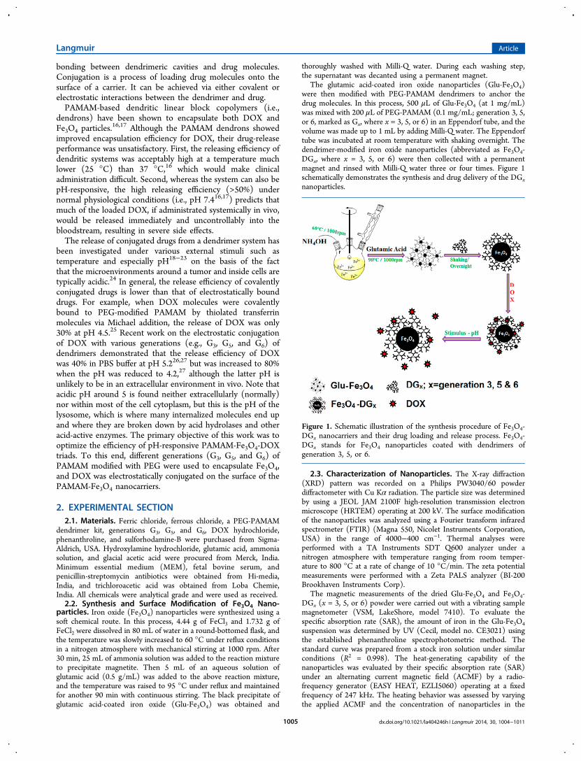
The glutamic acid-coated iron oxide nanoparticles (Glu-Fe3O4)were then modified with PEG-PAMAM dendrimers to anchor thedrug molecules. In this process, 500 μL of Glu-Fe3O4 (at 1 mg/mL)was mixed with 200 μL of PEG-PAMAM (0.1 mg/mL; generation 3, 5,or 6, marked as Gx, where x = 3, 5, or 6) in an Eppendorf tube, and thevolume was made up to 1 mL by adding Milli-Q water. The Eppendorftube was incubated at room temperature with shaking overnight. Thedendrimer-modified iron oxide nanoparticles (abbreviated as Fe3O4-DGx, where x = 3, 5, or 6) were then collected with a permanentmagnet and rinsed with Milli-Q water three or four times. Figure 1schematically demonstrates the synthesis and drug delivery of the DGxnanoparticles.
2.3. Characterization of Nanoparticles. The X-ray diffraction(XRD) pattern was recorded on a Philips PW3040/60 powderdiffractometer with Cu Kα radiation. The particle size was determinedby using a JEOL JAM 2100F high-resolution transmission electronmicroscope (HRTEM) operating at 200 kV. The surface modificationof the nanoparticles was analyzed using a Fourier transform infraredspectrometer (FTIR) (Magna 550, Nicolet Instruments Corporation,USA) in the range of 4000−400 cm−1. Thermal analyses wereperformed with a TA Instruments SDT Q600 analyzer under anitrogen atmosphere with temperature ranging from room temper-ature to 800 °C at a rate of change of 10 °C/min. The zeta potentialmeasurements were performed with a Zeta PALS analyzer (BI-200Brookhaven Instruments Corp).
The magnetic measurements of the dried Glu-Fe3O4 and Fe3O4-DGx (x = 3, 5, or 6) powder were carried out with a vibrating samplemagnetometer (VSM, LakeShore, model 7410). To evaluate thespecific absorption rate (SAR), the amount of iron in the Glu-Fe3O4suspension was determined by UV (Cecil, model no. CE3021) usingthe established phenanthroline spectrophotometric method. Thestandard curve was prepared from a stock iron solution under similarconditions (R2 = 0.998). The heat-generating capability of thenanoparticles was evaluated by their specific absorption rate (SAR)under an alternating current magnetic field (ACMF) by a radio-frequency generator (EASY HEAT, EZLI5060) operating at a fixedfrequency of 247 kHz. The heating behavior was assessed by varyingthe applied ACMF and the concentration of nanoparticles in the
Figure 1. Schematic illustration of the synthesis procedure of Fe3O4-DGx nanocarriers and their drug loading and release process. Fe3O4-DGx stands for Fe3O4 nanoparticles coated with dendrimers ofgeneration 3, 5, or 6.
Langmuir Article
dx.doi.org/10.1021/la404246h | Langmuir 2014, 30, 1004−10111005

suspension. The specific absorption rate (SAR) was calculated fromthe initial slope of the time−temperature curve.2.4. Analysis of Drug Loading and Release. The fluorescence
intensity is highly dependent on the state of the molecule (i.e., free orin attached form) and thus has been extensively used to study theinteractions between fluorophores and quencher.28−31 To evaluate theinteraction of the DOX molecules with the Fe3O4-DGx nanoparticles,fluorescence spectroscopy was conducted. An aqueous suspension ofvarying amounts of Fe3O4-DGx nanoparticles (200, 400, 600, 800,1000, and 1200 μg from a stock solution of 1 mg/mL) was added to 1mL of DOX solution (10 μg/mL). The mixture was incubated at roomtemperature for 15 min. The fluorescence spectrum of the supernatantafter magnetic sedimentation was recorded. The intensities of thesupernatant against the pure DOX solution were used to determinethe loading efficiency of the Fe3O4-DGx nanoparticles. The loadingefficiency (w/w%) was calculated using the following relation
=−
×I I
Iloading efficiency(%)
( )100
drug s
drug (1)
where Idrug is the intensity of the pure drug and Is is the intensity of thesupernatant.For drug-release experiments, the amount of DOX and the amount
of Fe3O4-DGx nanoparticles were quantified on the basis of the loadingefficiency. In a typical drug-loading experiment, 500 μg of Fe3O4-DGx(x = 3, 5, or 6) nanoparticles was incubated with 500 μg of the drug (1mg/mL) in the dark for 3 h. The drug-loaded particles were collectedwith a permanent magnet and were utilized for drug-releaseexperiments. For a typical drug-release study, DOX-loaded Fe3O4-DGx nanoparticles were immersed in 5 mL of sodium acetate buffer(pH 5) and put into a dialysis bag, which was then suspended in 200mL of phosphate-buffered saline (PBS, pH 7.34) to form the reservoirsink. Dialysis was performed under continuous stirring at 37 °C. Atdifferent time intervals, 1 mL aliquots from the sink (PBS) werewithdrawn and replaced with equal amounts of fresh PBS to maintainthe sink conditions. The amount of drug released was determined bythe measurement of fluorescence (λexc = 490 nm and λemi = 560 nm)from the aliquots against standard plots prepared under similarconditions (R2 = 0.999).2.5. Evaluation of Carriers’ Biocompatibility and Therapeu-
tic Efficacy. The biocompatibility of Fe3O4-DGx (x = 3, 5, or 6) wasassessed with the human cervical cancer cell line (HeLa). To establishthe potential of DOX-loaded Fe3O4-DG5 to release DOX, a dose-dependent study was undertaken to evaluate its 50% inhibitoryconcentration (IC50) values over 24 h. The viable cell population wasdetermined by a sulforhodamine B (SRB) colorimetric assay.32 For atypical assay, the cells were seeded in 96-well plates at a density of 2 ×10⁴ cells per well and incubated in MEM tissue culture medium,supplemented with fetal bovine serum and antibiotics, for 24 h at 37°C in a 5% CO2 environment. After 24 h, different concentrations ofFe3O4-DGx and DOX-loaded Fe3O4-DG5 were mixed with freshgrowth medium, and this replaced the exhausted medium. The cellswere then incubated for an additional 24 h and carefully washed withsterile PBS (pH 7.34), and an SRB assay was performed to determinethe cell viability. For the assay, cells were fixed with cold (at 4 °C) 10%trichloroacetic acid and dried. The fixed cells were then stained with0.4% SRB (in 1% acetic acid) and incubated in the dark for 1 h. Theunbound dye was then thoroughly washed with 1% acetic acid, and thecell-bound dye was then extracted with 10 mM Tris buffer (pH 10.5).The absorbance was recorded at 560 nm, and the cell viability wascalculated as follows:
= ×cell viability(%)Abs
Abs100sample
control (2)
3. RESULTS AND DISCUSSION3.1. Characterization of Fe3O4 Nanoparticles. The XRD
pattern revealed the formation of single-phase Fe3O4 nano-particles with inverse spinel structure having a lattice constant
of a ≈ 8.38 Å that was consistent with the reported value ofmagnetite (JCPDS card no. 88-0315, a = 8.375 Å). A highdegree of crystallinity in nanoparticles was indicated by thepresence of sharp and intense peaks (S1). The crystallite size ofGlu-Fe3O4 was calculated to be ∼8 nm using the Scherrerformula.33
The FTIR spectra of the synthesized nanoparticles showedthe successful grafting of glutamic acid onto the surface ofFe3O4 nanoparticles. Figure 2a shows that the absorption bands
for pure glutamic acid are well resolved as compared to thosefor Glu-Fe3O4, which are broadened and few. The strong IRband observed at around 580 cm−1 in Glu-Fe3O4 could beascribed to the Fe−O stretching vibrational mode of themagnetite. The spectral band at ∼1650 cm−1 (CO vibrationof −COOH groups for glutamic acid) shifted to 1620 cm−1,indicating the binding of glutamic acid to the surface of Fe3O4
Figure 2. FTIR spectra of (a) glutamic acid (Glu) and glutamic acid-coated Fe3O4 (Glu- Fe3O4) nanoparticles, (b) PEG-PAMAM ofgenerations 3, 5, and 6, and (c) Glu-Fe3O4 and Fe3O4-DGxnanoparticles.
Langmuir Article
dx.doi.org/10.1021/la404246h | Langmuir 2014, 30, 1004−10111006

nanoparticles by the chemical absorption of carboxylate(glutamate) ions. The carboxylate groups of glutamic acidform complexes with Fe atoms present on the surface of Fe3O4,rendering partial single-bond character to the CO bond.34
This is expected to weaken the CO bond, thereby shiftingthe stretching frequency to a lower value. The FTIR spectra ofthe pure PEG-PAMAM (Figure 2b) and PEG-PAMAM-modified Fe3O4 nanoparticles are shown in Figure 2c.Dendrimer-modified Fe3O4 (Fe3O4-DGx) samples show broadN−H stretching vibrations at 3400 cm−1, C−H stretchingvibrations at 2880 cm−1 (2900 cm−1), COO− asymmetricstretching at 1625 cm−1, CO stretching at 1550 cm−1, C−O−C stretching vibrations at 1250 cm−1, and C−O stretchinginterfering with C−N stretching at 1100 cm−1. Because of theintense fingerprint region of PEG-PAMAM dendrimers, themetal oxygen vibrations of iron oxide have been masked.The microstructure examination in TEM showed that the
particles were spherical in shape with regular morphology (S2a)and a size ranging from 10 to 15 nm (S2b). The selected-areaelectron diffraction rings in S2c were indexed to be (220),(311), (400), (511), and (440) diffraction planes of Fe3O4.From the high-resolution micrograph, the lattice spacing of thecrystallite (S2d) was measured to be 2.5 Å, which correspondsto the (311) plane of Fe3O4.Thermogravimetric analysis (TGA) and differential thermal
analysis (DTA) degradation profiles (Figure 3) show a weight
loss of ∼9.64 with a sharp DTA endothermic peak at ∼80 °Cthat could be attributed to the removal of physically absorbedwater molecules on the Fe3O4 nanoparticles. The ∼5.0% weightloss with a broad DTA exothermic peak at ∼290 °C could beassociated with the removal of chemically attached glutamicacid molecules from the surface of Fe3O4 nanoparticles duringdegradation. The weight loss of ∼2.8% beyond the temperatureof ∼400 °C indicated by a sharp DTA exothermic peak at ∼565°C is likely associated with the phase transformation ofmagnetite to maghemite (Fe2O3).
35
To investigate whether the magnetic performance of Fe3O4nanoparticles is compromised by coatings, the field-dependentmagnetization (M vs H) of Glu-Fe3O4 and dendrimer-modifiedFe3O4 nanoparticles was measured at 300 K in a field of 20 kOe(Figure 4). The Glu-Fe3O4 nanoparticles exhibit super-paramagnetic behavior, exhibiting a maximum magnetizationof 57 emu/g at 20 kOe. The magnetizations of the Fe3O4-DGx
nanoparticles were calculated to be approximately 48, 37, and33 emu/g for generations 3, 5, and 6, respectively. Althoughencapsulation of the nanoparticles with PEG-PAMAMdendrimers reduced the magnetization, obviously as a resultof the increase in the nonmagnetic organic components, theheating efficacy of the present Fe3O4-DGx as a hyperthermiaagent, one of the two added functions, was satisfactory, with thetypical working temperatures of 45−50 °C being established asusual, as shown in Figure 5.The colloidal stability and dispersion of the nanoparticles is
associated with the electric charge of the particle surface. Thezeta potentials of the different Glu-Fe3O4 and Glu-Fe3O4-DGx(x = 3, 5, or 6) nanoparticle systems were measured to be−26.4 ± 5.6, −19.0 ± 2.0, −20.4 ± 4.9, and −23.9 ± 6.4,respectively. Apparently, the attachment of glutamic acid to thesurface of Fe3O4 nanoparticles resulted in a negatively chargedsurface (−26.4 mV). Zeta-potential analysis also showed that allof the PEG-PAMAM dendrimer-modified nanoparticles(Fe3O4-DGx) exhibited a positive potential until pH 4;however, their surface charge became negative with increasingpH. Around the physiologically relevant pH of 7.4, the surfacecharges of the nanoparticles were measured to be −19.0, −20.4,or −23.9 mV for G3, G5, and G6, respectively. The change inthe surface charge of the three generations was due to theabundance of the PEG moiety on the dendrimers. The largenegative surface charge is believed to have enhanced thecolloidal stability of the nanoparticles. With the PEG chainspresent on the surfaces of PAMAM dendrimers, a highersurface charge density enhances the steric hindrance, which inturn improves the colloidal stability of the nanoparticles andthus prevents them from aggregating.
3.2. DOX Loading and Release. Fluorescence spectros-copy was used to investigate the interactions of DOX withFe3O4-DGx nanoparticles (Figure 6). The interactions of DOXwith the nanoparticles were evident by the decrease influorescence in the presence of the nanoparticles. The loadinginteractions were evaluated at λex = 490 nm and λem = 590 nmfor DOX. Using eq 1, we calculated the drug-loadingefficiencies of the Fe3O4-DGx nanoparticle systems to beapproximately 87, 93, and 96% for generations 3, 5, and 6,respectively (Figure 7). It was surprisingly found that theaverage loading efficiency of DOX onto Fe3O4-DGx increasedwith the increment in PAMAM generation. The above result isdifferent from that in previous work,27,36 though we also
Figure 3. TGA-DTA of glutamic acid-coated Fe3O4 (Glu-Fe3O4)nanoparticles.
Figure 4. Room-temperature field-dependent magnetization ofglutamic acid-coated Fe3O4 (Glu- Fe3O4) and Fe3O4 nanoparticlescoated with dendrimers of generations 3, 5, and 6 (Fe3O4-DG3−5) at20 kOe.
Langmuir Article
dx.doi.org/10.1021/la404246h | Langmuir 2014, 30, 1004−10111007

noticed that the increments were significant only between DG3and DG6.It has been reported that dendrimers of higher generations
are more capable of encapsulating drugs than lower generationones, whereas lower generations facilitate the conjugation ofdrugs.27,36 In previous work,27 the conjugation capacities ofhigher generations of dendrimers were impaired by their morecompact packing and steric hindrance posed by the surfaceamine groups. In the present work, drug loading was achievedthrough the electrostatic interactions between the −NH2 (δ
+)groups of DOX and the −OH (δ−) groups of PEG. Thenumber of PEG chains and thus the number of −OH groupson the surface of dendrimers generally increased with theincrement in the generation of the dendrimer. The benefit fromthe loosely packed long chains of PEG is twofold. First, theyoffer more space to host drug molecules. Second, they providemore electrostatic conjugation sites than the bare PAMAMdendrimers. Hence, the enhanced conjugation capacity ofhigher generations of dendrimers could be attributed to thePEGylation on the surface of the PAMAM dendrimer.The drug-release experiments were carried out under
reservoir-sink conditions at 37 °C, which mimic the cellularenvironment (reservoir, pH 5; sink, pH 7.3). Figure 8 showsthat the release of DOX from Fe3O4-DGx was collectively fasterat pH 5 than at pH 7.3. The electrostatic conjugation is via theinteraction of the cationic ends of DOX with the anionic endson the surface of PEG-PAMAM. The lowering of the pH value
leads to the protonation of the PEG-modified dendrimers,which results in the cleavage of the bond of PEG with the DOXmolecules.
Figure 5. Time-dependent calorimetric measurements of the presentnanocarriers in different alternating current magnetic fields (ACMFs)and concentrations of nanocarriers. (Top) 5 and (bottom) 2 mg/mL.The specific absorption rate (SAR) was calculated to be ∼134 W/gfrom the initial slope of the time−temperature curve.
Figure 6. Fluorescence intensity of doxorubicin-loaded Fe3O4-DGxnanoparticles. Fe3O4-DG3−5 stands for Fe3O4 nanoparticles coatedwith dendrimers of generations 3, 5, and 6.
Figure 7. Drug-loading efficiency vs dendrimer generation. Thedifference between DG3 and DG6 was significant (p < 0.05). Therewere no significant differences between DG3 and DG5 and betweenDG5 and DG6 (p > 0.05).
Langmuir Article
dx.doi.org/10.1021/la404246h | Langmuir 2014, 30, 1004−10111008

Figure 8 also shows that at pH 5 DOX was released rapidlyfor an initial 10 h before a plateau was reached. The plateaupercentages of DOX release observed over a period of 24 hwere ∼60 ± 8, 68 ± 9, and 80 ± 9% by Fe3O4-DG3, DG5, andDG6, respectively. The release efficacy of DOX from Fe3O4-DGx increased with the PAMAM generation, which was againin disagreement with previous work.27,36
3.3. In Vitro Evaluation of Cellular Toxicity andTherapeutic Effect. The Fe3O4-DGx nanoparticles with orwithout DOX loading were incubated with HeLa cells toevaluate their compatibility or toxicity to the cell proliferationactivity. The internalization of the nanoparticles was examina-tion using DOX-loaded Fe3O4-DG5 nanoparticles underconfocal laser scanning microscopy. HeLa cells (2 × 10³)were incubated with DOX-loaded Fe3O4-DG5 for 5 h. Becauseof the inherent fluorescence of DOX, no other tagging moietywas required. The confocal images (Figure 9a) demonstratedthat the control cells showed no discernible change in thecytoplasm, nucleus, nuclear membrane, and nucleoli. Figure 9bdemonstrates that the particles were internalized by the cells.Though the incubation time was not long enough for the drugto kill the cell (Figure 10), the DOX-loaded nanoparticles hadcaused significant changes in cellular morphology and relatedfeatures, viz., the granulation of the cytoplasm and thedegradation of the nuclear membrane.Quantitative evaluation showed that at physiological pH
(7.2−7.4) the drug-free Fe3O4-DGx nanoparticles had essen-tially no significant effect on the proliferation of HeLa cells(Figure 10), and DOX-loaded Fe3O4-DGx nanoparticlesexhibited very mild (insignificant, p > 0.05) toxicities (Figure10) compared to those of drug-free groups. The PAMAM-Fe3O4-DOX nanoparticles would cause a very mild side effectto normal tissues as a result of the very low level of DOXrelease. Hence, the DOX can be fairly benignly carried and notreleased until the PAMAM-Fe3O4-DOX is taken up into thecell.In conclusion, the PEG-modified PAMAM-Fe3O4-DOX
system has great potential to enhance the therapeutic effectof DOX with minimized side effects. The PAMAM of highergenerations possesses more favorable qualities in terms of botha higher conjugation capacity and a greater efficiency of releaseof a cytotoxic drug.
4. SUMMARYIn this study, we have explored the possible therapeuticefficiency of pH-responsive PAMAM-Fe3O4-DOX triads using
different generations (G3, G5, and G6) of PAMAM. Thenanocarriers were synthesized by using the PEG-modifiedPAMAM dendrimers to encapsulate glutamic acid-modifiedFe3O4 nanoparticles, and drug (DOX) molecules wereelectrostatically conjugated to the surface of nanocarriers.Unlike previous work, we surprisingly found that both theloading and releasing efficiencies of DOX increased with theincrement of PAMAM generation from 3 to 6. The loadingefficiencies of DOX molecules were approximately 87, 93, and96% for generations 3, 5, and 6, respectively. At pH 7.4, theDOX-releasing efficiency was as low as ∼15%. However, at pH5 the DOX-releasing efficiencies within 24 h were approx-imately 60, 68, and 80% for generations 3, 5, and 6, respectively.PAMAM-Fe3O4-DOX had a very mild (insignificant) adverse
Figure 8. Drug-release profile of doxorubicin-loaded Fe3O4-DGxnanoparticles. Fe3O4-DG3−5 stands for Fe3O4 nanoparticles coatedwith dendrimers of generations 3, 5, and 6.
Figure 9. Confocal laser scanning images of (a) control HeLa cells and(b) treated HeLa cells after 5 h of incubation (scale −20 μm).
Langmuir Article
dx.doi.org/10.1021/la404246h | Langmuir 2014, 30, 1004−10111009

effect on cellular proliferation and morphology at pH 7.2−7.4,showing the potential to be used as a safe drug-delivery systemwithout posing severe harm to normal cells.
■ ASSOCIATED CONTENT*S Supporting InformationX-ray diffraction pattern of glutamic acid-coated Fe3O4nanoparticles. TEM and SAD images and the particle sizedistribution of nanoparticles. This material is available free ofcharge via the Internet at http://pubs.acs.org.
■ AUTHOR INFORMATIONCorresponding Authors*(D.B.) Tel: +91 22 25767632. Fax: +91 22 25723480. E-mail:[email protected].*(Q.C.) Tel: +61 3 990 53599. E-mail: [email protected] authors declare no competing financial interest.
■ ACKNOWLEDGMENTSS.C. acknowledges the Department of Science and Technology(DST) for providing fellowships. Financial support from theDepartment of Information Technology (DIT) and theNanomission of DST, Government of India, is also gratefullyacknowledged. S.N. acknowledges financial assistance from theIITB-Monash Research Academy. Q.C. thanks ProfessorJunQing Chen from the Cancer Research Centre, Shenyang,PR China, for a valuable discussion of the results.
■ REFERENCES(1) Vial, T.; Descotes, J. Immunosuppressive drugs and cancer.Toxicology 2003, 185, 229−240.(2) Raschi, E.; Vasina, V.; Ursino, M. G.; Boriani, G.; Martoni, A.; DePonti, F. Anticancer drugs and cardiotoxicity: insights and perspectivesin the era of targeted therapy. Pharmacol. Ther. 2010, 125, 196−218.(3) Hayward, R.; Hydock, D.; Gibson, N.; Greufe, S.; Bredahl, E.;Parry, T. Tissue retention of doxorubicin and its effects on cardiac,smooth, and skeletal muscle function. J. Physiol. Biochem. 2013, 69,177−187.(4) Shen, M.; Shi, X. Dendrimer-based organic/inorganic hybridnanoparticles in biomedical applications. Nanoscale 2010, 2, 1596−1610.(5) Chandra, S.; Barick, K. C.; Bahadur, D. Oxide and hybridnanostructures for therapeutic applications. Adv. Drug Delivery Rev.2011, 63, 1267−1281.
(6) Løkling, K.-E.; Skurtveit, R.; Fossheim, S. L.; Smistad, G.;Henriksen, I.; Klaveness, J. pH-sensitive paramagnetic liposomes forMRI: assessment of stability in blood. Magn. Reson. Imag. 2003, 21,531−540.(7) Pakula, T.; Minkin, P.; Matyjaszewski, K., Polymers, Particles, andSurfaces with Hairy Coatings: Synthesis, Structure, Dynamics, andResulting Properties. In Advances in Controlled/Living RadicalPolymerization; American Chemical Society: Washington, DC, 2003;Vol. 854, pp 366−382.(8) Mignani, S.; Kazzouli, S. E.; Bousmina, M.; Majoral, J.-P.Dendrimer space concept for innovative nanomedicine: A futuristicvision for medicinal chemistry. Prog. Polym. Sci. 2013, 38 (), 993−1008.(9) Zhu, J.; Shi, X. Dendrimer-based nanodevices for targeted drugdelivery applications. J. Mater. Chem. B 2013, 1, 4199−4211.(10) Li, J.; Li, J.; Xu, S.; Zhang, D.; Liu, D. Hydrophobicoligopeptide-based star-block copolymers as unimolecular nanocarriersfor poorly water-soluble drugs. Colloids Surf., B 2013, 110, 183−190.(11) Pradhan, P.; Giri, J.; Rieken, F.; Koch, C.; Mykhaylyk, O.;Doblinger, M.; Banerjee, R.; Bahadur, D.; Plank, C. Targetedtemperature sensitive magnetic liposomes for thermo-chemotherapy.J. Controlled Release 2010, 142, 108−121.(12) Sinead M Ryan, G. M.; Wang, Xuexuan; Haddleton, David M;Brayden, D. J. Advances in PEGylation of important biotechmolecules: delivery aspects. Expert Opin. Drug Delivery 2008, 5,371−383.(13) Labieniec, M.; Watala, C. PAMAM dendrimersdiversebiomedical applications. Facts and unresolved questions. Cent. Eur. J.Biol. 2009, 4, 434−451.(14) Esfand, R.; Tomalia, D. A. Poly(amidoamine) (PAMAM)dendrimers: from biomimicry to drug delivery and biomedicalapplications. Drug Discovery Today 2001, 6, 427−436.(15) Mikiciuk-Olasik, E.; Szymanski, P.; Markowicz, M. Nano-technology in pharmaceutical and biomedical applications: den-drimers. Nano 2011, 06, 509−539.(16) Wu, X.; He, X.; Zhong, L.; Lin, S.; Wang, D.; Zhu, X.; Yan, D.Water-soluble dendritic-linear triblock copolymer-modified magneticnanoparticles: preparation, characterization and drug release proper-ties. J. Mater. Chem. 2011, 21, 13611−13620.(17) Lopez-Heredia, M. A.; Bongio, M.; Cuijpers, V. M. J. I.; vanDijk, N. W. M.; van den Beucken, J. J. J. P.; Wolke, J. G. C.; Jansen, J.A. Bone formation analysis: effect of quantification procedures on thestudy outcome. Tissue Eng., Part C 2012, 18, 369−373.(18) Castelli, D.; Boffa, C.; Giustetto, P.; Terreno, E.; Aime, S.Design and testing of paramagnetic liposome-based CEST agents forMRI visualization of payload release on pH-induced and ultrasoundstimulation. J. Biol. Inorg. Chem. 2013, 18, 1−8.(19) Wang, F. P.; Mu, H. P.; Zhang, J. Y.; Li, W. X.; Wang, Q. Z.; Du,X. Z. Study on preparation and swelling kinetics of P(AA-co-C8PhEO10Mac) pH-sensitive hydrogel in vitro drug release study. J.Appl. Polym. Sci. 2013, 130 (), 1981−1989.(20) Ding, F.; Shi, X.; Jiang, Z.; Liu, L.; Cai, J.; Li, Z.; Chen, S.; Du, Y.Electrochemically stimulated drug release from dual stimuli responsivechitin hydrogel. J. Mater. Chem. B 2013, 1, 1729−1737.(21) Peng, J.; Qi, T.; Liao, J.; Chu, B.; Yang, Q.; Li, W.; Qu, Y.; Luo,F.; Qian, Z. Controlled release of cisplatin from pH-thermal dualresponsive nanogels. Biomaterials 2013, 34, 8726−8740.(22) Jaiswal, M. K.; Banerjee, R.; Pradhan, P.; Bahadur, D. Thermalbehavior of magnetically modalized poly(N-isopropylacrylamide)-chitosan based nanohydrogel. Colloids Surf., B 2010, 81, 185−194.(23) Gary-Bobo, M.; Mir, Y.; Rouxel, C.; Brevet, D.; Hocine, O.;Maynadier, M.; Gallud, A.; Da Silva, A.; Mongin, O.; Blanchard-Desce,M.; Richeter, S.; Loock, B.; Maillard, P.; Morere, A.; Garcia, M.;Raehm, L.; Durand, J.-O. Multifunctionalized mesoporous silicananoparticles for the in vitro treatment of retinoblastoma: drugdelivery, one and two-photon photodynamic therapy. Int. J. Pharm.2012, 432, 99−104.(24) Tannock, I. F.; Rotin, D. Acid pH in tumors and its potential fortherapeutic exploitation. Cancer Res. 1989, 49, 4373−4384.
Figure 10. Viability of HeLa cells when cultured in a medium dopedwith either Fe3O4-DG5 or DOX-Fe3O4-DG5 nanoparticles in variousamounts. The difference between any pair of data at each dopingconcentration was insignificant (p > 0.05). Similar results were alsoobtained with DG3 and DG6 nanoparticles (data not shown).
Langmuir Article
dx.doi.org/10.1021/la404246h | Langmuir 2014, 30, 1004−10111010

(25) Li, Y.; He, H.; Jia, X.; Lu, W.-L.; Lou, J.; Wei, Y. A dual-targetingnanocarrier based on poly(amidoamine) dendrimers conjugated withtransferrin and tamoxifen for treating brain gliomas. Biomaterials 2012,33, 3899−3908.(26) He, H.; Li, Y.; Jia, X.-R.; Du, J.; Ying, X.; Lu, W.-L.; Lou, J.-N.;Wei, Y. PEGylated Poly(amidoamine) dendrimer-based dual-targetingcarrier for treating brain tumors. Biomaterials 2011, 32, 478−487.(27) Rouhollah, K.; Pelin, M.; Serap, Y.; Gozde, U.; Ufuk, G.Doxorubicin loading, release, and stability of polyamidoaminedendrimer-coated magnetic nanoparticles. J. Pharm. Sci. 2013, 102(), 1825−1835.(28) Chandra, S.; Dietrich, S.; Lang, H.; Bahadur, D. Dendrimer-doxorubicin conjugate for enhanced therapeutic effects for cancer. J.Mater. Chem. 2011, 21, 5729−5737.(29) Pietrzak, M.; Wieczorek, Z.; Stachelska, A.; Darzynkiewicz, Z.Interactions of chlorophyllin with acridine orange, quinacrine mustardand doxorubicin analyzed by light absorption and fluorescencespectroscopy. Biophys. Chem. 2003, 104, 305−313.(30) Szulawska, A.; Gniazdowski, M.; Czyz, M. Sequence specificityof formaldehyde-mediated covalent binding of anthracycline deriva-tives to DNA. Biochem. Pharmacol. 2005, 69, 7−17.(31) Das, K.; Jain, B.; Dube, A.; Gupta, P. K. pH dependent bindingof chlorin-p6 with phosphatidyl choline liposomes. Chem. Phys. Lett.2005, 401, 185−188.(32) Vichai, V.; Kirtikara, K. Sulforhodamine B colorimetric assay forcytotoxicity screening. Nat. Protocols 2006, 1, 1112−1116.(33) Patterson, A. L. The Scherrer formula for X-ray particle sizedetermination. Phys. Rev. 1939, 56, 978−982.(34) Max, J.-J.; Chapados, C. Infrared spectroscopy of aqueouscarboxylic acids: comparison between different acids and their salts. J.Phys. Chem. A 2004, 108, 3324−3337.(35) Fajaroh, F.; Setyawan, H.; Nur, A.; Lenggoro, I. W. Thermalstability of silica-coated magnetite nanoparticles prepared by anelectrochemical method. Adv. Powder Technol. 2013, 24, 507−511.(36) Markowicz, M.; Szymanski, P.; Ciszewski, M.; Kłys, A.;Mikiciuk-Olasik, E. Evaluation of poly(amidoamine) dendrimers aspotential carriers of iminodiacetic derivatives using solubility studiesand 2D-NOESY NMR spectroscopy. J. Biol. Phys. 2012, 38, 637−656.
Langmuir Article
dx.doi.org/10.1021/la404246h | Langmuir 2014, 30, 1004−10111011