Polímeros naturales ensamblados capa a capa (layer-by-layer ...
260
UNIVERSIDAD COMPLUTENSE DE MADRID FACULTAD DE CIENCIAS QUÍMICAS Departamento de Química Física I TESIS DOCTORAL Polímeros naturales ensamblados capa a capa (layer-by-layer) para aplicaciones biomédicas Layer-by-layer assembly of natural polymers for biomedical applications MEMORIA PARA OPTAR AL GRADO DE DOCTOR PRESENTADA POR Miryam Criado González Directoras Rebeca Hernández Velasco Carmen Mijangos Ugarte Madrid, 2017 © Miryam Criado González, 2017
-
Upload
khangminh22 -
Category
Documents
-
view
1 -
download
0
Transcript of Polímeros naturales ensamblados capa a capa (layer-by-layer ...

UNIVERSIDAD COMPLUTENSE DE MADRID FACULTAD DE CIENCIAS QUÍMICAS
Departamento de Química Física I
TESIS DOCTORAL
Polímeros naturales ensamblados capa a capa (layer-by-layer) para aplicaciones biomédicas
Layer-by-layer assembly of natural polymers for biomedical
applications
MEMORIA PARA OPTAR AL GRADO DE DOCTOR
PRESENTADA POR
Miryam Criado González
Directoras
Rebeca Hernández Velasco Carmen Mijangos Ugarte
Madrid, 2017
© Miryam Criado González, 2017

UNIVERSIDAD COMPLUTENSE DE MADRID FACULTAD DE CIENCIAS QUÍMICAS
Departamento de Química-Física
TESIS DOCTORAL
POLÍMEROS NATURALES ENSAMBLADOS CAPA A CAPA
(LAYER-BY-LAYER) PARA APLICACIONES BIOMÉDICAS
LAYER-BY-LAYER ASSEMBLY OF NATURAL POLYMERS
FOR BIOMEDICAL APPLICATIONS
MEMORIA PARA OPTAR AL GRADO DE DOCTOR
PRESENTADA POR
Miryam Criado González
Directores
Rebeca Hernández Velasco
Carmen Mijangos Ugarte
CONSEJO SUPERIOR DE INVESTIGACIONES CIENTÍFICAS
Instituto de Ciencia y Tecnología de Polímeros
Madrid, 2017


TABLE OF CONTENTS


Table of contents
iii
Resumen ix
Summary xiii
1. GENERAL INTRODUCTION AND OBJECTIVES 1
2. 7 STATE OF THE ART
2.1. Layer-by-layer assembly 9
2.1.1. Types of interactions 10
2.1.2. Mechanisms of LbL assembly 13
Linear growth 14 Exponential growth 15 Combination of both mechanisms, linear and exponential 15
2.1.3. LbL techniques 16
Dipping 17 Spray 17 Spin coating 18
2.2. Natural polymers 19
2.2.1. LbL of natural polymers 21
Experimental factors influencing the growth through LbL assembly 22 Crosslinking of LbL films obtained from natural polymers 25
2.2.2. Biomedical applications of LbL natural polymers 27
Coatings for tissue engineering 27 Platforms for cell adhesion 32 Materials for drug delivery 33
2.2.3. Nanocomposite LbL systems based on natural polymers and biomedical
applications 35
2.3. References 40
3. 53 MATERIALS AND METHODS 3.1. Materials 55
3.1.1. Starting polymers 55
Polysaccharides 55 Proteins 58 Synthetic polymers 58
3.1.2. Characterization of polysaccharides, chitosan and alginate 59
Determination of the molecular weight by viscosimetry 59 Determination of chitosan deacetylation degree by nuclear magnetic resonance
(NMR) 61
Determination of the thermal stability 62
3.1.3. Alginate based magnetic ferrofluid 63
Synthesis of alginate based magnetic ferrofluid 63 Characterization of alginate based magnetic ferrofluid 64
3.1.4. Conclusions 70

Table of contents
iv
3.2. Characterization techniques 70
3.2.1. Characterization techniques to follow the growth of LbL films 70
Ellipsometry 70 Quartz crystal microbalance (QCM) 72 Fourier transform Infra-Red spectroscopy (FTIR) 74
3.2.2. Characterization techniques to determine morphological properties 75
Scanning Electron Microscopy (SEM) 75 Atomic Force Microscopy (AFM) 75
3.2.3. Characterization techniques to determine mechanical properties 76
Atomic Force Microscopy (AFM) nanoindentation measurements 76
3.2.4. Characterization techniques to determine the structure of LbL films 78
Grazing-incidence small-angle X-ray scattering (GISAXS) 78 Dual-Beam (Focus Ion Beam (FIB) – Scanning Electron Microscopy (SEM)) 79
3.3. References 80
4. LAYER-BY-LAYER FILMS FROM NATURAL POLYMERS. STUDY OF THE GROWTH MECHANISM 85
4.1. Introduction 87
4.2. Experimental part 89
4.2.1. Materials 89
4.2.2. Fabrication of multilayer films through LbL assembly 90
Precipitation studies 90 Preparation of substrates 91 Multilayer films obtained through spray assisted LbL 91 Multilayer films obtained through dipping assisted LbL 93
4.2.3. Determination of the thickness 94
Ellipsometry 94 Scanning electron microscopy (SEM) 94 Quartz crystal microbalance 95
4.3. Results and discussion 95
4.3.1. Multilayer Alg/Chi films 95
Optimization of the experimental conditions for LbL assembly 95 Determination of interactions between Alg and Chi in LbL films 97 Determination of the thickness as a function of the number of bilayers 99
4.3.2. Polyallylamine/hyaluronic acid films 103
Optimization of the experimental conditions for LbL assembly 103 Determination of the thickness 104
4.4. Conclusions 108
4.5. References 109
5. DETERMINATION OF THE STRUCTURE-PROPERTIES RELATIONSHIP IN ALG/CHI FILMS 113
5.1. Introduction 115
5.2. Experimental part 117
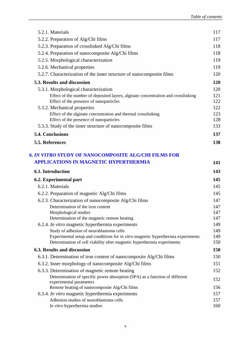
Table of contents
v
5.2.1. Materials 117
5.2.2. Preparation of Alg/Chi films 117
5.2.3. Preparation of crosslinked Alg/Chi films 118
5.2.4. Preparation of nanocomposite Alg/Chi films 118
5.2.5. Morphological characterization 119
5.2.6. Mechanical properties 119
5.2.7. Characterization of the inner structure of nanocomposite films 120
5.3. Results and discussion 120
5.3.1. Morphological characterization 120
Effect of the number of deposited layers, alginate concentration and crosslinking 121 Effect of the presence of nanoparticles 122
5.3.2. Mechanical properties 122
Effect of the alginate concentration and thermal crosslinking 123 Effect of the presence of nanoparticles 128
5.3.3. Study of the inner structure of nanocomposite films 133
5.4. Conclusions 137
5.5. References 138
6. IN VITRO STUDY OF NANOCOMPOSITE ALG/CHI FILMS FOR APPLICATIONS IN MAGNETIC HYPERTHERMIA 141
6.1. Introduction 143
6.2. Experimental part 145
6.2.1. Materials 145
6.2.2. Preparation of magnetic Alg/Chi films 145
6.2.3. Characterization of nanocomposite Alg/Chi films 147
Determination of the iron content 147 Morphological studies 147 Determination of the magnetic remote heating 147
6.2.4. In vitro magnetic hyperthermia experiments 149
Study of adhesion of neuroblastoma cells 149 Experimental setup and conditions for in vitro magnetic hyperthermia experiments 149 Determination of cell viability after magnetic hyperthermia experiments 150
6.3. Results and discussion 150
6.3.1. Determination of iron content of nanocomposite Alg/Chi films 150
6.3.2. Inner morphology of nanocomposite Alg/Chi films 151
6.3.3. Determination of magnetic remote heating 152
Determination of specific power absorption (SPA) as a function of different
experimental parameters 152
Remote heating of nanocomposite Alg/Chi films 156
6.3.4. In vitro magnetic hyperthermia experiments 157
Adhesion studies of neuroblastoma cells 157 In vitro hyperthermia studies 160

Table of contents
vi
6.4. Conclusions 164
6.5. References 165
7. STUDY OF CELL ADHESION AND APPLICATIONS IN DRUG DELIVERY OF ALG/CHI FILMS 169
7.1. Introduction 171
7.2. Experimental part 173
7.2.1. Materials 173
7.2.2. Preparation of multilayer films with tamoxifen 173
7.2.3. Characterization 174
Morphological characterization 174 Contact angle 174 Degradation assay 175 Biological behavior 175 In vitro release of TMX 176
7.3. Results and discussion 178
7.3.1. Morphological characterization of Alg/Chi films by AFM 178
7.3.2. Degradation assay 179
7.3.3. In vitro cell adhesion studies 180
Cytotoxicity assay 180 Cell adhesion 181 Morphology assay 187
7.3.4. Drug release experiments 188
7.4. Conclusions 191
7.5. References 191
8. 197
199
LBL HYDROGELS OBTAINED FROM NATURAL POLYMERS
8.1. Introduction
8.2. Experimental part 201
8.2.1. Materials 201
8.2.2. Determination of interactions in the multilayer assembly 201
FTIR spectroscopy 201 XRD diffraction 201
8.2.3. LbL hydrogels prepared through dipping assembly 202
Experimental conditions for the design of multilayer gels 202 Preparation of multilayer gels using the LbL method 202
8.2.4. Characterization of LbL hydrogels 203
Chemical characterization 203 Morphological characterization 203 Mechanical properties 203
8.3. Results and discussion 204

Table of contents
vii
8.3.1. Optimization of the conditions for the design of LbL hydrogels 204
8.3.2. Study of the interaction of gelatin and chondroitin sulphate in a multilayer
assembly 205
8.3.3. Preparation and characterization of LbL hydrogels 208
Chemical characterization 208 Morphological characterization 210
8.3.4. Determination of the compositional structure along the layer distribution of
LbL hydrogels 211
8.3.5. Determination of the mechanical properties 213
8.4. Conclusions 214
8.5. References 214
9. GENERAL CONCLUSIONS AND PERSPECTIVES 217
ANNEXES 225
i. Abbreviations 233
ii. List of publications 239


ix
Resumen
El objetivo de esta tesis es el desarrollo de sistemas poliméricos multicapa basados en polímeros
naturales mediante la técnica capa a capa (layer-by-layer, LbL) para aplicaciones en terapias de
hipertermia magnética local y administración controlada de fármacos.
Los capítulos uno y dos reúnen una introducción general y objetivos junto con el estado del arte
que muestra una revisión bibliográfica sobre los principales temas desarrollados durante la tesis
doctoral. Del estado del arte se puede concluir que: el ensamblado LbL de polímeros naturales
suscita un gran interés hoy en día debido a las potenciales aplicaciones de estos sistemas, sobre
todo en el área de biomedicina. Además, la mayoría de los materiales obtenidos a partir del
ensamblado de polímeros naturales se han obtenido mediante técnicas de inmersión LbL. La
deposición LbL asistida por spray es una técnica rápida y sencilla para fabricar materiales
nanoestructurados y ha sido menos explorada dejando un campo abierto para la investigación en
esta tesis.
El tercer capítulo de la tesis se centra en la descripción de los materiales de partida utilizados así
como en las técnicas de caracterización más relevantes empleadas en esta tesis. El estudio del
ensamblado LbL de dos sistemas poliméricos, alginato/quitosano (Alg/Chi) y ácido
hialurónico/poli(hidrocloruro de alilamina) (HA/PAH) se describe en el capítulo cuatro. Del
análisis del espesor de los filmes en función del número de capas depositadas se pudo concluir
que el espesor de los filmes (Alg/Chi) crecía de forma lineal mientras que el crecimiento de los
filmes HA/PAH era exponencial. Además, el crecimiento de los filmes Alg/Chi fabricados
mediante spray LbL era mucho mayor que el correspondiente a los filmes fabricados mediante
inmersión LbL. Se ha desarrollado además un sistema nanoestratificado derivado de los filmes
HA/PAH mediante la incorporación de una capa de gel formada por alginato entrecruzado con
iones hierro II con potenciales aplicaciones como sistema fotosensible de liberación de fármacos
que se desarrollará en el futuro.
La caracterización morfológica y mecánica de diferentes sistemas multicapa basados en alginato
y chitosano se muestra en el capítulo cinco. Además, se preparó un ferrofluido acuoso a base de
alginato mediante coprecipitación de sales de hierro para incorporarlo en los filmes Alg/Chi y
obtener así filmes nanocompuestos. Las propiedades mecánicas de los filmes Alg/Chi, filmes
Alg/Chi entrecruzados térmicamente y filmes Alg/Chi nanocompuestos se determinaron

x
mediante mapeo nanomecánico cuantitativo de fuerza máxima a través de microscopia de
fuerzas atómicas ((PF-QNM) AFM) mostrando que el módulo elástico de los filmes Alg/Chi
aumentaba con el número de bicapas depositadas y con el entrecruzamiento, mientras que los
valores de deformación eran prácticamente constantes. La incorporación de nanopartículas de
óxido de hierro (NPs) en los filmes Alg/Chi aumentaba su rugosidad y tenía una influencia
significativa en las propiedades mecánicas aumentando el módulo elástico y la deformación,
siendo este efecto más pronunciado cuando las NPs estaban presentes en la última capa. Además,
se estudió la estructura interna de los filmes nanocompuestos Alg/Chi mediante dispersión de
rayos X de ángulos bajos con incidencia rasante (GISAXS). Los filmes Alg/Chi nanocompuestos
fabricados mediante inmersión LbL presentaban dispersión de las NPs a lo largo de toda la
estructura del filme mientras que los ensamblados mediante spray LbL presentaban un cierto
grado de orden de las NPs en capas.
En el capítulo seis se describe la puesta a punto de la aplicación de filmes nanocompuestos de
Alg/Chi en hipertermia magnética local (MHT). Para ello, el calentamiento magnético remoto de
los filmes nanocompuestos se determinó mediante aplicación de un campo magnético alternante
(AMF) y los resultados mostraron un aumento lineal de temperatura desde 6 a 12 ºC con el
número de capas de NPs lo que sugiere el uso de estos filmes como filmes termomagnéticos
(TMFs) para la aplicación de hipertermia magnética local. Los experimentos in vitro llevados a
cabo con células de neuroblastoma (SH-SY5Y) mostraron que los TMFs presentaban
propiedades de adhesión celular y su calentamiento remoto usando un AMF proporcionaba
resultados distintos dependiendo del número de ciclos de MHT y del protocolo experimental
utilizado.
En el capítulo siete se muestra la aplicación de los filmes Alg/Chi como parches para liberación
controlada de fármacos. El estudio de biocompatibilidad se llevó a cabo en dos tipos de células,
fibroblastos dérmicos humanos (HDF) y células de adenocarcinoma de cáncer de mama (MCF-
7). Se varió la química superficial de los filmes mediante deposición de una capa de ácido
hialurónico (HA) al final del proceso de deposición para estudiar su influencia en la adhesión
celular junto con el entrecruzamiento. Los resultados mostraron que la adhesión de las células
MCF-7 aumentaba en los filmes entrecruzados químicamente cuya última capa era Alg y
disminuía en los que la última capa era HA debido a un aumento del ángulo de contacto. No se
observaron diferencias significativas en la adhesión de las células HDF ni con la química
superficial ni con el entrecruzamiento. La aplicación final de los filmes Alg/Chi como parches
para liberación de fármacos se demostró incorporando tamoxifeno, un fármaco contra el cáncer

xi
de mama, en distintas posiciones intermedias de los filmes Alg/Chi. Los resultados mostraron
una liberación más sostenida en el tiempo a medida que aumentaba el número de capas
depositadas y una disminución de la viabilidad celular de las células cancerígenas MCF-7.
El capítulo ocho de la tesis constituye una extensión del procedimiento de ensamblado LbL
desarrollado para la preparación de filmes a partir de polímeros naturales para el desarrollo de
hidrogeles multimembrana de gelatina y sulfato de condroitina. La deposición LbL de ambos
polímeros llevada a cabo sobre un núcleo de gelatina daba lugar a un aumento del módulo
elástico con respecto al núcleo de gelatina sin recubrir. Los resultados obtenidos permiten
anticipar el empleo de estos hidrogeles LbL como sistemas de encapsulación celular y liberación
de fármacos en el futuro. El capítulo nueve recoge las conclusiones generales derivadas de este
trabajo de tesis. La tesis también incluye cuatro anexos y una lista de publicaciones.


xiii
Summary
The objective of the present PhD work is the development of multilayer polymer systems based
on natural polymers through layer-by-layer (LbL) assembly for applications in magnetic
hyperthermia therapies and controlled drug delivery.
The first part of this thesis provides a general introduction and objectives together with a state-
of-the-art which collects a literature review about the main topics related to this thesis (Chapters
1 and 2). From this state-of-the-art, it is possible to conclude that: LbL assembly of natural
polymers constitutes an area of intensive research nowadays, due to the potential applications of
these materials mainly on the biomedical field. In addition, most of the studies regarding LbL of
natural polymers deal with dipping LbL. Spray assisted LbL is a simple and saving-time
technique to fabricate nanostructured materials and it has been much less explored leaving a
broad field open to research in this thesis.
The third chapter is focused on the description of materials and the most relevant
characterization techniques used in this thesis. The study of the LbL assembly of two different
systems based on natural polymers, alginate/chitosan (Alg/Chi) and hyaluronic
acid/poly(allylamine hydrochloride) (HA/PAH) is described in the fourth chapter. From the
determination of the thickness of (Alg/Chi) films as a function of the number of deposited layers,
a linear growth was observed up to 5 bilayers whereas (HA/PAH) films presented an exponential
growth. The growth of Alg/Chi films built up through spray LbL was much higher than that
obtained when films were constructed by dipping LbL. A nanostratified system was obtained
from the HA/PAH multilayer structure by incorporation of a gel-like layer formed by alginate
crosslinked with iron II ions to be employed as a light-responsive drug delivery system in the
future.
The morphological and mechanical characterization of different multilayer systems based on
alginate and chitosan is reported in the chapter five. An alginate-based ferrofluid was synthetized
by a coprecipitation method of iron salts in an alginate aqueous solution and incorporated within
Alg/Chi films to obtain nanocomposite films. The mechanical properties of Alg/Chi films,
thermally crosslinked Alg/Chi films and nanocomposite Alg/Chi films, built up through spray
assisted LbL, were determined by PeakForce Quantitative Nanomechanical Mapping-Atomic
Force Microscopy ((PF-QNM) AFM) revealing that the elastic moduli of Alg/Chi films

xiv
increased with the number of deposited bilayers and with the crosslinking, whereas deformation
values were almost constant. The incorporation of iron oxide NPs within the multilayer Alg/Chi
films increased the roughness of the films and greatly influenced the mechanical properties
increasing the elastic moduli and the deformation values, being this effect more pronounced
when NPs were on the last layer of nanocomposite films. Besides, the inner structure of
nanocomposite Alg/Chi films, evaluated employing Grazing incidence small-angle X-ray
scattering (GISAXS), revealed that NPs were thoroughly dispersed on nanocomposite Alg/Chi
films assembled by dipping LbL, whereas spray LbL gave rise to nanocomposite films with
some degree of ordering of the NPs into layers.
Chapter six describes the application of nanocomposite Alg/Chi films for local magnetic
hyperthermia (MHT). To that aim, magnetic remote heating was determined by application of an
alternating magnetic field (AMF) to nanocomposite Alg/Chi films with different number of NPs
layers and results showed a linear temperature increase from 6 to 12 ºC with the NPs layers
(from 80 to 160 layers) enabling to use these films as thermomagnetic films (TMFs) for MHT. In
vitro experiments with neuroblastoma cells (SH-SY5Y) showed that TMFs presented cell
adhesion properties and their remote heating using an AMF showed different results depending
on the number of MHT cycles with a reduction in cell viability up to 67% and 20% for one and
three cycles, respectively. Cell viability could decrease even more mimicking in vivo
applications by applying the MHT on TMFs placed over neuroblastoma cells previously cultured
on an Ibidi dish, giving rise to a cell viability reduction of 85% after two MHT cycles.
The application of Alg/Chi films as drug delivery patches is demonstrated in chapter seven. The
biocompatibility of the films was proven employing two different kinds of cells, human dermal
fibroblasts (HDF) and human caucasian breast adenocarcinoma (MCF-7) cells. The surface layer
of the films was varied by spraying a layer of hyaluronic acid (HA) at the end of the deposition
process in order to study their influence in the cell adhesion together with the effect of the
crosslinking process. Results showed that the MCF-7 cell adhesion increased on crosslinked
films with Alg-ending layer and decreased on crosslinked films ending in HA where the contact
angle increased after crosslinking. There were not significant differences in HDF cell adhesion
either with the surface chemistry or the crosslinking. The final application of Alg/Chi films as
drug delivery patches of tamoxifen, a drug employed against breast cancer, was proven as a
function of the position of TMX within the Alg/Chi films. Results showed a more sustained
release over time with the number of deposited bilayers resulting in a decrease of the cell
viability of MCF-7 cells.

xv
Chapter eight constitutes an extension of the procedure developed during the thesis for the
buildup of polymer films to the fabrication of multimembrane hydrogels of chondroitin sulphate
and gelatin. The LbL deposition of both polymers was carried out over a gelatin gel and the
mechanical properties of the resulting materials were determined through oscillatory
rheological measurements. The elastic modulus increased with respect to the uncoated gelatin
core without changes in their melting point, which points to a potential employment of these LbL
hydrogels as cell encapsulation and drug delivery systems in the future. Chapter nine
summarizes the general conclusions extracted from this thesis. The manuscript also includes four
annexes and a list of publications.


CHAPTER 1
General introduction and objectives


General introduction and objectives
3
The present PhD Thesis work “Layer-by-layer assembly of natural polymers for biomedical
applications”, aims to bring new insights and the establishment of a new methodology for the
nanostructuration of polymer materials and the development of hydrogels obtained from natural
polymers for their employment in biomedical applications.
A consolidated research line of our group at ICTP-CSIC is the study of the formation, structure,
properties and applications of polymer gels. During the last years, this research has been more
focused on the employment of natural polymers, specifically, those extracted from the biomass
(polysaccharides such as chitosan, alginate or agarose and proteins such as gelatin) as precursors
for the development of hydrogels for biomedical applications. This is motivated on the one hand,
by the fact that many natural polymers are able to assemble in water in response to different
stimuli (pH, temperature or ionic concentration) to give rise to hydrogels in the form of macro,
micro and nanogels. On the other hand, the intrinsic characteristics of natural polymers,
biocompatibility and biodegradation, make them suitable for the development of biomedical
applications. Another important mode of assembly of natural polymers is the establishment of
electrostatic interactions between a polycation and a polyanion. A well-known method to obtain
nanostructured films taking advantage of this kind of interaction is layer-by-layer assembly
(LbL), and nowadays, the growing interest on the employment of LbL with natural polymers is
shown in a great number of publications dealing with the subject that employs mainly dipping
techniques for the fabrication of nanostructured polymer materials. However, it is important to
note that literature regarding the employment of spray assisted LbL to fabricate nanostructured
systems based on natural polymers is still pretty scarce. Besides, nowadays the development of
free-standing polymer films employing LbL technique constitutes an area of intensive research
because it broadens the range of applications of multilayer polymer films.
In this work, spray assisted layer-by-layer has been implemented in our laboratory and
employed, throughout this thesis as an easy and scalable procedure to combine polyelectrolytes
into nanostructured materials. To the best of our knowledge, this technique is not settled up in
any other laboratories of CSIC or Spanish Universities up to date. To this aim, a new
collaboration was established with the team of Prof. F. Boulmedais (Institut Charles Sadron,
CNRS, Strasbourg, France), an expert in the field of Layer-by-layer assembly and more
specifically, spray assisted LbL. In these regards, this work constitute a deep study that aims to
establish the best conditions for the buildup of films from natural polymers through spray
assisted LbL and a comparison with the properties achieved when the fabrication is carried out
through dipping assisted LbL. Very importantly, in addition to the fabrication of polymer films, a

Chapter 1
4
new methodology has been implemented for the fabrication of multimembrane hydrogels based
on gelatin and chondroitin sulphate that opens new perspectives within this field.
A significant effort throughout this thesis has been devoted to the determination of the structure-
properties relationship through the employment of advanced characterization techniques in
collaboration with the group of Prof. T. Ezquerra from the Instituto de Estructura de la Materia
(CSIC). The main experimental proofs have been atomic force microscopy nanoindentation
measurements and grazing incidence small angle X-ray scattering (GISAXS) carried out at the
European Synchrotron Radiation Facility (ESRF), Grenoble France.
Finally, this thesis collects several exhaustive examples about the employment of the as prepared
materials in biomedical applications. Specifically, the thesis collects a deep study on cell
adhesion of fibroblasts and tumour cells on films made up of alginate and chitosan and the
development of drug delivery patches of tamoxifen in collaboration with the group of Prof. San
Roman (Instituto de Ciencia y Tecnología de Polímeros, CSIC). As a continuation of the
significant efforts that we have undertaken over the last years for the development of hybrid
polymer materials with iron oxide nanoparticles for magnetic hyperthermia application, the
thesis includes the development of magnetic films for the application of local hyperthermia. A
significant advance undertaken during this thesis is the development of a colloidal stable aqueous
ferrofluid that has been the subject of a patent recently filed.
In order to get the general objective of the PhD work “Layer-by-layer assembly of natural
polymers for biomedical applications”, the manuscript is structured in nine chapters,
corresponding two of them to introductory sections, one of them to Materials and Methods, four
of them to main objectives of the PhD work and one to the general conclusions.
The first chapter, General Introduction and objectives, intends to establish the scientific
framework in which the present work is carried out.
The second chapter, State of the art, is mainly devoted to the state of the art of LbL
methodology employing natural polymers and the biomedical applications reported in literature
up to date. It includes a summary of the LbL technique, the main experimental parameters that
influence this process and the main experimental LbL procedures (dipping, spray and spin
coating) with a comparison of their characteristics. An exhaustive table collecting relevant
literature in the field of LbL with natural polymers is included at the end of the chapter.

General introduction and objectives
5
The third chapter, entitled Materials and methods, is intended to describe the different
materials employed throughout this thesis and the theoretical background behind the main
experimental techniques employed for the characterization of the materials, among them,
ellipsometry, microscopy, quartz crystal microbalance, GISAXS and atomic force microscopy
nanoindentation measurements.
The fourth chapter, entitled Layer-by-layer films from natural polymers. Study of the growth
mechanism, gives a detailed description of the fabrication of two different systems: alginate
chitosan and polyallylamine/hyaluronic acid through spray and dipping procedure and the
determination of their growth mechanism through a combination of experimental techniques.
The fifth chapter, Determination of the structure-properties relationship in LbL Alg/Chi films,
aims to determine the influence of different experimental parameters such as alginate
concentration, crosslinking or presence of iron oxide nanoparticles on the mechanical properties
of the LbL films determined through atomic force microscopy nanoindentation measurements.
Moreover, it provides results on the characterization of Alg/Chi films through GISAXS
experiments aimed to elucidate differences on the inner structure of films prepared through
dipping and spray techniques.
The sixth chapter, entitled In vitro study of nanocomposite Alg/Chi films for applications in
magnetic hyperthermia, describes the cell studies carried out employing magnetic Alg/Chi films
aimed to prove their application as patches for local hyperthermia employing different
experimental protocols.
The seventh chapter, entitled Study of cell adhesion and applications in drug delivery of
Alg/Chi films, provides a thorough study aimed to elucidate the influence of surface chemistry,
roughness and film architecture on cell adhesion and the development of delivery patches of
tamoxifen.
The eighth chapter, entitled LbL hydrogels obtained from natural polymers, provides a novel
approach for the preparation of multimembrane hydrogels from gelatine and chondroitin sulphate
employing the methodology developed throughout the thesis. It includes the morphological and
rheological characterization of the resulting materials.
The ninth chapter, entitled General conclusions and perspectives, summarizes the most general
conclusions of the work and proposes new subjects of research in this field.


CHAPTER 2
State of the art


State of the art
9
This chapter summarizes the results found in literature about specific features related to the aims
of this thesis. It includes a state-of-the-art about the layer-by-layer (LbL) assembly, the
interactions driving the LbL assembly, factors influencing the LbL assembly, growth
mechanisms, LbL techniques, LbL assembly of natural polymers and their biomedical
applications.
2.1. LAYER-BY-LAYER ASSEMBLY
Over the last decades, the development of nanostructured polymer materials with tunable
properties has gained increasing attention for application in diverse fields. In this regard, the
bottom-up methodology to assemble small molecules into nanostructures has to be considered.1
During the half first of the 20th
century, the bottom-up strategies for the deposition of
monolayers were based on the Langmuir-Blodgett (LB) deposition method which consist of the
transfer of amphiphilic molecules from water-air interface to a solid-air interface. The necessity
of very clean substrates and a dust-free atmosphere together with the low robustness of
multilayers and slowness of the deposition process, hinder their widespread practical
applications.2 Time after, the layer-by-layer (LbL) assembly, based on the sequential deposition
of interacting species onto a substrate, emerged as a versatile, simple, efficient, reproducible and
flexible bottom-up technique3 and it has become one of the most used techniques to coat many
types of substrates, including planar surfaces,4, 5
spherical objects,6-8
porous matrices,9, 10
and
highly curved surfaces,11
giving rise to nanostructured polymer materials.
LbL technique originates from works carried out by Ralph Iller12
in 1966 when he proved the
step-by-step deposition of negatively charged silica particles and positively charged Boehmite
fibrils due to charge reversal after each deposition step. But it was not until 1991 that LbL
buildup concept was validated13
and the first work related to polyelectrolyte multilayer films
(PEMs) dates back to 1992 when Decher et al.14
studied the assembly of a multilayer film by
alternated deposition of poly(styrene sulfonate) (PSS), used as a polyanion, and poly(allylamine
hydrochloride) (PAH), used as a polycation.
The LbL assembly has advantages compared to the more conventional coating methods,
including the precise control over the thickness and compositions at the nanoscale, the simplicity
and versatility of the process, its suitability and flexibility to coat surfaces with irregular shapes
and sizes and the possibility of scaling at industrial level.15
These characteristics have made the
LbL assembly one of the most useful techniques for building up advanced multilayer polymer

Chapter 2
10
structures towards multiple applications in diverse fields such as biomedicine,16, 17
energy,18, 19
optics,20, 21
coatings,22, 23
etc.
The high impact and massive interest in the field of LbL assembly can be clearly demonstrated
by a search in the SCOPUS® database using the term “layer-by-layer” as the topic keywords
(Figure 2.1). As can be observed, the number of publications per year in the field of layer-by-
layer (LbL) over the past decades shows an exponential growth rate giving rise to more than
1000 publications per year from 2007 and reaching 1822 publications in 2016 demonstrating the
increasing interest in this technique.
Figure 2.1. Number of publications per year with topic keywords of “layer-by-layer” since 1992. Data source:
SCOPUS®.
2.1.1. Types of interactions
Multilayer assembly through the LbL technique involves different types of intermolecular
interactions.
The electrostatic interaction has been the most studied driving force for the development of
nanostructured multilayer films through the LbL technique.24
It takes place between molecules
and surfaces which are electrically charged. LbL assembly based on electrostatic interactions
gives rise to multilayer films with well controlled structure, composition and thickness by
alternate deposition of opposite charged molecules. There are multiple polymers, polycations and
polyanions, that give rise to this kind of electrostatic assembly. Polycations include
poly(allylamine hydrochloride) (PAH), poly(L-lysine) (PLL), poly(ethylenimine) (PEI) and
chitosan (Chi), and polyanions comprise poly(styrene sulfonate) (PSS), alginate (Alg),
hyaluronic acid (HA), poly(acrylic acid) (PAA), etc.
1990 1995 2000 2005 2010 20150
400
800
1200
1600
2000
Nu
mb
er
of
pu
bli
ca
tio
ns
on
"L
bL
"
Publication year
a)
2000 2005 2010 20150
5
10
15
20
25
30
35
40
Nu
mb
er
of
pu
bli
ca
tio
n o
n
"s
pra
y L
bL
"
Publication year
b)

State of the art
11
The electrostatic assembly of multilayer structures through LbL technique can be influenced by
different parameters such as pH, temperature, solvent, ionic strength and type and properties of
every polyelectrolyte as follows:
The properties of the PEMs depend on the pH of the polymer solutions from which the layers are
adsorbed.25
The pH controls the charge density of the adsorbing polymer layer as well as the
previously adsorbed polymer layer exerting influence on LbL assembly of a polycation and a
polyanion at the molecular level, that is, on the composition and structure of the multilayer
system. The effect of the pH depends at a great extent on the kind of polyelectrolyte. Strong
polyelectrolytes are fully charged independently of the pH; however, weak polyelectrolytes with
carboxylic acid or amine functional groups are highly sensitive to the pH. It has been proved that
tiny changes in the pH of weak polyelectrolyte solutions, such as polyacrylic acid (PAA),
poly(allylamine) (PAH) or poly(L-lysine) (PLL), could induce pronounced changes in the
growth mechanism and thickness of the LbL assembled multilayer films.26, 27
Regarding temperature, it has been explored that the thickness of multilayer films increases with
the temperature of the polymer solutions.28
This effect has been evaluated in different polymer
systems proving that the thickness of two strong polyelectrolytes,
poly(diallyldimethylammonium chloride) (PDDA) and PSS, increased in an approximately linear
fashion with the increase of temperature.29
In another example, it was demonstrated that PAH
and poly(styrenesulfonate) (PSS) films fabricated at elevated temperatures were significantly
thicker than similar films deposited at room temperature.30
The structure and growth of
multilayer films is also related to the solvent conditions. Decreasing the solvent quality by
addition of ethanol to the aqueous polymer solutions modulates the relative strength of
electrostatic and secondary intermolecular and intramolecular interactions giving rise to an
increase of the multilayer film thickness and mass loading due to the reduced solvation effect of
aqueous polymer solutions containing electrolyte ions.31, 32
The ionic strength influences the stability, permeability and thickness of multilayer films.33
Therefore, substantial differences are observed between multilayer films assembled in solutions
within or without salts,34
with low or high salt concentration,35
as well as with the kind of
counterion.36
Generally speaking, an increase in ionic concentration produces an increase on the
film thickness. However, high salt concentrations exceeding a certain limit compensates all
charge preventing multilayer adhesion of polyelectrolytes.37
Charges on polymer repeated units
can be balanced by those on oppositely charged chains or by salt ions occluded within the film.

Chapter 2
12
In the case of solutions without salt, a polymer positive charge is balanced by a polymer negative
charge. For solutions with salt ions, together with the polymer charge there is an extrinsic
compensation balanced by salt counterions. Because of that, multilayers containing salt ions
should be thicker, less interpenetrating, and individual chains would have more mobility,
yielding less stable structures.38
However, exceeding a certain threshold of salt concentration
produces that the adsorbed polyelectrolytes on the substrate surface could be displaced by the
salt ions.15
Besides the solution parameters influencing the polymer interactions, there are others related to
the polymer properties such as the molecular weight, charge density and chain architectures:
The molecular weight of polymers has influence on the structure of multilayer films leading to
thicker and rougher films when molecular weight increases. Generally, although the thickness
and roughness of films increase with the molecular weight, the kind of growth mechanism (linear
or exponential) does not depend on the polymer molecular weight.39
Polymers with high
molecular weight generally give rise to an increase of the interdiffusion in between layers
because the relaxation during and after deposition, attributed to residual stresses from PEMs,
increases with the molecular weight.40
It has been proven that the decrease of charge density of polyelectrolytes increases the thickness
of multilayer films either for strong polyelectrolytes, like cellulose derivatives,41
or weak
polyelectrolytes such as PAA or PAH.42
The chain architectures of polymers, which include
chain conformation and chain interpenetration, affect the formation of multilayer films. The
chain conformation dominates the growth of multilayer films at low salt concentration and the
chain interpenetration has great influence at high salt concentration.43
Although electrostatic interactions between polycation and polyanion layers44
are the most
employed for building up multilayer polyelectrolyte films (PEMs), the following interactions can
also be used to assemble LbL systems.5 i) Hydrogen bonding is one of the most investigated
driving forces, apart from electrostatic interactions, which allows the incorporation of uncharged
materials, which can act as hydrogen bonding donors and acceptors, into the multilayer structure.
The resulting materials are influenced by the temperature, pH and ionic strength being less stable
than those assembled by electrostatic interactions.45
ii) Covalent bonding is a chemical bond
produced by shared pairs of electrons between atoms increasing the stability and strength of the
multilayer structure.24, 46
The main drawback of this kind of interaction is that some side products
could be introduced into the multilayer assemblies.1 iii) Charge-transfer interactions are
State of the art
13
produced by alternate adsorption of non-ionic molecules with electron-donating and electron-
accepting groups in the side chains.47
This interaction allows to use organic solvents;24
however,
the charge-transfer complexes have low association constants limiting the achievement of well-
ordered and stable multilayers.1 iv) The highly selective and specific host-guest interaction is
used to assembly multilayers through strong interaction between host (e.g., cyclodextrins,
cucurbiturils, calixarenes, pillarenes, crown ethers or porphyrins) and guest (e.g., ferrocene,
adamantine or azobenzece) molecules.48, 49
v) The hydrophobic interaction takes place between
non-polar molecules or between non-polar parts of a molecule50
and it can be used to assemble
several layers of the same polymer;51
but they are weaker than electrostatic interactions. vi)
Biologically specific interactions or biospecific interactions are produced by some biomaterials
which are capable of interacting through different molecular interactions (e.g., electrostatic,
hydrophobic, etc.) with other components ensuring high specificity to the target molecules.52
The
most known are avidin-biotin, antibody-antigen and lectin-carbohydrate interactions.24
vii)
Coordination chemistry interactions are strong molecular interactions between a diversity of
metal ions and organic ligands that enable to construct well-ordered and highly oriented and
robust multilayer films.53
2.1.2. Mechanisms of LbL assembly
An schematic representation of LbL assembly through electrostatic interactions between a
polycation and a polyanion is shown in Figure 2.2. i) An usually negatively charged substrate is
put into contact with a solution of an oppositely charged polymer (polycation) to deposit the first
monolayer, ii) a washing step to remove unbound material, iii) the positively coated substrate is
put in touch with the polyanion solution to deposit a second layer and iv) a new washing step to
remove unbound material, giving rise to the formation of a bilayer structure. This cycle can be
repeated in order to obtain the desired number of bilayers to form a multilayer structure.54, 55
During the LbL assembly, a non-stoichiometric excess of charge is absorbed after each
deposition step with regards to the preceding layer. This surplus of charge provides the step-wise
mechanism for the reversal of the surface charge polarity, facilitating a favorable surface for the
adsorption of the subsequent layer.15
The adsorption process is very sensitive to drying.56

Chapter 2
14
Figure 2.2. Sequential deposition of polycations and polyanions during the LbL assembly. Modified figure from
Ref.3
Most of the substrates used for building up PEMs carry an excess of charge (e.g., glass,
polystyrene) which is the prerequisite for the successful adsorption of oppositely charged
polymer chains. In other cases (e.g., silicon, silicone rubber), it is necessary a special
pretreatment such as plasma or piranha cleaning, to functionalize these substrates in order to
allow the adsorption of PEMs. For a better attachment substrate-PEMs, very often the branched
polyethylenimine (PEI) is used as an intermediate layer between the substrate and the first
polymer layer.57
One of the main advantages of the LbL assembly is that the growth rate of the multilayer system
can be controlled at the nanometer scale. Two types of buildup mechanisms, linear and
exponential, have been reported.39, 58
Linear growth
The linear growth is the simplest growth mechanism in which the thickness and mass of the film
increase linearly with the number of deposited bilayers.3 At each layer deposition, the
polyelectrolyte from the solution (e.g., polyanion) is electrostatically attracted by the oppositely
charged polyelectrolyte (e.g., polycation), which forms the previously deposited layer, leaving a
charge over-compensation at the interface, which gives rise to an electrostatic repulsion,
restricting the polyelectrolyte adsorption to only one monolayer.59
Every polyelectrolyte layer
interpenetrates only with the adjacent ones (the previous layer and the subsequent layer).60
A
schematic representation of this kind of growth is shown in Figure 2.3a.

State of the art
15
Exponential growth
In contrast to linear growth, in the case of exponential growth (Figure 2.3b), films grow
exponentially with the number of deposited bilayers and they are characterized by high chain
mobility in the direction perpendicular to the film and in the plane of the film. The origin of this
exponential growth is the diffusion of at least one of the polymer constituents in and out of the
film architecture.39, 61
Figure 2.3. a) Linear growth with stratified layers through the multilayer structure and b) exponential growth with
diffusion through the multilayer showing no clear layer structure.
Combination of both mechanisms, linear and exponential
During the LbL assembly of a polycation and a polyanion different growth mechanisms can take
place as shown in Figure 2.4.61
Considering a film with an exponential growth in which only one
of the polyelectrolytes (e.g., polycation, PC+) diffuses into the whole structure and the other
(polyanion, PA-) does not diffuse, the exponential growth occurs up to a critical thickness where
the growth turns again into a linear regime.2 To explain this event, it has been considered that the
structure of a multilayer film is subdivided, at least, into three zones (1, 2 and 3) during the
deposition process.59, 61, 62
Zone 1 corresponds to the deposition of the first bilayers (at least three) which are organized in
distinct and stratified layers that do not interdiffuse and grow linearly due to the influence of the
substrate (Figure 2.4a).
Zone 3 is the region close to the surface and it is in contact with the polymer solution. In this
zone, the deposition of a PA- layer gives rise to a film with an outer negative excess charge.
Then, this film is put into contact with the PC+ solution and the PC
+ chains firstly interact with
the outer negative charges of the PA- layer but, immediately, they also diffuse into the film down
to the substrate giving rise to a film formed by PA- and PC
+ chains interacting strongly between
them and free PC+ chains. These free PC
+ chains diffuse into the film until its chemical potential
becomes equal to that of the PC+ chains in the solution. In addition, this diffusion could result in
Bilayers
Thic
kne
ss
Bilayers
Thic
kne
ss
substrate substrate
a) b)
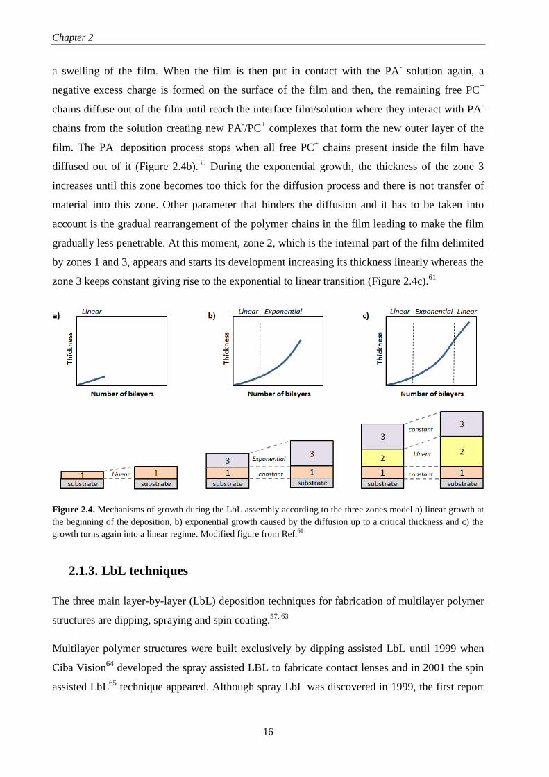
Chapter 2
16
a swelling of the film. When the film is then put in contact with the PA- solution again, a
negative excess charge is formed on the surface of the film and then, the remaining free PC+
chains diffuse out of the film until reach the interface film/solution where they interact with PA-
chains from the solution creating new PA-/PC
+ complexes that form the new outer layer of the
film. The PA- deposition process stops when all free PC
+ chains present inside the film have
diffused out of it (Figure 2.4b).35
During the exponential growth, the thickness of the zone 3
increases until this zone becomes too thick for the diffusion process and there is not transfer of
material into this zone. Other parameter that hinders the diffusion and it has to be taken into
account is the gradual rearrangement of the polymer chains in the film leading to make the film
gradually less penetrable. At this moment, zone 2, which is the internal part of the film delimited
by zones 1 and 3, appears and starts its development increasing its thickness linearly whereas the
zone 3 keeps constant giving rise to the exponential to linear transition (Figure 2.4c).61
Figure 2.4. Mechanisms of growth during the LbL assembly according to the three zones model a) linear growth at
the beginning of the deposition, b) exponential growth caused by the diffusion up to a critical thickness and c) the
growth turns again into a linear regime. Modified figure from Ref.61
2.1.3. LbL techniques
The three main layer-by-layer (LbL) deposition techniques for fabrication of multilayer polymer
structures are dipping, spraying and spin coating.57, 63
Multilayer polymer structures were built exclusively by dipping assisted LbL until 1999 when
Ciba Vision64
developed the spray assisted LBL to fabricate contact lenses and in 2001 the spin
assisted LbL65
technique appeared. Although spray LbL was discovered in 1999, the first report

State of the art
17
about this technique data from 2000 by Schlenoff et al.66
and the increasing attention on spray
LbL began in 2005.67
In that year a new modality of spray deposition, known as simultaneous
spray coating, was developed by Porcel et al.68
In 2009, a new LbL technique emerged by
combination of spin and spray LBL and it was called spin-spray LbL69
(Figure 2.5).
Figure 2.5. Evolution of LbL assembly since their apparition in 1991.
Dipping
Dipping LbL consists of immersing a substrate alternately into aqueous polycation and
polyanion solutions with a washing step between deposited layers to remove unbound material
and avoid contamination of the subsequent solution (Figure 2.6a). This cyclical process is
repeated until obtain the desired number of layers.3, 57
It is a simple technique which allows to
cover substrates of almost any shape and size; however, the deposition time required for an
adsorption step of polymers is around 15 – 20 minutes, making it a time-consuming process.32
In
order to scale-up this technique to industrial level, it requires a much higher volume of polymer
solutions than other technologies, such as spray or spin coating, and waste can be an issue,
although solutions can be reused as long as cross-contamination remains low.1
Spray
Spray LbL is a simple method where multilayers are assembled by sequentially spraying of
polycation and polyanion solutions onto a substrate (Figure 2.6b). It allows to coat not only large
and planar substrates, but also non-planar substrates. The film thickness is influenced by polymer
concentrations, spray flow rate, the time of spraying (a few seconds per layer) and the waiting
time whether the substrate is washed or not. Comparing with dipping LbL, the rinsing step can
be suppressed giving rise to thicker films without altering their quality and decreasing the
deposition time even more.55
The film properties, such as morphology, uniformity and chemical
composition, can be tailored to be similar to those prepared by dipping LbL. Spray LbL offers
rapid assembly times and it is amenable to both automation and scale-up to industrial level.67
1991 1999
SPRAY
2001
DIPPING SPIN
2005
SIMULTANEOUSSPRAY COATING
2009
SPIN-SPRAY

Chapter 2
18
Spin coating
Spin LbL assembly is based on spinning a substrate to facilitate the deposition of polymers. It is
performed by either casting the polycation and polyanion solutions onto a spinning substrate or
casting the solution onto a stationary substrate that is then spun (Figure 2.6c).65, 70
It is a time-
saving technique with a deposition time of ~30s per layer. The thickness depends on the spin
speed, with higher speeds leading to thinner films. The presence of simultaneous interactions
forces, including centrifugal, viscous and air-shear forces, gives rise to low interpenetration and
highly ordered films with specific layer interfaces and smoother than those obtained from
dipping LbL. However, this technique has some limitations, in fact, it cannot be used to deposit
uniform films on non-planar surfaces and it is not possible to coat large areas surfaces.71
Spin
LbL allows for automation, but standard spin coaters are generally designed for coating flat
substrates up to 10 cm in diameter width and they are not useful for complex shapes substrates.72
Figure 2.6. Different LbL techniques a) dipping, b) spray and c) spin coating.
Washing WashingPolycation Polyanion
Washing WashingPolycation Polyanion
a)
c)
Washing WashingPolycation Polyanion
b)

State of the art
19
The most relevant characteristic of these LbL techniques are collected in the Table 2.1 which
allows to compare them easily with the aim of choosing the most suitable technique for every
specific application.
Table 2.1. Characteristics of different LbL techniques and differences between them.
Dipping Spray Spin coating
Deposition time Long (minutes) Short (seconds) Short (seconds)
Films size Large surfaces Large surfaces Up to 10cm
Deposition surface
Planar, rough,
complex shape,
three-dimensional
Planar, rough,
complex shape Planar
Parameters which
influence the film
thickness
Dipping time,
solution
concentration and
washing time
Solution
concentration,
spray flux rate,
spray time,
evaporation time
and washing time
Spin speed, solution
concentrations and
washing time
Automation Yes Yes Yes
Visual appearance Opaque films Transparent films Transparent films
Scale up Yes Yes No
2.2. NATURAL POLYMERS
Natural polymers are those which are present in, or created by, living organisms. Figure 2.7
collects the different natural polymers and their classification in four groups: polysaccharides,
protein origin polymers, polyesters and other polymers.73
Polysaccharides are polymers constituted by monosaccharide units linked by O-glycosidic
bonds. They can be obtained from animal, vegetal and microbial sources. Physical properties of
polysaccharides, such as solubility, gelation and surface properties, are influenced by the
monosaccharide composition, chain shape and molecular weight. This group includes cellulose,
chitin, chitosan, starch, alginate, hyaluronic acid, chondroitin sulphate, dextran, agarose,
carrageenan, etc.74

Chapter 2
20
Proteins can be considered as polymer structures formed by 20 different amino acids linked by
amide (or peptide) bonds.75
This group comprises collagen, gelatin, silk, fibroin, fibrin, elastin,
soybean, etc. Polyamino acids are a small group of polyamides consisting of only one type of
amino acid linked by amide bonds. Among them are poly(L-lysine) (PLL), poly(g-glutamic acid)
(PGA), polyarginyl–polyaspartic acid, etc.73
Polyesters are polymers formed by a dicarboxylic acid and a diol. A special group of polyesters,
polyhydroxyalkanoates, are produced by a diverse variety of microorganisms as an internal
carbon and energy storage, as part of their survival mechanism. They are composed of 3-, 4-, or
rarely 5-hydroxy fatty acid monomers, which form linear polyesters.76, 77
Besides these, there are
other natural polymers, such as lipids, lignin, natural rubber or shellac, which have to be
considered.
Every group of natural polymers possesses its inherent properties. Generally speaking,
polysaccharides function in membranes and intracellular communication, proteins function as
structural materials and lipids as energy stores.74
Amongst them, polysaccharides and proteins
receive particular attention for the development of biomedical applications and they are the
subject of study in this PhD work. The most important characteristics of the natural polymer
employed as starting materials in this work will be described in the materials section (chapter 3).
Figure 2.7. Classification of natural polymers.
Polysaccharides
Proteins
Polyesters
Other polymers
● Cellulose
● Chitin/chitosan
●Alginate
● Hyaluronic acid
● Chondroitin sulphate
● Starch
● Other: dextran, agarose, carragenan, etc.
● Collagen
● Gelatin
● Silks
● Poly(L-lysine), poly(g-glutamic acid)
● Other: elastin, soy, casein, etc.Natural
polymers
● Lipids
● Lignin
● Natural rubber
● Shellac
● Polyhydroxyalkanoates

State of the art
21
2.2.1. LbL of natural polymers
Figure 2.8 illustrates a chronogram with the evolution of LbL assembly regarding natural
polymers. In 1999, Elbert et al.78
developed for first time the multilayer assembly of two natural
polymers, poly(L-lysine) (PLL) and alginate (Alg), through dipping LbL. Since then, different
multilayer systems based on natural polymers have been studied comprising four different
polycations, PLL, Chi, collagen (COL) and gelatin (GL), and a diversity of polyanions, HA, Alg,
Chondroitin sulphate (ChS), etc. In 2006, Porcel et al.61
built up PLL/HA films by spray-assisted
LbL and in 2007, Fujie et al.79
assembled Chi/Alg films through spin-assisted LbL.
Figure 2.8. Evolution of LbL assembly related to natural polymers from 1999.
In nature, there are few examples of natural polymers acting as polycations, mainly chitosan,
poly(L-lysine), collagen and gelatin.
The growth mechanism of multilayer films prepared from PLL as polycation and different
polyanions such as HA, Alg, ChS, heparin (HEP) or PGA, has been reported to be exponential.78,
80, 81 In the case of films of poly(L-lysine) (PLL) and hyaluronic acid (HA) prepared through
alternate dipping LbL, the assembly process is characterized by two growth regimes. At the
beginning of the deposition process, the surface of the substrate is covered by isolated islands
and islets which grow by the deposition of more polymer layers on their top and by mutual
coalescence until obtain a continuous film, approximately after eight deposited bilayers, showing
a linear growth. At this point, the second regime starts showing an exponential growth as number
of deposited bilayers increase due to the diffusion of free PLL chains ‘into’ whole film when the
film is in contact with a PLL solution and ‘out’ of the film when the film is further brought in
contact with a HA solution interacting with HA chains at the outer limit of the multilayer.58, 82, 83
In the specific case of PGA, it was checked that the exponential growth was not only attributed
1999 2006 2007

Chapter 2
22
to the diffusion of PLL chains into the whole structure,80
but also to the diffusion of PGA chains
into the whole film.84
As in the case of poly(L-lysine), chitosan can be assembled with diverse polyanions, such as HA,
Alg, HEP, Dex, ChS and PGA. One of the first multilayer systems comprising Chi as polycation
was obtained by means of assembly with dextran sulfate (Dex) and heparin (HEP).34
When Chi
is assembled with HA, at the beginning of the Chi/HA buildup process the surface of the
substrate was covered by isolated islets that grew and coalesced as the number of deposited
bilayers increased until obtain a continuous film, as in the case of PLL/HA.35
The multilayer
growth process of Chi/HA, Chi/Dex and Chi/HEP was exponential, being Chi the dominating
specie of the two polymers and electrostatic interactions are accompanied of other short-range
interactions such as hydrogen bonding.85
The exponential growth was due to the diffusion of Chi
molecules within the film and the structure of the Chi/HEP films was highly interpenetrated
without clear boundaries between each layer.35, 86
In contrast to the behavior observed for PLL and Chi, the assembly between COL and HA
through dipping LbL exhibited a linear growth and it was proven that COL did not diffuse into
the film and interacted only with its outer layer. However, the films were not constituted of
homogeneously distributed polyanion/polycation complexes, but they were formed of fibers
whose width increased with the number of deposition steps.87
The fibrillary structure of the
layers was also observed when COL was assembled with ChS and HEP.88
Experimental factors influencing the growth through LbL assembly
Even though the same general characteristics can be found regarding the growth mechanisms,
the growth process is influenced by a series of parameters such as molecular weight, pH, ionic
strength, solvent, method of preparation and nature of the polyanion. In this section, the
experimental factors influencing the LbL growth process of PLL and Chi acting as polycations
will be discussed due to the fact that they are the most studied in literature.
Molecular weight
The molecular weight of PLL can influence the growth process. In the linear growth regime, the
film thickness increases after each deposition step independently of the molecular weight. On the
contrary, in the exponential regime it has a significant influence. Low molecular weights allowed
that PLL chains diffused into the whole film during each deposition step, whereas high
molecular weights restricted the PLL chains diffusion to the upper part of the film.89

State of the art
23
The effect of the Chi molecular weights on the thickness and surface morphology has also been
evaluated. For a constant molecular weight of the polyanion (~400000 Da) and molecular
weights of chitosan of 30000 and 160000Da, at a constant pH and ionic strength, it was proven
that higher molecular weights gave rise to higher thicknesses; however, the exponential growth
rate was the same for high and low Chi molecular weights.39
When molecular weight of Chi
increased up to 460000 Da, it was observed that the tendency was the opposite and the
exponential growth was faster for a molecular weight of chitosan of 110000 Da than for 460000
Da.35
With regards to surface morphology, high molecular weights of Chi gave rise to a shorter
island growth and coalescence stage as well as an earlier transition from islands to a vermiculate
morphology than low molecular weights.39
pH, ionic strength and solvent
The driving force of the assembly is influenced by the pH. Multilayer PLL/PGA films showed
different behaviors depending on the pH of assembly. The main driving force of the assembly at
pH 7.4 is electrostatic interaction, whereas hydrogen bonding and hydrophobic interaction are
the dominant interactions in films built up at low pH.90
The growth of these films was higher at
acidic pH.91
When PLL is assembled with HA, the acid-base equilibria of multilayer PLL/HA
films showed that these films can be electrostatically adsorbed under highly charged “sticky”
conditions but then quickly transformed into stable low-friction films simply by altering their
pKa on adsorption, at the same pH environment.27
The effect of the pH and ionic strength in the growth process of Chi/Dex and Chi/HEP films
showed an increase in the film thickness with the increase in the NaCl concentration at a fixed
pH.34
The same effect was found for Chi/HEP films when the pH was increased at a fixed ionic
strength.85, 92
In the case of Chi/HA films, at low salt concentrations (10-4
M NaCl), the surface
of the substrate was covered by islets up to 50 bilayers with a linear increase of the film growth.
At high salt concentration (0.15 M NaCl), the formation of an uniform film took place only after
a few deposition steps showing an exponential growth.35
When Chi is assembled with Alg, the
study of the buildup process at different concentrations, pH and ionic strength allowed to
conclude that the fastest film growth took place for chitosan and alginate concentrations of 1.0
and 5.0 mg/mL and pH 5 and 3, respectively, conditions under which alginate is in high
concentration and only partially ionized in a way that its negative charge interact weakly with the
positively charged amino groups of Chi.93, 94

Chapter 2
24
The effect of the solvent in the assembly process of Chi and PGA showed that the adsorption
process from an aqueous phase was not stable; however, the use of an organic solvent as a less
soluble solvent gave rise to thicker films and achieved stable deposition.95
Method of preparation
The effect of the method of preparation in the buildup process was firstly studied by Porcel et
al.61
who built up PLL/HA films via spray assisted LbL and dipping LbL. In both cases the film
growth first evolved exponentially with the number of deposited bilayers and, after a given
number of deposition steps, its thickness evolution became linear again. This second transition
was investigated in detail through spray LbL reaching the conclusion that this transition always
took place after about 12 deposition steps independently of the parameters controlling the
deposition process, time of spraying and polyelectrolyte concentrations. These changes in the
growth process were explained using the model of the three zones described in the section 2.1.2.
in which the exponential to linear transition is attributed to the restructuration of the film that
progressively prevented the diffusion of one of the polyelectrolytes over part of the film and this
“forbidden” zone then grew linearly with the number of deposition steps.
Nature of the charged groups of the polyanion
The nature of the charged groups of the polyanion also influences the assembly of the multilayer
films. Taking into account that electrostatic interactions as well as hydrogen bond interactions
are important in the film buildup, the quantification of internal ion pairing (extrinsic versus
intrinsic charges) and water content were studied for three different systems based on PLL,
PLL/HA, PLL/ChS and PLL/HEP, in order to examine the influence of the COO- and SO3
-
groups on the film growth, the water content and the ion pairing. Although these polyanions
differed in their charge, the disaccharide units attracted approximately two lysine groups per
monomer. The percentage of free NH3+ in the films decreased as the charge density of the
disaccharide increased and it was related to PLL diffusion influencing directly the film growth. It
was also proven that PLL/HA and PLL/ChS films were the most hydrated ones. The selective
crosslinking of carboxylate and ammonium ions via carbodiimide chemistry allowed to
determine the COO-/NH3
+ and SO3
-/NH3
+ ion pairing showing that 46% of NH3
+ groups are
unpaired in PLL/HA films, 21% in PLL/ChS films and none in PLL/HEP films reaching to the
conclusion that this ratio was close to the stoichiometry of these groups in the dissacharide
monomeric unit (2:1 for PLL/HA films and 1:1 for PLL/ChS films).96

State of the art
25
Regarding the influence of the nature of the polyanion in the wettability and ion pairing, three
different multilayer films with Chi as polycation, Chi/HEP, Chi/HA and Chi/ChS, were studied.
The most hydrophilic films Chi/HA were formed by the assembly of a weak polycation (Chi)
and a weak polyanion (HA) and the most hydrophobic ones (Chi/HEP and Chi/ChS) were
formed by combination of weak (Chi) – strong (HEP or ChS) polyelectrolytes. The assembly of
two weak polyelectrolytes Chi/HA reduced ion pairing and enabled the swelling of the film,
whereas the combination weak – strong polyelectrolytes reduced the swelling because of an
increase of ion pairing.44
Crosslinking of LbL films obtained from natural polymers
The crosslinking process of multilayer polymer films built up from PLL and a determined
polyanion induces the following changes: i) the rigidity of films increases, ii) the diffusion of the
PLL chains in the network is reduced, iii) the adhesion of films to the substrate increases and iv)
the degradation decreases.97
The effect of the crosslinking on the mechanical properties of the resulting films has been
examined by different techniques such as Atomic force microscopy (AFM) nanoindentation
mesasurements and dynamic mechanical analysis (DMA).98, 99
Crosslinking gave rise to an
increase of the elastic modulus making films stiffer than native ones (Figure 2.9).99-101
Figure 2.9. Schematic representation corresponding to the increase of the stiffness of multilayer polymer films after
crosslinking.
There are different kinds of crosslinking processes. The most used are chemical crosslinking,
using the carbodiimide chemistry (1-ethyl-3-(3-(dimethylamino)-propyl)carbodiimide (EDC) in
combination with N-hydroxy-sulfosuccinimide (NHS)) or genipin, a natural origin polymer, and
thermal crosslinking, employing high temperatures during a time interval which are different for
every polymer system. Both crosslinking processes gives rise to amide bonds formation between
carboxylic groups of the polyanion (PA) and amine groups of the polycation (PC), as can be
observed in Figure 2.10.4, 97
As an example, multilayer PLL/HA films were crosslinked using
these two mechanisms, chemical (EDC/NHS) or thermal (90 ºC for 4h) crosslinking, and further
PC
COO-
NH3+NH3+
NH3+
PA COO-COO-
Crosslinking
PC
COO-
NH3+
PA
CO
NH
CO
NH
COO-COO- COO-COO-
NH3+ NH3+ NH3+ NH3+
NanoindentationNanoindentation
a)
b)
Crosslinking

Chapter 2
26
detached from the substrate to obtain free-standing PLL/HA films. The method employed for the
detaching process influenced the final structure of the film. Films built up on glass were
detached by immersing the film in a basic solution (0.1M NaOH) giving rise to non-porous and
smooth membranes. On the contrary, films built up on polystyrene can be detached by dissolving
the polystyrene substrate in tetrahydrofuran (THF) leading to a porous membrane with
micrometric holes.4
Figure 2.10. Schematic representation corresponding to the formation of amide bonds between carboxylic groups of
the polyanion (PA) and amine groups of the polycation (PC) after thermal or chemical crosslinking.
Other methods of crosslinking include the combination of covalent and ionic crosslinking, using
genipin and calcium chloride (CaCl2), provided free-standing Chi/Alg films with enhanced
mechanical properties and shape memory ability. CaCl2 was used to induce ionic crosslinking
due to the formation of stable complexes between the calcium ions and deprotonated carboxylic
groups of the Alg into multilayer membranes with and without previous genipin crosslinking.
This ionic crosslinking gave rise to free-standing membranes with improved mechanical
strength, calcium-induced adhesion and shape memory ability. Precisely, the use of CaCl2
allowed to reverse the crosslinking process by using a competing ligand,
ethylendiaminetetraacetic acid (EDTA), which acts as a chelating agent sequestering the calcium
ions. It was proved that the mechanical behavior of ionic and covalent crosslinking was different
and the application of a stress to ionically crosslinked materials gave rise to a relaxation of the
multilayers with water release and a plastic deformation as the crosslinking was dissociated,
whereas in covalently crosslinked films leaded also to a relaxation of the multilayers with water
release and an elastic deformation due to their inability to dissociate and reform bonds.102
PC
COO-
NH3+NH3+
NH3+
PA COO-COO-
Crosslinking
PC
COO-
NH3+
PA
CO
NH
CO
NH
COO-COO- COO-COO-
NH3+ NH3+ NH3+ NH3+
NanoindentationNanoindentation
a)
b)
Crosslinking

State of the art
27
The biodegradation is also influenced by the crosslinking degree. Multilayer PLL/HA films
crosslinked using the carbodiimide chemistry showed a highly resistant to hyaluronidase, an
enzyme that naturally degrades hyaluronan.97
The degradation of Chi/HA films, evaluated in
vitro, in contact with enzymes, plasma and macrophages, and in vivo, in mouse peritoneal cavity,
can be tuned by film crosslinking. Native films showed degradation by enzymes and crosslinked
films were more resistant to enzymatic degradation. Plasma also induced changes in the structure
of native films but not in crosslinked films. On the contrary, cells induced degradation in both
types of films. Regarding the in vivo degradation, native films showed an almost complete
degradation, whereas crosslinked films were only partially degraded.103
The stability of
multilayer films with COL as polycation is different depending on the nature of the polyanion.
COL/ChS films are stable in culture media at physiological conditions, whereas COL/HA,
COL/HEP and COL/Alg films are unstable.17, 88, 104
The dissolution and biodegradation of
COL/HA and COL/Alg films at physiological pH can be avoided by chemical crosslinking of
these films in order to obtain stable membranes.17, 104
2.2.2. Biomedical applications of LbL films from natural polymers
Natural polymers are of great interest in the biomedical field due to the fact that most of them
present the following features: i) biodegradability, they do not show adverse effects on the
environment or human being, ii) biocompatibility and non-toxicity, almost all of these materials
are carbohydrates in nature and composed of repeating monosaccharide units, iii) economic, they
are cheaper than synthetic polymers, iv) safety, they do not have side effects whereas synthetic
polymers could produce side effects, v) availability, they are produced naturally in large
quantity.73, 105
These characteristics make natural polymers have gained increasing attention for
the development of biomedical applications. With the aim of LbL systems can be employed for
biomedical applications, they have to be biocompatible. At the same time, it is necessary to
know their behavior at physiological conditions.
Among the different applications for LbL natural polymer films, three of the most studied are
coatings for tissue engineering and platforms for cell adhesion and for drug delivery.
Coatings for tissue engineering
Most of applications found in literature regarding LbL multilayer films obtained from natural
polymers are focused on their employment as coatings for tissue engineering. Some selected

Chapter 2
28
examples of this application, such as coatings for knee prostheses, dermal patches, coatings for
stents and dental implants, are shown in Figure 2.11.
Figure 2.11. LbL coatings from natural polymers for different tissue engineering applications: knee prostheses,
dermal patches, stents and dental implants.
A detailed description of the application of LbL materials from natural polymers as coatings for
tissue engineering is provided below as a function of the polymer which acts as a polycation.
Multilayer films with PLL acting as polycation
The ability of multilayer PLL/Alg films to generate thin coatings on tissue surfaces, the inherent
biocompatibility and degradability, together with the bioinertness, allows to control the wound
healing as well as the immunoisolation of the underlying surface to be used as barriers on
biological surfaces.78
In this regards, multilayer PLL/Alg films were used to coat magnesium
based degradable scaffolds to improve their surface bioactivity. For that purpose, they were
crosslinked using the carbodiimide chemistry and surface functionalized with fibronectin
immobilization. The in vitro cytocompatibility studies with MC3T3-E1 osteoblast cells
demonstrated that the pretreatment of the magnesium substrate greatly influenced the
biocompatibility of the films proving that fluoride pretreatment is necessary for the long-term
stability of PLL/Alg films and, therefore, for the slow corrosion of the magnesium substrates in
order to be applied as matrices for delivery of drugs and other biomolecules for successful bone
regeneration in vivo.106
The spray deposition of PLL and HA layers was further employed to coat
porous scaffolds of modified hyaluronic acid to give rise to a multilayer 3D structure, formed by

State of the art
29
the multilayer PLL/HA film covering the scaffold, which promoted the adhesion and
proliferation of keratinocytes creating an epidermal structure that mimicked the natural skin
microenvironment for potential use in skin tissue engineering applications.9
The ‘in’ and ‘out’ diffusion of PLL during the exponential growth of multilayer films was
explored to load and release growth factors. Besides, the pH tunable charge density within the
multilayer provided the possibility to adjust the amount of growth factors embedded. Different
kinds of growth factors, such as HIV-1 TAT (47-57), basic fibroblastic factor (bFGF) and alpha-
melanocyte stimulating hormone (α-MSH), have been embedded into PLL/HA, PLL/ChS and
PLL/PGA multilayer films. These growth factors maintained their long time activity when they
were embedded into the multilayer structure and after crossing the multilayer membrane,
whereas its short time activity depended on their integration depth.81, 107, 108
Multilayer PLL/PGA
films were the first example of a multilayer film whose biological activity was based on a
synergy effect of two active compounds, BMP2 and TGFb1 growth factors, in order to be
employed for cartilage and bone differentiation. In vitro studies with mesenchymal stem cells
revealed that cells came into contact with the growth factors through the PLL/PGA films due to
the degradation by cells and both growth factors, BMP2 and TGFb1, needed to be present
simultaneously in the film to drive the embryoid bodies (EBs) to cartilage and bone formation.108
Apart from growth factors, proteins and enzymes can also be incorporated in multilayer films
with PLL as polycation and they also maintained their activity while they were encapsulated.4,
109, 110 In the case of proteins, it has been proved that proteins interacted with cells through local
degradation of the PLL/PGA film.109, 110
Other approach to use multilayer films in tissue engineering applications is the formation of
micro-stratified structures composed of PLL-based multilayers with gel layers in between these.
For that purpose, micro-stratified structures composed of PLL/HA layers with alternate alginate
gel layers were built up through spray assisted LbL where Alg gel layers were obtained by
crosslinking Alg with CaCl2 using two different experimental methods, spraying or dipping,
which led to microporous gels or homogeneous gels, respectively.111
This micro-stratified
structure was further employed to incorporate fibroblastic 3T3 cells or melanocytic B16-F1 cells
into the Alg gel layers. The multilayers PLL/HA had the following functions: act as a separator
between two gel layers containing different types of cells, work as reservoirs for biologically
active compounds interacting with cells embedded in the adjacent gel layers due to their
exponential growing and offer mechanical stability to the gel. This was also studied for other
polymer system PLL/PGA acting as multilayer structure between gel layers. It was observed that

Chapter 2
30
the bioactivity of the multilayer films originates mainly from the local degradation carried out by
the cells and the cellular activity could be tuned as a function of the nature of polymer
multilayers and the position of active molecules in the architecture.112
Multilayer films with Chi acting as polycation
The use of Chi-based films as blood-compatibility materials depends on their thrombogenic
potential and the hemocompatibility. The extent of platelet adhesion and surface-induced
activation are early indicators of thrombogenic potential and the clotting time of activated partial
thromboplastin time (APTT) together with the prothrombin time (PT) are indicators of the
coagulation activation. The anti-vs procoagulant activity of films based on Chi was dependent on
the salt concentration, the number of deposited bilayers and the nature of the polyanion. The
bioactivity of Chi/HEP films was different from Chi/Dex films and the surface of the Chi/HEP
assembly showed strong anticoagulant activity which is useful for coating various blood
contacting biomedical materials.34
Multilayer Chi/HEP films have been employed to coat different materials such as stainless steel
coronary stents and NiTi endovascular stents, to accelerate the re-endothelialization and healing
process after coronary stent deployment. In vitro studies proved that these coatings decreased the
platelet adhesion in comparison with uncoated NiTi stents displaying good haemocompatibility
and enhanced thromboresistance. In vivo studies in a porcine coronary injury model and
arteriovenous shunt model demonstrating that this coating promoted re-endothelialization and
good haemocompatibility with improved anticoagulation properties.16, 113
The platelet adhesion
could be increased by incorporation of sodium nitroprusside (SNP) within the multilayer, as it
was proved by ex vivo assays on the vascular wall of a porcine model. The enhanced
thromboresistance of the multilayer together with the anti-inflammatory and wound healing
properties of HA and Chi are expected to reduce the neointimal hyperplasia associated with stent
implantation.16
Apart from cardiovascular coatings, Chi/HEP films can be employed to modify the surface of
titanium implants improving its biocompatibility for use in dental or orthopedic implants, as it
was checked in vitro with osteoblast cells improving their adhesion, proliferation and
differentiation.114
In this sense, basic fibroblast growth factor (FGF-2) was bound to Chi/HEP
multilayers to enhance the bone marrow-derived ovine (MSCs) cell proliferation to be employed
as promising surface coatings that can stabilize and potentiate the activity of growth factors for
therapeutic applications.115
Multilayer Chi/HA, Chi/Alg and Chi/HA-DN films have also been

State of the art
31
employed as titanium coatings.116, 117
Multilayer Chi/Alg films have been employed to load
antibodies with binding activity of the antigen to the immobilized antibody which can be tuned
by pH control converting these films into good candidates as sensitive immunosensors.118
Apart from the use of multilayer films based on Chi for coating different materials in order to
improve their biocompatibility, they can be applied directly in contact with human tissues as
wound healing patches. In this regards, multilayer Chi/HA films have been used to repair porcine
arteries by placing them on damaged arteries. It was observed a strong adhesion of the coating on
the artery when Chi was the contact surface due to the fact that it is a polycation and exhibited
excellent bioadhesive properties toward negatively charged surfaces presented by damaged
arteries.119
Chi-based films have also been employed as antibacterial coatings. Multilayer Chi/HEP films
were used to coat poly(ethylene terephthalate) (PET) films in order to control the degree of
interpenetration of the layers and the antibacterial properties by altering the assembly pH. The
antibacterial property was proved in vitro with Escherichia coli (E. coli) bacteria, showing that
the number of adhered bacteria decreased with a decrease in the assembly pH making these
nanostructured films useful as powerful anti-infection coatings for medical devices.120
Same
results were observed when these Chi/HEP films were used to coat polystyrene films.121
The
bacterial resistant properties were also observed for the combination of Chi with other
polyanions, such as HA.35
Multilayer films with COL acting as polycation
Multilayer films based on COL, can also be employed as coatings for different materials. As in
the case of Chi-based films, the use of these films as blood-compatibility materials depends on
their thrombogenic potential and the hemocompatibility. In this sense, COL/HEP films has been
applied to improve the blood-compatibility of titanium due to the fact that they decreased the
platelet adhesion and activation, and prolonged the APTT and PT, indicating low coagulation
activation. Multilayer titanium coated COL/HEP structures presented enhanced anticoagulation
properties for potential applications as cardiovascular implants.122-124
These COL/HEP
multilayers have also been used to cover stainless steel coronary stents.125
COL has also be
combined with other polyanions, HA and ChS, to be employed as coatings of different materials
such as, poly(L-lactic acid) (PLLA)126, 127
and polyurethane (PU)128
to enhanced their
biocompatibility for tissue engineering applications.

Chapter 2
32
Denaturalization of collagen by acid and alkaline processes gives rise to other natural polymer
known as gelatin (GL). The assembly of GL and HA has been employed as a biomaterial
coating, in this case for polyethylene terephthalate (PET) artificial ligament grafts. Their
biocompatibility was proved either in vitro with human dermal fibroblast (HDF) or in vivo on
anterior cruciate ligament reconstruction in rabbits and pigs, demonstrating that these multilayer
coatings inhibited inflammatory cell infiltration and promoted new ligament tissue regeneration
among the graft fibers enabling their use as coatings for ligament reconstruction applications.129
Although LbL films obtained from natural polymers have been mainly employed as coatings for
tissue engineering, they have also been used as platforms for cell adhesion and drug delivery
applications.
Platforms for studying cell adhesion
The main factors that influence cell adhesion on LbL films are crosslinking degree, pH of
assembly, number of deposited layers and nature of the ending layer. Multilayer PLL/HA films
crosslinked using the carbodiimide chemistry showed an improved cell adhesion, whereas the
native films were highly cell antiadhesive.97
This increase on cell adhesion is due to the increase
of the film rigidity after crosslinking as has been shown for different kinds of cells.99, 130-133
Cell
adhesion is also influenced by the swelling behavior. Multilayer PLL/PGA films built up at basic
pH shrank in contact with salt containing solutions were found to be highly cell adhesive,
whereas those assembled at acidic pH swelled being highly cell resistant.91
The cerebral cortical
NSPCs cell adhesion on these films achieved large neural network size and a large number of
functional neurons making them accurate for neural regeneration.134
The number of deposited
bilayers and the nature of the ending layer influenced the cell adhesion. Cell adhesion was more
favorable on multilayer PLL/HA films than PLL/ChS films.81
Cell adhesion can be enhanced by
the presence of growth factors (bFGF) into the multilayer structure. In this regards, it was proven
that the incorporation of bFGF into PLL/ChS and PLL/HA films gave rise to an increase in the
number of adhered cells.81
As in the case of PLL-based films, the crosslinking degree influences the cell adhesion on Chi-
based films. Multilayer Chi/HA and Chi/Alg films crosslinked using the carbodiimide chemistry
possessed enhanced cell adhesion due to the increase of their stiffness.98-100
The influence of the
surface chemistry and the number of bilayers on cell adhesion was studied for the system
Chi/Alg showing that Alg ending films were more adhesive to these ones with Chi as outermost
layer and cell adhesion increased with the number of bilayers.93, 94
The effect of the modification

State of the art
33
of HA with dopamine (HA-DN) in the multilayer assembly with Chi has been explored and
compared with Chi/HA films, showing that Chi/HA-DN films possessed enhanced cell adhesion,
proliferation and viability.116, 135
COL/Alg films possess excellent properties for cellular adhesion and proliferation of human
umbilical vein endothelial cells (HUVEC) promoting its use as cell stimulating materials to coat
prostheses for in vivo applications such as inner lining of lumens for vascular and tracheal
implants.17
The cell adhesion is greatly influenced by the nature of the outermost layer. In vitro
experiments with diverse kinds of cells, such as chondrocytes and chondrosarcoma cells, in
different films with collagen acting as polycation, COL/HA, COL/ChS and COL/HEP, proved
that films promoted excellent cell adhesion when COL was the outermost layer providing insight
into the use of these multilayer films for biomedical applications.87, 88
A schematic representation of cell adhesion in multilayer polymer films obtained from natural
polymer is illustrated in Figure 2.12.
Figure 2.12. Schematic representation of cell adhesion on LbL films from natural polymers.
Materials for drug delivery
The possibility of incorporating different therapeutic agents into multilayer films is of vital
importance to treat diverse diseases. In this context, these films have been employed to load
antitumor therapeutic agents, such as sodium diclofenac and paclitaxel, and anti-inflammatory
drugs, such as piroxicam (Px), inside films with different number of bilayers.99, 136
Multilayer
PLL/HA films loaded with antitumor drugs showed a decrease of human colonic
adenocarcinoma HT29 cell viability over three days99
and PLL/PGA films loaded with the anti-
inflammatory drug showed an anti-inflammatory activity controlled over different times by
adjusting the multilayer architecture.136

Chapter 2
34
Multilayer Chi/Alg systems have also been evaluated as drug delivery patches. For that purpose,
latanoprost, an antiglaucoma ophthalmic drug, was loaded into the multilayer structure to study
their effect in vitro and in vivo proving that this pad reduced the intraocular pressure (IOP) in
patients with glaucoma disease.137
Other therapeutic agent, adenosine deaminase inhibitor, was
loaded in an intermediate position of this multilayer film to study its release which was attributed
to a diffusion controlled mechanism. This free-standing nanofilm could act as a nanopatch for
targeted anti-inflammatory drug delivery to treat localized pathologies as inflammatory bowel
disease.138
Antitumor drugs, such as sodium diclofenac and paclitaxel, have also be incorporated
into Chi/HA films giving rise to a reduction of human colonic adenocarcinoma HT29 cell
viability over three days in contact with these loaded films.99
Furthermore, these Chi/HA
multilayers were evaluated to act as localized drug delivery systems promoting the artery healing
process, incorporating L-arginine into the multilayer structure. Results showed that these films
improved its protective effect against platelet adhesion, as compared to arteries protected by a
film without L-arginine.119
There are different factors such as the number of bilayers, the pH of the drug loading solution
and the ionic strength of solution, which influence the drugs load capacity into multilayer films
by diffusion. Multilayer Chi/HA films were immersed in myoglobin (Mb) solution at pH 5.0
giving rise to a gradual load of Mb into the films. Thicker films could load more Mb and the
incorporated Mb took longer time to reach the equilibrium. Positively charged Mb at pH 5.0
demonstrates more loading amount than negatively charged Mb at pH 9.0 and neutral Mb at pH
7.0 showing that the main driving force for the bulk loading of Mb was most probably the
electrostatic interaction between oppositely charged Mb in solution and HA in the films, while
other interactions such as hydrogen bonding and hydrophobic interaction may also play an
important role. The ionic strength or the concentration of NaCl in the Mb loading solution also
influenced the loading behavior. As the ionic strength of Mb loading solution increased, the
quantity of Mb loaded increased and the corresponding loading time decreased.139
A brief schematic representation regarding the employment of LbL films obtained from natural
polymers as platforms for drug delivery is shown in Figure 2.13.

State of the art
35
Figure 2.13. Schematic representation of multilayer films employed as patches for a) transdermal and b) injectable
drug delivery applications.
2.2.3. Nanocomposite LbL systems based on natural polymers and
biomedical applications
The LbL deposition method can be applied not only to polymers, but also to combinations of
polymers and nanoparticles. Almost any type of charged species, including inorganic molecular
clusters, nanoparticles, nanotubes and nanowires, can be successfully used as components to
prepare LbL nanocomposite films.140
The first multilayer films with nanoparticles built up via LbL assembly was developed in 1995
by Kotov et al.141
who fabricated ordered nanostructured films, composed of alternative layers of
cationic poly(diallylmethylammonium chloride) and different negatively charged semiconductor
particles, thiol and sodium hexametaphosphate-stabilized lead sulfide (PbS), titanium dioxide
(Ti02), and cadmium sulfide (CdS) particles. They showed the existence of a marked dependence
on the sequence in which the CdS and Ti02 particles were layered. This discovery together with
an accurate selection of components has given rise to nanostructured films with the desired
mechanical, optical, electrical and magnetic properties.142, 143
However, there are few reports regarding nanocomposite films based on natural polymers with
organic and inorganic nanoparticles embedded.
Nanocomposite multilayer films of chitosan and hyaluronic acid with polyelectrolyte complex
nanoparticles (PCNs) made of chitosan and heparin incorporated have been developed. The
homogeneously PCNs distributed on and into multilayer films were employed to introduce
Epidermis
Dermis
a) b)

Chapter 2
36
discrete nanoscale surface topographical features and varying surface chemistry into the
multilayer films in a controlled way. In addition, the position of the PCNs normal to the surface
can be adjusted by the number of polymer layers added on top of adsorbed PCNs.144
Very
recently, multilayer nanostructured biomimetic nanocomposite films similar to the extracellular
matrix (ECM) containing the arginine-glycine-aspartate (RGD) sequence and growth factors for
promoting cell adhesion and osteoinductivity have been explored. So that, RGD grafted oxidized
sodium alginate (OAlg), denoted as RGD-OAlg, and chitosan (Chi) were assembled into
multilayer films and chitosan-coated bovine serum albumin nanoparticles were encapsulated
giving rise to biomimetic ECM coatings with enhanced osteoinductivity in vitro and in vivo for
applications in bone formation.145
Regarding inorganic nanoparticles, silver, gold and magnetite nanoparticles have been
incorporated into multilayer films based on natural polymers.146-149
Silver nanoparticles (Ag-NPs) have high toxicity to microorganisms and low toxicity to animal
cells. This antimicrobial activity makes them promising candidates for antimicrobial coatings.
The potential of multilayer films containing silver NPs to be used as antimicrobial coatings
depends on the silver content in the multilayer, which in turn can be modulated via multiple
depositions of silver NPs layers.150
The hydrophilicity, antibacterial activity, hemocompatibility,
and cytocompatibility of poly(L-lactic acid) (PLLA) were improved through dipping assisted
LbL deposition of chitosan (Chi) and dextran sulfate-stabilized silver nanoparticles onto a PLLA
membrane. The multilayer film resisted platelet adhesion and human plasma fibrinogen
adsorption, while prolonging the blood coagulation time proving their hemocompatibility. It also
possessed antibacterial activity and improved proliferation and viability of human endothelial
cells (ECs). These characteristics turn it into a good antithrombogenic coating for hemodialysis
devices.146
Multilayer coatings for Ti surfaces, based on dipping LbL assembly of HA and
chitosan-silver nanoparticles (Chi-AgNPs), have also been developed exhibiting relatively long-
term antibacterial efficacy and favorable biocompatibility to prevent implant associated infection
and facilitate osseointegration in the early stage of implantation. Furthermore, this multilayer
coating could serve as the base layer to fabricate coatings for other medical devices such as
catheters, wound dressing and bone cements.147
Gold nanoparticles (Au-NPs) are relatively inert in the biological environment and have diverse
physical properties which make them suitable for multiple biomedical applications such as
biosensors, photothermolysis of cancer cells and tumors, targeted delivery of drugs and antigens,
State of the art
37
optical imaging of cells and tissues, immunoassays and clinical chemistry.151, 152
In this sense,
nanocomposite multilayer platforms based on the spin LbL assembly of chitosan and alginate
incorporating core-shell nanoparticles of gold coated with poly(vinylpyrrolidone) (PVP) have
been evaluated for photothermal ablative applications.148
Magnetic nanoparticles (NPs) have the ability to response to applied magnetic fields and can be
used for diverse applications such as magnetic separation, therapeutic vehicles for drug delivery,
heat carriers in hyperthermia treatments and visualization agents in magnetic resonance
imaging.153, 154
Recently, nanocomposite multilayered films based on alternate assembly of
alginate and chitosan with magnetic nanoparticles (NPs) incorporated into the structure have
been fabricated through dipping LbL giving rise to films with 500 bilayers Chi/Alg and only 5
NPs layers. The inclusion of NPs affected and enhanced L929 fibroblasts cell behavior in vitro
and, at the same time, they provided magnetic properties to these films. These characteristics
made them accurate for potential biomedical applications as contrast agents for magnetic
resonance imaging, local drug delivery or hyperthermia-based therapy.149
The incorporation of calcium phosphate into multilayer coatings is used for bone tissue
engineering to improve implant osseointegration leading to a tight and stable junction between
the implant and host bone. For that purpose, calcium chloride (CaCl2) and ammonium phosphate
dibasic ((NH4)2HPO4) solutions were put into contact with Chi/ChS films giving rise to the
formation of calcium phosphate precipitates (CaP) into the multilayers due to the fact that
multilayers could trap Ca2+
and PO43-
ions by ionic change giving rise to favorable nucleation
sites to initiate the precipitation of CaP. Chi/ChS multilayers acted as a template for calcification
to develop biomimetic structures for potential orthopedic applications.155
A summary about bibliography data of different multilayer systems based on natural polymers
with PLL, Chi, COL and GL as polycations is shown in Table 2.2 which collects the kind of LbL
assembly technique employed, studies carried out and potential biomedical applications.

Chapter 2
38
Table 2.2. Studies of multilayer films based on natural polymers with PLL, Chi, COL and GL as polycations, kind
of LbL assembly technique employed and potential biomedical application.
PC PA Study Method Potential application Refs
PLL
Alg
Growth
Crosslinking
In vitro experiments
Dipping
Wound healing
Barriers on biological
surfaces
78, 106, 156
HA
Growth
Crosslinking
Mechanical properties
Free-standing
In vitro studies
Dipping
Bioactive films
Biomaterial coating for
tissue engineering
Reservoir for growth
factors
4, 27, 58, 81-
83, 96, 97, 99,
100, 107, 130-
133, 157
Growth
Crosslinking
Coating of scaffolds
In vitro studies
Formation of Alg gel into the
multilayer structure
Spray
Coatings
Scaffolds to act as
reservoir
9, 111, 112
Growth
Comparison dipping vs. spray
Dipping
Spray -
61, 89
ChS Growth
In vitro studies Dipping
Reservoir for growth
factors 81, 96
HEP
Growth
Dissolution, controlled HEP
release
Dipping controlled local drug
delivery 96, 158
PGA
Growth
Crosslinking
In vitro studies
Dipping
Tissue engineering
Biosensors
Anti-inflammatory
properties
80, 84, 90, 91,
108-110, 134,
136, 159-161
Spray 112
Chi
HA
Growth
Crosslinking
Mechanical properties
Free-standing
Vascular diffusion of HA
Drug delivery
Thromboresistance studies
Haemocompatibility
In vitro studies
Ex vivo stability
Dipping
Antimicrobial coatings
Tissue engineering
Reduce the neointimal
hyperplasia associated
with stent implantation
Wound healing
16, 35, 39, 44,
99, 100, 103,
116, 117, 119,
135, 139, 162
Alg
Growth
Crosslinking
Mechanical properties
Free-standing
Drug delivery
Permeability
pH-responsiveness
In vitro studies
Shape memory
Dipping
Drug delivery, biosensors,
diagnostics and
biomimetic implantable
membranes for tissue
engineering
25, 93, 94, 98,
101, 102, 117,
118, 135, 163,
164

State of the art
39
Chi
Growth
Adhesive and mechanical
properties
Free-standing
Drug delivery
Skin adhesion
In vivo studies
Ex vivo studies
Spin
Drug delivery
Antiglaucoma pads
anti-inflammatory pads
Tissue engineering
79, 137, 138,
165
HEP
Growth
Viscoelastic properties
Coagulation assay
Drug delivery
In vitro experiments
Dipping
Anti-infection coatings, re-
endothelialization and
intimal healing for
implants
34, 44, 85, 86,
92, 113-115,
120, 121
Dex Growth
Coagulation assay Dipping
Coating materials in
contact with blood 34
ChS Growth
Interactions
Bioactive patches Tissue
engineering 44, 155
PGA Growth Dipping - 95, 166
COL
HA
Growth
Crosslinking
In vitro studies
Coating of PLLA
Dipping
Coating prostheses
Bone regeneration
Artificial extracellular
matrixes
87, 104, 126
HEP
Growth
Hemocompatibility
In vitro studies
Adhesion test
Dipping
In situ endothelialization
of blood contacting
materials
Coating titanium for
cardiovascular implants
Tissue engineering
88, 122-125
ChS
Growth
Crosslinking
In vitro studies
Coating of PU
Dipping
Coatings
Cartilage tissue
engineering
88, 127, 128
Alg
Growth
Crosslinking
In vitro studies
Dipping Coating prostheses 17
GL HA
Modify polyethylene
terephthalate (PET) artificial
ligament grafts
Mechanical properties
In vitro studies
In vivo experiments
Dipping Substitutes for ligament
reconstruction 129

Chapter 2
40
As conclusion, it is worth to mention that most multilayer systems based on natural polymers
reported on literature have been assembled by dipping LbL, a time consuming technique which
is not appropriate to scale at industrial level. In this regards, in this PhD work the development of
different polymer systems will be carried out by spray assisted LbL, a saving-time technique,
whenever possible. There are few studies regarding to the incorporation of nanoparticles into
natural multilayer films and, therefore, a scarce literature concerning to the study of their
nanomechanical properties and inner structure. Although there are diverse works reporting the
characteristics and properties of natural multilayer systems, few of them proved the final
application of the developed materials. In this sense, the incorporation of magnetic nanoparticles
into Alg/Chi films will be carried out to study their nanomechanical properties and their
application in magnetic hyperthermia therapies. Cell adhesion is modulated by several
parameters as shown in bibliography. Considering the multiples advantages of employing natural
polymers for biomedical applications, cellular adhesion of different kinds of cells will be carried
out on multilayer Alg/Chi films.
2.3. REFERENCES
1. Xiao, F.-X.; Pagliaro, M.; Xu, Y.-J.; Liu, B., Layer-by-layer assembly of versatile
nanoarchitectures with diverse dimensionality: a new perspective for rational construction of multilayer
assemblies. Chemical Society Reviews 2016, 45, (11), 3088-3121.
2. Michel, M.; Toniazzo, V.; Ruch, D.; Ball, V., Deposition mechanisms in layer-by-layer or step-
by-step deposition methods: From elastic and impermeable films to soft membranes with ion exchange
properties. ISRN Materials Science 2012, 2012.
3. Decher, G., Fuzzy Nanoassemblies: Toward Layered Polymeric Multicomposites. Science 1997,
277, (5330), 1232.
4. Lavalle, P.; Boulmedais, F.; Ball, V.; Mutterer, J.; Schaaf, P.; Voegel, J.-C., Free standing
membranes made of biocompatible polyelectrolytes using the layer by layer method. Journal of
Membrane Science 2005, 253, (1–2), 49-56.
5. Jiang, C.; Tsukruk, V. V., Freestanding Nanostructures via Layer-by-Layer Assembly. Advanced
Materials 2006, 18, (7), 829-840.
6. Aharinejad, S. H.; Lametschwandtner, A., Fundamentals of Scanning Electron Microscopy. In
Microvascular Corrosion Casting in Scanning Electron Microscopy, Springer: Wien, 1992; pp 44-51.
7. Yan, S.; Rao, S.; Zhu, J.; Wang, Z.; Zhang, Y.; Duan, Y.; Chen, X.; Yin, J., Nanoporous
multilayer poly(l-glutamic acid)/chitosan microcapsules for drug delivery. International Journal of
Pharmaceutics 2012, 427, (2), 443-451.
8. Feng, W.; Nie, W.; He, C.; Zhou, X.; Chen, L.; Qiu, K.; Wang, W.; Yin, Z., Effect of pH-
Responsive Alginate/Chitosan Multilayers Coating on Delivery Efficiency, Cellular Uptake and
Biodistribution of Mesoporous Silica Nanoparticles Based Nanocarriers. ACS Applied Materials &
Interfaces 2014, 6, (11), 8447-8460.

State of the art
41
9. Monteiro, I. P.; Shukla, A.; Marques, A. P.; Reis, R. L.; Hammond, P. T., Spray-assisted layer-
by-layer assembly on hyaluronic acid scaffolds for skin tissue engineering. Journal of Biomedical
Materials Research Part A 2015, 103, (1), 330-340.
10. Zhu, Y.; Gao, C.; He, T.; Liu, X.; Shen, J., Layer-by-Layer Assembly To Modify Poly(l-lactic
acid) Surface toward Improving Its Cytocompatibility to Human Endothelial Cells. Biomacromolecules
2003, 4, (2), 446-452.
11. Vautier, D.; Hemmerlé, J.; Vodouhe, C.; Koenig, G.; Richert, L.; Picart, C.; Voegel, J.-C.; Debry,
C.; Chluba, J.; Ogier, J., 3-D surface charges modulate protrusive and contractile contacts of
chondrosarcoma cells. Cell Motility and the Cytoskeleton 2003, 56, (3), 147-158.
12. Iler, R. K., Multilayers of colloidal particles. Journal of Colloid and Interface Science 1966, 21,
(6), 569-594.
13. Decher, G.; Hong, J.-D., Buildup of ultrathin multilayer films by a self-assembly process, 1
consecutive adsorption of anionic and cationic bipolar amphiphiles on charged surfaces.
Makromolekulare Chemie. Macromolecular Symposia 1991, 46, (1), 321-327.
14. Decher, G.; Hong, J. D.; Schmitt, J., Buildup of ultrathin multilayer films by a self-assembly
process: III. Consecutively alternating adsorption of anionic and cationic polyelectrolytes on charged
surfaces. Thin Solid Films 1992, 210, 831-835.
15. de Villiers, M. M.; Otto, D. P.; Strydom, S. J.; Lvov, Y. M., Introduction to nanocoatings
produced by layer-by-layer (LbL) self-assembly. Advanced Drug Delivery Reviews 2011, 63, (9), 701-
715.
16. Thierry, B.; Winnik, F. M.; Merhi, Y.; Silver, J.; Tabrizian, M., Bioactive Coatings of
Endovascular Stents Based on Polyelectrolyte Multilayers. Biomacromolecules 2003, 4, (6), 1564-1571.
17. Chaubaroux, C.; Vrana, E.; Debry, C.; Schaaf, P.; Senger, B.; Voegel, J.-C.; Haikel, Y.;
Ringwald, C.; Hemmerlé, J.; Lavalle, P.; Boulmedais, F., Collagen-Based Fibrillar Multilayer Films
Cross-Linked by a Natural Agent. Biomacromolecules 2012, 13, (7), 2128-2135.
18. Cassagneau, T.; Fendler, J. H., High density rechargeable lithium‐ ion batteries self‐ assembled
from graphite oxide nanoplatelets and polyelectrolytes. Advanced Materials 1998, 10, (11), 877-881.
19. Jiang, S. P.; Liu, Z.; Tian, Z. Q., Layer-by-Layer Self-Assembly of Composite Polyelectrolyte–
Nafion Membranes for Direct Methanol Fuel Cells. Advanced Materials 2006, 18, (8), 1068-1072.
20. Nuraje, N.; Asmatulu, R.; Cohen, R. E.; Rubner, M. F., Durable Antifog Films from Layer-by-
Layer Molecularly Blended Hydrophilic Polysaccharides. Langmuir 2011, 27, (2), 782-791.
21. Kyung, K.-H.; Fujimoto, K.; Shiratori, S., Control of structure and film thickness using spray
layer-by-layer method: application to double-layer anti-reflection film. Japanese Journal of Applied
Physics 2011, 50, (3R), 035803.
22. Pan, H.; Song, L.; Ma, L.; Pan, Y.; Liew, K. M.; Hu, Y., Layer-by-layer assembled thin films
based on fully biobased polysaccharides: chitosan and phosphorylated cellulose for flame-retardant cotton
fabric. Cellulose 2014, 21, (4), 2995-3006.
23. Andreeva, D. V.; Fix, D.; Möhwald, H.; Shchukin, D. G., Self-Healing Anticorrosion Coatings
Based on pH-Sensitive Polyelectrolyte/Inhibitor Sandwichlike Nanostructures. Advanced Materials 2008,
20, (14), 2789-2794.

Chapter 2
42
24. Borges, J.; Mano, J. F., Molecular Interactions Driving the Layer-by-Layer Assembly of
Multilayers. Chemical reviews 2014, 114, (18), 8883-8942.
25. Silva, J. M.; Caridade, S. G.; Costa, R. R.; Alves, N. M.; Groth, T.; Picart, C.; Reis, R. L.; Mano,
J. F., pH Responsiveness of Multilayered Films and Membranes Made of Polysaccharides. Langmuir
2015, 31, (41), 11318-11328.
26. Shiratori, S. S.; Rubner, M. F., pH-Dependent Thickness Behavior of Sequentially Adsorbed
Layers of Weak Polyelectrolytes. Macromolecules 2000, 33, (11), 4213-4219.
27. Burke, S. E.; Barrett, C. J., pH-Responsive Properties of Multilayered Poly(l-lysine)/Hyaluronic
Acid Surfaces. Biomacromolecules 2003, 4, (6), 1773-1783.
28. Lvov, Y.; Decher, G.; Moehwald, H., Assembly, structural characterization, and thermal behavior
of layer-by-layer deposited ultrathin films of poly(vinyl sulfate) and poly(allylamine). Langmuir 1993, 9,
(2), 481-486.
29. Tan, H. L.; McMurdo, M. J.; Pan, G.; Van Patten, P. G., Temperature Dependence of
Polyelectrolyte Multilayer Assembly. Langmuir 2003, 19, (22), 9311-9314.
30. Büscher, K.; Graf, K.; Ahrens, H.; Helm, C. A., Influence of Adsorption Conditions on the
Structure of Polyelectrolyte Multilayers. Langmuir 2002, 18, (9), 3585-3591.
31. Poptoshev, E.; Schoeler, B.; Caruso, F., Influence of Solvent Quality on the Growth of
Polyelectrolyte Multilayers. Langmuir 2004, 20, (3), 829-834.
32. Dubas, S. T.; Schlenoff, J. B., Factors Controlling the Growth of Polyelectrolyte Multilayers.
Macromolecules 1999, 32, (24), 8153-8160.
33. Salomäki, M.; Laiho, T.; Kankare, J., Counteranion-Controlled Properties of Polyelectrolyte
Multilayers. Macromolecules 2004, 37, (25), 9585-9590.
34. Serizawa, T.; Yamaguchi, M.; Akashi, M., Alternating Bioactivity of Polymeric Layer-by-Layer
Assemblies: Anticoagulation vs Procoagulation of Human Blood. Biomacromolecules 2002, 3, (4), 724-
731.
35. Richert, L.; Lavalle, P.; Payan, E.; Shu, X. Z.; Prestwich, G. D.; Stoltz, J.-F.; Schaaf, P.; Voegel,
J.-C.; Picart, C., Layer by Layer Buildup of Polysaccharide Films: Physical Chemistry and Cellular
Adhesion Aspects. Langmuir 2004, 20, (2), 448-458.
36. Salomäki, M.; Tervasmäki, P.; Areva, S.; Kankare, J., The Hofmeister Anion Effect and the
Growth of Polyelectrolyte Multilayers. Langmuir 2004, 20, (9), 3679-3683.
37. Buron, C. C.; Filiâtre, C.; Membrey, F.; Bainier, C.; Buisson, L.; Charraut, D.; Foissy, A.,
Surface morphology and thickness of a multilayer film composed of strong and weak polyelectrolytes:
Effect of the number of adsorbed layers, concentration and type of salts. Thin Solid Films 2009, 517, (8),
2611-2617.
38. Schlenoff, J. B.; Ly, H.; Li, M., Charge and Mass Balance in Polyelectrolyte Multilayers. Journal
of the American Chemical Society 1998, 120, (30), 7626-7634.
39. Kujawa, P.; Moraille, P.; Sanchez, J.; Badia, A.; Winnik, F. M., Effect of Molecular Weight on
the Exponential Growth and Morphology of Hyaluronan/Chitosan Multilayers: A Surface Plasmon
Resonance Spectroscopy and Atomic Force Microscopy Investigation. Journal of the American Chemical
Society 2005, 127, (25), 9224-9234.

State of the art
43
40. Soltwedel, O.; Nestler, P.; Neumann, H.-G.; Paßvogel, M.; Köhler, R.; Helm, C. A., Influence of
Polycation (PDADMAC) Weight on Vertical Diffusion within Polyelectrolyte Multilayers during Film
Formation and Postpreparation Treatment. Macromolecules 2012, 45, (19), 7995-8004.
41. Koetse, M.; Laschewsky, A.; Jonas, A. M.; Wagenknecht, W., Influence of Charge Density and
Distribution on the Internal Structure of Electrostatically Self-assembled Polyelectrolyte Films. Langmuir
2002, 18, (5), 1655-1660.
42. Choi, J.; Rubner, M. F., Influence of the Degree of Ionization on Weak Polyelectrolyte Multilayer
Assembly. Macromolecules 2005, 38, (1), 116-124.
43. Liu, G.; Zou, S.; Fu, L.; Zhang, G., Roles of Chain Conformation and Interpenetration in the
Growth of a Polyelectrolyte Multilayer. The Journal of Physical Chemistry B 2008, 112, (14), 4167-4171.
44. Almodóvar, J.; Place, L. W.; Gogolski, J.; Erickson, K.; Kipper, M. J., Layer-by-Layer Assembly
of Polysaccharide-Based Polyelectrolyte Multilayers: A Spectroscopic Study of Hydrophilicity,
Composition, and Ion Pairing. Biomacromolecules 2011, 12, (7), 2755-2765.
45. Stockton, W. B.; Rubner, M. F., Molecular-Level Processing of Conjugated Polymers. 4. Layer-
by-Layer Manipulation of Polyaniline via Hydrogen-Bonding Interactions. Macromolecules 1997, 30, (9),
2717-2725.
46. Kohli, P.; Blanchard, G. J., Applying Polymer Chemistry to Interfaces: Layer-by-Layer and
Spontaneous Growth of Covalently Bound Multilayers. Langmuir 2000, 16, (10), 4655-4661.
47. Shimazaki, Y.; Mitsuishi, M.; Ito, S.; Yamamoto, M., Preparation of the Layer-by-Layer
Deposited Ultrathin Film Based on the Charge-Transfer Interaction. Langmuir 1997, 13, (6), 1385-1387.
48. Crespo-Biel, O.; Dordi, B.; Reinhoudt, D. N.; Huskens, J., Supramolecular Layer-by-Layer
Assembly: Alternating Adsorptions of Guest- and Host-Functionalized Molecules and Particles Using
Multivalent Supramolecular Interactions. Journal of the American Chemical Society 2005, 127, (20),
7594-7600.
49. Li, C.; Luo, G.-F.; Wang, H.-Y.; Zhang, J.; Gong, Y.-H.; Cheng, S.-X.; Zhuo, R.-X.; Zhang, X.-
Z., Host–Guest Assembly of pH-Responsive Degradable Microcapsules with Controlled Drug Release
Behavior. The Journal of Physical Chemistry C 2011, 115, (36), 17651-17659.
50. Pace, C. N.; Fu, H.; Fryar, K. L.; Landua, J.; Trevino, S. R.; Shirley, B. A.; Hendricks, M. M.;
Iimura, S.; Gajiwala, K.; Scholtz, J. M.; Grimsley, G. R., Contribution of Hydrophobic Interactions to
Protein Stability. Journal of molecular biology 2011, 408, (3), 514-528.
51. K, J.; Hsu, S. L.; McCarthy, T. J., Versatile Multilayer Thin Film Preparation Using Hydrophobic
Interactions, Crystallization, and Chemical Modification of Poly(vinyl alcohol). Langmuir 2007, 23, (6),
3260-3264.
52. Cassier, T.; Lowack, K.; Decher, G., Layer-by-layer assembled protein/polymer hybrid films:
nanoconstruction via specific recognition. Supramolecular Science 1998, 5, (3), 309-315.
53. Cheetham, A. K.; Rao, C. N. R.; Feller, R. K., Structural diversity and chemical trends in hybrid
inorganic-organic framework materials. Chemical Communications 2006, (46), 4780-4795.
54. Hoogeveen, N. G.; Cohen Stuart, M. A.; Fleer, G. J.; Böhmer, M. R., Formation and Stability of
Multilayers of Polyelectrolytes. Langmuir 1996, 12, (15), 3675-3681.

Chapter 2
44
55. Izquierdo, A.; Ono, S. S.; Voegel, J. C.; Schaaf, P.; Decher, G., Dipping versus Spraying:
Exploring the Deposition Conditions for Speeding Up Layer-by-Layer Assembly. Langmuir 2005, 21,
(16), 7558-7567.
56. Cochin, D.; Laschewsky, A., Layer‐ by‐ layer self‐ assembly of hydrophobically modified
polyelectrolytes. Macromolecular Chemistry and Physics 1999, 200, (3), 609-615.
57. von Klitzing, R.; Köhler, R.; Chenigny, C., Neutron Reflectometry at Polyelectrolyte Multilayers.
In Multilayer Thin Films, Second ed.; Wiley-VCH: Germany, 2012; pp 219-268.
58. Picart, C.; Lavalle, P.; Hubert, P.; Cuisinier, F. J. G.; Decher, G.; Schaaf, P.; Voegel, J. C.,
Buildup Mechanism for Poly(l-lysine)/Hyaluronic Acid Films onto a Solid Surface. Langmuir 2001, 17,
(23), 7414-7424.
59. Ladam, G.; Schaad, P.; Voegel, J. C.; Schaaf, P.; Decher, G.; Cuisinier, F., In Situ Determination
of the Structural Properties of Initially Deposited Polyelectrolyte Multilayers. Langmuir 2000, 16, (3),
1249-1255.
60. Jomaa, H. W.; Schlenoff, J. B., Salt-Induced Polyelectrolyte Interdiffusion in Multilayered
Films: A Neutron Reflectivity Study. Macromolecules 2005, 38, (20), 8473-8480.
61. Porcel, C.; Lavalle, P.; Ball, V.; Decher, G.; Senger, B.; Voegel, J.-C.; Schaaf, P., From
Exponential to Linear Growth in Polyelectrolyte Multilayers. Langmuir 2006, 22, (9), 4376-4383.
62. Salomäki, M.; Vinokurov, I. A.; Kankare, J., Effect of Temperature on the Buildup of
Polyelectrolyte Multilayers. Langmuir 2005, 21, (24), 11232-11240.
63. Richardson, J. J.; Björnmalm, M.; Caruso, F., Technology-driven layer-by-layer assembly of
nanofilms. Science 2015, 348, (6233), aaa2491.
64. Winterton, J. V. L.; Lally, J.; Stockinger, F. Coating of polymers, Novartis AG. World Patent WO
9935520, 1999.
65. Cho, J.; Char, K.; Hong, J. D.; Lee, K. B., Fabrication of Highly Ordered Multilayer Films Using
a Spin Self-Assembly Method. Advanced Materials 2001, 13, (14), 1076-1078.
66. Schlenoff, J. B.; Dubas, S. T.; Farhat, T., Sprayed Polyelectrolyte Multilayers. Langmuir 2000,
16, (26), 9968-9969.
67. Schaaf, P.; Voegel, J.-C.; Jierry, L.; Boulmedais, F., Spray-Assisted Polyelectrolyte Multilayer
Buildup: from Step-by-Step to Single-Step Polyelectrolyte Film Constructions. Advanced Materials 2012,
24, (8), 1001-1016.
68. Porcel, C. H.; Izquierdo, A.; Ball, V.; Decher, G.; Voegel, J. C.; Schaaf, P., Ultrathin Coatings
and (Poly(glutamic acid)/Polyallylamine) Films Deposited by Continuous and Simultaneous Spraying.
Langmuir 2005, 21, (2), 800-802.
69. Merrill, M.; Sun, C., Fast, simple and efficient assembly of nanolayered materials and devices.
Nanotechnology 2009, 20, (7), 075606.
70. Chiarelli, P. A.; Johal, M. S.; Casson, J. L.; Roberts, J. B.; Robinson, J. M.; Wang, H. L.,
Controlled Fabrication of Polyelectrolyte Multilayer Thin Films Using Spin-Assembly. Advanced
Materials 2001, 13, (15), 1167-1171.
71. Li, Y.; Wang, X.; Sun, J., Layer-by-layer assembly for rapid fabrication of thick polymeric films.
Chemical Society Reviews 2012, 41, (18), 5998-6009.

State of the art
45
72. Vozar, S.; Poh, Y.-C.; Serbowicz, T.; Bachner, M.; Podsiadlo, P.; Qin, M.; Verploegen, E.;
Kotov, N.; Hart, A. J., Automated spin-assisted layer-by-layer assembly of nanocomposites. Review of
Scientific Instruments 2009, 80, (2), 023903.
73. Kaplan, D. L., Introduction to biopolymers from renewable resources. In Biopolymers from
renewable resources, Springer: Berlin, 1998; pp 1-29.
74. Malafaya, P. B.; Silva, G. A.; Reis, R. L., Natural–origin polymers as carriers and scaffolds for
biomolecules and cell delivery in tissue engineering applications. Advanced Drug Delivery Reviews 2007,
59, (4–5), 207-233.
75. Prasad, P. N., The human body. In Introduction to nanomedicine and nanobioengineering, John
Wiley & Sons: Hoboken, 2012; Vol. 7, pp 15-50.
76. Rydz, J.; Sikorska, W.; Kyulavska, M.; Christova, D., Polyester-Based (Bio)degradable Polymers
as Environmentally Friendly Materials for Sustainable Development. International Journal of Molecular
Sciences 2015, 16, (1), 564-596.
77. Follain, N.; Chivrac, F.; Girard, F.; Marais, S., Barrier Properties of Biodegradable Bacterial
Polyester Films. In Natural Polymers, Biopolymers, Biomaterials, and Their Composites, Blends, and
IPNs, Apple Academic Press: 2012; pp 61-78.
78. Elbert, D. L.; Herbert, C. B.; Hubbell, J. A., Thin Polymer Layers Formed by Polyelectrolyte
Multilayer Techniques on Biological Surfaces. Langmuir 1999, 15, (16), 5355-5362.
79. Fujie, T.; Okamura, Y.; Takeoka, S., Ubiquitous Transference of a Free-Standing Polysaccharide
Nanosheet with the Development of a Nano-Adhesive Plaster. Advanced Materials 2007, 19, (21), 3549-
3553.
80. Lavalle, P.; Gergely, C.; Cuisinier, F. J. G.; Decher, G.; Schaaf, P.; Voegel, J. C.; Picart, C.,
Comparison of the Structure of Polyelectrolyte Multilayer Films Exhibiting a Linear and an Exponential
Growth Regime: An in Situ Atomic Force Microscopy Study. Macromolecules 2002, 35, (11), 4458-
4465.
81. Tezcaner, A.; Hicks, D.; Boulmedais, F.; Sahel, J.; Schaaf, P.; Voegel, J.-C.; Lavalle, P.,
Polyelectrolyte Multilayer Films as Substrates for Photoreceptor Cells. Biomacromolecules 2006, 7, (1),
86-94.
82. Picart, C.; Mutterer, J.; Richert, L.; Luo, Y.; Prestwich, G. D.; Schaaf, P.; Voegel, J. C.; Lavalle,
P., Molecular basis for the explanation of the exponential growth of polyelectrolyte multilayers.
Proceedings of the National Academy of Sciences of the United States of America 2002, 99, (20), 12531-
12535.
83. Picart, C.; Mutterer, J.; Arntz, Y.; Voegel, J.-C.; Schaaf, P.; Senger, B., Application of
fluorescence recovery after photobleaching to diffusion of a polyelectrolyte in a multilayer film.
Microscopy Research and Technique 2005, 66, (1), 43-57.
84. Lavalle, P.; Vivet, V.; Jessel, N.; Decher, G.; Voegel, J.-C.; Mesini, P. J.; Schaaf, P., Direct
Evidence for Vertical Diffusion and Exchange Processes of Polyanions and Polycations in Polyelectrolyte
Multilayer Films. Macromolecules 2004, 37, (3), 1159-1162.
85. Lundin, M.; Solaqa, F.; Thormann, E.; Macakova, L.; Blomberg, E., Layer-by-Layer Assemblies
of Chitosan and Heparin: Effect of Solution Ionic Strength and pH. Langmuir 2011, 27, (12), 7537-7548.
86. Lundin, M.; Blomberg, E.; Tilton, R. D., Polymer Dynamics in Layer-by-Layer Assemblies of
Chitosan and Heparin. Langmuir 2010, 26, (5), 3242-3251.

Chapter 2
46
87. Zhang, J.; Senger, B.; Vautier, D.; Picart, C.; Schaaf, P.; Voegel, J.-C.; Lavalle, P., Natural
polyelectrolyte films based on layer-by layer deposition of collagen and hyaluronic acid. Biomaterials
2005, 26, (16), 3353-3361.
88. Mhanna, R. F.; Vörös, J.; Zenobi-Wong, M., Layer-by-Layer Films Made from Extracellular
Matrix Macromolecules on Silicone Substrates. Biomacromolecules 2011, 12, (3), 609-616.
89. Porcel, C.; Lavalle, P.; Decher, G.; Senger, B.; Voegel, J. C.; Schaaf, P., Influence of the
Polyelectrolyte Molecular Weight on Exponentially Growing Multilayer Films in the Linear Regime.
Langmuir 2007, 23, (4), 1898-1904.
90. Zhou, J.; Wang, B.; Tong, W.; Maltseva, E.; Zhang, G.; Krastev, R.; Gao, C.; Möhwald, H.;
Shen, J., Influence of assembling pH on the stability of poly(l-glutamic acid) and poly(l-lysine)
multilayers against urea treatment. Colloids and Surfaces B: Biointerfaces 2008, 62, (2), 250-257.
91. Richert, L.; Arntz, Y.; Schaaf, P.; Voegel, J.-C.; Picart, C., pH dependent growth of poly(L-
lysine)/poly(L-glutamic) acid multilayer films and their cell adhesion properties. Surface Science 2004,
570, (1–2), 13-29.
92. Boddohi, S.; Killingsworth, C. E.; Kipper, M. J., Polyelectrolyte Multilayer Assembly as a
Function of pH and Ionic Strength Using the Polysaccharides Chitosan and Heparin. Biomacromolecules
2008, 9, (7), 2021-2028.
93. Caridade, S. G.; Monge, C.; Gilde, F.; Boudou, T.; Mano, J. F.; Picart, C., Free-Standing
Polyelectrolyte Membranes Made of Chitosan and Alginate. Biomacromolecules 2013, 14, (5), 1653-
1660.
94. Deng, H.; Zhou, X.; Wang, X.; Zhang, C.; Ding, B.; Zhang, Q.; Du, Y., Layer-by-layer structured
polysaccharides film-coated cellulose nanofibrous mats for cell culture. Carbohydrate polymers 2010, 80,
(2), 474-479.
95. Serizawa, T.; Goto, H.; Kishida, A.; Endo, T.; Akashi, M., Improved alternate deposition of
biodegradable naturally occurring polymers onto a quartz crystal microbalance. Journal of Polymer
Science Part A: Polymer Chemistry 1999, 37, (6), 801-804.
96. Crouzier, T.; Picart, C., Ion Pairing and Hydration in Polyelectrolyte Multilayer Films Containing
Polysaccharides. Biomacromolecules 2009, 10, (2), 433-442.
97. Richert, L.; Boulmedais, F.; Lavalle, P.; Mutterer, J.; Ferreux, E.; Decher, G.; Schaaf, P.; Voegel,
J.-C.; Picart, C., Improvement of Stability and Cell Adhesion Properties of Polyelectrolyte Multilayer
Films by Chemical Cross-Linking. Biomacromolecules 2004, 5, (2), 284-294.
98. Silva, J. M.; Duarte, A. R. C.; Caridade, S. G.; Picart, C.; Reis, R. L.; Mano, J. F., Tailored
Freestanding Multilayered Membranes Based on Chitosan and Alginate. Biomacromolecules 2014, 15,
(10), 3817-3826.
99. Schneider, A.; Vodouhê, C.; Richert, L.; Francius, G.; Le Guen, E.; Schaaf, P.; Voegel, J.-C.;
Frisch, B.; Picart, C., Multifunctional Polyelectrolyte Multilayer Films: Combining Mechanical
Resistance, Biodegradability, and Bioactivity. Biomacromolecules 2007, 8, (1), 139-145.
100. Boudou, T.; Crouzier, T.; Auzély-Velty, R.; Glinel, K.; Picart, C., Internal Composition versus
the Mechanical Properties of Polyelectrolyte Multilayer Films: The Influence of Chemical Cross-Linking.
Langmuir 2009, 25, (24), 13809-13819.
101. Martins, G. V.; Mano, J. F.; Alves, N. M., Nanostructured self-assembled films containing
chitosan fabricated at neutral pH. Carbohydrate polymers 2010, 80, (2), 570-573.

State of the art
47
102. Silva, J. M.; Caridade, S. G.; Reis, R. L.; Mano, J. F., Polysaccharide-based freestanding
multilayered membranes exhibiting reversible switchable properties. Soft Matter 2016, 12, (4), 1200-
1209.
103. Picart, C.; Schneider, A.; Etienne, O.; Mutterer, J.; Schaaf, P.; Egles, C.; Jessel, N.; Voegel, J. C.,
Controlled Degradability of Polysaccharide Multilayer Films In Vitro and In Vivo. Advanced Functional
Materials 2005, 15, (11), 1771-1780.
104. Johansson, J. Å.; Halthur, T.; Herranen, M.; Söderberg, L.; Elofsson, U.; Hilborn, J., Build-up of
Collagen and Hyaluronic Acid Polyelectrolyte Multilayers. Biomacromolecules 2005, 6, (3), 1353-1359.
105. Beneke, E. C.; Viljoen, M. A.; Hamman, H. J., Polymeric Plant-derived Excipients in Drug
Delivery. Molecules 2009, 14, (7).
106. Kunjukunju, S.; Roy, A.; Ramanathan, M.; Lee, B.; Candiello, J. E.; Kumta, P. N., A layer-by-
layer approach to natural polymer-derived bioactive coatings on magnesium alloys. Acta Biomaterialia
2013, 9, (10), 8690-8703.
107. Wang, X.; Ji, J., Postdiffusion of Oligo-Peptide within Exponential Growth Multilayer Films for
Localized Peptide Delivery. Langmuir 2009, 25, (19), 11664-11671.
108. Dierich, A.; Le Guen, E.; Messaddeq, N.; Stoltz, J. F.; Netter, P.; Schaaf, P.; Voegel, J. C.;
Benkirane-Jessel, N., Bone Formation Mediated by Synergy-Acting Growth Factors Embedded in a
Polyelectrolyte Multilayer Film. Advanced Materials 2007, 19, (5), 693-697.
109. Jessel, N.; Atalar, F.; Lavalle, P.; Mutterer, J.; Decher, G.; Schaaf, P.; Voegel, J. C.; Ogier, J.,
Bioactive Coatings Based on a Polyelectrolyte Multilayer Architecture Functionalized by Embedded
Proteins. Advanced Materials 2003, 15, (9), 692-695.
110. Benkirane-Jessel, N.; Lavalle, P.; Meyer, F.; Audouin, F.; Frisch, B.; Schaaf, P.; Ogier, J.;
Decher, G.; Voegel, J. C., Control of Monocyte Morphology on and Response to Model Surfaces for
Implants Equipped with Anti-Inflammatory Agent. Advanced Materials 2004, 16, (17), 1507-1511.
111. Mjahed, H.; Porcel, C.; Senger, B.; Chassepot, A.; Netter, P.; Gillet, P.; Decher, G.; Voegel, J.-C.;
Schaaf, P.; Benkirane-Jessel, N.; Boulmedais, F., Micro-stratified architectures based on successive
stacking of alginate gel layers and poly(l-lysine)-hyaluronic acid multilayer films aimed at tissue
engineering. Soft Matter 2008, 4, (7), 1422-1429.
112. Grossin, L.; Cortial, D.; Saulnier, B.; Félix, O.; Chassepot, A.; Decher, G.; Netter, P.; Schaaf, P.;
Gillet, P.; Mainard, D., Step‐ by‐ Step Build‐ up of Biologically Active Cell‐ Containing Stratified
Films Aimed at Tissue Engineering. Advanced Materials 2009, 21, (6), 650-655.
113. Meng, S.; Liu, Z.; Shen, L.; Guo, Z.; Chou, L. L.; Zhong, W.; Du, Q.; Ge, J., The effect of a
layer-by-layer chitosan–heparin coating on the endothelialization and coagulation properties of a coronary
stent system. Biomaterials 2009, 30, (12), 2276-2283.
114. Shu, Y.; Ou, G.; Wang, L.; Zou, J.; Li, Q., Surface modification of titanium with heparin-chitosan
multilayers via layer-by-layer self-assembly technique. Journal of Nanomaterials 2011, 2011, 2.
115. Almodóvar, J.; Bacon, S.; Gogolski, J.; Kisiday, J. D.; Kipper, M. J., Polysaccharide-Based
Polyelectrolyte Multilayer Surface Coatings can Enhance Mesenchymal Stem Cell Response to Adsorbed
Growth Factors. Biomacromolecules 2010, 11, (10), 2629-2639.
116. Zhang, X.; Li, Z.; Yuan, X.; Cui, Z.; Yang, X., Fabrication of dopamine-modified hyaluronic
acid/chitosan multilayers on titanium alloy by layer-by-layer self-assembly for promoting osteoblast
growth. Applied Surface Science 2013, 284, 732-737.

Chapter 2
48
117. Neto, A. I.; Vasconcelos, N. L.; Oliveira, S. M.; Ruiz-Molina, D.; Mano, J. F., High-Throughput
Topographic, Mechanical, and Biological Screening of Multilayer Films Containing Mussel-Inspired
Biopolymers. Advanced Functional Materials 2016, 26, (16), 2745-2755.
118. Yuan, W.; Dong, H.; Li, C. M.; Cui, X.; Yu, L.; Lu, Z.; Zhou, Q., pH-Controlled Construction of
Chitosan/Alginate Multilayer Film: Characterization and Application for Antibody Immobilization.
Langmuir 2007, 23, (26), 13046-13052.
119. Thierry, B.; Winnik, F. M.; Merhi, Y.; Tabrizian, M., Nanocoatings onto Arteries via Layer-by-
Layer Deposition: Toward the in Vivo Repair of Damaged Blood Vessels. Journal of the American
Chemical Society 2003, 125, (25), 7494-7495.
120. Fu, J.; Ji, J.; Yuan, W.; Shen, J., Construction of anti-adhesive and antibacterial multilayer films
via layer-by-layer assembly of heparin and chitosan. Biomaterials 2005, 26, (33), 6684-6692.
121. Follmann, H. D. M.; Martins, A. F.; Gerola, A. P.; Burgo, T. A. L.; Nakamura, C. V.; Rubira, A.
F.; Muniz, E. C., Antiadhesive and Antibacterial Multilayer Films via Layer-by-Layer Assembly of
TMC/Heparin Complexes. Biomacromolecules 2012, 13, (11), 3711-3722.
122. Chen, J. L.; Li, Q. L.; Chen, J. Y.; Chen, C.; Huang, N., Improving blood-compatibility of
titanium by coating collagen–heparin multilayers. Applied Surface Science 2009, 255, (15), 6894-6900.
123. Chen, J.; Chen, C.; Chen, Z.; Chen, J.; Li, Q.; Huang, N., Collagen/heparin coating on titanium
surface improves the biocompatibility of titanium applied as a blood-contacting biomaterial. Journal of
Biomedical Materials Research Part A 2010, 95A, (2), 341-349.
124. Chou, C.-C.; Zeng, H.-J.; Yeh, C.-H., Blood compatibility and adhesion of collagen/heparin
multilayers coated on two titanium surfaces by a layer-by-layer technique. Thin Solid Films 2013, 549,
117-122.
125. Lin, Q.; Yan, J.; Qiu, F.; Song, X.; Fu, G.; Ji, J., Heparin/collagen multilayer as a
thromboresistant and endothelial favorable coating for intravascular stent. Journal of Biomedical
Materials Research Part A 2011, 96A, (1), 132-141.
126. Zhao, M.; Li, L.; Li, B.; Zhou, C., LBL coating of type I collagen and hyaluronic acid on
aminolyzed PLLA to enhance the cell-material interaction. Express Polymer Letters 2014, 8, (5).
127. Gong, Y.; Zhu, Y.; Liu, Y.; Ma, Z.; Gao, C.; Shen, J., Layer-by-layer assembly of chondroitin
sulfate and collagen on aminolyzed poly(l-lactic acid) porous scaffolds to enhance their chondrogenesis.
Acta Biomaterialia 2007, 3, (5), 677-685.
128. He, X.; Wang, Y.; Wu, G., Layer-by-layer assembly of type I collagen and chondroitin sulfate on
aminolyzed PU for potential cartilage tissue engineering application. Applied Surface Science 2012, 258,
(24), 9918-9925.
129. Li, H.; Chen, C.; Zhang, S.; Jiang, J.; Tao, H.; Xu, J.; Sun, J.; Zhong, W.; Chen, S., The use of
layer by layer self-assembled coatings of hyaluronic acid and cationized gelatin to improve the
biocompatibility of poly(ethylene terephthalate) artificial ligaments for reconstruction of the anterior
cruciate ligament. Acta Biomaterialia 2012, 8, (11), 4007-4019.
130. Schneider, A.; Francius, G.; Obeid, R.; Schwinté, P.; Hemmerlé, J.; Frisch, B.; Schaaf, P.;
Voegel, J.-C.; Senger, B.; Picart, C., Polyelectrolyte Multilayers with a Tunable Young's Modulus:
Influence of Film Stiffness on Cell Adhesion. Langmuir 2006, 22, (3), 1193-1200.
131. Ren, K.; Crouzier, T.; Roy, C.; Picart, C., Polyelectrolyte multilayer films of controlled stiffness
modulate myoblast cells differentiation. Advanced Functional Materials 2008, 18, (9), 1378-1389.

State of the art
49
132. Richert, L.; Schneider, A.; Vautier, D.; Vodouhe, C.; Jessel, N.; Payan, E.; Schaaf, P.; Voegel, J.-
C.; Picart, C., Imaging cell interactions with native and crosslinked polyelectrolyte multilayers. Cell
Biochemistry and Biophysics 2006, 44, (2), 273-285.
133. Richert, L.; Engler, A. J.; Discher, D. E.; Picart, C., Elasticity of Native and Cross-Linked
Polyelectrolyte Multilayer Films. Biomacromolecules 2004, 5, (5), 1908-1916.
134. Lee, I. C.; Wu, Y.-C., Facilitating neural stem/progenitor cell niche calibration for neural lineage
differentiation by polyelectrolyte multilayer films. Colloids and Surfaces B: Biointerfaces 2014, 121, 54-
65.
135. Neto, A. I.; Cibrão, A. C.; Correia, C. R.; Carvalho, R. R.; Luz, G. M.; Ferrer, G. G.; Botelho, G.;
Picart, C.; Alves, N. M.; Mano, J. F., Nanostructured Polymeric Coatings Based on Chitosan and
Dopamine-Modified Hyaluronic Acid for Biomedical Applications. Small 2014, 10, (12), 2459-2469.
136. Benkirane-Jessel, N.; Schwinté, P.; Falvey, P.; Darcy, R.; Haïkel, Y.; Schaaf, P.; Voegel, J. C.;
Ogier, J., Build-up of Polypeptide Multilayer Coatings with Anti-Inflammatory Properties Based on the
Embedding of Piroxicam–Cyclodextrin Complexes. Advanced Functional Materials 2004, 14, (2), 174-
182.
137. Kashiwagi, K.; Ito, K.; Haniuda, H.; Ohtsubo, S.; Takeoka, S., Development of Latanoprost-
Loaded Biodegradable Nanosheet as a New Drug Delivery System for GlaucomaLatanoprost-Loaded
Nanosheet for Glaucoma. Investigative Ophthalmology & Visual Science 2013, 54, (8), 5629-5637.
138. Redolfi Riva, E.; Desii, A.; Sartini, S.; La Motta, C.; Mazzolai, B.; Mattoli, V.,
PMMA/Polysaccharides Nanofilm Loaded with Adenosine Deaminase Inhibitor for Targeted Anti-
inflammatory Drug Delivery. Langmuir 2013, 29, (43), 13190-13197.
139. Lu, H.; Hu, N., Loading Behavior of {Chitosan/Hyaluronic Acid}n Layer-by-Layer Assembly
Films toward Myoglobin: An Electrochemical Study. The Journal of Physical Chemistry B 2006, 110,
(47), 23710-23718.
140. Tang, Z.; Wang, Y.; Podsiadlo, P.; Kotov, N. A., Biomedical Applications of Layer-by-Layer
Assembly: From Biomimetics to Tissue Engineering. Advanced Materials 2006, 18, (24), 3203-3224.
141. Kotov, N. A.; Dekany, I.; Fendler, J. H., Layer-by-Layer Self-Assembly of Polyelectrolyte-
Semiconductor Nanoparticle Composite Films. The Journal of Physical Chemistry 1995, 99, (35), 13065-
13069.
142. Paterno, L. G.; Fonseca, F. J.; Alcantara, G. B.; Soler, M. A. G.; Morais, P. C.; Sinnecker, J. P.;
Novak, M. A.; Lima, E. C. D.; Leite, F. L.; Mattoso, L. H. C., Fabrication and characterization of
nanostructured conducting polymer films containing magnetic nanoparticles. Thin Solid Films 2009, 517,
(5), 1753-1758.
143. Pichon, B. P.; Louet, P.; Felix, O.; Drillon, M.; Begin-Colin, S.; Decher, G., Magnetotunable
Hybrid Films of Stratified Iron Oxide Nanoparticles Assembled by the Layer-by-Layer Technique.
Chemistry of Materials 2011, 23, (16), 3668-3675.
144. Boddohi, S.; Almodóvar, J.; Zhang, H.; Johnson, P. A.; Kipper, M. J., Layer-by-layer assembly of
polysaccharide-based nanostructured surfaces containing polyelectrolyte complex nanoparticles. Colloids
and Surfaces B: Biointerfaces 2010, 77, (1), 60-68.
145. Wang, Z.; Dong, L.; Han, L.; Wang, K.; Lu, X.; Fang, L.; Qu, S.; Chan, C. W., Self-assembled
Biodegradable Nanoparticles and Polysaccharides as Biomimetic ECM Nanostructures for the Synergistic
effect of RGD and BMP-2 on Bone Formation. Scientific Reports 2016, 6, 25090.

Chapter 2
50
146. Yu, D.-G.; Lin, W.-C.; Yang, M.-C., Surface Modification of Poly(l-lactic acid) Membrane via
Layer-by-Layer Assembly of Silver Nanoparticle-Embedded Polyelectrolyte Multilayer. Bioconjugate
Chemistry 2007, 18, (5), 1521-1529.
147. Zhong, X.; Song, Y.; Yang, P.; Wang, Y.; Jiang, S.; Zhang, X.; Li, C., Titanium Surface Priming
with Phase-Transited Lysozyme to Establish a Silver Nanoparticle-Loaded Chitosan/Hyaluronic Acid
Antibacterial Multilayer via Layer-by-Layer Self-Assembly. PLOS ONE 2016, 11, (1), e0146957.
148. Redolfi Riva, E.; Desii, A.; Sinibaldi, E.; Ciofani, G.; Piazza, V.; Mazzolai, B.; Mattoli, V., Gold
Nanoshell/Polysaccharide Nanofilm for Controlled Laser-Assisted Tissue Thermal Ablation. ACS Nano
2014, 8, (6), 5552-5563.
149. Gil, S.; Silva, J. M.; Mano, J. F., Magnetically Multilayer Polysaccharide Membranes for
Biomedical Applications. ACS Biomaterials Science & Engineering 2015, 1, (10), 1016-1025.
150. Zan, X.; Su, Z., Polyelectrolyte multilayer films containing silver as antibacterial coatings. Thin
Solid Films 2010, 518, (19), 5478-5482.
151. Dykman, L.; Khlebtsov, N., Gold nanoparticles in biomedical applications: recent advances and
perspectives. Chemical Society Reviews 2012, 41, (6), 2256-2282.
152. Zhang, X., Gold Nanoparticles: Recent Advances in the Biomedical Applications. Cell
Biochemistry and Biophysics 2015, 72, (3), 771-775.
153. Pankhurst, Q. A.; Connolly, J.; Jones, S. K.; Dobson, J., Applications of magnetic nanoparticles
in biomedicine. Journal of Physics D: Applied Physics 2003, 36, (13), R167.
154. Frimpong, R. A.; Hilt, J. Z., Magnetic nanoparticles in biomedicine: synthesis, functionalization
and applications. Nanomedicine 2010, 5, (9), 1401-1414.
155. Leite, Á. J.; Sher, P.; Mano, J. F., Chitosan/chondroitin sulfate multilayers as supports for
calcium phosphate biomineralization. Materials Letters 2014, 121, 62-65.
156. Ball, V.; Bernsmann, F.; Betscha, C.; Maechling, C.; Kauffmann, S.; Senger, B.; Voegel, J.-C.;
Schaaf, P.; Benkirane-Jessel, N., Polyelectrolyte Multilayer Films Built from Poly(l-lysine) and a Two-
Component Anionic Polysaccharide Blend. Langmuir 2009, 25, (6), 3593-3600.
157. Hwang, J. J.; Jelacic, S.; Samuel, N. T.; Maier, R. V.; Campbell, C. T.; Castner, D. G.; Hoffman,
A. S.; Stayton, P. S., Monocyte activation on polyelectrolyte multilayers. Journal of Biomaterials
Science, Polymer Edition 2005, 16, (2), 237-251.
158. Boulmedais, F.; Tang, C. S.; Keller, B.; Vörös, J., Controlled Electrodissolution of
Polyelectrolyte Multilayers: A Platform Technology Towards the Surface-Initiated Delivery of Drugs.
Advanced Functional Materials 2006, 16, (1), 63-70.
159. Tsai, H.-A.; Wu, R.-R.; Lee, I. C.; Chang, H.-Y.; Shen, C.-N.; Chang, Y.-C., Selection,
Enrichment, and Maintenance of Self-Renewal Liver Stem/Progenitor Cells Utilizing Polypeptide
Polyelectrolyte Multilayer Films. Biomacromolecules 2010, 11, (4), 994-1001.
160. Vautier, D.; Karsten, V.; Egles, C.; Chluba, J.; Schaaf, P.; Voegel, J.-C.; Ogier, J., Polyelectrolyte
multilayer films modulate cytoskeletal organization in chondrosarcoma cells. Journal of Biomaterials
Science, Polymer Edition 2002, 13, (6), 712-731.
161. Chluba, J.; Voegel, J.-C.; Decher, G.; Erbacher, P.; Schaaf, P.; Ogier, J., Peptide Hormone
Covalently Bound to Polyelectrolytes and Embedded into Multilayer Architectures Conserving Full
Biological Activity. Biomacromolecules 2001, 2, (3), 800-805.

State of the art
51
162. Larkin, A. L.; Davis, R. M.; Rajagopalan, P., Biocompatible, Detachable, and Free-Standing
Polyelectrolyte Multilayer Films. Biomacromolecules 2010, 11, (10), 2788-2796.
163. Ye, S.; Wang, C.; Liu, X.; Tong, Z., Multilayer nanocapsules of polysaccharide chitosan and
alginate through layer-by-layer assembly directly on PS nanoparticles for release. Journal of Biomaterials
Science, Polymer Edition 2005, 16, (7), 909-923.
164. Ye, S.; Wang, C.; Liu, X.; Tong, Z.; Ren, B.; Zeng, F., New loading process and release
properties of insulin from polysaccharide microcapsules fabricated through layer-by-layer assembly.
Journal of Controlled Release 2006, 112, (1), 79-87.
165. Fujie, T.; Matsutani, N.; Kinoshita, M.; Okamura, Y.; Saito, A.; Takeoka, S., Adhesive, Flexible,
and Robust Polysaccharide Nanosheets Integrated for Tissue-Defect Repair. Advanced Functional
Materials 2009, 19, (16), 2560-2568.
166. Song, Z.; Yin, J.; Luo, K.; Zheng, Y.; Yang, Y.; Li, Q.; Yan, S.; Chen, X., Layer-by-Layer
Buildup of Poly(L-glutamic acid)/Chitosan Film for Biologically Active Coating. Macromolecular
Bioscience 2009, 9, (3), 268-278.


CHAPTER 3
Materials and methods


Materials and methods
55
In this chapter the main details of the starting polymers, the synthesis and characterization of an
alginate-based magnetic ferrofluid which will be employed in the chapters 5 and 6 and the
experimental methods employed throughout this thesis are presented.
3.1. MATERIALS
This section summarizes the main characteristics of the polymers and nanoparticles employed to
prepare multilayer films via layer-by-layer assembly. Also the reasons why each polymer has
been selected are explained. The fabrication of multilayer films through LbL techniques as well
as their properties and applications will be studied in the following chapters.
3.1.1. Starting polymers
Polysaccharides
Chitosan (Chi)
Chitosan (Chi), composed of N-acetyl D-glucosamine and D-glucosamine units, is obtained from
N-deacetylation of chitin, a biopolymer found in shells of crustacean or fungal mycelia.1 It is
biodegradable, biocompatible, bioadhesive and non-toxic.2, 3
Chi has a pKa of 6.3 which implies
that at pH values lower than 6.3 is positively charged being able to act as polycation for building
up multilayer films via LbL assembly.4
The chemical structure of Chi is illustrated in the Figure 3.1.
Figure 3.1. Chemical structure of chitosan.
For the fabrication of the LbL films reported on this PhD work, low molecular weight chitosan
(Chi) was employed (Aldrich 448869, lot SLBG1673V). According to the fabricant, viscosity
was 20 – 300 cps (1 %wt in acetic acid, 25 °C, Brookfield). Purified using the procedure
a)
b)

Chapter 3
56
described by Signini and Campana Filho,5 Chi was dissolved in acetic acid 1 %w/v with
magnetic stirring for 22 h and filtered through filter paper. Then, precipitation of Chi was
obtained by addition of 6.25 M NaOH drop by drop. After that, Chi was filtered and washed with
distilled water until neutral pH. At the end, it was rinsed with ethanol and dried in an oven at
55°C for 17 h.
Alginate (Alg)
Alginate (Alg) is extracted from brown seaweed and it is constituted of (1, 4) linked α-L-
guluronic acid and β-D-mannuronic acid. It is also biodegradable, biocompatible, bioadhesive
and non-toxic, characteristic that makes it an excellent candidate for biomedical applications.3
The pKa of the carboxylic acid group is 3.5 providing a negative charge to alginate at pH values
higher than pKa. In these conditions Alg can act as polyanion for building up multilayer films
via LbL assembly.4
The chemical structure of alginate is illustrated in Figure 3.2.
Figure 3.2. Chemical structure of sodium alginate.
For the fabrication of the LbL materials in this PhD work, sodium alginate (Alg) was supplied by
Sigma-Aldrich (A2158, lot 090M0092V) and used as received without further purification.
According to the fabricant, its viscosity was 136 cps (2 %w/v in water at 25 °C).
Hyaluronic acid (HA)
Hyaluronic acid (HA) is a glycosaminoglycan formed by β-1,3-N-acetyl glucosamine-β-1,4-
Glucuronic acid disaccharide repeating units which is present in the basal membrane of
connective tissues.6 HA is the major constituent of the skin, it is metabolized in the epidermis
and participates in cell proliferation and acts as a space-filling material in the extracellular spaces
in the dermis.7 The pKa is ~3 which means that at physiological pH the carboxyl groups are
a)
b)

Materials and methods
57
ionized.7 It is used as a polyanion in the LbL assembly due to the fact that it can be used as a
surface to support cell cultures8 and it can control the tissue hydration and inflammatory
response after a trauma.9
The chemical structure of hyaluronic acid is illustrated in Figure 3.3.
Figure 3.3. Chemical structure of hyaluronic acid.
Hyaluronic acid was supplied by Soliance (Primalhyal 400, lot 12274F) and used as received
without further purification. Molecular weight was 472000 with a polydispersity index of 1.55.
Chondroitin sulphate (ChS)
Chondroitin sulphate (ChS) is a glycosaminoglycan formed by alternating units of β-1,3-N-
acetyl-D glucosamine and β-1,4-Glucuronic acid which is abundant in the extracellular matrix
(ECM) and responsible, in many cases, for the mechanical resistance of tissues through the
electrostatic repulsions of its sulphate groups, making it attractive e.g., for cartilage or
intervertebral disk regeneration. It also plays roles in biological pathways including wound
healing and morphogenesis.10
The chemical structure of chondroitin sulphate is illustrated in Figure 3.4.
Figure 3.4. Chemical structure of chondroitin sulphate.
a) b)

Chapter 3
58
Chondroitin sulphate A sodium salt from bovine trachea was purchased from Sigma Aldrich
(C9819) and used as received.
Proteins
Gelatin (GL)
Gelatin (GL) is a fibrous protein obtained by partial denaturation of collagen which is the major
source of protein biopolymer. The composition and sequence of amino acids differs among
sources, e.g., amino acids in porcine skin gelatin do not contain cysteine but they have a high
content of glycine (Gly). Porcine skin gelatin is the only one that contains aspartic acid and
glutamic acid. The structural unit contains repeating sequences of Gly-X-Y-triplets being X and
Y the proline and hydroxyproline amino acids. Gelatin type A is obtained through an acidic pre-
treatment which does not affect the amide groups. Gelatin shows high biocompatibility and
biodegradability being widely employed for biomedical applications.11
The chemical structure of gelatin amino acids functional groups and terminal amino groups is
illustrated in Figure 3.5.
Figure 3.5. Chemical structure of gelatin.
Gelatin type A from porcine skin was purchased from Sigma Aldrich (G1890) and used as
received.
Synthetic polymers
Poly(allylamine hydrochloride) (PAH)
Poly(allylamine hydrochloride) (PAH) is a synthetic polycation obtained by the polymerization
of allylamine. It is biocompatible, non-biodegradable and insensitive to the action of intracellular
proteases which confers the robust property to the LbL structure in biomedical applications.12, 13

Materials and methods
59
The chemical structure of poly(allylamine hydrochloride) is illustrated in Figure 3.6.
Figure 3.6. Chemical structure of poly(allylamine hydrochloride).
Poly(allylamine hydrochloride) with a molecular weight was 58000 Da was supplied by Aldrich
(283223, lot MKBJ4277V) and used as received without further purification.
Polyethylenimine (PEI)
Polyethylenimine (PEI) is a polycation formed by repeating units of amine groups and two
aliphatic carbons. It is an important polymer in medicinal chemistry; it has been used in gene
therapy and for DNA complexation and transfection in several cell lines and tissues. However, it
is cytotoxic.14, 15
In LbL technique, PEI is used as an adhesion promoter base layer giving rise to
a positively charged substrate with an even coating in order to minimize the influence of the
surface on the growth of successive layers.16
The chemical structure of polyethylenimine is illustrated in Figure 3.7.
Figure 3.7. Chemical structure of polyethylenimine.
Poly(ethylenimine) (PEI) with a molecular weight of 25000 Da was supplied by Aldrich and
used as received.
3.1.2. Characterization of polysaccharides, chitosan and alginate
Determination of the molecular weight by viscosimetry
Chitosan and alginate are natural polymers that come from shells of crustacean or fungal
mycelia1 and brown seaweed,
3 respectively; so that, properties of every lot can be different. It is
a) b)

Chapter 3
60
well known that some polymer characteristics such as molecular weight have great influence in
the properties of the final product. Due to that, the molecular weight of these polymers was
determined by viscosimetry at 25 ºC using a capillary viscosimeter Ubbelohde I Schott (ɸ =
0.63mm).
In the case of chitosan, aqueous solutions of chitosan in acetic acid 0.3M/sodium acetate 0.2M
with different concentrations, 0.2, 0.25, 0.35, 0.4 and 0.5 mg/mL were prepared. For alginate, the
polymer was dissolved in aqueous solutions of sodium chloride 0.1M at different concentrations,
0.4, 0.6, 0.8, 1.0, 1.52 and 2.0 mg/mL.
Viscosity average molecular weight, 𝑀𝑣 , was determined using the Mark-Houwink equation.
[𝜂] = 𝑘 · 𝑀𝑣 𝑎
(𝑒𝑞. 3.1)
where [𝜂] is the intrinsic viscosity and k and a are constants for the same system polymer-solvent
at 25 °C. For chitosan,17
these parameters are k = 74·10-5
dL/g, 𝑎 = 0.76 and for alginate,18
k =
2·10-5
dL/g, and 𝑎 = 1.0. [𝜂] is obtained from the representation of the reduced viscosity versus
the polymer concentration by extrapolation of reduced viscosity to zero concentration.19
As can be observed in Figure 3.8, experimental data can be fitted to a linear curve whose
ordinate corresponds to intrinsic viscosity. The intrinsic viscosity of chitosan is 3.45 dL/g and its
viscosity average molecular weight is obtained using the equation 3.1 achieving a value of 67100
Da. In the case of alginate (Figure 3.8b), the intrinsic viscosity is 3.31 dL/g and the viscosity
average molecular weight calculated with the equation 3.1 is 165500 Da.
Figure 3.8. Reduced viscosity versus concentration of a) chitosan and b) alginate. Dashed lines represent the linear
fit to the experimental data.
0,02 0,03 0,04 0,050
1
2
3
4
5
6
7
8
9
10
Re
du
ce
d v
isc
os
ity
(d
L/g
)
Chi concentration (g/dL)
a)
0,04 0,08 0,12 0,16 0,202
3
4
5
6
Re
du
ce
d v
isc
os
ity
(d
L/g
)
Alg concentration (g/dL)
b)

Materials and methods
61
Determination of the chitosan deacetylation degree by nuclear magnetic resonance
(NMR)
The deacetylation degree (DD) is an important structural parameter to differentiate chitosan,
which has a high number of amino groups, from chitin, which has a high number of acetamide
groups. It is used to define the properties of chitosan, specially its solubility in water, which
influences in a decisive way its biomedical applications. As DD increases, solubility, viscosity,
biocompatibility, mucoadhesion, analgesic, antimicrobial, antioxidant and hemostatic properties
increase and crystallinity and biodegradability decrease.20
The deacetylation degree (DD) of purified chitosan was determined by Nuclear Magnetic
Resonance (NMR) in a Bruker Avance 300 at 70 °C. For that, 5 mg of chitosan were dissolved in
1 mL of 2 %wt CD3COOD in D2O water. The resulting solutions were filtered with cotton before
measurement by NMR.
Chemical structure of chitosan is shown in Figure 3.9. There are two different structural units, D-
glucosamine (deacetylated) and N-acetyl-D-glucosamine (acetylated). The three methyl protons
of the N-acetyl-D-glucosamine unit (Ha) and the proton of the D-glucosamine unit (Hb) are
marked in green.
Figure 3.9. Chemical structure of chitosan with the signals of three methyl protons (Ha) and the proton (Hb) marked
in green.
Figure 3.10 exhibits the 1H-NMR spectrum of chitosan in CD3COOD/D2O carried out at 70 ºC.
The signal at 2.48 ppm (Ha) corresponds to the three methyl protons of the structural unit N-
acetilglucosamine and the signal at 3.59 ppm (Hb) belongs to the proton of the structural unit D-
glucosamine. Bands located at 4 and 5 ppm correspond to anomeric protons of chitosan which
have similar electronic densities and chemical shifts and its signals are overlapped in the
spectrum of linear chitosan. The peak at 4.71 ppm belongs to the solvent.
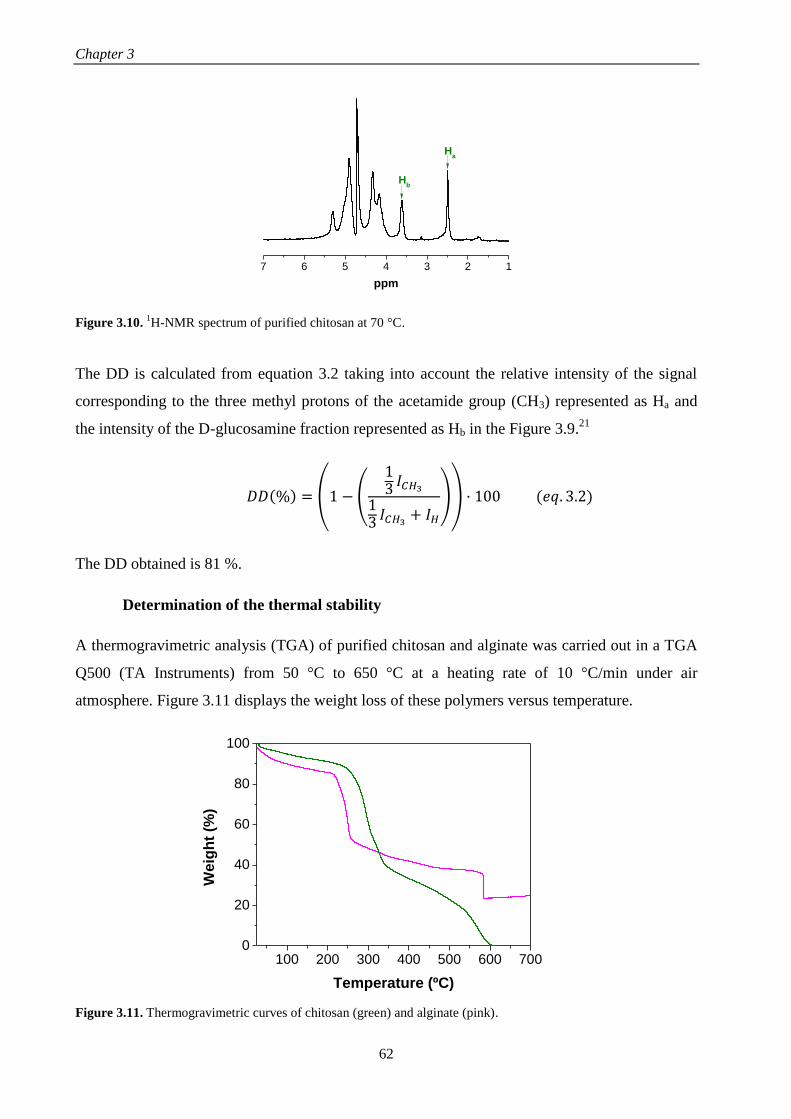
Chapter 3
62
Figure 3.10. 1H-NMR spectrum of purified chitosan at 70 °C.
The DD is calculated from equation 3.2 taking into account the relative intensity of the signal
corresponding to the three methyl protons of the acetamide group (CH3) represented as Ha and
the intensity of the D-glucosamine fraction represented as Hb in the Figure 3.9.21
𝐷𝐷(%) = (1 − (
13 𝐼𝐶𝐻3
13 𝐼𝐶𝐻3
+ 𝐼𝐻
)) · 100 (𝑒𝑞. 3.2)
The DD obtained is 81 %.
Determination of the thermal stability
A thermogravimetric analysis (TGA) of purified chitosan and alginate was carried out in a TGA
Q500 (TA Instruments) from 50 °C to 650 °C at a heating rate of 10 °C/min under air
atmosphere. Figure 3.11 displays the weight loss of these polymers versus temperature.
Figure 3.11. Thermogravimetric curves of chitosan (green) and alginate (pink).
7 6 5 4 3 2 1
Ha
ppm
Hb
100 200 300 400 500 600 7000
20
40
60
80
100
Weig
ht
(%)
Temperature (ºC)

Materials and methods
63
The TGA curve of chitosan exhibits three weight losses. The first one takes place in the interval
between 50 – 170 ºC and it is due to the evaporation of absorbed water in the sample (8.9 %).
The second, between 170 – 400 ºC, is related to depolymerization of chitosan chains,
decomposition of pyranose rings by dehydration and deamination and ring-opening reactions.
The last one between 400 – 600 ºC is due to decomposition of oxidized chitosan, chain scissions
and formation of volatile degradation products.22, 23
23
Ashes above 600 ºC forms a waste of
0.03% which indicates that chitosan is free from metal traces and impurities, therefore the
purification process was carried out correctly.
The alginate spectrum also shows three weight losses. The first one, between 25 – 170 ºC, is due
to evaporation of absorbed water in the sample (11.0 %). The second, between 170 – 500 ºC, is
related to the decomposition of alginate. The last one between 500 – 600 ºC is due to the
formation of Na2CO3.24
The waste formed by Na2CO3 constitutes 23.9%.
Results obtained from TGA analysis, onset degradation temperature, percentage of weight loss
and percentage of ashes calculated at 650 °C, are collected in Table 3.1.
Table 3.1. Onset temperature, weight loss and ashes obtained from TGA of chitosan and alginate.
3.1.3. Alginate based magnetic ferrofluid
Synthesis of alginate based magnetic ferrofluid
For the preparation of an alginate based magnetic ferrofluid, Fe3O4 magnetic nanoparticles (NPs)
were synthetized by a coprecipitation method carried out in an alginate aqueous solution
following the experimental procedure shown in Scheme 3.1. Briefly, 0.834 g of iron (II) sulfate
heptahydrate (FeSO4·7H2O) and 1.6218 g of iron (III) chloride hexahydrate (FeCl3·6H2O) were
dissolved in 10 mL of Milli-Q water under N2 atmosphere. Then, the mixture was added to a
three-necked flask containing 40 mL of alginate (2.5 mg/mL) under N2 atmosphere with
Sample Onset temperature (ºC) Weight loss (%) Ashes (%)
Chitosan 260
536
8.9
51.9
39.1
0.1
Alginate 206
446
11.0
50.7
14.4
23.9

Chapter 3
64
mechanical stirring at 350 rpm using a teflon stirrer and heated at 80 °C for 1 hour. After that,
this mixture was added dropwise under constant stirring to other three-necked flask containing
50 mL of ammonium hydroxide (NH4OH), taking place the color change of the mixture from
yellow orange to black. The mixture was held with stirring for 2 hours at 80 °C under N2
atmosphere. Afterwards, the solution was cooled in an ice bath and washed with Milli-Q water
until neutral pH by magnetic decantation. To obtain the final ferrofluid, this slurry was
redispersed either in 130 or 65 mL of Milli-Q water giving rise to different magnetite
concentrations (ferrofluid 1 and 2, respectively). The Scheme 3.1 includes a photograph
corresponding to the ferrofluid 2 whose colloidal stability was assessed by the inverted tube test.
Scheme 3.1. Sequential process followed for the synthesis of the alginate based magnetic ferrofluid with the
photograph corresponding to the ferrofluid 2. Note that the inverted tube test is showed to assess the colloidal
stability of the synthetized ferrofluid.
Characterization of alginate based magnetic ferrofluid
Determination of the iron content
Iron content was determined by UV-vis transmission spectrophotometry using the thiocyanate
complexation reaction and measuring the absorbance of the iron-thiocyanate complex at 478 nm
wavelength.
𝐹𝑒(𝑎𝑞)3+ + 6 𝑆𝐶𝑁(𝑎𝑞)
− → [𝐹𝑒(𝑆𝐶𝑁)6](𝑎𝑞)3−
Firstly, an aliquot of 100 L of ferrofluid was dissolved in 1:1 v/v HCl 6M/HNO3 (65%) for 2h
and, after that, potassium thiocyanate 1.5M was added to the solution to form the iron-
thiocyanate complex. Afterwards, the magnetite concentration of ferrofluid was determined by
comparing the sample absorbance to a calibration curve. The magnetite concentrations of
ferrofluids 1 and 2 were 4.0 and 8.0 mg/mL, respectively.

Materials and methods
65
To determine the mass of alginate in relation to the iron content, a thermogravimetric analysis
(TGA) was carried out on previously freeze-dried alginate based magnetic ferrofluid using an
analyzer TA Q-500 from 25 ºC to 750 ºC at a heating rate of 10 ºC/min under nitrogen
atmosphere. Figure 3.12 shows the weight loss divided in two steps, one in the range from 25 to
100 ºC and another from 100 to 350 ºC. The first weight loss (2 %wt) corresponds to the
evaporation of water absorbed in the NPs. The second and more significant weight loss (10 %wt)
is due to decomposition of sodium alginate used as surfactant of NPs in water.25, 26
The total
weight loss was 12 %wt and the waste obtained at temperatures above 400 ºC forms 88 %wt of
sample and corresponds to the amount of iron content.27
100 200 300 400 500 600 700
86
88
90
92
94
96
98
100
10%
Weig
ht
(%)
Temperature (ºC)
2%
Figure 3.12. Thermogravimetric analysis of freeze-dried ferrofluid under nitrogen atmosphere.
Determination of the chemical structure
Fourier transform infrared spectroscopy (FTIR) was performed on alginate and previously
freeze-dried ferrofluid using a spectrometer Perkin Elmer Spectrum One DT-IR from 4000 to
450 cm-1
with a resolution of 0.5 cm-1
. Figure 3.13 displays FTIR spectra corresponding to the
ferrofluid (black line) and alginate (pink line). In the spectrum of the ferrofluid. there is a strong
absorption band at 585 cm-1
which is attributed to the vibration of Fe–O and shows the formation
of magnetite nanoparticles. Peaks at 1420 and 1624 cm-1
, attributed to the stretching vibration of
C–O bond, and peak at 2924 cm-1
, due to the stretching vibration of C–H bond, are also
presented in the alginate spectrum. This fact allows to demonstrate the presence of both
components, alginate and iron, in the ferrofluid. The broad band centered at 3430 cm-1
is
assigned to the O–H stretching vibration arising from surface hydroxyl groups adsorbed on
nanoparticles.25, 26, 28

Chapter 3
66
4000 3500 3000 2500 2000 1500 1000 500
585142016242924
% T
ran
sm
itta
nc
e (
a.u
)
Wavenumber (cm-1)
3430
Figure 3.13. FTIR transmittance spectra of freeze-dried ferrofluid (black line) and alginate (pink line).
Morphological characterization
For the determination of the morphology, distribution and average size of the NPs, an aliquot of
the ferrofluid was diluted in ethanol and analyzed by transmission electron microscopy (TEM)
using a TECNAI T20 transmission electron microscope equipped with a CCD-camera Veleta
2Kx2K and operating at an acceleration voltage of 200 kV with LaB6 filament.
Figure 3.14a shows a TEM image of NPs which reveals that Fe3O4 NPs were quasi-spherical and
their size distribution is relatively homogenous. From this TEM image, it is possible to
determine the average particle size and particle size distribution. The histogram (Figure 3.14b)
was obtained from this TEM image by counting 100 particles using the software ImageJ. The
fitting Gaussian curve (orange line) was used to obtain the average size of NPs that was 7.3 ± 1.1
nm.

Materials and methods
67
0 10 20 300
5
10
15
20
25
30
35
40
Co
un
ts
Size (nm)
b)
Figure 3.14. a) TEM image of NPs showing the morphology and the general distribution of particle size and b)
histogram and the fitting Gaussian curve in orange.
To determine the hydrodynamic average size, the polydispersity index (PDI) and the zeta
potential of NPs dispersed in water (ferrofluid), the technique of Dynamic Light Scattering was
employed using a Malvern Nanosizer NanoZS instrument equipped with a 4 mW He-Ne laser (λ
= 633 nm) at a scattering angle of 173º. Samples were measured in polystyrene cuvettes
(SARSTEDT) at 25 ºC. The autocorrelation function was converted into an intensity particle size
distribution with ZetaSizer Software 7.10 by using the Stokes-Einstein equation.
The hydrodynamic diameter and polydispersity of ferrofluids prepared at two different magnetite
concentrations, 4.0 and 8.0 mg/mL, are collected in the Table 3.2. The hydrodynamic diameter
(Dhyd) increases with the concentration of magnetite from 83.1 ± 11.0 nm to 103.5 ± 1.3 nm,
probably due to the aggregation of magnetic nanoparticles. The polydispersity also increases
with the magnetite concentration.
Table 3.2. Dependence of hydrodynamic size and polidispersity with the magnetite concentration.
Figure 3.15a shows the hydrodynamic diameter of NPs dispersed in water (ferrofluid) with a
concentration of 8.0 mg/mL as a function of pH to study the influence of this parameter in the
colloidal stability. NPs have a hydrodynamic diameter around 100 nm with a polydispersity
[Fe3O4] (mg/mL) Dhyd (nm) PDI
4.0 83.1 ± 11.0 0.141 ± 0.030
8.0 103.5 ± 1.3 0.198 ± 0.016
a)

Chapter 3
68
about 0.2 for a pH range from 4.9 to 10. Finally, the electrophoretic mobility of NPs dispersed in
Milli-Q water was measured at 25 ºC increasing pH from 3 to 10. All measurements were
repeated three times. The electrophoretic mobility was transformed into zeta potential using the
Smoluchowski equation and results are exhibited in Figure 3.15b. Zeta potential decreases from
–8.5 mV to –43 mV as pH increases from 3 to 5.7 until reach a plateau. Zeta potential is
negative as expected due to the negative charge of carboxylic groups (COO-) of alginate at the
pH measured.29, 30
At pH higher than 5, alginate-coated NPs are stable due to the presence of
charged carboxylate groups on their surface which provides electrostatic repulsion. On the
contrary, at pH 4 these ionizable groups underwent protonation giving rise to aggregation and
consequent precipitation of the NPs.31
4 5 6 7 8 9 1080
100
120
140
160
Dia
m. E
ffe
c.
(nm
)
pH
a)
3 4 5 6 7 8 9-50
-45
-40
-35
-30
-25
-20
-15
-10
-5
Z
eta
po
ten
tia
l (m
V)
pH
b)
Figure 3.15. a) Hydrodynamic diameter and b) zeta potential of the ferrofluid as function of pH.
Determination of crystalline properties
The crystalline structure of the NPs previously freeze-dried was determined by X-Ray diffraction
(XRD) using a X-Ray diffractometer Bruker D8 Advance at a voltage of 40 kV, an intensity of
40 mA and a rate of 0.5 s/step and its spectrum is showed in Figure 3.16.

Materials and methods
69
10 20 30 40 50 60 70
18.362.8
57.353.8
43.4
35.8
Inte
ns
ity
(a
.u)
2
30.3
Figure 3.16. X-Ray diffraction spectrum of NPs.
Characteristics peaks at 18.3 (D 1 1 1), 30.3 (D 2 2 0), 35.8 (D 3 1 1), 43.4 (D 4 0 0), 53.8 (D 4 2
2), 57.3 (D 5 1 1) and 62.8 (D 4 4 0) correspond with those previously reported for crystalline
magnetite (JCPDS# 19-629). The peak broadening of XRD spectrum indicates the small size of
the resulting nanoparticles. It is difficult to distinguish magnetite (Fe3O4) from maghemite (γ-
Fe2O3) because maghemite has a crystal structure and a lattice spacing similar to magnetite, but
the absence of other characteristic peaks of maghemite in the XRD spectrum and the black color
of the synthetized NPs verifies that it contents mainly Fe3O4 and not γ-Fe2O3 which has a brown
color.25, 32
From the XRD spectrum, the average size of NPs can be estimated using the Debye-Scherrer
equation.28
𝐷 =𝐾 𝜆
𝛽 𝑐𝑜𝑠𝜃 (𝑒𝑞. 3.3)
where D is the average particle size (nm), K is the Scherrer’s constant (0.89), λ is the X-ray
wavelength (0,154 nm), β is the peak full width at half maximum (FWHM) of the highest
diffraction peak in radians and θ is the Bragg diffraction angle.28
The most intense peak (3 1 1)
was used to calculate the average particle size which is 6.8 nm. This value is consistent with the
size determined by TEM image (Figure 3.14) 7.3 ± 1.1 nm.

Chapter 3
70
3.1.4. Conclusions
The purification and characterization of chitosan were carried out obtaining the following data:
the deacetylation degree (DD) of chitosan, determined by RMN-1H, reached a value of 81% and
its molecular weight, determined by capillary viscosimetry at 25°C, was 67100 Da. Besides, the
molecular weight of alginate was determined on the same basis achieving a value of 165500 Da.
The synthesis and characterization of an alginate-based ferrofluid was carried out through a
coprecipitation method. The obtained ferrofluid possessed colloidal stability at neutral pH with
an average particle size of ~ 7 nm and a hydrodynamic diameter around 100 nm for a magnetite
concentration of 8.0 mg/mL. From pH 5 the hydrodynamic diameter and the zeta potential
reached stationary values around 100 nm and -40 mV, respectively. For lower pHs, both
parameters increased until 150 nm and -8 mV, respectively due to the aggregation and
precipitation of the magnetic NPs.
3.2. CHARACTERIZATION TECHNIQUES
This following section describes the most relevant characterization techniques employed to study
the growth, morphology and structure of multilayer films built up via LbL assembly. Some
techniques which have been used occasionally to characterize the properties of materials used in
this work, as for example transmission electron microscopy (TEM), Raman microscopy, X-ray
diffraction (XRD), etc., are not going to be developed in this section and their experimental
procedure will be described in the corresponding section.
3.2.1. Characterization techniques to follow the growth of LbL films
Ellipsometry
Ellipsometry is a non-destructive optical technique to determine the thickness of thin films. A
linearly polarized light beam having p-polarized and s-polarized components is incident on a
sample at an incidence angle to the normal. The reflected wave is, in general, elliptically
polarized (Figure 3.17).33
Ellipsometry measures this change in polarized light upon light
reflection on a sample (or light transmission by a sample). This change provides information
related to optical properties of the sample such as refraction index, thickness and roughness.
Single-wavelength ellipsometry employs a monochromatic light source, usually a laser in the
visible spectral region at a wavelength of 632.8 nm allowing to focus the beam on a small spot
size.34
One of the most important restrictions of ellipsometry measurements is that the surface

Materials and methods
71
roughness of samples has to be small (<30%) because light scattering of rough surfaces reduces
the reflected light intensity and therefore, the ellipsometry measurement becomes difficult.35
Figure 3.17. Schematic representation of a spectroscopic ellipsometry measurement with the change of polarized
light from linear to elliptical after reflection off of the sample.
In the case of multilayer films,36
the final reflected wave is formed by light reflected on all
interphases as shown in Figure 3.18. The light firstly hits in the interphase air-polymer layer
being a part of this light reflected and other transmitted through the polymer film thickness until
the next interphase polymer layer-substrate, where it takes place a phase difference which is
related to the polymer layer thickness by the following equation.
𝛽 = 2𝜋 (𝑑
𝜆) 𝑁2 𝑐𝑜𝑠𝛼2 (𝑒𝑞. 3.4)
where d is the polymer layer thickness, is the wavelength of the incident light, 2 is the
incidence angle and N2 is the complex refraction index of the polymer layer.
Figure 3.18. Schematic representation of light reflection upon a film supported on a substrate.
Ellipsometry does not directly measure the refraction coefficients but measures two values, the
amplitude ratio () and phase difference (Δ) between light waves known as p-polarized and s-
polarized light waves.35
These two parameters are related by the parameter expressed as.35, 37

Chapter 3
72
𝜌 =𝑟𝑝
𝑟𝑠= 𝑡𝑎𝑛𝜓 exp(𝑖𝛥) (𝑒𝑞. 3.5)
where rp and rs are the complex refraction coefficients for the perpendicular and parallel
polarization, respectively. In the case of multilayer films, these coefficients are expressed as:
𝑟𝑝 =𝑟12𝑝
+ 𝑟23𝑝exp (−𝑖2𝛽)
1 + 𝑟12𝑝+ 𝑟23𝑝
exp (−𝑖2𝛽) ; 𝑟𝑠 =
𝑟12𝑠+ 𝑟23𝑠
exp (−𝑖2𝛽)
1 + 𝑟12𝑠+ 𝑟23𝑠
exp (−𝑖2𝛽) (𝑒𝑞. 3.6)
The optical parameters such as the refraction index, n, the extinction coefficient, k, and the
thickness, d, can be related to parameters and Δ measured by ellipsometry through different
mathematical models. In the case of polymer thin films, models where the dependence of the
wavelength of the real part of the refraction index is fitted to the Cauchy equation (eq. 3.7) are
accurate.38
𝑛(𝜆) = 𝐴𝑛 +𝐵𝑛
𝜆2 (𝑒𝑞. 3.7)
where n is the refraction index, is the wavelength and A and B are the Cauchy parameters.
Measurements of the film thickness presented in this thesis were carried out with a PLASMOS
SD 2300 ellipsometer operating at the single wavelength of 632.8 nm and a constant angle of
70°. This technique was employed during a short stay in the group of Prof. Fouzia Boulmedais in
the Institute Charles Sadron, CNRS, Strasbourg.
Quartz Crystal Microbalance (QCM)
The buildup of multilayer films can be followed in situ by a quartz crystal microbalance (QCM).
QCM is a nanogram sensitive technique introduced by Sauerbrey that employs the change in the
frequency of a single quartz crystal resonator to measure the mass of a rigid film deposited on
the quartz resonator surface.39
𝛥𝑚 = −ℎ𝑞𝜌𝑞
𝑓0
∆𝑓𝑣
𝜈= −𝑐
∆𝑓𝑣
𝜈 (𝑒𝑞. 3.8)
where ℎ𝑞 is the thickness of the quartz, 𝜌𝑞 is the density of quartz (~17.7ng·cm-2
·Hz-1
for a
crystal of 5-MHz), 𝑓0 is the fundamental resonant frequency of quartz (5 MHz), ν is the overtone
number and c the mass sensitivity constant.

Materials and methods
73
The use of QCM for liquid applications reveals that the liquid phase often incorporates viscous
and elastic contributions to the frequency change contradicting the Sauerbrey’s assumption that
the adsorbed mass must be rigidly adsorbed, being necessary a new approach to describe the
mass deposits with frictional dissipative losses due to their viscoelastic character. This gave rise
to QCM with dissipation monitoring (QCM-D) which allows to simultaneously measure the
changes in the resonant frequency and in the viscoelastic properties (dissipation) when a film is
adsorbed at the quartz crystal surface.40
A decrease in ∆f/ν is usually associated to an increase of
the mass adsorbed on the quartz and a decrease of dissipation (D) at constant mass is usually
associated to a stiffer (less elastic) film.41
The dissipation parameter is given by equations 3.9 (dimensional) and 3.10 (dimensionless).40
𝐷 =1
𝜋𝑓𝜏 (𝑒𝑞. 3.9)
𝐷 =𝐸𝑑𝑖𝑠𝑠𝑖𝑝𝑎𝑡𝑒𝑑
2𝜋𝐸𝑠𝑡𝑜𝑟𝑒𝑑 (𝑒𝑞. 3.10)
where τ is the time constant of the amplitude decay of the oscillating crystal as the driving
voltage over the crystal is turned off, Edissipated is the energy dissipated during one oscillatory
cycle and Estored the energy stored in the oscillating system.
The QCM-D method allows to test f and D values at its fundamental frequency (~ 5 MHz) as
well as at several overtones. The QCM-D response of a viscoelastic film can be modelled using
the Voigt model to extract meaningful parameters such as thickness, viscosity or shear
modulus.40, 41
This model assumes that the film covers homogenously all the active area of the
sensor, has uniform thickness and density under no slip conditions and the medium in which the
sensor is resonating is a bulk Newtonian fluid. Taking into account these assumptions, the
resonant frequency and the dissipation can be described by the equations 3.11 and 3.12
respectively.42
∆𝑓 ≈ −1
2𝜋𝜌𝑞ℎ𝑞{
𝜂𝐵
𝛿𝐵+ ℎ𝑙𝜌𝑙 (
𝜂𝐵
𝛿𝐵)
2 𝜂𝑙𝜔2
𝜇𝑙2 + 𝜔2𝜂𝑙
2} (𝑒𝑞. 3.11)
Δ𝐷 ≈1
2𝜋𝑓𝜌𝑞ℎ𝑞{
𝜂𝐵
𝛿𝐵+ 2ℎ𝑙 (
𝜂𝐵
𝛿𝐵)
2 𝜇𝑙𝜔
𝜇𝑙2 + 𝜔2𝜂𝑙
2} (𝑒𝑞. 3.12)

Chapter 3
74
where 𝜔 is the angular frequency of the oscillation, 𝜌𝑞 and ℎ𝑞 are the density and thickness of
the quartz crystal respectively, ℎ𝑙, 𝜌𝑙, 𝜂𝑙 and 𝜇𝑙 are the thickness, density, viscosity and shear
modulus of the adsorbed layer respectively, 𝜂𝐵 and 𝜌𝐵 are the viscosity and density of the bulk
liquid and 𝛿𝐵 the viscous penetration depth of the shear wave in the bulk liquid calculated by the
following equation.
𝛿𝐵 = (2𝜂𝐵
𝜌𝐵𝜔)
1/2
(𝑒𝑞. 3.13)
QCM measurements described in this PhD work were carried out on a Q-sense D300 apparatus
from Q-Sense AB (Gothenburg, Sweden) during a short stay in the group of Prof. Fouzia
Boulmedais in the Institute Charles Sadron, CNRS, Strasbourg.
Fourier transform Infra-Red spectroscopy (FTIR)
Fourier transform Infra-Red spectroscopy (FTIR) is other technique that allows to follow the
growth of multilayer films.
Infrared (IR) spectroscopy is based on the interaction of IR light with matter. The infrared region
considered is the mid-infrared (4500 cm-1
to 400 cm-1
). IR light passes through a sample and
certain frequencies of the light are absorbed by the chemical bonds of the substance leading to
molecular vibrations. The frequencies and intensities of absorbed IR light depend on the specific
bond strengths and atoms of that molecule, being the absorption pattern known as a spectrum,
unique for each material. An absorption band is characterized by the wavelength at which
maximum absorption takes place and the intensity of absorption at this wavelength. In addition,
the intensity of this band is proportional to the number of molecules of the material, so that IR
spectrum provides not only qualitative, but also quantitative information. Taking into account
these characteristics, the layer-by-layer deposition process can be followed by FTIR
spectroscopy through recording the FTIR spectra after deposition of one or more polymer layers.
The growth process is determined following the absorbance of one or more characteristic bands
of the polymer or polymers that are being assembled. If the absorbance of these bands increases
their intensity with the number of deposited layers, the growth of the film takes place.
FTIR measurements presented in this thesis were carried out in a Perkin Elmer Spectrum One
FTIR Spectrometer. For the follow-up of the LbL process, the deposition of the polymer
solutions was carried out on a polystyrene (PS) substrate with a thickness of 70 µm. The signal
corresponding to PS was subtracted from the multilayer spectra.

Materials and methods
75
3.2.2. Characterization techniques to determine morphological properties
Scanning Electron Microscopy (SEM)
Scanning electron microscopy (SEM) is a technique to examine and analyze the surface
morphology of samples.
The electron beam from an electron gun column is demagnified by condenser lenses into a fine
spot as small as 1 nm of diameter which is scanned across a selected area of the sample surface.
Interactions between the electron beam and the sample give rise to different intensity signals
which are collected by detectors and stored in the computer memory to be subsequently mapped
as variations in brightness on the image display. The most widely used signal produced by the
interaction of the primary electron beam with the sample is the secondary electron emission
signal which varies with the topography of the sample surface being dark for recesses and bright
for edges. The magnification of the image is determined by the ratio of the size of the displayed
image to the size of the area scanned on the sample.43, 44
In this work a FESEM Hitachi model SU8000 with TE detector operated at 0.5-30 kV was
employed.
Atomic Force Microscopy (AFM)
The operation method of an AFM is based on the surface forces acting on a sharp tip in close
proximity to a surface.45
Because of the long-range nature of the interaction forces, the vertical
resolution of an AFM is typically less than a nanometer whereas the lateral resolution is
determined by the tip radius and sample roughness. The sharp tip is usually supported at the end
of a cantilever that is scanned over the surface of a sample. Information about the sample surface
can be obtained by measuring how the deflection of the cantilever, which is caused by
interactions between the tip and the surface, varies with position. In dynamic force microscopy
the cantilever is made to vibrate at a specific frequency and the deflection of the tip is measured
at this frequency. To measure cantilever motion while scanning, a laser is focused on the
cantilever and the reflected light is directed onto a segmented photodiode.45, 46
When discussing the nature of the interaction force between tip and substrate, it is often
convenient to approximate the tip as a sphere with radius R. This sphere then interacts with the
sample via a number of possible forces that cause the cantilever to deflect. In contact mode, the
tip exerts a force directly on the sample as it is scanned across it. In intermittent-contact or

Chapter 3
76
tapping mode, the tip vibrates at its resonance frequency by action of a piezoelectric giving rise
to a sinusoidal motion of the cantilever that is accurately monitored by the AFM control
electronics.45
This mode allows the characterization of soft materials such as polymers without
damaging them.46
In this work AFM experiments were carried out in two different equipments. Multimode
Scanning probe microscope (Veeco/Bruker), with a di NanoScope IVa Controller using the
conventional height mode (tapping mode, normal AFM) at 320 kHz resonant frequency and a
nominal force constant of 42 N/m in the Instituto de Ciencia y Tecnología de Polímeros, CSIC.
Bruker Multimode 8, with a NanoScope V controller using the conventional height mode
(tapping mode, normal AFM) at a scan rate of 0.5 and 1.5 in the group of Prof. T. Ezquerra in the
Instituto de Estructura de la Materia, CSIC. The analysis of data was performed using the
software NanoScope Analysis 1.5.
3.2.3. Characterization techniques to determine mechanical properties
Atomic Force Microscopy (AFM) nanoindentation measurements
AFM nanoindentation measurements were carried out using a Bruker Multimode 8, with a
Nanoscope V controller in the group of Prof. T. Ezquerra in the Instituto de Estructura de la
Materia, CSIC.
PeakForce Quantitative Nanomechanical Mapping (PF-QNM) is a contact AFM protocol which
is based on the force-volume method. Force-distance curves are collected by nanoindentation of
the sample in a point-by-point fashion. In this method, the piezo-scanner is oscillated at 2 kHz
while the probe remains at rest. This fact enables a high speed and simultaneous capture of force-
distance curves and topographic images.47
The maximum force (peak force) is controlled at each
pixel to obtain force-distance curves which are then used as feedback signal.
Figure 3.19 shows an example of the PF-QNM force-distance curve obtained from a
measurement of a polymer film. The Z-axis reflects the distance ramped by the piezo-scanner in
the vertical direction. The trace curve is obtained when the tip is approaching the surface of the
sample and the retrace curve when the tip is withdrawing from it. As it can be observed, an
attractive force appears when the tip is approaching the sample followed by an elastic regime in
which the force is proportional to the deformation. The adhesion force is related to the minimum
of the retrace force-distance curve (upon withdrawal) and the dissipation is the area between the
trace and retrace of the force-distance curves.

Materials and methods
77
Figure 3.19. Force-distance curve while approaching the surface (trace) and withdrawal (retrace). The dashed line
corresponds to fitting of the experimental results to the DMT model.
For the determination of the nanomechanical properties of multilayer films shown in Chapter 5,
every tip radius was calibrated against a polystyrene standard of known elastic modulus
(Bruker), considering the Derjaguin-Muller-Toporov (DMT) model48
through equation 3.14.
𝐸𝑟 =3(𝐹𝑡𝑖𝑝 − 𝐹𝑎𝑑ℎ)
4√𝑅𝑑3 (𝑒𝑞. 3.14)
where 𝐸𝑟 is the reduced Young modulus, 𝐹𝑡𝑖𝑝 is the force on the tip, 𝐹𝑎𝑑ℎ is the adhesive force
between the AFM tip and the sample, R is the AFM tip radius and d is the penetration depth.
Provided that the tip Young modulus is much higher than the sample modulus, the reduced
modulus (Er) and the Young modulus of the sample (Es) are related through equation 3.15.
𝐸𝑠 =𝐸𝑟
1 − 𝜈𝑠2
(𝑒𝑞. 3.15)
where ν is the Poisson’s ratio value.
Analysis of the force-distance curves is automatically performed by a software and allows to
extract the height, elastic modulus, adhesion force, deformation and dissipation simultaneously
by application of the DMT model. The employment of the DMT model is justified by the fact
that the chosen regime of applied forces allows us to study the nanomechanical response of
multilayer films in the elastic regime, i.e., without permanent deformation of the sample’s
surface. The Young´s Modulus is obtained by fitting the retrace curve using equations (3.14) and
(3.15) and a Poisson´s ratio of 0.3.48
Note in Figure 3.19 that the part of the force-distance curve
Trace
Retrace
Ad
hes
ion
Deformation
Dissipation
Pea
k fo
rce

Chapter 3
78
used to estimate the DMT modulus superimposed in both loading and unloading regime. To data
analysis the software NanoScope Analysis 1.5 was employed.
3.2.4. Characterization techniques to determine the structure of LbL films
Grazing-incidence small-angle X-ray scattering (GISAXS)
GISAXS is a powerful technique to study nanostructures at the surface and in buried structures
of thin films. GISAXS can provide information about lateral structures with sizes from few
nanometers to few hundred nanometers. The incident X-ray beam impinges the sample at
shallow angles (<1º), thus total external reflection on the surface may take place. By changing
slightly the incident angle it is possible to vary the depth of the incident beam into the sample
allowing to study the structural changes with the thickness. Due to the fact that the diffraction in
GISAXS experiments is induced by a weak evanescent superficial wave, the use of X-ray
synchrotron radiation is especially advantageous.49, 50
As can be shown in Figure 3.20, in a GISAXS experiment an X-ray beam impinges under an
incident angle (αi) onto the film surface. The intensity dispersed by the sample is collected by a
2D detector. α and ω are the scattering angles in vertical and horizontal direction. Both the direct
beam and the reflected beam are blocked by a beam stop which protects the detector. The
dispersion at low angles (GISAXS) takes place when the detector is far away from the sample
and the maximum dispersion angle collected by it is low.51
Figure 3.20. Schematic representation of a GISAXS experiment including a 2D GISAXS pattern of a multilayer
film.

Materials and methods
79
The information collected from these experiments can be interpreted as a function of the two
orthogonal scattering vectors, 𝑞𝑦 and 𝑞𝑧, which give information about structural correlations
parallel and perpendicular to the plane of the sample, respectively.50, 51
𝑞𝑦 =2𝜋
𝜆𝑠𝑖𝑛𝜔𝑐𝑜𝑠𝛼 (𝑒𝑞. 3.16)
𝑞𝑧 =2𝜋
𝜆(𝑠𝑖𝑛𝛼𝑖 + 𝑠𝑖𝑛𝛼) (𝑒𝑞. 3.17)
Lateral correlation between scattering objects on the film surface can induce some scattered
intensity which can be observed out of the meridian (line m) and generate intensity maximums
related to the correlation maximums.51
GISAXS experiments reported in this thesis were carried out at BM26 beamline of the European
Synchrotron Radiation Facility (ESRF) sited in Grenoble (France), using a wavelength of λ =
0,103 nm and a camera of 981 x 1043 pixels (size of the pixel 172 x 172 m2) to data
acquisition. The detector employed was a PILATUS 1M and the distance sample-detector used
in this work was 6.87 m. To data analysis the FIT2D software was employed.
Dual-Beam (Focused Ion Beam (FIB) – Scanning Electron Microscopy (SEM))
To determine the inner organization of the polymer films, dual beam FIB/SEM was employed.
The dual beam incorporates in a single system two columns, a focused ion beam (FIB) column
and a scanning electron microscopy column (SEM). This combination is well suited for cross-
section studies due to the fact that ion beam mills normal to the sample surface and the electron
beam views the cross-section face. These two beams intersect with the sample in a coincident
point allowing the ion milling and electron imaging of the same region. In addition, other
components such as gas injectors and energy-dispersive X-ray spectroscopy (EDX) are also
aligned in this position to achieve an optimum operation. The image obtained from a dual beam
is a combination of the electron beam imaging and the ion beam imaging. Although the electron
beam is the primary imaging tool which produces images with high resolution, the ion beam can
improve it giving rise to a strong image contrast.52
These experiments were carried out in a Nova 200 NanoLab, FEI Company during my stay in
the group of Prof. Gerardo Goya in the Institute of Nanoscience of Aragon. Films were coated
with platinum for imaging with the FIB-SEM. SEM images were taken at 5 kV with a Field

Chapter 3
80
Emission Gun (FEG) column, and a combined Ga-based 30 kV (10 pA) ion beam was used to
cross-sectioning films. These investigations were completed by energy-dispersive X-ray
spectroscopy (EDX) for chemical analysis.
Figure 3.21. Dual beam Nova 200 NanoLab, FEI Company installed in the Institute of Nanoscience of Aragon.
3.3. REFERENCES
1. Croisier, F.; Jérôme, C., Chitosan-based biomaterials for tissue engineering. European Polymer
Journal 2013, 49, (4), 780-792.
2. Xu, C.; Lei, C.; Meng, L.; Wang, C.; Song, Y., Chitosan as a barrier membrane material in
periodontal tissue regeneration. Journal of Biomedical Materials Research Part B: Applied Biomaterials
2012, 100, (5), 1435-1443.
3. Cook, M. T.; Tzortzis, G.; Khutoryanskiy, V. V.; Charalampopoulos, D., Layer-by-layer coating
of alginate matrices with chitosan–alginate for the improved survival and targeted delivery of probiotic
bacteria after oral administration. Journal of Materials Chemistry B 2013, 1, (1), 52-60.
4. Lawrie, G.; Keen, I.; Drew, B.; Chandler-Temple, A.; Rintoul, L.; Fredericks, P.; Grøndahl, L.,
Interactions between alginate and chitosan biopolymers characterized using FTIR and XPS.
Biomacromolecules 2007, 8, (8), 2533-2541.
5. Signini, R.; Campana Filho, S. P., On the preparation and characterization of chitosan
hydrochloride. Polymer bulletin 1999, 42, (2), 159-166.
6. Amado, I. R.; Vázquez, J. A.; Pastrana, L.; Teixeira, J. A., Microbial production of hyaluronic
acid from agro-industrial by-products: Molasses and corn steep liquor. Biochemical Engineering Journal
2017, 117, Part A, 181-187.
7. Mero, A.; Campisi, M., Hyaluronic acid bioconjugates for the delivery of bioactive molecules.
Polymers 2014, 6, (2), 346-369.
8. Larkin, A. L.; Davis, R. M.; Rajagopalan, P., Biocompatible, detachable, and free-standing
polyelectrolyte multilayer films. Biomacromolecules 2010, 11, (10), 2788-2796.

Materials and methods
81
9. Richert, L.; Lavalle, P.; Payan, E.; Shu, X. Z.; Prestwich, G. D.; Stoltz, J.-F.; Schaaf, P.; Voegel,
J.-C.; Picart, C., Layer by layer buildup of polysaccharide films: physical chemistry and cellular adhesion
aspects. Langmuir 2004, 20, (2), 448-458.
10. Thiele, J.; Ma, Y.; Bruekers, S.; Ma, S.; Huck, W. T., 25th Anniversary article: designer
hydrogels for cell cultures: a materials selection guide. Advanced Materials 2014, 26, (1), 125-148.
11. Su, K.; Wang, C., Recent advances in the use of gelatin in biomedical research. Biotechnology
letters 2015, 37, (11), 2139-2145.
12. Palama, I. E.; Musaro, M.; Coluccia, A. M.; D'Amone, S.; Gigli, G., Cell uptake and validation of
novel PECs for biomedical applications. Journal of drug delivery 2011, 2011.
13. Szarpak, A.; Cui, D.; Dubreuil, F. d. r.; De Geest, B. G.; De Cock, L. J.; Picart, C.; Auzely-Velty,
R., Designing hyaluronic acid-based layer-by-layer capsules as a carrier for intracellular drug delivery.
Biomacromolecules 2010, 11, (3), 713-720.
14. Lungu, C. N.; Diudea, M. V.; Putz, M. V.; Grudziński, I. P., Linear and Branched PEIs
(Polyethylenimines) and Their Property Space. International Journal of Molecular Sciences 2016, 17, (4),
555.
15. Vicennati, P.; Giuliano, A.; Ortaggi, G.; Masotti, A., Polyethylenimine in medicinal chemistry.
Current medicinal chemistry 2008, 15, (27), 2826-2839.
16. Brunot, C.; Ponsonnet, L.; Lagneau, C.; Farge, P.; Picart, C.; Grosgogeat, B., Cytotoxicity of
polyethyleneimine (PEI), precursor base layer of polyelectrolyte multilayer films. Biomaterials 2007, 28,
(4), 632-640.
17. Rinaudo, M.; Milas, M.; Dung, P. L., Characterization of chitosan. Influence of ionic strength and
degree of acetylation on chain expansion. International Journal of Biological Macromolecules 1993, 15,
(5), 281-285.
18. King, K., Changes in the functional properties and molecular weight of sodium alginate following
γ irradiation. Food hydrocolloids 1994, 8, (2), 83-96.
19. Pamies, R.; Cifre, J. G. H.; Martínez, M. d. C. L.; de la Torre, J. G., Determination of intrinsic
viscosities of macromolecules and nanoparticles. Comparison of single-point and dilution procedures.
Colloid and Polymer Science 2008, 286, (11), 1223-1231.
20. Dash, M.; Chiellini, F.; Ottenbrite, R. M.; Chiellini, E., Chitosan—A versatile semi-synthetic
polymer in biomedical applications. Progress in Polymer Science 2011, 36, (8), 981-1014.
21. Arteche Pujana, M. Nanogeles de quitosano modificados con entrecruzantes biocompatibles para
su aplicación en la liberación selectiva de fármacos anticancerígenos. Universidad del País Vasco, Leioa,
2013.
22. Zawadzki, J.; Kaczmarek, H., Thermal treatment of chitosan in various conditions. Carbohydrate
polymers 2010, 80, (2), 394-400.
23. Bengisu, M.; Yilmaz, E., Oxidation and pyrolysis of chitosan as a route for carbon fiber
derivation. Carbohydrate polymers 2002, 50, (2), 165-175.
24. Soares, J. P.; Santos, J. E.; Chierice, G. O.; Cavalheiro, E. T. G., Thermal behavior of alginic acid
and its sodium salt. Eclética Química 2004, 29, 57-64.

Chapter 3
82
25. Cai, W.; Wan, J., Facile synthesis of superparamagnetic magnetite nanoparticles in liquid polyols.
Journal of Colloid and Interface Science 2007, 305, (2), 366-370.
26. Sundar, S.; Mariappan, R.; Piraman, S., Synthesis and characterization of amine modified
magnetite nanoparticles as carriers of curcumin-anticancer drug. Powder Technology 2014, 266, 321-328.
27. Khalkhali, M.; Rostamizadeh, K.; Sadighian, S.; Khoeini, F.; Naghibi, M.; Hamidi, M., The
impact of polymer coatings on magnetite nanoparticles performance as MRI contrast agents: a
comparative study. DARU Journal of Pharmaceutical Sciences 2015, 23, (1), 1.
28. Yan, H.; Zhang, J.; You, C.; Song, Z.; Yu, B.; Shen, Y., Influences of different synthesis
conditions on properties of Fe 3 O 4 nanoparticles. Materials Chemistry and Physics 2009, 113, (1), 46-
52.
29. Jiang, W.; Chen, X.; Niu, Y.; Pan, B., Spherical polystyrene-supported nano-Fe3O4 of high
capacity and low-field separation for arsenate removal from water. Journal of Hazardous Materials 2012,
243, 319-325.
30. Calatayud, M. P.; Sanz, B.; Raffa, V.; Riggio, C.; Ibarra, M. R.; Goya, G. F., The effect of surface
charge of functionalized Fe3O4 nanoparticles on protein adsorption and cell uptake. Biomaterials 2014,
35, (24), 6389-6399.
31. Bloemen, M.; Brullot, W.; Luong, T. T.; Geukens, N.; Gils, A.; Verbiest, T., Improved
functionalization of oleic acid-coated iron oxide nanoparticles for biomedical applications. Journal of
Nanoparticle Research 2012, 14, (9), 1100.
32. Iida, H.; Takayanagi, K.; Nakanishi, T.; Osaka, T., Synthesis of Fe 3 O 4 nanoparticles with
various sizes and magnetic properties by controlled hydrolysis. Journal of Colloid and Interface Science
2007, 314, (1), 274-280.
33. Woollam, J. A.; Snyder, P. G., Fundamentals and applications of variable angle spectroscopic
ellipsometry. Materials Science and Engineering: B 1990, 5, (2), 279-283.
34. Cobet, C., Ellipsometry: A Survey of Concept. In Ellipsometry of functional organic surfaces and
films, Springer: Heidelberg, 2014; pp 1-26.
35. Fujiwara, H., Introduction to Spectroscopic Ellipsometry. In Spectroscopic Ellipsometry:
Principles and Applications, John Wiley & Sons, Ltd: Chichester, 2007; pp 1-11.
36. Bermejo, J. S. Modificación química y caracterización de superficies poliméricas. Determinación
del perfil de profundidad. Universidad Complutense de Madrid, Madrid, 2001.
37. Garcia-Caurel, E.; Ossikovski, R.; Foldyna, M.; Pierangelo, A.; Drévillon, B.; De Martino, A.,
Advanced Mueller Ellipsometry Instrumentation and Data Analysis. In Ellipsometry at the Nanoscale,
Springer: Heidelberg, 2013; pp 31-144.
38. Poelman, D.; Smet, P. F., Methods for the determination of the optical constants of thin films
from single transmission measurements: a critical review. Journal of Physics D: Applied Physics 2003,
36, (15), 1850.
39. Sauerbrey, G., Verwendung von Schwingquarzen zur Wägung dünner Schichten und zur
Mikrowägung. Zeitschrift für Physik 1959, 155, (2), 206-222.
40. Dixon, M. C., Quartz crystal microbalance with dissipation monitoring: enabling real-time
characterization of biological materials and their interactions. Journal of Biomolecular Techniques 2008,
19, (3), 151-158.

Materials and methods
83
41. Alves, N. M.; Picart, C.; Mano, J. F., Self Assembling and Crosslinking of Polyelectrolyte
Multilayer Films of Chitosan and Alginate Studied by QCM and IR Spectroscopy. Macromolecular
Bioscience 2009, 9, (8), 776-785.
42. Boudou, T.; Crouzier, T.; Auzély-Velty, R.; Glinel, K.; Picart, C., Internal Composition versus
the Mechanical Properties of Polyelectrolyte Multilayer Films: The Influence of Chemical Cross-Linking.
Langmuir 2009, 25, (24), 13809-13819.
43. Vernon-Parry, K., Scanning electron microscopy: an introduction. III-Vs Review 2000, 13, (4),
40-44.
44. Aharinejad, S. H.; Lametschwandtner, A., Fundamentals of Scanning Electron Microscopy. In
Microvascular Corrosion Casting in Scanning Electron Microscopy, Springer: Wien, 1992; pp 44-51.
45. Reifenberger, R., Introduction to Scanning Probe Microscopy. In Fundamentals of Atomic Force
Microscopy. Part I: Foundations, World Scientific: Singapore, 2015; Vol. 4, pp 1-20.
46. Garcia, R.; Herruzo, E. T., The emergence of multifrequency force microscopy. Nature
nanotechnology 2012, 7, (4), 217-226.
47. Marius, C.; Seth, L. Y.; Vladimir, V. T., Recent advances in micromechanical characterization of
polymer, biomaterial, and cell surfaces with atomic force microscopy. Japanese Journal of Applied
Physics 2015, 54, 1-13.
48. Derjaguin, B. V.; Muller, V. M.; Toporov, Y. P., Effect of contact deformations on the adhesion
of particles. Journal of Colloid and Interface Science 1975, 53, 314-326.
49. Santoro, G.; Yu, S., Grazing Incidence Small Angle X-Ray Scattering as a Tool for In- Situ Time-
Resolved Studies. In X-ray Scattering, InTech: Rijeka, 2017; pp 29-52.
50. Roth, S. V., A deep look into the spray coating process in real-time—the crucial role of x-rays.
Journal of Physics: Condensed Matter 2016, 28, (40), 403003.
51. Rebollar, E.; Pérez, S.; Hernández, J. J.; Martín-Fabiani, I.; Rueda, D. R.; Ezquerra, T. A.;
Castillejo, M., Assessment and formation mechanism of laser-induced periodic surface structures on
polymer spin-coated films in real and reciprocal space. Langmuir 2011, 27, (9), 5596-5606.
52. Young, R. J.; Moore, M. V., Dual-Beam (FIB-SEM) Systems. . In Introduction to Focused Ion
Beams: Instrumentation, Theory, Techniques and Practice, Springer US: Boston, 2005; pp 247-268.


CHAPTER 4
Layer-by-layer films from natural
polymers. Study of the growth
mechanism


Layer-by-layer films from natural polymers
87
In this chapter the LbL assembly of two different multilayer systems based on natural polymers
was studied. In the first one comprising alginate and chitosan, the optimal conditions for the
spray LbL assembly, concentration and pH, were evaluated and the growth mechanism for films
built up through spray LbL was studied. In addition, a comparison between dipping assisted LbL
and spray assisted LbL was stablished. In the second system, the optimal conditions for the LbL
assembly of hyaluronic acid and poly(allylamine hydrochloride), the kind of assembly process
and the influence of a third component, alginate-Fe2+
, in the buildup process were analyzed.
4.1. INTRODUCTION
In recent decades, layer-by-layer (LbL) assembly has attracted an increasing attention to
fabricate polymeric films due to its simplicity, versatility and nanoscale control of film
thickness.1 The fundamentals and differences between spray LbL and dipping LbL techniques
were described in detail in the chapter 2.
The growth of multilayer structures can be determined by multiple techniques. Besides the
employment of ellipsometry,2 quartz crystal microbalance
3 and FTIR spectroscopy
4 fully
explained in the chapter 3, there are other techniques to follow the multilayer deposition process.
Confocal laser microscopy (CLSM) which determines the thickness between fluorescently-
labeled polyelectrolytes assembled into the multilayer structure has been employed to determine
the thickness corresponding to the Alg layer incorporated between in PLL/HA or PAH/PSS
multilayers by utilization of fluorescent PLL or PAH layers.5, 6
Surface plasmon resonance (SPR)
is highly sensitive to refractive index changes at the surface and can be used to monitor the
adsorption process and cyclic voltammetry is an electrochemical procedure used to follow the
growth by acquiring information on the redox behavior of electrochemically active species.7, 8
Other useful technique to follow the multilayer assembly process is dual-polarization
interferometry (DPI), which measures changes in the thickness and the refractive index of
adsorbed layers in situ. In the case of PLL/PGA films, DPI data for the thickness and refractive
index were more consistent than these ones obtained by ellipsometry.9 Optical waveguide
lightmode spectroscopy (OWLS) is a highly sensitive method (precision ∼ 1 ng/cm2) for
measuring macromolecular adsorption kinetics of multilayer films at the solid-liquid interface.
Detection system is based on excitation of guided modes through a polarized laser beam directed
at the surface of the sample. Afterwards, the mass and thickness of every layer can be
determined making use of an optical model.10
Scanning angle reflectometry (SAR) is other
optical technique to study the structural parameters characterizing thin macromolecular adsorbed

Chapter 4
88
films. It measures the changes in the reflected intensity of a TM-wave around the Brewster angle
before and after each deposition step.11
In the case of polyelectrolyte multilayers films which can
be are formed by an homogeneous and isotropic monolayer, their optical properties can be
defined using the monolayer model in both techniques, SAR and OWLS. However, for
multilayer films more complex, the experimentally determined parameters depend strongly on
the model used for the analysis and there are significant differences between methods, the mass
and the thickness are model independent for OWLS and extremely sensitive to the model for
SAR.11
The growth process can also be studied by UV-Vis spectroscopy which determines the
cumulative absorption attributed to stepwise deposition of UV-active polyelectrolytes during the
LbL adsorption following the absorbance of one or more characteristic bands of the polymer or
polymers that we are assembling.12
For the assembly of oppositely charged polyelectrolytes, the
buildup process can be followed by charge reversal changes through the zeta potential
measurement by Dynamic light scattering (DLS). As an example, the assembly of anionic
poly(4-styrenesulfonic acid) and cationic poly(diallyldimethylammonium chloride) has been
followed employing this technique, but it has the disadvantage that it is difficult to translate the
zeta potential measurement to a real surface charge density, especially due to the fact that the
surface charge is spread out into a thin film.13
The growth of multilayer polymer structures can
be also monitored by measuring the wettability of every assembled polymer layer through
contact angle measurements of polymers with different hydrophobicity, as it is the case of
poly(methacrylic acid) (PMAA) and poly(4-vinylpyridine-co-(aminopropyl)methacrylamide)
(PVP-NH2) which have contact angles of ~15º and ~30º, respectively.14
In contrast to optical
techniques, although zeta potential and contact angle allow to follow the deposition process, the
thickness of every deposited layer cannot be obtained.
Most of these techniques allow to determine the thickness of every layer and/or the global
thickness of the multilayer structure. In addition to these, Atomic force microscopy (AFM) is
other useful technique to obtain the thickness by making a scratch on the sample and recording
height profile of the transition zone, then the thickness is obtained from the depth profile of this
image.2 Scanning electron microscopy also allows to determine the thickness of multilayer films
by measuring their cross-section.4
To the best of our knowledge, first studies regarding to the multilayer assembly of chitosan (Chi)
and alginate (Alg), the main polymer system employed in this PhD work, were carried out over
particulate substrates by dipping LbL with a centrifugation step after each deposited layer.15, 16
The buildup of multilayer Chi/Alg films on planar substrates through dipping LbL was carried

Layer-by-layer films obtained from natural polymers
89
out later to study the effect of the assembly pH on the kinetics and surface composition of these
Chi/Alg films in situ through surface plasmon resonance (SPR) spectroscopy.17
More studies
related to the assembly of Alg/Chi films have been explained in detail in the chapter 2.
With regards to HA/PAH films, one of the most important properties is their swelling behavior
which influences the hydration of the film and the loading and release performance. The
influence of the polymer concentration and pH on the HA/PAH assembly by dipping and later
swelling was studied with the objective of using these multilayer films for potential applications
of controlled release, membrane filtration and biomaterial coatings.18, 19
Assembly of HA/PAH
by dipping has been also used to make multilayer microcapsules with the ability of remaining
stable outside cells and breaking open once internalized by cells which allows to use them as
carriers for intracellular drug delivery.20, 21
The aim of this chapter is the determination of the growth mechanism of multilayer films
fabricated from two different polymer systems, Chi/Alg and PAH/HA. The main experimental
probes were ellipsometry and quartz crystal microbalance. As a first step, the determination of
the experimental conditions for the LbL assembly was evaluated through precipitation tests.
Multilayer Alg/Chi films obtained here will be employed in the chapters 5, 6 and 7 in order to
study their nanomechanical properties as well as their use in biomedical applications. In the
second system formed by PAH and HA, a gel-like layer made of Alg-Fe2+
was incorporated in an
intermediate position with the goal of investigate the use of these nanostratified films as drug
delivery systems in the future.
4.2. EXPERIMENTAL PART
4.2.1. Materials
Low molecular weight chitosan (Chi) (448869, lot SLBG1673V), sodium alginate (Alg) (A2158,
lot 090M0092V), poly(allylamine hydrochloride) (283223, lot MKBJ4277V), iron(II) chloride
tetrahydrate, poly(ethylenimine) (PEI) with a molecular weight (Mw) of 25000 and acetic acid
were supplied by Aldrich. Sodium acetate anhydrous was purchased from Panreac. Hyaluronic
acid was supplied by Soliance (Primalhyal 400, lot 12274F). More information about these
materials is detailed in the chapter 3.

Chapter 4
90
4.2.2. Fabrication of multilayer films through LbL assembly
Two different multilayer films have been prepared through LbL assembly and characterized
afterwards. The first system was based on the assembly of alginate (polyanion) and chitosan
(polycation) and, the second one, was formed by hyaluronic acid (polyanion) and
poly(allylamine hydrochloride) (polycation). In the last system, the influence of the introduction
of a gel-like layer, Alg-Fe2+
, in the buildup process was also studied.
Precipitation studies
A series of precipitation studies were first carried out to determine the optimal conditions of
concentration and pH for the formation of interactions between the polycation and the polyanion
which appear through the formation of a precipitate.
In the case of the assembly of alginate and chitosan, different buffer solutions at pH 3 and pH 5
were used to dissolve alginate and chitosan. These buffer solutions were prepared from acetic
acid 0.1M and sodium acetate 0.1M. Both polymers were dissolved at three different
concentrations, 1.0, 3.0 and 5.0 mg/mL. Aliquots of 1 mL of both polymer solutions varying the
concentrations and pH of alginate and chitosan were mixed in a vial and precipitation results
were observed after 72 hours.
With regards to the assembly of hyaluronic acid and poly(allylamine hydrochloride), both
polymers were dissolved in NaCl 0.15M at two different concentrations for each polymer, 1.2
and 2.5 mg/mL for HA and 1.0 and 2.0 mg/mL for PAH. Aliquots of 1 mL of both polymer
solutions varying the concentrations of PAH and HA were mixed in a vial and precipitation
results were observed after 24 hours at room temperature. A particular PAH/HA/Alg-Fe2+
multilayer system was prepared by formation of a gel-like layer in an intermediate position of the
PAH/HA multilayer structure giving rise to nanostratified films. The gel-like layer was formed
by an alginate gel crosslinked through ions Fe2+
. Therefore, the interactions between Alg and
ions Fe2+
were also studied. Aliquots of 1 mL of both solutions varying the Fe2+
concentration
from 10 to 40 mg/mL and using a fixed alginate concentration of 5.0 mg/mL were mixed in a
vial and results corresponding to the formation of a gel were observed after 24 hours by the
inverted tube method.

Layer-by-layer films obtained from natural polymers
91
Preparation of the substrates
All multilayer films were built on a substrate. Three different kinds of substrates were employed,
polystyrene, silicon and glass depending on the kind of characterization technique used.
Glass substrates with diameters of 12 and 14 mm were supplied by Marienfeld-Superior and
cleaned with ethanol. Silicon substrates were purchased from Siegert, cleaned with piranha
solution (3:1 H2SO4/H2O2) for 10 minutes and, then, rinsed extensively with water. Polystyrene
(PS) substrates with a thickness of 70 m were obtained by solvent casting of 5 mL of a
polystyrene solution (5 %w/v in tetrahydrofuran (THF)) in a Petri dish of 60 mm diameter.
As these substrates are neutral or negatively charged, a first layer of polyethylenimine (PEI) was
deposited providing a positively charged homogenous substrate. For that purpose, substrates
were immersed in a solution of 1.0 mg/mL PEI for 5 min, rinsed in a solution of Milli-Q water
for 2 min and subsequently dried with pressurized air at 1-1.5 bar.
Multilayer films obtained through spray assisted LbL
Multilayer films fabricated in this chapter as well as in chapters 5 and 6 were built up manually
using the spray equipment shown in the Figure 4.1.
Figure 4.1. Image of the manual spray deposition system installed in the laboratory and magnification of the
substrate area.
Multilayer films were built on a substrate (polystyrene, silicon or glass) functionalized with PEI.
During the spraying process, the substrate was inclined 45° with respect to the vertical to allow
Polyanion Polycation
Substrate
- +
-+

Chapter 4
92
the drainage of the solution. The multilayer films were built by a sequential spraying of the
polyanion and polycation solutions as can be observed in the Scheme 4.1. The spray process was
carried out manually and the repeatability is guaranteed because the air brush is placed at a fixed
distance in relation to the sample (100 mm) and the spray of the polymer solutions is controlled
by the air pressure (150 bar) and the deposition time (5 s).
Scheme 4.1. Schematic diagram of the sequential assembly process through spray LbL followed for the preparation
of multilayer films.
In the case of multilayer films composed by alginate and chitosan, the Alg solution was sprayed
onto the substrate for 5 s. After a waiting time of 15 s, Chi solution was sprayed for 5 s. After an
additional waiting time of 15 s, the cycle was repeated until obtaining the desired number of
bilayers. Samples were denoted as (Alg/Chi)n where n stands for the number of bilayers. Note
that a bilayer is the basic unit of the system, composed of a layer of each polymer. These films
will also be employed in the chapters 5, 6 and 7.
For the buildup of multilayer films based on hyaluronic acid and poly(allylamine hydrochloride),
HA and PAH solutions were sprayed sequentially onto the substrate for 5 s with a waiting time
of 15 s between deposition steps. The cycle was repeated until obtaining the desired number of
bilayers and samples were denoted as (HA/PAH)n where n stands for the number of bilayers.
Multilayer nanocomposite Alg/Chi films with magnetic nanoparticles (NPs) will be built up in
chapters 5 and 6 using this technique.
Note. At the end of this thesis, an automatic spray deposition equipment (ND-SP spray coater
from Nadetech innovations, Spain) has been bought (Figure 4.2). Multilayer Alg/Chi films with
tamoxifen will be fabricated in the chapter 7 using this equipment and following the same
procedure above mentioned with the same experimental conditions, an air brush-sample distance
of 100 mm, air pressure (150 bar) and deposition time (5 s).
Substrate
N times
Polyanion - Polycation + Multilayer film

Layer-by-layer films obtained from natural polymers
93
Figure 4.2. Image of the automatic spray deposition system installed in the laboratory.
Multilayer films obtained through dipping assisted LbL
Multilayer films were built by sequential dipping of the substrate in the polyanion and polycation
solutions with a rinsing step between them, as can be observed in the Scheme 4.2. Note that the
deposition takes place on both sides of the substrate; in the schematic representation it is only
shown one side but it must be taken into account that on the other side it occurs the same.
Scheme 4.2. Schematic diagram of the sequential assembly process through dipping LbL for the preparation of
multilayer films.
In the case of multilayer films based on alginate and chitosan, the silicon substrate functionalized
with PEI was immersed in Alg solution (1.0, 2.5 and 5.0 mg/mL) for 5 min and then in buffer pH
3 solution for 2 min. After that, it was immersed in Chi solution (1.0 mg/mL) for 5 min and then
in buffer pH 5 for 2 min. The cycle was repeated until obtaining the desired number of bilayers.
Samples were denoted as (Alg/Chi)n where n stands for the number of bilayers. Note that a
bilayer is the basic unit of the system, composed of a layer of each polymer. These films will
also be employed in the chapter 5.
RinsingRinsing
N times
Substrate
Polyanion- Polycation + MultilayerFilm

Chapter 4
94
Another multilayer system composed of Alg/Chi with alginate-based magnetic ferrofluid
(Alg/Chi/NPs) will be reported in chapter 5.
With regards to multilayer films formed by hyaluronic acid and poly(allylamine hydrochloride),
the silicon substrate functionalized with PEI was immersed in the HA solution (1.2 and 2.5
mg/mL) for 5 minutes and rinsed by immersion in NaCl 0.15M for 2 minutes. After that, the film
was immersed in the PAH solution (1.0 and 2.0 mg/mL) for 5 minutes and, then rinsed by
immersion in NaCl 0.15M for 2 minutes. The cycle could be repeated until getting the desired
number of bilayers. Samples were denoted as (HA/PAH)n where n stands for the number of
bilayers.
4.2.3. Determination of the thickness
Ellipsometry
Measurements of the film thickness were carried out with a PLASMOS SD 2300 ellipsometer
operating at the single wavelength of 632.8 nm and a constant angle of 70°. Due to the inherent
limit of ellipsometry to simultaneously determine the refractive index and the film thickness for
very thin films, the refractive index of all films was assumed to be constant at 1.465.22
While this
procedure will lead to slightly incorrect values with respect to the absolute film thicknesses, it
allows for the quick and precise determination of relative film thicknesses. Thickness values
obtained with the assumption of a fixed refractive index for all films are of better precision than
required for the comparison of film growth data as in this PhD work. For each substrate studied,
10 different thickness measurements were randomly taken on different film regions over an area
of a few cm2 of the film surface.
Scanning electron microscopy (SEM)
The cross section of films was observed by scanning electron microscopy (SEM) using a Hitachi
SU 8000 microscope operating at 0.8 kV. The average thickness was determined by measuring
the width of the cross section in the SEM micrographs in five different points.
Quartz crystal microbalance
The buildup of (HA/PAH)n films on a gold quartz crystal was followed in situ by quartz crystal
microbalance-dissipation (QCM-D) on a Q-sense D300 apparatus from Q-Sense AB
(Gothenburg, Sweden) at room temperature. This technique has been described in the Chapter 3.

Layer-by-layer films obtained from natural polymers
95
At the beginning, the crystal was put into contact with NaCl 0.15M (buffer baseline) for 10 min.
After that and before starting the experiment, a first layer of PEI (1.0 mg/mL) was deposited for
5 min. The multilayer (HA/PAH)5 film was built through alternate adsorption of the
polyelectrolyte solutions, HA (1.2 mg/mL) and PAH (0.3 mg/mL), on the crystal for 5 min
approximately, until reaching a plateau. A rinsing step of 5 min with NaCl 0.15M was included
between the adsorptions of each polyelectrolyte. These durations were sufficient to reach
constant values for the frequency changes and hence a constant amount of deposited
polyelectrolyte. After 5 HA/PAH bilayers, a layer of Alg with a concentration of 5.0 mg/mL was
adsorbed for 5 min and then, without rinsing, the iron solution (20 mg/mL) was injected for 5
min with the objective of interact with alginate to form a gel.23
After that, a new cycle of m
bilayers PAH/HA was carried out. Samples were denoted as (HA/PAH)5Alg-Fe2+
(PAH/HA)m,
where m stands for the number of bilayers after the Alg deposition.
The Δf and ΔD results obtained from QCM experiments were analyzed with the software QTools
3 data analysis. This software was also used to numerically fit the data to the Voigt model to
obtain the thickness and viscosity of the film. The fixed parameters used for this model were the
bulk liquid density (𝜌𝐵=1000 kg/m3), the bulk liquid viscosity (𝜂𝐵= 1 mPa·s) and a fixed layer
density (𝜌𝑙= 1000 kg/m3).
4.3. RESULTS AND DISCUSSION
4.3.1. Multilayer Alg/Chi films
Optimization of the experimental conditions for LbL assembly
It is expected that negatively charged Alg and positively charged Chi interact electrostatically
under determined pH conditions (Scheme 4.3). Chi has a pKa of 6.3 and at a pH lower than pKa,
it is positively charged. On the other hand, Alg has a pKa of 3.5 attributed to the carboxylic acid
group which provides a negative charge even at low pH due to its deprotonation at pH > pKa.24

Chapter 4
96
Scheme 4.3. Schematic representation of the electrostatic interactions between alginate (Alg) and chitosan
(Chi).
The interactions between the carboxylate group of alginate (COO-) and the protonated amine of
chitosan (NH3+) through electrostatic forces can give rise to the formation of a precipitate.
24
Chi and Alg solutions with concentrations from 1.0 to 5.0 mg/mL and pH 3 and 5 were mixed in
all possible combinations (pH and concentration) and kept at room temperature up to 72 hours,
to establish the conditions which produced greater amount of precipitate and, therefore, higher
number of electrostatic interactions. These results are illustrated in the Figure 4.3.
Figure 4.3. Experimental conditions used to carry out precipitation tests. *1/3, the first number indicates the
concentration in mg/mL and the second refers to pH.* M1 (Chi 1/3, Alg 1/3) corresponds to a mixture of chitosan
whose concentration is 1 mg/mL and pH 3, with alginate, whose concentration is 1 mg/mL and pH 3.
As can be observed in Figure 4.3, when alginate and chitosan concentrations are coincident,
samples were transparent (M1-M12) assessing that precipitate is not formed. In the cases where
Alg and Chi concentrations were 3.0 and 5.0 mg/mL respectively (M25-M28), the precipitate is
not observed indicating that minimum number of interactions takes place. These conditions are
similar for Alg and Chi concentrations of 5.0 and 3.0 mg/mL, respectively (M33-M36). An
5/5 5/3 5/5 1/3 1/5 1/3 1/5 1/3 1/5
5/5 5/5 5/3 3/3 3/5 3/5 3/3 5/3 5/5
3/5 5/3 5/5 5/3 5/5 5/3 5/5 5/3 5/5
5/3 1/3 1/5 1/5 1/3 3/3 3/5 3/5 3/3
M1 M2 M3 M4 M5 M6 M7 M8 M9
Chi 1/3 * 1/5 1/3 1/5 3/3 3/5 3/3 3/5 5/3
Alg 1/3 1/5 1/5 1/3 3/3 3/5 3/5 3/3 5/3
Chi 1/3 1/5 3/3 3/5 3/3 3/5 3/3 3/5 3/3
Alg 5/5 5/3 1/3 1/5 1/5 1/3 5/3 5/5 5/5
M10 M11 M12 M13 M14 M15 M16 M17 M18
M19 M20 M21 M22 M23 M24 M25 M26 M27 M28 M29 M30 M31 M32 M33 M34 M35 M36

Layer-by-layer films obtained from natural polymers
97
increase in polymer-aggregation precipitation is observed in samples with a Chi concentration of
1.0 mg/mL and alginate concentrations of 3.0 and 5.0 mg/mL (M13-M20), which show the
highest amount of precipitate. The precipitate induced by the polymer aggregation was also
observed in samples with Alg concentration of 1.0 mg/mL and Chi concentrations of 3.0 and 5.0
mg/mL (M21-M24, M29-M32) but in lower extension.
The greatest amount of precipitate was obtained when chitosan concentration was 1.0 mg/mL
indicating that a higher number of interactions took place. For that reason, it was the
concentration chosen for the next LbL experiments. With regards to alginate, only with
precipitation tests, we could not discard between 3.0 and 5.0 mg/mL.
Concerning the pH effect, at pH 3 for both polymer solutions, precipitate was formed when Chi
concentration was 1.0 mg/mL and Alg concentrations 3.0 and 5.0 mg/mL. At pH 5 for both
polymer solutions, the formation of a precipitate took place for Alg concentration of 1.0 mg/mL
and Chi concentrations of 3.0 and 5.0 mg/mL. With these results, it is not possible to choose the
pH conditions. Taking into account the previous results obtained by other authors for (Alg/Chi)n
films built up via dipping LbL,3, 25
knowing that the higher growth of these films took place at
pH 5 for chitosan and pH 3 for alginate,4 as well as the fact that when the assembly pH of
alginate is 3, the assembled structure is relatively homogeneous due to alginate molecules are
adsorbed homogeneously on the surface,26
we selected the following conditions to carry out the
layer-by-layer deposition process, Chi with a concentration of 1.0 mg/mL at pH 5 and Alg with
concentrations from 1.0 to 5.0 mg/mL at pH 3.
Determination of interactions between Alg and Chi in LbL films
In order to determine the presence of interactions between Alg and Chi in the multilayer films, a
FTIR analysis was carried out. FTIR spectra of chitosan, sodium alginate and a multilayer
(Alg/Chi)40 film are shown in Figure 4.4.
Chitosan (green line), with a deacetylation degree of 81%, shows a band at 1574 cm-1
assigned
to amide II and amine and a shoulder at 1650 cm-1
corresponding to amide I.24
In addition, it can
be observed the C-O-C vibrations and C-N vibrations at 1152 cm-1
as well as the CH2 flexion at
1412 cm-1
. Peaks in the region 1081 – 1014 cm-1
are attributed to the skeletal vibrations
involving C-O stretching.27
Sodium alginate (pink line) exhibits two characteristics peaks at 1611 and 1415 cm-1
assigned to
the asymmetric and symmetric vibrations of carboxylate group, respectively.24
The bands located

Chapter 4
98
at 1125, 1091, 1031 and 948 cm-1
are assigned to C-C stretching, C-O stretching, C-O-C
stretching and C-O stretching, respectively.27
In the multilayer film spectrum (blue line), two new bands appears at 1710 y 1735 cm-1
which
are not observed in the polymer spectra. The band at 1710 cm-1
is attributed to acetic acid
residues in the film and the peak at 1735 cm-1
is due to the protonation of the carboxylate group
of the alginate. The shoulder at 1530 cm-1
indicates that amine group of chitosan is protonated.
The band located at 1618 cm-1
is attributed to the amide II of chitosan which is, in turn,
overlapped with the band corresponding to the amine of chitosan together with the asymmetric
vibration of the carboxylate group of alginate. This band decreases their intensity due to the
electrostatic interactions between the amine groups of Chi and the carboxylate group of Alg. The
presence of the peak at 1416 cm-1
, characteristic of the carboxylate group of alginate (COO-),
together with the shoulder at 1530 cm-1
, belonging to the protonated amine group of chitosan
(NH3+), point out to the existence of electrostatic interactions between both polymers.
24 The new
band at 1735 cm-1
corroborates the existence of electrostatic interactions between both polymers
in the multilayer film.27
1800 1600 1400 1200 1000 800
1574
1650
1530
1044
1014
1152
1081
1412
1035
1089
141616181735 1710
1031
10911415
Ab
so
rban
ce (
a.u
.)
Wavenumber (cm-1)
1611
Figure 4.4. FTIR spectra of chitosan (green line), alginate (pink line) and multilayer (Alg/Chi)40 film (blue line)
with their characteristic peaks in the range 1800 – 800 cm-1
.

Layer-by-layer films obtained from natural polymers
99
Determination of the thickness as a function of the number of bilayers
Multilayer systems formed by Chi and Alg have been prepared with different conditions of
concentration and number of bilayers as well as two different LbL fabrication techniques, spray
or dipping.
Influence of the method of preparation (spray vs. dipping)
The influence of the method of preparation, spray LbL or dipping LbL, on the growth of
multilayer (Alg/Chi)n films was studied for films prepared with concentrations of Alg and Chi
fixed at 5.0 mg/mL and 1.0 mg/mL, respectively. Figure 4.6 shows the evolution of the thickness
of (Alg5/Chi)n films as a function of the number of bilayers (n).
As it can be observed, the thickness of (Alg5/Chi)n films increases linearly with the number of
bilayers, up to 5 bilayers, for both preparation methods being much higher in the case of spray
LbL. Films grow at a rate of 2 nm/layer in the dipping method whereas in the spray LbL growth
rate is 20 nm/layer, one order of magnitude greater. The total thickness of a film with 5 bilayers
is 23.4 ± 0.9 nm using dipping LbL and 217.2 ± 15.9 nm employing spray LbL.
2 4 6 8 100
50
100
150
200
250
Th
ickn
ess (
nm
)
Number of bilayers (n)
Figure 4.6. Evolution of (Alg5/Chi)n film thickness, measured by ellipsometry, vs. the number of bilayers. The
films were obtained by dipping LbL (■) and spray assisted LbL (●) with Alg concentration at 5.0 mg/mL. Dashed
lines are a guide to the eye.
Morphological changes in the cross section of (Alg5/Chi)n films prepared by spray LbL and
dipping LbL can be analyzed by SEM. Figure 4.7 shows the cross section micrographs of
representative (Alg5/Chi)20 films built up by dipping LbL (Figure 4.7a) and spray LbL (Figure
4.7b). The average thicknesses of these films, determined by measuring the cross section in the
SEM micrographs in five different points using the software ImageJ, are 352 ± 90 nm and 1837
M 79 a) M 79 b)

Chapter 4
100
± 659 nm, respectively. As can be observed, films prepared by dipping are thinner and less
roughs than those prepared by spray LbL. This is mainly due to the rinsing step between polymer
layers which is necessary in dipping but not in spray giving rise to thinner and more
homogeneous layers in the dipping process.
Figure 4.7. SEM micrographs corresponding to (Alg5/Chi)20 films built up by a) dipping and b) spray LbL.
Taking into account the inherent restrictions of dipping method, such as the long-time of
deposition of every layer (minutes) and the subsequent rinse step after every deposited layer, as
well as the favorable results obtained here for spray that provide a high growth rate, spray was
the LbL technique selected for building up Alg/Chi films in the following chapters with the aim
of studying their properties and applications.
Influence of the alginate concentration
In order to optimize the buildup conditions of the films via spray assisted LbL, (Algx/Chi)n films
were prepared at different concentrations of Alg solution keeping the concentration of Chi
solution fixed at 1.0 mg/mL. Figure 4.5 shows the evolution of the thickness of (Algx/Chi)n
films, measured by ellipsometry, as a function of the number of bilayers for different Alg
concentrations, where n stands for the number of bilayers and x refers to Alg concentration (1,
2.5 and 5 mg/mL).
As it can be observed, the thickness of multilayer (Algx/Chi)n films increases linearly with the
number of bilayers for all concentrations studied, up to 5 bilayers, with a slope that increases
with the concentration of Alg solution. Alg1/Chi film grows at a rate of 4 nm/layer whereas
Alg2.5/Chi and Alg5/Chi films grow at a similar rate of 20 nm/layer. Previous reports have
shown a linear increase of the thickness of Alg/Chi films with the number of layers when they
are prepared by dipping LbL at a rate of 3 nm/layer which corresponds to a total thickness of ~35
nm (5 bilayers) for Alg and Chi concentrations of 0.5 mg/mL at pH 5.5.3, 28
The growing of the

Layer-by-layer films obtained from natural polymers
101
film with the number of bilayers for the two highest concentrations of Alg, 2.5 and 5.0 mg/mL, is
similar and much higher than that obtained for 1.0 mg/mL of alginate. It is worth mentioning that
5.0 mg/mL Alg solutions tend to clog the spray. Therefore, this concentration was discarded for
the next experiments. So, the following experiments are focused on (Algx/Chi)n films prepared
with x = 1.0 and 2.5 mg/mL and a fixed concentration of 1.0 mg/mL for the chitosan solution.
Figure 4.5. Evolution of (Algx/Chi)n film thickness, measured by ellipsometry, vs. the number of bilayers. The films
were obtained by spray assisted LbL with Alg concentration at x = 1.0 (■), x = 2.5 (●) and x = 5.0 mg/mL (♦).
Dashed lines are a guide to the eye.
Determination of the growth mechanism
Figure 4.8 displays the evolution of the thickness of (Alg1/Chi)n and (Alg2.5/Chi)n films as a
function of the number of bilayers up to n = 40. Due to the ellipsometry restrictions to determine
film thicknesses of more than 8 bilayers, the thickness of films with n = 10, 20 and 40 bilayers
was determined by SEM through the measurement of their cross section. In order to visualize the
cross section of Alg/Chi films, a representative SEM micrograph (Alg2.5/Chi)20 has been
included in the inset of Figure 4.8. It can be observed that the thickness increases linearly up to 5
bilayers, exponentially from 5 to 20 deposited bilayers and then slightly increases toward a
plateau.
0 1 2 3 4 50
50
100
150
200
250
Th
ick
ne
ss
(n
m)
Number of bilayers (n)
M 79 a) M 79 b)

Chapter 4
102
Figure 4.8. a) Evolution of (Alg1/Chi)n (■) and (Alg2.5/Chi)n (●) film thickness, measured by SEM, vs. number of
bilayers. Dashed lines are a guide to the eye. The inset of the figure is a representative SEM micrograph
corresponding to the cross section of a film (Alg2.5/Chi)20.
Previous studies have shown that the multilayer growth regime is related to the internal structure
of the multilayer film.28, 29
For polymer films comprising several layers, the growth profile
typically proceeds either linearly or exponentially. A detailed description of both mechanisms is
provided in Chapter 2. Exponential film growth is currently understood to arise from the
diffusion of adsorbed species ‘in’ and ‘out’ of the multilayer film. In this process,
macromolecules first adsorb from solution to the exposed surface of the film via attractive
intermolecular interactions; then they diffuse through the bulk toward the substrate. Diffusion
from the bulk of the film toward the film surface also occurs during this process. In the case of
(Algx/Chi)n films reported in this study, the exponential growth could be due to the diffusion of
chitosan ‘in’ and ‘out’ of the film during the deposition process. The diffusion of chitosan during
the buildup process in different polyelectrolytes systems has been proved for chitosan/hyaluronic
acid30
and chitosan/heparin31
up to 10 bilayers. The change of growth from exponential to linear
at a number of bilayers higher than 20 might be due to the gradual rearrangement of the polymer
chains in the film which makes the film less penetrable for the diffusion process, so that the
growth becomes linear with the number of deposited bilayers when the time allowed for
deposition is constant.32
0 5 10 15 20 25 30 35 40 450,0
0,5
1,0
1,5
2,0
2,5
3,0
3,5
4,0
Th
ickn
ess (
m)
Number of bilayers (n)
10 m

Layer-by-layer films obtained from natural polymers
103
4.3.2. Polyallylamine/hyaluronic acid films
Optimization of the experimental conditions for LbL assembly
The capacity of polyallylamine and hyaluronic acid to form a polyelectrolyte complex (PEC)
through electrostatic interaction is studied through the formation of a precipitate. In order to
study the effect of the polymer concentrations in the buildup process, diverse precipitation tests
were carried out at different concentrations of hyaluronic acid, 1.2 and 2.5 mg/mL, and
polyallylamine, 1.0 and 2.0 mg/mL, and results are exhibited in the Figure 4.9. The precipitate
can be formed due to the ionic interaction between the carboxylic groups of hyaluronic acid and
the amine groups of polyallylamine through electrostatic forces.18
Due to the fact that the
formation of a precipitate is observed in all cases under study, this test is not enough to discard
any concentration and all of them are going to be tested in the buildup process studied by
ellipsometry.
Figure 4.9. Photograph of precipitation tests corresponding to mixtures of: a) 1.2 mg/mL HA and 1.0 mg/mL PAH,
b) 1.2 mg/mL HA and 2.0 mg/mL PAH, c) 2.5 mg/mL HA and 1.0 mg/mL PAH and d) 2.5 mg/mL HA and 2.0
mg/mL PAH.
A multilayer system composed by PAH/HA and an intermediate gel-like layer (Alg-Fe2+
) has
also been explored with the aim of being employed as a drug reservoir in future applications.
Previous to evaluate the influence of the introduction of Alg-Fe2+
into the multilayer buildup of
HA and PAH, the interaction between Alg and ions Fe2+
was studied. Therefore, a series of
mixtures of alginate (5.0 mg/mL) and Fe2+
at different concentrations, 10, 20, 30 and 40 mg/mL,
were made and results are shown in the Figure 4.10a. The formation of a gel was tested by the
inverted tube method after 24 hours (Figure 4.10b) proving that only the mixture corresponding
to 5.0 mg/mL Alg and 20 mg/mL Fe2+
formed a gel. The formation of a gel is induced by iron
cations and it is attributed to a combination of ionotropic gelation, due to the binding of Fe2+
and
free COO- groups of alginate, and acid gelation, typical of alginic acid salts.
23
a) b) c) d)

Chapter 4
104
Figure 4.10. Gel formation of samples with a fixed alginate concentration of 5.0 mg/mL and different iron
concentrations, 10, 20, 30 and 40 mg/mL a) as prepared and b) inverted tube after 24 h. *A5F10, the letters A and F
indicates the alginate and iron respectively and the number following refers to the concentration in mg/mL.
Determination of the thickness
Influence of the method of preparation (spray vs. dipping)
To elucidate the most adequate LbL technique for the assembly of (HA/PAH)n films, spray
experiments were carried out with different concentrations of HA, 1.2 and 2.5 mg/mL, and PAH,
1.0 and 2.0 mg/mL, solutions. The evolution of the thickness with the number of layers followed
by ellipsometry is shown in Figure 4.12. As can be observed, the growth of the films does not
take place by spray assisted LbL. Maybe this is due to the fact that PAH and HA are not given
enough time in contact to interact between them. Considering these results, the most appropriate
method for the preparation of (HA/PAH)n films is dipping LbL.
0 1 2 3 4 5 60
5
10
15
20
25
30
Th
ickn
ess (
nm
)
Number of layers
Figure 4.12. Evolution of the thickness vs. number of layers measured by ellipsometry of (HA1.2/PAH0.6)n (♦),
(HA1.2/PAH1.0)n (●) and (HA2.5/PAH2.0)n (▼) films prepared through spray LbL.
a)
b) A5F10 A5F20 A5F30 A5F40
A5F10* A5F20 A5F30 A5F40
M 79 a) M 79 b)
M 79 a) M 79 b)
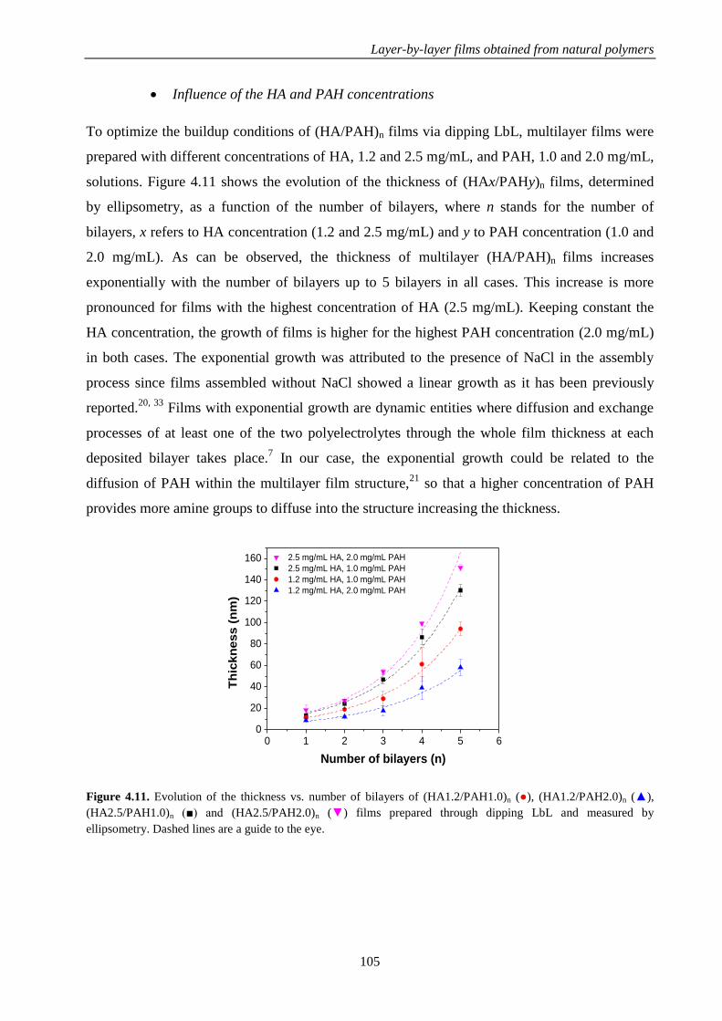
Layer-by-layer films obtained from natural polymers
105
Influence of the HA and PAH concentrations
To optimize the buildup conditions of (HA/PAH)n films via dipping LbL, multilayer films were
prepared with different concentrations of HA, 1.2 and 2.5 mg/mL, and PAH, 1.0 and 2.0 mg/mL,
solutions. Figure 4.11 shows the evolution of the thickness of (HAx/PAHy)n films, determined
by ellipsometry, as a function of the number of bilayers, where n stands for the number of
bilayers, x refers to HA concentration (1.2 and 2.5 mg/mL) and y to PAH concentration (1.0 and
2.0 mg/mL). As can be observed, the thickness of multilayer (HA/PAH)n films increases
exponentially with the number of bilayers up to 5 bilayers in all cases. This increase is more
pronounced for films with the highest concentration of HA (2.5 mg/mL). Keeping constant the
HA concentration, the growth of films is higher for the highest PAH concentration (2.0 mg/mL)
in both cases. The exponential growth was attributed to the presence of NaCl in the assembly
process since films assembled without NaCl showed a linear growth as it has been previously
reported.20, 33
Films with exponential growth are dynamic entities where diffusion and exchange
processes of at least one of the two polyelectrolytes through the whole film thickness at each
deposited bilayer takes place.7 In our case, the exponential growth could be related to the
diffusion of PAH within the multilayer film structure,21
so that a higher concentration of PAH
provides more amine groups to diffuse into the structure increasing the thickness.
0 1 2 3 4 5 60
20
40
60
80
100
120
140
160
2.5 mg/mL HA, 2.0 mg/mL PAH
2.5 mg/mL HA, 1.0 mg/mL PAH
1.2 mg/mL HA, 1.0 mg/mL PAH
1.2 mg/mL HA, 2.0 mg/mL PAH
Th
ick
ness
(n
m)
Number of bilayers (n)
Figure 4.11. Evolution of the thickness vs. number of bilayers of (HA1.2/PAH1.0)n (●), (HA1.2/PAH2.0)n (▲),
(HA2.5/PAH1.0)n (■) and (HA2.5/PAH2.0)n (▼) films prepared through dipping LbL and measured by
ellipsometry. Dashed lines are a guide to the eye.
M 79 a) M 79 b)

Chapter 4
106
Study of the buildup process of nanostratified HA/PAH/Alg-Fe2+
systems
A stratified system composed by a multilayer HA/PAH film with an intermediate gel-like layer,
Alg-Fe2+
, was explored and studied by QCM.
Quartz crystal microbalance with dissipation monitoring (QCM-D) was employed to study the
changes in the frequency and dissipation during the LbL process of HA and PAH in wetting
conditions. Figure 4.13a exhibits the evolution of Δf/v with the time which provides information
about mass changes due to the adsorption or desorption of the polyelectrolyte on the film33
during the buildup of a multilayer (HA/PAH)5Alg-Fe2+
(PAH/HA)m film with 5 bilayers
HA/PAH followed by a gel-like layer of Alg-Fe2+
and then another multilayer (PAH/HA)m
structure. As can be observed, Δf/v decreases with each HA or PAH deposition step and this Δf/v
decay is more pronounced for lower overtones. On the other hand, Figure 4.13b depicts the
variation of ΔD with the time providing information about the viscoelasticity of the film. An
increase of ΔD takes place after each HA or PAH deposition. Taking into account that the
decrease of normalized frequencies, Δf/v, is different for every overtone under study and do not
overlap as well as the increase in the dissipation, it can be concluded that the adsorbed layer does
not obey the Sauerbrey equation (eq. 3.8 in Chapter 3) as a consequence of the viscoelastic
behavior of the adsorbed film.3 This is corroborated by the representation of dissipation (ΔD3)
versus frequency for the third overtone (Δf3/3) in the Figure 4.13c. The high slope (ΔD/Δf) of
this curve also indicates the softness of the film.

Layer-by-layer films obtained from natural polymers
107
0 25 50 75 100 125 150 175 200-2500
-2000
-1500
-1000
-500
0
=7
=5
f/(
Hz)
Time (min)
=3
a)
0 25 50 75 100 125 150 175 200
0
100
200
300
400
500
600
D
Time (min)
=7
=5
=3
b)
0 -500 -1000 -1500 -2000 -25000
100
200
300
400
500
600
700
D
3
f3/3 (Hz)
c)
Figure 4.13. a) Normalized frequency (Δf/v) and b) dissipation changes measured by QCM during the buildup of a
(HA/PAH)5Alg-Fe2+
(HA/PAH)3 film with a concentration of 1.2 mg/mL HA and 0.3 mg/mL PAH. c) Dissipation
changes (ΔD3) versus frequency (Δf3/3) for the third overtone.
Thickness and viscoelastic properties of the films can be obtained applying the Voigt model and
the results corresponding to the third overtone are illustrated in Figure 4.14. Figure 4.14a shows
the exponential evolution of the film thickness with the number of HA/PAH deposited layers
before and after deposition of the gel-like layer Alg-Fe2+
. The deposition of this gel-like layer
Alg-Fe2+
(5.0 mg/mL Alg and 20 mg/mL Fe2+
) gives rise to a high increase of the thickness from
300 nm to 570 nm. Considering that every adsorbed layer has an uniform thickness, the
multilayer (HA/PAH)n film grows at a rate of ~30 nm/layer before and after Alg-Fe2+
deposition
and the Alg-Fe2+
layer has a thickness of 270 nm. The thickness of the HA layer is higher than
the PAH layer during the LbL process. This might be due to the fact that the concentration of
PAH (0.3 mg/mL) is much lower than HA (1.2 mg/mL). Figure 4.14b exhibits the viscosity of
the film. Viscosity increases exponentially with the number of layers and it is much higher for
every HA deposited layer (~1.6 mPa·s) than PAH layer (~0.4 mPa·s) which means that the HA
M 79 a) M 79 b)

Chapter 4
108
layer is more rigid than PAH layer and, as we mentioned above, this might be caused by
differences between PAH and HA concentrations, 0.3 mg/mL and 1.2 mg/mL, respectively.
0
100
200
300
400
500
600
700
800
Th
ickn
ess (
nm
)
Number of layersPAH
PAH
PAH
Fe2+
PAH
PAH
PAH
PAH
PAH H
AHA
HA
ALG
HA
HAH
AHA
HA
a)
Alg-Fe2+
0
4
8
12
16
20
24
28
32
Vis
co
sit
y (
mP
a·s
)
Number of layersPAH
PAH
PAH
Fe2+
PAH
PAH
PAH
PAH
PAH H
AHA
HA
ALG
HA
HAH
AHA
HA
b)
Figure 4.14. a) Thickness of the growing (HA/PAH)5Alg-Fe2+
(HA/PAH)3 film, where the incorporation of the gel-
like layer Alg-Fe2+
is marked with an arrow, and b) viscosity as a function of the number of layers calculated with
the Voigt model. Note that every polymer layer is marked with a different color, HA in blue, PAH in orange and Alg
and Fe2+
in pink.
4.4. CONCLUSIONS
In the study of the growth mechanism of layer-by-layer films obtained from natural polymers,
the following conclusions can be drawn:
The experimental conditions for spray LbL assembly were determined by precipitation tests and
follow up the layer by layer growth process was followed through ellipsometry.
In the case of films made of alginate and chitosan, differences in the growth of (Alg5/Chi)n films
up to 5 bilayers fabricated by two different LbL deposition methods, spray and dipping, were
determined by ellipsometry. The growth was linear in both cases, but the growth rate of films
built up by dipping was 2 nm/layer, one order of magnitude lower than films fabricated by spray
which was 20 nm/layer. Besides to a high growth rate, spray LbL allows a considerable time
saving to build up (Alg/Chi)n films due to the fact that the deposition time of a layer (seconds) is
lower than the dipping deposition time (minutes). In the case of films made of hyaluronic acid
and poly(allylamine hydrochloride), ellipsometry allowed to discard spray LbL technique,
because the growth of (HAx/PAHy)n films did not take place. Therefore, dipping LbL was
employed to fabricate these films.
M 79 a) M 79 b)

Layer-by-layer films obtained from natural polymers
109
The growth of (Algx/Chi)n films prepared by spray assisted LbL was studied by ellipsometry up
to 5 bilayers for a fixed Chi concentration (1.0 mg/mL) and three different Alg concentrations (x
= 1.0, 2.5 and 5.0 mg/mL). The thickness of these films showed a linear increase with the
number of deposited bilayers being higher as Alg concentration increased. However, the growth
rate for the two highest Alg concentrations, 2.5 and 5.0 mg/mL, was similar at 20 nm/layer. To
follow the growth above 5 bilayers, SEM was employed and a change of tendency from linear to
exponential was exhibited from 5 to 20 bilayers to continue with a slight increase towards a
plateau at a number of bilayers above 20 due to the rearrangements of polymer chains during the
deposition process. The experimental conditions to build up (HAx/PAHy)n films were optimized
following the growth of the films up to 5 bilayers by ellipsometry for two different
concentrations of HA (1.2 and 2.5 mg/mL) and PAH (1.0 and 2.0 mg/mL). The growth of these
films was exponential for all concentrations due to the presence of NaCl and the diffusion of
PAH within the multilayer film structure. The growth was higher for 2.5 mg/mL HA and 2.0
mg/mL PAH.
Finally, the development of a nanostratified system based on HA/PAH multilayers with a gel-
like layer Alg-Fe2+
incorporated in an intermediate position was probed by QCM-D. This
technique also allowed to determine the growth of (HA1.2/PAH0.3)n films in wetting conditions
as well as the thickness of the nanostratified system, up to a higher number of deposited layers,
using the Voigt model. In addition, the viscoelastic properties were determined revealing that the
thickness and rigidity of HA layers were higher than those of PAH layers.
4.5. REFERENCES
1. Richardson, J. J.; Björnmalm, M.; Caruso, F., Technology-driven layer-by-layer assembly of
nanofilms. Science 2015, 348, (6233).
2. Lefort, M.; Boulmedais, F.; Jierry, L.; Gonthier, E.; Voegel, J. C.; Hemmerlé, J.; Lavalle, P.;
Ponche, A.; Schaaf, P., Simultaneous Spray Coating of Interacting Species: General Rules Governing the
Poly(styrene sulfonate)/Poly(allylamine) System. Langmuir 2011, 27, (8), 4653-4660.
3. Alves, N. M.; Picart, C.; Mano, J. F., Self Assembling and Crosslinking of Polyelectrolyte
Multilayer Films of Chitosan and Alginate Studied by QCM and IR Spectroscopy. Macromolecular
Bioscience 2009, 9, (8), 776-785.
4. Caridade, S. G.; Monge, C.; Gilde, F.; Boudou, T.; Mano, J. F.; Picart, C., Free-Standing
Polyelectrolyte Membranes Made of Chitosan and Alginate. Biomacromolecules 2013, 14, (5), 1653-
1660.
5. Mjahed, H.; Porcel, C.; Senger, B.; Chassepot, A.; Netter, P.; Gillet, P.; Decher, G.; Voegel, J.-C.;
Schaaf, P.; Benkirane-Jessel, N.; Boulmedais, F., Micro-stratified architectures based on successive
stacking of alginate gel layers and poly(l-lysine)-hyaluronic acid multilayer films aimed at tissue
engineering. Soft Matter 2008, 4, (7), 1422-1429.

Chapter 4
110
6. Kerdjoudj, H.; Boulmedais, F.; Berthelemy, N.; Mjahed, H.; Louis, H.; Schaaf, P.; Voegel, J. C.;
Menu, P., Cellularized alginate sheets for blood vessel reconstruction. Soft Matter 2011, 7, (7), 3621-
3626.
7. Ball, V.; Hübsch, E.; Schweiss, R.; Voegel, J.-C.; Schaaf, P.; Knoll, W., Interactions between
Multivalent Ions and Exponentially Growing Multilayers: Dissolution and Exchange Processes. Langmuir
2005, 21, (18), 8526-8531.
8. Díaz, B.; Freire, L.; Montemor, M. F.; Nóvoa, X. R., Oxide film growth by CSV on AISI 316L: a
combined electrochemical and analytical characterization. Journal of the Brazilian Chemical Society
2013, 24, 1246-1258.
9. Halthur, T. J.; Claesson, P. M.; Elofsson, U. M., Immobilization of Enamel Matrix Derivate
Protein onto Polypeptide Multilayers. Comparative in Situ Measurements Using Ellipsometry, Quartz
Crystal Microbalance with Dissipation, and Dual-Polarization Interferometry. Langmuir 2006, 22, (26),
11065-11071.
10. Wittmer, C. R.; Phelps, J. A.; Saltzman, W. M.; Van Tassel, P. R., Fibronectin Terminated
Multilayer Films: Protein Adsorption and Cell Attachment Studies. Biomaterials 2007, 28, (5), 851-860.
11. Picart, C.; Ladam, G.; Senger, B.; Voegel, J.-C.; Schaaf, P.; Cuisinier, F. J. G.; Gergely, C.,
Determination of structural parameters characterizing thin films by optical methods: A comparison
between scanning angle reflectometry and optical waveguide lightmode spectroscopy. The Journal of
Chemical Physics 2001, 115, (2), 1086-1094.
12. Dante, S.; Hou, Z.; Risbud, S.; Stroeve, P., Nucleation of Iron Oxy-Hydroxide Nanoparticles by
Layer-by-Layer Polyionic Assemblies. Langmuir 1999, 15, (6), 2176-2182.
13. Ferriz-Mañas, M.; Schlenoff, J. B., Zeta Potential of Polyelectrolyte Multilayers Using the
Spinning Disk Method. Langmuir 2014, 30, (29), 8776-8783.
14. Wang, Y.; Kozlovskaya, V.; Arcibal, I. G.; Cropek, D. M.; Kharlampieva, E., Highly swellable
ultrathin poly(4-vinylpyridine) multilayer hydrogels with pH-triggered surface wettability. Soft Matter
2013, 9, (39), 9420-9429.
15. Ye, S.; Wang, C.; Liu, X.; Tong, Z., Multilayer nanocapsules of polysaccharide chitosan and
alginate through layer-by-layer assembly directly on PS nanoparticles for release. Journal of Biomaterials
Science, Polymer Edition 2005, 16, (7), 909-923.
16. Ye, S.; Wang, C.; Liu, X.; Tong, Z.; Ren, B.; Zeng, F., New loading process and release
properties of insulin from polysaccharide microcapsules fabricated through layer-by-layer assembly.
Journal of Controlled Release 2006, 112, (1), 79-87.
17. Yuan, W.; Dong, H.; Li, C. M.; Cui, X.; Yu, L.; Lu, Z.; Zhou, Q., pH-Controlled Construction of
Chitosan/Alginate Multilayer Film: Characterization and Application for Antibody Immobilization.
Langmuir 2007, 23, (26), 13046-13052.
18. Burke, S. E.; Barrett, C. J., Swelling behavior of hyaluronic acid/polyallylamine hydrochloride
multilayer films. Biomacromolecules 2005, 6, (3), 1419-1428.
19. Burke, S. E.; Barrett, C. J., pH-dependent loading and release behavior of small hydrophilic
molecules in weak polyelectrolyte multilayer films. Macromolecules 2004, 37, (14), 5375-5384.
20. Szarpak, A.; Pignot-Paintrand, I.; Nicolas, C.; Picart, C.; Auzély-Velty, R., Multilayer assembly
of hyaluronic acid/poly (allylamine): control of the buildup for the production of hollow capsules.
Langmuir 2008, 24, (17), 9767-9774.

Layer-by-layer films obtained from natural polymers
111
21. Szarpak, A.; Cui, D.; Dubreuil, F. d. r.; De Geest, B. G.; De Cock, L. J.; Picart, C.; Auzely-Velty,
R., Designing hyaluronic acid-based layer-by-layer capsules as a carrier for intracellular drug delivery.
Biomacromolecules 2010, 11, (3), 713-720.
22. Burke, S. E.; Barrett, C. J., pH-Responsive Properties of Multilayered Poly(l-lysine)/Hyaluronic
Acid Surfaces. Biomacromolecules 2003, 4, (6), 1773-1783.
23. Hernández, R.; Sacristán, J.; Mijangos, C., Sol/Gel Transition of Aqueous Alginate Solutions
Induced by Fe2+ Cations. Macromolecular Chemistry and Physics 2010, 211, (11), 1254-1260.
24. Lawrie, G.; Keen, I.; Drew, B.; Chandler-Temple, A.; Rintoul, L.; Fredericks, P.; Grøndahl, L.,
Interactions between alginate and chitosan biopolymers characterized using FTIR and XPS.
Biomacromolecules 2007, 8, (8), 2533-2541.
25. Silva, J. M.; Duarte, A. R. C.; Caridade, S. G.; Picart, C.; Reis, R. L.; Mano, J. F., Tailored
Freestanding Multilayered Membranes Based on Chitosan and Alginate. Biomacromolecules 2014, 15,
(10), 3817-3826.
26. Yao, K.; Li, J.; Yao, F.; Yin, Y., Formation of chitosan-based hydrogels network. In Chitosan-
based hydrogels. Functions and applications, CRC Press: 2012; pp 183-197.
27. Kulig, D.; Zimoch-Korzycka, A.; Jarmoluk, A.; Marycz, K., Study on Alginate–Chitosan
Complex Formed with Different Polymers Ratio. Polymers 2016, 8, (5), 167.
28. Sham, A. Y. W.; Notley, S. M., Graphene–polyelectrolyte multilayer film formation driven by
hydrogen bonding. Journal of Colloid and Interface Science 2015, 456, 32-41.
29. Shenoy, D. B.; Antipov, A. A.; Sukhorukov, G. B.; Möhwald, H., Layer-by-Layer Engineering of
Biocompatible, Decomposable Core−Shell Structures. Biomacromolecules 2003, 4, (2), 265-272.
30. Richert, L.; Lavalle, P.; Payan, E.; Shu, X. Z.; Prestwich, G. D.; Stoltz, J.-F.; Schaaf, P.; Voegel,
J.-C.; Picart, C., Layer by Layer Buildup of Polysaccharide Films: Physical Chemistry and Cellular
Adhesion Aspects. Langmuir 2004, 20, (2), 448-458.
31. Lundin, M.; Blomberg, E.; Tilton, R. D., Polymer Dynamics in Layer-by-Layer Assemblies of
Chitosan and Heparin. Langmuir 2010, 26, (5), 3242-3251.
32. Porcel, C.; Lavalle, P.; Ball, V.; Decher, G.; Senger, B.; Voegel, J.-C.; Schaaf, P., From
Exponential to Linear Growth in Polyelectrolyte Multilayers. Langmuir 2006, 22, (9), 4376-4383.
33. Pettersson, T. r.; Pendergraph, S. A.; Utsel, S.; Marais, A.; Gustafsson, E.; Wagberg, L., Robust
and Tailored Wet Adhesion in Biopolymer Thin Films. Biomacromolecules 2014, 15, (12), 4420-4428.


CHAPTER 5
Determination of the structure-
properties relationship in Alg/Chi films


Determination of the structure-properties relationship in Alg/Chi films
115
In this chapter, as a first step, the morphological and nanomechanical properties of multilayer
Alg/Chi films developed in the Chapter 3 have been determined. In addition, these properties
were also determined for nanocomposite Alg/Chi films with magnetic nanoparticles and
crosslinked Alg/Chi films which will be employed for different biomedical applications in
Chapters 6 and 7, respectively. As a second step, the inner structure of nanocomposite Alg/Chi
films was studied by Grazing incidence small-angle X-ray scattering.
5.1. INTRODUCTION
Over the last few years, multilayer polymer films and nanocomposite films, obtained through
their combination with nanoparticles, have drawn a lot of attention for the development of
several applications in organic microelectronics, as biosensors or scaffolds for tissue
engineering.1, 2
The control of the mechanical properties of these films is specifically attractive in
the biomaterials area where the mechanical properties of polymer films allow to modulate
cellular behaviors regarding adhesion, proliferation and cell differentiation.3-5
Until now, most of the mechanical studies performed on LbL polymer films and nanocomposite
multilayer films have been accomplished by macroscopic stress-strain experiments. Focusing on
multilayer Alg/Chi films, measurements of their mechanical strength using the bulge test have
revealed an increase in the elastic modulus with the film thickness for films in the range of tens
of nanometers.6 Very recently, it was shown that the incorporation of magnetic nanoparticles
within free-standing multilayer Alg/Chi films gave rise to an increase in the elastic moduli
measured in the wet state by stress-strain measurements.7 Probe-based indentation is a versatile
method to study local mechanical properties of materials from the nanoscale to the macroscale.8-
10 Within this method, nanoindenter and atomic force microscopy (AFM) are the most used
techniques.11
An early study reported the employment of AFM indentation, with 2.5 m radius
sphere, to determine the mechanical properties of hydrated multilayer PLL/HA films in the
micron range either native or crosslinked and supported by an underlying glass coverslip.12
However, this technique presents some drawbacks such as poor lateral resolution and the large
time required to obtain the quantitative mechanical mapping of the surface.8-10
To solve these problems, two AFM modes for mapping mechanical properties have been
introduced, HarmoniX and PeakForce (PF) Quantitative Nanomechanical Mapping (QNM).8-10
These modes use the Derjaguin-Muller-Toporov (DMT) model13
to obtain the elastic modulus
from the force-indentation curves. It is important to note that the employment of regular sharp

Chapter 5
116
probes may cause overestimation of the film modulus by the DMT method. An approach would
be using special probes, such as colloidal ones for example, that would allow increasing the
applied force without producing large penetration. However, in that case, lateral resolution
would be compromised.8 There are only few studies aimed to determine the nanomechanical
properties of LbL multilayer films by means of (PF-QNM) AFM, mainly focused on determining
the effect of the number of deposited layers on the elastic moduli on LbL polymer films and the
effect of the stiff underlying substrate14, 15
and, none of them, deal with the determination of the
nanomechanical properties of films fully fabricated from polysaccharides.
In this chapter, the nanomechanical properties (elastic moduli, deformation and adhesion values)
of multilayer Alg/Chi films will be determined. To achieve this aim, not only the effect of the
number of deposited bilayers on the resulting mechanical properties was considered, but it was
also considered the effect of crosslinking and the effect of the incorporation of nanoparticles to
obtain nanocomposite films during the buildup of the films. All of these parameters are known to
influence the performance of these materials in biomedical applications. Specifically,
nanocomposite Alg/Chi films with magnetic nanoparticles will be explored as pads for local
magnetic hyperthermia in chapter 6 and the cell adhesion properties of Alg/Chi films will be
examined in the chapter 7.
Small-angle X-ray scattering (SAXS) is proved to be a powerful tool for studying the inner
structure of multilayer films by measuring changes into the structure during the buildup
process.16
In addition to this, Grazing-incidence small-angle X-ray scattering (GISAXS) is a
powerful technique to study nanostructures at the surface and in buried structures of thin films,
by changing slightly the incident angle it is possible to vary the depth of the incident beam into
the sample allowing to study the structural changes with the thickness.17
There are other two X-
ray techniques, XRD and Grazing-incidence X-ray diffraction (GIXD), which can be applied to
the study of the inner structure of multilayer polymer films, with the difference that GIXD
controls the penetration depth through the incident angle.17
Regarding the inner structure of nanocomposite Alg/Chi films, to the best of our knowledge, it
has not been previously reported although recent results has pointed to the feasibility of
performing GISAXS experiments on spray deposited arrays of polymer nanoparticles.18, 19
It is
generally accepted that films formed from synthetic polymers, such as poly(styrene sulfonate)
and poly(allylamine hydrochloride) (PSS/PAH) are stratified in layers whereas some degree of
interpenetration is present in natural polymers,20
such as chitosan and hyaluronic acid

Determination of the structure-properties relationship in Alg/Chi films
117
(Chi/HA).21
In this context, the distribution of NPs within polysaccharidic films could vary from
films with well separated monolayers to films with dispersed NPs all over the thickness. In the
chapter 4 it was described the exponential growth of multilayer films based on alginate and
chitosan (Alg/Chi) which was attributed to a possible diffusion of chitosan along the structure. In
multilayer polymer films containing inorganic nanoparticles, the distribution of NPs and
structuration of NPs layers inside films influence the final properties of the resulting
nanocomposite materials. A varying architecture in multilayer films fabricated from polymers
and magnetic NPs have revealed different magnetic properties depending on the distance
between NPs layers.22
The incorporation of magnetic nanoparticles into the bulk structure of different diblock
copolymer films has been studied by neutron reflection,23
neutron scattering24
and grazing
incident small-angle X-ray scattering (GISAXS).25, 26
Grazing incident small-angle X-ray
scattering (GISAXS) allows to determine structural information on the arrangement of polymers
and nanoparticles in the direction perpendicular to the free surface, as it has been previously
reported for polymer thin films.27, 28
In this chapter, GISAXS will be employed to elucidate the
internal structure of nanocomposite multilayer films based on alginate and chitosan with
magnetic nanoparticles (NPs) incorporated.
5.2. EXPERIMENTAL PART
5.2.1. Materials
Chitosan (Chi) of low molecular weight (448869, lot SLBG1673V), sodium alginate (Alg)
(A2158, lot 090M0092V), poly(ethylenimine) (PEI) with a molecular weight (Mw) of 25000 and
acetic acid were supplied by Aldrich and used as received. Sodium acetate anhydrous was
supplied by Panreac. More information about these materials is detailed in the chapter 3.
A lab-made alginate based magnetic ferrofluid containing the iron oxide nanoparticles (NPs)
with a magnetite concentration of 8.0 mg/mL, whose characteristics have been described in the
Chapter 3, was employed for the preparation of nanocomposite Alg/Chi films.
5.2.2. Preparation of Alg/Chi films
All films were built on silicon wafers (20 × 40 mm, Siegert) following the procedure
corresponding to spray assisted LbL deposition described in the Chapter 4 using a fixed Chi
concentration (1 mg/mL) and two different Alg concentrations, 1.0 and 2.5 mg/mL. Samples

Chapter 5
118
were denoted as (Algx/Chi)n where n stands for the number of bilayers (n = 5, 10, 20 or 40) and x
refers to Alg concentration (x = 1.0 or 2.5 mg/mL).
5.2.3. Preparation of crosslinked Alg/Chi films
For the preparation of crosslinked Alg/Chi films, (Algx/Chi)n films were thermally crosslinked in
an oven at 120 ºC for 2 hours.
5.2.4. Preparation of nanocomposite Alg/Chi films
Nanocomposite Alg/Chi films were obtained by spraying a bilayer of alginate and chitosan
followed by a bilayer of a lab-made alginate based magnetic ferrofluid containing the iron oxide
NPs and a chitosan layer (Scheme 5.1). In this case, fixed chitosan and alginate concentrations of
1.0 mg/mL and 2.5 mg/mL, respectively, were used and each layer was sprayed for 5 s. Three
nanocomposite films were obtained by applying different number of cycles and samples were
denoted as (Alg/Chi)3/(NPs/Chi)2, (Alg/Chi)5/(NPs/Chi)5 and (Alg/Chi)10/(NPs/Chi)10. Note that
the sample, (Alg/Chi)3/(NPs/Chi)2, has not the ending NPs/Chi bilayer.
Scheme 5.1. Schematic diagram of the sequential spraying process followed for the preparation of nanocomposite
Alg/Chi films.
For the GISAXS experiment, an additional nanocomposite Alg/Chi film was prepared through
dipping following the procedure shown in the Scheme 5.2. The silicon substrate was sequentially
immersed in Alg (2.5 mg/mL) and Chi (1.0 mg/mL) solutions to build up a bilayer Alg/Chi and
then it was immersed in the alginate based magnetic ferrofluid and Chi solutions to build up a
bilayer NPs/Chi. The time of immersion was 5 min for every deposited layer with a subsequent
rinsing step of 2 min. The obtained nanocomposite film was denoted as (Alg/Chi)2/(NPs/Chi)1.

Determination of the structure-properties relationship in Alg/Chi films
119
Scheme 5.2. Schematic diagram of the sequential dipping process followed for the preparation a nanocomposite
(Alg/Chi)2/(NPs/Chi)1 film.
5.2.5. Morphological characterization
The topography and surface roughness were measured by Atomic Force Microscopy (AFM)
using a Bruker Multimode 8, with a Nanoscope V controller. All measurements were performed
in air, at room temperature, on films supported on silicon wafers. Samples (Algx/Chi)n were
scanned over square regions of 2 m size and nanocomposite (Alg/Chi)n/(NPs/Chi)m films over
square regions of 1 m size. The average roughness (Ra) was calculated from the height images
in four different square points of 1m × 1m.
The cross section of films was observed by scanning electron microscopy (SEM) using a Hitachi
SU 8000 microscope operating at 0.8 kV. The average thickness was determined by measuring
the width of the cross section in the SEM micrographs in five different points using the software
ImageJ.
5.2.6. Mechanical properties
The mechanical properties of films were evaluated using a Bruker Multimode 8, with a
Nanoscope V controller, by the PeakForce Quantitative Nanomechanical Mapping (PF-QNM)
method. All measurements were performed in air, at room temperature, on films supported on
silicon wafers. Samples (Algx/Chi)n were scanned over square regions of 2 m size and
nanocomposite (Alg/Chi)n/(NPs/Chi)m films over square regions of 1 m size. All quantitative
measurements were carried out with different probes depending on the actual mechanical
properties of every sample. In particular, TAP150 and RTESPA525 probes (Bruker) were
employed. Cantilever spring constants for each probe were measured by the thermal tune method

Chapter 5
120
and found to be around 1-5 and 50-100 N/m for TAP150 and RTESPA525, respectively. More
details about this technique can be consulted in the Chapter 3.
The precise experimental procedure was the following: an initial force on the tip, Ftip, was set,
and an initial estimation of the tip radius, based on the provider specifications was considered.
The Ftip was chosen to produce a deformation depth of about 5 nm. Under these experimental
conditions, the adhesive force, Fadh, between the tip and the sample can be neglected. With this,
an experiment on the polystyrene standard was carried out. The tip radius value was modified
systematically until the measured elastic modulus of the reference matched the one provided by
the provider. The obtained value for the tip radius was typically around 10 nm. The same sample
was measured with different tips, after careful calibration for tip radius, for cantilever deflection
sensitivity and also at different times. In addition, the z distance in the piezo is regularly
calibrated using a grating of known depth.
Analysis of the force-distance curves is automatically performed by a software program,
NanoScope Analysis 1.5, which allows to extract the height, elastic modulus, adhesion force,
deformation and dissipation simultaneously by application of the DMT model.
5.2.7. Characterization of the inner structure of nanocomposite films
To study the inner structure of nanocomposite films, GISAXS measurements were carried on
(Alg/Chi)n films and nanocomposite (Alg/Chi)n(NPs/Chi)m films prepared by spray and dipping
LbL on BM26 beamline of the European Synchrotron Radiation Facility (ESRF) sited in
Grenoble (France). A two-dimensional (2D) detector (Pilatus 1M) and a camera of 981 x 1043
pixels (size of the pixel 172 x 172 m2) was used at a sample to detector distance of 6.87 m to
data acquisition. The X-ray beam had a wavelength of λ = 0.103 nm. The angle of incidence (αi)
was varied between 0.1 and 0.4º. To data analysis the FIT2D software program was employed.
More details about this technique can be accessed in the Chapter 3.
5.3. RESULTS AND DISCUSSION
5.3.1. Morphological characterization
The effect of the number of layers, the alginate concentration, as well as the crosslinking and the
incorporation of NPs, in the thickness and surface average roughness (Ra) of (Alg/Chi)n films
were studied by AFM and SEM.

Determination of the structure-properties relationship in Alg/Chi films
121
Effect of the number of deposited layers, alginate concentration and crosslinking
Results corresponding to the surface average roughness (Ra), determined from the AFM height
images, and the thickness, determined by SEM through the measurement of the cross-section of
(Algx/Chi)n films are shown in Table 5.1. Multilayer (Alg2.5/Chi)n films were subjected to a
thermal treatment at 120 ºC for 2 hours in order to induce crosslinking through formation of
amide bonds between the amine groups of chitosan and the carboxylic acid groups of alginate.29
The thickness and average roughness (Ra) are also shown in Table 5.1 as a function of the
number of bilayers.
Table 5.1. Evolution of the thickness and average roughness (Ra) of non-crosslinked (Algx/Chi)n films and
crosslinked (Alg2.5/Chi)n films as a function of the number of bilayers (n).
For non-crosslinked films, the thickness increases with the number of bilayers (n) from 0.13 ±
0.10 m (n =5) to 1.59 ± 0.65 m (n = 40) for films with an alginate concentration of 1.0
mg/mL. In the case of films with a higher alginate concentration, 2.5 mg/mL, the thickness also
increases with the number of bilayers from 0.29 ± 0.09 m (n =5) to 3.06 ± 1.71 m (n = 40),
but in a two-fold scale with regards to films with 1.0 mg/mL alginate. Concerning the average
roughness, an increase can be detected with the number of deposited bilayers for both alginate
concentrations without showing significant differences between them. For example, in the case
of non-crosslinked (Alg2.5/Chi)n films, Ra increases from 8.9 ± 3.6 to 49.0 ± 17.5 nm when the
number of bilayers increases from 5 to 40. This might be attributed to the self-assembly of
opposite charges between alginate and chitosan that gives rise to their aggregation resulting in
rougher surfaces as the number of bilayers increases.30
The Ra values obtained for crosslinked films, 4.8 ± 3.6 (n = 5) and 18.5 ± 0.5 (n = 40), are lower
than those corresponding to native (Alg2.5/Chi)n films in agreement with previous reports in
n
(Alg1.0/Chi)n films (Alg2.5/Chi)n films Crosslinked
(Alg2.5/Chi)n films
Thickness
(m) Ra (nm)
Thickness
(m) Ra (nm)
Thickness
(m) Ra (nm)
5 0.13 ± 0.10 18.4 ± 5.3 0.29 ± 0.09 8.9 ± 3.6 0.43 ± 0.21 4.8 ± 3.6
10 0.16 ± 0.11 13.8 ± 6.5 0.82 ± 0.06 17.1 ± 3.4 0.85 ± 0.75 3.8 ± 0.6
20 1.23 ± 0.98 33.1 ± 5.0 1.75 ± 1.34 29.8 ± 11.9 1.04 ± 0.98 22.3 ± 8.9
40 1.59 ± 0.65 35.6 ± 7.3 3.06 ± 1.71 49.0 ± 17.5 2.15 ± 1.25 18.5 ± 0.5

Chapter 5
122
which it was demonstrated that the crosslinking of multilayer films did not significantly change
the thickness in the dry state, giving rise to more uniform films due to the reorganization of the
film.31
Notice that Ra values obtained after crosslinking do not follow a clear trend with the
number of deposited bilayers probably due to the fact that Ra arises during the process of the
preparation of the films before the crosslinking step.
Effect of the presence of nanoparticles
Figure 5.1 shows AFM topographic images corresponding to two samples prepared with the
same number of total bilayers, (Alg2.5/Chi)10 and nanocomposite (Alg/Chi)5/(NPs/Chi)5 films,
which provide information on their morphology and their roughness. The film without NPs
displays finer grain texture and a more homogeneous surface morphology, with an average
roughness of 17.1 ± 3.4 nm, than the nanocomposite film, which exhibits aggregated NPs and an
average roughness of 42 ± 3.4 nm. The formation of aggregates is attributed to the roughness
accumulation induced by the inner NPs layers. Cluster formation could also be influenced by the
aggregation of small particles during the spray of a colloidal solution.32
Figure 5.1. AFM topographic images (3m × 3m) corresponding to a representative sample a) (Alg2.5/Chi)10 and
b) nanocomposite (Alg/Chi)5/(NPs/Chi)5 films.
5.3.2. Mechanical properties
To characterize the nanomechanical properties of Alg/Chi LbL films by means of (PF-QNM)
AFM, the number of bilayers and the alginate concentration were varied. The effect of the
crosslinking and the incorporation of NPs were also studied.
M 79 a) M 79 b)

Determination of the structure-properties relationship in Alg/Chi films
123
Effect of the alginate concentration and thermal crosslinking
Representative (PF-QNM) AFM images corresponding to the height, elastic modulus,
deformation and adhesion of (Alg1.0/Chi)20 films are shown in Figure 5.2a. Results of
mechanical properties are obtained by analysis of the cross sectional profiles in four different
points of each image, marked with different colored lines, corresponding to areas with different
height profiles, in order to obtain an average measurement of every mechanical property (Figure
5.2b).
0 100 200 300 400 500 600
1,8
3,6
5,4
7,2
0 100 200 300 400 500 6000
3
6
9
0 100 200 300 400 500 6000
25
50
75
100
0 100 200 300 400 500 6000
50
100
150
200
250
Ad
he
sio
n (
nN
)
µm
b)
De
form
ati
on
(n
m)
µm
Ela
sti
c m
od
ulu
s (
GP
a)
µm
He
igh
t (n
m)
µm
Figure 5.2. a) (PF-QNM) AFM images (2m × 2m), height, elastic modulus, deformation and adhesion
corresponding to (Alg1.0/Chi)20 films with colored lines incorporated and situated in the zones where the cross
sectional profiles were taken and b) variation of height, elastic modulus, deformation and adhesion along the
different cross sectional profiles marked with their specific color in the AFM images (left).
A comparison of the AFM height images and those corresponding to the mechanical properties
(elastic modulus, deformation and adhesion) allows to observe that the topography of the
M 79 a) M 79 b)

Chapter 5
124
samples is not associated to changes of the mechanical properties. For example, centering our
attention in green and blue lines which represent areas of the sample with different height
according to the different color scale, there are not significant differences in the elastic modulus
and the values of deformation and adhesion are similar.
Figure 5.3 illustrates representative (PF-QNM) AFM images corresponding to (Alg2.5/Chi)20
films before and after thermal crosslinking. As in the previous case, the topography of the
samples is not associated to changes of the mechanical properties and this is more evident for
crosslinked (Alg2.5/Chi)20 films. For easier visualization, a white square (500 nm× 500 nm) was
placed in the same position within the height AFM image and that corresponding to the
determination of elastic modulus. As it can be observed, this region presents an homogeneous
roughness whereas differences in the elastic moduli can be observed according to the different
color scale. This result suggests the presence of heterogeneous regions which might be related to
regions of different degree of crosslinking arising from differences on the rearrangement of the
polymer layers after thermal crosslinking or even from inhomogeneous deposition of the
polymers, regions with more polymer mass give rise to a higher number of crosslinking points.
In addition, there is a correlation between AFM images corresponding to deformation and those
corresponding to adhesion: regions that deform more are those that show a higher tip-adhesion
force. This might be attributed to the establishment of more effective interactions, probably of
Van der Waals nature, between the tip and the polymer surface.

Determination of the structure-properties relationship in Alg/Chi films
125
Figure 5.3. (PF-QNM) AFM images (2m × 2m), height, elastic modulus, deformation and adhesion,
corresponding to (Alg2.5/Chi)20 films a) before and b) after crosslinking. For the sake of discussion, the white
square (500 nm × 500 nm) placed in the same position within the height AFM image and the elastic moduli AFM
image indicates a small roughness region with differences in the elastic moduli.
The mechanical properties (elastic modulus, deformation and adhesion), quantified at four
different positions, of native (Algx/Chi)n films as a function of the number of bilayers are shown
in Figure 5.4a.
M 79 a) M 79 b)

Chapter 5
126
Figure 5.4. Evolution of the elastic modulus, deformation and adhesion of a) (Alg1.0/Chi)n (■) and (Alg2.5/Chi)n
films (●) and b) crosslinked (Alg2.5/Chi)n films (○) vs. film thickness and number of bilayers. Dashed lines are a
guide to the eye.
The elastic modulus of (Alg1/Chi)n films slightly increases with the number of bilayers being 8.5
± 2.0 GPa for 5 Alg/Chi bilayers and 12.4 ± 2.3 GPa for 40 Alg/Chi bilayers. The increase in the
elastic moduli with the number of bilayers from 5 to 40 Alg/Chi bilayers is more significant in
the case of (Alg2.5/Chi)n increasing from 0.8 ± 0.1 GPa to 11.8 ± 4.3 GPa, respectively. The
elastic moduli obtained for (Alg2.5/Chi)n films are lower than those corresponding to
(Alg1.0/Chi)n ones. For both alginate concentrations, the elastic moduli reach a plateau value of
~12GPa at 40 bilayers. Note that this value is similar to those reported and literature obtained by
bulge tests carried out on free-standing Alg/Chi films prepared by spray assisted LbL which
showed that the elastic modulus of Alg/Chi films with 35 nm was 1.1 ± 0.4 GPa and increased to
similar values for Alg/Chi films with thicknesses of 75 nm and 114 nm (8.1± 2.5 GPa and 11.0 ±
1.6 GPa, respectively). For these samples, Fujie et al.6 obtained a linear increase in the thickness
with a single Alg/Chi bilayer of approximately 3.3 nm thickness, suggesting that each
polysaccharide layer constitutes an almost single molecular layer. In our study, spray assisted
LbL of (Alg1.0/Chi)n and (Alg2.5/Chi)n films present a thickness of a single Alg/Chi bilayer of ~
0,0 0,5 1,0 1,5 2,0 2,5
0
15
30
45
60
0 5 10 15 20 25 30 35 40 45
0
15
30
45
60
0 5 10 15 20 25 30 35 40 45
0,0
0,9
1,8
2,7
0 5 10 15 20 25 30 35 40 45
01234567
Ela
sti
c m
od
ulu
s (
GP
a)
Thickness (m)
Ela
sti
c m
od
ulu
s (
GP
a)
Number of bilayers (n)
Defo
rmati
on
(n
m)
Number of bilayers (n)
Ad
hesio
n (
nN
)
Number of bilayers (n)
0,0 0,5 1,0 1,5 2,0 2,5 3,0 3,5
0
5
10
15
20
0 5 10 15 20 25 30 35 40 45
0
5
10
15
20
0 5 10 15 20 25 30 35 40 45
0,0
0,9
1,8
2,7
0 5 10 15 20 25 30 35 40 45
01234567
Ela
sti
c m
od
ulu
s (
GP
a)
Thickness (m)
Number of bilayers (n)
Ela
sti
c m
od
ulu
s (
GP
a)
Number of bilayers (n)
Defo
rmati
on
(n
m)
Ad
hesio
n (
nN
)
Number of bilayers (n)
a) b)

Determination of the structure-properties relationship in Alg/Chi films
127
7.8 nm and ~ 42.4 nm, respectively and a plateau value for the elastic modulus of 12.4 ± 2.3GPa
and 11.8 ± 4.3GPa, respectively is obtained for (Algx/Chi)n films with thicknesses above 1 µm,
i.e., above 20 bilayers. This fact may be attributed to the change in growth mechanism from
exponential to linear that takes place above 20 bilayers as it was shown in the Chapter 4. Thus, it
follows that the effective interaction established between Alg and Chi is weaker for sprayed LbL
than for spin-coated LbL. It was proposed that the initial exponential growth observed in Figure
4.8 (Chapter 4) is controlled by the interdiffusion of both Alg and Chi leading to a significant
increase of the elastic moduli with the increase of bilayers. Once the thickness of the sprayed
LbL film reaches a certain value, the buildup changes from an exponential to linear growth due
to a hindering of the diffusion process and the elastic modulus tends to a plateau value.
The deformation of Alg/Chi films is almost constant with the number of bilayers and only varies
in a range between 0.5 and 1.5 nm due to the similar peak force used for all measurements.
Similarly, adhesion values are independent of the number of deposited bilayers with values
ranging between 1 and 4 nN. This is expected, as the adhesion is derived from the pull off force
that is a measure of the attractive forces between the tip and outermost layers of the samples, i.e.,
the adhesion measurements are sensitive only to the surface and thus they should be similar for
all the samples. Moreover, there are no differences in deformation and adhesion between films
prepared at the two concentrations of Alg.
Now, we turn our attention to the effect of the crosslinking on the mechanical properties
measured on (Alg2.5/Chi)n films as a function of the number of bilayers (n) (Figure 5.4b). For
n<10, the elastic moduli of crosslinked films are similar to those of the native ones. However, as
the number of bilayers increases, the elastic moduli drastically increase to reach a maximum
value of ≈ 47.4 ± 3.8 GPa at 40 Alg/Chi bilayers. In addition, the deformation values of
crosslinked (Alg2.5/Chi)n films remain below 0.9 nm regardless the number of bilayers.
Adhesion values are on the same range as those encountered for native films with the exception
of (Alg2.5/Chi)5 film for which the adhesion force increases to 6 nN. Although no conclusive
explanation for this effect exists, it can be speculated about a lower chemical heterogeneity for
the crosslinked sample at low number of bilayers arising from a lower degree of interpenetration
between alginate and chitosan as shown in Figure 4.8 (Chapter 4). This result would imply that
the adhesion promoted by the final chitosan layer would be higher than that resulting from the
interpenetration with alginate. It is important to note that in ambient conditions, the values of
adhesion are likely facilitated by capillary forces of a surface adsorbed water layer in air.33
This

Chapter 5
128
is especially relevant in the case of hydrophilic polymers such as chitosan that constitutes the last
layer of all the samples under study.34, 35
To partially summarize, crosslinked films present a higher elastic modulus and less deformation
than native films. As expected, crosslinking increases the stiffness of the multilayer films
provoking an increase in the elastic modulus. This is due to the fact that thermal crosslinking
results in an amidization reaction between the carboxylate groups of alginate (COO-) and the
ammonium groups of the chitosan (NH3+) to form amide crosslinks (NHCO) that rigidify the
multilayers.36
The crosslinking reduces the molecular mobility of polymer chains because it
decreases the intermolecular space between these chains.7 The effect of film crosslinking on the
elastic moduli was expected since the degree of crosslinking is known to influence the
mechanical properties of multilayer films. As an example, in a recent study, the Young Modulus
of Alg/Chi films, determined on 31.7 ± 4.7 µm film thickness by dynamic mechanical analysis in
a hydrated environment at 37°C, increased from 4.8 to 56.4 MPa after crosslinking with
genipin.31
Effect of the presence of nanoparticles
In this section, the effect of the presence of iron oxide nanoparticles (NPs), inside the multilayer
structures as well as on the outermost layer, on the mechanical properties is discussed.
In a first step, the presence of NPs inside multilayer films is studied. Representative (PF-QNM)
AFM images corresponding to nanocomposite (Alg/Chi)10/(NPs/Chi)10 films are shown in Figure
5.5. The mechanical properties (elastic modulus, deformation and adhesion) were analyzed in
four different points of these images. It is worth to emphasize that the analysis was carried out in
1 m2 regions.

Determination of the structure-properties relationship in Alg/Chi films
129
Figure 5.5. (PF-QNM) AFM images (1m × 1m), height, elastic modulus, deformation and adhesion,
corresponding to a nanocomposite (Alg/Chi)10/(NPs/Chi)10 film.
Table 5.2 summarizes the results corresponding to the thickness, determined by SEM, and the
mechanical properties, determined by (PF-QNM)-AFM, for three nanocomposite
(Alg/Chi)n/(NPs/Chi)m films with Chi as ending layer.
M 79 a) M 79 b)

Chapter 5
130
Table 5.2. Summary of mechanical properties of nanocomposite (Alg/Chi)n/(NPs/Chi)m films determined by (PF-
QNM) AFM and thickness determined by SEM.
Samples Thickness
(m)
Elastic
modulus (GPa)
Deformation
(nm)
Adhesion
(nN)
(Alg/Chi)3/(NPs/Chi)2 0.24 ± 0.10 18.8 ± 6.3 1.1 ± 0.9 2.3 ± 1.5
(Alg/Chi)5/(NPs/Chi)5 0.38 ± 0.13 34.4 ± 5.6 1.1 ± 0.2 21.4 ± 2.8
(Alg/Chi)10/(NPs/Chi)10 1.11 ± 0.34 26.2 ± 3.9 0.8 ± 0.2 11.7 ± 3.9
As it can be observed, for the same number of bilayers, nanocomposite (Alg/Chi)n/(NPs/Chi)m
films present a higher elastic moduli than (Alg2.5/Chi)n films (results depicted in Figure 5.4a).
These results corroborate that the mechanical properties, found by (PF-QNM) AFM, are
consistent with intrinsic properties of the sample and are not influenced by the substrate
properties. It is interesting to note that the values of deformation increased significantly in
nanocomposite films with respect to Alg/Chi films for which the values of deformation range
between 0.5 and 1.5 nm. A careful examination of the results depicted in Figure 5.5 for the
(Alg/Chi)10/(NPs/Chi)10 film shows that regions that deform more correspond to regions with no
NPs. It might be hypothesized that the incorporation of NPs might decrease the number of
interactions between polyelectrolyte layers, thus promoting the formation of ‘empty regions’ in
between polymer layers which might lead to the increase of deformation. In fact, previous
reports on the incorporation of NPs during the buildup of the multilayer film have revealed that
the deposition process can give rise to islands that create empty regions into the internal structure
of the film.37, 38
For the nanocomposite films under study, SEM images shown in Figure 5.6
corroborate the formation of empty spaces or islands during the spray deposition. The film with a
lower number of NPs layers (Figure 5.6a) presents separated islands on different points of the
surface of the film. As the number of bilayers increases, islands decrease their size and the
deposition becomes more homogeneous (Figures 5.6b and c).

Determination of the structure-properties relationship in Alg/Chi films
131
Figure 5.6. SEM micrographs corresponding to the surface of nanocomposite films a) (Alg/Chi)3/(NPs/Chi)2; b)
(Alg/Chi)5/(NPs/Chi)5 and c) (Alg/Chi)10/(NPs/Chi)10.
Furthermore, it is noteworthy that the adhesion value is a factor of 2−3 higher when iron oxide
NPs are incorporated within Alg/Chi films (see Table 5.2) compared to native Alg/Chi films
(results depicted in Figure 5.4), indicating that NPs play a significant role on the adhesion
values. As shown in Figure 5.1, the roughness of nanocomposite films increases with respect to
films without nanoparticles. As previously described for samples without nanoparticles, adhesion
values are likely to be influenced by the formation of a water neck between the AFM tip and the
sample due to capillary condensation and adsorption of thin water films at surface. In addition,
surface roughness has also a great influence on the adhesion force measured through AFM
although this effect is hard to quantify.33
In the case of nanocomposite films, which show higher
values of roughness with respect to films without nanoparticles, it might be hypothesized that the
formation of empty spaces or islands during the spray deposition might induce an increase in the
contact area between the tip and the surface which would have an effect on the increase of the
adhesion values found for these samples.
The effect of the presence of NPs on the outermost layer of nanocomposite films on the
mechanical properties was also evaluated. Figure 5.7 shows representative (PF-QNM) AFM
images for nanocomposite (Alg/Chi)5/(NPs/Chi)5 (Figure 5.7a) and (Alg/Chi)5/(NPs/Chi)5-NPs
(Figure 5.7b) films with the same number of bilayers, but with different ending layer, Chi and
NPs, respectively.
c) b) a)

Chapter 5
132
Figure 5.7. (PF-QNM) AFM images (1m × 1m), height, elastic modulus, deformation and adhesion
corresponding to nanocomposite a) (Alg/Chi)5/(NPs/Chi)5 and b) (Alg/Chi)5/(NPs/Chi)5-NPs films.
The incorporation of an additional NPs layer on the top of the nanocomposite films increases
their thickness from 0.38 ± 0.13 µm to 0.76 ± 0.23 µm. The elastic modulus of nanocomposite
(Alg/Chi)5(NPs/Chi)5-NPs films in which the iron oxide nanoparticles are incorporated on the
last layer takes values of 50.0 ± 7.5 GPa, showing a two-fold increase with respect to
nanocomposite (Alg/Chi)5(NPs/Chi)5 films in which the last layer is chitosan (Table 5.2) due to
the fact that NPs are stiffer than chitosan. This has been corroborated for other polymer systems.
The deposition of a layer of gold nanoparticles on the surface of multilayer films of hyaluronic
acid and poly-L-Lysine, (HA/PLL)24-AuNPs, gave rise to a 16-fold increase of the elastic
modulus with regards to films without metallic nanoparticles on the surface.39
In addition, the
high values of elastic modulus obtained here (up to 50 GPa) for iron oxide nanoparticles as
M 79 a) M 79 b)

Determination of the structure-properties relationship in Alg/Chi films
133
ending layer are in agreement with the order of magnitude of elastic modulus of other metallic
nanoparticles, such as gold or silver.40
The deformation values, 1.3 ± 0.1 nm, almost remain constant in nanocomposite films whether
the last layer is chitosan or NPs. This corroborates that deformation is more influenced by empty
spaces into the multilayer structure than the number of layers or the nature of the outermost
layer.
The presence of NPs on the top of multilayer films decreases the adhesion to 11.3 ± 1.4 nm.
Taking into account that the adhesion values are sensitive to attractive interactions between the
tip and the sample surface, as mentioned above, this result suggests that chitosan has higher
adhesion than magnetic nanoparticles.
5.3.3. Study of the inner structure of nanocomposite films
Grazing incident small-angle X-ray scattering (GISAXS) is able to examine the lateral structure
at the surface and the morphology inside the films. In addition, GISAXS provides information
about inner structures of thin films at the nanoscale along a macroscopic area, on the contrary to
direct imaging techniques such as SEM and AFM, which only gives information about the
surface morphology along a small region of the sample.27, 28
The influence of the method of preparation, spray and dipping LbL, and the number of NPs
layers in the inner structure of nanocomposite films has been evaluated by GISAXS.
Figure 5.8 (left) shows the 2D GISAXS patterns at an incident angle of 0.4º of a film (Alg/Chi)5
prepared by spray LbL (Figure 5.8a) and nanocomposite (Alg/Chi)n(NPs/Chi)m films prepared by
dipping LbL (Figure 5.8b) and spray LbL (Figure 5.8c). The scattering pattern corresponding to
a (Alg/Chi)5 film prepared by dipping LbL is similar to this one prepared by spray LbL and for
that reason it is not displayed. The horizontal black bars are caused by the intermodular detector
gap (IDG) between adjacent modules of the detector. The vertical streak in the center of the
pattern indicates the reflection plane where the horizontal component of the scattering vector
equals zero (qy = 0). In these images the specular beam stop (SBS) to prevent the damage of the
detector is observed. The intensity maximum observed in these patterns corresponds to the
Yoneda peak41
which appears at the critical angle of the scattering material. To visualize this
zone in more detail, Figure 5.8 (right) shows a zoom corresponding to the area delimited by a
dashed red rectangle performed along the qy and qz directions which are directly related to ω and
α, respectively as it was described in the chapter 3. As can be observed, the scattering intensity at

Chapter 5
134
the Yoneda peak position increases in nanocomposite films (Figures 5.8 b and c) due to the
presence of NPs with regards to the film without NPs (Figure 5.8a). In the case of a film without
NPs, the ring of intensity around the Yoneda peak represents the isotropic orientation of the
domains in the film.42
With regards to films with one NPs layer, spray LbL deposition gives rise
to certain anisotropy in the qy direction (Figure 5.8c), whereas the scattering pattern
corresponding to films prepared by dipping LbL shows the isotropic character of the sample
(Figure 5.8b).
Figure 5.8. GISAXS patterns at i = 0.4º of a) (Alg/Chi)5 film prepared by spray LbL, b) (Alg/Chi)2/(NPs/Chi)1 film
prepared by dipping LbL and c) (Alg/Chi)2/(NPs/Chi)1 film prepared by spray LbL (left) and zoom in at the Yoneda
peak region with horizontal cuts at = 0.57º and vertical cuts at = 0.3º corresponding to areas delimited by a red
rectangle in the GISAXS patterns (right).
With the aim of analyzing these patterns in more detail and extract lateral structure information
from inside multilayer films, horizontal cuts at = 0.57º and vertical cuts at = 0.3º were
integrated over 20 pixels and performed along the ω and α directions, respectively and results
are shown in Figure 5.9.
0
0
0
qy
qz
-0.4 0.2 0.0 0.2 0.4
0.650.600.550.500.450.40
-0.4 0.2 0.0 0.2 0.4
0.650.600.550.500.450.40
-0.4 0.2 0.0 0.2 0.4
0.650.600.550.500.450.40
a)
-0.4 0.2 0.0 0.2 0.4
0.650.600.550.500.450.40
c)
b)
d)
0
Inte
nsi
ty (
cou
nts
)
105
103
101
10-1
Inte
nsi
ty (
cou
nts
)
105
103
101
10-1
Inte
nsi
ty (
cou
nts
)
105
103
101
10-1
Inte
nsi
ty (
cou
nts
)
105
103
101
10-1
IDG
SBS

Determination of the structure-properties relationship in Alg/Chi films
135
Figure 5.9. Horizontal cuts at = 0.57º (left) and vertical cuts at = 0.3º (right) from GISAXS patterns at i = 0.4º
of a) (Alg/Chi)5 film prepared by spray LbL, b) (Alg/Chi)2/(NPs/Chi)1 film prepared by dipping LbL and c)
(Alg/Chi)2/(NPs/Chi)1 film prepared by spray LbL.
The representations of log I versus ω, obtained from the horizontal cuts, do not show any
scattering intensity maxima out of the meridiam (ω ≠ 0) for any sample. However, the graphic
displays of log I versus α, obtained from vertical cuts, show a maximun in scattering intensity
around α = 0,55º in Figures 5.9 c and d.
To clarify what is intended to be analyzed with every cut, the Scheme 5.3 illustrates a picture of
a nanocomposite film with one NPs layer (Scheme 5.3a) and their corresponding horizontal
(Scheme 5.3b) and vertical (Scheme 5.3c) cuts in an ideal process where every layer is deposited
as a monolayer without interpenetration between them or diffussion of NPs along the multilayer
structure.
-0,4 -0,2 0,0 0,2 0,4
0,0
0,4
0,8
1,2
1,6
2,0
2,4
2,8
log
I
-0,4 -0,2 0,0 0,2 0,40,0
0,4
0,8
1,2
1,6
2,0
2,4
2,8
log
I
0,40 0,45 0,50 0,55 0,60 0,65
0,0
0,4
0,8
1,2
1,6
2,0
log
I
0,40 0,45 0,50 0,55 0,60 0,65
0,0
0,4
0,8
1,2
1,6
2,0
log
I
a)
b)
c)
-0,4 -0,2 0,0 0,2 0,4
0,0
0,4
0,8
1,2
1,6
2,0
2,4
2,8
log
I
0,40 0,45 0,50 0,55 0,60 0,65
-0,4
0,0
0,4
0,8
1,2
1,6
2,0
log
I
HORIZONTAL CUTS VERTICAL CUTS
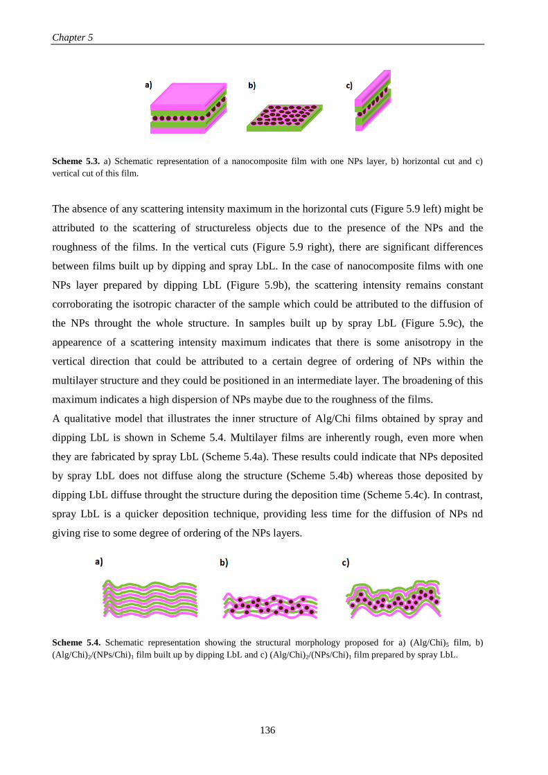
Chapter 5
136
Scheme 5.3. a) Schematic representation of a nanocomposite film with one NPs layer, b) horizontal cut and c)
vertical cut of this film.
The absence of any scattering intensity maximum in the horizontal cuts (Figure 5.9 left) might be
attributed to the scattering of structureless objects due to the presence of the NPs and the
roughness of the films. In the vertical cuts (Figure 5.9 right), there are significant differences
between films built up by dipping and spray LbL. In the case of nanocomposite films with one
NPs layer prepared by dipping LbL (Figure 5.9b), the scattering intensity remains constant
corroborating the isotropic character of the sample which could be attributed to the diffusion of
the NPs throught the whole structure. In samples built up by spray LbL (Figure 5.9c), the
appearence of a scattering intensity maximum indicates that there is some anisotropy in the
vertical direction that could be attributed to a certain degree of ordering of NPs within the
multilayer structure and they could be positioned in an intermediate layer. The broadening of this
maximum indicates a high dispersion of NPs maybe due to the roughness of the films.
A qualitative model that illustrates the inner structure of Alg/Chi films obtained by spray and
dipping LbL is shown in Scheme 5.4. Multilayer films are inherently rough, even more when
they are fabricated by spray LbL (Scheme 5.4a). These results could indicate that NPs deposited
by spray LbL does not diffuse along the structure (Scheme 5.4b) whereas those deposited by
dipping LbL diffuse throught the structure during the deposition time (Scheme 5.4c). In contrast,
spray LbL is a quicker deposition technique, providing less time for the diffusion of NPs nd
giving rise to some degree of ordering of the NPs layers.
Scheme 5.4. Schematic representation showing the structural morphology proposed for a) (Alg/Chi)5 film, b)
(Alg/Chi)2/(NPs/Chi)1 film built up by dipping LbL and c) (Alg/Chi)2/(NPs/Chi)1 film prepared by spray LbL.

Determination of the structure-properties relationship in Alg/Chi films
137
5.4. CONCLUSIONS
The influence of three different experimental parameters, alginate concentration, thermal
crosslinking and number of NPs layers, on the thickness and mechanical properties of multilayer
Alg/Chi films, crosslinked Alg/Chi films and nanocomposite Alg/Chi films built up via spray
LbL were determined by SEM and (PF-QNM) AFM, respectively.
The elastic moduli increased with the number of deposited bilayers up to a value of ~12 GPa for
alginate concentrations of 1.0 and 2.5 mg/mL. Thermal crosslinking of Alg/Chi films reduced
the roughness of multilayer films and increased the elastic modulus. This increase was much
more significant for films obtained with higher alginate concentration. For both series of
samples, non-crosslinked and crosslinked Alg/Chi films, deformation values are below 2 nm and
the adhesion force varies between 1 and 4 nN regardless the number of bilayers or the
concentration of alginate. This was attributed to the fact that the composition of the outer layer is
similar for all the studied samples and the adhesion measurements were sensitive only to the
surface.
The incorporation of iron oxide NPs within the multilayer films increased their roughness and
induced an increase of the elastic moduli of the resulting films. The increase in the values of
deformation found for nanocomposite Alg/Chi films with respect to Alg/Chi films suggested the
presence of empty spaces originated from the fabrication process of the films.
Adhesion values of nanocomposite films were higher than those corresponding to (Alg/Chi)n
films which pointed to a significant effect of the presence of nanoparticles on the adhesion
values found for nanocomposite films. The incorporation of NPs on the last layer of
nanocomposite films gave rise to a two-fold increase in the elastic modulus with respect to
nanocomposite films in which the last layer is chitosan, whereas the deformation was not
affected.
The study of the inner structure of nanocomposite films by GISAXS revealed that films
assembled by dipping LbL gave rise to an isotropic structure, whereas these ones assembled by
spray LbL could form separated NPs layers with dispersion of NPs along the structure due to the
inherent roughness of these films. These results were visualized with the aid of a proposed
qualitative model.

Chapter 5
138
5.5. REFERENCES
1. Langhe, D.; Ponting, M., Introduction to Multilayered FIlms. In Manufacturing and Novel
Applications of Multilayer Polymer Films, William Andrew Publishing: Boston, 2016; pp 1-15.
2. Wong, H. C.; Higgins, A. M.; Wildes, A. R.; Douglas, J. F.; Cabral, J. T., Patterning Polymer–
Fullerene Nanocomposite Thin Films with Light. Advanced Materials 2013, 25, (7), 985-991.
3. Wang, L.-m.; Chang, H.; Zhang, H.; Ren, K.-f.; Li, H.; Hu, M.; Li, B.-c.; Martins, M. C. L.;
Barbosa, M. A.; Ji, J., Dynamic stiffness of polyelectrolyte multilayer films based on disulfide bonds for
in situ control of cell adhesion. Journal of Materials Chemistry B 2015, 3, (38), 7546-7553.
4. Ventrelli, L.; Fujie, T.; Turco, S. D.; Basta, G.; Mazzolai, B.; Mattoli, V., Influence of
nanoparticle-embedded polymeric surfaces on cellular adhesion, proliferation, and differentiation.
Journal of Biomedical Materials Research Part A 2014, 102, (8), 2652-2661.
5. Discher, D. E.; Mooney, D. J.; Zandstra, P. W., Growth Factors, Matrices, and Forces Combine
and Control Stem Cells. Science 2009, 324, (5935), 1673.
6. Fujie, T.; Matsutani, N.; Kinoshita, M.; Okamura, Y.; Saito, A.; Takeoka, S., Adhesive, Flexible,
and Robust Polysaccharide Nanosheets Integrated for Tissue-Defect Repair. Advanced Functional
Materials 2009, 19, (16), 2560-2568.
7. Gil, S.; Silva, J. M.; Mano, J. F., Magnetically Multilayer Polysaccharide Membranes for
Biomedical Applications. ACS Biomaterials Science & Engineering 2015, 1, (10), 1016-1025.
8. Dokukin, M. E.; Sokolov, I., Quantitative Mapping of the Elastic Modulus of Soft Materials with
HarmoniX and PeakForce QNM AFM Modes. Langmuir 2012, 28, (46), 16060-16071.
9. Martínez-Tong, D. E.; Najar, A. S.; Soccio, M.; Nogales, A.; Bitinis, N.; López-Manchado, M.
A.; Ezquerra, T. A., Quantitative mapping of mechanical properties in polylactic acid/natural
rubber/organoclay bionanocomposites as revealed by nanoindentation with atomic force microscopy.
Composites Science and Technology 2014, 104, 34-39.
10. Schön, P.; Bagdi, K.; Molnár, K.; Markus, P.; Pukánszky, B.; Julius Vancso, G., Quantitative
mapping of elastic moduli at the nanoscale in phase separated polyurethanes by AFM. European Polymer
Journal 2011, 47, (4), 692-698.
11. Nguyen, H. K.; Fujinami, S.; Nakajima, K., Elastic modulus of ultrathin polymer films
characterized by atomic force microscopy: The role of probe radius. Polymer 2016, 87, 114-122.
12. Richert, L.; Engler, A. J.; Discher, D. E.; Picart, C., Elasticity of Native and Cross-Linked
Polyelectrolyte Multilayer Films. Biomacromolecules 2004, 5, (5), 1908-1916.
13. Derjaguin, B. V.; Muller, V. M.; Toporov, Y. P., Effect of contact deformations on the adhesion
of particles. Journal of Colloid and Interface Science 1975, 53, (2), 314-326.
14. Sham, A. Y. W.; Notley, S. M., Graphene–polyelectrolyte multilayer film formation driven by
hydrogen bonding. Journal of Colloid and Interface Science 2015, 456, 32-41.
15. Fernández, R.; Ocando, C.; Fernandes, S. C. M.; Eceiza, A.; Tercjak, A., Optically Active
Multilayer Films Based on Chitosan and an Azopolymer. Biomacromolecules 2014, 15, (4), 1399-1407.

Determination of the structure-properties relationship in Alg/Chi films
139
16. Tian, Y.; Li, L.; Han, H.; Wang, W.; Wang, Y.; Ye, Z.; Guo, X., Modification of Spherical
Polyelectrolyte Brushes by Layer-by-Layer Self-Assembly as Observed by Small Angle X-ray Scattering.
Polymers 2016, 8, (4), 145.
17. Singh, M. A.; Groves, M. N., Depth profiling of polymer films with grazing-incidence small-
angle X-ray scattering. Acta Crystallographica Section A: Foundations of Crystallography 2009, 65, (Pt
3), 190-201.
18. Zhang, P.; Santoro, G.; Yu, S.; Vayalil, S. K.; Bommel, S.; Roth, S. V., Manipulating the
Assembly of Spray-Deposited Nanocolloids: In Situ Study and Monolayer Film Preparation. Langmuir
2016, 32, (17), 4251-4258.
19. Herzog, G.; Benecke, G.; Buffet, A.; Heidmann, B.; Perlich, J.; Risch, J. F. H.; Santoro, G.;
Schwartzkopf, M.; Yu, S.; Wurth, W.; Roth, S. V., In Situ Grazing Incidence Small-Angle X-ray
Scattering Investigation of Polystyrene Nanoparticle Spray Deposition onto Silicon. Langmuir 2013, 29,
(36), 11260-11266.
20. Ho, T. T. M.; Bremmell, K. E.; Krasowska, M.; MacWilliams, S. V.; Richard, C. J. E.; Stringer,
D. N.; Beattie, D. A., In Situ ATR FTIR Spectroscopic Study of the Formation and Hydration of a
Fucoidan/Chitosan Polyelectrolyte Multilayer. Langmuir 2015, 31, (41), 11249-11259.
21. Picart, C., Polyelectrolyte multilayer films: from physico-chemical properties to the control of
cellular processes. Current medicinal chemistry 2008, 15, (7), 685-697.
22. Pichon, B. P.; Louet, P.; Felix, O.; Drillon, M.; Begin-Colin, S.; Decher, G., Magnetotunable
Hybrid Films of Stratified Iron Oxide Nanoparticles Assembled by the Layer-by-Layer Technique.
Chemistry of Materials 2011, 23, (16), 3668-3675.
23. Lauter-Pasyuk, V.; Lauter, H.; Gordeev, G.; Müller-Buschbaum, P.; Toperverg, B. P.; Petry, W.;
Jernenkov, M.; Petrenko, A.; Aksenov, V., Parallel and perpendicular lamellar phases in copolymer–
nanoparticle multilayer structures. Physica B: Condensed Matter 2004, 350, (1–3, Supplement), E939-
E942.
24. Lauter-Pasyuk, V.; Lauter, H. J.; Gordeev, G. P.; Müller-Buschbaum, P.; Toperverg, B. P.;
Jernenkov, M.; Petry, W., Nanoparticles in Block-Copolymer Films Studied by Specular and Off-
Specular Neutron Scattering. Langmuir 2003, 19, (19), 7783-7788.
25. Kashem, M. M. A.; Perlich, J.; Schulz, L.; Roth, S. V.; Müller-Buschbaum, P., Correlated
Roughness in Polymer Films Containing Maghemite Nanoparticles. Macromolecules 2008, 41, (6), 2186-
2194.
26. Abul Kashem, M. M.; Perlich, J.; Diethert, A.; Wang, W.; Memesa, M.; Gutmann, J. S.; Majkova,
E.; Capek, I.; Roth, S. V.; Petry, W.; Müller-Buschbaum, P., Array of Magnetic Nanoparticles via Particle
Co-operated Self-Assembly in Block Copolymer Thin Film. Macromolecules 2009, 42, (16), 6202-6208.
27. Song, L.; Wang, W.; Körstgens, V.; González, D. M.; Yao, Y.; Minar, N. K.; Feckl, J. M.; Peters,
K.; Bein, T.; Fattakhova-Rohlfing, D.; Santoro, G.; Roth, S. V.; Müller-Buschbaum, P., Spray Deposition
of Titania Films with Incorporated Crystalline Nanoparticles for All-Solid-State Dye-Sensitized Solar
Cells Using P3HT. Advanced Functional Materials 2016, 26, (10), 1498-1506.
28. Philipp, M.; Körstgens, V.; Magerl, D.; Heller, C.; Yao, Y.; Wang, W.; Santoro, G.; Roth, S. V.;
Müller-Buschbaum, P., Sorption of Water and Initial Stages of Swelling of Thin PNIPAM Films Using in
Situ GISAXS Microfluidics. Langmuir 2015, 31, (35), 9619-9627.
29. Rydzek, G.; Schaaf, P.; Voegel, J.-C.; Jierry, L.; Boulmedais, F., Strategies for covalently
reticulated polymer multilayers. Soft Matter 2012, 8, (38), 9738-9755.
Chapter 5
140
30. Luo, H.; Shen, Q.; Ye, F.; Cheng, Y.-F.; Mezgebe, M.; Qin, R.-J., Structure and properties of
layer-by-layer self-assembled chitosan/lignosulfonate multilayer film. Materials Science and
Engineering: C 2012, 32, (7), 2001-2006.
31. Silva, J. M.; Duarte, A. R. C.; Caridade, S. G.; Picart, C.; Reis, R. L.; Mano, J. F., Tailored
Freestanding Multilayered Membranes Based on Chitosan and Alginate. Biomacromolecules 2014, 15,
(10), 3817-3826.
32. Taccola, S.; Desii, A.; Pensabene, V.; Fujie, T.; Saito, A.; Takeoka, S.; Dario, P.; Menciassi, A.;
Mattoli, V., Free-Standing Poly(l-lactic acid) Nanofilms Loaded with Superparamagnetic Nanoparticles.
Langmuir 2011, 27, (9), 5589-5595.
33. Butt, H.-J.; Cappella, B.; Kappl, M., Force measurements with the atomic force microscope:
Technique, interpretation and applications. Surface Science Reports 2005, 59, (1–6), 1-152.
34. Nogales, A.; Ezquerra, T. A.; Rueda, D. R.; Martinez, F.; Retuert, J., Influence of water on the
dielectric behaviour of chitosan films. Colloid and Polymer Science 1997, 275, (5), 419-425.
35. Stukalov, O.; Murray, C. A.; Jacina, A.; Dutcher, J. R., Relative humidity control for atomic force
microscopes. Review of Scientific Instruments 2006, 77, (3), 033704.
36. Yang, S. Y.; Rubner, M. F., Micropatterning of Polymer Thin Films with pH-Sensitive and
Cross-linkable Hydrogen-Bonded Polyelectrolyte Multilayers. Journal of the American Chemical Society
2002, 124, (10), 2100-2101.
37. Huang, H.; Spaepen, F., Tensile testing of free-standing Cu, Ag and Al thin films and Ag/Cu
multilayers. Acta Materialia 2000, 48, (12), 3261-3269.
38. He, J.; Kanjanaboos, P.; Frazer, N. L.; Weis, A.; Lin, X.-M.; Jaeger, H. M., Fabrication and
Mechanical Properties of Large-Scale Freestanding Nanoparticle Membranes. Small 2010, 6, (13), 1449-
1456.
39. Schmidt, S.; Madaboosi, N.; Uhlig, K.; Kohler, D.; Skirtach, A.; Duschl, C.; Mohwald, H.;
Volodkin, D. V., Control of cell adhesion by mechanical reinforcement of soft polyelectrolyte films with
nanoparticles. Langmuir 2012, 28, (18), 7249-7257.
40. Guo, D.; Xie, G.; Luo, J., Mechanical properties of nanoparticles: basics and applications.
Journal of Physics D: Applied Physics 2013, 47, (1), 013001.
41. Yoneda, Y., Anomalous Surface Reflection of X Rays. Physical Review 1963, 131, (5), 2010-
2013.
42. Busch, P.; Posselt, D.; Smilgies, D. M.; Rauscher, M.; Papadakis, C. M., Inner Structure of Thin
Films of Lamellar Poly(styrene-b-butadiene) Diblock Copolymers As Revealed by Grazing-Incidence
Small-Angle Scattering. Macromolecules 2007, 40, (3), 630-640.

CHAPTER 6
In vitro study of nanocomposite Alg/Chi
films for applications in magnetic
hyperthermia


In vitro study of nanocomposite Alg/Chi films for applications in magnetic hyperthermia
143
This chapter describes the potential application of nanocomposite Alg/Chi films for magnetic
hyperthermia therapy. The iron content of nanocomposite Alg/Chi films described in the Chapter
5 and their heating induced by an alternating magnetic field were determined. It was also
evaluated the human neuroblastoma SH-SY5Y cell adhesion onto these films and their
application for in vitro magnetic hyperthermia treatment through a proof of concept using
neuroblastoma cells. In addition to this, the specific power absorption of the alginate based
magnetic ferrofluid described in the Chapter 3 and the effect of its immobilization in a gel-like
material were determined.
6.1. INTRODUCTION
Magnetic nanoparticles (NPs) provide LbL films with the ability to respond to a magnetic field.1-
3 In biomedical applications, magnetic responsive polymers also find applications in controlled
drug release mediated through external magnetic fields4 and magnetic hyperthermia therapies
(MHT).5 Hyperthermia is a treatment in which the temperature of the body tissue is increased in
a range between 42 – 45 ºC to kill cancer cells.6, 7
The three main kinds of treatments for clinical
applications of MHT therapy are: i) the whole-body hyperthermia where the entire body is
heated up, ii) the regional hyperthermia which is applied by perfusion of a limb, organ or body
cavity with heated fluids and iii) the local hyperthermia where the heat is applied in a localized
area.8 In local hyperthermia, heat is applied to small and superficial tumor areas with the aim of
reaching temperatures up to 42 ºC for one hour within the cancer tumor to cause cell death, being
a less invasive technique as compared with regional and whole hyperthermia. Clinical cases
where local hyperthermia is a viable alternative include chest wall recurrences, superficial
malignant melanoma lesions and head/neck tumors.9, 10
There are several approaches for the
application of local hyperthermia including superficial applicators of different shapes and kinds
that can be heated by the action of external energy sources such as radiofrequencies, microwave,
ultrasound or near infrared radiation laser.11-13
Polymer films with embedded gold nanoparticles
have been succesfully employed for tissue laser ablation when placed in contact with the tissue
to be treated inducing local exogeneus hyperthermia. While the photothermal therapy offers the
possibility to control the energy dose delivered to the target area by both the intensity of the laser
beam and the concentration of NPs, this technique is limited by the short penetration depth of the
NIR wavelength into the human body (~0.3 cm in breast tissue) even at the known ‘optical
window’ of water.14

Chapter 6
144
Magnetic hyperthermia (MHT) is based on the employment of an alternating magnetic field
(AMF) to increase the temperature of magnetic nanoparticles used as heating agents.15-18
The
potential of magnetic NPs to be used as heating agents depends on their specific power
absorption (SPA), defined as the power absorbed by unit mass of NPs which it is released to the
surrounding medium to generate heat. Iron oxide nanoparticles (NPs) have been reported to have
SPA values large enough to heat macroscopic tumors, and also to display lower toxicity than
other heating materials investigated (e.g., Co, Ni and their oxides).19
There exist multiple
research approaches for the design of polymer materials, e.g., in the form of nanogels,
nanoparticles or micelles, able to encapsulate NPs in order to promote selective internalization
into tumor cells and hence, promote intracellular magnetic hyperthermia.16-18
Currently, there are
very promising results regarding the application of intracellular magnetic hyperthermia for
clinical oncology.20, 21
Intracellular hyperthermia consist of using magnetic nanoparticles to remotely induce local heat
when an alternating magnetic field is applied giving rise to the required temperature increase of
42 - 45 ºC to induce cell apoptosis in the tumor site (tissues and organs).22
Cancer cells are more
sensitive to temperature oscillations than normal cells due to the fact that tumor cells show an
increased metabolism, higher rates of glycolysis and enhanced radiosensitivity than normal cells,
which makes them more vulnerable to hyperthermia therapy. In order to be used for intracellular
hyperthermia therapy, NPs have to be non-toxic, biocompatible and injectable.23
The main
disadvantages of this technique are: the difficulty to drive them to the target of interest, the
difficulty to internalize a concentration of NPs enough to achieve an effective treatment. At this
point it has to be considered the surface functionalization of NPs which influences their
attachment to target species (cells, tissues, organs) and the NPs size, because the bigger size of
NPs the higher magnetic heating produced, but if NPs are too big they could not be able to cross
the endothelial barrier and if they are too small could not produce enough heating to induce cell
death.16, 23
An alternative approach to apply MHT therapy could be the incorporation of NPs
inside a polymer film in order to control the concentration of NPs, directly related to the increase
of temperature achieved through the application of MHT. Such thermomagnetic films (TMFs)
could be placed in contact with the tissue affected by cancer disease to induce exogenous
magnetic hyperthermia.
The exogenous heating strategy using magnetic fields that it is described in this chapter can
deliver energy density values to the target area that are lower than the typical values produced by
photothermal therapy. However, as biological tissues essentially do not interact with ac magnetic

In vitro study of nanocomposite Alg/Chi films for applications in magnetic hyperthermia
145
fields of 100-500 kHz, the energy can be delivered essentially into any depth of the human body.
This property makes our strategy based on the employment of thermomagnetic polymer films
(TMFs) more adapted to subcutaneous or deeper applications. To the best of our knowledge,
such approach has not been undertaken before. In the chapter 5 the incorporation of magnetic
nanoparticles within multilayer polymer films obtained from chitosan and alginate through spray
assisted LbL was described and in this chapter, their thermal effect and their application in
magnetic hyperthermia treatment was evaluated through a proof of concept on human
neuroblastoma SH-SY5Y cells. The neuroblastoma-derived SH-SY5Y cell line was selected as a
model of human malignant metastatic neuroblastoma which exhibits a notable resistance to
hyperthermic stress.24
6.2. EXPERIMENTAL PART
6.2.1. Materials
Chitosan (Chi) of low molecular weight (448869, lot SLBG1673V), sodium alginate (Alg)
(A2158, lot 090M0092V), poly(ethylenimine) (PEI) with a molecular weight (Mw) of 25000 and
acetic acid were supplied by Aldrich. Sodium acetate anhydrous was supplied by Panreac. More
information about these materials is detailed in the chapter 3.
It was also employed a lab-made alginate based magnetic ferrofluid containing the iron oxide
nanoparticles (NPs) with a magnetite concentration of 8.0 mg/mL whose characteristics have
been described in the Chapter 3.
Live/Dead viability/cytotoxicity kit, for mammalian cells was supplied by Molecular Probes,
ThermoFisher scientific (L3224). The kit contains calcein AM (4 mM in anhydrous DMSO) and
ethidium homodimer-1 (2 mM in DMSO/H2O 1:4 (v/v)).
6.2.2. Preparation of nanocomposite Alg/Chi films
All multilayer films were built on functionalized glass slides (12 mm ) by spray assisted layer-
by-layer (LbL) of aqueous solutions of alginate (2.5 mg/mL), chitosan (1 mg/mL) and a lab-
made alginate-based magnetic ferrofluid containing the NPs (8.0 mg/mL) following the
procedure described in chapter 5 with some variations, such as the waiting time after the
deposition of the NPs layer and the increase of the NPs/Chi bilayers with regards to Alg/Chi
bilayers, as shown in Scheme 6.1. The spray time was fixed at 5 s with a waiting time of 15 s
between layers of chitosan and alginate and 30 s between NPs and Chitosan layers. The

Chapter 6
146
structural unity was formed by a bilayer Alg/Chi followed by four bilayers NPs/Chi repeated as
many times as necessary until the desired number of layers was obtained. Samples were denoted
as (Alg/Chi)n(NPs/Chi)m where n stands for the number of Alg/Chi bilayers (n = 2, 4, 8, 12, 20
and 40) and m for the corresponding number of NPs/Chi bilayers (m = 8, 16, 32, 48, 80 and 160).
Scheme 6.1. Schematic representation of the spray assisted layer-by-layer procedure and the repeating structural
unit of the nanocomposite (Alg/Chi)n/(NPs/Chi)m multilayer films.
In order to achieve free-standing films, a polyvinyl alcohol (PVA) layer with a concentration of
100 mg/mL was deposited over the multilayer films with a pipette and dried for 24 hours until
become robust. The resulting multilayer film with the ending PVA layer was peeled from the
glass substrate with tweezers; after that, the resulting free-standing film was immersed in Milli-Q
water and the PVA layer was removed by dissolution in water (Scheme 6.2).
Scheme 6.2. Schematic representation to obtain a free-standing film using a PVA sacrificial layer.

In vitro study of nanocomposite Alg/Chi films for applications in magnetic hyperthermia
147
6.2.3. Characterization of nanocomposite Alg/Chi films
Determination of the iron content
The determination of the iron content of (Alg/Chi)n(NPs/Chi)m films was carried out through
UV-Vis transmission spectrophotometry using the thiocyanate complexation reaction and
measuring the absorbance of the iron-thiocyanate complex at 478 nm wavelength.
𝐹𝑒(𝑎𝑞)3+ + 6 𝑆𝐶𝑁(𝑎𝑞)
− → [𝐹𝑒(𝑆𝐶𝑁)6](𝑎𝑞)3−
(Alg/Chi)n(NPs/Chi)m free-standing films were dissolved in HCl 6M/HNO3 (65%) for 2h. After
that, potassium thiocyanate was added to the solution to form the iron-thiocyanate complex. The
magnetite content was determined by comparing the sample absorbance to a calibration curve
(Annex I, Figure A.I.1).
Morphological studies
To determine the inner organization of the polymer films, dual beam FIB/SEM (Nova 200
NanoLab, FEI Company) was carried out on nanocomposite (Alg/Chi)n(NPs/Chi)m films. Films
were coated with platinum for imaging with the FIB-SEM. SEM images were taken at 5 kV with
a Field Emission Guns (FEG) column, and a combined Ga-based 30 kV (10 pA) ion beam was
used to cross-sectioning films. These investigations were completed by energy-dispersive X-ray
spectroscopy (EDX) for chemical analysis.
Determination of magnetic remote heating
The magnetic heating was determined separately for the alginate based magnetic ferrofluid and
for the nanocomposite films.
The heating capacity of the as prepared ferrofluid under alternating magnetic fields (AMF) was
measured using a commercial magnetic field applicator (DM-100, nB Nanoscale Biomagnetics,
Spain) (Figure 6.1) at frequencies 250 < f < 820 kHz and field amplitudes 0 ≤ H ≤ 24 kA/m. The
fluoroptic thermometer integrated into this equipment allowed to measure the temperature as a
function of time.

Chapter 6
148
Figure 6.1. Magnetic field applicator DM-100, nB Nanoscale Biomagnetics.
The heating of nanocomposite (Alg/Chi)n(NPs/Chi)m films was remotely induced by an
alternating magnetic field (AMF) using a commercial induction heating station (EasyHeat,
Ambrell B.V., The Netherlands) working at f = 180 kHz and field amplitude of H = 35 kA/m.
The AMF was applied over the samples placed at the center of 11-turn solenoid. The samples
were thermalized at 37 ºC by a water-heated jacket, which has also the function of thermally
detach the samples from the joule heating at the coil. The temperature was measured using a
thermal camera (FLIR Systems, USA) in a zenithal position over the film. Before each
experiment, the samples were placed within the solenoid and allowed to thermalize at 37 ºC for 5
min before the AMF was turned on. After that, the heating time for each sample was 5 min. A
photograph of this experimental setup in the Institute of Nanoscience of Aragon is shown in
Figure 6.2.
Figure 6.2. Experimental setup for the measurements of the magnertic remote heating of nanocomposite films as
well as for the application of in vitro magnetic hyperthermia and magnification of solenoid.

In vitro study of nanocomposite Alg/Chi films for applications in magnetic hyperthermia
149
6.2.4. In vitro magnetic hyperthermia experiments
Study of adhesion of neuroblastoma cells
Direct contact assays to test cell adhesion were carried out on (Alg/Chi)n(NPs/Chi)m films with
different number of layers. Films were sterilized under UV light for 5 hours. Human
neuroblastoma SH-SY5Y cells (ATCC CRL-26) were cultured in Dulbecco’s modified Eagle’s
medium (DMEM) and Ham’s F12 (1:1) with 15% fetal bovine serum, 100 IU/mL penicillin, 100
g/mL streptomycin and 2 mM L-glutamine. Cells were maintained at 37 ºC in a saturated
humidity atmosphere containing 95% air and 5% of CO2. At confluence, cells were trypsinized
and seeded on the films surface with a cellular density of 1.5 x 10⁵ cells. Cells were cultured for
48 hours. For adhesion tests, the cell growth was followed at 24 and 48 hours by scanning
electron microscopy (Nova 200 NanoLab, FEI Company). For that, cells adhered onto the films
were fixed with 2 %v/v glutaraldehyde for 2 h at 4°C, treated with 2.5 %v/v potassium
ferrocyanate and 1 %v/v osmium tetroxide. Later, SH-SY5Y cells were dehydrated and coated
with platinum for imaging with the FIB-SEM. SEM images were taken at 5 kV with a FEG
column, and a combined Ga-based 30 kV (10 pA) ion beam was used to cross-sectioning single
cells. These investigations were completed by energy-dispersive X-ray spectroscopy (EDX) for
chemical analysis.
Experimental setup and conditions for in vitro magnetic hyperthermia experiments
In order to test nanocomposite (Alg/Chi)n(NPs/Chi)m films as a suitable material for magnetic
hyperthermia (MHT) applications, human neuroblastoma SH-SY5Y cells with a cellular density
of 1.5 x 10⁵ cells were cultured on the film surface for 48 hours in an Ibidi dish at 37 ºC in a
saturated humidity atmosphere containing 95% air and 5% of CO2. After that, the film was
subjected to 1 cycle of MHT (f = 180 kHz, H = 35 kA/m, t = 30 min) as shown in Scheme 6.3a.
In the case of application of more than one cycle of MHT, the waiting time between cycles was 5
h and samples were maintained at 37 ºC in a saturated humidity atmosphere 95% air and 5%
CO2.
A second experimental protocol (Scheme 6.3b) consisted on placing a thermomagnetic film
above human neuroblastoma SH-SY5Y cells with a cellular density of 1.5 x 10⁵ cells cultured on
an Ibidi dish for 48h at 37 ºC in a saturated humidity atmosphere containing 95% air and 5% of
CO2. After that, the Ibidi dish was subjected to two cycles of MHT (f = 180 kHz, H = 35 kA/m, t
= 30 min, waiting time between cycles = 5h).

Chapter 6
150
Scheme 6.3. Schematic representation of the two different experimental protocols employed to measure in vitro
magnetic hyperthermia.
Determination of cell viability after magnetic hyperthermia experiments
Viability of cells cultured on (Alg/Chi)n(NPs/Chi)m films and subjected to hyperthermia
treatment was determined with the Live/Dead viability/cytotoxicity kit 12 hours after the
application of the last MHT cycle in the first experimental protocol and after 3 hours in the
second experimental protocol. Cells were washed with 1 mL of PBS, after that, 1 mL of the stain
kit was added and kept for 30 minutes at room temperature in darkness. The Live/Dead kit
contains calcein AM (4 mM in anhydrous DMSO) and ethidium homodimer-1 (2 mM in
DMSO/H2O 1:4 (v/v)) which allows staining live cell in green and dead cells in red, respectively.
The stained cells were visualized with a Leica SP2 AOBS confocal scanning microscope. Images
were collected using the microscope in sequential mode with a 20x (lens specification, HCPL
FLUOTAR NA 0.50; Leica), a line average of 16 and a format of 1024x1024 pixels. The
confocal pinhole was 1 Airy unit.
6.3. RESULTS AND DISCUSSION
The characterization of multilayer films fabricated with different number of NPs layers, from 8
to 160, is carried out in order to prove their application in magnetic hyperthermia therapy in vitro
with neuroblastoma cells at the end of this chapter.
6.3.1. Determination of iron content of nanocomposite Alg/Chi films
Previous to determine the iron content of nanocomposite films, the incorporation of NPs within
the multilayer structure of Alg/Chi films was assessed visually (Figure 6.3a). All nanocomposite
(Alg/Chi)n(NPs/Chi)m films under study present an homogeneous colour which suggests a good
dispersion of NPs within the films and they become darker with the increase in the number of

In vitro study of nanocomposite Alg/Chi films for applications in magnetic hyperthermia
151
NPs layers. The magnetite content (Fe3O4) incorporated within the multilayer films was
determined by UV-Vis transmission spectrophotometry and plotted in Figure 6.3b as a function
of the number of NPs layers. The magnetite content increases linearly with the number of NPs
layers and could be fitted with the following equation
𝑓(𝐹𝑒3𝑂4) = −0.44 + 0.052𝑁 (𝑒𝑞. 6.1)
where N is the number of NPs layers.
The linear trend observed up to the maximum number of NPs layers deposited suggests that the
final Fe3O4 concentration per unit area could be even increased, although there is likely a limit in
the mechanical integrity of the films over N = 200. However, as discussed within this chapter,
the deposition of the NPs layers during the buildup process of multilayer polymer films was
enough to produce a high temperature increase during the magnetic hyperthermia experiments.
Figure 6.3. a) Image of films assembled on glass substrates with different number of NPs layers from 0 to 160 as
indicated in the figure. b) Determination of Fe3O4 content as a function of the number of NPs layers deposited.
Dashed line represents the linear fit of the data.
6.3.2. Inner morphology of nanocomposite Alg/Chi films
The inner structural organization of multilayer (Alg/Chi)n(NPs/Chi)m films was characterized
through dual-beam (FIB-SEM) images. Representative results corresponding to the cross section
of sample (Alg/Chi)8(NPs/Chi)32 are shown in Figure 6.4a. A magnification of the SEM image
shown in Figure 6.4b showed that the thickness of the sample is about 1 m and it is
nanostructured in layers, each of them with an average thickness of ~50 nm. An EDX analysis of
these cross-sectioned samples revealed the layered distribution of the NPs (Figure 6.4c and 6.4d-
f) as shown from elementary Fe mapping of the material. These results demonstrate not only the
incorporation of Fe3O4 NPs into the multilayers but also their ordered intercalation between
0 20 40 60 80 100 120 140 160 1800
1
2
3
4
5
6
7
8
9
mass F
e3O
4 (
mg
)
Number of NPs layers
b)
16032 80
160 8a)

Chapter 6
152
layers in the film. The carbon is due to the polysaccharides, alginate and chitosan. The results are
in agreement with those obtained by GISAXS reported in Chapter 5.
Figure 6.4. Dual beam FIB-SEM micrographs corresponding to the cross-sectional view of the
(Alg/Chi)8(NPs/Chi)32 film (a) scale bar 1m and b) scale bar 400 nm. (c) EDX spectrum and area of individual
components of EDX spectrum, carbon (d), iron (e) and oxygen (f).
6.3.3. Determination of magnetic remote heating
Determination of the Specific Power Absorption (SPA) as a function of different
experimental parameters
Previous to the measurements of the magnetic remote heating of nanocomposite films, a study of
the alginate based magnetic ferrofluid, whose preparation and characterization were described in
Chapter 3, was carried out.
To measure the efficiency of the ferrofluid as a heating agent under alternating magnetic fields, it
is necessary to know its specific power absorption (SPA), defined as the power absorbed by unit
mass of the magnetic material. SPA was measured in a commercial equipment DM-100
(Nanoscale Biomagnetics, NB). This equipment measures the heating of NPs dispersed in Milli-
Q water (ferrofluid) with the time when an alternating magnetic field (AMF) is applied. Figure
6.5 shows the heating curve of a ferrofluid with a concentration of 8.0 mg/mL applying a
magnetic field amplitude of 24 kA/m and a frequency of 570 kHz. The temperature increases
from 23 to 27 ºC in 10 seconds (dashed arrow).

In vitro study of nanocomposite Alg/Chi films for applications in magnetic hyperthermia
153
0 20 40 60 80 100
20
25
30
35
40
45
50
T
em
pe
ratu
re (
ºC)
time (s)
Figure 6.5. Magnetic heating of ferrofluid with a concentration of 8.0 mg/mL of magnetite in water in the
commercial equipment DM-100 (Nanoscale Biomagnetics) at f = 571 kHz and H = 24 kA/m. The part of the curve
employed to determine the SPA is indicated by the dashed arrow.
SPA value is obtained from the slope of the heating curve using the following equations,25
𝑆𝑃𝐴 = (𝑚𝑙 · 𝐶𝑒𝑙 + 𝑚𝑝 · 𝐶𝑒𝑝
𝑚𝑝) · (
Δ𝑇
Δ𝑡)
𝑚𝑎𝑥 (𝑒𝑞. 6.2)
where 𝐶𝑒 and 𝑚 are the specific heat and the mass, respectively and the subscripts 𝑝 and 𝑙,
denotes the nanoparticles and the liquid carrier, respectively. Taking into account that the
concentration of NPs is less than 1 %wt, this equation can be approximated by
𝑆𝑃𝐴 = (𝜌𝑙 · 𝐶𝑒𝑙
𝜙) · (
Δ𝑇
Δ𝑡)
𝑚𝑎𝑥 (𝑒𝑞. 6.3)
where 𝜌𝑙 is the density of the liquid carrier and 𝜙 is the weight concentration of the NPs in the
ferrofluid. In this case, the liquid carrier is water with 𝜌𝑙 = 103 kg/m
3 and 𝐶𝑒𝑙 = 4.18 kcal/kgºC.
Ferrofluids were tested at different frequencies and magnetic field amplitudes and every
measurement was repeated three times. Figure 6.6a shows the SPA variation with the magnetic
field amplitude at a fixed frequency of 571 kHz for two different ferrofluid concentrations, 4.8
and 8.0 mg/mL, as prepared and after 4 months for the ferrofluid with higher magnetite
concentration. Figure 6.6b depicts the SPA variation with the frequency at a fixed magnetic field
amplitude of 24 kA/m for two different ferrofluid concentrations (𝜙), 4.8 and 8.0 mg/mL, as
prepared and after 4 months for the ferrofluid with higher concentration.

Chapter 6
154
0 4 8 12 16 20 24 280
200
400
600
800
10008.0 mg/mL as prepared
4.8 mg/mL as prepared
8.0 mg/mL after 4 months
SP
A (
W/g
)
H (kA/m)
a)
0 100 200 300 400 500 6000
200
400
600
800
1000 8.0 mg/mL as prepared
4.8 mg/mL as prepared
8.0 mg/mL after 4 months
SP
A (
W/g
)
Frequency (kHz)
b)
Figure 6.6. SPA dependence with a) magnetic field amplitude at f = 571 kHz and b) frequency at H = 24 kA/m for
ferrofluids with 4.8 mg/mL as prepared (●) and 8.0 mg/mL as prepared (■) and after 4 months (□). Dashed lines are
a guide to the eye.
As can be observed, SPA increases with the magnetic field amplitude and the frequency and it is
highly influenced by the magnetite concentration. Focusing the attention in the SPA at H = 24
kA/m and f = 571 kHz, in the case of the highest concentration of magnetite (8.0 mg/mL) SPA is
868.4 ± 1.9 W/g and for the lowest concentration (4.8 mg/mL) SPA decreases up to 295.3 ± 3.5
W/g.
To study the effect of the aging in the SPA, the ferrofluid with a magnetite concentration of 8.0
mg/mL was measured 4 months after its preparation and results are also shown in the Figure 6.6.
After 4 months, SPA follows the same tendency with the frequency and with the magnetic field
amplitude as immediately after its preparation; however, the SPA values are lower. For example,
at f =571 kHz and H = 24 kA/m the SPA is 868.4 ± 1.9 W/g immediately after its synthesis and
626.7 ± 7.2 W/g 4 months after its preparation. These results suggest the loss of magnetic
properties of the ferrofluid probably due to some degree of oxidation of the NPs.
Note that the movement of magnetite NPs in the ferrofluid is not restricted, whereas in
nanocomposite multilayer films they are immobilized. As an approach, to study the influence of
the immobilization of these NPs on the heating properties, the SPA of NPs immobilized in
gelatin was evaluated. To that aim, 500 µL of ferrofluid with a concentration of 8.0 mg/mL were
precipitated at 14500 rpm for 15 min, then the supernatant was removed and NPs were dispersed
in 1mL of 1 %w/v or 2 %w/v gelatin in a vortex for 1 min and kept at -4 ºC for 1 hour to form
the gel. After that, SPA was measured in the commercial equipment DM-100 (NanoScale
Biomagnetics) at a frequency of 571 kHz and a magnetic field amplitude of 24 kA/m and results
are shown in Figure 6.7.

In vitro study of nanocomposite Alg/Chi films for applications in magnetic hyperthermia
155
0 20 40 60 80 100
10
15
20
25
30
35
40
45
50
Tem
pera
ture
(ºC
)
time (s)
NPs in water (ferrofluid)
NPs in 1 % w/v gelatin
NPs in 2 % w/v gelatin
Figure 6.7. Magnetic heating of NPs in water (black line) and in 1%w/v (blue line) and 2 %w/v gelatin (red line) at f
= 571 kHz and H = 24 kA/m. The part of the curve employed to determine the SPA is showed by the dashed arrow.
SPA is obtained from the initial slope of the curves exhibited in Figure 6.7 and results are
provided in Table 6.1. SPA decreases when nanoparticles are immobilized in gelatin and this
decrease is higher as gelatin concentration increases. The exposition of magnetic NPs to a radio-
frequency magnetic field gives rise to a single domain particle that aligns its magnetic moment
M to the field H to decrease the interaction free energy. There are two dissipation mechanisms,
Brownian relaxation and Néel relaxation, which influence the magnetization of ferrofluids when
they are exposed to external time varying magnetic fields. Brownian relaxation is due to the
physical rotation of the nanoparticles against viscous torques from the surrounding media, i.e.,
carrier liquid, films, etc., and Néel relaxation is caused by the rotation of the magnetic moment
inside the magnetic core.26, 27
Due to the fact that both mechanisms take place simultaneously in the dissipation mechanism of
the ferrofluid, the immobilization of NPs in gelatin tends to block the physical rotation of NPs as
gelatin concentration increases and, subsequently, the Brownian contribution to the SPA value
decreases being the Néel relaxation the main dissipation mechanism. It is expected that the same
physical process will take place when NPs are encapsulated into the multilayer films based on
chitosan and alginate as it will be discussed in next section.
Table 6.1. SPA of NPs dispersed in water and in 1%w/v and 2 %w/v gelatin.
Sample ΔT/t (ºC/min) SPA (W/g)
NPs in water 10.1 868.4 ± 1.9
NPs in 1%w/v gelatin 4.8 130.5 ± 6.3
NPs in 2%w/v gelatin 1.9 77.2 ± 0.1

Chapter 6
156
Remote heating of nanocomposite Alg/Chi films
To assess the efficiency of the thermomagnetic films produced, a series of (Alg/Chi)n(NPs/Chi)m
films with different number of NPs layers were heated under a fixed AMF amplitude and
frequency. The temperature was measured using a thermal camera (FLIR Systems, USA) in a
zenithal position over the film. Figure 6.8 illustrates the sequential photographs obtained with
this camera during the heating process of two different films, (Alg/Chi)20(NPs/Chi)80 (Figure
6.8a) and (Alg/Chi)40(NPs/Chi)160 (Figure 6.8b).
Figure 6.8. Sequential photographs obtained by a thermal camera (FLIR Systems, USA) during the heating of a) a
film (Alg/Chi)20(NPs/Chi)80 and b) a film (Alg/Chi)40(NPs/Chi)160. Points 1 and 2 represent two different points of
the sample and point 3 represents the temperature of the water-heated jacket.
t = 0 t = 0.5 t = 1 t = 1.5
t = 2 t = 3 t = 4 t = 5
t = 0 t = 0.5 t = 1 t = 1.5
t = 2 t = 3 t = 4 t = 5
a)
b)

In vitro study of nanocomposite Alg/Chi films for applications in magnetic hyperthermia
157
The temperature increase, T, measured from the basal value T0 = 37 ºC, is plotted as a function
of time (Figure 6.9a) and as a function of the NPs layers (Figure 6.9b) for the different samples.
The heating of the films, from the basal value T0 = 37 ºC, with time by action of the AMF was
followed for 5 minutes for the different samples (Figure 6.9a). As can be observed, temperature
increases during the first two minutes, then a plateau is reached. It can be seen from Figure 6.9b
that T increases linearly with the number of NPs layers (i.e., films with 80 or 160 NPs layers
yield T of 6 and 12 ºC, respectively). Notice that the magnetic heating of the ferrofluid with a
magnetite concentration of 8.0 mg/mL gave rise to a temperature increase of 48 ºC in 1.5 min
(Figure 6.5) and nanocomposite films with 80 and 160 NPs layers give rise to a temperature
increase of 5.8 and 11.2 ºC in 1.5 min, respectively (Figure 6.9a). The immobilization of NPs
into a multilayer Alg/Chi film through spray LbL produces a similar effect to this observed for
the immobilization of NPs into a gel-like material decreasing the temperature increase with
respect to the ferrofluid.
Figure 6.9. a) Temperature increase of (Alg/Chi)n(NPs/Chi)m films measured as a function of the number of NPs
layers and b) Temperature increase with the time of application of the magnetic field for films with different number
of NPs layers: (■) 16, (●) 32, (▼) 80 and (▲) 160.
6.3.4. In vitro magnetic hyperthermia experiments
Adhesion studies of neuroblastoma cells
As (Alg/Chi)n(NPs/Chi)m films are intended to be employed as adhesive pads for exogenous
magnetic hyperthermia treatment, the first step in order to optimize the proposed application was
to ascertain the cell adhesion onto the surface of the thermomagnetic films.
0 20 40 60 80 100 120 140 160 180
0
2
4
6
8
10
12
14
T
(ºC
)
Number of NPs layers
b)
0 1 2 3 4 5
0
2
4
6
8
10
12
14a) 40/16
80/32
200/80
400/160
T
(ºC
)
Time (min)

Chapter 6
158
To study the influence of NPs on cell adhesion, neuroblastoma cells were cultured on (Alg/Chi)n
and (Alg/Chi)n(NPs/Chi)m films and the cell adhesion process was followed at different times, 24
and 48 hours, by optical microscopy and the results are shown in Figure 6.10. At 24 hours the
cell adhesion is higher in films with NPs than without NPs and at 48 hours cell confluence is
observed in both cases. The presence of NPs within films influences cell adhesion. In the chapter
5 it had been demonstrated that stiffness of (Alg/Chi)n(NPs/Chi)m films built up via spray LbL
increases as number of NPs increases. This could be one of the reasons why neuroblastoma cell
adhesion is higher in films with NPs than in films without NPs.
Figure 6.10. Optical micrographs of SH-SY5Y cell adhesion on (Alg/Chi)n films (n = 10 and 20) and on
(Alg/Chi)n(NPs/Chi)m films (n = 2, 4 and m =8, 16) after 24 and 48 hours
In the case of (Alg/Chi)20(NPs/Chi)80 films with a high number of NPs layers, films become very
dark as can be seen in the Figure 6.3a and it is not possible to follow the cell adhesion by optical
microscopy. An alternative technique consists of dehydration of samples and visualization by
SEM (Figure 6.11). With the aim of checking that similar results are obtained by SEM
microscopy, the cell adhesion on (Alg/Chi)n(NPs/Chi)m films with different number of layers
after 48 hours of culture was studied. Figure 6.12a shows a general view of neuroblastoma cells
cultured for 24 h on a (Alg/Chi)20(NPs/Chi)80 film and cell adhesion and proliferation can be
observed along the film surface. At this point, it is important to note that there are not noticeable
differences regarding neuroblastoma cell adhesion and proliferation on (Alg/Chi)n(NPs/Chi)m
films with different number of NPs layers. In fact, cell confluence is also observed for films with
8 and 16 NPs layers after 24 h (Figure 6.11).
48 h
24 h
(Alg/Chi)10
(Alg/Chi)20
(Alg/Chi)2(NPs/Chi)
8 (Alg/Chi)
4(NPs/Chi)
16

In vitro study of nanocomposite Alg/Chi films for applications in magnetic hyperthermia
159
Figure 6.11. SEM micrographs of SH-SY5Y cells cultured on (Alg/Chi)n(NPs/Chi)m films (n = 2, 4 and m = 8, 16)
for 24 h.
A magnification of the SEM image (Figure 6.12b) allows observing an individual cell adhered
onto the film surface. Further information on cell adhesion can be obtained from the cross
section of the film. The cell adhered completely to the film surface as can be observed in a
lamella cross section of the cell shown (Figure 6.12c). The analysis of the EDX spectrum
corresponding to this area (Figure 6.12d) with the different elements, carbon in green (Figure
6.12e), iron in red (Figure 6.12f), oxygen in grey (Figure 6.12g) and silicon in blue (Figure
6.12h) allows to clearly differentiate the iron component embedded on the film (red color) from
the nucleus of the cell (black color) marked with the arrow. Note that the presence of silicon is
due to the fact that the film is supported on a glass substrate. The composition of the film can be
quantitatively determined from the EDX spectrum (Figure 6.12i) where the major component is
iron (64.67%), followed by O (28.69%) and C (6.64%).
50 m 100 m
(Alg/Chi)2(NPs/Chi)
8 (Alg/Chi)
4(NPs/Chi)
16

Chapter 6
160
Figure 6.12. SEM micrographs corresponding to neutoblastoma cells cultured on a (Alg/Chi)20(NPs/Chi)80 film for
24 hours taken at two different magnifications: (a) 50 µm and (b) 5 µm , (c) FIB-SEM image corresponding to the
cross-sectional view of an individual neuroblastoma cell adhered onto a (Alg/Chi)20(NPs/Chi)80 film, (d) EDX
spectrum and area of individual components of EDX spectrum delimited by a black rectangle, carbon (e), iron (f),
oxygen (g) and silicon (h) and EDX spectrum (i).
In vitro hyperthermia studies
The temperature range for activating apoptotic cell mechanisms depends on the cellular type
involved, but it can be generalized that above 43-45ºC different metabolic pathways are activated
to reverse the effects of the temperature.16, 28
Firstly, the effect of magnetic hyperthermia (MHT) on the cell viability was evaluated using the
experimental protocol showed in Scheme 6.3a where neuroblastoma cells were cultured on the
surface of films with 80 and 160 NPs layers and then, they were subjected to 1 cycle of MHT (H
= 35 kA/m and f = 180 kHz; 30 min) and 3 cycles of MHT with a waiting time of 5 h between
cycles. The results were compared to those obtained for neuroblastoma cells cultured on a
control film not subjected to MHT treatment.

In vitro study of nanocomposite Alg/Chi films for applications in magnetic hyperthermia
161
Figure 6.13. Images corresponding to the live/dead test carried out on neuroblastoma cells cultured onto a)
(Alg/Chi)20(NPs/Chi)80 and b) (Alg/Chi)40(NPs/Chi)160 films subjected to 0 cycles of MHT (control), 1 cycle of
MHT (30 min) and 3 cycles of MHT (30 min, waiting time between cycles of 5h). Note that cells are labelled with
calcein (live cells in green) and EthD-1 (dead cells in red). (c) Determination of % cell viability obtained from the
images corresponding to live cells stained in green.
The results corresponding to the live/dead test performed on neuroblastoma cells cultured on
films with 80 and 160 NPs layers are shown in Figures 6.13a and 6.13b, respectively. From a
qualitative point of view, control experiments carried out on neuroblastoma cells cultured on
films with 80 NPs layers and not subjected to MHT, present a lower number of live cells (stained
in green) adhered to the film than that observed for cells cultured on films with 160 NPs layers
(Figure 6.13b). This is in general agreement with the results found in literature regarding cell
adhesion on multilayer films that show that, generally speaking, cells tend to adhere more on
films with higher number of layers.29
Other studies have also shown that the incorporation of
nanoparticles into polymer films improved cell adhesion due to the increase in surface
roughness.30, 31
For cells cultured onto films with 80 NPs layers (Figure 6.13a), the application of
one and three cycles of MHT does not significantly change the area covered by live cells with
respect to the control experiment. In contrast, Figure 6.13b shows that the application of one
cycle of MHT on cells cultured on films with 160 NPs layers results in a decrease of area
covered by live cell population with respect to the control film, being this decrease much more
0
20
40
60
80
100
160 NPs layers
Control 1 cycle 3 cycles
% c
ell
via
bil
ity
80 NPs layers
Control 1 cycle 3 cycles
Eth
D-1
Cal
cein
b)
100 µm100 µm100 µm
100 µm 100 µm 100 µm100 µm 100 µm 100 µm
100 µm 100 µm100 µm
Control 1 cycle 3 cycles
Eth
D-1
Cal
cein
a)
c)

Chapter 6
162
noticeable after the application of 3 cycles of MHT. It is worth to note that the percentage of
dead cells stained in red (Ethidium homodimer-1 in Figures 6.13a and 6.13b) does not
correspond to the total number of dead cells due to the fact that cell death results in the loss of
cell adhesion and hence detachment from the film.
As can be observed in Figure 6.13c, the cell viability data (%) obtained from the quantification
of the area covered by live cells stained in green cultured on films with 80 NPs layers
demonstrates that the application of MHT results in negligible cytotoxic effects after the
application of one (96% viable cells) or three MHT cycles (94% viable cells). However, the cell
viability data (%) corresponding to neuroblastoma cells cultured on films with 160 NPs
decreases to 69% with respect to the control experimental with the application of one cycle of
MHT and to 21% with the application of three cycles of MHT. At this point, it is important to
take into account the final temperature reached by the TMFs with the application of AMF which
is related to the number of NPs layers as shown in Figure 6.9. TMFs with 80 NPs layers reach a
final temperature of 42 ºC, which under the experimental protocol employed, is not high enough
to induce a cytotoxic effect on neuroblastoma cells whereas the temperature reached by TMFs
with 160 NPs layers increases to 49 ºC which induces a decrease of cell viability to 69% with
respect to the control experiment. These results demonstrate the importance of controlling the
concentration of NPs and hence, the temperature generated during the magnetic field application
in order to produce a measurable magnetic hyperthermia effect. The temperature range for
activating apoptotic cell mechanisms depends on the cellular type involved, but it can be
generalized that above 43-45 ºC different metabolic pathways are activated to reverse the effects
of the temperature.16, 22
For our experiments, the temperature gradient was expected to be large
due to the bidimensional nature of the samples (and thus large surfaces for exchanging heat).
It can be observed from Figure 6.13 that films with 80 magnetic layers did not produce enough
thermal effect to affect the cell viability even after three application cycles. However, for those
films with 160 magnetic layers the application of several cycles increased the cell mortality
through MHT. This is consistent with previous studies showing an increase in cell death in cell
cultures subjected to several expositions to AMF.32
33
Because of the potential therapeutic applications require these polymer films to be placed on the
surface of the treated area, we conducted further in vitro MHT experiments aimed to test the cell
viability of neuroblastoma cells grown underneath the thermomagnetic films. To that aim,
human neuroblastoma cells were cultured on the surface of an Ibidi dish for 48 hours and a film

In vitro study of nanocomposite Alg/Chi films for applications in magnetic hyperthermia
163
with 80 NPs layers was placed above cells as shown in Figure 6.14. As control experiment,
neuroblastoma cells were cultured in an Ibidi dish with cells and not subjected to MHT. Cells
were subjected to two cycles of MHT (30 min with a waiting time of 5 hours between cycles; H
= 35 kA/m, f = 180 kHz) which causes a significant decrease in the calcein signal that
corresponds to live cells stained in green. In addition, it is possible to observe that cellular death
takes place only within the area in contact with the thermomagnetic film which proves the local
effect of the magnetic hyperthermia.
In a recent study reported on the same SH-SY5Y cell line studied here, it was demonstrated that
MHT required a lower temperature than an exogenous heating source (i.e., water bath) to
provoke a given level of cell death.34
The results depicted in the inset of Figure 6.13a show a
much higher efficiency of the TMFs under study with respect to exogeneous heating provided by
a thermalized bath. The quantification of the calcein signal after the application of 2 MHT cycles
yields a cell viability of 15% which represents a significant decrease with respect to the control
film. It is important to note that the temperature achieved by the TMF after each cycle was 43 ºC
(see Figure 6.9a). In the case of the application of hyperthermia through a thermalized water
bath, the temperature needed to achieve a similar decrease in cell viability was 52 ºC.34
Hence,
the TMFs under study show a much higher efficiency in the application of exogenous
hyperthermia with respect to the results found by an exogenous heating source (i.e., water bath)
which can be explained because of the intimate contact of the cells with the heating element.
Secondly, taking into account that the potential therapeutic applications require these polymer
films to be placed on the surface of the treated area, in vitro MHT experiments aimed to test the
cell viability of neuroblastoma cells grown underneath the thermomagnetic films were conducted
following the protocol showed in Scheme 6.3b. To that aim, human neuroblastoma cells were
cultured on the surface of an Ibidi dish for 48 hours and a film (Alg/Chi)20(NPs/Chi)80 was
placed above cells as shown in Figure 6.14. As control, neuroblastoma cells were cultured in an
Ibidi dish with cells and not subjected to MHT. Cells were subjected to two cycles of MHT (30
min with a waiting time of 5 hours between cycles; H = 35 kA/m, f = 180 kHz). The
quantification of the calcein signal yields a cell viability of 15% which represents a significant
decrease with respect to the control film. In addition, it is possible to observe that cellular death
takes place only within the area in contact with the thermomagnetic film which proves the local
effect of the magnetic hyperthermia. Taking into account that the temperature achieved by
(Alg/Chi)20(NPs/Chi)80 with the application of the AMF is T = 43 ºC, the effect of 2 cycles of
MHT on cell viability is much higher for thermofilms than that obtained by exogenous

Chapter 6
164
hyperthermia (EHT) and even by local intra-tumoral magnetic hyperthermia for which a T = 52
ºC and 47 ºC respectively are needed to reach a decrease of cell viability of 15%.
Figure 6.14. Live/dead test carried out on neuroblastoma cells cultured onto an Ibidi dish, covered by a
(Alg/Chi)20(NPs/Chi)80 film and subjected to 2 cycles of MHT. Neuroblastoma cells cultured on an Ibidi dish and
not subjected to MHT are employed as control. Cells are labelled with calcein (live cells in green) and EthD-1 (dead
cells in red). The bar chart shows the determination of % cell viability obtained from the images corresponding to
live cells stained in green.
6.4. CONCLUSIONS
Thermomagnetic polymer films have been successfully prepared by alternate deposition of
chitosan and alginate layers and magnetic nanoparticles using a spray assisted layer-by-layer
assembly. Precisely, the presence of NPs into the multilayer structure of these films provides
them the ability to response to a magnetic field and allows to use them in magnetic hyperthermia
therapies. The incorporation of NPs into the multilayer structure was assessed visually due to a
change of colour of films with NPs with regards to films without NPs and the magnetite content
was determined by UV-Vis transmission spectrophotometry showing a linear increase of
magnetite content with the number of NPs layers deposited.
Dual Beam scanning electron microscopy of the cross section of the films revealed their
nanostructuration in layers and the EDX analysis of this area showed the ordered distribution and
intercalation of NPs between layers and determined the composition of these films, being the
iron the major component in a 65%. The layered structure together with the ordered distribution
and intercalation of NPs between layers provided the expected mechanical stability and thermal
responsiveness.
0
20
40
60
80
100
2 cy
cles
% c
ell v
iab
ilit
y
Contr
ol

In vitro study of nanocomposite Alg/Chi films for applications in magnetic hyperthermia
165
The SPA of NPs in Milli-Q water (ferrofluid) with a concentration of magnetite of 8.0 mg/mL
applying a magnetic field amplitude of 24 kA/m and a frequency of 571 kHz was 868.4 ± 1.9
W/g. The immobilization of NPs in 2 %w/v gelatin produced a decrease of SPA up to 77.2 ± 0.1
W/g due to the block of the Brownian relaxation.
Thermomagnetic films were heated by application of a magnetic field amplitude of 35 kA/m and
a frequency of 180 kHz for 5 minutes from 6 to 12 ºC varying the number of NPs layers from 80
to 160 layers. The ΔT increases linearly with the number of NPs layers, that is with the
magnetite content. Indeed, the concentration of NPs (up to 65 % in mass) into the multilayer
structure of these films was enough to reach target temperatures up to 14 ºC above the control
samples by the application of AMF.
In vitro experiments showed good adhesion properties to grow neuroblastoma cells on these
films with different number of NPs layers as proven by optical microscopy and SEM. By
remotely heating the films using ac magnetic fields we induced a reduction in cell viability to
67% and 20% of the control samples for one and three heating cycles, respectively. The results
in experimental conditions mimicking in vivo applications yielded a reduction of 85% of the cell
viability only within the contact area with the film after two cycles of MHT.
6.5. REFERENCES
1. Pichon, B. P.; Louet, P.; Felix, O.; Drillon, M.; Begin-Colin, S.; Decher, G., Magnetotunable
Hybrid Films of Stratified Iron Oxide Nanoparticles Assembled by the Layer-by-Layer Technique.
Chemistry of Materials 2011, 23, (16), 3668-3675.
2. Paterno, L. G.; Fonseca, F. J.; Alcantara, G. B.; Soler, M. A. G.; Morais, P. C.; Sinnecker, J. P.;
Novak, M. A.; Lima, E. C. D.; Leite, F. L.; Mattoso, L. H. C., Fabrication and characterization of
nanostructured conducting polymer films containing magnetic nanoparticles. Thin Solid Films 2009, 517,
(5), 1753-1758.
3. Schaaf, P.; Voegel, J.-C.; Jierry, L.; Boulmedais, F., Spray-Assisted Polyelectrolyte Multilayer
Buildup: from Step-by-Step to Single-Step Polyelectrolyte Film Constructions. Advanced Materials 2012,
24, (8), 1001-1016.
4. Bruvera, I.; Hernández, R.; Mijangos, C.; Goya, G. F., An integrated device for magnetically-
driven drug release and in situ quantitative measurements: design, fabrication and testing. Journal of
Magnetism and Magnetic Materials 2015, 377, 446-451.
5. Zamora-Mora, V.; Fernández-Gutiérrez, M.; Román, J. S.; Goya, G.; Hernández, R.; Mijangos,
C., Magnetic core–shell chitosan nanoparticles: Rheological characterization and hyperthermia
application. Carbohydrate polymers 2014, 102, 691-698.
6. Kerr, J. F. R.; Winterford, C. M.; Harmon, B. V., Apoptosis. Its significance in cancer and cancer
Therapy. Cancer 1994, 73, (8), 2013-2026.

Chapter 6
166
7. Mahmoudi, K.; Hadjipanayis, C. G., The application of magnetic nanoparticles for the treatment
of brain tumors. Frontiers in Chemistry 2014, 2, 109.
8. Van der Zee, J., Heating the patient: a promising approach? Annals of oncology 2002, 13, (8),
1173-1184.
9. Kok, H. P.; Crezee, J., A comparison of the heating characteristics of capacitive and radiative
superficial hyperthermia. International Journal of Hyperthermia 2016, 1-9.
10. Gao, S.; Zheng, M.; Ren, X.; Tang, Y.; Liang, X., Local hyperthermia in head and neck cancer:
mechanism, application and advance. Oncotarget 2016, 7, (35), 57367-57378.
11. Wust, P.; Hildebrandt, B.; Sreenivasa, G.; Rau, B.; Gellermann, J.; Riess, H.; Felix, R.; Schlag, P.
M., Hyperthermia in combined treatment of cancer. The Lancet Oncology 2002, 3, (8), 487-497.
12. Sardari, D.; Verga, N., Cancer Treatment with Hyperthermia. In Current Cancer Treatment -
Novel Beyond Conventional Approaches, InTech: 2011; pp 455-474.
13. Habash, R. W. Y.; Bansal, R.; Krewski, D.; Alhafid, H. T., Thermal Therapy, Part 2:
Hyperthermia Techniques. Critical Reviews™ in Biomedical Engineering 2006, 34, (6), 491-542.
14. Redolfi Riva, E.; Desii, A.; Sinibaldi, E.; Ciofani, G.; Piazza, V.; Mazzolai, B.; Mattoli, V., Gold
Nanoshell/Polysaccharide Nanofilm for Controlled Laser-Assisted Tissue Thermal Ablation. ACS Nano
2014, 8, (6), 5552-5563.
15. P Calatayud, M.; Asin, L.; Tres, A.; F Goya, G.; R Ibarra, M., Cell Bystander Effect Induced by
Radiofrequency Electromagnetic Fields and Magnetic Nanoparticles. Current Nanoscience 2016, 12, (3),
372-377.
16. Hervault, A.; Thanh, N. T. K., Magnetic nanoparticle-based therapeutic agents for thermo-
chemotherapy treatment of cancer. Nanoscale 2014, 6, (20), 11553-11573.
17. Sailor, M. J.; Park, J.-H., Hybrid Nanoparticles for Detection and Treatment of Cancer. Advanced
Materials 2012, 24, (28), 3779-3802.
18. Zamora-Mora, V.; Fernández-Gutiérrez, M.; González-Gómez, Á.; Sanz, B.; Román, J. S.; Goya,
G. F.; Hernández, R.; Mijangos, C., Chitosan nanoparticles for combined drug delivery and magnetic
hyperthermia: From preparation to in vitro studies. Carbohydrate polymers 2017, 157, 361-370.
19. Cristofolini, L.; Szczepanowicz, K.; Orsi, D.; Rimoldi, T.; Albertini, F.; Warszynski, P., Hybrid
Polyelectrolyte/Fe3O4 Nanocapsules for Hyperthermia Applications. ACS Applied Materials & Interfaces
2016, 8, (38), 25043-25050.
20. Maier-Hauff, K.; Rothe, R.; Scholz, R.; Gneveckow, U.; Wust, P.; Thiesen, B.; Feussner, A.; von
Deimling, A.; Waldoefner, N.; Felix, R.; Jordan, A., Intracranial Thermotherapy using Magnetic
Nanoparticles Combined with External Beam Radiotherapy: Results of a Feasibility Study on Patients
with Glioblastoma Multiforme. Journal of Neuro-Oncology 2007, 81, (1), 53-60.
21. Johannsen, M.; Thiesen, B.; Wust, P.; Jordan, A., Magnetic nanoparticle hyperthermia for
prostate cancer. International Journal of Hyperthermia 2010, 26, (8), 790-795.
22. IP Soares, P.; MM Ferreira, I.; AGBN Igreja, R.; MM Novo, C.; PMR Borges, J., Application of
hyperthermia for cancer treatment: recent patents review. Recent patents on anti-cancer drug discovery
2012, 7, (1), 64-73.

In vitro study of nanocomposite Alg/Chi films for applications in magnetic hyperthermia
167
23. Bañobre-López, M.; Teijeiro, A.; Rivas, J., Magnetic nanoparticle-based hyperthermia for cancer
treatment. Reports of Practical Oncology & Radiotherapy 18, (6), 397-400.
24. Cheng, L.; Smith, D. J.; Anderson, R. L.; Nagley, P., Human neuroblastoma SH-SY5Y cells
show increased resistance to hyperthermic stress after differentiation, associated with elevated levels of
Hsp72. International Journal of Hyperthermia 2011, 27, (5), 415-426.
25. Gonzalez-Fernandez, M. A.; Torres, T. E.; Andrés-Vergés, M.; Costo, R.; de la Presa, P.; Serna,
C. J.; Morales, M. P.; Marquina, C.; Ibarra, M. R.; Goya, G. F., Magnetic nanoparticles for power
absorption: Optimizing size, shape and magnetic properties. Journal of Solid State Chemistry 2009, 182,
(10), 2779-2784.
26. Sagué, B. S. Study of the cell death induced by magnetic hyperthermia in neural cells using
Fe3O4 nanoparticles. Universidad de Zaragoza, Zaragoza, 2015.
27. Kötitz, R.; Weitschies, W.; Trahms, L.; Semmler, W., Investigation of Brownian and Néel
relaxation in magnetic fluids. Journal of Magnetism and Magnetic Materials 1999, 201, (1–3), 102-104.
28. Paula, I. P. S.; Isabel, M. M. F.; Rui, A. G. B. N. I.; Carlos, M. M. N.; Joao, P. M. R. B.,
Application of Hyperthermia for Cancer Treatment: Recent Patents Review. Recent patents on anti-
cancer drug discovery 2012, 7, (1), 64-73.
29. Neto, A. I.; Vasconcelos, N. L.; Oliveira, S. M.; Ruiz-Molina, D.; Mano, J. F., High-Throughput
Topographic, Mechanical, and Biological Screening of Multilayer Films Containing Mussel-Inspired
Biopolymers. Advanced Functional Materials 2016, 26, (16), 2745-2755.
30. Ventrelli, L.; Fujie, T.; Turco, S. D.; Basta, G.; Mazzolai, B.; Mattoli, V., Influence of
nanoparticle-embedded polymeric surfaces on cellular adhesion, proliferation, and differentiation.
Journal of Biomedical Materials Research Part A 2014, 102, (8), 2652-2661.
31. Lipski, A. M.; Pino, C. J.; Haselton, F. R.; Chen, I. W.; Shastri, V. P., The effect of silica
nanoparticle-modified surfaces on cell morphology, cytoskeletal organization and function. Biomaterials
2008, 29, (28), 3836-3846.
32. Ito, A.; Tanaka, K.; Honda, H.; Abe, S.; Yamaguchi, H.; Kobayashi, T., Complete regression of
mouse mammary carcinoma with a size greater than 15 mm by frequent repeated hyperthermia using
magnetite nanoparticles. Journal of Bioscience and Bioengineering 2003, 96, (4), 364-369.
33. Balivada, S.; Rachakatla, R. S.; Wang, H.; Samarakoon, T. N.; Dani, R. K.; Pyle, M.; Kroh, F. O.;
Walker, B.; Leaym, X.; Koper, O. B.; Tamura, M.; Chikan, V.; Bossmann, S. H.; Troyer, D. L., A/C
magnetic hyperthermia of melanoma mediated by iron(0)/iron oxide core/shell magnetic nanoparticles: a
mouse study. BMC Cancer 2010, 10, 119-119.
34. Sanz, B.; Calatayud, M. P.; Torres, T. E.; Fanarraga, M. L.; Ibarra, M. R.; Goya, G. F., Magnetic
hyperthermia enhances cell toxicity with respect to exogenous heating. Biomaterials 2017, 114, 62-70.


CHAPTER 7
Study of cell adhesion and applications
in drug delivery of Alg/Chi films


Study of cell adhesion and applications in drug delivery of Alg/Chi films
171
This chapter firstly investigates the influence of the number of bilayers, surface chemistry and
crosslinking of multilayer Alg/Chi films obtained through spray assisted LbL on cell adhesion of
human dermal fibroblasts (HDF) and human caucasian breast adenocarcinoma (MCF-7).
Secondly, the employment of these films for drug delivery applications was evaluated by
incorporation of an anticancer drug (tamoxifen) in different intermediate positions of multilayer
Alg/Chi films to study its controlled release over time.
7.1. INTRODUCTION
Multilayer films prepared through layer-by-layer (LbL) assembly of polyelectrolytes are
extensively employed in biomedical applications.1-5
For example, LbL technique allows the
encapsulation of different therapeutic drugs into multilayer films by direct assembly during the
buildup process.4-8
Taking into account that this assembly is carried out in aqueous solutions
under mild conditions, the activity of the loaded drugs is retained in the films.9 Thanks to the
control of the thickness, composition and surface chemistry, as well as the satisfactory adhesion
ability, LbL assembled polymer films can be specifically customized to function as a promising
platform for sustained drug delivery.10, 11
Compared with the traditional oral drug delivery
systems, such as tablets or liquid formulations, polymer films can be attached to the pathological
site of the target organ or tissue for direct drug delivery of the therapeutic agent which enhances
therapeutic activity, increases the efficacy of drugs, reduces the dose frequency, minimizes side
effects to normal tissue, improves patient compliance and reduces gastrointestinal side effects.5,
10, 12 In addition to this, multilayer polymer films offer the possibility of providing sustained drug
delivery over the time as a function of the number of layers.13
These characteristics increase their
potential clinical applications. Different therapeutic drugs have been encapsulated in multilayer
films for biomedical applications.4, 8, 13-15
An hydrophobic anti-inflammatory drug, adenosine
deaminase inhibitor, was assembled in the middle of a multilayer (Chi/Alg)10 film built up via
spin-coating LbL to treat localized inflammatory bowel disease.4 An anti-fibrotic agent,
pirfenidone, was encapsulated in a (Chi/Alg)15 film via dipping LbL for excisional wound
healing. In the ocular field, latanoprost isopropyl ester was cast on the surface of (Chi/Alg)n
films (n = 5, 10 and 20) to reduce intraocular pressure13
and norfloxacin and timolol were loaded
in multilayer (Ha/Chi)10 films built up via dipping LbL to decrease the wearing hydrogel contact
lenses.14
Curcumin, which has a pleiotropic therapeutic effect in cancer, was dissolved in
nanocrystalline cellulose (NCC) and multilayer films (Chi/NCC) were made by dipping LbL.8
Another important biomedical application of multilayer polymer films is their employment as

Chapter 7
172
model surfaces for the study of the adhesion of living cells provided that the chemical and
physical surface properties of the films are key to the modulation of cell adhesion.16-19
. In fact,
LbL allows to tune the final biological and physicochemical properties of multilayer films by
changing the nature of the outermost layer and the thickness and roughness of the films through
the number of deposited layers.20
These features make LbL a powerful technique to modulate
cell adhesion on the surface of a multilayer film.17
In chapter 4, the fabrication of multilayer films based on chitosan and alginate by means of spray
assisted LbL assembly was described. Then, the nanomechanical properties of these multilayer
Alg/Chi films were studied in the chapter 5 concluding that the elastic modulus increased with
the number of deposited bilayers and with the thermal crosslinking process at 120 ºC, whereas
the deformation remained constant. Multilayer (Alg/Chi)n films built up through spray assisted
LbL possess chemical and physical properties for exploring cell adhesion. Cells are sensible to
the elasticity or rigidity of the surface, for that reason nanomechanical properties of the films are
of crucial importance in their adhesion.21
Among the different parameters that affect to the
elastic modulus of a surface and influence cell adhesion, crosslinking is one of the most decisive
leading to an increase of the stiffness and cell attachment in many cases.22
Lee et al.23
demonstrated that different types of cells, human umbilical artery endothelial cells (HUAECs),
3T3 fibroblasts, osteoblasts MC3T3-E1 and HeLa (epithelial) cells grew at similar rates on
different poly(dimethylsiloxane) surfaces regardless of the stiffness for surfaces having elastic
moduli ranging from 0.6 to 2.6 MPa. On the contrary, Silva et al.22
observed that (Chi/Alg)100
films built up via dipping LbL increased their elastic modulus from 4.8 MPa to 12.8 and 56.4
MPa after crosslinking with two different genipin concentrations and probed that the adhesion of
fibroblasts L929 cells was sensitive to the stiffness of films increasing a ~15% and 40% with
regards to non-crosslinked films.
In this chapter, in vitro cell adhesion studies will be carried out with dermal fibroblast and
human breast cancer MCF-7 cells aimed to elucidate the influence of physicochemical properties
and different architectures of multilayer Alg/Chi films on cell adhesion. To that aim, it was
varied the number of deposited bilayers and the outermost layer, alginate or hyaluronic acid, and
the effect of crosslinking was studied in order to elucidate the influence of these variables on cell
adhesion properties. Taking into account that these multilayer films are intended to be used as
patches for sustained drug delivery, the encapsulation of tamoxifen, a therapeutic drug to treat
the breast cancer, was carried out within different intermediate positions of the multilayer
(Alg/Chi)n films and results corresponding to in vitro drug release are also presented.

Study of cell adhesion and applications in drug delivery of Alg/Chi films
173
7.2. EXPERIMENTAL PART
7.2.1. Materials
Chitosan (Chi) of low molecular weight (448869, lot SLBG1673V), sodium alginate (Alg)
(A2158, lot 090M0092V), poly(ethylenimine) (PEI) with a molecular weight (Mw) of 25000
were supplied by Aldrich and used as received. Hyaluronic acid (HA) was supplied by Lifecore
Biomedical (lot 024276, part HA500K-5). More information about these materials is detailed in
the chapter 3.
Acetic acid, tamoxifen citrate salt 99% (TMX), 1-Ethyl-3-(3-dimethylaminopropyl)-
carbodiimide (EDC) and N-Hydroxysuccinimide (NHS) (Aldrich) were supplied by Aldrich and
used as received. Sodium acetate anhydrous was purchased from Panreac.
For biological assays, phosphate buffered solution (PBS) of pH 7.4 (Sigma-Aldrich) and fetal
bovine serum (FBS) (Gibco) were used as received. Tissue culture media, additives, trypsin,
glutaraldehyde and Tween 80 were purchased from Sigma-Aldrich. Alamar Blue reagent was
supplied from Invitrogen, DAPI (4',6-Diamidino-2-Phenylindole, Dihydrochloride) fluorescent
stain was purchased by Molecular Probes and crystal violet was supplied by Prolab diagnostics.
7.2.2. Preparation of multilayer films with tamoxifen
All films were built on functionalized glass slides (14 mm ) through spray assisted layer-by-
layer (LbL) technique of aqueous solutions of alginate (2.5 mg/mL) and chitosan (1.0 mg/mL)
using an automatic ND-SP spray coater from Nadetech innovations (Spain). Three different
systems were prepared. i) Alg/Chi films was prepared following the procedure described in the
chapter 4 to obtain (Alg/Chi)n films. In order to study the influence of the surface chemistry, a
layer of Alg or HA was deposited at the end of the cycle for 5 s and samples were denoted as
(Alg/Chi)nAlg and (Alg/Chi)nHA films, where n stands for the number of bilayers. ii) The
second type of samples was obtained by crosslinking of the previously prepared (Alg/Chi)nAlg
and (Alg/Chi)nHA films by immersing them in a solution prepared dissolving 30 mg EDC and
19.9 mg NHS in 1 ml of Milli-Q water for 1 hour at 4 ºC and then they were washed in deionized
water for 30 min three times to remove crosslinking NHS-ester residues. Samples were denoted
as crosslinked (CL) (Alg/Chi)nAlg and crosslinked (Alg/Chi)nHA films. iii) For the third series
of samples which tamoxifen incorporated in an intermediate position, it was followed the same
procedure, but in the middle of the preparation process a layer of tamoxifen (200 µL of a

Chapter 7
174
solution of 1.0 mg/mL of TMX in ethanol) was deposited with a pipette and, after a waiting time
of 2 hours to evaporate the solvent, the spray deposition process was followed (Scheme 7.1).
Samples were denoted as (Alg/Chi)5TMX(Chi/Alg)m where m stands for the number of
(Chi/Alg) bilayers after deposition of TMX.
Scheme 7.1. Sequential process followed for the preparation of (Alg/Chi)5TMX(Chi/Alg)m films.
7.2.3. Characterization
Morphological characterization
The topography and surface roughness of films were evaluated using a Multimode Scanning
probe microscope (Veeco/Bruker), with a di NanoScope IVa Controller using the conventional
height mode (tapping mode, normal AFM) at 320 kHz resonant frequency and a nominal force
constant of 42 N/m. All measurements were performed in air, at room temperature, on films
supported on silicon wafers. Non-crosslinked and crosslinked (Alg/Chi)nAlg and (Alg/Chi)nHA
films were scanned over square regions of 2 m size and the average roughness (Ra) was
calculated from the height images.
Contact angle
To determine films hydrophilicity, sessile drop air/water contact angle measurements were
carried out on the surface of non-crosslinked and crosslinked (Alg/Chi)nAlg and (Alg/Chi)nHA
films with different number of bilayers at room temperature using a goniometer KSV tetha (KSV
instruments, Ltd., Finland) with water droplets of 3 µL. The average contact angle was
determined by measuring three different points of every film.

Study of cell adhesion and applications in drug delivery of Alg/Chi films
175
Degradation assay
The degradation of non-crosslinked and crosslinked (Alg/Chi)10Alg and (Alg/Chi)10HA films
over time was performed by immersing the film in a vial containing 3 mL of culture medium at
37 ºC in a shaker plate at 30 rpm. At different times, 4, 8 and 15 days, the surface of films was
observed by Scanning Electron Microscopy using a Hitachi SU 8000 microscope operating at 1.5
kV. The same experimental protocol was employed in order to evaluate the influence of the
encapsulation and drug delivery of TMX in the degradation process of
(Alg/Chi)5TMX(Chi/Alg)n films (n = 5, 10 and 20).
Biological behavior
The biological response of the as-prepared materials was tested with human dermal fibroblasts
(HDF, Innoprot) and human caucasian breast adenocarcinoma (MCF-7, ECACC) cells. The
culture medium was Dulbecco’s Modified Eagle’s Medium, enriched with 4500 mg/mL of
glucose (DMEM) supplemented with 10% fetal bovine serum (FBS), 200 mM L-glutamine, 100
units/mL penicillin and 100 mg/mL streptomycin, modified with 4-(2-hydroxyethyl)-1-
piperazineethanesulfonic acid (HEPES).
The culture medium was changed at selected time intervals with little disturbance to culture
conditions. Glass slides (14 mm ) were used as a negative control. Samples were sterilized
under UV light (HNS OSRAM, 263 nm, 3.6UVC/W) at a power of 11W for 2 hours.
Cytotoxicity assay
Cytotoxicity assay was carried out on (Alg/Chi)10Alg films. Films were immersed in a falcon
tube of 15 mL containing 5 mL of culture medium and placed in a shaker plate (30 rpm) at 37
ºC. At predetermined intervals, 2, 4, 8 and 11 days, the medium was removed and replaced with
other 5 mL. All the extracts were obtained under sterile conditions. Cells were seeded at a
density of 9 x10⁴ cells/mL in complete medium in a sterile 96 well culture plate and incubated to
confluence. After 24 h of incubation, the medium was replaced with the corresponding extracts
and incubated at 37 ºC in humidified air with 5% CO2 for 24 h.
A solution of Alamar Blue (AB) (10 %v/v) was prepared in warm DMEM without phenol red
(Gibco). Then, 100 μL of this solution were added to each well and the plates were incubated at
37 °C for 3 h. After that, fluorescence was measured with a Biotek Synergy HT detector using an

Chapter 7
176
emission wavelength of 590 nm and an excitation wavelength of 530 nm. Cell viability (CV) was
calculated with the following equation:
𝐶𝑉 = (𝐹𝐷𝑆 − 𝐹𝐷𝐵
𝐹𝐷𝐶 − 𝐹𝐷𝐵) · 100 (𝑒𝑞. 7.1)
where FDS, FDB and FDC are the fluorescence density of the AB for the sample (S), blank (B)
(culture medium without cells) and control (C), respectively.
Adhesion and morphology assays
Direct contact assays to test cell adhesion were carried out on non-crosslinked and crosslinked
(Alg/Chi)nAlg and (Alg/Chi)nHA films with different number of bilayers. Glass slides were used
as negative controls. At confluence, cells were trypsinized and seeded on the surface of films
with a cellular density of 4.9 x 10⁴ cells. For adhesion tests, the cell growth was followed after 3
days in culture by optical microscopy (Nikon eclipse TE2000-S). After that, a solution of AB (10
%v/v), prepared in a warm medium without phenol red, was added to the plate and incubated at
37 ºC for 3 h. Finally, fluorescence was measured with a Biotek Synergy HT detector and cell
viability (CV) was calculated with equation 7.1.
To observe cellular adhesion and morphology of cells onto the surface of films, cells were
stained with different dyes according to the following protocol. Films were washed with PBS
and fixed with glutaraldehyde 2.5 %v/v in PBS for 10 min at room temperature. After that,
glutaraldehyde was removed and film was rinsed with PBS again. In the case of cellular
adhesion, the cell nuclei were stained with 10 µL/mL of DAPI dye solution and left it for 10 min
at room temperature in the darkness and, for cellular morphology; cell cytoskeletal was stained
with crystal violet solution (0.02 %w/v) and kept it for 10 min at room temperature. Finally,
stained cells were observed using an epifluorescence microscope (Nikon eclipse TE2000-S) and
three different filters.
In vitro release of TMX
The release of TMX from (Alg/Chi)5TMX(Chi/Alg)n films with n = 5, 10 and 15 bilayers was
performed by immersing the film in a vial containing 3 mL of PBS pH 7.4 with 1% w/v Tween
80 at 37 ºC in a shaker plate at 30 rpm. At predetermined intervals, the solution was replaced by
a fresh one to ensure constant release conditions. The amount of TMX released was evaluated
measuring the absorbance at 277 nm in PBS with 1 %w/v Tween 80 using a UV-Vis

Study of cell adhesion and applications in drug delivery of Alg/Chi films
177
spectrophotometer (NanoDrop One/Onec, Thermo scientific). The cumulative amount of released
TMX was determined from the standard calibration curve of TMX in PBS with 1 %w/v Tween
80 (Annex II, Figure A.II.1). All experiments were repeated three times. The total amount of
TMX loaded into the films was determined by dissolving films in 5 mL buffer pH5 (acetic acid
0.1M/sodium acetate 0.1M) with magnetic stirring, the resulting solution was freeze-dried and
solved in 4 mL of PBS with 1 %w/v Tween 80 to measure the absorbance at 277 nm.
For in vitro assays of tamoxifen delivery in contact with HDF and MCF-7 cells, the following
experimental protocol was carried out. (Alg/Chi)5TMX(Chi/Alg)n films with n = 5, 10 and 15
bilayers and (Alg/Chi)n films with n = 10, 15 and 20 bilayers were immersed in falcon tubes of
15 mL containing 3 mL of culture medium and placed in a shaker plate (30 rpm) at 37 ºC. At
predetermined intervals, 1, 2, 4 and 8 hours, the medium was removed and replaced with other 3
mL. All the extracts were obtained under sterile conditions. Cells were seeded at a density of 6
x104 cells/mL in complete medium in a sterile 96 well culture plate and incubated to confluence.
After 24 h of incubation, the medium was replaced with the corresponding extracts and
incubated at 37 ºC in humidified air with 5% CO2 for 24 h. Afterwards, the medium was
removed, wells were washed with PBS and fixed with glutaraldehyde 2.5 %v/v in PBS for 10
min at room temperature. After that, glutaraldehyde was removed and wells were rinsed with
PBS again. Then, 100 μL of crystal violet solution at 10%w/v were added to each well and kept
at room temperature for 15 min. After that, the solution was removed and wells were washed
with Milli-Q water to remove the excess of dye. After drying at room temperature, 100 L of
acetic acid (10 %v/v in PBS) were added to each well and absorbance was measured with a
Biotek Synergy HT detector using an emission wavelength of 690 nm and an excitation
wavelength of 620 nm. Cell viability (CV) was calculated using the equation 7.2.
𝐶𝑉 = (𝐴𝐷𝑆 − 𝐴𝐷𝐵
𝐴𝐷𝐶 − 𝐴𝐷𝐵) · 100 (𝑒𝑞. 7.2)
where ADS, ADB and ADC are the absorbance density of the crystal violet for the sample (S),
blank (B) (culture medium without cells) and control (C), respectively.

Chapter 7
178
7.3. RESULTS AND DISCUSSION
7.3.1. Morphological characterization of Alg/Chi films by AFM
Figure 7.1 shows representative AFM images corresponding to the topography of multilayer
polymer films with the same number of bilayers in which the last layer is Alg ((Alg/Chi)10Alg,
Figure 7.1a) or HA ((Alg/Chi)10HA, Figure 7.1b) and the corresponding crosslinked samples
(Figures 7.1c and 7.1d respectively). AFM images corresponding to non-crosslinked and
crosslinked (Alg/Chi)nAlg and (Alg/Chi)nHA films (n = 5, 7, 15 and 25) can be accessed in the
Annex III (Figure A.III.1).
Figure 7.1. Representative AFM topographic images corresponding to non-crosslinked and crosslinked
(Alg/Chi)10Alg and (Alg/Chi)10HA films to calculate the average roughness (Ra).
The analysis of AFM topographic images of non-crosslinked and crosslinked (Alg/Chi)nAlg and
(Alg/Chi)nHA films with different number of bilayers allows to obtain the average roughness
(Ra) of the films. The results corresponding to the average roughness of the films under study are
tabulated in Table 7.1. As can be observed, Ra increases with the number of deposited bilayers
for all samples under study regardless surface chemistry (ending layer, HA or Alg) or chemical
crosslinking, but not in great extension. At first deposited bilayers, the roughness is attributed to
the formation of islands which decrease their size as the number of deposited bilayers increases.
In this case, the roughness is also influenced by the spraying deposition process and by the
absence of additional external ionic strength (e.g., NaCl).24, 25
The surface chemistry produces
differences in the roughness being (Alg/Chi)nHA films rougher than (Alg/Chi)nAlg films,
whereas after crosslinking there are not differences between both. Conversely, it is not possible
to find a general trend regarding the influence of crosslinking on roughness for films with the
same surface chemistry.

Study of cell adhesion and applications in drug delivery of Alg/Chi films
179
Table 7.1. Evolution of the average roughness (Ra) of non-crosslinked and crosslinked (Alg/Chi)nAlg and
(Alg/Chi)nHA films as a function of the number of bilayers (n).
7.3.1. Degradation essay
Taking into account the increasing interest and the great number of applications of these
multilayer polymer films, it is important to know the degradation behavior in fluids that resemble
biological media.26
The degradation of non-crosslinked and crosslinked (Alg/Chi)10Alg and
(Alg/Chi)10HA films in culture medium (DMEM) at 37 ºC was evaluated by physical changes
within the surface of multilayer films by SEM for a period of 15 days at different time intervals
of 4 and 8 days as can be observed in Figure 7.2.
Number of
bilayers
(n)
Ra (nm)
(Alg/Chi)nAlg
films
(Alg/Chi)nHA
films
Crosslinked
(Alg/Chi)nAlg
films
Crosslinked
(Alg/Chi)nHA
films
5 8.7 ± 1.2 7.2 ± 1.2 7.2 ± 2.2 15.0 ± 4.0
7 13.4 ± 3.7 14.9 ± 3.8 10.1 ± 3.2 9.9 ± 4.3
10 16.9 ± 3.9 27.1 ± 6.3 18.2 ± 4.3 17.2 ± 2.3
15 16.3 ± 2.4 31.7 ± 9.8 26.1 ± 7.0 24.1 ± 4.6
25 14.7 ± 7.8 25.2 ± 1.4 32.0 ± 1.3 31.2 ± 6.0

Chapter 7
180
Figure 7.2. SEM micrographs of non-crosslinked a) (Alg/Chi)10Alg and b) (Alg/Chi)10HA films and crosslinked c)
(Alg/Chi)10Alg and d) (Alg/Chi)10HA films in contact with DMEM at 37ºC during different time intervals.
In the case of non-crosslinked films, when Alg is the outermost layer (Figure 7.2a), small
spherical objects are noted with time but there are not cracks on the surface. On the contrary,
when HA is the last layer (Figure 7.2b), cracks and voids are observed after 4 days. After 15
days, the surface has completely changed, indicating that films ending in HA are more prone to
degradation. For crosslinked films (Figures 7.2 c and d), although cracks appear after 4 days, less
degradation is observed after 15 days with respect to non-crosslinked films, reflecting a decrease
in the degradation process.
7.3.2. In vitro cell adhesion studies
Cytotoxicity assay
In this work, chemical crosslinking of (Alg/Chi)n films was carried out through carbodiimide
chemistry, using EDC and NHS as chemical crosslinkers. Considering that EDC has been
reported to be cytotoxic in literature,27
as a first step it was evaluated the biocompatibility of
non-crosslinked and crosslinked (Alg/Chi)nAlg films by means of a cytotoxicity assay performed
4 days 15 days
1 µm 1 µm
1 µm 1 µm
1 µm 1 µm
1 µm 1 µm
0 days
1 µm
1 µm
1 µm
1 µm
a)
b)
c)
d)

Study of cell adhesion and applications in drug delivery of Alg/Chi films
181
in vitro with HDF and MCF-7 cells (Figure 7.3). A comparison of the results obtained for non-
crosslinked and crosslinked films with the control sample corresponding to cells cultured with
culture medium shows that HDF and MCF-7 cell viability remained unaffected up to 11 days in
contact with non-crosslinked films. Likewise, the crosslinking of the films with EDC and NHS
did not produce a decrease in cell viability over the same period of time. These results
demonstrate that non-crosslinked and crosslinked (Alg/Chi)10Alg films are biocompatible and
could be incorporated in cell culture applications.
Figure 7.3. Cytotoxicity assay of non-crosslinked and crosslinked (Alg/Chi)10 films in contact with a) HDF and b)
MCF-7 cells.
Cell adhesion
Taking into account that interactions between cells and multilayer films surface could be
influenced by the number of bilayers21
and the chemical nature of the outermost layer,28
the
spray assisted LbL technique allows to easily change the last deposited layer of the multilayer
films keeping constant the overall chemical composition in order to modulate cell adhesion.29
Hence, it provides a methodology to produce polymer films with tailored physicochemical
properties for systematic studies of cell adhesion. Cell adhesion tests were carried out on
multilayer (Alg/Chi)n films built up with the same polyelectrolyte pairs, alginate and chitosan,
and spraying the corresponding polyanion solution, alginate or hyaluronic acid, at the end of the
spray LbL (Alg/Chi)n process to obtain films with different ending layer, (Alg/Chi)nAlg and
(Alg/Chi)nHA, in contact with cells. Cells were seeded on the films and kept in culture for three
days. Figure 7.4 shows representative images of the cell nuclei stained with DAPI (up) and
bright field images (down) of non-crosslinked and crosslinked (Alg/Chi)10Alg and
(Alg/Chi)10HA films after three days of culture with HDF (Figure 7.4a) and MCF-7 cells (Figure
7.4b). Images corresponding to non-crosslinked and crosslinked (Alg/Chi)nAlg and
0
20
40
60
80
100
120
11 days8 days4 days
Ce
ll v
iab
ilit
y (
%)
Control Non CL CL
2 days
a)
0
20
40
60
80
100
120
Cell v
iab
ilit
y (
%)
Control Non CL CL
2 days 4 days 8 days 11 days
b)

Chapter 7
182
(Alg/Chi)nHA films (n = 5, 7, 15 and 25) are shown in the Annex IV (Figures A.IV.1 and
A.IV.2). Multilayer films with Alg as ending layer show lower cell attachment compared with
multilayer films ending on Ha. The crosslinking process produces an increase of cell adhesion
for both kind of cells, HDF and MCF-7, and chemical surfaces, Alg and HA, under study.
Figure 7.4. Fluorescence microscope images of a) HDF and b) MCF-7 cell adhesion on non-crosslinked and
crosslinked (Alg/Chi)10Alg and (Alg/Chi)10HA films after 3 days. Scale bars 100 m.
Cell adhesion was quantified by means of an Alamar blue assay carried out on (Alg/Chi)nAlg
and (Alg/Chi)nHa films after three days of cell culture of HDF and MCF-7 cells. Besides the type
of cells, HDF or MCF-7, three different experimental parameters were taken into account,
namely, number of bilayers, effect of crosslinking and effect of the chemistry of the last layer
(alginate or hyaluronic acid) and results are depicted in Figure 7.5.
Alg Alg CL
HA HA CL
Alg Alg CL
HA HA CL
a) b)

Study of cell adhesion and applications in drug delivery of Alg/Chi films
183
Figure 7.5. a) HDF and b) MCF-7 cell adhesion on non-crosslinked and crosslinked (Alg/Chi)nAlg films. c) HDF
and d) MCF-7 cell adhesion on non-crosslinked and crosslinked (Alg/Chi)nHa films. The diagrams include the
mean, the standard deviation (n = 4) and the ANOVA results at a significance level of * p < 0.05.
Effect of the number of bilayers
As shown in Figure 7.5a, the percentage of HDF cells adhered on (Alg/Chi)nAlg films remains
constant at ~14% with the number of bilayers. After crosslinking, the adhesion of HDF cells
increases up to 22% only for the sample with 25 bilayers. Likewise, the percentage of MCF-7
adhered cells remains constant (~14%) within the experimental error for (Alg/Chi)nAlg films
with different number of bilayers (Figure 7.5b). However, after crosslinking, there is a
remarkable increase in MCF-7 cell adhesion with the number of deposited bilayers increasing
from 23% for (Alg/Chi)5Alg films to 81% for (Alg/Chi)25Alg films.
In the case of (Alg/Chi)nHA films (Figure 7.5c), the percentage of HDF adhered cells remains
constant with the number of bilayers (~18%), being slightly higher than that corresponding to
(Alg/Chi)nAlg films (~14%). Generally speaking, crosslinking gives rise to a slight increase in
HDF cell adhesion up to 22% for (Alg/Chi)15HA films with respect to non-crosslinked films.
0
10
20
30
40
50
60
70
80
Ad
he
red
MC
F-7
ce
lls
(%
)
Alg
Alg CL
b)
257
Number of bilayers
5 1510
0
10
20
30
40
50
60
70
80
Ad
he
red
MC
F-7
ce
lls
(%
)
Ha
Ha CL
d)
257
Number of bilayers
5 15100
10
20
30
40
50
60
70
80
Ad
he
red
HD
F c
ells
(%
)
Ha
Ha CL
c)
257
Number of bilayers
5 1510
0
10
20
30
40
50
60
70
80
257
Ad
he
red
HD
F c
ells (
%)
Alg
Alg CL
a)
Number of bilayers
5 1510
*
*
* *
*

Chapter 7
184
Similar results were obtained regarding the quantification of MCF-7 adhered cells on
(Alg/Chi)nHA films (Figure 5.7d), that is percentage of adhered cells remains constant with the
number of bilayers (~20%). But in this case, crosslinking produces a decrease of MCF-7 cell
adhesion that also remains constant with the number of bilayers.
From the results shown in Figure 7.5, it can be concluded that there is no effect of the number of
bilayers on cell adhesion, neither for HDF nor for MCF-7 cells. The increase of the number of
bilayers in multilayer polysaccharidic films prepared through spray assisted LbL results in a
concomitant increase of roughness (Table 7.1). This is in agreement with previous reports, Neto
et al.30
studied the adhesion of L929 cells on the surface of (Chi/Alg)n films (n = 5, 10, 15)
functionalized with fibronectin giving rise to ~5% cell adhesion for all three multilayer films
without noting increase of adhesion with the number of bilayers. Elbert et al.31
studied adhesion
of human fibroblast cells on (PLL/Alg)n films (n = 5, 10, 15) and did not observe changes in the
cell adhesion as number of bilayers increased. Cado et al.32
demonstrated that cell viability and
spreading are quite similar for films with different roughness. It follows, that roughness when it
is homogeneously distributed throughout the film surface does not have an appreciable effect on
cell adhesion for the films under study in this report.
Effect of the surface chemistry
As to the surface chemistry, there are slight differences on cell adhesion between films ending in
alginate and those ending in hyaluronic acid. The percentage of HDF adhered cells increase from
14% for (Alg/Chi)nAlg films to 18% for (Alg/Chi)nHA films. Similarly, MCF-7 cells adhesion
increased from 15% on (Alg/Chi)nAlg films to 20% on (Alg/Chi)nHA films. One of the reasons
for a higher cell attachment of MCF-7 with respect to HDF could be that HDF cells prefer
smoother surfaces to grow33
and multilayer films built up via spray LbL have a roughness which
increases with the number of bilayers. Other kind of cells, e.g., human osteosarcoma cells
MG63, exhibit a higher level of adhesion with the increase in surface roughness.34
A rough
surface induces morphological mutations in tumor cells probably due to the fact that this kind of
cells are more prone to the invasion of their living environment and can adapt to it more easily.
In addition, rougher surfaces tend to form clusters of cells which does not occur when cells are
seeded onto smooth surfaces.35

Study of cell adhesion and applications in drug delivery of Alg/Chi films
185
Effect of crosslinking
Results point to a significant effect of crosslinking on MCF-7 cell adhesion measured on
(Alg/Chi)nAlg films which increases from 14% to up 81%. Crosslinking gives rise to an increase
of the elastic modulus as it was demonstrated by means of PeakForce Quantitative
Nanomechanical Mapping atomic force microscopy (PF-QNM) AFM in the chapter 5. The
elastic modulus of a film with 20 bilayers increases from 9.2 ± 1.6 GPa to 32.4 ± 11.5 GPa after
crosslinking. This increase in the elastic modulus makes the film stiffer and gives advantage to
cell adhesion as it has been corroborated by earlier studies about cell adhesion carried out for
different types of cells.1, 30, 36, 37
Interestingly, it is important to note that crosslinking does not seem to have an effect on HDF
cell adhesion which remains almost constant at ~14% on (Alg/Chi)nAlg films and ~18% on
(Alg/Chi)nHA films.
Contact angle studies
Some studies relate cell adhesion to the contact angle values of the film surface,34, 38
a sensitive
method to the chemical nature and the surface morphology of the last layer in a multilayer film
architecture. The wetting properties of non-crosslinked and crosslinked (Alg/Chi)nAlg and
(Alg/Chi)nHA films were evaluated by water contact angle measurements and results are
exhibited in Figure 7.6 as a function of the number of bilayers. In the case of non-crosslinked
(Alg/Chi)nAlg films, there is a linear increase of the contact angle from 17º to 41º up to 10
bilayers and then a plateau is reached. For crosslinked films, the same trend is observed, that is
the contact angle increases with the number of bilayers from 12º to 44º for (Alg/Chi)nAlg films
up to 10 bilayers and then a plateau is reached. However, it is important to highlight that there
are not significant differences in the contact angle between non-crosslinked and crosslinked
(Alg/Chi)nAlg films.

Chapter 7
186
Figure 7.6. Water contact angle measurements on non-crosslinked and crosslinked a) (Alg/Chi)nAlg and b)
(Alg/Chi)nHA films with different number of bilayers.
Figure 7.6b shows the evolution of the contact angle for non-crosslinked and crosslinked
(Alg/Chi)nHA films as a function of the number of bilayers. The trend is similar to the one found
for (Alg/Chi)nAlg films, that is, for non-crosslinked (Alg/Chi)nHA films, there is a linear
increase of the contact angle from 25º to 50º up to 10 bilayers and then a plateau is reached. For
crosslinked films, the same trend is observed but the contact angles are higher with regards to
non-crosslinked (Alg/Chi)nHA films, that is the contact angle increases with the number of
bilayers from 28º to 65º up to 10 bilayers and then a plateau is reached. However, in this case,
crosslinking produces an increase of the contact angle in all cases, that is, samples become less
hydrophilic with crosslinking, which could alter cell adhesion in some cases as it was observed
in the Figure 7.5d.
It is important to note that all films under study are hydrophilic as shown by contact angle values
lower than 90º.27
The hydrophilic character of both polymers, Alg and HA, is attributed to
hydroxyl and carboxyl groups.33
The pKa values of hyaluronic acid and sodium alginate are ~339
and ~3.540
respectively, so that at physiological pH these groups are ionized. Therefore, the small
differences found regarding the contact angle of (Alg/Chi)nAlg and (Alg/Chi)nHA films cannot
account for the slight differences regarding cell adhesion of HDF. On the contrary, in the case of
MCF-7 cell attachment, the hydrophilicity of the surface might play a more important role
decreasing the cell attachment on crosslinked (Alg/Chi)nHA films, where the surface became less
hydrophilic.
Although wettability of a surface reflects its surface chemistry, there is not unanimity with
regards to cell adhesion. Some authors consider that more hydrophilic surfaces provide a higher
0 5 10 15 20 25 3010
20
30
40
50
60
70
Alg
Alg CL
Co
nta
ct
an
gle
(º)
Number of bilayers
a)
0 5 10 15 20 25 3010
20
30
40
50
60
70
Co
nta
ct
an
gle
(º)
Ha
Ha CL
Number of bilayers
b)
Study of cell adhesion and applications in drug delivery of Alg/Chi films
187
cell adhesion41
and others report that cells adhered better when the surface is hydrophobic or at
intermediate contact angles33
. Taking into account these considerations, it is possible to think
that different kinds of cells will preferentially adhere to surfaces with particular contact angle
values. In the case of HDF, cell attachment is slightly higher as increasing hydrophobicity of
surfaces being HA the surface where more HDF cells adhered. Maybe this is due to the fact that
HA provides an appropriate combination of hydrophilicity and hydrophobicity properties on the
adhesion surface which favors the growth of fibroblast cells as reported Wang et al.33
for L929
cells.
Morphology assay
Not only the percentage of adhered cells but also the cell morphology after adhesion is important
for the development of biocompatible polymer patches.42
Figure 7.7 shows representative
images corresponding to the overall cell morphology of HDF and MCF-7 cells adhered on non-
crosslinked and crosslinked (Alg/Chi)10Alg and (Alg/Chi)10HA films with two different ending
layers, Alg and HA. The morphology of these two cell lines, HDF (Figure 7.7a) and MCF-7 cells
(Figure 7.7b), were characterized by visualizing stained cells with crystal violet using an optical
microscopy. HDF cells show their characteristic elongated shape43
with a more slender
appearance for Alg ending layer than for HA. Significant differences on morphology were not
observed with the crosslinking process. MCF-7 has a cobblestone-like phenotype with strong
cell-cell adhesion and exhibit round mass morphology with formation of colonies distinguished
from the round morphological grouping.44
MCF-7 cells show a similar morphology as Alg or HA
are the outermost layer. In this case, crosslinking produces a change of morphology decreasing
the size of colonies.

Chapter 7
188
Figure 7.7. Cytoskeletal organization of a) HDF and b) MCF-7 cells on non-crosslinked and crosslinked
(Alg/Chi)10Alg and (Alg/Chi)10HA films after 3 days culture. Scale bars 1000 m.
7.3.3. Drug release experiments
The total amount of TMX loaded into (Alg/Chi)5TMX(Chi/Alg)n films was 0.11 ± 0.01 mg TMX
in a surface of 14 mm diameter which corresponds to 0.071 mg/cm2. With this approach, the
quantity of loaded drug for the specific application as a function of the film area can be
modulated. Prior to test the drug delivery in vitro with HDF and MCF-7cells, the TMX release
was quantified and results are shown in Figure 7.8 in which the cumulative release profiles of
TMX from three different multilayer films, (Alg/Chi)5TMX(Chi/Alg)n where n = 5, 10 and 20
bilayers, are compared as a function of time. A fast initial release takes place during the first
hour for the three different multilayer films under study; the film with lower number of bilayers
(n = 5) releases the 50% of TMX (0.037 mg/cm2), followed by the film with n = 10 which
releases 37% of TMX (0.028 mg/cm2) and n = 20 with 28% (0.020 mg/cm
2). The complete
release of TMX occurred at 2, 5 and 10 days for n = 5, 10 and 20 bilayers respectively. The spray
LbL technique allows to modulate the release rate as a function of the number of deposited
bilayers being slower and sustained over time as the number of bilayers increases.13, 45
The cumulative drug release data up to 0.6 for the two films with higher number of bilayers (n =
10 and 20) were fitted to different diffusion models, Higuchi,46
Ritger-Peppas,47
Peppas-Sahlin48
and zero-order,49
to elucidate the diffusion release mechanism, being the Ritger-Peppas47
described by equation 7.3 the most accurate model (r2 > 0.99 in all cases).
a)
b)
Alg Ha Alg CL Ha CL

Study of cell adhesion and applications in drug delivery of Alg/Chi films
189
𝑀𝑡
𝑀∞= 𝑘 · 𝑡𝑚 (𝑒𝑞. 7.3)
where Mt/M is the released fraction and k and m are fitting parameters characteristic of the
film/solution medium and transport mechanism, respectively. The value of m indicates Fickian
diffusion (m = 0.5), anomalous transport (0.5 < m < 1) or Case-II transport (m = 1).48
The inset of
the Figure 7.8 shows the fitting to the Ritger-Peppas model up to 0.6 cumulative drug release and
the fitting parameters obtained from this model are summarized in Table 7.2. The value of m =
0.43 for the film (Alg/Chi)5TMX(Chi/Alg)20 confirms the diffusion controlled drug release
mechanism.
Figure 7.8. Cumulative time-dependent release profiles of TMX from (Alg/Chi)5TMX(Chi/Alg)5 (■),
(Alg/Chi)5TMX(Chi/Alg)10 (●) and (Alg/Chi)5TMX(Chi/Alg)20 (▲) films. The inset shows the fitting with dashed
lines to the Ritger-Peppas model up to 0.6 cumulative drug release for (Alg/Chi)5TMX(Chi/Alg)10 (●) and
(Alg/Chi)5TMX(Chi/Alg)20 (▲) films.
Table 7.2. Fitting parameters from the Ritger-Peppas model.
Sample k m r2
(Alg/Chi)5TMX(Chi/Alg)10 0.38 ± 0.00 0.33 ± 0.00 0.999
(Alg/Chi)5TMX(Chi/Alg)20 0.26 ± 0.01 0.43 ± 0.02 0.993
The surface morphology of (Alg/Chi)5TMX(Chi/Alg)n films (n = 5, 10 and 20) after the drug
delivery experiment is shown in Figure 7.9. No significant differences are observed between
films after the drug delivery experiment which seems to indicate the absence of degradation of
the films. This result confirms that the release mechanism follows a diffusion controlled
mechanism.
0 1 2 3 4 5 6 7 8 9 10 110,0
0,2
0,4
0,6
0,8
1,0
0 1 2 3 4 5 6 7 8 90,0
0,2
0,4
0,6
0,8
Mt/M
time (h)
M(t
)/M
time (days)

Chapter 7
190
Figure 7.9. SEM micrographs of a) (Alg/Chi)5TMX(Chi/Alg)5, b) (Alg/Chi)5TMX(Chi/Alg)10 and c)
(Alg/Chi)5TMX(Chi/Alg)20 films after drug delivery experiment.
Considering that the higher amount of TMX is released during the first 8 hours, in vitro cell
viability experiments were evaluated in this period of time. As can be observed in Figure 7.10a,
the release of TMX does not decrease the cell viability of non-tumor HDF cells for any film
under study up to 8 hours. On the contrary, the delivery of TMX affects significantly the cell
viability of breast adenocarcinoma MCF-7 cells (Figure 7.10b), as it was expected due to the fact
that TMX is a therapeutic agent against breast cancer. A comparison of the results depicted in
Figure 7.10b with the cumulative TMX delivery profile shown in Figure 7.8 allows to observe
that cell viability obtained during the first hour correlates well with the amount of TMX released.
That is, the highest TMX concentration released, the lowest the cell viability. For films with
n=5, the cell viability decreases to~ 36% (release of TMX = 0.037 mg/cm2), whereas for films
with n=10 and n=20 the cell viability decreases to ~29% (0.028 mg/cm2) and ~27% cell viability
(0.020 mg/cm2), respectively. After 2 hours, the cell viability also decreases with respect to the
control for all the films under study but this decrease is lower than that observed after one hour.
Figure 7.10. a) % HDF cell viability and b) % MCF-7 cell viability after the TMX delivery from
(Alg/Chi)5TMX(Chi/Alg)m (m = 5, 10 and 20) films at different times comparing with control films without TMX.
Diagrams include the mean, the standard deviation (n = 2) and the ANOVA results at a significance level of * p <
0.05.
1h 2h 4h 8h0
20
40
60
80
100
(Alg/Chi)
5TMX(Chi/Alg)
10 (Alg/Chi)
5TMX(Chi/Alg)
20
b)
% M
CF
-7 c
ell v
iab
ilit
y
* * *
* **
*** ***
1h 2h 4h 8h0
20
40
60
80
100
Control (Alg/Chi)5TMX(Chi/Alg)
5
% H
DF
cel via
bilit
y
a)
c)
1 µm
a) b)
1 µm 1 µm

Study of cell adhesion and applications in drug delivery of Alg/Chi films
191
7.4. CONCLUSIONS
The influence of the surface chemistry and the crosslinking on the morphological characteristics,
degradation and cell adhesion of multilayer Alg/Chi films has been studied.
The degradation of these multilayer films in physiological environment (DMEM at 37ºC)
revealed that films ended in HA were more prone to degradation than films ended in Alg after 15
days. The degradation process could be diminished by crosslinking films with the carbodiimide
chemistry.
The cytotoxicity assay showed that non-crosslinked and chemically crosslinked Alg/Chi films
were not cytotoxic. It was proven that the number of bilayers of multilayer Alg/Chi films did not
influence the cell adhesion of dermal fibroblast (HDF) and breast adenocarcinoma (MCF-7)
cells. Non-significant differences on cell adhesion were observed with regards to the roughness
of multilayer films. In contrast, the crosslinking process produced an increase of the stiffness of
multilayer films favoring the MCF-7 cell adhesion on films whose ending layer was Alg.
Contact angle measurements showed that there are not significant differences between non-
crosslinked multilayer films ending on Alg or HA. Chemical crosslinking did not produce any
effect on the hydrophilicity of multilayer films ending on Alg, whereas gave rise to an increase
of the hydrophobicity of those ending on Alg.
With the objective of using these multilayer films as platforms for sustained drug release,
tamoxifen, a therapeutic agent against breast cancer, was assembled in different intermediate
positions of the multilayer (Alg/Chi)n films achieving a more sustained release over time as
number of deposited bilayers increases. The release mechanism is controlled diffusion, due to the
fact that there was not degradation of these films after the drug delivery experiment, being the
Ritger-Peppas model the most accurate to describe the diffusion mechanism. The incorporation
of TMX within Alg/Chi films decreased the cell viability of MCF-7 cells whereas the HDF cell
viability remained unaffected.
7.5. REFERENCES
1. Silva, J. M.; Duarte, A. R. C.; Caridade, S. G.; Picart, C.; Reis, R. L.; Mano, J. o. F., Tailored
freestanding multilayered membranes based on chitosan and alginate. Biomacromolecules 2014, 15, (10),
3817-3826.

Chapter 7
192
2. Pensabene, V.; Taccola, S.; Ricotti, L.; Ciofani, G.; Menciassi, A.; Perut, F.; Salerno, M.; Dario,
P.; Baldini, N., Flexible polymeric ultrathin film for mesenchymal stem cell differentiation. Acta
Biomaterialia 2011, 7, (7), 2883-2891.
3. Monteiro, I. P.; Shukla, A.; Marques, A. P.; Reis, R. L.; Hammond, P. T., Spray‐ assisted layer‐by‐ layer assembly on hyaluronic acid scaffolds for skin tissue engineering. Journal of Biomedical
Materials Research Part A 2015, 103, (1), 330-340.
4. Redolfi Riva, E.; Desii, A.; Sartini, S.; La Motta, C.; Mazzolai, B.; Mattoli, V.,
PMMA/Polysaccharides Nanofilm Loaded with Adenosine Deaminase Inhibitor for Targeted Anti-
inflammatory Drug Delivery. Langmuir 2013, 29, (43), 13190-13197.
5. Chen, D.; Chen, J.; Wu, M.; Tian, H.; Chen, X.; Sun, J., Robust and flexible free-standing films
for unidirectional drug delivery. Langmuir 2013, 29, (26), 8328-8334.
6. Hammond, P. T., Building biomedical materials layer-by-layer. Materials Today 2012, 15, (5),
196-206.
7. Chen, D.; Wu, M.; Chen, J.; Zhang, C.; Pan, T.; Zhang, B.; Tian, H.; Chen, X.; Sun, J., Robust,
flexible, and bioadhesive free-standing films for the co-delivery of antibiotics and growth factors.
Langmuir 2014, 30, (46), 13898-13906.
8. Mohanta, V.; Madras, G.; Patil, S., Layer-by-layer assembled thin films and microcapsules of
nanocrystalline cellulose for hydrophobic drug delivery. ACS Applied Materials & Interfaces 2014, 6,
(22), 20093-20101.
9. Su, X.; Kim, B.-S.; Kim, S. R.; Hammond, P. T.; Irvine, D. J., Layer-by-layer-assembled
multilayer films for transcutaneous drug and vaccine delivery. ACS Nano 2009, 3, (11), 3719-3729.
10. Vilela, C.; Figueiredo, A. R.; Silvestre, A. J.; Freire, C. S., Multilayered materials based on
biopolymers as drug delivery systems. Expert Opinion on Drug Delivery 2016, 1-12.
11. De Villiers, M. M.; Otto, D. P.; Strydom, S. J.; Lvov, Y. M., Introduction to nanocoatings
produced by layer-by-layer (LbL) self-assembly. Advanced drug delivery reviews 2011, 63, (9), 701-715.
12. Karki, S.; Kim, H.; Na, S.-J.; Shin, D.; Jo, K.; Lee, J., Thin films as an emerging platform for
drug delivery. Asian Journal of Pharmaceutical Sciences 2016.
13. Kashiwagi, K.; Ito, K.; Haniuda, H.; Ohtsubo, S.; Takeoka, S., Development of Latanoprost-
Loaded Biodegradable Nanosheet as a New Drug Delivery System for GlaucomaLatanoprost-Loaded
Nanosheet for Glaucoma. Investigative Ophthalmology & Visual Science 2013, 54, (8), 5629-5637.
14. Hu, X.; Tan, H.; Li, D.; Gu, M., Surface functionalisation of contact lenses by CS/HA multilayer
film to improve its properties and deliver drugs. Materials Technology 2014, 29, (1), 8-13.
15. Jain, A. K.; Swarnakar, N. K.; Godugu, C.; Singh, R. P.; Jain, S., The effect of the oral
administration of polymeric nanoparticles on the efficacy and toxicity of tamoxifen. Biomaterials 2011,
32, (2), 503-515.
16. Pavlukhina, S.; Sukhishvili, S., Polymer assemblies for controlled delivery of bioactive molecules
from surfaces. Advanced Drug Delivery Reviews 2011, 63, (9), 822-836.
17. Zelikin, A. N., Drug releasing polymer thin films: new era of surface-mediated drug delivery.
ACS Nano 2010, 4, (5), 2494-2509.

Study of cell adhesion and applications in drug delivery of Alg/Chi films
193
18. Larkin, A. L.; Davis, R. M.; Rajagopalan, P., Biocompatible, detachable, and free-standing
polyelectrolyte multilayer films. Biomacromolecules 2010, 11, (10), 2788-2796.
19. Fujie, T.; Ricotti, L.; Desii, A.; Menciassi, A.; Dario, P.; Mattoli, V., Evaluation of substrata
effect on cell adhesion properties using freestanding poly (l-lactic acid) nanosheets. Langmuir 2011, 27,
(21), 13173-13182.
20. Costa, R. R.; Alatorre-Meda, M.; Mano, J. F., Drug nano-reservoirs synthesized using layer-by-
layer technologies. Biotechnology advances 2015, 33, (6), 1310-1326.
21. Arias, C. J.; Surmaitis, R. L.; Schlenoff, J. B., Cell Adhesion and Proliferation on the “Living”
Surface of a Polyelectrolyte Multilayer. Langmuir 2016, 32, (21), 5412-5421.
22. Silva, J. M.; Duarte, A. R. C.; Caridade, S. G.; Picart, C.; Reis, R. L.; Mano, J. F.,
Biomacromolecules 2014, 15, 3817-3826.
23. Lee, J. N.; Jiang, X.; Ryan, D.; Whitesides, G. M., Compatibility of mammalian cells on surfaces
of poly (dimethylsiloxane). Langmuir 2004, 20, (26), 11684-11691.
24. Elosua, C.; Lopez-Torres, D.; Hernaez, M.; Matias, I. R.; Arregui, F. J., Comparative study of
layer-by-layer deposition techniques for poly(sodium phosphate) and poly(allylamine hydrochloride).
Nanoscale Research Letters 2013, 8, (1), 539-539.
25. Picart, C.; Lavalle, P.; Hubert, P.; Cuisinier, F. J. G.; Decher, G.; Schaaf, P.; Voegel, J. C.,
Buildup Mechanism for Poly(l-lysine)/Hyaluronic Acid Films onto a Solid Surface. Langmuir 2001, 17,
(23), 7414-7424.
26. Pena, J.; Corrales, T.; Izquierdo-Barba, I.; Doadrio, A. L.; Vallet-Regí, M., Long term
degradation of poly (ɛ-caprolactone) films in biologically related fluids. Polymer Degradation and
Stability 2006, 91, (7), 1424-1432.
27. Powell, H. M.; Boyce, S. T., EDC cross-linking improves skin substitute strength and stability.
Biomaterials 2006, 27, (34), 5821-5827.
28. Caridade, S. G.; Monge, C.; Gilde, F.; Boudou, T.; Mano, J. o. F.; Picart, C., Free-standing
polyelectrolyte membranes made of chitosan and alginate. Biomacromolecules 2013, 14, (5), 1653-1660.
29. Sergeeva, Y. N.; Huang, T.; Felix, O.; Jung, L.; Tropel, P.; Viville, S.; Decher, G., What is really
driving cell–surface interactions? Layer-by-layer assembled films may help to answer questions
concerning cell attachment and response to biomaterials. Biointerphases 2016, 11, (1), 019009.
30. Neto, A. I.; Vasconcelos, N. L.; Oliveira, S. M.; Ruiz‐ Molina, D.; Mano, J. F., High‐Throughput Topographic, Mechanical, and Biological Screening of Multilayer Films Containing
Mussel‐ Inspired Biopolymers. Advanced Functional Materials 2016.
31. Elbert, D. L.; Herbert, C. B.; Hubbell, J. A., Thin polymer layers formed by polyelectrolyte
multilayer techniques on biological surfaces. Langmuir 1999, 15, (16), 5355-5362.
32. Cado, G.; Kerdjoudj, H.; Chassepot, A.; Lefort, M.; Benmlih, K.; Hemmerle, J.; Voegel, J.-C.;
Jierry, L.; Schaaf, P.; Frere, Y., Polysaccharide films built by simultaneous or alternate spray: a rapid way
to engineer biomaterial surfaces. Langmuir 2012, 28, (22), 8470-8478.
33. Wang, Y.-W.; Wu, Q.; Chen, G.-Q., Reduced mouse fibroblast cell growth by increased
hydrophilicity of microbial polyhydroxyalkanoates via hyaluronan coating. Biomaterials 2003, 24, (25),
4621-4629.

Chapter 7
194
34. Dowling, D. P.; Miller, I. S.; Ardhaoui, M.; Gallagher, W. M., Effect of surface wettability and
topography on the adhesion of osteosarcoma cells on plasma-modified polystyrene. Journal of
biomaterials applications 2010.
35. De Vitis, S.; Coluccio, M. L.; Gentile, F.; Malara, N.; Perozziello, G.; Dattola, E.; Candeloro, P.;
Di Fabrizio, E., Surface enhanced Raman spectroscopy measurements of MCF7 cells adhesion in
confined micro-environments. Optics and Lasers in Engineering 2016, 76, 9-16.
36. Blacklock, J.; Sievers, T. K.; Handa, H.; You, Y.-Z.; Oupicky, D.; Mao, G.; Mohwald, H., Cross-
linked bioreducible layer-by-layer films for increased cell adhesion and transgene expression. The
Journal of Physical Chemistry B 2010, 114, (16), 5283-5291.
37. Picart, C.; Elkaim, R.; Richert, L.; Audoin, F.; Arntz, Y.; Da Silva Cardoso, M.; Schaaf, P.;
Voegel, J. C.; Frisch, B., Primary Cell Adhesion on RGD‐ Functionalized and Covalently Crosslinked
Thin Polyelectrolyte Multilayer Films. Advanced Functional Materials 2005, 15, (1), 83-94.
38. Kim, S. H.; Ha, H. J.; Ko, Y. K.; Yoon, S. J.; Rhee, J. M.; Kim, M. S.; Lee, H. B.; Khang, G.,
Correlation of proliferation, morphology and biological responses of fibroblasts on LDPE with different
surface wettability. Journal of Biomaterials Science, Polymer Edition 2007, 18, (5), 609-622.
39. Brown, M.; Jones, S. A., Hyaluronic acid: a unique topical vehicle for the localized delivery of
drugs to the skin. Journal of the European Academy of Dermatology and Venereology 2005, 19, (3), 308-
318.
40. Hu, L.; Sun, C.; Song, A.; Chang, D.; Zheng, X.; Gao, Y.; Jiang, T.; Wang, S., Alginate
encapsulated mesoporous silica nanospheres as a sustained drug delivery system for the poorly water-
soluble drug indomethacin. Asian Journal of Pharmaceutical Sciences 2014, 9, (4), 183-190.
41. Altankov, G.; Groth, T., Reorganization of substratum-bound fibronectin on hydrophilic and
hydrophobic materials is related to biocompatibility. Journal of Materials Science: Materials in Medicine
1994, 5, (9-10), 732-737.
42. Grover, C. N.; Gwynne, J. H.; Pugh, N.; Hamaia, S.; Farndale, R. W.; Best, S. M.; Cameron, R.
E., Crosslinking and composition influence the surface properties, mechanical stiffness and cell reactivity
of collagen-based films. Acta Biomaterialia 2012, 8, (8), 3080-3090.
43. Hakkinen, K. M.; Harunaga, J. S.; Doyle, A. D.; Yamada, K. M., Direct comparisons of the
morphology, migration, cell adhesions, and actin cytoskeleton of fibroblasts in four different three-
dimensional extracellular matrices. Tissue Engineering Part A 2010, 17, (5-6), 713-724.
44. Kenny, P. A.; Lee, G. Y.; Myers, C. A.; Neve, R. M.; Semeiks, J. R.; Spellman, P. T.; Lorenz, K.;
Lee, E. H.; Barcellos-Hoff, M. H.; Petersen, O. W.; Gray, J. W.; Bissell, M. J., The morphologies of
breast cancer cell lines in three-dimensional assays correlate with their profiles of gene expression.
Molecular oncology 2007, 1, (1), 84-96.
45. Su, X.; Kim, B.-S.; Kim, S. R.; Hammond, P. T.; Irvine, D. J., Layer-by-Layer Assembled
Multilayer Films for Transcutaneous Drug and Vaccine Delivery. ACS Nano 2009, 3, (11), 3719-3729.
46. Higuchi, T., Mechanism of sustained‐ action medication. Theoretical analysis of rate of release of
solid drugs dispersed in solid matrices. Journal of pharmaceutical sciences 1963, 52, (12), 1145-1149.
47. Ritger, P. L.; Peppas, N. A., A simple equation for description of solute release II. Fickian and
anomalous release from swellable devices. Journal of Controlled Release 1987, 5, (1), 37-42.
48. Peppas, N. A.; Sahlin, J. J., A simple equation for the description of solute release. III. Coupling
of diffusion and relaxation. International Journal of Pharmaceutics 1989, 57, (2), 169-172.

Study of cell adhesion and applications in drug delivery of Alg/Chi films
195
49. Serra, L.; Doménech, J.; Peppas, N. A., Drug transport mechanisms and release kinetics from
molecularly designed poly (acrylic acid-g-ethylene glycol) hydrogels. Biomaterials 2006, 27, (31), 5440-
5451.

CHAPTER 8
LbL hydrogels obtained from natural
polymers


LbL hydrogels obtained from natural polymers
199
This chapter is an extension of the LbL assembly from films to hydrogels. Firstly, the multilayer
assembly and chemical characterization of layer-by-layer hydrogels based on two natural
polymers, gelatin and chondroitin sulphate, is described. Secondly, the internal structure of these
LbL hydrogels is studied by a combination of scanning electron microscopy and confocal Raman
spectroscopy and the mechanical properties are determined through rheological measurements.
8.1. INTRODUCTION
Hydrogels are polymer networks crosslinked by physical, ionic or covalent interactions that
exhibit the ability to swell and retain a significant fraction of water within its structure without
being dissolved by it.1 Hydrogels have received increasing attention in the past 50 years due to
the fact that they possess a degree of flexibility very similar to natural tissue, because of their
large water content, allowing their use in a wide range of biomedical applications.2-5
Hydrogels obtained from natural polymers are currently the focus of considerable scientific
research for the development of biomedical applications due to their inherent biocompatility and
biodegradability, low cost and simplicity of synthesis.6 There are still some limitations
concerning the use of hydrogels obtained from natural polymers, mainly derived from the
crosslinking mechanisms used or the characteristics of its constituents in vivo. This includes their
limited mechanical properties, which may be additionally hard to control (with some
exceptions), and problems in terms of reproducibility.2 In order to maximize the functionality of
natural hydrogels and improve their mechanical properties, thus widening the amount of
biomedical applications, there exists a necessity to investigate new strategies for the preparation
of novel hydrogel architectures.7 These include the development of interpenetrating polymer
networks (IPNs) in which there is a synergistic combination of the properties of both polymers,8
nanocomposite hydrogels prepared by incorporation of inorganic nanoparticles that provide
further functionalities to the hydrogels (e.g., silver nanoparticles for antibacterial materials9 or
iron oxide nanoparticles for controlled drug release and magnetic hyperthermia therapy)10-12
or
development of double-network hydrogels that leads to significant toughening of the resulting
hydrogels.13
A representative example of an efficient and novel gel architecture are LbL hydrogels. A
pioneering work published by Ladet et al.14
, showed that it is possible to induce an onion-like
structure from chitosan solutions just by controlling the time of neutralization in basic pH of its
chains which, in turn, determines the gelation of chitosan by controlled precipitation of chitosan

Chapter 8
200
chains. A recent study has revealed that the mechanical properties of these materials are
dependent on the layer organization.15
LbL hydrogels can be developed through the layer-by-
layer (LbL) method, by the incorporation of successive polymer layers that interact with each
other electrostatically, H-bonded or covalently onto a hydrogel core.16
The LbL method applied
to hydrogels provides several advantages, for example, drugs can be loaded in the
intermembrane spaces providing sustained release, so that the higher the surface area of such
spaces, the greater loading capacity of the LbL gel.14
They can be built with single-component or
multicomponent layers. Single-component hydrogels have been used for augmenting drug
release and preventing the initial burst characteristic of commonplace hydrogels.17
On the other
hand, multicomponent LbL hydrogels have been designed for the rapid release of a certain drug
and the slow release of a second drug in order to overcome the drug resistance effect.18
In
addition, LbL hydrogels are very interesting for tissue engineering: not only drugs can be loaded
into their intermembrane spaces, but also cells. In particular, LbL hydrogels resemble the
complexity of complete organs such as intervertebral disk (IVD), and have been used as
bioreactors for chondrocytes with potential applications as IVD replacements.19
The aim of this chapter is to investigate the assembly of these two natural polymers, gelatin and
chondroitin sulfate by dipping LbL, which has not been described in the literature to the best of
our knowledge. The main characteristics of both polymers have been described in Chapter 3. The
capacity of ChS to enhance the mechanical properties of gelatin gels and its bioactivity, together
with the fact that ChS and gelatin are among the most abundant biopolymers in the ECM of
mammals, makes the combination of these two polymers highly attractive for both cell
encapsulation and drug delivery. Gelatin and ChS hydrogels have been applied in corneal
regeneration and encapsulation of chondrocytes for mimicking articular cartilage.20, 21
In this
chapter, the LbL assembly of these polymers will be carried out onto gelatin cores to obtain LbL
hydrogels. In addition, a detailed study about their composition along the depth of the multilayer
coating will be performed. Due to the fact that these LbL hydrogels are intended to be used for
potential biomedical applications, such as tissue engineering and drug delivery, their mechanical
properties will be studied by rheological measurements.
LbL hydrogels obtained from natural polymers
201
8.2. EXPERIMENTAL PART
8.2.1. Materials
Chondroitin sulphate A sodium salt from bovine trachea (C9819), gelatin type A from porcine
skin (G1890), poly(ethylenimine) (PEI) with a molecular weight (Mw) of 25000 and acetic acid
were supplied by Aldrich and used as received.
8.2.2. Determination of interactions in the multilayer assembly
FTIR spectroscopy
Prior to the fabrication of LbL hydrogels, the assembly of gelatin and chondroitin sulphate over a
polystyrene substrate (PS) of 70 m thickness was followed through FTIR spectra acquired at
different steps of the material buildup for concentrations of GL and ChS of 10 mg/mL and 25
mg/mL with pH 3 and 5, respectively. IR transmittance was carried out in a Perkin Elmer
Spectrum One FT-IR Spectrometer in the range 450-4000 cm-1
with a resolution of 4 cm-1
,
performing 4 scans per sample.
For that, PS was immersed in the GL solution (10 mg/mL, pH 3) during 15 minutes, then washed
in Milli-Q water for 3 minutes, followed by immersion in the ChS solution (25 mg/mL, pH 5) for
15 minutes and a final rinsing in Milli-Q water for 3 minutes. The pH of the washing solutions
were identical to the preceding polymer solutions in which the gels were immersed. In order to
follow the deposition process, sequential spectra were acquired at different number of deposited
layers.
In addition, the FTIR spectra of both polymer solutions in isolation was carried out through the
measurement of a gelatin film and a chondroitin sulphate film prepared by film casting of its
aqueous solutions at pH 3 and 5, respectively.
XRD diffraction
X-Ray diffraction (XRD) experiments were performed using a X-Ray diffractometer Bruker D8
Advance by using Cu Kα radiation (λ=1.5418 Å) in the range 2θ = 2−50°. Previously, ChS/GL
hydrogels were dried at room temperature under the hood.

Chapter 8
202
8.2.3. LbL hydrogels prepared through dipping assembly
Experimental conditions for the design of multilayer gels
A study was performed to determine the optimal concentration and pH conditions for generating
interactions between gelatin (GL) and chondroitin sulphate (ChS), which take place by the
formation of a precipitate. Both polymers were dissolved separately in water at three different
concentrations, 5, 10 and 25 mg/mL. The selected pH values were 3 and 5 and they were
adjusted with acetic acid 1M. 250 mL of each polymer solution were mixed in vials and
observed after 3 hours in order to determine the presence of a precipitate. A total of 36
combinations of gelatin and ChS were considered.
Preparation of multilayer gels using the LbL method
LbL hydrogels have been prepared through the dipping LbL method by immersing a gelatin
hydrogel acting as core (10 %w/v, 6 mm diameter, volume 0.6 mL) in solutions of ChS (10 and
25 mg/mL) and GL (10 mg/mL) at different pH (5, 3 or unaltered, that is, 6.75 for ChS and 5.2
for GL). The process is represented in Scheme 8.1, which shows how the gelatin core is
immersed in the ChS solution during 15 minutes, then washed in Milli-Q water for 3 minutes,
followed by immersion in the GL solution for 15 minutes and a final washing in water for 3
minutes. The pH of the washing solutions were the same as those corresponding to the polymer
solutions. The process was repeated 10 times. Hydrogels were designated as (ChS10/GL10)n (pH
6.75, 5.2); (ChS10/GL10)n (pH 5, 3); and (ChS25/GL10)n (pH 5, 3). (ChSx/GL10)n, where x =
10 and 25 are the different ChS concentrations in mg/mL used for dipping, n stands for the
number of bilayers and finally the pH of ChS and GL respectively are indicated.
Scheme 8.1. Schematic representation of the dipping procedure to obtain LbL hydrogels (ChSx/GL)n.

LbL hydrogels obtained from natural polymers
203
8.2.4. Characterization of LbL hydrogels
Chemical characterization
Fourier transform Infra-Red – Attenuated total reflectance (FTIR-ATR) spectroscopy was
carried out in a Perkin Elmer spectromether equipped with a diamond cell in the range 4000 -
450 cm-1
with a resolution of 4 cm-1
, performing 4 scans/sample. Prior to this, LbL hydrogels
were left to dry at room temperature under the hood.
Raman spectroscopy was performed in a Renishaw InVia Reflex confocal Raman microscope
(Renishaw PLC, UK). An excitation laser of 514 nm was used, and Raman shift was measured in
the range 770 - 3200 cm-1
. The confocal microscope was then used to obtain a depth profile with
2 m steps for a single sample. Raman spectra were taken on ChS/GL hydrogels dried at room
temperature under the hood. The spectra corresponding to powder gelatin and ChS were also
recorded for reference.
Morphological characterization
The surface and internal morphology of the LbL hydrogels were observed by scanning electron
microscopy (SEM) using a Hitachi SU 8000 microscope operating at 1.5 kV. Previously, LbL
hydrogels were freeze-dried for 24 hours, then frozen in liquid nitrogen and split in half with the
aid of tweezers, in order to study their cross section. Then, an ultrathin coating of platinum was
deposited on the samples by high-vacuum metallization.
Mechanical properties
The rheological properties of ChS/GL hydrogels were evaluated in an AR-G2 rheometer (TA
instruments, USA) using parallel steel plates of 20 mm. A solvent trap was used to prevent
solvent evaporation during the course of the experiments. Temperature sweeps were carried out
from 20 to 90 ºC at 10 ºC/min and at 1Hz frequency. All experiments were performed at a
constant strain in the linear viscoelastic region (LVR) of the gels, determined with the aid of
strain sweeps carried out at 1 Hz and 20 ºC. Results were visualized using the Rheology
Advantage Data Analysis software (TA instruments, USA).

Chapter 8
204
8.3. RESULTS AND DISCUSSION
8.3.1. Optimization of conditions for the design of LbL hydrogels
Positively charged GL and negatively charged ChS are expected to interact electrostatically at
the appropriate pH conditions. Gelatin has an isoelectric point at neutral to basic pH (between 7
and 9),22
implying that when diluted in deionized water (pH = 5.2) it has a slightly positive
character. On the other hand, ChS has carboxylate and O-sulphate groups with pKa of 4.6 and
2.6 respectively,23
implying that it has a strong negative charge even at low pH (both groups are
deprotonated and attain negative charge at pH > pKa).
Scheme 8.2. Schematic representation of the electrostatic interactions between gelatin and chrondroitin sulphate.
GL and ChS solutions with pH 3 and 5 in concentrations 5-25 mg/mL were mixed in all possible
combinations to observe which conditions gave the greatest amount of precipitate and, therefore,
higher degree of electrostatic interactions (Figure 8.1a). Samples with pH 5 in both ChS and GL
were transparent after 4 hours, indicating that minimum interactions take place whenever the pH
of the solutions is equal and 5. These conditions are similar to the pH of unaltered ChS and GL
solutions (5.2 and 6.75 respectively), which did not produce observable interactions (P0) either.
A consistent trend involving an increase in polymer-aggregate precipitation was observed in
samples with ChS at pH 3; as a general rule, the amount of precipitate increased with the ChS
concentration. Samples with high gelatin concentration gelified after 4 hours and, hence, they
were not employed for the LbL method (P9, P10, P12, P22, P23, P32-36).
Taking into account these results, the experimental conditions chosen to carry out the LbL
experiments were a fixed GL concentration of 10 mg/mL and two different ChS concentrations,
10 and 25 mg/mL. The pH of GL and ChS solutions were maintained at pH 3 and 5, respectively.
These conditions were employed in the preparation of samples labelled as P19 and P31 as shown
in Figure 8.1a. An additional sample prepared with unaltered pH and 10 mg/ml of ChS and GL

LbL hydrogels obtained from natural polymers
205
will also be used in LbL experiments as control (P0 in Figure 8.1b), conditions at which no
significant electrostatic interactions are expected.
Figure 8.1. a) Experimental conditions used to carry out precipitation tests. *5/3, the first number indicates the
concentration in mg/mL and the second refers to pH.* P1 (GL 5/3, ChS 5/3) corresponds to a mixture of gelatin
whose concentration is 5 mg/mL and pH 3, with chondroitin sulphate, whose concentration is 5 mg/mL and pH 3
and b) selected mixtures to make LbL ChS/GL hydrogels by dipping LbL.
8.3.2. Study of the interactions of gelatin and chondroitin sulphate in a
multilayer assembly
Prior to study the assembly of ChS and GL onto the gelatin cores, the FTIR spectra
corresponding to a GL film and a ChS film, prepared by film casting of aqueous solutions at pH
3 and 5 respectively, as well as a (ChS25/GL10)10 (pH 5, 3) film, were plotted in the Figure 8.2
in the range 2000 - 800 cm-1
with their corresponding assignment of bands.

Chapter 8
206
2000 1800 1600 1400 1200 1000 800
1550 cm-1
1651 cm-1
1240 cm-1
928 cm-1
1053 cm-1
1240 cm-1
1414 cm-1
Inte
ns
ity
(a
.u.)
Wavenumber (cm-1)
ChS pH5
GL pH3
1630 cm-1
1550 cm-1
1240 cm-1
1030 cm-1
(GL10/ChS25)10
(pH 3, 5)
Figure 8.2. FTIR spectra of GL at pH 3, ChS at pH 5 and the sample (ChS25/GL10)10 (pH 5, 3).
Gelatin shows a characteristic band at 1651 cm-1
assigned to amide I representing C=O
stretching/hydrogen bonding coupled with COO-. The band located at 1550 cm
-1 is attributed to
amide II arises from the bending vibration of N–H groups and stretching vibrations of C–N
groups. The band corresponding to amide III is located at 1240 cm-1
and it is related to vibrations
in the plane of C–N and N–H groups of bound amide or vibrations of CH2 groups of glycine, one
of the amino acids that takes part in the polypeptidic structure of gelatin.24,25
In the chondroitin
sulphate spectrum, bands located at 1630, 1414, and 1053 cm−1
are assigned to the bending
vibrations of the N–H (N-acetylated residues, amide band), C–O–C and C–O stretching, and O–
H angular coupling, indicating the existence of free carboxyl groups, respectively. Notice that
the band located at 1630 cm-1
shows a shoulder at 1570 cm-1
which can be attributed to amide II
C-N stretch and N-H bend. The absorption band assigned to the S=O stretching related to the
sulfate groups on ChS, appeared at 1240 cm−1
and the absorption band assigned to the α-(1,4)
glycoside bond is observed at 928 cm−1
. 26, 27
In the spectrum corresponding to the sample (ChS25/GL10)10 (pH 5, 3), the band at 1550 cm-1
is
assigned to amide II of gelatin and arises from the bending vibration of N–H groups and
stretching vibrations of C–N groups. This band is also present in chondroitin sulphate as a
shoulder located at 1570 cm-1
and it is assigned to amide II C-N stretch and N-H bend
corresponding to N-acetylated residues in ChS. The band at 1240 cm-1
can be assigned to S=O
stretching related to the sulfate groups of ChS overlapped with that corresponding to amide III of

LbL hydrogels obtained from natural polymers
207
gelatin.25-27
The band at 1650 cm-1
is attributed to COO- groups of ChS and gelatin and peaks
around 1030 cm-1
are due to the C-O-C stretching vibration attributed to the saccharide structure
of both components.28-30
Figure 8.3a shows representative FTIR spectra plotted in the range 2000 to 800 cm-1
acquired at
different steps of the buildup of the sample (ChS25/GL10)n (pH 5, 3). During the film buildup,
the absorbance increased with the successive depositions of the polyelectrolytes, indicative of
film growth and hence of the establishment of interactions between both polymers. This increase
in absorbance was mainly verified on the bands located at 1650 cm-1
, 1550 cm-1
and 1240 cm-1
.
2000 1800 1600 1400 1200 1000 8000,0
0,1
0,2
0,3
0,4
0,5
0,6
0,71650 cm
-1
1030 cm-11550 cm
-1
1240 cm-1
Inte
ns
ity
(a
.u.)
Wavenumber (cm-1)
2 bilayers
4 bilayers
6 bilayers
10 bilayers
14 bilayers
18 bilayers
a)
0 2 4 6 8 10 12 14 16 18 200,00
0,05
0,10
0,15
0,20
0,25
0,30
0,35
Inte
ns
ity
1240 c
m-1 (
a.u
.)
Number of bilayers (n)
b)
Figure 8.3. a) FTIR spectra of (ChS25/GL10)n pH (5, 3) films by layers and b) evolution of the absorbance band at
1240 cm-1
with the number of bilayers (n) (dashed line is a guide to the eye).
To have more insight about growth during the multilayer process, the absorbance of the band
located at 1240 cm-1
was plotted against the number of layers and results are shown in Figure
8.3b. The absorbance of this band increases with the number of bilayers confirming the growth
of the multilayer system ChS/GL. It is important to note that the extensive overlapping of the
main characteristic FTIR bands of both polymers prevents the observation of possible shifting
due to the establishment of ionic interactions between both polymers.
The X-ray patterns might provide additional information about the molecular interactions
occurring upon the assembly of gelatin and chondroitin sulphate. XRD spectra are shown in
Figure 8.4. The diffraction pattern corresponding to gelatin contains an extensive broadening
peak in the 2θ range of 5–45°, which is a typical XRD pattern of pure gelatin.31
Similarly,
chondroitin sulphate presents a broad peak centered at 22° with a shoulder at 11.5°. Note the
absence of diffraction peaks associated with 3D crystalline order for GL or ChS which shows
that both polymers present a backbone with low crystalline profile. Interestingly, the XRD

Chapter 8
208
spectrum corresponding to sample (ChS25/GL10)10 (pH 5, 3) besides to a broad peak located at
20° presents a distinct peak at 8.1° which indicates the occurrence of characteristic ordering at
higher distances. This might be attributed to the formation of novel ordered regions as a
consequence of the electrostatic interaction between the cationic NH3+ groups of gelatin and the
anionic groups (SO3- and COO
-) of ChS in agreement with previous results on polyelectrolyte
complexes.32
5 10 15 20 25 30 35 40 45 50
(ChS25/GL10)10
(pH 5, 3)
ChSInte
ns
ity (
a.u
)
2
GL
Figure 8.4. X-ray diffractograms of gelatin, chondroitin sulphate and sample (ChS25/GL10)10 (pH 5, 3).
8.3.3. Preparation and characterization of LbL hydrogels
In the present chapter an electrostatic assembly of ChS and GL with modulation of pH is
attempted. So that, LbL hydrogels were prepared by alternate immersion of gelatin cores (10
%w/v) into ChS and GL solutions. The concentrations and pH of the studied solutions were
selected from the optimization of the experimental conditions described above and hydrogels
were named as (ChS10/GL10)10 (pH 6.75, 5.2), (ChS10/GL10)10 (pH 5, 3) and (ChS25/GL10)10
(pH 5, 3).
Chemical characterization
To characterize the chemical structure of these three LbL hydrogels and compare them with the
spectra of raw polymers, chondroitin sulphate and gelatin, ATR-FTIR spectroscopy was
employed. Spectra corresponding to LbL (ChSx/GL)n hydrogels are plotted in Figure 8.5. The
presence of ChS and GL in all samples suggesting that the protocol for depositing layers over a
gelatin core is effective. This is confirmed by the presence of a band centered at 1034 cm-1
which
is characteristic of ChS and can be assigned to polysaccharide C-O stretching and the band at

LbL hydrogels obtained from natural polymers
209
1630 cm-1
attributed to amide I of gelatin. In addition, it is interesting to note that samples
denoted as (ChS10/GL10)10 (pH 5, 3) and (ChS25/GL10)10 (pH 5, 3) presented a higher intensity
of the band centered at 1034 cm-1
than that observed in the spectrum of the sample
(ChS25/GL10)10 (pH 6.75, 5.2). This might be related to the existence of a higher number of
interactions and hence to an increase of the concentration of ChS deposited.
1750 1500 1250 1000 750
Gelatin
Chondroitin sulphate
LbL G1-ChS1 (pH 5.2, 6.75)
LbL G1-ChS1 (pH 3, 5)
LbL G1-ChS2.5 (pH 3, 5)
Ab
so
rba
nc
e (
a.u
.)
Wavanumber (cm-1)
1630 cm-1
1034 cm-1
GL
(ChS10/GL10)10
(pH 6.75, 5.2)
(ChS10/GL10)10
(pH 5, 3)
(ChS25/GL10)10
(pH 5, 3)
ChS
Figure 8.5. FTIR spectra of individual polymers and three different LbL (ChSx/GL)n hydrogels.
Raman spectroscopy was also employed in order to extend the information about the chemical
structure of the three LbL hydrogels under study. The Raman spectra corresponding to all the
samples under study and pristine polymers, GL and ChS, were acquired in order to determine the
most characteristic bands and they are shown in the Figure 8.6. ChS present bands located at
1340, 1374 and 1411 cm-1
, corresponding to CH2 deformation, symmetric –CH3 and symmetric
COO-, respectively
33 and gelatin has a characteristic band at 1460 cm
-1 assigned to the CH2 and
CH3 deformations.34
A similar result to that obtained through ATR-FTIR (Figure 8.5) can be
retrieved by focusing on the band located at 1374 cm-1
, characteristic of ChS and not present in
pure gelatin. The Raman intensity corresponding to this band is higher in samples ChS10/GL10
(pH 5, 3) and ChS25/GL10 (pH 5, 3) with respect to ChS10/GL10 (pH 6.75, 5.2), which
confirms the presence of a higher concentration of ChS adsorbed in these samples and higher as
ChS concentration increases.

Chapter 8
210
1200 1250 1300 1350 1400 1450 1500
(ChS10/GL10)10
(pH 6.75, 5.2)
(ChS10/GL10)10
(pH 5, 3)
1374 cm-1
1411 cm-1
Inte
nsit
y (
a.u
.)
Raman shift (cm-1)
1340 cm-1
ChS
GL
(ChS25/GL10)10
(pH 5, 3)
Figure 8.6. Raman spectra of individual polymers and three different LbL (ChSx/GL)n hydrogels.
Morphological characterization
Figure 8.7 illustrates SEM images corresponding to the cross section of a gelatin core (Figure
8.7a) and three different LbL hydrogels, (ChS10/GL10)10 (pH 6.75, 5.2) (Figure 8.7b),
(ChS10/GL10)10 (pH 5, 3) (Figure 8.7c) and (ChS25/GL10)10 (pH 5, 3) (Figures 8.7d). LbL
hydrogels assembled at pH 5 for ChS and pH 3 for GL showed a bilayer structure with two
distinguishable layers surrounding the gelatin core which were not observed neither on the
control gelatin gel nor in the sample (ChS10/GL10)10 (pH 6.75, 5.2). In the two LbL hydrogels
with a bilayer structure, the thickness corresponding to the outer layer increased with the ChS
concentration from 9 ± 1 μm for LbL hydrogel (ChS10/GL10)10 (pH 5, 3) to 20 ± 3 μm for LbL
hydrogel (ChS25/GL10)10 (pH 5, 3), as well as the thickness corresponding to the inner layer
which increased from 34 ± 8 μm in LbL hydrogel (ChS10/GL10)10 (pH 5, 3) to 54 ± 4 μm in
LbL hydrogel (ChS25/GL10)10 (pH 5, 3). A higher ChS concentration provides more SO3- and
COO- groups to interact with the NH3
+ groups of gelatin, giving rise to a high number of
electrostatic interactions increasing the thickness.

LbL hydrogels obtained from natural polymers
211
Figure 8.7. SEM images corresponding to the cross-section of a) a gelatin control gel and b) (ChS10/GL10)10 (pH
6.75, 5.2), c) (ChS10/GL10)10 (pH 5, 3) and d) (ChS25/GL10)10 (pH 5, 3) LbL hydrogels.
To partially summarize, the ATR-FTIR and Raman results showed a higher number of
interactions when the pH of ChS and GL solution was adjusted to 5 and 3, respectively. In
addition, SEM results proved that the bilayer structure is only formed for these pHs as well as a
higher thickness of the inner and outer layers with the increase of ChS concentration. Therefore,
the assembly of the LbL hydrogel (ChS25/GL10)10 (pH 5, 3) is going to be studied in detail to
elucidate the chemical composition along the bilayer structure and mechanical properties.
8.3.4. Determination of the compositional structure along the layer
distribution of LbL hydrogels
As it was mentioned before, LbL hydrogels (ChS25/GL10)10 (pH 5, 3) exhibit a bilayer structure.
In order to supply information related to the chemical composition at different depths of this
multilayer structure, confocal Raman spectroscopy was used.
Figure 8.8a shows the confocal Raman spectra of a LbL hydrogel (ChS25/GL10)10 (pH 5, 3) at
different depths. Steps of 10 µm were measured until a total depth of 80 µm into the LbL
hydrogel and spectra were normalized with respect to the band at 1460 cm-1
, which does not shift
in any spectra and it is attributed to the CH2 deformation of ChS and CH2 and CH3 deformations
of gelatin.34
Bands located at 1340, 1374 and 1411 cm-1
, corresponding to CH2 deformation,
symmetric –CH3 deformation and symmetric COO-, respectively33
are characteristic of the ChS.
Through observation of the bands corresponding to CH2 deformation and symmetric –CH3
deformation of ChS (1340 and 1374 cm-1
respectively), it is possible to conclude that the amount
of ChS seems to be scarce or almost absent in the surface of the sample, corresponding to
incorporation of gelatin during the last LbL cycle. There is also scarce ChS at depths -10 and -20
µm. In addition, the amount of ChS increases with the depth analyzed, considering the higher
total intensity of the band at 1340 cm-1
until depths of -80 µm. This may correspond to the first
layer of ChS assembled on the gelatin core. The analysis of greater depths would have
presumably showed the gelatin core, but there was significant light scattering as profundity was
500 µm 50 µm50 µm10 µm
a) b) c) d)
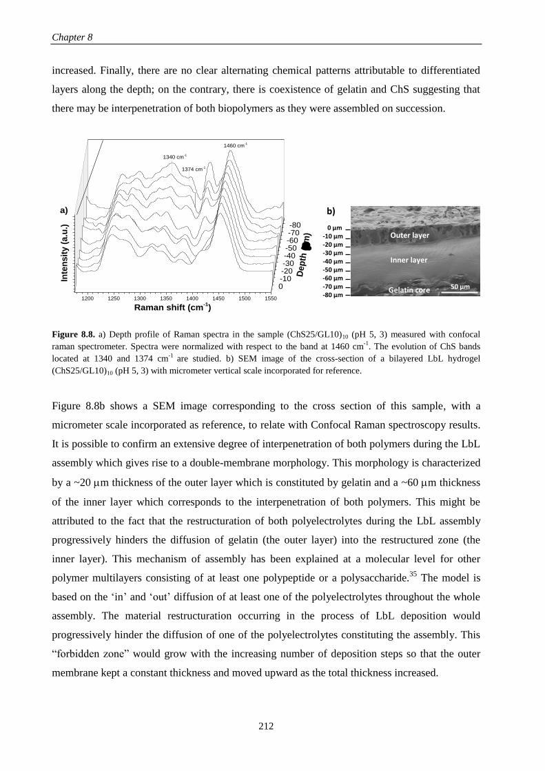
Chapter 8
212
increased. Finally, there are no clear alternating chemical patterns attributable to differentiated
layers along the depth; on the contrary, there is coexistence of gelatin and ChS suggesting that
there may be interpenetration of both biopolymers as they were assembled on succession.
1200 1250 1300 1350 1400 1450 1500 1550
-80-70-60-50-40-30-20-100
Inte
nsit
y (
a.u
.)
1460 cm-1
1374 cm-1
Raman shift (cm-1)
Dep
th (
m)
1340 cm-1
a)
Figure 8.8. a) Depth profile of Raman spectra in the sample (ChS25/GL10)10 (pH 5, 3) measured with confocal
raman spectrometer. Spectra were normalized with respect to the band at 1460 cm-1
. The evolution of ChS bands
located at 1340 and 1374 cm-1
are studied. b) SEM image of the cross-section of a bilayered LbL hydrogel
(ChS25/GL10)10 (pH 5, 3) with micrometer vertical scale incorporated for reference.
Figure 8.8b shows a SEM image corresponding to the cross section of this sample, with a
micrometer scale incorporated as reference, to relate with Confocal Raman spectroscopy results.
It is possible to confirm an extensive degree of interpenetration of both polymers during the LbL
assembly which gives rise to a double-membrane morphology. This morphology is characterized
by a ~20 m thickness of the outer layer which is constituted by gelatin and a ~60 m thickness
of the inner layer which corresponds to the interpenetration of both polymers. This might be
attributed to the fact that the restructuration of both polyelectrolytes during the LbL assembly
progressively hinders the diffusion of gelatin (the outer layer) into the restructured zone (the
inner layer). This mechanism of assembly has been explained at a molecular level for other
polymer multilayers consisting of at least one polypeptide or a polysaccharide.35
The model is
based on the ‘in’ and ‘out’ diffusion of at least one of the polyelectrolytes throughout the whole
assembly. The material restructuration occurring in the process of LbL deposition would
progressively hinder the diffusion of one of the polyelectrolytes constituting the assembly. This
“forbidden zone” would grow with the increasing number of deposition steps so that the outer
membrane kept a constant thickness and moved upward as the total thickness increased.
50 µm
0 µm -10 µm -20 µm -30 µm -40 µm -50 µm -60 µm -70 µm -80 µm
Outer layer
Inner layer
Gelatin core
b)

LbL hydrogels obtained from natural polymers
213
8.3.5. Determination of the mechanical properties
In order to determine the effect of the morphology of hydrogels on their mechanical properties
and gel melting temperature, oscillatory rheological experiments were carried out as a function
of temperature on a gelatin core (10 %w/v) and a LbL hydrogel (ChS25/GL10)10 (pH 5, 3) and
results are depicted in Figure 8.9.
In both cases, the elastic modulus (G’) is higher than the loss modulus (G’’), corroborating that
the gel is formed. Focusing on the LbL hydrogel (ChS25/GL10)10 (pH 5, 3), a plateau in the
elastic modulus is observed below its melting temperature, Tm, which is located at 33 ºC. Above
Tm, the value of G´ dramatically decreased and ultimately reached a second plateau, above
which the hydrogel completely melted down. The elastic modulus of the (ChS25/GL10)10 (pH 5,
3) hydrogel, G´ = 4775 ± 158 Pa, greatly increased with respect to that corresponding to the
gelatin core, G´ = 511 ± 198 Pa. This increase on the mechanical properties is due to the
interactions between both polymers that take place all over the sample.8
Figure 8.9. Evolution of the elastic modulus G´ and the loss modulus G´´ as a function of temperature for a LbL
hydrogel (ChS25/GL10)10 (pH 5, 3) (G´ (■) and G´´ (□)) and a gelatin core (10 %w/v) (G´ (●) and G´´ (○)).
It is important to remark that LbL ChS/GL hydrogels reported here are generated just by
electrostatic interactions without the employment of any chemical crosslinker. Still the
reinforcement is very remarkable taking into account that there exists layer formation that
restricts the number of interactions notwithstanding some degree of interpenetration between
both polymers as it was demonstrated through Confocal Raman Spectroscopy. Note that the
20 25 30 35 40 45 50
0,01
0,1
1
10
100
1000
10000
G' ((ChS25/GL10)10
pH (5, 3))
G'' ((ChS25/GL10)10
pH (5, 3))
G' (GL)
G'' (GL)
G´,
G´´
(P
a)
Temperature (ºC)
Chapter 8
214
value of Tm depends on the gelatin concentration and thus it is similar for ChS25/GL10 (pH 5, 3)
and the gelatin core (10 %w/v) reaching a value of Tm = 33 ºC.
8.4. CONCLUSIONS
A novel method to prepare hydrogels from gelatin and chondroitin sulphate based on
electrostatic layer-by-layer assembly is presented, being this a combination of polymers not
studied in literature for LbL hydrogels before.
Concentration and pH conditions were optimized to ensure electrostatic assembly of alternating
layers of ChS and GL by dipping LbL in aqueous solutions of the natural polymers.
The assembly of ChS and GL at different steps of the LbL process was studied by FTIR
spectroscopy following the absorbance of the band located at 1240 cm-1
which increased with the
number of deposited bilayers confirming the growth of the multilayer system ChS/GL.
A structure organized in layers was identified in SEM images and compositionally characterized
with Raman confocal microscopy, to conclude that a double membrane structure is formed on
gelatin cores and there was interpenetration between both polymers along the whole LbL
estructure. The thickness of the inner layer could be increased with adequate pH conditions and
increasing concentration of ChS.
The coating of gelatin cores with LbL ChS/GL by dipping gave rise to an increase in the elastic
modulus without affecting its melting point. The thermal responsiveness together with enhanced
mechanical properties might mimic the in vivo rheological characteristics of soft tissues
organized in layers as, for example, intervertebral discs or employed in drug delivery
applications for which their inner structure organized in layers might provide materials for
complex drug codelivery.
8.5. REFERENCES
1. Elisseeff, J., Hydrogels: Structure starts to gel. Nature Materials 2008, 7, (4), 271-273.
2. Lee, K. Y.; Mooney, D. J., Hydrogels for Tissue Engineering. Chemical reviews 2001, 101, (7),
1869-1880.
3. Qiu, Y.; Park, K., Environment-sensitive hydrogels for drug delivery. Advanced Drug Delivery
Reviews 2001, 53, (3), 321-339.
4. Dong, L.; Agarwal, A. K.; Beebe, D. J.; Jiang, H., Adaptive liquid microlenses activated by
stimuli-responsive hydrogels. Nature 2006, 442, (7102), 551-554.

LbL hydrogels obtained from natural polymers
215
5. Discher, D. E.; Mooney, D. J.; Zandstra, P. W., Growth Factors, Matrices, and Forces Combine
and Control Stem Cells. Science 2009, 324, (5935), 1673.
6. Hoffman, A. S., Hydrogels for biomedical applications. Advanced Drug Delivery Reviews 2012,
64, Supplement, 18-23.
7. Costa, A. M. S.; Mano, J. F., Extremely strong and tough hydrogels as prospective candidates for
tissue repair – A review. European Polymer Journal 2015, 72, 344-364.
8. Zamora-Mora, V.; Velasco, D.; Hernández, R.; Mijangos, C.; Kumacheva, E., Chitosan/agarose
hydrogels: Cooperative properties and microfluidic preparation. Carbohydrate Polymers 2014, 111, (0),
348-355.
9. Rescignano, N.; Hernandez, R.; Lopez, L. D.; Calvillo, I.; Kenny, J. M.; Mijangos, C.,
Preparation of alginate hydrogels containing silver nanoparticles: a facile approach for antibacterial
applications. Polymer International 2016, 65, (8), 921-926.
10. Hernandez, R.; Sacristan, J.; Asin, L.; Torres, T. E.; Ibarra, M. R.; Goya, G. F.; Mijangos, C.,
Magnetic Hydrogels Derived from Polysaccharides with Improved Specific Power Absorption: Potential
Devices for Remotely Triggered Drug Delivery. The Journal of Physical Chemistry B 2010, 114, (37),
12002-12007.
11. Zamora-Mora, V.; Fernández-Gutiérrez, M.; Román, J. S.; Goya, G.; Hernández, R.; Mijangos,
C., Magnetic core–shell chitosan nanoparticles: Rheological characterization and hyperthermia
application. Carbohydrate Polymers 2014, 102, 691-698.
12. Bruvera, I.; Hernández, R.; Mijangos, C.; Goya, G. F., An integrated device for magnetically-
driven drug release and in situ quantitative measurements: design, fabrication and testing. Journal of
Magnetism and Magnetic Materials 2015, 377, 446-451.
13. Zhao, Y.; Nakajima, T.; Yang, J. J.; Kurokawa, T.; Liu, J.; Lu, J.; Mizumoto, S.; Sugahara, K.;
Kitamura, N.; Yasuda, K.; Daniels, A. U. D.; Gong, J. P., Proteoglycans and Glycosaminoglycans
Improve Toughness of Biocompatible Double Network Hydrogels. Advanced Materials 2014, 26, (3),
436-442.
14. Ladet, S.; David, L.; Domard, A., Multi-membrane hydrogels. Nature 2008, 452, (7183), 76-79.
15. Nie, J.; Lu, W.; Ma, J.; Yang, L.; Wang, Z.; Qin, A.; Hu, Q., Orientation in multi-layer chitosan
hydrogel: morphology, mechanism, and design principle. Scientific Reports 2015, 5, 7635.
16. Kozlovskaya, V.; Kharlampieva, E.; Erel, I.; Sukhishvili, S. A., Multilayer-derived, ultrathin,
stimuli-responsive hydrogels. Soft Matter 2009, 5, (21), 4077-4087.
17. Lynam, D.; Peterson, C.; Maloney, R.; Shahriari, D.; Garrison, A.; Saleh, S.; Mehrotra, S.; Chan,
C.; Sakamoto, J., Augmenting protein release from layer-by-layer functionalized agarose hydrogels.
Carbohydrate Polymers 2014, 103, 377-384.
18. Lin, N.; Gèze, A.; Wouessidjewe, D.; Huang, J.; Dufresne, A., Biocompatible Double-Membrane
Hydrogels from Cationic Cellulose Nanocrystals and Anionic Alginate as Complexing Drugs Codelivery.
ACS Applied Materials & Interfaces 2016, 8, (11), 6880-6889.
19. Ladet, S. G.; Tahiri, K.; Montembault, A. S.; Domard, A. J.; Corvol, M. T. M., Multi-membrane
chitosan hydrogels as chondrocytic cell bioreactors. Biomaterials 2011, 32, (23), 5354-5364.
20. Lai, J.-Y., Corneal Stromal Cell Growth on Gelatin/Chondroitin Sulfate Scaffolds Modified at
Different NHS/EDC Molar Ratios. International Journal of Molecular Sciences 2013, 14, (1), 2036-2055.

Chapter 8
216
21. Zhu, Y.; Gao, C.; He, T.; Liu, X.; Shen, J., Layer-by-Layer Assembly To Modify Poly(l-lactic
acid) Surface toward Improving Its Cytocompatibility to Human Endothelial Cells. Biomacromolecules
2003, 4, (2), 446-452.
22. Ahmed, J., Chapter 15 - Rheological Properties of Gelatin and Advances in Measurement. In
Advances in Food Rheology and its Applications, Woodhead Publishing: 2017; pp 377-404.
23. Umerska, A.; Corrigan, O. I.; Tajber, L., Design of chondroitin sulfate-based polyelectrolyte
nanoplexes: Formation of nanocarriers with chitosan and a case study of salmon calcitonin. Carbohydrate
Polymers 2017, 156, 276-284.
24. Payne, K. J.; Veis, A., Fourier transform ir spectroscopy of collagen and gelatin solutions:
Deconvolution of the amide I band for conformational studies. Biopolymers 1988, 27, (11), 1749-1760.
25. Nur Hanani, Z. A.; Roos, Y. H.; Kerry, J. P., Use of beef, pork and fish gelatin sources in the
manufacture of films and assessment of their composition and mechanical properties. Food Hydrocolloids
2012, 29, (1), 144-151.
26. Mader, K. T.; Peeters, M.; Detiger, S. E. L.; Helder, M. N.; Smit, T. H.; Le Maitre, C. L.;
Sammon, C., Investigation of intervertebral disc degeneration using multivariate FTIR spectroscopic
imaging. Faraday Discussions 2016, 187, (0), 393-414.
27. Fajardo, A. R.; Silva, M. B.; Lopes, L. C.; Piai, J. F.; Rubira, A. F.; Muniz, E. C., Hydrogel based
on an alginate-Ca2+/chondroitin sulfate matrix as a potential colon-specific drug delivery system. RSC
Advances 2012, 2, (29), 11095-11103.
28. Radev, L.; Mostafa, N. Y.; Michailova, I.; Salvado, I. M.; Fernandes, M. H., In vitro bioactivity
of collagen/calcium phosphate silicate composites, cross-linked with chondroitin sulfate. International
Journal of Materials and Chemistry 2012, 2, (1), 1-9.
29. Lai, J.-Y.; Li, Y.-T.; Cho, C.-H.; Yu, T.-C., Nanoscale modification of porous gelatin scaffolds
with chondroitin sulfate for corneal stromal tissue engineering. International Journal of Nanomedicine
2012, 7, 1101-1114.
30. Pezeshki‐Modaress, M.; Mirzadeh, H.; Zandi, M.; Rajabi‐Zeleti, S.; Sodeifi, N.; Aghdami, N.;
Mofrad, M. R., Gelatin/Chondroitin Sulfate Nanofibrous Scaffolds for Stimulation of Wound Healing: In‐Vitro and In‐Vivo Study. Journal of Biomedical Materials Research Part A 2016.
31. Karthika, A.; Kavitha, L.; Surendiran, M.; Kannan, S.; Gopi, D., Fabrication of divalent ion
substituted hydroxyapatite/gelatin nanocomposite coating on electron beam treated titanium: mechanical,
anticorrosive, antibacterial and bioactive evaluations. RSC Advances 2015, 5, (59), 47341-47352.
32. Fajardo, A. R.; Lopes, L. C.; Pereira, A. G. B.; Rubira, A. F.; Muniz, E. C., Polyelectrolyte
complexes based on pectin–NH2 and chondroitin sulfate. Carbohydrate Polymers 2012, 87, (3), 1950-
1955.
33. Ellis, R.; Green, E.; Winlove, C. P., Structural Analysis of Glycosaminoglycans and
Proteoglycans by Means of Raman Microspectrometry. Connective Tissue Research 2009, 50, (1), 29-36.
34. Frushour, B. G.; Koenig, J. L., Raman scattering of collagen, gelatin, and elastin. Biopolymers
1975, 14, (2), 379-391.
35. Borges, J.; Mano, J. F., Molecular Interactions Driving the Layer-by-Layer Assembly of
Multilayers. Chemical Reviews 2014, 114, (18), 8883-8942.

CHAPTER 9
General conclusions and perspectives


General conclusions and perspectives
219
This PhD work has been focused on “Layer-by-layer assembly of natural polymers for
biomedical applications“. To achieve this objective, different multilayer systems based on
natural polymers have been built up using the LbL technique and morphologically and
mechanically characterized. An alginate-based magnetic ferrofluid was developed in order to be
incorporated into multilayer films for magnetic hyperthermia applications. Finally, the
biomedical applications of alginate/chitosan films were tested in vitro with neuroblastoma cells
(SH-SY5Y) for magnetic hyperthermia therapy and human dermal fibroblasts (HDF) and human
caucasian breast adenocarcinoma cells (MCF-7) for sustained drug delivery applications.
Taking into account the experimental results, the following conclusions can be obtained:
a) Different multilayer polymer structures based on natural polymers have been built up
through spray LbL and dipping LbL. The main polymer system developed in this thesis
formed by alginate and chitosan was assembled by spray LbL because of is a quicker
technique which allowed to obtain greater thickness than dipping LbL. The growth of
Alg/Chi films by spray assisted LbL, studied by ellipsometry up to 5 bilayers, showed a
linear increase of the thickness with the number of deposited bilayers being higher as Alg
concentration increased. From 5 bilayers, the growth was followed by SEM illustrating a
change of tendency from linear to exponential up to 20 bilayers, point wherein growth
slightly increased towards a plateau due to the rearrangements of polymer chains during the
deposition process. The growth rate of Alg/Chi films assembled by spray was 20 nm/layer,
ten-fold higher than by dipping (2 nm/layer).
The buildup process of hyaluronic acid/poly(allylamine hydrochloride) was carried out by
dipping LbL due to the fact that no growth was observed by ellipsometry when both
polymers were assembled by spray LbL. The growth of these films was exponential for all
concentrations due to the presence of NaCl and the diffusion of PAH within the multilayer
film structure and increased with the PAH concentration. A gel-like layer formed by alginate
crosslinked with iron II ions was successfully incorporated into the HA/PAH multilayer
structure as it was demonstrated by QCM-D experiments.
b) The synthesis and characterization of an alginate-based magnetic ferrofluid was carried out
through a coprecipitation method. The obtained ferrofluid was formed by magnetite
nanoparticles with an average particle size of ~ 7 nm dispersed in Milli-Q water with

Chapter 9
220
colloidal stability at neutral pH and a hydrodynamic diameter around 100 nm for a
magnetite concentration of 8.0 mg/mL.
c) The morphological and mechanical characterization of Alg/Chi films was carried out
reaching the following conclusions:
The mechanical properties of multilayer Alg/Chi films, thermally crosslinked Alg/Chi
films and nanocomposite Alg/Chi films built up via spray LbL were determined by (PF-
QNM) AFM revealing that: the elastic moduli increased with the number of deposited
bilayers until a value of ~12 GPa, thermal crosslinking of Alg/Chi films increased the
elastic modulus, being much more significant as alginate concentration increased.
Deformation values were almost constant and below 2 nm either in non-crosslinked or
crosslinked Alg/Chi films and adhesion force varied between 1 and 4 nN. The
incorporation of iron oxide NPs increased the roughness of multilayer Alg/Chi films and
induced an increase of their elastic moduli and the deformation values. The presence of
NPs on the ending layer of nanocomposite films produced a two-fold increase in the
elastic modulus with respect to chitosan ending nanocomposite films, without altering the
deformation values.
The study of the inner structure of nanocomposite films by GISAXS revealed that films
assembled by dipping LbL gave rise to a structure in which nanoparticles were dispersed
throughout the films, whereas those assembled by spray LbL could form separated NPs
layers with some degree of dispersion of NPs along the structure.
d) The biomedical applications of Alg/Chi films and nanocomposite Alg/Chi films were tested
in vitro.
According to the results obtained from the study carried out to determine the efficiency of
nanocomposite Alg/Chi films to act as thermomagnetic films (TMFs) for magnetic
hyperthermia on neuroblastoma cells, we concluded that:
Nanocomposite Alg/Chi films obtained by incorporation of the alginate-based magnetic
ferrofluid within Alg/Chi films through spray assisted LbL showed an ordered
distribution in layers being the iron the major component with a percentage of 65% as it

General conclusions and perspectives
221
was determined by EDX analysis. Nanocomposite Alg/Chi films with different iron
contents, modulated by the number of magnetic nanoparticles (NPs) layers, were
fabricated in order to examine their magnetic remote heating by application of an
alternating magnetic field (AMF). The final magnetite content in every film was
determined by UV-Vis transmission spectrophotometry showing a linear increase of
magnetite content with the number of NPs layers deposited. The specific power
absorption (SPA) of the alginate-based magnetic ferrofluid with different magnetite
concentrations, 4.84 and 8.0 mg/mL, was measured at different frequencies (f) and
magnetic field amplitudes (H) showing an increase of the SPA with the magnetite
concentration. The immobilization of NPs in gelatin produced a decrease of SPA (H = 24
kA/m and f = 571 kHz) from 868.4 ± 1.9 W/g to 77.2 ± 0.1 W/g due to the block of the
Brownian relaxation. Nanocomposite Alg/Chi films could act as TMFs as revealed by the
linear temperature increase from 6 to 12 ºC for films with a varying number of NPs layers
from 80 to 160 layers when they were subjected to an H = 35 kA/m and f = 180 kHz for 5
minutes.
The employment of these TMFs as patches for magnetic hyperthermia applications was
tested in vitro with human neuroblastoma cells. TMFs showed good cell adhesion
properties and biocompatibility with neuroblastoma cells. In vitro experiments by remote
heating of TMFs using an AMF (H = 35 kA/m and f = 180 kHz) showed different results
depending on the number of cycles of MHT and the experimental protocol. MHT applied
on TMFs with neuroblastoma cells adhered onto their surface showed a reduction in cell
viability up to 67% and 20% for one and three heating cycles, respectively. In addition, in
order to mimic in vivo applications, other experimental protocol was carried out by
application of the MHT on TMFs placed over neuroblastoma cells previously cultured on
an ibidi dish, showing a cell viability reduction of 85% after two MHT cycles.
Considering the results obtained from the study carried out to determine the cell adhesion on
Alg/Chi films and their efficiency as controlled drug delivery systems, it was concluded
that:
Multilayer Alg/Chi films obtained through spray assisted LbL can be chemically
crosslinked using the carbodiimide chemistry and their surface can be easily tuned by
spraying a layer of alginate or hyaluronic acid (HA) at the end of the Alg/Chi LbL
deposition process. The average roughness (Ra) of films with different number of

Chapter 9
222
bilayers, determined by AFM, showed an increase of Ra with the number of bilayers.
Native and crosslinked Alg/Chi films were biocompatible, as it was demonstrated
through in vitro experiments with human dermal fibroblast (HDF) and human caucasian
breast adenocarcinoma (MCF-7) cells. Films ended in HA were more prone to
degradation in physiological environment (DMEM at 37ºC) than Alg ending ones after
15 days and this degradation was diminished by the chemical crosslinking.
Cell adhesion experiments carried out in vitro with HDF and MCF-7 cells proved that the
number of bilayers did not influenced the cell adhesion of these two kind of cells, the
crosslinking process favored the MCF-7 cell adhesion on Alg-ending films where the
contact angle remained constant after crosslinking and decreased the MCF-7 cell
adhesion on HA-ending films where the contact angle increased after crosslinking. With
regards to HDF cell adhesion, non-significant differences were observed with the surface
chemistry or the crosslinking.
The final application of Alg/Chi films as platforms for sustained drug release was tested
in vitro. For that purpose, tamoxifen, a therapeutic agent against breast cancer, was
incorporated in different intermediate positions of the multilayer Alg/Chi films and
results showed a more sustained release over time as number of deposited bilayers
increased. It was proven that there was not degradation of these films after the drug
delivery experiment and the release mechanism was diffusion controlled and could be
described by the Ritger-Peppas model. The release of TMX from Alg/Chi films during in
vitro experiments decreased the cell viability of MCF-7 cells whereas the HDF cell
viability remained unaffected.
e) Finally, a new LbL system composed of ChS and GL, which had not been studied in
literature before, has been investigated, reaching the following conclusions: the FTIR
absorbance intensity increased with the number of ChS/GL deposited bilayers confirming
the growth of the multilayer system through dipping LbL assembly. This assembly process
carried out over a gelatin core gave rise to a layered organized structure, as it was observed
by SEM, where multilayers formed a double membrane structure and there was
interpenetration between both polymers along the LbL structure. The thickness of the
multilayers could be increased by increasing the ChS concentration at pH 3 for gelatin and
pH 5 for ChS. Mechanical properties of LbL hydrogels revealed that the elastic modulus
increased with respect to the uncoated gelatin core from 511 ± 198 Pa to 4775 ± 158 Pa

General conclusions and perspectives
223
without altering their melting point at 33 ºC, allowing to employ these LbL hydrogels as cell
encapsulation and drug delivery systems in the future.
From this PhD work several fruitful national and international collaborations have been
established with different groups as, for example, with F. Boulmedais (ICS-CNRS, Strasbourg),
T. Ezquerra (CSIC, Spain), J. San Román (CSIC, Spain) and G. Goya (INA, Spain).
Considering the results obtained in the present PhD work, new challenges arise for other PhD
and Master projects. Future work is focused on the development of light-responsive
nanostratified materials based on combining polymer multilayers in between gel layers to
encapsulate drugs for dermal applications, as well as the development of multilayer LbL
hydrogels assembled over gelatin cores with drugs incorporated with the objective of developing
materials for complex drug delivery.


ANNEXES

Annex I
227
ANNEX I. Iron calibration curve
0,000 0,001 0,002 0,003 0,004 0,005 0,006 0,007 0,0080,0
0,1
0,2
0,3
0,4
0,5
0,6
0,7
0,8
0,9
1,0
Ab
so
rba
nc
e_
47
8n
m (
a.u
)
mass Fe3O4 (mg)
Abs = 0.02 + 127.48 ·Fe3O
4(mg)
R2 = 0.996
Figure A.I.1. Standard calibration curve for magnetite determination by UV-Vis spectrophotometry at 478 nm.
Dashed line represents the linear fit of the data.

Annex II
228
ANNEX II. Tamoxifen calibration curve
0,000 0,005 0,010 0,015 0,020 0,025 0,030 0,0350,0
0,2
0,4
0,6
0,8
1,0
Ab
so
rba
nc
e_
27
7n
m (
a.u
.)
Concentration TMX (mg/mL)
Abs = 0.02 + 34.68·TMX(mg/mL)
R2 = 0.999
Figure A.II.1. Standard calibration curve of TMX in PBS with 1 %w/v Tween 80 at 277 nm. Dashed line represents
the linear fit of the data.

Annex III
229
ANNEX III. AFM morphological images of (Alg/Chi)nAlg and (Alg/Chi)nHA
films
Figure A.III.1. AFM topographic images (2m × 2m) corresponding to non-crosslinked films a) (Alg/Chi)nAlg
and b) (Alg/Chi)nHA and crosslinked films c) (Alg/Chi)nAlg and d) (Alg/Chi)nHA with different number of bilayers.
n=5 n=7 n=10 n=15 n=25a)
b)
c)
d)

Annex IV
230
ANNEX IV. Fluorescence microscope images of HDF and MCF-7 cell
adhesion on (Alg/Chi)nAlg and (Alg/Chi)nHA films
Figure A.IV.1. Fluorescence microscope images of HDF cell adhesion on non-crosslinked films a) (Alg/Chi)nAlg
and b) (Alg/Chi)nHA and crosslinked films c) (Alg/Chi)nAlg and d) (Alg/Chi)nHA with different number of bilayers
after 3 days. Scale bars 100 m.
c)
n=5 n=7 n=10 n=15 n=25
d)
a)
b)

Annex IV
231
Figure A.IV.2. Fluorescence microscope images of MCF-7 cell adhesion on non-crosslinked films a) (Alg/Chi)nAlg
and b) (Alg/Chi)nHA and crosslinked films c) (Alg/Chi)nAlg and d) (Alg/Chi)nHA with different number of bilayers
after 3 days. Scale bars 100 m.
c)
n=5 n=7 n=10 n=15 n=25
d)
a)
b)


ABBREVIATIONS


Abbreviations
235
AB Alamar blue
AFM Atomic Force Microscopy
AG-NPs Silver nanoparticles
Alg Sodium alginate
AMF Alternating Magnetic Field
APTT Activated partial thromboplastin time
Au-NPs Gold nanoparticles
bFGF Basic fibroblastic factor
CaP Phosphate precipitates
Chi Chitosan
ChS Chondroitin sulphate
CL Crosslinked
CLSM Confocal laser microscopy
COL Collagen
CV Cell viability
DAPI 4’,6-Diamidino-2-(Phenylindole, Dihydrochloride)
DD Deacetylation degree
Dex Dextran
Dhyd Hydrodynamic diameter
DLS Dynamic Light Scattering
DMA Dynamic mechanical analysis
DMEM Dulbecco’s Eagle’s Medium
DMSO Dimethylsulfoxide
DMT Derjaguin-Muller-Toporov
DN Dopamine
DPI Dual-polarization interferometry
E.coli Escherichia coli
EBs Embryoid bodies
EDC 1-Ethyl-3-(3-dimethylaminopropyl)-carbodiimide
EDTA Ethylendiaminetetraacetic acid
EDX Energy-dispersive X-ray spectroscopy
EHT Exogenous hyperthermia

Abbreviations
236
EMC Extracellular matrix
EthD-1 Ethidium homodimer-1
f Frequency
FBS Fetal bovine serum
FEG Field emission gun
FIB Focus Ion Beam
FT-IR Fourier Transform Infra-Red spectroscopy
FTIR-ATR Fourier Transform Infra-Red spectroscopy – Attenuated total reflectance
FWHM Full width at half maximum
GISAXS Grazing-incident small angle X-ray scattering
GIXD Grazing-incidence X-ray diffraction
GL Gelatin
1H Proton
H Magnetic field amplitude
HA Hyaluronic acid
HDF Human dermal fibroblasts
HEP Heparin
HEPES 4-(2-hydroxylethyl)-1-piperazineethanesulfonic acid
HUAECs Human umbilical artery endothelial cells
HUVEC Human umbilical vein endothelial cells
IDG Intermodular detector gap
IOP Intraocular pressure
IPNs Interpenetrating polymer networks
IVD Intervertebral disk
LB Langmuir-Blodgett
LbL Layer-by-layer
LVR Linear viscoelastic region
Mb Myoglobin
MCF-7 Human caucasian breast adenocarcinoma
MHT Magnetic hyperthermia
MSCs Bone marrow-derive ovine cells
𝐌𝐯 Viscosity average molecular weight
Mw Molecular weight

Abbreviations
237
NCC Nanocrystalline cellulose
NHS N-Hydroxysuccinimide
NMR Nuclear Magnetic Resonance
NPs Magnetic nanoparticles
OAlg Oxidized sodium alginate
OWLS Optical waveguide lightmode spectroscopy
PA Polyanion
PAA Poly(acrylic acid)
PAH Poly(allylamine hydrochloride)
PBS Phosphate buffered solution
PC Polycation
PCNs Polyelectrolyte complex nanoparticles
PDDA Poly(diallyldimethylammonium chloride)
PDI Polydispersity index
PEC Polyelectrolyte complex
PEI Poly(ethylenimine)
PEMs Polyelectrolyte multilayer films
PET Polyethylene terephthalate
PET Poly(ethylene terephthalate)
PF-QNM PeakForce Quantitative Nanomechanical Mapping
PGA Poly(g-glutamic acid)
PLL Poly(L-lysine)
PLLA Poly(L-lactic acid)
PS Polystyrene
PSS Poly(styrene sulfonate)
PT Prothrombin time
PU Polyurethane
PVA Polyvinyl alcohol
PVP Poly(vynil pyrrolidone)
Px Piroxicam
QCM Quartz Crystal Microbalance
QCM-D Quartz Crystal Microbalance with dissipation monitoring
Ra Average roughness

Abbreviations
238
RGD Arginine-glycine-aspartate
SAR Scanning angle reflectometry
SAXS Small angle X-ray scattering
SBS Specular beam stop
SEM Scanning Electron Microscopy
SH-SY5Y Human neuroblastoma cells
SNP Sodium nitroprusside
SPA Specific Power Adsorption
SPR Surface plasmon resonance
TEM Transmission Electron Microscopy
TGA Thermogravimetric analysis
THF Tetrahydrofuran
TMFs Thermomagnetic films
TMX Tamoxifen citrate salt
UV-Vis Ultraviolet-visible spectroscopy
XPS X-ray photoelectron spectroscopy
XRD X-Ray Diffraction
α-MSH Alpha-melanocyte stimulating hormone
ΔT Temperature increase

LIST OF PUBLICATIONS


List of publications
241
Publications directly related to this thesis:
1. M. Criado, E. Rebollar, A. Nogales, T. Ezquerra, F. Boulmedais, C. Mijangos, R.
Hernández. Quantitative nanomechanical properties of multilayer films made of
polysaccharides through spray assisted layer-by-layer assembly. Biomacromolecules, 2017,
18, 169-177.
2. M. Criado, B. Sanz, G. F. Goya, C. Mijangos, R. Hernández. Magnetically responsive
biopolymeric multilayer films for controlled hyperthermia. ACS Applied Materials &
Interfaces, submitted 2017, Manuscript ID am-2017-03852f.
3. M. Criado, J. M. Rey, C. Mijangos, R. Hernández. Double-membrane Thermoresponsive
Hydrogels from Gelatin and Chondroitin Sulphate with Enhanced Mechanical Properties.
RSC Advances, 2016, 6, 105821-105826.
4. M. Criado, M. Fernández-Gutiérrez, J. San Román, C. Mijangos, R. Hernández. Multilayer
polymer films with tailored structures for sustained drug delivery and cellular adhesion.
Submitted, biomacromolecules, 2017.
5. M. Criado, C. Mijangos, R. Hernández. Procesos de fabricación y aplicaciones avanzadas
de filmes poliméricos nanoestructurados capa a capa (layer-by-Layer). Revista Plásticos
Modernos, 2015, 110, 13-17.
6. M. Criado, C. Mijangos, R. Hernández. Hidrogeles multimembrana obtenidos a partir de
polímeros naturales. Revista Plásticos Modernos, en prensa, Mayo, 2017.
7. M. Criado, E. Rebollar, A. Nogales, T. Ezquerra, C. Mijangos, R. Hernandez.
Determinación of the inner structure of hybrid multilayer polymer films through grazing
incidence X-ray scattering (GISAXS), in preparation.
8. M. Criado, L. Lerma, F. Boulmedais, C. Mijangos, R. Hernández. Light-responsive
nanostratified polymer materials for drug delivery applications, in preparation.
Patents:
9. M. Criado, C. Mijangos, R. Hernández. Ferrofluidos acuosos de base polimérica con altos
coeficientes de absorción específica y procedimiento de obtención, ES, No. 201631442,
2016.
Other related publications:
10. A. Lejardi, R. Hernández, M. Criado, J. I. Santos, A. Etxeberria, J. R. Sarasua, C.
Mijangos. Novel hydrogels of chitosan and poly(vinyl alcohol)-g-glycolic acidcopolymer
with enhanced rheological properties. Carbohydrate Polymers, 2014, 103, 267-273.
11. R. Hernández, M. Criado, A. Nogales, M. Sprung, C. Mijangos, T. Ezquerra. Deswelling of
Poly(N-isopropylacrylamide) Derived Hydrogels and Their Nanocomposites with Iron
Oxide Nanoparticles As Revealed by X-ray Photon Correlation Spectroscopy.
Macromolecules, 2015, 48, 393-399.