Entity Linking to Dynamically-Evolving Personal Knowledge ...
Upload
univ-lille2Category
view
4download
0
M I N I R E V I E W
Pneumocystis: fromadoubtful unique entity toagroupof highlydiversi¢ed fungal speciesMagali Chabe1,2, Cecile-Marie Aliouat-Denis1,2, Laurence Delhaes1,3, El Moukhtar Aliouat1,2,Eric Viscogliosi1 & Eduardo Dei-Cas1,2,3
1Biology and Diversity of Emergent Eukaryotic Pathogens (BDEEP)–Center for Infection and Immunity of Lille, Pasteur Institute of Lille, Inserm U1019,
CNRS UMR 8204, University Lille-Nord-de-France, Lille, France; 2Department of Parasitology–Mycology, Faculty of Biological and Pharmaceutical
Sciences, University of Lille-Nord-de-France, Lille, France; and 3Department of Parasitology–Mycology, Faculty of Medicine, University of Lille-Nord-
de-France, Biology-Pathology Centre, University Hospital Center, Lille, France
Correspondence: Magali Chabe,
Department of Parasitology–Mycology,
Faculty of Biological and Pharmaceutical
Sciences, University of Lille-Nord-de-France,
3, rue du Professeur Laguesse BP83, 59006
Lille, France. Tel.: 133 3 2096 4010; fax: 133
3 2087 7276; e-mail: magali.chabe@univ-
lille2.fr
Received 11 May 2010; revised 10 October
2010; accepted 12 October 2010.
Final version published online 26 November
2010.
DOI:10.1111/j.1567-1364.2010.00698.x
Editor: Teun Boekhout
Keywords
Pneumocystis spp.; phylogenetic species
concept; life cycle; epidemiology.
Abstract
At the end of the 20th century the unique taxonomically enigmatic entity called
Pneumocystis carinii was identified as a heterogeneous group of microscopic Fungi,
constituted of multiple stenoxenic biological entities largely spread across
ecosystems, closely adapted to, and coevolving in parallel with, mammal species.
The discoveries and reasoning that led to the current conceptions about
the taxonomy of Pneumocystis at the species level are examined here. The present
review also focuses on the biological, morphological and phylogenetical features
of Pneumocystis jirovecii, Pneumocystis oryctolagi, Pneumocystis murina, P. carinii
and Pneumocystis wakefieldiae, the five Pneumocystis species described until
now, mainly on the basis of the phylogenetic species concept. Interestingly,
Pneumocystis organisms exhibit a successful adaptation enabling them to
dwell and replicate in the lungs of both immunocompromised and healthy
mammals, which can act as infection reservoirs. The role of healthy carriers in
aerial disease transmission is nowadays recognized as a major contribution to
Pneumocystis circulation, and Pneumocystis infection of nonimmunosuppressed
hosts has emerged as a public health issue. More studies need to be undertaken
both on the clinical consequences of the presence of Pneumocystis in healthy
carriers and on the intricate Pneumocystis life cycle to better define its epidemio-
logy, to adapt existing therapies to each clinical context and to discover new
drug targets.
Introduction
The present review examines the discoveries and reasoning
that led to the current conceptions about the taxonomy of
Pneumocystis at the species level. Considered for a long time
an enigmatic pulmonary unicellular parasite of unknown
biological significance in man and other mammals, it is now
well established that the ascomycetous Pneumocystis genus
contains numerous host species-specific species widely spread
across ecosystems. Interestingly, advances in this field have
had a beneficial impact on the understanding of the natural
history of Pneumocystis infection. On the basis of the identi-
fication of several species in the genus and the demonstration
that each Pneumocystis species can only infect its own specific
host (Aliouat et al., 1993b, 1994; Furuta et al., 1993; Gigliotti
et al., 1993; Atzori et al., 1999; Durand-Joly et al., 2002), new
patterns of Pneumocystis pneumonia (PcP) epidemiology are
emerging. These new concepts, associated with the fact that
Pneumocystis organisms are actively transmitted by the air-
borne route (Walzer et al., 1977; Hughes, 1982; Soulez et al.,
1991; Dumoulin et al., 2000; Chabe et al., 2004), and perhaps
also vertically (Cere et al., 1997; Demanche et al., 2003;
Sanchez et al., 2007; Montes-Cano et al., 2009), provide new
insights to identify reservoirs and infection sources, and
therefore to conceive rational, efficient and cost-effective
prevention measures against PcP (Vargas et al., 2000; Miller
et al., 2001; Rabodonirina, 2001; Durand-Joly et al., 2003;
Calderon et al., 2004).
FEMS Yeast Res 11 (2011) 2–17c� 2010 Federation of European Microbiological SocietiesPublished by Blackwell Publishing Ltd. All rights reserved
YEA
ST R
ESEA
RC
H

The Pneumocystis genus: from Protoctiststo Fungi
Carlos Chagas (1879–1934) first discovered Pneumocystis
cystic forms in 1909 after he had been charged with the task
of malaria control during the construction of the central
railroad in Minas Gerais state, Brazil (Delaporte, 1999).
Chagas observed such forms in the lungs of guinea pigs
inoculated with the blood of two children with trypanoso-
miasis. He misidentified them as a new schizogonic state of
Trypanosoma cruzi and proposed a new trypanosome genus
named Schizotrypanum (Chagas, 1909). Chagas observed
the same cystic forms in the lungs of a human case of acute
American trypanosomiasis (Chagas, 1911; Delaporte, 1999).
In 1910, at the Pasteur Institute of Sao Paulo, Antonio
Carini (1872–1950) found similar cysts in the lungs of
Rattus norvegicus infected by Trypanosoma lewisi (Carini,
1910). He sent the slides to Alphonse Laveran, whose two
fellows, husband and wife Delanoe, observed similar pul-
monary cysts in trypanosome-free Parisian rats (R. norvegi-
cus). After inoculating Trypanosoma-free rats with the cysts
and checking that they did not lead to blood-circulating
trypanosomes, Delanoe & Delanoe concluded that pulmon-
ary cystic forms reported by Chagas and Carini indeed
constituted a new microorganism unrelated to trypano-
somes (Delanoe & Delanoe, 1912; Calderon-Sandubete
et al., 2002). Pneumocystis carinii was named and described
as a new biological entity (Delanoe & Delanoe, 1912). The
choice of the Latin name stems from the tropism of the
organism for the lungs (Pneumo-), its round shape
(-cystis) and the name of Dr Antonio Carini, who provided
the tissue samples (Delanoe & Delanoe, 1912). Chagas then
retracted his first description and ascertained the French
couple’s position by supportive data (Chagas, 1913).
At this time, P. carinii was hypothetically linked with
coccidian protozoa (Aragao, 1913; Chagas, 1913). The
controversy about the taxonomic classification of Pneumo-
cystis, especially its fungal vs. protozoan nature, began and
continued to gain momentum until the end of the 1980s.
However, since 1988 many pieces of evidence have been
brought together that indicate that Pneumocystis belongs in
the group of Fungi. For example, phylogenetic analyses of
Pneumocystis based on rRNA gene (Edman et al., 1988,
1989a; Stringer et al., 1989) as well as mitochondrial gene
sequences (Pixley et al., 1991; Sinclair et al., 1991; Wakefield
et al., 1992) showed significant homology with fungal DNA
sequences. Recently, analyses of P. carinii expressed sequence
tags (EST) and cDNA sequences in the framework of the
Pneumocystis genome project (PGP) initiative (http://pgp.
cchmc.org) confirmed the overwhelming homology of
Pneumocystis to Fungi (Cushion et al., 2007). Furthermore,
Pneumocystis genes encoding thymidylate synthase (TS) and
dihydrofolate reductase (DHFR) were found to be located
on different chromosomes, indicating that TS and DHFR
enzymatic functions reside in two distinct polypeptide
chains, unlike all protozoa studied to date (Krungkrai et al.,
1990; Anderson, 2005; Sienkiewicz et al., 2008). Finally, a
gene encoding a protein that showed marked structural
similarity with the elongation factor 3 (EF-3) of Saccha-
romyces cerevisiae and Candida albicans, was characterized
in Pneumocystis (Ypma-Wong et al., 1992). Whereas EF-1
and EF-2 are conserved among Fungi and higher eukaryotes,
EF-3 is unique to Fungi.
Today, the assignment of the Pneumocystis genus to the
group of Fungi needs no further proof, but the identity of
the closest extant relative to the Pneumocystis genus has been
the subject of debate. In fact, the Pneumocystis genus was
placed in the fungal phylum Ascomycota, subphylum Taph-
rinomycotina (Eriksson & Winka, 1997), Order Pneumocys-
tidales (Eriksson, 1994), Class Pneumocystidomycetes (sensu
Eriksson & Winka, 1997), Family Pneumocystidaceae (Eriks-
son, 1994), Genus Pneumocystis (Delanoe & Delanoe, 1912).
The Taphrinomycotina (formerly Archiascomycota) taxon
was initially created based on the rRNA gene phylogeny and
comprises highly diverse members such as the fission yeast
Schizosaccharomyces pombe, the plant pathogen Taphrina
deformans, the anamorphic yeast-like Saitoella complicata,
Protomyces sp. and Neolecta irregularis, the only member
bearing a fruiting body structure (Nishida & Sugiyama,
1993, 1994). The question whether Taphrinomycotina is a
monophyletic (James et al., 2006; Liu et al., 2006; Spatafora
et al., 2006; Sugiyama et al., 2006) or a paraphyletic
(Eriksson, 1999; Baldauf et al., 2000; Landvik et al., 2001;
Diezmann et al., 2004) group has long been debated. Indeed,
the inferred phylogenetic analyses testing this issue differ in
the sampling of genes and fungal species as well as in the
chosen method of comparison. However, a recent phyloge-
nomic analysis, combining both nuclear and mitochondrial
gene sequences, revealed the monophyly of Taphrinomyco-
tina, which was placed as a sister group of Saccharomycoti-
na1Pezizomycotina (Liu et al., 2009). This confirmed the
assignment of the Pneumocystis genus to the group of
Taphrinomycotina, with S. pombe as the closest extant
relative species (Liu et al., 2009).
Pneumocystis organisms: what are they?
Little notice was paid to Pneumocystis organisms until they
were linked to the epidemics of interstitial plasma cell
pneumonia that plagued malnourished infants and small
children living in European orphanages as a consequence of
World War II (see Calderon-Sandubete et al., 2002 for
review). As a primary cause of mortality among AIDS
patients in the 1980s, Pneumocystis and pneumocystosis
have became a focus of research attention.
FEMS Yeast Res 11 (2011) 2–17 c� 2010 Federation of European Microbiological SocietiesPublished by Blackwell Publishing Ltd. All rights reserved
3Pneumocystis species

Lack of efficient culture systems to propagate Pneumocys-
tis organisms in vitro has hindered the understanding of the
basic biology of Pneumocystis. In addition, as culture meth-
ods to isolate current Pneumocystis jirovecii from clinical
samples are unavailable, most Pneumocystis cell biology
studies have been developed using parasites of animal
origin. Most data about Pneumocystis cell structure resulted
from research on Pneumocystis oryctolagi in the 1990s,
thanks to significant improvements in transmission electron
microscopy (TEM)-fixation methods (Goheen et al., 1989;
Palluault et al., 1992a, b), ultrastructural cytochemistry
(Yoshikawa & Yoshida, 1987; Yoshikawa et al., 1988; Pal-
luault et al., 1990, 1992a, b; Kwon et al., 1998), and compu-
ter-aided 3D reconstructions (Palluault et al., 1991a, b).
These studies revealed specific ultrastructural features of
P. oryctolagi (Dei-Cas et al., 2006), but most morphological
data on this species can be extended to other Pneumocystis
species (Dei-Cas et al., 2004) and are briefly summarized
below.
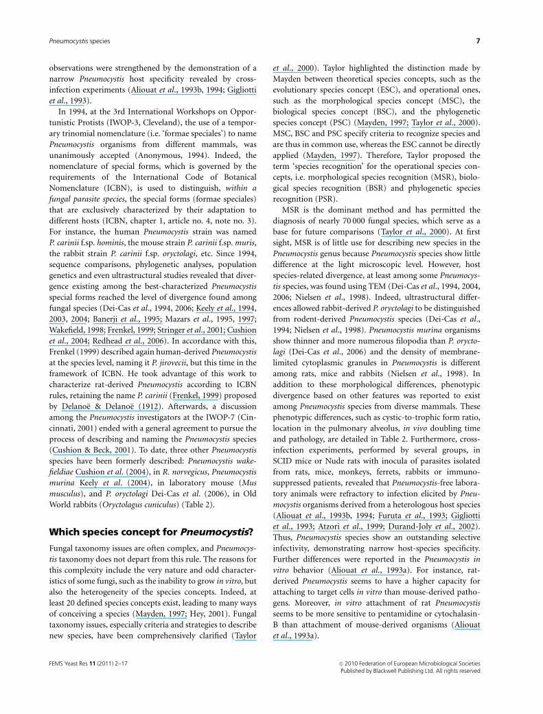
All known Pneumocystis life-cycle stages are found in the
lungs of infected hosts, although parasites may spread
exceptionally to other organs. The usually accepted life cycle
(Fig. 1) of Pneumocystis species involves an ameboid, thin-
walled, mononuclear vegetative or trophic form which
becomes a thick-walled cystic stage (ascus), in which a
multiple nuclear division leads to the formation of eight
ascospores. These forms are able to leave the cyst, presum-
ably by a pore-like zone located at the thickest part of the
cyst cell wall (Itatani, 1994), to attach specifically to type-I
epithelial alveolar cells and to evolve towards the cystic stage.
The transition from trophic forms to mature cyst or ascus
occurs in three consecutive sporocytic stages (early, inter-
mediate and late sporocytes) (Yoshida, 1989; Dei-Cas et al.,
2004).
Ascospores, which result from invaginations of the spor-
ocyte cell membrane, present a single mitochondrion, a
well-developed rough or smooth endoplasmic reticulum
and an electron-dense one-layered cell wall with a clearly
visible outer membrane (Barton & Campbell, 1967; Vavra &
Kucera, 1970; Haque et al., 1987; Palluault et al., 1992a, b).
Pneumocystis trophic forms and early sporocytes also have a
thin, monolayered, electron-dense cell wall with an outer
membrane (Palluault et al., 1992a). Mononuclear trophic
forms are irregular in size (4–8 mm long) and shape and
present numerous filopodia, filiform structures markedly
abundant and tree-like in mouse-derived Pneumocystis
(Dei-Cas et al., 1991). A single mitochondrion with budding
zones occupies a considerable part of the cell volume
(Palluault et al., 1991a, b). Early sporocytes are usually
rounded, mononuclear cells with a cell wall similar to that
Cyst
Late sporocyte
n
Intermediatesporocyte
n
Earlysporocyte
R!
2n
n + n
Trophicforms
Conjugation• ste2• ste3• mapk• ste11/ste20
Synaptonemalcomplexe
Meiosis:• mei2• ran1
Mitosis:• cdc2• cdc13• cdc25• cdc42
Spindle polebody
Fig. 1. A hypothetical life-cycle of Pneumocystis species. Parasites are represented as observed in the lung using TEM. Pleomorphic, thin-walled
mononuclear trophic forms are shown attached to type-I epithelial alveolar cells (at the top). Trophic form (2n) evolves into early sporocyte in which a
synaptonemal complex is indicated. Meiotic nuclear division (R!) leads to thick-walled sporocytic and cystic stages, in which multiple nuclear divisions in
intermediate and late sporocytes lead to the formation of eight haploid spores or ascospores (n). These forms are able to leave the cyst, to attach
specifically to type-I epithelial alveolar cells and, likely, to develop conjugation as illustrated at the top (left) (n1n), where spindle pole bodies are clearly
visible. Main molecular factors involved in conjugation, meiosis or mitosis identified in Pneumocystis carinii have been listed in the figure (see
homologies, functions and references in Table 1). Conjugation and synaptonemal complexes have been drawn according to Yoshida et al. (1984) and
Itatani (1996), respectively.
FEMS Yeast Res 11 (2011) 2–17c� 2010 Federation of European Microbiological SocietiesPublished by Blackwell Publishing Ltd. All rights reserved
4 M. Chabe et al.

of trophic forms. Transition from early to intermediate
sporocyte is denoted (1) by an increase of the cell wall
thickness (from 40 to 100 nm), resulting mainly from the
synthesis of a b-(1,3) glucan-rich, electron-lucent middle
layer, and (2) by the occurrence of multiple nuclear divi-
sions. In the late sporocytes, the resulting eight nuclei are
visible and a plasma membrane invagination progressively
demarcates the ascospores. The mature cyst or ascus is about
4–6mm in diameter, round and thick-walled. This life stage
also presents an outer membrane (De Stefano et al., 1990;
Palluault et al., 1992a). It contains a maximum of eight
spores. Mature cysts were detected in the bronchial lumen,
suggesting that they could gain the external environment to
be transmitted to other hosts by the airborne route (Dei-
Cas, 2000). However, the infectious forms of Pneumocystis
species have not been identified (Chabe et al., 2009).
In the parasite cell wall there is a highly polymorphic
116–120-kDa mannose-rich glycoprotein (major surface
glycoprotein) that constitutes the major surface antigen of
Pneumocystis (Kovacs et al., 1993). Cholesterol constitutes
about 78% of the total sterols in Pneumocystis (Kaneshiro,
1998) and is largely present in the parasite plasma mem-
brane. Pneumocystis organisms, like rust fungi, do not
contain ergosterol (Kaneshiro, 1998).
Pneumocystis fungi: an ascomycetous lifecycle
Pneumocystis species are ascomycetous pathogens appar-
ently able to sexually reproduce (mating, meiotic division,
production of ascospores) within their hosts, a property
more frequent in pathogenic fungi of plants than animals
(Dei-Cas & Vernes, 1986; Sexton & Howlett, 2006).
On the basis of detailed ultrastructural investigations
(Matsumoto & Yoshida, 1984; Yoshida, 1989; Peters et al.,
2001; Dei-Cas et al., 2004), cell sorting (Aliouat-Denis et al.,
2009; Martinez et al., 2009) and other approaches (Yamada
et al., 1986; Wyder et al., 1994, 1998; Cornillot et al., 2002),
crucial steps in the Pneumocystis life cycle are being clarified
(Fig. 1). At least in the species P. carinii, meiosis occurs in
early sporocytes, as revealed by the detection of synaptone-
mal complexes in the nucleus of these life-cycle stages
(Matsumoto & Yoshida, 1984; Peters et al., 2001). The
nucleus of the ascospore should be haploid, and a process
of nuclear fusion may therefore occur (Itatani, 1996; Smu-
lian et al., 2001) to produce diplophasic forms able to
resume the process of cyst generation. Although haploidy
has been predicted for most stages based on karyotype and
quantitative fluorescence analyses (Yamada et al., 1986;
Wyder et al., 1994, 1998; Cushion, 1998; Stringer & Cushion,
1998), more recent data indicate that haploidy and diploidy
coexist in Pneumocystis populations (Cornillot et al., 2002;
Dei-Cas et al., 2004; Aliouat-Denis et al., 2009). Thus,
replication of Pneumocystis organisms results at least
from the generation of haploid ascospores in the cyst, and
in vitro observations have suggested that trophic forms
could result exclusively from cyst development (Aliouat
et al., 1999).
In Fungi, the mating process is usually initiated after
mutual secretion of pheromones by fungal cells of opposite
mating types. Pheromone secretion is also stimulated by
environmental stress such as nutrition deprivation (Li et al.,
2007). Pheromones recognize a heterotrimeric G-coupled
transmembrane receptor located at the cell surface of the
opposite mating type. In turn, a mitogen-activated protein
kinase (Mapkp) signal transduction cascade is activated (Li
et al., 2007). Once activated, Mapkp controls many cell
effectors that halt the mitotic cell cycle, initiate transcription
of genes involved in mating, and eventually allow the fusion
of both cells (Harigaya & Yamamoto, 2007).
Many genetic factors involved in mating, meiosis and
mitosis regulation (Table 1, Fig. 1) have been identified in
P. carinii, thanks, at least in part, to the PGP (http://pgp.
cchmc.org/) (Cushion, 2004; Cushion et al., 2007). Also,
using degenerate PCR, library screening, heterologous
expression, yeast complementation, computer modeling,
immunoprecipitation and biochemical characterization,
key Pneumocystis genes or proteins potentially involved in
mating, meiosis or mitosis have been identified (Table 1, Fig.
1). The topic was reviewed recently by Aliouat-Denis et al.
(2009). Thomas et al. (1998b) identified a gene encoding a
Mapkp in P. carinii that is homologous to other fungal
Mapkps. Thus, heterologous expression of P. carinii mapk
was shown to restore pheromone signaling in S. cerevisiae
fus3/kss1 double mutants (Vohra et al., 2003a). In addition,
P. carinii Mapkp was reported to phosphorylate the P. carinii
homologue of S. pombe ste11 (ste12 in S. cerevisiae), a gene
which encodes a transcriptional factor needed for the
pheromone-induced expression of genes required for mat-
ing (Vohra et al., 2003b).
Once conjugation has occurred, activation of ste11 indir-
ectly turns on mei2, which plays pivotal roles in both the
induction and the progression of meiosis (Harigaya &
Yamamoto, 2007). Pneumocystis carinii mei2, a homologue
of S. pombe mei2, has recently been identified using the PGP
database (Burgess et al., 2008). The same group also
identified another kinase gene, P. carinii ran1, as an S. pombe
ran1 homologue. Ran1p is known to directly phosphorylate
and inhibit the activity of Mei2p. Both P. carinii genes
exhibited functional activity in meiotic control when ex-
pressed in S. pombe.
Pneumocystis carinii ste3, an a-factor pheromone receptor
homologue, was identified from an EST database that was
created as part of the PGP (Smulian et al., 2001). This
G-protein-coupled receptor was later reported to be exclu-
sively expressed in a subpopulation of trophic forms (Vohra
FEMS Yeast Res 11 (2011) 2–17 c� 2010 Federation of European Microbiological SocietiesPublished by Blackwell Publishing Ltd. All rights reserved
5Pneumocystis species

et al., 2004), a finding that is consistent with the expression
pattern of pheromone receptors in other fungi. So far, no
ligand has been identified for this receptor. Genes encoding
functional elements of the pheromone response signal
transduction cascade, such as ste12 and ste20 orthologues,
were found to be clustered around the Pneumocystis ste3
gene (Smulian et al., 2001). Pneumocystis carinii ste20 was
shown to be expressed following adherence of the fungus to
extracellular matrix components or lung epithelial cells
(Kottom et al., 2003). Heterologous expression of P. carinii
ste20 conferred pseudohyphal growth and also revealed the
gene to be functional in mating signaling pathways in S.
cerevisiae mutant strains (Kottom et al., 2003). Additional
orthologues of fungal genes associated with either the
mating/sexual mode of replication or stress/nutritional
deprivation were identified in the P. carinii EST database,
pointing to these conditions as triggers of Pneumocystis
mating (Cushion et al., 2007). A b-1,3-endoglucanase gene,
potentially involved in the regulation of cell wall biosynthe-
sis, was recently characterized in P. carinii (Villegas et al.,
2010). Lastly, many cell division-cycle genes have been
identified and characterized in Pneumocystis species (Tho-
mas et al., 1998a; Kaiser et al., 1999; Kottom et al., 2000;
Gustafson et al., 2001; Chabe et al., 2004; Arcenas et al.,
2006; Cushion et al., 2007; Krajicek et al., 2010).
Pneumocystis genus: from one tomultiple species
A notion, which lasted for almost a century after the
description of Pneumocystis in rats, guinea pigs, humans
and other mammals, was that Pneumocystis pneumonia
(PcP) has a zoonotic pattern. This misleading belief was
consistent with the erroneous concept of a unique Pneumo-
cystis species (i.e. P. carinii Delanoe & Delanoe, 1912).
Frenkel (1976) was the first to consider rat- and human-
derived Pneumocystis as distinct species on the basis of
apparent host species specificity, antigenic and ultrastruc-
tural differences. He proposed to name the human Pneumo-
cystis organisms ‘P. jiroveci’ (Frenkel, 1976). But it was only
in the early 1990s that the idea really emerged of a
Pneumocystis genus containing numerous highly divergent
genuine taxonomic entities. Indeed, molecular techniques
have revealed the existence of great genomic diversity
among isolates of Pneumocystis from different mammalian
species (see Aliouat-Denis et al., 2008 for review). These
Table 1. Pneumocystis life cycle: main Pneumocystis genes/proteins associated with mating, meiosis or mitosis
Gene Protein Homologue to� Function References
ste2w – Saccharomyces cerevisiae a-mating factor pheromone
receptor
Smulian et al. (2001); Cushion (2004)
ste3 Ste3p Coprinopsis cinerea a-mating factor pheromone
receptor
Smulian et al. (2001); Vohra et al. (2004)
mapk Mapkp Fusarium solani,
Schizosaccharomyces pombe SPK1,
Saccharomyces cerevisiae FUS3
Mitogen-activated protein kinase
(fungal differentiation and
proliferation)
Thomas et al. (1998b); Vohra et al.
(2003a)
ste11/ste20z Ste11p/Ste20p Schizosaccharomyces pombe
Ste11p, Cryptococcus neoformans
Ste20p
Ste11/Ste20p are involved in
pheromone response signal
transduction cascade
Smulian et al. (2001); Vohra et al.
(2003a); Kottom et al. (2003)
mei2 Mei2p Schizosaccharomyces pombe Induction and progression of
meiosis
Burgess et al. (2008)
ran1 Ran1p Pneumocystis carinii Temperature-dependent meiosis
inhibition activity
Burgess et al. (2008); Burgess et al.
(2009)
cdc2 Cdc2p Schizosaccharomyces pombe Cell-division-cycle serine–threonine
kinase (mitosis inductor)
Thomas et al. (1998a)‰
cdc13 Cdc13p Pneumocystis carinii Cdc13p binds to Cdc2 (needed for
Cdc2p kinase activity)
Kottom et al. (2000)
con7w – Magnaporthe grisea Likely involved in fungal
sporogenesis
Cushion et al. (2007)
cdc25 Cdc25p Schizosaccharomyces pombe Cell-division-cycle mitotic inducer
phosphatase
Gustafson et al. (2001)
cdc42 Cdc42p Schizophyllum commune Interacting with Ste20. Regulatory
GTPase function.
Krajicek et al. (2010)
�Only the greatest or most significant fungal homologies are mentioned.wGenes that were shown to be transcribed in Pneumocystis carinii.zAlso homologue to Saccharomyces cerevisiae Ste12p.‰cdc2 has also been described in Pneumocystis murina (Chabe et al., 2004) and in Pneumocystis jirovecii (Kaiser et al., 1999; Arcenas et al., 2006).
FEMS Yeast Res 11 (2011) 2–17c� 2010 Federation of European Microbiological SocietiesPublished by Blackwell Publishing Ltd. All rights reserved
6 M. Chabe et al.
observations were strengthened by the demonstration of a
narrow Pneumocystis host specificity revealed by cross-
infection experiments (Aliouat et al., 1993b, 1994; Gigliotti
et al., 1993).
In 1994, at the 3rd International Workshops on Oppor-
tunistic Protists (IWOP-3, Cleveland), the use of a tempor-
ary trinomial nomenclature (i.e. ‘formae speciales’) to name
Pneumocystis organisms from different mammals, was
unanimously accepted (Anonymous, 1994). Indeed, the
nomenclature of special forms, which is governed by the
requirements of the International Code of Botanical
Nomenclature (ICBN), is used to distinguish, within a
fungal parasite species, the special forms (formae speciales)
that are exclusively characterized by their adaptation to
different hosts (ICBN, chapter 1, article no. 4, note no. 3).
For instance, the human Pneumocystis strain was named
P. carinii f.sp. hominis, the mouse strain P. carinii f.sp. muris,
the rabbit strain P. carinii f.sp. oryctolagi, etc. Since 1994,
sequence comparisons, phylogenetic analyses, population
genetics and even ultrastructural studies revealed that diver-
gence existing among the best-characterized Pneumocystis
special forms reached the level of divergence found among
fungal species (Dei-Cas et al., 1994, 2006; Keely et al., 1994,
2003, 2004; Banerji et al., 1995; Mazars et al., 1995, 1997;
Wakefield, 1998; Frenkel, 1999; Stringer et al., 2001; Cushion
et al., 2004; Redhead et al., 2006). In accordance with this,
Frenkel (1999) described again human-derived Pneumocystis
at the species level, naming it P. jirovecii, but this time in the
framework of ICBN. He took advantage of this work to
characterize rat-derived Pneumocystis according to ICBN
rules, retaining the name P. carinii (Frenkel, 1999) proposed
by Delanoe & Delanoe (1912). Afterwards, a discussion
among the Pneumocystis investigators at the IWOP-7 (Cin-
cinnati, 2001) ended with a general agreement to pursue the
process of describing and naming the Pneumocystis species
(Cushion & Beck, 2001). To date, three other Pneumocystis
species have been formerly described: Pneumocystis wake-
fieldiae Cushion et al. (2004), in R. norvegicus, Pneumocystis
murina Keely et al. (2004), in laboratory mouse (Mus
musculus), and P. oryctolagi Dei-Cas et al. (2006), in Old
World rabbits (Oryctolagus cuniculus) (Table 2).
Which species concept for Pneumocystis?
Fungal taxonomy issues are often complex, and Pneumocys-
tis taxonomy does not depart from this rule. The reasons for
this complexity include the very nature and odd character-
istics of some fungi, such as the inability to grow in vitro, but
also the heterogeneity of the species concepts. Indeed, at
least 20 defined species concepts exist, leading to many ways
of conceiving a species (Mayden, 1997; Hey, 2001). Fungal
taxonomy issues, especially criteria and strategies to describe
new species, have been comprehensively clarified (Taylor
et al., 2000). Taylor highlighted the distinction made by
Mayden between theoretical species concepts, such as the
evolutionary species concept (ESC), and operational ones,
such as the morphological species concept (MSC), the
biological species concept (BSC), and the phylogenetic
species concept (PSC) (Mayden, 1997; Taylor et al., 2000).
MSC, BSC and PSC specify criteria to recognize species and
are thus in common use, whereas the ESC cannot be directly
applied (Mayden, 1997). Therefore, Taylor proposed the
term ‘species recognition’ for the operational species con-
cepts, i.e. morphological species recognition (MSR), biolo-
gical species recognition (BSR) and phylogenetic species
recognition (PSR).
MSR is the dominant method and has permitted the
diagnosis of nearly 70 000 fungal species, which serve as a
base for future comparisons (Taylor et al., 2000). At first
sight, MSR is of little use for describing new species in the
Pneumocystis genus because Pneumocystis species show little
difference at the light microscopic level. However, host
species-related divergence, at least among some Pneumocys-
tis species, was found using TEM (Dei-Cas et al., 1994, 2004,
2006; Nielsen et al., 1998). Indeed, ultrastructural differ-
ences allowed rabbit-derived P. oryctolagi to be distinguished
from rodent-derived Pneumocystis species (Dei-Cas et al.,
1994; Nielsen et al., 1998). Pneumocystis murina organisms
show thinner and more numerous filopodia than P. orycto-
lagi (Dei-Cas et al., 2006) and the density of membrane-
limited cytoplasmic granules in Pneumocystis is different
among rats, mice and rabbits (Nielsen et al., 1998). In
addition to these morphological differences, phenotypic
divergence based on other features was reported to exist
among Pneumocystis species from diverse mammals. These
phenotypic differences, such as cystic-to-trophic form ratio,
location in the pulmonary alveolus, in vivo doubling time
and pathology, are detailed in Table 2. Furthermore, cross-
infection experiments, performed by several groups, in
SCID mice or Nude rats with inocula of parasites isolated
from rats, mice, monkeys, ferrets, rabbits or immuno-
suppressed patients, revealed that Pneumocystis-free labora-
tory animals were refractory to infection elicited by Pneu-
mocystis organisms derived from a heterologous host species
(Aliouat et al., 1993b, 1994; Furuta et al., 1993; Gigliotti
et al., 1993; Atzori et al., 1999; Durand-Joly et al., 2002).
Thus, Pneumocystis species show an outstanding selective
infectivity, demonstrating narrow host-species specificity.
Further differences were reported in the Pneumocystis in
vitro behavior (Aliouat et al., 1993a). For instance, rat-
derived Pneumocystis seems to have a higher capacity for
attaching to target cells in vitro than mouse-derived patho-
gens. Moreover, in vitro attachment of rat Pneumocystis
seems to be more sensitive to pentamidine or cytochalasin-
B than attachment of mouse-derived organisms (Aliouat
et al., 1993a).
FEMS Yeast Res 11 (2011) 2–17 c� 2010 Federation of European Microbiological SocietiesPublished by Blackwell Publishing Ltd. All rights reserved
7Pneumocystis species

Table 2. Genotypic and phenotypic signatures differentiating Pneumocystis species (modified from Aliouat-Denis et al., 2008)
Features Pneumocystis carinii
Pneumocystis
wakefieldiae Pneumocystis murina
Pneumocystis
oryctolagi Pneumocystis jirovecii
Specific host Rat (Rattus norvegicus) Rat (Rattus norvegicus) Mouse (Mus musculus)
Rabbit (Oryctolagus
cuniculus) Man (Homo sapiens)Genes�
mtLSUrRNA U20169 (Cushion,
1998)
U20173 (Cushion,
1998)
AF257179 (S. Ruan
et al., unpublished
data)
S42915 (Wakefield
et al., 1992)
S42926 (Wakefield
et al., 1992)
mtSSUrRNA Hunter & Wakefield
(1996)
Hunter & Wakefield
(1996)
Hunter & Wakefield
(1996)
Hunter & Wakefield
(1996)
nucrRNA M86760 (Edman et al.,
1988)
L27658 (Ortiz-Rivera
et al., 1995; Cushion
et al., 1993)
AY532651 (Keely
et al., 2004)
DQ010098 (L. Li et al.,
unpublished data)
Liu et al. (1992)
ITS Ortiz-Rivera et al.
(1995)
L27658 (Ortiz-Rivera
et al., 1995)
AY532651 (Keely
et al., 2004)
DQ010098 (L. Li et al.,
unpublished data)
Ortiz-Rivera et al.
(1995); Lu et al. (1994)
TS M25415 (Edman et al.,
1989b)
Keely et al. (1994) Mazars et al. (1995) Mazars et al. (1995) Mazars et al. (1995)
HSP70 U80967 (Stedman
et al., 1998)
U80969 (Stedman
et al., 1998)
AY353254 (C.R.
Icenhour et al.,
unpublished data)
DQ435616 (Dei-Cas
et al., 2006)
U80970 (Stedman
et al., 1998)
Electrophoretic
karyotypes (number
and size range of
bands)
12–16 bands
(308–680 kb) Cushion
et al. (2004)
14 bands
(308–660 kb) Cushion
et al. (2004)
17 bands
(309–634 kb) Keely
et al. (2004)
14 bands
(300–700 kb) Cho
et al. (1999)
13 bands
(370–810 kb) Stringer
& Cushion (1998)
Size of
Pneumocystis
genome
8.2 Mb (Keely et al.,
2004)
7.7 Mb (Keely et al.,
2004)
8.2 Mb (Keely et al.,
2004)
7.7 Mb (Stringer &
Cushion, 1998)
Main phenotypic signatures (from Dei-Cas et al., 2006 unless otherwise indicated)
Organisms in lung
dry smears (TBO or
Giemsa stains)
Closely clustered Closely clustered Clustered Detached from each
other
Closely clustered
Cystic-to-trophic
form ratio
0.02–0.05 ? 0.02–0.05 0.10–0.15 ?
Size of cystic forms 4.03–4.42mm
(Laakkonen & Sukura,
1997)
5–8mm (Cushion
et al., 2004)
5–8mm (Keely et al.,
2004)
4–6 mm (Dei-Cas et al.,
2006)
3.5–5mm (Frenkel,
1976)
Filopodia thickness
and richness
Smaller than those of
rabbit
? Thinner and more
numerous than those
of rabbit, human and
macaque
Thicker and less
abundant than those
of mice
Thicker and less
abundant than those
of mice
Location Filling alveolar lumen Filling alveolar lumen Filling alveolar lumen Lining alveolar
epithelium
Filling alveolar lumen
(AIDS) or lining
alveolar epithelium
(epidemic or infantile
PcP)
In vivo doubling
time
4.5 days (in corticoid-
treated rats) (Aliouat
et al., 1999)
? 10.5 days (SCID mice)
(Aliouat et al., 1999)
1.7 days (untreated
rabbits) (Aliouat et al.,
1999)
?
Intra-alveolar
eosinophilic
honeycomb material
Present ? Present Rare Present
Fibrosis Frequent ? Frequent Rare Frequent
�Gene sequences available from at least two different Pneumocystis species. Accession numbers of the Pneumocystis DNA sequences and/or references
are given when they are available.
mtLSUrRNA, large subunit of mitochondrial rRNA; mtSSUrRNA, small subunit of mitochondrial rRNA; nucrRNA: rRNA from nuclear genome; ITS,
internal transcribed spacer from nuclear rRNA gene locus; TS, thymidylate synthase; HSP70, heat shock protein 70; ?, unknown.
FEMS Yeast Res 11 (2011) 2–17c� 2010 Federation of European Microbiological SocietiesPublished by Blackwell Publishing Ltd. All rights reserved
8 M. Chabe et al.

Concepts of biological species (‘groups of actually or
potentially interbreeding natural populations, which are
reproductively isolated from other such groups’; Mayr,
1963) play a prominent role in describing new fungal species
(Taylor et al., 2000). In Fungi, BSR has been used to identify
groups of mating compatible individuals, thus equated
with species. To some degree, BSC could be applied to
identify new Pneumocystis species. Indeed, sexual forms of
Pneumocystis have not been identified and sustained in vitro
cultures are not yet feasible, thus preventing laboratory
mating experiments from being set up. To answer the
question of whether gene flow occurs between Pneumocystis
subpopulations, population genetic approaches were uti-
lized (Mazars et al., 1997). Actually, multilocus enzyme
electrophoresis (MEE) was used to analyze five Pneumocystis
enzyme systems: malate dehydrogenase (MDH), glucose
phosphate isomerase (GPI), leucine aminopeptidase
(LAP), malic enzyme (ME) and 6-phosphogluconate
dehydrogenase (6PGDH) in Pneumocystis isolates from
rabbits, mice and rats (22 weaning rabbits, 30 corticoste-
roid-treated rats and 17 corticosteroid-treated mice) (Ma-
zars et al., 1997). Linkage disequilibrium analysis of allele
segregation and recombination of genotypes occurring
at these five loci clearly showed that Pneumocystis strains
from different host species constitute genetically distinct
populations, isolated from each other for a very long time
and therefore representing truly diverse lineages (Mazars
et al., 1997).
Today, the PSC (evolutionary lineages with a unique
combination of DNA orthologue sequences) is of growing
importance in fungal taxonomy (Taylor et al., 2000). For
Taylor, PSR comes closer than MSR and BSR to recognizing
species consistent with the ESC because, once progeny
evolutionary species have formed from an ancestor, changes
in gene sequences occur and can be recognized before
changes have occurred in mating behavior or morphology
(Taylor et al., 2000). Moreover, the genealogical concor-
dance phylogenetic species recognition method (Taylor
et al., 2000), which assesses the relationships among several
phylogenetic trees constructed on the basis of gene
sequences, increases the reliability of PSC-based species deter-
mination, making it possible to detect the occurrence of
genetic exchange (i.e. a concordance of gene trees is indica-
tive of genetic isolation) (Taylor et al., 2000). Beyond doubt,
PSC is the main species concept for use in the description of
new Pneumocystis species. Thanks to the abundance of
Pneumocystis gene sequence data, the genealogical concor-
dance condition, required to describe new fungal species
(Taylor et al., 2000), has been fulfilled by all Pneumocystis
species described until now, and all have emerged as mono-
phyletic clades on the basis of several genes (Fig. 2; Dei-Cas
et al., 2006). In agreement with this, the topological con-
cordance among seven Pneumocystis gene trees suggests that
the Pneumocystis organisms found in different host species
do not mate (Keely & Stringer, 2005). Genetic variation at
rRNA-encoding and DHFR loci between Pneumocystis spe-
cies were also used to estimate the times of Pneumocystis
speciation (Keely et al., 2003, 2004). According to these
estimations, P. carinii and P. jirovecii diverged early from
each other, about 100 million years ago (Keely et al., 2003).
Although this kind of estimation has to be viewed with
caution, the chronology matches well the divergence time of
primates from rodents (Nei et al., 2001). The same approach
applied to Pneumocystis from rodent subfamily Murinae
(P. carinii and P. murina) suggested they diverged from each
other 30–40 million years ago (Keely et al., 2004), which is
Fig. 2. Maximum-likelihood phylogeny of 12
Pneumocystis taxa, including the five described
Pneumocystis species, inferred from
mitochondrial large subunit ribosomal RNA
(mtLSUrRNA) and mitochondrial small-subunit
rRNA (mtSSUrRNA) concatenated gene
sequences, carried out using MRBAYES v3.1.2
(Ronquist & Huelsenbeck, 2003). Bayesian
posterior probabilities, calculated using a
Markov chain Monte Carlo sampling approach
(Green, 1995) implemented in MRBAYES v3.1.2,
are given as percentages near the individual
nodes. Nodes with values of o 50% are not
shown. Scale bar: 0.1 substitutions (corrected)
per base pair (from Aliouat-Denis et al., 2008).
FEMS Yeast Res 11 (2011) 2–17 c� 2010 Federation of European Microbiological SocietiesPublished by Blackwell Publishing Ltd. All rights reserved
9Pneumocystis species

consistent with the evolutionary time frame of their hosts
(Nei et al., 2001).
To date, the five accepted Pneumocystis species (P. carinii
Frenkel, 1999; P. jirovecii Frenkel, 1999; P. wakefieldiae
Cushion et al., 2004; P. murina Keely et al., 2004; P. oryctolagi
Dei-Cas et al., 2006) have all been described in the frame-
work of ICBN, on the basis of the morphological and/or
biological (Mayr, 1963) and phylogenetic (Taylor et al.,
2000) species concepts (Dei-Cas et al., 2006) examined
above.
Practical and theoretical implications ofnew notions on Pneumocystis taxonomy
The discovery of the genetic diversity of Pneumocystis genus
was likely the most important achievement in Pneumocystis
research for the last 20 years. Research on this topic revealed
that the genus contains numerous species with diverse
biological properties, each one being stenoxenous, i.e.
strictly infectious to one mammal species. Relatively large
molecular studies have been developed to explore the
presence and the circulation of Pneumocystis species in
ecosystems. Thus, Pneumocystis isolates from domestic,
synanthropic or wild mammals could be identified on the
basis of new species-specific DNA sequences that hitherto
confirmed the narrow host range of Pneumocystis species
(see Aliouat-Denis et al., 2008 for review). The results of
these researches have radically changed our conceptions
about Pneumocystis development, infection sources and
reservoir.
It was considered until recently that Pneumocystis organ-
isms were able to multiply exclusively in deeply immunode-
pressed hosts. Recent research has drastically changed
this view. Sensitive molecular methods allowed the detection
of Pneumocystis organisms in respiratory samples from
healthy or hospitalized subjects without severe immuno-
depression (Calderon et al., 1996, 2004; Dei-Cas, 2000;
Peterson & Cushion, 2005). In addition, experiments
focused on the behavior of Pneumocystis organisms in
immunocompetent hosts, demonstrated that Pneumocystis
cells are able to multiply in their lungs (Chabe et al., 2004).
It was also long believed that infection could be con-
tracted by the airborne route from animal or hypothetical
environmental sources, where the unique species of the
genus (P. carinii) able to cause PcP in both humans and
animals developed saprophytic growth. We know now
that the Pneumocystis genus includes numerous species,
each one restricted to one mammal species. Therefore,
animals cannot constitute a source of Pneumocystis infection
in humans (Gigliotti et al., 1993; Durand-Joly et al., 2002).
The idea of a zoonotic pattern to Pneumocystis infection,
although still retained by some authors (Stewart et al., 2005;
Youn, 2009), is no longer valid, as only human beings
could represent a potential infection source for other human
beings.
Which humans play the role of Pneumocystis infection
source? It might be considered that only hosts with PcP
transmit the infection. However, it was shown that immu-
nocompetent hosts with subclinical Pneumocystis infection
can transmit the infection by the airborne route to both
susceptible and healthy hosts (Chabe et al., 2004). Several
pieces of evidence suggest that the real impact of Pneumo-
cystis infection in humans or other mammals is beyond PcP.
For instance, seroconversion revealed Pneumocystis primary
infection in 4 90% of healthy children worldwide (re-
viewed in Aliouat-Denis et al., 2008), a condition that could
be associated with benign respiratory symptoms (Stagno
et al., 1980; Totet et al., 2004; Larsen et al., 2007). Immuno-
competent adults are often found to be Pneumocystis carriers
( = subjects without PcP but harboring very low Pneumocys-
tis rates), and Pneumocystis carriage was shown to be more
frequent in subjects with chronic respiratory conditions, e.g.
chronic obstructive pulmonary disease (COPD) (Calderon
et al., 1996, 2007), which is a major cause of disability and
the fourth leading cause of death in the world (Tan & Ng,
2008). Pneumocystis infection of immunocompetent hosts is
emerging as a relevant issue for human as well as animal
health (Rajagopalan-Levasseur et al., 1998; Cavallini-
Sanches et al., 2007) and, interestingly, typical PcP seems to
be a rare event in the natural history of Pneumocystis
infection.
What about the environment as a potential Pneumocystis
infection source? It cannot be formally excluded that (1)
Pneumocystis growth could be supported by unknown
inanimate, abiotic or vegetal substrates, or (2) some Pneu-
mocystis life-cycle stages could retain their infectious power
for some time after being released into the environment
from infected hosts. Hypotheses (1) and (2) were strength-
ened by the detection of Pneumocystis DNA in air- and
water-filtrate samples (Wakefield, 1996; Casanova-Cardiel &
Leibowitz, 1997). Also, Pneumocystis DNA was successfully
amplified from the air at conventional animal facilities
where Pneumocystis-infected laboratory animals were
housed (Olsson et al., 1996; Latouche et al., 1997) and from
hospital wards housing PcP patients (Bartlett et al., 1994;
Olsson et al., 1998). Against hypothesis (1) is that no
evidence of Pneumocystis environmental growth has been
reported so far. However, some Pneumocystis forms could
retain their viability after being in the air, as P. jirovecii
mRNA could be amplified from hospital air samples
(Latouche et al., 2001; Maher et al., 2001). In agreement with
this, there is some evidence of the existence of a Pneumocystis
‘dormant’ life-cycle stage (Kaneshiro & Maiorano, 1996; Chin
et al., 1999). Alternatively, the fact that Pneumocystis DNA or
even mRNA can be detected in the environment of Pneumo-
cystis-infected animals or humans (Bartlett et al., 1997;
FEMS Yeast Res 11 (2011) 2–17c� 2010 Federation of European Microbiological SocietiesPublished by Blackwell Publishing Ltd. All rights reserved
10 M. Chabe et al.

Olsson et al., 1998; Sing et al., 1999; Latouche et al., 2001;
Maher et al., 2001) suggests that Pneumocystis cells are
released with the exhaled air from infected hosts. This
observation supports the existence of an active host-to-host
transmission rather than the existence of hypothetical Pneu-
mocystis life stages able either to grow out of the hosts or to
remain viable in the environment for extended periods.
As Pneumocystis organisms are able to replicate in the
lungs of immunocompetent hosts, healthy members of
mammal populations, as well as severely infected susceptible
hosts, could play a major role as a Pneumocystis reservoir
(Dei-Cas, 2000; Chabe et al., 2004; Cushion et al., 2007;
Aliouat-Denis et al., 2008). The following features of Pneu-
mocystis organisms support this hypothesis: (1) a long
genetic isolation and coevolution with their host species
entailing Pneumocystis speciation and narrow host-species
specificity (Banerji et al., 1995; Mazars et al., 1995, 1997;
Demanche et al., 2001; Guillot et al., 2001; Hugot et al.,
2003; Keely et al., 2003, 2004; Dei-Cas et al., 2006; Aliouat-
Denis et al., 2008); (2) a highly compatible relationship with
mammalian host physiology (Dei-Cas et al., 1991, 2004;
Settnes & Nielsen, 1991; Aliouat et al., 1993a; Beck et al.,
1998; Cushion et al., 2007); and (3) a low pathogenic power
in healthy hosts (Chabe et al., 2004). In addition, Pneumo-
cystis colonized hosts are usually much more numerous than
hosts with PcP (Aliouat-Denis et al., 2008).
On the whole, molecular taxonomy research has played a
crucial role in the shift from an old conceptual Pneumocystis
infection framework to a new one. Main issues involve
taxonomy, host range and terminology. In the old concep-
tual framework, Pneumocystis organisms were considered
undefined or even enigmatic protists belonging to a unique
euryxenic taxonomic entity (P. carinii) transmissible by the
airborne route between mammals of different species. In this
old framework, terminology used to name Pneumocystis life-
cycle stages was taken from protistology (trophozoites,
precysts, cysts, intracystic bodies). In the new conceptual
framework, the Pneumocystis genus is a highly diversified
group of parasitic microfungi that contains numerous
stenoxenic species closely adapted to, and coevolving with,
mammal species. Accordingly, in this new framework, the
terminology used to name Pneumocystis life-cycle stages was
taken from mycology (trophic forms, sporocytes, ascus,
ascospores).
Conclusion
Hitherto, the identification of Pneumocystis species
(P. jirovecii excepted) was performed on parasites harvested
from immunosuppressed laboratory rodents or nonimmuno-
compromised laboratory, domestic, meat or wild Old World
rabbits. We need to explore Pneumocystis species diversity in
natural ecosystems to avoid bias derived from conventional
breeding. In fact, besides the potential impact of breeding
conditions on the Pneumocystis populations (e.g. housing in
close host-to-host proximity and isolation from the natural
environment), it could also be speculated that laboratory
animals come from a limited number of standard colonies
infected originally by a small number of Pneumocystis
strains. Furthermore, the characterization of Pneumocystis
species living in the lungs of wild animals remains a crucial
way forward to understand how these pathogens dissemi-
nate across ecosystems. Thanks to long adaptation of
Pneumocystis to its mammalian host, the topology of
Pneumocystis phylogenetic trees could shed light on the
mammalian host evolution history, helping to solve taxo-
nomic uncertainties. Although MSR and BSR have signifi-
cantly contributed to new species descriptions in the
Pneumocystis genus, the PSR seems to be a promising tool
to describe new Pneumocystis species, which are usually
represented in wild mammals by low parasite rates (Mazars
et al., 1997; Laakkonen, 1998; Palmer et al., 2000; Aliouat-
Denis et al., 2008; Chabe et al., 2010).
As a result of advancing knowledge about the natural
history of Pneumocystis infection, the perception of its
clinical impact on public health is evolving. The role of
healthy carriers in airborne disease transmission is nowa-
days recognized as a major contribution to Pneumocystis
circulation (Chabe et al., 2004). Recent data indicate that the
transplacental route is another human-to-human mode of
transmission (Montes-Cano et al., 2009). Finally, the low
parasite burden usually found in Pneumocystis carriers could
be the cause of benign respiratory symptoms in healthy
small children (Stagno et al., 1980; Totet et al., 2004; Larsen
et al., 2007) or worsening of the symptoms in COPD
patients (Calderon et al., 2007). More studies need to be
undertaken both on the clinical consequences of the pre-
sence of Pneumocystis in chronic pulmonary contexts and on
the intricate Pneumocystis life cycle to better define preven-
tion measures, adapt existing therapies to each clinical
context and discover new drug targets.
Acknowledgements
This work was supported by the ANR-ERA-NET ‘Pneumo-
cystis’ PathoGenoMics Program (ANR-06-PATHO-009-01),
by ANR ‘Biodiversite’ Program (CERoPath network, ANR-
07-BDIV-012) and by the French Ministry of Research
(EA3609-University Lille-Nord-de-France & Lille Pasteur
Institute).
References
Aliouat EM, Dei-Cas E, Ouaissi A, Palluault F, Soulez B & Camus
D (1993a) In vitro attachment of Pneumocystis carinii from
mouse and rat origin. Biol Cell 77: 209–217.
FEMS Yeast Res 11 (2011) 2–17 c� 2010 Federation of European Microbiological SocietiesPublished by Blackwell Publishing Ltd. All rights reserved
11Pneumocystis species

Aliouat EM, Mazars E, Dei-Cas E, Cesbron J & Camus D (1993b)
Intranasal inoculation of mouse, rat or rabbit-derived
Pneumocystis in SCID mice. J Protozool Res 3: 94–98.
Aliouat EM, Mazars E, Dei-Cas E, Delcourt P, Billaut P & Camus
D (1994) Pneumocystis cross infection experiments using SCID
mice and Nude rats as recipient host, showed strong host-
species specificity. J Eukaryot Microbiol 41: S71.
Aliouat EM, Dujardin L, Martinez A, Duriez T, Ricard I & Dei-
Cas E (1999) Pneumocystis carinii growth kinetics in culture
systems and in hosts: involvement of each life cycle parasite
stage. J Eukaryot Microbiol 46: S116–S117.
Aliouat-Denis CM, Chabe M, Demanche C, Aliouat EM,
Viscogliosi E, Guillot J, Delhaes L & Dei-Cas E (2008)
Pneumocystis species, co-evolution and pathogenic power.
Infect Genet Evol 8: 708–726.
Aliouat-Denis CM, Martinez A, Aliouat EM, Pottier M, Gantois
N & Dei-Cas E (2009) The Pneumocystis life cycle. Mem I
Oswaldo Cruz 104: 419–426.
Anderson AC (2005) Targeting DHFR in parasitic protozoa. Drug
Discov Today 10: 121–128.
Anonymous (1994) Revised nomenclature for Pneumocystis
carinii. The Pneumocystis Workshop. J Eukaryot Microbiol 41:
S121–S122.
Aragao HB (1913) Nota sobre as schizogonias e gametogonidas
dos trypanosomas. Bras Med 11: 271.
Arcenas RC, Uhl JR, Buckwalter SP, Limper AH, Crino D, Roberts
GD & Wengenack NL (2006) A real-time polymerase chain
reaction assay for detection of Pneumocystis from
bronchoalveolar lavage fluid. Diagn Microbiol Infect Dis 54:
169–175.
Atzori C, Agostoni F, Angeli E, Mainini A, Micheli V & Cargnel A
(1999) P. carinii host specificity: attempt of cross infections
with human derived strains in rats. J Eukaryot Microbiol 46:
S112.
Baldauf SL, Roger AJ, Wenk-Siefert I & Doolittle WF (2000) A
kingdom-level phylogeny of eukaryotes based on combined
protein data. Science 290: 972–977.
Banerji S, Lugli EB, Miller RF & Wakefield AE (1995) Analysis of
genetic diversity at the arom locus in isolates of Pneumocystis
carinii. J Eukaryot Microbiol 42: 675–679.
Bartlett MS, Lee CH, Lu JJ, Bauer NL, Bettz JF, McLaughlin GL &
Smith JW (1994) Pneumocystis carinii detected in air.
J Eukaryot Microbiol 41: S75.
Bartlett MS, Vermund SH, Jacobs R et al. (1997) Detection of
Pneumocystis carinii DNA in air samples: likely environmental
risk to susceptible persons. J Clin Microbiol 35: 2511–2513.
Barton EG & Campbell WG (1967) Further observations on the
ultrastructure of Pneumocystis. Arch Pathol 83: 527–534.
Beck JM, Preston AM, Wagner JG, Wilcoxen SE, Hossler P,
Meshnick SR & Paine R III (1998) Interaction of rat
Pneumocystis carinii and rat alveolar epithelial cells in vitro. Am
J Physiol 275: L118–L125.
Burgess JW, Kottom TJ & Limper AH (2008) Pneumocystis carinii
exhibits a conserved meiotic control pathway. Infect Immun
76: 417–425.
Burgess JW, Kottom TJ, Villegas LR, Lamont JD, Baden EM,
Ramirez-Alvarado M & Limper AH (2009) The Pneumocystis
meiotic PCRan1p kinase exhibits unique temperature-
regulated activity. Am J Resp Cell Mol 41: 714–721.
Calderon EJ, Regordan C, Medrano FJ, Ollero M & Varela JM
(1996) Pneumocystis carinii infection in patients with chronic
bronchial disease. Lancet 347: 977.
Calderon EJ, Varela JM, Medrano FJ et al. (2004) Epidemiology of
Pneumocystis carinii pneumonia in southern Spain. Clin
Microbiol Infect 10: 673–676.
Calderon EJ, Rivero L, Respaldiza N, Morilla R, Montes-Cano
MA, Friaza V, Munoz-Lobato F, Varela JM, Medrano FJ & De
La Horra C (2007) Systemic inflammation in patients with
chronic obstructive pulmonary disease who are colonized with
Pneumocystis jirovecii. Clin Infect Dis 45: 17–19.
Calderon-Sandubete EJ, Varela-Aguilar JM, Medrano-Ortega FJ,
Nieto-Guerrer V, Respaldiza-Salas N, de la Horra-Padilla C &
Dei-Cas E (2002) Historical perspective on Pneumocystis
carinii infection. Protist 153: 303–310.
Carini A (1910) Formas de eschizogonia do Trypanosoma lewisi.
Bol Soc de Med e Cir de Sao Paulo 18: 204.
Casanova-Cardiel L & Leibowitz MJ (1997) Presence of
Pneumocystis carinii DNA in pond water. J Eukaryot Microbiol
44: S28.
Cavallini-Sanches EM, Pescador C, Rozza D, Spanamberg A,
Borba MR, Ravazzolo AP, Driemeier D, Guillot J & Ferreiro L
(2007) Detection of Pneumocystis spp. in lung samples from
pigs in Brazil. Med Mycol 45: 395–399.
Cere N, Drouet-Viard F, Dei-Cas E, Chanteloup N & Coudert P
(1997) In utero transmission of Pneumocystis carinii sp. f.
oryctolagi. Parasite 4: 325–330.
Chabe M, Dei-Cas E, Creusy C, Fleurisse L, Respaldiza N, Camus
D & Durand-Joly I (2004) Immunocompetent hosts as a
reservoir of Pneumocystis organisms: histological and RT-PCR
data demonstrate active replication. Eur J Clin Microbiol 23:
89–97.
Chabe M, Nevez G, Totet A, Frealle E, Delhaes L, Aliouat EM &
Dei-Cas E (2009) Transmission de Pneumocystis. J Mycol Med
19: 276–284.
Chabe M, Herbreteau V, Hugot JP, Bouzard N, Deruyter L,
Morand S & Dei-Cas E (2010) Pneumocystis carinii and
Pneumocystis wakefieldiae in wild Rattus norvegicus trapped in
Thailand. J Eukaryot Microbiol 57: 213–217.
Chagas C (1909) Nova tripanozomiazaea humana. Mem I
Oswaldo Cruz 1: 159–218.
Chagas C (1911) Nova entidade morbida do homen: regumo
general de estudos etiologicos e clinicos. Mem I Oswaldo Cruz
3: 219–275.
Chagas C (1913) Revisao do ciclo evolutivo do Trypanosoma
cruzi. Braz Med 11: 225.
Chin K, Luttrell TD, Roe JD, Shadzi S, Wyder MA & Kaneshiro ES
(1999) Putative Pneumocystis dormant forms outside the
mammalian host, and long-term culture derived from them:
initial characterizations. J Eukaryot Microbiol 46: S95–S99.
FEMS Yeast Res 11 (2011) 2–17c� 2010 Federation of European Microbiological SocietiesPublished by Blackwell Publishing Ltd. All rights reserved
12 M. Chabe et al.

Cho SR, Park YG, Moon HN, Lee SH & Hong ST (1999)
Karyotypes of Pneumocystis carinii derived from several
mammals. Korean J Parasitol 37: 271–275.
Cornillot E, Keller B, Cushion MT, Metenier G & Vivares CP
(2002) Fine analysis of the Pneumocystis carinii f. sp. carinii
genome by two-dimensional pulsed-field gel electrophoresis.
Gene 293: 87–95.
Cushion M, Keely S & Stringer J (2004) Molecular and
phenotypic description of Pneumocystis wakefieldiae sp.nov., a
new species in rats. Mycologia 96: 429–438.
Cushion MT (1998) Genetic heterogeneity of rat-derived
Pneumocystis. FEMS Immunol Med Mic 22: 51–58.
Cushion MT (2004) Pneumocystis: unraveling the cloak of
obscurity. Trends Microbiol 12: 243–249.
Cushion MT & Beck JM (2001) Summary of Pneumocystis
research presented at the 7th International Workshop on
Opportunistic Protists. J Eukaryot Microbiol 48: S101–S105.
Cushion MT, Zhang J, Kaselis M, Giuntoli D, Stringer SL &
Stringer JR (1993) Evidence for two genetic variants of
Pneumocystis carinii coinfecting laboratory rats. J Clin
Microbiol 31: 1217–1223.
Cushion MT, Smulian AG, Slaven BE, Sesterhenn T, Arnold J,
Staben C, Porollo A, Adamczak R & Meller J (2007)
Transcriptome of Pneumocystis carinii during fulminate
infection: carbohydrate metabolism and the concept of a
compatible parasite. PLoS ONE 2: e423.
Dei-Cas E (2000) Pneumocystis infections: the iceberg? Med Mycol
1: 23–32.
Dei-Cas E & Vernes A (1986) Parasitic adaptation of pathogenic
fungi to the mammalian hosts. CRC Crit Rev Microbiol 13:
173–218.
Dei-Cas E, Jackson H, Palluault F, Aliouat EM, Hancock V, Soulez
B & Camus D (1991) Ultrastructural observations on the
attachment of Pneumocystis carinii in vitro. J Protozool 38:
S205–S207.
Dei-Cas E, Mazars E, Odberg-Ferragut C et al. (1994)
Ultrastructural, genomic, isoenzymatic and biological features
make it possible to distinguish rabbit Pneumocystis from other
mammal Pneumocystis strains. J Eukaryot Microbiol 41: S84.
Dei-Cas E, Aliouat EM & Cailliez JC (2004) Cellular structure.
Pneumocystis Pneumonia, 3rd edn (Walzer PD & Cushion MT,
eds), pp. 61–94. Marcel Dekker, New York.
Dei-Cas E, Chabe M, Moukhlis R et al. (2006) Pneumocystis
oryctolagi sp. nov., an uncultured fungus causing pneumonia
in rabbits at weaning: review of current knowledge, and
description of a new taxon on genotypic, phylogenetic and
phenotypic bases. FEMS Microbiol Rev 80: 853–871.
Delanoe P & Delanoe E (1912) Sur les rapports des kystes de
carinii du poumon des rats avec le Trypanosoma lewisi. CR
Acad Sci 155: 658–660.
Delaporte F (1999) La Maladie de Chagas. Histoire d’un Fleau
Continental. Payot & Rivages, Paris.
Demanche C, Berthelemy M, Petit T, Polack B, Wakefield AE,
Dei-Cas E & Guillot J (2001) Phylogeny of Pneumocystis carinii
from 18 primate species confirms host specificity and suggests
coevolution. J Clin Microbiol 39: 2126–2133.
Demanche C, Petit T, Moisson P, Ollivet F, Rigoulet J, Chermette
R, Dei-Cas E, Wakefield AE & Guillot J (2003) Assessment of
Pneumocystis species carriage in captive primates. Vet Rec 28:
811–813.
De Stefano JA, Cushion MT, Sleught R & Walzer PD (1990)
Analysis of Pneumocystis carinii cyst wall. I. Evidence for an
outer surface membrane. J Protozool 37: 428–435.
Diezmann S, Cox CJ, Sconian G, Vilgalys RJ & Mitchell TG
(2004) Phylogeny and evolution of medical species of Candida
and related taxa: a multigenic analysis. J Clin Microbiol 42:
5624–5635.
Dumoulin A, Mazars E, Seguy N, Gargallo-Viola D, Vargas S,
Cailliez JC, Aliouat EM, Wakefield AE & Dei-Cas E (2000)
Transmission of Pneumocystis carinii disease from
immunocompetent contacts of infected hosts to susceptible
hosts. Eur J Clin Microbiol 19: 671–678.
Durand-Joly I, Aliouat EM, Recourt C, Guyot K, Francois N,
Wauquier M, Camus D & Dei-Cas E (2002) Pneumocystis
carinii f. sp. hominis is not infectious for SCID mice. J Clin
Microbiol 40: 1862–1865.
Durand-Joly I, Soula F, Chabe M, Dalle JH, Lafitte JJ, Senechal M,
Pinon A, Camus D & Dei-Cas E (2003) Long-term
colonization with Pneumocystis jirovecii in hospital staffs: a
challenge to prevent nosocomial pneumocystosis. J Eukaryot
Microbiol 50: S614–S615.
Edman JC, Kovacs JA, Masur H, Santi DV, Elwood HJ & Sogin
ML (1988) Ribosomal RNA sequence shows Pneumocystis
carinii to be a member of the fungi. Nature 334: 519–522.
Edman JC, Kovacs JA, Masur H, Santi DV, Elwood HJ & Sogin
ML (1989a) Ribosomal RNA sequence shows Pneumocystis
carinii to be a member of the fungi. J Protozool 36: S18–S20.
Edman U, Edman JC, Lundgren B & Santi DV (1989b) Isolation
and expression of the Pneumocystis carinii thymidylate
synthase gene. P Natl Acad Sci USA 86: 6503–6507.
Eriksson OE (1994) Pneumocystis carinii, a parasite in lungs of
mammals, referred to a new family and order
(Pneumocystidaceae, Pneumocystidales, Ascomycota). Systema
Ascomycetum 13: 165–180.
Eriksson OE (1999) Outline of Ascomycota – 1999. Myconet 3:
1–88.
Eriksson OE & Winka K (1997) Supraordinal taxa of the
Ascomycota. Myconet 1: 1–16.
Frenkel JK (1976) Pneumocystis jiroveci n. sp. from man:
morphology, physiology, and immunology in relation to
pathology. Natl Cancer I Monogr 43: 13–30.
Frenkel JK (1999) Pneumocystis pneumonia, an
immunodeficiency-dependent disease (IDD): a critical
historical overview. J Eukaryot Microbiol 46: S89–S92.
Furuta T, Fujita M, Mukai R, Sakakibara I, Sata T, Miki K,
Hayami M, Kojima S & Yoshikawa Y (1993) Severe pulmonary
pneumocystosis in simian acquired immunodeficiency
syndrome induced by simian immunodeficiency virus: its
characterization by the polymerase-chain-reaction method
FEMS Yeast Res 11 (2011) 2–17 c� 2010 Federation of European Microbiological SocietiesPublished by Blackwell Publishing Ltd. All rights reserved
13Pneumocystis species

and failure of experimental transmission to immunodeficient
animals. Parasitol Res 79: 624–628.
Gigliotti F, Harmsen AG, Haidaris CG & Haidaris PJ (1993)
Pneumocystis carinii is not universally transmissible between
mammalian species. Infect Immun 61: 2886–2890.
Goheen M, Blumershine R, Hull M, Bartlett M & Smith J (1989)
Enhancement of the fine structure of Pneumocystis carinii
using potassium ferrocyanide and tannic acid. Proceedings of
the 47th Annual Meeting of the Electron Microscopy Society of
America (Bailey GW, ed), pp. 984–985. San Francisco.
Green PJ (1995) Reversible jump Markov chain Monte Carlo
computation and Bayesian model determination. Biometrika
82: 711–732.
Guillot J, Demanche C, Hugot JP, Berthelemy M, Wakefield AE,
Dei-Cas E & Chermette R (2001) Parallel phylogenies of
Pneumocystis species and their mammalian hosts. J Eukaryot
Microbiol 47: S113–S115.
Gustafson MP, Thomas CF Jr, Rusnak F, Limper AH & Leof EB
(2001) Differential regulation of growth and checkpoint
control mediated by a Cdc25 mitotic phosphatase from
Pneumocystis carinii. J Biol Chem 276: 835–843.
Haque AU, Plattner SB, Cook RT & Hart MN (1987)
Pneumocystis carinii: taxonomy as viewed by electron
microscopy. Am J Clin Pathol 87: 504–510.
Harigaya Y & Yamamoto M (2007) Molecular mechanisms
underlying the mitosis–meiosis decision. Chromosome Res 15:
523–537.
Hey J (2001) The mind of the species problem. Trends Ecol Evol
16: 326–329.
Hughes WT (1982) Natural mode of acquisition for de novo
infection with Pneumocystis carinii. J Infect Dis 145: 842–848.
Hugot JP, Demanche C, Barriel V, Dei-Cas E & Guillot J (2003)
Phylogenetic systematics and evolution of primate-derived
Pneumocystis based on mitochondrial or nuclear DNA
sequence comparison. Syst Biol 52: 735–744.
Hunter JA & Wakefield AE (1996) Genetic divergence at the
mitochondrial small subunit ribosomal RNA gene among
isolates of Pneumocystis carinii from five mammalian host
species. J Eukaryot Microbiol 43: S24–S25.
Itatani CA (1994) Ultrastructural demonstration of a pore in the
cyst wall of Pneumocystis carinii. J Parasitol 80: 644–648.
Itatani CA (1996) Ultrastructural morphology of intermediate
forms and forms suggestive of conjugation in the life cycle of
Pneumocystis carinii. J Parasitol 82: 163–171.
James TY, Kauff F, Schoch CL et al. (2006) Reconstructing the
early evolution of Fungi using a six-gene phylogeny. Nature
443: 818–822.
Kaiser K, Rabodonirina M, Mayencon M & Picot S (1999) Cdc2
gene of Pneumocystis carinii hominis and its expression during
culture. J Eukaryot Microbiol 46: S130.
Kaneshiro ES (1998) The lipids of Pneumocystis carinii. Clin
Microbiol Rev 11: 27–41.
Kaneshiro ES & Maiorano JN (1996) Survival and infectivity of
Pneumocystis carinii outside the mammalian host. J Eukaryot
Microbiol 43: S35.
Keely S, Pai HJ, Baughman R, Sidman C, Sunkin C, Stringer JR &
Stringer SL (1994) Pneumocystis species inferred from analysis
of multiple genes. J Eukaryot Microbiol 41: S94.
Keely SP & Stringer JR (2005) Nomenclature and genetic
variation of Pneumocystis. Pneumocystis Pneumonia, 3rd edn
(Walzer PD & Cushion MT, eds), pp. 39–59. Marcel Dekker,
New York.
Keely SP, Fischer JM & Stringer JR (2003) Evolution and
speciation of Pneumocystis. J Eukaryot Microbiol 50:
S624–S626.
Keely SP, Fischer JM, Cushion MT & Stringer JR (2004)
Phylogenetic identification of Pneumocystis murina sp. nov., a
new species in laboratory mice. Microbiology 150: 1153–1165.
Kottom TJ, Thomas CF Jr, Mubarak KK, Leof EB & Limper AH
(2000) Pneumocystis carinii uses a functional cdc13 B-type
cyclin complex during its life cycle. Am J Resp Cell Mol 22:
722–731.
Kottom TJ, Kohler JR, Thomas CF, Fink GR & Limper AH (2003)
Lung epithelial cells and extracellular matrix components
induce expression of Pneumocystis carinii STE20, a gene
complementing the mating and pseudohyphal growth defects
of STE20 mutant yeast. Infect Immun 71: 6463–6471.
Kovacs JA, Powell F, Edman JC, Lundgren B, Martinez A, Drew B
& Angus CW (1993) Multiple genes encode the major surface
glycoprotein of Pneumocystis carinii. J Biol Chem 268:
6034–6040.
Krajicek BJ, Kottom TJ, Villegas L & Limper AH (2010)
Characterization of the PcCdc42 small G protein from
Pneumocystis carinii, which interacts with the PcSte20 life cycle
regulatory kinase. Am J Physiol Lung C 298: L252–L260.
Krungkrai J, Webster HK & Yuthavong Y (1990) Folate and
cobalamin metabolism in Plasmodium falciparum. Parasitol
Today 6: 388–391.
Kwon KY, Kim SP & Limper AH (1998) Recognition of
Pneumocystis carinii antigen on its surface by
immunohistochemistry and immunoelectron microscopy.
J Korean Med Sci 13: 131–137.
Laakkonen J (1998) Pneumocystis carinii in wildlife. Int J Parasitol
28: 241–252.
Laakkonen J & Sukura A (1997) Pneumocystis carinii of the
common shrew, Sorex araneus, shows a discrete phenotype.
J Eukaryot Microbiol 44: 117–121.
Landvik S, Eriksson OE & Berbee ML (2001) Neolecta – a fungal
dinosaur? Evidence from b-tubulin amino acid sequences.
Mycologia 93: 1151–1163.
Larsen HH, von Linstow ML, Lundgren B, Hogh B, Westh H &
Lundgren JD (2007) Primary Pneumocystis infection in infants
hospitalized with acute respiratory tract infection. Emerg Infect
Dis 13: 66–72.
Latouche S, Olsson M, Polack B, Brun-Pascaud M, Bernard C &
Roux P (1997) Detection of Pneumocystis carinii f. sp. in air
samples collected in animal rooms. J Eukaryot Microbiol 44:
46–47.
Latouche S, Totet A, Lacube P, Bolognini J, Nevez G & Roux P
(2001) Development of an RT-PCR on the heat shock protein
FEMS Yeast Res 11 (2011) 2–17c� 2010 Federation of European Microbiological SocietiesPublished by Blackwell Publishing Ltd. All rights reserved
14 M. Chabe et al.

70 gene for viability detection of Pneumocystis carinii f. sp.
hominis in patients with pneumocystosis and in air sample.
J Eukaryot Microbiol 48: S176–S177.
Li L, Wright SJ, Krystofova S, Park G & Borkovich KA (2007)
Heterotrimeric G protein signaling in filamentous fungi. Annu
Rev Microbiol 61: 423–452.
Liu Y, Rocourt M, Pan S, Liu C & Leibowitz MJ (1992) Sequence
and variability of the 5.8S and 26S rRNA genes of Pneumocystis
carinii. Nucleic Acids Res 20: 3763–3772.
Liu Y, Leigh JW, Brinkmann H, Cushion MT, Rodriguez-Ezpeleta
N, Philippe H & Lang BF (2009) Phylogenomic analyses
support the monophyly of Taphrinomycotina, including
Schizosaccharomyces fission yeasts. Mol Biol Evol 26: 27–34.
Liu YJ, Hodson MC & Hall BD (2006) Loss of flagellum
happened only once in the fungal lineage: phylogenetic
struture of kingdom Fungi inferred from RNA polymerase II
subunit genes. BMC Evol Biol 6: 74.
Lu JJ, Bartlett MS, Shaw MM, Queener SF, Smith JW, Ortiz-
Rivera M, Leibowitz MJ & Lee CH (1994) Typing of
Pneumocystis carinii strains that infect humans based on
nucleotide sequence variations of internal transcribed spacers
of rRNA genes. J Clin Microbiol 32: 2904–2912.
Maher NH, Vermund SH, Welsh DA, Dillon HK, Awooda A &
Unnasch TR (2001) Development and characterization of a
molecular viability assay for Pneumocystis carinii f sp hominis.
J Infect Dis 183: 1825–1827.
Martinez A, Aliouat EM, Pottier M, Gantois M, Pincon C,
Standaert-Vitse A, Dei-Cas E & Aliouat CM (2009) High-
speed cell sorting of infectious trophic and cystic forms of
Pneumocystis carinii. J Eukaryot Microbiol 56: 446–453.
Matsumoto Y & Yoshida Y (1984) Sporogony in Pneumocystis
carinii: synaptonemal complexes and meiotic nuclear divisions
observed in precysts. J Protozool 31: 420–428.
Mayden RL (1997) A hierarchy of species concepts: the
denouement in the saga of the species problem. Species: The
Units of Biodiversity (Claridge MF, Dawah HA & Wilson MR,
eds), pp. 381–424. Chapman & Hall, London.
Mayr E (1963) Animal Species and Evolution. Harvard University
Press, Cambridge, MA.
Mazars E, Odberg-Ferragut C, Dei-Cas E, Fourmaux MN, Aliouat
EM, Brun-Pascaud M, Mougeot G & Camus D (1995)
Polymorphism of Pneumocystis carinii from different host
species. J Eukaryot Microbiol 42: 26–32.
Mazars E, Guyot K, Durand I, Dei-Cas E, Boucher S, Ben
Abderrazak S, Banuls AL, Tibayrenc M & Camus D (1997)
Isoenzyme diversity in Pneumocystis carinii from rats, mice
and rabbits. J Infect Dis 175: 655–660.
Miller RF, Ambrose HE & Wakefield AE (2001) Pneumocystis
carinii f. sp. hominis DNA in immunocompetent health care
workers in contact with patients with P. carinii pneumonia.
J Clin Microbiol 39: 3877–3882.
Montes-Cano MA, Chabe M, Fontillon-Alberdi M, de-Lahorra C,
Respaldiza N, Medrano FJ, Varela JM, Dei-Cas E & Calderon
EJ (2009) Vertical transmission of Pneumocystis jirovecii in
humans. Emerg Infect Dis 15: 125–127.
Nei M, Xu P & Glazko G (2001) Estimation of divergence times
from multiprotein sequences for a few mammalian species and
several distantly related organisms. P Natl Acad Sci USA 98:
2497–2502.
Nielsen MH, Settnes OP, Aliouat EM, Cailliez JC & Dei-Cas E
(1998) Different ultrastructural morphology of Pneumocystis
carinii derived from mice, rats, and rabbits. APMIS 106:
771–779.
Nishida H & Sugiyama J (1993) Phylogenetic relationships
among Taphrina, Saitoella, and other higher fungi. Mol Biol
Evol 10: 431–436.
Nishida H & Sugiyama J (1994) Archiascomycetes: detection of a
major new lineage within the Ascomycota. Mycoscience 35:
361–366.
Olsson M, Sukura A, Lindberg LA & Linder E (1996) Detection of
Pneumocystis carinii DNA by filtration of air. Scand J Infect Dis
28: 279–282.
Olsson M, Lidman C, Latouche S, Bjorkman A, Roux P, Linder E
& Wahlgren M (1998) Identification of Pneumocystis carinii f.
sp. hominis gene sequences in filtered air in hospital
environments. J Clin Microbiol 36: 1737–1740.
Ortiz-Rivera M, Liu Y, Felder R & Leibowitz MJ (1995)
Comparison of coding and spacer region sequences of
chromosomal rRNA-coding genes of two sequevars of
Pneumocystis carinii. J Eukaryot Microbiol 42: 44–49.
Palluault F, Dei-Cas E, Slomianny C, Soulez B & Camus D (1990)
Golgi complex and lysosomes in rabbit-derived Pneumocystis
carinii. Biol Cell 70: 73–82.
Palluault F, Pietrzyck B, Dei-Cas E, Slomianny C, Soulez B &
Camus D (1991a) Three-dimensional reconstruction of
rabbit-derived Pneumocystis carinii. I. Trophozoite. J Protozool
38: 402–407.
Palluault F, Pietrzyck B, Dei-Cas E, Slomianny C, Soulez B &
Camus D (1991b) Three-dimensional reconstruction of
rabbit-derived Pneumocystis carinii. II. Intermediate precyst.
J Protozool 38: 407–411.
Palluault F, Slomianny C, Soulez B, Dei-Cas E & Camus D
(1992a) High osmotic pressure enables fine ultrastructural and
cytochemical studies on Pneumocystis carinii. I: epon
embedding. Parasitol Res 78: 437–444.
Palluault F, Soulez B, Slomianny C, Dei-Cas E, Cesbron JY &
Camus D (1992b) High osmotic pressure for Pneumocystis
carinii London Resin White embedding enables fine
immunocytochemistry studies: I. Golgi complex and cell-wall
synthesis. Parasitol Res 78: 482–488.
Palmer RJ, Settnes OP, Lodal J & Wakefield AE (2000) Population
structure of rat-derived Pneumocystis carinii in Danish wild
rats. Appl Environ Microb 66: 4954–4961.
Peters SE, English K, Rana A, Akter S, Malik S, Warburton NC &
Duckett JG (2001) Synaptonemal complexes in the pre-cyst of
Pneumocystis carinii. J Eukaryot Microbiol 48: S134.
Peterson JC & Cushion MT (2005) Pneumocystis: not just
pneumonia. Curr Opin Microbiol 8: 393–398.
FEMS Yeast Res 11 (2011) 2–17 c� 2010 Federation of European Microbiological SocietiesPublished by Blackwell Publishing Ltd. All rights reserved
15Pneumocystis species

Pixley FJ, Wakefield AE, Banerji S & Hopkin JM (1991)
Mitochondrial gene sequences show fungal homology for
Pneumocystis carinii. Mol Microbiol 5: 1347–1351.
Rabodonirina M (2001) La pneumocystose est-elle une maladie
contagieuse? Lettre l’Infectiol 16: 110–112.
Rajagopalan-Levasseur P, Allaert A, Dridba M, Odberg-Ferragut
C, Jouault T, Creusy C, Camus D & Dei-Cas E (1998) Response
to Pneumocystis infection in an immunocompetent host.
FEMS Immunol Med Mic 22: 107–121.
Redhead SA, Cushion MT, Frenkel JK & Stringer JR (2006)
Pneumocystis and Trypanosoma cruzi: nomenclature and
typifications. J Eukaryot Microbiol 53: 2–11.
Ronquist F & Huelsenbeck JP (2003) MrBayes 3: Bayesian
phylogenetic inference under mixed models. Bioinformatics 19:
1572–1574.
Sanchez CA, Chabe M, Aliouat el M, Durand-Joly I, Gantois N,
Conseil V, Lopez C, Duriez T, Dei-Cas E & Vargas SL (2007)
Exploring transplacental transmission of Pneumocystis
oryctolagi in first-time pregnant and multiparous rabbit does.
Med Mycol 45: 701–707.
Settnes OP & Nielsen MJ (1991) Host–parasite relationship in
Pneumocystis carinii infection: activation of the plasmalemmal
vesicular system in type I alveolar epithelial cells. J Protozool
38: S174–S176.
Sexton AC & Howlett BJ (2006) Parallels in fungal pathogenesis
on plant and animal hosts. Eukaryot Cell 5: 1941–1949.
Sienkiewicz N, Jaroslawski S, Wyllie S & Fairlamb AH (2008)
Chemical and genetic validation of dihydrofolate reductase-
thymidylate synthase as a drug target in African trypanosomes.
Mol Microbiol 69: 520–533.
Sinclair K, Wakefield AE, Banerji S & Hopkin JM (1991)
Pneumocystis carinii organisms derived from rat and human
hosts are genetically distinct. Mol Biochem Parasitol 45:
183–184.
Sing A, Wonhas C, Bader L, Luther M & Heesemann J (1999)
Detection of Pneumocystis carinii DNA in the air filter of a
ventilated patient with AIDS. Clin Infect Dis 29: 952–953.
Smulian AG, Sesterhenn T, Tanaka R & Cushion MT (2001) The
ste3 pheromone receptor gene of Pneumocystis carinii is
surrounded by a cluster of signal transduction genes. Genetics
157: 991–1002.
Soulez B, Palluault F, Cesbron JY, Dei-Cas E, Capron A & Camus
D (1991) Introduction of Pneumocystis carinii in a colony of
SCID mice. J Protozool 38: S123–S125.
Spatafora JW, Sung GH, Johnson D et al. (2006) A five-gene
phylogeny of Pezizomycotina. Mycologia 98: 1018–1028.
Stagno S, Pifer LL, Hughes WT, Brasfield DM & Tiller RE (1980)
Pneumocystis carinii pneumonitis in young
immunocompetent infants. Pediatrics 66: 56–62.
Stedman TT, Butler DR & Buck GA (1998) The HSP70 gene
family in Pneumocystis carinii: molecular and phylogenetic
characterization of cytoplasmic members. J Eukaryot Microbiol
45: 589–599.
Stewart WC, Pollock KG, Browning LM, Young D, Smith-Palmer
A & Reilly WJ (2005) Survey of zoonoses recorded in Scotland
between 1993 and 2002. Vet Rec 157: 697–702.
Stringer JR & Cushion MT (1998) The genome of Pneumocystis
carinii. FEMS Immunol Med Mic 22: 15–26.
Stringer JR, Cushion MT & Wakefield AE (2001) New
nomenclature for the genus Pneumocystis. J Eukaryot Microbiol
48: 184–189.
Stringer SL, Stringer JR, Blase MA, Walzer PD & Cushion MT
(1989) Pneumocystis carinii: sequence from ribosomal RNA
implies a close relationship with fungi. Exp Parasitol 68:
450–461.
Sugiyama J, Hosaka K & Suh SO (2006) Early diverging
Ascomycota: phylogenetic divergence and related evolutionary
enigmas. Mycologia 298: 996–1005.
Tan WC & Ng TP (2008) COPD in Asia: where east meets west.
Chest 133: 517–527.
Taylor JW, Jacobson DJ, Kroken S, Kasuga T, Geiser DM, Hibbett
DS & Fisher MC (2000) Phylogenetic species recognition and
species concepts in fungi. Fungal Genet Biol 31: 21–32.
Thomas CF, Anders RA, Gustafson MP, Leof EB & Limper AH
(1998a) Pneumocystis carinii contains a functional cell-
division-cycle Cdc2 homologue. Am J Resp Cell Mol 18:
297–306.
Thomas CF Jr, Kottom TJ, Leof EB & Limper AH (1998b)
Characterization of a mitogen-activated protein kinase from
Pneumocystis carinii. Am J Physiol 275: L193–L199.
Totet A, Latouche S, Lacube P, Pautard JC, Jounieaux V, Raccurt
C, Roux P & Nevez G (2004) Pneumocystis jirovecii
dihydropteroate synthase genotypes in immunocompetent
infants and immunosuppressed adults, Amiens, France. Emerg
Infect Dis 10: 667–673.
Vargas SL, Ponce CA, Gigliotti F, Ulloa AV, Prieto S, Munoz MP &
Hughes WT (2000) Transmission of Pneumocystis carinii DNA
from a patient with P. carinii pneumonia to
immunocompetent contact health care workers. J Clin
Microbiol 38: 1536–1538.
Vavra J & Kucera K (1970) Pneumocystis carinii delanoe, its
ultrastructure and ultrastructural affinities. J Protozool 17:
463–483.
Villegas LR, Kottom TJ & Limper AH (2010) Characterization of
PCEng2 a {beta}-1,3-endoglucanase homologue in
Pneumocystis carinii with activity in cell wall regulation. Am J
Resp Cell Mol 43: 192–200.
Vohra PK, Puri V, Kottom TJ, Limper AH & Thomas CF Jr
(2003a) Pneumocystis carinii STE11, an HMG-box protein, is
phosphorylated by the mitogen activated protein kinase PCM.
Gene 312: 173–179.
Vohra PK, Puri V & Thomas CF Jr (2003b) Complementation
and characterization of the Pneumocystis carinii MAPK, PCM.
FEBS Lett 551: 139–146.
Vohra PK, Park JG, Sanyal B & Thomas CF Jr (2004) Expression
analysis of PCSTE3, a putative pheromone receptor from the
lung pathogenic fungus Pneumocystis carinii. Biochem Bioph
Res Co 319: 193–199.
FEMS Yeast Res 11 (2011) 2–17c� 2010 Federation of European Microbiological SocietiesPublished by Blackwell Publishing Ltd. All rights reserved
16 M. Chabe et al.

Wakefield AE (1996) DNA sequences identical to Pneumocystis
carinii f. sp. carinii and Pneumocystis carinii f. sp. hominis in
samples of air spora. J Clin Microbiol 34: 1754–1759.
Wakefield AE (1998) Genetic heterogeneity in Pneumocystis
carinii: an introduction. FEMS Immunol Med Mic 22: 97–101.
Wakefield AE, Peters SE, Banerji S, Bridge PD, Hall GS,
Hawksworth DL, Guiver LA, Allen AG & Hopkin JM (1992)
Pneumocystis carinii shows DNA homology with the
Ustomycetous red yeast fungi. Mol Microbiol 6: 1903–1911.
Walzer PD, Schnelle V, Armstrong D & Rosen PP (1977) Nude
mouse: a new experimental model for Pneumocystis carinii
infection. Science 197: 177–179.
Wyder MA, Rasch EM & Kaneshiro ES (1994) Assessment of
Pneumocystis carinii DNA content. J Eukaryot Microbiol 41:
S120.
Wyder MA, Rasch EM & Kaneshiro ES (1998) Quantitation of
absolute Pneumocystis carinii nuclear DNA content. Trophic
and cystic forms isolated from infected rat lungs are haploid
organisms. J Eukaryot Microbiol 45: 233–239.
Yamada M, Matsumoto Y, Hamada S, Fujita S & Yoshida Y (1986)
Demonstration and determination of DNA in Pneumocystis
carinii by fluorescence microscopy with 40,6-diamidino-2-
phenylindole (DAPI). Zbl Bakt Mik Hyg A 262: 240–246.
Yoshida Y (1989) Ultrastructural studies of Pneumocystis carinii. J
Protozool 36: 53–60.
Yoshida Y, Matsumoto Y, Yamada M, Okabayashi K, Yoshikawa H
& Nakazawa M (1984) Pneumocystis carinii: electron
microscopic investigation on the interaction of
trophozoite and alveolar lining cell. Zbl Bakt Mik Hyg A 256:
390–399.
Yoshikawa H & Yoshida Y (1987) Localization of silver deposits
on Pneumocystis carinii treated with Gomori’s methenamine
silver nitrate stain. Zbl Bakt Mik Hyg A 264: 363–372.
Yoshikawa H, Morioka H & Yoshida Y (1988) Ultrastructural
detection of carbohydrates in the pellicle of Pneumocystis
carinii. Parasitol Res 74: 537–543.
Youn H (2009) Review of zoonotic parasites in medical and
veterinary fields in the Republic of Korea. Korean J Parasitol 47:
S133–S141.
Ypma-Wong MF, Fonzi WA & Sypherd PS (1992) Fungus-specific
translation elongation factor 3 gene present in Pneumocystis
carinii. Infect Immun 60: 4140–4145.
FEMS Yeast Res 11 (2011) 2–17 c� 2010 Federation of European Microbiological SocietiesPublished by Blackwell Publishing Ltd. All rights reserved
17Pneumocystis species
Copyright © 2022 FDOKUMEN