GRAMMICA (CONVOLVULACEAE) USING PLASTID AND NUCLEAR DNA SEQUENCES1
Upload
independentCategory
view
10download
0
on November 20, 2013rspb.royalsocietypublishing.orgDownloaded from
rspb.royalsocietypublishing.org
ResearchCite this article: Christa G, Zimorski V,
Woehle C, Tielens AGM, Wagele H, Martin WF,
Gould SB. 2014 Plastid-bearing sea slugs fix
CO2 in the light but do not require photo-
synthesis to survive. Proc. R. Soc. B 281:
20132493.
http://dx.doi.org/10.1098/rspb.2013.2493
Received: 23 September 2013
Accepted: 25 October 2013
Subject Areas:evolution, plant science, biochemistry
Keywords:Gastropoda, Sacoglossa, Kleptoplasty, Elysia,
photoautotroph, photosynthetic slugs
Author for correspondence:Sven B. Gould
e-mail: [email protected]
& 2013 The Author(s) Published by the Royal Society. All rights reserved.
Plastid-bearing sea slugs fix CO2in the light but do not requirephotosynthesis to survive
Gregor Christa1, Verena Zimorski2, Christian Woehle2, Aloysius G. M. Tielens3,4,Heike Wagele1, William F. Martin2 and Sven B. Gould2
1Zoologisches Forschungsmuseum Alexander Koenig, Centre for Molecular Biodiversity Research (zmb),Bonn 53113, Germany2Institute for Molecular Evolution, Heinrich Heine-University Dusseldorf, Dusseldorf 40225, Germany3Department of Biochemistry and Cell Biology, Faculty of Veterinary Medicine, Utrecht University, Utrecht,The Netherlands4Department of Medical Microbiology and Infectious Diseases, Erasmus University Medical Center, Rotterdam,The Netherlands
Several sacoglossan sea slugs (Plakobranchoidea) feed upon plastids of
large unicellular algae. Four species—called long-term retention (LtR)
species—are known to sequester ingested plastids within specialized cells of
the digestive gland. There, the stolen plastids (kleptoplasts) remain photo-
synthetically active for several months, during which time LtR species can
survive without additional food uptake. Kleptoplast longevity has long been
puzzling, because the slugs do not sequester algal nuclei that could support
photosystem maintenance. It is widely assumed that the slugs survive star-
vation by means of kleptoplast photosynthesis, yet direct evidence to
support that view is lacking. We show that two LtR plakobranchids, Elysiatimida and Plakobranchus ocellatus, incorporate 14CO2 into acid-stable products
60- and 64-fold more rapidly in the light than in the dark, respectively. Despite
this light-dependent CO2 fixation ability, light is, surprisingly, not essential
for the slugs to survive starvation. LtR animals survived several months of
starvation (i) in complete darkness and (ii) in the light in the presence of the
photosynthesis inhibitor monolinuron, all while not losing weight faster
than the control animals. Contrary to current views, sacoglossan kleptoplasts
seem to be slowly digested food reserves, not a source of solar power.
1. IntroductionSymbioses between animals or heterotrophic protist and algae are fairly common
in nature [1]; prominent examples include the zoochlorellae of Hydra viridis [2,3]
or dinoflagellates (zooxanthellae) of corals [4] and the many different species of
algae found in ciliates [5]. A more curious kind of symbiosis is found among
the sacoglossan molluscs (marine slugs) from the Plakobranchoidea. These ani-
mals establish a symbiosis with only a part of their algal partner: the plastid.
Nearly 150 species of plakobranchoids have been described to date, and similar
to all sacoglossans they feed upon algae by sucking the cytoplasm out of the
large, syncytial algal cells upon which they feed. While most sacoglossan species
simply digest the plastids, plakobranchoidean species (termed plakobranchids
for convenience) exhibit a delayed digestion, and four species—Elysia chlorotica,
Elysia timida, Elysia crispata and Plakobranchus ocellatus—retain the ingested plas-
tids, the kleptoplasts, in their digestive gland for several months [6–10]. This
gland fills most of the animal’s body and gives them a distinctive green colour
(figure 1), which is why they are sometimes called ‘leaves that crawl’ [6] or
‘solar-powered slugs’ [9]. Because these four species can maintain plastids with
functional photosystems for several months, they are designated as long-term
retention (LtR) species in contrast to those plakobranchids that are only able to
maintain functional plastids for up to two weeks, and which are hence termed
short-term retention (StR) species [7]. In E. timida, the undigested plastids

90
86
95
86
88
7465
68
71
78
8069
100
94
100
10084
79
96
82
71
90
73
63
100
94
75100
100100
100
100
Placida dendriticaCyerce nigricans
Mourgona osumi
Bosellia sp.Bosellia mimetica
Thuridilla hoffaeThuridilla kathae
Thuridilla carlsoni Thuridilla albopustulosa
Thuridilla vataeThuridilla lineolataThuridilla bayeriThuridilla gracilis
Thuridilla hopei Thuridilla livida
Elysia styliferaElysia translucens
Elysia patina Elysia zuleicae
Elysia papillosaElysia pusilla
Elysia benettae Elysia tuca
Elysia serca
Elysia viridis
Elysia marcusi Elysia amakusana
Elysia ornata Elysia cf. setoensis
Elysia sp. 1Elysia asbeckiElysia macnaei
Elysia tomentosa Elysia pratensis
Elysia subornata Elysia obtusa
Elysia cornigera Elysia timida
Elysia crispata
Elysia chlorotica
Plakobranchus ocellatus
0.01
Plakobranchoidea~150 species
Acetabularia acetabulum
food algae
Limapontioidea and Oxynoacea~110 species
Halimeda incrassata andseveral other chlorophytes
Vaucheria litorea
Halimeda macroloba andseveral other chlorophytes
Figure 1. Overview of phylogenetic relationships of Plakobranchoidea (Sacoglossa). Blue rectangles indicate photosynthetic activity over at least two weeks ofstarvation (based on pulse – amplitude – modulation (PAM) measurements), whereas grey squares indicate species that immediately digest plastids. Slugs showingphotosynthetic activities over two months are highlighted in green and the respective pictures of the species and food alga are provided on the right. Photo ofE. chlorotica with permission of M. Rumpho, and that of Vaucheria litorea with permission of C. F. Carter. Phylogeny based on Wagele et al. [8].
rspb.royalsocietypublishing.orgProc.R.Soc.B
281:20132493
2
on November 20, 2013rspb.royalsocietypublishing.orgDownloaded from
remain ultrastructurally intact and photosynthetically active,
as determined by photosystem fluorescence, for more than
two months [7,8]. Having acquired a load of plastids, the ani-
mals can be kept in the laboratory, in the light, for months
without additional food [7–10].
How LtR plakobranchids maintain their kleptoplasts
for such long periods of time has been the subject of much
speculation and considerable recent research. The predicted
proteome of Arabidopsis plastids ranges from 1000 to approxi-
mately 3500 proteins [11], but plastid genomes only encode
for 60 (higher plants) to 200 (red algae) protein-coding genes
in the organelle’s DNA [12]. The remaining plastid proteins
(more than 90%) are encoded in the nucleus, synthesized as
precursor proteins on cytosolic ribosomes and imported from
the cytosol through the plastid-specific protein translocon
machinery (reviewed in [13–15]). Because plakobranchids do
not sequester algal nuclei, which can however be ingested for
a short time during feeding [16], and because some proteins
in higher plant chloroplasts can have turnover rates on the
order of 30–120 min [17–19], it has been widely assumed
that sequestered plastids of plakobranchids also require
imported proteins to remain photosynthetically active. The
most popular theory for the source of those assumedly essen-
tial genes has been lateral gene transfer (LGT) from the algae
to the slug, and some PCR-based reports provided evidence
in favour of that view, for example involving the gene for the
light-harvesting complex protein LHC [10].
The by far most prominent report for putative involve-
ment of LGT in sacoglossans concerns a sequence for the
manganese cluster-stabilizing protein PsbO of photosystem
II in E. chlorotica [20]. The PCR amplification products for
PsbO obtained from E. chlorotica were identical in sequence
to those from Vaucheria, including a canonical bipartite tar-
geting signal [21,22] that directs the PsbO precursor across
the four membranes that surround the plastid in Vaucheria.
However, the Vaucheria plastids that are sequestered in
E. chlorotica are only surrounded by two membranes; the
outer two are removed during sequestration [23]. As a conse-
quence, were the E. chlorotica PsbO precursor protein [20]
really expressed, the gene product would enter the secretory
pathway, and thus be excreted from the cell because of its
intact and highly conserved signal peptide, rather than
being targeted to the remaining inner two membranes of
the sequestered Vaucheria plastid [8]. This aspect of targeting

rspb.royalsocietypublishing.orgProc.R.Soc.B
281:20132493
3
on November 20, 2013rspb.royalsocietypublishing.orgDownloaded from
renders the case for LGT of PsbO in E. chlorotica very proble-
matic and raises the question: is there LGT from algae to slugs
in LtR plakobranchids, or not?
To address this, Wagele et al. [8] sequenced expressed
sequence tags (ESTs) from the LtR species E. timida and
P. ocellatus and found no evidence in either species for the
expression of any genes of demonstrably green algal nuclear
provenance. Similar results for E. chlorotica were subsequently
obtained, with no transcripts for PsbO or any other Vaucheria-
derived nuclear genes identified [24], leading to the con-
clusion that, contrary to earlier claims, LGT probably does
not underpin photosynthetic activity of sequestered plastids
in E. chlorotica after all. However, Pierce et al. [25] reported
that among the 100 million E. chlorotica transcripts that they
sequenced, about 100 reads might indicate LGT in E. chlorotica,
although only one pointed to an essential function in photosyn-
thesis (a light-harvesting complex protein). But photosynthesis
requires the expression of thousands of nuclear genes [11,26],
not 100. Moreover, transcripts for photosynthetic functions
are generally abundant: for example, the small subunit of
RuBisCO and LHC together constitute approximately 20% of
all transcripts in Arabidopsis leaves [27]. The 100 genes that
Pierce et al. [25] found comprise 0.000001% of the mRNA
each, or 0.0001% of the total, so even if those 100 genes are
LGTs, they cannot underpin a photosynthetic lifestyle. While
Pierce et al. [25] interpret those 100/100 000 000 reads as evi-
dence of LGT from alga to mollusc, we would interpret that
same data to indicate that their sequencing substrate was
99.9999% free of contaminating algal nucleic acids.
The very expectation that some sacoglossans have under-
gone LGT stems from the inference that plastids require many
proteins in order to support a photosynthetic lifestyle. As the
genes for the proteins are missing, the next question is: how
strong is the evidence that the slugs depend upon photo-
synthesis to begin with? The main evidence supporting the
view that plakobranchids are photosynthetic (in the sense of
being photoautotrophic) comes from earlier studies and is
of two main types. First, Trench and co-workers [28,29]
showed that E. viridis incorporates 14C from 14CO2. A number
of other studies also reported the incorporation of 14C from14CO2 in plakobranchids that sequester plastids [30–33], but
animals can also incorporate CO2 via carboxylation reactions.
The second line of evidence for plakobranchids being photo-
synthetic comes from the observation that once the plastids
have been incorporated into the digestive gland, LtR species
can survive for months in the absence of additional food
[7,10,23,24,34,35]. Such plakobranchids are said to be ‘starved’
and are typically cultivated in the light [8,9].
However, a subtlety of such experiments that is not
immediately evident to the observer (who is understandably
fascinated by the sight of plastid-bearing slugs), but that has
been pointed out in earlier work [36–40], is that starved animals
become smaller as starvation progresses. Starved animals also
tend to lose their green colour with time, getting pale as star-
vation progresses [37,38,41]. Here, we take a step back in the
study of ‘photosynthetic slugs’—as many, including ourselves,
have called them in the past—by re-inspecting the role of
light. We test the light dependence of 14CO2 incorporation
into acid-stable compounds in E. timida and P. ocellatus, the
long-term starvation survival of plastid-bearing slugs in light
versus dark, and the effect of the photosynthesis inhibitor
monolinuron on the ability of P. ocellatus to survive starvation
in the light. Surprisingly, photosynthesis was not essential for
the slugs to survive months of starvation, which explains the
lack of gene transfer from alga to animal in these species
and, more importantly, calls for a general rethinking of the
‘photosynthetic slug’ story.
2. Results and discussionThe relationship between sacoglossans that perform LtR of
their sequestered plastids is now widely reported in the lit-
erature as an example of acquired photoautotrophy in
animals [9,24,42], typically leading to questions of how
many and what kinds of genes have been transferred to sup-
port this photoautotrophic lifestyle [20]. Critical of that view,
we recently tested the gene transfer hypothesis in sacoglos-
sans that perform LtR and found no evidence for the
expression of any genes of demonstrably green algal nuclear
provenance to support plastid longevity in two of the four
known LtR species, E. timida and P. ocellatus [8]. That eye-
brow-raising result prompted us to further re-inspect the
degree to which plakobranchid sacoglossans exhibiting LtR
depend on photosynthesis in the first place.
(a) Elysia timida and Plakobranchus ocellatus displaylight-dependent CO2 fixation
Previous studies on several plastid-bearing sea slugs have
shown that green animals can fix 14CO2 [29,32,33,43–45].
However, there are also exchange reactions and carboxylation
steps in animal metabolism that would allow 14CO2 to be incor-
porated into animal tissue in a light-independent manner. For
example, propionate is a main primary source of reduced
carbon in many animals; it is absorbed from the gut, where it
is released from ingested food by the gut microbial flora. Propio-
nate is channelled into metabolism as propionyl-CoA, which is
then carboxylated to methylmalonyl-CoA and rearranged in a
vitamin B12-dependent reaction to the citric acid cycle inter-
mediate succinyl-CoA and then succinate, which can be used
either for biosynthetic (amino acids, haem, etc.) or for energetic
purposes [45]. Thus, via succinate, 14CO2 can be incorporated
into animal tissue, but in a light-independent manner. Further-
more, the fixation rates reported so far vary substantially
between different species studied [23,32,33,43–45].
We investigated the ability of E. timida bearing Acetabulariaplastids to fix 14CO2 in the absence and presence of light. In
total, we used 24 slugs for three individual experiments. We
analysed the light-dependent incorporation of [14C]-labelled
CO2 after 2 min, 1 and 2 h. Four slugs were used for every
time point and kept either in the light or in the dark. After-
wards, incorporation of labelled carbon was measured. We
note that adult slugs lacking plastids cannot be used as a con-
trol here, because individuals of these sacoglossan species do
not develop into adults unless they feed, at the larval stage,
upon their specific algae, and because plastid-bearing adults
die before they can be starved to the stage of lacking plastids
altogether. After 2 min incubation with [14C]-labelled CO2,
incorporation in the light was slightly higher than that in
the dark (0.05 nmol in the light versus 0.04 nmol in the dark).
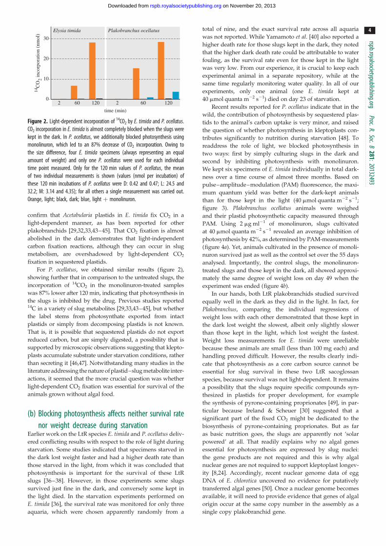
After 1 h, slugs in the light showed incorporation 23-fold
higher than slugs in the dark (6.73 nmol incorporated 14CO2
in the light and 0.30 nmol in the dark). In the light, 14CO2 incor-
poration after 2 h was 60 times greater than for slugs kept in the
dark (28.1 versus 0.46 nmol 14CO2; figure 2). Thus, we can
Elysia timida Plakobranchus ocellatus14
CO
2 inco
rpor
atio
n (n
mol
)30
20
10
02 60 120 2 60 120
time (min)
Figure 2. Light-dependent incorporation of 14CO2 by E. timida and P. ocellatus.CO2 incorporation in E. timida is almost completely blocked when the slugs werekept in the dark. In P. ocellatus, we additionally blocked photosynthesis usingmonolinuron, which led to an 87% decrease of CO2 incorporation. Owing tothe size difference, four E. timida specimens (always representing an equalamount of weight) and only one P. ocellatus were used for each individualtime point measured. Only for the 120 min values of P. ocellatus, the meanof two individual measurements is shown (values (nmol per incubation) ofthese 120 min incubations of P. ocellatus were D: 0.42 and 0.47; L: 24.5 and32.2; M: 3.14 and 4.35); for all others a single measurement was carried out.Orange, light; black, dark; blue, light þ monolinuron.
rspb.royalsocietypublishing.orgProc.R.Soc.B
281:20132493
4
on November 20, 2013rspb.royalsocietypublishing.orgDownloaded from
confirm that Acetabularia plastids in E. timida fix CO2 in a
light-dependent manner, as has been reported for other
plakobranchids [29,32,33,43–45]. That CO2 fixation is almost
abolished in the dark demonstrates that light-independent
carbon fixation reactions, although they can occur in slug
metabolism, are overshadowed by light-dependent CO2
fixation in sequestered plastids.
For P. ocellatus, we obtained similar results (figure 2),
showing further that in comparison to the untreated slugs, the
incorporation of 14CO2 in the monolinuron-treated samples
was 87% lower after 120 min, indicating that photosynthesis in
the slugs is inhibited by the drug. Previous studies reported14C in a variety of slug metabolites [29,33,43–45], but whether
the label stems from photosynthate exported from intact
plastids or simply from decomposing plastids is not known.
That is, it is possible that sequestered plastids do not export
reduced carbon, but are simply digested, a possibility that is
supported by microscopic observations suggesting that klepto-
plasts accumulate substrate under starvation conditions, rather
than secreting it [46,47]. Notwithstanding many studies in the
literature addressing the nature of plastid–slug metabolite inter-
actions, it seemed that the more crucial question was whether
light-dependent CO2 fixation was essential for survival of the
animals grown without algal food.
(b) Blocking photosynthesis affects neither survival ratenor weight decrease during starvation
Earlier work on the LtR species E. timida and P. ocellatus deliv-
ered conflicting results with respect to the role of light during
starvation. Some studies indicated that specimens starved in
the dark lost weight faster and had a higher death rate than
those starved in the light, from which it was concluded that
photosynthesis is important for the survival of these LtR
slugs [36–38]. However, in those experiments some slugs
survived just fine in the dark, and conversely some kept in
the light died. In the starvation experiments performed on
E. timida [36], the survival rate was monitored for only three
aquaria, which were chosen apparently randomly from a
total of nine, and the exact survival rate across all aquaria
was not reported. While Yamamoto et al. [40] also reported a
higher death rate for those slugs kept in the dark, they noted
that the higher dark death rate could be attributable to water
fouling, as the survival rate even for those kept in the light
was very low. From our experience, it is crucial to keep each
experimental animal in a separate repository, while at the
same time regularly monitoring water quality. In all of our
experiments, only one animal (one E. timida kept at
40 mmol quanta m22 s21) died on day 23 of starvation.
Recent results reported for P. ocellatus indicate that in the
wild, the contribution of photosynthesis by sequestered plas-
tids to the animal’s carbon uptake is very minor, and raised
the question of whether photosynthesis in kleptoplasts con-
tributes significantly to nutrition during starvation [48]. To
readdress the role of light, we blocked photosynthesis in
two ways: first by simply culturing slugs in the dark and
second by inhibiting photosynthesis with monolinuron.
We kept six specimens of E. timida individually in total dark-
ness over a time course of almost three months. Based on
pulse–amplitude–modulation (PAM) fluorescence, the maxi-
mum quantum yield was better for the dark-kept animals
than for those kept in the light (40 mmol quanta m22 s21;
figure 3). Plakobranchus ocellatus animals were weighed
and their plastid photosynthetic capacity measured through
PAM. Using 2 mg ml21 of monolinuron, slugs cultivated
at 40 mmol quanta m22 s21 revealed an average inhibition of
photosynthesis by 42%, as determined by PAM-measurements
(figure 4a). Yet, animals cultivated in the presence of monoli-
nuron survived just as well as the control set over the 55 days
analysed. Importantly, the control slugs, the monolinuron-
treated slugs and those kept in the dark, all showed approxi-
mately the same degree of weight loss on day 49 when the
experiment was ended (figure 4b).
In our hands, both LtR plakobranchids studied survived
equally well in the dark as they did in the light. In fact, for
Plakobranchus, comparing the individual regressions of
weight loss with each other demonstrated that those kept in
the dark lost weight the slowest, albeit only slightly slower
than those kept in the light, which lost weight the fastest.
Weight loss measurements for E. timida were unreliable
because these animals are small (less than 100 mg each) and
handling proved difficult. However, the results clearly indi-
cate that photosynthesis as a core carbon source cannot be
essential for slug survival in these two LtR sacoglossan
species, because survival was not light-dependent. It remains
a possibility that the slugs require specific compounds syn-
thesized in plastids for proper development, for example
the synthesis of pyrone-containing proprionates [49], in par-
ticular because Ireland & Scheuer [30] suggested that a
significant part of the fixed CO2 might be dedicated to the
biosynthesis of pyrone-containing proprionates. But as far
as basic nutrition goes, the slugs are apparently not ‘solar
powered’ at all. That readily explains why no algal genes
essential for photosynthesis are expressed by slug nuclei:
the gene products are not required and this is why algal
nuclear genes are not required to support kleptoplast longev-
ity [8,24]. Accordingly, recent nuclear genome data of egg
DNA of E. chlorotica uncovered no evidence for putatively
transferred algal genes [50]. Once a nuclear genome becomes
available, it will need to provide evidence that genes of algal
origin occur at the same copy number in the assembly as a
single copy plakobranchid gene.

0 10 20 30 40 50 60 70 80 90
100
200
300
400
500
600
700
800
900
continuous darkness12 L : 12 D at 40 µmol m–2 s–1
days of starvation
max
imum
qua
ntum
yie
ld F
v/F
m
12 L : 12 D at 25 µmol m–2 s–1
Figure 3. PAM measurements of E. timida. The maximum quantum yields of slugs kept in the dark (black) were compared to slugs kept under low- (orange) and high-light (red) conditions. Those kept under high light show the strongest decrease over the three months measured, whereas the linear regression of those kept in the darkruns in parallel to that of those kept under low-light conditions. Six specimens were used for each condition tested. The error bars present the standard deviation.
lightand fed
dark andstarved
0 5 10 15 20 25 30 35 40 45 500.2
0.3
0.4
0.5
0.6
0.7
0.8
0.9
1.0
1.1
1.2
wei
ght (
g)
days of starvation
slug 2slug 1
slug 5slug 6
slug 4slug 312 L : 12 D + monolinuron
12 L : 12 D
continuous darkness
0 10 20 30 40 50 600.1
0.2
0.3
0.4
0.5
0.6
0.7
0.8
0.9
days of starvation
(a)
(b)
12 L : 12D + monolinuron12 L : 12 D continuous darkness
max
imum
qua
ntum
yie
ld F
v/Fm
(i)
(ii)
(c)
Figure 4. Influence of photosynthesis inhibition on P. ocellatus. (a) PAM measurements of monolinuron-treated slugs in a 12 L : 12 D cycle (25 mmol quanta m22 s21;blue) in comparison to those kept in the dark (black) and under at a 12 L : 12 D cycle (25 mmol quanta m22 s21; red). Two specimens were used for each conditiontested and the error bars present the standard deviation. (b) Weight measurements of the P. ocellatus specimens shown in (a). (c) Exemplary images of P. ocellatusspecimens. Image (i) shows a slug kept in the light and which was regularly fed, hence best representing natural conditions. Image (ii) shows a slug after 55 daysof starvation in the dark.
rspb.royalsocietypublishing.orgProc.R.Soc.B
281:20132493
5
on November 20, 2013rspb.royalsocietypublishing.orgDownloaded from

rspb.royalsocietypublishing.orgProc.R.Soc.B
281:20132493
6
on November 20, 2013rspb.royalsocietypublishing.orgDownloaded from
The plastids of these LtR species remain capable of photo-
synthesis, as the PAM and 14CO2 incorporation results show
(figures 3, 4 and 2, respectively), but the observation that
light has no detectable effect on animal survival or weight
loss during starvation indicates that whatever the plastids do,
they do not have a life-extending effect on the animals whose
survival does not depend upon plastid photosynthetic activity
over the three-month period that we analysed. Photosynthesis
in sequestered plastids of LtR species might be important for
plastid longevity, but this has yet to be shown. Our exper-
iments measured the survival of the slugs, not the survival
of the plastids directly, although the data in figure 4a show
that plastids maintained in the dark are, by the measure of
PAM fluorescence, just as viable as those maintained under
12 L : 12 D. Thus, the plastids of the LtR species P. ocellatusand E. timida appear to be a source of stored food, and although
similar experiments have yet to be reported for the other two
LtR species—E. chlorotica and E. crispata—the implication
from our findings is that light is probably not required for
long-term survival during starvation of those species either.
If plastid fitness had a direct impact on slug fitness, then
blocking photosynthesis, whether through light deprivation
or monolinuron, should influence the weight and survival
rate of the animals. Yet, those Plakobranchus specimens kept
in the dark or treated with monolinuron lost weight at the
same rate as the starving control specimens (figure 4b) and
the E. timida slugs kept in the dark appeared as healthy as
the control set, too. Furthermore, the linear regression of the
maximum quantum yield of slugs kept in the dark (figure 3)
and those treated with monolinuron (figure 4a) runs parallel
to those of the control animals, rather than declining more
rapidly over time as a proxy of reduced and essential plastid
contribution to nutrition. Klochkova et al. [39] recently
observed that specimens of the StR species Elysia nigrocapitatasurvived for five months without performing photosynthesis,
during which time the starved animals dramatically lost
weight. Notably, E. nigrocapitata animals go from 3 cm in
length to 3 mm during starvation, but reversibly, if provided
with food [39].
3. ConclusionIt has been established that LGT is not involved in kleptoplast
maintenance following starvation either in slugs [8,24] or in
Foraminifera [51]. The present findings go a step further by
showing that sacoglossan slugs survive for months with the
help of kleptoplasts, but without the help of photosynthesis.
While the plastids are photosynthetically active, they do not
confer a photoautotrophic lifestyle upon the slugs. It rather
appears that the slugs sequester their plastids not directly as a
source of photosynthetic capabilities, but as a source of stored
food reserves, whose nutritional value does not depend on
light subsequent to sequestration. Plastid longevity in LtR saco-
glossan slugs remains an interesting phenomenon, but the
present results prompt a shift in emphasis from viewing the
kleptoplasts as green solar panels towards viewing them as
green food reserves.
4. Experimental proceduresElysia timida individuals were collected in Banyuls-sur-Mer
(France) between July and September 2012 and transferred
to Bonn (Germany). Specimens were kept with food algae
in Petri dishes with artificial seawater (Tropic Marin) at
208C and water changed every 2 days. For acclimation to
laboratory conditions, the slugs were illuminated at
25 mmol quanta m22 s21 and a 12 L : 12 D cycle under a ‘day-
light lamp’ (Androv Medical, model AND1206-CH) for 6
days. Then six individuals of E. timida were separately
starved in Petri dishes under 25 and 40 mmol quanta m22 s21
under a 12 L : 12 D cycle and under complete darkness for a
maximum of 88 days. Analyses of photosynthetic activity
were performed with a pulse–amplitude–modulated fluo-
rometer (Diving PAM, Walz, Germany) by measuring the
maximum quantum yield of chlorophyll a fluorescence in
photosystem II. Specimens kept under light conditions were
dark acclimated for 15 min prior to the measurement.
Plakobranchus ocellatus individuals were collected in the
Philippines in November 2012 and transferred to Bonn
(Germany). Two aquaria were set up with 20 l artificial
seawater (Tropic Marin) at 228C with two specimens of
P. ocellatus, respectively, each in individual fishnets. Addition-
ally, two specimens were placed in an aquarium in individual
fishnets in 10 l artificial seawater at 228C. One-third of the
water in every aquarium was changed weekly and the best
water quality established through the use of an internal filter
(Eheim, Germany). To one 20 l aquarium, 2 mg 3-(4-Chlorophe-
nyl)-1-Methoxy-1-Methylurea (monolinuron) ml21 seawater
was added. This and the 10 l aquarium were illuminated at
25 mmol quanta m22 s21 under a 12 L : 12 D. The second 20 l
aquarium was kept in the dark. All specimens were starved
for 55 days and afterwards fixed in 4% formaldehyde for further
analysis not addressed here. PAM-measurements were taken
using a Diving PAM (Walz, Germany). Weights of all six speci-
mens were measured on days 0, 14, 28 and 49 of the experiment
by placing the slugs on a spoon, gently removing all remaining
water with a paper towel and placing them into a preset water
container on a scale. Measurements were taken three times and
mean values determined. Data of each trial were pooled. For
each individual, the weights were scaled to a maximum of 1
and the linear regression calculated. With 20.0059, ‘dark’ had
the lowest slope followed by the monolinuron-treated slugs
with 20.0067 and the control set with 20.0114. Using a Tukey
test, we tested pairwise whether the slopes of the linear
regression lines were equal (H0). The resulting q-values with a
significance level of 0.05 showed a significant difference between
‘dark’ and ‘normal’ conditions, while the remaining slopes did
not significantly differ ( p-values: control to monolinuron-
treated � 0.0567; monolinuron-treated to darkness � 0.9193;
and darkness to control � 0.023).
Elysia timida used for incubations with [14C]-labelled CO2
were collected in the Mediterranean Sea (Banyuls-sur-Mer,
France) in October 2012 and transferred to Dusseldorf
(Germany). They were maintained for six weeks at 158C and
33 mmol quanta m22 s21 in 12 l-aquaria containing 25 speci-
mens, artificial seawater (37 g l21 hw-Marinemix professional
(Wiegandt, Germany)) and Acetabularia acetabulum as food
source. They were starved for several days before labelling. To
test the light-driven incorporation of CO2 by Elysia, the slugs
were incubated in 1.2 ml artificial seawater supplemented
with 0.32 mM [14C]-NaHCO2 (18 mCi per incubation, NEN-
radiochemicals, MA, USA). For each measurement, four
slugs were incubated together in a transparent plastic 1.5 ml
tube. Plakobranchus ocellatus individuals used for incubations
with [14C]-labelled CO2 were collected in the Philippines in

rspb.royalsocietypublishing.orgProc.R.Soc.
7
on November 20, 2013rspb.royalsocietypublishing.orgDownloaded from
April 2013 and transferred to Bonn (Germany). To test the light-
driven incorporation of CO2, they were incubated in 5 ml
artificial seawater supplemented with 0.16 mM [14C]NaHCO2
(36 mCi per incubation). Each measurement contained one
single organism in an 8 ml glass tube. The measurements for
time point 120 were carried out twice and the mean values are
shown in figure 2. All incubations of both species were per-
formed at room temperature either in the dark or illuminated
(72 mmol quanta m22 s21). The incubations lasted 2 min, 1 or
2 h, and afterwards the slugs were separated from the radioactive
incubation medium, rinsed five times with seawater, and then
homogenized in a small glass teflon Potter-Elvehjem tissue grin-
der in 1 ml (Elysia) or 3 ml H2O (Plakobranchus). The homogenates
of Elysia were removed and the Potter tube was rinsed twice with
1 ml H2O. These 3 ml, containing the homogenized slugs, were
acidified with 150 ml 1M HCl and the open vial was then
shaken overnight to remove all the substrate, that is (labelled)
carbon dioxide. Afterwards, incorporation of labelled carbon
atoms by the Elysia slugs was determined in a scintillation coun-
ter after the addition of 12 ml LUMA-Gel scintillation cocktail
(LUMAC, The Netherlands). To the homogenates of Plakobran-chus 3 ml H2O and 300 ml 1M HCl were added, while the rest
of the method to measure acid-stable incorporation of carbon
dioxide was the same as for Elysia.
Acknowledgement. We thank Margarete Stracke and Valerie Schmitt forcollecting and cultivating animals and algae.
Funding statement. This study was financially supported by an ERCgrant to W.F.M. and DFG grants to H.W. and S.B.G.
B281:201
References32493
1. Venn AA, Loram JE, Douglas AE. 2008Photosynthetic symbioses in animals. J. Exp. Bot.59, 1069 – 1080. (doi:10.1093/jxb/erm328)2. Bosch TCG. 2012 What Hydra has to say about therole and origin of symbiotic interactions. Biol. Bull.223, 78 – 84.
3. Kovacevic G, Franjevic D, Jelencic B, Kalafatic M.2010 Isolation and cultivation of endosymbioticalgae from green Hydra and phylogenetic analysisof 18S rDNA sequences. Folia Biol. 58, 135 – 143.(doi:10.3409/fb58_1-2.135-143)
4. Archibald JM. 2009 The puzzle of plastid evolution.Curr. Biol. 19, R81 – R88. (doi:10.1016/j.cub.2008.11.067)
5. Johnson MD. 2011 Acquired phototrophy in ciliates:a review of cellular interactions and structuraladaptations. J. Eukaryot. Microbiol. 58, 185 – 195.(doi:10.1111/j.1550-7408.2011.00545.x)
6. Trench RK. 1975 Of ‘leaves that crawl’, functionalchloroplasts in animal cells. Symp. Soc. Exp. Biol. 29,229 – 265.
7. Handeler K, Grzymbowski Y, Krug JP, Wagele H.2009 Functional chloroplasts in metazoan cells:a unique evolutionary strategy in animal life. Front.Zool. 6, 28. (doi:10.1186/1742-9994-6-28)
8. Wagele H et al. 2011 Transcriptomic evidence thatlongevity of acquired plastids in the photosyntheticslugs Elysia timida and Plakobranchus ocellatus doesnot entail lateral transfer of algal nuclear genes.Mol. Biol. Evol. 28, 699 – 706. (doi:10.1093/molbev/msq239)
9. Rumpho ME, Summer EJ, Manhart JR. 2000Solar-powered sea slugs. Mollusc/algal chloroplastsymbiosis. Plant Physiol. 123, 29 – 38. (doi:10.1104/pp.123.1.29)
10. Pierce SK, Curtis NE, Hanten JJ, Boerner SL,Schwartz JA. 2007 Transfer, integration andexpression of functional nuclear genes betweenmulticellular species. Symbiosis 43, 57 – 64.
11. van Wijk KJ, Baginsky S. 2011 Plastid proteomicsin higher plants: current state and future goals.Plant Physiol. 155, 1578 – 1588. (doi:10.1104/pp.111.172932)
12. Timmis JN, Ayliffe MA, Huang CY, Martin W. 2004Endosymbiotic gene transfer: organelle genomesforge eukaryotic chromosomes. Nat. Rev. Genet. 5,123 – 135. (doi:10.1038/nrg1271)
13. Gould SB, Waller RF, McFadden GI. 2008Plastid evolution. Annu. Rev. Plant Biol. 59,491 – 517. (doi:10.1146/annurev.arplant.59.032607.092915)
14. Strittmatter P, Soll J, Bolter B. 2010 The chloroplastprotein import machinery: a review. Methods Mol.Biol. 619, 307 – 321. (doi:10.1007/978-1-60327-412-8_18)
15. Shi LX, Theg SM. 2013 The chloroplast proteinimport system: from algae to trees. Biochim.Biophys. Acta 1833, 314 – 331. (doi:10.1016/j.bbamcr.2012.10.002)
16. Mujer CV, Andrews DL, Manhart JR, Pierce SK,Rumpho ME. 1996 Chloroplast genes are expressedduring intracellular symbiotic association ofVaucheria litorea plastids with the sea slug Elysiachlorotica. Proc. Natl Acad. Sci. USA 93, 12 333 –12 338. (doi:10.1073/pnas.93.22.12333)
17. Greenberg BM, Gaba V, Mattoo AK, Edelman M.1987 Identification of a primary in vivo degradationproduct of the rapidly-turning-over 32 kd protein ofphotosystem II. EMBO J. 6, 2865 – 2869.
18. Godde D, Schmitz H, Weidner M. 1991 Turnover ofthe D-1 reaction center polypeptide fromphotosystem-II in intact spruce needles and spinachleaves. Z. Naturforsch. 46, 245 – 251.
19. Sundby C, McCaffery S, Anderson JM. 1993 Turnoverof the photosystem-II D1 protein in higher-plantsunder photoinhibitory and nonphotoinhibitoryirradiance. J. Biol. Chem. 268, 25 476 – 25 482.
20. Rumpho ME, Worful J, Lee J, Kannan K, Tyler MS,Bhattacharya D, Moustafa A, Manhart JR. 2008Horizontal gene transfer of the algal nuclear genepsbO to the photosynthetic sea slug Elysiachlorotica. Proc. Natl Acad. Sci. USA 105, 17 867 –17 871. (doi:10.1073/pnas.0804968105)
21. Gruber A, Vugrinec S, Hempel F, Gould SB, MaierUG, Kroth PG. 2007 Protein targeting into complexdiatom plastids: functional characterisation of a
specific targeting motif. Plant Mol. Biol. 64,519 – 530. (doi:10.1007/s11103-007-9171-x)
22. Gould SB. 2008 Ariadne’s thread: guiding aprotein across five membranes in cryptophytes.J. Phycol. 44, 23 – 26. (doi:10.1111/j.1529-8817.2007.00437.x)
23. Rumpho ME, Summer EJ, Green BJ, Fox TC, ManhartJR. 2001 Mollusc/algal chloroplast symbiosis: howcan isolated chloroplasts continue to function formonths in the cytosol of a sea slug in the absenceof an algal nucleus? Zoology 104, 303 – 312.(doi:10.1078/0944-2006-00036)
24. Rumpho ME, Pelletreau KN, Moustafa A,Bhattacharya D. 2011 The making of aphotosynthetic animal. J. Exp. Biol. 214, 303 – 311.(doi:10.1242/jeb.046540)
25. Pierce SK, Fang X, Schwartz JA, Jiang X, Zhao W,Curtis NE, Kocot KM, Yang B, Wang J. 2012Transcriptomic evidence for the expression ofhorizontally transferred algal nuclear genes inthe photosynthetic sea slug Elysia chlorotica.Mol. Biol. Evol. 29, 1545 – 1556. (doi:10.1093/molbev/msr316)
26. Richly E, Dietzmann A, Biehl A, Kurth J, Laloi C,Apel K, Salamini F, Leister D. 2003 Covariations inthe nuclear chloroplast transcriptome reveal aregulatory master-switch. EMBO Rep. 4, 491 – 498.(doi:10.1038/sj.embor.embor828)
27. Bhalero R et al. 2003 Gene expression in autumnleaves. Plant Physiol. 131, 430 – 442. (doi:10.1104/pp.012732)
28. Trench RK, Greene RW, Bystrom BG. 1969Chloroplasts as functional organelles in animaltissue. J. Cell Biol. 42, 404 – 417. (doi:10.1083/jcb.42.2.404)
29. Trench RK. 1973 Further studies on themucopolysaccharide secreted by the pedal gland ofthe marine slug Tridachia crispata (Opisthobranchia,Sacoglossa). Bull. Mar. Sci. 23, 299 – 312.
30. Ireland C, Scheuer PJ. 1979 Photosynthetic marinemollusks: in vivo 14C incorporation into metabolitesof the sacoglossan Placobranchus ocellatus. Science205, 922 – 923. (doi:10.1126/science.205.4409.922)

rspb.royalsocietypublishing.orgProc.R.Soc.B
281:20132493
8
on November 20, 2013rspb.royalsocietypublishing.orgDownloaded from
31. Hinde R. 1978 The metabolism ofphotosynthetically fixed carbon by isolatedchloroplasts from Codium fragile (Chlorophyta:Siphonales) and by Elysia viridis (Mollusca:Sacoglossa). Biol. J. Linn. Soc. 10, 329 – 342.(doi:10.1111/j.1095-8312.1978.tb00019.x)
32. Kremer BP, Schmitz K. 1976 Aspects of 14CO2-fixation by endosymbiotic rhodoplasts in the marineOpisthobranchiate Hermaea bifida. Mar. Biol. 34,313 – 316. (doi:10.1007/BF00398124)
33. Green RW, Muscatine L. 1972 Symbiosis insaccoglossan opisthobranchs: phostosytheticproducts of animal – chloroplast associations. Mar.Biol. 14, 253 – 259.
34. Middlebrooks ML, Pierce SK, Bell SS. 2011 Foragingbehavior under starvation conditions is altered viaphotosynthesis by the marine gastropod, Elysiaclarki. PLoS ONE 6, e22162. (doi:10.1371/journal.pone.0022162)
35. Christa G, Westcott L, Schaberle TF, Koenig G, WageleH. 2013 What remains after 2 months of starvation?Analysis of sequestered algae in a photosyntheticslug, Plakobranchus ocellatus (Sacoglossa,Opisthobranchia), by barcoding. Planta 237,559 – 572. (doi:10.1007/s00425-012-1788-6)
36. Gimenez Casalduero F, Muniain C. 2008 The role ofkleptoplasts in the survival rates of Elysia timida(Risso 1818): (Sacoglossa: Opisthobranchia) duringperiods of food shortage. J. Exp. Mar. Biol. Ecol.357, 181 – 187. (doi:10.1016/j.jembe.2008.01.020)
37. Hinde R, Smith DC. 1972 Persistence of functionalchloroplasts in Elysia viridis (Opisthobranchia,Sacoglossa). Nature 239, 30 – 31. (doi:10.1038/239041a0)
38. Hinde R, Smith DC. 1975 The role of photosynthesisin the nutrition of the mollusc Elysia viridis.Biol. J. Linn. Soc. 7, 161 – 171. (doi:10.1111/j.1095-8312.1975.tb00738.x)
39. Klochkova TA, Han JW, Chah KH, Kim RW, Kim JH,Kim KY, Kim GH. 2013 Morphology, molecularphylogeny and photosynthetic activity of thesacoglossan mollusc, Elysia nigrocapitata, fromKorea. Mar. Biol. 160, 155 – 168. (doi:10.1007/s00227-012-2074-7)
40. Yamamoto S, Hirano YM, Hirano YJ, Trowbridge CD,Akimoto A, Sakai A, Yusa Y. 2013 Effects ofphotosynthesis on the survival and weight retentionof two kleptoplastic sacoglossan opisthobranchs.J. Mar. Biol. Ass. UK 1, 1 – 7. (doi:10.1017/S0025315412000628)
41. Curtis NE, Massey SE, Pierce SK. 2006 The symbioticchloroplasts in the sacoglossan Elysia clarki are fromseveral algal species. Invert. Biol. 125, 336 – 345.(doi:10.1111/j.1744-7410.2006.00065.x)
42. Weber APM, Osteryoung KW. 2010 Fromendosymbiosis to synthetic photosynthetic life. PlantPhysiol. 154, 593 – 597. (doi:10.1104/pp.110.161216)
43. Clark KB, Jensen KR, Stirts HM, Fermin C. 1981Chloroplast symbiosis in a non-Elysiid mollusc,Costasiella lilianae Marcus (Hermaeidae: Ascoglossa(¼ Sacoglossa): effects of temperature, lightintensity, and starvation on carbon fixation rate.Biol. Bull. 160, 42 – 54. (doi:10.2307/1540899)
44. Greene RW. 1970 Symbiosis in sacoglossanopisthobranchs: functional capacity of symbioticchloroplasts. Mar. Biol. 7, 138 – 142. (doi:10.1007/BF00354917)
45. Marın A, Ros J. 1989 The chloroplast – animalassociation in four Iberian sacoglossanopisthobranchs: Elysia timida, Elysia translucens,Thuridilla hopei, and Bosellia mimetica. Sci. Mar. 53,429 – 440.
46. Hawes CR, Cobb AH. 1980 The effects of starvationon the symbiotic chloroplasts in Elysia viridis: a finestructural study. New Phytol. 84, 375 – 379. (doi:10.1111/j.1469-8137.1980.tb04437.x)
47. Evertsen J, Johnsen G. 2009 In vivo and in vitrodifferences in chloroplast functionality in the twonorth Atlantic sacoglossans (Gastropoda,Opisthobranchia) Placida dendritica and Elysiaviridis. Mar. Biol. 156, 847 – 859. (doi:10.1007/s00227-009-1128-y)
48. Maeda T et al. 2012 Algivore or phototroph?Plakobranchus ocellatus (Gastropoda) continuouslyacquires kleptoplasts and nutrition from multiplealgal species in nature. PLoS ONE 7, e42024.(doi:10.1371/journal.pone.0042024)
49. Cutignano A, Cimino G, Villani G, Fontana A. 2009Shaping the polypropionate biosynthesis in thesolar-powered mollusc Elysia viridis. Chembiochem10, 315 – 322. (doi:10.1002/cbic.200800531)
50. Bhattacharya D, Pelletreau KN, Price DC, Sarver KE,Rumpho ME. 2013 Genome analysis of Elysiachlorotica egg DNA provides no evidence forhorizontal gene transfer into the germ line ofthis kleptoplastic mollusc. Mol. Biol. Evol. 30,1843 – 1853. (doi:10.1093/molbev/mst084)
51. Pillet L, Pawlowski J. 2013 Transcriptome analysis offoraminiferan Elphidium margaritaceum questionsthe role of gene transfer in kleptoplastidy. Mol. Biol.Evol. 30, 66 – 69. (doi:10.1093/molbev/mss226)
Copyright © 2022 FDOKUMEN