Phytochemical analysis and effect of whole Eclipta alba ethanolic extract on the central nociception...
12
JOURNAL OF PHARMACEUTICAL AND BIOMEDICAL SCIENCES Rahman Atiar et al.. Phytochemical analysis and effect of whole Eclipta alba ethanolic extract on the central nociception and inflammation in rodent models. Journal of pharmaceutical and biomedical sciences (J Pharm Biomed Sci.) 2012, Decemeber; 25(25); 130- 140. (Article no 2) The online version of this article, along with updated information and services, is located on the World Wide Web at: www.jpbms.info Journal of Pharmaceutical and Biomedical Sciences (J Pharm Biomed Sci.), Member journal. Committee of Publication ethics (COPE) and Journal donation project (JDP).
-
Upload
chittagong -
Category
Documents
-
view
1 -
download
0
Transcript of Phytochemical analysis and effect of whole Eclipta alba ethanolic extract on the central nociception...
JOURNAL OF PHARMACEUTICAL AND BIOMEDICAL SCIENCES Rahman Atiar et al.. Phytochemical analysis and effect of whole Eclipta alba ethanolic extract on the central nociception and inflammation in rodent models. Journal of pharmaceutical and biomedical sciences (J Pharm Biomed Sci.) 2012, Decemeber; 25(25); 130-140. (Article no 2)
The online version of this article, along with updated information and services, is located on the World Wide Web at: www.jpbms.info Journal of Pharmaceutical and Biomedical Sciences (J Pharm Biomed Sci.), Member journal. Committee of Publication ethics (COPE) and Journal donation project (JDP).
Rahman Atiar et al. J Pharm Biomed Sci. 2012, December; 25 (25); 130-140. Available at jpbms.info
130
ISSN NO- 2230 – 7885
CODEN JPBSCT NLM Title: J Pharm Biomed Sci.
Research article
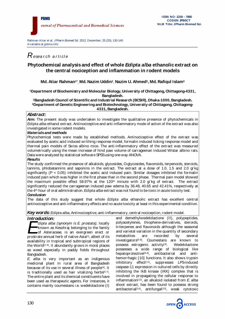
Phytochemical analysis and effect of whole Eclipta alba ethanolic extract on the central nociception and inflammation in rodent models
Md. Atiar Rahman1*, Md. Nazim Uddin1, Nazim U. Ahmed2, Md. Rafiqul Islam3
1Department of Biochemistry and Molecular Biology, University of Chittagong, Chittagong-4331,
Bangladesh. 2Bangladesh Council of Scientific and Industrial Research (BCSIR), Dhaka-1000, Bangladesh.
3Department of Genetic Engineering and Biotechnology, University of Chittagong, Chittagong-4331, Bangladesh.
Abstract: Aim: The present study was undertaken to investigate the qualitative presence of phytochemicals in Eclipta alba ethanol extract. Antinociceptive and anti-inflammatory mode of action of the extract was also investigated in some rodent models. Materials and methods Phytochemical tests were made by established methods. Antinociceptive effect of the extract was evaluated by acetic acid induced writhing response model, formalin induced licking response model and thermal pain models of Swiss albino mice. The anti-inflammatory effect of the extract was measured volumetrically using the mean increase of hind paw volume of carrageenan induced Wistar albino rats. Data were analyzed by statistical software SPSS using one way ANOVA. Results The study confirmed the presence of alkaloids, glycosides, O-glycosides, flavonoids, terpenoids, steroids, tannins, phlobatannins and saponins in the extract. The extract at a dose of 1.0, 1.5 and 2.0 g/kg significantly (P < 0.05) inhibited the acetic acid induced pain. Similar dosages inhibited the formalin induced pain which was higher in the first phase than in the second phase. Thermal pain model showed the maximum possible effect 58.97% at the 120th minute with 2.0 g/kg of extract. The extract significantly reduced the carrageenan induced paw edema by 36.48, 40.65 and 42.41%, respectively at the 4th hour of oral administration. Eclipta alba extract was not found to be toxic in acute toxicity test. Conclusion The data of this study suggest that whole Eclipta alba ethanolic extract has excellent central antinociceptive and anti-inflammatory effects and no acute toxicity at least in this experimental condition.
Key words: Eclipta alba, Antinociceptive, anti-inflammatory, central nociception, rodent model. Introduction:
clipta alba (synonym is E. prostata), locally known as Kesohraj belonging to the family of Asteraceae, is an evergreen erect or
prostrate annual herb of native Asia[1], albeit of its availability in tropical and subtropical regions of the World[2-3]. It abundantly grows in moist places as weed especially in paddy fields throughout Bangladesh. E. alba is very important as an indigenous medicinal plant in rural area of Bangladesh because of its use in several illness of people[4]. It is traditionally used as hair vitalizing herbs[5-7]. The entire plant and its chemical constituents have been used as therapeutic agents. For instances, it contains mainly coumestans i.e. wedelolactone (I)
and demethylwedelolactone (II), polypeptides, polyacetylenes, thiophene-derivatives, steroids, triterpenes and flavonoids although the seasonal and varietal variation in the quantity of secondary metabolites are recorded by several investigators[5-6]. Coumestans are known to possess estrogenic activity[3]. Wedelolactone possesses a wide range of biological like hepatoprotective[8-9], antibacterial and anti-hemorrhagic [10] functions. It also shows trypsin inhibitory effect[11], suppresses LPS-induced caspase-11 expression in cultured cells by directly inhibiting the IkB kinase (IKK) complex that is involved in propagating the cellular response to inflammation[12], an alkaloid isolated from E. alba shoot extract, has been found to possess strong antibacterial[14], antifungal[15], weak cytotoxic
E
Rahman Atiar et al. J Pharm Biomed Sci. 2012, December; 25 (25); 130-140. Available at jpbms.info
131
ISSN NO- 2230 – 7885
CODEN JPBSCT NLM Title: J Pharm Biomed Sci.
activity against the M-109 cell lines and potent anti-parasitic activity against Giardia intestinalis[16-
17]. The plant is also used in the treatment of memory disorders, edema, fevers, rheumatic joint pains, indigestion, enlarged spleen, oxidative stress and skin disorders[18-20]. Sawant et al.[21] reported that the total alkaloids and alcohol extracts of E. alba has potent analgesic effect in tail flick and acetic acid induced model of albino mice. Arunachalam et al.[22] found the anti-inflammatory effect of the methanol extract of E. alba leaves in albino Wistar rats. Leal et al.[23] described the antinociceptive and anti-inflammatory effect of hydroalcoholic (20% alcohol) extract of E. alba leaves. According to the above-mentioned reports, although a number of studies have been investigated to examine the hepatoprotective, anti-bacterial, anti-hemorrhagic, anti-inflammatory, anti-parasitic and especially analgesic effects of the various extracts of the various sections of this plant, the anti-nociceptive and anti-inflammatory effects of the whole plant and their mechanisms behind have not yet been investigated. The present study investigated the anti-nociceptive and anti-inflammatory effects of the ethanolic extract of whole plant and their mechanisms of actions behind on a central and/or peripheral route in an experimentally induced animal model of rats.
Materials and methods Plant material E. alba whole plants were collected from Chittagong area, Bangladesh in the month of March-April, 2009. The plant was taxonomically identified by Dr. Shaikh Bokhtear Uddin (Taxonomist and Associate Professor, Department of Botany, University of Chittagong, Bangladesh) and identification was confirmed by Sarder Nasir Uddin (Taxonomist, Bangladesh National Herbarium, Ministry of Environment and Forest, Bangladesh). A voucher specimen has been preserved in Bangladesh National Herbarium bearing the accession no. DACD-32908.
Preparation of whole plant extract Freshly harvested E. alba was washed with distilled water immediately after collection. The collected plants were chopped into small pieces, air dried at room temperature (25±1°C) for about 10 days and ground into powder by blender with grinder (Moulinex Blender AK-241, Moulinex,
France) and stored in an airtight container until used for subsequent studies. The resulting powder (750g, mesh size 80-120 mm) was extracted with 6.5 L ethanol for 10 days at room temperature (25±1°C) with occasional stirring. The filtered (Whatman filter paper No. 1) supernatant was then concentrated under reduced pressure using a rotary vacuum evaporator (RE200 Sterling, UK). The concentrated extract (45g, blackish-green semisolid) was preserved at 4 °C for further use.
Chemicals and drugs Acetic acid, morphine hydrochloride and ethanol were purchased from Sigma-Aldrich, Germany. Morphine was dissolved in saline solution just before use. Diclofenac sodium (powder form) was kindly donated by GlaxoSmithKline Ltd., Bangladesh. A commercially available (Sigma-Aldrich, Germany) concentrated formalin solution was diluted with saline to the appropriate concentration (2.5%). Carrageenan (lambda form, FMC Marine Colloids Division, Sigma-Aldrich, Poole, UK) was used for paw oedema test.
Experimental animals and diets Six-week-old Swiss albino mice of both sexes weighing 25-30g and seven-week-old Wistar Albino rats of the either sex weighing 150-200g were obtained from the animal house of Bangladesh Council for Scientific and Industrial Research (BCSIR) Laboratories, Chittagong, Bangladesh. The animals were housed individually in stainless steel wire meshed plastic cages in a temperature (25±2) oC and humidity (55 - 60%) controlled room with a 12h light-dark cycle. The animals were supplied with standard pellet diet and drinking water ad libitum during the entire period of the study. Animals were maintained and experiment was carried out according to the rules and regulations of the Institutional Animal Ethics Committee (AEIIUC-Pharm/2011-02).
Phytochemical screening The plant extract was phytochemically screened using standard techniques for the qualitative detection of alkaloids, flavonoids, steroids, tannins, saponins, phlobatannins, cardiac glycosides, O-glycosides and terpenoids as described below.
Tests for alkaloids The alkaloids test has been done according to the method described by El-olemy et al. (24). To conduct this test, a 0.5g of extract was stirred with 5ml 1% HCl on a steam bath then the solution was
Rahman Atiar et al. J Pharm Biomed Sci. 2012, December; 25 (25); 130-140. Available at jpbms.info
132
ISSN NO- 2230 – 7885
CODEN JPBSCT NLM Title: J Pharm Biomed Sci.
cooled and filtered. One ml of the filtrate was treated separately with drops of Mayer’s, Dragendoff’s and Wagner’s reagents and formation of dirty/dark brown, yellow-brown or reddish brown precipitate respectively indicates the presence of alkaloids. Test for flavonoids The presence of flavonoids in the whole plant extract was confirmed by NaOH and lead acetate tests as described by Harborne[25].
I. NaOH test: Two ml of extract was acidified with few drops of 1% HCl and dissolved in 2ml of 20% NaOH. A canary yellow color indicates the presence of flavonoids.
II. Lead (II) acetate test: Two ml of extract was treated with a few drops of 10% lead aceated [Pb(OOCCH3)2] solution. A light yellow–milky precipitate indicates the presence of flavonoids.
Test for steroids The presence of flavonoids in the whole plant extract was confirmed by Liebermann’s and Salkowski’s tests as described by Harborne[25].
I. Liebermann’s test: Two ml of ethanoic acid anhydride was added to the 2ml of extract in a test tube. The content was then cooled on ice for 5 minutes. Then 1ml of concentrated H2SO4 was added along the walls of the test tube. The change in color from violet to blue and then to green indicates the presence of steroidal nucleus i.e. aglycone component of cardiac glycosides.
II. Salkowski’s test: Two ml of concentrated H2SO4 was added to the 2ml of extract in a test tube. Appearance of a clear reddish brown color at the interface confirms the presence of steroidal ring.
Test for tannins, saponins and phlobatannins The presence of tannins, saponins, and phlobatannins in the plant extract was confirmed by different tests as described by Sofowora[26].
I. Test for tannis: Two ml of extract was treated with 3drops of 5% FeCl3 in a test tube. A dark black colored precipitate, which giv es a g reen-black to blue-black coloration on dilution, indicates the presence o f tannins in the extract.
II. Test for saponins: Two ml of extract was vigorously shaken with 5ml of distilled water in test tube then allowed to stand for a while at room temperature. A persistent frothing indicates the presence of saponins.
III. Test for phlobatannins: Two ml of extract was added to 5ml of 2% HCl. Formation of turbidity/precipitate indicates the presence of phlobatannins.
Test for cardiac glycosides The presence of cardiac glycosides and O-glycosides in the plant extract was confirmed by Keller-Killani and Baljet tests as described by Sofowora[26].
I. Keller-Killani test: Five ml of plant extract was treated with 2 ml of glacial acetic acid containing one drop of ferric chloride solution in a test tube. Subsequently, this was underlayed with 1 ml of concentrated sulfuric acid. A brown ring of the interface indicates a deoxysugar characteristic of cardenolides. A violet ring may appear below the brown ring, while in the acetic acid layer and a greenish ring may form just gradually throughout thin layer.
II. Baljet test: A drop of Baljet's reagent (95 ml 1% picric acid + 5 ml 10% NaOH) was added to a 2ml of an alcoholic extract of plant material. A yellow orange color indicates the presence of five membered lactone ring at C-17 of the aglycone in cardiac glycoside
III. O-glycosides test: Two ml of extract was boiled with dil. HCl/H2SO4 (2-3 minutes) and filtered. Subsequently, after cooling and shaking with organic solvent (chloroform) organic layer was separated. Further shaking with NH4OH (10% ammonia solution) the solution was allowed to keep standing for 30 min. The rose pink or cherry red or violates aqueous layer indicates the presence of anthraquinones.
Test for terpenoids The presence of terpenoids in the plant extract was confirmed by the methods as described by Trease and Evans[27]. To perform this test, 0.5 g of extract and 2 ml of chloroform was mixed in a test tube then 3ml of concentrated H2S04 was added carefully to form a layer. A reddish brown color in the interface confirms the presence of terpenoids in the extract. Assay for antinociceptive activity Acetic acid induced writhing test The antinociceptive activity of E. alba extract was measured by the acetic acid induced writhing test in Swiss albino mice as described by Koster et al.[28]. Briefly, the inhibition of writhing produced by the extract was determined by comparing with the inhibition produced by the control group. Diclofenac sodium at a dose of 40 mg/kg was used as standard analgesic agent (positive control).
Rahman Atiar et al. J Pharm Biomed Sci. 2012, December; 25 (25); 130-140. Available at jpbms.info
133
ISSN NO- 2230 – 7885
CODEN JPBSCT NLM Title: J Pharm Biomed Sci.
Intraperitoneal injection of (1%) acetic acid at a dose of 2.3ml/kg of was used to create pain sensation. The number of writhing and stretching was counted over 20 min (20min after the application of acetic acid). The ethanol extract of E. alba (1.0, 1.5 and 2.0 g/kg) and distilled water (control) were administered orally 30 minutes before acetic acid injection as treatment and control. The percent of analgesic action was determined by the following formula:
Formalin test The procedure was similar to that described previously by Gaertner et al. [29]. The ethanolic extract of E. alba (1.0, 1.5 and 2.0 g/kg), reference analgesic drug morphine (0.5 mg/kg) and distilled water were administered orally 30 minutes before formalin injection. A 20 µL of 2.5% formalin (0.92% formaldehyde) made in phosphate buffer was injected under the right hind paw surface of experimental mice. Each mouse was placed individually in a cage and observed from 0 to 5 min followed by the injection of formalin to analyze the first phase and 15 to 40 min to analyze the second phase of formalin induced pain. The length of time the animal spent for licking the injected paw was counted with a chronometer and was considered as indicative of pain. Percent reduction of licking is measured by the following formula:
Where, L represents the licking time.
Hot plate test Thermal pain test in mice was done by the following hot plate method as described by Eddy and Leimback[30]. Briefly, mice were placed in a glass cylinder of 24 cm diameter on a metal plate (Scored DS37, UGO Basile, Italy) maintained at temperature (54±0.5° C), and the latency period until nociceptive responses such as licking of the hind paw, shaking of the limbs or jumping was measured. Prior to dosing, the nociceptive threshold was measured three times, and the mean of the reaction times was used as the pre-drug latency for each mouse. The cut-off time of 30
sec was used to prevent tissue damage. Mice were then administered (i.p.) with E. alba extract (1.0, 1.5 and 2.0 g/kg) as treatment and morphine (5 mg/kg, i.p.) was used as a positive control. The reaction time was measured at 30 min intervals till 120th minute of treatment and used as post-drug latency. Observation started after 30 min of E. alba extract and 15 min of morphine administration. The antinociception was quantified as maximum possible effect (%MPE) using the formula[31]:
Assay for anti-inflammatory activity Anti-inflammatory activity of E. alba extract was assessed using carrageenan induced paw edema in the hind paw of rat by the reported method of Winter et al.[32]. Breifly, an acute inflammation was induced in Wistar albino rats by subplantar injection of 0.1 ml of 1 % (w/v) carrageenan after measuring the initial right hind paw volume of each rat. The volume of right hind paw was measured at 1st, 2nd, 3rd and 4th hr after carrageenan injection and the paw edema was determined using plethysmometer (7150, UGO Basile, Italy). The E. alba extract (0.5, 1.0 and 1.5 g/kg), standard anti-inflammatory drug diclofenac sodium (40 mg/kg), and distilled water were administered orally to treated, positive control and control groups, respectively one hour before the injection of carrageenan. Percent of anti-inflammatory (% inhibition of paw edema) activity was determined using the following formula:
Where, Co is the paw thickness volume (in mm3) before carrageenan injection, Ct is the paw thickness volume (in mm3) at time t, (Ct -Co) is paw edema
Acute toxicity studies In order to perform the acute toxicity test, the crude ethanolic extract of E. alba was injected intraperitoneally to mice at various dose levels such as 0.5, 1.0, 2.0, 3.0 and 4.0 g/kg. Five mice in each dose group were closely observed for 24 hours for any mortality and next ten days for any delayed toxic effect.
Rahman Atiar et al. J Pharm Biomed Sci. 2012, December; 25 (25); 130-140. Available at jpbms.info
134
ISSN NO- 2230 – 7885
CODEN JPBSCT NLM Title: J Pharm Biomed Sci.
Statistical analysis All data are presented as mean ± standard deviation (SD) of 6 animals and were analyzed by one-way Analysis of Variance (ANOVA) (SPSS for windows, version 18.0, IBM Corporation, NY, USA). The values were considered significantly different at P < 0.05.
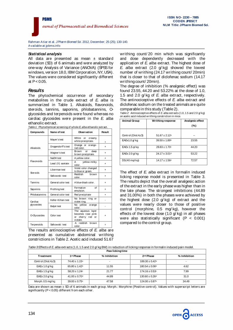
Results The phytochemical occurrence of secondary metabolites in the crude extract of E. alba is summarized in Table 1. Alkaloids, flavonoids, steroids, tannins, saponins, phlobatannins, O-glycosides and terpenoids were found whereas no cardiac glycosides were present in the E. alba ethanolic extract. Table 1: Phytochemical screening of whole E. alba ethanolic extract.
Compounds Name of test Observation Result
Alkaloids
Mayer’s test White or creamy white precipitate. +
Dragendorff’s test Orange or orange-red color. +
Wagner's test Brown or deep brown precipitate. +
Flavonoids NaOH test A yellow color. +
Lead (II) acetate A yellow-milky color. +
Steroids Liberman test Violet color changed
to blue or green. +
Salkowski test Reddish brown color. +
Tannins General color test A blue-black color. +
Saponins Frothing test Formation of emulsion. +
Phlobatannins General color test Red precipitate +
Cardiac glycosides
Keller-Killani test No brown ring or violet ring. -
Baljet test No yellow orange color. -
O-Glycosides Color test
The aqueous layer becomes rose pink or cherry red or violate.
+
Terpenoids Salkowski test A reddish brown color. +
The results antinociceptive effects of E. alba are presented as cumulative abdominal writhing constrictions in Table 2. Acetic acid induced 51.67
writhing count/20 min which was significantly and dose dependently decreased with the application of E. alba extract. The highest dose of E. alba extract (2.0 g/kg) showed the lowest number of writhing (24.17 writhing count/20min) that is closer to that of diclofenac sodium (14.17 writhing count/20min). The degree of inhibition (% analgesic effect) was found 23.55, 44.20 and 53.22% at the dose of 1.0, 1.5 and 2.0 g/kg of E. alba extract, respectively. The antinociceptive effects of E. alba extract and diclofenac sodium on the treated animals are quite comparable in this study (Table 2). Table 2: Antinociceptive effects of E. alba extracts (1.0, 1.5 and 2.0 g/kg) on acetic acid induced writhing constriction in mice.
The effect of E. alba extract in formalin induced licking response model is presented in Table 3. The results depict that the overall analgesic action of the extract in the early phase was higher than in the late phase. The strongest inhibitions (44.89 and 31.00%) in both the phases were achieved by the highest dose (2.0 g/kg) of extract and the values were nearly closer to those of positive control (morphine, 0.5 mg/kg), however the effects of the lowest dose (1.0 g/kg) in all phases were also statistically significant (P < 0.001) compared to the control group.
Table 3:Effects of E. alba extracts (1.0, 1.5 and 2.0 g/kg BW) in reduction of licking response in formalin induced pain model.
Data are shown as mean ± SD of 6 animals in each group. Morph.: Morphine (Positive control). Values with superscript letters are significantly (P < 0.05) different from each other.
Animal Group Writhing response Analgesic effect
(%)
Control (Dist.H2O) 51.67 ± 2.12a --
EAEx1.0 g/kg 39.50 ± 1.89b 23.55
EAEx 1.5 g/kg 28.83 ± 1.70c 44.20
EAEx 2.0 g/kg 24.17 ± 3.01d 53.22
DS (40 mg/kg) 14.17 ± 1.56e 72.57
Paw licking time
Treatment 1st Phase % Inhibition 2nd Phase % Inhibition
Control (Dist.H2O) 74.40 ± 1.15a - 189.30 ± 0.42a -
EAEx 1.0 g/kg 65.80 ± 1.42b 11.55 180.54 ± 0.06b 4.62
EAEx 1.5 g/kg 58.20 ± 1.24c 21.77 174.16 ± 0.53c 7.99
EAEx 2.0 g/kg 41.00 ± 0.70d 44.89 130.60 ± 0.26d 31.0
Morph. 0.5 mg/kg 39.00 ± 0.75e 47.58 124.00 ± 0.87e 34.49
Rahman Atiar et al. J Pharm Biomed Sci. 2012, December; 25 (25); 130-140. Available at jpbms.info
135
ISSN NO- 2230 – 7885
CODEN JPBSCT NLM Title: J Pharm Biomed Sci.
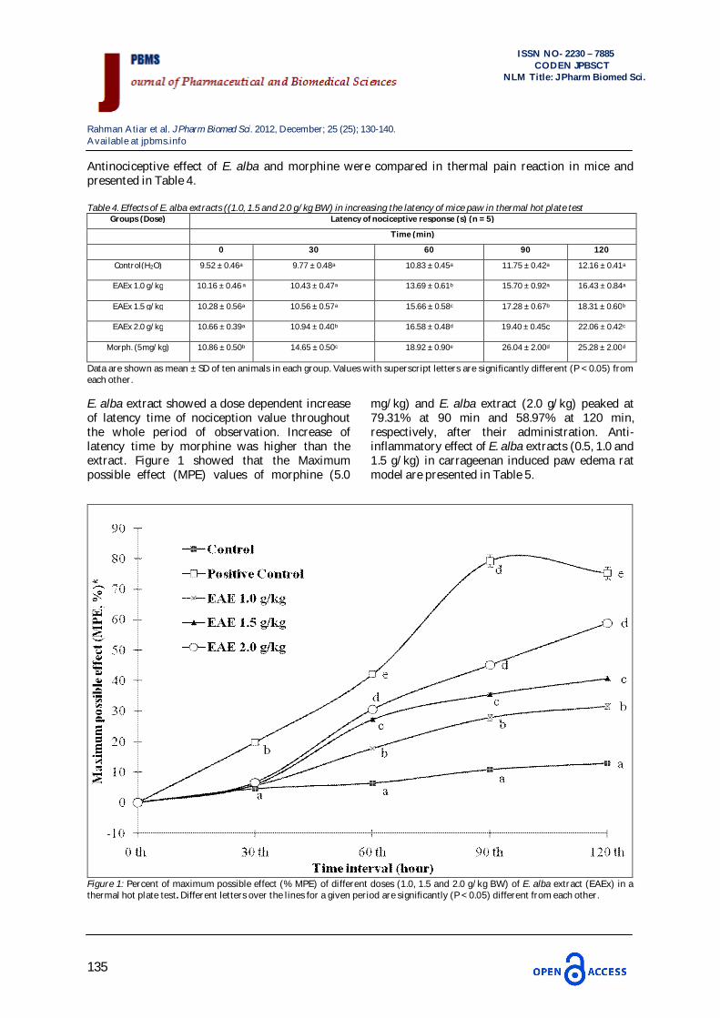
Antinociceptive effect of E. alba and morphine were compared in thermal pain reaction in mice and presented in Table 4. Table 4. Effects of E. alba extracts ((1.0, 1.5 and 2.0 g/kg BW) in increasing the latency of mice paw in thermal hot plate test
Groups (Dose) Latency of nociceptive response (s) (n = 5)
Time (min)
0 30 60 90 120
Control(H2O) 9.52 ± 0.46a 9.77 ± 0.48a 10.83 ± 0.45a 11.75 ± 0.42a 12.16 ± 0.41a
EAEx 1.0 g/kg 10.16 ± 0.46 a 10.43 ± 0.47a 13.69 ± 0.61b 15.70 ± 0.92a 16.43 ± 0.84a
EAEx 1.5 g/kg 10.28 ± 0.56a 10.56 ± 0.57a 15.66 ± 0.58c 17.28 ± 0.67b 18.31 ± 0.60b
EAEx 2.0 g/kg 10.66 ± 0.39a 10.94 ± 0.40b 16.58 ± 0.48d 19.40 ± 0.45c 22.06 ± 0.42c
Morph. (5mg/kg) 10.86 ± 0.50b 14.65 ± 0.50c 18.92 ± 0.90e 26.04 ± 2.00d 25.28 ± 2.00d
Data are shown as mean ± SD of ten animals in each group. Values with superscript letters are significantly different (P < 0.05) from each other. E. alba extract showed a dose dependent increase of latency time of nociception value throughout the whole period of observation. Increase of latency time by morphine was higher than the extract. Figure 1 showed that the Maximum possible effect (MPE) values of morphine (5.0
mg/kg) and E. alba extract (2.0 g/kg) peaked at 79.31% at 90 min and 58.97% at 120 min, respectively, after their administration. Anti-inflammatory effect of E. alba extracts (0.5, 1.0 and 1.5 g/kg) in carrageenan induced paw edema rat model are presented in Table 5.
Figure 1: Percent of maximum possible effect (% MPE) of different doses (1.0, 1.5 and 2.0 g/kg BW) of E. alba extract (EAEx) in a thermal hot plate test. Different letters over the lines for a given period are significantly (P < 0.05) different from each other.
Rahman Atiar et al. J Pharm Biomed Sci. 2012, December; 25 (25); 130-140. Available at jpbms.info
136
ISSN NO- 2230 – 7885
CODEN JPBSCT NLM Title: J Pharm Biomed Sci.
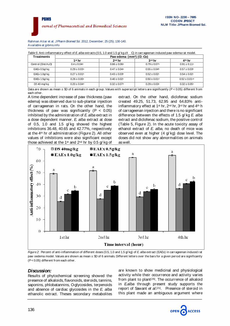
Table 5: Anti-inflammatory effect of E. alba extracts (0.5, 1.0 and 1.5 g/kg zX C) in carrageenan induced paw edema rat model. Treatments Paw edema (mm3) (Ct -Co)
1st hr 2nd hr 3rd hr 4th hr Control (Dist.H2O) 0.4 ± 0.04a 0.66 ± 0.06a 0.79 ± 0.07a 0.91 ± 0.11a
EAEx 0.5g/kg 0.29 ± 0.03a 0.47 ± 0.04a 0.55 ± 0.04b 0.57 ± 0.03b
EAEx 1.0g/kg 0.27 ± 0.01b 0.43 ± 0.03b 0.52 ± 0.02c 0.54 ± 0.02c
EAEx 1.5g/kg 0.26 ± 0.00c 0.40 ± 0.02c 0.50 ± 0.01d 0.52 ± 0.01cd
DS 40 mg/kg 0.20 ± 0.04d 0.32 ± 0.07d 0.29 ± 0.03e 0.32 ± 0.05e
Data are shown as mean ± SD of 6 animals in each group. Values with superscript letters are significantly (P < 0.05) different from each other. A time dependent increase of paw thickness (paw edema) was observed due to sub-plantar injection of carrageenan in rats. On the other hand, the thickness of paw was significantly (P < 0.05) inhibited by the administration of E. alba extract in a dose dependent manner. E. alba extract at dose of 0.5, 1.0 and 1.5 g/kg showed the highest inhibitions 36.48, 40.65 and 42.77%, respectively at the 4th hr of administration (Figure 2). All other values of inhibitions were also significant except those achieved at the 1st and 2nd hr by 0.5 g/kg of
extract. On the other hand, diclofenac sodium created 49.25, 51.73, 62.95 and 64.83% anti-inflammatory effect at 1st hr, 2nd hr, 3rd hr and 4th h of carrageenan injection and there is no significant difference between the effects of 1.5 g/kg E. alba extract and diclofenac sodium, the positive control (Table 5, Figure 2). In the acute toxicity assay of ethanol extract of E. alba, no death of mice was observed even at higher (4 g/kg) dose level. The doses did not show any abnormalities on animals as well.
Figure 2: Percent of anti-inflammation of different doses (0.5, 1.0 and 1.5 g/kg) of E. alba extract (EAEx) in carrageenan induced rat paw oedema model. Values are shown as mean ± SD of 6 animals. Different letters over the bars for a given period are significantly (P < 0.05) different from each other. Discussion: Results of phytochemical screening showed the presence of alkaloids, flavonoids, steroids, tannins, saponins, phlobatannins, O-glycosides, terpenoids and absence of cardiac glycosides in the E. alba ethanolic extract. Theses secondary metabolites
are known to show medicinal and physiological activity while their occurrence and activity varies from plant to plant[26]. The occurrence of alkaloid in E.alba through present study supports the report of Sawant et al.[21]. Presence of steroid in this plant made an ambiguous argument where
Rahman Atiar et al. J Pharm Biomed Sci. 2012, December; 25 (25); 130-140. Available at jpbms.info
137
ISSN NO- 2230 – 7885
CODEN JPBSCT NLM Title: J Pharm Biomed Sci.
Kumar et al. [33] found no steroid but Arunachalam et al.[22] ensured the presence of steroid in this plant which is consistent with our study. Report on terpenoids in E.alba has also been proved by other researchers which is similar with this study. Presence of other metabolites like tannins, saponins, phlobatannins, carbohydrate and reducing sugar confirms the previous report of Kumar et al.[33] and Arunachalam et al.[22]. Present study also reports the existence of flavonoids in the ethanolic extract of E.alba. Studies on such qualitative phytochemical screening seem useful to interpret the antinociceptive and anti-inflammatory effect of this plant extract. Acetic acid induced abdominal constriction model is a useful experimental tool in the testing of new antinociceptive and analgesic drug[34]. The abdominal injection of acetic acid in mice has been attributed to the release of arachidonic acid, which results the synthesis of prostaglandin via the cyclooxygenase (COX) enzyme[35]. The special nerve endings that sense pain are very sensitive to prostaglandin. Nerve endings respond to the released prostaglandin through prostaglandin E2 (PGE2) receptor by picking up and transmitting the pain and injury message through the nervous system to the brain and cause visceral writhing stimuli in mice[36-37]. Therefore, it has been suggested that the inhibition of prostaglandin synthesis is remarkably efficient as an anti-nociceptive mechanism in visceral pain[38]. Since, ethanol extract of E. alba showed significant inhibition of acetic acid induced writing response of mice, it can be suggested that E. alba extract has potential antinociceptive activity which can be contributed by the occurrence of secondary metabolites especially steroids, alkaloids and terpenoids as they inhibit the synthesis of the arachidonic acid metabolite[39] responsible for the nociceptive effect. Formalin test is a tonic model of continuous pain resulting from formalin-induced tissue injury. It is a useful vehicle, particularly for the screening of novel compounds, since it encompasses inflammatory, neurogenic and central mechanisms of nociception[40-41]. The test is sensitive for various classes of analgesic drugs of two distinct phases, reflecting different types of pain. The early phase (initial pain) reflects a direct effect of formalin on nociceptors (neurogenic pain) whereas the late phase reflects tissue injury or inflammatory pain[42-43]. It is showed that the inhibition in the first phase for all the doses of E. alba and morphine is higher than in the second
phase. This result indicates the role of extract is more prominent in neurogenic pain than in tissue injury. This result could also be explained by the fact that the centrally acting drugs such as narcotic (morphine) inhibits the initial phase more than the late phase while a peripherally acting drug only inhibits the late phase[44]. Our results showed that the time spent in licking and biting of the injected paw was significantly reduced by intraperitoneal administration of the ethanol extract of E. alba, in both phases but it prevails on the initial phase. Such effect could be contributed by the flavonoids existed in the extract as it is established that flavonoids have very good effect as antinociceptive and anti-inflammatory agent[45]. The hot-plate test was employed to verify if the extract could show any central antinociceptive effect as the test is specific central model for antinociceptive capacity. The test is also considered to be selective for opioid-like compounds, which are centrally acting analgesics in several animal species[46]. In the present study, it was observed that the animals treated with the E. alba extract did show the increase in their response of latency period in comparison with the control group because the ethanolic extract of E. alba possesses antinociceptive activity in hot plate test may in part be mediated by opioid receptors. These findings indicate that intraperitoneal treatment with E. alba extract may extort sufficiently opioid-like compounds out of the plant which are responsible for the antinociceptive activity of the plant[47]. Morphine was used as a positive control in this test because morphine is a prototypical opioid analgesic, is metabolized in vivo primarily to morphine-3-glucuronide (M3G) and morphine-6-glucuronide (M6G). These metabolic products account for ~65% of a dose of morphine, with the remaining drug biotransformed to multiple minor species or excreted unchanged[44]. In this test morphine peaked it’s MPE 79.31% and at the 90th min and E. alba extract (1.0, 1.5 and 2.0g/kg) reached their MPE 31.60, 40.72 and 58.97%, respectively at 120th min of observation indicating that both of them showed a potent antinociceptive effect on the acute noxious thermal stimulation and confirming the central activity. The results of three different methods mentioned in our study consistently showed both peripherally and centrally acting anti-nociceptive effects. However, no visible sluggish movement at dose level 4g/kg body weight was observed at the time of acute toxicity test and this made us not to
Rahman Atiar et al. J Pharm Biomed Sci. 2012, December; 25 (25); 130-140. Available at jpbms.info
138
ISSN NO- 2230 – 7885
CODEN JPBSCT NLM Title: J Pharm Biomed Sci.
do the locomotion activity or muscle relaxant activity to nullify the false positive result. Apart from this, the anti-nociceptive effects for the control groups and test groups are quite different indicate the less or no possibility of false positive result. Although Muscle relaxation test, locomotion test or swimming performance test are widely employed to evaluate the effects of various agents on central nervous system (CNS), such as CNS depressants, antidepressants, sedative-hypnotics, psychostimulants, euphoria, adaptogens, etc[48]. Anti-inflammatory activity of E. alba extract was evaluated using carrageenan induced paw edema model. Carrageenan induced rat paw edema is suitable for screening the agents for anti-inflammatory activity which are frequently used to assess the anti-edematous effect of natural products[49] because of its sensitivity in detecting orally active anti-inflammatory agents particularly in the acute phase of inflammation. Development of edema in the paw of rat after injection of carrageenan is a biphasic event[50]. The first phase observed during the first hour is attributed to the release of histamine and serotonin. The second phase of edema is due to the release of prostaglandins, (COX product of recent focus), protease and lysosome[51]. This leads to a dilation of the arterioles and venules and to an increased vascular permeability. As a consequence, fluid and plasma proteins are extravasated, and edema forms[52]. The anti-oedematous effect showed by ethanolic extract of E. alba was significant during the first phase of edema (except at a dose of 0.5g/kg) development and significantly maintained in the second phase, suggesting an inhibitory effect on the release of active pain substance such as histamine, serotonin, kinins or prostaglandins. It has been reported that steroids, alkaloids, flavonoids and terpenoids have anti-inflammatory effect[22,53-54] and their presence in E. alba should work as an inducer to exert a modulatory effect on anti-inflammation. Conclusion: The results demonstrate that whole ethanolic extract of E. alba possesses very good antinociceptive and anti-inflammatory effect prevailing on the central or neurogenic system with no acute toxicity and its antinociceptive action. Acknowledgement: The authors wish to thank Bangladesh Council of Scientific and Industrial Research (BCSIR),
Chittagong, Bangladesh to provide their animal house facilities in progress of the research.
References: 1. Stone, Benjamin C. The flora of Guam. Micronesica 1970; 6:
1-659. 2. Wagner H, Geyer B, Yoshinobu K, Govind SR. Coumestans
as the main active principles of the liver drugs Eclipta alba and Wedelia calendulacea. Planta Med 1986; 5: 370-4.
3. Franca SC, Bertoni BW, Pereira AMS. Antihepatotoxic agent in micropropagated plantlets of Eclipta alba. Plant Cell Tiss Org Cul 1995; 40(3): 297-299.
4. Uddin MN, Rahman MA, Ahmed NU, Rana MS, Akter R, Chowdhury AMMA. Antioxidant, cytotoxic and antimicrobial properties of Eclipta alba ethanol extract. Intl J Biol Med Res 2010; 1: 341-346.
5. Jadhav VM, Thorat RM, Kadam VJ, Gholve SB. Kesharaja: hair vitalizing herbs. Intl J PharmTech Res 2009; 1(3): 454-467.
6. Datta K, Singh AT, Mukherjee A, Bhat B, Ramesh B, Burman AC. Eclipta alba extract with potential for hair growth promoting activity. J Ethnopharmacol 2009; 124(3): 450-456.
7. Roy RK, Thakur M, Dixit VK. Hair growth promoting activity of Eclipta alba in male albino rats. Arch Dermatol Res 2008; 300(7): 357-364.
8. Lin SC, Yao CJ, Lin CC, Lin YH. Hepatoprotective activity of Taiwan folk medicine: Eclipta prostrata Linn. Against various hepatotoxin induced acute hepatotoxicity. Phytother Res 1996; 10(6):483-90.
9. Saxena AK, Singh B, Anand KK. Hepatoprotective effects of Eclipta alba on subcellular levels in rats. J Ethnopharmacol 1993; 40(3):155-61.
10. Kosuge T, Yokota M, Sugiyama K, Yamamoto T, Ni M, Yan S. Studies on antitumor activities and antitumor principles of Chinese herbs. I. Antitumor activities of Chinese herbs. Yakugaku Zasshi 1985; 105(8): 791-795.
11. Syed SD, Deepak M, Yogisha S, Chandrashekar AP, Muddarachappa KA, D’Souza P, Agarwal A, Venkataraman BV. Trypsin inhibitory effect of wedelolactone and demethylwedelolactone. Phytother Res 2003;17(4): 420-1.
12. Kobori M, Yang Z, Gong D, Heissmeyer V, Zhu H, Jung YK, Gakidis MA, Rao A, Sekine T, Ikegami F, Yuan C, Yuan J. Wedelolactone suppresses LPS-induced caspase-11 expressions by directly inhibiting the IKK complex. Cell Death Diff 2004; 11(1): 123-30.
13. Abdal-Kader MS, Bahler BD, Malone S, Werkhoven MC, Van Troon F, David Wisse JH, Bursuker I, Neddermann KM, Mamber SW, Kingston DG. DNA-damaging steroidal alkaloids from Eclipta alba from the suriname rain forest. J Nat Prod 2000; 63 (8): 1184-1208.
14. Wiart C, Mogana S, Khalifah S, Mahan M, Ismail S, Buckle M, Narayana AK, Sulaiman M. Antimicrobial screening of plants used for traditional medicine in the state of Perak, Penisular Malaysia. Fitoterapia 2004; 75(1): 68-73.
15. Venkatesan S, Ravi R. Antifungal activity of Eclipta alba. Ind J Pharm Sci 2004; 66(1): 97-8.
16. Lans C, Harper T, Georges K, Bridgewater E. Medicinal and ethnoveterinary remedies of hunters in Trinidad. BMC Com Alt Med 2001; 1: 10.
17. Sawangjaroen N, Subhadhirasakul S, Phongpaichit S, Siripanth C, Jamjaroen K, Sawangjaroen K. The in vitro antigiardial activity of extracts from plants that are used for self-medication by AIDS patients in southern Thailand. Parasitol Res 2005; 95(1): 17-21.
Rahman Atiar et al. J Pharm Biomed Sci. 2012, December; 25 (25); 130-140. Available at jpbms.info
139
ISSN NO- 2230 – 7885
CODEN JPBSCT NLM Title: J Pharm Biomed Sci.
18. Dalal S, Sudhir Kataria SK, Sastry KV, Rana SVS. Phytochemical Screening of Methanolic Extract and Antibacterial Activity of Active Principles of Hepatoprotective Herb, Eclipta alba. Ethnobot Leaf 2010; 14(3): 248-58.
19. Karnick CR, Kulkarni M. Ethnobotanical studies of some medicinal plants used in skin diseases. Maharasthra Med J 1990; 37:131-4.
20. Karthikumar S, Vigneswari K, Jegatheesan K. Screening of antibacterial and antioxidant activities of leaves of Eclipta prostrata (L). Sci Resh Essay 2007; 2(4): 101-4.
21. Sawant M, Isaac JC, Narayanan S. Analgesic studies on total alkaloids and alcohol extracts of Eclipta alba (Linn.) Hassk. Phytother Res 2004; 18(2):111–3.
22. Arunachalam G, Subramanian N, Pazhani GP, Ravichandran V. Anti-inflammatory activity of methanolic extract of Eclipta prostrata L. (Astearaceae). Afr J Pharm Pharmacol 2009; 3(3): 97-100.
23. Leal LK, Ferreira AA, Bezerra GA, Matos FJ, Viana GS. Antinociceptive, anti-inflammatory and bronchodilator activities of Brazilian medicinal plants containing coumarin: a comparative study. J Ethnopharmacol 2000; 70(2): 51-9.
24. El Olemy MM, Al-Muhtadi FJ, Afifi AA. Experimental Phytochemistry. A Laboratory Manual. King of Saudi Arabia: King Saud University Press; 1994.
25. Harborne JB: Phytochemical Methods. A guide to Modern Techniques of Plant Analysis. 1st edition. London: Chapman and Hall Ltd; 1973.
26. Sofowora A. Medicinal plants and Traditional medicine in Africa. 1st edition. Nigeria: Spectrum Books Ltd; 1984.
27. Trease GE, Evans WC. Pharmacognosy. 11th edn. London: Brailliar Tiridel and Macmillian publishers;1989.
28. Koster R, Anderson M, De Beer EJ. Acetic acid for analgesic screening. Fed Pro 1959; 18: 412-6.
29. Gaertner M, Müller L, Roos JF, Cani G, Santos AR, Niero R, Calixto JB, Yunes RA, Delle Monache F, Cechinel-Filho V. Analgesic triterpenes from Sebastiania schottiana roots. Phytomed 1999; 6(1): 41–4.
30. Eddy NB, Leimbach D. Synthetic analgesics. II. Dithienylbutenyl and dithienylbutylamines. J Pharmacol Exp Therap 1953; 107(3): 385–93.
31. Matsumoto K, Horie S, Takayama H, Ishikawa H, Aimi N, Ponglux D, Murayama T, Watanabe K. Antinociception, tolerance and withdrawal symptoms induced by 7-hydroxymitragynine, an alkaloid from the Thai medicinal herb Mitragyna speciosa. Life Sci 2005; 78(1): 2-7.
32. Winter CA, Risley EA, Nuss GW. Carrageenin-induced edema in hind paw of the rat as an assay for antiinflammatory drugs. Pro Soc Exp Biol Med 1962; 111: 544-7.
33. Kumar GS, Jayaveera KN, Ashok Kumar CK, Sanjay UP, Swamy BMV, Kumar DVK. Antimicrobial effects of Indian medicinal plants against acne-inducing bacteria. Trop J Pharm Res 2007; 6(2): 717-23.
34. Otterness IG, Bliven ML. Laboratory models for testing nonsteroidal antiinflammatory drugs. In Nonsteroidal antiinflammatory drugs. Edited by Lombardino JG. Groton, John Wiley and Sons, Connecticut;1985.
35. Katori M, Majima M. Cyclooxygenase-2: its rich diversity of roles and possible application of its selective inhibitors. Inflamm Res 2000; 49(8): 367-92.
36. Hosoi M, Oka T, Abe M, Hori T, Yamamoto H, Mine K, Kubo C. Prostaglandin E(2) has antinociceptive effect through EP(1) receptor in the ventromedial hypothalamus in rats. Pain 1999; 83(2): 221-7.
37. Seibert K, Zhang Y, Leahy K, Hauser S, Masferrer J, Perkins W, Lee L, Isakson P. Pharmacological and biochemical demonstration of the role of cyclooxygenase 2 in inflammation and pain. Pro Nat Aca Sci, USA 1994; 91(25): 12013-12017.
38. Franzotti EM, Santos CV, Rodriques HM, Mourão RH, Andrade MR, Antoniolli AR. Anti-inflammatory, analgesic activity and acute toxicity of Sida cordifolia L. (Malva-branca). J Ethnopharmacol 2000; 72(1-2): 273-8.
39. Vane J. The evolution of non-steroidal anti-inflammatory drugs and their mechanisms of action. Drugs 1987; 1: 18-27..
40. Lee IO, Kong MH, Kim NS, Choi YS, Lim SH, Lee MK. Effects of different concentrations and volumes of formalin on pain response in rats. Acta Anaesthesiol Sin 2000; 38(2): 59-64.
41. Tjølsen A, Berge OG, Hunskaar S, Rosland JH, Hole K. The formalin test: an evaluation of the method. Pain 1992; 51(1): 5-17.
42. Hunskaar S, Hole K. The formalin test in mice: dissociation between inflammatory and non-inflammatory pain. Pain 1987; 30(1): 103-14.
43. Elisabetsky E, Amador TA, Albuquerque RR, Nunes DS, Carvalho Ado C. Analgesic activity of Psychotria colorata (Wild.ex R. & S.) Muell. Arg. Alkaloids. J Ethnopharmacol 1995; 48(2): 77-83.
44. Shibata M, Ohkubo T, Takahashi H, Inoki R. Modified formalin test: Characteristic biphasic pain response. Pain 1989; 38(3): 347-52
45. Toker G, Küpeli E, Memisoğlu M, Yesilada E. Flavonoids with antinociceptive and anti-inflammatory activities from the leaves of Tilia argentea (silver linden). J Ethnopharmacol 2004; 95(2-3): 393-7.
46. Abbott FV, Young SN. Effect of 5-hydroxytryptamine precursors on morphine analgesia in the formalin test. Pharmacol Biochem Behav 1988; 31(4): 855–60.
47. Barua CC, Talukdar A, Begum SA, Lahon LC, Sharma DK, Pathak DC, Borah P. Antinociceptive activity of methanolic extract of leaves of Achyranthes aspera Linn. (Amaranthaceae) in animal models of nociception. Ind J Exp Biol 2010; 48(8): 817-21.
48. Ozturk Y, Aydin S, Tecik B and Başer KHC. Effects of Essential Oils from Certain Ziziphora Species on Swimming Performance in Mice .Phytother Res 1995; 9: 225-227.
49. Akah PA, Okogun JI, Ekpendu TO. Antioedema and analgesic activity of Diodia scandans extract in rats and mice. Phytother Res 1993; 7: 317-9.
50. Vinegar R, Schreiber W, Hugo R. Biphasic development of carrageenan edema in rats. J Pharm Exp Therap 1969; 166(1): 96-103.
51. Asongalem EA, Foyet HS, Ekobo S, Dimo T, Kamtchouing P. Anti-inflammatory, lack of central analgesia and antipyretic properties of Acanthus montanus (Ness) T. Anderson. J Ethnopharmacol 2004; 95(1): 63-8.
52. Ozaki Y. Antiinflammatory effect of Curcuma xanthorrhiza Roxb, and its active principles. Chem Pharm Bull 1990; 38(4): 1045-8.
53. Meyre-Silva C, Yunes R, Santos AR, Magro JD, Delle-Monache F, Cechinel-Filho V. Isolation of a C-Glycoside Flavonoid with Antinociceptive Action from Aleurites moluccana leaves. Planta Med 1999; 65(3): 263-94.
54. Bittar M, de Souza MM, Yunes RA, Lento R, Delle Monache F, Cechinel-Filho V. Antinociceptive activity of I3, II8-Binaringenin, a biflavonoid present in plants of the Guttiferae. Planta Med 2000; 66(1): 84-6.
Rahman Atiar et al. J Pharm Biomed Sci. 2012, December; 25 (25); 130-140. Available at jpbms.info
140
ISSN NO- 2230 – 7885
CODEN JPBSCT NLM Title: J Pharm Biomed Sci.
Competent interest:- The authors declare that they have no competing interests. Source of funding:- None *Corresponding author:- Md. AtiarRahman Ph.D., Department of Biochemistry and Molecular Biology University of Chittagong, Chittagong-4331, Bangladesh Contact: Office: +88 31 2606001-10, Extn-4334, Cell: +88-01711709084, FAX: +88 31 726310, E.mail:[email protected]
Submit your next manuscript to Journal of Pharmaceutical and Biomedical Sciences (JPBMS) (International member journal of COPE and JDP) and take benefit of: • Convenient online submission with persistent Authors support. • Thorough peer review. • High visibility and citation of article with readers /authors across the boundaries. • Immediate publication on acceptance. •Inclusion in COPE, JDP, CAS, DOAJ, NLM catalog, Google Scholar and many more. Submit your manuscript at: www.jpbms.info. Member journal of committee of Publication ethics (COPE) and Journal donation project (JDP).
Quick Response code (QR-Code) for mobile user to access JPBMS website electronically. Website link:- www.jpbms.info