PHYLOGENETIC POSITION OF CRUSTOMASTIX STIGMATICA SP. NOV. AND DOLICHOMASTIX TENUILEPIS IN RELATION...
16
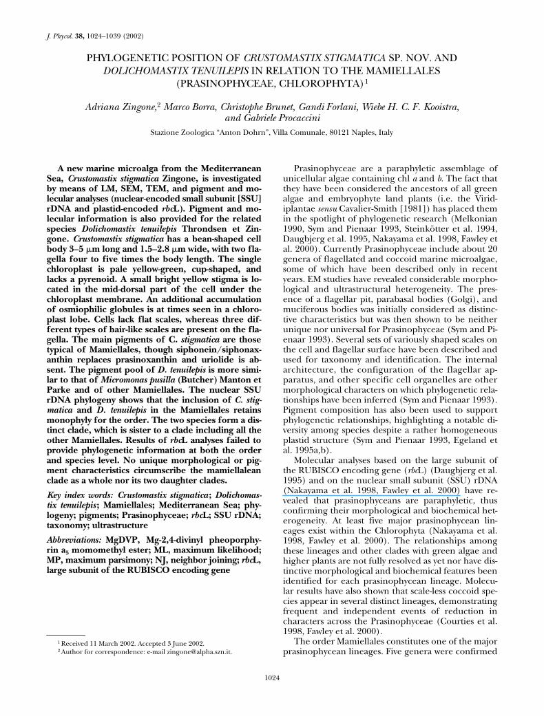
1024 J. Phycol. 38, 1024–1039 (2002) PHYLOGENETIC POSITION OF CRUSTOMASTIX STIGMATICA SP. NOV. AND DOLICHOMASTIX TENUILEPIS IN RELATION TO THE MAMIELLALES (PRASINOPHYCEAE, CHLOROPHYTA) 1 Adriana Zingone, 2 Marco Borra, Christophe Brunet, Gandi Forlani, Wiebe H. C. F. Kooistra, and Gabriele Procaccini Stazione Zoologica “Anton Dohrn”, Villa Comunale, 80121 Naples, Italy A new marine microalga from the Mediterranean Sea, Crustomastix stigmatica Zingone, is investigated by means of LM, SEM, TEM, and pigment and mo- lecular analyses (nuclear-encoded small subunit [SSU] rDNA and plastid-encoded rbcL). Pigment and mo- lecular information is also provided for the related species Dolichomastix tenuilepis Throndsen et Zin- gone. Crustomastix stigmatica has a bean-shaped cell body 3–5 m long and 1.5–2.8 m wide, with two fla- gella four to five times the body length. The single chloroplast is pale yellow-green, cup-shaped, and lacks a pyrenoid. A small bright yellow stigma is lo- cated in the mid-dorsal part of the cell under the chloroplast membrane. An additional accumulation of osmiophilic globules is at times seen in a chloro- plast lobe. Cells lack flat scales, whereas three dif- ferent types of hair-like scales are present on the fla- gella. The main pigments of C. stigmatica are those typical of Mamiellales, though siphonein/siphonax- anthin replaces prasinoxanthin and uriolide is ab- sent. The pigment pool of D. tenuilepis is more simi- lar to that of Micromonas pusilla (Butcher) Manton et Parke and of other Mamiellales. The nuclear SSU rDNA phylogeny shows that the inclusion of C. stig- matica and D. tenuilepis in the Mamiellales retains monophyly for the order. The two species form a dis- tinct clade, which is sister to a clade including all the other Mamiellales. Results of rbcL analyses failed to provide phylogenetic information at both the order and species level. No unique morphological or pig- ment characteristics circumscribe the mamiellalean clade as a whole nor its two daughter clades. Key index words: Crustomastix stigmatica ; Dolichomas- tix tenuilepis ; Mamiellales; Mediterranean Sea; phy- logeny; pigments; Prasinophyceae; rbcL; SSU rDNA; taxonomy; ultrastructure Abbreviations: MgDVP, Mg-2,4-divinyl pheoporphy- rin a 5 momomethyl ester; ML, maximum likelihood; MP, maximum parsimony; NJ, neighbor joining; rbcL, large subunit of the RUBISCO encoding gene Prasinophyceae are a paraphyletic assemblage of unicellular algae containing chl a and b. The fact that they have been considered the ancestors of all green algae and embryophyte land plants (i.e. the Virid- iplantae sensu Cavalier-Smith [1981]) has placed them in the spotlight of phylogenetic research (Melkonian 1990, Sym and Pienaar 1993, Steinkötter et al. 1994, Daugbjerg et al. 1995, Nakayama et al. 1998, Fawley et al. 2000). Currently Prasinophyceae include about 20 genera of flagellated and coccoid marine microalgae, some of which have been described only in recent years. EM studies have revealed considerable morpho- logical and ultrastructural heterogeneity. The pres- ence of a flagellar pit, parabasal bodies (Golgi), and muciferous bodies was initially considered as distinc- tive characteristics but was then shown to be neither unique nor universal for Prasinophyceae (Sym and Pi- enaar 1993). Several sets of variously shaped scales on the cell and flagellar surface have been described and used for taxonomy and identification. The internal architecture, the configuration of the flagellar ap- paratus, and other specific cell organelles are other morphological characters on which phylogenetic rela- tionships have been inferred (Sym and Pienaar 1993). Pigment composition has also been used to support phylogenetic relationships, highlighting a notable di- versity among species despite a rather homogeneous plastid structure (Sym and Pienaar 1993, Egeland et al. 1995a,b). Molecular analyses based on the large subunit of the RUBISCO encoding gene (rbcL) (Daugbjerg et al. 1995) and on the nuclear small subunit (SSU) rDNA (Nakayama et al. 1998, Fawley et al. 2000) have re- vealed that prasinophyceans are paraphyletic, thus confirming their morphological and biochemical het- erogeneity. At least five major prasinophycean lin- eages exist within the Chlorophyta (Nakayama et al. 1998, Fawley et al. 2000). The relationships among these lineages and other clades with green algae and higher plants are not fully resolved as yet nor have dis- tinctive morphological and biochemical features been identified for each prasinophycean lineage. Molecu- lar results have also shown that scale-less coccoid spe- cies appear in several distinct lineages, demonstrating frequent and independent events of reduction in characters across the Prasinophyceae (Courties et al. 1998, Fawley et al. 2000). The order Mamiellales constitutes one of the major prasinophycean lineages. Five genera were confirmed 1 Received 11 March 2002. Accepted 3 June 2002. 2 Author for correspondence: e-mail [email protected].
Transcript of PHYLOGENETIC POSITION OF CRUSTOMASTIX STIGMATICA SP. NOV. AND DOLICHOMASTIX TENUILEPIS IN RELATION...
1024
J. Phycol.
38,
1024–1039 (2002)
PHYLOGENETIC POSITION OF
CRUSTOMASTIX STIGMATICA
SP. NOV. AND
DOLICHOMASTIX TENUILEPIS
IN RELATION TO THE MAMIELLALES
(PRASINOPHYCEAE, CHLOROPHYTA)
1
Adriana Zingone,
2
Marco Borra, Christophe Brunet, Gandi Forlani, Wiebe H. C. F. Kooistra,and Gabriele Procaccini
Stazione Zoologica “Anton Dohrn”, Villa Comunale, 80121 Naples, Italy
A new marine microalga from the Mediterranean
Sea,
Crustomastix stigmatica
Zingone, is investigatedby means of LM, SEM, TEM, and pigment and mo-
lecular analyses (nuclear-encoded small subunit [SSU]rDNA and plastid-encoded
rbc
L). Pigment and mo-lecular information is also provided for the relatedspecies
Dolichomastix tenuilepis
Throndsen et Zin-gone.
Crustomastix stigmatica
has a bean-shaped cell
body 3–5
�
m long and 1.5–2.8
�
m wide, with two fla-gella four to five times the body length. The singlechloroplast is pale yellow-green, cup-shaped, andlacks a pyrenoid. A small bright yellow stigma is lo-cated in the mid-dorsal part of the cell under thechloroplast membrane. An additional accumulationof osmiophilic globules is at times seen in a chloro-plast lobe. Cells lack flat scales, whereas three dif-ferent types of hair-like scales are present on the fla-gella. The main pigments of C.
stigmatica
are thosetypical of Mamiellales, though siphonein/siphonax-anthin replaces prasinoxanthin and uriolide is ab-sent. The pigment pool of
D. tenuilepis
is more simi-lar to that of
Micromonas pusilla
(Butcher) Manton etParke and of other Mamiellales. The nuclear SSUrDNA phylogeny shows that the inclusion of
C. stig-matica
and
D. tenuilepis
in the Mamiellales retains
monophyly for the order. The two species form a dis-tinct clade, which is sister to a clade including all the
other Mamiellales. Results of
rbc
L analyses failed toprovide phylogenetic information at both the orderand species level. No unique morphological or pig-ment characteristics circumscribe the mamiellaleanclade as a whole nor its two daughter clades.
Key index words: Crustomastix stigmatica
;
Dolichomas-tix tenuilepis
; Mamiellales; Mediterranean Sea; phy-logeny; pigments; Prasinophyceae;
rbc
L; SSU rDNA;taxonomy; ultrastructure
Abbreviations:
MgDVP, Mg-2,4-divinyl pheoporphy-
rin a
5
momomethyl ester; ML, maximum likelihood;
MP, maximum parsimony; NJ, neighbor joining;
rbc
L,large subunit of the RUBISCO encoding gene
Prasinophyceae are a paraphyletic assemblage of
unicellular algae containing chl
a
and
b.
The fact thatthey have been considered the ancestors of all greenalgae and embryophyte land plants (i.e. the Virid-iplantae
sensu
Cavalier-Smith [1981]) has placed themin the spotlight of phylogenetic research (Melkonian1990, Sym and Pienaar 1993, Steinkötter et al. 1994,Daugbjerg et al. 1995, Nakayama et al. 1998, Fawley etal. 2000). Currently Prasinophyceae include about 20genera of flagellated and coccoid marine microalgae,some of which have been described only in recentyears. EM studies have revealed considerable morpho-logical and ultrastructural heterogeneity. The pres-ence of a flagellar pit, parabasal bodies (Golgi), andmuciferous bodies was initially considered as distinc-tive characteristics but was then shown to be neitherunique nor universal for Prasinophyceae (Sym and Pi-enaar 1993). Several sets of variously shaped scales onthe cell and flagellar surface have been described and
used for taxonomy and identification. The internal
architecture, the configuration of the flagellar ap-
paratus, and other specific cell organelles are othermorphological characters on which phylogenetic rela-tionships have been inferred (Sym and Pienaar 1993).Pigment composition has also been used to supportphylogenetic relationships, highlighting a notable di-versity among species despite a rather homogeneousplastid structure (Sym and Pienaar 1993, Egeland etal. 1995a,b).
Molecular analyses based on the large subunit ofthe RUBISCO encoding gene (
rbc
L) (Daugbjerg et al.1995) and on the nuclear small subunit (SSU) rDNA(Nakayama et al. 1998, Fawley et al. 2000) have re-vealed that prasinophyceans are paraphyletic, thusconfirming their morphological and biochemical het-erogeneity. At least five major prasinophycean lin-eages exist within the Chlorophyta (Nakayama et al.1998, Fawley et al. 2000). The relationships amongthese lineages and other clades with green algae andhigher plants are not fully resolved as yet nor have dis-tinctive morphological and biochemical features beenidentified for each prasinophycean lineage. Molecu-lar results have also shown that scale-less coccoid spe-cies appear in several distinct lineages, demonstratingfrequent and independent events of reduction incharacters across the Prasinophyceae (Courties et al.1998, Fawley et al. 2000).
The order Mamiellales constitutes one of the majorprasinophycean lineages. Five genera were confirmed
1
Received 11 March 2002. Accepted 3 June 2002.
2
Author for correspondence: e-mail [email protected].

1025
CRUSTOMASTIX STIGMATICA
SP. NOV.
to belong to this order based on molecular data:
Mamiella
,
Mantoniella
,
Micromonas
(SSU and
rbc
L data;Daugbjerg et al. 1995, Nakayama et al. 1998, Fawley et al.2000),
Bathycoccus
(
rbc
L data; Daugbjerg et al. 1995), and
Ostreococcus
(SSU data; Courties et al. 1998). For twoother genera,
Dolichomastix
and
Crustomastix
, only mor-phological features are known (Manton 1977, Moestrup1990, Throndsen and Zingone 1997, Nakayama et al.2000). Based only on morphological information, thephylogenetic position of these genera is difficult to infer.Indeed, some
Dolichomastix
species possess scales that dif-fer from those of other Mamiellales, whereas
Crustomas-tix didyma
Nakayama, Kawachi et Inouye, the only spe-cies known for this genus, has a specialized cell coveringbut lacks body and flagellar scales and a pyrenoid (Na-kayama et al. 2000). Lack of features could be inter-preted as a primitive condition, yet in the Mamiellales ithas been demonstrated to result from secondary losses(Daugbjerg et al. 1995, Nakayama et al. 1998).
Here we describe a new flagellate species,
Crustomastixstigmatica
Zingone sp. nov., based on morphological, pig-ment, and molecular data (nuclear-encoded SSU rDNAand plastid-encoded
rbc
L). We also provide informationon pigments, nuclear SSU rDNA, and
rbc
L of
Dolichomas-tix tenuilepis
Throndsen et Zingone and discuss the phy-logenetic position of the two genera,
Crustomastix
and
Dolichomastix
, in relation to the Mamiellales.
materials and methods
Cultures.
A unialgal culture of
C. stigmatica
, pras1, was estab-lished by serial dilution of a seawater sample collected with a Ni-skin bottle in the Gulf of Naples (station MC, 40
�
49
�
N, 14
�
15
�
E),Mediterranean Sea on 14 May 1995. The authentic strain of
D.tenuilepis
, Caprim2-4II, was obtained from serial dilution cul-tures of a seawater samples collected with Niskin bottles at a sta-tion south of the Island of Capri (14
�
16
�
E, 40
�
30
�
N), Mediter-ranean Sea on 6 July 1994. The strain of
Micromonas pusilla
,MP1, used for pigment analysis, was isolated from the stationMC on 8 April 1993. The three cultures were maintained in Kmedium (Keller et al. 1987) without the addition of silicates,adjusted at a salinity of 36 psu. Cultures were kept at 20–24
�
Cand a 12:12-h light:dark cycle at 100
�
mol photons
�
m
�
2
�
s
�
1
emitted from cool-white fluorescent tubes.LM observations were made on exponentially growing cul-
tures with a Zeiss Axiophot microscope (Carl Zeiss, Oberkochen,Germany) equipped with Nomarski differential interferentialcontrast, phase contrast, and bright-field optics. Light micro-graphs were taken using a Zeiss Axiocam digital camera.
EM preparations.
For TEM ultrathin sections, culture mate-rial of
C. stigmatica
was fixed in glutaraldehyde 1% or 2% fol-lowed by osmium tetroxide as described in Zingone et al.(1995). Additional preparations were made using buffered glu-taraldehyde 2.5% with and without the addition of a few dropsof osmium tetroxide, followed by osmium tetroxide, as indi-cated in Nakayama et al. (2000). For whole-mount observa-tions, a drop of culture was placed on a TEM grid and fixedwith osmium tetroxide vapors (Moestrup and Thomsen 1980)or with glutaraldehyde 2.5% or 1.25% (Marin and Melkonian1994). Grids were rinsed with distilled water, dried, and thenstained with 0.5% uranyl acetate. Whole mounts and ultrathinsections were observed using a Philips 400 transmission elec-tron microscope (Philips Electron optics BV, Eindhoven, TheNetherlands). Specimens prepared for SEM were fixed with os-mium tetroxide, treated as indicated in Zingone et al. (1995),and observed using a Philips 505 SEM.
Morphological information provided for
C. stigmatica
is basedon approximately 250 EM micrographs. In the description, the
cell orientation proposed by Christensen (1980) is followed,with the flagellar insertion defining the ventral side of the celland the foremost part during swimming considered as anterior.When cells are observed from the dorsal side, with their anteriorpart at the top, the cell’s right side is on the observer’s right. Thisorientation also allows the distinction of the right and the left fla-gellum. The numbering of the basal bodies and root terminologycorrespond to those indicated in Moestrup (2000).
Pigment analysis.
Between 5 and 10 mL of cultures of
C. stig-matica
,
D. tenuilepis
, and
M. pusilla
in exponential growth phasewere filtered through GF/F filters (25 mm, Whatman Ltd., Maid-stone, UK) that were frozen at
�
80
�
C until analysis. Frozen fil-ters were extracted in 4 mL of absolute methanol by mechani-cal grinding, and the extracts were filtered through WhatmanGF/F filters. The three strains were analyzed twice on two dif-ferent HPLC systems, using two different methods, LaboratoireInterdisciplinaire des Sciences de l’Environnement (LISE) andStazione Zoologica di Napoli (SZN), to obtain a more effectiveseparation of carotenoids in case of coelution of different pig-ments. Identification and quantification of single pigments wererealized using chl and carotenoid standards obtained from theVKI (Water Quality Institute) International Agency for
14
C De-termination, Denmark. Unfortunately, some pigments (unknownpigments) were not quantified. In the LISE method, the cultureextract was injected in a Beckman System Gold HPLC (BeckmanCoulter, Fullerton, CA, USA), following the procedure of Vidussiet al. (1996). A 3-
�
m C
8
BDS column (100
�
4.6 mm, ThermoHypersil-Keystone, Runcorn, UK) was used, and the mobile phasewas composed of two solvent mixtures: A, methanol:aqueous am-monium acetate (70:30) and B, methanol. The gradient betweenthe solvents was the same as in Vidussi et al. (1996). Absorbancewas detected at 440 nm using a model 168 Beckman photo-diode array detector to obtain the 400–600 nm spectrum foreach pigment. Fluorescence was measured using an Sfm 25Kontron spectrofluorometer (Kontron Instruments, Watford,UK) with excitation at 407 nm and emission at 660 nm. For theSZN method, the culture extract was injected in a Hewlett Packardseries 1100 HPLC (Hewlett-Packard, Wilmington, NC, USA).The column was a C
18
ODS Ultrasphere Beckman (150
�
4.6 mm).The two solvent mixtures were as follows: A, methanol:aqueousammonium acetate (80:10:10) and B, methanol:ethyl acetate(70:30). The solvent flux was 1.5 mL
�
min
�
1
, and the analysisduration was 20 min. Pigments were detected at 440 nm using aHewlett Packard photodiode array detector model DAD series1100, which gives the 400- to 700-nm spectrum for each de-tected pigment. Names and abbreviations used for pigmentsfollow the SCOR nomenclature (Jeffrey et al. 1997).
DNA extraction, PCR, and sequencing.
For molecular analysis,cultures of
C. stigmatica
and
D. tenuilepis
were harvested duringthe exponential growth phase by centrifugation. Total nucleicacid extractions were performed using a modified 2
�
hexa-decyltrimethylammonium bromide procedure (Doyle and Doyle1990). Total nucleic acid preparations were used as templatefor the amplification of the nuclear SSU rDNA and
rbc
L genes.PCR amplifications were performed in a Hybaid PCR Expressmachine (Thermo Hybaid, Ashford, UK) using the primers ss5and ss3 (Rowan and Powers 1992) for nuclear SSU rDNA (30cycles of 1 min at 94
�
C, 1 min at 52
�
C, and 2 min at 72
�
C) orthe primers RH1S and CE1161R (Daugbjerg et al. 1994) for
rbc
L (30 cycles of 1 min at 94
�
C, 1 min at 55
�
C, and 2 min at72
�
C) and Taq DNA polymerase (Boehringer, Mannheim, Ger-many). Additional primers were designed for PCR amplifica-tions of the internal regions of both genes (Table 1). AmplifiedDNA fragments were purified with the QIAEX II purificationkit (Qiagen Genomics, Bothell, WA, USA) and directly se-quenced on a Beckman Ceq 2000 automatic sequencer, usingDye-Terminator cycle sequencing kit (Beckman). Resulting se-quence phenograms were analyzed using the software packageDNASTAR (DNASTAR, Madison, WI, USA).
Phylogenetic analysis.
Genes from different compartments ofthe cell (i.e. the nuclear-encoded SSU rDNA and the plastid-encoded
rbc
L) were selected to obtain independent hypothesesof the phylogenetic relationships between
C. stigmatica
and
D.tenuilepis.
Sequences used for comparison were chosen across

1026
ADRIANA ZINGONE ET AL.
the chl
a
�
b
algae (Tables 2 and 3). As outgroups for sequencecomparison we selected
Cyanophora paradoxa
(Glaucocystophyceae)for the
rbc
L and the latter species and
Chlorarachnion reptans
(Chlor-arachniophyceae) for the SSU rDNA. Merged sequences werealigned by eyeball in Se-Al v1.d1 (Rambaud 1995). The
rbc
L se-quences aligned without problems; only those positions wereremoved that would otherwise disrupt first, second, and thirdcodon positional arrangement. Two regions in the nuclear SSUrDNA sequence (i.e. the tip of the V4 region and the loop atthe tip of the terminal stem region in the secondary structure)revealed ambiguous multiple alignment possibilities and weretherefore removed. The final alignment comprised approxi-mately 1700 base pairs per sequence over 1774 positions.
Phylogenetic significance of informative sites was assessed bycomparing the measure of skewedness (g
1
value) obtained un-der the random trees option in PAUP* (version 4.0b8; Swofford2001) with empirical threshold values in Hillis and Huelsenbeck(1992). Substitution model and parameters describing amongsite-rate variation were estimated using Modeltest 3.0 (Posada andCrandall 1998) and then imported into neighbor joining (NJ)and maximum likelihood (ML) analyses in PAUP*. Weighted max-imum parsimony (MP) phylogenies were inferred using the heuris-tic search procedure with the tree bisection/reconnection, branchswapping algorithm, and the Goloboff fit criterion (K
�
2) optionin PAUP*. Bootstrap values for clades were obtained with 1000replicates for NJ and MP (without weighting). Extra base changesneeded to explain alternative topologies were evaluated usingMacClade (Maddison and Maddison 1992). This program was alsoused to translate
rbc
L DNA sequences into amino acid sequences.
results
Crustomastix stigmatica
Zingone sp. nov.
Cellula phaseoliformis, 3–5
�
m longa et 1.5–2.8 �m lata,concava cum superficie ex qua duo flagella pauci dissimili
longitudine oriuntur cum cellulae superficie acutissimumangulum facerent. Chloroplastus unus, lobis duobus, sinepyrenoide. Stigma quod singula lamella constitutum est, subchloroplasti membrana ab opposita parte flagellorum prin-cipii, collocatum est. Cellulae sine squamis. Breves pili (lon-gitudine 0.3 �m circiter) a flagellorum lateribus dispositi sunt;pili terminales (longitudine 0.5 �m circiter) terni dispositi suntet 40 sub-unitates comprehendunt; pauci et longi pili (1.9 �mcirciter) in parte basali unius ex flagellis sunt et 36–38 sub-unitates comprehendunt.
Cells bean-shaped, 3–5 �m long, 1.5–2.8 �m wide.Flagella inserted in the concave side of the cell, with avery acute angle to the cell surface. One chloroplastwith two lobes and without a pyrenoid. Stigma consist-ing of a single layer in a parietal position in the mid-dorsal part of the cell, opposite to the flagellar inser-tion. Cells lacking flat scales. Flagellar T-hair scalesabout 0.3 �m long. Tip hair scales in bundles of three,about 0.5 �m long, composed of 40 subunits. A few Pl-hairs, about 1.9 �m long located along the proximalpart of one flagellum, with 36–38 globular subunits.
Holotype: Embedding Pras1/N6OS deposited at theStazione Zoologica “Anton Dohrn”, Naples, Italy.
Type locality: Gulf of NaplesEtymology: The specific epithet refers to the pres-
ence of an eyespot, or stigma.LM and SEM. Cells are bean-shaped (Figs. 1, A–E,
and 2, A and B), usually about 3–4 �m long and 2 �mwide. The two flagella range from 10.4 to19.2 �m andare subequal in size, the right flagellum or flagellum 2
Table 1. Primer sequences used to amplify and sequence the nuclear SSU rDNA and rbcL in Crustomastix stigmatica and Dolichomastixtenuilepis.
Primer code Directiona Sequence (5�-3�) Position
Nuclear SSU rDNACommon
ss5b F GGTTGATCCTGCCAGTACTCATATGCTTGss3b R GATCCTTCCGAGGTTCACCTACGGAAACCRev R ATGGTAGGCCTCTATCCTACCATCGA 291 (D. tenuilepis)
298 (C. stigmatica)Specific for C. stigmatica
PraD1 F CTACCACATCCAAAGGAAAGGCAG 384PraD3 F GCATGGAATAACACTATAGGACCT 784PraR1 R CCAGAACATCTAAGGGCATCACAG 1485PraR4 R TCCGTCAATTCCCTTTAAGTTTCA 1180
Specific for D. tenuilepisD1 F ACGAAGGGACGTGTTTATTAGA 102D2 F AAATCCCTTAACGAGGATCCATTG 496D4 F CATTGTCAGAGGTGAAATTCTTGG 863D5 R TTTGATTTCTCATAAGGTGCTGAC 1040D3 R CACATTGTCCCTCTAAGAAGTCAG 1328
rbcLCommon
RH1Sc F ATGTCACCACAAACAGAAACTCE1161Rc R CATGTGCAATACGTCAATACC
Specific for C. stigmaticaPRH1S2 F CTGTTACGTTTCATACCCACTTG 255PCE1161R2 R TACAACAGTTCCTGAGTGTAGG 936PDW3 R GCACGGTGAATGTGAAGAAG 829
Specific for D. tenuilepisDRH1S2 F GCATACGTAGCATACCCACTTG 259DCE1161R2 R GCTGCACGCTTCATCATTTCATC 706
aF, forward; R, reverse.bPCR primers, designed by Rowan and Powers (1992).cPCR primers, designed by Daugbjerg et al. (1994).
1027CRUSTOMASTIX STIGMATICA SP. NOV.
(F2) being slightly shorter than the left one (F1)(Figs. 1C and 2A). They emerge laterally from a slightdepression in the concave side of the cell (Fig. 2B).They have pointed ends (Fig. 2A) and are directed al-most always backward (Figs. 1, B–E, and 2, A and B).The chloroplast is pale yellow-green, with two lobes. Adistinct bright-yellow stigma lies in a parietal positionin the dorsal part of the cell, opposite to the flagellarinsertion (Fig. 1, A, B, and D). An additional eyespotis seen at times in the posterior part of the cell (Fig.1D). At SEM no scales are seen. The cell surface issmooth, especially in the area corresponding to thechloroplast lobes. Dividing cells are found with each oldflagellum paired with a short new flagellum (Fig. 2C).
Cells move along circular paths, returning more orless in the same position regularly. Some cells appear
to rotate around their anterior pole (Fig. 1E). Flagellaare kept backward, each forming a wide S-shaped fig-ure, with the two “S” mirror imaged to each other(Fig. 1E). Slower cells are seen to rotate around theanteroposterior axis while moving, whereas faster cellsseem to glide around this axis. It is difficult, however,to establish whether this gliding is real or an opticaleffect caused by rotation. When cells stop, the flagellaare shed quickly and dissolve in a few seconds, leavingonly a pale trace.
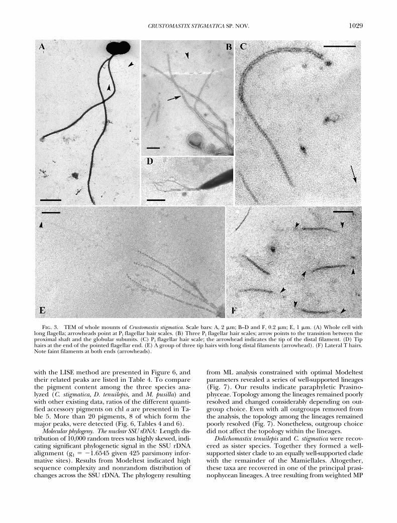
TEM. TEM whole mounts (Fig. 3, A–F) confirmthe absence of body and flagellar scales other thanthree types of hair scales. A few Pl-hairs (sensu Marinand Melkonian 1994), 1.9 �m long, are found alongthe proximal part of the shorter flagellum (Fig. 3A).These hair scales comprise a proximal shaft (Fig. 3, Band C), 1.2 �m long, a distal part consisting of 36–38
Table 2. GenBank accession numbers for sequenced taxaused in nuclear SSU rDNA phylogenetic analysis.
Organism Accession no.
Acrosiphonia duriuscula (Ruprecht) Yendo AB049418Chara foetida Braun X70704Chlorarachnion reptans Geitler X70809Chlorarachnion CCMP242 U03479Coccoid green alga CCMP1205 U40921Coccoid green alga CCMP1220 U40920Coccoid green alga CCMP1407 U40919Coccoid prasinophyte CCMP1193 AF203399Coccoid prasinophyte CCMP1413 AF203402Coleochaete scutata Brébisson X68825Crustomastix stigmatica Zingone AF509628Cyanophora paradoxa Korshikoff X68483Cymbomonas tetramitiformis Schiller AB017126Doliochomastix tenuilepis Throndsen et Zingone AF509625Dunaliella salina (Dunal) Teodoresco M84320Genicularia spirotaenia (Ramb.) de Bary X74753Halosphaera sp. Shizugawa AB017125Hydrodictyon reticulatum (L.) Lagerheim M74497Mamiella sp. AB017129Mantoniella antarctica Marchant AB017128Mantoniella squamata (Manton et Parke)
DesikacharyX73999
Marchantia polymorpha L. X75521Mesostigma viride Lauterbom AJ250109Micromonas pusilla (Butcher) Manton et Parke AJ010408Nephroselmis olivacea Stein X74754Nephroselmis pyriformis CCMP717a X75565Ostreoccocus tauri Courties et Chrétiennot-Dinet Y15814Prasinococcus sp. CCMP1194 AF203400Prasinophyte symbiont of radiolarian AF166381Pseudoscourfieldia marina (Throndsen) Manton AF122888Pterosperma cristatum Schiller AB01727Pycnococcus provasolii Guillard X91264Pycnococcus provasolii Guillard CCMP1198b AJ010406Pyramimonas disomata Butcher AB017121Pyramimonas olivacea N. Carter AB017122Pyramimonas parkeae Norris et Pearson AB017124Pyramimonas propulsa Moestrup et Hill AB017123Scenedesmus pupukensis (Kalina et Pun ochá ová)
Kessler et al.X91267
Scherffelia dubia (Perty) Pascher X68484Staurastrum sp. X77452Tetraselmis striata Butcher X70802Trebouxia impressa Ahmadjian Z21551Ulothrix zonata (Weber et Mohr) Kütz Z47999Zamia pumila L. M20017
aListed in GenBank as Pseudoscourfieldia marina.bListed in GenBank as “unidentified prasinophyte.”
c r
Table 3. GenBank accession numbers for sequenced taxaused in rbcL phylogenetic analysis.
Organism Accession no.
Bathycoccus prasinos Eikrem et Throndsen U30275Bryopsis plumosa (Hudson) C. Agardh AB038480Caulerpa racemosa (Forsskål) J. Agardh AB038486Codium lucasii Setchell AB038481Coleochaete orbicularis Pringsheim L13477Crustomastix stigmatica Zingone AF509626Cyanophora paradoxa Korshikoff X53045Cymbomonas tetramitiformis Schiller L34687Dolichomastix tenuilepis Throndsen et Zingone AF509627Genicularia spirotaenia (Ramb.) de Bary U71439Halimeda discoidea Decaisne AB038488Halimeda opuntia (L.) J.V. Lamouroux AB038489Mamiella sp. U30277Mantoniella squamata (Manton et Parke)
DesikacharyU30278
Marchantia polymorpha L. U87079Mesostigma viride Lauterbom U30282Micromonas pusilla (Butcher) Manton et Parke U30276Nephroselmis minuta (N. Carter) Butcher U30286Nephroselmis olivacea Stein U30285Pedinomonas sp. PCC441 U30287Pseudoscourfieldia marina (Throndsen) Manton U30279Pterosperma cristatum Schiller U30281Pycnococcus provasolii Guillard U30280Pyramimonas cirolanae Pennick L34776Pyramimonas cyclotreta Daugbjerg L34814Pyramimonas cyrtoptera Daugbjerg L34819Pyramimonas grossii Parke L34779Pyramimonas mantoniae Moestrup et Hill L34810Pyramimonas mitra Moestrup et Hill L34812Pyramimonas moestrupii McFadden L34811Pyramimonas octopus Moestrup et Aa. Kristiansen L34817Pyramimonas olivacea N. Carter L34815Pyramimonas orientalis MacFadden L34813Pyramimonas parkeae Norris et Pearson L34816Pyramimonas propulsa Moestrup et Hill L34777Pyramimonas sp. L34834Pyramimonas sp. “greenland” L34818Pyramimonas tetrarhyncus Schmarda L34833Pyramimonas tychotreta Daugbjerg L34778Resultor mikron (Throndsen) Moestrup U30288Staurastrum pingue Teiling AF203506Tetraselmis aff. maculata Butcher U30283Tetraselmis marina (Cienk.) Norris, Hori et Chihara U30284Trebouxia anticipata (Ahmadjian in Ed.) Archibald AF189069Zamia furfuracea L. fil. AF202959

1028 ADRIANA ZINGONE ET AL.
globular subunits, and a faint terminal filament 0.5�m long. Tip hair scales, 0. 5 �m long, are present atthe pointed ends of both flagella in bundles of three(Fig. 3, D and E). They comprise one larger basal sub-unit and about 50 globular subunits and bear a distalfilament 0.7 �m long (Fig. 3E). Lateral T-hair scales(Fig. 3F) are about 0.4 �m long, tubular in shape, andbear a short (0.1 �m) faint filament at both ends.
In ultrathin sections (Figs. 4, A–L, and 5, A–E), around-oval nucleus with a large nucleolus occupies alarge part of the chloroplast-free cell space in theright side of the cell (Fig. 4, A, C–E, and F). To theleft, a long sausage-shaped mitochondrion (Fig. 4G),circular to oval in cross-section (Fig. 4, A–C, and E),runs more or less adjacent to the inner chloroplastface along the anteroposterior axis of the cell, slightlyobliquely. A well-developed Golgi apparatus is presentventrally with respect to the mitochondrion, in theleft side of the cell close to the flagellar bases (Fig. 4,B and C). An inconspicuous microbody is at times de-tected adjacent to the ventral side of the chloroplast(Fig. 4B). Vesicles with granular or particulate mate-rial abound (Fig. 4, B and E). A membrane similar tothe external cell envelope surrounds these structures.
Some vesicles contain a fibrous material (Fig. 4I). Asingle chloroplast is located in a parietal positionalong the dorsal and lateral sides of the cell (Fig. 4, A–F), expanding into two lobes (Fig. 4, A and F) at theanterior and posterior poles. The chloroplast is sur-rounded by a double membrane and consists of threeto five lamellae, each formed by three thylakoids, im-mersed in a granular matrix (Fig. 4H). A pyrenoid isabsent, and one or two starch accumulations are seenin some sections (Fig. 4D). An eyespot or stigmaformed by a single layer of osmiophilic globules is lo-cated in the dorsal part of the chloroplast, opposite tothe flagellar insertion (Fig. 4, A–C). Up to nine glob-ules are counted in a single tangential section (Fig.4K). A similar aggregate of osmiophilic globules is vis-ible at times in one of the chloroplast lobes (Fig. 4J).Cell body and flagella appear to be covered by a dou-ble-layer membrane, with the external layer not mark-edly thicker than the internal one (Fig. 4L).
The basal bodies (Fig. 5, A–E) are about 700 nmlong. They form an acute angle with the ventral sur-face of the cell (Figs. 4A and 5, A and B) and amongthemselves (Fig. 5E). A distal fiber connects the basalbodies (Fig. 5, C–E). Two microtubular roots emergefrom basal body 1 (Fig. 5, C and D). The 1d root, cor-responding to R1 in the root terminology proposedby Moestrup (2000), consists of three microtubules(Fig. 5, C and D) and runs under the cell membranetoward the posterior pole of the cell (Fig. 5A). The 1s(� R2) root shows a 3�1 microtubular pattern in itsproximal part (Fig. 5D). Neither a multilayered struc-ture nor a rhizoplast were observed.
Pigments. The two methods used gave comparableresults. Absorbance chromatograms (440 nm) obtained
Fig. 1. Light microscope images of Crustomastix stigmatica.(A–D) Nomarski differential interferential contrast. (E) Phasecontrast. Scale bar, 2 �m. (A) Lateral outline of a cell, with adorsal eyespot (arrow). (B) Cell with two subequal flagella anda dorsal eyespot (arrow). (C) Cell in ventral view, showing theinsertion point of the flagella. (D) Cell with a dorsal eyespot(arrow) and an additional eyespot (arrowhead) in the posteriorend. (E) Living cell rotating around the anterior pole, with thetwo flagella directed backward, forming a wide S-shaped figure.
Fig. 2. SEM of Crustomastix stigmatica. Scale bar, 1 �m. (A)Whole cell with the two flagella, the right one shorter than theleft one. (B) Cell in lateral-ventral view, showing the bean-shaped body and the flagellar insertion. (C) A division stages,showing a very short new flagellum to the left.
1029CRUSTOMASTIX STIGMATICA SP. NOV.
with the LISE method are presented in Figure 6, andtheir related peaks are listed in Table 4. To comparethe pigment content among the three species ana-lyzed (C. stigmatica, D. tenuilepis, and M. pusilla) andwith other existing data, ratios of the different quanti-fied accessory pigments on chl a are presented in Ta-ble 5. More than 20 pigments, 8 of which form themajor peaks, were detected (Fig. 6, Tables 4 and 6).
Molecular phylogeny. The nuclear SSU rDNA: Length dis-tribution of 10,000 random trees was highly skewed, indi-cating significant phylogenetic signal in the SSU rDNAalignment (g1 � �1.6545 given 425 parsimony infor-mative sites). Results from Modeltest indicated highsequence complexity and nonrandom distribution ofchanges across the SSU rDNA. The phylogeny resulting
from ML analysis constrained with optimal Modeltestparameters revealed a series of well-supported lineages(Fig. 7). Our results indicate paraphyletic Prasino-phyceae. Topology among the lineages remained poorlyresolved and changed considerably depending on out-group choice. Even with all outgroups removed fromthe analysis, the topology among the lineages remainedpoorly resolved (Fig. 7). Nonetheless, outgroup choicedid not affect the topology within the lineages.
Dolichomastix tenuilepis and C. stigmatica were recov-ered as sister species. Together they formed a well-supported sister clade to an equally well-supported cladewith the remainder of the Mamiellales. Altogether,these taxa are recovered in one of the principal prasi-nophycean lineages. A tree resulting from weighted MP
Fig. 3. TEM of whole mounts of Crustomastix stigmatica. Scale bars: A, 2 �m; B–D and F, 0.2 �m; E, 1 �m. (A) Whole cell withlong flagella; arrowheads point at Pl flagellar hair scales. (B) Three Pl flagellar hair scales; arrow points to the transition between theproximal shaft and the globular subunits. (C) Pl flagellar hair scale; the arrowhead indicates the tip of the distal filament. (D) Tiphairs at the end of the pointed flagellar end. (E) A group of three tip hairs with long distal filaments (arrowhead). (F) Lateral T hairs.Note faint filaments at both ends (arrowheads).

1030 ADRIANA ZINGONE ET AL.
analysis (K � 2, tree not shown) revealed the same to-pology for the Mamiellales. Results of bootstrap analy-sis showed high support for this topology, yet topol-ogy among the principal lineages of the green algaeremains poorly resolved. Choice of radically different
values for K in weighted MP analyses resulted in dif-ferent topologies among the principal lineages but didnot affect topology within Mamiellales.
The rbcL: Initial K2P distance analysis of only thirdpositions in the rbcL alignment revealed massive satu-
Fig. 4. Ultrastructural features of Crustomastix stigmatica, TEM. Scale bars: A–G, 0.5 �m; H–K, 0.2 �m; J and L, 0.1 �m. (A) Longi-tudinal section through the insertion of flagellum 1. (B) Transversal section showing a well-developed Golgi body. (C) Transversalsection at the level of the flagellar insertion. (D) Transverse section showing starch granules (arrows) in both chloroplast lobes. (E)Transverse section showing an array of circular inclusions (arrows). (F) Longitudinal section perpendicular to the one showed in A.(G) Oblique section through the mitochondrion. (H) Ultrastructural detail of the chloroplast, with four lamellae. (I) A vesicle withfibrillar material. (J) Tangential section of an eyespot in a chloroplast lobe. (K) Tangential section of an eyespot with nine osmio-philic globules. (L) Section through the cell envelope, close to the chloroplast, showing the double layer of electron-dense material.c, chloroplast; g, Golgi body; m, mitochondrion; n, nucleus; nu, nucleolus.

1031CRUSTOMASTIX STIGMATICA SP. NOV.
ration, with distances among taxa ranging between0.3 and 1.5. Even within Pyramimonas species, dis-tances between 0.11 and 0.55 were observed. High sub-stitution saturation was also inferred from a virtuallynormal distribution of tree lengths among 10,000 ran-domly generated trees. If only first and second codon
positions were included, obtained K2P distances withinthe ingroups ranged between 0.02 and 0.15, yet boot-strap support for most clades was low. In an attemptto improve resolution within and among ingroups, weremoved the outgroup taxon. Nevertheless, basal ram-ifications among clades obtained insufficient boot-
Fig. 5. Ultrastructural features of flagellar bases of Crustomastix stigmatica, TEM. Scale bar, 0.2 �m. (A) Basal body of flagellum 1,with root 1d running backward under the cell surface (arrowhead). (B) Basal body of flagellum 2. (C) Transverse section across theflagellar bases showing roots 1s, 1d, and the connecting fiber. (D) Proximal transverse section across the flagellar bases. (E) Tangen-tial section of the flagellar bases. cf, connecting fiber; 1s, 1d, right and left microtubular flagellar roots.
Table 4. Pigment distribution in the three species analyzed.
Peak no. Retention time M. pusilla D. tenuilepis C. stigmatica Pigment
1 4.01 � � � MgDVP2 5.84 � Traces � Uriolide3 6.30 � � � Siphonaxanthin4 6.80 � � � Prasinoxanthin5 7.01 � � � Violaxanthin6 7.35 � � Traces Unk. aa
7 7.67 � � � Unk. ba
8 8.09 � � � Neoxanthin9 8.17 � � � Antheraxanthin10 8.36 � � � Unk. c11 8.63 � � � Unk. d12 9.50 � � � Lutein13 9.58 � � � Zeaxanthin14 10.02 � � � Unk. e15 11.88 � � � Unk. f16 12.25 � � � Chl b allom17 12.51 � � � Chl b18 12.80 � � � Chl b�19 14.20 � � � Chl a allom20 14.40 � � � Chl a allom21 14.70 � � � Chl a22 15.04 � � � Chl a�23 15.40 � � � Unk. g24 15.67 � � � Unk. h25 16.17 � � � Unid. M1-likea
26 17.32 � � � -Carotene27 17.38 � � � -Carotene
aPigments considered in Table 6 for comparison.

1032 ADRIANA ZINGONE ET AL.
strap support. If only D. tenuilepis, C. stigmatica, andthe Mamiellales were included as ingroups and Neph-roselmis spp. as outgroup, the data set did not containsufficient phylogenetic signal (insignificant g1 value).The same is true for larger groupings with only prasi-nophyceans and Tetraselmis as outgroup and with only1 and 2 positions included. However, if all positionswere included, the bootstrap values basically showedD. tenuilepis and C. stigmatica collapsing in a basal poly-tomy. An NJ tree inferred from the alignment ofamino acid residues of the rbcL sequences (tree notshown) at least grouped Mamiellales with D. tenuilepisand C. stigmatica in a clade, although with only 51%bootstrap support and lacking any internal structure.Results of MP bootstrap analysis also provided onlymarginal support (53%) for such a grouping. There-fore, basically rbcL failed to provide phylogenetic in-formation at the taxonomic level of relationshipsacross mamiellalean genera.
discussionMorphology and taxonomy of Crustomastix stigmatica.
The flagellate described here is attributed to the mono-specific genus Crustomastix based on the morphologicalsimilarity with its type species, C. didyma, recently de-scribed from the northwestern Pacific (Nakayama etal. 2000). The two species share a number of features,which include the bean-like cell shape, lateral inser-tion of the flagella, general arrangement of the bodyorganelles, and absence of a pyrenoid. Both specieslack flat scales and have similar flagellar hair scales.T-hair scales only are different in C. didyma, having onerow of distal globular subunits in addition to the prox-imal tubular shaft. The thin crust forming the cell cov-ering, which gives the name to the genus (crusta �crust, mastix � a whip, flagellum), is a character lessprominent and difficult to detect. Similarly to M. pu-silla (Manton 1959), C. stigmatica possesses a membraneconsisting of two distinct electron dense layers, butdifferently from C. didyma, the outermost layer is notsignificantly thicker than the inner one. In LM, cellslose their shape as soon as they stop moving, whereasflagella dissolve in a few seconds once they are shed.These observations confirm the extremely fragile na-
ture of the external cell and flagellar envelope, whichprobably explains the irregularities seen in EM sec-tions at the cell surface. In C. didyma, a “crust” is onlyrevealed with specific fixation procedures (Nakayamaet al. 2000), and therefore the cell envelope could beas delicate as in C. stigmatica. Unless the crust is an ar-tifact of the fixation procedure, it is also possible thatit can be revealed in other scale-bearing or naked pra-sinophyceans as well, provided that the appropriatefixation procedures are applied.
The main character distinguishing C. stigmaticafrom C. didyma (Table 7) is the consistent presence ofa conspicuous stigma in the central part of the chloro-
Table 5. Pigment ratios (�g/�g) in the three speciesanalyzed.
Pigment ratios M. pusilla D. tenuilepis C. stigmatica
ChlMgDVP/chl a 0.28 0.22 0.11Chl b/chl a 0.24 0.46 0.37MgDVP/total chl 0.18 0.13 0.08Chl b/total chl 0.16 0.27 0.25Chl a/total chl 0.66 0.59 0.67
CarotenoidsSiphonaxanthin/chl a 0 0 0.12Prasinoxanthin/chl a 0.61 0.22 0Violaxanthin/chl a 0.25 0.65 0.20-Carotene/chl a 0.009 0.013 0.038Lutein/chl a 0.001 0.021 0.003
Fig. 6. HPLC absorbance (440 nm) chromatogram of (A)Dolichomastix tenuilepis, (B) Micromonas pusilla, and (C) Crusto-mastix stigmatica obtained with the LISE method (Vidussi et al.1996). For peak identification, see Table 4.

1033CRUSTOMASTIX STIGMATICA SP. NOV.
Fig. 7. ML phylogram inferred from nuclear SSU rDNA sequences of 41 taxa (see Table 2). Proportion of sites assumed to be invari-able � 0.4689; rates assumed to follow a gamma distribution with shape parameter � 0.6224. Assumed substitution rates: A⇔G � 2.447,C⇔T � 4.784, all other substitution rates are 1.000 by default (Modeltest: TrN � I � G); �Ln likelihood � 12328.739; length � 1266, consis-tency index � 0.467, retention index � 0.600. Values above internodes of major clades indicate MP bootstrap values (1000 replicates, fullheuristic search with TBR branch swapping option); those below internodes are bootstrap values (1000 replicates NJ trees of ML distancesunder the same Modeltest parameter settings). A “-” indicates a bootstrap value less than 50%. Bootstrap values associated with minor cladeshave been omitted for clarity. Streptophyta have been positioned as outgroup. Prasinophycean clade numbering follows Fawley et al. (2000).
1034 ADRIANA ZINGONE ET AL.
plasts. This is visible in LM as a bright-yellow refrin-gent body and in TEM as a group of electron-densevesicles tightly apressed. Some lipid vesicles are de-scribed in the same position in C. didyma; however,they are smaller and not seen in LM. In addition, asecond eyespot in a chloroplast lobe, which is fre-quently seen in C. stigmatica, is not described for C.didyma. The presence of additional pigmented gran-ules in the chloroplast has previously been used as dis-tinctive character, for example, in Pyramimonas plurio-culata Butcher (Butcher 1959).
Crustomastix didyma differs from C. stigmatica also inthe presence of a duct, a deep invagination of theplasmalemma opening in the ventral surface of thecell. This structure was revealed in C. didyma only by aspecial fixation procedure (Nakayama et al. 2000),like the cell coat. We repeated the same procedurethree times and observed 100 ultrathin TEM sectionswithout finding any convincing evidence of the pres-ence of a duct in C. stigmatica. Though it seems un-likely, we cannot exclude the possibility that a ductwas not observed due to unnoticed differences in thefixation procedure or in culture conditions.
In the flagellar apparatus, a multilayered structureassociated with root 1d is described for C. didyma (Na-kayama et al. 2000). This structure was not detected inC. stigmatica. Other peculiar characteristics of C. stig-matica are the absence of a rhizoplast and the ex-tremely small size and scarce visibility of the micro-body. The latter was ambiguously identified onlybased on its presumed position. These differencesagain could reflect fixation problems; however, both arhizoplast and a microbody were clearly discernible inultrathin sections of D. tenuilepis prepared for TEM inthe same laboratory with the same procedure as thatused for C. stigmatica (Throndsen and Zingone 1997).
Despite the resemblance in shape, most cells of C.stigmatica are consistently shorter than the minimum
length indicated for C. didyma (i.e. 4 �m) in both liv-ing and glutaraldehyde-fixed material. The width ofC. stigmatica fell more frequently within the range in-dicated for C. didyma (2–3 �m). Differences were alsonoticed in the swimming behavior. However, theseobservations need verification using the same condi-tions of vessels and light.
In conclusion, differences and affinities between C.didyma and C. stigmatica support the erection of a newspecies and its attribution to Crustomastix, respectively.Yet the taxonomic importance of characters inMamiellales is not fully known; therefore, molecularinformation on C. didyma will be valuable to confirmthe relationships between the two species.
Pigment composition. The pigment suites of C. stigmat-ica and D. tenuilepis reflect the high diversity and hetero-geneity found in the pigment pool of the class Prasino-phyceae (Sym and Pienaar 1993, Egeland et al. 1995a,b,1997, Wilhelm et al. 1997). The two species share withM. pusilla chl a and b and Mg-2,4-divinyl pheoporphyrina5 momomethyl ester (MgDVP). Ratios of chl a over to-tal chl in the three species are similar to the values re-ported for another mamiellalean alga, Bathycoccus prasi-nos (Egeland et al. 1995b). The chl b/chl a ratios arelower than values reported in many studies (Hooks et al.1988, Fawley 1992, Simon et al. 1994, Chrétiennot-Dinetet al. 1995, Jeffrey and Vesk 1997) but similar to thosefound by Egeland et al. (1995a).
Large differences exist in the carotenoid contentamong C. stigmatica, D. tenuilepis, and M. pusilla. Crus-tomastix stigmatica lacks prasinoxanthin and uriolidebut possesses siphonaxanthin. A similar pigment suitehas been found in other prasinophyceans, includingthe mamiellalean Ostreococcus tauri (Chrétiennot-Dinetet al. 1995). The pigment suite of D. tenuilepis is in-stead more similar to that of M. pusilla and of the restof known Mamiellales. However, the relative contentof prasinoxanthin is much lower in D. tenuilepis than
Table 7. Main morphological characteristics separating Crustomastix didyma and C. stigmatica.
Species Size (�m) Eyespot DuctMultilayered
structure Rhizoplast Microbody Flagellar T hairs
C. didyma 2–3 � 4–6 Small lipid globules,not visible in LM
Present Present Present Clearly detected With globular subunits
C. stigmatica 1.5–2.8 � 3–5 Clearly visible in LM Undetected Undetected Undetected Inconspicuous Without globular subunits
Table 6. Comparison of absorption characteristics of three unidentified carotenoids.
Pgiment Reference Species Absorption (nm)
Unid. M1-like This study Micromonas pusilla (420), 443, 471M1 Fawley 1992 Mantoniella squamata (420), 443, 470Pigment 11 Wilhelm et al. 1997 M. squamata 445–474Unk. a This study Crustomastix stigmatica, M. pusilla (406), 427, 453Unknown 1 Fawley 1992 M. pusilla (405), 427, 453Micromonol Wilhelm et al. 1997 M. squamata 428.1 and 454.6Micromonol Egeland et al. 1995b M. pusilla, M. squamata 427 and 452Unk. b This study M. pusilla 458Micromonal Wilhelm et al. 1997 M. squamata 459.5Micromonal Egeland et al. 1995a Bathycoccus prasinos 450 (in acetone)Micromonal Egeland et al. 1995b M. pusilla, M. squamata, Pseudoscourfieldia marina 450 (in acetone)

1035CRUSTOMASTIX STIGMATICA SP. NOV.
in M. pusilla. A comparably wide range of prasinoxan-thin/chl a ratios has been found in prasinoxanthin-containing Prasinophyceae (Fawley 1992, Simon et al.1994). Hooks et al. (1988) separated them into twogroups, with a discriminating threshold ratio of 0.2.Following this classification, D. tenuilepis would corre-spond to the limit between the two groups, whereasM. pusilla belongs to the second group (ratio � 0.2).Thus, the three species examined encompass thewhole range of variability for prasinoxanthin content,including its absence.
The pigment uriolide also shows considerable dif-ferences among the three species examined. It is ab-sent in C. stigmatica but produces a high surface peakin M. pusilla. Its presence in D. tenuilepis is not fullyconfirmed, because only traces of a pigment with thesame retention time as uriolide were found. Otherdifferences are the higher values of -carotene/chl aratio in C. stigmatica and the absence of -carotene inM. pusilla and D. tenuilepis. Some of the unidentifiedcarotenoids found in our analyses, that is, the uniden-tified M1-like “unknown a” and “unknown b” pig-ments, have similar absorption properties and proba-bly correspond to unidentified pigments found in M.pusilla, M. squamata, and other prasinophyceans byother authors (Table 6).
Another peculiar feature in pigment compositionof C. stigmatica is the presence of high amounts of ze-axanthin. In green algae, zeaxanthin is involved in thephotodependent xanthophyll interconversion cycleand is synthesized from violaxanthin under light stress(Demming-Adams 1990). In C. stigmatica, zeaxanthincould be synthesized under lower light intensity thanin M. pusilla and D. tenuilepis, which lacked zeaxan-thin. Dolichomastix tenuilepis showed a rather high vio-laxanthin/chl a ratio as compared with C. stigmaticaand M. pusilla. Violaxanthin is the principal carot-enoid in some prasinophyceans, including Ostreococcustauri (Chrétiennot-Dinet et al. 1995) and Pyramimonasparkeae (Kohata and Watanabe 1989) and the Meso-stigmatophyceae Mesostigma viride (Fawley and Lee1990). However, differences in pigment ratios couldalso be related to growth conditions, especially lightquantity (Fawley 1992), day length, and diel variations(Kohata and Watanabe 1989), as well as to ecophysio-logical characteristics of the clones examined (Guil-lard et al. 1991).
Differences in pigment compositions in closely re-lated species are difficult to explain, because func-tional implications of presence/absence and replace-ments of pigments are not fully clarified. Highproportion of chl b could be an adaptation for growthat low light (Yokohama 1981). The presence ofMgDVP, with maximum of absorption at 439 nm (i.e.between the absorption maxima of chl a and chl b)could further increase the capacity of light absorptionin the blue region of the spectrum, thus suggesting anadaptation to an ecological niche deep in the watercolumn. The xanthophylls siphonein and siphonax-anthin also absorb light in the blue-green and green
part of the spectrum (500–550 nm) (Yokohama 1981,Anderson 1983) and efficiently transfer light excita-tion energy to chl a (Anderson 1983). They could cor-respond to an adaptation of algae to deep greencoastal waters (Yokohama 1981, 1983). Prasinoxan-thin and siphonaxanthin seem to be functionallycomparable (Wilhelm et al. 1986), although the ab-sorption maximum of prasinoxanthin (457 nm) isnarrower than that of siphonaxanthin (446–466 nm).Notably, many prasinophyceans have been isolatedfrom deep layer (Guillard et al. 1991, Simon et al.1994), so an efficient light-harvesting and excitationtransfer systems to chl a could be an adaptation ofthese algae to a low light environment.
Phylogeny of C. stigmatica and D. tenuilepis. The phy-logeny inferred from nuclear SSU rDNA shows that C.stigmatica and D. tenuilepis are distantly related sisterspecies within Mamiellales. To confirm both their in-clusion and their basal position in this order, we hadto add representatives of all other chl a�b orders tothe phylogenetic survey. The nuclear SSU rDNA phy-logeny failed to resolve relationships among these or-ders unambiguously but adequately resolved relation-ships within them, confirming results obtained withsimilar data by other authors (Courties et al. 1998, Na-kayama et al. 1998, Fawley et al. 2000). Based on mor-phological characters of C. didyma, Nakayama et al.(2000) suggested that the genus Crustomastix repre-sents an early divergence from other Mamiellalesand a “missing link” between the Mamiellales and thePyramimonadales. Although nuclear SSU rDNA anal-ysis confirms the basal position of Crustomastix withinthe Mamiellales, low bootstrap values do not allow toresolve properly the phylogenetic relationships of thisgenus with other orders of the prasinophyceans.
Results of our rbcL analysis did not resolve phyloge-netic relationships at the levels needed for this study;they even failed to assess the phylogenetic position ofspecies within single clades. These results are similarto those shown by Daugbjerg et al. (1995), who ob-tained little or no bootstrap support for major prasi-nophycean clades.
Recovery of C. stigmatica and D. tenuilepis as nearestneighbors in a sister clade to the remainder of theMamiellales allows erection of a monophyletic sisterorder to monophyletic Mamiellales sensu stricto. Yettheir inclusion in the Mamiellales also retains mono-phyly for the order. The questions that we have todeal with next is whether a single clade with Mamiel-lales can be circumscribed with shared derived char-acter states and whether the two sister clades can bedelimited likewise.
Phylogenetic status of morphological features in Mamiel-lales. The addition of C. stigmatica and D. tenuilepis re-tains monophyly for the Mamiellales but does not helpidentify any unifying and distinctive morphologicalcharacter for the order (Table 8). The order Mamiel-lales was erected for scale-bearing Prasinophyceae lack-ing a proximal layer of square- or diamond-shapedscales (Moestrup 1984). Initially, the order only in-

1036 ADRIANA ZINGONE ET AL.
cluded the flagellate genera Mantoniella, Mamiella,Dolichomastix, and the coccoid Bathycoccus (Moestrup1990). Most species in these genera possess body andflagellar scales with a spider-web pattern formed byconcentric and radiating ribs. Only two of the fourDolichomastix (i.e. D. nummulifera and D. tenuilepis) havescales without radiating ribs. Subsequently, phyloge-nies inferred from rbcL (Daugbjerg et al. 1995) andSSU rDNA (Nakayama et al. 1998) demonstrated thatthe scale-less uniflagellate M. pusilla also belongs toMamiellales. Both phylogenies demonstrated that ab-sence of scales and presence of a single very shortflagellum in M. pusilla result from secondary loss be-cause the species appears in an advanced position inthe Mamiellales. Lack of traits is even more pro-nounced in Ostreococcus tauri, the smallest eukaryoticorganism known at present (Courties et al. 1994). Thisspecies not only lacks scales but also flagella and apyrenoid (Chrétiennot-Dinet et al. 1995). Our SSUphylogeny confirms results of Courties et al. (1998) thatthis tiny coccoid species belongs to the Mamiellales.
The genus Dolichomastix was erected to include spe-cies with very long flagella (Manton 1977), a featurethereafter shown not to be diagnostic for the genus(Throndsen and Zingone 1997). It was included inthe Mamiellales due to the lack of underlayer scales(Moestrup 1984, 1990, Moestrup and Throndsen 1988).The position of the flagellar insertion and the generalarchitecture of body organelles of D. tenuilepis (Thrond-sen and Zingone 1997), very similar to those ofMamiella and Mantoniella species, further supportedthis inclusion. As for the genus Crustomastix, Na-kayama et al. (2000) tentatively attributed the scale-less species C. didyma to the Mamiellales based on thecombination of several microanatomical features, in-cluding organelle configuration, position and struc-ture of transitional region of the flagella, and absenceof a microtubular root associated with the secondbasal bodies. However, as these authors pointed out,single states associated to these characters in theMamiellales are probably homoplasies, because theyalso exist in species belonging to other prasinophycean
orders. Indeed, no single morphological feature canbe identified that is unique to the Mamiellales northat is present in all the species of the order.
Our results also confirm nonmonophyly for thetwo families of the Mamiellales, namely, the Mamiel-laceae and Micromonadaceae, which group scale-bearing and scale-less Mamiellales, respectively. Infact, the phylogenetic position of M. pusilla and Ostreo-coccus tauri clearly showed that the two families arenonmonophyletic (Fawley et al. 2000). Similarly tothe main clade of the Mamiellales, the clade formedby D. tenuilepis and C. stigmatica is not homogeneous,the former species having body and flagellar scaleswith concentric ribs, the latter lacking scales. There-fore, the families Mamiellaceae and Micromona-daceae have representatives in both clades of theMamiellales. This is not surprising, because the ab-sence of scales, and in general morphological reduc-tion or missing characters, have occurred as a result ofsecondary loss all across prasinophycean clades andtherefore cannot reliably define phylogenetic groups.Another example is provided, in the order Pseudo-scourfieldiales, by the scale-bearing flagellate Pseudo-scourfieldia marina and the scale-less coccoid Pycnococ-cus provasolii. These two species show a high geneticsimilarity (Daugbjerg et al. 1995, Fawley et al. 1999),which suggests that they represent different stages ofthe life cycle of two closely related species.
The only morphological character that could cir-cumscribe the clade formed by D. tenuilepis and C. stig-matica as distinct from the main mamiellalean clade isthe shape of flagellar T hairs. Species of the genusMantoniella and Mamiella have two rows of globularsubunits on the flagellar T hairs, which according toNakayama et al. (1998) would be a truly shared de-rived state of the order. In D. tenuilepis and C. stigmat-ica, T hairs consist instead only of a tubular shaft withthin and short filaments at one or both ends, respec-tively. However, the state of this character in otherspecies attributed to the genera Dolichomastix andCrustomastix is not homogeneous. In C. didyma, flagel-lar T hairs have one row of subunits in addition to the
Table 8. Morphological features and pigments of species attributed to the order Mamiellales.
Species FlagellaSpider-web
scales
Circularpatterned
scales Pyrenoid MgDVP Prasinox. Siphonax. Uriolide
T hairs:tubular
shaft
T hairs:globularsubunits
Bathycoccus prasinos � � � � � � � �Crustomastix didyma 2 � � � ? ? ? ? � �Crustomastix stigmatica 2 � � � � � � � � �Dolichomastix eurylepidea 2 � � ? ? ? ? ? ? ?Dolichomastix lepidota 2 � � ? ? ? ? ? � �Dolichomastix nummulifera 2 � � ? ? ? ? ? ? ?Dolichomastix tenuilepis 2 � � � � � � ? � �Mamiella gilva 2 � � � � � � � � �Mantoniella squamata 2 � � � � � � � � �Mantoniella antarctica 2 � � � � � � � � �Micromonas pusilla 1 � � � � � � � � �Ostreococcus tauri � � � � � � � �
Characters were selected based on their relevance to the phylogenetic discussion.

1037CRUSTOMASTIX STIGMATICA SP. NOV.
tubular shaft (Nakayama et al. 2000). In Dolichomastix,T hairs have only been described for D. lepidota Man-ton (Throndsen and Zingone 1997), which has thetwo rows of globular subunits like Mamiella and Man-toniella species. Also the spider-web scales covering thecell body and flagella in D. lepidota and in D. eurylepi-dea Manton are closer to those of Mantoniella squamatathan to the ones with concentric pattern of D. nummu-lifera Manton and D. tenuilepis. This suggests that thegenus Dolichomastix could in fact include more thanone genus (Moestrup 1990) and that D. lepidota couldbelong to Mantoniella (Throndsen and Zingone 1997).
Phylogenetic status of pigments in the Mamiellales. Differ-ent classification systems of Prasinophyceae have beenproposed based on pigment composition. In the systemproposed by Egeland et al. (1997), three groups arerecognized. Dolichomastix tenuilepis would belong tothe group 3, which is characterized by the presence ofprasinoxanthin and/or uriolide and hence also in-cludes M. pusilla, whereas C. stigmatica would belongto the group 2, due to the presence of siphonaxan-thin. A higher heterogeneity within Mamiellales isshown according to a classification based on six dis-tinct pigment groups (Sym and Pienaar 1993). In thissystem, most Mamiellales including M. pusilla belongto group 6, which is characterized by the presence ofprasinoxanthin, MgDVP, and uriolide, along with mi-nor unknown pigments. Crustomastix stigmatica, togetherwith O. tauri, fits instead in group 4, which presentssome pigment characteristics of Ulvophyceae (sipho-nein/siphonaxanthin and absence of prasinoxanthin)and others of Prasinophyceae (MgDVP). Finally, D.tenuilepis probably belongs to group 5, differing fromspecies of group 6 due to the lack of a clear peak of uri-olide as well as of several unknown pigments.
Phylogenetic structure of the Mamiellales as shownby molecular data (Fawley et al. 2000, this study) doesnot correspond with patterns in pigment data. Not asingle pigment defines Mamiellales, nor the two cladesidentified within the order. Crustomastix stigmatica havea pigment suite clearly different from that of D. tenuile-pis but similar to that of the naked coccoid O. tauri,which belongs to the major clade of the Mamiellales.In addition, C. stigmatica and O. tauri lack uriolide,which was previously believed to be shared by all spe-cies in the order Mamiellales (Nakayama et al. 1998),though is present in other clades as well.
Many pigments appear to be ancestral features ofspecies. For instance, MgDVP is a common feature inall Mamiellales so far analyzed for pigments (Fawley1992, this study). However, MgDVP is not unique forthis order, because it has been found in other prasino-phyceans (Ricketts 1970, Hooks et al. 1988) and evenin a cryptophyte (Schimek et al. 1994). Another ex-ample of a possibly ancestral pigment is siphonein/siphonaxanthin, which is found in the Mamiellales C.stigmatica and O. tauri and in several unrelated speciesof the group 4, scattered across Pyramimonadales,Pseudoscourfieldiales, and Chlorodendrales (Sym andPienaar 1993). Siphonein/siphonaxanthin have been
considered an evolutionary relict of some Chloro-phytes (Anderson 1983). Their loss in M. pusilla andin the other Mamiellales of the pigment groups 5 and6 (Sym and Pienaar 1993) would be a more derivedcharacter state. Apparently, secondary loss or substitu-tion of pigments are easy to occur, as also demon-strated by a strain of M. pusilla (CS-170) from theCoral Sea, Australia, indistinguishable from otherstrains, which lacks MgDVP but has a chl c3-like pig-ment (Jeffrey 1989).
The mismatch between phylogeny and pigmentcomposition revealed by the Mamiellales, and in gen-eral by the Prasinophyceae, also has meaningful con-sequences for chemotaxonomy. Despite the impor-tance of pigment suites in taxonomic identification ofspecies, the use of single pigments as taxonomicmarkers of higher taxonomic groups within prasino-phyceans is presently impossible.
In conclusion, the addition of two new taxa to theMamiellales has shown a higher level of diversity thanoriginally assumed for this order. Our study has alsoconfirmed that the former circumscriptions of Mamiel-lales and of families within the order are weak. In-deed, no sound character redefines the order Mamiel-lales based on morphological or pigment features orto distinguish families within the order. We also con-firmed the nonmonophyly of the Prasinophyceae. Toobtain a classification of Prasinophyceae that is bothnatural and taxonomically meaningful, a multitude oforders is needed along the grade leading to Ulvo-phyceae, Chlorophyceae, and Trebouxiophyceae. Thisconclusion is not surprising if one takes into accountthat all the lineages considered are very ancient andhave presumably undergone many morphologicaland biochemical changes in the course of their longevolutionary history.
We thank Gennaro Iamunno for TEM embeddings and ul-trathin sections and Augusto Passarelli and Ciro Chiaese forHPLC analyses with the SZN method. Thanks are also due toProf. Jahn Throndsen (University of Oslo, Norway) for readingand commenting on the manuscript.
Anderson, J. M. 1983. Chlorophyll-protein complexes of a Codiumspecies, including a light-harvesting siphonaxanthin-chloro-phyll a/b protein complex, an evolutionary relic of some Chlo-rophyta. Biochim. Biophys. Acta 724:370–80.
Butcher, R. W. 1959. An introductory account of the smaller algaeof the British coastal waters. Part I. Introduction and Chloro-phyceae. Fish. Invest. 4:1–74.
Cavalier-Smith, T. 1981. The origin and early evolution of the eu-karyotic cell. In Carlile, M. J., Collins, J. F. & Moseley, B. E. B.[Eds.] Molecular and Cellular Aspects of Microbial Evolution. Cam-bridge University Press, Cambridge, pp. 33–84.
Chrétiennot-Dinet, M. J., Courties, C., Vaquer, A., Neveux, J., Claus-tre, H., Lautier, J. & Machado, M. C. 1995. A new marine pi-coeucaryote: Ostreococcus tauri gen. et sp. nov. (Chlorophyta,Prasinophyceae). Phycologia 34:285–92.
Christensen, T. 1980. Algae: A Taxonomic Survey. AiO Tryk, Odense,216 pp.
Courties, C., Perasso, R., Chrétiennot-Dinet, M.-J., Gouy, M., Guil-lou, L. & Troussellier, M. 1998. Phylogenetic analysis and ge-nome size of Ostreococcus tauri (Chlorophyta, Prasinophyceae).J. Phycol. 34:844–9.

1038 ADRIANA ZINGONE ET AL.
Courties, C., Vaquer, A., Troussellier, M., Lautier, J., Chrétiennot-Dinet, M. J., Neveux, J., Machado, C. & Claustre, H. 1994.Smallest eukaryiotic organism. Nature Lond. 370:255.
Daugbjerg, N., Moestrup, Ø. & Arctander, P. 1994. Phylogeny ofthe genus Pyramimonas (Prasinophyceae, Chlorophyta) in-ferred from the rbcL gene. J. Phycol. 30:991–9.
Daugbjerg, N., Moestrup, Ø. & Arctander, P. 1995. Phylogeny ofgenera of Prasinophyceae and Pedinophyceae (Chlorophyta)deduced from molecular analysis of the rbcL gene. Phycol. Res.43:203–13.
Demming-Adams, B. 1990. Carotenoids and photoprotection inplants: a role for the xanthophyll zeaxanthin. Biochim. Biophys.Acta 1020:1–24.
Doyle, J. J. & Doyle, J. L. 1990. Isolation of plant DNA from fresh tis-sue. Focus 12:13–5.
Egeland, E. S., Eikrem, W., Throndsen, J., Wilhelm, C., Zapata, M.& Liaaen-Jensen, S. 1995a. Carotenoids from further prasino-phytes. Biochem. Syst. Ecol. 23:747–55.
Egeland, E. S., Guillard, R. R. L. & Liaaen-Jensen, S. 1997. Addi-tional carotenoid prototype representatives and a generalchemosystematic evaluation of carotenoids in Prasinophyceae(Chlorophyta). Phytochemistry 44:1087–97.
Egeland, E. S., Johnsen, G., Eikrem, W., Throndsen, J. & Liaaen-Jensen, S. 1995b. Pigments of Bathycoccus prasinos (Prasino-phyceae): methodological and chemosystematic implications.J. Phycol. 31:554–61.
Fawley, M. W. 1992. Photosynthetic pigments of Pseudoscourfieldiamarina and selected green flagellates and coccoid ultraphy-toplankton: implications for the systematics of the Micromona-dophyceae (Chlorophyta). J. Phycol. 28:26–31.
Fawley, M. W. & Lee, C. M. 1990. Pigment composition of the scalygreen flagellate Mesostigma viride (Micromonadophyceae) issimilar to that of the siphonous green alga Bryopsis plumosa (Ul-vophyceae). J. Phycol. 26:666–70.
Fawley, M. W., Qin, M. & Yun, Y. 1999. The relationship betweenPseudoscourfieldia marina and Pycnococcus provasolii (Prasino-phyceae, Chlorophyta): evidence from 18S rDNA sequencedata. J. Phycol. 35:838–43.
Fawley, M. W., Yun, Y. & Qin, M. 2000. Phylogenetic analyses of 18SrDNA sequences reveal a new coccoid lineage of the Prasino-phyceae (Chlorophyta). J. Phycol. 36:387–93.
Guillard, R. R. L., Keller, M. D., O’Kelly, C. J. & Floyd, G. L. 1991.Pycnococcus provasolii gen. et sp. nov; a coccoid prasinoxanthin-containing phytoplankter from the western North Atlantic andGulf of Mexico. J. Phycol. 27:39–47.
Hillis, D. M. & Huelsenbeck, J. P. 1992. Signal, noise, and reliabilityin molecular phylogenetic analyses. J. Hered. 83:189–95.
Hooks, C. E., Bidigare, R. R., Keller, M. D. & Guillard, R. R. L. 1988.Coccoid eukaryotic marine ultraplankters with four differentHPLC pigment signatures. J. Phycol. 24:571–80.
Jeffrey, S. W. 1989. Chlorophyll c pigments and their distribution inthe chromophyte algae. In Green, J. C. & Leadbeater, B. S. C.[Eds.] The Chromophyte Algae: Problems and Perspectives. Claren-don Press, Oxford, pp. 13–36.
Jeffrey, S. W., Mantoura, R. F. C. & Bjørnland, T. 1997. Data foridentification of 47 key pigments. In Jeffrey, S. W., Mantoura,R. F. C. & Wright, S. W. [Eds.] Phytoplankton Pigments in Ocean-ography. UNESCO, Paris, pp. 343–60.
Jeffrey, S. W. & Vesk, M. 1997. Introduction to marine phytoplank-ton and their pigment signatures. In Jeffrey, S. W., Mantoura,R. F. C. & Wright, S. W. [Eds.] Phytoplankton Pigments in Ocean-ography. UNESCO, Paris, pp. 37–84.
Keller, M. D., Selvin, R. C., Claus, W. & Guillard, R. R. L. 1987. Me-dia for the culture of oceanic ultraphytoplankton. J. Phycol.23:633–8.
Kohata, K. & Watanabe, M. 1989. Diel changes in the composi-tion of photosynthetic pigments and cellular carbon and ni-trogen in Pyramimonas parkeae (Prasinophyceae). J. Phycol.25:377–85.
Maddison, W. P. & Maddison, D. R. 1992. MacClade: Analysis of Phy-logeny and Character Evolution. V.3.0. Sinauer, Sunderland, Mas-sachusetts.
Manton, I. 1959. Electron microscopical observations on a very
small flagellate: the problem of Chromulina pusilla. J. Mar. Biol.Assoc. U.K. 38:319–33.
Manton, I. 1977. Dolichomastix (Prasinophyceae) from arctic Can-ada, Alaska and South Africa: a new genus of flagellates withscaly flagella. Phycologia 16:427–38.
Marin, B. & Melkonian, M. 1994. Flagellar hairs in prasinophytes(Chlorophyta): ultrastructure and distribution on the flagellarsurface. J. Phycol. 30:659–78.
Melkonian, M. 1990. Prasinophyceae. In Margulis, L., Corliss, J. O.,Melkonian, M. & Chapman, D. J. [Eds.] Handbook of Protoctista.Jones and Bartlett Publishers, Boston, pp. 600–7.
Moestrup, Ø. 1984. Further studies on Nephroselmis and its allies(Prasinophyceae). II. Mamiella gen. nov., Mamiellaceae fam.nov., Mamiellales ord. nov. Nord. J. Bot. 4:109–21.
Moestrup, Ø. 1990. Scale structure in Mantoniella squamata, withsome comments on the phylogeny of the Prasinophyceae(Chlorophyta). Phycologia 29:437–42.
Moestrup, Ø. 2000. The flagellate cytoskeleton. In Leadbeater,B. S. C. & Green, J. C. [Eds.] The Flagellates. Francis & Taylor,London, pp. 69–94.
Moestrup, Ø. & Thomsen, H. A. 1980. Preparation of shadowcastwhole mounts. In Gantt, E. [Ed.] Handbook of Phycological Meth-ods. Developmental and Cytological Methods. University Press, Cam-bridge, pp. 385–90.
Moestrup, Ø. & Throndsen, J. 1988. Light and electron microscopi-cal studies on Pseudoscourfieldia marina, a primitive scaly greenflagellate (Prasinophyceae) with posterior flagella. Can. J. Bot.66:1415–34.
Nakayama, T., Kawachi, M. & Inouye, I. 2000. Taxonomy and phy-logenetic position of a new prasinophycean alga, Crustomastixdidyma gen. & sp. nov. (Chlorophyta). Phycologia 39:337–48.
Nakayama, T., Marin, B., Kranz, H. D., Surek, B., Huss, V. A. R., In-ouye, I. & Melkonian, M. 1998. The basal position of scalygreen flagellates among the green algae (Chlorophyta) is re-vealed by analyses of nuclear-encoded SSU rRNA sequences.Protist 149:367–80.
Posada, D. & Crandall, K. A. 1998. Modeltest: testing the model ofDNA substitution. Bioinformatics 14:817–8.
Rambaud, A. 1995. Se-Al, sequence alignment program v1-d1, De-partment of Zoology, University of Oxford.
Ricketts, T. R. 1970. The pigments of Prasinophyceae and relatedorganisms. Phytochemistry 9:1835–42.
Rowan, R. & Powers, D. A. 1992. Ribosomal RNA sequences and thediversity of symbiotic dinoflagellates (zooxanthellae). Proc.Natl. Acad. Sci. USA 89:3639–43.
Schimek, C., Stadnichuck, I. N., Knaust, R. & Wehrmeyer, W. 1994.Detection of chlorophyll c1 and magnesium-2,4-divinylpheo-porphyrin a5 monomethylester in cryptophytes. J. Phycol. 30:621–7.
Simon, N., Barlow, R. G., Marie, D., Partensky, F. & Vaulot, D. 1994.Characterization of oceanic photosynthetic picoeukaryotes byflow cytometry. J. Phycol. 30:922–55.
Steinkötter, J., Bhattacharya, D., Semmelroth, I., Bibeau, C. &Melkonian, M. 1994. Prasinophytes from independent lineageswithin the Chlorophyta: evidence from ribosomal RNA se-quence comparisons. J. Phycol. 30:340–5.
Swofford, D. L. 2001. PAUP* Phylogenetic Analysis Using Parsimony(*and Other Methods), version 4.0b8. Sinaer Associates, Sunder-land, Massachusetts.
Sym, S. D. & Pienaar, R. N. 1993. The class Prasinophyceae. Prog.Phycol. Res. 9:281–376.
Throndsen, J. & Zingone, A. 1997. Dolichomastix tenuilepis sp. nov., a firstinsight in the microanatomy of the genus Dolichomastix (Mamiel-lales, Prasinophyceae, Chlorophyta). Phycologia 36: 244–54.
Vidussi, F., Claustre, H., Bustillos-Guzman, J., Caillau, C. & Marty,J.-C. 1996. Determination of chlorophylls and carotenoids ofmarine phytoplankton: separation of chlorophyll a from divinyl-chlorophyll a and zeaxanthin from lutein. J. Plankton Res.18:2377–82.
Wilhelm, C., Kolz, S., Meyer, M., Schmitt, A., Zuber, H., Egeland,E. S. & Liaan-Jensen, S. 1997. Refined carotenoid analysis ofthe major light-harvesting complex of Mantoniella squamata.Photosynthetica 33:161–71.

1039CRUSTOMASTIX STIGMATICA SP. NOV.
Wilhelm, C., Lenartz-Weiler, I., Wiedeman, I. & Wild, A. 1986. Thelight-harvesting system of a Micromonas species (Prasino-phyceae): the combination of three different chlorophyll spe-cies in one sign chlorophyll-protein complex. Phycologia 25:304–12.
Yokohama, Y. 1981. Distribution of the green algae light-absorbing
pigments siphonaxanthin and siphonein in marine green al-gae. Bot. Mar. 24:637–40.
Yokohama, Y. 1983. A xanthophyll characteristic of deep-watergreen algae lacking siphonaxanthin. Bot. Mar. 26:45–8.
Zingone, A., Throndsen, J. & Forlani, G. 1995. Pyramimonas olt-mannsii (Prasinophyceae) reinvestigated. Phycologia 34:241–9.