Photocatalytic Degradation of RhB by Fluorinated Bi 2 WO 6 and Distributions of the Intermediate...
7
Photocatalytic Degradation of RhB by Fluorinated Bi 2 WO 6 and Distributions of the Intermediate Products HONGBO FU, † SHICHENG ZHANG, † TONGGUANG XU, ‡ YONGFA ZHU,* ,‡ AND JIANMIN CHEN* ,† Department of Environmental Science and Engineering, Fudan University, Shanghai 200433, People’s Republic of China, and Department of Chemistry, Tsinghua University, Beijing 100084, People’s Republic of China; Received October 2, 2007. Revised manuscript received January 6, 2008. Accepted January 7, 2008. Fluorinated Bi 2 WO 6 catalyst was synthesized by a simple hydrothermal process. The effects of fluorine doping on crystal structure, optical property, photoinduced hydrophilicity, surface acidity, and photocatalytic activity of the as-prepared sample were observed in detail. Fluorinated Bi 2 WO 6 presented the enhanced photoactivity for the RhB degradation under the simulative sunlight ( λ > 290 nm), which could be a synergetic effect of the surface fluorination and the doping of crystal lattice. To get a better handle on the mechanistic details of this photocatalytic system, the photodegradation process of RhB was examined. In the fluorinated Bi 2 WO 6 system, five intermediates, namely, N, N-diethyl-N′ -ethylrhodamine, N, N- diethylrohodamine, N-ethyl- N′ -ethylrhodamine, N-ethylrhodamine, and rhodamine were thus identified, whereas the first three intermediates could only be identified in the case of the Bi 2 WO 6 system. This result indicated that more RhB molecules were degraded via the deethylation process in the fluorinated Bi 2 WO 6 system. It was proposed that the F - -containing function on the catalyst surface could serve as an electron-trapping site and enhance interfacial electron-transfer rates by tightly holding trapped electrons. On the basis of the experimental results, a photocatalytic mechanism was discussed in detail. 1. Introduction Semiconductor photocatalytic processes have been widely applied as techniques of destruction of organic pollutants in wastewater and effluents. To date, the major research on photocatalytic oxidation techniques is focus on TiO 2 , as it is of low cost, is environmentally friendly, and has high stability (1). Very recently, some tungstates such as Bi 2 WO 6 and ZnWO 4 have also presented high photoactivities for the pollutant degradation (2–8). Their unique combination of physical and chemical properties, in terms of molecular and electronic versatility, reactivity, and stability, make us have reason to believe they may be a promising class of photocatalysts for use in degrading organic pollutants. A major limitation of achieving high photocatalytic efficiency in semiconductor systems is the quick recombina- tion of charge carriers. Recombination, which has faster kinetics than surface redox reactions, is a major drawback as it reduces the quantum efficiency of photocatalysis. Therefore, ways to minimize the recombination rate are important if we are interested in maximizing the photoca- talysis efficiency. For this purpose, fluorinated TiO2 has been investigated in relation to doping (TiO2-x F x ) or surface complexation (F-TiO2 )(9–13). Hattori et al. reported that fluoride doping improves the crystallinity of anatase and the photocatalytic reactivity. In addition, TiO2-x F x has fewer anion vacancies with a lower density of midgap states and is more stable against photocorrosion (9). Yu et al. proposed that the doping F - ions convert Ti 4+ to Ti 3+ by charge compensation and that the presence of a certain amount of Ti 3+ reduces the electron-hole recombination rate and thus enhances the photocatalytic activity (10). Recent study confirmed that F-doping in TiO2 can also induce a visible photocatalytic activity by the creation of oxygen vacancies (11). On the other hand, surface fluorination of TiO2 is a simple ligand exchange between F - ions and surface hydroxyl groups on TiO 2 . It was reported that the surface fluorination of TiO2 improves the photocatalytic oxidation rate of phenol and tetramethylam- monium at a specific pH range (12). Since the surface fluorides themselves should not be reactive with valence band (VB) holes [E°(F • /F - ) ) 3.6 V vs NHE], the higher photo- catalytic oxidation rate in the F-TiO2 suspension has been ascribed to the enhanced generation of mobile free OH radicals. Park and Choi reported that surface fluorination of TiO2 changes not only the photodegradation rate of the pollutant but also the mechanistic pathways of the pollutant degradation, as well as the intermediates and product distribution subsequently (13). More recently, a few F-doped non-TiO2 catalysts also exhibited a similar enhancement of photocatalytic activities. F-doped SrTiO3 shows about three times the photocatalytic activity compared with that of undoped SrTiO3 (14). The enhancement of photocatalytic activity was mainly ascribable to the formation of oxygen vacancies and increase of effective electron mobility. Fluorine interstitially doped ZnWO4 has been reported recently by our group (8). The doping fluorine ions could increase the coordination sphere around the W atom in a WO6 octahedron and cause the distortion of the WO6 octahedron in a ZnWO 4 crystal, resulting in an increased transfer rate of photogenerated electrons to the photocatalyst surface and the enhanced photoactivity for rhodamine B (RhB) degradation. On the basis of these results, it could be inferred that the introduction of fluorine, irrespective of the approaches of fluorination, really enhanced the activity of photocatalysts. We report herein the photocatalytic performances of fluorinated Bi2 WO 6 prepared by a hydrothermal process. Bulk and surface characterizations of the resulting powders were carried out by means of X-ray diffraction (XRD), determi- nation of Brunauer-Emmett-Teller (BET) specific surface areas, porosity measurements, and diffuse reflectance spec- troscopy (DRS) techniques. A RhB dye was used as a model to examine the activity of the photocatalyst. To provide an overall understanding of the reaction pathway(s), high- performance liquid chromatography (HPLC), liquid chro- matography/mass spectrometry (LC-MS), and gas chroma- tography/mass spectrometry (GC-MS) were exploited to identify the N-dealkylated intermediates and the final products of RhB from ring cleavage during the photodeg- radation. This work may provide new insights and under- * Address correspondence to either author. E-mail: zhuyf@mail. tsinghua.edu.cn (Y.Z.); [email protected] (J.C.). † Fudan University. ‡ Tsinghua University. Environ. Sci. Technol. 2008, 42, 2085–2091 10.1021/es702495w CCC: $40.75 2008 American Chemical Society VOL. 42, NO. 6, 2008 / ENVIRONMENTAL SCIENCE & TECHNOLOGY 9 2085 Published on Web 02/15/2008
-
Upload
independent -
Category
Documents
-
view
1 -
download
0
Transcript of Photocatalytic Degradation of RhB by Fluorinated Bi 2 WO 6 and Distributions of the Intermediate...
Photocatalytic Degradation of RhBby Fluorinated Bi2WO6 andDistributions of the IntermediateProductsH O N G B O F U , † S H I C H E N G Z H A N G , †
T O N G G U A N G X U , ‡ Y O N G F A Z H U , * , ‡ A N DJ I A N M I N C H E N * , †
Department of Environmental Science and Engineering,Fudan University, Shanghai 200433, People’s Republic ofChina, and Department of Chemistry, Tsinghua University,Beijing 100084, People’s Republic of China;
Received October 2, 2007. Revised manuscript receivedJanuary 6, 2008. Accepted January 7, 2008.
Fluorinated Bi2WO6 catalyst was synthesized by a simplehydrothermal process. The effects of fluorine doping on crystalstructure, optical property, photoinduced hydrophilicity,surface acidity, and photocatalytic activity of the as-preparedsample were observed in detail. Fluorinated Bi2WO6 presentedthe enhanced photoactivity for the RhB degradation under thesimulative sunlight (λ > 290 nm), which could be a synergeticeffect of the surface fluorination and the doping of crystal lattice.To get a better handle on the mechanistic details of thisphotocatalytic system, the photodegradation process of RhBwas examined. In the fluorinated Bi2WO6 system, fiveintermediates, namely, N,N-diethyl-N′-ethylrhodamine, N,N-diethylrohodamine,N-ethyl-N′-ethylrhodamine,N-ethylrhodamine,and rhodamine were thus identified, whereas the first threeintermediates could only be identified in the case of the Bi2WO6system. This result indicated that more RhB molecules weredegraded via the deethylation process in the fluorinated Bi2WO6system. It was proposed that the F--containing function onthe catalyst surface could serve as an electron-trapping siteand enhance interfacial electron-transfer rates by tightly holdingtrapped electrons. On the basis of the experimental results,a photocatalytic mechanism was discussed in detail.
1. Introduction
Semiconductor photocatalytic processes have been widelyapplied as techniques of destruction of organic pollutants inwastewater and effluents. To date, the major research onphotocatalytic oxidation techniques is focus on TiO2, as it isof low cost, is environmentally friendly, and has high stability(1). Very recently, some tungstates such as Bi2WO6 and ZnWO4
have also presented high photoactivities for the pollutantdegradation (2–8). Their unique combination of physical andchemical properties, in terms of molecular and electronicversatility, reactivity, and stability, make us have reason tobelieve they may be a promising class of photocatalysts foruse in degrading organic pollutants.
A major limitation of achieving high photocatalyticefficiency in semiconductor systems is the quick recombina-tion of charge carriers. Recombination, which has fasterkinetics than surface redox reactions, is a major drawbackas it reduces the quantum efficiency of photocatalysis.Therefore, ways to minimize the recombination rate areimportant if we are interested in maximizing the photoca-talysis efficiency. For this purpose, fluorinated TiO2 has beeninvestigated in relation to doping (TiO2-xFx) or surfacecomplexation (F-TiO2) (9–13). Hattori et al. reported thatfluoride doping improves the crystallinity of anatase and thephotocatalytic reactivity. In addition, TiO2-xFx has fewer anionvacancies with a lower density of midgap states and is morestable against photocorrosion (9). Yu et al. proposed that thedoping F- ions convert Ti4+ to Ti3+ by charge compensationand that the presence of a certain amount of Ti3+ reducesthe electron-hole recombination rate and thus enhancesthe photocatalytic activity (10). Recent study confirmed thatF-doping in TiO2 can also induce a visible photocatalyticactivity by the creation of oxygen vacancies (11). On the otherhand, surface fluorination of TiO2 is a simple ligand exchangebetween F- ions and surface hydroxyl groups on TiO2. It wasreported that the surface fluorination of TiO2 improves thephotocatalytic oxidation rate of phenol and tetramethylam-monium at a specific pH range (12). Since the surfacefluorides themselves should not be reactive with valence band(VB) holes [E°(F•/F-) ) 3.6 V vs NHE], the higher photo-catalytic oxidation rate in the F-TiO2 suspension has beenascribed to the enhanced generation of mobile free OHradicals. Park and Choi reported that surface fluorination ofTiO2 changes not only the photodegradation rate of thepollutant but also the mechanistic pathways of the pollutantdegradation, as well as the intermediates and productdistribution subsequently (13).
More recently, a few F-doped non-TiO2 catalysts alsoexhibited a similar enhancement of photocatalytic activities.F-doped SrTiO3 shows about three times the photocatalyticactivity compared with that of undoped SrTiO3 (14). Theenhancement of photocatalytic activity was mainly ascribableto the formation of oxygen vacancies and increase of effectiveelectron mobility. Fluorine interstitially doped ZnWO4 hasbeen reported recently by our group (8). The doping fluorineions could increase the coordination sphere around the Watom in a WO6 octahedron and cause the distortion of theWO6 octahedron in a ZnWO4 crystal, resulting in an increasedtransfer rate of photogenerated electrons to the photocatalystsurface and the enhanced photoactivity for rhodamine B(RhB) degradation. On the basis of these results, it could beinferred that the introduction of fluorine, irrespective of theapproaches of fluorination, really enhanced the activity ofphotocatalysts.
We report herein the photocatalytic performances offluorinated Bi2WO6 prepared by a hydrothermal process. Bulkand surface characterizations of the resulting powders werecarried out by means of X-ray diffraction (XRD), determi-nation of Brunauer-Emmett-Teller (BET) specific surfaceareas, porosity measurements, and diffuse reflectance spec-troscopy (DRS) techniques. A RhB dye was used as a modelto examine the activity of the photocatalyst. To provide anoverall understanding of the reaction pathway(s), high-performance liquid chromatography (HPLC), liquid chro-matography/mass spectrometry (LC-MS), and gas chroma-tography/mass spectrometry (GC-MS) were exploited toidentify the N-dealkylated intermediates and the finalproducts of RhB from ring cleavage during the photodeg-radation. This work may provide new insights and under-
* Address correspondence to either author. E-mail: [email protected] (Y.Z.); [email protected] (J.C.).
† Fudan University.‡ Tsinghua University.
Environ. Sci. Technol. 2008, 42, 2085–2091
10.1021/es702495w CCC: $40.75 2008 American Chemical Society VOL. 42, NO. 6, 2008 / ENVIRONMENTAL SCIENCE & TECHNOLOGY 9 2085
Published on Web 02/15/2008
standing on the photocatalytic mechanisms of the fluori-nation of non-TiO2 catalysts.
2. Experimental Section2.1. Materials and Sample Preparation. Fluorinated Bi2WO6
samples were prepared by a two-step hydrothermal process.The starting materials of WO3, Na2O, and NaF were mixedand calcined at 1100 °C. The final Na2WO4 containing F-
ions was obtained from XRD patterns (see SupportingInformation, Figure S1). In what followed, the value of RF
was used to describe the molar ratio of NaF to Na2O; thesewere 0, 0.1, 0.2, 0.4, and 0.6 nominal ratios. The calcinedsample and Bi(NO3)3 (the molar ratio of 1:1) were used tosynthesize fluorinated Bi2WO6 by a hydrothermal process asdescribed in our previous report (3). The resulting productswere collected by filtration and then were washed bydeionized water several times until no F- ions were left inthe solution as tested. The samples were then dried at 80 °Cfor 4 h for characterization. TEM and HRTEM images of thetypical sample (RF ) 0.4) show that the crystals are sheet-shaped (see Supporting Information, Figure S2 and FigureS3). All chemicals used were analytical grade reagents withoutfurther purification. Deionized water was used throughoutthis study.
2.2. Characterization. X-ray diffraction patterns of thepowders were recorded at room temperature by a Bruker D8Advance X-ray diffractometer using Cu KR radiation and a2θ scan rate of 2 min-1. The diffuse reflectance absorptionspectra of the samples were recorded on a UV-visiblespectrophotometer (Hitachi UV-3100) equipped with anintegrated sphere attachment. BET surface area measure-ments were performed by a Micromeritics (ASAP 2010V5.02H) surface area analyzer. The nitrogen adsorption anddesorption isotherms were measured at -196 °C afterdegassing the samples on a Sorptomatic 1900 Carlo Erbainstrument. A photoinduced change of water contact angle(CA) was measured by a commercial contact angle meter(DropMaster 300, Kyowa Interface Science, Japan) at ambientcondition, and UV irradiation was produced by a black lampbulb. To observe the surface acidity of the as-preparedsample, temperature-programmed desorption (TPD) of thepreadsorbed pyridine was performed in a vacuum chamberequipped with a Nicolet 470 FTIR spectrometer with a MCT/Binfrared detector.
2.3. Photocatalytic Experiments. The photocatalyticactivities of the as-prepared samples for the degradation ofRhB in solution were tested under the simulative sunlight.A 500 W xenon lamp (λ > 290 nm, CHF-XM-500 W, theTrusttech. Co. Ltd., Beijing) was used as light source, and theaverage light intensity was 50 mW · cm-2.In a 100 mLsuspension containing 2 × 10⁻⁵ M RhB, 100 mg of the powdersamples was dispersed in a rectangular vessel. Prior toirradiation, the suspensions were magnetically stirred in thedarkfor0.5htoensuretheestablishmentofanadsorption-de-sorption equilibrium. The suspensions were kept underconstant air-equilibrated conditions before and duringthe irradiation. At given time intervals, 1 mL aliquots weresampled and centrifugated to remove the particles. Thefiltrates were analyzed by recording the variations in theabsorption band (553 nm) in the UV-visible spectra of RhBusing a Hitachi U-3010 UV-vis spectrometer.
2.4. Analyses of Intermediate Products. The N-dealky-lated intermediates and the original dye were detected by anHPLC technique. The HPLC system consisted of a DionexP580 pump, a UVD 340S diode array detector, and an intersilODS-3 C18 reverse column (5 µm, 250 × 4.6 mm2). Fiveintermediates, namely, N,N-diethyl-N′-ethylrhodamine (DER),N,N-diethylrohodamine (DR), N-ethyl-N′-ethylrhodamine(EER), N-ethylrhodamine (ER), and rhodamine (R) were thusidentified. LC-MS analyses were carried out using a com-
mercial Thermo Finnigan (San Jose CA) LCQ Advantage iontrap mass spectrometer equipped with an ESI ion source.GC-MS analyses were carried out on a Finnigan Trace DSOUltra instrument equipped with a DB-5 MS capillary column(30 m × 0.25 mm).
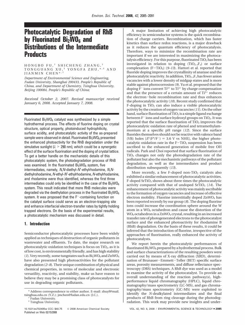
3. Results and Discussion3.1. Catalyst Characterization. X-ray diffraction patternswere used to investigate the changes of the phase struc-tures of the as-prepared samples. Figure 1 shows XRDpatterns of the samples prepared by the hydrothermalprocess. All of the as-prepared samples appeared to bephase-pure Bi2WO6 [JCPDS No. 73-1126]. Further observa-tion showed that the peak intensities of Bi2WO6 becamesteadily weaker with increasing RF, and the diffraction peaksof Bi2WO6 were wider, indicating the reducing of crystal-lization and formation of smaller Bi2WO6 crystallites. Thepresence of F- ions could play an important role in theformation of Bi2WO6 crystals. It was well-known that thephase formation of Bi2WO6 crystal occurred due to therearrangement of WO6 octahedra (3, 5). The crystallizationprocess of the Bi2WO6 phase from the amorphous precur-sors was postulated to initiate through face-sharingpolycondensation of these octahedrons. According to thesurface acidic/basic properties, the surface Bi-OH groupin the Bi2WO6 crystal should be at least partially protonatedto give BiOH2
+. Consequently, the free F- ions liberatedin the solution would interact with the small crystal.Nucleophilic substitution was favored due to the highelectronegativity of F- ions, which in turn reduced thechemical reactivity of the precursor. In the process of thecrystallization, F- ions were involved, resulting in the morevacancies in the Bi2WO6 crystal. Although more work isneeded to illustrate clearly these rather complex interac-tions, it is believed that the interaction of these anionicspecies with the surface Bi3+ ion prohibited the enlarge-ment of the crystal nuclei.
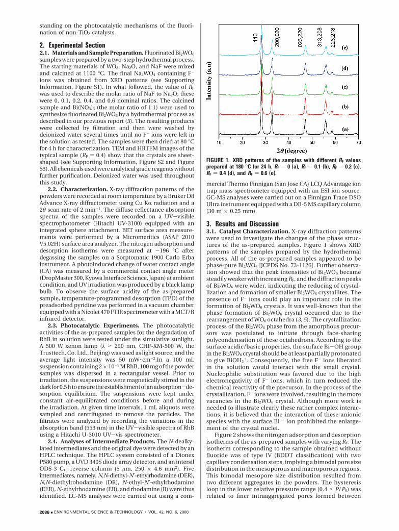
Figure 2 shows the nitrogen adsorption and desorptionisotherms of the as-prepared samples with varying RF. Theisotherm corresponding to the sample obtained withoutfluoride was of type IV (BDDT classification) with twocapillary condensation steps, implying a bimodal pore sizedistribution in the mesoporous and macroporous regions.This bimodal mesopore size distribution resulted fromtwo different aggregates in the powders. The hysteresisloop in the lower relative pressure range (0.4 < P/P0) wasrelated to finer intraaggregated pores formed between
FIGURE 1. XRD patterns of the samples with different RF valuesprepared at 180 °C for 24 h. RF ) 0 (a), RF ) 0.1 (b), RF ) 0.2 (c),RF ) 0.4 (d), and RF ) 0.6 (e).
2086 9 ENVIRONMENTAL SCIENCE & TECHNOLOGY / VOL. 42, NO. 6, 2008
intraagglomerated primary particles, and that in the higherrelative pressure range (0.8 < P/P0 < 1) was associatedwith larger interaggregated pores produced by interag-gregated secondary particles (15). The bimodal mesoporesize distribution was further confirmed by the corre-sponding pore size distributions shown in the inset ofFigure 2. The powder contained small mesopores of ca.1.6 nm and larger mesopores with a maximum porediameter of ca. 2.6 nm. The presence of fluoride in thesynthetic system exerted a significant influence on thepore structures of the obtained products. With increasingRF, the isotherms showed higher adsorption at high relativepressures, indicating the formation of the macropores and/or an increasing pore volume. The two separate hysteresisloops gradually joined together as one loop, implying thatthe pore size distributions of the intraaggregated andinteraggregated pores tended to overlap, as confirmed inthe inset of Figure 2. Furthermore, the pore volumes ofintraaggregated mesopores were negligible compared withthose of the interaggregated ones, based on their corre-sponding integral areas. The BET surface areas of Bi2WO6
products were also found to be highly dependent on RF.Each sample showed a monotonic increase in the BETsurface area with increasing RF, which was in goodagreement with the XRD results.
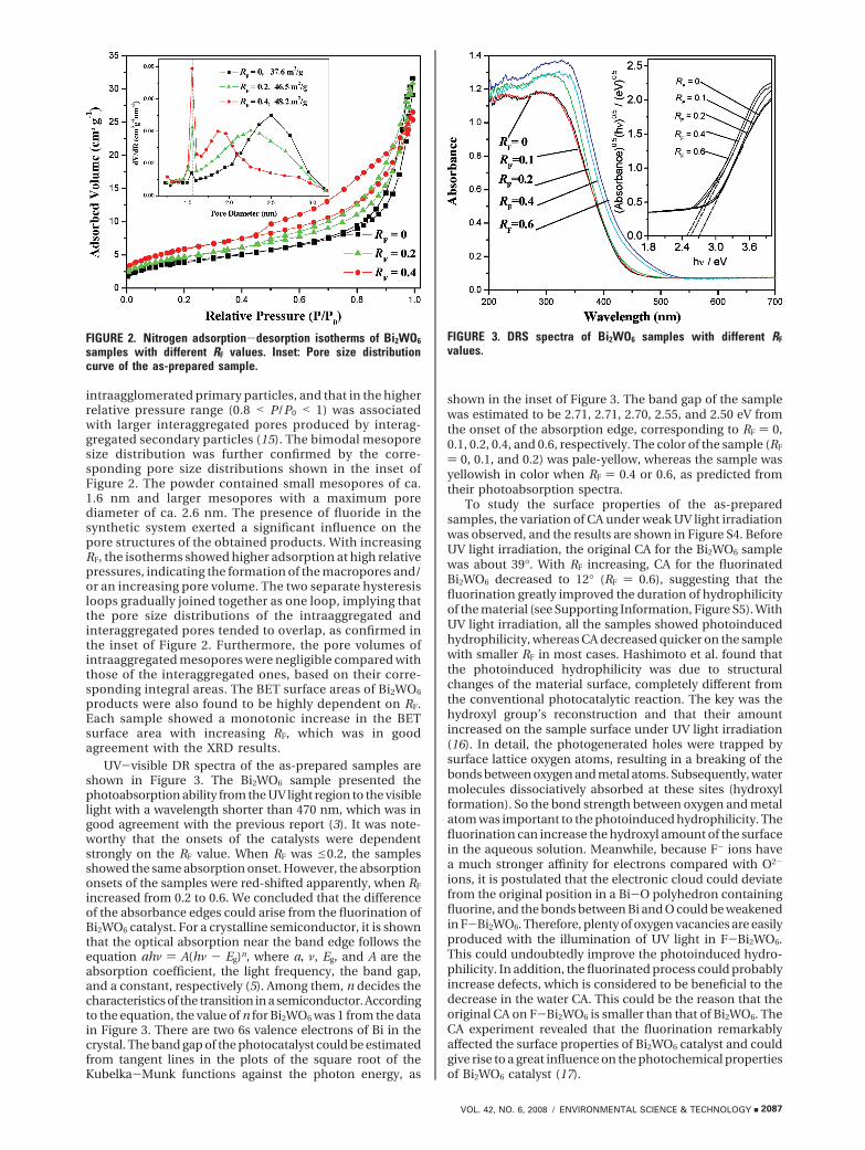
UV-visible DR spectra of the as-prepared samples areshown in Figure 3. The Bi2WO6 sample presented thephotoabsorption ability from the UV light region to the visiblelight with a wavelength shorter than 470 nm, which was ingood agreement with the previous report (3). It was note-worthy that the onsets of the catalysts were dependentstrongly on the RF value. When RF was e0.2, the samplesshowed the same absorption onset. However, the absorptiononsets of the samples were red-shifted apparently, when RF
increased from 0.2 to 0.6. We concluded that the differenceof the absorbance edges could arise from the fluorination ofBi2WO6 catalyst. For a crystalline semiconductor, it is shownthat the optical absorption near the band edge follows theequation ahν ) A(hν - Eg)n, where a, ν, Eg, and A are theabsorption coefficient, the light frequency, the band gap,and a constant, respectively (5). Among them, n decides thecharacteristics of the transition in a semiconductor. Accordingto the equation, the value of n for Bi2WO6 was 1 from the datain Figure 3. There are two 6s valence electrons of Bi in thecrystal. The band gap of the photocatalyst could be estimatedfrom tangent lines in the plots of the square root of theKubelka-Munk functions against the photon energy, as
shown in the inset of Figure 3. The band gap of the samplewas estimated to be 2.71, 2.71, 2.70, 2.55, and 2.50 eV fromthe onset of the absorption edge, corresponding to RF ) 0,0.1, 0.2, 0.4, and 0.6, respectively. The color of the sample (RF
) 0, 0.1, and 0.2) was pale-yellow, whereas the sample wasyellowish in color when RF ) 0.4 or 0.6, as predicted fromtheir photoabsorption spectra.
To study the surface properties of the as-preparedsamples, the variation of CA under weak UV light irradiationwas observed, and the results are shown in Figure S4. BeforeUV light irradiation, the original CA for the Bi2WO6 samplewas about 39°. With RF increasing, CA for the fluorinatedBi2WO6 decreased to 12° (RF ) 0.6), suggesting that thefluorination greatly improved the duration of hydrophilicityof the material (see Supporting Information, Figure S5). WithUV light irradiation, all the samples showed photoinducedhydrophilicity, whereas CA decreased quicker on the samplewith smaller RF in most cases. Hashimoto et al. found thatthe photoinduced hydrophilicity was due to structuralchanges of the material surface, completely different fromthe conventional photocatalytic reaction. The key was thehydroxyl group’s reconstruction and that their amountincreased on the sample surface under UV light irradiation(16). In detail, the photogenerated holes were trapped bysurface lattice oxygen atoms, resulting in a breaking of thebonds between oxygen and metal atoms. Subsequently, watermolecules dissociatively absorbed at these sites (hydroxylformation). So the bond strength between oxygen and metalatom was important to the photoinduced hydrophilicity. Thefluorination can increase the hydroxyl amount of the surfacein the aqueous solution. Meanwhile, because F- ions havea much stronger affinity for electrons compared with O2-
ions, it is postulated that the electronic cloud could deviatefrom the original position in a Bi-O polyhedron containingfluorine, and the bonds between Bi and O could be weakenedin F-Bi2WO6. Therefore, plenty of oxygen vacancies are easilyproduced with the illumination of UV light in F-Bi2WO6.This could undoubtedly improve the photoinduced hydro-philicity. In addition, the fluorinated process could probablyincrease defects, which is considered to be beneficial to thedecrease in the water CA. This could be the reason that theoriginal CA on F-Bi2WO6 is smaller than that of Bi2WO6. TheCA experiment revealed that the fluorination remarkablyaffected the surface properties of Bi2WO6 catalyst and couldgive rise to a great influence on the photochemical propertiesof Bi2WO6 catalyst (17).
FIGURE 2. Nitrogen adsorption-desorption isotherms of Bi2WO6
samples with different RF values. Inset: Pore size distributioncurve of the as-prepared sample.
FIGURE 3. DRS spectra of Bi2WO6 samples with different RF
values.
VOL. 42, NO. 6, 2008 / ENVIRONMENTAL SCIENCE & TECHNOLOGY 9 2087
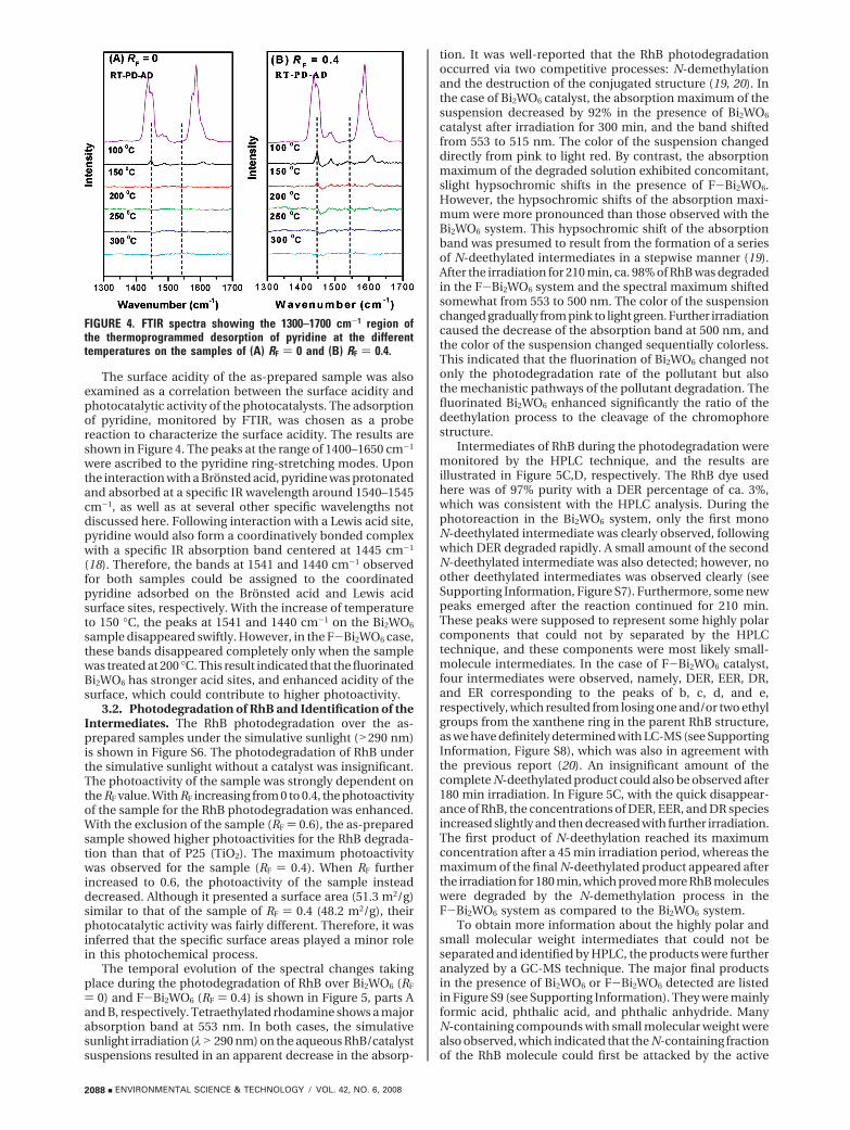
The surface acidity of the as-prepared sample was alsoexamined as a correlation between the surface acidity andphotocatalytic activity of the photocatalysts. The adsorptionof pyridine, monitored by FTIR, was chosen as a probereaction to characterize the surface acidity. The results areshown in Figure 4. The peaks at the range of 1400–1650 cm-1
were ascribed to the pyridine ring-stretching modes. Uponthe interaction with a Brönsted acid, pyridine was protonatedand absorbed at a specific IR wavelength around 1540–1545cm-1, as well as at several other specific wavelengths notdiscussed here. Following interaction with a Lewis acid site,pyridine would also form a coordinatively bonded complexwith a specific IR absorption band centered at 1445 cm-1
(18). Therefore, the bands at 1541 and 1440 cm-1 observedfor both samples could be assigned to the coordinatedpyridine adsorbed on the Brönsted acid and Lewis acidsurface sites, respectively. With the increase of temperatureto 150 °C, the peaks at 1541 and 1440 cm-1 on the Bi2WO6
sample disappeared swiftly. However, in the F-Bi2WO6 case,these bands disappeared completely only when the samplewas treated at 200 °C. This result indicated that the fluorinatedBi2WO6 has stronger acid sites, and enhanced acidity of thesurface, which could contribute to higher photoactivity.
3.2. Photodegradation of RhB and Identification of theIntermediates. The RhB photodegradation over the as-prepared samples under the simulative sunlight (>290 nm)is shown in Figure S6. The photodegradation of RhB underthe simulative sunlight without a catalyst was insignificant.The photoactivity of the sample was strongly dependent onthe RF value. With RF increasing from 0 to 0.4, the photoactivityof the sample for the RhB photodegradation was enhanced.With the exclusion of the sample (RF ) 0.6), the as-preparedsample showed higher photoactivities for the RhB degrada-tion than that of P25 (TiO2). The maximum photoactivitywas observed for the sample (RF ) 0.4). When RF furtherincreased to 0.6, the photoactivity of the sample insteaddecreased. Although it presented a surface area (51.3 m2/g)similar to that of the sample of RF ) 0.4 (48.2 m2/g), theirphotocatalytic activity was fairly different. Therefore, it wasinferred that the specific surface areas played a minor rolein this photochemical process.
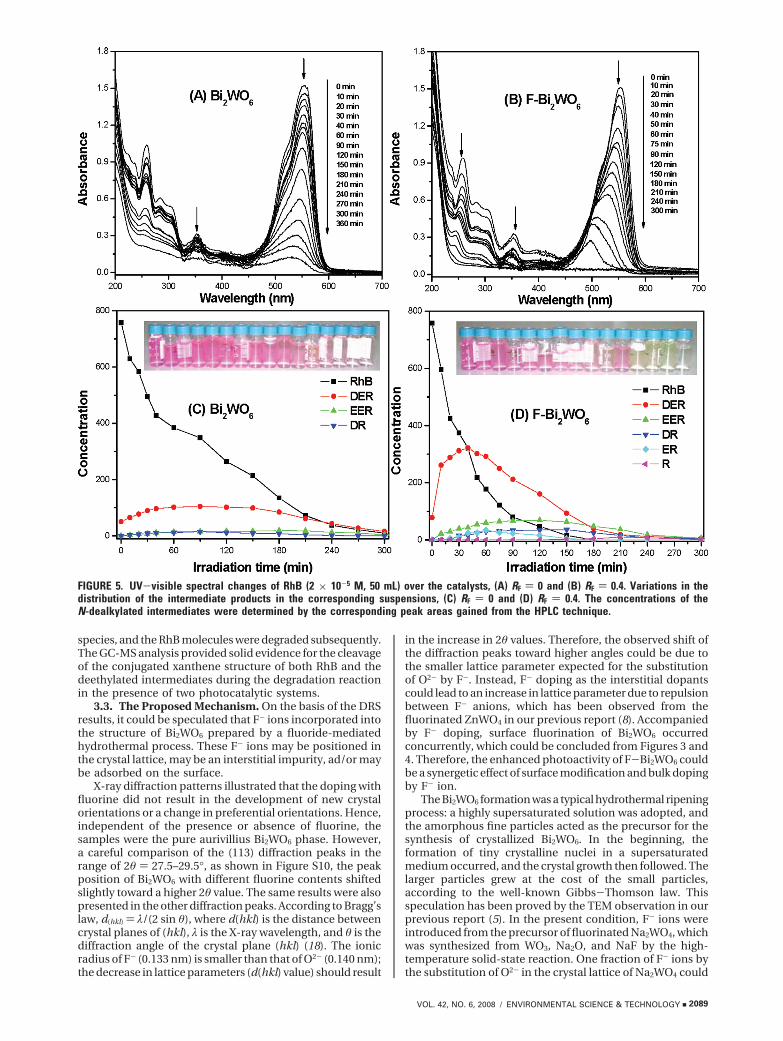
The temporal evolution of the spectral changes takingplace during the photodegradation of RhB over Bi2WO6 (RF
) 0) and F-Bi2WO6 (RF ) 0.4) is shown in Figure 5, parts Aand B, respectively. Tetraethylated rhodamine shows a majorabsorption band at 553 nm. In both cases, the simulativesunlight irradiation (λ>290 nm) on the aqueous RhB/catalystsuspensions resulted in an apparent decrease in the absorp-
tion. It was well-reported that the RhB photodegradationoccurred via two competitive processes: N-demethylationand the destruction of the conjugated structure (19, 20). Inthe case of Bi2WO6 catalyst, the absorption maximum of thesuspension decreased by 92% in the presence of Bi2WO6
catalyst after irradiation for 300 min, and the band shiftedfrom 553 to 515 nm. The color of the suspension changeddirectly from pink to light red. By contrast, the absorptionmaximum of the degraded solution exhibited concomitant,slight hypsochromic shifts in the presence of F-Bi2WO6.However, the hypsochromic shifts of the absorption maxi-mum were more pronounced than those observed with theBi2WO6 system. This hypsochromic shift of the absorptionband was presumed to result from the formation of a seriesof N-deethylated intermediates in a stepwise manner (19).After the irradiation for 210 min, ca. 98% of RhB was degradedin the F-Bi2WO6 system and the spectral maximum shiftedsomewhat from 553 to 500 nm. The color of the suspensionchanged gradually from pink to light green. Further irradiationcaused the decrease of the absorption band at 500 nm, andthe color of the suspension changed sequentially colorless.This indicated that the fluorination of Bi2WO6 changed notonly the photodegradation rate of the pollutant but alsothe mechanistic pathways of the pollutant degradation. Thefluorinated Bi2WO6 enhanced significantly the ratio of thedeethylation process to the cleavage of the chromophorestructure.
Intermediates of RhB during the photodegradation weremonitored by the HPLC technique, and the results areillustrated in Figure 5C,D, respectively. The RhB dye usedhere was of 97% purity with a DER percentage of ca. 3%,which was consistent with the HPLC analysis. During thephotoreaction in the Bi2WO6 system, only the first monoN-deethylated intermediate was clearly observed, followingwhich DER degraded rapidly. A small amount of the secondN-deethylated intermediate was also detected; however, noother deethylated intermediates was observed clearly (seeSupporting Information, Figure S7). Furthermore, some newpeaks emerged after the reaction continued for 210 min.These peaks were supposed to represent some highly polarcomponents that could not by separated by the HPLCtechnique, and these components were most likely small-molecule intermediates. In the case of F-Bi2WO6 catalyst,four intermediates were observed, namely, DER, EER, DR,and ER corresponding to the peaks of b, c, d, and e,respectively, which resulted from losing one and/or two ethylgroups from the xanthene ring in the parent RhB structure,as we have definitely determined with LC-MS (see SupportingInformation, Figure S8), which was also in agreement withthe previous report (20). An insignificant amount of thecomplete N-deethylated product could also be observed after180 min irradiation. In Figure 5C, with the quick disappear-ance of RhB, the concentrations of DER, EER, and DR speciesincreased slightly and then decreased with further irradiation.The first product of N-deethylation reached its maximumconcentration after a 45 min irradiation period, whereas themaximum of the final N-deethylated product appeared afterthe irradiation for 180 min, which proved more RhB moleculeswere degraded by the N-demethylation process in theF-Bi2WO6 system as compared to the Bi2WO6 system.
To obtain more information about the highly polar andsmall molecular weight intermediates that could not beseparated and identified by HPLC, the products were furtheranalyzed by a GC-MS technique. The major final productsin the presence of Bi2WO6 or F-Bi2WO6 detected are listedin Figure S9 (see Supporting Information). They were mainlyformic acid, phthalic acid, and phthalic anhydride. ManyN-containing compounds with small molecular weight werealso observed, which indicated that the N-containing fractionof the RhB molecule could first be attacked by the active
FIGURE 4. FTIR spectra showing the 1300–1700 cm-1 region ofthe thermoprogrammed desorption of pyridine at the differenttemperatures on the samples of (A) RF ) 0 and (B) RF ) 0.4.
2088 9 ENVIRONMENTAL SCIENCE & TECHNOLOGY / VOL. 42, NO. 6, 2008
species, and the RhB molecules were degraded subsequently.The GC-MS analysis provided solid evidence for the cleavageof the conjugated xanthene structure of both RhB and thedeethylated intermediates during the degradation reactionin the presence of two photocatalytic systems.
3.3. The Proposed Mechanism. On the basis of the DRSresults, it could be speculated that F- ions incorporated intothe structure of Bi2WO6 prepared by a fluoride-mediatedhydrothermal process. These F- ions may be positioned inthe crystal lattice, may be an interstitial impurity, ad/or maybe adsorbed on the surface.
X-ray diffraction patterns illustrated that the doping withfluorine did not result in the development of new crystalorientations or a change in preferential orientations. Hence,independent of the presence or absence of fluorine, thesamples were the pure aurivillius Bi2WO6 phase. However,a careful comparison of the (113) diffraction peaks in therange of 2θ ) 27.5–29.5°, as shown in Figure S10, the peakposition of Bi2WO6 with different fluorine contents shiftedslightly toward a higher 2θ value. The same results were alsopresented in the other diffraction peaks. According to Bragg’slaw, d(hkl) ) λ/(2 sin θ), where d(hkl) is the distance betweencrystal planes of (hkl), λ is the X-ray wavelength, and θ is thediffraction angle of the crystal plane (hkl) (18). The ionicradius of F- (0.133 nm) is smaller than that of O2- (0.140 nm);the decrease in lattice parameters (d(hkl) value) should result
in the increase in 2θ values. Therefore, the observed shift ofthe diffraction peaks toward higher angles could be due tothe smaller lattice parameter expected for the substitutionof O2- by F-. Instead, F- doping as the interstitial dopantscould lead to an increase in lattice parameter due to repulsionbetween F- anions, which has been observed from thefluorinated ZnWO4 in our previous report (8). Accompaniedby F- doping, surface fluorination of Bi2WO6 occurredconcurrently, which could be concluded from Figures 3 and4. Therefore, the enhanced photoactivity of F-Bi2WO6 couldbe a synergetic effect of surface modification and bulk dopingby F- ion.
The Bi2WO6 formation was a typical hydrothermal ripeningprocess: a highly supersaturated solution was adopted, andthe amorphous fine particles acted as the precursor for thesynthesis of crystallized Bi2WO6. In the beginning, theformation of tiny crystalline nuclei in a supersaturatedmedium occurred, and the crystal growth then followed. Thelarger particles grew at the cost of the small particles,according to the well-known Gibbs-Thomson law. Thisspeculation has been proved by the TEM observation in ourprevious report (5). In the present condition, F- ions wereintroduced from the precursor of fluorinated Na2WO4, whichwas synthesized from WO3, Na2O, and NaF by the high-temperature solid-state reaction. One fraction of F- ions bythe substitution of O2- in the crystal lattice of Na2WO4 could
FIGURE 5. UV-visible spectral changes of RhB (2 × 10⁻⁵ M, 50 mL) over the catalysts, (A) RF ) 0 and (B) RF ) 0.4. Variations in thedistribution of the intermediate products in the corresponding suspensions, (C) RF ) 0 and (D) RF ) 0.4. The concentrations of theN-dealkylated intermediates were determined by the corresponding peak areas gained from the HPLC technique.
VOL. 42, NO. 6, 2008 / ENVIRONMENTAL SCIENCE & TECHNOLOGY 9 2089
be directly introduced to the resulting sample as the dopingof the crystal lattice. Instead, the other fraction of F- ionscould be liberated into the solution in the hydrothermalprocess. It is well-known that the aurivillius structure ofBi2WO6 consists of alternating fluorite-like [Bi2O2]2+ layersand [WO4]2- layers which comprise the corner-linked oc-tahedral units. Due to the high electron density of F- ions,there is a strong repulsion between F- and [WO4]2-. Thus,the dissolution of F- ions into the interstitial lattice between[Bi2O2]2+ layers and [WO4]2- layers is limited. However, thisfraction of F- ions could interact strongly with the surfaceof the Bi2WO6 crystal, which prohibited the enlargement ofthe crystal, and enhanced the surface acidity of the Bi2WO6
catalyst.A few researchers have reported the improvement of
photocatalytic activity by the fluorination of TiO2 powders(9–13, 21). This high photocatalytic activity was ascribed toseveral beneficial effects produced by the F- ion doping inbulk TiO2 crystal: enhancement of the crystallinity, creationof oxygen vacancies, or increase of active sites by conversionof some Ti4+ to Ti3+. As for the surfaced fluorination of TiO2,the enhanced photoactivity of TiO2 was commonly ascribedto higher surface acidity, enhanced generation of mobilefree OH radicals, and surface oxygen vacancies. In this case,the resulting Bi2WO6 sample prepared by a fluoride-mediatedsynthesis showed the pure aurivillius phase but a fall in thecrystallinity. Since the production of •OH in the presentsystem was almost impossible from the theoretical viewpointdue to the standard redox potential of BiV/BiIII being morenegative than that of •OH/OH-(+1.99) (3). The enhancedphotoactivity of the as-prepared sample could not be ascribedto the generation of mobile free OH radicals. On the CAexperiment, the as-prepared samples showed the enhance-ment of the surface acidity, which could be one of the reasonsthat fluorinated Bi2WO6 showed higher photoactivity for theRhB degradation. The catalyst with an acidic surface couldeasily adsorb polarized organic reactants and thus promotetheir photocatalytic decomposition. The enhanced hydro-philicity of the as-prepared sample could also be attributedto more oxygen vacancies, which was considered to bebeneficial to the photoactivity of the catalyst. Beyond that,the absorption spectra of the as-prepared sample showed astronger absorption in the UV-visible range and a red shiftin the band gap transition. Therefore, the fluorination ofBi2WO6 increased the number of photogenerated electronsand holes to participate in the photocatalytic reaction, whichcould partly explain the enhanced photoactivity. Followingthis general information, we assume that the enhancedphotoactivity of F-Bi2WO6 could be a synergetic effect,including more oxygen defects, enhancement of surfaceacidy, and light absorbance.
In the present study, the fluorination of Bi2WO6 affectednot only the rate but also the mechanistic pathways of theRhB degradation. In the Bi2WO6-mediated photocatalysis,the degradation of RhB was completely ascribed to aphotochemical process involved by photoinduced holes.Furthermore, there could be two photochemical mecha-nisms, namely, a photocatalytic process and a photosensi-tized process. On one hand, Bi2WO6 was an active photo-catalyst, and RhB could be degraded by the direct interactionwith a strong oxidizing hole originating from the hybridizationof the Bi6s and O2p orbitals. The RhB molecule was attackedand then degraded via the destruction of the conjugatedstructure. On the other hand, RhB dye could absorb the visiblelight, which was attributed to the ground state and the excitedstate of the dye. Therefore, RhB could be degraded by aphotosensitized process. In this process, the degradation ofRhB did not involve any VB hole; mineralization of dyes foundits origins with the active oxygen radical species, O2
•-, andthe radical cations, dye•+. The photosensitized degradation
of RhB was commonly via the N-demethylation process (19).Compared to the photocatalytic process, the photosensitizedprocess required the stronger interaction between the dyeand the Bi2WO6 catalyst, which was beneficial to the transferof photoinduced electron. The F-containing function on thefluorinated surface seemed to serve as an electron-trappingsite and enhanced interfacial electron-transfer rates by tightlyholding trapped electrons. As a result, more RhB moleculeswere degraded via the N-demethylation process. It has beenreported that the surface fluorination of TiO2 by thedisplacement of OH by F- ions changed the surface interac-tions. Then, the rate of electron scavenging by O2 andsuperoxide formation would be influenced (21).
AcknowledgmentsThis work is supported by the National Natural ScienceFoundation of China (Grants 20433010, 20571047, 20747002and 20377008) and the National Program of China (Grant2007CB613303).
Supporting Information AvailableXRD patterns of F-Na2WO4, TEM, HRTEM, and CA variationof F-Bi2WO6, photodegradtion of RhB, HPLC, LC-MS, andGC-MS analysis of the intermediate products, and diffractionpeak variation of F-Bi2WO6, Figures S1–S10. This materialis available free of charge via the Internet at http://pubs.acs.org.
Literature Cited(1) Hoffmann, M. R.; Martin, S. T.; Choi, W.; Bahnemann, D. W.
Environmental applications of semiconductor photocatalysis.Chem. Rev. 1995, 95, 69–96.
(2) Zhu, S.; Xu, T.; Fu, H.; Zhao, J.; Zhu, Y. Synergetic effect ofBi2WO6 photocatalyst with C60 and enhanced photoactivityunder visible irradiation. Environ. Sci. Technol. 2007, 41, 6234–6239.
(3) Fu, H.; Pan, C.; Yao, W.; Zhu, Y. Visible-light-induced degradationof rhodamine B by nanosized Bi2WO6. J. Phys. Chem. B 2005,109, 22432–22439.
(4) Fu, H.; Yao, W.; Zhang, L.; Zhu, Y. Photocatalytic properties ofnanosized Bi2WO6 catalysts synthesized via a hydrothermalprocess. Appl. Catal., B 2006, 66, 100–110.
(5) Zhang, C.; Zhu, Y. Synthesis of square Bi2WO6 nanoplates ashigh-activity visible-light-driven photocatalysts. Chem. Mater.2005, 17, 3537–3545.
(6) Yu, J. G.; Xiong, J. F.; Cheng, B.; Yu, Y.; Wang, J. B. Hydrothermalpreparation and visible-light photocatalytic activity of Bi2WO6
powders. J. Solid State Chem. 2005, 178, 1968–1972.(7) Fu, H.; Lin, J.; Zhang, L.; Zhu, Y. Synthesis and photocatalytic
activities of ZnWO4 nanorod. Appl. Catal., A 2006, 306, 58–67.(8) Huang, G.; Zhu, Y. Enhanced photocatalytic activity of ZnWO4
catalyst via fluorine doping. J. Phys. Chem. C 2007, 111, 11952–11958.
(9) Hattori, A.; Shimoda, K.; Tada, H.; Ito, S. Photoreactivity ofsol-gel TiO2 films formed on soda-lime glass substrates: effectof SiO2 underlayer containing fluorine. Langmuir 1999, 15, 5422–5425.
(10) Yu, J. C.; Yu, J. G.; Ho, W.; Jiang, Z.; Zhang, L. Effects of F-
doping on the photocatalytic activity and microstructures ofnanocrystalline TiO2 powders. Chem. Mater. 2002, 14, 3808–3816.
(11) Li, D.; Haneda, H.; Hishita, S.; Ohashi, N.; Labhsetwar, K. N. J.Fluorine Chem. 2005, 126, 69–77.
(12) Vohra, S. M.; Kim, S.; Choi, W. Effects of surface fluorinationof TiO2 on the photocatalytic degradation of tetramethylam-monium. J. Photochem. Photobiol., A: Chem. 2003, 160, 55–60.
(13) Park, H.; Choi, W. Effects of TiO2 surface fluorination onphotocatalytic reactions and photoelectrochemical behaviors.J. Phys. Chem. B 2004, 108, 4086–4093.
(14) Wang, J. S.; Yin, S.; Zhang, Q. W.; Saito, F.; Sato, T. Mechan-ochemical synthesis of SrTiO3-xFx with high visible lightphotocatalytic activities for nitrogen monoxide destruction. J.Mater. Chem. 2003, 13, 2348–2352.
(15) Yu, J. G.; Liu, S.; Yu, H. Microstructures and photoactivity ofmesoporous anatase hollow microspheres fabricated by fluoride-mediated self-transformation. J. Catal. 2007, 249, 59–66.
2090 9 ENVIRONMENTAL SCIENCE & TECHNOLOGY / VOL. 42, NO. 6, 2008
(16) Mryauchi, M.; Nakajima, A.; Watanabe, T.; Hashimoto, K.Photocatalysis and photoinduced hydrophilicity of various metaloxide thin films. Chem. Mater. 2002, 14, 2812–2816.
(17) Tang, J.; Quan, H.; Ye, J. Photocatalytic properties and photo-induced hydrophilicity of surface-fluorinated TiO2. Chem. Mater.2007, 19, 116–122.
(18) Platon, A.; Thomson, W. J. Quantitative Lewis/Brönsted ratiosusing DRIFTS. Ind. Eng. Chem. Res. 2003, 42, 5988–5992.
(19) Lei, P.; Chen, C.; Yang, J.; Ma, W.; Zhao, J.; Zang, L. Degradationof dye pollutants by immobilized polyoxometalate with H2O2
under visible-light irradiation. Environ. Sci. Technol. 2005, 39,8466–8474.
(20) Chen, C.; Zhao, W.; Li, J.; Zhao, J. Formation and identificationof intermediates in the visible-light-assisted photodegradationof sulforhodamine-B dye in aqueous TiO2 dispersion. Environ.Sci. Technol. 2002, 36, 3604–3611.
(21) Calza, P.; Pelizzetti, E.; Mogyorosi, K.; Kun, R.; Dekany, I. Sizedependent photocatalytic activity of hydrothermally crystal-lized titania nanoparticles on poorly adsorbing phenol inabsence and presence of fluoride ion. Appl. Catal., B 2007, 72,314–321.
ES702495W
VOL. 42, NO. 6, 2008 / ENVIRONMENTAL SCIENCE & TECHNOLOGY 9 2091