PHENOLICS OF CYPERUS ALOPECUROIDES ROTTB. INFLORESCENCES AND THEIR BIOLOGICAL ACTIVITIES
24
Bull. Pharm. Sci., Assiut University, Vol. 29, Part 1, June 2006, pp. 9-32. PHENOLICS OF CYPERUS ALOPECUROIDES ROTTB. INFLORESCENCES AND THEIR BIOLOGICAL ACTIVITIES H. M. Sayed 1 , M. H. Mohamed 2 , S. F. Farag 1 , G. A. Mohamed 2 , R. Ebel 3 , O. R. M. Omobuwajo 4 and P. Proksch 3 1 Department of Pharmacognosy, Faculty of Pharmacy, Assiut University, Assiut, Egypt 2 Department of Pharmacognosy, Faculty of Pharmacy, Al-Azhar University, Assiut, Egypt 3 Institute of Pharmaceutical Biology and Biotechnology, Heinrich- Heine-University, Universitätsstraße 1, 40225 Düsseldorf, Germany 4 Department of Pharmacognosy, Faculty of Pharmacy, Obafemi Awolowo University, Ile-Ife, Nigeria اﻟﻜﺤﻮل ﺧﻼﺻﺔ ﻣﻦ ﻓﻴﻨﻮﻟﻴﺎ ﻣﺮآﺒﺎ ﻋﺸﺮ ﺳﺘﺔ ﻓﺼﻞ اﻟﺒﺤﺚ هﺬا ﻓﻰ ﺗﻢ أﻟﻮﺑﻴﻜ ﺳﻴﺒﺮس ﻧﺒﺎت ﻟﻨﻮرات اﻟﻤﻴﺜﻴﻠﻰ ﺮو روﺗﺐ ﻳﺪس. ﻋﻠﻴﻬﺎ اﻟﺘﻌﺮف ﺗﻢ وﻗﺪ آﺎﻵﺗﻰ: ﺳﻜﻮﺑﻮﻟﻴﺘﻴﻦ) 1 ( , أﻳﺰوﻟﻴﻜﻮﻳﺮﻳﺘ ﻴﺠﻴﻨﻴﻦ4 ′ - إﻳﺜﺮ ﻣﻴﺜﻴﻞ) 2 ( , ﻟﻴﻮﺗﻴﻮﻟﻴﻦ7 و3 ′ - إﻳﺜﺮ ﻣﻴﺜﻴﻞ ﺛﻨﺎﺋﻰ) 3 ( , ﻟﻴﻮﺗﻴﻮﻟﻴﻦ5 و3 ′ - ﻣﻴﺜﻴﻞ ﺛﻨﺎﺋﻰ إﻳﺜﺮ) 4 ( , أورﻳﻮﺳﻴﺪﻳﻦ4 - إﻳﺜﺮ ﻣﻴﺜﻴﻞ) 5 ( , أﺑﻴﺠﻴﻨﻴﻦ) 6 ( , ﻟﻴﻮﺗﻴﻮﻟﻴﻦ) 7 ( , اﻟﻔﺮﻳﻮﻟﻴﻚ ﺣﻤﺾ) 8 ( , ﻟﻴﻮﺗﻴﻮﻟﻴﻦ4 ′ - أ- ﺑﻴﺘﺎ- ﺟﻠﻮآﻮﺑﻴﺮ ا ﻧﻮزﻳﺪ) 9 ( , ﻟﻴﻮﺗﻴﻮﻟﻴﻦ7 - أ- ﺑﻴﺘﺎ- ﺟﻠﻮآﻮﺑﻴﺮ ا ﻧﻮز ﻳﺪ) 10 ( , آﻮﻳﺮﺳﻴﺘﻴﻦ3 - أ- ﺑﻴﺘﺎ- ﺟﻠﻮآﻮﺑﻴﺮاﻧﻮزﻳﺪ) 11 ( , أﺑﻴﺠﻴﻨﻴﻦ7 - أ- ﻧﻴﻮهﺴﺒﻴﺮﻳﺪوزﻳﺪ) 12 ( , آﻤﺒﻔﻴﺮول3 - أ- روﺗﻴﻨﻮزﻳﺪ) 13 ( , آﻮﻳﺮﺳﻴﺘﻴﻦ3 - أ- روﺗﻴﻨﻮزﻳﺪ) 14 ( , آﻤﺒﻔﻴﺮول3 - أ- ] 2 - أ- زﻳﻠﻮﺑﻴﺮاﻧﻮزﻳﺪ- 6 - أ- أﻟﻔﺎ- راﻣﻨﻮﺑﻴﺮاﻧﻮزﻳﺪ[ ) 15 ( , آﻤﺒﻔﻴﺮول3 - أ- ] 2 - أ- ﺟﻠﻮآﻮﺑﻴﺮاﻧﻮزﻳﺪ- 6 - أ- أﻟﻔﺎ- را ﻣﻨﻮﺑﻴﺮاﻧﻮزﻳﺪ[ ) 16 .( اﻟﻤﺮآﺒﺎ ن) 2 و5 ( ﻳﻔﺼﻼ واﻟﻤﺮآﺒﺎن اﻟﺴﻴﺒﺮس ﺟﻨﺲ ﻣﻦ ﻣﺮة ﻷول) 15 و16 ( ﻳﻔﺼﻼ ﻷول اﻟﺴﻌﺪﻳﺔ اﻟﻌﺎﺋﻠﺔ ﻣﻦ ﻣﺮة اﻷﺧﺮى واﻟﻤﺮآﺒﺎت) ﻣﺎﻋﺪا7 و10 و14 ( ﺗﻔﺼﻞ اﻟﻨﺒﺎت ﻣﻦ ﻣﺮة ﻷول. ﺑﺪراﺳﺔ اﻟﻤﺮآﺒﺎت ﻟﻬﺬﻩ اﻟﺪﻗﻴﻖ اﻟﺘﺮآﻴﺐ ﻋﻠﻰ اﻟﺘﻌﺮف ﺗﻢ وﻗﺪ واﻟﻜ اﻟﻄﺒﻴﻌﻴﺔ ﺧﻮاﺻﻬﺎ اﻟﻤﺨﺘﻠﻔﺔ اﻟﻄﻴﻔﻰ اﻟﺘﺤﻠﻴﻞ ﻃﺮق وﺑﺈﺳﺘﺨﺪام ﻴﻤﻴﺎﺋﻴﺔ اﻟﻤﺨﺘﻠﻔﺔ ﺑﺎﻟﻤﺮاﺟﻊ ﻣﻘﺎرﻧﺘﻬﺎ إﻟﻰ ﺑﺎﻹﺿﺎﻓﺔ اﻟﻘﻴﺎﺳﻴﺔ اﻟﻌﻴﻨﺎت ﻣﻦ ﺑﻤﺜﻴﻼﺗﻬﺎ أو. آﻤﻀﺎدات ﻓﻌﺎﻟﺔ ﺗﺄﺛﻴﺮات اﻟﻤﻔﺼﻮﻟﺔ اﻟﻤﺮآﺒﺎت ﺑﻌﺾ أﻇﻬﺮت وﻗﺪ اﻷﻟﻔﺎ ﻷﻧﺰﻳﻢ وﻣﺜﺒﻄﺎت اﻟﺴﺮﻃﺎﻧﻴﺔ اﻟﺨﻼﻳﺎ ﺑﻌﺾ ﻋﻠﻰ وﺳﺎﻣﺔ ﻟﻸآﺴﺪة أﻣﻴﻠﻴﺰ. ــــــــــــــــــــــــــــــــــــــــــــــــــــــــــــــــــــــــــReceived in 7/8/2005 & Accepted in 22/9/2005
-
Upload
independent -
Category
Documents
-
view
1 -
download
0
Transcript of PHENOLICS OF CYPERUS ALOPECUROIDES ROTTB. INFLORESCENCES AND THEIR BIOLOGICAL ACTIVITIES

Bull. Pharm. Sci., Assiut University, Vol. 29, Part 1, June 2006, pp. 9-32.
PHENOLICS OF CYPERUS ALOPECUROIDES ROTTB. INFLORESCENCES AND THEIR BIOLOGICAL ACTIVITIES H. M. Sayed1, M. H. Mohamed2, S. F. Farag1, G. A. Mohamed2, R. Ebel3, O. R. M. Omobuwajo4 and P. Proksch3
1Department of Pharmacognosy, Faculty of Pharmacy, Assiut University, Assiut, Egypt
2Department of Pharmacognosy, Faculty of Pharmacy, Al-Azhar University, Assiut, Egypt
3Institute of Pharmaceutical Biology and Biotechnology, Heinrich-Heine-University, Universitätsstraße 1, 40225 Düsseldorf, Germany
4Department of Pharmacognosy, Faculty of Pharmacy, Obafemi Awolowo University, Ile-Ife, Nigeria
تم فى هذا البحث فصل ستة عشر مرآبا فينوليا من خالصة الكحول وقد تم التعرف عليها . يدس روتب روالميثيلى لنورات نبات سيبرس ألوبيك
, )2( ميثيل إيثر -′4يجينين أيزوليكويريت, )1(سكوبوليتين : آاآلتى ثنائى ميثيل -′3 و 5ليوتيولين , ) 3( ثنائى ميثيل إيثر -′3و7ليوتيولين
, )7(ليوتيولين , )6(أبيجينين , )5(ميثيل إيثر -4أوريوسيدين , )4(إيثر , )9(نوزيد ا جلوآوبير -بيتا-أ-′4ليوتيولين, )8(حمض الفريوليك
-بيتا-أ-3آويرسيتين , )10(يد نوزاجلوآوبير-بيتا-أ-7ليوتيولين آمبفيرول , )12( نيوهسبيريدوزيد -أ-7أبيجينين , )11(جلوآوبيرانوزيد
[-أ-3آمبفيرول , )14(روتينوزيد -أ-3آويرسيتين , )13(روتينوزيد -أ-3-2[-أ-3آمبفيرول , )15 (]رامنوبيرانوزيد- ألفا -أ-6-زيلوبيرانوزيد-أ-2) 5 و 2 (نالمرآبا). 16 (]منوبيرانوزيدرا-ألفا-أ-6-جلوآوبيرانوزيد-أ
ألول يفصال) 16 و 15( ألول مرة من جنس السيبرس والمرآبان يفصالتفصل ) 14 و 10 و 7ماعدا ( والمرآبات األخرى مرة من العائلة السعدية .ألول مرة من النبات
وقد تم التعرف على الترآيب الدقيق لهذه المرآبات بدراسة يميائية وبإستخدام طرق التحليل الطيفى المختلفة خواصها الطبيعية والك
. أو بمثيالتها من العينات القياسيةباإلضافة إلى مقارنتها بالمراجع المختلفةوقد أظهرت بعض المرآبات المفصولة تأثيرات فعالة آمضادات لألآسدة وسامة على بعض الخاليا السرطانية ومثبطات ألنزيم األلفا
.أميليز
ــــــــــــــــــــــــــــــــــــــــــــــــــــــــــــــــــــــــــReceived in 7/8/2005 & Accepted in 22/9/2005

H. M. Sayed, et al.
Sixteen phenolic compounds, scopoletin (1), isoliquiritigenin 4′-methyl ether (2), luteolin 5,3′-dimethyl ether (3), luteolin 7,3′-dimethyl ether (4), aureusidin 4-methyl ether (5), apigenin (6), luteolin (7), trans-ferulic acid (8), luteolin 4′-O-β-D-gluco-pyranoside (9), luteolin 7-O-β-D-glucopyranoside (10), quercetin 3-O-β-D-glucopyranoside (11), apigenin 7-O-neohesperidoside (12), kaempferol 3-O-rutinoside (13), quercetin 3-O-rutinoside (14), kaempferol 3-O-[2-O-D-xylopyranosyl-6-O-α-L-rhamno-pyranosyl]-β-D-glucopyranoside (15) and kaempferol 3-O-[2-O-D-glucopyranosyl-6-O-α-L-rhamnopyranosyl]-β-D-gluco-pyranoside (16) were isolated from the methanolic extract of the inflorescences of Cyperus alopecuroides Rottb. for the first time. Their structures have been established on the basis of physical, chemical and spectroscopic methods in addition to comparison with literature data and/or authentic samples. The antioxidant and cytotoxic activities in addition to α-amylase inhibitory activity of the isolated compounds have been studied. INTRODUCTION
The sedge family, Cyperaceae
comprises about 4000 species within 90 genera. Cyperus is the largest genus that includes about 550 species.1
Cyperus alopecuroides Rottb. (Foxtail sedge; Samar) is a perennial stout leafy herb, reaching up to 150 cm high, having triangular culm, broad flat leaves, large numerous lanceolate inflorescences and widely distributed in tropical areas. In Egypt, it is naturally growing at the Nile Delta, the borders between fields and also cultivated in limited areas for commercial manufacture of mat and traditional chair-making.2
Previous phytochemical studies on Cyperus species led to the isolation of quinones,3-5 flavonoids,6-9 coum-
arins,10&11 furanochromones,12 sesqui-terpenes,13-17 alkaloids,18 saponins,19 sterols,19&20 fatty acids20 and phenolic acids.21
Many Cyperus species are used as foods. In folk medicine, they are used as spermatogenic, aphrodisiac, galactagogue, pectoral, emollient, anthelmintic, diaphoretic, astringent, digestive tonic, anti-inflammatory, antispasmodic, antirheumatic and hepatoprotective. Also, they are used as diuretic and to promote menstruation.17,22-25
The ethanolic extract of the overground parts of Cyperus alopecuroides Rottb. produced signs of pain and allergy on rabbit’s skin.26&27 These results revealed that the extract contained histamine or histamine like substance.26&27 Also, the ethanolic and the ethereal extracts
10
of the overground parts showed antimicrobial activity10&27 while the ethanolic extract of the inflorescences showed moderate estrogenic activity.28
Concerning the diversity of chemical classes of C. alopecuroides Rottb., several previous reports were cited: sesquiterpenes and a diterpene from the essential oil of the tubers and rhizomes;15,29&30 quinones from the rhizomes and the inflore-scences;3&28 coumarins from the aerial parts10 in addition to flavonoids from the leaves and inflorescences6&28 were isolated. Also, the carbohydrate, lipid, protein and amino acid contents from the overground parts of the plant were studied.26 In the present paper, we wish to describe the isolation and characterization in addition to investigation of the biological activities of the phenolic constituents of the inflorescences of this plant.
EXPERIMENTAL
General procedures
The UV spectra were carried out in methanol (Merck) using a Perkin-Elmer Lambda 25 UV/VIS spectrophotometer. Electron impact mass spectra (EIMS) were recorded on a Finnigan MAT TSQ 7000 mass spectrometer. Positive-ion electro-spray ionization mass spectra (ESIMS) were performed on a Thermofinnigan LCQ DECA mass spectrometer coupled to an Agilent 1100 HPLC system equipped with a photodiode-array detector. Gas chromatographic (GC) analysis was carried out using an Agilent 6850
series gas chromatograph coupled to a flame ionization detector (FID) and HP-5 column (cross linked 5% PHM/Siloxan, 30 m × 250 µm diameter × 0.25 µm film thickness, Macherey und Nagel, Düren, Germany). 1H- and 13C-NMR spectra were measured on Brüker DRX 500 spectrometer (Brüker, Rheinstetten, Germany). High performance liquid chromatography was performed on a HPLC system (Merck, Darmstadt, Germany) consisting of a Lachrom-Merck Hitachi L-7100 pump and an L-7400 UV detector using a C-18 column (250 × 8 mm i.d., prefilled with Eurospher 100, Knauer, Berlin, Germany), a flow rate 5.0 ml/ min, UV detection at λ 280 nm and HPLC gradient programm: 60:40 methanol/water at 0 and 5 min; 100:0 methanol/water at 38 and 45 min. Vacuum liquid chromatography (VLC) was carried out on silica gel 60 (0.04-0.063 mm, 500 g, Merck). Column chromatographic separations were performed over silica gel 60 (0.040-0.063 mm, Merck), Sephadex LH-20 (0.25-0.1 mm, Merck) and RP-18 columns (40-63 µm, Merck). TLC analyses were carried out on pre-coated silica gel F254 aluminium sheets and RP-18 F254 S glass plates (Merck). Compounds were detected by UV absorption at λ 254 and 366 nm followed by spraying with anisaldehyde/H2SO4 reagent and heating at 110°C for 1-2 min. The solvent systems used for TLC analyses were: dichloromethane-methanol (9.5:0.5, solvent system I), dichloromethane-methanol (9:1, solvent system II), dichloromethane-
11
H. M. Sayed, et al.
methanol (8:2, solvent system III), dichloromethane-methanol (6:4, solvent system IV) and n-butanol−acetone−formic acid−water (60:17:8:15, solvent system V). Authentic flavonoids were obtained from the Institute of Pharmaceutical Biology and Biotechnology, Heinrich -Heine-University, Universitätsstraße 1, 40225 Düsseldorf, Germany. 1,1-Diphenyl-2-picrylhydrazyl radical (DPPH), propyl gallate (PG) and reference sugars were purchased from Sigma Chemical Co. (Germany). The EnzCheck Amylase Assay Kit (E-11954) was purchased from Molecular Probes (GmbH, Germany). Acarbose (Ac) was purchased from Kohlpharma (GmbH, Germany).
Plant material
The inflorescences of Cyperus alopecuroides Rottb. were collected in July 2002 from plants growing on the margins of the ponds and banks of the Nile-River, Mankabad, Assiut, Egypt. The plant was kindely identified by Prof. Dr. Salah El-Nagar, Professor of Plant Taxonomy, Faculty of Science, Assiut University, Assiut, Egypt. A voucher specimen has been deposited at the Herbarium of Pharmacognosy Department, Faculty of Pharmacy, Assiut University, Assiut, Egypt.
Extraction and isolation
The air-dried powdered inflorescences of C. alopecuroides Rottb. (2 kg) were extracted with methanol (6 L × 4) at room temperature. The combined extract was concentrated under reduced pressure to afford a dark brownish
green residue (130.0 g). The latter was suspended in distilled water (500 ml) then partitioned with n-hexane (500 ml × 4), ethyl acetate (500 ml × 4) and n-butanol (500 ml × 3), successively. Each fraction was concentrated under reduced pressure to give n-hexane (36.5 g), ethyl acetate (31.8 g), n-butanol (28.5 g) and aqueous (33.0 g) residue.
About 30.8 g of the ethyl acetate fraction was subjected to VLC using n-hexane-ethyl acetate and ethyl acetate-methanol gradients to obtain seven group fractions: fraction E-1 (4.3 g, eluted with n-hexane-ethyl acetate 75:25), fraction E-2 (5.3 g, eluted with n-hexane-ethyl acetate 50:50), fraction E-3 (3.0 g, eluted with n-hexane-ethyl acetate 25:75), fraction E-4 (3.7 g, eluted with ethyl acetate), fraction E-5 (3.0 g, eluted with ethyl acetate-methanol 75:25), fraction E-6 (4.0 g, eluted with ethyl acetate- methanol 50:50) and fraction E-7 (6.0 g, eluted with methanol). About 500 mg of fraction E-2 was submitted to HPLC to yield pure compounds 1 (8 mg), 2 (3 mg), 3 (9 mg), 4 (5 mg) and 5 (9 mg). Fraction E-3 was subjected to silica gel column chromatography (90.0 g, 50 × 3 cm) using dichloromethane-methanol 94:6 to afford pure compounds 6 (11 mg), 7 (15 mg) and 8 (11 mg). About 2.0 g of fraction E-6 was chromatographed over Sephadex LH-20 column (100 g, 60 × 2 cm) using methanol as an eluent to obtain two main subfractions A (500 mg) and B (1.0 g). Repeated crystallization of subfraction E-6-A from methanol yielded pure
12
compound 9 (17 mg). The subfraction E-6-B was rechromatographed over RP-18 column (100 g, 30 × 1.5 cm) using methanol-water 2:3 to afford pure compounds 10 (16 mg) and 11 (12 mg).
About 27.5 g of the n-butanol fraction was submitted to VLC using gradient dichloromethane-methanol to get four group fractions: fraction B-1 (4.0 g, eluted with dichloromethane-methanol 75:25), fraction B-2 (7.2 g, eluted with dichloromethane-methanol 50:50), fraction B-3 (8.8 g, eluted with dichloromethane-methanol 25:75) and fraction B-4 (6.0 g, eluted with methanol). About 2.0 g of fraction B-2 was chromatographed on Sephadex LH-20 column (100 g, 60 × 2 cm) using methanol to give two main subfractions B-2-I (700 mg) and B-2-II (1.0 g). Rechromatography of the subfraction B-2-I on RP-18 column (100 g, 30 × 1.5 cm) using methanol-water 30:70 afforded pure compounds 12 (18 mg) and 13 (12 mg). Purification of the subfraction B-2-II on silica gel column (30 g, 30 × 1.5 cm) using dichloromethane-methanol (70:30) yielded pure compound 14 (21 mg). About 2 g of fraction B-3 was purified by Sephadex LH-20 column chromatography (100 g, 60 × 2 cm) using methanol followed by rechromatography on RP-18 column
(100 g, 30 × 1.5 cm) using methanol-water 30:70 to give pure compounds 15 (26 mg) and 16 (13 mg).
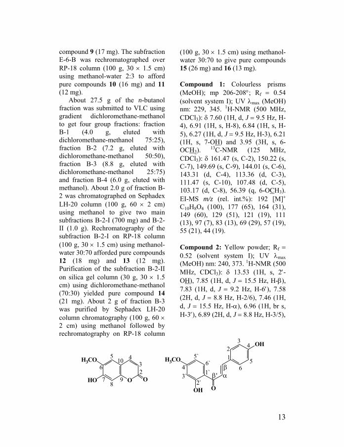
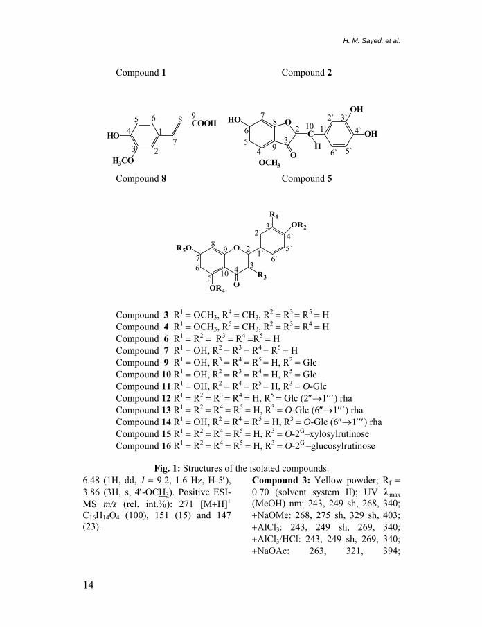
Compound 1: Colourless prisms (MeOH); mp 206-208°; Rf = 0.54 (solvent system I); UV λmax (MeOH) nm: 229, 345. 1H-NMR (500 MHz, CDCl3): δ 7.60 (1H, d, J = 9.5 Hz, H-4), 6.91 (1H, s, H-8), 6.84 (1H, s, H-5), 6.27 (1H, d, J = 9.5 Hz, H-3), 6.21 (1H, s, 7-OH) and 3.95 (3H, s, 6-OCH3). 13C-NMR (125 MHz, CDCl3): δ 161.47 (s, C-2), 150.22 (s, C-7), 149.69 (s, C-9), 144.01 (s, C-6), 143.31 (d, C-4), 113.36 (d, C-3), 111.47 (s, C-10), 107.48 (d, C-5), 103.17 (d, C-8), 56.39 (q, 6-OCH3). EI-MS m/z (rel. int.%): 192 [M]+ C10H8O4 (100), 177 (65), 164 (31), 149 (60), 129 (51), 121 (19), 111 (13), 97 (7), 83 (13), 69 (29), 57 (19), 55 (21), 44 (19). Compound 2: Yellow powder; Rf = 0.52 (solvent system I); UV λmax (MeOH) nm: 240, 373. 1H-NMR (500 MHz, CDCl3): δ 13.53 (1H, s, 2′-OH), 7.85 (1H, d, J = 15.5 Hz, H-β), 7.83 (1H, d, J = 9.2 Hz, H-6′), 7.58 (2H, d, J = 8.8 Hz, H-2/6), 7.46 (1H, d, J = 15.5 Hz, H-α), 6.96 (1H, br s, H-3′), 6.89 (2H, d, J = 8.8 Hz, H-3/5),
OHO
H3CO
O
105
6
78
9
432
O
OH
OH
H3CO 12
3
56
4
1`
2`3`4`
5`6`
αβ
β'
13
H. M. Sayed, et al.
Compound 1 Compound 2
6` 5`
4`3`2`
1`10
9
87
65
43
2O
OCH3
HO
C
OH
OH
OH
98
7
654
3 2
1HO
H3CO
COOH
Compound 8 Compound 5
6`5`4`
3`2`
1`
10
98
76
54 3
2O
O
R1
R3
OR4
R5O
OR2
Compound 3 R1 = OCH3, R4 = CH3, R2 = R3 = R5 = H Compound 4 R1 = OCH3, R5 = CH3, R2 = R3 = R4 = H Compound 6 R1 = R2 = R3 = R4 =R5 = H Compound 7 R1 = OH, R2 = R3 = R4 = R5 = H Compound 9 R1 = OH, R3 = R4 = R5 = H, R2 = Glc Compound 10 R1 = OH, R2 = R3 = R4 = H, R5 = Glc Compound 11 R1 = OH, R2 = R4 = R5 = H, R3 = O-Glc Compound 12 R1 = R2 = R3 = R4 = H, R5 = Glc (2″→1′′′) rha Compound 13 R1 = R2 = R4 = R5 = H, R3 = O-Glc (6″→1′′′) rha Compound 14 R1 = OH, R2 = R4 = R5 = H, R3 = O-Glc (6″→1′′′) rha Compound 15 R1 = R2 = R4 = R5 = H, R3 = O-2G–xylosylrutinose Compound 16 R1 = R2 = R4 = R5 = H, R3 = O-2G –glucosylrutinose
Fig. 1: Structures of the isolated compounds.
6.48 (1H, dd, J = 9.2, 1.6 Hz, H-5′), 3.86 (3H, s, 4′-OCH3). Positive ESI-MS m/z (rel. int.%): 271 [M+H]+ C16H14O4 (100), 151 (15) and 147 (23).
Compound 3: Yellow powder; Rf = 0.70 (solvent system II); UV λmax (MeOH) nm: 243, 249 sh, 268, 340; +NaOMe: 268, 275 sh, 329 sh, 403; +AlCl3: 243, 249 sh, 269, 340; +AlCl3/HCl: 243, 249 sh, 269, 340; +NaOAc: 263, 321, 394;
14
+NaOAc/H3BO3: 268, 347. 1H-NMR (500 MHz, CD3OD): δ 7.47 (1H, dd, J = 8.5, 2.2 Hz, H-6′), 7.45 (1H, d, J = 2.2 Hz, H-2′), 6.92 (1H, d, J = 8.5 Hz, H-5′), 6.55 (1H, s, H-3), 6.54 (1H, d, J = 2.2 Hz, H-8), 6.41 (1H, d, J = 2.2 Hz, H-6), 3.95 (3H, s, 3′-OCH3), 3.88 (3H, s, 5-OCH3). Positive-ion ESI-MS m/z (rel. int.%): 315 [M+H]+ C17H14O6 (100). Compound 4: Yellow powder; Rf = 0.65 (solvent system II); UV λmax (MeOH) nm: 245 sh, 252, 267, 347; +NaOMe: 262, 292 sh, 402; +AlCl3: 267 sh, 274, 296, 369 sh, 392; +AlCl3/HCl: 262, 276, 291 sh, 355, 390; +NaOAc: 254, 263 sh, 288, 350, 406; +NaOAc/H3BO3: 250, 267, 348. 1H-NMR (500 MHz, CDCl3): δ 12.79 (1H, s, 5-OH), 7.49 (1H, dd, J = 7.5, 2.2 Hz, H-6′), 7.33 (1H, d, J = 2.2 Hz, H-2′), 7.04 (1H, d, J = 7.5 Hz, H-5′), 6.57 (1H, s, H-3), 6.49 (1H, d, J = 2.2 Hz, H-8), 6.38 (1H, d, J = 2.2 Hz, H-6), 4.01 (3H, s, 3′-OCH3), 3.89 (3H, s, 7-OCH3). Positive-ion ESI-MS m/z (rel. int.%): 315 [M+H]+ C17H14O6 (100). Compound 5: Yellow powder; Rf = 0.59 (solvent system II); UV λmax (MeOH) nm: 254 sh, 270 sh, 330 sh, 400; +NaOMe: 253, 273 sh, 383 sh, 402, 465; +AlCl3: 259 sh, 287, 342, 448; +AlCl3/HCl: 255 sh, 270 sh, 329 sh, 404; +NaOAc: 268, 316 sh, 384 sh, 420; +NaOAc/H3BO3: 264, 280, 346 sh, 435. 1H-NMR (500 MHz, DMSO-d6): δ 7.38 (1H, d, J = 1.9 Hz, H-2′), 7.16 (1H, dd, J = 8.2, 1.9 Hz, H-6′), 6.80 (1H, d, J = 8.2 Hz, H-5′),
6.46 (1H, s, H-10), 6.29 (1H, d, J = 1.6 Hz, H-7), 6.14 (1H, d, J = 1.6 Hz, H-5), 3.82 (3H, s, 4-OCH3). 13C-NMR (125 MHz, DMSO-d6): δ 178.40 (s, C-3), 168.40 (s, C-6), 167.76 (s, C-8), 159.28 (s, C-4), 147.57 (s, C-4′), 145.76 (s, C-2), 145.46 (s, C-3′), 124.02 (d, C-6′), 123.53 (s, C-1′), 117.57 (d, C-2′), 115.95 (d, C-5′), 110.01 (d, C-10), 102.86 (s, C-9), 94.66 (d, C-5), 91.30 (d, C-7), 55.76 (q, 4-OCH3). Positive-ion ESI-MS m/z (rel. int.%): 301 [M+H]+ C16H12O6 (100). Compound 6: Yellow powder; Rf = 0.58 (solvent system II); UV λmax (MeOH) nm: 268, 337; +NaOMe: 275, 392; +AlCl3: 276, 300, 345, 384; +AlCl3/HCl: 276, 298, 341, 380; +NaOAc: 275, 300, 375; +NaOAc/H3BO3: 268, 338. 1H-NMR (500 MHz, DMSO-d6): δ 12.95 (1H, s, 5-OH), 7.91 (2H, d, J = 8.8 Hz, H-2′/6′), 6.91 (2H, d, J = 8.8 Hz, H-3′/5′), 6.76 (1H, s, H-3), 6.46 (1H, d, J = 1.9 Hz, H-8), 6.18 (1H, d, J = 1.9 Hz, H-6). 13C-NMR (125 MHz, DMSO-d6): δ 181.69 (s, C-4), 164.39 (s, C-2), 163.68 (s, C-7), 161.42 (s, C-5), 161.19 (s, C-9), 157.30 (s, C-4′), 128.45 (d, C-2′/6′), 121.12 (s, C-1′), 115.94 (d, C-3′/5′), 103.58 (s, C-10), 102.79 (d, C-3), 98.87 (d, C-6), 93.98 (d, C-8). Positive-ion ESI-MS m/z (rel. int.%): 271 [M+H]+ C15H10O5 (100). Compound 7: Yellow powder; Rf = 0.51 (solvent system II); UV λmax (MeOH) nm: 242 sh, 255, 267, 290 sh, 350; +NaOMe: 267, 329, 402;
15
H. M. Sayed, et al.
+AlCl3: 274, 300 sh, 328, 427; +AlCl3/HCl: 266 sh, 276, 294 sh, 355, 385; +NaOAc: 270, 326 sh, 384; +NaOAc/H3BO3: 260, 300 sh, 370, 430 sh. 1H-NMR (500 MHz, DMSO-d6): δ 12.97 (1H, s, 5-OH), 7.40 (1H, dd, J = 8.2, 2.2 Hz, H-6′), 7.39 (1H, d, J = 2.2 Hz, H-2′), 6.88 (1H, d, J = 8.2 Hz, H-5′), 6.66 (1H, s, H-3), 6.43 (1H, d, J = 2.2 Hz, H-8), 6.18 (1H, d, J = 2.2 Hz, H-6). Positive-ion ESI-MS m/z (rel. int.%): 287 [M+H]+ C15H10O6 (100). Compound 8: White needles (MeOH); mp 169-171°; Rf = 0.21 (solvent system I); UV λmax (MeOH) nm: 244, 296, 323. 1H-NMR (500 MHz, DMSO-d6): δ 12.19 (1H, s, 9-COOH), 9.17 (1H, s, 4-OH), 7.43 (1H, d, J = 16.0 Hz, H-7), 7.08 (1H, dd, J = 8.2, 1.9 Hz, H-6), 7.06 (1H, d, J = 1.9 Hz, H-2), 6.93 (1H, d, J = 8.2 Hz, H-5), 6.23 (1H, d, J = 16.0 Hz, H-8), 3.79 (3H, s, 3-OCH3). EI-MS m/z (rel. int.%): 194 [M]+ C10H10O4 (100), 179 (42), 161 (7), 151 (10), 149 (9), 133 (31), 123 (7), 117 (5), 105 (7), 95 (5), 89 (6), 77 (10), 67 (3), 51 (4), 43 (3), 40 (3). Compound 9: Yellow powder; Rf = 0.60 (solvent system III); UV λmax (MeOH) nm: 250 sh, 268, 291 sh, 340; +NaOMe: 270, 303 sh, 361; +AlCl3: 267 sh, 273, 296, 362, 390; +AlCl3/HCl: 264 sh, 276, 295, 351, 382; +NaOAc: 276, 323, 366; +NaOAc/H3BO3: 253 sh, 268, 347. 1H-NMR (500 MHz, DMSO-d6): δ 12.89 (1H, s, 5-OH), 7.50 (1H, dd, J = 8.5, 2.2 Hz, H-6′), 7.49 (1H, br s,
H-2′), 7.23 (1H, d, J = 8.5 Hz, H-5′), 6.80 (1H, s, H-3), 6.49 (1H, d, J = 1.9 Hz, H-8), 6.19 (1H, d, J = 1.9 Hz, H-6), 4.89 (1H, d, J = 7.3 Hz, H-1″), 3.73-3.19 (m, sugar protons). 13C-NMR (125 MHz, DMSO-d6): δ 181.79 (s, C-4), 164.45 (s, C-2), 163.20 (s, C-7), 161.47 (s, C-5), 157.39 (s, C-9), 148.58 (s, C-4′), 146.98 (s, C-3′), 124.76 (s, C-1′), 118.54 (d, C-6′), 116.03 (d, C-5′), 113.64 (d, C-2′), 104.00 (d, C-3), 103.80 (s, C-10), 101.24 (d, C-1″), 99.00 (d, C-6), 94.09 (d, C-8), 77.33 (d, C-5″), 75.89 (d, C-3″), 73.27 (d, C-2″), 69.80 (d, C-4″), 60.74 (t, C-6″). Positive-ion ESI-MS m/z (rel. int.%): 449 [M+H]+ C21H20O11 (100), 287 [M−Glc+H]+ (23). Compound 10: Yellow powder; Rf = 0.57 (solvent system III); UV λmax (MeOH) nm: 255, 268, 291 sh, 349; +NaOMe: 263, 300 sh, 395; +AlCl3: 274, 298 sh, 329, 433; +AlCl3/HCl: 273, 294 sh, 358, 388; +NaOAc: 259, 266, 367 sh, 404; +NaOAc/H3BO3: 259, 373. 1H-NMR (500 MHz, DMSO-d6): δ 12.98 (1H, s, 5-OH), 7.44 (1H, dd, J = 8.2, 2.2 Hz, H-6′), 7.41 (1H, d, J = 2.2 Hz, H-2′), 6.89 (1H, d, J = 8.2 Hz, H-5′), 6.78 (1H, d, J = 2.2 Hz, H-8), 6.74 (1H, s, H-3), 6.43 (1H, d, J = 2.2 Hz, H-6), 5.08 (1H, d, J = 7.6 Hz, H-1″), 3.71-3.15 (m, sugar protons). 13C-NMR (125 MHz, DMSO-d6): δ 181.90 (s, C-4), 164.47 (s, C-2), 162.94 (s, C-7), 161.13 (s, C-5), 156.94 (s, C-9), 149.92 (s, C-4′), 145.78 (s, C-3′), 121.38 (s, C-1′), 119.18 (d, C-6′), 116.00 (d, C-5′), 113.56 (d, C-2′),
16

105.34 (s, C-10), 103.17 (d, C-3), 99.86 (d, C-1″), 99.53 (d, C-6), 94.71 (d, C-8), 77.16 (d, C-5″), 76.39 (d, C-3″), 73.12 (d, C-2″), 69.53 (d, C-4″), 60.60 (t, C-6″). Positive-ion ESI-MS m/z (rel. int.%): 449 [M+H]+ C21H20O11 (100), 287 [M−Glc+H]+ (35). Compound 11: Yellow powder; Rf = 0.55 (solvent system III); UV λmax (MeOH) nm: 257, 266 sh, 296 sh, 357; +NaOMe: 271, 327, 400; +AlCl3: 275, 304 sh, 332, 437; +AlCl3/HCl: 271, 303 sh, 353, 402; +NaOAc: 273, 325 sh, 379; +NaOAc/H3BO3: 261, 298 sh, 374. 1H-NMR (500 MHz, DMSO-d6): δ 12.63 (1H, s, 5-OH), 7.57 (1H, dd, J = 8.8, 2.2 Hz, H-6′), 7.56 (1H, br s, H-2′), 6.83 (1H, d, J = 8.8 Hz, H-5′), 6.40 (1H, d, br s, H-8), 6.19 (1H, d, br s, H-6), 5.46 (1H, d, J = 7.3 Hz, H-1″), 3.58-3.08 (m, sugar protons). Positive-ion ESI-MS m/z (rel. int.%): 465 [M+H]+ C21H20O12 (100), 303 [M−Glc+H]+ (62). Compound 12: Yellowish white powder; Rf = 0.32 (solvent system III); UV λmax (MeOH) nm: 267, 339; +NaOMe: 245 sh, 266, 392; +AlCl3: 274, 300, 348, 388; +AlCl3/HCl: 275, 299, 340, 384; +NaOAc: 268, 354, 393; +NaOAc/H3BO3: 268, 347. 1H-NMR (500 MHz, DMSO-d6): δ 12.96 (1H, s, 5-OH), 7.92 (2H, d, J = 8.8 Hz, H-2′/6′), 6.93 (2H, d, J = 8.8 Hz, H-3′/5′), 6.85 (1H, s, H-3), 6.78 (1H, d, J = 1.9 Hz, H-8), 6.36 (1H, d, J = 1.9 Hz, H-6), 5.23 (1H, d, J = 7.3 Hz, H-1″), 5.12 (1H, s, H-1′′′), 5.37-3.21
(m, sugar protons), 1.20 (3H, d, J = 6.0 Hz H-6′′′). 13C-NMR (125 MHz, DMSO-d6): δ 182.05 (C-4), 164.34 (C-2), 162.59 (C-7), 161.43 (C-5), 161.17 (C-4′), 157.04 (C-9), 128.65 (C-2′/6′), 121.05 (C-1′), 116.10 (C-3′/5′), 105.49 (C-10), 103.24 (C-3), 100.54 (C-1′′′), 99.38 (C-6), 97.85 (C-1″), 94.57 (C-8), 77.28 (C-5″), 77.07 (C-2″), 76.35 (C-3″), 71.93 (C-4′′′), 70.55 (C-3′′′), 70.47 (C-2′′′), 69.70 (C-4″), 68.43 (C-5′′′), 60.54 (C-6″), 18.15 (C-6′′′). Positive-ion ESI-MS m/z (rel. int.%): 579 [M+H]+ C27H30O14 (100), 271 [M-Rha-Glc+H]+ (20). Compound 13: Yellow powder; Rf = 0.28 (solvent system III); UV λmax (MeOH) nm: 258 sh, 266, 298, 347; +NaOMe: 275, 325, 401; +AlCl3: 257 sh, 275, 304, 354, 400; +AlCl3/HCl: 257 sh, 276, 303, 349, 398; +NaOAc: 274, 316, 390; +NaOAc/H3BO3: 266, 298 sh, 352. 1H-NMR (500 MHz, DMSO-d6): δ 12.52 (1H, s, 5-OH), 7.96 (2H, d, J = 8.8 Hz, H-2′/6′), 6.86 (2H, d, J = 8.8 Hz, H-3′/5′), 6.34 (1H, d, J = 1.5 Hz, H-8), 6.14 (1H, d, J = 1.5 Hz, H-6), 5.28 (1H, d, J = 7.6 Hz, H-1″), 4.37 (1H, s, H-1′′′), 3.88-3.05 (m, sugar protons), 1.20 (3H, d, J = 6.0 Hz, H-6′′′). Positive-ion ESI-MS m/z (rel. int.%): 595 [M+H]+ C27H30O15 (100), 449 [M−Rha+H]+ (43), 287 [M−Rha−Glc+H]+ (45). Compound 14: Yellow powder; Rf = 0.21 (solvent system III); UV λmax (MeOH) nm: 256, 266 sh, 299 sh, 357; +NaOMe: 272, 327, 400; +AlCl3: 275, 304 sh, 335, 433;
17
H. M. Sayed, et al.
+AlCl3/HCl: 273, 300 sh, 355, 404; +NaOAc: 272, 325 sh, 380; +NaOAc/H3BO3: 260, 300 sh, 375. 1H-NMR (500 MHz, DMSO-d6): δ 12.59 (1H, s, 5-OH), 7.53 (1H, dd, J = 8.5, 2.2 Hz, H-6′), 7.52 (1H, br s H-2′), 6.83 (1H, d, J = 8.5 Hz, H-5′), 6.38 (1H, d, J = 2.2 Hz, H-8), 6.19 (1H, d, J = 2.2 Hz, H-6), 5.34 (1H, d, J = 7.6 Hz, H-1″), 4.37 (1H, d, J = 0.6 Hz, H-1′′′), 0.98 (3H, d, J = 6.3 Hz, H-6′′′), 5.33-3.05 (m, sugar protons). 13C-NMR (125 MHz, DMSO-d6): δ 177.44 (s, C-4), 164.14 (s, C-7), 161.28 (s, C-5), 156.69 (s, C-9), 156.49 (s, C-2), 148.47 (s, C-4′), 144.81 (s, C-3′), 133.36 (s, C-3), 121.66 (d, C-6′), 121.25 (s, C-1′), 116.34 (d, C-5′), 115.30 (d, C-2′), 104.04 (s, C-10), 101.24 (d, C-1″), 100.81 (d, C-1′′′), 98.75 (d, C-6), 93.66 (d, C-8), 76.50 (d, C-5″), 75.96 (d, C-3″), 74.13 (d, C-2″), 71.90 (d, C-4′′′), 70.61 (d, C-3′′′), 70.44 (d, C-2′′′), 70.06 (d, C-4″), 68.31 (d, C-5′′′), 67.06 (t, C-6″), 17.80 (q, C-6′′′). Positive-ion ESI-MS m/z (rel. int.%): 611 [M+H]+ C27H30O16 (100), 465 [M-Rha+H]+ (25), 303 [M-Rha-Glc+H]+ (35). Compound 15: Yellowish brown powder; Rf = 0.84 (solvent system IV); UV λmax (MeOH) nm: 258 sh, 266, 295, 345; +NaOMe: 275, 325, 398; +AlCl3: 257 sh, 274, 304, 350, 395; +AlCl3/HCl: 256 sh, 275, 303, 349, 394; +NaOAc: 275, 315, 390; +NaOAc/H3BO3: 265, 298 sh, 348. 1H-NMR (500 MHz, DMSO-d6): δ 12.62 (1H, s, 5-OH), 8.01 (2H, d, J =
8.8 Hz, H-2′/6′), 6.88 (2H, d, J = 8.8 Hz, H-3′/5′), 6.36 (1H, br s, H-8), 6.16 (1H, br s, H-6), 5.57 (1H, d, J = 7.3 Hz, 3-Glc H-1), 4.59 (1H, d, J = 7.3 Hz, 2"-Xyl H-1), 4.34 (1H, s, Rha H-1), 3.74-3.03 (m, sugar protons), 0.94 (3H, d, J = 6.0 Hz, H-6′′′). 13C-NMR (125 MHz, DMSO-d6): see Table 1. Positive-ion ESI-MS m/z (rel. int.%): 727 [M+H]+ C32H38O19 (100), 595 [M-Xyl+H]+ (30), 449 [M-Xyl-Rha+H]+ (28), 287 [M-Xyl-Rha-Glc+ H]+ (55). Compound 16: Yellowish brown powder; Rf = 0.82 (solvent system IV); UV λmax (MeOH) nm: 266, 296, 346; +NaOMe: 274, 325, 399; +AlCl3: 257 sh, 275, 302, 354, 398; Table 1: 13C-NMR data for
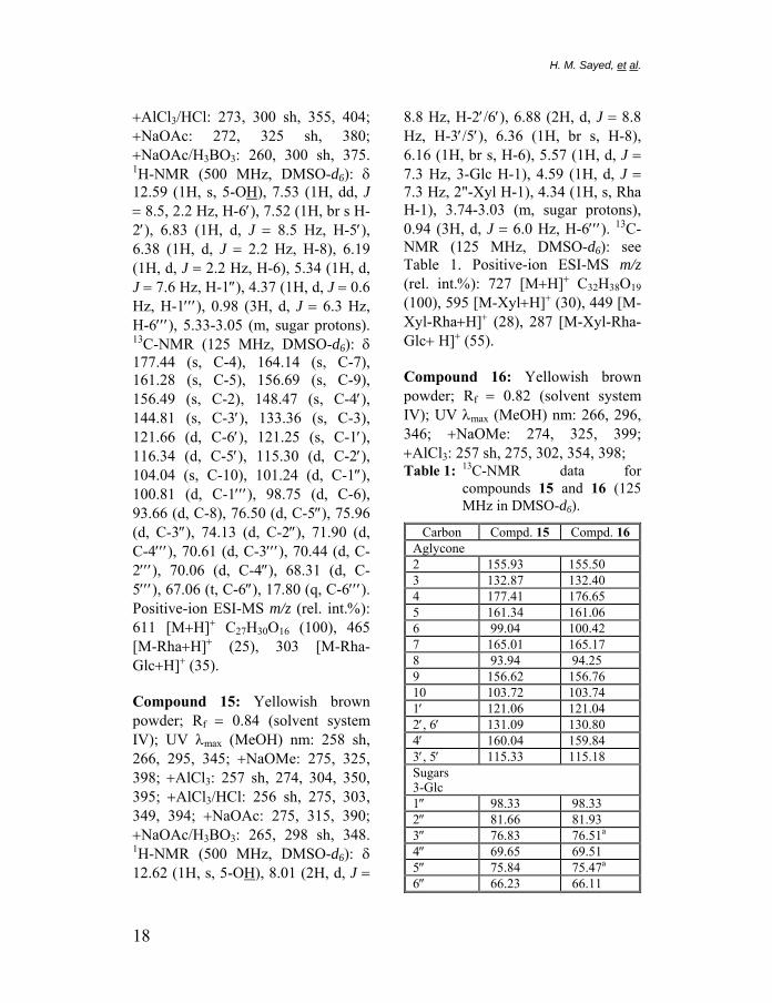
compounds 15 and 16 (125 MHz in DMSO-d6).
Carbon Compd. 15 Compd. 16 Aglycone 2 155.93 155.50 3 132.87 132.40 4 177.41 176.65 5 161.34 161.06 6 99.04 100.42 7 165.01 165.17 8 93.94 94.25 9 156.62 156.76 10 103.72 103.74 1′ 121.06 121.04 2′, 6′ 131.09 130.80 4′ 160.04 159.84 3′, 5′ 115.33 115.18 Sugars 3-Glc 1″ 98.33 98.33 2″ 81.66 81.93 3″ 76.83 76.51a
4″ 69.65 69.51 5″ 75.84 75.47a
6″ 66.23 66.11
18
2″-Glucose 1 103.74 2 74.17 3 76.33b
4 70.53 5 77.03b
6 60.80 2″-Xylose 1 104.49 2 73.88 3 76.16 4 69.54 5 65.76 6″-Rhamnose 1 100.54 100.42 2 70.44a 70.26c
3 70.71a 69.66c
4 71.96 71.80 5 68.34 68.18
aglycone was extracted with EtOAc, concentrated under reduced pressure, purified on Sephadex LH-20 column using methanol and identified by co-TLC with an authentic sample using solvent system II. The sugars in the aqueous layer were identified by co-TLC with authentic materials using solvent system V.
Acid hydrolysis and GC analysis32
A solution of compound 15 (5 mg in 25 ml methanol) was treated with 3N HCl (15 ml) and stirred at 80° for about 5 h. Upon drying with a flow of nitrogen, the residue was dissolved in (−)-2-butanol (0.5 ml) and one drop of trifluoroacetic acid. The solution was transferred to an ampoule which
6 17.78 17.66 +AlCl3/HCl: 256 sh, 274, 300, 349, the spectrum a-cInterchangeable assignments within
396; +NaOAc: 275, 316, 390; +NaOAc/H3BO3: 266, 298 sh, 347. 1H-NMR (500 MHz, DMSO-d6): δ 12.56 (1H, s, 5-OH), 7.94 (2H, d, J = 8.8 Hz, H-2′/6′), 6.87 (2H, d, J = 8.8 Hz, H-3′/5′), 6.19 (1H, br s, H-8), 6.00 (1H, br s, H-6), 5.50 (1H, d, J = 7.0 Hz, 3-Glc H-1), 4.58 (1H, d, J = 7.6 Hz, 2″-Glc H-1), 4.29 (1H, br s, Rha H-1), 5.42-3.04 (m, sugar protons), 0.95 (3H, d, J = 6.3 Hz, H-6′′′). 13C-NMR (125 MHz, DMSO-d6): see Table 1. Positive-ion ESI-MS m/z (rel. int.%): 757 [M+H]+ C33H40O20 (100), 595 [M−Glc+H]+ (15), 449 [M−Glc−Rha+H]+ (20), 287 [M−Glc−Rha−Glc+ H]+ (40).
Acid hydrolysis31
A solution of the isolated glycoside (5 mg in 10 ml methanol) was treated with 3% H2SO4 (1.5 ml) and heated at 100 °C for 1 hr. The
was sealed and heated at 130° in an oven overnight until complete butanolysis. After being taken to dryness, the resulting residue was reacted with hexamethyldisilazane / chlorotrimethylsilane / pyridine (1:1:5, 0.1 ml) for 30 min at room temperature. The solution was then centrifuged and the supernatant (1 µl) was analysed by an Agilent 6850 gas chromatograph. A temperature gradient from 135° to 200° at 1°/min was applied. The injection volume was 1 µl. The carrier gas used was nitrogen with a split ratio: 50:1 and 1 ml/min constant flow rate. The injection port and detector temperatures were set at 200° and 220°, respectively. For the hydrolysate, four peaks were detected at 42.9, 48.6, 27.0 and 31.7 min. Authentic standards prepared in a similar manner from commercially
19

H. M. Sayed, et al.
available L-glucose, D-glucose and D-xylose, gave peaks at: 42.9 and 48.6 min for D-glucose; 41.7 and 48.4 min for L-glucose; 27.0 and 31.7 min for D-xylose.
Antioxidant activity33
The antioxidant activity was quantified by the decrease in the absorption of each of the isolated compounds or soluble fractions in 118×10−5 % DPPH solution (final concentration of the sample in the cuvette was 20 µM for pure compounds and 0.5 and 1.0 mg/ml for soluble fractions) monitored at 517 nm using a spectrophotometer. The absorbance of DPPH in methanol (with or without compounds) was measured after 2 min. The antioxidant activity of each compound was measured in relation to propyl gallate (a known synthetic antioxidant) set as 100% antioxidant activity. Determinations were performed in triplicate. The antioxidant activity was calculated using the following equation:34
Antioxidant activity = 100 x
⎟⎠⎞
⎜⎝⎛ −
blank theof absorbancecompound with absorbance1
Cytotoxic activity35
L5178Y mouse lymphoma cells were grown in Eagle’s minimal essential medium supplemented with 10% horse serum in roller tube culture. For the dose-response experiments, 5 ml cultures were initiated by inoculation of 5×10³
cells/ml and incubated at 37° for 72 h. Controls showed a population doubling time of 10.5 h. Cell growth was determined by a cell count with a Cytocomp counter (128-channel counter, system Michaelis, Mainz, Germany) incorporating a 32-channel size-distribution plotter.
α-amylase inhibitory activity36
The method is based on assay of α-amylase by EnzCheck® Amylase Assay Kit (E-11954). The provided stock solution of DQ starch and α-amylase enzyme were diluted with the reaction buffer (pH 6.9) according to the reported protocol.36 To the microplate wells, 50 µg/10 µl of the tested compound in DMSO, 50 µl of the diluted enzyme and 40 µl of the reaction buffer were added and allowed to stand for 5 min at room temperature then 100 µl of DQ starch was added. The fluorescence intensity of the digestion products from the DQ starch (with or without compounds) was measured using a kinetic assay program in the Tecan Genios microplate reader at λmax 485±10 nm starting from zero min to 60 min at 10 min intervals. All determinations were performed in triplicate. The α-amylase inhibitory activity of each test compound was measured in relation to acarbose (Ac) set as 100% α-amylase inhibitory activity. The percentage of α-amylase activity and α-amylase inhibition were calculated using the following equations:
20
( ) ( )0c
60c
0s
60s FFFF
x100 activity amylase- %−−
=α
where : Fluorescence with the sample at 60 min, : Fluorescence with the sample at 0 min, : Fluorescence of the control at 60 min,
: Fluorescence of the control at 0 min.
60sF
0sF
60cF
0cF
% α-amylase inhibition = 100 − % α-amylase activity
RESULTS AND DISCUSSION The methanolic extract of C.
alopecuroides Rottb. inflorescences was successively partitioned between water and n-hexane, ethyl acetate and n-butanol. The ethyl acetate and n-butanol soluble fractions through series of different chromatographic fractionation techniques, afforded sixteen phenolic compounds 1-16.
Compound 1 was assigned a molecular formula of C10H8O4 by EIMS analysis that appeared [M]+ at m/z 192 (100%) in addition to mass fragment peaks at m/z 177 (65%), 164 (30%) and 149 (60%). The 1H-NMR spectrum revealed signals of two olefinic protons with cis-coupling at δ 6.27 and 7.60 (each 1H, d, J= 9.5 Hz) indicative of the H-3 and H-4 of the coumarin nucleus;37 two aromatic protons (para to each others) at δ 6.84 and 6.91 (each 1H, s, H-5, H-8) and a singlet for one methoxyl group at δ 3.95 (3H). 13C-NMR spectrum exhibited signals for 10 carbon atoms including a signal for δ-lactone
function at δ 161.47. These spectral data showed good agreement with those reported for scopoletin.37&38 Thus, compound 1 was established as scopoletin.
Compound 2 possessed the molecular formula C16H14O4 by ESIMS that showed [M+H]+ at m/z 271, together with mass fragment peaks at m/z 151 (15%) and 147 (23%). The UV spectrum exhibited two absorption bands at λmax 240 and 373 nm for chalcones.39 This was supported by the 1H-NMR spectrum which showed signals for two trans-olefinic protons at δ 7.46 and 7.85 (each 1H, d, J= 15.5 Hz, H-α and β); two sets of ortho-coupled aromatic protons of B-ring at δ 6.89 (2H, d, J= 8.8 Hz, H-3/5) and 7.58 (2H, d, J= 8.8 Hz, H-2/6); three aromatic protons of A-ring at δ 6.48 (1H, dd, J= 9.2, 1.6 Hz, H-5′), 6.96 (1H, br s, H-3′) and 7.83 (1H, d, J= 9.2 Hz, H-6′) in addition to a singlet at δ 3.86 (3H) for one methoxyl group.40 The dominent mass fragment peaks at m/z 147 and 151 indicated the presence of one hydroxyl group in the B-ring in addition to one hydroxyl and one methoxyl group in the A-ring.41 The placement of the methoxyl group at position 4′ was supported by the HMBC spectrum that displayed a correlation between the methoxyl protons (δ 3.86) and C-4′ (δ 166.4).41 Therefore, compound 2 was concluded to have the structure 4,2′-dihydroxy-4′-methoxychalcone (isoliquiritigenin 4′-methyl ether).
21

H. M. Sayed, et al.
ESIMS of compound 5 showed [M+H]+ at m/z 301 for C16H12O6. It had a UV spectrum typical of an aurone having ortho-dihydroxyl groups on the B-ring and lacking a free hydroxyl group at C-4 since the complex formed with AlCl3 was acid labile.39 The 1H-NMR spectrum exhibited the pattern of aurones of the aureusidin type40 by the appearance of one singlet at δ 6.46 (1H, s, H-10); three protons with the shifts and splitting of a catechol B-ring at δ 6.80 (1H, d, J= 8.2 Hz, H-5′), 7.16 (1H, dd, J= 8.2, 1.9 Hz, H-6′) and 7.38 (1H, d, J= 1.9 Hz, H-2′); two doublets at δ 6.14 and 6.29 (each 1H, J= 1.6 Hz) characteristic for meta-protons on A-ring and a singlet at δ 3.82 (3H) for a methoxyl group. The 13C-NMR spectrum revealed the presence of 16 carbon atoms were attributed to aureusidin derivative.42 The methoxyl group was located at the 4-position according to long-range HMBC correlation between the methoxyl protons at δ 3.82 and C-4 at δ 159.28. On the basis of these data, compound 5 was shown to be aureusidin 4-methyl ether (rengasin).
The EI mass spectrum of compound 8 showed [M]+ at m/z 194 (100%) corresponding to C10H10O4, in addition to mass fragment peaks at m/z 179 (42%), 151 (10%), 149 (9%), 133 (31%) and 77 (10%). The 1H-NMR spectrum showed signals of trans-3,4-disubstituted cinnamic acid: two olefinic protons with trans-coupling at δ 6.23 and 7.43 (each 1H, d, J= 16.0 Hz); three aromatic protons
at δ 6.93 (1H, d, J= 8.2 Hz), 7.06 (1H, d, J= 1.9 Hz) and 7.08 (1H, dd, J= 8.2, 1.9 Hz) in addition to a signal of one methoxyl group at δ 3.79 (3H, s). By comparison of these data with those published,43&44 compound 8 was characterized as trans-4-hydroxy-3-methoxycinnamic acid (trans-ferulic acid).
Compounds 3, 4, 6, 7, 9, 10 and 12 displayed characteristic UV absorption maxima for flavone skeleton.39 The 1H-NMR spectral data for these compounds also confirmed the presence of flavone nucleus in these molecules.40
The ESI mass spectrum of compound 3 showed [M+H]+ at m/z 315 was consistent with the molecular formula C17H14O6. It had UV spectrum of a flavone having free hydroxyl groups at C-4′ and C-7 and lacking a free hydroxyl group at C-5.39 The 1H-NMR spectrum exhibited the pattern of luteolin derivative40 by the appearance of a singlet aromatic proton at δ 6.55 (1H, H-3); two meta-coupled protons in the A-ring at δ 6.41, 6.54 (each 1H, d, J= 2.2 Hz, H-6 and H-8); three aromatic protons in the 3′,4′-disubstituted B-ring at δ 6.92 (1H, d, J= 8.2 Hz, H-5′), 7.45 (1H, d, J= 2.2 Hz, H-2′) and 7.47 (1H, dd, J= 8.2, 2.2 Hz, H-6′) in addition to two singlets of two methoxyl groups at δ 3.88 and 3.95 (each 3H). Considering the UV results, the two methoxyl groups could be placed at C-5 and C-3′. These placements are supported by the HMBC spectrum, the protons of the methoxyl groups at δ 3.88 and
22
3.95 showed correlations with the carbons at position 5 (δ 161.5) and 3′ (δ 148.6), respectively. By comparison of these spectral data with those in literature,45 compound 3 was deduced as luteolin 5,3′-dimethyl ether.
Compound 4 deduced the molecular formula C17H14O6 by ESIMS analysis ([M+H]+ at m/z 315). The UV spectral data suggested its structure to be a flavone having free hydroxyl groups at the 5- and 4′-positions and lacking a free hydroxyl group at the 7-position.39 Its 1H-NMR spectral data were similar to those of compound 3. According to the UV spectral data, the methoxyl groups could be placed at positions 7 and 3′. On the basis of these spectral data and by comparison with the reported data,46 compound 4 was concluded to be luteolin 7,3′-dimethyl ether (velutin).46
The ESI mass spectra of compounds 6 and 7 showed [M+H]+ at m/z 271 and 287 corresponding to the molecular formulae C15H10O5 and C15H10O6, respectively. They were characterized as apigenin and luteolin by comparison of their spectral data with published data.39,40,42&47
ESIMS of compound 9 showed [M+H]+ at m/z 449 corresponding to the molecular formula C21H20O11 in addition to a prominent fragment peak at m/z 287 [M−162 (hexose unit)+H]+ indicating its monoside nature. The UV spectral data suggested its structure to be 5,7-dihydroxyflavone derivative.39 In the
1H- and 13C-NMR spectra, there were signals for luteolin nucleus40&47 and a β-glucose moiety.48 In the HMBC spectrum the correlation from the glucose anomeric proton (H-1″, δ 4.89) to C-4′ (δ 148.58) confirmed the O-glycosidation at C-4′. Acid hydrolysis31 of compound 9 yielded luteolin and glucose (co-TLC with authentic samples). On the basis of these results and by comparing the spectral data with literature,49 compound 9 could be identified as luteolin 4′-O-β-D-glucopyranoside.
Compound 10 had the same ESI mass spectrum of compound 9. Its UV spectra showed absorption pattern of 5,3′,4′-trihydroxy flavone derivative.39 The 1H- and 13C-NMR spectra were similar to those of compound 9. The HMBC spectrum showed correlation between the anomeric proton signal (δ 5.08, H-1″) to C-7 (δ 162.94) confirmed the O-glycosidation at C-7. Acid hydrolysis31 of compound 10 gave luteolin and glucose (co-TLC with authentic samples). From these results and comparison with literature data,39,40&47 the structure of compound 10 was assigned as luteolin 7-O-β-D-glucopyranoside.
ESIMS of compound 12 showed [M+H]+ at m/z 579 for C27H30O14 in addition to a significant fragment peak at m/z 271 [M−146 (methyl pentose unit)−162 (hexose unit)+H]+ indicating its bioside nature. UV spectral data of 12 suggested its structure to be 5,4′-dihydroxyflavone derivative.39 The 1H- and 13C-NMR
23
H. M. Sayed, et al.
spectra displayed signals owing to apigenin,40&47 one β-D-glucopyrano-syl and one α-L-rhamnopyranosyl48 units. The significant deshielding of the 13C signal associated with C-2″ of the glucose unit indicated further glucosylation at C-2″. In the HMBC spectrum, the 1′′′→2″ link between the two sugars was confirmed by a 3J correlation from the anomeric proton δ 5.12) of the rhamnose unit to 2″ (δ 77.07) of the glucose unit. Also, the O-diglycosidic linkage at the 7-position was supported from the correlation between the anomeric proton (δ 5.23) of the glucose unit and C-7 (δ 162.59). Acid hydrolysis31 of compound 12 liberated apigenin, glucose and rhamnose (co-TLC with authentic samples). From these results and by comparison with the reported data,39,40&47 compound 12 was identified as apigenin 7-O-neohisperidoside.
The UV spectral data of compounds 11 and 14 suggested their structures to be flavonols with a substituted 3-hydroxyl group and free hydroxyl groups at positions 5, 7, 3′ and 4′.39 By comparison of their 1H-NMR and 13C-NMR spectral data with those reported in literature,40&47 compounds 11 and 14 were identified as quercetin 3-O-β-D-gluco-pyranoside and quercetin 3-O-rutinoside (rutin), respectively.
The UV spectra of compounds 13, 15 and 16 exhibited characteristic absorption bands of flavonols with a substituted 3-hydroxyl group and free hydroxyl groups at the 5-, 7- and 4′-
positions.39 The 1H-NMR spectral data for these compounds supported their structures as kaempferol derivatives.40
Compound 13 showed [M+H]+ at m/z 595 in its ESI mass spectrum of corresponding to the molecular formula C27H30O15. Also, the spectrum revealed two prominent fragment peaks at m/z 449 [M−146 (methyl pentose unit)+H]+ and 287 [M−146−162 (hexose unit)+H]+ indicating compound 13 as kaempferol 3-O-bioside. Its 1H-NMR spectrum displayed signals due to kaempferol skeleton40 together with two anomeric protons [δ 5.28 (1H, d, J = 7.6 Hz, Glc H-1) and 4.37 (1H, s, Rha H-1)].48 Acid hydrolysis31 of compound 13 gave kaempferol, glucose and rhamnose (co-TLC with authentic samples). Thus, compound 13 was identified as kaempferol 3-O-rutinoside.
The molecular formula of compound 15 was established as C32H38O19 by ESI mass analysis that showed [M+H]+ at m/z 727. The three significant fragment peaks at m/z 595 [M−132 (pentose unit)+H]+, 449 [M−132−146 (methyl pentose unit)+H]+ and 287 [M−132−146−162 (hexose unit)+H]+ indicated compound 15 as kaempferol 3-O-triglycoside. The 1H-NMR spectrum exhibited signals of kaempferol40 in addition to three anomeric signals [δ 5.57 (1H, d, J= 7.3 Hz, Glc H-1), 4.59 (1H, d, J= 7.3 Hz, Xyl H-1) and 4.34 (1H, s, Rha H-1)].48 The high field position of H-1 of β-xylose (δ
24

4.59) indicated that it was a terminal sugar. Because the H-1 signal of the C-3 linked glucose was shifted downfield (∆δ = 0.29 ppm) compared to that of rutinosyl unit in compound 13 it is suggested that xylose is attached to C-2″.50 The 13C-NMR spectrum displayed thirteen carbon signals were attributed to kaempferol nucleus42&47 and seventeen carbon signals in the region of sugars including three anomeric signals at δ 104.49, 100.54 and 98.33 (see Table 1). The 13C-NMR shifts of the three sugars are consistent with those corresponding to one β-D-gluco-pyranosyl, one β-D-xylopyranosyl and one α-L-rhamnopyranosyl moiety.48 The deshielding of the 13C signals associated with C-2″ (δ 81.66) and C-6″ (δ 66.23) of the glucose unit indicated the glycosylation at C-2″ and C-6″. The identity of the three sugars and their sequence were determined by the exhaustive analysis of NMR spectral data (HMQC and HMBC). In the HMBC spectrum the anomeric proton signal at δ 5.57 (Glc H-1) showed correlation with the carbon signal at δ 132.87 (C-3) indicating the O-triglycosidic linkage at the 3-position of kaempferol. Also, the anomeric proton signal at δ 4.59 (Xyl H-1) showed correlation with the carbon signal at δ 81.66 (C-2″) confirmed the interglycosidic linkage xylose (1→2) glucose. Furthermore, the correlation between the anomeric proton signal at δ 4.34 (Rha H-1) and the carbon signal at δ 66.23 (C-6″) confirmed the interglycosidic linkage
rhamnose (1→6) glucose. To confirm the nature and the absolute stereochemistry of the sugar moieties, an aliquot of 15 was hydrolysed and reacted with (−)-2-butanol.32 GC analysis revealed the presence of D-glucose and D-xylose by comparison of their retention time data with those of the relevant standards. From the above evidences and by comparison with literature data,31&50 compound 15 was established as kaempferol 3-O-[2-O-D-xylopyranosyl-6-O-α-L-rhamnopyranosyl]-β-D-glucopyrano-side31 or kaempferol 3-[2G-xylosyl-rutinoside]50 which is isolated here for the first time from family Cyperacea. It was previously reported from the leaves of Hosta ventricosa (Salisb) Stearn (Liliaceae)50 and seed cake of Camellia sinensis O. Kuntez (Theaceae).31
The ESI mass spectrum of compound 16 showed [M+H]+ at m/z 757 corresponding to the molecular formula C33H40O20. Also, three prominent fragment peaks appeared at 595 [M−162 (hexose unit)+H]+, 449 [M−162−146 (methyl pentose unit)+H]+ and 287 [M−162−146−162 (hexose unit)+H]+ indicating compound 16 as kaempferol 3-O-triglycoside. Its 1H-NMR spectrum exhibited signals owing to kaempferol40 and three sugar units that were indicated by the appearance of three anomeric signals at δ 5.50 (1H, d, J = 7.0 Hz, Glc H-1), 4.58 (1H, d, J= 7.6 Hz, Glc H-1) and 4.29 (1H, s, Rha H-1).48 The strongest deshielded sugar doublet at δ 5.50
25
H. M. Sayed, et al.
apparently corresponding to the anomeric proton of glucose directly attached to C-3 position of the aglycone.50 The 13C-NMR spectrum displayed 30 carbon signals (see Table 1) were attributed to kaempferol,42&47 two β-glucopyrano-syl units and one α-rhamnopyranosyl unit that were indicated by the appearance of three anomeric signals at δ 98.33, 103.74 and 100.42, respectively.48 The interglycosidic linkages at C-2″ and C-6″ were identified by the downfield shifts of C-2″ (δ 81.93) and C-6″ (δ 66.11). The HMQC and HMBC spectra identified the three sugars and their sequence. The HMBC spectrum showed a cross peak between the anomeric proton signal at δ 4.58 (2″-Glc H-1) and the carbon signal at δ 81.93 (3-Glc C-2″), this confirmed the interglycosidic linkage glucose (1→2) glucose. Also, the anomeric proton signal at δ 4.29 (6″-Rha H-1) showed a cross peak with the carbon signal at δ 66.11 (3-Glc C-6″), this supported the interglycosidic linkage rhamnose (1→6) glucose. Acid hydrolysis31 of compound 16 yielded kaempferol, glucose and rhamnose (co-TLC). The aforementioned data showed good agreement with those reported for kaempferol 3-O-[2-O-D-glucopyrano-syl-6-O-α-L-rhamno-pyranosyl]-β-D-glucopyranoside or kaempferol 3-[2G-glucosylrutino-side]50 which is isolated for the first time from the family Cyperaceae. It was previously reported from the
leaves of Hosta ventricosa (Salisb) Stearn (Liliaceae).50
In conclusion, luteolin (7), luteolin 7-O-β-D-glucopyranoside (10) and quercetin 3-O-rutinoside (14) were previously reported from the leaves of the studied plant6 while the others are isolated for the first time from C. alpoecuroides Rottb. To the best of our knowledge, compounds 2 and 5 are first report from the genus Cyperus while compounds 15 and 16 are first report from the family Cyperaceae.
Flavonoids have the property of inhibiting autoxidation reactions and scavenging of free radicals but the relation between their structure and activity remains unclear.51 There are three functional groups that have been attributed to increase the antiradical activities among the flavonoids: the presence of an ortho-dihydroxylation of the B-ring of the flavonoid molecule; the C2−C3 double bond in concert with 4-oxo functionally of the C-ring; and the additional presence of both a 3- and a 5-hydroxyl moiety of the C and A rings, respectively.51&52 Figure 2 shows the results obtained for antioxidant activity of fourteen flavonoids isolated from C. alopecuroides Rottb. Flavones and flavonols with a substituted hydroxyl group at the C-3 position, which have only a C-4′ hydroxyl group in the B-ring showed low antioxidant activity falls within the range of 1.0-21.1%. A strong antioxidant activity was shown by compounds with an ortho-
26
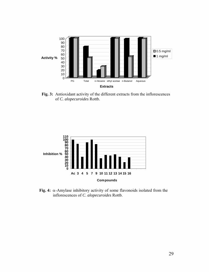
dihydroxy system in the B-ring: quercetin 3-O-β-D-rutinoside (79.2%), luteolin (75.0%), luteolin 7-O-β-D-glucopyranoside (71.9%) and quercetin 3-O-β-D-glucopyranoside (70.7%), aureusidin 4-methyl ether (46.5%). It appears that the reason for their high reactivity is the strong effect of the C-3′ hydroxyl group on the reactivity of the hydroxyl at C-4′. This is in agreement with conclusions that the ortho-dihydroxy system in the B-ring of the flavonoids is highly effective against free radicals.34&51 The synergistic effects of flavonoid mixtures may be responsible for high antioxidant activity of the ethyl acetate, n-butanol and aqueous extracts53 as shown in Figure 3.
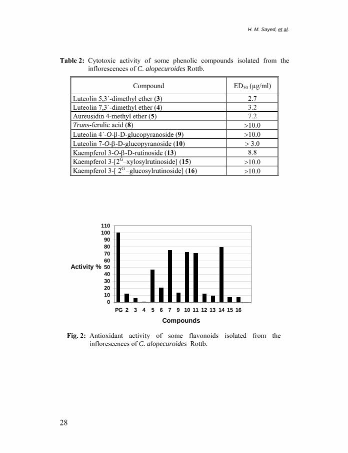
The cytotoxicity of nine phenolic compounds isolated from C. alopecuroides Rottb. was assessed in vitro with a mouse lymphoma cell line using the microculture tetrazolium (MTT) assay.54 The compounds were tested for their cytotoxic activities at a range of concentrations of (3−10 µg/ml). All compounds were found to be active against the cell line chosen (Table 2). Luteolin 5,3´-dimethyl ether and luteolin 7,3'-dimethyl ether are the most active compounds in this study with ED50 of 2.7 and 3.2 µg/ml, respectively.
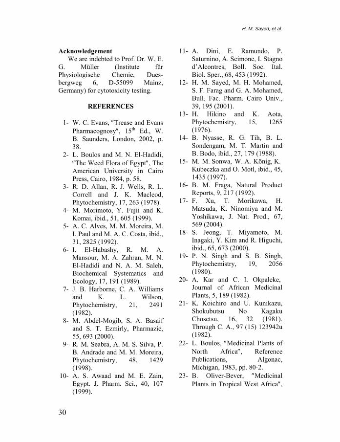
Twelve flavonoids were evaluated for α-amylase inhibitory activity. As shown in Figure 4, luteolin (99.6%), aureusidin 4-methyl ether (90.5%), luteolin 5,3´-dimethyl ether (85.0%) and luteolin 4´-O-β-D-gluco-pyranoside (83.8%) showed strong inhibitory activity of α-amylase. The α-amylase inhibitory activity of luteolin (IC50 50-125 µg/ml) was similar to acarbose (IC50 50-120 µg/ml). Apparently, the α-amylase inhibitory activity increased with the presence of hydroxyl groups at 3′ and 4′-position of the B-ring.55
In conclusion, the present study revealed that the titled plant contains a large number of phenolics with strong antioxidant, cytotoxic activities and an inhibitory activity of α-amylase. Thus, the plant could be considered of potential impact on human health.
27
H. M. Sayed, et al.
Table 2: Cytotoxic activity of some phenolic compounds isolated from the inflorescences of C. alopecuroides Rottb.
Compound ED50 (µg/ml)
Luteolin 5,3´-dimethyl ether (3) 2.7 Luteolin 7,3´-dimethyl ether (4) 3.2 Aureusidin 4-methyl ether (5) 7.2 Trans-ferulic acid (8) >10.0 Luteolin 4´-O-β-D-glucopyranoside (9) >10.0 Luteolin 7-O-β-D-glucopyranoside (10) > 3.0 Kaempferol 3-O-β-D-rutinoside (13) 8.8 Kaempferol 3-[2G–xylosylrutinoside] (15) >10.0 Kaempferol 3-[ 2G –glucosylrutinoside] (16) >10.0
0102030405060708090
100110
PG 2 3 4 5 6 7 9 10 11 12 13 14 15 16
Compounds
Activity %
2
Fig. 2: Antioxidant activity of some flavonoids isolated from theinflorescences of C. alopecuroides Rottb.
8
0102030405060708090
100
Activity %
PG Total n-Hexane ethyl acetae n-Butanol Aqueous
Extracts
0.5 mg/ml1 mg/ml
Fig. 3: Antioxidant activity of the different extracts from the inflorescencesof C. alopecuroides Rottb.
0102030405060708090
100110
Ac 3 4 5 7 9 10 11 12 13 14 15 16
Compounds
Inhibition %
Fig. 4: α-Amylase inhibitory activity of some flavonoids isolated from theinflorescences of C. alopecuroides Rottb.
29
H. M. Sayed, et al.
Acknowledgement We are indebted to Prof. Dr. W. E.
G. Müller (Institute für Physiologische Chemie, Dues-bergweg 6, D-55099 Mainz, Germany) for cytotoxicity testing.
REFERENCES 1- W. C. Evans, ″Trease and Evans
Pharmacognosy″, 15th Ed., W. B. Saunders, London, 2002, p. 38.
2- L. Boulos and M. N. El-Hadidi, ″The Weed Flora of Egypt″, The American University in Cairo Press, Cairo, 1984, p. 58.
3- R. D. Allan, R. J. Wells, R. L. Correll and J. K. Macleod, Phytochemistry, 17, 263 (1978).
4- M. Morimoto, Y. Fujii and K. Komai, ibid., 51, 605 (1999).
5- A. C. Alves, M. M. Moreira, M. I. Paul and M. A. C. Costa, ibid., 31, 2825 (1992).
6- I. El-Habashy, R. M. A. Mansour, M. A. Zahran, M. N. El-Hadidi and N. A. M. Saleh, Biochemical Systematics and Ecology, 17, 191 (1989).
7- J. B. Harborne, C. A. Williams and K. L. Wilson, Phytochemistry, 21, 2491 (1982).
8- M. Abdel-Mogib, S. A. Basaif and S. T. Ezmirly, Pharmazie, 55, 693 (2000).
9- R. M. Seabra, A. M. S. Silva, P. B. Andrade and M. M. Moreira, Phytochemistry, 48, 1429 (1998).
10- A. S. Awaad and M. E. Zain, Egypt. J. Pharm. Sci., 40, 107 (1999).
11- A. Dini, E. Ramundo, P. Saturnino, A. Scimone, I. Stagno d’Alcontres, Boll. Soc. Ital. Biol. Sper., 68, 453 (1992).
12- H. M. Sayed, M. H. Mohamed, S. F. Farag and G. A. Mohamed, Bull. Fac. Pharm. Cairo Univ., 39, 195 (2001).
13- H. Hikino and K. Aota, Phytochemistry, 15, 1265 (1976).
14- B. Nyasse, R. G. Tih, B. L. Sondengam, M. T. Martin and B. Bodo, ibid., 27, 179 (1988).
15- M. M. Sonwa, W. A. König, K. Kubeczka and O. Motl, ibid., 45, 1435 (1997).
16- B. M. Fraga, Natural Product Reports, 9, 217 (1992).
17- F. Xu, T. Morikawa, H. Matsuda, K. Ninomiya and M. Yoshikawa, J. Nat. Prod., 67, 569 (2004).
18- S. Jeong, T. Miyamoto, M. Inagaki, Y. Kim and R. Higuchi, ibid., 65, 673 (2000).
19- P. N. Singh and S. B. Singh, Phytochemistry, 19, 2056 (1980).
20- A. Kar and C. I. Okpaleke, Journal of African Medicinal Plants, 5, 189 (1982).
21- K. Koichiro and U. Kunikazu, Shokubutsu No Kagaku Chosetsu, 16, 32 (1981). Through C. A., 97 (15) 123942u (1982).
22- L. Boulos, ″Medicinal Plants of North Africa″, Reference Publications, Algonac, Michigan, 1983, pp. 80-2.
23- B. Oliver-Bever, ″Medicinal Plants in Tropical West Africa″,
30

Cambridge University Press, Cambridge, 1986, pp. 144- 217.
24- L. M. Perry and J. Metzger, ″Medicinal Plants of East and Southeast Asia: Attributed Properties and Uses″, MIT Press, London, 1980, pp. 121-22.
25- A. Chevallier, ″The Encyclo-pedia of Medicinal Plants″, Dorling Kindersley Ltd., London, 1996, p. 197.
26- M. S. Hifnawy, Y. Y. El-Hyatmy, S. A. Kenawy, A. K. Yossef and A. S. Awaad, Bull. Fac. Pharm. Cairo Univ., 37, 99 (1999).
27- M. S. Hifnawy, H. H. Ammar, S. K. Kenawy, M. E. Zaki, A. K. Yossef and A. S. Awaad, ibid., 37, 107 (1999).
28- M. I. Nassar, A. F. Abdel-Razik, E. A. M. El-Khrisy, A. M. Dawidar, A. Bystrom and T. J. Mabry, Phytochemistry, 60, 385 (2002).
29- H. M. A. El-Gohary, Bull. Fac. Pharm. Cairo Univ., 42, 157 (2004).
30- M. M. Sonwa and W. A. König, Phytochemistry, 56, 321 (2001).
31- T. Sekine, J. Arita, A. Yamaguchi, K. Saito, S. Okonogi, N. Morisaki, S. Jwasaki and I. Murakoshi, ibid., 30, 991 (1991).
32- M. Fouad, K. Al-Trabeen, M. Badran, V. Wray, R. A. Edrada, P. Proksch and R. Ebel, Arkivoc, xiii, 17 (2004).
33- B. Steffan, W. Wätjen, G. Michels, P. Niering, V. Wray, R. Ebel, R. A. Edrada, R. Kahl and
P. Proksch, J. Pharm. Pharmacol., 57, 233 (2005).
34- M. Joyeux. A. Lobstein, R. Anton and F. Mortier, Planta Med., 61, 126 (1995).
35- R. A. Edrada, P. Proksch, V. Wray, L. Witte, W. E. Müller and R. W. M. Van Soest, J. Nat. Prod., 59, 1056 (1996).
36- Molecular Probes website: www.probes.com.
37- S. Öksüz, A. Ulubelen, A. Barla and W. Voelter, Turk. J. Chem., 26, 457 (2002).
38- J. M. J. Vasconcelos, A. M. S. Silva and J. A. S. Cavaleiro, Phytochemistry, 49, 1421 (1998).
39- T. J. Mabry, K. R. Markham and M. B. Thomas, ″The Systematic Identification of Flavonoids″, Springer, New York (1970).
40- J. B. Harborne, ″The Flavonoids: Advances in Research Since 1986″, Chapman and Hall, London (1994).
41- V. Seidel, F. Bailleul and P. G. Waterman, Phytochemistry, 55, 439 (2000).
42- P. K. Agrawal, ″Carbon-13 NMR of Flavonoids″, Elsevier Science, New York, Tokyo (1989).
43- H. Tominaga, Y. Kobayashi, T. Goto, K. Kasemura and M. Nomura, Yakugaku Zasshi, 125, 371 (2005).
44- A. M. Dawidar, S. T. Ezmirly, M. Abdel-Mogib, Y. El-Dessouki and R. F. Angawi, Pharmazie, 55, 848 (2000).
31
H. M. Sayed, et al.
45- H. Achenbach, M. Stöcker and M. A. Constenla, Phyto-chemistry, 27, 1835 (1988).
46- M. Sakakibara, D. Jr. Difeo, N. Nakatani, B. Timmermann and T. J. Mabry, ibid., 15, 727 (1976).
47- J. B. Harborne and T. J. Mabry, ″The Flavonoids: Advances in Research″, Chapman and Hall, London (1982).
48- P. K. Agrawal, Phytochemistry, 31, 3307 (1992).
49- M. Yoshizaki, H. Fujino, M. Masuyama, M. Arisawa and N. Morita, ibid., 26, 2557 (1987).
50- J. Budzianowski, ibid., 29, 3643 (1990).
51- S. Burda and W. Oleszek, J. Agric. Food Chem., 49, 2774 (2001).
52- A. J. Jr. Dugas, J. Castaneda-Acosta, G. C. Bonin, K. L. Price, N. H. Fischer and G.W. Winston, J. Nat. Prod., 63, 327 (2000).
53- J. B. Harborne and C. A. Williams, Phytochemistry, 55, 481 (2000).
54- M. H. Kreuter, A. Robitzki, S. Chang, R. Steffen, M. Michaelis, Z. Kljajic, M. Bachmann, H. C. Schröder and W. E. G. Müller, Comp. Biochem. Physiol., 101 C, 183 (1992).
55- T. Matsui, M. Kobayashi, S. Hayashida and K. Matsumoto, Biosci. Biotechnol. Biochem., 66, 689 (2002).
32