Pharmacological evaluation of extracts from Buxus macowanii ...
102
Pharmacological evaluation of extracts from Buxus macowanii, Polygala myrtifolia, Scilla sp. and Xanthocercis zambesiaca By Brian Ngobeni Dissertation submitted in fulfilment of the requirements for the degree: MAGISTER TECHNOLOGIAE: BIOMEDICAL TECHNOLOGY in the DEPARTMENT OF HEALTH SCIENCES at the CENTRAL UNIVERSITY OF TECHNOLOGY, FREE STATE BLOEMFONTEIN 2016 Study Leader: Prof. SS Mashele Co-study leader: Dr IT Madamombe-Manduna Co-study leader: Dr E Van Der Watt. © Central University of Technology, Free State
-
Upload
khangminh22 -
Category
Documents
-
view
1 -
download
0
Transcript of Pharmacological evaluation of extracts from Buxus macowanii ...

Pharmacological evaluation of extracts from Buxus
macowanii, Polygala myrtifolia, Scilla sp. and Xanthocercis
zambesiaca
By
Brian Ngobeni
Dissertation submitted in fulfilment of the requirements for the degree:
MAGISTER TECHNOLOGIAE: BIOMEDICAL TECHNOLOGY
in the
DEPARTMENT OF HEALTH SCIENCES
at the
CENTRAL UNIVERSITY OF TECHNOLOGY, FREE STATE
BLOEMFONTEIN
2016
Study Leader: Prof. SS Mashele
Co-study leader: Dr IT Madamombe-Manduna
Co-study leader: Dr E Van Der Watt.
© Central University of Technology, Free State

I
Declaration of independent work
I Brian Ngobeni, identity number and student number , do hereby declare
that this research project submitted to the Central University of Technology, Free State for the Degree
Magister Technologiae: Biomedical Technology, is my own independent work. It complies with the
Code of Academic Integrity, as well as other relevant policies, procedures, rules and regulations of the
Central University of Technology, Free state. It has not been submitted before to any institution by
myself or any other person in fulfilment of the requirements for the attainment of any qualification.
……………………………………… ..……………………………….. Brian Ngobeni Date
© Central University of Technology, Free State

II
Table of Contents
Contents Page number
Declaration of independent work Summary Acknowledgements List of tables List of figures Abbreviations
I IV VI VII VIII IX
Chapter 1. Introduction 1.1. The burden of infectious diseases 1.2. Challenges in the treatment of infectious diseases 1.3. Drug-resistant pathogens 1.4. Plants and traditional medicine 1.5. The therapeutic potential of secondary metabolites 1.6. Medicinal plants as a source of novel drugs 1.7. Safety of medicinal plants 1.8. Plants used in this study 1.9. Aims and Objectives 1.10. Overview of the study 1.11. References
1-26 1 1 2 3 4 8 9 10 13 14 16
Chapter 2. Antimicrobial activity of Buxus macowani Oliv, Polygala myrtifolia L, Scilla and Xanthocercis zambesiaca (Baker). 2.1. Introduction 2.2. Methods 2.2.1. Plant extracts 2.2.2. Extracts preparation and storage 2.2.3. Microbial cultures 2.2.4. Antimicrobial activity 2.2.5. TLC bioautography 2.2.6. The microscopic analysis of the bacterial cell wall. 2.3. Results and discussions 2.4 Conclusions 2.5. References
27-44 27 28 28 29 30 30 31 32 32 39 40
Chapter 3. The In vitro cytotoxicity of Buxus macowanii Oliv, Polygala myrtifolia L, Scilla sp and Xanthocercis zambesiaca (Baker). 3.1. Introduction 3.2. Methods 3.2.1. Extracts preparations 3.2.2. The Sulforhodamine cytotoxicity assay 3.3. Results and discussions 3.4. Conclusions 3.5. References
45-53 45 47 47 47 48 50 51
Chapter 4. Phytochemical content of Buxus macowanii Oliv. Polygala myrtifolia L, Scilla and Xanthocercis zambesiaca (Baker). 4.1. Introduction 4.2. Methods 4.2.1. Extracts preparation 4.2.2. Phytochemical analysis of the plant extracts 4.2.3. TLC fingerprinting
54-69 54 56 56 56 58
© Central University of Technology, Free State

III
4.2.4. Gas chromatographic mass spectrometry 4.3. Results and discussions 4.4. Conclusions 4.5. References
58 59 64 65
Chapter 5. General discussions and conclusions 5.1. General discussions 5.2. Conclusions and recommendations 5.3. References
70-76 70 75 76
Annexures 77-92
© Central University of Technology, Free State

IV
Summary
The outbreak of drug resistant pathogens, the high cost of health care, limited accessibility of the
conventional drugs and their side effects are problems that make the treatment of infectious diseases
difficult all over the world. These challenges have led to the search for novel drugs and drug leads that
can surpass the quality of the currently available antimicrobial agents. Medicinal plants are considered
to be the best candidates for the discovery of new drugs because of their long history of use in the
treatment of various ailments in communities. The current study was aimed at investigating the
antimicrobial activity, cytotoxic activity and phytochemical composition of the methanol extracts from
Buxus macowanii, Polygala myrtifolia, Scilla sp. and Xanthocercis zambesiaca.
Staphylococcus aureus, Clostridium perfringens, Pseudomonas aeruginosa, Enterococcus faecalis,
Escherichia coli, Staphylococcus epidermidis, and the fungal species Candida albicans and Candida
tropicalis were used to evaluate the antimicrobial activity of the selected plant extracts using the broth
Microdilution method. All the plants extracts tested showed no activity against all the bacterial and
fungal species except Buxus macowanii. Buxus macowanii inhibited the growth of Staphylococcus
aureus, Clostridium perfringens, Pseudomonas aeruginosa, Staphylococcus epidermidis, Candida
albicans and Candida tropicalis at the MIC of 2.5 mg/ml while Enterococcus faecalis and Escherichia
coli were inhibited at 1.2 mg/ml.
Buxus macowanii was selected for further studies because it presented the best antimicrobial
properties. Antimicrobial compounds were located using TLC bioautography. Four clear zones possibly
flavonoids and alkaloids were detected on the TLC chromatogram. These findings suggest that the
antimicrobial activity of Buxus macowanii was not attributed to a single compound but to a synergy of
compounds. The effect of Buxus macowanii on the bacterial cell morphology was also evaluated.
Morphological changes such as damage to the cell wall, loss of intracellular contents, incomplete cell
© Central University of Technology, Free State
V
division and shrinkage of the cells were observed using Scanning and Transmission Electron
Microscopy. Bacterial cells were affected morphologically after treatment with the extracts of B.
macowanii.
In order to evaluate the safety of the extracts used in the study, the Sulforhodamine cytotoxicity assay
was carried out using the WI-38 cell line (Normal human fetal lung fibroblast). P. myrtifolia was inactive
against the WI-38 cell line whereas B. macowanii and X. zambesiaca were found to be moderately
hazardous. Scilla extracts were found to be hazardous. These results indicate that caution should be
exercised when employing plants like B. macowanii, X. zambesiaca and Scilla sp. for treatment of
ailments.
The phytochemical screening of B. macowanii, P. myrtifolia, Scilla and X. zambesiaca using standard
methods, TLC and GCMS revealed compounds that have important health benefits. Bioactive
compounds such as flavonoids, alkaloids, terpenes, cardiac glycosides, steroids, saponins and tannins
were found in most of the extracts and their presence may explain the medicinal usage of the plants.
GCMS also revealed compounds such as neophytadiene that was found in the extracts of Buxus
macowanii, n-hexadecanoic was also found in the extracts of scilla sp and X. zambesiaca. 2-methoxy-
4-vinylphenol was found in the extracts of P. myrtifolia and X. zambesiaca.
The results obtained in this study show that B. macowanii is a promising source of antimicrobial drugs.
Further investigation into the isolation and identification of the bioactive compounds as well as in vivo
screening is recommended.
© Central University of Technology, Free State
VI
Acknowledgements
I would like to sincerely thank the following:
1. God of mount Zion for all his blessings, courage, support, wisdom and perseverance to
progress in my project,
2. Prof S. Mashele, Dr I.T Manduna and Dr E van Der Watt for your guidance and support
throughout the journey of this project and I have learnt a lot from you.
3. My family (The Mochike’s and Ngobeni’s) even though you were far away from me, your love
and support were a driving force in my life to keep working hard.
4. To my dearest mom and dad you ensured that I get the best in life, I will always make you
proud.
5. To my dearest friends Mr R.E Rammutla, Ms R. Mhaladi, Ms E. Ntimane and Ms G. Setlhare
your presence in my life helped me to deal with stressful situations much better.
6. The Free State University, Department of Soil, Crop and Climate Sciences for supplying us with
plant extracts and the lab equipment that was needed for this project to be a success.
7. Dr Ntsoaki Malebo from Central University of Technology, Department of Life Sciences who
assisted with the Microscopy analysis of the samples.
8. The Biomedical Technology programme colleagues for their support and advices.
© Central University of Technology, Free State

VII
List of Tables
Table Description Page number
1.1 Plant derived drugs 9
2.1 Information about the collection of the selected plants species 29
3.1 The IC50 of the extracts of Buxus macowanii Oliv, Polygala myrtifolia L, Scilla sp and Xanthocercis zambesiaca (Baker).
49
4.1 Phytochemical content of the selected plant extracts 60
4.2 The GCMS analysis of the selected plant extracts 63
5.1 The summary of the results obtained in the study 70
© Central University of Technology, Free State
VIII
List of Figures
Figure Description Page number
1.1 Buxus macowanii Oliv. 11
1.2 Polygala myrtifolia L. 12
1.3 Xanthocercis zambesiaca (Baker). 13
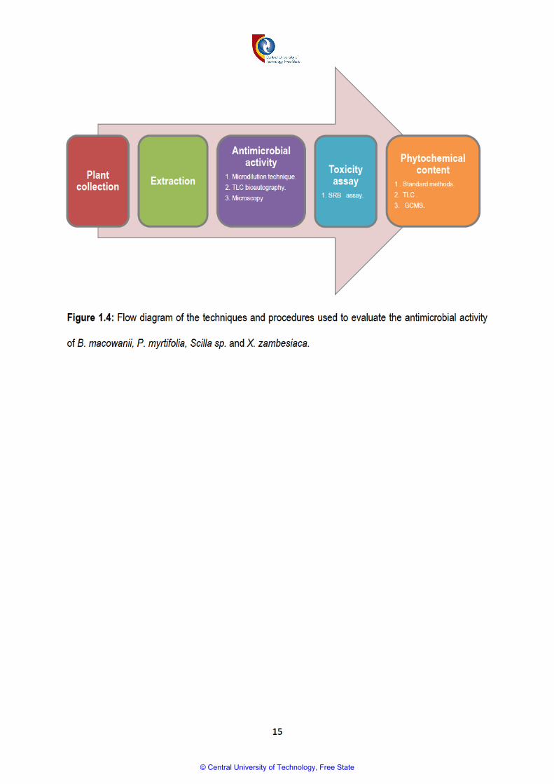
1.4 Flow-diagram of techniques and procedures used to evaluate the antimicrobial activity of B. macowanii, P. myrtifolia, Scilla sp and X. zambesiaca
15
2.1 A chromatogram developed with solvent system Toluene/Chloroform/Ethanol indicates the antibacterial spots of B. macowanii against Enterococcus faecalis.
34
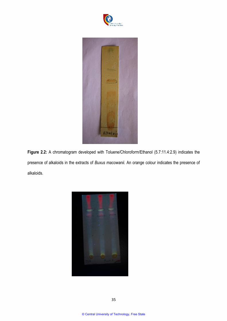
2.2 A chromatogram developed with solvent system Toluene/Chloroform/Ethanol shows the presence of alkaloids in the extracts of B. macowanii.
35
2.3 A chromatogram developed with solvent system Toluene/Chloroform/Ethanol shows the presence of flavonoids in the extracts of B. macowanii under UV light.
35
2.4 SEM photographs of the plant treated and untreated cells of Bacillus cereus 38
2.5 TEM photographs of the plant treated and untreated cells of Bacillus cereus 39
4.1 TLC fingerprinting of B. macowanii, P. myrtifolia, Scilla sp and X. zambesiaca 61
© Central University of Technology, Free State
IX
Abbreviations
Abbreviation Meaning
AChE Acetylcholinesterase
AIDS Acquired Immune Deficiency Syndrome
ATCC American Type Culture Collection
CO2 Carbon dioxide
CNS Central nervous system
CSIR Council for Scientific and Industrial Research
DMSO Dimethyl sulfoxide
EACCC European Collection of Cell Culture
EMEM Eagle’s Minimal Essential Medium
FBS Fetal bovine serum
HIV Human Immunodeficiency Virus
INH Isoniazid
GCMS Gas Chromatographic Mass spectrometry
Mg/ml Milligram per millilitre
MIC Minimum inhibitory concentration
NIST National Institute of Standard Technology
PBS Phosphate Buffer Solution
RIF Rifampicin
SEM Scanning Electron Microscopy
SRB Sulforhodamine B assay
TB Tuberculosis
TEM Transmission Electron Microscopy
TIC Total ion chromatograms
TLC Thin Layer Chromatography
WHO World health Organization
WI-38 Normal human foetal lung fibroblast cell line
© Central University of Technology, Free State

1
CHAPTER 1
1. INTRODUCTION
1.1. The burden of infectious diseases
Infectious diseases continue to kill millions of people every year. They are the second leading cause of
death in the world and the leading cause of death in individuals under the age of 50 (Hamburg, 2008).
The human immunodeficiency virus (HIV), tuberculosis (TB) and malaria are among the five major
infectious diseases that contribute to the death rate of people world-wide (Feachem, 2004). Statistics
released by the World Health Organisation (WHO) in 2014 indicated that approximately 36.9 million
people live with HIV, while 2 million people were recorded as newly infected (WHO, 2014a). TB has a
high rate of drug resistance. In 2013, approximately 9 million people developed the disease and 1.5
million people died from it (WHO, 2014b). Infectious diseases are clearly a worrisome factor in human
health.
1.2. Challenges in the treatment of infectious diseases
The health care sector implemented different strategies to eradicate infectious diseases, including the
development of antibiotics, vaccines, improved hygiene and sanitation, and vector control. This
improved the health of many people to such an extent that many people predicted the end of infectious
diseases (Hamburg, 2008). However, factors such as an increase in international trade and travel, the
movement of people to urban areas, reduced hygiene and sanitation in developing countries, among
other factors, interfered with these management strategies, and resulted in the spread of infectious
diseases. In recent times, a number of irresponsible practices, such unsafe sex and the careless use of
syringes by drug addicts, also contribute to the widespread occurrence of infectious diseases
(Hamburg, 2008). These problems are augmented by the development of resistant pathogens as well
as difficulties with the accessibility and affordability of treatment. Moreover, drugs that are currently
available pose problems such as side effects; toxicity and high production costs (Kisangau et al., 2007;
© Central University of Technology, Free State

2
Adwan et al., 2011; Rawat, 2012). The indiscriminate use of modern drugs or antimicrobial agents
usually leads to the emergence of resistant pathogens that may cause nosocomial or hospital-acquired
infections (Ibrahim et al., 2011; Daboor and Haroon, 2012).
1.3. Drug-resistant pathogens
Antibiotics were introduced into the health practice some years ago to combat infections or diseases
(Hawkey, 2008). Antimicrobial agents such as penicillin, methicillin, streptomycin, vancomycin,
chloroquine and many more have been a source of medication for years. However, during the past
decades, we have witnessed a major increase in micro-organisms that developed multidrug resistance
to antimicrobial agents (Oliphant and Eroschenko, 2015; Roca et al., 2015). Multidrug-resistant
pathogens can develop due to mutations that can take place even in the presence of antimicrobial
agents. Another driving force that leads to resistance is found in the abuse or misuse of antimicrobials
on patients and livestock, or the release thereof into the environment (Oliphant and Eroschenko, 2015).
Resistance to penicillin and sulphonamides in particular was reported in the 1940s, followed by
resistance to other antimicrobials (Jindal et al., 2015). The pathogens that currently indicate
antimicrobial resistance include Enterococcus faecium, Staphylococcus aureus, Klebsiella pneumoniae,
Acinetobacter baumanii, Pseudomonas aeruginosa, Plasmodium falciparum, Enterobacter species and
Mycobacterium tuberculosis (Boucher et al., 2009; Rice, 2008). In 2011b, WHO estimated that
approximately 630 000 people were diagnosed with multidrug-resistant TB, and this affected 84
countries (WHO, 2011b). Multidrug-resistant TB strains are responsible for 3.6% of all TB cases, and
require two years of treatment using potentially toxic drugs such as floroquinolones and injectable
aminoglycosides to replace the resisted Isoniazid (INH) and Rifampicin (RIF) (Winston and Mitruka,
2012). Extensively drug-resistant strains are usually difficult or impossible to cure, and only a few
countries are able to detect and isolate those strains (Goldberg et al., 2012).Two hundred and forty
million infections and 860 000 deaths caused by malaria are recorded annually (WHO, 2010). Among
© Central University of Technology, Free State

3
all the species that cause malaria, Plasmodium falciparum is the most severe species, and accounts for
approximately 90% of all malarial infections in Africa (Lee et al., 2013). Malarial pathogens usually
develop resistance to antimalarial drugs such as chloroquine and mefloquine, therefore increasing the
number of deaths that occur as a result of malaria (Lee et al., 2013; Olliaro and Bloland, 2001).
Community and hospital-acquired infections that are caused by methicillin-resistant Staphylococcus
aureus are on the rise, and occur at an alarming rate, creating a major challenge for clinicians
(Motamedi et al., 2010; Jindal et al., 2015). Shigella is another species that resists ciprofloxacin, which
is the only recommended drug for Shigella-related infections (Jindal et al., 2015). Vancomycin-resistant
Enterococci, Pseudomonas, Acinetobacter, antifungal-resistant fungi and antiviral-resistant viruses
significantly increases the number of deaths that occur world-wide. They also contribute to the delay in
the healing of patients who suffer infections (Jindal et al., 2015). It is for all these reasons that sources
of drugs and drug leads should be found to overcome this major challenge posed by drug resistance.
The problems associated with the antimicrobial drugs that are currently available have prompted the
search for new antimicrobial remedies that are effective and present fewer problems (Obeidat et al.,
2012). Researchers have turned to natural sources such plants for solutions, and traditional medicinal
plants are considered the best option (Adwan et al., 2011).
1.4. Plants and traditional medicine
For thousands of years, people have relied on plants to meet their essential needs, such as the need
for shelter, food, transport, clothing and medicine (Gurib-Fakim, 2006). Plants have been part of
traditional medical systems for thousands of years, and they continue to supply people with remedies to
treat various ailments (Normann and Snyman, 1996). Africa has diverse vegetation types, such as the
tropical rain forests, coastal and alpines forests, savannas, woodlands and scrublands. In addition,
Africa has diverse climatic and geographic factors that contribute to the survival of approximately 68
© Central University of Technology, Free State

4
000 plant species (Cunningham, 1997). Tropical and subtropical Africa contains approximately 45 000
plant species, of which 5 000 are medicinally used and contribute to 25% of the world trade in
biodiversity (Iwu, 2014). Due to their availability and natural advantage, African people often use
medicinal plants for their health care benefits, such as the treatment of colds, memory enhancement,
immunity improvement, etc. (Dzoyen et al., 2013).
It is estimated that approximately 70 – 95% of the world population in developing countries continue to
rely on medicinal plants for the treatment of various diseases (Lewu and Afolayan, 2009; WHO, 2011a;
Daughari, 2012). In South Africa, there is a rich tradition in the use of medicinal plants, which is based
on approximately 3 000 species (Taylor et al., 2001), and approximately 60 – 80% of the country’s
population depend on medicinal plants to meet their health care and psychological needs (Fuku et al.,
2013), this translates to approximately 27 million people (Mander, 1998; Street et al., 2008). People
depend on medicinal plants to treat diseases, as it sometimes is the only system available in rural
areas (Mabona and Van Vuuren, 2013). Challenges such as the remote location of health care centres
and an inability to afford conventional/modern drugs make traditional medicine the first choice in rural
areas. The use of traditional medicine is also attributed to the high accessibility of medicinal plants and
traditional healers, the low toxicity thereof, the fact that it has less side effects, extensive local
knowledge and the affordability thereof (Cheikhyoussef et al., 2011). However, there are indications
that indigenous knowledge about medicinal plants is rapidly decreasing as modern education is
increasing (Zerabruk and Yirga, 2012). Thus, there is an urgent need for the documentation of the use
of medicinal plants in South Africa before this knowledge is lost forever (Masevhe et al., 2015).
1.5. The therapeutic potential of secondary metabolites
Medicinal plants are known to contain secondary metabolites that present unlimited opportunities for
identifying new drug leads as a result of their supreme chemical diversity (Cos et al., 2006; O'Bryan et
© Central University of Technology, Free State

5
al., 2008). Secondary metabolites such as alkaloids, flavonoids, tannins, saponins, glycosides, etc. are
usually found in plants where they fulfil a selective and survival function for the plants (Esterhuizen et
al., 2006; Manimozhi et al., 2012). Secondary metabolites serve an important purpose for plants, as
they protect plants from herbivores and microbial invasions, act as attractants for pollinators and
seed-dispensing animals, and protecting plants from ultraviolet (UV) light (Gomez-Caracava et al.,
2014). Secondary metabolites are also responsible for the taste, odour and color of plants, and most of
them have important applications in pharmacology, chemistry, agricultural and novel drug sectors
(Cannes do Nascimento and Fett-Neto, 2010). Various secondary metabolites have the ability to
indicate biological properties such as antimicrobial, antioxidant, anti-inflammatory, antifungal, antiviral
and anthelmintic activities (Daboor and Haroon, 2012). The effectiveness of medicinal plants as
therapeutic agents is established through the presence of these important bioactive constituents (Bedir
et al., 2003; Bhat and Karim, 2010). The most important bioactive compounds in plants are alkaloids,
flavonoids, tannins, phenols, glycosides and tannins (Kubmarawa et al., 2007).
1.5.1. Polyphenolics
Phenols are compounds that occur naturally as colour pigments that are responsible for the colour of
fruits and vegetables. They are synthesised from the phenylalanine through the phenylalanine
ammonia lyase (PAL) (Doughari, 2012). They fulfil important functions in plants, such as defence
mechanisms against pathogens and herbivores, and thus can be considered as potential candidates for
the management of pathogenic infections (Doughari, 2012). Polyphenolics with benefits for human
health are phenolic acids, flavonoid polyphenolics (flavonones, flavones, xanthones and catechins),
lignans and stilbenes (Hooper and Cassidy, 2006). The recent interest in polyphenolics is attributed to
their beneficial effects on human health, such as their antioxidant properties. As a result, they are used
to combat cancer and heart disease, and can sometimes be used as anti-inflammatory agents (Manach
et al., 2004; Fraga et al., 2010; Barbehenn and Constabel, 2011). They were also reported to have
© Central University of Technology, Free State

6
antimicrobial, antiviral, cytotoxic, and vasodilatory effects (Kuete et al., 2012; Kougan et al., 2013;
Ngameni et al., 2013).
1.5.2. Flavonoids
Flavonoids are an important group of polyphenols that are present in many types of plants. They are
structurally composed of more than one benzene ring (Kar, 2007) and over 4 000 flavonoids are known.
They are the largest group of phenols, forming half of approximately 8 000 naturally occurring phenols
(Harborne and Baxter, 1999). Flavonoids fulfil important functions in plants, such as flower and seed
pigmentation; plant fertility and reproduction; and defence from UV light, predators and pathogens
(Harborne and William, 2000). For many years, medicinal plants that contain flavonoids have been
used to treat human diseases, and isolated flavonoids have also been found to perform important
biological activities, such as antifungal and antibacterial activities (Havsteen, 2002; Cushnie and Lamb,
2005).
1.5.3. Alkaloids
Alkaloids are usually found in higher plants, lower plants, insects, marine and micro-organisms (Kuete
and Efferth, 2010; Wansi et al, 2013). Alkaloids are used as medication, as recreational drugs, or in
entheogenic rituals. However, many of them are toxic, and can be used as toxins for weapons (Kuete et
al., 2008). They are also used as anaesthetics, stimulants, psychedelics, analgesics, antibacterial
agents, anticancer drugs, antihypertensive agents, spasmolysis agents, vasodilators, antiarrhythmia
drugs, antiasthma therapeutics and antimalarials (Kuete and Efferth, 2010; Wansi et al., 2013; Zofou et
al., 2013).
© Central University of Technology, Free State

7
1.5.4. Tannins
Tannins are the most abundant secondary metabolites in plants. They are known to have biological
properties such as antifungal, antioxidant, anthelmintic, antidiarrhoeal and healing properties (Zuanazzi
and Montanha, 2004). They are also known to defend plant leaves from insect herbivores by toxicity
and deterrence (DeGabriel et al., 2009; Barbehenn and Constabel, 2011). Some types of tannins act on
the arachidonic acid metabolism in leucocytes, which leads to the reversal of inflammation (Okuda,
2005).
1.5.5. Saponins
Saponins are known for their soap-like properties when combined with water. They were named after
Quillaja saponaria, a plant once used as soap (Doughari, 2012). Basically, saponins are produced by
plants, marine animals and some bacteria. They are glycosides, synthesised from the mevalonic acid
pathway via the isoprenoid pathway (Gomez-Caravaca et al., 2014). The first group of saponins
consists of steroidal saponins common in monocotyledonous angiosperms, while the second group
consists of triterpenoid saponins that are found in dicotyledonous angiosperms (Doughari, 2012).
Saponins protect plants against attack by pathogens and herbivores (Augustin et al., 2011). They are
therefore known to have antimolluscicidal, antidiabetic, antifungal, antiyeast, antibacterial, antimicrobial,
antiparasitic, antitumoral and anti-inflammatory activity (Sparg et al., 2004; Elekofehinti, 2015).
1.5.6. Terpenoids and steroids
Terpenes are diverse volatile oily compounds that are derived from isoprene units. They are usually
found in essential oils and resins, and include monoterpenes, diterpenes, triterpenes and
sesquiterpenoids (Firn, 2010). Examples of monoterpenes include thujone, camphor, eugenol, menthol
and terpinen-4-ol. Diterpenes consist of taxols and resins and they are used as anticancer agents
(Sandjo and Kuete, 2013). Triterpenes include steroids, sterols and cardiac glycosides. They possess
© Central University of Technology, Free State

8
properties such as anti-inflammatory, sedative and cytotoxicity activity (Chimene et al., 2013; Sandjo
and Kuete, 2013). Sesquiterpenes such as betulinic acid, lupeol, oleanic acid and ursolic acid are
known to have antimicrobial, antiplasmodial and neurotoxin properties. They act as irritants when
applied on human skin and when consumed (Awoufack et al., 2013). Most of the terpenes have
activities against cancer, malaria, inflammation and infectious diseases caused by viruses and bacteria.
Terpene-based drugs, such as the anticancer drug Taxol and the antimalarial drug Artemisinin, were
the most popular drugs; generating approximately US $ 12 billion in 2002 (Wang et al., 2005). Steroids
are naturally occurring compounds that are produced biologically from terpenoid precursors. They have
therapeutic action on cardiac muscles when injected into the human body (Firn, 2010). Hunters also
use them as arrow poisons, indicating that cardiac glycosides should be used with caution, as
excessive doses may cause death (Doughari, 2012).
1.6. Medicinal plants as a source of novel drugs
The high rate of traditional medicine usage and the variety of indigenous plant species indicate much
potential for the discovery of new bioactive compounds that can be used in the discovery of new drugs
(Copp and Pearce, 2007). The interest in medicinal plants as potential new drugs is on the rise, and
medicinal plants contribute 50% of all the drugs in the clinical world (Gurib-Fakim, 2006). The isolation
of bioactive compounds from plants contributed to early discoveries of drugs such as aspirin, morphine,
codeine, digoxin, atropine, quinine and artemisinin (Mueller et al., 2000; Samuelsson, 2004). Medicinal
plants are considered the best option to overcome the challenges that are associated with the drugs
that are currently available (Kaur et al., 2005). Table 1.1 illustrates some of the important drugs that
were manufactured from plants.
© Central University of Technology, Free State
9
Table 1.1: Plant-derived drugs
Plant species Drug Use References
Discorea spp Diosgenin Contraceptive Sarkar and Nahar,
2007
Rauwolfia sp Reserpine Antihypertensive Gurib-Fakim, 2006
Pilocarpus spp Pilocarpin Treats glaucoma and dry
mouth
Gurib-Fakim, 2006
Galanthus woronowii Galantamine Treats alzheimer’s disease Pirttila et al., 2004
Artemisia annua Arteether Antimalarial Graul, 2001
Callistemon citrinus Nitisinone Treats tyrosinaemia Frantz, 2004
1.7. Safety of medicinal plants
Medicinal plants are usually preferred for medicinal purposes, as they are presumed to have fewer side
effects and are regarded as safer to use than conventional drugs. However, some species are known to
be toxic to humans (Ndhlala et al., 2013). The toxicity of plants is usually attributed to the presence of
compounds that can contribute to the survival of plants. Toxic plants can cause irritation or discomfort
through skin contact, and serious poisoning when indigested (Van Wyk et al., 2002). Serious poisoning
by plants may cause damage to major organs such as the brain, kidneys, central nervous system,
lungs and the liver (Ndhlala et al., 2013). Humans have managed to use the toxicity of plants to their
benefit for purposes of hunting, war, rituals, murder, suicide and abortion (Doughari, 2012; Ndhlala et
al., 2013). The fast-acting cardiac glycosides such as Acokanthera, Boophone, Strophanthus and
Adenium were used by San hunters in Southern Africa to create poisoned arrows (Wink and Van Wyk,
2008). Some poisonous phytochemicals such as saponins (Kar, 2007), alkaloids, phorbol esters, lectins
and cyanogenic glycoside possess medicinal properties at lower concentrations (Ndhlala et al., 2013).
© Central University of Technology, Free State

10
Despite the toxicity that cardiac glycosides possess, western doctors still prescribe digoxin from genus
Digitalis for patients with congestive heart failure. This practice indicates that toxic plants may be used
to treat ailments; however, they should be used with caution by regulating the dosage to be
administered to a patient to avoid lethal side effects (Botha and Penrith, 2008).
1.8. The plants used in this study
Plants such as Buxus macowanii (B. macowanii), Polygala myrtifolia (P. myrtifolia), Scilla sp. and
Xanthocercis zambesiaca (X. zambesiaca) are traditionally used to treat different ailments. Methanolic
extracts from these plants were supplied by the Alternative Crop Development Programme of the
University of the Free State (UFS), Bloemfontein, for use in this study.
1.8.1. Buxus macowanii Oliv.
B. macowanii (Figure 1.1) is commonly known as a “Cape Box”. In Xhosa it is called “Umgalagala” or
“Igalagala” (Pooley, 1993). It belongs to the family Buxaceae, which contains four genera and
approximately 100 species (Glen, 1996). It is a small-growing, evergreen plant that is found in the
Eastern Cape, Mpumalanga and Limpopo provinces of South Africa. This plant is commonly used by
traditional healers to treat wounds, pain, gout, malaria, rheumatism and skin disorders (Wink and Van
Wyk, 2008). Plants from the genus Buxus are rich in steroidal alkaloids (Ata and Andresh, 2008). Lam
et al. (2015) reported the isolation of five new steroidal alkaloids (31-hydroxybuxatrienone,
macowanioxazine, 16a-hydroxymacowanitriene, macowanitriene and macowamine) along with another
five known steroidal bases (Nb-demethylpapillotrienine, moenjodaramine, irehine, buxbodine B and
buxmicrophylline C). These aforementioned steroidal alkaloids were reported as having moderate to
weak anti–Acetylcholinesterase activity (Rosenbery, 1975; Ata, 2012).
© Central University of Technology, Free State

11
Figure 1.1: Buxus macowanii Oliv.
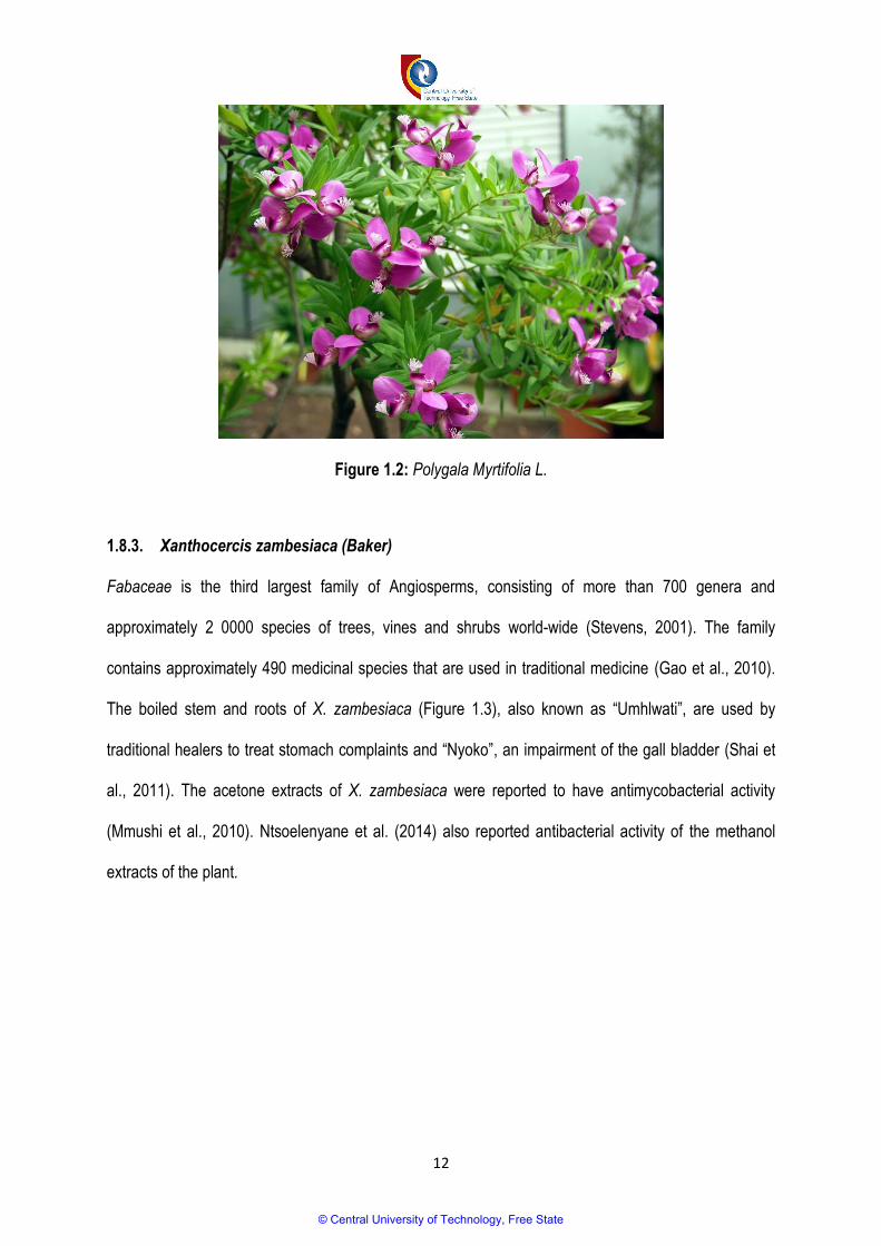
1.8.2. Polygala myrtifolia L.
The Polygala genus belongs to the family Polygalaceae, which contains approximately 600 species
(Fenner et al., 2005). P. myrtifolia (Figure 1.2), commonly known as “Myrtle milkwort” is a leafy
perennial shrub usually found in South Africa’s coastal and elevated environments between Cape Town
and KwaZulu-Natal. Chemical investigation of the genus Polygala indicated the presence of secondary
metabolites such as xanthones (Cristiano et al., 2003), saponins (Jia et al., 2004; Mitaine-Offer et al,
2003), oligosaccharides (Ikeya et al., 2004), flavonoids (Rao and Raman, 2004; Pizzolatti et al., 2008),
coumarins, and styryl pyrones (Pizzolatti et al., 2004). Different biological activities were found on
various species of Polygala genus, such as antibacterial, anti-inflammatory, trypanocidal and
antinociceptive, antifungal and antimycobacterial activity (Lall and Meyer, 1999; Motsei et al., 2003;
Pizzolatti et al., 2003; Kou et al., 2006; Pizzolatti et al., 2008; Ribas et al., 2008).
© Central University of Technology, Free State
12
Figure 1.2: Polygala Myrtifolia L.
1.8.3. Xanthocercis zambesiaca (Baker)
Fabaceae is the third largest family of Angiosperms, consisting of more than 700 genera and
approximately 2 0000 species of trees, vines and shrubs world-wide (Stevens, 2001). The family
contains approximately 490 medicinal species that are used in traditional medicine (Gao et al., 2010).
The boiled stem and roots of X. zambesiaca (Figure 1.3), also known as “Umhlwati”, are used by
traditional healers to treat stomach complaints and “Nyoko”, an impairment of the gall bladder (Shai et
al., 2011). The acetone extracts of X. zambesiaca were reported to have antimycobacterial activity
(Mmushi et al., 2010). Ntsoelenyane et al. (2014) also reported antibacterial activity of the methanol
extracts of the plant.
© Central University of Technology, Free State

13
Figure 1.3: Xanthocercis zambesiaca (Baker)
1.8.4. Scilla Species
The genus Scilla sp. belongs to the Hyacinthaceae family and Scilloideae subfamily. Species in
Hyacinthaceae are known to treat diseases such as urinary diseases, gastrointestinal problems,
respiratory problems, headaches, swelling and skin problems, and they are used as internal purifiers
(Hutchings., 1996; Louw et al., 2002). Members of this family are known to have traces of
phytochemicals such as saponins and homoisoflavanones. However, the presence of alkaloids and
steroids in some species may indicate cytotoxicity (Speta, 1998).
1.9. Aims and objectives
The aim of the study was to evaluate the antimicrobial activity of Buxus macowanii, Scilla sp., Polygala
myrtifolia and Xanthocercis zambesiaca. This was achieved by meeting the following objectives:
Determining the antimicrobial activity of methanol extracts from Buxus macowanii, Polygala
myrtifolia, Scilla sp and Xanthocercis zambesiaca.
© Central University of Technology, Free State

14
Locating the antimicrobial compounds using Thin Layer Chromatography (TLC).
Examining the effect of the plant extracts on the microbial cells using Scanning Electron
Microscopy (SEM) and Transmission Electron Microscopy (TEM).
Determining the phytochemical content of Buxus macowanii, Polygala myrtifolia, Scilla sp. and
Xanthocercis zambesiaca; and
Evaluating the cytotoxicity of the methanol extracts of Buxus macowanii, Polygala myrtifolia, Scilla
sp. and Xanthocercis zambesiaca.
1.10. Overview of the study
This study is presented in four sections. In Chapter 2, the antimicrobial activity of the plant extracts was
evaluated using the microdilution method, TLC bioautography and microscopy. The cytotoxicity of the
extracts was tested using the Sulforhodamine B cytotoxicity assay (SRB), the results of which are
discussed in Chapter 3. Chapter 4 focused on the determination of the phytochemical contents of the
plant extracts using the standard phytochemical screening methods, TLC and Gas Chromatographic
Mass Spectrometry (GCMS). Chapter 5 provides general discussions of the results obtained in this
study, conclusions and recommendations. The techniques and procedures used to evaluate the
antimicrobial activity of B. macowanii, P. myrtifolia, Scilla sp. and X. zambesiaca are illustrated in Figure
1.4.
© Central University of Technology, Free State
© Central University of Technology, Free State
16
1.11. References
1. Adwan, G., Salameh, Y., and Adwan, K., 2011. Effect of ethanolic extract of Ecballium elaterium
against Staphylococcus aureus and Candida albicans. Asian Pacific Journal of Tropical
Biomedicine: 456-460.
2. Ata, A., and Andersh, B. J., 2008. Buxus steroidal alkaloids: chemistry and biology. The Alkaloids:
Chemistry and Biology. 66: 191-213.
3. Ata, A., 2012. Novel biomedical agents from plant. In: Orhan IE, editor. Biotechnological production
of plant secondary metabolites. Dubai: Bentham Science Publishers: 53–66.
4. Awouafack, M.D., Tane, P., Kuete, V., Eloff, J.N., 2013. 2—Sesquiterpenes from the medicinal
plants of Africa. Medicinal plant research in Africa: 33103.
5. Augustin, J. M., Kuzina, V., Anderson, S. B., Bak, S., 2011. Molecular activities, biosynthesis and
evolution of triterpenoid saponins. Phytochemistry. 72: 435–457.
6. Barebehenn, R.V., and Constabel, C.P., 2011. Tannins in plant herbivores interactions.
Phytochemistry. 72: 1551-1565.
7. Bedir, E., Abou-Gazar, H., Ngwendson, J.N. and Khan, I.A., 2003. Eurycomaoside: a new
quassinoid-type glycoside from the roots of Eurycoma longifolia. Chemical and pharmaceutical
bulletin. 51(11): 1301-1303.
8. Bhat, R. and Karim, A.A., 2010. Tongkat Ali (Eurycoma longifolia Jack): a review on its ethnobotany
and pharmacological importance. Fitoterapia. 81(7): 669-679.
9. Botha, C.J., Penrith, M.-L., 2008. Poisonous plants of veterinary and human importance in
Southern Africa. Journal of Ethnopharmacology. 119: 549–558
10. Boucher, H., Talbot, G.H., Bradley, J.S. 2009. Bad bugs, no drugs: no ESKAPE! An Update from
the Infectious Diseases Society of America. Clinical Infectious Diseases. 425: 1–12.
11. Cannes do Nascimento, N., Fett-Neto, A.G., 2010. Plant secondary metabolism and challenges in
modifying its operation: An overview. Humana Press Inc., New York: 1–13.
© Central University of Technology, Free State

17
12. Cheikhyoussef, A., Shapi, M., Matengu, K., and Ashekele, H.M.U., 2011. Ethnobotanical study of
indigenous knowledge on medicinal plant use by traditional healers in Oshikoto region, Namibia.
Journal of Ethnobiology and Ethnomedicine. 7: 10.
13. Chimene, M.K., Okunji, C.O., Iwu, M.M., Kuete., V., 2013. 1—Monoterpenes and related
compounds from the medicinal plants of Africa. In: Kuete V, editor. Medicinal plant research in
Africa. Oxford: Elsevier: 132
14. Cristiano, R., Pizzolatti, M. G., Monache, F. D., Rezende, C. M., Branco, A., 2003. Two xanthones
from Polygala paniculata and confirmation of the 1-hydroxy-2, 3, 5-trimethoxy-xanthone at trace
level by HRGC-MS. Zeitschrift für Naturforschung C. 58(7-8): 490-494.
15. Copp, B.R., and Pearce, A.N., 2007. Natural product growth inhibitors of Mycobacterium
tuberculosis. Natural products reports. 24: 278-297.
16. Cos, P., Vlietinck, A.J., Berghe, D.V., Maes, L., 2006. Anti-infective potential of natural products:
how to develop a stronger in vitro ‘proof-of-concept’. Journal of Ethnopharmacology. 106: 290–
302.
17. Cunningham, A. B., 1997. An Africa-wide overview of medicinal plant harvesting, conservation and
health care. Medicinal plants for forest conservation and health care. 11: 116-129.
18. Cushnie, T. T., and Lamb, A. J., 2005. Antimicrobial activity of flavonoids. International journal of
antimicrobial agents. 26(5): 343-356.
19. Daboor, S.M., and Haroon, A.M., 2012. In vitro: Antimicrobial potential and phytochemical
screening of some Egyptian aquatic plants. Egyptian Journal of Aquatic Research. 38: 233-239.
20. DeGabriel, J. L., Moore, B. D., Foley, W. J., Johnson, C. N., 2009. The effects of plant defensive
chemistry on nutrient availability predict reproductive success in a mammal. Ecology. 90(3): 711-
719.
21. Doughari, J. H., 2012. Phytochemicals: Extraction methods, basic structures and mode of action
as potential chemotherapeutic agents. INTECH Open Access Publisher.
© Central University of Technology, Free State
18
22. Dzoyen, J.P., Tshikalange, E., Kuete, V., 2013. Medicinal Plants Market and Industry in Africa.
Medicinal Plant Research in Africa. 859-890.
23. Elekofehinti, O. O., 2015. Saponins: Anti-diabetic principles from medicinal plants–A review.
Pathophysiology. 22(2): 95-103.
24. Esterhuizen, L.L., Meyer, R., and Dubery, I.A., 2006. Antimicrobial compounds from Coleonema
album (Rutaceae). Journal of Biosciences. 61. (7-8): 489-498.
25. Feachem, R. G., 2004. Editorial: the research imperative: fighting AIDS, TB and malaria. Tropical
Medicine and International Health. 9(11): 1139-1141.
26. Fenner, R., Sortino, M., Rates, S.M.K., Dall’Agnol, R., Ferraz, A., Bernardi, A.P., Albring, D., Nör,
C., Von Poser, G., Schapoval, E., Zacchino, S., 2005. Antifungal activity of some Brazilian
Hypericum species. Phytomedicine. 12: 236-240.
27. Firn, R., 2010. Nature’s Chemicals. Oxford University Press, Oxford: 74-75.
28. Frantz, S., 2004 Approvals: The demise of the blockbuster? Nature Reviews Drug Discovery. 4(2):
93-94.
29. Fraga, C. G., Galleano, M., Verstraeten, S. V., Oteiza, P. I., 2010. Basic biochemical mechanisms
behind the health benefits of polyphenols. Molecular Aspects of Medicine. 31(6): 435-445.
30. Fuku, S., Al-Azzawi, A.M., Madamombe-Manduna, I.T., and Mashele, S., 2013. Phytochemistry
and Free Radicals Scavenging activity of Asparagus laricinus. International Journal of
Pharmacology. 9(5): 312-317.
31. Gao, T., Yao, H., Song, J., Liu, C., Zhu, Y., Ma, X., Pang, X., Xu, H., Chen, S., 2010. Identification
of medicinal plants in the family Fabaceae using a potential DNA barcode ITS2. Journal of
Ethnopharmacology. 130(1): 116–121.
32. Glen, H.L., 1996. FSA contributions 5: Buxaciae. Bothalia. 26(1): 37-40.
33. Goldberg, D. E., Siliciano, R. F., Jacobs, W. R. 2012. Outwitting evolution: fighting drug-resistant
TB, malaria, and HIV. Cell. 148(6): 1271-1283.
© Central University of Technology, Free State
19
34. Gómez-Caravaca, A. M., Verardo, V., Segura-Carretero, A., Fernández-Gutiérrez, A., Caboni, M.
F., 2014. Phenolic compounds and saponins in plants grown under different irrigation regimes.
Polyphenols in Plants: Isolation, Purification and Extract Preparation: 37-52.
35. Graul, A. I., 2001. The year’s new drugs. Drug News Perspectives. 14: 12-31.
36. Gurib-Fakim, A., 2006. Medicinal plants: traditions of yesterday and drugs of tomorrow. Molecular
aspects of Medicine. 27(1): 1-93.
37. Havsteen, B. H., 2002. The biochemistry and medical significance of the flavonoids.
Pharmacology and Therapeutics. 96(2): 67-202.
38. Hawkey, P.M., 2008. Molecular epidemiology of clinically significant antibiotic resistance genes.
British Journal of Pharmacology. 153: 406-413.
39. Hamburg, M.A., 2008. Considerations for infectious disease research and practice. Technology in
Society. 30: 383– 387.
40. Harborne, J. B., and Baxter, H., 1999. The handbook of natural flavonoids. Volume 1 and Volume
2. John Wiley & Sons.
41. Harborne, J.B., William, C.A., 2000. Advances in flavonoids research since 1992. Phytochemistry.
55: 481-504.
42. Hooper, L. and Cassidy, A., 2006. A review of the health care potential of bioactive compounds.
Journal of the Science of Food and Agriculture. 86(12): 1805-1813.
43. Hutchings, A., 1996. Zulu medicinal plants: an inventory. University of Natal Press.
44. Ibrahim, T.A., Opawale, B.O., Oyinloye, J.M.A., 2011. Antibacterial activity of herbal extracts
against multi drug resistant strains of bacteria from clinical origin. Life Sciences Leaflets. 15: 490-
498.
45. Ikeya, Y., Takeda, S., Tunakawa, M., Karakida, H., Toda, K., Yamaguchi, T. and Aburada, M.,
2004. Cognitive improving and cerebral protective effects of acylated oligosaccharides in Polygala
tenuifolia. Biological and Pharmaceutical Bulletin. 27(7): 1081-1085.
© Central University of Technology, Free State
20
46. Iwu, M. M., 2014. Handbook of African medicinal plants. CRC press.
47. Jia, H., Jiang, Y., Ruan, Y., Zhang, Y., Ma, X., Zhang, J., Zhang, D., 2004. Tenuigenin treatment
decreases secretion of the Alzheimer’s disease amyloid β-protein in cultured cells. Neuroscience
letters. 367(1): 123-128.
48. Jindal, B.A.K., Pandya, M.K., Khan, M.I.D., 2015. Antimicrobial resistance: A public health
challenge. Medical Journal Armed Forces India. 71: 178 -181.
49. Kar, A., 2007. Pharmaocgnosy and Pharmacobiotechnology (Revised-Expanded Second Edition).
New Age International Limited Publishers New Delhi: 332-600.
50. Kaur, S., Micheal, H., Arora, S., Harkonen, P.L., Kumar, S., 2005. The In vitro cytotoxicity and
apoptotic activity of Triphala-an Indian herbal drug. Journal of Ethno pharmacology. 10: 15-20.
51. Kisangau, D.P., Lyaruu, H.V.M., Hosea, K.M., Joseph, C.C., 2007. Use of traditional medicines in
the management of HIV/AIDS opportunistic infections in Tanzania: a case in the Bukoba rural
district. Journal of Ethnobiology and Ethno medicine. 3 (29).
52. Kou, J., Si, M., Dai, G., Lin, Y., Zhu, D., 2006. Antiinflammatory activity of Polygala japonica
extract. Fitoterapia. 77(6): 411-415.
53. Kougan, G.B., Taboada, T., Kuete, V., Verpoorte, R., 2013. Simple phenols, phenolic acids, and
related esters from the medicinal plants of Africa. Medicinal plant research in Africa: 225-49.
54. Kubmarawa, D., Ajoku, G.A., Enwerem, N.M. and Okorie, D.A., 2007. Preliminary phytochemical
and antimicrobial screening of 50 medicinal plants from Nigeria. African Journal of Biotechnology,
6(14): 1690-1696.
55. Kuete, V., Wansi, J. D., Mbaveng, A. T., Sop, M. K., Tadjong, A. T., Beng, V. P., Lall, N., 2008.
Antimicrobial activity of the methanolic extract and compounds from Teclea afzelii (Rutaceae).
South African Journal of Botany. 74(4): 572-576.
56. Kuete, V., and Efferth, T., 2010. Cameroonian medicinal plants: pharmacology and derived natural
products. Frontiers in Pharmacology. 1.
© Central University of Technology, Free State
21
57. Kuete, V., Eichhorn, T., Wiench, B., Krusche, B., Efferth, T., 2012. Cytotoxicity, anti-angiogenic,
apoptotic effects and transcript profiling of a naturally occurring naphthyl butenone, guieranone A.
Cell division. 7(1): 1-12.
58. Lall, N., and Meyer, J.J., 1999. In vitro inhibition of drug-resistant and drug-sensitive strains of
Mycobacterium tuberculosis by ethnobotanically selected South African plants. Journal Ethno
pharmacology. 66: 347-354.
59. Lam, C.W., Wakeman, A., James, A., Ata, A., Gengan, R.M., Ross, S.A., 2015. Bioactive steroidal
alkaloids from Buxus macowanii Oliv. Steroids. 95: 73–79.
60. Lee, S.J., Seo, E., Cho, Y., 2013. Proposal for a new therapy for drug-resistant malaria using
Plasmodium synthetic lethality inference. International Journal for Parasitology: Drugs and Drug
Resistance. 3: 119–128.
61. Lewu, F.B., Afolayan, A.J., 2009. Ethnomedicine in South Africa: the role of weedy species.
African Journal of Biotechnology. 8 (6): 929–934.
62. Louw, C.A.M., Regnier, T.J.C., Korsten, L., 2002. Medicinal bulbous plants of South Africa and
their traditional relevance in the control of infectious diseases. Journal of Ethno pharmacology. 82:
147-154.
63. Mabona, U., and Van Vuuren, S.F., 2013. Southern African medicinal plants used to treat skin
diseases. South African Journal of Botany. 87: 175–193.
64. Manach, C., Scalbert, A., Morand, C., Rémésy, C., Jiménez, L., 2004. Polyphenols: food sources
and bioavailability. The American Journal of Clinical Nutrition. 79(5): 727-747.
65. Mander, M., 1998. Marketing of indigenous medicinal plants in South Africa: A case study in
KwaZulu-Natal.
66. Manimozhi, D.M., Sankaranarayanan, S., and Sampathkumar, G., 2012. Evaluating the
antibacterial activity of flavonoids extracted from Ficus benghalensis. International Journal of
Pharmaceutical and Biological Research. Vol 3, ISSN: 0976-285X.
© Central University of Technology, Free State

22
67. Masevhe, N.A., McGaw, L.J., Eloff, J.N., 2015. The traditional use of plants to manage candidiasis
and related infections in Venda, South Africa. Journal of Ethno pharmacology. 168: 364–372.
68. Mitaine‐Offer, A. C., Miyamoto, T., Laurens, V., Delaude, C., Lacaille‐Dubois, M. A. 2003. New
acylated triterpene saponins from Polygala arenaria. Helvetica chimica acta. 86(7): 2404-2413.
69. Mmushi, T.J., Masoko, P., Mdee, L.k., Mokgotho, M.P., Mampuru, L.J., and Howard, R.L., 2010.
Antimycobacterial evaluation of fifteen medicinal plants in South Africa. African Journal of
Traditional, Complementary and Alternative Medicines. 7 (1): 34–39.
70. Motamedi, H., Mirzabeigi, H., and Shirali, T., 2010. Determining of antibiotic resistance profile in
Staphylococcus aureus isolates. Asian Pacific Journal of Tropical Medicine. 3(9): 734-737.
71. Motsei, M.L., Lindsey, K.L., van Staden, J., Jager, A.K., 2003. Screening of traditionally used
South African plants for antifungal activity against Candida albicans. Journal of Ethno
pharmacology. 86: 235–241.
72. Mueller, M. S., Karhagomba, I. B., Hirt, H. M., Wemakor, E. 2000. The potential of Artemisia
annua L. as a locally produced remedy for malaria in the tropics: agricultural, chemical and clinical
aspects. Journal of Ethno pharmacology. 73(3): 487-493.
73. Ndhlala, A.R., Ncube, B., Okem, A., Mulaudzi, R.B., Van Staden, J., 2013. Toxicology of some
important medicinal plants in southern Africa. Food and Chemical Toxicology. 62: 609–621.
74. Ngameni, B., Fotso, G. W., Kamga, J., Ambassa, P., Abdou, T., Fankam, A. G., Kuete, V., 2013. 9
Flavonoids and Related Compounds from the Medicinal Plants of Africa. Medicinal Plant Research
in Africa: Pharmacology and Chemistry, 301.
75. Normann, H., and Snyman, I. 1996. Indigenous knowledge and its uses in southern Africa (Vol.
61). HSRC Press.
76. Ntsoelenyane, P.H., Mashele, S.S., Manduna, I.T., 2014. The anticancer, antioxidant and
phytochemical screening of Philenoptera violacea and Xanthocercis zambesiaca leaf flower and
twig extracts. International Journal of Pharmacological Research. 4 (3): 100-105.
© Central University of Technology, Free State

23
77. Obeidat, M., Shatnawi, M., Al-alawi, M., Enas, A., Hanee, A., Masia, A., El-Quadah, J., Otr, I.,
2012 Antimicrobial activity of crude extracts of some plant leaves. Research Journal of
Microbiology. 7:59-67.
78. O'Bryan, C.A., Crandall, P.G., Ricke, S.C., 2008. Organic poultry pathogen control from farm to
fork. Foodborne pathogens and disease. 5: 709–720.
79. Okuda, T., 2005. Systematics and health effects of chemically distinct tannins in medicinal plants.
Phytochemistry. 66(17): 2012-2031.
80. Oliphant, C. M., and Eroschenko, K., 2015. Antibiotic Resistance, Part 1: Gram-positive
Pathogens. The Journal for Nurse Practitioners. 11(1): 70-78.
81. Olliaro, P. L., and Bloland, P. B., 2001. Clinical and public health implications of antimalarial drug
resistance. In Antimalarial Chemotherapy: Mechanisms of Action, Resistance, and New Directions
in Drug Discovery (ed. P. J. Rosenthal). Totowa, NJ: Humana Press: 65-83.
82. Pirttilä, T., Wilcock, G., Truyen, L., Damaraju, C. V., 2004. Long‐term efficacy and safety of
galantamine in patients with mild‐to‐moderate Alzheimer's disease. multicenter trial. European
Journal of Neurology.11(11): 734-741.
83. Pizzolatti, M.G., Koga, A.H., Grisard, E.C., Steindel, M., 2003. Trypanocidal activity of extracts
from Brazilian Atlantic Rain Forest plant species. Phytomedicine. 10: 422-426.
84. Pizzolatti, M. G., Cunha, A., Pereira, W. S., Delle Monache, F., 2004. A new styryl-2-pyrone
derivative from Polygala sabulosa (Polygalaceae). Biochemical systematics and ecology. 32(6):
603-606.
85. Pizzolatti, M. G., Mendes, B. G., Cunha Jr, A., Soldi, C., Koga, A. H., Eger, I., Steindel, M., 2008.
Trypanocidal activity of coumarins and styryl-2-pyrones from Polygala sabulosa AW Bennett
(Polygalaceae). Revista Brasileira de Farmacognosia. 18(2): 177-182.
86. Pooley, E., 1993. The complete guide to trees of Natal, Zululand and Transkei. Durban: Natal
Flora Publication Trust.
© Central University of Technology, Free State

24
87. Rao, M.S., and Raman, N.V., 2004. A novel flavonoid from Polygala chinensis. Biochemical
systematics and ecology. 32(4): 447-448.
88. Rawat, A., 2012. Gaps and shortages in South Africa’s health work force. Back- grounder. 31:1–6.
89. Rosenberry, T.L., 1975. Acetylcholinesterase. Advanced Enzymology Related Areas of Molecular
Biology. 43: 103–218.
90. Ribas, C. M., Meotti, F. C., Nascimento, F. P., Jacques, A. V., Dafre, A. L., Rodrigues, A. L. S.,
Santos, A. R. 2008. Antinociceptive effect of the Polygala sabulosa hydroalcoholic extract in mice:
evidence for the involvement of glutamatergic receptors and cytokine pathways. Basic & clinical
pharmacology & toxicology. 103(1): 43-47
91. Rice, L. B., 2008. Federal funding for the study of antimicrobial resistance in nosocomial
pathogens: no ESKAPE. Journal of Infectious Diseases. 197(8): 1079-1081.
92. Roca, I., Akova, M., Baquero, F., Carlet, J., Cavaleri, M., Coenen, S., Vila, J., 2015. The global
threat of antimicrobial resistance: science for intervention. New Microbes and New Infections.
93. Rao, M. S., & Raman, N. V., 2004. A novel flavonoid from Polygala chinensis. Biochemical
systematics and ecology. 4(32): 447-448.
94. Samuelsson, G., 2004. Drugs of Natural Origin: a Textbook of Pharmacognosy, 5th Swedish
Pharmaceutical Press, Stockholm.
95. Sarker, S., and Nahar, L., 2007. Chemistry for pharmacy students: general, organic and natural
product chemistry. John Wiley & Sons.
96. Sandjo, L.P., Kuete, V., 2013. 4—Triterpenes and steroids from the medicinal plants of Africa.
Medicinal plant research in Africa: 135-202.
97. Shai, L.J., Magano, S.R., Lebelo, S.L., and Mogale, A.M., 2011. Inhibitory effects of five medicinal
plants on rat alpha-glucosidase: Comparison with their effects on yeast alpha-glucosidase. Journal
of Medicinal Plants Research. 5(13): 2863-2867.
© Central University of Technology, Free State
25
98. Sparg, S., Light, M. E., Van Staden, J., 2004. Biological activities and distribution of plant
saponins. Journal of ethno pharmacology. 94(2): 219-243.
99. Speta, F., 1998. Hyacinthaceae. In Flowering Plants· Monocotyledons. Springer Berlin Heidelberg:
261-285.
100. Stevens, P. F., 2001. Onwards. Angiosperm Phylogeny Website. Version 12, July 2012 and
more or less continuously updated since.
101. Street, R.A., Stirk, W.A. and Van Staden, J., 2008. South African traditional medicinal plant trade
challenges in regulating quality, safety and efficacy. Journal of Ethnopharmacology. 119(3): 705-
710.
102. Taylor, J.L.S., Rabe, T., McGaw, L.J., and Van Staden, J., 2001. Toward the scientific validation
of medicinal plants. Plant Growth Regulations. 34: 23-37.
103. Van Wyk, B. E., Heerden, F. V., Oudtshoorn, B. V., 2002. Poisonous plants of South Africa. Briza
Publications.
104. Wansi, J. D., Devkota, K. P., Tshikalange, E., and Kuete, V., 2013. Alkaloids from the medicinal
plants of Africa. Medicinal Plant Research in Africa: 557-605.
105. Wang, G., Tang, W., and Bidigare, R. R., 2005. Terpenoids as therapeutic drugs and
pharmaceutical agents. In Natural products (pp. 197-227). Humana Press.
106. Wink, M., Van Wyk, B.-E., 2008. Mind-Altering and Poisonous Plants of the World. Briza
Publications, Pretoria.
107. Winston, C. A., and Mitruka, K., 2012. Treatment duration for patients with drug-resistant
tuberculosis, United States. Emerging infectious diseases. 18(7): 1201.
108. World Health Organisation., 2010. Global Tuberculosis report: WHO report 2010. World Health
Organization
109. World Health Organisation (WHO), 2011a. The world medicines situation traditional medicines:
global situation, issues and challenges. Geneva. Available at:
© Central University of Technology, Free State
26
http://www.who.int/medicines/areas/policy/world_medicines_situation/WMS_ch18_wTraditionalM
ed.pdf (Accessed on: 11/01/2013).
110. World Health Organization. World Health Day 2011b: Policy Briefs. Geneva: WHO; 2011.
Available at: http://www.who.int/ world-health-day/2011/policybriefs/en/index.html. Accessed Nov
2012.
111. World Health Organisation., 2014a. Global summary of the AIDS epidemic. WHO- HIV
department. World Health Organisation. Global Tuberculosis report 2014.
http://apps.who.int/iris/bitstream/10665/137094/1/9789241564809 eng.pdf?ua=1
112. World Health Organisation., 2014b. World Malaria Report. WHO Library Cataloguing-in-
Publication Data, ISBN 978 92 4 156483 0.
113. Zerabruk, S., and Yirga, G., 2012. Traditional knowledge of medicinal plants in Gindeberet
district, Western Ethiopia. South African Journal of Botany. 78: 165-169.
114. Zofou, D., Kuete, V., Titanji, V. P. K., 2013. Antimalarial and other antiprotozoal products from
African medicinal plants. Medicinal Plant Research in Africa: Pharmacology and Chemistry.
Elsevier Inc., London, UK.
115. Zuanazzi, J. A. S., and Montanha, J. A., 2004. Flavonóides In: Farmacognosia da Planta ao
medicamento. Porto Alegre/Floriano polis, (In Portuguese).
© Central University of Technology, Free State

27
CHAPTER 2
ANTIMICROBIAL ACTIVITY OF BUXUS MACOWANII, POLYGALA MYRTIFOLIA, SCILLA SP AND
XANTHOCERCIS ZAMBESIACA
2.1. Introduction
The use of western medicine to treat infectious diseases has always been associated with a number of
problems that make the treatment of infectious diseases more challenging. The use of antimicrobial
drugs that are currently available include problems such as limited accessibility for people living in rural
areas or places remote to health care facilities, high purchase prices, and side effects (Bodeker and
Graz, 2013). The emergence of drug-resistant pathogens and an increase in opportunistic infections in
people with HIV/AIDS and those receiving chemotherapy are additional problems that make the
treatment of infectious diseases very difficult (Fargana et al., 2014). These problems have encouraged
researchers and scientists to devise new alternative manners of treatment, which can lead to the
discovery of novel drugs that will overcome the challenges associated with the drugs that are currently
used.
For thousands of years, plants have been used by millions of people as a source of food and for
medicinal purposes (Brouwer et al., 2005). An estimated 80% of the world’s population, and 72% of
black South Africans, still rely on traditional medicine for their primary health care needs (Gurib-Fakim,
2006). Traditional medicine has not only gained popularity due to its effectiveness against diseases, but
because it is sometimes the only available system in rural areas (Mabona and Van Vuuren, 2013). The
use of traditional medicine is further attributed to advantages such as accessibility, affordability, fewer
side effects, and extensive knowledge and expertise thereof by people within communities (Fennell et
al., 2004; Runyoro et al., 2006).
© Central University of Technology, Free State

28
Medicinal plants, especially those used by traditional healers, have become the focus of research, and
are considered as the best option for the production of novel drugs, as they have an unmatched
chemical diversity (Cos et al., 2006; O’Bryan et al., 2008). They contain bioactive compounds such as
alkaloids, terpenoids, tannins, flavonoids, peptides and phenolic compounds that are known to have
antimicrobial, antiviral, antifungal, anti-inflammatory, anthelmintic and antioxidant activity (Kubmarawa
et al., 2007). Moreover, medicinal plants are known to work on different target sites than those targeted
by conventional drugs (Kimberlin and Whitley, 1996). They are also cheaper and have fewer side
effects, and thus medicinal plants remain an important and better alternative source from which new
therapeutic agents can be manufactured.
Traditional knowledge and ethnobotany help researchers to identify medicinally relevant plants that can
lead to the discovery and manufacturing of new drugs. Available literature indicated that plants such as
B. macowanii, P. myrtifolia, X. zambesiaca and other Scilla sp. are traditionally used for the treatment of
wounds, skin disorders, stomach problems, back problems, fractures and other major diseases, such
as cancer (Pooley, 1993; Hutchings et al., 1996; Crouch et al., 1999, Shai et al., 2011). It is therefore
necessary that this chapter focuses on studying the antimicrobial activity of these plants, and the
minimum concentration at which the extracts inhibit the growth of bacteria and fungi, by using the
microdilution method.
2.2. Methods
2.2.1. Plant extracts
Buxus macowanii Oliv (Buxuceae), Polygala myrtifolia L. (Polygalaceae), Scilla sp. (Asparagaceae) and
Xanthocercis zambesiaca (Baker) (Fabaceae) were selected for the investigation of their antimicrobial
activity against bacterial and fungal species. Methanol plant extracts and information about the plants
© Central University of Technology, Free State

29
(as presented in Table 2.1) were supplied by the Department of Soil, Crop and Climate Sciences at the
University of the Free State (UFS) because they were the only type of extracts available.
Table 2.1. Information about plant extracts
Plant name Plant part Collection
number
Extract
number
Place of collection
B. macowanii Oliv Leaves and twigs RB 829 a
(Brand 795)
1544 Mpumalanga
Scilla sp. Whole plant RB 512
(Brand 614)
1272.2 Eastern Cape
Scilla sp. Roots and bulbs JV 9852b
(Venter 251)
1163 Free State
P. myrtifolia L Leaves and twigs RB 801 a
(Brand 753)
1561 Eastern Cape
X. zambesiaca
(Baker)
Stem
-
1584 Mpumalanga
2.2.2. Extract preparation and storage
The plants were collected, washed, oven dried at 40ºC for 72 hours, and ground to powder. The dry,
powdered material of the plants was extracted with 100% methanol because the solvent is easy to
evaporate and does not affect results of any screening. The methanol was evaporated at 40ºC, under a
vacuum, using a Buchi Rotavapor. All extracts were stored in the fridge at -20ºC until required (Zaidan
et al., 2005).
© Central University of Technology, Free State

30
2.2.3. Microbial cultures
The antibacterial activity of the plant extracts was evaluated against Staphylococcus aureus (ATCC
25923), Clostridium perfringens (ATCC 13126), Pseudomonas aeruginosa (ATCC 27853),
Enterococcus faecalis (ATCC 29212), Escherichia coli (ATCC 25922) and Staphylococcus epidermidis
(ATCC 12228). The fungal species used were Candida albicans (ATCC 90028) and Candida tropicalis
(ATCC 756). Bacillus cereus (ATCC 13061) was selected for examination of the cell wall after treatment
with the plant extract, which exhibited the best antibacterial activity using microscopy. All the bacterial
and fungal species were supplied by the National Health Laboratory Services, Bloemfontein, South
Africa. All the microbial species were maintained in Mueller Hinton agar plates at temperatures of 4ºC.
Prior to treatment with the plant extracts, the bacteria were inoculated in Mueller Hinton broth, and
incubated and shaken at 100 revolutions per minute (rpm) for 24 hours, to ensure purity and viability.
Thereafter, one millilitre of the culture was diluted in 100 ml of Mueller Hinton broth (1:100) (Meyer and
Afolayan, 1995).
2.2.4. Antimicrobial activity
The antibacterial and antifungal activity of five plant extracts was investigated using the microdilution
method developed by Eloff (1998). The microdilution method was selected because it is easier to
perform, and it can determine the minimum inhibitory concentration of plant extracts, as opposed to the
agar diffusion method. One hundred microliters of the bacterial suspension was pipetted into the 96
microwell plates already containing 100 µl of diluted plant extract to make a final volume of 200 µl in
each well. The concentration of the plant extracts ranged from 0.16 mg/ml to 2.5 mg/ml. The control
wells were respectively filled with culture medium only, bacterial suspension, 5% dimethyl sulfoxide
(DMSO) (Nostro et al., 2000; Baris et al., 2006), and plant extract only. Chloramphenicol (0.125 mg/ml)
was used as a positive control in bacteria, while amphotericin B (0.03-1 µg/ml) was used in fungi
because it is one of the antifungal agents that were used to treat fungal infections (Wanger et al., 2005;
© Central University of Technology, Free State

31
Andrews, 2001). The microwell plates were incubated for 24 hours, where after 40 µl of 4 mg/ml
Iodonitrotetrazolium salt solution was added to each well. Growth was indicated by a change of colour
ranging from pink to violet after 10 to 30 minutes’ incubation. All samples were tested in triplicates. The
minimum inhibitory concentration (MIC) was recorded as the lowest concentration at which the plant
indicated bacterial or fungal growth inhibition.
2.2.5. Thin Layer Chromatography (TLC) bioautography
The bioautography method was used to screen and identify compounds with antibacterial activity
present in the plant extracts of B. macowanii. The detection of antimicrobial compounds was performed
using aluminium-coated DC-fertigfolien alugram Xtra Sil G/ Uv 254 TLC plates. The plant that exhibited
the best antibacterial activity was loaded onto three TLC plates, and eluted using a mobile polar solvent
system Toluene, Chloroform and Ethanol (5.7:11.4:2.9), which provided the best separation of the
compounds. The developed plate was left to dry for 24 hours to remove traces of the solvent on the
plate. The prepared TLC plate was sprayed with the Enterococcus faecalis bacterial suspension until
wet, whereafter it was incubated overnight at 37ºC and at 100% relative humidity for 8 hours
(Esterhuizen et al., 2006). The plate was sprayed with 4 mg/ml solution of Iodonitrotetrazolium chloride,
and further incubated for eight hours in a sealed container for colour development. The antibacterial
compounds were identified as white areas against a violet- or pink-coloured background, which
indicated bacterial growth. On the other two plates, the separated alkaloids were detected using
Dragendorff’s reagent, and flavonoids were detected using natural product-polyethylene glycol reagent.
The detected alkaloids and flavonoids were visualised under ultraviolet light at a wavelength of 365 nm.
All the TLC plates were run in duplicates, and one was used as a reference chromatogram.
© Central University of Technology, Free State

32
2.2.6. The microscopic analysis of the bacterial cell wall.
The effect of B. macowanii on bacterial morphology was examined using scanning and transmission
electron microscopy. B. cereus was treated with different concentrations (0.2 to 2.5 mg/ml) of the
methanol extracts of B. macowanii. The untreated samples (control) and the treated samples were
incubated at 37ºC for 24 hours. After incubation, the cells were washed twice with 0.1 M phosphate
buffer solution (PBS, pH 7.0), were subjected to fixation using 3% glutardialdehyde and 1%
osmiumtetroxide, and were kept for two hours at -4ºC. Thereafter, fixation of the cells were further
subjected to dehydration in ethanol at successive concentrations of 50%, 70%, 95% and 100%,
followed by critical point-drying using Carbon dioxide (CO2) to remove the ethanol. The samples were
finally mounted on a specimen stub and coated with gold under vacuum, followed by microscopic
examination using SEM (Moosavy et al., 2008; Lv et al., 2011). For TEM-dehydrated bacterial cells,
cells were embedded by replacing ethanol with epoxy to make slim sections suitable for microscopic
examination. Samples were further embedded using epoxy for eight hours at 70ºC in a special mould.
The samples were cut into sections using ultramicrotome, and were stained with 6% Uranyl and lead
citrate, followed by TEM examination (Joshi et al., 2010; He et al., 2014).
2.3. Results and discussion
2.3.1. The antimicrobial activity of the selected plant extracts
P. myrtifolia, Scilla sp. and X. zambesiaca showed no activity against all the microbial strains against
which they were tested. Previous studies documented antimicrobial activity of X. zambesiaca (Mmushi
et al., 2010; Masoko, 2013) and antifungal activity of P. myrtifolia (Motsei et al., 2003). A factor such as
the choice of solvent might have affected the antimicrobial activity of extracts from X. zambesiaca and
P. myrtifolia, because bioactive phytochemicals are affected by factors such as solvent polarity. The
extracts of B. macowanii showed antimicrobial activity against S. aureus, S. epidermidis, C. albicans, C.
tropicalis, C. perfrengens and P. aeruginosa, where the minimum inhibitory concentration (MIC) was 2.5
© Central University of Technology, Free State

33
mg/ml. B. macowanii also showed antimicrobial activity against E. faecalis and E. coli (MIC:1.2 mg/ml).
Usually one may expect a variation in the inhibitory activity against Gram-negative and Gram-positive
bacteria due to their differences in cell wall composition; however, it was found that both bacteria were
affected in the same way. The inhibition of Gram-negative bacteria, Gram-positive bacteria and
Candida is therefore of critical importance, because of their drug resistance to current antibacterial and
antifungal agents. The B. macowanii’s inhibitory activity against Gram-negative bacteria, Gram-positive
bacteria and Candida may indicate a possible breakthrough in the fight against the challenges caused
by drug-resistant micro-organisms. Numerous studies have reported that most species in the Buxus
genus are a rich source of steroidal alkaloids. It can be suggested that the antimicrobial activity B.
macowanii extracts can be attributed towards alkaloids, which, according to literature, are abundantly
found in most plants that belong to the Buxus genus (Loru et al., 2000; Atta-ur-Rahman, et al., 2001;
Babar et al., 2006; Ata and Andresh, 2008). Thus B. macowanii was considered the best option for
further investigation.
2.3.2. TLC bioautography
Antimicrobial compounds from the extracts of B. macowanii were identified using the TLC
bioautography. The clear zones on the TLC chromatogram indicated that the extracts of B. macowanii
contain compounds that have antibacterial activity against E. faecalis using a solvent system of
Toluene, Chloroform and Ethanol (5.7:11.4:2.9) (Figure 2.1). Four clear zones were identified on the
bioautography chromatogram, suggesting that the antibacterial activity of B. macowanii cannot only be
attributed to one compound. A yellowish-orange colour under visible light showed that the extracts of B.
macowanii have alkaloids, as indicated in Figure 2.2. Figure 2.3 showed traces of flavonoids in B.
macowanii by a dark yellow, green or blue fluorescence colour under UV light at a wavelength of 354
nm. Some studies reported over 200 or more steroidal alkaloids isolated in various species belonging to
the genus Buxus (Ata et al., 2002; Meshkatalsadat et al., 2006; Babar et al., 2006; Ata et al., 2010;
© Central University of Technology, Free State

34
Matochko et al., 2010). Flavonoids have also been reported to have antibacterial and antifungal
properties (Manimodzi et al., 2012). Therefore, the presence of alkaloids and flavonoids may explain
the antibacterial effects of B. macowanii.
Figure 2.1: Chromatogram developed with Toluene/Chloroform/Ethanol (5.7:11.4:2.9) indicates the
antibacterial activity of the plant B. macowanii against E.faecalis.
© Central University of Technology, Free State
35
Figure 2.2: A chromatogram developed with Toluene/Chloroform/Ethanol (5.7:11.4:2.9) indicates the
presence of alkaloids in the extracts of Buxus macowanii. An orange colour indicates the presence of
alkaloids.
© Central University of Technology, Free State

36
Figure 2.3: A chromatogram developed with solvent system Toluene/Chloroform/Ethanol (5.7:11.4:2.9)
indicates the presence of flavonoids in the extracts of Buxus macowanii under UV light. A green-blue
colour indicates the presence of flavonoids.
2.3.3. The effect of Buxus macowanii on the cell morphology
The effect of the extracts of B. macowanii on the bacterial cell morphology was examined using
Scanning Electron Microscopy (SEM) and Transmission Electron Microscopy (TEM). SEM was used to
examine the morphology of the cells of Bacillus cereus treated with extracts of B. macowanii, for 24
hours at 37ºC. The untreated samples of B. cereus were considered as a control. Under normal
conditions, the cells of B. cereus under a microscope appear as long bacilli cells (Figure 2.4 (A)). The
cells of B. cereus that were treated with the extracts of B. macowanii at the MIC concentration of 2.5
mg/ml in SEM showed major structural changes compared to the untreated cells that lead to cell death
(Figure 2.4 (B) and (C)). Figure 2.4 (C) indicates some damage to cell walls (DCW), which is evident by
holes on the surface of the cell. The extracts also caused incomplete cell division (ICD), and
swollenness of the cells (SC), caused by the penetration of the plant extracts into the cell. The swelling
of the cell lead to cytoplasmic membrane damage, which eventually resulted in loss of the intracellular
contents (LCC), which were observed outside the cells. The loss of cellular or cytoplasmic contents
resulted in shrinkage of the cell (SC), which was explained by the wrinkled surface of the cell (Figure
2.4 (B) and (C)). The effects of B. macowanii on the morphology of the bacterial cells also
demonstrated some damage in the structure of the cells because treated cells examined using TEM
showed cell-wall distortion, which resulted in increased membrane permeability (Figure 2.5(B)). When
cell-membrane permeability increased, the loss of intracellular or cytoplasmic contents occurred.
Incomplete cell division, roughness of the cell and separation of the cytoplasmic membrane from the
cell wall were some of the morphological changes that were caused by the extracts of B. macowanii on
the bacterial cells. Vancomycin is one of the drugs that are used to treat infections that are caused by
© Central University of Technology, Free State

37
Bacillus cereus. It usually inhibits the second stage of cell-wall synthesis, alter the cell-membrane
permeability, and inhibits ribonucleic acid synthesis (Watabakunakorn, 1981). Given the results that
were obtained via SEM and TEM, B. macowanii was identified as the best treatment and may be the
best option for the development of a new drug that can control resistant pathogens causing infectious
diseases.1
1 This section has been submitted for publication with the title: Antimicrobial activity of Buxus macowanii and its morphological effects on Bacillus cereus. (See annexure B).
© Central University of Technology, Free State

© Central University of Technology, Free State
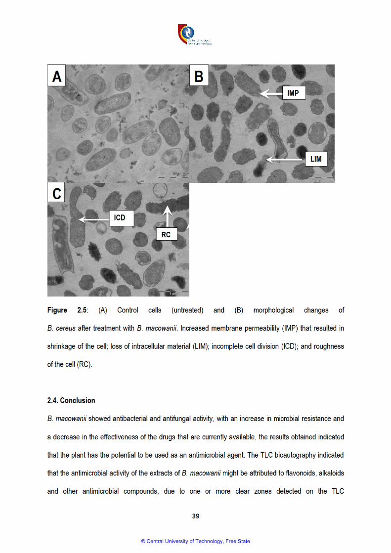
© Central University of Technology, Free State

40
chromatogram. Further analysis using a solvent such as diethylamine to achieve an ideal separation of
compounds and direct isolation of antimicrobial compounds, is recommended.
2.5. References.
1. Andrews, J.M., 2001. Determination of minimum inhibitory concentrations. Journal of
antimicrobial Chemotherapy, 48(suppl 1), pp.5-16.
2. Ata, A., and Andersh, B. J., 2008. Buxus steroidal alkaloids: chemistry and biology. The
Alkaloids: Chemistry and Biology. 66: 191-213.
3. Ata, A., Iverson, C. D., Kalhari, K. S., Akhter, S., Betteridge, J., Meshkatalsadat, M. H., Sener,
B., 2010. Triterpenoidal alkaloids from Buxus hyrcana and their enzyme inhibitory, anti-fungal
and anti-leishmanial activities. Phytochemistry. 71(14): 1780-1786.
4. Ata, A., Naz, S., Choudhary, M. I., Sener, B., & Turkoz, S., 2002. NewTriterpenoidal Alkaloids
from Buxus sempervirens. Zeitschrift für Naturforschung C. 57(1-2): 21-28.
5. Atta-ur-Rahman, Parveen, S., Khalid, A., Farooq, A., Choudhary, M.I., 2001. Acetyl and
butyrylcholinesterase-inhibiting triterpenoid alkaloids from Buxus papillosa. Phytochemistry. 58:
963-968.
6. Babar, Z.U., Ata A., Meshkatalsadat, M.H., 2006. New bioactive steroidal alkaloids from Buxus
hyrcana. Steroids. 71: 1045–1051.
7. Baris, O., Gulluce, M., Sahin, F., Ozer, H., Kilic, H., Ozkan, H., Sokmen, M., and Ozbek T.,
2006. Biological activities of the essential oil and methanol extract of Achillea Biebersteinii
Afan. (Asteraceae). Turkish Journal of Biology. 30(2): 65-73.
8. Bodeker, G., and Graz, B., 2013. 25 – Traditional Medicine. Hunter's Tropical Medicine and
Emerging Infectious Disease (Ninth Edition): 197-202.
© Central University of Technology, Free State

41
9. Brouwer, N., Liu, Q., Harrington, D., Kohen, J., Vemulpad, S., Jamie, J., Randall, M., Randall,
D., 2005. An ethnopharmacological study of medicinal plants in New South Wales. Molecules.
10: 1252–1262.
10. Crouch, N.R., Bangani, V., Mulholland, D.A., 1999. Homoisoflavanones from three South
African Scilla species. Phytochemistry. 51 (7): 943-946.
11. Cos, P., Vlietinck, A. J., Berghe, D. V., Maes, L., 2006. Anti-infective potential of natural
products: how to develop a stronger in vitro ‘proof-of-concept’. Journal of Ethnopharmacology.
106(3): 290-302.
12. Eloff, J.N., 1998. A sensitive and quick microplate method to determine the minimal inhibitory
concentration of plant extracts for bacteria. Department of pharmacology, University of Pretoria,
South Africa.
13. Esterhuizen, L.L., Meyer, R., and Dubery, I.A., 2006. Antimicrobial compounds from
Coleonema album (Rutaceae). Journal of Biosciences. 61 (7-8): 489-498.
14. Fargana, A., Zerin, N., Kabir, M.D.S., 2014. Antimicrobial activity of medicinal plant leaf
extracts against pathogenic bacteria. Asian Pacific Journal of Tropical Diseases. 4: 920-923.
15. Fennell, C.W., Lindsey, K.L., McGaw, L.J., Sparg, S.G., Stafford, G.I., Elgorashi, E.E., 2004.
Assessing African medicinal plants for efficacy and safety: pharmacological screening and
toxicology. Journal of Ethnopharmacology. 94: 205-17.
16. Gurib-Fakim, A., 2006. Medicinal plants: traditions of yesterday and drugs of tomorrow.
Molecular aspects of Medicine. 27(1): 1-93.
17. He, M., Wu, T., Pan, S., Xu, X., 2014. Antimicrobial mechanism of flavonoids against
Escherichia coli ATCC25922 by model membrane study. Applied Surface Science. 305: 515–
521.
© Central University of Technology, Free State

42
18. Joshi, S., Bisht, G.S., Rawat, D.S., Kumar, A., Kumar, R., Maiti, R., Pasha, S., 2010.
Interaction studies of novel cell selective antimicrobial peptides with model membranes and E.
coli ATCC 11775, Biochimica et Biophysica Acta (BBA)-Biomembranes, 1798: 1864–1875.
19. Kimberlin, D. W., and Whitley, R. J., 1996. Antiviral resistance: mechanisms, clinical
significance, and future implications. Journal of Antimicrobial Chemotherapy. 37(3): 403-421.
20. Kubmarawa, D., Ajoku, G. A., Enwerem, N. M., Okorie, D. A. 2007. Preliminary phytochemical
and antimicrobial screening of 50 medicinal plants from Nigeria. African Journal of
Biotechnology. 6(14): 1690–1696
21. Loru, F., Duval, D., Aumelas, A., Akeb, F., Guedon, D., Guedj, R., 2000. Four steroidal
alkaloids from the leaves of Buxus sempervirens. Phytochemistry. 54: 951-957.
22. Lv, F., Liang, H., Yuang, Q., Li, C., 2011. In vitro antimicrobial effects and mechanism of action
of selected plant essential oil combinations against four-related microorganisms. Food
Research International. 44: 3057–3064.
23. Mabona, U., and Van Vuuren, S.F., 2013. Southern African medicinal plants used to treat skin
diseases. South African Journal of Botany. 87: 175–193
24. Manimozhi, D.M., Sankaranarayanan, S., Sampathkumar, G., 2012. Evaluating the
antibacterial activity of flavonoids extracted from Ficus benghalensis. International Journal of
Pharmaceutical and Biological Research. 3(1): 7-18.
25. Masoko, P., 2013. Ethnobotanical study of some selected medicinal plants used by traditional
healers in Limpopo Province (South Africa). American Journal of Research Communication.
1(8): 15-17.
26. Matochko, W. L., James, A., Lam, C. W., Kozera, D. J., Ata, A., Gengan, R. M. 2010.
Triterpenoidal alkaloids from Buxus natalensis and their acetylcholinesterase inhibitory activity.
Journal of Natural Products. 73(11): 1858-1862.
© Central University of Technology, Free State
43
27. Meshkatalsadat, M.H., Mollataghi, A., Ata, A., 2006. New triterpenoidal alkaloids from Buxus
hyrcana. Zeitschrift Fuer Naturforschung B. 61: 201-206.
28. Meyer, J.J.M. and Afolayan, A.J., 1995. Antibacterial activity of Helichrysum aureonitens
(Asteraceae). Journal of Ethnopharmacology, 47(2):109-111.
29. Mmushi, T., Masoko, P., Mde, L., Mokgotho, M., Mampuru, L. J., Howard, R., 2010.
Antimycobacterial evaluation of fifteen medicinal plants in South Africa. African Journal of
Traditional, Complementary and Alternative Medicines. 7(1): 34-39.
30. Motsei, M.L., Lindsey, K.L., van Staden, J., Jager, A.K., 2003. Screening of traditionally used
South African plants for antifungal activity against Candida albicans. Journal of
Ethnopharmacology. 86: 235-241.
31. Moosavy, M. H., Basti, A. A., Misaghi, A., Salehi, T. Z., Abbasifar, R., Mousavi, H. A. E., 2008.
Effect of Zataria multiflora Boiss, essential oil and nisin on Salmonella typhimurium and
Staphylococcus aureus in a food model system and on the bacterial cell membranes. Food
Research International. 41: 1050–1057.
32. Nostro, A., Germano, M.P., D’Angelo, V., Marino, A., Cannatelli, M.A., 2000. Extraction
methods and bioautography for evaluation of medicinal plant antimicrobial activity. Letters in
applied Microbiology. 30(1): 379-384.
33. O'Bryan, C.A., Crandall, P.G., Ricke, S.C., 2008. Organic poultry pathogen control from farm to
fork. Foodborne Pathogen and Diseases. 5: 709–720.
34. Pooley, E., 1993. The complete guide to trees of Natal, Zululand and Transkei. Durban: Natal
Flora Publication Trust.
35. Runyoro, D.K.B., Ngassapa, O.B., Matee, M.I.N., Joseph, C.C., Moshi, M.J., 2006. Medicinal
plants used by Tanzanian traditional healers in the management of Candida infections. Journal
of Ethnopharmacology. 106: 158-165.
© Central University of Technology, Free State
44
36. Shai, L.J., Magano, S.R., Lebelo, S.L., Mogale, A.M., 2011. Inhibitory effects of five medicinal
plants on rat alpha-glucosidase: Comparison with their effects on yeast alpha-glucosidase.
Journal of Medicinal Plant Research. 5(13): 2863-2867.
37. Wanger, A., Mills, K., Nelson, P.W. and Rex, J.H., 1995. Comparison of Etest and National
Committee for Clinical Laboratory Standards broth macrodilution method for antifungal
susceptibility testing: enhanced ability to detect amphotericin B-resistant Candida isolates.
Antimicrobial Agents and Chemotherapy, 39(11): 2520-2522.
38. Watanakunakorn, C., 1981. The antibacterial action of vancomycin. Review of Infectious
Diseases. 3 (supplementary): 210-215.
39. Zaidan, M.R., Noor Rain, A., Badrul, A.R., Adlin, A., Norazah, A. and Zakiah, I., 2005. In vitro
screening of five local medicinal plants for antibacterial activity using disc diffusion method.
Trop biomed, 22(2):165-70.
© Central University of Technology, Free State

45
CHAPTER 3
THE IN VITRO CYTOTOXICITY OF BUXUS MACOWANII, SCILLA SP., POLYGALA MYRTIFOLIA
AND XANTHOCERCIS ZAMBESIACA
3.1. Introduction
Most indigenous medicinal plants in Africa are still widely used in unprocessed form, without any
scientific validations for the safety and efficacy thereof (Fennell et al., 2004; Ndhlala et al., 2013). There
are approximately 1 000 known poisonous plant species, containing 750 poisonous substances, which
occur in 150 000 plant secondary metabolites (Wink and Van Wyk, 2008). When traditional medicine is
used for the treatment of ailments, misidentification, incorrect preparation and incorrect dosage may
result in poisoning (Fennel et al., 2004). Poisoning is common in South Africa, and it is estimated that
approximately 8 000 to 20 000 deaths occur from poisoning on an annual basis. The forensic database
for Johannesburg revealed that, from 1991 to 1995, 43% of poisoning resulted from the usage of
traditional medicine (Thomson, 2000). The current rate of mortality is expected to be higher moreover;
most of the cases remain unrecorded, as the use of traditional medicine is usually kept secret (Stewart
and Steenkamp, 2000; Thomson, 2000).
Plants that are used as traditional medicine have and are still presumed to be safe to use in any way,
as they have been used for the treatment of different ailments for a long time. However, research
indicates that most plants that are used as traditional medicine or food are potentially toxic, mutagenic
and carcinogenic (Kassie et al., 1996; De Sa Ferrira and Ferrao Vargas, 1999). The toxicity of plants is
attributed to the concept that plants produce compounds or substances that prevent infections by
bacteria and viruses but at the same time these substances can be toxic to humans, even when
consumed in small quantities (Wink and Van Wyk, 2008). Secondary metabolites, such as alkaloids,
saponins, terpenes, phenolic compounds and many more of these compounds that have human health
benefits, can also be poisonous.
© Central University of Technology, Free State

46
Saponins are known to have hypolipidemic and anticancer activity; however, they can cause toxic
effects such as haemolysis and cattle poisoning (Kar, 2007). Alkaloids are known to have antibacterial,
anticancer, antimalarial, antiasthma and antiarrhythmia activity, but higher doses can cause death,
cardiac or respiratory arrest, mutations, and even cancer (Wink, 2009; Kuete and Efferth, 2010; Wansi
et al., 2013). Phenolic compounds such as hydroquinone have shown mutagenic potency, while natural
phenolics have been reported to have antimicrobial, antioxidant, antiviral, anti-inflammatory and
vasodilatory effects (Roza et al., 2003; Chen et al., 2010). With the forementioned compounds present
in most of the plants that are used in traditional medicine, it is unsafe to assume that these plants can
be used for the treatment of diseases without regulatory measures.
Punica granatum L. has been used to enhance fertility, but if used without any regulations, it can be
toxic, causing disturbances in vision, spasms, nausea, vomiting, gastroenteritis, convulsions and death
(Wink and Van Wyk, 2008; Lee et al., 2010). The leaves of the Aloe ferox can be used as a laxative
and emetic; for the relief of arthritis, sinusitis, conjunctivitis and opthalmia for healing skin and wounds;
and in the treatment of sexually transmitted infections. However, the plant can also cause intestinal
bleeding, enhanced menstrual and uterus bleeding, kidney disturbance, and hypertrophy of intestinal
tissues (Kambizi et al., 2007; Wink and Van Wyk, 2008).
Plants such as B. macowanii, P. myrtifolia, Scilla sp. and X. zambesiaca are often used for the
treatment of different ailments, but information about their toxic effects on human cells, mode of
administration, and administration dosages is scarce. It is against this background that the cytotoxicity
of extracts from these plants was studied. In this chapter, the cytotoxicity of B. macowanii, P. myrtifolia,
Scilla sp. and X. zambesiaca against WI-38 (normal fetal lung fibroblast cells) is presented using the
Sulforhodamine cytotoxicity assay.
© Central University of Technology, Free State
47
3.2. Methods
3.2.1. Extracts of B. macowanii, P. myrtifolia, Scilla sp. and X. zambesiaca were prepared in the same
manner described under 2.2.2 in Chapter 2 (see page 29).
3.2.2. The Sulforhodamine cytotoxicity assay
The Sulforhodamine B (SRB) assay was conducted to evaluate the toxicity of the extracts of B.
macowanii, Scilla sp., P.myrtifolia and X. zambesiaca against the WI-38 (normal human fetal lung
fibroblast) cell line, as described by Itharat et al. (2004). The method is based on the ability of the
protein dye Sulforhodamine B to bind electrostatically to (Potential hydrogen) pH dependent protein-
basic amino acid residues of trichloroacetic acid-fixed cells. The WI-38 cell line from the European
Collection of Cell Culture (ECACC) were maintained at 37ºC, 5% carbon dioxide CO2, 95% air and
100% relative humidity as a monolayer cell culture in Eagle’s Minimal Essential Medium (EMEM),
supplemented with 10% fetal bovine serum (FBS), 2 mM L-glutamine and 50 µg/ml gentamicin. For
screening processes, the cells (21-50 passages) were inoculated in 96-well microtiter plates at a plating
density of 10 000 cells/wells, and incubated for 24 hours. After 24 hours, the cells were treated with the
plant extracts at five different concentrations, ranging from 6.25 to 100 µg/ml (five x two-fold serial
dilutions), and were incubated for another 48 hours (Skehan et al., 1990). The untreated cells served as
a control, the blank contained the medium without cells, and etoposide was used as a standard at
concentrations ranging from 100 µg/ml to 0.05 µg/ml (eight x three-fold serial dilutions) (Wellington and
Kolesnicova, 2012). After 48-hours incubation, viable cells were fixed to the bottom of each well with
50% cold trichloroacetic acid, washed, dried and dyed by SRB. The protein-bound dye was removed
with 10 mM Tris base, and the optical density determined at a wavelength of 540 nm, using a multiwell
spectrophotometer. The IC50 was considered as the 50% cell growth inhibition, and it was determined
by non-linear regression. GraphPad Prism software was used to perform data analysis and create the
graphs.
© Central University of Technology, Free State
48
3.3. Results and discussion
The bulb, roots and entire plant extracts of Scilla sp. showed toxicity against the human fetal lung
fibroblast cells, with an IC50 less than 6.25 µg/ml, even at lower concentrations. The extracts decreased
the viability of the normal fetal fibroblast cell (Table 3.1.). These results correlate with the results
published by Sparg et al. (2000), who confirmed that Scilla natalensis, which belongs to the Scilla
genus, was cytotoxic, and should be used with caution as herbal medicine. The toxicity can be
attributed to the presence of steroids, cardiac glycosides and alkaloids (Van Wyk et al., 1997).
According to Council for Scientific and Industrial Research (CSIR) criteria, the extracts of B. macowanii
showed moderately hazardousness against the WI-38 cell line, as they showed an IC50 of less than 30
µg/ml and greater than 5 µg/ml. As indicated by the graph in Table 3.1, results confirmed that the
extracts of B. macowanii had lower toxicity at lower concentrations, but proved to be toxic at higher
concentrations, as the cell viability increased to 50% at 20.4 µg/ml. A number of articles have reported
that plants belonging to the Buxus genus have an abundance of steroidal alkaloids (Loru et al., 2000;
Atta-ur-Rahman et al., 2001). The steroidal alkaloids found in B. macowanii were indicated to have
various biological properties such as anti-Human Immune Virus (HIV), antimalarial, anti-Tuberculosis
(TB) properties, to mention only a few (Babar et al., 2006; Ata and Andersh, 2008). Despite the fact that
alkaloids can be associated with the aforementioned good biological properties, it has been reported
that alkaloids can be toxic to humans and animals; therefore caution must be taken in consumption
(Allgaier and Franz, 2015). X. zambesiaca showed moderately hazardousness at a concentration of
22.96 µg/ml, whereas P. myrtifolia exhibited low toxicity against the WI-38 cell line, as the cell viability
did not decrease as the concentration of the extract increased, therefore, it can be suggested that P.
myrtifolia is safer to use.
© Central University of Technology, Free State

49
Table 3.1: The IC50 of plant extracts of B.macowanii, Scilla sp., P.myrtifolia and X.zambesiaca
Plant extract IC50 (µg/ml) Cytotoxicity
Etoposide (standard) 5.1
Buxus macowanii 20.4
Scilla sp. (bulb and roots)
<6.25 Scilla sp. (entire plant)
<6.25 P. myrtfolia 22.96
X. zambesiaca >100
© Central University of Technology, Free State

50
3.4. Conclusion
This chapter investigated the toxicity of different plant extracts of B. macowanii, P. myrtifolia, Scilla sp.
and X. zambesiaca. With many people relying on plants as traditional medicine for their health care,
toxicity to humans is one of the major factors that must be taken into consideration. The above-
mentioned plant extracts are usually used in traditional medicine to treat ailments without hospital
supervision; therefore, it was necessary to highlight or investigate their toxicity to human cells. Although
plants such as Scilla, B. macowanii and X. zambesiaca have been highlighted as toxic to WI-38, it does
not mean their use should be prohibited. Rather, care should be taken in their consumption. Structure-
related activity studies and chemical-modification experiments that will reduce toxicity and maintain
activity of the plant extracts can be employed, confirming that these plants are the best candidates in
manufacturing of new alternative drugs.
© Central University of Technology, Free State

51
3.5. References
1. Allgaier, C., and Franz, S., 2015. Risk assessment on the use of herbal medicinal products
containing pyrrolizidine alkaloids. Regulatory Toxicology and Pharmacology. 73: 494-500.
2. Ata, A., Andersh, B.J., 2008. The Alkaloids, vol. 66. Elsevier Science Publishers, San Diego, CA:
191–213.
3. Atta-ur-Rahman, Parveen, S., Khalid, A., Farooq, A., Choudhary, M.I., 2001. Acetyl and
butyrylcholinesterase-inhibiting triterpenoid alkaloids from Buxus papillosa. Phytochemistry. 58: 963-
968.
4. Babar, Z.U., Ata, A., Meshkatalsadat, M.H., 2006. New bioactive steroidal alkaloids from Buxus
hyrcana. Steroids. 71: 1045–1051
5. Chen, H., Yao, J., Wang, F., Zhou, Y., Chen, K., Zhuang, R., 2010. Toxicity of three phenolic
compounds and their mixtures on the gram-positive bacteria Bacillus subtilis in the aquatic
environment. Science of the total Environment. 408(5): 1043-9.
6. De Sã Ferrira, I.C.F., Ferrão Vargas, V.M., 1999. Mutagenicity of medicinal plant extracts in
salmonella/microsome assay. Phytotherapy Research. 13: 397–400.
7. Fennell, C.W., Lindsey, K.L., McGaw, L.J., Sparg, S.G., Stafford, G.I., Elgorashi, E.E., Grace, O.M.,
Van Staden, J., 2004. Assessing African medicinal plants for efficacy and safety: pharmacological
screening and toxicology. Journal of Ethnopharmacology. 94:205–217.
8. Itharat, A., Houghton, P.J., Eno-Amooquaye, E., Burke, P.J., Sampson, J., Raman, A., 2004. In vitro
cytotoxic activity of Thai medicinal plants used traditionally to treat cancer. Journal of
Ethnopharmacology. 90: 33–38.
9. Kambizi, K., Goosen, B.M., Taylor, M.B., Afolayan, A.J., 2007. Anti-viral effects of aqueous extracts
of Aloe ferox and Withania somnifera on herpes simplex virus type 1 in cell culture. South African
Journal of Science. 103 (9-10): 359-360.
© Central University of Technology, Free State
52
10. Kar, A., 2007. Pharmaocgnosy and Pharmacobiotechnology (Revised-Expanded Second Edition).
New Age International Limited Publishers New Delhi: 332-600.
11. Kassie, F., Parzefall, W., Musk, S., Johnson, I., Lamprecht, G., Sontag, G., Knasmueller, S., 1996.
Genotoxic effects of crude juices from Brassica vegetables and juices and extracts from
phytopharmaceutical preparations and spices of cruciferous plants origin in bacterial and
mammalian cells. Chemico-Biological Interactions. 102: 1 -16.
12. Kuete, V., and Efferth, T., 2010. Cameroonian medicinal plants: pharmacology and derived natural
products. Front Pharmacology. 1: 123.
13. Lee, C.J., Chen, L.G., Liang, W.L. and Wang, C.C., 2010. Anti-inflammatory effects of Punica
granatum Linne in vitro and in vivo. Food chemistry. 118(2): 315-322
14. Loru, F., Duval, D., Aumelas, A., Akeb, F., Guedon, A., Guedj, R., 2000. Four steroidal alkaloids
from the leaves Buxus sempervirens. Phytochemistry. 54: 951-957.
15. Ndhlala, A.R., Ncube, B., Okem, A., Mulaudzi, R.B. and Van Staden, J., 2013. Toxicology of some
important medicinal plants in southern Africa. Food and chemical toxicology, 62: 609-621.
16. Roza, L., De Vogel, N. and Van Delft, J.H.M., 2003. Lack of clastogenic effects in cultured human
lymphocytes treated with hydroquinone. Food and chemical toxicology. 41(10):1299-1305.
17. Skehan, P., Storeng, R., Scudiero, D., Monks, A., McMahon, J., Vistica, D., Warren, J.T., Bokesch,
H., Kenney, S. and Boyd, M.R., 1990. New colorimetric cytotoxicity assay for anticancer-drug
screening. Journal of the National Cancer Institute, 82(13): 1107-1112.
18. Sparg, S.G., van Staden, J., Jager, A.K. 2000. Efficiency of tradition-ally used South African plants
against schistosomiasis. Journal of Ethnopharmacology. 73: 209-214.
19. Stewart, M.J., Steenkamp, V., 2000. Toxicology of African herbal remedies. South African
Ethnobotany. 1: 32–33.
20. Thomson, S.A., 2000. South African government genocide and ethnopiracy. The Gaia Research
Institute, 12 April 2002.
© Central University of Technology, Free State

53
21. Van Wyk, B., Van Oudtshoorn, B., Gericke, N., 1997. Medicinal Plants of South Africa. Briza,
Pretoria.
22. Wansi, J.D., Devkota, K.P., Tshikalange, E., Kuete, V., 2013. Alkaloids from medicinal plants of
Africa. Medicinal plants research in Africa. 14: 557-605.
23. Wink, M., Van Wyk, B.-E., 2008. Mind-Altering and Poisonous Plants of the World. Briza
Publications, Pretoria.
24. Wink, M., 2009. Mode of action and toxicology of plant toxins and poisonous plants.
Wirbeltierforschung in der Kulturlandschaft. 421: 93-12.
© Central University of Technology, Free State

54
CHAPTER 4
PHYTOCHEMICAL CONTENT OF THE EXTRACTS OF B. MACOWANII, P. MYRTIFOLIA, SCILLA
SP. AND X. ZAMBESIACA
4.1. Introduction
Plants produce a large number of bioactive compounds of selective and survival importance. These
bioactive compounds possess important therapeutic properties for use in the treatment and
management of diseases by producing physiological actions in the human body (Mitscher et al., 1987;
Reayi and Arya, 2005; Edeoga et al., 2005; Akinpelu et al., 2008). Recently, there is great interest in
medicinal plants, as the pharmaceutical industry considers them as a great option in the provision of a
large number of leads that are essential in the production of new drugs (Soejarto, 1999).
The most important bioactive compounds in plants are alkaloids, tannins, saponins, flavonoids, steroids
and phenolic compounds (Hill, 1952; Kubmarawa, 2007). Plants containing such bioactive compounds
are usually used by traditional healers, and are known to have antimicrobial, anthelmintic, antiviral,
antifungal, anti-inflammatory and antioxidant activity (McGaw et al., 2000). Alkaloids made up of
ammonia compounds, containing a nitrogen base, are the largest group of secondary metabolites
(Doughari, 2012). There are more than 12 000 alkaloids that exist or are present in approximately 12%
of plant species. In combination with vegetable acids, alkaloids are usually present in the seeds and
roots of plants. Alkaloids make up most of the drugs used pharmacologically as anaesthetics and
central nervous system (CNS) stimulants (Madziga et al., 2010).
Other important chemical compounds are polyphenols that are responsible for the pigmentation and
protection of plants from herbivores and pathogens. Polyphenolics are used in the control of human
pathogenic infections, and as antioxidants and anticancer agents (Kar, 2007). Secondary metabolites,
© Central University of Technology, Free State
55
such as flavonoids, are an important group of polyphenols. They are made of C15 aromatic
compounds, and traces of them are usually found in many plant flora (Doughari, 2012). They have
been recognised as having a protective effect against microbial invasion in plants, which could explain
their positive effect in protecting humans from microbial invasion (Horborne and William, 2000;
Doughari, 2012). Isolated flavonoids are known to possess biological activities such as antifungal and
antibacterial activities (Sathiamoorthy et al., 2007). Terpenes are another important class of secondary
compounds that are made from isoprene units. They can occur as monoterpenes, diterpenes,
triterpenes, tetraterpenes and sesquiterpenes. Terpenes have important biological activities, such as
antimicrobial activities, against Candida species (Alves et al., 2013). Other secondary metabolites with
antimicrobial activity include phenols and steroids; further indicating the potential of plants and their
phytochemicals in the treatment of infectious diseases.
The contribution of plants in the fight against diseases is indicated by the fact that an estimated 25% of
prescribed drugs world-wide are manufactured from plants (Gurib-Fakim, 2006). Examples of
antimicrobial drugs that were manufactured from plants include the fluoroquinolone and quinolone
antibiotics that are used to control malaria, which were derived from alkaloid quinine that was isolated
from a fever tree called Cinchona succirubra (Demain and Sanchez, 2009). Arteether is an antimalarial
drug that was derived from artemisinin, which was isolated from a plant called Artemisia annua(Graul,
2001). Secondary metabolites have contributed significantly to the control of infections, the
development of the pharmaceutical world, and an increase in the life expectancy of people world-wide
(Lederberg, 2000).
The screening of natural products is of vital importance, as it assists in the discovery of novel active
compounds, and the efficacy and safety of their use (Shihabudeen et al., 2010; Ncube et al., 2008).
The study of phytochemicals also assists in establishing the authenticity of plants, especially when the
© Central University of Technology, Free State

56
plants are not easily identifiable, and documentation of such information can be useful even in the
future. Hence, in this chapter, a phytochemical study of the methanol extracts of B. macowanii, P.
myrtifolia, Scilla sp. and X. zambesiaca was conducted using Thin Layer Chromatography (TLC)
fingerprinting, Gas Chromatography Mass Spectrometry (GCMS) and standard phytochemical
screening methods.
4.2. Methods
4.2.1. Extract preparation
The extracts of B. macowanii, P. myrtifolia, Scilla sp and X. zambesiaca were prepared in the same
manner as described under 2.2.2 in Chapter 2 (see page 29).
4.2.2. Phytochemical analysis of the plant extract
The extracts were subjected to phytochemical screening tests for tannins, flavonoids, alkaloids,
saponins, steroids and phenols (Kubmarawa et al., 2007) using standard methods.
4.2.2.1. Detection of tannins and phenols
The dried powder of the plant extract was boiled in 20 ml of water in a test tube, whereafter it was
filtered. A few drops of 0.1% ferric chloride were added, and observed for a brownish-green or a blue-
black colouration (Sofowara, 1993; Ayoola et al., 2006).
4.2.2.2. Detection of steroids
The plant extract was dissolved in 3 ml of CHCl3 and filtered. Concentrated H2SO4 was added to the
filtrate to form a lower layer. A reddish-brown colour was taken as a positive indication of a steroid ring
(Igwe et al., 2007).
© Central University of Technology, Free State

57
4.2.2.3. Detection of flavonoids
The flavonoids were tested using The Alkaline Reagent Test, where the extracts were subjected to a
few drops of sodium hydroxide solution. The formation of an intense yellow colour, which becomes
colourless when a dilute acid is added to it, indicates the presence of flavonoids (Tiwari et al., 2011).
4.2.2.4. Detection of alkaloids
Alkaloids were detected using the Dragendroff’s test, where the extracts were individually dissolved in
water and filtered. The filtrates were treated with Dragendroff’s reagent (a solution of potassium
Bismuth Iodide). The formation of a red or yellow precipitate indicates the presence of alkaloids
(Doughari, 2012).
4.2.2.5. Detection of saponins
The extracts were dissolved in distilled water, and made up to 20mL. The suspension was shaken in a
graduated cylinder for 15 minutes. A two-centimetre layer of foam indicated the presence of saponins
(Kokate, 1999; Ayoola et al., 2006).
4.2.2.6. Test for carbohydrates
One ml of the Fehling’s reagent A and B were mixed; the solution was shaken, and then boiled for
1 minute. An equal volume of the test solution was added into the mixture and heated in a boiling water
bath for 5 – 10 minutes. The formation of a red precipitate indicated the presence of carbohydrates
(Boxi et al., 2010).
© Central University of Technology, Free State
58
4.2.2.7. Test for terpenes (Salkowski test)
The extracts were mixed in 2 ml of chloroform, and 3 ml of concentrated H2S04 was carefully added to
the mixture to form a layer. A reddish-brown colouration of the interface indicated positive results for the
presence of terpenoids (Edeoga et al., 2005).
4.2.2.8. Test for glycosides (Keller-kiliani test)
The filtrate of the extracts was mixed with 1ml of a mixture glacial acetic acid, ferric chloride and
concentrated H2SO4. A green-blue colour indicated the presence of glycosides (Parekh and Chanda,
2007).
4.2.3. TLC fingerprinting
Chemical components of plant extracts were separated or eluted using aluminium-backed,thin layer
chromatography plates and solvent systems Ethyl acetate/Methanol/Water (40:5.4:5)(polar/neutral),
Chloroform/Ethylacetate/Formic acid (5:4:1) (intermediate polarity/acidic), Benzene/Ethanol/Ammonium
hydroxide (18:2:0.2) (Kotze et al., 2002) (non-polar/basic), and Toluene/Acetone/Ethanol (28.5:57:14.5)
because they are usually used to isolate compounds that show antimicrobial properties (Loru et al.,
2000). The separated chemical compounds were detected by spraying acidified vanillin (0.1g vanillin:
28ml methanol: 1ml Sulfuric acid) on the TLC plates where after it was heated at 110 ºC in an incubator
to allow colour development (Suleiman et al., 2010). The plates were viewed under ultraviolet (UV) light
at a wavelength of 354 nm.
4.2.4. Gas chromatography mass spectrometry (GCMS)
The dried extracts were dissolved in ethyl acetate, samples were then centrifuged and analysed by
GCMS using an Agilent 6890N linked to a Mass Detector 5975B. Total ion chromatograms (TIC) and
their associated spectra were acquired in full scan mode using the Chemstation software linked to the
© Central University of Technology, Free State
59
GCMS. Peaks and their associated spectra were then searched using the Wiley 375 Mass Spectral
Database. Due to the complexity of the chromatograms, there is a certain degree of spectral overlap.
To overcome this, Automated Mass Spectral Deconvolution and Identification Software (AMDIS) was
used to separate multiple overlapping spectra lying within single chromatographic peaks.
4.3. Results and discussion
4.3.1. Phytochemical screening (Standard methods)
The results of the phytochemical screening of B. macowanii, P. myrtifolia, Scilla sp. and X. zambesiaca
using standard methods are indicated in Table 4.1. The presence of secondary metabolites such as
alkaloids may suggest that the use of B. macowanii might be a good option for the production of
antimalarial, anticancer, antibacterial (see Chapter 2) and antihypertensive drugs (Wansi et al., 2013).
The presence of tannins may also indicate that B. macowanii, P. myrtifolia, Scilla sp. and X.
zambesiaca can be potential sources of antioxidant, antitumor, antiviral and antimicrobial agents
(Haslam, 1996; Kar, 2007). The presence of flavonoids and phenolics also contribute to anti-
inflammatory and antioxidant activity (Kar, 2007, Walch et al., 2011). Traces of saponins also indicate
that B. macowanii, P. myrtifolia, Scilla sp. and X. zambesiaca can be considered for the manufacturing
of antifungal, anticancer, anti-inflammatory, immune-stimulating and antithrombotic agents (Morrissey
and Osbourn, 1999; Lacalle-Dubois and Wagner, 2000). The presence of saponins was also identified
in the aqueous extracts of P. myrtifolia, which were also found to have antifungal properties (Bruneton,
1995). Terpenes tested positive in all the plant extracts and are known for antimicrobial properties, such
as antiprotozoal, anthelmintic and antifungal activities (Doughari, 2012; Rubio et al., 2013). The test for
cardiac glycosides was positive only B. macowanii, suggesting its potential in the treatment of heart
diseases (Doughari, 2012). The medicinal effects of plants can be attributed to the activity of a single
compound, or to a synergism of compounds (Eloff et al., 2008).
© Central University of Technology, Free State
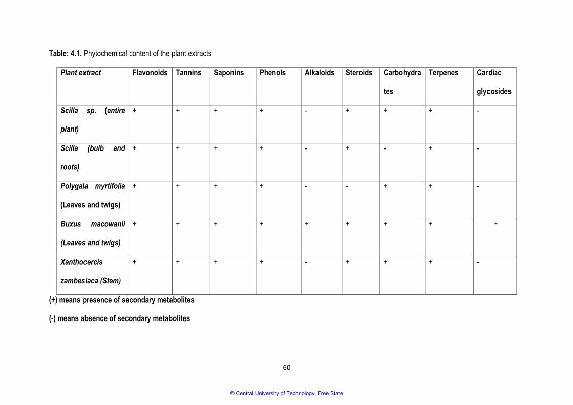
60
Table: 4.1. Phytochemical content of the plant extracts
Plant extract Flavonoids Tannins Saponins Phenols Alkaloids Steroids Carbohydra
tes
Terpenes Cardiac
glycosides
Scilla sp. (entire
plant)
+ + + + - + + + -
Scilla (bulb and
roots)
+ + + + - + - + -
Polygala myrtifolia
(Leaves and twigs)
+ + + + - - + + -
Buxus macowanii
(Leaves and twigs)
+ + + + + + + + +
Xanthocercis
zambesiaca (Stem)
+ + + + - + + + -
(+) means presence of secondary metabolites
(-) means absence of secondary metabolites
© Central University of Technology, Free State

61
4.3.2. TLC fingerprinting
The phytochemical analysis of the plant extracts using TLC fingerprinting after the elution of the
compounds resulted in different colour changes due to their reaction with the spray reagent used
(Vanillin/Sulfuric acid) (Figure 4.1). The spray reagent (Vanillin/Sulfuric acid) is usually used to detect
terpenes and phenylpropanoids. Plants such as B. macowanii and Scilla sp. revealed violet spots after
being eluted with the solvent system Benzene/Ethanol/Ammonia hydroxide (BEA), Chloroform/Ethyl
acetate/Formic acid (CEF) and Toluene/Chloroform/Ethanol (TCE). Diterpenes exhibit a violet colour on
the chromatograms when sprayed with Vanillin/Sulfuric acid. Therefore, it can be suggested that
diterpenes can also be found in extracts of B. macowanii and Scilla extracts. Research has shown that
diterpenes can inhibit cancer multiplicity, and therefore plant extracts that indicated the presence of
diterpenes can be used to manufacture anticancer agents (Martinez et al., 2008). Rf values were not
indicated because it was a qualitative evaluation of compounds.
Figure 4.1: Vanillin-sprayed TLC chromatograms of the methanol extracts developed in
Benzene/Ethanol/Ammonia hydroxide: 90:10:1 (BEA), Ethyl acetate/Methanol/Water: 40:5.4:4 (EMW),
Toluene/Chloroform/Ethanol: 28.5:57:14.5 (TCE), and Chloroform/Ethyl acetate/Formic acid: 5:4:1
© Central University of Technology, Free State
62
(CEF). F = B. macowanii, A = Scilla sp. (entire plant), B = Scilla sp. (roots and bulb), E = P. myrtifolia,
and H = X. zambesiaca.
4.3.3. Gas chromatography mass spectrometry (GCMS)
GCMS analysis of the plant extracts was conducted using the National Institution of Standard
Technology (NIST) software and the Wiley Online Library. The identification of the compounds was
based on the molecular weight, peak area and molecular formula, and only prominent peaks were
selected for identification. A compound was only regarded as positively identified when the percentage
of similarities was more than 90%. See annexure A for total ion chromatograms of suggested
compounds detected from plant extracts of B. macowanii, Scilla sp, P. myrtifolia and X. zambesiaca.
The GCMS analysis of the leaves and twigs of the extracts of B. macowanii showed that neophytadiene
was detected at a retention time of 10.92 (as indicated in Table 4.2), and it has been proposed to have
some antimicrobial activities (Inoue et al., 2005). Neophytadiene has also been reported for the
treatment of headaches, rheumatism and some skin diseases (Suresh et al., 2010). It has been
reported that other plants from the genus Buxus, such as B. papillosa, is used to treat rheumatism and
skin disorders (Cordell, 1981). It can be suggested that the presence of neophytadiene may be the
reason why B. macowanii and B. papillosa have the ability to treat such ailments. The antimicrobial
activity (see Chapter 2) of B. macowanii can also be explained by the presence of neophytadiene in its
extracts. Therefore, it suggests that the plant can be an option for use in the manufacturing of
antimicrobial drugs and other remedies used to treat headaches, rheumatism and skin conditions.
The GCMS analysis of the plant extracts of Scilla sp. indicated a detection of a number of compounds,
such as hexadecanoic acid, which was detected at a retention time of 11.8. A study conducted by
Joshi-Barve et al., (2007) reported that palmitic acid can cause an increase in levels of biologically
active neutrophil chemoattractant, IL-8, from hepatocytes that can potentially cause hepatic
© Central University of Technology, Free State
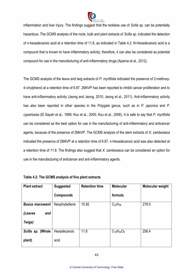
63
inflammation and liver injury. The findings suggest that the reckless use of Scilla sp. can be potentially
hazardous. The GCMS analysis of the roots, bulb and plant extracts of Scilla sp. indicated the detection
of n-hexadecanoic acid at a retention time of 11.8, as indicated in Table 4.2. N-Hexadecanoic acid is a
compound that is known to have inflammatory activity; therefore, it can also be considered as potential
compound for use in the manufacturing of anti-inflammatory drugs (Aparna et al., 2012).
The GCMS analysis of the leave and twig extracts of P. myrtifolia indicated the presence of 2-methoxy-
4-vinylphenol at a retention time of 6.87. 2M4VP has been reported to inhibit cancer proliferation and to
have anti-inflammatory activity (Jeong and Jeong, 2010; Jeong et al., 2011). Anti-inflammatory activity
has also been reported in other species in the Polygala genus, such as in P. japonica and P.
cyparissias (El Sayah et al., 1999; Kou et al., 2005; Kou et al., 2006). It is safe to say that P. myrtifolia
can be considered as the best option for use in the manufacturing of anti-inflammatory and anticancer
agents, because of the presence of 2M4VP. The GCMS analysis of the stem extracts of X. zambesiaca
indicated the presence of 2M4VP at a retention time of 6.87. n-Hexadecanoic acid was also detected at
a retention time of 11.8. The findings also suggest that X. zambesiaca can be considered an option for
use in the manufacturing of anticancer and anti-inflammatory agents.
Table 4.2: The GCMS analysis of five plant extracts
Plant extract Suggested
Compounds
Retention time Molecular
formula
Molecular weight
Buxus macowanii
(Leaves and
Twigs)
Neophytadiene 10.92 C20H38 278.5
Scilla sp. (Whole
plant)
Hexadecanoic
acid
11.8 C16H32O2 256.4
© Central University of Technology, Free State
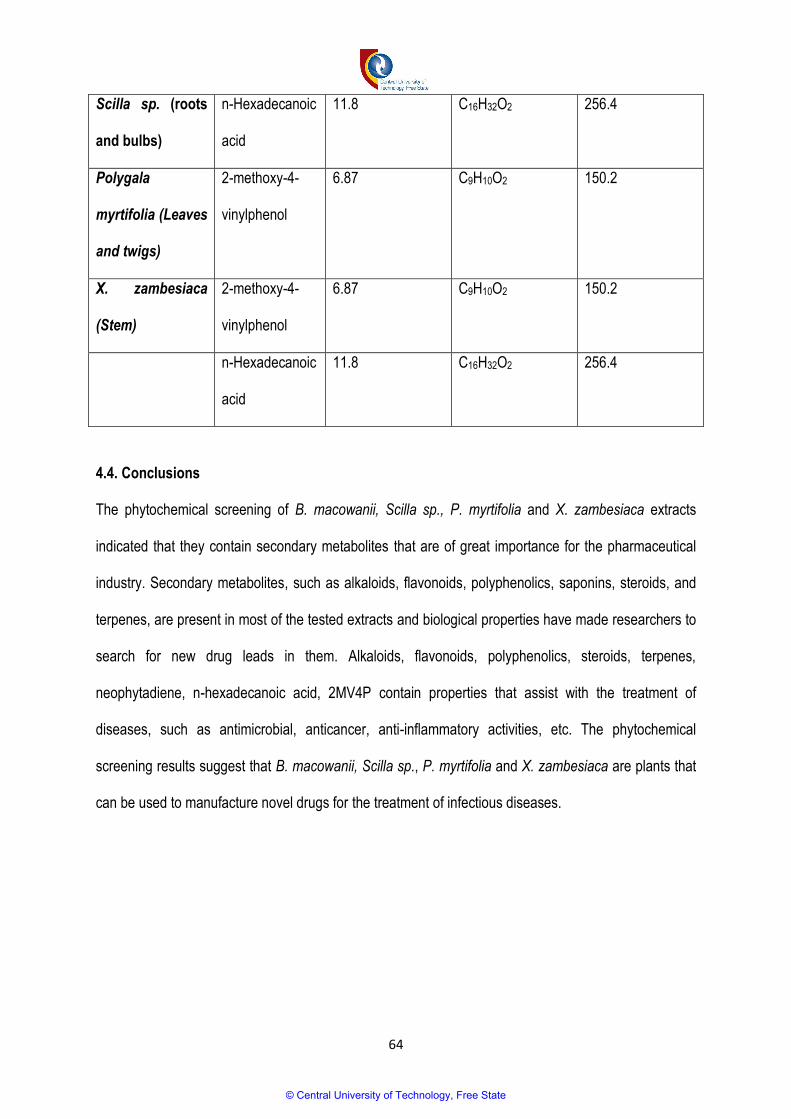
64
Scilla sp. (roots
and bulbs)
n-Hexadecanoic
acid
11.8 C16H32O2 256.4
Polygala
myrtifolia (Leaves
and twigs)
2-methoxy-4-
vinylphenol
6.87 C9H10O2 150.2
X. zambesiaca
(Stem)
2-methoxy-4-
vinylphenol
6.87 C9H10O2 150.2
n-Hexadecanoic
acid
11.8 C16H32O2 256.4
4.4. Conclusions
The phytochemical screening of B. macowanii, Scilla sp., P. myrtifolia and X. zambesiaca extracts
indicated that they contain secondary metabolites that are of great importance for the pharmaceutical
industry. Secondary metabolites, such as alkaloids, flavonoids, polyphenolics, saponins, steroids, and
terpenes, are present in most of the tested extracts and biological properties have made researchers to
search for new drug leads in them. Alkaloids, flavonoids, polyphenolics, steroids, terpenes,
neophytadiene, n-hexadecanoic acid, 2MV4P contain properties that assist with the treatment of
diseases, such as antimicrobial, anticancer, anti-inflammatory activities, etc. The phytochemical
screening results suggest that B. macowanii, Scilla sp., P. myrtifolia and X. zambesiaca are plants that
can be used to manufacture novel drugs for the treatment of infectious diseases.
© Central University of Technology, Free State
65
4.5. References
1. Akinpelu, D.A., Adegboye, M.F., Adeloye, O.A., and Okoh, A.I., 2008. Biocidal activity of
partially purified fractions from methanolic extracts of Garcinia kola (Heckel) seeds on bacterial
isolates. Biological Research. 41: 277-287.
2. Alves, M.J., Ferreira, I.C.F.R., Dias, J., Teixeira, V., Martins, A., Pintado, M., 2013. A review on
antifungal activity of mushroom (basidiomycetes) extracts and isolated compounds. Current
Topics Medicinal Chemistry. 13: 2648–2659.
3. Aparna, V., Dileep, K.V., Mandal, P.K., Karthe, P., Sadasivan, C., Haridas, M., 2012. Anti-
inflammatory properties of n-Hexadecanoic acid: Structural evidence and Kinetic assessment.
Chemical Biology and Drug Design. 80: 434-439.
4. Ayoola, G.A., Sofidiya, T., Odukoya, O., Coker, H.A.B., 2006. Phytochemical screening and
free radical scavenging activity of some Nigerian medicinal plants. Journal of Pharmaceutical
Science & Pharmacy Practice. 8: 133-136.
5. Boxi, M., Ragesh, Y., Raja, Kumar, V., Praveen, B., Mangamma, K., 2010. Extraction,
phytochemical screening and in-vitro evaluation of anti-oxidant properties of Commicarpus
chinesis (aqueous leaf extract). International Journal of Pharma Biosciences. 1: 537-547.
6. Bruneton, J., 1995. Terpenoids and steroids. J. Bruneton. Lavoisier Technique and
Documentation, Paris: 385-621. Cordell, G.A., 1981. Introduction to alkaloids: A Biogenetic
Approach. Wiley-Interscience, New York.
7. Cordell, G. A., 1981. Introduction to alkaloids: A biogenetic approach. New York: Wiley.
8. Demain, A.L., and Sanchez, S., 2009. Microbial drug discovery: 80 years of progress. The
Journal of Antibiotics. 62: 5–16.
9. Doughari, J.H., 2012. Phytochemicals: Extraction Methods, Basic Structures and Mode of
Action as Potential Chemotherapeutic Agents. ISBN: 978-953-51-0296-0.
© Central University of Technology, Free State

66
10. Edeoga, H.O., Okwu. D.E., and Mbaebie, B.O., 2005. Phytochemical constituents of some
Nigerian medicinal plants. African Journal of Biotechnology. 4: 685-688.
11. Eloff, J.N., Katerere, D.R., McGraw, L.J., 2008. The biological activity and chemistry of the
southern African Combretaceae. Journal of Ethnopharmacol. 119: 686-699.
12. El Sayah, M., Cechinel Filho, V., Pinheiro, T.R., Yunes, R.A., Calixto, J.B., 1999. In vitro effect
of the extract and the 1, 7-dihydroxy-2, 3- dimethoxy xanthone from Polygala cyparissias on the
contractions induced by inflammatory mediators and albumin in normal and actively sensitized
trachea from guinea pig. Inflammation Research. 48: 218-223.
13. Graul, A.I., 2001. The year’s new drugs. Drug News and Perspectives. 14 (1): 12–31.
14. Gurib-Fakim, A., 2006. Medicinal plants: Traditions of yesterday and drugs of tomorrow.
Molecular Aspects of Medicine. 27: 1–93.
15. Hill, R.T., 1952. On discontinuous plastic states, with special reference to localized necking in
thin sheets. Journal of the Mechanics and Physics of Solids, 1(1): 19-30.
16. Haslam, E., 1996. Natural polyphenols (vegetable tannins) as drugs: possible modes of action.
Journal of Natural Products. 59: 205-215.
17. Harborne, J.B., and Baxter, H., 1999. The handbook of natural flavonoids, Vols 1 and 2.
Chichester, UK: John Wiley and Sons.
18. Igwe, C.U., Nwaogu, L.A., and Ujuwandu, C.O., 2007. Assessment of the hepatic effects,
phytochemical and proximate composition of Phyllanthus amarus; African Journal
Biotechnology. 6: 728-731.
19. Inoue, Y., Hada, T., Shiraishi, A., Hirose, K., Hamashima, H., Kobayashi, S., 2005. Biphasic
effects of gernylgeraniol, teprenone, and phytol on the growth of Staphylococcus aureus.
Antimicrobial Agents and Chemotherapy. 49: 1770–1774.
© Central University of Technology, Free State

67
20. Jeong, J. B., and Jeong, H. J., 2010. 2-methoxy-4-vinylphenol can induce cell cycle arrest by
blocking the hyper-phosphorylation of retinoblastoma protein in benzo[a]pyrene-treated NIH3T3
cells. Biochemical and Biophysical Research Communications. 400: 752–757.
21. Jeong, J.B., Hong, S.C., Jeong, H.J., Koo, J.S., 2011. Anti-inflammatory effect of 2-methoxy-4-
vinylphenol via the suppression of NF-kB and MAPK activation, and acetylation of histone H3.
Achieves of Pharmacal Research. 34 (12): 2109-2116.
22. Joshi-Barve, S., Barve, S.S., Amancherla, K., Gobejishvili, L., Hill, D., Cave, M., Hote, P.,
McClain, C.J., 2007. Palmitic acid induces production of proinflammatory cytokine interleukin-8
from hepatocytes. Hepatology. 46(3).
23. Kar, A., 2007. Pharmaocgnosy and Pharmacobiotechnology (Revised-Expanded Second
Edition). New Age International Limited Publishers New Delhi. pp 332-600.
24. Kokate, C.K., 1999. Practical Pharmacology. 4th edn. Vallabh Prakashan Publication, New
Delhi, India.
25. Kotze, M., Eloff, J. N., Houghton, P. J. 2002. Extraction of antibacterial compounds from
Combretum microphyllum (Combretaceae). South African Journal of Botany. 68(1): 62-67.
26. Kou, J., Sun, Y., Lin, Y., Cheng, Z., Zheng, W., Yu, B. and Xu, Q., 2005. Anti-inflammatory
activities of aqueous extract from Radix Ophiopogon japonicus and its two constituents.
Biological and Pharmaceutical Bulletin, 28(7): 1234-1238.
27. Kou, J., Si, M., Dai, G., Lin, Y., Zhu, D., 2006. Anti-inflammatory activity of Polygala japonica
extract. Fitoterapia. 77: 411-415.
28. Kubmarawa, D., Ajoku, G. A., Enwerem, N. M., Okorie, D. A., 2007. Preliminary phytochemical
and antimicrobial screening of 50 medicinal plants from Nigeria. African Journal of
Biotechnology. 6(14): 1690-1696.
29. Lacalle-Dubois, M.A., Wagner, H., 2000. Bioactive saponins from plants: an update. In: Atta-Ur-
Rahman (Ed.), Studies in Natural Products Chemistry Series. 21: 633–687.
© Central University of Technology, Free State
68
30. Lederberg, J., 2000. Infectious history. Science. 288: 287–293.
31. Loru, F., Duval, D., Aumelas, A., Akeb, F., Guedon, D., and Guedj, R., 2000. Four steroidal
alkaloids from the leaves of Buxus sempervirens. Phytochemistry. 54: 951-957.
32. Madziga, H.A., Sanni, S., and Sandabe, U.K., 2010. Phytochemical and Elemental Analysis of
Acalypha wilkesiana Leaf. Journal of American Science. 6(11): 510-514.
33. Martinez, M.J.A., Lazaro, R.M., del Olmo, L.M.B., Benito, P.B., 2008. Anti-infectious activity in
the Anthemideae tribe. Studies in Natural Products Chemistry. 35: 445-516.
34. McGaw, L.J., Jager, A.K., van Staden, J., 2000. Antibacterial, antihelminthic and antiamoebic
activity in South African medicinal plants. Journal of Ethnopharmacol. 72: 247–263.
35. Mitscher, L.A., Drake, S., Gollapudi, S.R., and Okwute, S.K., 1987. A modern look at the
folkloric use of anti-infective agents. Journal of Natural Products. 50: 1025-1040.
36. Morrissey, J.P., Osbourn, A.E., 1999. Fungal resistance to plant antibiotics as a mechanism of
pathogenesis. Micribiology and Molecular Biology Reviews. 63: 708–724.
37. Ncube, B., Finnie, J.F. and Van Staden, J., 2008. Seasonal variation in antimicrobial and
phytochemical properties of frequently used medicinal bulbous plants from South Africa. South
African Journal of Botany, 77(2): 387-396.
38. Parekh, J., and Chanda, S., 2007. Antibacterial and phytochemical studies on twelve species
of Indian medicinal plants. African Journal of Biomedical Research. 10: 175 – 181.
39. Reayi, A., and Arya, P., 2005. Natural product-like chemical space: Search for chemical
dissectors of macromolecular interactions. Current Opinion in Chemical Biology. 9: 240-247.
40. Rubió, L., Motilva, M.J., Romero, M.P., 2013. Recent advances in biologically active
compounds in herbs and spices: a review of the most effective antioxidant and anti-
inflammatory active principles. Critical Reviews in Food Science and Nutrition. 53: 943–953.
© Central University of Technology, Free State

69
41. Sathiamoorthy, B., Gupta, P., Kumar, M., Chaturvedi, A. K., Shukla, P. K., Maurya, R., 2007.
New antifungal flavonoid glycoside from Vitex negundo. Bioorganic and Medicinal chemistry
Letters. 17(1): 239-242.
42. Shihabudeen, M.S., Priscilla, H.H., and Thirimurugan, D.K., 2010. Antimicrobial activity and
phytochemical analysis of Indian folk medicinal plants. International Journal of Pharma
Sciences and Research. 1(10): 430-434.
43. Soejarto, D.D., 1999. Biodiversity prospecting and benefit-sharing: perspective from the field.
Journal of Ethnopharmacology. 51: 1-15.
44. Sofowara, A., 1993. Medicinal plants and traditional medicines in Africa. Chichester John Wiley
& Sons New York: 97-145.
45. Suleiman, M.M., McGaw, L.J., Naidoo, V., Eloff, J.N., 2010. Detection of antimicrobial
compounds by bioautography of different extracts of leaves of selected South African tree
species. African Journal of Traditional Complementary and Alternative Medicines. 7 (10): 64-
67.
46. Suresh, L., Veerabah, R. M., Gnanasingh, S. R., 2010. GC‐MS analysis of ethanolic extract of
Zanthoxylum rhetsa (roxb.) dc spines. Journal Herbal Medicine and Toxicology. 4: 191‐192.
47. Tiwari, P., Kumar, B., Kaur, M., Kaur, G., Kaur, H., 2011. Phytochemical screening and
extraction: a review. Internationale Pharmaceutica Sciencia. 1(1): 98-106.
48. Walch, S.G., Tinzoh, L.N., Zimmermann, B.F., Stühlinger, W., Lachenmeier, D.W., 2011.
Antioxidant capacity and polyphenolic composition as quality indicators for aqueous infusions
of Salvia officinalis L. (sage tea). Frontiers in Pharmacology. 2: 1–6.
49. Wansi, J.D., Devkota, K.P., Tshikalange, E., Kuete, V., 2013. Alkaloids from the medicinal
plants of Africa. Medicinal Plant Research in Africa: 557-605.
© Central University of Technology, Free State

70
CHAPTER 5
GENERAL DISCUSSION AND CONCLUSIONS
5.1. General discussion
World-wide, medicinal plants are important for the treatment of diseases and are potential sources of
novel drugs that can overcome the challenges associated with the drugs that are currently available,
such as drug-resistant pathogens, toxicity and inaccessibility. B. macowanii, Scilla species, P. myrtifolia
and X. zambesiaca are traditionally used for the treatment of various ailments, such as rheumatism,
skin disorders, high sugar concentrations, headaches, respiratory problems and gastrointestinal
problems. The above-mentioned traditional uses classify these plants as major contributors to the
health care of people who cannot afford or access modern medicine. With all the challenges pertaining
to the treatment of infectious diseases and the traditional use of the selected plants, it was necessary to
investigate the antimicrobial activity of the selected plants. The results from the study are summarised
in Table 5.1 below.
Table 5.1: A summary of the results obtained in the study
Plant extract Antimicrobial
activity
Phytochemical screening Cytotoxicity (CSIR
Criteria)
IC50,µg/ml
B. macowanii
(Leaves and
Twigs)
+ Standard methods: Alkaloids, steroids,
flavonoids, terpenes, carbohydrates,
tannins and cardiac glycosides
Thin Layer Chromatography
fingerprinting: Diterpenes
Gas Chromatrophy Mass Spectrometry:
Neophytadiene
Moderate hazard
< 30
>5
© Central University of Technology, Free State

71
P. myrtifolia
(Leaves and
Twigs)
- Standard methods: Alkaloids, tannins,
taponins, phenols, carbohydrates and
terpenes
TLC fingerprinting: Negative
GCMS: 2-methoxy-4-vinylphenol
Low hazard
> 100
Scilla sp. (Whole
Plant)
- Standard methods: Flavonoids, tannins,
phenols, steroids, carbohydrates and
terpenes
TLC fingerprinting: Diterpenes
GCMS: Hexadecanoic acid
High hazard
<5
Scilla sp. (Roots
and Bulbs)
- Standard methods: Flavonoids, tannins,
saponins, phenols, steroids and terpenes
TLC fingerprinting: Diterpenes
GCMS: n-hexadecanoic acid
High hazard
<5
X. zambesiaca
(Stem)
- Standard method: Flavonoids, tannins,
saponins, phenols, steroids,
carbohydrates and terpenes
TLC fingerprinting: Negative
GCMS: 2-methoxy-vinylphenol and n-
hexedecanoic acid
Moderate hazard
< 30
>5
(+) Activity
(-) No activivity
The extracts of Buxus macowanii showed antimicrobial activity against S. aureus, C. perfringens, P.
aeriginosa, S. epidermidis, C. albicans and C. tropicalis at Minimum Inhibitory Concentration (MIC) of
2.5 mg/ml, and against E. faecalis and E. coli at an MIC of 1.2mg/ml. B. macowanii was the only plant
© Central University of Technology, Free State

72
that showed growth inhibition of all bacterial and fungal species against which it was tested; for this
reason, it was selected for further studies. This study is the first to report on the antimicrobial activity of
B. macowanii. P. myrtifolia, Scilla sp and X. zambesiaca did not show any inhibitory effects against the
tested bacterial and fungal species, although P. myrtifolia was reported to have antibacterial and
antifungal activities (Lall and Meyer, 1999; Motsei et al., 2003). The inactivity of the plant extracts of X.
zambesiaca and P. myrtifolia may be due to the choice of the solvent of extraction and the age of the
extracts.
B. macowanii was further investigated using the TLC bioautography method discussed in Chapter 2,
for purposes of identifying and locating the antimicrobial compounds against the most affected bacteria
(E. faecalis). Clear zones on the chromatogram indicated that B. macowanii has antimicrobial
compounds. Alkaloid and flavonoid spots were located in close proximity to the antimicrobial spots of
the extracts of B. macowanii. Therefore, it can be suggested that flavonoids and alkaloids contributed
positively to the antimicrobial activity of the extracts of B. macowanii. The effect of the extracts of B.
macowanii on the morphology of bacteria (B. cereus) was also studied using the scanning electron
microscope and the transmission electron microscope (see Chapter 2). The cells of B. cereus indicated
abnormal morphological properties after treatment with the extracts of B. macowanii, such as a
distortion of the cell membrane, leakage of the cytoplasmic contents and separation of the cell
membrane from the cell wall. Most plants that belong to the genus Buxus have been reported to contain
steroidal alkaloids. Steroidal alkaloids are known to cause cell-membrane disruption, which also leads
to cytoplasmic content leakage (Wink and Twardowski, 1992; Wink and Latz-Bruning, 1995).The
morphological properties of bacterial cells that were observed under microscopy after treatment with B.
macowanii can be attributed to the presence of steroidal alkaloids. B. macowanii can be considered as
the best option to use for the manufacturing of novel antimicrobial drugs that can overcome the
challenges associated with the remedies that are currently available.
© Central University of Technology, Free State
73
Plants such as B. macowanii, P. myrtifolia, Scilla sp. and X. zambesiaca are generally used by
traditional healers to treat various ailments. However, these traditional healers often do not have
sufficient knowledge of the plants’ toxicity to human cells. Therefore, it is necessary to ensure that the
plants are safe to be used by humans. The cytotoxicity of the methanol extracts of B. macowanii, P.
myrtifolia, Scilla sp. and X. zambesiaca was tested against the WI-38 cell line (Normal human fetal lung
fibroblast) using Sulforhodamine cytotoxicity assay (see Chapter 3), the results of which are
summarised in Table 5.1. According to CSIR criteria, the extracts of B. macowanii and X. zambesiaca
exhibited moderate hazardous toxicity against the WI-38 cell line with 20.4 µg/ml and 22.96 µg/ml IC50
respectively. The bulb, root and entire plant extracts of Scilla sp. showed high-hazard toxicity against
the WI-38 cell line, with both less than 6.25µg/ml IC50. The leave and twig extracts of P. myrtifolia
showed low-hazard toxicity or no toxicity against the WI-38 cell line, according to the Council for
Scientific and Industrial Research (CSIR) criteria. The compounds such as steroids, cardiac glycosides
and alkaloids (Van Wyk et al., 1997) found in the plant extracts may have contributed to the toxicity that
the extracts exhibited against the WI-38 cell lines. Although plant extracts such as B. macowanii, Scilla
sp. and X. zambesiaca exhibited toxicity against the WI-38 cell line, their use in the treatment of
ailments should not be prohibited; rather, caution must be applied by regulating their administration
dosage. Structure activity studies and chemical modification experiments that will reduce toxicity and
maintain the activity of the plant extracts can also be employed, so that the plants can be the best
option for consideration in the manufacturing of new drugs.
The methanol extracts of B. macowanii, P. myrtifolia, Scilla sp. and X. zambesiaca were subjected to a
screening of the most important phytochemicals, such as the phenols, flavonoids, alkaloids, terpenes,
cardiac glycosides, tannins, saponins and steroids (see Chapter 4). The phytochemical screening was
conducted by using the standard methods, TLC and GCMS, the results of which are summarised in
© Central University of Technology, Free State

74
Table 5.1. The leaves and twigs of B. macowanii were found to contain all the phytochemicals screened
for, and may explain why the extracts showed antimicrobial activity against all the microbial species
against which it was tested. The presence of diterpenes in the extracts of B. macowanii, P. myrtifolia,
Scilla sp. and X. zambesiaca was tested using TLC (see Chapter 4). The separated compounds were
detected using acidified vanillin. B. macowanii and bulb, root and entire plant extracts of Scilla sp
showed the presence of diterpenes on the chromatograms after being eluted by the solvent systems
Benzene/Ethanol/Ammonia hydroxide (BEA), Chloroform/Ethyl acetate/Formic acid (CEF) and
Toluene/Chloroform/Ethanol (TCE). GCMS managed to isolate and identify very important compounds
from the extracts of B. macowanni, P. myrtifolia, Scilla sp. and X. zambesiaca. Neophytadiene was
detected from the extracts of B. macowanii. The compound was reported to have antimicrobial
activities, and it can be used to treat headaches, rheumatism and skin disorders (Inoue et al., 2005). N-
hexadecanoic acid was also identified in the extracts of Scilla sp. and X. zambesiaca, which suggests
that the extracts may be good options for the manufacturing of anti-inflammatory agents. 2M4VP is
another compound that was identified in the extracts of P. myrtifolia and X. zambesiaca. 2M4VP is
known for its anticancer and anti-inflammatory activities. According to the CSIR criteria, Scilla sp.
extracts were reported to have toxicity against human fetal lung fibroblasts (see Chapter 3), and the
phytochemical screening revealed that the extracts contain Palmitic acid (see Chapter 4). Palmitic acid
has been reported to be a causative agent of hepatic inflammation and liver injury; therefore, the
findings could suggest that palmitic acid contributed to the toxicity potential that Scilla sp. extracts
exhibited against WI-38 (see Chapter 3). The overall phytochemical screening of the plant extracts
indicated that they contain important compounds that can provide leads to the manufacturing of new
drugs that can overcome the problems that are associated with the agents that are currently available.
© Central University of Technology, Free State

75
5.2. Conclusions and recommendations
These findings suggest that, to date, plants can still be considered as an important source of medicine
for the treatment of various diseases. B. macowanii excellently showed that it can be the best option to
use in the manufacturing of antimicrobial agents. However, isolation and identification of antimicrobial
compounds using solvents such as diethylamine must be considered for future studies. Although plant
extracts of P. myrtifolia, Scilla sp. and X. zambesiaca did not show inhibition against the bacterial
species against which they were tested, phytochemical screening revealed that these plants possess
other important pharmacological properties, based on the compounds that were identified in them.
© Central University of Technology, Free State
76
5.3. References
1. Lall, N., and Meyer, J.J., 1999. In vitro inhibition of drug-resistant and drug-sensitive strains of
Mycobacterium tuberculosis by ethnobotanically selected South African plants. Journal of
Ethnopharmacology. 66: 347-354.
2. Motsei, M.L., Lindsey, K.L., van Staden, J., Jager, A.K., 2003. Screening of traditionally used
South African plants for antifungal activity against Candida albicans. Journal of
Ethnopharmacology. 86: 235–241.
3. Inoue, Y., Hada, T., Shiraishi, A., Hirose, K., Hamashima, H., Kobayashi, S., 2005. Biphasic
effects of gernylgeraniol, teprenone, and phytol on the growth of Staphylococcus aureus,
Antimicrobial Agents and Chemotherapy. 49: 1770–1774.
4. Van Wyk, B., Van Oudtshoorn, B., Gericke, N., 1997. Medicinal Plants of South Africa. Briza,
Pretoria.
5. Wink, M., and Latz-Bruning, B., 1995. Allelopathic properties of alkaloids and other natural
products: possible modes of actions, in: Allelopathy: Organisms, processes and Applications.
ACS. Symo.Serv. 582: 117-126.
6. Wink, M., and Twardowski, T., 1992. Allelochemical properties of alkaloids. Effects on plants,
bacteria and protein biosynthesis. In: Allelopathy: Basic and Applied Aspects. Chapman and
Hall, London: 129-150.
© Central University of Technology, Free State
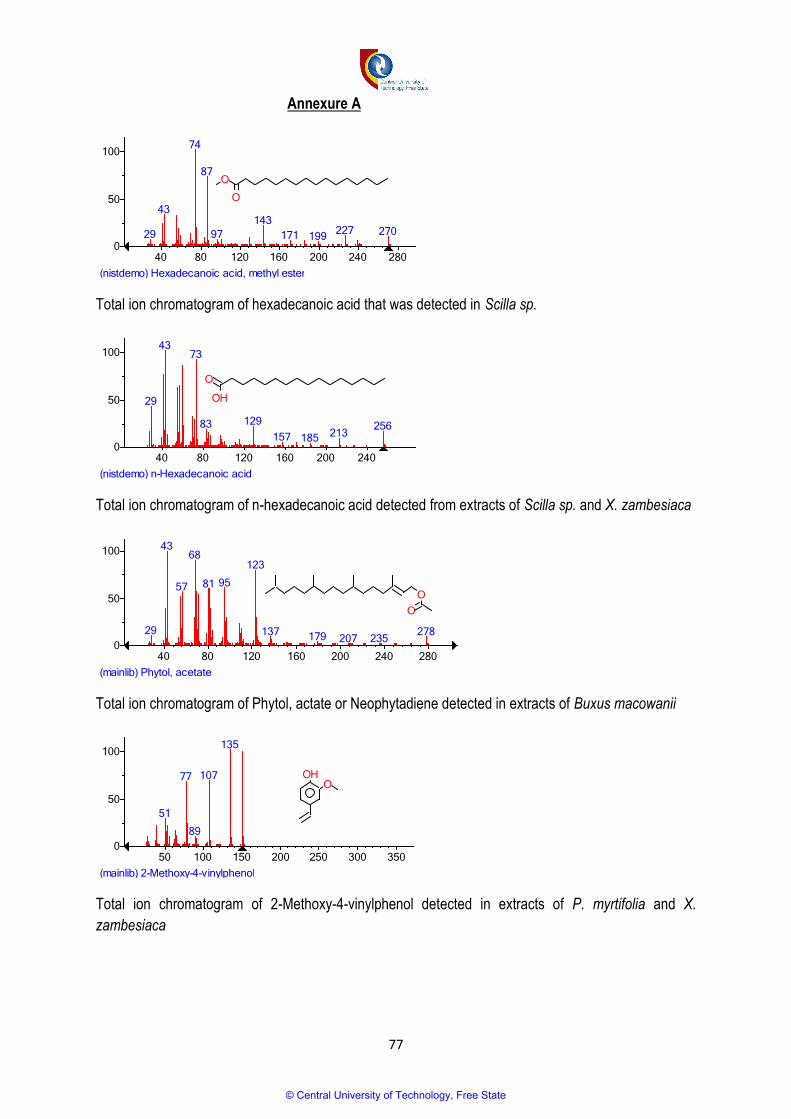
77
Annexure A
Total ion chromatogram of hexadecanoic acid that was detected in Scilla sp.
Total ion chromatogram of n-hexadecanoic acid detected from extracts of Scilla sp. and X. zambesiaca
Total ion chromatogram of Phytol, actate or Neophytadiene detected in extracts of Buxus macowanii
Total ion chromatogram of 2-Methoxy-4-vinylphenol detected in extracts of P. myrtifolia and X.
zambesiaca
(nistdemo) Hexadecanoic acid, methyl ester
40 80 120 160 200 240 2800
50
100
29
43
74
87
97
143
171 199227 270
O
O
(nistdemo) n-Hexadecanoic acid
40 80 120 160 200 2400
50
100
29
4373
83 129
157 185 213256
OH
O
(mainlib) Phytol, acetate
40 80 120 160 200 240 2800
50
100
29
43
57
68
81 95
123
137 179 207 235278
O
O
(mainlib) 2-Methoxy-4-vinylphenol
50 100 150 200 250 300 3500
50
100
51
77
89
107
135
OOH
© Central University of Technology, Free State

78
© Central University of Technology, Free State

79
© Central University of Technology, Free State

80
© Central University of Technology, Free State

81
© Central University of Technology, Free State

82
© Central University of Technology, Free State

83
© Central University of Technology, Free State

84
© Central University of Technology, Free State

85
© Central University of Technology, Free State

86
© Central University of Technology, Free State

87
© Central University of Technology, Free State

88
© Central University of Technology, Free State

89
© Central University of Technology, Free State

90
© Central University of Technology, Free State

91
© Central University of Technology, Free State

92
© Central University of Technology, Free State