Permian productidina of Britain and Malaysia
305
• • •
-
Upload
khangminh22 -
Category
Documents
-
view
0 -
download
0
Transcript of Permian productidina of Britain and Malaysia

Durham E-Theses
Permian productidina of Britain and Malaysia
Bin Leman, Mohd Shafeea
How to cite:
Bin Leman, Mohd Shafeea (1990) Permian productidina of Britain and Malaysia, Durham theses, DurhamUniversity. Available at Durham E-Theses Online: http://etheses.dur.ac.uk/6293/
Use policy
The full-text may be used and/or reproduced, and given to third parties in any format or medium, without prior permission orcharge, for personal research or study, educational, or not-for-pro�t purposes provided that:
• a full bibliographic reference is made to the original source
• a link is made to the metadata record in Durham E-Theses
• the full-text is not changed in any way
The full-text must not be sold in any format or medium without the formal permission of the copyright holders.
Please consult the full Durham E-Theses policy for further details.
Academic Support O�ce, Durham University, University O�ce, Old Elvet, Durham DH1 3HPe-mail: [email protected] Tel: +44 0191 334 6107
http://etheses.dur.ac.uk
The copyright of this thesis rests with the author.
No quotation from it should be published without
his prior written consent and information derived
from it should be acknowledged.
PERMIAN PRODUCTIDINA
OF
BRITAIN AND MALAYSIA
by
Mohd Shafeea Bin Leman
A Thesis submitted in partial fulfilment of the requirements for the degree of
Doctor of Philosophy
Department of Geological Sciences
The University of Durham 1990
1 5 NOV 1991
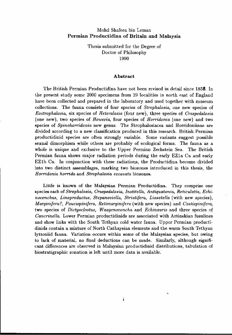
Mohd Shafeea bin Leman Permian Productidina of Britain and Malaysia
Thesis submitted for the Degree of Doctor of Philosophy
1990
Abstract
The British Permian Productidina have not been revised in detail since 185~. In the present study some 2000 specimens from 29 localities in north east of England have been collected and prepared in the laboratory and used together with museum collections. The fauna consists of four species of Strophalosia, one new species of Eostrophalosia, six species of Heteralosia (four new), three species of Craspedalosia (one new), two species of Howseia, four species of Horridonia (one new) and two species of Spinohorridonia new genus. The Strophalosiacea and Horridoniinae are divided according to a new classification produced in this research. British Permian productidinid species are often strongly variable. Some variants suggest possible sexual dimorphism while others are probably of ecological forms. The fauna as a whole is unique and exclusive to the Upper Permian Zechstein Sea. The British Permian fauna shows major radiation periods during the early EZ1a Ca and early EZ1b Ca. In conjunction with these radiations, the Productidina become divided into two distinct assemblages, marking two biozones introduced in this thesis, the H orridonia horrida and Strophalosia excavata biozones.
Little is known of the Malaysian Permian Productidina. They comprise one species each of Strophalosia, Craspedalosia, Institella, Antiquatonia, Reticulatia, Echinoconchus, Linoproductus, Stepanoviella, Striatifera, Liosotella (with new species), M arginifera '?, Paucispinifera, Retimarginifera (with new species) and Costispinifera, two species of Dictyoclostus, Waagenoconcha and Echinauris and three species of Cancrinella. Lower Permian productidinids are associated with Artinskian fusulines and show links with the South Tethyan cold water fauna. Upper Permian productidinids contain a mixture of North Cathaysian elements and the warm South Tethyan lyttoniid fauna. Variation occurs within some of the Malaysian species, but owing to lack of material, no final deductions can be made. Similarly, although significant differences are observed in Malaysian productidinid distributions, tabulation of biostratigraphic zonation is left until more data is available.

Acknowledgements
I would like to thank very much Dr. G. A. L. Johnson for his supervision during the progress of this study and for his help and advice during my stay in Durham.
The Universiti Kebangsaan Malaysia is greatfully acknowledged for funding this research and the Department of Geological Sciences (University of Durham) is acknowledged for hosting the research.
I would like to thank Dr. Denys Smith of GEOPERM, Dr. Neville Hollingworth of Oxford College, Dr. Howard Brunton of British Museum (Natural History), Mr. Jack Pattison of British Geological Survey, Dr. David Harper of University College of Galway, Dr. Kamal Roslan Mohamad of Universiti Kebangsaan Malaysia, Dr. Ian Metcalfe of University of Malaya and Dr. Henri Fontaine whose ideas and suggestions are very valuable for the production of this thesis.
Appreciation is extended to Mr. Tim Pettigrew of Sunderland Museum, Mr. Andrew Newman of Hancock Museum, Dr. Howard Brunton of British Museum (Natural History), Dr. Cocks and Mr. Pattison of British Geological Survey, Dr. David Harper of University College of Galway and Dr. ldris Mohamad of University of Malaya for allowing unlimited access to their Permian collections.
Special thanks are addressed to the staffs of the Geological Survey of Malaysia, especially Mr. Ibrahim Amnan, Mr. Khoo Han Peng, Mr. Amran Nali and the photographer (sorry your name slipped away from my memory) for every help given during this research. Not forgotten is Than Syed Khairuddin and family for providing free accom111odation and transport while the research is carried out in the Geological Survey of Malaysia.
Sincere thank is due to Gerry Dresser and Alan Carr for the help in producing the plates, to George Randall for the thin sections and Dave Asbery for his quick response towards any problems I faced in the department. Thank is also extended to Dr. Larwood and Prof. Bott for continuously asking about my progress.
This research is also much benefited by unlimited access to the sites granted by many landlords, to whom I owe very much.
ii

To Ian and the everchanging members of Room 223, AbdulKadir, Alex, Angela, Stuart, Pete and Ivan and to Dr. Samsudin and other members of the Department of Geological Sciences and the Graduate Society who have helped in anyway in the production of this thesis, I wish to thank you all very much.
Finally, to my very patient wife and daughters and the rest of the family m Malaysia - you all make this happen.
l1l

Declaration
The contents of this thesis are the original work of the author and no other work is included without acknowledgement. This thesis has not been previously submitted for a higher degree at this, or any other University.
Mohd Shafeea bin Leman
December 1990
Copyright
The copyright of this thesis rest with the author. No quotation from it should be published without prior writen consent from him, and information from it should be acknowledged.
IV

LIST OF CONTENTS
Titles . . . . . . . . . . . . . . . . . . . . . . . . . . . . . . . . . . . . . . . . . . . . . . . . . . page numbers
Abstract ............................................................ i Acknowledgements . . . . . . . . . . . . . . . . . . . . . . . . . . . . . . . . . . . . . . . . . . . . . . . . . n Declaration . . . . . . . . . . . . . . . . . . . . . . . . . . . . . . . . . . . . . . . . . . . . . . . . . . . . . . . . IV
List of contents . . . . . . . . . . . . . . . . . . . . . . . . . . . . . . . . . . . . . . . . . . . . . . . . . . . . v List of text-figures . . . . . . . . . . . . . . . . . . . . . . . . . . . . . . . . . . . . . . . . . . . . . . . . . XI
List of tables . . . . . . . . . . . . . . . . . . . . . . . . . . . . . . . . . . . . . . . . . . . . . . . . . . . . . Xlll
Lists of plates . . . . . . . . . . . . . . . . . . . . . . . . . . . . . . . . . . . . . . . . . . . . . . . . . . . . x1v
CHAPTER 1. INTRODUCTION
The scope of study . . . . . . . . . . . . . . . . . . . . . . . . . . . . . . . . . . . . . . . . . . . . . . . . . 1
The brachiopod Suborder Productidina . . . . . . . . . . . . . . . . . . . . . . . . . . . . . 1
The Permian Productidina . . . . . . . . . . . . . . . . . . . . . . . . . . . . . . . . . . . . . . 3
The British Permian Productidina . . . . . . . . . . . . . . . . . . . . . . . . . . . . . . 3
The Malaysian Permian Productidina . . . . . . . . . . . . . . . . . . . . . . . . . . . 4
Methods of study . . . . . . . . . . . . . . . . . . . . . . . . . . . . . . . . . . . . . . . . . . . . . . . . . . 5
Field sampling . . . . . . . . . . . . . . . . . . . . . . . . . . . . . . . . . . . . . . . . . . . . . . . . . 5
Laboratory treatment . . . . . . . . . . . . . . . . . . . . . . . . . . . . . . . . . . . . . . . . . . 6
Taxonomic classification procedures . . . . . . . . . . . . . . . . . . . . . . . . . . . . . . . . . 8
Problems in taxonomic classification . . . . . . . . . . . . . . . . . . . . . . . . . . . . 8
Taxonomic classification employed in this study . . . . . . . . . . . . . . . . . 9
Some taxonomically important morphological features . . . . . . . . . . . 9
Research problems . . . . . . . . . . . . . . . . . . . . . . . . . . . . . . . . . . . . . . . . . . . . . . . . 13
Glossary of productidinid terminology . . . . . . . . . . . . . . . . . . . . . . . . . . . . . 14
CHAPTER 2. BRITISH PERMIAN AND ITS PRODUCTIDINA
Introduction . . . . . . . . . . . . . . . . . . . . . . . . . . . . . . . . . . . . . . . . . . . . . . . . . . . . . . 17
The English Zechstein . . . . . . . . . . . . . . . . . . . . . . . . . . . . . . . . . . . . . . . . . . . . . 19
v

English Zechstein Cycle 1 . . . . . . . . . . . . . . . . . . . . . . . . . . . . . . . . . . . . . . . . . 19
The Marl Slate . . . . . . . . . . . . . . . . . . . . . . . . . . . . . . . . . . . . . . . . . . . . . . . . . . . . 19
The Cadeby Formation . . . . . . . . . . . . . . . . . . . . . . . . . . . . . . . . . . . . . . . . . . . . 20
The Raisby Formation (EZ1a Ca) . . . . . . . . . . . . . . . . . . . . . . . . . . . . . . . . . 21
The Sherburn Hill Quarry . . . . . . . . . . . . . . . . . . . . . . . . . . . . . . . . . . . . . 23
The White Quarry . . . . . . . . . . . . . . . . . . . . . . . . . . . . . . . . . . . . . . . . . . . . 23
The Ford Formation (EZ1b Ca) . . . . . . . . . . . . . . . . . . . . . . . . . . . . . . . . . . . 24
Reef facies - Thnstall Member . . . . . . . . . . . . . . . . . . . . . . . . . . . . . . . . . 24
Basal Coquina . . . . . . . . . . . . . . . . . . . . . . . . . . . . . . . . . . . . . . . . . . . . . 26
Reef-core sub-facies . . . . . . . . . . . . . . . . . . . . . . . . . . . . . . . . . . . . . . . . 27
Reef-fiat sub-facies . . . . . . . . . . . . . . . . . . . . . . . . . . . . . . . . . . . . . . . . . 27
Reef-front sub-facies . . . . . . . . . . . . . . . . . . . . . . . . . . . . . . . . . . . . . . . 28
Back-reef or lagoonal facies . . . . . . . . . . . . . . . . . . . . . . . . . . . . . . . . . . . . 29
Patch reef sub-facies . . . . . . . . . . . . . . . . . . . . . . . . . . . . . . . . . . . . . . . 29
Off-reef facies - Hesleden Dene Member . . . . . . . . . . . . . . . . . . . . . . . . 30
Throw Point Bed . . . . . . . . . . . . . . . . . . . . . . . . . . . . . . . . . . . . . . . . . . . . . . 31
Post EZ1 Ca sediments . . . . . . . . . . . . . . . . . . . . . . . . . . . . . . . . . . . . . . . . . . . . 31
British Permian Productidinids . . . . . . . . . . . . . . . . . . . . . . . . . . . . . . . . . . . . 31
CHAPTER 3. SYSTEMATIC
Superfamily STROPHALOSIACEA Schuchert . . . . . . . . . . . . . . . . . . . . . 35
Family STROPHALOSIIDAE Schuchert . . . . . . . . . . . . . . . . . . . . . . . . . . . 36
Subfamily STROPHALOSIINAE Schuchert ........................ 39
Genus STROPHALOSIA W. King 1844 . . . . . . . . . . . . . . . . . . . . . . . . . . . 40
Strophalosia lewisiana (de Koninck) . . . . . . . . . . . . . . . . . . . . . . . . . . . . 48
Strophalosia excavata ( Geinitz) . . . . . . . . . . . . . . . . . . . . . . . . . . . . . . . . 56
Strophalosia goldfussi (Munster) . . . . . . . . . . . . . . . . . . . . . . . . . . . . . . . 61
Genus EOSTROPHALOSIA Stainbrook 1943 . . . . . . . . . . . . . . . . . . . . . . 67
Eostrophalosia permiana new species . . . . . . . . . . . . . . . . . . . . . . . . . . . 69
Vl

Subfamily HETERALOSIINAE Muir-Wood & Cooper . . . . . . . . . . . . . 74
Genus HETERALOSIA R. H. King 1938 . . . . . . . . . . . . . . . . . . . . . . . . . . 75
Heteralosia morrisiana (W. King) . . . . . . . . . . . . . . . . . . . . . . . . . . . . . . 77
Heteralosia humbletonensis (w. King) . . . . . . . . . . . . . . . . . . . . . . . . . . 83
Heteralosia hyltonensis new species . . . . . . . . . . . . . . . . . . . . . . . . . . . . 87
H eteralosia seahamensis new species . . . . . . . . . . . . . . . . . . . . . . . . . . . 92
H eteralosia aycliffensis new species . . . . . . . . . . . . . . . . . . . . . . . . . . . . 95
Genus CRASPEDALOSIA Muir-Wood & Cooper 1960 ............. 99
Craspedalosia langtonensis new species . . . . . . . . . . . . . . . . . . . . . . . . 100
Craspedalosia lamellosa (Geinitz) . . . . . . . . . . . . . . . . . . . . . . . . . . . . . 105
Craspedalosia sp. A . . . . . . . . . . . . . . . . . . . . . . . . . . . . . . . . . . . . . . . . . . 107
Heteralosia'? quarringtonensis new species . . . . . . . . . . . . . . . . . . . . 108
Strophalosia? parva W. King . . . . . . . . . . . . . . . . . . . . . . . . . . . . . . . . . 111
Superfamily AULOSTEGACEA Cooper & Grant . . . . . . . . . . . . . . . . . 114
Family AULOSTEGIDAE Muir-Wood & Cooper . . . . . . . . . . . . . . . . . 115
Subfamily AULOSTEGINAE Muir-Wood & Cooper . . . . . . . . . . . . . . 115
Genus HOWSEIA Logan 1963 . . . . . . . . . . . . . . . . . . . . . . . . . . . . . . . . . . . . 116
Howseia latirostrata (Howse) . . . . . . . . . . . . . . . . . . . . . . . . . . . . . . . . . 121
Howseia umbonillata (W. King) . . . . . . . . . . . . . . . . . . . . . . . . . . . . . . . 130
Superfamily PRODUCTACEA Gray . . . . . . . . . . . . . . . . . . . . . . . . . . . . . . 136
Family DICTYOCLOSTIDAE Stehli . . . . . . . . . . . . . . . . . . . . . . . . . . . . . 138
Subfamily HORRIDONIINAE Muir-Wood & Cooper . . . . . . . . . . . . . 139
Genus HORRIDONIA Chao ...................................... 146
Horridonia horrida (Sowerby) . . . . . . . . . . . . . . . . . . . . . . . . . . . . . . . . 150
Horridonia hoppeiana (Eisel) . . . . . . . . . . . . . . . . . . . . . . . . . . . . . . . . . 159
Horridonia dunelmensis new species . . . . . . . . . . . . . . . . . . . . . . . . . . 163
Horridonia sp. A . . . . . . . . . . . . . . . . . . . . . . . . . . . . . . . . . . . . . . . . . . . . . 168
Genus SPINOHORRIDONIA new genus . . . . . . . . . . . . . . . . . . . . . . . . . . 169
Spinohorridonia geinitziana (de Koninck) . . . . . . . . . . . . . . . . . . . . . 170
Spinohorridonia sp. cf. S. laspeana (Eisel) . . . . . . . . . . . . . . . . . . . . 174
Vll
CHAPTER 4. THE PERMIAN SYSTEM
AND PERMIAN BRACHIOPODS OF MALAYSIA
The Permian System in Malaysia . . . . . . . . . . . . . . . . . . . . . . . . . . . . . . . . . 176
The Northwest Peninsular Malaysia . . . . . . . . . . . . . . . . . . . . . . . . . . . 178
The Kinta Limestone and Kenny Hill Formation . . . . . . . . . . . . . . 178
The Central Peninsular Malaysia 179
The Eastern Peninsular Malaysia . . . . . . . . . . . . . . . . . . . . . . . . . . . . . 182
Permian brachiopods . . . . . . . . . . . . . . . . . . . . . . . . . . . . . . . . . . . . . . . . . . . . . 182
Current research and material studied . . . . . . . . . . . . . . . . . . . . . . . . . . . . 184
The Malaysian Geological Survey collection . . . . . . . . . . . . . . . . . . . . 184
The University of Malaya collection . . . . . . . . . . . . . . . . . . . . . . . . . . . . 185
Current research collection . . . . . . . . . . . . . . . . . . . . . . . . . . . . . . . . . . . . . 185
Perlis area (localities 1-2) . . . . . . . . . . . . . . . . . . . . . . . . . . . . . . . . . . . 186
Kuala Lipis area, Pahang (localities 3-5) . . . . . . . . . . . . . . . . . . . . . 188
Dada Kering- Gua Musang Highway (localities 6-16) . . . . . . . . 190
Maran area, Pahang (localities 17-19) . . . . . . . . . . . . . . . . . . . . . . . 196
Kuala Pilah Area, Negeri Sembilan (localities 20-21) . . . . . . . . . 197
Permian brachiopods studied in this thesis . . . . . . . . . . . . . . . . . . . . . . . . 198
CHAPTER 5. SYSTEMATICS
OF THE MALAYSIAN PERMIAN PRODUCTIDINA
Superfamily STROPHALOSIACEA Schuchert . . . . . . . . . . . . . . . . . . . . 200
Strophalosia sp. A . . . . . . . . . . . . . . . . . . . . . . . . . . . . . . . . . . . . . . . . . . . . 200
Craspedalosia sp. A . . . . . . . . . . . . . . . . . . . . . . . . . . . . . . . . . . . . . . . . . . 201
Superfamily AULOSTEGACEA Muir-Wood & Cooper ............ 202
Institella sp. A . . . . . . . . . . . . . . . . . . . . . . . . . . . . . . . . . . . . . . . . . . . . . . . 202
Vlll

Superfamily PRODUCTACEA Gray . . . . . . . . . . . . . . . . . . . . . . . . . . . . . . 203
Family DICTYOCLOSTIDAE Stehli . . . . . . . . . . . . . . . . . . . . . . . . . . . . . 203
Dictyoclostus? gratiosus (Waagen) . . . . . . . . . . . . . . . . . . . . . . . . . . . . 203
Dictyoclostus? sp. indet. . . . . . . . . . . . . . . . . . . . . . . . . . . . . . . . . . . . . . 208
Antiquatonia sp. A . . . . . . . . . . . . . . . . . . . . . . . . . . . . . . . . . . . . . . . . . . . 209
Reticulatia sp. indet. . . . . . . . . . . . . . . . . . . . . . . . . . . . . . . . . . . . . . . . . . 210
Family ECHINOCONCHIDAE Stehli .. . . . . .. . . . . . .. . .. .. . . .. . . .. . 211
Subfamily ECHINOCONCHINAE Stehli . . . . . . . . . . . . . . . . . . . . . . . . . 211
Echinoconchus sp. A . . . . . . . . . . . . . . . . . . . . . . . . . . . . . . . . . . . . . . . . . 211
Subfamily WAAGENOCONCHINAE Muir-Wood & Cooper . . . . . . 212
Waagenoconcha sp. cf. W. abichi (Waagen) . . . . . . . . . . . . . . . . . . . 212
Waagenoconcha sp. indet. . . . . . . . . . . . . . . . . . . . . . . . . . . . . . . . . . . . . 213
Family LINOPRODUCTIDAE Stehli . . . . . . . . . . . . . . . . . . . . . . . . . . . . . 214
Subfamily LINOPRODUCTINAE Stehli . . . . . . . . . . . . . . . . . . . . . . . . . . 214
Linoproductus sp. cf. L.? sinosus (Huang) . . . . . . . . . . . . . . . . . . . . 214
Stepanoviella sp. cf. S. fiexuosa (Waterhouse) . . . . . . . . . . . . . . . . . 215
Cancrinella cancrini (de Koninck) . . . . . . . . . . . . . . . . . . . . . . . . . . . . 216
Ca.ncrinella sp. cf. C. cancriniformis (Tschernyschew) . . . . . . . . 219
Cancrinella sp. A . . . . . . . . . . . . . . . . . . . . . . . . . . . . . . . . . . . . . . . . . . . . 220
Subfamily STRIATIFERINAE Muir-Wood & Cooper .............. 221
Striatifera sp. A . . . . . . . . . . . . . . . . . . . . . . . . . . . . . . . . . . . . . . . . . . . . . . 221
Family MARGINIFERIDAE Stehli . . . . . . . . . . . . . . . . . . . . . . . . . . . . . . . 221
Subfamily MARGINIFERIN AE Stehli . . . . . . . . . . . . . . . . . . . . . . . . . . . . 221
Liosotella nakamurai new species . . . . . . . . . . . . . . . . . . . . . . . . . . . . . 221
M arginifera? sp. A . . . . . . . . . . . . . . . . . . . . . . . . . . . . . . . . . . . . . . . . . . . 226
Subfamily PAUCISPINIFERINAE Muir-Wood & Cooper . . . . . . . . . 227
Paucispinifera sp. A . . . . . . . . . . . . . . . . . . . . . . . . . . . . . . . . . . . . . . . . . . 227
Retimarginifera lipisensis new species . . . . . . . . . . . . . . . . . . . . . . . . . 228
Subfamily COSTISPINIFERINAE Muir-Wood & Cooper . . . . . . . . . 232
Costispinifera sp. A . . . . . . . . . . . . . . . . . . . . . . . . . . . . . . . . . . . . . . . . . . 232
Echinauris sp. indet. . . . . . . . . . . . . . . . . . . . . . . . . . . . . . . . . . . . . . . . . . 233
Echinauris? sp. A . . . . . . . . . . . . . . . . . . . . . . . . . . . . . . . . . . . . . . . . . . . . 234
IX

CHAPTER 6. SUMMARY AND CONCLUSIONS
Summary of British Permian Productidina . . . . . . . . . . . . . . . . . . . . . . . . 236
Biostratigraphic significance of British Permian Productidina . . . . . 238
Summary of Malaysian Permian Productidina . . . . . . . . . . . . . . . . . . . . 240
Comparison between British and Permian fauna . . . . . . . . . . . . . . . . . . 241
Further research . . . . . . . . . . . . . . . . . . . . . . . . . . . . . . . . . . . . . . . . . . . . . . . . . 241
Explanation of plates . . . . . . . . . . . . . . . . . . . . . . . . . . . . . . . . . . . . . . . . . . . . 243
List of references . . . . . . . . . . . . . . . . . . . . . . . . . . . . . . . . . . . . . . . . . . . . . . . . . 273
AppendiJC . . .. . . .. . . . . .. .. . . .. . . . .. . . .. .. .... .. . . . . . . . . . . .. . . . . .... 286
X
LIST OF TEXT-FIGURES
Titles . . . . . . . . . . . . . . . . . . . . . . . . . . . . . . . . . . . . . . . . . . . . . . . . . . . . . page numbers
1.1 Dimensions of Productidina . . . . . . . . . . . . . . . . . . . . . . . . . . . . . . . . . . . . . . . . . . . 15
2.1 Distribution of Permian strata in Britain . . . . . . . . . . . . . . . . . . . . . . . . . . . . . . 18
2.2 Zechstein rock formations of Durham Province . . . . . . . . . . . . . . . . . . . . . . . . 22
2.3 Lithostratigraphic correlation in Zechstein of Durham Province . . . . . . . . 25
3.1 Reconstruction of Strophalosia lewisiana (de Koninck) . . . . . . . . . . . . . . . . . 50
3.2 Variation in Strophalosia excavata (Geinitz)
from Beacon Hill railway cut . . . . . . . . . . . . . . . . . . . . . . . . . . . . . . . . . . . . . . . . . . 60
3.3 Comparison of cardinal process and mode of life between species of
British Permian Strophalosia . . . . . . . . . . . . . . . . . . . . . . . . . . . . . . . . . . . . . . . . . . 62
3.4 Reconstruction of Eostrophalosia permiana new species . . . . . . . . . . . . . . . . 71
3.5 Reconstruction of Heteralosia morrisiana (King) . . . . . . . . . . . . . . . . . . . . . . 79
3.6 Camera Iucida reproduction of Heteralosia humbletonensis (King) . . . . . . 87
3. 7 Reconstruction of H eteralosia hyltonensis new species . . . . . . . . . . . . . . . . . . 91
3.8 Reconstruction of Heteralosia seahamensis new species . . . . . . . . . . . . . . . . 91
3.9 Reconstructions of Heteralosia ayclifjensis new species and
H eteralosia? quarringtonensis new species . . . . . . . . . . . . . . . . . . . . . . . . . . . . . 97
3.10 Reconstructions of British species of Craspedalosia . . . . . . . . . . . . . . . . . . . 102
3.11 Reconstruction of H owseia latirostrata (Howse) . . . . . . . . . . . . . . . . . . . . . . . 124
3.12 Howseia latirostrata: attachment by auricular spines
and articulation mechanism . . . . . . . . . . . . . . . . . . . . . . . . . . . . . . . . . . . . . . . . . . 125
3.13 Ontogenic development of Howseia latirostrata
from Tunstall Hill Rock Cottage . . . . . . . . . . . . . . . . . . . . . . . . . . . . . . . . . . . . . 128
3.14 Camera Iucida reproduction of the holotype of
Howseia umbonillata (King) . . . . . . . . . . . . . . . . . . . . . . . . . . . . . . . . . . . . . . . . . 132
3.14 Ontogenic development of Howseia umbonillata
from Gilleylaw builder's trench . . . . . . . . . . . . . . . . . . . . . . . . . . . . . . . . . . . . . . 135
3.16 Reconstruction of Horridonia horrida (Sowerby) . . . . . . . . . . . . . . . . . . . . . . 153
Xl

3.17 Postulation of various moving mechanisms in Horridonia horrida . . . . . 157
3.18 Reconstruction of Horridonia dunelmensis new species . . . . . . . . . . . . . . . 164
3.19 Reconstruction of Spinohorridonia geinitziana (de Koninck) . . . . . . . . . . 172
3.20 Reconstruction of Horridonia hoppeiana (Eisel) . . . . . . . . . . . . . . . . . . . . . . 172
4.1 Distribution of Permian rocks in Peninsular Malaysia
and sampling localities . . . . . . . . . . . . . . . . . . . . . . . . . . . . . . . . . . . . . . . . . . . . . . 177
4.2 Stratigraphic column of the transitional beds
at Bukit Tungku Lembu, Perlis . . . . . . . . . . . . . . . . . . . . . . . . . . . . . . . . . . . . . . 187
4.3 Distributions of localities 6 to 16 along the Dada Kering -
Gua Musang highway, northwest of Pahang . . . . . . . . . . . . . . . . . . . . . . . . . . 191
4.4 Stratigraphic column at locality 16 - north of Sungai Toh,
Merapoh, Pahang
5.1 Reconstructions of Malaysian Permian Productidina I;
Strophalosia sp. A, Antiquatonia sp. A,
Dictyoclostus? gratiosus (Waagen),
Linoproductus sp. cf. L.? sinosus Huang,
Stepanoviella sp. cf. S. fiexuosa Waterhouse
195
Cancrinella cancrini (de Koninck) . . . . . . . . . . . . . . . . . . . . . . . . . . . . . . . . . . . 205
5.2 Reconstructions of Liosotella nakamurai new species
and M arginifera? sp. A . . . . . . . . . . . . . . . . . . . . . . . . . . . . . . . . . . . . . . . . . . . . . . 223
5.3 Reconstructions of Retimarginifera lipisensis new species
and Echinauris? sp. A . . . . . . . . . . . . . . . . . . . . . . . . . . . . . . . . . . . . . . . . . . . . . . 230
Xll
LIST OF TABLES
Titles . . . . . . . . . . . . . . . . . . . . . . . . . . . . . . . . . . . . . . . . . . . . . . . . . . . . . page numbers
2.1 Permian productidinids listed in Geological Survey Memoirs . . . . . . . . . . 32
2.2 Productidina from the Raisby Formation . . . . . . . . . . . . . . . . . . . . . . . . . . . . . . 33
2.3 Productidina from the Ford Formation . . . . . . . . . . . . . . . . . . . . . . . . . . . . . . . 34
3.16 Various criteria used by Muir-Wood & Cooper in sub-dividing
the Superfamily Productacea . . . . . . . . . . . . . . . . . . . . . . . . . . . . . . . . . . . . . . . . 137
3.17 The classificattion scheme for Subfamily Horridoniinae . . . . . . . . . . . . . . . 145
4.1 Permian stratigraphic correlation of West and Central
Peninsular Malaysia . . . . . . . . . . . . . . . . . . . . . . . . . . . . . . . . . . . . . . . . . . . . . . . . . 180
4.2 List of known Permian brachiopods in Malaysia . . . . . . . . . . . . . . . . . . . . . . 183
4.3 List of the studied Malaysian Permian Productidina . . . . . . . . . . . . . . . . . . 199
6.1 Stratigraphic distribution of British Permian Productidina . . . . . . . . . . . 239
Xlll

LIST OF PLATES
1 British Strophalosia lewisiana (de Koninck) and
Strophalosia excavata ( Geinitz) . . . . . . . . . . . . . . . . . . . . . . . . . . . . . . . . . . . . . . 244
2 British Strophalosia lewisiana (de Koninck ), Strophalosia excavata
( Geinitz) and Strophalosia goldfussi (Munster) . . . . . . . . . . . . . . . . . . . . . . . 246
3 British Eostrophalosia permiana new species, Heteralosia morrisiana
(King) and Heteralosia humbletonensis (King) . . . . . . . . . . . . . . . . . . . . . . . . 248
4 British Heteralosia humbletonensis (King) and Heteralosia
aycliffensis new species . . . . . . . . . . . . . . . . . . . . . . . . . . . . . . . . . . . . . . . . . . . . . . 250
5 British Heteralosia hyltonensis new species and Heteralosia
seahamensis new species . . . . . . . . . . . . . . . . . . . . . . . . . . . . . . . . . . . . . . . . . . . . . 252
6 British Craspedalosia langtonensis new species, Craspedalosia
lamellosa ( Geinitz) and Craspedalosia sp. A . . . . . . . . . . . . . . . . . . . . . . . . . . 254
7 British Heteralosia? quarringtonensis new species, Strophalosia
parva (King) and H owseia latirostrata (Howse) . . . . . . . . . . . . . . . . . . . . . . . 256
8 British Howseia latirostrata (Howse) and Howseia umbonillata (King) . 258
9 British Horridonia horrida (Sowerby) and Horridonia sp. A . . . . . . . . . . 260
10 British Horridonia hoppeiana (Eisel) and
Spinohorridonia sp. cf. S. laspeana (Eisel) 262
11 British Horridonia dunelmensis new species and
Spinohorridonia geinitziana (de Koninck) . . . . . . . . . . . . . . . . . . . . . . . . . . . . 264
12 Malaysian Craspedalosia sp. A, Institella sp. A,
Dictyoclostus? gratiosus (Waagen), Antiquatonia sp. A,
Reticulatia sp. indet., Echinoconchus sp. A,
Waagenoconcha sp. indet., Waagenoconcha sp. cf. W. abichi (Waagen)
and Linoproductus sp. cf. L? sino sus Huang . . . . . . . . . . . . . . . . . . . . . . . . . 266
13 Malaysian Striatifera sp. A, Cancrinella cancrini (de Koninck),
Cancrinella sp.cf. C. cancriniformis (Tschernyschew),
Cancrinella sp. A, Costispinifera sp. A,
Echinauris sp. indet. and Echinauris? sp. A . . . . . . . . . . . . . . . . . . . . . . . . . 268
14 Malaysian Marginifera? sp. A, Liosotella nakamurai new species,
Retimarginifera lipisensis new species and Echinauris? sp. A 270
XIV

15 Malaysian Strophalosia sp. A, Dictyoclostus sp. indet.,
Antiquatonia sp. A, Stepanoviella sp. cf. S. fiexuosa Waterhouse,
Costispinifera sp. A, Dictyoclostus'? gratiosus (Waagen),
Retimarginifera lipisensis new species and Paucispinifera sp. A . . . . . . . 272
XV

CHAPTER 1
INTRODUCTION
The Scope of Study
The object of this study is to revise, redescribe and reclassify, when ever necessary,
the Permian brachiopod Suborder PRODUCTIDINA from Britain and Malaysia. In
achieving this goal, the study is concentrated on close examination of various mor
phological features within different productidinid taxa. This, in turn, contributes to
the understanding of morphological variation, functional morphology and their signif
icance in ontogeny, phylogeny and taxonomy of the group. One of the main purposes
of this research is to tackle the long standing problems in taxonomic classification of
the brachiopod Suborder Productidina, by establishing a more systematic scheme of
classification and less ambiguous definitions of different taxa studied based on widely
developed and reliable morphological criteria.
The brachiopod Suborder PRODUCTIDINA
Phylum BRACHIOPODA Dumeril, 1806
Class ARTICULATA Huxley, 1869
Order STROPHOMENIDA Opik, 1934
Suborder PRODUCTIDINA Waagen, 1883
Diagnosis. Strophomenida with open tubular spines.
Discussion. The Productidina range from the Lower Devonian to Upper Permian
and has a world wide distribution particularly during Carboniferous and Permian
time. The suborder Productidina was introduced in the Lower Devonian of North
America (Quebec) for two strophalosiacean genera Devonalosia and Spinulicosta. The
1

Productidina diversified steadily during Devonian time, and during the Carboniferous
the 'J became very rich and diverse, dominating in most of the Carboniferous paralic
environments. Among the celebrated Carboniferous productidinids is the largest bra
chiopod genus Gigantoproductus. The Productidina declines slightly at the end of
Carboniferous period with the closure of some Carboniferous Seas, but it regains its
diversifying momentum in the Permian, introducing many new taxa including the
aberrant Family Richthofeniidae. At its peak of diversification at the end of the
Permian period, sadly the Productidina dwindled and became extinct together with
many other Palaeozoic invertebrates.
The Productidina has a very wide range of modes of life from sessile (attached by
various technology, but not by the pedicle) to free living (adapted to various attitudes
suiting various types of substratum), and possibly vagrant forms too. In conjuntion
with these variable modes of life, the Productidina adopts a very wide range of mor
phological variation, the study of which has produced a series of never ending hy
photheses. The earlier group (the Strophalosiacea), however, are more primitive and
somewhat close to some of the advanced Chonetidina. Although Muir-wood (1965)
related the origin of Productidina toihsetrophomenidinid genus Leptaenisca (via the
construction of brachial ridges), the possible link between Productidina and Chone
tidina cannot be ruled out. Current observation shows that some of the primitive
Strophalosiacea have much closer affinities towards some of the advance chonetids
such as N eochonetes than towards the Strophomenidina.
Owing to its very diverse morphological variation, the taxonomic classification of
the Productidina becomes very complicated and the scheme of classification is very
difficult to erect systematically. At present, most of the productidinid taxa seem to
be very dynamic, and many new methods of nomenclature have been introduced from
time to time. It is noticeable, that many authors prefer to split the older nomenclature
and introduce their own new names without attention to the possible extinction of a
former name. There are so many cases in which we can see once popular and diverse
genera being reduced to a mono-specific genera or to worst producing empty genera
(see discussion on genus Strophalosia).
2

The Permian Prod uctidina
The study of Permian Brachiopoda or the study of Permo-Triassic extinction
seems to be incomplete without mentioning a word about the remarkable variation of
somewhat bizarre Permian Productidina. It is especially during the late Permian time the
that,(Productidina became very diverse and produced some of the most bizarre forms
of all brachiopods. Such a great ability to evolve did not guarantee the Productidina
a safe passage through the Permo-Triassic event which eliminated most of the marine
invertebrates from the earth's surface. The Productidina finally disappeared from the
sea at the end of the Palaeozoic era.
It is certainly very interesting as well as challenging to study this vast group, the
morphology of which is very difficult to interpret because of its strong variation and
because of the occurrence of so many cases of convergence, parallelism, etc. which
require extra close attention.
The British Permian Productidina
The British Permian Productidina are not as diverse as they are in some other
parts of the world (e.g. the Texas or the Salt Range Faunas). The fauna is restricted to
the lowermost horizons of the Upper Permian, and they belong to the Zechstein fauna,
where similar forms are found in East Germany, Poland and the USSR (Lithuania).
Major systematic classifications of the British Permian Productidina have been
set up and revised several times since the work of Sowerby (1823). Several catalogues
and monographs on the British Permian brachiopods have been published by Howse
{1848, 1857, 1858), King {1846, 1848, 1850) and Davidson {1853, 1859, 1880). Some
very useful descriptions and remarks have also been been produced by various authors
including Trechmann {1921, 1945, 1954), Logan (1962), Smith & Francis (1967),
Hollingworth (1987) and Hollingworth & Pettigrew (1988). In addition to all these,
British Permian faunal lists have been compiled in a countless number of papers and
memoirs.
3

The British Permian Productidina is dominated by the Strophalosiacea and the
productacean Subfamily Horridoninae, with a few member of the aulostegacean Genus
Howseia. Although various species names are encountered in the past literature, only
some of them were generally accepted prior to this thesis. They include H orrido
nia horrid a, H owseia latirostrata, Strophalosia morrisiana, Orthothrix excavata, Or
thothrix lewisiana, Dasyalosia goldfussi and Craspedalosia lamellosa. However, as a
result of current revision, several new taxa are introduced, while some generic names
are changed. In Chapter 3, 22 species of 7 genera of the British Permian Productidina
are described and revised.
Besides pure taxonomic treatment, the British Permian Productidina has also in
the last few years, been the subject of palaeoecological study by Hollingworth (1987)
and Hollingworth & Pettigrew (1988). Unfortunately, these authors give very little
consideration to variation within and between species of Productidina.
The Malaysian Permian Productidina
In contrast to the British forms, the Malaysian Permian Productidina have re
ceived very little attention in the past. Although the Permian Productinida of
Malaysia are much more widely distributed spatially as well as stratigraphically than
they are in Britain, detailed information about them is not well known. Lack of local
pal ntologists is the primary reason for the poor understanding of the Malaysian
macrofauna in general. From the list given in table 4.2, it can be seen that more than
half of the Permian productidinids listed are undetermined species while some of the
determined species also prove to be a hasty identifications.
The Malaysian Permian Productidina range from the lowermost to the upper
most Permian, but their affinities are not very clear. This has partly resulted from the
poorly understood Permian geology of the region. The Malaysian Permian Productid
ina are dominated by various productacean species, with rare species of Strophalosi
acea and Aulostegacea. Described in Chapter 5 are 23 species of 19 genera of the
Malaysian Permian Productidina, tentatively identified from all the available mate
rial during this study.
4
Methods of Study
Productidina is a brachiopod suborder in which most of the individuals are nor-the
mally macroscopic in size and can be easily observed with ~aked eye. As in many
other studies in macrofossils, the working techniques employed are rather conven
tional and do not require much modern and expensive equipment. Microscopic study
of the brachiopod shell is not stressed because the published information on the shell
microstructure is sufficient for use in this research. Those species which perhaps de
served microscopic treatment, were unfortunately not well preserved because of their
thin shells (e.g. the British Genus Heteralosia).
In this research, a great deal of attention was given to mastering every single
morphological detail of the studied faunas in order to make detailed comparisons
between individuals, between species, genera and between higher taxonomic ranks.
Functional morphology is a very useful tool for this comparison. Here, models and
deductions introduced by earlier workers were examined and compared with the avail
able material, and whenever necessary new models were introduced. The knowledge
gathered on the variation of morphological features and the published data on its
functional morphology were then carefully and systematically scrutinized to establish
new schemes of classification wherever possible.
In general, the preservation of the British and Malaysian Permian Productidina
is not particularly good. Therefore, in this study, the results very much depend on
good observation and good sample treatment. Samples are cleaned and prepared at
various stages, both in the field and in the laboratory.
Field Sampling
Since this study is taxonomically orientated, the sampling work was carried out
at random at places where the productidinids could be collected. The Productidina
were collected entire as far as possible without leaving fragments of shells on the site.
No measure was taken on the population density. Notes, however were taken on the
faunal assemblages, lithological types as well as the types of preservation.
5

In the field, the productidinids can easily be recognized by naked eye or with the
aid of a hand lens Loose samples were cleaned thoroughly with a brush or with
water whichever is suitable. As far as possible, the fossils were prepared (loosened)
in the field by using hammers and chisels of various weight and size, without harming
the specimens. Delicate fossils were usually taken straight away to the laboratory for
finer treatment. Labelling specimens in the field is the normal practice.
Laboratory treatment.
In !he labor-atory, , the productidinids were extracted or cleaned by various
techniques. Each technique was carefully chosen depending on the mode of preserva
tion, the nature and composition of the shell and the enclosing rock to avoid further
damage to the fossil.
Loose, friable and highly weathered rocks were removed from the specimens by
repetitive washing and brushing. Some very fragile and soft fossils were cleaned by
blower (sandblast). When the shell is tough, friable matrix was sometime freed from
the shell by using the ultrasonic bath.
The drilling method is the most commonly used to extract specimens. This is
primarily because most of the specimens are preserved in forms of mould and cast.
Some other specimens needed this technique because the shells and the matrix have
the same solubility to acid reaction, or the same hardness (either too soft or too hard).
Treatment with hydrochloric acid ( 5-10%) was tried on some of the shells preserved
in limestone, but in many cases it failed to produce good results; parts of the shells
being disolved together with the matrix. Some specimens were also treated with high
heat and cold water, but again the result was far from satisfactory; the rocks tend to
crack at random and the fossils were damaged.
In the drilling method, needles of various sizes, fine blades, small chisels and
dentist's drills were used to remove the rock matrix from fossils. Chisels and blades
are useful for those fossils preser'ved in shaly or cleaved rocks. Hard dolomite can be
roughly removed from the fossils by using dentist's drills, followed by finer drilling
6
with needles.
Thin and polish cross-sections of the shell were made to study the shell nu
crostructure. This study is not stressed partly because of the homog,ane.\ty of the inner
structure in Productidina, and partly because of the lack of shelly material available.
The sections are sometime used for the reconstruction of the shell profile. Partial se
rial sections of some shelly fossils were made to monitor the development of the shell,
but the results do not seem to be any better than observations made on naturally
preserved shell interiors.
In contrast, negatives produced by rubber latex impressions or acetate peels are
very helpful in reproducing very fine morphological details of the shells. Since many
specimens are preserved in the form of moulds (external or internal moulds), the
rubber latex casts made give better details than the actual moulds. This is a very
good technique especially in reproducing projecting morphology such as the cardinal
process and the spines from hollow moulds.
Several parameters of the brachiopod shells were measured (see text-figure 1.1 ),
mainly to give data about dimensions of the fossils to support the descriptive work.
Illustration
Because variation occurs within the brachiopod species, reconstruction is neces
sary to illustrate the average morphology of each species. Average dimensions and
collective morphological details were combined together to form the reconstruction of
the species. To add to the precision of the reconstruction, details from photographs
and camera Iucida drawings were also used in preparing the reconstruction. Camera
Iucida drawings and photographs were also used to reveal some detailed features or
the whole shell of selective specimens, when reconstructions were difficult to make.
These techniques are also very useful to illustrate variation in ontogenic development
and variation within species.
Photography is essential in all palaeontological work. For this purpose, the speci
mens were usually coated with ammonium chloride to produce a better picture and to
7

hide unnecessary colour changes, label marks, etc. The coating was applied as lightly
as possible, because thick coating gives poor definition to the photograph. The pho
tographs were cut and arranged neatly on an A4 size hard backing, re-photographed
and then printed as the final plate.
Taxonomic Classification Procedures
Problems in taxonomic classification
In the past, various criteria have been used to classify the productidinid bra
chiopods. However, no firm and systematic scheme of classification has ever been
produced for the recognition, sub-division and understanding of the group. The
schemes used in the past are always complicated and very difficult to apply. In gen
eral, experts agree that the classification above the generic level should be based on
the presence or absence of a single or several morphological features. Most commonly,
however, more than one feature is used and often more than one standard is employed
at the same taxonomic level.
To accommodate all the features used in these classifications, the past diagnosis
and definition of genera, subfamilies, families and superfamilies are usually very long
and rather ambiguous. Recently, however, there are attempts to simplify and produce
a schematic sub-division of Productidina,whils{ works such as those of Muir-wood and
Cooper (1960), Muir-wood (1965) are not always directly applicable. This is partly
because these authors give little explanation of the scheme of classification, and mainly
because the major part of their work still uses the older non-schematic methods of
classification. As a result, the schematic classification introduced by them has been
much debated by many later authors.
8

Taxonomic classification employed in this study
In this study, the author has outlined a scheme for classification of the super
families in Productidina, the families in Strophalosiacea and all the subfamilies in
Strophalosiidae. The limited time available, does not permit the author to tackle the
problems in family ranking of the productaceans and aulostegaceans. It is hoped that
the successful division of the Superfamily Strophalosiacea will be used in the future,
as the basis in producing similar scheme for Productacea and Aulostegacea.
Most of the criteria used in these schemes of classification have already been
employed by previous workers, but here the author emphasizes the use of as few
criteria as possible and applies this as rigidly as possible. Each morphological criterion
is chosen carefully to avoid any overlapping in its usage. The lower ranking (genus,
species and variety) are not greatly linked by a scheme of classification, and as much
preference as possible is given to the original description. In addition to this, a study
on the ontogeny is introduced to solve some of the problems on variation.
Some taxonomically important morphological features
Interarea. The presence of the interarea in some productidinids has long been consid-.
ered as an important feature that linked this suborder with its possible strophomeni
dinid or chonetidinid ancestor. One important feature shown by productidinid evo
lution is that this group progressively looses its interarea. The presence or absence of
the interarea has been widely used in determining the superfamilies in the large group
of the Productidina (see Muir-wood & Cooper, 1960, Muir-Wood, 1965). Cooper &
Grant(1975o,b) recognised that a large number of interarea bearing productidinids have
interarea on their ventral valve only, and thus suggested that superfamilies of Pro-or abseoce
ductidina should be based not only on the presence[ot interarea, but also whether both
the ventral and dorsal interareas or only the ventral interarea are present.
or absence These sub-divisions of Suborder Productidina based on the presence [of inter-
area on each valve reflects relationship between superfamilies as well as showing the
evolutionary path in which each superfamily lies. With four possible groupings of
, 9

interarea occurrence, only three groups are known. They are Productidina with 1) in
terarea on both ventral and dorsal valves (Superfamily Strophalosiacea); 2) interarea
on ventral valve only (Superfamily Aulostegacea); and 3) no interarea (Superfamily
Productacea); no productidinid is known to have the interarea only on the dorsal
valve.
. _ There is another small, but very important group owing to iis
abberant nature, the Richthofeniacea, which since it was elevated to a superfamily
by Muir-Wood (1955) has not been questioned as to its validity. The Superfamily
Richthofeniacea was not erected on the basis of the interarea, but detailed study
on the group shows that it generally has a ventral interarea, but no dorsal interarea.
Therefore, to maintain the consistency in classification of Productidina, it is suggested
that the Family Richthofeniidae be included to the Superfamily Aulostegacea. This
is very important, in order to prevent the creation of separate superfamily on a basis
other than by the presence of interarea.
Also in accordance with this rigid scheme, some of the interarea-bearing families,
subfamilies and genera which were included by Muir-Wood & Cooper (1960) into
the Superfamily Productacea, should be redistributed into the other two sure..,families
(see p. 137 in chapter 3). The Superfamily Strophalosiacea which has both ventral
and dorsal interareas is sub-divided into families based on interarea-related mor-Sub~ivi_:iion of
phology, the teeth and sockets. Other criteria should be chosen for.{the ::;uperfamilies
Aulostegacea and Productacea because of the absence of teeth and sockets in these
groups.
Teeth and sockets. In this research, in which a large collection of strophalosiaceans are
studied in great detailed, the author found that the interarea and related features at
the cardinal region show some degree of evolution within the group. With Family
Productellidae {interarea present on both ventral and dorsal valves) considered as a
strophalosiacean (see also page 36 & 137), this superfamily . comprises two large
groups with different strengths of development of the inter area, teeth and sockets. The
Productellidae, with its degenerate interarea, teeth and sockets provides a possible
intermediary form between Strophalosiacea and Productacea.
10

Another strophalosiacean family introduced in this study is a small, but interest
ing mono-generic group Ctenalosiidae which was classified by Muir-Wood & Cooper
(1960) as a strophalosiid Subfamily Ctenalosiinae, based on the presence of a series
of hinge teeth while other subfamilies was classified on the basis of spine distribu
tions. The unique hinge teeth in Ctenalosia should not be lowly regarded, because it
provides an excellent example of possible link between Productidina and the rest of
Strophomenida (see further discussion on page 36).
Spine distributions. Classification based on spine distribution has been introduced
by Muir-Wood & Cooper (1960) at the subfamily level of Strophalosiidae, and since
then has gained various opinions, both pro- and contra- (a long discussion on this
matter is written in Chapter 3, page 36 and so on). The importance of spines in
productidinids has been stressed by many authors although their significance in the
evolution of the group is scarcely known. However, the important function of hollow
spines described by Rudwick (1970) and the consistent pattern of distribution should
clear any doubt about the significance of spine distribution in sub-dividing subfamilies
of Strophalosiacea.
The new scheme introduced in this thesis, suggests that only the presence or
absence of body spines in each of the valves should be used in the classification
of strophalosiacean subfamilies. Similar to the case of the interarea, there should
be a maximum of four groups in each strophalosiacean family, but for the family
Strophalosiidae, studied in this thesis, only two groups are commonly known. They
are Strophalosiidae with 1) body spines on both ventral and dorsal valves (Subfamily
Strophalosiinae); and 2) body spines on ventral valve only (Subfamily Heteralosiinae);
Lialosia which has no body spines at all is suggested in this thesis to represent a new
Subfamily Lialosiinae; no strophalosiids are known to have body spines on dorsal
valve only. This scheme seems to work well in the other big strophalosiacean Family
Productellidae, but the author has delayed this sub-classification until more material
is available for comparison.
The distribution of body spines and marginal spines are also tentatively used for
classifying the genera in subfamily Horridoniinae (see p. 144), while species within
11

these genera are subdivided on the basis of the density of the body spines as well as
on the dimensions of the shell.
Other morphological features. Other features are limited to use at generic and lower
categories. There is no valid scheme of sub-division at these lower taxonomic ranks,
but generally features closely associated with the interarea, cardinal extremities and
surface ornamentation, such as the presence of squamose lamellae, are highly con
sidered at generic level. The dimensions and shape of the shell, the growth pattern,
including the growth of the cicatrix as well as the detail of types of spines, their
arrangement and distribution are usually used in describing the species.
Morphological features like cardinal process and brachial ridges are often used in
the past to described productidinid taxa at various level. At the same time, many
workers described the variable nature of these two internal morphological features.
The variation in the configuration of the cardinal process and brachial ridges is de
scribed in many places in this thesis. The development of the alveolus and antron,
anterior to the cardinal process might have some taxonomic significance, but the lo
bation of the cardinal process ~s. · · v a.-table . even within the
same species. The size of the brachial loop probably has a little taxonomic value,
but the configuration of the brachial loop seems to depend on the shape of the shell.
H owseia umbonillata, described in this thesis gives a good example of how the shape
of the brachial impression differs between the male and female dimorphs. Therefore,
in this study the cardinal process and brachial ridges are not highly regarded in out
lining the scheme of classification. These morphological features, are only used as
supporting features to describe some of the highly variable species.
12
Research Problems
Apart from the problems of the taxonomic classification procedures, the author
faced great problems with variation within the studied species. Some of the variants
of one species sometimes seem to be very close to varieties of other species. This
is particularly problematic in the rich and abundant species such as Strophalosia
excavata and S. lewisiana, which Logan (1962) suggested had ~brid forms in between
them. In order to avoid error in assigning a hybrid form or extreme variant, the author
has tried to use several alternative features in describing the two species.
The problem of variation is made worse by the problem of preservation. Often,
even the various alternative distinguishing features are not helpful owing to poor
preservation. The reef fauna from Durham is very commonly preserved in the form
of moulds and casts in which many morphological features are destroyed during their
formation. The dolomitization of the reef is responsible for the formation of these
moulds and casts, while the development of coarse-grained dolomite destroyed the
shell even further. Some fossils on the bedding plane in the Raisby Formation and
most of those from Malaysia are very difficult to study owing to burial deformation
as well as tectonic deformation in the case of Malaysian fauna. Some of the frag
mentary material from allochtonous assemblages in Malaysia are sometimes hardly
recognisable.
13

GlossarJ of Productidinid Terminology
Adductor muscle platform - platform to which the muscle for closing the valves are attached.
Adductor muscle scars- impression of adductor 111uscle attaehrnent arear.
Alveolus- pit formed anterior to cardinal process when the cardinal process shaft is not supported
by a median ridge.
Antron- triangular gap due to incomplete fusion of cardinal process buttress plates and breviseptum
as in Buxtonia.
Auricle- Flat or arched posterolateral extension of the shell, separated by a break of slope from the
convex visceral disc (termed as wing or ala in some past literature).
Beak - pointed or incurved extremity of the umbo.
Brachial ridges - ridges of brachial apparatus originating from adductor muscle platforms, extending
laterally and forming open loop (brachial loop) anteriorly; often only their impressions
(brachial impressions) are preserved.
Breviseptum - median ridge not supporting the cardinal process.
Cardinal process - a more or less elevated boss for attachment of the diductor muscle; also assisting
in articulation. It consists of a shaft and lobate myophore.
Chilidium - flat or concave plate closing the nothyrium.
Cicatrix of attachment- flattened scar in the ventral umbonal region for cementation against foreign
objects.
Costae- coarse (less than 15 in 10mm) radial ridges on the exterior shell surface. It is called costellae
when the radius is smaller (more than 15 in lOmm).
Delthyrium - triangular opening of the ventral interarea.
Diaphragm - thin crescentic plate developed around visceral disc of brachial valve, bridging the gap
between the trail of ventral and dorsal valves.
Diductor muscle- Muscle for opening the valves, attached to the cardinal process myophores on the
dorsal valve and to the large low flabellate scars on the ventral valve.
Dorsal valve- formerly brachial or small valve which bears cardinal process and lophopore, commonly
concave or flat in nature.
Elytridium- Convex puckered cover of the delthyrium in Aulostegacea.
Endospines - Fine spines or protruding taleolae in interior of both ventral and dorsal valves. The
toarser outer rims of endospines probably serve as strainers.
Fold - major radial ridges such as median fold.
Geniculate - sharp bend of the shell at the begining of the trail.
Ginglymus - secondary interarea? in ventral valve of some large Productacea: the function is not
fully understood.
Hinge - posterior line of the valve junction.
Hinge teeth - a pair of articulating process at posterolateral margin of delthyrium.
Interarea - formerly cardinal area.
Lamellae- thin dead shell layer overlapping the newly developed outer layer.
Lateral ridges- thick ridges extending laterally from the cardinal process, probably for strengthening
the shell and assisting in articulation.
14

Lophidium - triangular plate projecting posterior to the hinge line of the dorsal valve to form a
posteriorly extended muscle attachment base and to close the opening in the delthyrium
of the Aulostegacea.
Marginal ridges- concentric interior ridge bounding the visceral disc of Marginiferidae, often bearing
coarse strainer endospines.
Median septum - median ridge of varying extent in both ventral and dorsal valves.
Myophore- distal expanded part of cardinal process to which diductor muscles were attached.
Notothyrium - triangular opening in interarea of dorsal valve.
Pseudodeltidium - single flat or convex plate covering part of the delthyrium.
Reticulation - nodelike enlargement formed by intersection of concentric rugae and radial costae or
costellae.
Rugae- concentric wrinkles or thick overlapping growth lamellae.
Sockets - excavation for placing teeth at the lateral base of cardinal process of Strophalosiacea,
bounded by socket ridges which often buttress the cardinal process.
Spine- hollow tubular projection out of the shell, classified according to their angle against the shell
surface at point of origin: erect spines (75-90" ); recumbent spines (0-45° ); suberect ( 45-75° ).
by their nature: clasping spines (concentrically arranged marginal and body spines that
clasped foreign objects. exceptionally well developed in almost all productidinid juveniles):
Halteroid spines (long large spines. four or six in number, symmetrically placed and acting
as a strut-like support); prostrate spines (straight spines that lie prone on shell surface):
rhizoid spines (root-like spines for attachment by entanglement or cementation against
foreign objects) or by their position: auricular spines (erect to suberect spines born on the
auricle except for the row at posterior margin); body spines (all spines apart from auricular
and marginal spines): marginal spines (a row of spines on the posterior margin, acting as
cementing, clasping. balancing or supporting spines).
Squamose - ragged concentric structure of overlapping lamellae or rugae.
Sulcus - major depression in either valve, normally median in position.
Taleolae- non-fibrous calcite rods. commonly with median perforation and embedded in fibrous shell
layer or protruded as endospines on the interior of the shell.
Trail - extension of shell of either valve beyond the visceral disc.
Umbo - posterior region just anterior to the beak.
ventral v-cilve - formerly pedicle or larger valve, commonly convex in nature.
Visceral cavity- includes body cavity and mantle or brachial cavity.
Visceral disc - exterior equivalent of visceral disc.
{Most of the terminology listed above was taken from Muir-Wood (1965) and William & Rowell
(1965)}
15

hinge width
maximum Width
-:5 bo
] curved length
height
__r--- height of ventral interarea
«1t:s - -----height of dorsal interarea
Text-figure 1.1. Measured dimensions
\0 -

CHAPTER 2
THE BRITISH PERMIAN AND ITS PRODUCTIDINA
Introduction
The British Permian began with the development of a desertland, after the closure
of the sea way across Europe during the Upper Carboniferous time (Johnson, 1981,
1982). This was later followed by the development of confined marine basins, in which
lived all the Hermian productidinids described in this study. The Permian rocks are
well developed in the northeast, ·nvrthwest, Midlands and southwest of England as
well as in southwest of Scotland, North Wales and Northern Ireland (see text-figure
2.1). They consist largely of continental breccias or sandstones and marine dolomitic
(magnesian) limestones, evaporites and mudstones. The desert sandstone deposition
takes place during Lower Permian time, while the emergence of the confined hyper
saline Bakevelia and Zechstein basins marked the begining of the Upper Permian in
Britain. The Lower and Upper Permian in Britain have been well described in a
countless number of individual papers and memoirs of British Geological Survey, and
has been summarized several times by various authors including the works of Smith
(1980b,1981, 1989 etc.), Smith et al. (1974) and Smith et al. (1986) which are the
main sources of reference for this chapter.
British Permian brachiopods are well known in past literature, and they have
been studied in great detail over the last two centuries. Despite their very limited
lateral and vertical distribution, the brachiopod fauna is very rich and diverse in
composition. Up to the present time, the British Permian brachiopods are restricted
to the English Zecshtein Cycle 1 (EZ1 ), and their occurence in the Bakevelia Sea in
Northern Ireland, has been reported to be questionable (Pattison, 1970). For this
reason, the author will discuss only the English Zechstein Cycle 1 in this chapter,
while other Permian rocks will only be mentioned in relation to it.
17

N
(DURHAM PROVINCE)
' ' /YORKSHIRE PROVINCE)
Jj!;:.:~':iJ Continental depo~its I I ; 1 ; f Marine deposits f. · . ·. ·f Concealed Permian rocks
0
Text-figure 2.1. Distribution of Permian strata In Britain
[After Smith P.t al. (1974) and Smith (1989))
JB

The English Zechstein
During Lower Permian time, an arid climate produced widespread piedmont and
eolian as well as fluvial deposits in Britain. Marine trangression which takes place
during the Upper Permian time, submerged the northern part of England and North
ern Ireland. This newly formed basin was divided into the Zechstein Sea to the east
and the Bakevelia Sea in the west. The English Zechstein represents the western shelf
of the larger Zechstein Basin wich extends to the east as far as Poland and Lithuania.
In Britain, the Zechstein Sea is known to cover most of the old County Durham, York
shire and Nottinghamshire. The Bakevelia Sea is widespread in northwest England,
the Irish Sea and part of southern Scotland and Northern Ireland. It is in these two
basins that the celebrated British Magnesian Limestone and the associated evaporites
were deposited.
The English Zechstein was deposited in two provinces, the Durham and York
shire Provinces which were separated by a palaeo-high called the Cleveland High or
Cleveland Axis (Smith et al., 1986). Five cycles of carbonate - evaporite deposition
are recognised in both provinces, with substantial differences in lithology between the proviocet.
two~ Several names have been used to described these carbonates and evaporites in
the past, but for the purpose of this study the nomenclature proposed by Smith, et
al. (1986) is employed.
English Zechstein Cycle 1
The Marl Slate
The begining of the Zechstein trangression is marked by the deposition of water
laid Yellow or Basal Permian Sands (Smith, et al. 1974), followed by the deposition
of thinly bedded or laminated sapropelic dolomite (Turner & Magaritz, 1986) of the
Marl Slate. The Marl Slate occasionally gains thickness of more than 5m, although
it is normally about 1m in thickness. This formation is generally thicker in County
Durham and thinning towards the west and south, and towards the basin in the east.
19

The Marl Slate in County Durham .was in· places ·anoxic and ·yields fish faunas,
land floras and shallow marine invertebrate faunas. Among brachiopods commonly
known from the Marl Slate are the inarticulates Lingula credneri and Discina konicki,
but Mills & Hull (1976) also reported some Horridonia horrida as well as Dielasma
elongatum and Streptorhynchus pelargonatus from the Marl Slate at Eldon Hill in
County Durham. Bells et al. (1979) also recorded the occurrence of H. horrida from
the Marl Slate at Middridge. The Marl Slate is succeded by the deposition of the
Cycle 1 carbonate (Raisby and Ford Formations in Durham Province, and Cadeby
Formation in Yorkshire Province), followed by Cycle 1 anhydrite, Cycle 2 carbonate
and so on (see text-figure 2.3).
The Cadeby Formation
Although the current study qoes not involve any collecting or field work in the
Cadeby Formation, several important specimens studied and figured in this thesis
were obtained from several BGS boreholes from Yorkshire and Nottinghamshire.
These faunas came either from the lower part of the Lower Magnesian Limestone
or from the Lower Permian Marl (probably equivalent to the Wetherby Member of
the Cadeby Formation of Smith et al., 1986). Limited time available, did not per
mit the author to gather detailed information on the lithology and fauna of these
boreholes.
Productidinids encountered in these boreholes include Horridonia horrida, H.
hoppeiana, Strophalosia goldfussi, Cmspedalosia lamellosa, ?C. langtonensis, ?Heter
alosia morrisiana, and ?H. aycliffensis. The occurrrence of these productidinids has
been recorded in various memoirs of the Geological Survey of Great Britain, but some
of the species names used in these memoirs differ from the above list (see Edwards
(1951, p. 98-101, & 1967, p. 149), Eden, Stevenson & Edwards (1957, p. 141) and
Smith, Rhys & Goosens (1973, pl. 10,11); see also table 2.1). Fossils from the higher
section (Sprotborough Member (EZ1b Ca)) are not very well known.
20

The Raisby Formation (EZla Ca)
The Raisby Formation which is more popularly known in the past as the Lower
Magnesian Limestone, is well developed in County Durham (Smith & Francis.,. 1967';,
Smith, in Johnson, 1970, 1972; Mills & Hull, 1976.; etc.), and it extends to the north
to Tyne and Wear (Land,'1974; Smith (in publication), Smith in Robson,f1980l etc.)
and to the south to North Yorkshire (Smith et al., 1986) (see text-figure 2.2).
This formation was-defined by Smith et al. (1986) as strata of cream, brown and ~he
grey fine-grained dolomite with thick grey fine-grained limestone in power part. The
base of the formation is a sharp boundary above the finely laminated Marl Slate,
and in its lower half the Raisby Formation is thinly bedded, locally sparingly to
richly fossiliferous. Toward the top the Raisby Formation becomes medium to thickly
bedded and less fossiliferous, and the top of this formation is overlain by the oolitic
dolomite of the Ford Formation. The dolomite of the Raisby Formation commonly
shows abundant replacive gypsum and anhydrite, and thin argillaceous dolomite beds
and small scale cross-lamination in lower part. Evidence of eastward slope movements
are also found at various levels especially at the bottom and the top of this formation.
Fossil occurrences have been recorded from numerous localities by various authors.
During the tenure of this research, most of these localities have been visited. The
author managed to collect specimens of productidinids from eight localities mentioned
by earlier authors, while two new productidinid localities were also found for the first
time during this study. These localities are Sherburn Hill Quarry , (GR. NZ 345417)
and White Quarry 1 (GR. NZ 257245). The later locality had been studied by Mills
& Hull (1976), but these authors did not find any brachiopods here.
All the fossils collected by the author during this research come from the [ower
part of the Raisby Formation. The faunas are usually found in limestones, either
in biocoenosis assemblages (e.g. East Thickley Quarry, White Quarry and Raisby
Hill Quarry faunas) or a thanatocoenosis assemblages like that of the Pen shaw Hill,
Sherburn Hill and Old Quarrington Quarry faunas. The fauna at Langton Quarry
has been interpreted as a lagoonal or near-shore fauna. The list of productidinid
brachiopod species collected and studied from the Raisby Formation can be seen in
21
'Ihassic strata
Roxby Formation
Seabam Formation
Edlington Formation
Roker Dolomite - Concretionary Limestone
Ford Formation with reef crest
Raisby Formation
Basal or Yellow Sands
Carboniferous strata
1-29 Localities 1 to 29 refer to table 2.2 and 2.3
0 SKm &.....------'
• DURHAM
Sea ham
Text-figure 2.2. Zechstein rock formations in Durham Province [After Smith (1980) and Smith et al. {1986)]
22
N
I
HARTLEPOOL

table 2.2 at the end of this chapter.
The Sherburn Hill Quarry (Nz. 345417)
Sherburn Hill Quarry displays a good section of the top of the Yellow Sands, the
Marl Slate and the base of the Raisby Formation. At the northeast corner of the
Quarry (around NZ 345417), the quarry face cuts through about lm of Marl
Slate which is directly overlain by thin to medium-bedded buff to brown dolomite.
The strata dip at about 5° to the east. There are no macrofossils found in the
finely laminated Marl Slate, but numerous shelly fossils are found at the top of the
first bed of the Raisby Formation. Fortunately, the excavation has left a substan
tial surface of this first bed on the abandoned quarry floor. This bed ranges from
8 to 12cm thick and the top 3 or 4cm yields various fossils randomly distributed.
The fauna is disorientated and includes some fragmentary bryozoans and many dis
articulated brachiopods and bivalves suggesting that they have been transported,
probably by downslope movement from the west. The fauna includes Fenestella reti
formis, Heteralosia? quarringtonensis, Pterospirifer alatus, Crurythyris clanyana,
Stenoscisma schlotheimi, H orridonia horrida, ? Lingula credneri, Streblochondria pus
sila and Peripetoceras freieslebeni.
The White Quarry (NZ 2.'57245)
At southeast corner of White Quarry (or Old Town Quarry in Mills & Hull
(1976)), the top of the Marl Slate is exposed at the quarry floor, yielding a rich
fauna of inarticulate brachiopods Lingula credneri and Discina konicki. The Marl
Slate is succeeded by thin to medium bedded bluish grey, fine-grained limestone of
the Raisby Formation. The limestone sequence is about lOrn thick and 30m wide
in the quarry and passes gradually laterally and upwards into a thin to medium
bedded pale cream to brown dolomite. Scattered fossils are found in the limestone
over an interval of 1.5m, begining at about 2m above the base of the Raisby forma
tion. Most of the fauna is found on bedding planes except for the large Horrido
nia horrida and some other large productidinids and bivalves. The fauna is dom
inated by H orridonia horrida which is commonly orientated with its trail on top.
Other fossils include Horridonia hoppeiana, Horridonia sp. A, Heteralosia aycliffen-
23

sis, Strophalosia goldfussi, Craspedalosia lamellosa, Pterospirifer alatus, Stenoscisma
schlotheimi, Dielasma elongatum, Cleiothyris pectinifera, Crurithyris clanyana, Per
mophorus costatus, Peripetoceras freieslebeni, Fenestella retiformis, Synocladia vir
gulacea, Discritella columnaris, Acanthocladia sp. and Kingopora? sp. Burrowing
traces are also commonly observed on the shell of larger Horridonia horrida
The Ford Formation (EZlb Ca)
Formerly known as the Middle Magnesian Limestone of County Durham, Tyne
& Wear, Cleveland and northern-most Yorkshire, the Ford Formation is defined by lit
Smith et al. (1986) as the Upper Permian carbonate strata lying between the Raisby
Formation and the Edlington Formation in the west and the Hartlepool Anhydrite in
the east~ The Ford Formation is divided into two interdigitate main facies, the reef
and back-reef facies, and a minor basinal facies. Each of these three facies contains
its own distinct fauna and the reef facies has produced most of the Permian fossils
found in Britain. Productidinid brachiopods are well known from this formation and
the vast majority of described specimens came from the reef facies of this forma
tion. From the current research twenty productidinid localities (all of which have
been recorded earlier by various authors) have been studied and the productidinid
faunas encountered are listed in table 2.3 at the end of this chapter. The distribution
of the Ford Formation can be seen on text-figure 2.2, while the lithostrathigraphic
correlation between various facies in the Ford Formation and between Zechstein rock
formations in Durham Province is illustrated on text-figure 2.3.
Reef facies - Tunstall Member
The Durham Permian reef is developed on a more or less north-south trend, ex
tending from West Boldon in the north as several elongate discontinuous outcrops for
more than 30 km to Hartlepool in the south. Erosion of the younger cover exposes the
reef in the north in several low hills, whereas beyond Castle Eden Dene in the south
the reef was covered by the younger Zechstein sediments. Owing to the difficulty in
determining the actual boundaries between the reef and its surrounding rock forma
tions, the lateral extension of the Zechstein reef cannot be precisely mapped. The
24

West East
r ovO~
~-- 1\0~ ~~~
:'{ ~0
:= EZ5
- :::2 -= l EZ4
0 oi--~ ROTTEN MARL ~ - I :
BOULBY HALITE
BILLINGHAM ANHYDRITE. ~ EZ3 SEAHAM FORMATION
EDLINGTON FORMAT!bN
lagoon- with Q patch reef _
~
YELLOW SANDS ~M::A~RL~-S;L~A~T;E,, ==:~ -----•
Text-figure 2.3. Lithostratographic correlation in the Zechstein of Durham . Province [Modified from Smith (1989)]
EZ2
EZl
l{)
c:-1
best known limit is the outline of the reeffront which was illustrated by Smith (1978),
and this outline has been used successfully by Hollingworth (1987), Hollingworth &
Pettigrew (1988) and Hollingworth & Tucker (1987) to reconstruct the paleoecology
of the reef. The western limit of the reef is known to interdigitate with the back-reef
or lagoonal rocks, but such ft=:._ature is only known at the type locality of the formation, (N1 36355130}
i.e. at Ford Quarry.{ and Claxheugh Rock (Nz 36?>5'14).
The Durham Permian reef is made of buff to brown altered dolomite with con
spicious bryozoan frameworks and dominant algal encrustation, laminae and stroma
tolite at the top. Several reef sub-facies are recognisable in the field, where each sub
facies displays different faunal assemblage from the other (see Hollingworth (1987),
Hollingworth & Pettigrew (1988)). Productidinid brachiopods are dominant in places
in the lower part of the reef, especially at the reef base and the lower reef core, while
towards the reef fiat and the fore-reef the brachiopods decline significantly in num
ber and diversity. The last brachiopod to survive on the reef in upward succession is
the terebratulid Dielasma elongatum which disappears not long after the last Permian
productidinid Strophalosia excavata. Extinction was caused by changing environmen
tal conditions particularly the increase in salinity of the Zechstein Sea towards the
top of EZl.
Basal Coquina. An excellent exposure of undolomitized limestone at Tunstall Hill 392545
Rock Cottage (NZ{) provides a good example of lithology and fauna of the reef base.
This locality is formed of bedded shelly or coquina layers of various faunal elements.
Although the coquina bed is not a part of the actual reef (Smith, in publication),
its role in the development of the eventual reef is undeniable. The basal coquina at
Tunstall Hill Rock Cottage yields a very rich brachiopod fauna, and a striking feature
of this fauna is the nesting habit of Dielasma elongatum. The productidinids are not a
very prolific group at this locality (see list in text-figure 2.3) with the sessile Howseia
latirostrata and Heteralosia morrisiana dominated the suborder.
Hollingworth & Pettigrew (1988) mention that the Rock Cottage fauna was pre
viously unknown, but the author believes that most of the specimens in Howse and
Kirkby collections (marked from Tunstall Hill) formed in the nineteenth century might
26

have come from an extension of this locality. The preservation of brachiopods in this
huge collection by Kirkby and Howse (studied by the author in Hancock Museum),
shows remarkable resemblaces with those collected by the author from the coquina
bed at Tunstall Hill Rock Cottage. Pattison (1977) also suggested a locality around the
present Rock Cottage coquina bed for the origin of the King collection labelled from
Tunstall Hill. Although the King collection is not as big as the Kirkby collection, the
fauna and its mode of preservation is unique as far as is known and suggests that it
belongs to no other locality, but the Tunstall Hill Rock Cottage.
Reef-core sub-facies. Massive buff to brown autochthonous dolomite (biolithite, bound
stone) forms much of the Durham Permian shelf-edge reef (Smith, in publication).
The reef-core exposures, sometimes display turbid dolomite surrounding the frame
work of bryozoa, and pockets of detritus or sometimes pockets of Horridonia dunel
mensis and / or Strophalosia excavata - S. lewisiana. H. dunelmensis, however does
not appear in the upper reef-core (Hollingworth, 1987). Also very common in the reef
core, is lamellar encrustations of dolomite of different sizes, which are often confused
with the primary frame organisms.
Among the well known reef core exposures are Hylton Castle road cut (NZ 360589)
and Humbleton Hill (NZ 381553) in which a rich and diverse productidinid fauna are
encountered. The well preserved spines in most of the productidinid brachiopods
strongly suggested that the fauna belongs to a biocoenosis. Comparison in faunal
elements (especially the productidinids) between the two localities above and Tunstall
Hill old trench (NZ 397541) suggests that the later locality is positioned much closer CNz. 397541)
to the reef-core. Tunstall Hill old trench[ has been tentatively positioned at the reef
crest by Hollingworth (1987) and Hollingworth & Pettigrew (1988), although they
mentioned elements of uncertainty in their proposal. For a detail list and the relative
abundance of productidinid brachiopods from these three localities see text-figure 2.3.
Reef-flat sub-facies. Uneven, more or less horizontal, thick-bedded, complex assem
blages of encrusted bryozoan biolithite and algal stromatolite of the reef-fiat are ex
posed in many localities in the vicinity of Ryhope, Seaham, Hawthorn and Easing
ton. The sequence of this sub-facies sometimes reaches more than 10m thick (e.g. at
27
Nz. 439463
Hawthorn Quarry~, but the actual thickness is not known due to surface erosion of the
reef. In places, the reef-flat contains pockets of derived debris, and quite often these
pockets are full of molluscs and brachiopods. While Smith (in publication) suggests
that the fauna is part of the debris, the presence of nests, very rich in fauna including
Strophalosia excavata - Strophalosia lewisiana such as that exposed at Ryhope Mental
Hospital (NZ 40055215) (or Quarry Heads in Smith, 1958 and Smith, in publication),
suggests that some of the fauna might belong to biocoenosis assemblages, living in
a pocket she Here& by algal bushes.
Current research encountered several productidinid localities in the reef-flat sub
facies, but only S. excavata and S. lewisiana are known from them. Among these local
ities include the Ryhope Mental Hospital, Stony Bank cut (NZ 41854740), Hawthorn
Quarry (NZ 438463), Townfield Quarry (NZ 431438) and ?Garden of Eden Bridge
(NZ 43803975). In general, the fossil occurrences are rare and scattered, while in some
of the localities only stunted S. excavata recorded. This suggests that the rich fauna
at Ryhope Mental Hospital might be positioned at the lower part of the reef-flat.
Reef-front sub-facies. Smith {in publication) divided the reef-front sub-facies into the
reef-crest, the mid reef-slope and the lower reef-slope.
The reef-crest and uppermost slope are seen in the eastern part of the exposure
of Claxheugh old railway cut (NZ 363574) and Ford Quarry (NZ 36355730). The
lithology and the productidinid fauna of the reef-crest are quite similar to that of
the reef-flat, except that the bedding of the dip-crest is highly lenticular with the dip
changes from nearly horizontal in the reef-flat to 45° or more in the reef-crest. S.
excavata from the reef-crest is very small in average size and often shows a twisted
umbo.
The mid reef-slope sub-facies exposes in several localities at Tunstall. Hill, while
the best exposure for the productidinids is exposed at the railway cut at Beacon Hill NZ 447.464
easti The rocks comprise thick to massive sheets of encrusted bryozoan biolithite
with thinner sheets of laminated dolomite. The beds is often irregular, but gener
ally dip very steeply ( 45-90°) indicating the contemporary slope of the original reef
front. A rich and diverse productidinid fauna is seen at Beacon Hill railway cut, with
28

several faunal assemblages found within a close proximity. An assemblage of rich S.
excavata - S. lewisiana - Eostrophalosia permiana exposed at the southern exposure
(NZ 44214528), while an assemblage of Heteralosia seahamensis - Howseia latiros
trata with less abundant Strophalosia is found less than 50m to the north of the first
assemblage. Horridonia dunelmensis and Howseia umbonillata are also found in very
small number.
The lower reef-slope or reef-talus subfacies is well exposed at the old railway cut (Nz ~~05385)
at Ryhopek The rocks comprise multi-range of reef rock types, fallen from the higher
part of the reef, ranging from blocks of several metres across to sand and silt-size
fragments. The rocks usually contain authochtonous biota derived from the reef-slope
and reef-crest as well as some indigenous fauna living in pockets of rubble between
larger biolithite blocks. Although not rich, the productidinid fauna of the reef-talus
at Ryhope old railway cut (NZ 39605385) is quite diverse and this suggests that it is
positioned quite low in the Durham reef.
Back-reef or lagoonal facies
The back-reef and lagoonal facies forms most of the Ford Formation. However,
owing to lack of interesting elements in this sub-facies it is probably the least known
part of the formation. In many exposures of the lagoonal facies, the rocks are made of
thin to medium-bedded,pale cream and buff granular and oolitic dolomite with abun-tht ;!_i!,solu+ion of
dant cavities froul former sulphates. Fossils are very rare except for a few bivalves.
Smith (in publication) suggests that the thick-bedded soft shelly granular dolomite (Nl. 3765~) (Nl 381563)
below the reef dolomite at Gilleylaw Plantation QuarrY!, and Humbleton Hill[to belong
to this facies. From the thick-bedded dolomite at Humbleton Hill the author found
rare occurences of S. excavata as well as numerous bivalves and gastropods. Should
this proposal of Smith (in publication) be true, the reef core of Humbleton Hill must
be substantially younger than the underlying lagoonal deposits, and the reef must
have been retrograding towards the shore.
Patch Reef sub-facies. The lagoonal facies of Ford Formation also includes several
patch reefs and the best examples of patch reefs are shown at the Gilleylaw Planta
tion Quarry (NZ 376536) and at High Newport old railway cut (NZ 387538). The
29

patch reefs developed in the east, at least 30m above the base of the lagoonal facies.
The patch reef at Gilleylaw Plantation Quarry is made of about 5m of thick-bedded
stromatolite and bryozoan biolithite with a wedge of highly fossiliferous rubbly talus ' at the northern margin, from where a rich fauna of S. excavata and S. lewisiana is
found. The patch reef is overlain by coarsely oncolitic dolomite.
The High Newport patch reef comprises a rigid arched core of bryozoan and algal
biolithite in which numerous remarkably preserved S. lewisiana and S. excavata with
hairy spines are found. The flank of the reef is made of bedded dolomite of uncertain
origin. The dolomite is locally fossiliferous and from a grey bedded dolomite the
author found several specimens of small Horridonia hoppeiana with very strongly
curved ventral valve together with some Strophalosia lewisiana.
From a temporary builder's trench (or Newport Hill of Smith (in publication) -
NZ 379539), the author examined a large collection of fossils in Dr. G. A. L. Johnson's
possession. This collection included the productidinids H. latirostrata, H. umbonillata
and a few S. excavata. It should be noted that Hollingworth (1987) recorded that
H orridonia is absent from these patch reefs.
Off-reef facies - Hesleden Dene Member
The off-reef facies of the Ford Formation is not well known and its relation to
the other formations is also not well established. At Dene Holme (NZ 455405), algal
laminated dolomite is exposed, forming a large stromatolite dome (Smith & Francis,
1967), overlying basal conglomerate (up to 2m thick). The conglomerate comprises
encrusted rolled cobbles and boulders of dolomite of the underlying reef with very
little matrix. Some of the cobbles and boulders contain reef fauna includes some S.
excavata and S. lewisiana. The basal conglomerate is deposited on the eroded surface
shelf-edge reef.
Thick-bedded dolomite exposed at foot of the cliff at Chourdon Point (NZ 442468)
was suggested by Smith & Francis (1967) as an off-reef basinal equivalent of the
reef. However, the presence of inarticulate brachiopods such as Lingula credneri and
Discina konicki indicates a shallow water origin of these beds. Also included in the
30

biota is Neochonetes sp. cf. N. davidsoni which are commonly found in the lagoonal
facies in Sunderland area. The rest of the fauna consists of ?Streptorhynchus pelarg
onatus, '?Heteralosia quarringtonensis, Bakevelia ?antiqua, Strophalosia ?excavata,
Permophorus costatus, Crurythyris clanyana (majority) and Fenestella retiformis.
Trow Point Bed. A thin layer of oncoids, peloids and columnar stromatolite
named as Trow Point Bed by Smith (1986), extensively covers the Zechstein Cycle
1 and Smith (in publication) includes this thin layer as part of Ford Formation.
Although it is better known off the reef, Smith (in publication) indicated that there is
no evidence to suggest that this layer is a basinal equivalent of the reef, and according
to him the whole layer might be younger than reef and other facies of the Ford
Formation.
Post EZl Ca sediments
The Trow Point Bed is in turn overlain by the Cycle 1 evaporite (The Hartlepool
Anhydrite of Durham Province and Hayton Anhydrite of the Yorkshire Province).
The evaporite is thicker towards the basins and is succeeded by the Cycle 2 carbonates
(the Concretionary Limestone and the Roker Dolomite of Durham Province and
Edlington Formation of Yorkshire Province). However, none of these formations
yields any brachiopods and thus the author will not described them in this thesis.
These carbonates are in turn succeeded by the cycle 2 evaporite, cycle 3 carbonates
and so on.
British Permian Productidina
The hypersaline nature and the very short life of the Pemian marine basin did
not allow the British Permian productidinids to expand. The fauna is unique and
has very little equivalent outside the Zeschstein. All British Permian productidinids
occur in the English Zechstein carbonate Cycle 1, with the base of the reef developing
the most rich and diverse fauna. The list of British Permian productidinids studied
in this thesis, is separated in two tables, 2.2 for the Raisby Formation and 2.3 for the
Ford Formation.
31

Table 2.1. Permian productidinids listed in Geological Survey Memoirs.
Sheet number 15 21 27
Species list
Craspedalosia lamellosa ?F(r)
Dasyalosia goldfussi * 1
H orridonia horrid a *2 R R,F(r) R,F(l,r,b)
Howseia latirostrata *3 ?R
Orthothrix excavata * 1 ?R F(l,r) F(r,b)
Orthothrix lewisiana * 1 F(r,b)
Strophalosia morrisiana * 4 R R,F(l) R, F(l,r,b)
Memoir sheet 15, Land (1974) Memoir sheet 21, Smith (D. B.) (in publication) Memoir sheet 27, Smith (D. B.) & Francis (1967) Memoir sheet 32, Mills & Hull (1976) Memoir sheet 100, Eden, Stevenson & Edwards (1957) Memoir sheet 101, Smith (E. G.), Rhys & Goosens (1973) Memoir sheet 113, Edwards (1967)
M - Marl Slate R - Raisby Formation C - Cadeby Formation F(l,r,b)- Ford Formation (lagoonal, reef, basinal facies)
32 100
M,R c R
R
101 113
c c c c
c c
*1 === Dasyalosia goldfussi, Orthothrix excavata and 0. lewisiana are described under the genus Strophalosia in this thesis.
*2 === The genus Horridonia is represented by four species in the studied British Permian (see lists in table 2.2 and 2.3).
*3 === In this thesis two species of Howseia are accepted (see table 2.3).
*4 ==h..Strophalosia morrisiana is now yeferred to the genus Heteralosia in which the authoflintrod!Aced 4 new species from the fermian of Britain, as well as upgrading King's (1850) variety humbletonensis into/species (see table 2.2 and 2.3). ,.
32

Table 2.2. Productidina from the Raisby Formation (al.lfhor s colledi on)
Localities 1 2 3 4 5 6 7 8 9
List of species
Craspedalosia lamellosa r r
Craspedalosia langtonensis c
H eteralosia aycliffensis ? c r r
H eteralosi a? quarringtonensis r c c ?
H orridonia hoppeiana r r ? ?
H orridonia horrid a c r c c c r c r c
H orridonia sp. A r r
Strophalosia goldfussi r r r
c = common r = rare
Localities in Raisby Formation;
1 -Langton (or Hobgate) Quarry, Langton Village (NZ 165189)
2- High Bank Quarry, Heighington (NZ 24002265)
3- White (or Old Town) Quarry, School Aycliffe (NZ 257245)
4- East Thickley Quarry, Shildon (NZ 24082564)
5- Eldon Hill Quarry, Eldon (NZ 24202715)
6- Raisby Hill Quarry, Coxhoe (NZ 346353)
7 - Old Quarrington Quarry, Coxhoe (NZ 326381)
8 - Sherburn Hill Quarry, Sherburn Hill (NZ 345417)
9 - Dawson Plantation Quarry, Penshaw (NZ 335544)
33
Table 2.3. Productidina from the Ford Formation
List of species Localities 1( 11 ·~ 1:J 14 u Hl'i 18 H 2( 21 22 2:J 24 2~ 2( 2'i 28 29
Craspedalosia sp. A r
Eostrophalosia permiana ? r r r ? c c
Heteralosia humbletonensis r c
Heteralosia hyltonensis c
Heteralosia morrisiana c
H eteralosia seahamensis r r c c
Horridonia dunelmensis r c r c r ? r
Horridonia hoppeiana r r ? ? r r
Horridonia sp. A r
Howseia latirostrata c r c r r
Howseia umbonillata c r r r ?
Spinohorridonia geinitziana r r r
Spinohorridonia. sp. cf. S. laspeana r r
Strophalosia excavata c r c c c c c c r c r r c c r r c c c r
Strophalosia goldfussi r r
Strophalosia lewisiana c ? c r c ? ? c r c r ? c c c c c r
Strophalosia? parva r
r =rare c =common
Ford Formation facies and related localities;
Back-reef or lagoonal facies (patch reef subfacies)- 10)-Gilleylaw Plantation Quarry, Silkworth; 11)-Gilleylaw builder's trench (temporary exposure), Silkworth; 12}-High Newport old railway cut
Reeffacies- 13}-Downhill Quarry, West Boldon; 14)-Hylton Castle; 15}-Claxheugh old railway cut; 16)-Ford Quarry (east); 17)-Humbleton Hill; 18)-Tunstall Hill (rock cottage); 19}-Tunstall Hill (old trench); 20)-Ryhope old railway cut; 21)-Tunstall Hill sewage tunnel (temporary exposure); 22)Ryhope Mental Hospital; 23)-Dalton-le-Dale; 24)-Stony bank cut, Seaham; · 25)-Hawthorn Quarry, Hawthorn; 26)-Beacon Hill railway cut, Easington; 27)-Town Field Quarry, Easington; 28)-Garden of Eden Bridge, Castle Eden Deue.
Basinal or transitional facies - 29)-Dene Holme, Blackball.
34

CHAPTER 3
SYSTEMATICS
Suborder PRODUCTIDINA Waa<3en
Superfamily STROPHALOSIACEA Schuchert
Diagnosis. Productidina with an interarea on both dorsal and ventral valves. Hinge
operated by teeth and sockets.
Discussion. In 1844, when W. King described the first Strophalosiacean genus Strophalosia,
he gave broad diagnostic features which allow it to include almost all the described
strophalosiaceans (for further detail see discussion on Strophalosia, later in this chap
ter). Strophalosia was well known in the nineteenth century and the increasing num
bers of new interarea bearing productidinid genera needed to be grouped together into
one subfamily. Schuchert (1913) introduced subfamily Strophalosiinae to describe the
Productidae (farni~) which is anchored to foreign objects by their spines or by ce
mentation of the pedicle valve. Genera included in this subfamily were Chonopectus,
Strophalosia, Aulosteges and Etheridgina. This subfamily was upgraded to family
Strophalosiidae by Stehli (1954), but no further subdivision of this new family was
given.
Superfamily Strophalosiacea was introduced by Muir-Wood & Cooper (1960) to
include Strophalosiidae and seven other families, most of which were new. One of
the new families introduced, the Aulostegidae was later upgraded to a separate su
perfamily Aulostegacea by Cooper & Grant (1975a) in which they include five other
families previously belonging to Strophalosiacea.
Detailed diagnostic features of the superfamily Strophalosiacea given by Muir
Wood and Cooper (1960), also covers those forms now known as aulostegaceans.
Unfortunately, Cooper & Grant (1975a) did not give a new or amended diagnosis of
the superfamily Strophalosiacea when they extracted some of the former strophalosi
aceans to form their new superfamily Aulostegacea. The above diagnosis is therefore
important for future reference.
35

Note: Family Productellidae Schuchert & le Vene was considered in the past
to be a primitive productacean by having low dorsal and ventral interareas,
vestigial teeth and sockets and reduced cardinal process shaft. Having all
these features, the Productellidae seem to have much more in common with
the Strophalosiacea than with the Productacea. The degenerate interarea,
teeth and sockets and cardinal process shaft seem to mark the transition
between Strophalosiacea and Productacea. Some other productidinids which
have interareas on both ventral and dorsal valves, like K ansuella which was
classified as a gigantoproductid by Muir-Wood & Cooper (1960) should also
be included into the family Productellidae.
Family STROPHALOSIIDAE Schuchert 1913
Amended diagnosis. Strophalosiacea with one pair of strong hinge teeth and sockets
and a stalked cardinal process.
Discussion. For a long time, the Strophalosiidae has been accepted as a sole family
of Strophalosiacea and several criteria have been used for dividing strophalosiids into
their subfamilies. The presence or absence of body spines on dorsal valves, which was
suggested by Muir-Wood & Cooper (1960), seems to play an important role in the
subfamily division of the strophalosiids. However, at the same time they also intro
duced the Ctenalosiinae Muir-Wood & Cooper as another subfamily on the basis of
its hinge criteria. Ctenalosia Cooper and Stehli, the sole genus of ctenalosiinids has
a spineless dorsal valve and in fact looks very similar to some of the Heteralosiinae
Muir-Wood & Cooper, but this genus has a series of teeth and sockets along the hinge
line, a character which has never been seen in any other strophalosiaceans. All other
strophalosiaceans have only one pair of teeth on either side of the delthyrium and a
pair of sockets at the base of the cardinal process. The unique nature of hinge teeth
seem to have a greater meaning in terms of the evolution of the strophalosiaceans than
the body spines. The presence of the hinge teeth link the strophalosiaceans with their
possible chonetoid ancestor although the ctenalosiinids appear in the Permian, much
younger than the first strophalosiaceans. At present state of knowledge ctenalosiinids
36

are only recorded from the North American Permian while strophalosiids Devonalosia
and Leptalosia have flourished from the Middle Devonian times. For these reasons,
subfamily Ctenalosiinae should be upgraded to a separate family Ctenalosiidae (see
note I at the end of this discussion), and the diagnosis for the remaining Strophalosi
idae amended as the above.
With the exclusion of Ctenalosiinae, the Strophalosiidae can now be divided into
its subfamilies solely based on the presence or absence of body spines on dorsal and
ventral valves. Here, it would be best to exclude the hinge or marginal spines from
discussion at this level of classification for it is almost essential for all strophalosi
aceans to posess these spines. Two subfamilies, the Strophalosiinae Schuchert {1913)
and Heteralosiinae Muir-Wood & Cooper (1960) have been introduced for strophalosi
ids with body spines on both valves and on the ventral valve only, respectively. To
complete the sub-division of family Strophalosiidae, another subfamily should be in
troduced for those forms that have no body spines on either of the valves. Lialosia
Muir-Wood & Cooper from the Australian Permian lacks spines except the hinge
or marginal spines. The subfamily Lialosiinae is hereby suggested to fill this gap in
strophalosiid classification for forms with no body spines on either dorsal or ventral
valves (see note II at the end of this discusion).
When Dr. C. H. C. Brunton discovered that Muir-Wood & Cooper's (1960) type
species of Strophalosia W. King i.e. S. gerardi W. King has a spineless dorsal valve he
amended the diagnosis of the genus Strophalosia and the subfamily Strophalosiinae
(Brunton, 1966). He believed that the subfamily Heteralosiinae should be included
into his newly defined subfamily Strophalosiinae and for this reason he suppressed
the name Heteralosiinae for being a junior synonym of Strophalosiinae. Brunton
(1966) included all those strophalosiids without spines on the dorsal valve in subfamily
Strophalosiinae, while in place of what was formerly known as Strophalosiinae he
introduced a new name Dasyalosiinae for strophalosiids with spines on both valves.
In my opinion, when the type-species of any genus appears to be misidentified, the
case should be referred to the International Commission on Zoological Nomenclature
(see the rule set by ICZN (1961) - Article 70 (b)) to designate another species as a
37

new type-species, rather than changing the diagnosis of the genus. It should be noted
that Brunton was not the first to notice that S. gerardi bears no spines on its dorsal
valve. Further details regarding this problem will be discused under the heading of
genus Strophalosia.
In contrast to Muir-Wood & Cooper's (1960) subfamily classification, Waterhouse
(1967, 1969 and 1982.a) and Waterhouse & Shah (1966) have stressed that spine dis
tribution patterns are unreliable beyond generic classification. Waterhouse & Shah
(1966) have shown that Costalosia argentea Waterhouse & Shah possesses spines on
its dorsal valve whereas the type and all other Costalosia Waterhouse & Shah are
spineless on their dorsal valves. On this evidence they considered that the spinosity
of the dorsal valve as only a variable feature of the genus. Waterhouse (1969) further
shows that the spinosity of the dorsal valve is of no generic value when he described
a Craspedalosia pulchella (Dunbar) with spines on dorsal valve whereas Dunbar's
early description of this species clearly shows that this species has a spineless dor
sal valve. Waterhouse (1969) also mentioned the resemblence between Craspedalosia
Muir-Wood & Cooper and Dasyalosia Muir-Wood & Cooper apart from the difference
in the spinosity of their dorsal valves. However, he did not consider the possibility
that his C. pulchella with spinose dorsal valve might belong to a different genus in
another subfamily.
Having seen a large number of Zechstein species assigned to Craspedalosia and
Dasyalosia by Muir-Wood & Cooper, I have to agree with Waterhouse's opinion,
that apart from the difference in dorsal valve spinosity, both have a very similar
internal and external morphologies. However, I cannot refute the genetic importance
of body spines in the subfamily division given by Muir-Wood & Cooper (1960) and
Brunton (1966). In particular, it is important to note that strophalosiacean spines
are hollow throughout most of their life and a prolongation of the mantle is thought
to continue to the tip of the spine and performs a sensory function (Rudwick, 1970).
As they involve both external morphology and internal organs, the presence and
distribution of spines is clearly genetically important and should be highly regarded
in taxonomic classification of strophalosiaceans. Similarities in external and internal
morphology between Muir-Wood & Cooper's Craspedalosia and Dasyalosia should
38
thus be regarded as morphological convergence between two separate subfamilies as
is commonly shown in many other productinid brachiopods.
Note I. Family: CTENALOSIIDAE new family
Diagnosis. Strophalosiacea with articulation provided by more than one pair
of teeth and sockets along the hinge.
Discussion. To simplify later descriptive work it is more convenient to leave
the distribution of spines to be discussed at subfamily level, similar to the
systematic classification of other strophalosiaceans.
Note II: Subfamily: LIALOSIINAE new subfamily
Diagnosis. Strophalosiidae with marginal spines only on ventral valve. Body
spines absent on both dorsal and ventral valves.
Discusion. At present state of knowledge lialosiinids are restricted to the
Permian of Australia.
Subfamily STROPHALOSIINAE Schuchert 1913
Synonymy; 1938 Productellinae Schuchert & le Vene; Sutton, p. 548 1954 Strophalosiinae Schuchert: Maxwell, p 538 1960 Strophalosiinae Schuchert: Muir-Wood & Cooper, p 72 1966 Dasyalosiinae Brunton, p 190
Diagnosis. Strophalosiidae with body spines on both dorsal and ventral valves.
Type-genus. Strophalosia W. King, 1844, p. 313.
Other genera. Echinalosia Waterhouse, Eostrophalosia Stainbrook and Crossalosia
Muir-Wood & Cooper.
39
Genus STROPHALOSIA W. King 1844
Synonymy; 1844 Strophalosia W. King, p. 313 1846 Strophalosia W. King; W. King, p. 92-94 184 7 Orthothrix Geinitz, p. 84-86 1848 Orthothrix Geinitz; Geinitz, p. 14-15 1850 Strophalosia W. King; W. King, p. 93-96 1853· . Strophalosia W. King; Davidson, p. 115-6 1858 Strophalosia W. King; Geinitz, p. 208 1861 Strophalosia W. King; Geinitz, p. 93 1913 Strophalosia W. King; Schuchert in Zittel, p. 391 1932 Strophalosia W. King; Dunbar & Condra, p. 190 1938 Strophalosia W. King; Sutton, p. 549 1943 Strophalosia W. King; Prendergast, p. 37-41 1943 Strophalosia W. King; Stainbrook, p. 57-58 194 7 Strophalosia W. King; Stainbrook, p. 38 1954 Strophalosia W. King; Maxwell, p. 538-42 1955 Strophalosia W. King; Dunbar, p. 80-81 1960 ?Strophalosia W. King; Muir-Wood & Cooper, p. 72-75 1960 Orthothrix Geinitz; Muir-Wood & Cooper, p. 79-80 1960 Dasyalosia Muir-Wood & Cooper, p. 76-77 1965 ?Strophalosia W. King; Muir-Wood, p. 450 1965 Orthothrix Geinitz; Muir-Wood, p. 450-1 1965 Dasyalosia Muir-Wood & Cooper; Muir-Wood, p. 450 1966 Dasyalosia Muir-Wood & Cooper; Brunton, p. 192-3 1967 Orthothrix Geinitz; Smith & Francis, p. 181
Amended diagnosis. Strophalosiinid with heavy spines on both dorsal and ventral
valves arranged in two series on ventral valve in younger stages, specialized in adults.
Dorsal valve variously shaped, moderately convex with umbo either flattened and
pointed or incurved. Cicatrix of attachment commonly absent, the shell is attached by
means of its clasping and cementing spines. Dorsal valve moderate to weakly concave
and smaller than the ventral valve. Dorsal interarea is lower than ventral interarea
and they face each other at various attitudes depending on the shape of the umbo.
Pseudodeltidium and chilidium are well developed. Teeth stout. Cardinal process
with trilobed or bilobed myophore set on small shaft. Adductor muscle platforms
smooth or striated and weakly elevated. Brachial loops large. Shell surface covered
with growth striae or lamellae.
40

Type-species. Productus lewisianus de Koninck 1847a
Other species. Spondylus goldfussi Munster, Orthis excavata Geinitz, Dasyalosia pan
icula Brunton, D. lamnula Brunton, Strophalosia butlerensis Stainbrook, S. multi
spinifera Prendegast and S. inexpectans Cooper & Grant.
Branson (1948) listed about 60 specific and subspecific names once associated with
the genus Strophalosia. Some of these names have been assigned to other genera while
the identity of others is not certain. It should be noted that many of these uncertain
species were described on an inadequate basis due to lack of multiple specimens.
Discusion and revision. Various names given in the synonymy show that this genus
has been the subject of long misunderstanding by various authors, and revision is
needed to clarify controversial elements regarding status and diagnosis of the genus
as well as its much debated type-species. In this context, it is important to quote
some of the points made by various authors to strengthen their particular argument.
The genus Strophalosia was first proposed by W. King in 1844 for a Productus-like
shell with an area and possessing a condyloid hinge as in terebratulas and not a simple
one as in true productuses. Unfortunately, that is all we know about Strophalosia
until 1846 when W. King added further diagnostic featu'res for the genus and fO£ the
first time listed several species under the name Strophalosia.
In 1846, W. King described Strophalosia as both valves possess an area, that of the
ventral being merely a hinge-plate thickened; the area of the dorsal valve is furnished
with circatrized deltidium, at the base of which are situated two condyles which fit
into sockets excavated in the hinge plate of the opposite valve, one on each side of
the cardinal muscular fulcrum; the umbone of the large valve is generally flattened or
irregularly indented and the entire face of ventral valve is often furnished with spines.
If we follow W. King's statements closely, it is clear that he was actually address
ing his ventral valve which bears thickened hinge plate as an area, cardinal muscular
fulcrum (or cardinal process) and sockets to the brachial valve or what is currently
accepted as the dorsal valve. Similarly, the dorsal valve of W. King which carries del-
41
tidium and condyles (or teeth) corresponds to the pedicle or ventral valve. This point
should be stressed because W. King mentioned that entire face of ventral (which
should be called dorsal) valve is often furnished with spines. The spinosity of this
valve distinguishes it from the closely allied genus Heteralosia R. H. King which has
no spines on dorsal valve. The word often should be cancelled in W. King's 1846
diagnosis after 1938 when R. H. King introduced the genus Heteralosia and the word
entire face should carry meaning that spines are numerous on this particular valve.
It is most regrettable that in his 1846 publication W. King only described one of
his strophalosiid species i.e. S. gerardi W. King, a species in which he said that the
spines on ventral (now dorsal) valve are not known. R. H. King (1938) later discovered
that this species has a spineless dorsal valve and included it into his newly described
genus Heteralosia.
Other species listed by W. King (1846) as Strophalosia include; S. spinifera n.
sp., S. morrisiana n. sp., S. subaculeata (Murchison), S. productoides (Murchison)
and S. ?spinulosa. W. King did not choose any type-species for genus Strophalosia in
his 1846 publication. So it is up to later authors to choose any of W. King's validly
listed species as the type-species of this genus. However, all species listed by W. King
(1846) under the name Strophalosia are invalid to be chosen as type-species for the
following reasons.
1) S. spinifera and S. morrisiana were not described in the 1846 paper and had not
been previously described, thus these species cannot be considered as valid 'nominal
species;
2) S. subaculeata and S. productoides were later proved to be species of Productella
Hall by Hall & Clarke (1893) and Hamlingella Reed by Reed (1943);
3) S. gerardi was proved to be a Heteralosia by R. H. King (1938);
4) The identity of S. spinulosa is not known, but it is beleived that W. King
might have referred it to the present Krotovia spinulosa (Sowerby).
42

Meanwhile, in 184 7 de Koninck described Productus lewisianus from English and
German Zechstein for forms similar to Geinitz's Orthis excavatus, but with a curved
umbo (see de Koninck, 1847a, 1847b). In 1847, Geinitz described a new genus Or
thothrix which is characteristically very similar to W. King's genus Strophalosia and
in 1848 he described and figured several species which fall into his genus Orthothrix.
Among these species are 0. excavatus (Geinitz) (including both forms with flattened
umbo and the curved one), 0. lamellosus n. sp., 0. goldfussi (Munster) and 0.
horrescens (de Verneuil). However, in his following papers, Geinitz (1857, 1861) ac
cepted the priority of W. King's genus Strophalosia above his own genus Orthothrix
and re-described all his species as Strophalosia.
In his 1848 catalogue, Howse described for the first time two of W. King's invalidly
assigned species of Strophalosia into P. spiniferus (W. King) and P. morrisianus (W.
King). These two species appeared to be very similar descriptively with W. King's
description of his S. morrisiana ( = P. spiniferus Howse) and S. spin if era ( = P.
morrisianus Howse) which was published two days after Howse's work, except that
the species name was applied in opposite to each other. Although Howse was probably
unaware that W. King's name was still invalid at the time of his own publication, his
names should still have priority above W. King's names. It is regrettable that Muir
Wood & Cooper asked for permission from the ICZN to suppress Howse's name
P. spiniferus in favour of W. King's S. morrisiana and their request was granted by
ICZN 1962, opinion 625.
Since de Koninck (1847a,b), Geinitz (1848) and Howse (1848) did not acknowledge
Strophalosia in their publications, the discussion will be focussed more on W. King's
work. In his 1848 catalogue, W. King only described two species of Strophalosia (S.
spinifera and S. morrisiana) and mentioned the occurrence of S. goldfussi (Munster)
in England. Out of this list of Strophalosia, S. morrisiana ( = P. spiniferus Howse)
should now be included into R. H. King's genus Heteralosia for it does not bear any
spines on the dorsal valve. W. King (1850) and Davidson (1858) mentioned that there
are a few examples of this species (or variety as it is described by Davidson) which
bear spines on dorsal valve. From my current study, it appears that these specimens
which bear spines on their dorsal valves, come from a separate population than that
43

of H. morrisiana, and a new species Eostrophalosia permiana is established here for
them (see further discussions under the heading of this species later in this chapter).
The other described species, S. spinifera ( = P. morrisianus Howse) seems to be a
junior synonym of P. lewisianus de Koninck. Regarding S. goldfussi (Munster), it
was not until 1850 that it was realised that W. King actually mis-used this name for
the species which actually belongs to S. excavata ( Geinitz ). It is important to note
that no one in England recognised the true S. goldfussi before Thechmann (1921 ).
Even though his description of S. spinifera does not contradict the description of
the genus, W. King did not make any attempt to choose any type-species for his genus
Strophalosia until 1850 when he subsequently chose : this species as the type-species
of Strophalosia.
In W. King (1850), S. excavata (Geinitz) was chosen as the type-species of
Strophalosia with full list of synonymy including his ownS. spinifera and de Koninck's
P. lewisianus. W. King was obviously confused with the names given by Munster
(1839), Geinitz (1842, 1848) and de Koninck (1847a,b) when he described S. excavata
( Geinitz) for forms which actually belong to S. lewisiana (de Koninck ), while S. gold
fussi (Munster) was described for the true S. excavata (Geinitz). W. King's mistake
was probably triggered by Geinitz's(1848) paper in which he included de Koninck's
P. lewisianus together with his own form of 0. excavatus into a single species without
acknowledging de Koninck's work. Apart from W. King (1848, 1850), Howse (1848)
and de Koninck (1847a,b) the formS. lewisiana has not been accepted as a separate
species by various authors (Geinitz (1848, 1861), Davidson (1858), Maxwell (1954)).
However, based on Dr. A. Logan's unpublished thesis (Logan, 1962), this species was
re-introduced by Smith & Francis (1967) under Orthothrix lewisiana (de Koninck)
and has since then commonly been used locally by various authors. Current study
on these forms strongly supports the validity of this species as is discussed in the
following pages.
Later authors were divided in their usage of the type-species of Strophalosia.
Some tried to avoid this confusion by not mentioning the type-species at all. David
son (1853), Hall & Clarke (1892), Scuchert & Le Vene (1929), Dunbar & Condra
44

(1932), R. H. King (1938), Sutton (1938), Prendergast (1943) and Dunbar (1955)
chose S. excavata as the type-species of Strophalosia. Davidson's S. excavata, how
ever is ascribed to that of W. King and not to the original Geinitz's species. It is
not known to which forms that other authors refer·red their S. excavata. Other
authors like Quendstedt (1871, 1885), Gaudry (1883), Koken (1896), Zittel (1913)
and Maxwell (1954) chose S. goldfussi (Munster) while Muir-Wood & Cooper (1960,
1965) and Brunton (1966) used S. gerardi W. King as the type-species of Strophalosia.
In 1960, Muir-Wood & Cooper split the previously known genus Strophalosia
into several genera, some of them are new and many of them consist of one or two
species only. They also discussed the uncertainty of species previously assigned to
Strophalosia and concluded that only S. gerardi should definitely belong to this genus,
hence it was elected as the type-species. All other species previously assigned to this
genus were transfered to either new or re-established genera, while the status of S.
morrisiana was undecided.
If we follow Muir-Wood & Cooper's classification, we end up with a very well
known genus (i.e. Strophalosia) without any species in it. S. gerardi (W. King)
was proved by R. H. King (1938) to belong to genus Heteralosia while with similar
reasoning W. King's S. morrisiana should also be included in this genus.
The emendation of Brunton (1966) of the generic diagnosis mentioned earlier, does
not seem to be the best solution to the problem created by Muir-Wood & Cooper.
Brunton (1966), however, clearly stated that the younger stage of S. gerardi (W.
King) is not very different from the juveniles of Heteralosia. This fact shows how
close they are ontogenically, and as often shown in many other genera, the shape of
the adult differs from one species to another and quite commonly it is influenced by
the mode of life and other ecological factors.
From current study, I have also collected evidence that the development of spines
in younger stages of S. lewisiana and S. goldfussi (previously known as Orthothrix
lewisiana and Dasyalosia goldfussi) are very similar to each other. The hairy and
vermiform spines in the adults of S. lewisiana and S. goldfussi are therefore acquired
separately during ontogeny. There is little doubt that the mode of life and ecological
45
factors play an important role in the specific development of spines in these two
species of Strophalosia. For comparison, see plate 2, figure 8 which shows two sets
of spines at the umbonal region in S. goldfussi and compare it with similar spine
arrangement in S. lewisiana in plate 1, figure 10 to 12.
New type-species. Since Strophalosia gerardi W. King is no longer a valid species
of the genus Strophalosia W. King (for reasons discussed earlier), it is also become
invalid as the type-species of the genus. Therefore, it is important to find a more
genuine and suitable species to replace this invalid type-species. The most sensible
way to do this is go back through the history of the genus.
The first name that was elected as the type-species of Strophalosia is S. exca
vata (Geinitz; W. King, 1850). However, it is very clear that W. King misidentified
this name (i.e. S. excavata) for the form which actually belongs to S. lewisiana (de
Koninck). Since they are both genuine species of Strophalosia, we have to choose
the correct name for the species described by W. King (1850) rather than the wrong
name given by him for type-species of the genus. Thus S. lewisiana (de Koninck)
should be chosen as the type-species.
Another reason for this assigoment is that W. King (1848)'s S. spinifera {=P.
lewisianus de Koninck) is the first species that was correctly described by any author
as belonging to Strophalosia W. King. All names assigned to Strophalosia prior to
this have been proven invalid for various reasons given before. The identity of S.
goldfussi (Munster) mentioned in W. King (1848) is uncertain. As W. King (1850)
misidentified his S. goldfussi with Geinitz's original 0. excavatus (Geinitz, 1842), it
is therefore inappropriate to choose this name to be the type-species of the genus
as did many authors. Although Spondylus goldfussi Munster is the first described
species of Strophalosia, it is not until 1857 that it is correctly assigned to this genus.
Based on the work of W. King (1850), choosing S. goldfussi as the type-species of
Strophalosia would- create a very similar problem as to choose W. King's S. excavata
for they both were mistakenly identified by him. Again, in my opinion the best way
to solve this problem is to accept W. King's proposal of type-species by its description
only. I suggest that the name which fits W. King's description, Productus lewisianus
46
de Koninck should be elected as the type-species of the genus Strophalosia W. King.
Since Muir-Wood & Cooper (1960) and Brunton {1966) have changed the generic
diagnosis of Strophalosia for the wrong reasons, the amended diagnosis for the genus
given above is necessary to clarify the true morphology.
To summarize this lengthy discussion I would like to propose the following;
1. The diagnosis of genus Strophalosia W. King should be referred as far as possible
to the original proposed by W. King in 1844 and supplemented by him in 1846;
2. In King's 1846 diagnosis, the sentence entire face of ventral valve is often
furnished with spines should be interpreted as dorsal valve surface is heavily spinose;
3. All strophalosiid species without spines on dorsal valve should be regarded as
heteralosiinid;
4. From the original description, the type-species of Strophalosia suggested by
W. King in 1850 should be accepted as S.lewisiana (de Koninck), not S. excavata
(Geinitz);
5. Productus lewisianus de Koninck should be elected as the new type-species of
the genus Strophalosia W. King.
6. The names Orthothrix Geinitz and Dasyalosia Muir-wood & Cooper should
be regarded as junior synonyms of Strophalosia W. King and therefore should be
suppressed.
7. Subfamily Strophalosiinae and Heteralosiinae should stand as they were re
defined and introduced by Muir-Wood & Cooper (1960) but their diagnoses should
be revised as stated in this thesis.
47

STROPHALOSIA LEWIS/ANA (de Koninck)
(Text-figures 3.1 and 3.3A, Plate 1, figures 1-12, Plate 2, figure 1)
Synonymy: 1847a Productus lewisianus de Koninck, p. 262-4, pl. 15, fig. 5a-e 1847b Productus lewisianus de Koninck; de Koninck, p. 150-2, pl. 15, fig. 5a-e 1848 Orthothrix excavatus (Geinitz); Geinitz, pl. 5, fig. 35, 37-38 1848 Productus morrisianus Howse, p. 257 1848 Strophalosia spinifera W. King, p. 9 1850 Strophalosia excavata (not-Geinitz); W. King, p. 98-99, pl. 12, fig. 13 -22
1851 · Strophalosia excavata (not-Geinitz); Davidson, pl. 8, fig. 203-5 1858 Strophalosia goldfussi (not-Munster) var. lewisiana (de Koninck); Davidson, p.
43-4, pl. 3, fig. 19-22 1857 Strophalosia excavata (Geinitz); Geinitz, p. 209 1857 Strophalosia goldfussi (not-Munster); Howse, p. 47-8 1858 Strophalosia goldfussi (not-Munster); Howse, p. 251-3 1861 Strophalosia excavata (Geinitz); Geinitz, p. 93-4, pl. 17, fig. 5, 7-12, 14-15 1937 Strophalosia excavata (Geinitz); Malzahn, p. 53-4, pl. 2, fig. 19-20 1967 Orthothrix lewisiana (de Koninck); Smith & Francis, p. 181 1988 Strophalosia sp. Hollingworth & Pettigrew, p. 19, fig. 5
Diagnosis. Small to medium size Strophalosia with almost circular outline. Ventral
valve moderate to strongly convex with poorly developed sulcus and incurved umbo
covering most of the ventral interarea. Ventral interarea high with elongate triangular pseudo
dell..idium. Teeth stout. Ventral adductor muscle platform narrow and low. Ventral
surface covered with growth striae or lamellae and closely packed hair-like spines ar
ranged in two series posteriorly and one series anteriorly. Dorsal valve moderately
concave with poorly developed fold and very low interarea. Cardinal process with
short and posteriorly inclined shaft and trilobed or bilobed myophores. Dorsal ad-
ductor muscle platform lobed or spoon-shaped with smooth or striated scar. Brachial
loop wide and median septum low, extending to the middle of the dorsal valve.
Type specimen. Specimen described and figured by de Koninck in 184 7a should be
elected as th~ holotype of the species.
Type locality. The material described by de Koninck (1847a) came from M. A. Lewis
collection from Humbleton Hill, and this locality should be named as the type locality
48

for the species. The actual point of origin of this species is not known, but it is very
likely to be the same as that of W. King and Kirkby. As suggested by Pattison (1977)
for W. King's collection, Humbleton Hill (GR. around NZ 381553) is tentatively
suggested here as the type locality of S. lewisiana (de Koninck).
Other localities. This species has been reported from various localities in the Ford
Formation of County Durham and around Sunderland including Tunstall Hill (?Rock
Cottage) and Dalton-le-Dale (Howse, 1848i W. King,. 1848) ?Garmondsway (Howse,
1848) ?I:Iylton North Farm, ?Tynemouth and Whitley (W. King, 1848) Ford Patt.son
Quarry ~mith & Warrin§+on)970) and Dene Holme and ?Garden of Eden Bridge
(Smith & Francis, 1970)
Among localities in which this species is first recorded include Downhill Quarry
(GR. NZ 348642), Hylton Castle road cut (CR. NZ 360589), Claxheugh abandoned
railway cut (GR. NZ 363574), Tunstall Hill's old trench (~R. NZ 397542), Ryhope
abandoned railway cut (GR. NZ 397538), Gilleylaw Plantation Quarry (GR. NZ
376536), High Newport abandoned railway cut C~R. NZ 387538), Ryhope Mental
Hospital (GR. NZ 40055215) and Beacon Hill railway cut (CR. NZ 442454).
This species is also known from the Zechstein Dolomite of Posneck, Kanitz,
Liebenstein and Hirschberg in Germany.
Material. Most of the specimens studied are preserved in the form of moulds and
casts. The original shell material has been found in some of the specimens from Rock
Cottage, Tunstall Hill.
Horizon. Ford Formation (England).
Age. Upper Permian (EZ1).
Description. The shell is small to medium in size with the largest individual attaining
a length of about 20 mm. The average size differs from one locality to another.
The shell is generally sub-circular in outline, but it may vary from sub-elliptical to
sub-triangular. Occasionally, the shape becomes slightly asymmetrical due to uneven
49
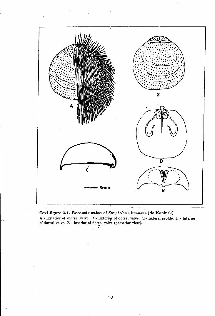
B
5mm
Text-figure 3.1. Reconstruction of ~trophalosia lewisiana (de Koninck) A - Exterior of ventral valve. B - Exteriqr of dorsal valve. G- Lateral profile. D - Interior of dorsal valve. E - Interior of dor~al valye (posterior view).
50
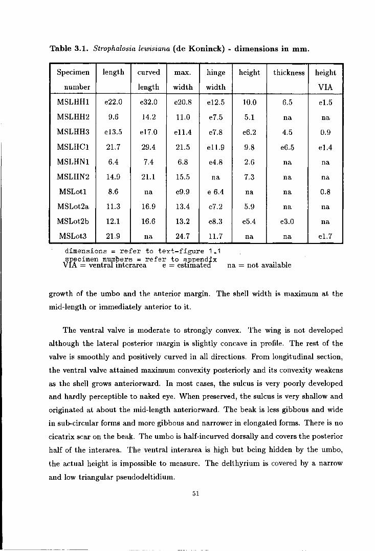
Table 3.1. Strophalosia lewisiana (de Koninck) - dimensions in mm.
Specimen length curved max. hinge
number length width width
MSLHH1 e22.0 e32.0 e20.8 e12.5
MSLHH2 9.6 14.2 11.0 e7.5
MSLHH3 e13.5 e17.0 e11.4 e7.8
MSLHC1 21.7 29.4 21.5 e11.9
MSLHN1 6.4 7.4 6.8 e4.8
MSLHN2 14.9 21.1 15.5 na
MSLotl 8.6 na e9.9 e 6.4
MSLot2a 11.3 16.9 13.4 e7.2
MSLot2b 12.1 16.6 13.2 e8.3
MSLot3 21.9 na 24.7 11.7
dimensions= refer to text-figure 1o1 specimen numbers = refer to appendix
VIA = ventral interarea e = estnnated
height thickness
10.0 6.5
5.1 na
e6.2 4.5
9.8 e6.5
2.6 na
7.3 na
na na
5.9 na
e5.4 e3.0
na na
na = not available
height
VIA
el.5
na
0.9
el.4
na
na
0.8
na
na
el.7
growth of the umbo and the anterior margin. The shell width is maximum at the
mid-length or immediately anterior to it.
The ventral valve is moderate to strongly convex. The wing is not developed
although the lateral posterior margin is slightly concave in profile. The rest of the
valve is smoothly and positively curved in all directions. From longitudinal section,
the ventral valve attained maximum convexity posteriorly and its convexity weakens
as the shell grows anteriorward. In most cases, the sulcus is very poorly developed
and hardly perceptible to naked eye. When preserved, the sulcus is very shallow and
originated at about the mid-length anteriorward. The beak is less gibbous and wide
in sub-circular forms and more gibbous and narrower in elongated forms. There is no
cicatrix scar on the beak. The umbo is half-incurved dorsally and covers the posterior
half of the interarea. The ventral interarea is high but being hidden by the umbo,
the actual height is impossible to measure. The delthyrium is covered by a narrow
and low triangular pseudodeltidium.
51
Internally, the ventral valve is provided with a pair of stout teeth, divergent at
an angle of about 50°. The adductor muscle platform is narrow and low and divided
into two halves by a shallow median furrow. The attachment scar is smooth. The
adductor muscle platform is surrounded by a pair of very low fan-shaped diductor
muscle platforms, the surface of which is covered by distant radial striae.
The surface of the ventral valve is covered by thin concentric striae which some
times turn to coarser growth lamellae and occasional rugae. The anterior margin
of the larger adult is sometimes folded into several (less than 10) broad ridges and
grooves. The ventral valve is covered with sub-erect and erect, posteriorly directed
spines. The sub-erect spines are distributed throughout the whole valve while the
the erect and posteriorly directed spines are arranged concentrically at the posterior
region only. The spines are very fine and hairy in nature, extending to about 10
to 15lll'i'IJY>) in length. In most cases only the spine tubercles are preserved. These
spine tubercles have a diameter of about 0.5mm• They are very closely spaced with
more than 100 tubercles being counted in one square em in the middle of the valve in
some cases. In a smooth growing shell the tubercles are normally roughly arranged in
staggered rows, while in most cases the tubercles are controlled by the development
of growth lamellae or rugae. The number of spines in the rugate forms is normally
less than those which have smoother striate shell-growth.
The dorsal valve has a sub-circular outline, truncated at the hinge line which
has a length of about two thirds of the maximum width or slightly shorter. The
shell is moderately concave with maximum depression immediately posterior to its
mid-length. Anteriorly the valve follows the curvature of the ventral valve without
any geniculation. The median fold is only weakly developed, best seen at the anterior
margin and extended to about one third of the valve length posteriorward. The umbo
is small, raised and pointed. The interarea is very low.
The interior of the dorsal valve is furnished with a shafted cardinal process with
trilobed or bilobed myophores and very narrow muscle attachment base. The cardinal
process shaft is small and usually stands at a low angle to the imaginary shell plane
posteriorly. The diductor muscle scar runs along this shaft and converges toward its
52
base. At the base of the shaft there are a pair of fairly deep sockets. These sockets are
confined by a slightly raised posterior margin and a pair of strong socket ridges which
seem to buttress the cardinal process shaft. The posterior margin develops a weakly
raised ridge laterally. The socket ridges are short, narrow, straight or hook-shaped and
separated by furrows from the raised oval-shaped concentrically striated or smooth
adductor muscle platform. Medianly, the cardinal process shaft is buttressed by a
strong median ridge which extends anteriorly as a low median septum to the mid
length of the valve or shorter. The brachial loop is wide with its outer ridge parallel
to the lateral shell margin and its anterior ridge extending beyond the mid-length
of the valve. It is not known excactly where the brachial ridges meet medially, but
the posterior extensions of these ridges seem to fade into the furrows in between the
muscle boundary ridges and the muscle platforms.
The dorsal surface is ornamented the same way as that of the ventral, except that
it does not possess erect and posteriorly directed spines.
Ontogeny and variation. The ontogenic history of S. lewisiana cannot be described
with certainty owing to the lack of a good collection of juvenile growth stages. From
the available material, it can be seen that only limited variation occurs during growth
to the adult. Apart from the development of ribs at the anterior margin of larger
adults, the only noteworthy variation occurs in the development of the beaks of
the adult. However, there is no particular trend in this variation. It is more likely,
therefore, that this variation represents self-adjustment to the slightly different niches
in which the individuals lived. The degree of incurving of the umbo changes from
one individual to another and depends on the convexity and the general shape of the
beak.
Regarding the development of spines, it is assummed that S. lewisiana clasped
other objects only during the juvenile stages. The erect and posteriorly directed
clasping spines are only developed posteriorly and as the shell grows larger the pro
duction of further clasping and cementing spines ceases. This suggests that the species
adopted a free living mode during adulthood. The multiple occurr~l\ces of this species
together with S. excavata at some localities suggests that both these species lived to-
53

gether in the form of a nest one on top of another. The nest is usually associated with
reef building algae and occasionally with bryozoan colonies with which they might live
together symbiotically. The dense body spines that were developed on both valves of
this species might also have functioned as anchors that interlocked themselves into
algal and bryozoan mats or into the spines of neighbouring individuals of the same
species or of S. excavata. At the same time, these dense spines would protect the
species from the attack of predators.
An interesting phenomenon is observed in the development of S. lewisiana through
time. General decrease in size of individuals of this species is observed from the
bottom to the top of the Durham Permian reef. In the upper reef flat fauna, the size
of the largest individuals is commonly less than half of those found at the base of the
reef. The dwarfing of the species toward the reef top is usually associated with faunal
intolerence of the steadily increasing salinity of the Permian sea.
Discussion. When de Koninck first described this species in 1847, he only based his
description on a single ventral valve. However, this description is sufficient to recog
nise the species from other strophalosias and productuses. De Koninck mentioned
that without its dorsal valve it is difficult to distinguish the species from P. granulo
sus Phillips, P. murchisonianus (= P. productoides Murchison) and P. geinitzianus
de Koninck. Since only P. geinitzianus has the same age range as S. lewisiana, it
is unlikely that it belongs to either of the other two species. As for P. geinitzianus
de Koninck, although there is a possibility that de Koninck's ventral valve of P.
lewisianus might belong to a small specimen of his P. geinitzianus, all the rest of
the collection from the type locality with similar ventral valves have the interarea
on both valves and thus are true strophalosiaceans. De Koninck's P. geinitzianus on
the other hand is a true productacean (see de Koninck (1847a, b), Geinitz (1861),
W. King (1856) and also this thesis under the name Spinohorridonia geinitziana (de
Koninck)). A few remains of Sp. geinitziana recently found at Humbleton Hill are
much larger in size when compared to S. lewisiana, and also have a distinguishable
productacean shape.
S. excavata ( Geinitz) is the closest species to S. lewisiana and it is not suprising
54
that many previous authors have described them both as a single species. The dif
ferences between the two species is only limited to the general shape of the shell and
umbo while the shell ornament and most of the internal features are almost identical
between the two species.
The first description of S. lewisiana by de Koninck (1847a) clearly stated that
the shape of the shell is rounded sub-semiglobulose and the umbo is regularly curved
upon itself, not prominent and pointed to its end. These features sufficiently dis
tinguish S. lewisiana from S. excavata which has a sub-triangular shape and rather
flattened and pointed umbo. With the dorsal valve and internal details now available
for comparison, the differences between S. lewisiana and S. excavata can be further
established. S. lewisiana has most of its ventral interarea hidden by the umbo and its
cardinal process shaft strongly inclined posteriorward, while the ventral interarea of
S. excavata is high and open and its cardinal process shaft is raised and more vertical
than that of S. lewisiana (see text-figure 3.3A and 3.3B).
The Visean S. panicula (Brunton) and S. lamnula (Brunton) resembleS. lewisiana
in having sub-rounded shape and very similar dorsal valve ornamentation, but the
ventral valve of these Visean species retain their juvenile spines throughout their
whole life. Thus, S. panicula and S. lamnula have two series of spines on the ventral
valves in the adult stages. S. panicula and S. lamnula also differ from S. lewisiana in
having an open interarea and a bilobed cardinal process myophore.
The Texas Permian S. inexpectans Cooper & Grant differs from S. lewisiana in
having a transversely sub-oval shape, a geniculated dorsal valve, an open interarea, a
bilobed myophore and a much longer median septum.
S. goldfussi (Munster) differs from S. lewisiana in having an open ventral inter
area, a higher dorsal interarea and most important of all, specialization of the adult
spines. The vermiform clasping and cementing spines on the ventral valve and con
centric spines on the dorsal valve has led Muir-Wood & Cooper (1960) to isolate
this species further from other Strophalosia. Muir-Wood & Cooper created a genus
Dasyalosia to acco11'1'!odate this species. However, detailed studies on the arrangement
of spines at the umbonal region and the distribution of spines on both valves shows
55

that S. goldfussi is ontogenically very close to all other species of Strophalosia. The
specialization of the spines in S. goldfussi is an adaptational feature linked to its spe
cial way of life. It should be noted that Craspedalosia lamellosa ( Geinitz) which is
usually found together with S. goldfussi, also has very similar spines on the ventral
valve. Although it is uncommon to find S. goldfussi preserved without spines, when
all its spines are stripped off the shell ornament is identical with other species of
Strophalosia.
STROPHALOSIA EXCAVATA (GEINITZ)
(Text-figures 3.2 and 3.3A, Plate 1, figures 13-19, Plate 2, figures 2-6)
Synonymy; 1842 Orthis excavatus Geinitz, p. · 578-9, pl. 10, fig. 12 - 13 1848 Orthothrix excavatus (Geinitz); Geinitz, p. 14-15, pl. 5, fig. 36, 39-40 not 35,
37-38, pl. 6, fig. 23 not 20-21 1848 ?Productus asperimus Howse, p. 257-8 1850 Strophalosia goldfussi (not-Munster); W. King, p. 96-98, pl. 12, fig 1-12 1853 Strophalosia goldfussi (not-Munster); Davidson, pl. 8, fig. 206, 209 1856 ?Strophalosia excavata ( Geinitz) var. whitleyensis W. King, p. 268 1857 Strophalosia goldfussi (not-Munster); Howse, p. 47-48 1858 Strophalosia goldfussi (not-Munster); Howse, p. 251-253 1858 Strophalosia goldfussi (not-Munster); Davidson, p. 39-43, pl. 3, fig. 1-18 1858 Strophalosia goldfussi (not-Munster) var whitleyensis W. King: Davidson, pl. 3,
fig. 23 1861 Strophalosia excavata (Geinitz); Geinitz, p· 93, pl. 1-4, 13, 16-19 not 5-12 and
14-15 1937 Strophalosia excavata (Geinitz); Malzahn, p. 53-54, pl. 2, fig. 18 1960 Orthothrix excavata (Geinitz); Muir-Wood & Cooper, p. 79-80, pl. 7, fig. 7-16 1965 Orthothrix excavata (Geinitz); Muir-Wood p. 450-1, fig. 304,2a-d
Diagnosis. Small to medium size Strophalosia with variable sub-triangular shape.
Ventral valve moderately convex with broad shallow sulcus and pointed umbo. Ven
tral interarea high with narrow triangular pseudodeltidium. Ventral surface with fine
concentric striae or coarser growth lamellae and closely spaced hairy spines with two
series posteriorly and one series anteriorly. Dorsal valve moderately concave with
broad fold posteriorly. Dorsal interarea low. Dorsal surface concentrically striated
56
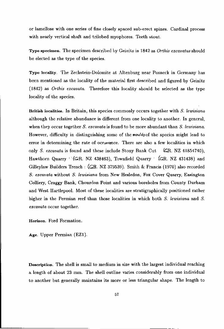
or lamellose with one series of fine closely spaced sub-erect spines. Cardinal process
with nearly vertical shaft and trilobed myophores. Teeth stout.
Type specimen. The specimen described by Geinitz in 1842 as Orthis excavatus should
be elected as the type of the species.
Type locality. The Zechstein-Dolomite at Altenburg near Posneck in Germany has
been mentioned as the locality of the material first described and figured by Geinitz
(1842) as Orthis excavata. Therefore this locality should be selected as the type
locality of the species.
British localities. In Britain, this species commonly occurs together with S. lewisiana
although the relative abundance is different from one locality to another. In general,
when they occur together S. excavata is found to be more abundant than S. lewisiana.
However, difficulty in distinguishing some of the mouldsof the species might lead to
error in determining the rate of occ!Arrence. There are also a few localities in which
only S. excavata is found and these include Stony Bank Cut (~R. NZ 41854740),
Hawthorn Quarry · '· (C.R. NZ 438463), Townfield Quarry (GR. NZ 431438) and
Gilleylaw Builders Trench t (GB. NZ 379539). Smith & Francis (1970) also recorded
S. excavata without S. lewisiana from New Hesledon, Fox Cover Quarry, Easington
Colliery, Craggy Bank, Chourdon Point and various boreholes from County Durham
and West Hartlepool. Most of these localities are stratigraphically positioned rather
higher in the Permian reef than those localities in which both S. lewisiana and S.
excavata occur together.
Horizon. Ford Formation.
Age. Upper Permian (EZ1).
Description. The shell is small to medium in size with the largest individual reaching
a length of about 23 mm. The shell outline varies considerably from one individual
to another but generally maintains its more or less triangular shape. The length to
57
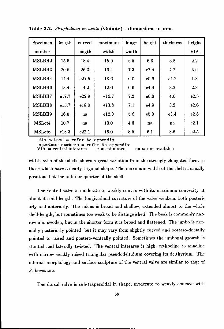
Table 3.2. Strophalosia excavata (Geinitz) - dimensions in mm.
Specimen length curved maxtmum hinge height thickness
number length width width
MSLBH2 15.5 18.4 15.0 6.5 6.6 3.8
MSLBH3 20.6 26.3 16.4 7.3 e7.4 4.2
MSLBH4 14.4 e21.5 13.6 6.0 e5.6 e4.2
MSLBH6 13.4 14.2 12.6 6.6 e4.9 3.2
MSLBH7 e17.7 e22.9 e16.7 7.2 e6.8 4.6
MSLBH8 e15.7 e18.0 e13.8 7.1 e4.9 3.2
MSLBH9 16.8 na e12.0 5.6 e5.0 e3.4
MSLot4 10.7 na 10.0 4.5 na na
MSLot6 e18.3 e22.1 16.0 8.5 6.1 3.6
dimensions "" refer to apperrdix spec·:imen numbers = refer t.o appendix
VIA = ventral interarea e = estrmated na = not available
height
VIA
2.2
3.0
1.8
2.3
e2.3
e2.6
e2.8
e2.1
e2.5
width ratio of the shells shows a great variation from the strongly elongated form to
those which have a nearly trigonal shape. The maximum width of the shell is usually
positioned at the anterior quarter of the shell.
The ventral valve is moderate to weakly convex with its maximum convexity at
about its mid-length. The longitudinal curvature of the valve weakens both posteri
orly and anteriorly. The sulcus is broad and shallow, extended almost to the whole
shell-length, but sometimes too weak to be distinguished. The beak is commonly nar
row and swollen, but in the shorter form it is broad and flattened. The umbo is nor
mally posteriorly pointed, but it may vary from slightly curved and postero-dorsally
pointed to raised and postero-ventrally pointed. Sometimes the umbonal growth is
stunted and laterally twisted. The ventral interarea is high, orthocline to anacline
with narrow weakly raised triangular pseudodeltidium covering its delthyrium. The
internal morphology and surface sculpture of the ventral valve are similar to th~t of
S. lewisiana.
The dorsal valve is sub-trapezoidal in shape, moderate to weakly concave with
58
its maximum depression just a little posterior to its mid-length. The median fold is
low and broad, appears on the anterior half of the valve only. The umbo is small and
weakly raised. The dorsal interarea is very low and only perceptible medially, facing
the interarea of the ventral valve at various angles. Internally, the cardinal process
shaft points to the posterior at an angle between 60° to vertical to the shell plane.
The rest of the internal features fall within variations of those found in S. lewisiana.
The dorsal surface is similar to that of S. lewisiana.
Ontogeny and variation. For similar reason to those discussed on S. lewisiana the
ontogenic history of this species cannot be precisely illustrated. Apart from the
development of the umbo, the changes during the ontogeny of S. excavata are very
similar to that of S. lewisiana.
The degree of variation on the umbonal development of S. excavata seems to
be much greater than on S. lewisiana (see text-figure 3.2). Self-adjustment to very
crowded niches might play an important role in the development of the highly diver
sified shape and attitude of the umbo of this species. This variation is less apparent
in places where the species is a minority in the fauna.
Stunting of the species is also observed toward the top of the Permian reef where
it became the last representative of the suborder Productidinae in this region.
Discussion. When Geinitz first described S. excavata in 1842, he clearly stated
that Die Rucken-Schale ist gleichmasig gewolbt nach dem kleinen erhabenen Wirbel
zu ••..... or The ventral valve is symetrically arched toward that small raised umbo .......
This feature is well presented in his figure 12, but his figure 13 does shows a slightly
curved umbo. He also mentioned that the shell has a pocket shape like Orthis tes
tudinaria. Unfortunately, when Geinitz (1848) elaborated his earlier description, he
mentioned that Rucken-Schale halbkugelig quer-oval, mit einem kleinen niedergeboge
nen spitzen Wirbel ....... or Ventral valve hemispherically transverse oval, with a small
weakly curved pointed umbo........ In his figures he included some of the forms which
have a rather sub-circular shape with strongly curved umbo, the form which has al
ready been described by de Koninck (1847a,b) as P. lewisianus (see Geinitz (1848,
pl. 5, fig. 35, 37-48 and pl. 6, fig. 20). Since then, the name S. excavata has been
59
~ll: ' ' \
D
~-------. ·--------~
I I
. I
. . ,
'incurved umbo' I ' . , '
' ' . . .. ' ' ' ' ' '
' ' ,
' ' -...
l!!!l!!!!l!l!-==s10mm
-I' I I . . .:,,
Text-figure 3.2. Variation in Strophalosia excauata (Geinitz) from Beacon Hill railway cut ------ possible intervariety relation11hip. ====== possible relationship with Strophalosia lewisiana (de Koninck).
60

confused by various authors with forms that should be called S. lewisiana, while these
same authors usually describe this species as S. goldfussi (see W. King (1850, 1856),
Davidson (1853, 1858), Howse (1857, 1858) etc.). Some other authors chose to fol
low Geinitz (1848) in grouping these two species together as S. excavata (see Geinitz
(1861 ), Malzahn (1937) ).
The most closely related species isS. lewisiana and a comparison between the two
species has already been given. S. panicula and S. lamnula differ from S. excavata in
having two series of spines on the ventral valve of the adult, a lower ventral interarea
and a bilobed myophores. S. goldfussi differs from S. excavata in having vermiform
and tangled spines in the adult. S. inexpectans differs from S. excavata in having a
transversely sub-oval shape, a geniculated dorsal valve, a bilobed myophore and a
very long median septum.
S. excavata reported from the Salt Range by Waagen (1884) and Reed (1944)
seems to have fewer, but more stout and recumbent spines than the S. excavata
described in this thesis. These very rare specimens described as S. excavata from the
Central Asian Permian, might belong to a distant variation of other species in the
area.
STROPHALOSIA GOLDFUSSI (Munster)
(Text-figure 3.3C, Plate 2, figures 7-13)
Synonymy; 1839 Spondylus goldfussi (Munster), p. 43, pl. 4, fig. 3a-b 1847a Productus goldfussi (Munster); de Koninck p. 257-9, pl. 11, fig. 4a-e and pl.
15, fig. 4a-b 1847b Productus goldfussi (Munster); de Koninck p. 148-151, pl. 11, fig. 4a-e and pl.
15, fig. 4a-b 1848 Strophalosia goldfussi (Munster); Geinitz, p. 14, pl. 5, fig 27-28 1861 Strophalosia goldfussi (Munster); Geinitz, p. 96-97, pl. 17, fig. 21-29 1871 Productus goldfussi (Munster); Quenstedt, p. 639, pl. 20, fig. 16-20,22 1921 Strophalosia goldfussi (Munster); Trechmann, p. 542, pl. 12 1901 Strophalosia goldfussi (Munster); Zittel, p. 391, fig. 581a-c
61

a b a b a b
A a ~pb
c
~b sh
. ~ ~ c
c c
d d
.... a ... &;;,;b-..._ 5mm i"-smm
Text-figure 3.3. Comparison ofcardinaJ. process and mode of life between species of British Permian Strophalosia ·.
A- Strophalosia lewisiana (de Koninck). B- Strophalo.9ia excavata (Geinitz). C- Straphalosi€4 . goldfussi (Munster). a,b - plan and side vie~ of cardinal process. c,d - juvenile and adult mode of life. ab - algal mat or bryozoan colqnies. sh - spine of Horridonia. {Ad and Dd arc not attached by spines} ·
62

1937 Strophalosia goldfussi (Munster); Malzahn, p. 50-52, pl. 2, fig. 12-15 1960 Dasyalosia goldfussi (Munster); Muir-Wood & Cooper, p. 76-7, pl. 6, fig. 12-15 1965 Dasyalosia goldfussi (Munster); Muir-Wood, p. 450, fig. 304,4a-c 1985 Dasyalosia goldfussi (Munster); Cocks, p. 64, pl. 5.2.36A,B,C 1988 Dasyalosia goldfussi (Munster); Threk, Marek & Benes, p. 146-7, fig. 1-2
Diagnosis. Small to medium size Strophalosia with sub-triangular to sub-circular shell
outline. Ventral valve moderately to strongly convex with poorly developed sulcus
and small pointed umbo. Ventral interarea high with weakly raised pseudodeltid
ium. Ventral surface covered with fine concentric striae to coarse growth lamellae
with two series of spines in younger stages, but in adult stages nearly all the spines
are vermiform and posteriorly directed. Dorsal valve moderate to strongly concave.
Dorsal surface with concentric striae or lamellae and vermiform spines centrally di
rected. Dorsal interarea low with small weakly raised chilidium and small triangular
lophidium. Cardinal process large with trilobed myophores, shaft supported by strong
lateral and median ridges. Adductor muscle platform highly elevated with radially
grooved scars.
Type specimens. The original specimen described and figured by Munster in 1839 as
Spondylus goldfussi should be elected as the type of the species.
Type Locality. Munster (1839) described and figured his Spondylus goldfussi from a
specimen which came from Rospen near Gera. This locality should therefore be used
as the type locality of the species.
Localities in Britain. S. goldfussi is a rare species in Great Britain and so far it has
only been found at East Thickley Quarry (M. R. NZ 24082564), Old Town (=White)
Quarry (M. R. NZ 257245), Old Quarrington Quarry (M. R. NZ 326381), Tunstall Hill
old trench (M. R. NZ 397542) and Humbleton Hill (M. R. NZ 381553). The species
was also obtained from the Mattersey Borehole in N ottinghamshire. Currently, there
are about twenty specimens of this species known in Britain. The rarity of the species
in this part of the world is the reason why it was poorly understood by British
researchers in the last century. As mentioned earlier, the first true S. goldfussi in
Britain was reported and figured in 1921 by Trechmann, long after most other British
strophalosiids have been described.
63
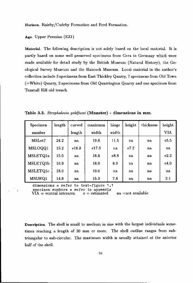
Horizon. Raisby /Cadeby Formation and Ford Formation.
Age. Upper Permian (EZ1)
Material. The following description is not solely based on the local material. It is
partly based on some well preserved specimens from Gera in Germany which were
made available for detail study by the British Museum (Natural History), the Ge
ological Survey Museum and the Hancock Museum. Local material in the author's
collection include 3 specimens from East Thickley Quarry, 7 specimens from Old Town
(=White) Quarry, 2 specimens from Old Quarrington Quarry and one specimen from
Tunstall Hill old trench.
Table 3.3. Strophalosia goldfussi (Munster) - dimensions in mm.
Specimen length curved maximum hinge height thickness
number length width width
MSLot7 24.2 na 19.6 11.5 na na
MSLOQQ1 15.2 e18.9 e17.6 na e7.2 na
MSLETQ1a 15.0 na 18.8 e8.8 na na
MSLETQ1b 16.9 na 18.0 8.9 na na
MSLETQ1c 18.0 na 19.6 na na na
MSLWQ1 14.8 na 15.3 7.8 na na
dimensions= refer to text-figure 1.1 specimen numbers = refer to appendix
VIA = ventral interarea e = estimated na =not available
height
VIA
e5.5
na
e2.2
e4.0
na
2.1
Description. The shell is small to medium in size with the largest individuals some
times reaching a length of 20 mm or more. The shell outline ranges from sub
triangular to sub-circular. The maximum width is usually attained at the anterior
half of the shell.
64

The ventral valve is moderately to strongly convex. The median sulcus is poorly
developed and often imperceptible. The beak is weakly swollen with small flat ci
catrix of attachment or without cicatrix. The umbo is small and usually pointed to
the posterior, but sometimes the tip is slightly incurved or truncated. The ventral
interarea is high and open with its surface transversely striated. The delthyrium is
partly covered by a weakly swollen, narrow triangular pseudodeltidium. Internally, a
strong pair of teeth are projected at a narrow angle to one another at the base of the
pseudodeltidium.
The ventral surface is covered with fine concentric striae or with coarser lamellae.
It is also covered with dense fine hollow vermiform spines. The straighter recumbent
and adpressed spines can only be seen in the younger stages at the posterior region
when the thick cover of the vermiform spines is stripped off. At this posterior region
the spines are arranged in two series; the recumbent and anteriorly directed and the
vermiform and posteriorly directed. In adult stages all spines are erect to sub-erect at
their point of origin, but immediately turn posteriorward and become entangled with
each other. These spines extend beyond the posterior shell margin before clasping or
cementing themselves to other hard objects.
The dorsal valve has a sub-circular to sub-trapezoidal shape. It is moderate to
strongly concave with obscure trace of a median fold anteriorly. The beak is small tharo of
and moderately raised, so that the dorsal interarea is usually higher[thatLany other
Strophalosia. The dorsal interarea is about one-third to half the height of the ventral
interarea. The width of the dorsal interarea is extended to the full length of the hinge
line which is about two-thirds the maximum shell width or slightly shorter. The
dorsal interarea has a transversely striated surface and furnished with a small weakly
raised chilidium which is extended posteriorly as a smalllophidium.
The dorsal surface is covered with fine concentric striae or coarser growth lamellae
and a series of centrally directed vermiform spines, entangled with each others in the
central depression of the shell.
The interior of the dorsal valve is provided with a large cardinal process which has
a trilobed myophore. The cardinal process shaft inclines at a low angle towards the
65

ventral, and is supported by a strong pair of posteriorly pointed lateral ridges which
also act as the socket ridges. Medianly, the shaft is supported by a strong median
ridge which narrows and continues anteriorly as a median septum. The adductor
muscle platform is subtriangular in shape, highly elevated anteriorly with radially
grooved scars.
Discussion. As discussed in Muir-Wood & Cooper (1960) the internal morphology of
S. goldfussi seems to resemble very closely that of S. lewisiana and S. excavata, except
that the exact form of the cardinal process is not well known in S. goldfussi. Brunton
(1966) stressed that Muir-Wood & Cooper's description of the cardinal process was
based on incomplete specimens. A single specimen from Tunstall Hill old trench
collected during this research exhibits a well preserved internal morphology including
the cardinal process which is very similar to that of S. excavata (see text-figures 3.3B
and 3.3C).
Current study on some S. goldfussi from the German Zechstein in which the
vermiform spines on the umbonal region are stripped off has shown that S. goldfussi
has a very similar type and arrangement of spines to all other species of Strophalosia
in the younger stages (see plate 2, figure 8). The vermiform nature of the spines
seems to be a special character of the adult of this species. Furthermore, comparison
with Brunton {1966) species from the Visean of County Fermanagh, shows that these
Visean species actually show intermediate morphological features between S. goldfussi
and S. excavata - S. lewisiana. For this reason I suggest that these three species be
included in one genus and the best genus to fit them all is the genus Strophalosia.
Other similarities and differences between the three species of Strophalosia from the
British Permian have been described and discussed in earlier pages. In general S.
goldfussi differs from all other species of Strophalosia in its unique vermiform spines
which are slightly larger in diameter than in the other species.
Considering the small number of specimens of S. goldfussi available in the British
Permian, one is inclined to speculate that these specimens might have been trans
ported from a different part of the Permian sea. The specimens, however, show that
they lived in the Durham Permian Sea at least during their adulthood. There are
66

times in which a Zechstein storm might have transported larvae of this species west
ward from the ·.aast Zechstein. The British Zechstein Sea might not be the ideal en
vironment for this species, but they certainly survived their adulthood here although
they were probably not sufficiently numerous or fertile to further expand their gener
ation. This is shown by the general dwarfing of the species in the British Permian as
compared to average size of the adult of the species which lived in the East German
Zechstein. Evidence shows that all the British S. goldfussi lived at more or less the
same stratigraphic horizon in both East Thickley Quarry and Old Town (=White)
Quarry. This indicates that they were probably carried westwards by the same storm
current from the East Zechstein Sea.
Although the Old Quarrington Quarry S. goldfussi bearing bed is apparently
lower than the other localities, evidence shows that this bed is not an in situ deposit.
It is a slump deposit which was derived from a shallower sea to the west by downslope
movement (Smith, 1970). Since the species is only known from a limited stratigraphic
horizon in the British Zechstein, and the two localities with S. goldfussi bearing
biocoenoces are situated to the west of the Old Quarrington Quarry, it is likely that
this slump bed originated from the S. goldfussi bearing horizon in the west.
Rare occurrences of S. goldfussi in the Ford Formation is difficult to understand.
More extensive study is needed in order to get a better understanding of this species,
and to either prove or disprove various assumptions given above.
Genus EOSTROPHALOSIA Stainbrook 1943
Synonymy; 1943 Eostrophalosia Stainbrook, p. 58 1960 Eostrophalosia Stainbrook; Muir-Wood & Cooper, p. 77-79 1965 Eostrophalosia Stainbrook; Muir-Wood, p. 450
Amended diagnosis. Small to medium size productiform strophalosiinid with distant
recumbent body spines on both valves and clasping marginal spines on ventral valve
only. Ventral valve moderately to strongly convex with cicatrix of attachment small
67
or absent. Interarea low with small pseudodeltidium and chilidium. Cardinal process
with bilobed myophore on small shaft. Teeth moderately developed. Adductor muscle
platform low. Brachial impression small. Shell surface smooth or lightly lamellose.
Discussion. Apart from the new species E. permiana, the genus Eostrophalosia has
only been recorded from the Devonian of North America, and thus it holds the name
"Eos" for being older than the true Strophalosia. It has been known as a rare genus
and indeed all its species have also been considered as rare species by previous au
thors (Stainbrook, 1943; Muir-wood & Cooper, L960), For this reason, the genus
has not been fully described and currently only the external morphology is known
with certainty. From the study of the external morphology of E. permiana, it seems
that this species should best be described as Eostrophalosia rather than any other
strophalosiinid. With a larger and better collection of specimens of this species, more
details of the genus including the internal morphology can now be described.
A specimen of E. permiana has already been figured by Davidson {1858, pl. 3,
fig. 38) as S. lamellosa (Geinitz) var. humbletonensis W. King, but since then the
identity of this specimen has not been discussed by authors. In Logan's unpublished 85
thesis (Univ. of Durham, 1962) the same specimen, as welltsome other similar forms
were figured and described as S. morrisiana W. King. Although the ornamentation
on the ventral valve and the internal morphology of E. permiana and S. morrisiana
(=Heteralosia morrisiana in this thesis) are very similar, the spinosity of the dorsal
valve separates the~ into different genera and indeed a different subfamily. This is
also supported by their distribution patterns in East Durham.
In terms of the external morphology Eostrophalosia can be best described as
an intermediary between the slightly less productiform and larger strophalosiinid
genus Echinalosia Waterhouse and the productellid genus Orbinaria Muir-Wood and
Cooper. More detailed comparison is needed before any further conclusion on the
true relationship between these three genera can be established.
Type-species : Strophalosia rockfordensis Hall & Clarke 1893
Other species Strophalosia callawayensis Swallow, S. littletonensis Stainbrook, S.
68
independensis Stainbrook and Eostrophalosia permiana new species. All these species
belong to the Devonian of North America except E. permiana which occurs in the
British Permian.
EOSTROPHALOSIA PERM/ANA new species
(Text-figure 3.4, Plate 3, figures 1-13)
The species is named after the Permian System, in which the genus was unexpectedly
discovered.
Synonymy; 18.58 Strophalosia lamellosa (not-Geinitz) var humbletonensis (not W. King); David
son, pl. 3, fig. 38.
Diagnosis. Small to medium size Eostrophalosia with transversely sub-elliptical to
sub-circular shell outline. Ventral valve moderate to strongly convex with inflated
umbo and fully developed auricle. Sulcus absent. Cicatrix of attachment small or
absent. Ventral interarea low with small pseudodeltidium. Ventral valve smooth or
lightly lamellose with a row of clasping marginal spines, two to three discontinuous
rows of auricular spines and scattered distant recumbent to adpressed body spines.
Auricles lightly wrinkled. Dorsal valve moderately concave with small pointed umbo.
Dorsal interarea low with small chilidium. Cardinal process with bilobed myophore
set on small shaft. Dorsal adductor muscle platform low smooth or striated. Median
septum short. Brachial impression small. Dorsal surface smooth or lightly lamellose
with sparsely distributed adpressed body spines.
Type specimens. Specimens no. "" MSLBHlO, MSLBHll, MSLBH12, MSLBH13,
MSLBH14 and MSLBH15 in the author's collection are chosen as a series of paratypes
showing various important features of the species.
Type locality. Beacon Hill railway cut (GR. NZ 442454). This locality is chosen
because from here most of the described specimens were collected, despite the fact
that the first figured specimen (Davidson, 1858) came from Ryhope Field House.
69
-------------

Other localities. Material for this species has only been reported from Ryhope Field
House (Davidson, 1858) and Fox Cover Quarry (Logan, 1962 unpublished thesis).
Current study shows that there are a large number of specimens from Dalton-le-Dale
in Kirkby's collection in Hancock Museum which were labelled as various species but
belong to E. permiana. During the tenure of this research, the author also found this
species from Tunstall Hill old trench (CR. NZ 397542) and Ryhope Mental Hospital
(GR. NZ 40055215).
Material. The following description is mainly based on material collected personally
by the present author which include 45 specimens from Beacon Hill, 3 specimens from
Tunstall Hill old trench and 1 specimen each from Dalton-le-Dale and Ryhope Mental
Hospital. Most of these specimens are preserved in form of moulds and casts, while
a few show patches of original shell material.
Horizon. Ford Formation.
Age. Upper Permian (EZ1).
Table 3.4. Eostrophalosia permiana new species - dimensions in mm.
Specimen length curved max. hinge height thickness height
number length width width VIA
MSLBH10 e24.5 e37.2 29.7 23.9 12.8 7.6 0.8
MSLBH11 19.6 e30.4 25.1 17.2 10.3 6.8 1.3
MSLBH12 16.5 24.0 22.8 e18.7 10.3 na na
MSLBH13 10.2 11.9 11.5 e9.4 3.7 na na
MSLBH14 23.8 34.6 27.4 na 12.8 na na
MSLBH18 16.1 20.2 20.6 na 6.8 na na
MSLot8 e20.0 na e24.2 e17.6 na na el.3 a1.mens1.orrs - refer to text-fi re 1 gu • 1 specimen numbers = refer to appendix VIA = ventral interarea e = estimated na = not available
Description. The shell is small to medium in size with the largest individuals reaching
70

'* (/ I • '
~~'~ " ~.
A
c
B
-y-0
Text-figure 3.4. Reconstruction of Eostrophalosia penniana new species
---smm
A - ventral view (exterior). B - dorsal view (exterior). C - lateral profile. D - cardinal
process.
r::

a length of about 25 mm. The shell is usually transversely sub-elliptical to sub-circular
in outline with the maximum width acquired at about the mid-length.
The ventral valve is moderate to strongly convex with maximum convexity shown
posteriorly at the beak. The sulcus is usually absent, but sometimes a slight flattening
in the median part of the shell is observed anteriorly. The auricles are not very
prominent but they are fully developed. The beak is narrow and small, but it is
strongly inflated. Vestiges of cicatrix of attachment can sometimes be traced on the
umbo, but most commonly this scar is absent in this species. The umbo is slightly
overhanging and incurved, but it does not cover the ventral interarea. The ventral
interarea is low with smooth or transversely striated surface. The delthyrium is
covered by a small weakly raised triangular pseudodeltidium.
The ventral valve interior is provided with a very narrow, short and low adductor
muscle platform with smooth or lightly striated scar. This platform is divided in
the middle by a shallow median furrow and laterally surrounded by a lower fan
shaped coarsely radially striated diductor muscle platform. Judged from the size of
the socket on the dorsal valve, the teeth are quite small in size as compared to other
strophalosiinids in this region.
The external of the ventral valve is smooth or lightly lamellose with occasional
rugae. At the auricle the shell is lightly wrinkled. The shell is provided with the
following spines; 1 - one row of clasping marginal spines which are erect, posteriorly
and concentrically directed; 2 - two to three discontinuous radial rows of auricular
spine bases in which the true nature of the spines is not fully known. The spine
bases show that these spines are erect at their point of origin, but it is not known
whether they provided additional clasping tools to the marginal spines or just posed as
ornament; 3- random and distantly distributed recumbent to adpressed body spines.
In general, these body spines tend to be concentrated on the thicker growth lamellae
or rugae. All spines are small with diameter around 0. 7 mm and short and do not
seem to be strong enough to support the large adult by their clasping mechanism.
The dorsal valve is moderately concave with maximum depression at about the
centre of the valve. There are no traces of the median fold. The umbo is small,
72

rounded and weakly elevated. The interarea is very low with a small chilidium.
The cardinal process shaft is small and posteriorly inclined, and the myophore
is bilobed. The cardinal process is buttressed by a pair of low socket ridges and a
low median ridge which continues anteriorly as a low and short median septum. The
median septum extends to about one third of the valve length. The adductor muscle
platform is roughly pear-shaped. It is weakly elevated with its scars either smooth
or lightly concentrically striated. Brachial impressions are not completely preserved
but are smaller compared to that of other strophalosiinids. The lateral and anterior
margins only extended to about halfway to the lateral and anterior shell margins
respectively. The rest of the internal features are not known.
The dorsal surface is smooth or lightly concentrically lamellose with very light
wrinkles on the auricle. The shell is covered by very few (less than 30) distantly
spaced adpressed spines. Sometimes, these spines are strongly adpressed and leave
radial grooves on the shell surface to give the shell a rather costate appearence.
Discussion. The elongated form which is much more convex than the average form
(see plate 3, figure 7), is normally found among the larger individuals, and therefore
they might represent sexual dimorphs of the species. Apart from this, the complete
ontogeny of this species is not well known because of the lack of younger individuals
in current collection.
As compared to other Eostrophalosia, this species seems to resemble very closely
to the type-species i.e. E. rockfordensis (Hall & Clarke) in its shape, surface sculpture
and shape of the interarea. However, E. rockfordensis differs from E. permiana in its
smaller size and more prominent cicatrix of attachment. Further comparison cannot
be made because of the limited knowledge about the Devonian type-species. Other
species of Eostrophalosia are also poorly described in past literature and thus no
detailed comparison can be made. It is noteworthy that E. permiana is the largest and
the youngest of all Eostrophalosia and it is also the most comprehensively collected
species.
73

Subfamily HETERALOSIINAE Muir-Wood & Cooper 1960
Diagnosis. Strophalosiidae without spines on dorsal valve.
Type genus. Heteralosia R. H. King, 1838
Other genera. ?Devonalosia Muir-Wood & Cooper, Craspedalosia Muir-Wood &
Cooper, Leptalosia Dunbar & Condra, Sphenalosia Muir-Wood & Cooper, Wynd
hamia Booker, Costalosia Waterhouse & Shah, ?Truncalosia Imbrie and ?Oligorachis
Imbrie.
Discussion. The stability of the type genus i.e. Heteralosia R. H. King has con
tributed to the persistence of this subfamily since it was introduced in 1960 by Muir
Wood & Cooper. Only a few authors mentioned earlier in the discussion of the genus
Strophalosia have argued about the validity of this subfamily. For reasons I have
already given earlier, I have no doubt that this subfamily should stand as it is.
Note on the radial costae. One common phenomenon among the British Permian
heteralosiinid is that the shell is almost always radially costate. The degree of the
development of the costae is varied from one species to another, but generally the
costae are developed stronger in the dorsal valve than in the ventral valve. The
costae are regularly spaced with the width of each costa rarely exceeding hnm. The
costae are usually continuous from the umbo to the anterior margin, but bifurcation
sometimes occurs anteriorly in the larger individuals. The ventral valve is usually
weakly costate with the degree of costation sometimes usefull to divide some species
from the others.
British Permian heteralosiinid. In previous papers, the subfamily Heteralosiinae is
only represented by the genus Craspedalosia Muir-Wood & Cooper in the Zechstein
Permian. More detailed study of W. King's S. morrisiana shows that it belongs to
the genus Heteralosia R. H. King. Several other new species of Heteralosia have also
been discovered during the present study on the British Permian productidinids.
74
Genus HETERALOSIA R. H. King 1938
Synonymy: 1938 Heteralosia R. H. King, p. 278. 1938 Heteralosia R. H. King; Prendergast, p. 41. 1952 Heteralosia R. H. King; Moore, Lalicker & Fischer, p. 252. 1960 Heteralosia R. H. King; Muir-wood & Cooper, p. 80-82. 1965 Heteralosia R. H. King; Muir-Wood, p. 451
Amended diagnosis. Small to medium size heteralosiinids with moderate to strongly
convex ventral valve, recumbent to adpressed and erect body spines and umbonal
cicatrix of attachment. Dorsal valve moderately concave to almost flat, sometimes
geniculated. Interarea low on both valves with delthyrium partly covered by pseudo
deltidium and lophidium and dorsal interarea with small chilidium. Cardinal process
with bilobed or quadrilobed myophore. Teeth small. Adductor muscle platform mod
erately elevated. Brachial impressions medium to large in size.
Discussion. R. H. King (1938) described this genus as having interarea on both
valves, spines on ventral valve only and umbonal cicatrix of attachment in order
to distinguished it from Strophalosia W. King, Aulosteges Helmerson and Leptalosia
Dunbar & Condra. With the above description he listed several species as Heteralosia
including H. sclocomi n. sp. (as the type-species), Strophalosia gerardi W. King and
S. tenuispina Waagen.
Later in 1960, Muir-Wood & Cooper revised the diagnostic features of this genus
to distinguish it from many more new heteralosiinid genera introduced by them as
well as various other authors (see the list of other genera above). Since not many of
these genera are available in the present research collection, I cannot give any further
comments on tlus subfamily division although some of the listed genera seem to lack
a strongly convincing back ground.
The genus Heteralosiais known to have flourished from Mississippian time (Hinchey
& Ray (1935)) and persisted through to the Upper Permian. Species of Heteralosia
are usually zone restricted as well as spatially confined, except H. tenuispina (Waa
gen) which is known to have lived from Visean to Permian times. In general, it seems
75
that the Carboniferous species have slightly more spines than those from the Permian.
Variation in other features does not seems to be age related. In practice, the generic
sub-division is based on the general size and shape of the shell, the size of cicatrix of
attachment and the surface ornametation of the shell.
In Britain, the genus Heteralosia was only known previously from the Visean
of County Fermanagh (Brunton, 1966). This is the first time that this genus is
recognised in the Zechstein Permian, although one of its species, H. morrisiana (W.
King) is famous in previous literature. This species is well described and illustrated
by previous authors as a Strophalosia, probably because Davidson (1858) included
in this species forms with spinose dorsal valves described here as E. permiana new
species. The recognition of the genus H eteralosia has led the author to recognise
four other species of the genus which might have been included by various authors
in S. morrisiana. From the present study, Heteralosia is represented in Britain by
H. morrisiana (W. King), H. humbletonensis (W. King), H. hyltonensis n. sp .. H.
seahamensis n. sp. and H. aycliffensis n. sp. H eteralosia? quarringtonensis n. sp. is
tentatively assigned to this genus although some of the features suggest that it might
belong to one of the productellids.
Note. The above amended diagnosis is a simplified version of that given by Muir
Wood & Cooper (1960).
Type-species. Heteralosia sclocomi R. H. King, 1938, p. 278-9, pl. 39, fig. 15-18.
Other species. Strophalosia gerardi W. King, S. morrisiana W. King, S. tenuispina
Waagen, S. hystricula Girty, S. orbiculata Hinchey & Ray, S. subelliptica Hinchey &
Ray, S. fortispinosa Hinchey & Ray, H. magnispina Cooper & Grant, H. paucispina
Cooper & Grant, S. ?vidriensis (=H. subelliptica) Cooper & Grant, H. humbletonensis
(W. King), H. hyltonensis n. sp., H. seahamensis n. sp., H. aycliffensis n. sp. and
H?. quarringtonensis n. sp.
76
HETERALOSIA MORRIS/ANA (W. King)
(Text-figure 3.5, Plate 3, figures 14-22).
Synonymy; 1846 ?Strophalosia morrisiana W. King, p. 28. 1848 Productus spiniferus (W. King); Howse, p. 257. 1848 Strophalosia morrisiana W. King; W. King, p. 9. 1848 Productus cancrini (not-de Verneuil); Geinitz, p. 16, pl. 1, fig. 16-17. 1850 Strophalosia morrisiana W. King; W. King, p. 99-102, pl. 12, fig. 18-19. 1853 Strophalosia morrisiana W. King; Schauroth, p. 31, fig. 7. 1854 Strophalosia morrisiana W. King; Schauroth, p. 221. 1856 Strophalosia morrisiana W. King; W. King, p. 264-268. 1857 Strophalosia cancrini (not-de Verneuil); Howse, p. 49. 1858 Strophalosia lamellosa (not-Geinitz); Howse, p. 253-4. 1858 Strophalosia lamellosa (not-Geinitz) var. morrisiana (King); Davidson, p. 44-
49, pl. 3, fig. 24-31, 33. 1861 Strophalosia morrisiana W. King; Geinitz, p. 98-100, pl. 18, fig. 8-22. 1871 Productus morrisianus (W. King); Quenstedt, p. 641, pl. 60, fig. 28. ,-1913 Strophalosia morrisiana W. King; Czarnocki & Samsonovich, p. 433, pl. 49, fig.
5. 1937 Strophalosia morrisiana W. King; Malzahn, p. 54, pl. 3, fig. 22. 1945 Strophalosia morrisiana W. King; Trechmann, p. 349, pl. 15, fig. 13. 1960 Strophalosia morrisiana W. King; Muir-wood & Cooper, p. 318. 1975 ?Heteralosia sp. 4, Cooper & Grant, p. 802, pl. 193, fig. 9.
Diagnosis. Shell small to medium in size, sub-circular to transversely sub-elliptical
in shape. Ventral valve moderately convex with small cicatrix of attachment on
irregular and asymmetrical umbo. Ventral surface weakly lamellose and costate.
Body spines rare, adpressed, or erect and posteriorly directed. Ventral interarea low
with delthyrium partly covered by pseudodeltidium. Dorsal valve with radial costae
and weak growth lamellae. Dorsal interarea low with small chilidium and lophidium.
Teeth small. Cardinal process with bilobed myophore set on small shaft. Adductor
muscle platform low. Brachial impressions wide. Median septum extended to half
the length of the shell.
Type specimens. Pattison (1977) designated specimens no. D326A-C (three speci
mens) as syntypes of this species although he reports that the figured type ofW. King
is missing. All these specimens are kept in the Department of Geology, University of
77

Galway in Republic of Ireland. From my own examination of the specimens I have no
doubt that none of them are illustrated in W. King.(l850) monograph. The specimen
no. D326B is the most perfect syntype among the three and it shows very similar
features to that of W. King's figure.
Type locality W. King (1850, 1856) stated that the type of his Strophalosia morrisiana
should be chosen among the material from Tunstall Hill. According to Pattison
(1977), W. King's Tunstall Hill locality is probably a fairly large area with quarries
and natural exposures around NZ 392545. More new material was collected recently
from this locality which is now known as the Rock Cottage (Hollingworth, 1986,
1987).
Other localities. H. morrisiana has also been reported from H umbleton Hill and
Dalton-le-Dale (Howse (1848), Davidson (1858)), Ryhope Field House, Weston, Clax
heugh and Midderidge (Davidson (1858). The species is also known from the Ger
man Zechstein (Geinitz (1848, 1861) Schauroth (1853, 1854), Quendstedt (1871) and
Malzahn (1937)) and from ?Bone Spring Formation of West Texas (Cooper & Grant
(1975)).
Material. This description is based on all the syntypes mentioned above and 48
specimens collected by the author from Tunstall Hill Rock Cottage. Most of these
specimens are partly preserved with the original shell material and partly in form of
moulds and casts. They are commonly preserved as complete shells and the internal
details can be studied from original casts or artificial casts made from latex.
Horizon. Ford Formation.
Age: Upper Permian (EZ1).
Description. The shell is small to medium in size, sub-circular to transversely sub
elliptical in outline with posterior margin often irregularly developed. The lateral
and anterior margins are usually smooth. The shell width is usually a little broader
than its length, and widest at around mid-length. The shell profile is moderately
concavo-convex with a narrow visceral cavity.
78

,,, '
I
E
D
--1mm
Text-figure 3.5. Reconstruction of Heteralosia morrisiana (King)
5mm
A - Exterior of ventral valve showing cicatrix of attachment (crossed). B - Exteriorof dorsal valve. C - Interior of dorsal valve showing f!ndonodes. D - Lateral profile. E - Came1·a Iucida reproduction of intcrarea of syutype 1~0 D326B (UCG collection).
79
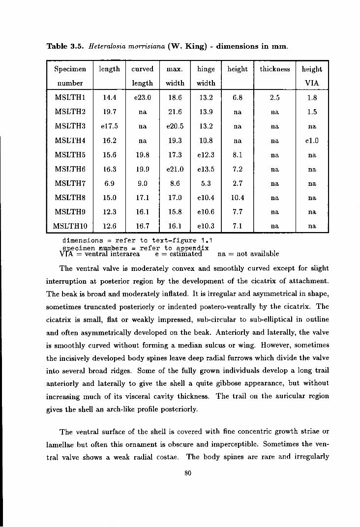
Table 3.5. Heteralosia morrisiana (W. King) - dimensions in mm.
Specimen length curved max. hinge
number length width width
MSLTH1 14.4 e23.0 18.6 13.2
MSLTH2 19.7 na 21.6 13.9
MSLTH3 e17.5 na e20.5 13.2
MSLTH4 16.2 na 19.3 10.8
MSLTH5 15.6 19.8 17.3 e12.3
MSLTH6 16.3 19.9 e21.0 e13.5
MSLTH7 6.9 9.0 8.6 5.3
MSLTH8 15.0 17.1 17.0 e10.4
MSLTH9 12.3 16.1 15.8 e10.6
MSLTH10 12.6 16.7 16.1 e10.3
dimensions= refer to text-figure 1.1 s~ecimen numbers = refer to appendix
Vl:A = ventral interarea e = estimated
height thickness
6.8 2.5
na na
na na
na na
8.1 na
7.2 na
2.7 na
10.4 na
7.7 na
7.1 na
na = not available
height
VIA
1.8
1.5
na
el.O
na
na
na
na
na
na
The ventral valve is moderately convex and smoothly curved except for slight
interruption at posterior region by the development of the cicatrix of attachment.
The beak is broad and moderately inflated. It is irregular and asymmetrical in shape,
sometimes truncated posteriorly or indented postero-ventrally by the cicatrix. The
cicatrix is small, flat or weakly impressed, sub-circular to sub-elliptical in outline
and often asymmetrically developed on the beak. Anteriorly and laterally, the valve
is smoothly curved without forming a median sulcus or wing. However, sometimes
the incisively developed body spines leave deep radial furrows which divide the valve
into several broad ridges. Some of the fully grown individuals develop a long trail
anteriorly and laterally to give the shell a quite gibbose appearance, but without
increasing much of its visceral cavity thickness. The trail on the auricular region
gives the shell an arch-like profile posteriorly.
The ventral surface of the shell is covered with fine concentric growth striae or
lamellae but often this ornament is obscure and imperceptible. Sometimes the ven
tral valve shows a weak radial costae. The body spines are rare and irregularly
80

distributed. They are small (less than 1 mm in diameter), but long hollow spines of
two different types, the recumbent, adpressed and anteriorly directed spines and the
erect and posteriorly directed spines. The second type is only developed posteriorly
and probably functioned as clasping or cementing spines together with the marginal
spines. The marginal spines are similar in sizes with the rest of the body spines and
are of clasping or cementing types.
The ventral interarea is low and narrow with its full width about two thirds
of the maximum shell width. It is apsacline to orthocline in profile with its surface
longitudinally and transversely striated. The delthyrium is small and is partly covered
by a slightly arched pseudodeltidium. The teeth are small. Other details of internal
morphology of the ventral valve are not known.
The dorsal valve is slightly smaller than the ventral valve and moderately concave
in profile. The valve curvature is smooth and continuous to both lateral and anterior
margins. However, slight geniculation is sometimes shown in the larger individual.
The concavity of the valve is usually interrupted posteriorly by a raised reflection
of the cicatrix of attachment. The apex is always slightly raised above the plain of
the valve. The dorsal surface of the valve is radially costate and weakly concentri
cally lamellose. The dorsal interarea is very low with small weakly raised chilidium,
extended posteriorly to form a small triangular lophidium.
The cardinal process is small, with bilobed myophore elevated on a small postero
ventrally directed shaft which is butressed by a pair of slightly raised lateral ridges
and a prominent median ridge. The median ridge extends anteriorly as a low median
septum to about the mid-length of the shell. The adductor muscle platform is only
slightly raised anteriorly. It has a pear-shaped outline and the muscle scars are lightly
striated. The brachial impressions are wide with the lateral margin closely following
the shell margin. The inner shell surface is smooth or covered with rare endonodes
and traces of radial striae.
Discussion. Changes during ontogeny of H. morrisiana are not very obvious, except
that the adult increases its gibbosity steadily by forming a trail anteriorly and later
ally. Shell growth in the umbonal region seems to be very slow and limited probably
81
due to its attachment strategies. The juveniles of H. morrisiana seem to be attached
by cementation at the umbo to the hard objects, supported by clasping marginal
spines. The restricted growth of the cicatrix of attachment in the species suggests
that it freed itself from the host during early ontogeny. Variation occurs at the um
bonal region where the shell is most unevenly developed. This might be due to shell
adjustment to the very crowded niche in which the species lives, and it is not sig
nificant taxonomically. There are also small variations in the shell gibbosity, but it
is difficult to determine whether this is a criterion for sexual dimorphism or merely
another self adjustment feature. It is easy to mis-identify loose specimens of the vis
ceral disk which are commonly much flattened, owing to the loss of the very thin and
delicate trail.
Heteralosia sp. 4 (Cooper and Grant, 1975, p. 802, pl. 193, fig. 9) seems
to be identical to H. morrisiana (W. King), though more detail is needed before
a convincing deduction can be made regarding the relationship between these two
separate populations. The material described by Cooper and Grant came from the
Bone Spring Formation of West Texas (North American Permian).
H. morrisiana (W. King) differs from H. humbletonensis (W. King) in its smaller
size, broader shell, narrower visceral cavity and less convex and less spinose ventral
valve. The umbo of H. morrisiana is less inflated, irregular and asymmetrical with
more pronounced cicatrix of attachment and a relatively higher interarea, compared
to that of H. humbletonensis. As compared to H. hyltonensis, H. morrisiana differs
in its larger size, lesser convex ventral valve and more symmetrical shape. H. sea
hamensis differs from H. morrisiana in its smaller size, more convex ventral valve
and smaller cicatrix of attachment. H. aycliffensis differs from H. morrisiana in its
larger size, larger cicatrix and denser, finer and longer spines. Both H. morrisiana
and H. aycliffensis exhibit the best development of costae of all the British Permian
Heteralosia.
82

HETERALOSIA HUMBLETONENSIS W. King
Text-figure 3.6. Plate 3, figure 23, Plate 4, figures 1-11.
The species name is taken from W. King (1850, 1856) Strophalosia morrisiana var.
humbletonensis. W. King (1850) mentioned that if this variety is proven to be a
separate species from the type of S. morrisiana, the given variety name should be
promoted to the new species.
Synonymy; 1850 Strophalosia morrisiana var. humbletonensis W. King, p. 100-101, pl. 12, fig.
20-25, 29-30. 1856 Strophalosia morrisiana var. humbletonensis W. King; W. King p. 266-267. 1863 Strophalosia lamellosa (not-Geinitz) var. humbletonensis W. King; Davidson, p.
44-49, pl. 3, fig. 34-41 not 38. 1871 Strophalosia morrisiana W. King; Quendstedt, p. 641, pl. 60, fig. 23.
Diagnosis. Shell small to medium in size, outline variable elongate oval in plan. Ven
tral valve moderate to strongly convex with extensive trail. Umbo inflated, cicatrix
of attachment very small or absent. Ventral body spines recumbent to strongly ad
pressed. Dorsal valve moderate to strongly concave. Interarea low and narrow. Car
dinal process small with bilobed myophore. Median septum short. Dorsal adductor
muscle platform slightly raised.
Type specimen. Pattison (1977) listed several specimens as syntypes of S. morrisiana
var. humbletonensis W. King. They are specimen nos. D173A,C,D, D318A-F,
D266A-F and D283A-C of W. King collection which are kept in the Department
of Geology, University College of Galway, Republic of Ireland. All these specimens
came from Humbleton Hill near Sunderland.
Type locality. All the type and figured specimens of this species came from Humbleton
Hill, Sunderland. Pattison {1977) referred King's Humbleton locality as the quarry
on the east side of Humbleton Hill around NZ 381553. A few specimens were recently
83
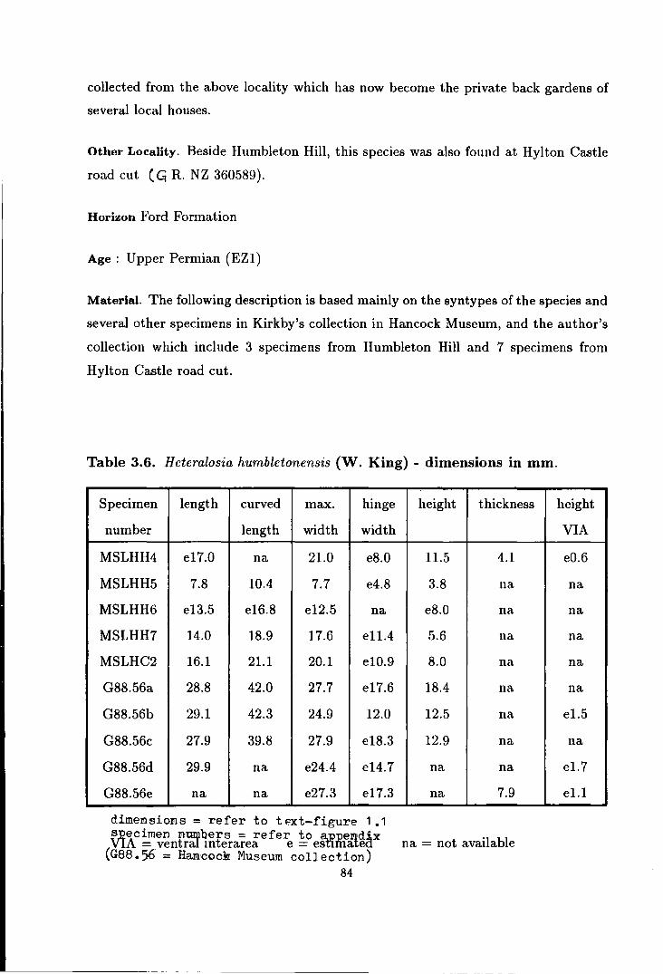
collected from the above locality which has now become the private back gardens of
several local houses.
Other Locality. Beside Humbleton Hill, this species was also found at Hylton Castle
road cut ( ~ R. NZ 360589).
Horizon Ford Formation
Age: Upper Permian (EZ1)
Material. The following description is based mainly on the syntypes of the species and
several other specimens in Kirkby's collection in Hancock Museum, and the author's
collection which include 3 specimens from Humbleton Hill and 7 specimens from
Hylton Castle road cut.
Table 3.6. Heteralosia humbletonensis (W. King) - dimensions in mm.
Specimen length curved max. hinge
number length width width
MSLHH4 e17.0 na 21.0 e8.0
MSLHH5 7.8 10.4 7.7 e4.8
MSLHH6 e13.5 e16.8 e12.5 na
MSLHH7 14.0 18.9 17.6 e11.4
MSLHC2 16.1 21.1 20.1 e10.9
G88.56a 28.8 42.0 27.7 e17.6
G88.56b 29.1 42.3 24.9 12.0
G88.56c 27.9 39.8 27.9 e18.3
G88.56d 29.9 na e24.4 e14.7
G88.56e na na e27.3 e17.3
dimensioms = refer to text-figure 1 .1 !J~ecimen nwvllers = refer to a.ppem<dix VIA = ventral mterarea e = esnmatea
(G88.56 = Ha.x:J:cock! Museum collection) 84
height thickness
11.5 4.1
3.8 na
e8.0 na
5.6 na
8.0 na
18.4 na
12.5 na
12.9 na
na na
na 7.9
na = not available
height
VIA
e0.6
na
na
na
na
na
e1.5
na
el.7
el.1

c
-1mm
, 5mm
E D
-5mm
Text-figure 3.6. Camera Iucida reP,roduction of Heteralosia h·ambletonensis (King). A,B - Posterior and lateral views of syl}type no D318B (UCG collection). C - Interior of ventral interaren of syutype no D318C. (UCG collection) showing a pair of deep sockets (darken). D,E- ventral and lateral view of specimen no G88.&Ga (HM collection).
85

Description. The shell is small to medium in size, sometimes reaching a maximum
length of more than 30 mm. The shell width is generally narrower than its length
although in some flattened forms these dimensions are nearly the same. The maximum
width of the shell is acquired at the posterior half of the shell. The shell outline varies
considerably in shape from elongately subelliptical to vase shaped. The flatter forms
are usually broad with a more or less subcircular plan outline.
The ventral valve is moderate to strongly convex with trail extensively developed
especially anteriorly and on the auricular region. The anterior expansion sometimes to the
forms a broad sinus dueL sudden increase in curvature of its lateral margin. The
cross profile of the posterior region is strongly arched. The umbo is strongly inflated,
overhanging and slightly curved, sometimes slightly dented or flattened by vestiges
of cicatrix of attachment immediately posterior to the ventral interarea. The ventral
interarea is apsacline, low and narrow with its full width often less than two thirds of
the maximum shell width. The delthyrium is covered by a very small weakly raised
pseudodeltidium.
The exterior of the shell is covered by distant recumbent and adpressed spines
which sometimes leave deep grooves that disect the shell into several radial lobes.
Posteriorly, beside the recumbent and adpressed spines, there are few erect and pos
teriorly directed clasping spines. The spines are hollow and very small in size ( di
ameter less than 1 mm). Traces of radial costae and concentric growth lamellae are
weak to obscurely developed, but occasional thickened growth lamellae or rugae are
developed.
The dorsal valve is moderate to strongly concave, sub-rounded with even smooth
curvature during the younger stages as shown by its growth lamellae. The curvature
of the valve increases in the older stages, though geniculation is not very obvious. In
general, the curvature of the dorsal valve follows that of the ventral valve quite closely.
The exterior of the shell is radially costate and concentrically lamellose or occasionally
rugate. The interarea on the dorsal valve is very low and almost imperceptible.
Interior of the dorsal valve is provided with a small shafted bilobed cardinal
process with a small socket at its base. The adductor muscle platform is small and
86
slightly raised. The brachial impression is small compared to the size of the shell.
Its lateral margin is at a distance from the lateral shell margin while anteriorly it
extends only slightly anterior to the mid-length. The median septum is low and
short, extending to less than half the shell length. Exterior of the dorsal valve is
similarly ornamented to the ventral valve except that it is spineless.
Discussion. The fact that the cicatrix of attachment is very small or absent suggests
that H. humbletonensis adopted a free mode of living early in its ontogeny. During
its ontogeny, steady increase in the shell gibbosity is observed. The dorsal valve
becomes slightly geniculated and the shape of the shell becomes varied at a late stage
in ontogeny, probably a characteristic of senility.
Comparison with H. morrisiana has been made earlier in this chapter. Although
the flat and broad form of H. humbletonensis seems to resemble the shape of H. mor
risiana, it differs from H. morrisiana in its more spinose ventral valve and smoother
umbo without cicatrix of attachment. Other Zechstein species of Heteralosia are
very small compared to H. humbletonensis, except H. aycliffensis n. sp. However,
H. aycliffensis can be distinguished from H. humbletonensis by its denser, but less
adpressed spines, larger cicatrix of attachment, smoother growth lines and stronger
radial costae. The occasional development of the spine grooves, might lead one to
confuse the juveniles or the younger individuals of H. humbletonensis with H. hylto
nenszs n. sp., but H. hyltonensis is characterised by its small size and asymmetrical
shape.
HETERALOSIA HYLTONENSIS new species
(Text-figure 3. 7, Plate 5, figures 1-13).
Named after Hylton Castle, the locality at which this species is found.
Diagnosis. Small, asymmetrical, transversely sub-elliptical to sub-circular shaped
H eteralosia with small to minute cicatrix of attachment and strongly ad pressed body
spmes. Some of the posterior body spines and the marginal spines are rhizoid in
87

nature. Ventral valve strongly convex with broad unevenly plicated trail and lamellose
exterior. Ventral interarea low and narrow, asymmetrical in shape. Dorsal valve weak
to moderately concave with slightly geniculated margin, low interarea and lamellose
and costate exterior. Cardinal process with stout shaft. Adductor muscle platform
moderately elevated and pear shaped. Median septum short.
Type specimen. Specimens no . ..,.MSLHC3, MSLHC4, MSLHC5, MSLHC6, MSLHC7
and MSLH C8 are chosen as a series of para types of H eteralosia hyltonensis, showing
various morphological features of the species.
Type Locality. This species is only known from Hylton Castle road cut (GR. NZ
360589) and this locality should be named as the type locality of this species.
Material. The following description of the species is based on 59 specimens collected
by the present author from the type locality mentioned above. Most of the specimens
are preserved with the original shell material while a few are preserved in form of
moulds.
Description. The shell is small in size with the largest individuals having a length of
about 15mm, and characterised by strongly asymmetrical shell growth. The shell is
variable in shape from asymmetrical transversely sub-elliptical to sub-circular. The
shell width is usually greater than its length with the maximum width situated at
anterior half of the shell. The hinge line is also asymmetrical in length and the beak
is usually pointed toward the longer hinge line.
The ventral valve is strongly convex with maximum convexity at the umbonal
region. The confexity of the valve is reduced toward the antero-lateral margin which
shows evidence of strong growth. The posterior margin is provided with a poorly
developed auricle. The anterior and lateral margins are usually broadly plicated.
This plication is produced when the adpressed spines undercut the shell anteriorly to
form several profound grooves at a distance away from one another. Generally, these
grooves are bent following the twisted direction of the major shell growth. The umbo
88

Table 3. 7. H eteralosia hyltonensis new species - dimensions in mm.
Specimen length curved plane max. hinge hinge height
number length curved width width width
{max.) length {HW1) {HW2)
MSLHC3 11.5 18.2 14.6 15.1 3.9 6.5 6.8
MSLHC4 e11.9 e18.7 e15.1 e14.3 e3.9 na 7.5
MSLHC5a 11.4 16.0 13.6 12.8 na 5.2 6.2
MSLHC5b 9.6 e16.0 12.5 11.9 4.4 na 5.4
MSLHC6 7.8 14.7 12.4 e10.8 na 4.3 5.1
MSLHC10 6.8 e9.5 e7.5 8.5 e2.7 e3.2 3.4
MSLHC12a 3.9 e5.5 e4.2 4.8 na na 1.8
MSLHC12b 4.9 6.5 e5.1 5.5 1.5 1.8 1.5
VIA = ventral interarea e = estimated na = not available specimen num'l!Yers = refer to appendix
(see text-figure 3.7E,F for details on measured parameters)
is small, but usually broad and pointed toward the longer side of the hinge line which
is also the major growth direction of the shell. The cicatrix of attachment small,
flat or slightly concave and asymmetrically positioned on the umbo. Sometimes the
cicatrix is so small as to be hardly observable.
The ventral sudace is lightly lamellose or smooth, wrinkled posteriorly. The
valve is furnished with strongly adpressed to sub-erect body spines which are sparsely
distributed over the convex part of the valve. These spines do not show radial growth,
but are inclined toward the major growth direction of the shell. Posteriorly, some of
the body spines are erect and posteriorly directed with their ends rhizoid in nature.
The marginal spines are made of rhizoid spines.
The ventral interarea is low and narrow with slightly asymmetrical triangular
shape. The surface is transversely striated and the delthyrium is covered by a small
and pseudodeltidium. Adductor muscle platform is narrow and short. Full details of
89

the internal morphology are not known.
The dorsal valve is weak to moderately concave with slightly geniculated margin
stronger at the lesser growing side. The umbo is small and weakly raised. The surface
is prominently concentrically lamellose and finely costate. The dorsal interarea is
very low with small chilidium. The interior of the dorsal valve is provided with a
stout cardinal process shaft supported by a strong median ridge and a pair of lateral
ridges which make an acute angle between them. The adductor muscle platform is
pear shaped and is moderately elevated above the shell plane. The muscle scar is
concentrically striated. The median septum is low and extends to the mid-length of
the valve. Other internal features are not known.
Remarks. The asymmetrical shape of the shell and the position of the cicatrix of
attachment clearly indicate that this species does not grow in a normal anterior
direction, but laterally instead. The stronger growth of the shell at one lateral margin,
creates a siphon-like structure at this margin. This adopted molluscan feature might
have resulted from the very crowded niche in which this species lives. H. hyltonensis
lived in a confined pockets within or under overgrown algal stromatolites or bryozoan
colonies, where a type of siphonic apparatus would be beneficial for an immobile
species to get fresh nutrient from the sea water. The reason for the development of
a single lateral siphon in this brachiopod is not fully understood. It is possible that
the attachment strategy employed by the species does not permit the re-positioning
of the shell during ontogeny when the growth space was blocked. The attachment
strategy of H. hyltonensis seems to be reinforced with the development of rhizoid
spines at its posterior.
Comparison. The development of rhizoid spines and the strongly asymmetrical shell
growth of H. hyltonensis is unique and have not been observed in any other Het
eralosia. Apart from these, H. hyltonensis resembles some H. humbletonensis in its
grooved trail and resembles H. seahamensis in its small glob bose shell. However, H.
hyltonensis can be distinguished from H. humbletonensis by its small size and asym
metrical shape, while it differs from H. seahamensis in its asymmetrical shape and
its grooved trail.
90

A 5mm
c
D
~Oih hw2 hw1
F
Text-figure 3.7. Reconstruction of Heteralosia hyltonensis new species. A- Exterior of ventral valve (plan view). B- Exterior of ventral valve (posterior view). C - Exterior of dorsal valve. D - Lateral profile. E,F - Measured parameters in table 3. 7 [I = length clp = plane curved length mw = maximum width hwl = shorter hinge width hw2 = longer hinge width h = height].
A B
---5mm D c
Text-figure 3.8. Reconstruction of Heterolosia seahamensis new species. A- Exterior of ventral valve. B,C- Exterior and Interior of dorsal valve. D- Lateral profile.

HETERALOSIA SEAHAMENSIS new species
(Text-figure 3.8, Plate 5, figures 14-27).
Named after Seaham Harbour, peripheral to which the two major localities for this
species are sited.
Synonymy; 1858 Strophalosia lamellosa (not-Geinitz) var. morrisiana (W. King); Davidson, pl.
3, fig. 32, ?33.
Diagnosis. Small globose H eteralosia with poorly defined cicatrix of attachment.
Ventral valve strongly convex without sulcus or plicae. Ventral interarea high with
small pseudodeltidium. Ventral surface lightly lamellose with sub-erect body spines.
Dorsal valve moderately concave with slightly geniculated margin. Dorsal interarea
low. Dorsal surface concentrically lamellose and finely radially costate. Cardinal
process on small shaft. Teeth prominent. Adductor muscle platform moderately
elevated with concentrically striated scar. Median septum short. Brachial impressions
wide.
Type specimens. Specimens no MSLBH19, MSLBH20, MSLBH21, MSLBH22 and
MSLBH23 are chosen as a series of paratypes representing various morphological
features of Heteralosia seahamensis.
Type locality. The type specimens named above came from Beacon Hill railway cut
(CR. NZ 442454 ), collected during the tenure of this research. This locality is
chosen because of its availability for future collecting. Although a large collection
of this species in Kirkby's collection in Hancock Museum came from Dalton-le-Dale,
current research at Dalton-le-Dale failed to encounter any specimens of this species.
Other localities. Dalton-le-Dale (Kirkby's collection- probably the same as W. King's
locality at around CR. NZ 40884744); Tunstall Hill old trench,' (GR. NZ 397541);
temporary sewage tunnel at South Tunstall Hill.
Material. 49 specimens from Beacon Hill, 3 specimens from Tunstall Hill temporary
92
------------------------- ---- -~

tunnel and 1 specimen from Thnstall Hill old trench in my own collection. Most of
the specimens are preserved in form of moulds and casts while a few are preserved
with patches of original shell.
Table 3.8. Heteralosia seahamensis new species - dimensions in mm.
Specimen length curved max. hinge height thickness
number length width width
MSLBH19 e12.3 e17.7 e12.5 6.9 5.9 3.1
MSLBH20a 12.2 18.1 13.1 9.9 6.6 na
MSLBH21 11.2 17.1 e12.3 7.0 5.2 na
MSLBH22 9.0 e10.9 9.1 6.7 4.0 na
MSLBH23 8.8 10.0 9.3 e5.4 3.3 na
MSLBH24 10.7 na 10.9 5.4 na na
MSLBH25 9.6 na 10.0 9.3 na na
MSLBH26 e11.2 na e13.0 e8.5 na na
MSLBH28a 12.1 17.4 13.1 e9.7 5.8 na
MSLBH28b 11.6 17.6 12.6 e8.7 7.1 na
MSLBH28c e4.9 6.3 5.3 3.9 1.7 na
MSLBH28d 9.2 13.4 11.2 8.7 5.8 na
dimensions= refer to text-figure 1.1 specimen numbers = refer to appendix
VIA = ventral interarea e = estimated na = not available
height
VIA
1.1
na
na
na
na
1.3
0.9
na
na
na
na
na
Description. The shell is small and rarely reaches 12 mm in dimensions. It is globose
with almost circular outline but sometimes the shell outline is slightly asymmetrical
in shape. The maximum shell width is situated at about the mid-length.
The ventral valve is strongly convex with maximum convexity normally situated
posteriorly. There is no sulcus and no auricle. The beak varies from narrow to broad
with overhanging and slightly curved umbo. Narrower beaks are commonly more
93

inflated than the broader ones. The cicatrix of attachment is usually absent but
when present it is small in size with sub-circular outline and flat profile. The umbo is
not strongly incurved so that the ventral interarea is left open. The ventral interarea
is quite high, but narrow with the hinge width about half to two thirds the maximum
shell width. The pseudodeltidium is small and weakly raised.
The ventral surface is smooth or covered by fine concentric striae and sometimes
lamellae or rugae. Traces of radial costae are occasionally seen. The ventral valve
is furnished with sparse randomly distributed body spines and a row of posteriorly
directed clasping marginal spines. Posteriorly, the body spines are divided into the
recumbent to adpressed, and the erect posteriorly directed spines, while anteriorly,
only suberect spines prevailed. The adpressed body spines sometimes leave radial
grooves on the shell at the umbonal region. The spines are small and not extended
very far beyond the shell margin. Interior of the ventral valve is provided with a
pair of strong teeth and a short, but broad adductor muscle platform. The adductor
muscle platform is moderately elevated and has a slightly flabellate shape, divided
medianly by a well developed median ridge. The adductor muscle scar is smooth.
Other internal features are not known.
The dorsal valve is moderately concave with slightly geniculated margin in the
adult. The median fold is absent. Posteriorly, the umbo is slightly raised. The
interarea is low and narrow with small chilidium. The dorsal surface is concentrically
lamellose and finely costate. Interior of the dorsal valve is provided with a small, short
shafted cardinal process with bilobed myophore. The cardinal process is supported
by a pair of socket ridges and a low median ridge which is continued anteriorly by
a low median septum to about the mid-length of the valve. The adductor muscle
platform is pear shaped and moderately raised with the scar concentrically striated.
The brachial impressons are wide. The interior surface is smooth, but sometimes it
shows traces of radial striae and rare endonodes. The interior of the anterior margin
is rimmed by numerous marginally pointed endospines.
Discussion. Not much ontogenic detail is known in H. seahamensis owing to the lack
of completely preserved juveniles. However, it can be generalised that the globosity of
94
the shell and the geniculation of the dorsal valve is achieved during the adulthood of
the species. Regarding the attachment, it seems that the majority of H. seahamensis
were only attached to other objects during their juvenile stages and by clasping spines
only. A few individuals, however, are attached to the substratum for a much longer
period of their lives. These attached forms usually show more irregular shell growth
with small cicatrix on the umbo, and are often asymmtrical in its shape.
H. seahamensis closely resembles H. hyltonensis and the Mississippian H. orbic
ulata (Hinchey & Ray) from North America in its smaller size and globose shell.
However, it differs from the H. orbiculata in its poorly developed cicatrix of attach
ment and the presence of radially costate ornament. The differences between H.
seahamensis and H. hyltonensis has been outlined earlier in this thesis. H. seahamen
sis can be distinguished from other H eteralosia by its very small size, globose shell
with almost circular outline and its less adpressed body spines.
H. seahamensis has probably been confused by Davidson (1858) with the juveniles
or younger individuals of H. morrisiana because of its very small size. The genicula
tion of the dorsal valve and the globbosity of the ventral valve in very small individuals
of this species indicate that they are adults and not juveniles of H. morrisiana, and
that they belong to a separate species of H eteralosia which is characteristically very
small in size. The juvenile or younger individuals of H. morrisiana are much flatter
than H. seahamensis at the same size.
HETERALOSIA AYCLIFFENSIS new species
(Text-figure 3.9A,B,C, Plate 4, figures 12-22).
Named after Newton Aycliffe, the nearest town to the cited type locality of the species.
Diagnosis. Medium size Heteralosia with radial costae and weak growth lines on both
valves. Ventral valve moderately convex with posterior margin truncated by cicatrix
of attachment. Fine and long recumbent to adpressed body spines numerous. Ventral
interarea low, trapezoidal in shape with small pseudodeltidium. Dorsal valve weakly
95

concave with raised umbo and low interarea.
Type specimens. Specimens no MSLWQ4, MSLWQ5, MSLWQ6, MSLWQ7 and MSLWQ8
are chosen as a series of para types of H eteralosia aycliffensis, showing various mor
phological features of the species.
Type locality. Old Town (=White) Quarry (GR. NZ 257245), where most of the
specimens studied were collected is chosen as the type locality of this species.
Other localities include Raisby Hill Quarry ·(GR. NZ 346353) and East Thickley
Quarry (GR. NZ 24082564).
Horizon. Raisby Formation.
Age. Upper Permian (EZl).
Material. 43 specimens from Old Town (=White) Quarry; 10 specimens from Raisby
Hill Quarry; 5 specimens from East Thickley Quarry.
Description. The shell is of medium size with the length of the largest individual
around 30 mm. It has a sub-elliptical to pear shaped outline with truncated poste
rior. The shell length is normally greater than the maximum shell width, although
deformed specimens are sometimes vice versa.
The ventral valve is weakly to moderately convex longitudinally, but rather stronger
in convexity transversely. The posterior margin of the ventral valve is truncated by a
small sub-circular to transversely sub-elliptical cicatrix of attachment which is slightly
concave or flat in profile. The sulcus and auricle are absent. The ventral interarea
is low and narrow, trapezoidal in shape with small pseudodeltidium weakly raised
above the plane of the interarea. Traces of a pair of small teeth are sometime seen at
the base of the pseudodeltidium.
The growth lines of the ventral valve are obscure, but fine radial costae are usually
96

9
c -smm
F 5mm
0 E
Text-figure 3.9. Reconstruction of Hetem/osia aycliffen.~is new species (figure A to C) and Heteralosia? quarringtonensis new species (figure D to F). A.B - Exterior of ventral and dorsal valves. C - Camera Iucida reproduction of specimen no MSLWQlO showing an overgrown cicatrix. D - Exterior of ventral valve. E.F - Exterior and interior of dorsal valve.
97

Table 3.9. Heteralosia aycliffensis new species - dimensions in mm.
Specimen length curved max. hinge
number length width width
MSLWQ4 17.2 21.9 e16.4 na
MSLWQ6 15.7 18.8 e15.6 e9.1
MSLWQ7a* 20.0 na 17.0 7.0
MSLWQ8 4.8 5.2 5.6 3.1
MSLWQ9 e16.2 e19.0 e16.0 e10.5
MSLWQlla 5.8 na 7.0 4.0
MSLETQ2* 26.7 28.8 23.4 e7.8
dJ.mensioms = refer to text-f'igure 1 .1 spec,imen numbers = refer to appendix
height
7.3
6.0
na
1.3
5.2
na
e3.0
cicatrix
length
na
2.0
na
1.5
5.0+
e3.5
na
VIA = ventral interarea e = estimated na = not available
* badly deformed specimens + measured from dorsal valve.
height
VIA
na
na
0.8
na
1.4
e0.5
na
well developed. The exterior shell surface is covered by fine recumbent and adpressed
spines, but no spine groove is developed. The spines are long and sometime extend
more than 20 mm beyond the shell margin. A few posteriorly directed clasping spines
are also developed in some specimens.
The dorsal valve is weakly concave with semi-circular umbo raised on its posterior
margin. The diameter of this raised umbo ranges from half to two thirds of the hinge
width. There is no median fold. The interarea is low with small and weakly raised
chilidium. The dorsal surface is radially costate with occasional traces of faint growth
striae. The internal morphology of this species is unknown.
Discussion. This is the most ill-preserved species of Heteralosia in the Durham Per
mian. The currently available material, however, is sufficient to show that the external
morphology is different from all known species of this genus. Therefore, it is without
question that this material, as described above, should be placed in a new species
of Heteralosia. Comparison with other species of Heteralosia has already been made
earlier in this thesis.
98
H. aycliffensis might be confused with C. langtonensis, especially when the spines
and the dorsal valve are missing. In this case H. aycliffensis can only be distinguished
from C. langtonensis by its larger size, slightly smaller cicatrix of attachment and
smaller number of spine bases.
Genus CRASPEDALOSIA Muir-wood & Cooper 1960
Synonymy; 1960 Craspedalosia Muir-Wood & Cooper, p. 82-83. 1965 Craspedalosia Muir-Wood & Cooper; Muir-Wood, p. 451.
Diagnosis. Small to medium size heteralosiinid with sub-pentagonal to sub-circular
shell outline. Ventral valve strongly convex with no sulcus. Umbo usually truncated
by cicatrix of attachment. Ventral interarea variable in height with small weakly
raised pseudodeltidium. Ventral surface lightly lamellose with two series of spines pos
teriorly and specialized spines anteriorly. Dorsal valve with raised umbo, low interarea
and squamose growth lamellae. Teeth prominent. Cardinal process with bilobed or
trilobed myophore. Adductor muscle platform moderately elevated. Brachial impres
sions moderate to large in size. Median septum short. Dorsal valve with endospinose
rim separated from geniculated margin by sulcus.
Discussion. The name Craspedalosia was suggested by Muir-Wood & Cooper {1960)
to highlight the presence of the endospinose rim in the dorsal valve of this genus. It
is well known that the internal features of the productidinids are unreliable, partly
due to their ubiquity and partly due to their variable nature. It is very difficult to
obtain the internal details of this genus, because individuals are usually preserved
complete with the original shell and with both valves articulated. For this reason,
it is not practicable to determine the genus Craspedalosia just by the meaning of its
name. It is therefore, very important that other diagnostic features are considered in
recognising the genus.
The genus Craspedalosia has been recognised in the past as heteralosiinid with
strong squamose lamellae or rugae on its dorsal valve. This feature is not related to
99
the name Craspedalosia. In past literatures, the sub-division of Craspedalosia has been
based on the general shape and the nature of the adult spines. C. pulchella (Dunbar)
is distinguished from the type-species C. lamellosa (Geinitz) by its triangular or wedge
shaped and its two series of body spines in the adult.
The similarity of the dorsal valve of new species C. langtonensis with the other
two species of Craspedalosia has led the author to assign the new species to this genus.
With the new species the genus Craspedalosia is now represented in Britain by two
species, C. lamellas a ( Geinitz) and C. langtonensis n. sp. Another uncertain species
of the genus is also recorded in this thesis.
Type species. Orthothrix lamellosus Geinitz, 1848, p. 18, pl. 5, fig. 16-26.
Other species. Strophalosia pulchella Dunbar and Craspedalosia langtonensis n. sp.
CRASPEDALOSIA LANGTONENSIS new species
(Text-figure 3.10B-E, Plate 6, Figures 1-10).
Named after Langton Village in which the type locality is sited.
Diagnosis. Small to medium sized, sub-circular to sub-triangular shaped Craspedalosia.
Ventral valve moderately convex with large cicatrix of attachment and truncated
umbo. Ventral interarea high and narrow with trapezoidal outline. Ventral surface
with closely spaced spine bases. Dorsal valve with large and high umbo and very low
100

interarea. Dorsal surface with radial costae and squamose concentric growth rugae
anteriorly. Cardinal process small and shafted with prominent oblique lateral ridges.
Adductor muscle platform weakly elevated. Median septum not extended beyond the
first squamose rugae.
Type specimens. Specimens no MSLLQ1, MSLLQ2, MSLLQ3, MSLLQ4, MSLLQ5
and MSLLQ6 are chosen as a series of paratypes of Craspedalosia langtonensis, show
ing various morphological features of the species.
Type locality. The type specimens above are collected from Hobgate ( = Langton)
Quarry, south of Langton Village (CR. NZ 165189), near Barnard Castle.
Other Localities. Several specimens obtained from boreholes such as Fishburn BH,
Welbeck Abbey BH, Sutton BH and Wressell BH which show close affinity to C.
langtonensis are tentatively included in this species.
Horizon. Raisby Formation and Cadeby Formation.
Age. Upper Permian (EZ1).
Material. The following description is mainly based on 133 specimens in the author's
collection from the type locality. Also observed are several specimens from Hobgate
(=Langton) Quarry, Fishburn BH, Welbeck Abbey BH, Sutton BH and Wressell BH
in the BGS Museum collection.
Description. The shell is small to medium in size with the largest individuals about
20 mm in length. It is sub-circular to sub-triangular in outline. The maximum width
is reached at about its mid-length. The profile is often flattened with strong influence
of the large cicatrix of attachment at the posterior. The visceral cavity is very narrow
because of the development of a large cicatrix of attachment on the ventral valve and
the strong undulation of the dorsal valve.
The ventral valve is weakly to moderately convex, surrounding the very large and
101

---smm
E
B c ---smm --smm
---- - , ,
H - .. - . -
F G
Text-figure 3.10. Reconstruction of British species of Cra.~pcdalosia. A - Dorsal view of Craspedalosia lamellosa (Geinitz)
sh
B - E : Craspedalo.4ia langtonensis new species; B - Exterior of ventral valve showing large cicatrix of attachment (crossed). B- Exterior of dorsal valve. D- Lateral profile. E- Camera Iucida reproduction of juvenile specimen no MSLLQ5 attached to a spine of Horridonia (sh). F- H : Craspedalosia sp. A (camera Iucida reproduction of specimen no YEF 2367a.bBGS collection): F .G - Internal mould of ventral and dorsal valves. H - External mould of dorsal valve.
102

Table 3.10. Craspedalosia langtonensis new species - dimensions in mm.
Specimen length max. hinge height cicatrix
number width width length
MSLLQl e14.3 el6.2 e8.8 na na
MSLLQ2 e11.3 12.7 na na 2.7+
MSLLQ4 e8.9 e9.9 6.8 na 2.8+
MSLLQ5 4.4 6.0 e4.8 el.5 na
MSLLQ6 e5.3 6.7 4.5 2.1 na
MSLLQ7 e8.3 e8.3 e4.3 2.6 3.1
MSLLQ9 e12.8 12.9 e9.9 na na
MSLLQ10 e12.3 e12.3 e9.0 na 3.6+
dimensions= refer to text-figure 1.1 ~P-ecimen numbers = refer to aP.pendix VIA = ventral interarea e = estimated na = not available
+ measured from impression of cicatrix on the dorsal valve
height
1.6
na
na
na
na
na
na
na
smoothly concave cicatrix of attachment. The cicatrix of attachment usually covers a
quarter to one thirds of the shell length, although in some individuals it is relatively
smaller. It is sub-circular to semi-circular in outline with truncated posterior. The
beak does not seem to be developed owing to the development of the large cicatrix. A
few specimens, however, show a narrow and weakly inflated beak with :flat or slightly
concave attachment scar of varying shape, anterior to the umbo. The ventral interarea
is high and narrow with trapezoidal shape and small weakly raised pseudodeltidium.
The exterior of the ventral valve is fine to moderately lamellose and weakly costate
with some closely spaced spine bases whose points of origin suggest that they might
range from erect to recumbent. A few spines were preserved in matrix a short distant
from the lateral and anterior margins in some specimens and indicate the presence
of recumbent spines in this species. These spines extend at a very low angle to the
plane of the ventral valve. The marginal and clasping spines are not preserved.
The dorsal valve is sub-circular to sub-elliptical in outline with large sub-circular
103
to semi-circular convex umbo, slightly smaller in size than the cicatrix of attachment.
The umbo is rimmed anteriorly by several strong squamose growth rugae which are
consistently developed toward the valve margin. These rugae give the valve a pro
nounced concentrically undulating surface. Beside these rugae the dorsal surface also
shows traces of fine concentric lamellae and radial costae.
The dorsal interarea is very low and often compressed against the ventral inter
area. The width of the hinge line is about two thirds the maximum width of the shell.
The cardinal process has a small shaft, buttressed only by a pair of lateral ridges at an
angle of 110° to 130° to each other. The adductor muscle platform is weakly elevated
and has a radially striated surface and pear shape outline. The median septum is
prominent but only extends to the first growth rugae, anteriorly.
Remarks. C. langtonensis shows an interesting ontogenic development (see text-figure
3.10D). It begins its early stage with a convexo-concave profile due to cementation
of its umbo to smooth objects, probably rounded pebbles, plant fragments or on
Horridonia or its spines (see Text-figure 3.10E). The normal productidinid profile is
only produced half way through its life where at this stage the craspedalosian features
begin to develop. At this stage, the dorsal valve begins to form undulations, owing
to the development of consistent strong rugae which might correspond to a seasonal
reduction of the shell growth. However, the growth reduction is not shown on the
ventral valve. Unfortunately, owing to the absence of the original shell substance, it
has not been possible to prove this assumption of growth behavior. The consistent
truncation of the umbo in this species suggests that the shell is attached to the large
host throughout its whole life. A few specimens, however, have a smaller attachment
scar which is positioned a little anterior to the tip of the umbo, suggesting either that
the host had a rather smaller surface area or perhaps these individuals might have
adopted a free mode of life during the adult stage.
Comparison. Although the spines are not preserved in the available adults of this
species, the spine bases show a roughly similar spine density with all other Craspedalosia.
This species can easily be distinguished from other Craspedalosia by its large cicatrix
of attachment, strongly truncated ventral umbo, trapezoidal shaped ventral interarea,
104
-----------------

large smoothly convex dorsal umbo and costate shell.
It is interesting that the incompletely preserved speCimens and younger indi
viduals of this species have been confused by previous authors with H. morrisiana.
This has happened when only a few incompletely preserved specimens were obtained
from various boreholes, from the Cadeby Formation in Nottinghamshire and from
the Raisby Formation in County Durham. At a glance, these specimens in which the
anterior half is missing or not developed look very similar to H. morrisiana, but they
differ from H. morrisiana in the larger cicatrix of attachment and the much denser
spines. In fact, it was not until I obtained a large collection of C. langtonensis from
Langton ( =Hobgate) Quarry, including several complete casts and moulds of dorsal
and ventral valves of adult individuals, that I decided that this species should belong
to the genus Craspedalosia.
CRASPEDALOSIA LAMELLOSA (GEINITZ)
(Text-figure 3.10A, Plate 6, figures 11-14).
Synonymy; 1848 Orthothrix lamellosus Geinitz, p. 14, pl. 5, fig 16-26, 33. 1861 Strophalosia lamellosa (Geinitz); Geinitz, p. 97, pl. 18, fig. 1-7. 1871 Productus lamellosus (Geinitz); Geinitz, p. 640, pl 60, fig 21. 1921 Strophalosia lamellosa (Geinitz); Trechmann, p. 541, fig. 2b. 1937 Strophalosia lamellosa (geinitz); Malzahn, p. 52, pl. 2, fig. 16-17. 1960 Craspedalosia lamellosa (Geinitz); Muir-Wood & Cooper, p. 82, pl. 6, fig. 1-9.
Material. Specimens studied include 1 incomplete specimen of my own collection from
Old Town (=White) Quarry, and 5 specimens from Mattersey BH and 1 specimen
from Barnby Moore BH in the BGS Museum collection. Trechmann's collections in
British Museum (Natural History) have also been observed.
105

Table 3.11. Craspedalosia lamellosa (Geinitz) - dimensions in mm.
Specimen length curved max. hinge height thickness
number length width width
YPF4168 e22.8 na e22.9 el3.8 e8.4 7.8
BN8223d 20.2 23.2 18.2 na 7.7 4.7
BN8223i 20.0 24.9 22.4 e15.6 9.4 4.8
dimensions= refer to text-figure 1 .. 1 (BGS collection)
VIA = ventral interarea e = estimated na = not available
height
VIA
2.8
na
2.4
Discussion. C. lamellosa was first correctly identified in the British Permian by Trech
mann in 1921. It is a rare species in Britain and it is restricted to the lower part of the
Raisby Formation and Cadeby Formation, commonly associated with the strophalosi
inid S. goldfussi. Both of these species are known to be abundant in the East German
Zechstein. As suggested for S. goldfussi, C. lamellosa was also probably carried to
the British Zechstein Sea during its larval stage by westward storm currents from the
Eastern Zechstein.
British C. lamellosa are generally smaller in size as compared to those from East
Germany. They are commonly rather circular in shape with slightly overhanged umbo.
The sub-elliptical and sub-triangular forms which are common in East Germany are
less common in Britain. Other details of the surface ornamentaion of both valves are
identical between the British and the German C. lamellosa. They both have ventral
valves with a mass of tangled vermiform spines which are posteriorly directed and
often still attached to the spines or shell of H orridonia. The dorsal valve is strongly
squamose, except at the slightly raised umbo where it is only lightly lamellose.
Comparison with C. langtonensis has already been outlined in the previous sec
tion. Regarding C. lamellosa and S. goldfussi, it is fortunate that both species are
almost always completely preserved, both in Germany and in Britain. Lack of ex
ternal detail normally results from the process of weathering or preparation of the
samples. Since the weathering process is not very severe in the present climate in
which this species occurs and the preparational damage can be avoided, these two
106
species can easily be distinguished by the spinosity of their dorsal valves. S. goldfussi
is a strophalosiinid with its dorsal valve spinose, while C. lamellosa, as a heteralosi
inid has no spines on its dorsal valve. It is noteworthy that there are no intermediate
forms between these two species. This evidence strongly supports the sub-division of
the family given earlier in this chapter.
CRASPEDALOSIA sp. A
Text-figure 3.10F-H, Plate 6, figures 15-19.
Material. 7 incomplete specimens from Hylton Castle temporary sewage trench in
Dr. Denys Smith's collection, in BGS Museum collection (no. YEF 2367D).
Table 3.12. Craspedalosia sp. A- dimensions in mm.
Specimen length curved max. hinge height height
number length width width VIA
YEF2367Da e15.0 na 14.2 e8.0 4.0 na
YEF2367Dc 13.1 15.0 13.4 e10.2 3.8 e1.6
YEF2367Dd e13.8 e17.7 e16.0 e9.2 e3.5 e2.2
YEF2367De e12.8 e16.7 e16.0 e8.6 e3.5 e2.0
YEF2367Df e18.5 na e17.5 na e5.0 na
dimensions= refer to text-figure 1.1
VIA = ventral interarea e = estimated na = not available
Description. The shell is small in size with its outline almost conical or pear shaped.
The ventral valve is weakly to moderately convex with flattened beak and sharply
pointed umbo. Sometimes a poorly defined sulcus is also developed. The ventral
surface is lamellose or occasionally rugate. The ventral interarea is high but narrow,
the height of which ranges between one thirds to half the shell length. It is trigonal in
shape with small weakly raised pseudodeltidium. Ventral adductor muscle platform
107
---------
is moderately raised, narrow and triangular in shape.
The dorsal valve is sub-circular to semi-circular in outline, rather flat or moder
ately concave with slightly geniculated margin. The umbo is slightly raised above the
shell plane. The dorsal surface is covered with strong lamellae and squamose rugae.
The dorsal interarea is low with small chilidium.
Discussion. The limited number of specimens available and lack of completely pre
served specimens do not support the nomination of a specific name or the designation
of a new species to the material described above. Although these specimens were la
belled in the BGS Museum as C. lamellosa, I have a suspicion that they might belong
to a separate new species.
Craspedalosia sp. A differs from H. morrisiana found at the same locality in
its rugate and squamose dorsal valve and its high ventral interarea. In general, the
shape of Craspedalosia sp. A might fall within the range of C. lamellosa or C. pul
chella, except for its smaller size as compared to the other two species. Compared
to C. lamellosa, Craspedalosia sp. A has a higher interarea, while compared to C.
pulchella, Craspedalosia sp. A has no cicatrix of attachment and no radial ribs on the
dorsal valve. The absence of a ventral valve preserved with spines in Craspedalosia
sp. A makes further comparison impossible. More material and some completely
preserved specimens are needed before the material described above can be named
with certainty.
HETERALOSIA? QUARRINGTONENSIS new species
(Text-figure 3.9D-F, Plate 7, figures 1-11).
Named after Quarrington Village in which type locality of the species is sited.
Diagnosis. Medium size productiform heteralosiinid with weakly to moderately con
vex ventral valve, distinct auricles and small pointed umbo. Sulcus and cicatrix of
attachment are absent. Ventral surface smooth or lightly lamellose with rare clasping
108
-------------------------------------------------------------------

marginal spines and sub-erect body spines which are rather recumbent in younger
individuals. lnterarea very low on both valves with parallel outline and minute pseu
dodeltidium and chilidium. Dorsal valve weak to moderately concave with light to
strong growth lamellae. Teeth small. Cardinal process small and short shafted with
poorly developed alveolus. Lateral ridges prominent but short. Median septum short.
Adductor muscle platform pear-shaped with radially striated scar.
Type specimens. Specimens no MSLOQQ3, MSLOQQ4, MSLOQQ5, MSLOQQ6
and MSLOQQ7 are chosen as a series of paratypes of Heteralosia? quarringtonensis,
showing various morphological features of the species.
Type locality. Old Quarrington Quarry (GR. NZ 326381) is selected as the type
locality of this species because the outcrop is extensive and the specimens are better
preserved here. Specimens from Sherburn Hill Quarry are commonly found in a
deformed state. Specimens from the type locality have been previously recorded as
Strophalosia morrisiana by Smith & Francis (1970) and Pattison et al. (1981).
Other localities. Sherburn Hill Quarry
(QR. NZ 24082564) and ?Chourdon Point
Horizon. Raisby Formation.
Age. Upper Permian (EZ1).
(QR. NZ345417), East Thickley Quarry
(GR. NZ 44254650).
Material. 43 specimens from Old Quarrington Quarry; 42 specimens from Sherburn
Hill Quarry; 3 specimens from East Thickley Quarry and 1 specimen from Chourdon
Point.
Description. The shell is small to medium in size with transversely elongated sub
trapezoidal to sub-elliptical in outline. The shell is less than 20 mm in length but
the maximum width varies from slightly wider than the shell length to more than
1.5 times the length. The maximum shell width is at the anterior half of the shell,
normally very close to the mid-length.
The ventral valve is weakly to moderately convex without sulcus or cicatrix of
109

Table 3.13. Heteralosia? quarringtonensis new species - dimensions in mm.
Specimen length curved
number length
MSLOQQ3a 7.8 9.0
MSLOQQ3b 11.5 14.8
MSLOQQ4 9.1 13.0
MSLOQQ6 9.0 10.3
MSLOQQ7 8.2 9.8
MSLSHQ1 7.4 8.1
MSLSHQ2 14.3 17.4
MSLSHQ 11.0 13.4
MSLSHQ 6.6 7.2
dimem,sions = refer to text-figure 1 • 1 specimen numbers = refer to appendix e = estimated
max. hinge height
width width
10.2 e7.7 e2.5
e16.0 e12.5 4.7
12.4 10.0 3.5
12.3 9.7 3.0
11.6 9.2 3.2
10.2 8.0 el.O
20.0 e16.0 e5.0
15.0 e13.2 5.2
8.2 e6.2 el.O
attachment. The umbo is small, flattened and pointed or slightly overhanging and
incurved. The auricle is flat and narrow, triangular in shape and it is better developed
in the less convex specimens. The ventral interarea in very low but wide, almost
parallel in outline with minute weakly raised pseudodeltidium. The ventral surface
is weakly lamellose and distantly spinose. The spines consist of not more than three
pairs of posteriorly directed marginal spines and about ten or lesser sub-erect body
spines. The body spines in younger individuals seem to be more recumbent and
adpressed in nature than in the adults. A pair of small teeth are observed from the
internal mould of the ventral valve.
The dorsal valve is weakly to moderately concave with lightly lamellose surface
which sometimes becomes squamose anteriorly. The umbo is very small and hardly
lifted above the concave dorsal valve plane. The dorsal interarea is very low and wide
with parallel outline and poorly developed chilidium.
Casts from the internal mould of the dorsal valve show that the cardinal process
is short shafted with poorly developed alveolus at its posterior. The myophore is
110
not well preserved. It is knob-like in appearance but probably was originally trilobed
with the lateral lobes broken in the specimens available. The cardinal process is only
buttressed by a pair of strong, but short lateral ridges. The median septum is low,
appears between the muscle platforms, and extends to about the mid-length of the
valve. The adductor muscle platform is small, low and pear-shaped with the scars
radially striated. Other internal features are unknown.
Discussion. The lack of completely preserved specimens prevents the present author
from making a complete description of this species, but distinctive characters shown
by the available material distinguish them from any other heteralosiinid species. The
productiform shape with very rare spines and the reduced interarea, cardinal pro
cess shaft, teeth and sockets and a more strongly lamellose dorsal valve exclude this
species from all other Zechstein heteralosinid species and probably from all known
heteralosiinids elsewhere. Although the external features of the species shows a close
resemblance to the productellids, the presence of a cardinal process shaft and func
tional teeth and socket in this species prove that it belongs to the family Strophalosi
idae. The absence of spines on the dorsal valve place this species in the subfamily
Heteralosiinae and distinguished it from the strophalosiinids genus Eostrophalosia.
STROPHALOSIA? PARVA W. KING
(Plate 7, figures 9-10).
Synonymy; 1850 Strophalosia parva W. King, p. 102, pl. 12, fig. 33
Note. The following description and discussion is based on the syntype of S. parva
(specimen no. D 319 of W. King's collection in the Department of Geology, Univer
sity College of Galway) and several specimens of Kirkby's collection in the Hancock
Museum, all of which were collected from the Humbleton Hill, near Sunderland. The
author only managed to find a single specimen during recent research on this type
locality. The following description is given because it has been discovered that W.
King (1850)'s description is incorrect.
111

Material. The syntype as well as other specimens available for study are only rep
resented by internal moulds of the cicatrix of attachment of the ventral valve with
moulds of several clasping spines. Most of the specimens are attached to the interior
of the ventral valve of H. horrida, and in all cases the shell of the host is not preserved.
Description. The size of the cicatrix of attachment is large with the length in the
syntype reaches 4mm .. The mould of the cicatrix of attachment is usually larger than
its actual size because of its convex nature. The outline of the cicatrix varied from
hemispherical to truncated sub-elliptical shape with straight posterior. The profile of
the cicatrix of attachment is weakly convex with a rather flattened posterior. However,
when the shell is cemented to the convex exterior ventral valve of the host, the cicatrix
is weakly concave or flat in profile. The internal mould is provided with a pair of large
teeth and a moderately raised adductor muscle platform which is divided anteriorly
into two halves by a median furrow. From the elongation of the muscle platform, it
can be interpretted that the cicatrix of attachment is asymmetrically placed on the
beak.
The mould of the rim of the cicatrix of attachment shows that the unattached or
free ventral valve is more convex than the cicatrix.
The spines are numerous and consist of creeping rhizoid spines that emerge from
the matrix lateral and anterior to the cicatrix of attachment. These spines appear
from the matrix at various angles, but follow the curvature of the host immediately
upon reaching it. They spread out randomly over the host, sometimes crossing one
another. Individually, the spines are small in size (0.4- 0.5mm in diameter), but long
and hair-like with the longest spine reaching a length of about 20mm. The posterior
of the cicatrix of attachment is devoid of spines, but some of the body spines are
sometimes posteriorly oriented. Other details are unknown.
Discussion. W. King (1850) gave the following diagnosis of S. parva; Form irregu
larly circular marginally. Large valve somewhat convex; umbone very small and much
impressed; spines numerous, long and closely packed; area small. Rarely exceeding a
quarter of an inch in diameter. In this diagnosis W. King thought that his specimen
(the syntype) represented the complete ventral valve. Detailed study of the syntype
112

clearly shows that the actual ventral valve is partly hidden underneath the matrix,
and the shell exposed is in fact a mould of the complete cicatrix of attachment. As
shown by the point and angle of emergence of the spines, the ventral valve must
extend beneath the matrix for at least another length of the cicatrix. Most of the
cicatrix of attachment is convex because it is attached to a concave surface of the in
terior ventral valve of H. horrida. The convexity of the cicatrix of attachment might
have confused W. King (1850) into thinking that it was the true free ventral valve.
Lack of completely preserved specimens prevent the full description of S?. parva
and the status of the genus of this species remains uncertain. However, the existing
information might be a basis for further investigation of the species. The large teeth
and numerous hair-like spines put this species into the genus Strophalosia. However,
there is no known Strophalosia s. I. or other strophalosiinid which has such a large
cicatrix of attachment. The cicatrix is even larger than that of Heteralosia (possibly
H. humbletonensis) sharing the same host with the syntype of the species; this later
specimen shows smaller teeth and fewer spines than S?. parva. Although the above
features suggest that this species might belong to the genus Strophalosia (if so the
name S. parva should prevail), the possibility that it might belong to another genus in
subfamily Heteralosiinae cannot be denied since the details of the dorsal valve are still
unknown. It is important that full details of the shell are seen before the taxonomic
status of this species is confirmed.
113

Superfamily AULOSTEGACEA Cooper & Grant
Diagnosis. Productidina with no teeth and sockets, interarea confined to ventral valve
with delthyrium commonly closed by elytridium and lophidium.
Discussion. The superfamily Aulostegacea was elevated by Cooper & Grant (1975a)
from the family Aulostegidae which was previously placed under the superfamily
Strophalosiacea by Muir-Wood & Cooper (1960). According to Cooper & Grant
(1975a)the aulostegaceans were derived from the strophalosiaceans by reduction and
elimination of teeth and sockets, modification of pseudodeltidium and elimination of
chilidium, but retaining the ventral interarea with its delthyrium open or closed by
an elytridium and a lophidium. Unfortunately, Cooper & Grant did not give any
formal diagnosis for the new superfamily.
In 1988, Brunton and Mundy elaborated the description of the Superfamily
Aulostegacea and suggested that the relationship of the aulostegids should be re
viewed. A case could be made, in their opinion for many of the genera being
considered as productaceans specialized for living on hard surfaces rather than as
strophalosiaceans which have lost their teeth and sockets. It appears that some of
the features used by Brunton & Mundy are variable, and therefore the above diagno
sis is introduced to avoid any use of variable features and to fit in with the diagnoses
of other productidinid superfamilies.
Cooper & Grant (1975a) listed at least eight families in this superfamily based
on several features. Some of the features however, seem to be adaptational and
not significant at the family level. For instance, the size and shape of the family
Agelesiidae might be acquired for adaptation to special living conditions, while some
of the features of internal morphology which they used to describe this family can
be highly variable, as is often shown by other productidinids (i.e. see Fagerstrom &
Boellstorff 1964, etc.). With limited material available, no attempt can be made here
to reclassify the aulostegacean families, but it is strongly suggested that a single or
few criteria be chosen more systematically for this purpose.
Howseia, which seems to fit well to the above definition of Aulostegacea, should
114

therefore be excluded from other productidinid superfamilies and the lengthy com
parison with Horridonia by many previous authors now seems to be irrelevant. Sim
ilarities between the two genera are rather superficial as compared to the details of
the development of the interarea. Here, Howseia clearly differs from Horridonia in
having an interarea on the ventral valve, while as a productacean, Horridonia has no
interarea. Therefore, Howseia should not be considered as a variety or a stunted form
of H orridonia horrid a.
Family AULOSTEGIDAE Muir-Wood and Cooper
Discussion. Muir-Wood & Cooper (1960) identified the Aulostegidae as a progressive
Strophalosiacea having lost teeth, sockets and chilidium; interarea on pedicle valve
only; delthyrium closed by elytridium (formerly pseudodeltidium) and lophidium;
shell attached by umbo and anchored by spines. Brachial valve interior with trilobate
to quadrilobate cardinal process; alveolus present, adductor scars dendritic, accessory
diductor scars on cardinal process shaft, brachial ridges given off horizontally.
Most of the diagnostic features above have been used by Cooper & Grant (1975a)
to describe the superfamily Aulostegacea. Therefore a new diagnosis is needed to
describe and distinguish the family Aulostegidae from other aulostegaceans. Owing
to lack of time and material available for the present study, the author cannot fulfil
this requirement and for this work the diagnosis of the family Aulostegidae and its
subfamilies is taken from Muir-Wood & Cooper (1960).
Subfamily ECHINOSTEGINAE Muir-wood & Cooper
Diagnosis. Aulostegidae with spines on ventral valve only.
Discussion. Muir-wood & Cooper (1960) gathered in this sub-family eight genera
including Xenosteges, a genus which according to them is very different from other
aulostegids in its specialized attachment strategies and the lack of ornamentation,
as well as the simplicity of the internal morphology. The genus Howseia, revised in
115
--------- - - -----
this thesis closely resembles the genus Xenosteges. They both differ from other echi
nostegids in having a shorter interarea, less advanced cardinal process, low adductor
muscle platform and less spinose and ornamented shell.
Genus HOWSE/A Logan 1963
Synonym; 1963 Howseia Logan, p. 758.
Diagnosis. Echinosteginae with ventral valve moderately convex, weak median sul
cus and poorly developed auricle. Dorsal valve weakly concave and slightly genicu
late. Hinge-line short. Interarea on ventral valve low, delthyrium covered by lophid
ium with or without elytridium. Ventral adductor muscle scar dendritic or radially
grooved. Brachial ridges given off at variable angle to horizontal. Ventral valve with
two to three rows of posteriorly directed rhizoid spines and a few erect body spines.
Type species. Productus latirostratus Howse, 1848, p. 256 (chosen by virtue of being
published two days earlier than King's Productus umbonillatus)
Other species. P. umbonillatus King.
Discussion. An examination of more than 200 specimens of Howseia s.l. recently col
lected from various localities in Durham and augmented by museum material clearly
shows that they are ontogenically divided into two separate groups. This makes the
need to erect two species out of them undeniable. When divided, it appears that
each of these two species to some extent agrees with the earliest description of P.
latirostratus (Howse) and P. umbonillatus (King), respectively. The major differences
between the two species occurs mainly in the posterior region where overgrowing of
the umbo affected the whole configuration of both external and internal morphology
of the shell as reviewed later in this chapter. Since each species has its own series of
ontogenic stages, any possibility of ecotypic variation should be ruled out.
Logan (1963) questionably assigned the Russian species P. litkei Licharev to the
116

genus H owseia, but from the description and figures given by Licharev and Einor
(1939), it seems that P. litkei is quite different from any species of Howseia. P.
litkei has strongly concave dorsal valve with very thin visceral cavity, weakly elevated
adductor muscle platform on the ventral valve and very long hinge line equal to the
maximum width of the shell. P. litkei develops about twenty furrows on its ventral
valve while the largest individuals of H. latirostrata only has about six furrows on its
trail. More importantly, P. litkei does not have an interarea.
Further details of the spines and the internal morphology of P. litkei have not been
described, and therefore, cannot be compared with Howseia. However, significant
differences outlined above indicate that P. litkei lacks any relationship with Howseia,
and perhaps it is not even related to the aulostegaceans. Licharev and Einor (1937)
pointed out that this species is somewhat reminiscent of Chonete<~ in shape, but later
compared it with P. latirostratus. More detailed study of the original material is
needed to enable it to be assigned to its correct genus rather than to H owseia.
Background history of the genus Howseia and its species. The history of uncertainty
involving Howseia s.l. has persisted since the middle of the nineteenth century when
both Richard Howse and William King claimed priority for the species described
by them at about the same time. A description of Productus latirostratus Howse
was published on 17 August 1848, two days before an account of P. umbonillatus
King was published. Although both species were described without illustration, P.
latirostratus Howse can be readily recognised by its large and very much flattened
umbo, while P. umbonillatus King has an umbo which is small, pointed and not
incurved. The problem arises because all the past authors recognised only one species
from this group, i.e Howseia s.l. Confusion started when King (1850) included in his
Monograph, a figure which illustrates P. latirostratus under his species P. umbonillatus
(see King, 1850, pl. 10, fig. 14), and when Howse figured his species P. latirostatus
for the first time in 1857 and claimed that his species had never been described before
his 1848 work.
Considering that Kings (1850) monograph was based on imperfect material
which was not properly illustrated, Davidson (1858) accepted the priority of Howse's
117

P.latirostratus. Davidson also gave a better description of P. latirostratus with some
good illustrations to support his opinion. Regardless of this redescription, later au
thors remained uncertain about the specific name they should use to describe How
seia s.l. Among those who used King's name P. umbonillatus are Quenstedt (1871 ),
Malzahn (1937), Trechmann (1945), Hill (1950) and Gobbett (1961) while Geinitz
(1861), Licharev and Einar (1939) and Logan (1963) accepted Howse's name P. latiro
stratus in their work. Branson (1948) used both specific names, but unfortunately he
did not give his reasons of doing so.
Despite uncertainty regarding the specific name, the search for the true taxonomic
position of Howseia s.l. progressively acheived its goal. It was King who initiated the
search for an alternative generic name for this group. In 1850, W. King mentioned
that P. umbonillatus is distinct from both Strophalosia and P. horridus Sowerby and
possibly belongs to Helmerson's genus Aulosteges, but it was not until 1856 that he
firmly included his species into the genus A ulosteges.
Malzahn (1937) concluded that there were insufficient grounds for separating P.
horridus from P. umbonillatus, and thus he assigned the latter as a variety of P.
horridus. Based on similarities between P. umbonillatus from the Shell Limestone
(=Ford Formation) and P. horridus, Trechmann (1945) placed the former into the
genus Horridonia Chao, a genus name which was later adopted by him for his P.
horridus. However, in the same paragraph he mentioned that P. umbonillatus from
East Thickley has a well defined ventral area and that it might belong to a different
genus.
In 1950, Dorothy Hill grouped P. umbonillatus together with A. horrescens (de
Verneuil) A. fragilis (Netschajew) and A. longa (Netschajew) into a possible subgenus
of Aulosteges which according to her, is characterized by a thin-visceraed shell with a
low, wide area divided by oblique lines from the umbo. Gobbett (1961) gave the same
opinion as Malzahn in that P. latirostratus ( = P. umbonillatus) is only a varia ton of H.
horrida which differs from other forms of the species in its smaller size, low convexity
of the ventral valve and flattened umbo. He added that it appears to be a stunted
form which perhaps lived in a less favourable environment.
118

After a comprehensive study of this group, Logan (1963) finally erected a new
genus called H owseia with P. latirostratus as the type species and the Russian P. litkei
Licharev as the only other possible species of the genus. Logan (1963) refuted W.
King's work and stated that figures in King (1850) monograph were rather inaccurate
and were obviously drawn from very scanty material (see Logan, 1963, p. 754).
Logan (1963) also gave a full account of the background history of the genus as well
as detail comparison with genus Horridonia, with which many previous authors had
confused it. Logan however, was not certain on the higher taxonomic position of the
genus Howseia, and tentatively placed it in the subfamily Echinosteginae Muir-Wood
& Cooper despite significant differences highlighted by him between H owseia and
other genera in the subfamily (see Logan, 1963,. p. 756-8).
Conclusion from literature studies and results of the present sudies.
Uncertainty regarding the status of the genus Howseia in the past, was mainly
caused by failure of previous authors to recognise the presence of a true interarea in
the group. Only Trechmann (1945) recognised this feature within his P. umbonillatus
King from Thickley, but unfortunately I have failed to locate his specimens in any
possible depository museums in Britain. Specimen no. GSM 72557 (BGS Museum
collection) from this locality which was donated by Trechmann in 1945 under the
name of P. umbonillatus has interareas on both valves and thus could not be an
aulostegacean. It is likely to be the strophalosiacean Heteralosia? quarringtonensis
n. sp. with the dorsal valve lightly lamellose and non-spinose. Current attempts
to collect more material from this well known locality failed to discover any further
specimens of H owseia.
The works by King (1850), Howse (1857 and 1858), Davidson (1858), and Logan
(1963) lack a full explanation of the interarea. While studying internal moulds of
specimen no. GSM 59737 (BGS Museum collection), described and figured by Howse
(1857, 1858), Davidson (1858) and Logan (1963), and more than a hundred other
internal moulds from various collections, I have recognised that a substantial amount
of the body cavity, lies posterior to the cardinal process, where the hinge line should
generally be positioned in productidinids. Therefore, the umbo of the ventral valve
119
must extend a little posterior to the hinge line. Since this umbo is never incurved,
a flat extension of the shell is formed dorsally to enclosed this extended body cav
ity. This dorsally developed posterior margin of the ventral valve unquestionably
represents the interarea.
The interarea of the ventral valve generally meets the hinge sheet of the dorsal
valve at a very accute angle to leave only a very narrow space between them, possibly
to accommodate a kind of resilium between the two valves. Logan also figured spec
imen no. BB 51034 (BM(NH) collection) (see Logan, 1963, pl. 108, fig. 4b) which
undoubtedly has a true interarea with the posterior body cavity narrowed laterally
on the wing and the hinge sheet being separated from the interarea by inclusions of
foreign material. Logan (1963, p. 756) interpreted this as a thickened hinge margin
which tends to resemble an interarea and related this structure with the ginglymus
structure which is commonly found in true productaceans. The hollow body cavity
which is clearly visible near the umbo of the specimen figured by Logan (1963, pl. 108,
fig. 4b ), indicates that this part of the umbo was not formed by only one thickened
hinge margin and it must have been enclosed by a true interarea. The other specimen
(no. BB 51041, Logan (1963), pl. 108, Fig. 13), which he used to described the same
feature is pathologically deformed and should not be used for describing features of
high taxonomic value.
During this study, the author identified some common generic features which
should be highlighted in order to gain a better understanding of the genus including;
i) The development of a triangular lophidium which extends posterior to the hinge
line of the dorsal valve and over which the posteriorly directed diductor muscle groove
passes (see text-figure 3.11C).
ii) The prescence of a low interarea on the ventral valve (very low in H. latirostrata
(Howse)) almost parallel to the shell of the dorsal valve. The interarea has a triangular
aperture to match the lophidium on the dorsal valve. The delthyrium is either covered
posteriorly by the elythridium (in some H. umbonillata (King)- see text-figure 3.14B)
or closed entirely by the lophidium.
iii) The lophidium is extended beyond the hinge axis, therefore to open the valves
the lophidium should be inserted into the triangular aperture on the interarea of the
120

ventral valve (see text-figure 3.12B,C). The posterior extension of the shell beyond
the lateral ridges is very thin and possibly rather flexible in nature, so that it can be
pressed against the ?resilium in the same process.
iv) The presence of rhizoid auricular (not marginal) spines functioning as both clasp
ing (during the younger stages) and attaching or cementing spines for the older stages
(see text-figure 3.11A and 3.12A).
v) In detail, most internal features vary considerably between individuals and there
fore are not significant for the taxonomic classification. However, the cardinal process
is generally vertical to the shell plane with bilobed myophores, while the ventral ad
ductor muscle scars are generally dendritic.
Some of the listed features above do not seem to fit the earlier diagnosis of
the genus given by Logan (1963). Logan (1963) identified Howseia as a productoid,
without cardinal and body spines on concave dorsal valve; cardinal process bilobate
with each lobed grooved and extending at right angle to to the plane of the valve;
hinge-line sometimes angulated and shell margin at hinge thickened, simulating an
interaea; no delthyrium or elytridium; dendritic adductor scars in ventral valve only;
ventral valve moderately convex with sinus weak; two to three cardinal spines, body
spines are rare. New observations highlighted above show that the genus Howseia
needs a new diagnosis, and this is given on page 115.
HOWSE/A LATIROSTRATA (Howse)
(Text-figure 3.11, 3.12 and 3.13, Plate 7, figures 14-16, Plate 8, figures 1-11).
synonymy; 1848 Productus latirostratus Howse, p. 256. 1850 Productus umbonillatus King; King, pl. 11, fig. 14. 1856 Aulosteges umbonillatus (King); King, pl. 12, fig 6. 1857 Productus latirostratus Howse; Howse, p. 46, pl. 4, fig. 1-2. 1858 Productus latirostratus Howse; Howse, p. 18, pl. 11, fig. 1-2. 1858 Productus latirostratus Howse; Davidson, p. 36, pl. 4, fig. 1-12. 1861 Productus latirostratus Howse; Geinitz, p. 102, pl. 19, fig. 7-10. 1871 Productus latirostratus Howse; Quendstedt, p. 640, pl. 60, fig. ?8. 1963 Howseia latirostrata (Howse); Logan, p. 758, pl. 108, fig. 1-14 (not 2and 7).
121

Diagnosis. Shell small to medium in size, subquadrate, tapering posteriorly with
short umbo. Ventral valve moderately convex with weakly developed sulcus, dendritic
adductor muscle scars and low interarea. Dorsal valve weakly concave with poorly
defined median fold, striated adductor muscle scars and a small triangular lophidium.
Cardinal process with bilobed myophore facing posteriorly; shaft of cardinal process
perpendicular to the plane of the shell and buttressed by lateral ridges. Shell smooth
with concentric growth lines. Sparse body spines and two to three rows of rhizoid
auricular spines developed on the ventral valve.
Type specimen. The lectotype of the species was chosen by Logan (1963) from the
original material (GSM 59737- BGS Museum collection) illustrated by Howse (1857,
pl. 4, fig. 1-2). Out of seventeen topotype specimens (Logan, 1963) from Howse's
collection, now kept in Hancock Museum, only seven of them belong to H. latirostrata
(Howse), thus validating them to be assigned as topotypes. The rest of the specimens
belong to H. umbonillata (King)
Type locality. All the type material of H. latirostrata (Howse) mentioned above came
from Dalton-le-Dale (Howse, 1848), thus this locality should be designated the type
locality. The map reference of the type locality was not given by Howse (1848),
but Dr. Denys Smith (personal communication) recommends the locality around NZ
4084 77 as the possible origin of Howse's type species. A new attempt to uncover the
position of the type locality has produced some specimens of H. umbonillata (King)
from the old quarry face at South Farm, about 60 meters south of the waterwork
buildings (NZ 40884744) and this gives better evidence for recognising Howse's type
locality.
Comments. Although the type species and the type locality have been chosen as
described above, only the type species has been figured by the past authors from
this original type locality. Most other figured material for the species came from
the second locality mentioned by Howse (1848), i.e. Tunstall Hill, Sunderland (see
Davidson, L858, pl. 4, fig. 1-4 and 6-12), probably from below the Rock Cottage
(NZ 392545), the locality from which much of the current collection came.
Material. The following description covers a total of more than 200 specimens from
122

various collections, including: The BGS Museum collection; specimens no. GSM
59737 from Dalton-le-Dale, GSM 10623-4 from Tunstall Hill and YGF 2460 from
Hylton Castle: The BM(NH) collection; specimens no. B 13797 and 13830 from
'I\mstall Hill, B 13798 from Dalton-le-Dale and BB 51035-41 from Hylton Castle:
The Hancock Museum collection; specimens no. G 86.57, G 86.64 and G 86.71 from
Tunstall Hill and G 88.53 and some unnumbered specimens from Dalton-le-Dale: The
University College of Galway (King's) collection; specimens no. D 184A,C,D from
Tunstall Hill; The Tyne and Wear Museum and Art Gallery collection; specimens no.
B 1902 from Tunstall Hill and B 1903 from Gilleylaw.
Also studied are 82 specimens from Gilleylaw builders trench . (NZ 397539)
in Dr. Johnson's collection; and from my own collection there are 5 specimens from
Hylton Castle road cut (NZ 360589), 80 specimens from Rock Cottage, Tunstall
Hill (NZ 392545) and 35 specimens recorded for the first time from Beacon Hill railway
cut.
15 specimens from Poessneck, East Germany on loan from the BM(NH) (speci
mens no. 73160 and 73292) and Hancock Museum (specimens no. G 85.40, G 85.62
and G 85.69) are also included in this study.
Horizon. Ford Formation.
Age: Upper Permian (EZ1).
Description. The shell is small to medium in size, concavo-convex to plano-convex
in profile with a narrow visceral cavity. The plan outline of the shell is slightly
asymmetrically subquadrate with the lateral margin tapering slightly posteriorly. The
shell length is slightly shorter than the maximum shell width which is acquired at the
posterior quarter of the shell.
The ventral valve is moderately convex with the highest convexity at about the
mid-length. The median sulcus only forms a shallow depression from about the mid
length anteriorward. The posterior margin only obtusely angulated. The beak is
flattened or weakly inflated, extends slightly beyond the cardinal extremity, but does
123

c~ ,--A ---smm 8
c D
;.
E F
Text-figure 3.11. Recoftstruction ~f Hour!Jeia latiro!Jtrata (Howse). A,B - Exterior and interid~ of ventral ~alve. C - Exterior of dorsal valve. D,E,F - Interior of dorsal valve; plan, lateral and posterior views.
124
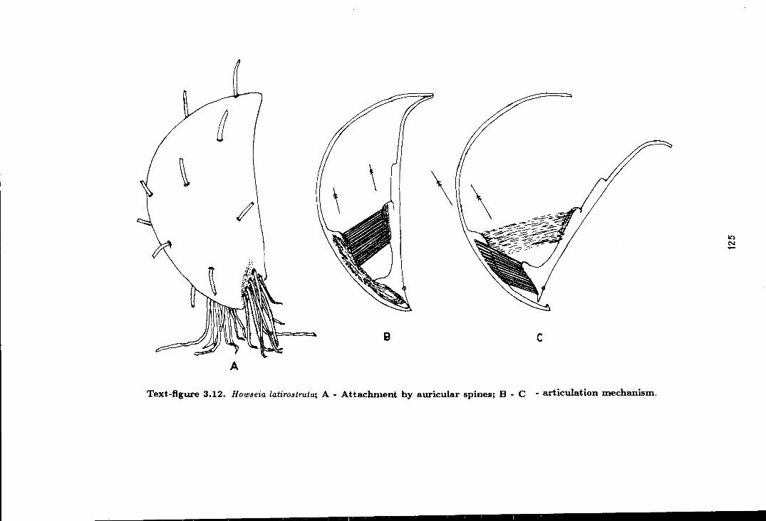
~ \\\\
B c
A
Text-figure 3.12. Ho11Jseia latirostrata; A - Attachment by auricular spines; B - C - articulation mechanism.
10 N

Table 3.14. Howseia latirostrata (Howse) - dimensions in mm.
Specimen length curved max. hinge height thickness
number length width width
MSLTH12 12.9 14.6 17.0 e13.4 3.7 na
MSLTH13 15.6 20.1 18.8 13.9 5.2 na
G88.57a 18.5 24.9 22.4 18.8 e8.7 6.0
G88.57b 19.0 31.2 e24.0 e19.1 11.6 na
MSLTH20 20.3 30.5 24.9 e20.5 9.8 5.3
MSLTH14 22.2 31.4 28.0 e21.8 10.3 na
MSLTH15 22.7 37.3 28.4 24.3 11.3 na
G88.57c 29.7 49.8 39.8 e32.0 19.6 na
dimensions= refer to text-figure 1.1 specimen numbers = refer to crappendix (G88 .57 = HB.l'lcock Museum• coli.)
e = estimated na = not available
not overhang. The tip of the umbo normally points towards the shorter hinge line.
The hinge line is slightly shorter than the maximum shell width, asymmetrical in
length and interrupted medianly. Auricular expansions are formed by very thin shell
laminae beyond the spinose edges of the shell. The interarea is very low with no
elythridium, but with a small triangular aperture for recieving the tiny lophidium of
the dorsal valve.
Interior of the ventral valve is covered with fine marginally pointed endospines,
which become coarser towards the periphery of the shell. The adductor muscle plat
form is moderately to highly elevated with smooth or dendritic muscle field, divided
into two elongated lobes by a median furrow. The muscle platform extends almost
to the mid shell length anteriorly, while posteriorly it is preceeded by a low ridge of
an aborted younger muscle platform. On each sides of the adductor muscle platform
are the fan shaped, flat and radially striated diductor muscle impressions.
The exterior of the ventral valve is smooth apart from some concentric growth
lamellae (very seldom rug ate) and some sparsely distributed body spines and auricular
spmes. The body spines are erect and irregularly distributed. The auricular spines
126
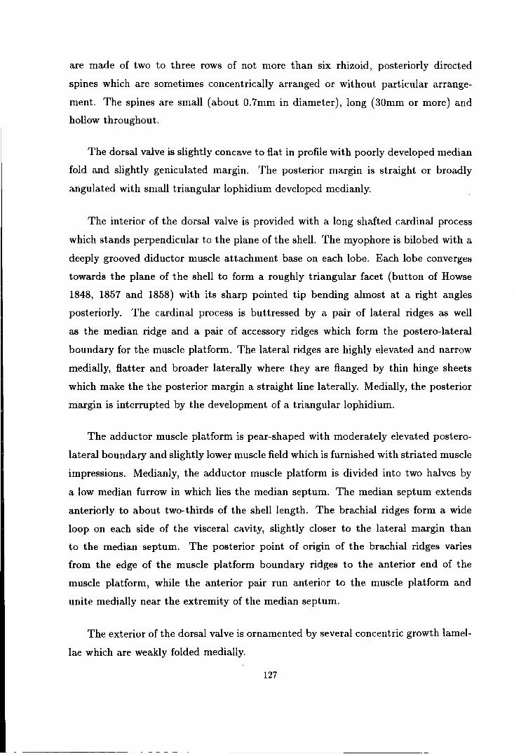
are made of two to three rows of not more than six rhizoid, posteriorly directed
spines which are sometimes concentrically arranged or without particular arrange
ment. The spines are small (about 0.7mm in diameter), long (30mm or more) and
hollow throughout.
The dorsal valve is slightly concave to flat in profile with poorly developed median
fold and slightly geniculated margin. The posterior margin is straight or broadly
angulated with small triangular lophidium developed medianly.
The interior of the dorsal valve is provided with a long shafted cardinal process
which stands perpendicular to the plane of the shell. The myophore is bilobed with a
deeply grooved diductor muscle attachment base on each lobe. Each lobe converges
towards the plane of the shell to form a roughly triangular facet (button of Howse
1848, 1857 and 1858) with its sharp pointed tip bending almost at a right angles
posteriorly. The cardinal process is buttressed by a pair of lateral ridges as well
as the median ridge and a pair of accessory ridges which form the postero-lateral
boundary for the muscle platform. The lateral ridges are highly elevated and narrow
medially, flatter and broader laterally where they are flanged by thin hinge sheets
which make the the posterior margin a straight line laterally. Medially, the posterior
margin is interrupted by the development of a triangular lophidium.
The adductor muscle platform is pear-shaped with moderately elevated postero
lateral boundary and slightly lower muscle field which is furnished with striated muscle
impressions. Medianly, the adductor muscle platform is divided into two halves by
a low median furrow in which lies the median septum. The median septum extends
anteriorly to about two-thirds of the shell length. The brachial ridges form a wide
loop on each side of the visceral cavity, slightly closer to the lateral margin than
to the median septum. The posterior point of origin of the brachial ridges varies
from the edge of the muscle platform boundary ridges to the anterior end of the
muscle platform, while the anterior pair run anterior to the muscle platform and
unite medially near the extremity of the median septum.
The exterior of the dorsal valve is ornamented by several concentric growth lamel
lae which are weakly folded medially.
127

~ A
A- MSLTHll
~····· . -
8
B- MSLTH12 D- G88.57a E- MSLTH14 G - MSLTH15 H - G88.57c G88.57a-c (HM collection) - - - - - - profile of dorsal valve . . . . . . reconstructed margin
-smm
c1 ~---.. ~ ~ -~0 .... ""
\ r---~ --
~''-? 0
~' c
C- MSLTH13 F- G88.57b
F
\..._ -- ))
-\1\ ...__-/
···-~
~' ~/
E
..
-----
G
Text-figure 3.13. Ontogenic development of Hotl•8eia latiro.~trata from Tunstall Hill rock cottage.
A - H - Camera Iucida reproduction of the listed specimens showing plan and lateral views (with profile of dorsal valve).
········ ·~-~--• 0-------..:.:.::
/ '--- / ______ .-/ ....
H
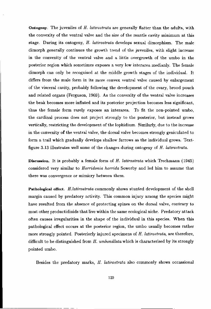
Ontogeny. The juveniles of H. latirostrata are generally flatter than the adults, with
the convexity of the ventral valve and the size of the mantle cavity minimum at this
stage. During its ontogeny, H. latirostrata develops sexual dimorphism. The male
dimorph generally continues the growth trend of the juveniles, with slight increase
in the convexity of the ventral valve and a little overgrowth of the umbo in the
posterior region which sometimes exposes a very low interarea medianly. The female
dimorph can only be recognised at the middle growth stages of the individual. It
differs from the male form in its more convex ventral valve caused by enlargement
of the visceral cavity, probably following the development of the ovary, brood pouch
and related organs (Ferguson, 1969). As the convexity of the ventral valve increases
the beak becomes more inflated and its posterior projection becomes less significant,
thus the female form rarely exposes an interarea. To fit the non-pointed umbo,
the cardinal process does not project strongly to the posterior, but instead grows
vertically, restricting the development of the lophidium. Similarly, due to the increase
in the convexity of the ventral valve, the dorsal valve becomes strongly geniculated to
form a trail which gradually develops shallow furrows as the individual grows. Text
figure 3.13 illustrates well some of the changes during ontogeny of H. latirostrata.
Discussion. It is probably a female form of H. latirostrata which Trechmann (1945)
considered very similar to Horridonia horrida Sowerby and led him to assume that
there was convergence or mimicry between them.
Pathological effect. H.latirostrata commonly shows stunted development of the shell
margin caused by predatory activity. This common injury among the species might
have resulted from the absence of protecting spines on the dorsal valve, contrary to
most other productidinids that live within the same ecological niche. Predatory attack
often causes irregularities in the shape of the individual in this species. When this
pathological effect occurs at the posterior region, the umbo usually becomes rather
more strongly pointed. Posteriorly injured specimens of H. latirostrata, are therefore,
difficult to be distinguished from H. umbonillata which is characterised by its strongly
pointed umbo.
Besides the predatory marks, H. latirostrata also commonly shows occassional
129

thickening of the growth lines. This phenomenon is caused by the occasional paucity
of the shell growth which might be related with differential supply of calcium carbon
ate due to the high and low tide (Clark, 1974, Norton, 1988). This indicates that H.
latirostrata must have been living in a relatively shallow water condition.
Related species. H. umbonillata (King) differs from H. latirostrata in having a nar
rower shell with shorter hinge line, narrower, but more pointed and overhanging
umbo. The interarea of H. umbonillata is higher than that of H. latirostrata with the
delthyrium sometimes partly covered by the elythridium posteriorly. The lophidium
of H. umbonillata is larger than that of H. latirostrata. The two species are, how
ever, similar in having a smooth dorsal valve and ventral valve with scattered body
spines and two to three rows of rhizoid auricular spines. The general configuration of
cardinal process, muscle scars and brachial ridges is very similar in these two species.
HOWSE/A UMBONILLATA (King)
(Text-figure 3.14, 3.15, Plate 8, figure 12-25).
synonymy; 1848 Productus umbonillatus King, p. 8. 1850 Productus umbonillatus King; King, pl. 11, fig. 14. 1854 Productus umbonillatus King; Schauroth, pl. 1, fig. 9-10. 1871 Productus umbonillatus King; Quenstedt, p. 640, pl. 60, fig. 8. 1937 Productus horridus var. umbonillatus (King); Malzahn, p. 34. 1963 Howseia latirostrata (Howse); Logan, pl. 108, fig. 2.
Diagnosis. Small, subquadrate to subtriangular Howseia with narrow, strongly pointed
and overhanging umbo. Ventral valve moderately convex with faint median sulcus
and high interarea. Delthyrium covered with elythridium posteriorly, open anteri
orly. Ventral surface moderate to strongly lamellose, with few body spines and two
to three rows of auricular spines. Dorsal valve weakly concave with large lophidium.
Cardinal process low, but strongly extended posteriorly. Lateral ridges and brachial
ridges usually oblique to growth axis.
130

Type specimens. Pattison (1977, p 38) selected all four of King's specimens in the
University College of Galway collection (specimens no. D 184A-D) as syntypes of
P. umbonillatus King (1848), which he renamed as H. latirostrata (Howse). Among
these specimens, only specimen no D 184B (see King,, 1850, pl. 11, fig. 15-18) and
Logan (1963, pl. 108, fig. 2) belongs to H. umbonillata (King), while the remaining
specimens belong to H. latirostrata (Howse). Therefore, specimen no. D 184B should
be elected as the holotype of the species.
Type locality. The holotype of H. umbonillatus mentioned above, came from Tunstall
Hill. Pattison (1977), suggested that King's collection from Tunstall Hill probably
came from a large area with quarries and natural exposures, mostly from arround NZ
392545. Recent collecting work around Thnstall Hill produce a few specimens of H.
umbonillata from an excavation site below the Rock Cottage (NZ 392544).
Material. The following description is based on about 80 studied specimens, including
the holotype from Thnstall Hill (specimen no. D184B in University College of Galway
collection), 36 specimens in Dr. Johnson's collection from Gilleylaw builders trench
(NZ 379539), and in the author's collection are 12 specimens from the type
locality, 7 specimens from Hylton Castle road cut , (NZ 360589) and 5 specimens
from Dalton-le-Dale ( NZ 40884474). Also studied are several specimens in
Hancock Museum collection from the type locality, Hylton Castle and Dalton-le-Dale
(including specimen no G 88.53), and several specimens in BM(NH) collection from
Hylton Castle and Dalton-le-Dale.
Horizon. ?Raisby Formation (Trechmann, 1945) to Ford Formation.
Age. Upper Permian (EZ1).
Description. The species is similar to that of H. latirostrata (Howse), except for the
following details;
The plan outline of the shell ranges from subquadrate to subtriangular and it is
almost always asymmetrical. The shell length is more or less equal to the maximum
width which is normally acquired at the anteriormost part of the shell, but some
131
I I I
G
A
' \
\
'
\ \' / .J -- _..,.
~ Ct
(~~/
5mm
""f ~ I • r '}'
(i J,lj .~ \ \~111 '': ·' -·~ __ .. ...
'- ~
8
Text-figure 3.14 Camera Iucida reproduction of the holotype of How.~eia umbonillata (King); specimen no D184B (UCG collection). A - ventral view. B - dorsal view {rock matrix is dott1~d: - - - - - fl'I"OIIstructed uutli1w of various internal morphology}.
N
~
~~ -,

Table 3.15. Howseia umbonillata (King) - dimensions in mm.
Specimen length curved max.
number length width
MSLTH25 e9.0 na e8.0
GALJ1 10.3 11.8 8.3
GALJ2 15.5 16.6 13.5
GALJ3 e17.9 22.1 16.8
MSLTH23 20.8 33.2 23.4
MSLTH24 21.3 25.5 23.0
GALJ5 e21.9 e33.2 e23.0
GALJ4 e22.5 e29.7 e22.8
GALJ6 e26.6 e45.0 e29.5
dimensions= refer to text-figure 1.1 specimen numbers = rerer to appendix
hinge height
width
e7.3 e2.2
6.4 e3.2
e9.9 4.2
e13.5 e6.7
e20.2 el2.6
el9.0 e5.7
e19.5 e10.7
e17.7 e9.1
e23.8 e13.7
VIA =ventral interarea na = not available e = estimated
thickness
na
2.4
na
5.0
na
na
e8.9
e6.4
e9.8
elongated forms show a greater length compared to the maximum width.
height
VIA
na
e1.5
na
e2.4
na
na
e5.1
e4.6
e4.0
The ventral valve is moderately to strongly convex in profile with faint median
sulcus developed anteriorly. In some of the juveniles and the male dimorphs, the
sulcus is imperceptible. The posterior margin is asymmetrical with poorly developed
auricle. The hinge line is about two thirds the length of the maximum width, with
each side inequal in length. The beak is narrow, but long. It is strongly pointed and
overhanging posterior to the hinge line. Usually the tip of the umbo twists and points
toward the shorter half of the hinge line. The interarea is high, sometimes attaining
a height of up to one fifth of the shell length. The delthyrium is provided with a large
triangular aperture anteriorly and covered by a weakly raised triangular elytridium.
The interior of the ventral valve shows a moderate to highly elevated, smooth
or dendritic adductor muscle platform surrounded by low radially striated diductor
muscle field. The exterior of the ventral valve is moderately to strongly lamellose,
often rugate. The true nature of the spines is unknown, but two to three rows of up
to six spine bases are seen on the auricle, while the body spine bases are sparse and
133

randomly distributed.
The dorsal valve is slightly concave in profile with poorly defined median fold
and auricle. The posterior margin is provided by a large triangular lophidium. The
interior morphology of the dorsal valve shows great variation between the male and
female dimorphs, as shown in the remarks on the ontogeny below.
Remarks on the ontogeny (see text-figure 3.15). Similar to H. latirostrata the juveniles
of H. umbonillata seems to be flatter than the adults. The juveniles of H. umbonillata
differ from those of H. latirostrata in their more elongated shell and pointed umbo.
These features are retained by the male adults with gradual curving of the umbo
during growth. Changes during early maturity of H. umbonillata apparently produced
very distinct dimorphs, and the male and female forms are characterised as follows;
The beak is gently curved in male forms and strongly curved in female forms. The
cardinal process in male dimorphs is narrower, with lower shaft and longer posterior
extension than it is in female forms. The lateral ridges are better developed in female
forms than in male forms. They are developed at a right angle to the shell growth axis
in female dimorphs, while in male dimorphs the lateral ridges are obliquely developed,
sometimes making an angle of up to 60° to the growth axis. The adductor muscle
platform is normally wider in female forms, and slightly expanded longitudinally in the
male forms. The brachial impressions of male forms also appear to be longitudinally
expanded, where the brachial ridges are not given-off horizontally as they are in female
forms.
134
0 -
0 -h . .
.... a" !{, \...
_,\~ .../
\\•:ri, / "/ ~
'
----·· .. ·················· ~
~ A ~ ~
······-················-,
. . B
A - GALJl B - GAL.J2 C- GALJ3
E - G88.53a (HM collection) F- GALJ5
() ····-c T
D- GAL.14
G- GALJ6
0
- - - - - - profile of dorsal valve . . . . . . . reconstructed margin \
·· .....
-smm
F
Text-figure 3.15. Ontogenic development of lfoU'.~eia umbonillata from Gilleylaw builders trench, except figure E which came from Dalton-le-Dale. A - G - Camera Iucida reproduction of plan and latPral view (with profile of ventral valve) of the listed specimens.
E
G
in ('r)

Superfamily PRODUCTACEA Gray
Amended diagnosis. Productidina with no interarea and no true teeth and sockets.
Hinge operated by linear interlocking of the hinge margins.
Discussion. The above diagnosis is introduced for the superfamily Productacea in
order to simplify the earlier diagnosis given by Muir-Wood & Cooper in 1960. Muir
Wood & Cooper (1960) stated that Productacea is "Productoidea generally fixed by
ringlike attachment spines in early growth stages and rarely cemented by beak, living
free in adult stages supported by halteroid spines; interareas, teeth and sockets are
normally absent but weakly developed in primitive forms; ginglymus in gerontic stages
only; cardinal process of several types, bilobate or trilobate, rarely quadrilobate; di
aphragm in brachial valve and crenulated marginal ridges in one or both valves in some
genera". This diagnosis appears to be ambiguous and not always applicable for dis
tinguishing productaceans from other productidinids. For instance, the productacean
mode of living varies from one species to another and the same modes of life are also
observed in other productidinid superfamilies. It is important to consider ecological
factors before conclusions are made on the mode of life of any productidinid. Simi
larly, as indicated in the original diagnosis, the cardinal process is very variable, and
similar types of cardinal process occur in other productidinid superfamilies.
From the current investigation, the cardinal process seems to be relatively more
sessile in Productacea than it is in Aulostegacea or Strophalosiacea, though there are
exceptions. Likewise, Productacea generally tend to adopt a free mode of life, but ex
ception is needed for those genera which fixed themselve to the substrate during most
of their lives. Therefore, in my view, these generalised statements should not be used
as diagnostic features of any superfamily of Productidina. Further, the diaphragm
and marginal ridges are present in certain productacean families only, and therefore
employing these criteria in the diagnosis of the superfamily is inappropriate.
Regarding the interarea, it has been stressed earlier about the importance of the
presence of interarea, whether in ventral valve only or in both ventral and dorsal valves
or absent in both valves. Therefore, suborder Productidina should be sub-divided on
this basis, and the superfamily Productacea should be restricted to productidinid with
136
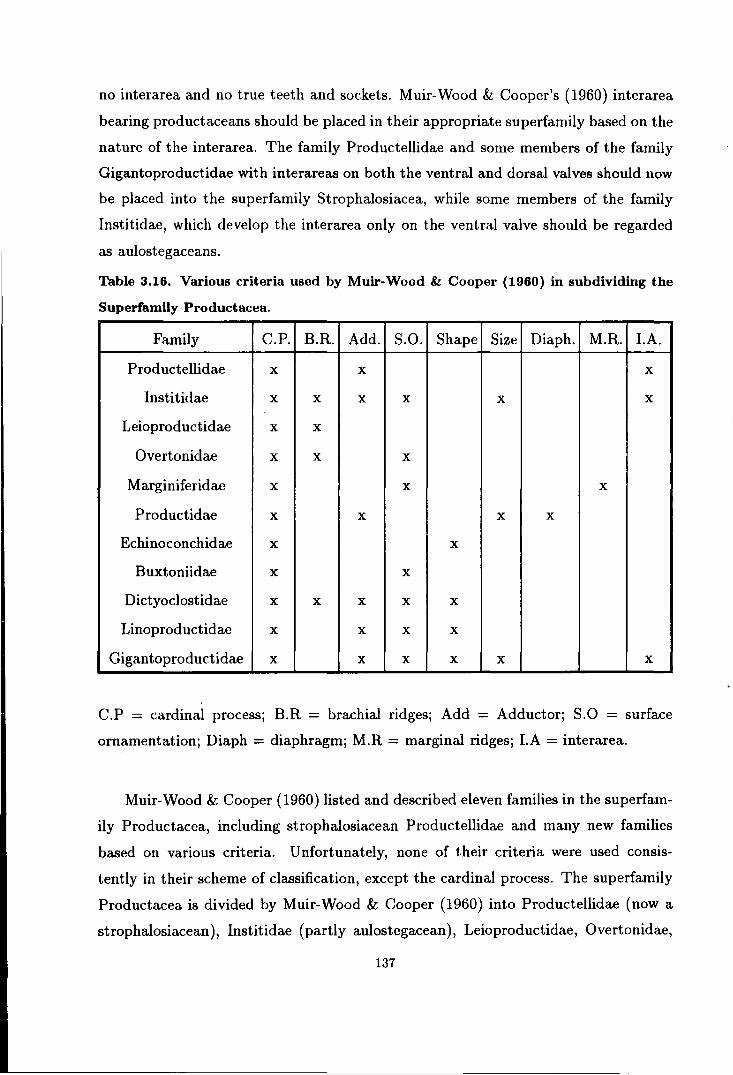
no interarea and no true teeth and sockets. Muir-Wood & Cooper's (1960) interarea
bearing productaceans should be placed in their appropriate superfamily based on the
nature of the interarea. The family Productellidae and some members of the family
Gigantoproductidae with interareas on both the ventral and dorsal valves should now
be placed into the superfamily Strophalosiacea, while some members of the family
lnstitidae, which develop the interarea only on the ventral valve should be regarded
as aulostegaceans.
Table 3.16. Various criteria used by Muir-Wood & Cooper (1960) in subdividing the
Superfamily Productacea.
Family C.P. B.R. Add. s.o. Shape Size Diaph. M.R. I.A.
Productellidae X X X
lnstitidae X X X X X X
Leioproductidae X X
Overtonidae X X X
Marginiferidae X X X
Productidae X X X X
Echinoconchidae X X
Buxtoniidae X X
Dictyoclostidae X X X X X
Linoproductidae X X X X
Gigantoproductidae X X X X X X
C.P = cardinal process; B.R = brachial ridges; Add = Adductor; S.O = surface
ornamentation; Diaph = diaphragm; M.R =marginal ridges; LA= interarea.
Muir-Wood & Cooper (1960) listed and described eleven families in the superfam
ily Productacea, including strophalosiacean Productellidae and many new families
based on various criteria. Unfortunately, none of their criteria were used consis
tently in their scheme of classification, except the cardinal process. The superfamily
Productacea is divided by Muir-Wood & Cooper (1960) into Productellidae (now a
strophalosiacean), Institidae (partly aulostegacean), Leioproductidae, Overtonidae,
137

Marginiferidae, Productidae, Echinoconchidae, Buxtoniidae, Dictyoclostidae, Lino
productidae and Gigantoproductidae (partly strophalosiacean). The following table
gives a full list of criteria used by Muir-Wood & Cooper (1960) for the diagnosis of
their productacean families. It shows how inconsistent the used of these criteria were
in acquiring the above sub-division.
Although the sub-division of the Productacea into families is unsatisfactory, lim
ited time and material available during the current study has not allowed the author
to proceed further on this matter. However, a major revision of the taxonomic clas
sification of this large and problematic superfamily is required in the future, in order
to gain a true understanding of its families and subfamilies.
From the British Permian, only one species of Productacea has been documented
previously. This species is Horridonia horrida (Sowerby) from the subfamily Horri
doniinae which is tentatively placed by Muir-Wood & Cooper into the family Dicty
oclostidae. Current study, however, shows that H. horrida from the British Permian
is made of several separate species of H orridonia, while a new genus Spinohorridonia
is described from the area of study for the first time.
Family DICTYOCLOSTIDAE Stehli
Diagnosis. Usually strongly geniculated Productacea having reticulate visceral re
gion, long trail, and brachial valve with sessile or short shafted strongly trilobed
cardinal process, having large median lobe; adductor dendritic; brachial ridges given
off horizontally.
Discussion. The above diagnosis is taken from Muir-Wood & Cooper (1960) without
amendment. Limited time and material available prevented the author from elabo
rating or commenting in great detail about this diagnosis. Most of the criteria used
are very variable and difficult and impractical to use to establish the family. How
ever, until a better alternative is available, the family status of Dictyoclostidae and
its sub-division, as introduced by Muir-Wood & Cooper (1960) will be maintained,
but the diagnosis of the subfamily Horridoniinae must be amended as follows.
138

Subfamily HORRIDONIINAE Muir-Wood & Cooper
Amended diagnosis. Dictyoclostidae with no reticulate ornament on the visceral disc,
often smooth or with weakly developed costae and rugae.
Type genus. Horridonia Chao {1927).
Other genera. Sowerbina Fredericks {1928), Bailliena Nelson & Johnson {1968) and
Spinohorridonia new genus.
Discussion. Muir-Wood & Cooper {1960) introduced the subfamily Horridoniinae as
Dictyoclostidae mostly with smooth surface; pedicle valve with posterior spine rows
and few scattered spines; brachial valve with large halteroid spines along posterior
margin and ears; adductor dendritic. With the large new collection of specimens from
the Durham Permian and more information about horridoniinids currently available,
some of these features have proved to be no longer reliable for the identification of
the subfamily. The posterior spine rows, for instance, are not always present on both
valves; in some species they are only found on the ventral valve while the same valve
of other species are completely devoid of marginal spines. Similarly, the adductor has
been found to be non-dendritic in some individuals of Horridonia horrida, a species
which is known to develop a dendritic adductor. Therefore, the diagnosis of the
subfamily Horridoniinae should be amended as above to avoid confusion.
The Horridoniinae was erected by Muir-Wood & Cooper (1960) as a mono-generic
subfamily, with the sole genus recognised by these authors was Horridonia Chao.
Sowerbina Fredericks and Pleurohorridonia Dunbar were considered by them as syn
onyms of Horridonia. Owing to poor description and illustration of Horridonia hor
rida (Sowerby) and related forms, the taxa at the generic level becomes uncertain.
Therefore, to solve this problem, the background history of the development of H.
horrida and related genera, must be studied.
It is important to note that only the external features will be employed to divide
different genera of Horridoniinae. Needless to say, the internals of productaceans
have often proved to be inconsistent from one individual to another. Such is the
139
case with the H. horrida of the Durham Permian, where the cardinal process varies
from bilobate to quadrilobate, and the adductor varies from dendritic to radially
grooved. The extension of the median septum and the configuration of the brachial
ridges are also individual rather than a specific or generic criteria. Regarding the
external features, some are apparently more valuable taxonomically than the others.
As discussed earlier in Strophalosiidae, the distribution of spines should be considered
very important genetically and therefore, should be highly regarded in classifying the
subfamily Horridoniinae.
Background history of Horridonia horrida and related forms. Horridonia horrida was
first introduced as Productus horridus by Sowerby in 1823 although similar forms had
been previously attributed to other names by various authors. As with most other
early determinations, Sowerby's species has been subjected to prolonged discussion
and changes. Similar to other early publications, it is either the wrong or insuffi
cient description or the lack of sufficient supporting figures that contribute to the
misunderstanding among the later authors. In Sowerby's {1823) paper, although he
described P. horridus as having marginal and body spines on both the ventral and
dorsal valves, his figures of the type specimen which came from the Cadeby Forma
tion in Derbyshire clearly shows the lack of body spines on the dorsal valve. The
absence of body spines is indeed very consistent among the British Horridonia, but
unfortunately none of the later authors except Davidson (1858) give any note about
it. Forms with a spinose visceral part of the dorsal valve are very rare in the British
Permian, but they are quite often found in the East European Permian. Further
discussion about these forms will be given later in this chapter.
Meanwhile, de Koninck (1846 and 1847) described P. horridus with body spines
on the dorsal valve from the East European Zechstein. At the same time he also
introduced P. geinitzianus, a species with a rather heavy spines on both valves but
retaining some features of P. horridus. This species was accepted by King (1856) and
Geinitz (1861), but was later considered by Eisel {1909), Malzahn (1937) and Gobbett
(1961) to be one of the many varieties of P. horridus introduced by Eisel in 1909. In
Geinitz's (1848 and 1861) papers on P. horridus, he also figured two different forms
with body spines on the dorsal valve, which were later named by Eisel {1909) as P.
140

horridus var. laspeanus and P. horridus var. geranus.
In Britain, W. King and R. Howse both described P. horridus in a very similar
way to Sowerby (1823), i.e. with marginal and body spines in both the ventral and
dorsal valves, see King (1850) and Howse (1848, 1857 and 18.58). However, none of
King's (1850) figures show any sign of body spines on the dorsal valve. About the
spines distribution, Davidson (1858, p. 34) mentioned that on the dorsal valve they
are confined to the vicinity of the cardinal edge where they assume much regularity
in both valves, from being arranged in one or two rows in the proximity of the hinge
line, and illustrated well his P. horridus with body spines present on the ventral valve
only.
In 1858, Haughton described P. sulcatus var. borealis based on two specimens,
one of which belongs to the true H. horrida and the other one is a species of Liosotella
(see Gobbett (1961)). Haughton's invalid name has unfortunately became a subject
of confusion among later authors including Stepanov (1936, 1937) and Licharev &
Einor (1939).
In 1875, Stuckenberg described P. timanicus from the Timan Tundra, the form
of which differs from Sowerby's (1823) P. horridus only in detail of ornamentation
and the shape of the shell. Later in the same year, Toula described P. horridus var.
granuliferus from Spitzbergen, a form with no marginal or auricular spines on the
ventral valve while the dorsal valve is devoid of body spines. Unfortunately, subse
quent authors failed to realise the differences between the two species from Timan and
Spitzbergen. Thus, assuming that Toula's P. granuliferus is similar to Stuckenberg's
P. timanicus, authors like Wiman (1914), Frebold (1931a), Dunbar (1955) and Gob
bett (1961 and 1963) described their species under Stuckenberg's name P. timanicus.
Interestingly, as revealed later by Logan (1966) all those authors mentioned above
were actually describing Toula's species as P. timanicus, while Stuckenberg's true
species remained relatively unknown.
Eisel {1909) described and illustrated several varieties of P. horridus including
var. initialis, var. auritulus, var. laspeanus, var. bufoninus, var. hoppeianus and var.
geranus. He also considered that P. geinitzianus as a variety of P. horridus. Eisel's
141

concept of varieties among P. horridus was well accepted by many later authors.
Malzahn (1937), treated these varieties statistically, and found out that apart from
P. horridus var. hoppeianus, all other varieties as well as it P. timanicus share a very
similar growth patterns. He also proved that the varieties of P. horridus do not show
any stratigraphical significance.
In 1927, Chao introduced the subgenus Horridonia to differentiate the only two
accepted species, P. (H.) horridus and P. (H.) timanicus from other species of the
overgrowinil genus Productus. Probably ignorant of Chao's work, Fredericks (1928)
introducedfgenus Sowerbina which is similar to Chao's (1927) subgenus Horridonia,
but with P. timanicus as the type species. In the same year, Chao elaborated his
earlier definition of the genus Horridonia, while in 1929, Schuchert & le Vene chose P.
horridus as the type species of this genus. To avoid confusion, Frebold (1931a,1931b
and 1933), Malzahn (1937) and Trechmann (1944) used the original designation of the
species prior to Chao's and Frederick's names. The name Horridonia was later used
by Dunbar & Condra (1932) and Branson (1948), who listed 13 species of Horridonia
in his Bibliograhic Index of the Permian Invertebrates.
In 1955, while working on horridoniinids from East Greenland, Dunbar found
it important to separate those forms with marginal spines on both the ventral and
dorsal valves from those with marginal spines on the dorsal valve only. He introduced
the genus Pleurohorridonia for those forms with marginal spines on both the ventral
and dorsal valves and with ribs on the trail of the ventral valve, while for those forms
with marginal spines on the dorsal valve only he re-introduced Fredericks's name
Sowerbina with amended diagnosis. Unfortunately, he gave little commentary about
the actual genus Horridonia which had already been accepted by him in Dunbar &
Condra (1932). In this paper, Dunbar introduced P. scoresbyensis, a form which
was previously attributed by Frebold (1933) to both P. horridus var. initialis and
P. horridus var. hoppeianus. Dunbar (1955) also introduced S. maynci which was
identified by later authors as H. granulifera, and S. rudis which is a genuine new
species. Although it is quite comprehensive, Dunbar's (1955) proposals have not been
well received by other authors. Waterhouse (1982) employed the term Sowerbina, but
unfortunately for just one uncertain species.
142
In 1960, Muir-Wood & Cooper re-established Chao's name Horridonia with new
amended definition of the genus while at the same time they suppressed all other
generic names once attributed to H. horrida and related forms. Muir-Wood &
Cooper's (1960) amended diagnosis appeared to be very long and it covered all forms
of Horridoniinae. Cobbett ( 1961) gave another amended diagnosis of H orridonia when
he published a detailed account of the genus and its species. Unfortunately, Cobbett
as many other authors before him, mistook Toula's P. horridus var. granuliferus with
his H. timanica (not Stuckenberg's P. timanicus).
Following his unpublished PhD. thesis, Logan (1966) produced an account to
clarify points of misunderstanding among earlier authors regarding the status of H.
timanica and H. granulifera. Regrettably, he did not discuss the status of horridoni
inids in the Durham Permian which he had studied in great detail.
Following Nelson's (1962) work on the horridoniinid stratigraphy of the Yukon
Territory, Nelson & Johnson (1968) discovered a new horridoniinid genus Bailliena.
Its new species B. yukonensis closely resembles P. geinitzianus de Koninck in many
ways, but differs from the later in having no marginal spines on the dorsal valve.
Nelson & Johnson (1968) apparently misunderstood the genus Horridonia, as they
assigned two species, which they thought might fit Dunbar's (1955) description of
the genus Sowerbina, into H. bullocki new species and H. granulifera (Toula). Both
H. granulifera and H. bullocki have no marginal spines on the ventral valve and thus
should be placed in the genus Sowerbina.
Conclusion: New scheme for generic classification of subfamily Horridoniinae.
The long and interesting history of interpretation of H orridonia horrid a and re
lated forms has given sufficient grounds to establish the following scheme of classifi
cation. It should be noted that one of the criteria used in the following taxonomic
classification has already been used by Dunbar in 1955, but he failed to convince later
authors of its value. Dunbar (1955) separated the genus Sowerbina from the rest of
horridoniinids on the basis of the lack of marginal and auricular spines on the ventral
143

valve. The genus Sowerbina is maintained in the following classification, so too is the
genus Bailliena Nelson & Johnson (1968). With these two genera being extracted
from the whole group of horridoniinids, it leaves us with a large genus H orridonia
with marginal spines present on both the ventral and dorsal valves.
As stressed earlier, about the significance of the distribution of spines in tax
onomic classification of the subfamily Horridoniinae, it is important to divide the
subgenus not only on the distribution of the marginal spines, but also other spines
including the body spines. With regard to the body spines, the remaining genus Hor
ridonia can be divided into two groups; those with body spines on both the ventral
and dorsal valves and those with body spines on the ventral valve only. The genus
H orridonia should now include only the type species of H. horrida and related forms
which have marginal spines on both the ventral and dorsal valves, and body spines
on the ventral valve only. Forms with body spines on both the ventral and dorsal
valves should be excluded from the genus Horridonia. A new genus Spinohorridonia
is introduced here for those horridoniinids formerly assigned to Horridonia, but with
both marginal and body spines present on both the ventral and dorsal valves. P.
geinitzianus de Koninck is suggested in this thesis as the type species of the new
genus Spinohorridonia.
This new scheme of classification reserves the size and shape of the shell and the
detailed arrangement and type of the spines to descriminate at the specific level, as
discussed later in this chapter. The generic scheme of classification can be generalized
and tabulated as follows;
The British horridoniinid genera. Only Horridonia and Spinohorridonia are present
in the British Permian.
144

Table 3.17. The classification scheme for subfamily Horridoniinae.
Genus VVMS VVBS DVMS
(Type Species)
HORRIDONIA Chao X X X
( P. horridus Sower by)
SOWERBINA Fredericks X X
( P. horrid us var granuliferus Toula)
BAILLIENA Nelson & Johnson X X
(B. yukonensis Nelson & Johnson)
SPINOHORRIDONIA new genus X X X
( P. geinitzianus de Koninck)
VVMS: ventral valve marginal spines; VVBS: ventral valve body spines DVMS: dorsal valve marginal spines; DVBS: dorsal valve body spines
DVBS
X
X
Note on Sowerbina. The genus Sowerbina should be described as horridoni
inids with marginal spines only present on the dorsal valve while body spines
present on the ventral valve only. Since Productus timanicus Stuckenberg has
been proved to have carried marginal spines on both the ventral and dor
sal valves (Logan, 1966) and thus belongs to the genus Horridonia, it is no
longer available as the type species of the genus Sowerbina. Productus hor
ridus Sowerby var. granuliferus Toula, which genuinely belongs to the genus
Sowerbina is proposed here to be the new type species of this genus. At
present, apart from S. granulifera (Toula), the genus Sowerbina also includes
S. rudis Dunbar, H. bullocki Nelson & Johnson and H. texana R.E. King.
145
Genus HORRIDONIA Chao 1927
Synonymy; 1927 Productus subgenus Horridonia Chao, p. 25. 1932 Horridonia Chao; Dunbar & Condra, p. 191. 1955 Pleurohorridonia Dunbar, p. 89. 1960 Horridonia Chao; Muir-Wood & Cooper, p. 292-294. 1960 Pleurohorridonia Dunbar; Muir-Wood & Cooper, p. 294. 1961 Horridonia Chao; Gobbett, p. 43. 1963 Horridonia Chao; Gobbett, p. 94. 1965 Horridonia Chao; Muir-Wood, p. 498.
Diagnosis. Horridoniinae with marginal spines on both the ventral and dorsal valves,
and few smaller body spines on the ventral valve only.
Type species. Productus horridus Sowerby 1823 was assigned as the type species by
Schuchert & le Vene (1929).
Other species. Productus timanicus Stuckenberg (1875), Horridonia hoppeiana (Eisel)
and H. dunelmensis new species.
Discussion. Chao (1927) defined Horridonia as Characterised by strong median fold,
slightly convex (?) brachial valve and a few but very stout hollow spines, but he did
not single out the type species between P. horridus and P. timanicus.
In 1928, Chao corrected the above diagnosis and added that Horridonia possess
essentially the same characteristic outline of typical Productus, but is without either
plicae or wrinkles. The median fold and sinus are rather pronounced and the surface is
marked only by irregularly distributed, strong and erect spines. Some indefinite plicae
are sometimes present, indicating occasional survival of the plicae making impulse
Chao's diagnosis appeared to be very broad with many variable features, some of
which are best used to described the differences between species while others seem
to be more of individual characters. This unsatisfactory diagnosis has caused many
later authors to write their own definition of the genus H orridonia, some short versions
while others give a more detailed definition (see Dunbar & Condra (1932), Muir-Wood
146

& Cooper (1960), Gobbett (1961 and 1963) and Muir-Wood (1965)). However, none
of these diagnoses suit the new scheme of taxonomic classification of the subfamily
Horridoniinae. Therefore, the new diagnosis cited above is introduced to describe
the genus Horridonia and comforming to the new scheme of classification introduced
earlier in this chapter. In producing this diagnosis, care was taken to avoid the use of
variable criteria which might cause confusion in the future. Features like the fold and
sulcus, the shape of the shell, the detail surface sculpture and most of the internal
features are left to be discussed at specific or variety level.
Specific division of the genus H orridonia.
Under the amended diagnosis above, the genus Horridonia is limited to the for
merly known Horridonia horrida var. initialis, var. auritulus, var. bufoninus and
var. hoppeianus and H. timanica. However, as mentioned earlier, Malzahn (1937)
has clearly shown that P. horridus var. initialis, var. auritulus, var. bufoninus and
H. timanica have a growth pattern which is very similar to one another; the only
variety which seems to have its own growth pattern is the var. hoppeianus. Later in
1967, Kazmierczak pointed out that the whole Polish H. horrida belongs to the var.
hoppeianus. The same variety has also been reported to dominate the Permian fauna
of the East Greenland (Frebold (1933) and Dunbar (1955) ).
Based on Malzahn's (1937) analysis, geographical distribution and new data which
will be elaborated later under individual species, it is suggested that the specific status
of the genus H orridonia can be described as follows;
1) At present, the genus H orridonia comprises four species, i.e. H orridonia hor
rid a (Sowerby), H. hoppeiana (Eisel), H. timanica (Stuckenberg) and H. dunelmensis
new spec1es.
2) Productus horridus var. hoppeianus does not belong to H. horrida because
it has a different growth pattern when compared to other varieties of H. horrida
and because it has characteristic juvenile stages which was previously considered by
Malzahn (1937) as mut. hoppeianus. The juvenile population has also been found
from High Newport near Sunderland in Britain during this research. Furthermore,
147

the occurrence of var. hoppeianus alone in some region, strongly suggested that it
belongs to a separate species, not only as a variety of H. horrida, see Kazmierczak
(1967), Frebold (1933) and Dunbar (1955). Therefore, Eisel's (1909) var. hoppeianus
should be elevated to form a separate species H orridonia hoppeiana, and a new full
description is now required for this newly assigned species.
3) H orridonia horrid a should be identified based on the type spectes and the
remaining varieties initialis, auritulus and bufoninus) should not be used for the
various reasons given below;
a) the var. initialis appears to be easily recognised only among smaller in
dividuals. Malzahn's (1937) term mut. initialis should therefore represent
a juvenile population of H. horrida, whereas none of the other two varieties
mentioned are recognisable among the juveniles of the species or as mutant
varieties by some authors. Var. initialis should th~refore, be regarded as ju
venile and younger individuals of the species and, as might be expected, this
supposed variety seems to be found more often than any of the other varieties.
b) The var. bufoninus seems to be found among the very large H. horrida
only, and it is not a common variety. From the White Quarry fauna, near
Newton Aycliffe, the author found that none of the smaller individuals can
be associated with var. bufoninus, but most of the very large individuals
definitely belong to this supposed variety. Therefore, var. bufoninus can only
be regarded as senile individuals of H. horrida.
With regard to Malzahn's figure which shows the presence of body spines on
the dorsal valve of var. hoppeianus (see Malzahn (1937, pl. 1, fig. 20)), this
particular specimen might be regarded as the larger representative of his var.
laspeanus or Spinohorridonia laspeana in this thesis.
c) The difficulty in distinguishing var. initialis from var. auritulus in medium
sized individuals is usually caused by the incomplete preservation of the del
icate shell margins, especially around the auricle. Medium or large sized var.
initialis always seem to have a broken auricle, while the impressively expanded
148

auricle in some var. auritulus could sometimes have resulted from the slight
flattening of the shell close to the plane of the auricle, or due to damage of
the anterolateral margin of the shell. Since var. initialis and var. auritulus
are commonly based on differences of preservation, their varietal status are
not significant and therefore should be suppressed.
4) The reasoning above suggests that the actual shape of adult individual of H.
horrida should lie in between the supposed var. initialis - auritulus, perhaps closer
to var. auritulus because of its more complete preservation, while the juvenile and
senile stages have a less expanded auricle.
5) Similar growth patterns between H. horrida and H. timanica and the close
resemblance of the external morphology (discussed later) show that these two species
are much closer to each other than they are to H. hoppeiana. Gobbett (1961) con
sidered that morphological similarities between var. hoppeianus and other varieties
of H. horrida should put them closer to each other and further away from H. timan
ica. lt should be noted, however, that Gobbett (1961) restricts this morphology to
the distribution of the marginal spines and his description of H. timanica should be
refered to Sowerbina granulifera (Toula), not H. timanica (Stuckenberg). More detail
study is, however, required to clarify the true status of it H. timanica.
6) A new form of Horridonia recorded in this thesis from the British Permian,
which differs from other known H orridonia in its more numerous body spines should
be regarded as a separate species. H. dunelmensis is introduced for this new form.
British H orridonia. The genus H orridonia is only represented in Britain by H. horrid a,
H. hoppeiana, H. dunelmensis new species and another rare form ( H orridonia sp. A)
whose specific status is uncertain at the present time.
149

HORRIDONIA HORRIDA Sowerby
(Text-figures 3.16, 3.17, Plate 9, figures 1-14)
Synonymy; 1823 Productus horridus Sowerby, p. 17, pl.319, fig. 1. 1846 Productus horridus Sowerby; Geinitz, p. 52, pl. 21, fig. 17, pl. 22, fig.8. 1848 Productus horridus Sowerby; Geinitz, p. 15-16, pl. 6, fig. 1-14 not 8. 1848 Productus horridus Sowerby; Howse, p. 256. 1850 Productus horridus Sowerby; W. King, p. 87-92, pl. 11, fig. 2-7 and 10. 1853 Productus horridus Sowerby; Davidson, pl. 9, fig. 219-220. 1857 Productus horridus Sowerby; Howse, p. 44-46. 1858 Productus horridus Sowerby; Howse, p. 247-250. 1858 Productus horridus Sowerby; Davidson, p. 33-36, pl. 4, fig. 13-26. 1861 Productus horridus Sowerby; Geinitz, p. 103, pl. 19, fig. 11-12, 15-17. 1871 Productus aculeatus Quendstedt (not-Schlotheim), p. 635-6, pl. 60, fig. 1-4. 1909 Productus horridus Sowerby var. initialis Eisel, p. 33-34, fig. 1. 1909 Productus horridus Sowerby var. auritulus Eisel, p. 35, fig. 4. 1909 Productus horridus Sowerby var. bufoninus Eisel, p. 35, fig. 5, 8-10. 1933 Productus horridus Sowerby var. initialis Eisel; Frebold, p. 20-21, pl. 2, fig.
10-12. 1933 Productus horridus Sowerby var. granuliferus Frebold {not-Toula), p. 21-22, pl.
2, fig. 13. 1937 Productus horridus Sowerby var. initialis Eisel; Malzahn. 1937 Productus horridus Sowerby var. auritulus Eisel; Malzahn. 1937 Productus horridus Sowerby var. bufoninus Eisel; Malzahn. 1944 Productus horridus Sowerby; Trechmann, p. 349. 1955 Pleurohorridonia scoresbyensis Dunbar, p. 90-95, pl. 12, fig. 1-3. 1959 Horridonia horrida (Sowerby) var. inicialis Eisel; Stepanov, p. 192, pl. 1, fig 1
and 6. 1960 Horridonia horrida (Sowerby); Muir-Wood & Cooper, pl. 108, fig. ?8-11. 1961 Horridonia horrida {Sowerby); Gobbett, p. 43-46, pl. 3, fig. 1, ?2, 4, 6 and 8,
pl. 5, fig. 5, 7-8. 1980 Horridonia horrida (Sowerby); Pettigrew, pl. 6g. 1988 Horridonia horrida (Sowerby); Hollingworth & Pettigrew, p. 18, fig. 3, text-fig.
5. 1988 Horridonia horrida (Sowerby); Turek, Marek & Benes, p. 147, fig. c-f.
150
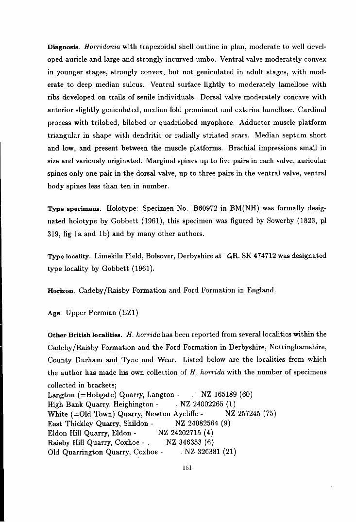
Diagnosis. Horridonia with trapezoidal shell outline in plan, moderate to well devel
oped auricle and large and strongly incurved umbo. Ventral valve moderately convex
in younger stages, strongly convex, but not geniculated in adult stages, with mod
erate to deep median sulcus. Ventral surface lightly to moderately lamellose with
ribs developed on trails of senile individuals. Dorsal valve moderately concave with
anterior slightly geniculated, median fold prominent and exterior lamellose. Cardinal
process with trilobed, bilobed or quadrilobed myophore. Adductor muscle platform
triangular in shape with dendritic or radially striated scars. Median septum short
and low, and present between the muscle platforms. Brachial impressions small in
size and variously originated. Marginal spines up to five pairs in each valve, auricular
spines only one pair in the dorsal valve, up to three pairs in the ventral valve, ventral
body spines less than ten in number.
Type specimens. Holotype: Specimen No. B60972 in BM(NH) was formally desig
nated holotype by Gobbett (1961), this specimen was figured by Sowerby (1823, pl
319, fig 1a and 1b) and by many other authors.
Type locality. Limekiln Field, Bolsover, Derbyshire at- GR. SK 474712 was designated
type locality by Gobbett (1961).
Horizon. Cadeby /Raisby Formation and Ford Formation in England.
Age. Upper Permian (EZ1)
Other British localities. H. horrida has been reported from several localities within the
Cadeby /Raisby Formation and the Ford Formation in Derbyshire, Nottinghamshire,
County Durham and Tyne and Wear. Listed below are the localities from which
the author has made his own collection of H. horrida with the number of specimens
collected in brackets; Langton ( =Hobgate) Quarry, Langton- NZ 165189 (60) High Bank Quarry, Heighington - . NZ 24002265 (1) White (=Old Town) Quarry, Newton Aycliffe- NZ 257245 (75) East Thickley Quarry, Shildon- NZ 24082564 (9) Eldon Hill Quarry, Eldon- NZ 24202715 (4) Raisby Hill Quarry, Coxhoe- NZ 346353 (6) Old Quarrington Quarry, Coxhoe - _ NZ 326381 (21)
151

NZ 345417 (1) Sherburn Hill Quarry, Sherburn Hill -Dawson Plantation Quarry, Penshaw Hill -
?Tunstall Hill Rock Cottage, Tunstall -NZ 334544 (7)
NZ 392545 (13) ? Ryhope old railway cut, Tunstall?Hylton Castle roadcut, North Hylton-
NZ 39665385 (13) NZ 360589 ( 6)
Table 3.18. Horridonia horrida (Sowerby) - dimensions in mm.
Specimen no length curved length
MSLWQ14 6.0 e8.7
MSLWQ15 e11.1 na
MSLWQ16 22.5 e38.0
MSLWQ17 e33.5 e59.5
MSLWQ18 e40.0 e68.0
MSLWQ12 e45.0 e78.0
MSLWQ19 46.7 e88.0
dimensions= refer to text-figure 1.1 specimen numbers = refer to appendix
hinge width
e7.2
e16.4
e30.6
e38.5
e50.0
e59.0
e62.0
na = not available e = estimated maximum width = hinge width
height
e2.5
e5.3
e11.4
e19.2
e23.7
e29.5
e31.2
Description. The shell is medium to large in size with the maximum shell width
reaching 65mm or more at the hinge line. The shell outline is anteriorly tapered sub
trapezoidal shape with the angle of anterior convergence varying from one individual
to another. Posteriorly, the shell outline is marked by a sub-circular extension of the
umbo beyond the hinge line. The maximum shell width is at the hinge line, and it is
greater than the shell length.
The ventral valve is moderately convex in the smaller individuals and strongly
convex in larger individuals. The convexity of the valve is strongest posteriorly, and
weakens anteriorly. The ventral valve is divided into two visceral humps by a median
sulcus which is deepest at about the mid-length, shallowing in both longitudinal
directions, sometimes becoming very shallow and imperceptible at the umbo and on
the trail. The umbo is large and tapers slightly towards the posterior across the
hinge line. At the posterior extremity, the umbo is strongly incurved and convergent
towards the dorsal and then towards the anterior. The tip of the beak usually covers
152
' .. : ::.~ . ....-· ·-··· .. .-:·_····. ......... ~./
·--~
A B
E
-10mm
Text-figure 3.16. Reconstruction of Horridonia horrida (Sowerby). A,B - Exterior of ventral and dorsal valves. C - Posterior view showing the special arrangement of marginal spines. D - Common lifP position of Horridonia horr-ida showing the role of marginal spines in support.ing the shell {- - - - - - sediment iiJterface: - - - - - hinge axis]. E - Life profile { = = = = = = spine horizon}.

the slightly elevated posterior extension of the dorsal valve. The trail is generally
smooth, but in larger individuals it is sometimes broadly ribbed. The auricle is
well developed, but sometimes it appears to be fused with the visceral disc when the
posterolateral margin of the valve slopes rather gently towards the auricle. When well
developed, the auricle is triangular in shape and slightly arched in its longitudinal
profile. Posteriorly, the hinge margin is straight laterally, but slightly bent towards
the dorsal medianly, following the growth of the beak.
The exterior of the ventral valve is smooth or covered with light to moderate
concentric growth lamellae or sometimes with unusual rugae. The assemblage of
spines on the ventral valve consist of;
i) - a row of up to five pairs of coarse marginal spines, vertical to the shell plane
longitudinally and inclined outwards at about 20° to 50° transversely. Three or four of
the inner pairs however, occur slightly anterior to the margin and later join the other
marginal spines to form a single plane of spines. The marginal spines are normally
straight at the base and bend outwards at the end, but an exceptional case where
these spines curve concentrically inward has also been observed. These spines are
hollow throughout and increase in size from the inner pair to the outer pair. The
largest outer spine pair observed has a diameter of about 2 mm, while the longest
spines observed are more than 45 mm in length.
ii) - one to three pairs of large hollow auricular spines, very similar in many
respects to those of the marginal spines, except that they are slightly inclined ante
riorward.
iii) - Few (less than ten), irregularly distributed body spines on the beak, visceral
humps and in the median sulcus. Owing to poor preservation, most of the specimens
studied show hardly any traces of body spines except for very few small spine bases
in the umbonal region. Judging from these spine bases, the body spines were erect
to sub-erect in form.
The interior of the ventral valve is fully covered by short marginally pointed en
dospines, coarser at the anterior margin. In some larger individuals, the endospines
at each side of the median sulcus are pointed medianly as well as anteriorly. Posteri-
154

orly, nearly one third of the valve is covered by large, low, radially striated diductor
muscle platforms. In between this diductor muscle platforms lies the highly elevated
adductor muscle platform which is divided into two halves by a low median furrow.
Each half of the adductor muscle platform can be divided into the lower posterior
section and the higher anterior section. The muscle scars are dendritic in pattern. At
the posterior margin, an anteriorly inclined ridge appears at each sides of the umbo.
This ridge is flattened and becomes broader laterally.
The dorsal valve is moderately concave, with the concavity stronger anteriorly;
sometimes the trail is slightly geniculated. The median fold is well developed, ap
pears shortly after the posterior margin and extends almost to the anterior margin.
The posterior margin is straight laterally, but a little elevated medianly at the small
triangular ?lophidium. The auricle is flat or concave in profile.
The exterior of the valve is smooth or lightly to moderately lamellose concentri
cally. The spines are restriced to a row of up to five pairs of large marginal spines
and a pair of auricular spines. The nature of the marginal spines are similar to those
of the ventral valve, so that when the valve is closed all marginal spines are set in a
single linear plane. The dorsal marginal spines usually curve inward or concentrically
arranged, but sometimes several outer pairs do curve outward. The auricular spines
are larger than the marginal spines and develop at a low angle to both the shell plane
and the plane of the marginal spines, and curve away from both planes.
Internally, the shell is covered by short, marginally pointed endospines, coarser
at the margin of the viseral disc and on the trail. The cardinal process is large and
short shafted or sessile with the myophore commonly trilobed, although some are
bilobed or quadrilobed. The muscle attachment base is divided into two sections in
the bilobed forms and four in the quadrilobed forms and some of the trilobed forms
develop a very large median lobe. These divided muscle grooves converge toward the
anterior into a small lophidium. The cardinal process shaft is posteriorly directed or
inclined at a low angle (up to 20°) either to the ventral or to the dorsal. The shaft is
supported by a pair of strong lateral ridges and a weakly developed median ridge. The
lateral ridges are thick and posteriorly inclined in the middle, but are flattened and
155
broaden towards the lateral margins. The median ridge is sometimes very low and
the median septum is separated from the cardinal process shaft by a low depression
resembling an alveolus. The median septum appears in the middle of the adductor
muscle platform and continues anteriorward to about two-thirds of the shell length.
The median septum is thick and low posteriorly and sharp and moderately elevated
anteriorly.
The adductor muscle platform is triangular in shape, and is divided into four
sections. The anterior sections are more highly elevated than the posterior ones. The
adductor muscle platform is divided centrally into two halves by a well developed
furrow in which the median septum lies. The muscle scars are usually dendritic in
pattern, but may be radially striated or grooved. In the non-dendritic forms, it is
dificult to divide the adductor muscle platform into the posterior and anterior sections.
The brachial ridge is introduced posteriorly at various positions roughly at the
anterior end of the adductor muscle platform. The ridge appears either horizontally
or at a low angle towards the growth axis. The brachial impressions are small in size,
and only occupy an area of about one quarter of the visceral disc.
Discussion. Ontogeny (see plate 9, figures 3 to 8). Juvenile and younger individuals
of H. horrida compared to the larger adults, have a less convex ventral valve, smaller
and poorly developed auricle, less concave dorsal valve and smoother external surface.
As it grows larger, H. horrida increases the convexity of its ventral valve especially
at the umbonal region, where the beak becomes strongly incurved. H. horrida also
produces a better auricle and trail as it reaches adulthood. When it becomes very
large or ~eronb'e,H. horrida tends to develop a broadly ribbed trail, while the median
sulcus fades away on the trail. The dorsal valve of the serontic individuals are sometimes
slightly geniculated. The trend of ontogenic development of the internal features of H.
horrida are not well known because of the lack of material in which the internals are
shown, especially among juvenile and senile individuals. Smaller adults, however tend
to be more commonly found with bilobed cardinal process and non-dendritic muscle
scars, but some of the larger adults of the species also inherited these features. The
quadrilobed myophores are acquired only in very large individuals.
156

A
8
c
a
a
>
a
' ' =o:Ji'''''''''''''h .. <)'>)~~·'-'~~:~ . c: _,,,,,,, •< ----("' ......
b
....... ,
b
b
. . \ . .
--
. '. .
c
c
Text-figure 3.17. Postulation of various moving mechanisms in Horridonia horrida.
A - simple opening of the valve. B - Vertical movement {a-b - all ma.rgimil spines sink into the sediment wl1ile the hinge a.xis is fixed: b-e - the shell sinks towards the new spine horizon: reversing the process will lift the shell upward}.
C- Lateral movement [a-b- the shell is tangently lifted by pulling the dorsal marginal spines while holding the edge of the ventral marginal spines: b-e - by holding the edge of dorsal marginal spines the shell can be lowered tangently by pushing the ventral marginal spines: fnr rnnv;:.rn~nf fntv!:lrrlC!I 1-J.an. .rl,..,-..... "' J ,.;_ ... _ t •• ------- .... 1 •
1
. . I I

Note on the distribution and arrangment of spines and mode of life - material from
East Thickley Quarry and White (=Old Town) Quarry. Based on material from East
Thickley, Kazmierczak (1967) suggested that some H. horrida lived with its trail lifted
above the surface and the marginal spines which are arranged in a single plane play
the role of supporting platform. Similar interpretation has also been given by Muir
Wood & Cooper (1960) when they discussed the role of spines in Horridonia s.s. New
material, which shows very similar preservation to that discussed by Kazmierczak,
was collected from the White (=Old Town) Quarry (about 1.5km east of the East
Thickley Quarry) during this research. With the shell and spines preserved in position
exac.tly as figured by Kazmierczak (1967), these remarkable H. horrida must have
been buried alive. It is also noted that H. horrida from these quarries only develop
body spines at the umbonal region, whereas the rest of the ventral valve is smooth.
Adopting the suggested living position of Kazmierczak, reduces the buried part of
H. horrida to the umbonal region only, and this should reduce the significance of the
body spines. The remaining body spines are very small in size as compared to the
marginal spines and are usually preserved as spine bases in smaller individuals only.
These spines were probably aborted during the ontogeny of H. horrida as they became
less significant in supporting the larger individuals. The auricular spines were also
probably aborted for a similar reason. Usually only a pair or two of auricular spines
are left during the adult stage of this species. The nature of the auricular spines
shows that they might have adopted a sensory function during part of the ontogeny,
instead of supporting the shell.
The special arrangment of the marginal spines in H. horrida appears to have given
it partial mobility including:-
i) By moving both valves equally and simultaneously, H. horrida could possibly
lift itself up through muddy sediment to get clearer water conditions, and similarly
it could bury itself further down in the mud to gain protection against predators or
unfavourable environmental conditions (see text figure 3.17B).
ii) On a less muddy substratum, H. horrida could perhaps plough the mud surface
by moving only one valve while holding the other still. In this way, it could move
158
itself horizontally as well as vertically (see text-figure 3.17C). The ability to move
horizontally would be of much advantage to secure a satisfactory substratum on which
to settle down.
Comparison. H. horrida closely resembles the Carboniferous H. timanica in many
respects, except that H. timanica has a slightly less convex ventral valve, slightly
flatter dorsal valve, slightly broarder auricle and slightly better developed median
sulcus and fold compared to H. horrida. H. dunelmensis differs from H. horrida only
in its more numerous marginal and body spines, while the shape of the shell and the
interior morphology of the two species are similar.
H. hoppeiana can be distinguished from H. horrida in its more convex ventral
valve, more concave dorsal valve, less prominent auricle, median sulcus and fold and
smaller marginal and auricular spines.
HORRIDONIA HOPPE/ANA (Eisel)
{Text-figure 3.20, Plate 10, figures 1-11}.
Synonymy; 1909 Productus horridus Sowerby var. hoppeianus Eisel, p. 34, pl. 2 and 3. 1933 Productus horridus Sowerby var. hoppeianus Eisel; Frebold, p. 18-19, pl. 3, fig
9 and pl. 5, fig. 1. 1933 Productus sp., Frebold, p. 19, pl. 1, fig. 7-8. 1937 Productus horridus Sowerby var. hoppeianus Eisel; Malzahn, pl. 1, fig. 18-23. 1955 Pleurohorridonia scoresbyensis Dunbar, p. 90-95, pl. 11 and pl. 12, fig. 4-13. 1959 Horridonia horrida (Sowerby) var. hoppeiana Eisel; Stepanov, p. 192, pl. 1, fig.
5. 1961 Horridonia horrida (Sowerby); Gobbett, pl. 3, fig. 3,5 and 7. 1967 Horridonia horrida (Sowerby); Kazmierczak, p. 239, pl. 1 and 2.
Diagnosis. Horridonia with sub-trapezoidal to rectangular shell outline, moderately
developed auricles and large and strongly incurved umbo. Ventral valve very strongly
convex, but not geniculated. Median sulcus poorly developed. Ventral surface lightly
lamellose, sometimes ribbed on the trail. Body spines rare. Dorsal valve moderately
159
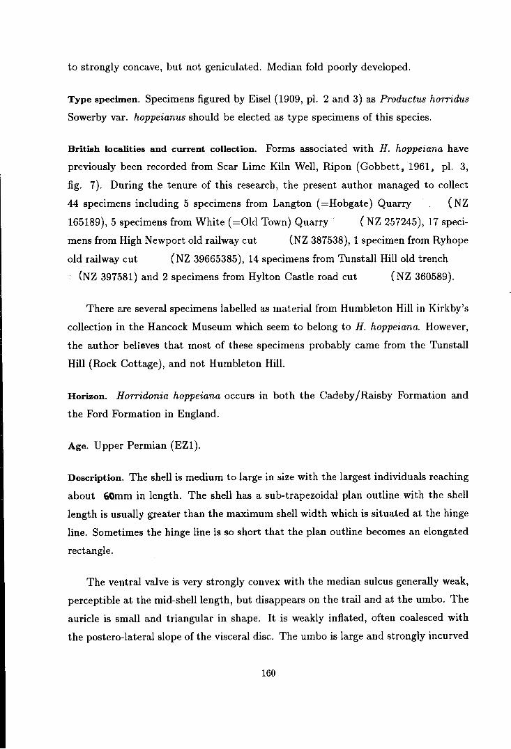
to strongly concave, but not geniculated. Median fold poorly developed.
Type specimen. Specimens figured by Eisel (1909, pl. 2 and 3) as Productus horridus
Sowerby var. hoppeianus should be elected as type specimens of this species.
British localities and current collection. Forms associated with H. hoppeiana have
previously been recorded from Scar Lime Kiln Well, Ripon (Gobbett, 1961, pl. 3,
fig. 7). During the tenure of this research, the present author managed to collect
44 specimens including 5 specimens from Langton ( =Hobgate) Quarry ( NZ
165189), 5 specimens from White (=Old Town) Quarry · ( NZ 257245), 17 speci
mens from High Newport old railway cut (NZ 387538), 1 specimen from Ryhope
old railway cut (NZ 39665385), 14 specimens from Tunstall Hill old trench
(NZ 397581) and 2 specimens from Hylton Castle road cut ( NZ 360589).
There are several specimens labelled as material from Humbleton Hill in Kirkby's
collection in the Hancock Museum which seem to belong to H. hoppeiana. However,
the author believes that most of these specimens probably came from the Tunstall
Hill (Rock Cottage), and not Humbleton Hill.
Horizon. Horridonia hoppeiana occurs in both the Cadeby/Raisby Formation and
the Ford Formation in England.
Age. Upper Permian (EZ1).
Description. The shell is medium to large in size with the largest individuals reaching
about 60mm in length. The shell has a sub-trapezoidal plan outline with the shell
length is usually greater than the maximum shell width which is situated at the hinge
line. Sometimes the hinge line is so short that the plan outline becomes an elongated
rectangle.
The ventral valve is very strongly convex with the median sulcus generally weak,
perceptible at the mid-shell length, but disappears on the trail and at the umbo. The
auricle is small and triangular in shape. It is weakly inflated, often coalesced with
the postero-lateral slope of the visceral disc. The umbo is large and strongly incurved
160

Table 3.19. Horridonia hoppeiana (Eisel) - dimensions in mm.
Specimen no length curved length
MSLHN7 17.5 e27.8
MSLHN5 19.5 33.2
MSLHN4 e20.2 e29.0
MSLHN6 21.4 e37.4
MSLWQ24 31.4 e65.0
MSLWQ25 e42.0 e90.0
MSLLQ11 45.0 e84.0
MSLLQ12 e49.5 e90.0
dimensions= refer to text-figure 1.1 specimen numbers = refer to appendix e = estimated
hinge width
17.8
19.6
17.0
20.7
e30.4
e40.8
e40.5
e48.8
height
9.7
10.5
10.0
10.8
21.2
31.5
29.2
e35.0
beyond the hinge line. The trail is long, weakly convex to almost flat in longitudinal
profile.
The exterior of the ventral valve is covered with fine to moderate growth lamellae
with occasional randomly distributed rugae developed. Rare spine bases are seen on
the umbonal region and around the hinge area. Detail distribution and nature of the
body spines as well as the marginal and auricular spines are not known in the British
H. hoppeiana, except young specimens from High Newport old railway cut show that
the marginal spines and auricular spines are laterally directed almost similar to those
figured by Dunbar (1955). The interior of the ventral valve is only known in the
younger population which will be discussed later.
The dorsal valve is moderately to strongly concave, but seldom geniculated. The
median fold is absent or very poorly developed. The exterior of the valve is poorly
known.
Discussion. Most of the material collected from the British Permian during this
research is incompletely preserved. It can be differentiated from other Horridonia
by the very strongly coiled shell with long trail and very shallow median sulcus.
161

Details of the distribution of spines and the internals are not fully known in British
H. hoppeiana, except among the younger population of the species discussed below.
Note on H. hoppeiana from High Newport. A population of small Horridonia, majority
of which have a strongly convex ventral valve have been found at an old railway cut
at High Newport, near Sunderland. Considering this population of Horridonia alone,
a case might be made that it belongs to a dwarf fauna or to a separate species.
However, since a population of S. lewisiana from the same locality have average adult
size for the species, these small H orridonia cannot be considered as part of a dwarf
fauna. They should be considered as the juveniles of a species of Horridonia with
very strongly convex ventral valve, i.e. H. hoppeiana. Other juveniles of Horridonia
known from the British Permian are commonly much flatter than these particular
juveniles. The largest individuals of the fauna has a length of about 21mm, and
at this stage they have already developed a trail, some even show traces of radial
costae on it. This evidence should clear any doubt in the authenticity of the species
H. hoppeiana, and separates this species far from any other species of H orridonia.
Some of these young H. hoppeiana show a few large spine bases on the convex
part of the ventral valve and two or more pairs each of marginal and auricular spines
on the ventral valve, all of which are laterally directed. The nature of these spines are
very similar to those figured by Dunbar (1955) for his P. scoresbyensis except that
the auricular spines figured by him are very much larger than the marginal and body
spmes.
The ventral adductor muscle platform is divided into a highly raised anterior
section and a low posterior section which are separated by a deep oblique furrow.
The anterior section narrows posteriorly, continues in between the posterior section
to the tip of the umbo. The anterior section or middle section (posteriorly) is divided
into two halves by a shallow median furrow. The muscle scars are smooth, but may be
dendritic in the larger adult stages. Surrounding the adductor muscle platforms are
the radially striated scars of the diductor muscle platforms which are hardly raised
from the shell plane. The dorsal adductor muscle platforms are pear shaped and
moderately elevated, with the scars obliquely to radially striated or grooved.
162

The cardinal process is large with bilobed myophores. The shaft is supported by
a pair of strong posteriorly inclined lateral ridges, but no median ridge is developed
at the base of the shaft. The median ridges, however, appear shortly anterior to
the cardinal process, in between the adductor muscle platforms, and continue as a
moderately raised median septum to the mid shell length.
HORRIDONIA DUNELMENSIS new species
[Text-figure 3.18, Plate 11, figures 1-18}.
{Named after the old County Durham where all the cited localities of this species lie}.
Diagnosis. Horridonia with a very similar shape and profile to H. horrida but with up
to 7 pairs of marginal spines on each valve, two rows of two to five pairs of auricular
spines and thirty to fourty body spines on the ventral valve; dorsal valve with no
auricular or body spines. Interior of the shell is similar to that of H. horrida.
Type specimen. Specimens no MSLHH8, MSLHH9, MSLHH10, MSLHH16, MSLHH17
and MSLHH18 are chosen as paratypes of Horridonia dunelmensis, to show variation
in the morphology of the species.
Type locality. The type specimens above came from Humbleton Hill (GR. NZ
381553).
Other localities include Tunstall Hill old trench (CR. NZ 397541), Ryhope old
railway cut :{rC.R. NZ 39665385) and Beacon Hill railway cut (CR. NZ 442454).
Horizon. Ford Formation.
Age. Upper Permian (EZ1).
Material. Humbleton Hill (?80 specimens), Tunstall Hill old trench (?90 specimens),
Ryhope old railway cut (1 specimen) and Beacon Hill railway cut (4 specimens).
163
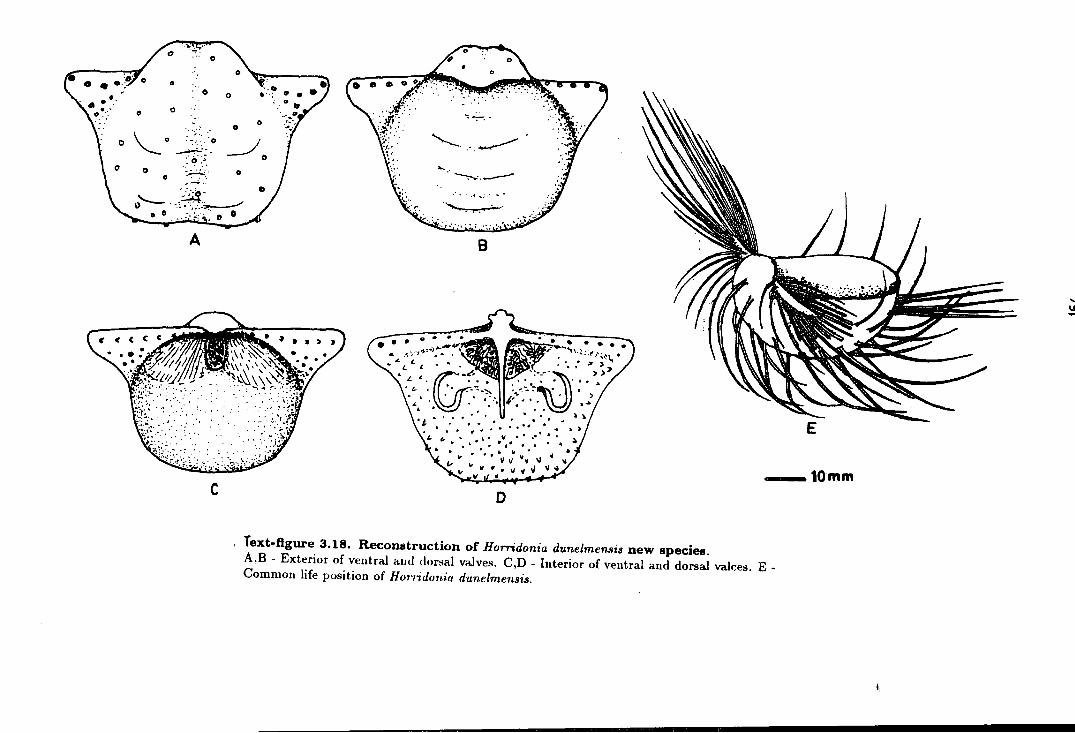
0 6
0 0
0 \_ 0 0
~~~~ 0 :~. D ..............
A
c
0
0 0
"":--:--:-
__/0 "------,~ 0
.... -,...._. _ __... .· . . -~ .....; ...
. ...,.__ -·--·
8
• • .. • "- L
.'-,£ :_ r;·. ·.& . ~ . ... . :.. .. .. . . .. - ...... ~ J.~ • • .. .. ': ·',
• J, •• : • • • • ~ .. . .. " . . . ·, ... "<I - •••• ., • ..
, ...... , .• v.,·····~~ " • " ••• 4o ~
.. " .. " . vvv--\1~ ....
E
~ V II ..
--10mm 0
Text-figure 3.18. Reconstruction of Horridonia du.nelmen..~is new species. A,B - Exterior of ventral and dorsal valves. C,D - Interior of ventral and dorsal valces. E -Common life position of Hunidonia d·unelmensis.

Table 3.20. Horridonia dunelmensis new species- ditnensions in mm.
Specimen no length curved length
MSLHH12 8.7 14.6
MSLR1 10.3 15.6
MSLHH13 14.2 23.3
MSLHH14 e16.4 e25.5
MSLHH15 18.1 30.5
MSLHH16 24.3 40.2
MSLHH9 26.3 44.7
MSLotll 27.3 48.8
MSLot12 29.3 52.0
MSLotlO 30.2 e52.0
MSLHH17 33.8 e60.5
MSLHH18 e41.0 e67.0
dimensions= refer to text-figure 1.1 specimen numbers = refer to appendix na = not available e = not available
hinge width
e10.8
e12.2
e17.0
e21.7
na
e29.3
e35.0
e37.5
e40.0
e39.8
e48.0
e53.0
height
4.1
3.8
e5.5
e7.0
8.3
14.7
14.7
16.8
18.7
21.2
22.3
27.5
Description. Medium to large sized Horridonia with the largest individuals having a
maximum shell width of 55mm or more at the hinge line. The range of shape and
profile of the shell is similar to that of H. horrida. The ventral valve is similar in form
to H. horrida, but differs in details of its external morphology. The exterior of the
ventral valve is weakly lamellose, but often furnished with broad folding on the trail.
The spines of the ventral valve consist of;
1) a row of up to seven pairs of large marginal spines usually pointed at a low
angle towards the postero-lateral direction. The extremities of the marginal spines
are usually laterally pointed, with no particular geometric orientation. However,
occasionally more uniformly orientated postero-laterally pointing spines are seen. The
outermost pair of spines have a diameter of up to 2mm and a length of 30mm or more,
with the size and length of spines reducing gradually towards the inner pairs.
165

2) two rows of two to five pairs of auricular spines similar in many respects to the
marginal spines.
3) thirty to fourty erect to sub-erect body spines, sparsely and often randomly
distributed all over the valve. Sometimes, however, more systematic radial rows are
developed over the convex part of the valve. Occasionally, a band of denser parallel
erect spines is developed at a short distant from the anterior shell margin. These
bands of spines apparently form on a parallel plane with the marginal and auricular
spmes.
The internal morphology of H. dunelmensis is similar to that of H. horrida except
that the posterior shell substance is thicker in this species than it is in H. horrida.
The dorsal valve of H. dunelmensis is very similar to that of H. horrida except
that the quadrilobed myophores have not been observed in this species. The cardinal
process of H. dunelmensis has a trilobed or bilobed myophores. The marginal spines
consist of up to seven pairs of spines which are dorsa-laterally directed at their bases,
but their extremities appeared to be pointing laterally to end up in a similar direction
to the marginal spines of the other valve. The auricular spines are not developed.
Remarks. Up to the present time, this species is only known from the Ford Formation,
and thus it is stratigraphically younger than H. horrida.
Although some individuals of H. dunelmensis retain the arrangement of spines of
H. horrida, the majority of the species have all their marginal and auricular spines
directed laterally instead of pointing longitudinally. This suggests that this species
might be derived from H. horrida, but acquired more body spines and a new ar
rangement of marginal and auricular spines to adapt itself to the reef environment.
The fact that H. dunelmensis has more numerous erect to sub-erect body spines and
laterally directed marginal and auricular spines shows that it adopted a normal pro
ductidinid life position, with the ventral valve facing the sea floor and the dorsal
valve exposed. The larger number of body spines was required to support the shell
on the hard sea floor or to anchor into algal mats or loose sediment. The spine planes
166

which develop at the postero-lateral margin and occasionally at the anterior margin
provided a stronger anchoring system probably to suit a more muddy substratum or
a denser algal mat.
Note on the articulation mechanism. The detailed articulation mechanism of the valves
of H. horrida and related forms has never been described, probably owing to the lack
of well preserved specimens especially those with their valves disarticulated. Several
longitudinal sections and cas ~s of some exceptionally well preserved moulds have been
made during the present research and better reconstruction can now be made of the
articulation mechanism of the valves of H. horrida and its related forms. The hinge
elements of the ventral valve are formed of a pair of lateral ridges which bear a row
of coarse endospines; these lateral ridges are sharp, anteriorly inclined medianly, and
becomes gradually flattened and extended towards the lateral margins; in the middle,
these ridges are divided by a narrow opening (see text-figure 3.18C and plate 11,
figure 13).
On the dorsal valve, the cardinal process fits exactly into the median opening of
the ventral valve. The lateral ridges that buttress the cardinal process are posteriorly
inclined and form a well defined anteriorly dipping furrows in between the ridges and
the posterior shell margin. These furrows receive the lateral ridges of the ventral
valve when the shell is fully articulated. As the lateral ridges become flatter laterally
these furrows also become obsolete. The flattened lateral furrows accommodate a row of
aper.tures of marginal spines (see text-figure 3.18D and plate 11, figure 12). Although
the number of these openings is less than the number of endospines on the ventral
valve, they are placed in the right position so that when the valves are closed some of
the endospines of the ventral valve fill or cover the spine openings of the dorsal valve
and together they functioned as pseudo-teeth and -sockets.
167
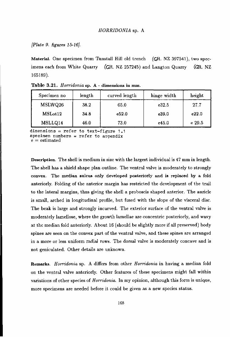
HORRIDONIA sp. A
{Plate 9. figures 15-16}.
Material. One specimen from Thnstall Hill old trench (GR. NZ 397541), two spec
imens each from White Quarry (GR. NZ 257245) and Langton Quarry (GR. NZ
165189).
Table 3.21. Horridonia sp. A- dimensions in nun.
Specimen no length curved length
MSLWQ26 38.2 65.0
MSLot12 34.8 e52.0
MSLLQ14 46.0 73.0
dimensions= refer to text-figure 1.1 specimen numbers = refer to appendix e = estimated
hinge width height
e32.5 27.7
e39.0 e22.0
e45.0 e 29.5
Description. The shell is medium in size with the largest individual is 47 mm in length.
The shell has a shield shape plan outline. The ventral valve is moderately to strongly
convex. The median sulcus only developed posteriorly and is replaced by a fold
anteriorly. Folding of the anterior margin has restricted the development of the trail
to the lateral margins, thus giving the shell a proboscis shaped anterior. The auricle
is small, arched in longitudinal profile, but fused with the slope of the visceral disc.
The beak is large and strongly incurved. The exterior surface of the ventral valve is
moderately lamellose, where the growth lamellae are concentric posteriorly, and wavy
at the median fold anteriorly. About 16 (should be slightly more if all preserved) body
spines are seen on the convex part of the ventral valve, and these spines are arranged
in a more or less uniform radial rows. The dorsal valve is moderately concave and is
not geniculated. Other details are unknown.
Remarks. H orridonia sp. A differs from other H orridonia in having a median fold
on the ventral valve anteriorly. Other features of these specimens might fall within
variations of other species of Horridonia. In my opinion, although this form is unique,
more specimens are needed before it could be given as a new species status.
168

Genus SPINOHORRIDONIA new genus
Diagnosis. Horridoniinae with both ventral and dorsal valves densely covered with
undifferentiated body and marginal spines.
Type species. Productus geinitzianus de Koninck 1846.
Other species. Spinohorridonia gerana new species and S. laspeana new species.
Discussion. Based on the type species Productus geinitzianus de Koninck, Spinohor
ridonia should be distinguished from other Zechstein and Arctic horridoniinids by its
heavily spinose valves. Spinohorridonia seems to be an intermediary form between
the dictyoclostinid genus Tyloplecta Muir-Wood & Cooper and the true Horridonia.
For the same reasons given by earlier authors who included P. geinitzianus into the
genus Horridonia and Nelson & Johnson (1968) who introduced the genus Bailliena in
the Horridoniinae, I believe that Spinohorridonia should be included into the subfam
ily Horridoniinae instead of the Dictyoclostinae. Although the anterior region of S.
geinitziana (de Koninck) is very similar to other Tyloplecta, the visceral and umbonal
regions are characteristically horridoniinid in nature. The non-reticulate ornament
on the visceral disc places this genus in subfamily Horridoniinae. The similar North
American genus Bailliena differs from this genus in the absence of marginal spines on
the dorsal valve.
As a horridoniinid, the above diagnosis is acquired using the taxonomic classifi
cation of the Horridoniinae previously stated (p. 144). Spinohorridonia is divided
into its species on the basis of the density of the spines, the shape of the shell and
the surface sculpture. On this basis, we can distinguish S. ge i nitziana (de Koninck)
from S. gerana (Eisel) and S. laspeana (Eisel). The last two species were upgraded
from Eisel's (1909) original P. horridus var. geranus and var. laspeanus, respectively.
Spinohorridonia and its species have not been reported from the British Per
mian in the past, but several species belonging to this genus have been described
under different names in the East European Permian. In this thesis, Spinohorridonia
geinitziana (de Koninck) and S. laspeana (Eisel) are described from the Permian of
169

Great Britain for the first time.
Note on Spinohorridonia gerana (Eisel). From Zone 4 of the East German
Zechstein, Eisel (1909) described P. horridus Sowerby var. geranus for the
forms which is very similar to de Koninck's (1847) P. geinitzianus in terms
of the shape and profile of the shell and the distribution of spines, but differs
from the later in having a more erect body spines and a non-costate anterior.
Details of the internal morphology of S. gerana have not been discribed in the
past, and therefore, cannot be compared with those of S. geinitziana which is
discussed for the first time in this thesis. Up till now, S. gerana has not been
found in the British Zechstein.
SPINOHORRIDONIA GEINITZIANA (de Koninck)
{Text-figure 3.19, Plate 11, figures 19-26}.
Synonymy; 1846 Productus geinitzianus de Koninck, p. 264-266, pl. 15, fig. 3. 1847 Productus geinitzianus de Koninck; de Koninck, p. 156-157, pl. 15, fig. 3. 1848 Orthothrix excavatus (part-Geinitz ); Geinitz, p. 14-15, pl. 6, fig. 20-21. 1856 Productus geinitzianus de Koninck; King, p. 260-261, pl. 12, fig. 1-2. 1861 Productus geinitzianus de Koninck; Geinitz, p. 105, pl. 19, fig. 20-21. 1909 Productus horridus Sowerby var. geinitzianus de Koninck; Eisel, p. 36.
Diagnosis. Spinohorridonia with sub-trapezoidal to rectangular shell plan, moder
ately developed auricle and large and strongly incurved umbo. Ventral valve very
strongly convex, but not geniculated. Median sulcus poorly developed. Ventral sur
face lightly lamellose with costate anterior. Marginal, auricular and body spines are
numerous. Body spines in quincunx arrangement, erect to suberect on the convex
part of the shell, recumbent on the trail. Dorsal valve moderately to strongly con
cave with flat auricle and poorly developed median fold. Dorsal surface lightly to
moderately lamellose with undifferentiated spines all over the valve, and costate trail.
Cardinal process with quadrilobed myophores, strong lateral and median ridges. Me
dian septum highly elevated anteriorly. Adductor muscle platform triangular in shape
170

with scars dendritic or radially grooved. Brachial impression small in size.
Type specimen. Material figured by de Koninck (1846, pl. 15, fig. 3) should be
elected as type specimen of Spinohorridonia geinitziana.
Type locality. The type specimen of S. geinitziana mentioned above was acquired by
de Koninck from Milbitz near Gera, where the type locality should be designated.
British Localities and Specimens Studied. Up till now, only 15 specimens of this
species are known in Britain, all from around Sunderland; 7 of them were collected
from Hylton Castle road cut (GR. NZ 360589), 6 came from Tunstall Hill old trench
(~R. NZ 397541) and 2 came from Humbleton Hill · (GR. NZ 381553).
Horizon. Ford Formation.
Age. Upper Permian EZl.
Table 3.22. Spinohorridonia geinitziana (de Koninck) - dimensions in mm.
Specimen no length curved length
MSHot13 20.2 34.3
MSLHC18 e22.5 e40.5
MSLot14 23.0 e39.2
MSLHC17 e25.0 na
MSLHC14 30.0 57.2
dimensions = refer to text-figure 1 .1 specimen numbers = refer to appendix na = not available e = estimated
hinge width
e24.5
e26.5
e29.0
e31.0
e34.5
height
9.7
13.7
13.5
na
20.4
Description. The shell is medium in size with the largest individual about 30mm
in length and ~6mm in maximum width which is at the hinge line. The shell has
a sub-trapezoidal to rectangular plan outline with small auricle and large strongly
incurved umbo.
The ventral valve is strongly convex with short trail and no geniculation. The
171
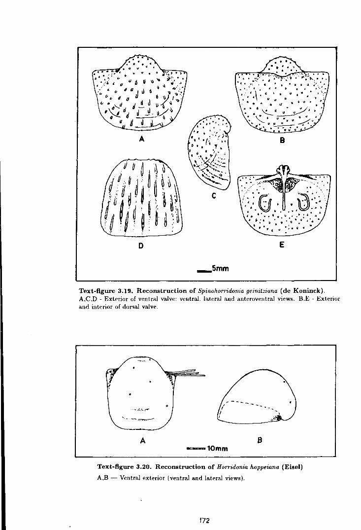
A B
c
D E
_smm
Text-figure 3.19. Reconstruction of Spinohorridonia geinitziana (de Koninck). A,C.D - Exterior of ventral valve: ventral. lateral and anteroveutral views. B.E - Exterior and interior of dorsal valve.
A B -10mm
Text-figure 3.20. Reconstruction of Horridonia hoppeiana (Eisel)
A,B - Ventral exterior (ventral and lateral views).
172

median sulcus is shallow at the mid shell length, obscure posteriorly and anteriorly.
The ventral surface is lightly to moderately lamellose and covered by numerous un
differentiated spines all over the valve. The spines are small, but long, sub-erect to
erect in nature except on the trail, where they are more recumbent. On the convex
part of the valve, the spines are a more or less quincunx in arrangement with a space
of about 2 mm or less in between spines in the same concentric rows, and 2 to 3 mm
in between spines in the same radial rows. There are about 40 rows of spines in the
larger adult individuals. The trail of larger individuals has about the same number
of sharp radial costae.
The interior of the ventral valve has a series of sharp radial furrows reflecting the
costae on the exterior; the internal mould usually carries a series of radial blades.
The adductor muscle platform is moderately elevated and is divided into two halves
by a shallow median furrow. The muscle platform appears shortly after the tip of the
umbo and it continues up to one third of the shell length anteriorward. The scars
are concentrically striated or grooved. The diductor muscle platform is very low and
radially scarred.
The dorsal valve is weakly to moderately concave with the median fold clear at
the mid shell length, but fades away towards the posterior and anterior. The auricle
is flat and triangular in shape. The exterior is regularly pitted in between sub-erect
spines which are similar in arrangement and nature to those of the ventral valve. Shell
surface is lightly to moderately lamellose. The trail is costate.
The interior of the dorsal valve is covered by fine, closely spaced , marginally
pointed endospines which are coarser at the trail. The cardinal process bears a
quadrilobed myophore, and its shaft is supported by strong pairs of posteriorly in
clined lateral ridges and median ridge. The median ridge narrows anteriorly, and
continues as the median septum which is highly elevated at its anterior extremities
(at about the mid shell length). The adductor muscle platform is triangular in shape
with dendritic or radially grooved scars. The brachial impression is small.
Comparison. S. geinitziana differs from S. gerana in having a recumbent body spines
and costate anterior margin. This species differs from S. laspeana in its densely
173
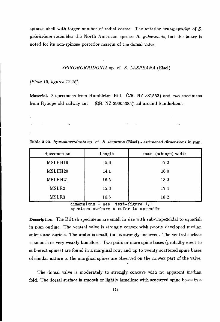
spinose shell with larger number of radial costae. The anterior ornamentation of S.
geinitziana resembles the North American species B. yukonensis, but the latter is
noted for its non-spinose posterior margin of the dorsal valve.
SPINOHORRIDONIA sp. cf. S. LASPEANA (Eisel)
{Plate 10, figures 12-16}.
Material. 3 specimens from Humbleton Hill (GR. NZ 381553) and two specimens
from Ryhope old railway cut (GR. NZ 39665385 ), all around Sunderland.
Table 3.23. Spinohorridonia sp. cf. S. laspeana (Eisel) - estimated dimensions in mm.
Specimen no
MSLHH19
MSLHH20
MSLHH21
MSLR2
MSLR3
Length max. (=hinge) width
15.6
14.1
16.5
15.3
16.5
dimensJ.ons =see text-figure 1.1 specimen numbers = refer to appendix
17.2
16.0
18.2
17.4
18.2
Description. The British specimens are small in size with sub-trapezoidal to squarish
in plan outline. The ventral valve is strongly convex with poorly developed median
sulcus and auricle. The umbo is small, but is strongly incurved. The ventral surface
is smooth or very weakly lamellose. Two pairs or more spine bases (probalby erect to
sub-erect spines) are found in a marginal row, and up to twenty scattered spine bases
of similar nature to the marginal spines are observed on the convex part of the valve.
The dorsal valve is moderately to strongly concave with no apparent median
fold. The dorsal surface is smooth or lightly lamellose with scattered spine bases in a
174

marginal row and on the concave part of the valve. The number and nature of these
spines are similar to that of the ventral valve. Other details are not known.
Remarks. The small size of the specimens from Sunderland, might represent younger
individuals of the species while some much larger specimens have been reported and
figured from the East European Zechstein. The broad folding of the trail in the East
European material is not seen in the British specimens, while the spines of the British
specimens are more erect in nature as compared to those from East Europe. However,
the distribution and density of the body spines on both valves is similar between the
British and the East European S. laspeana.
175

CHAPTER 4
THE PERMIAN SYSTEM AND PERMIAN BRACHIOPODS
OF MALAYSIA
The Permian System in Malaysia
The Permian System is well developed throughout West and East Malaysia. For
the purpose of this study discussion will be limited to the Permian of West Malaysia
from where a number of productidinid brachiopods have been described in the lit
erature. The Permian of East Malaysia is restricted to the West Sarawak and no
Permian brachiopods have been reported from there.
In West Malaysia or better known to geologists as Peninsular Malaysia (formerly
Malaya or Malay Peninsula), Permian rocks crop out extensively to .the west and east
of the Main Range. However, due to lack of detailed studies, the lateral distribution
and stratigraphic correlation are not fully understood and in many instances Permian
occurrences are only inferred (see Table 4.1 for the correlation of Permian rock for
mations in Peninsular Malaysia). For the same reason, the Permian of Peninsular
Malaysia is very seldom described as a separate system. It is usually discussed in a
brief note under an Upper Palaeozoic or Permo-Triassic heading.
Gobbett (1973b) divided the Upper Palaeozoic of Peninsular Malaysia into four
zones; the Northwest, Kinta Valley, Central and East Malaya (Peninsular Malaysia).
Discussion will be focused on the first three zones, for the East Peninsular Malaysia
Zone does not yield any productinid brachiopods. (The distribution of Permian rocks
and the localities studied in Peninsular Malaysia are illustrated in text-figure 4.1,
page 177).
176
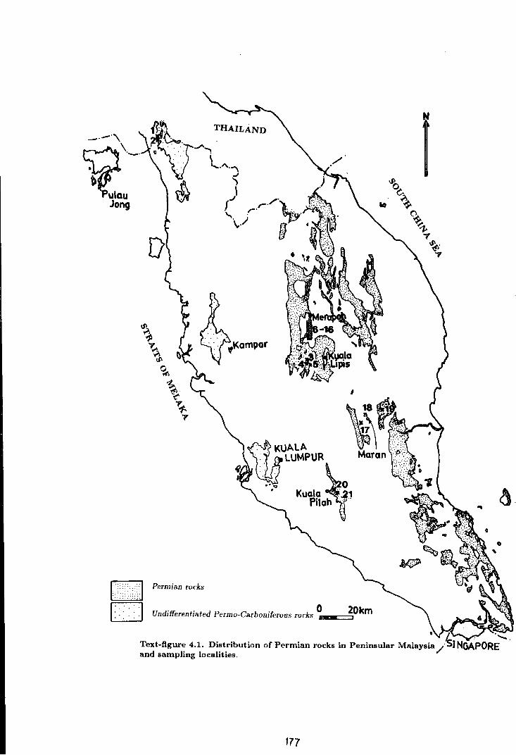
-·-·"\
t2:-Jong
~ L.jj_J ~ ~
I
Permian rocks
0 20km Undifferentiated Permo-Carboniferous rocks --=~
• L. . . ........ Text-figure 4.1. Distribution of Permian rocks in Peninsular Malaysia /SINGAPORE and sampling localities.
177

The Northwest Peninsular Malaysia
In the Northwest Peninsular Malaysia, Permian rocks occupy part of Perlis and
North Kedah and this outcrop extends across the border to the Thai Peninsula. The
Permian sedimentary rocks of this zone are deposited on a more stable platform as
compared to those of the Central zone. It is mainly made up of an unfossiliferous pure
calcitic limestone of the Chuping Formation ( Gobbett, 1973b ). The basal part of this
formation, however, is highly fossiliferous and the fauna gives an upper Lower Permian
age to this horizon (Newton, 1926~ Scrivenor, 1926, 1931, etc.). The exposed basal
Chuping Formation comformably overlies the argillaceous Singa Formation in the
Langkawi islands and the arenaceous K ubang Pasu Formation in Perlis. Although
the detailed stratigraphy of these underlying formations is not fully understood, it
is believed that the basal Permian lies somewhere within these beds. The top of
the Chuping Formation is not well exposed, but in Kodiang area, North Kedah, it
is in places comformably overlain by the impure Kodiang Limestone which is Lower
Triassic in age (Metcalfe, 1981 ).
Several Permian fossil localities have been recorded from the transitional or pas
sage beds from Singa and Kuban[ Pasu formations to the Chuping Formation in the
Langkawi islands and in Perlis. Permian brachiopods have been reported from Pu
lau Jong, Pulau Singa Besar and Kisap in Langkawi islands and from Bukit Tungku
Lembu and Bukit Temiang in Perlis (see table 4.2 for detail brachiopod lists).
The Kinta Limestone and Kenny Hill Formation
-the limesione A series of limestone hills expose within the Kinta Valley in Perak, bu~has , been
subjected to a certain degree of metamorphism, such that fossils, especially the macro
fauna, are scarce. However, from a series of a bioclastic limestones exposed in several
tin mine pits in the 1960s, very rich and diverse macrofaunas have been recorded.
These macrofaunas include many species of gastropods, bivalves, brachiopods, bry
ozoans, corals, algae, ostracods and fusulinid forams (see Toong, 1965, Sunthar
alingam, 1968, Batten, 1972, Runegar , & Gobbett, 1975, and Ishii, 1966). The
fusulines from H. S. Lee Beds belong to the Parafusulina Zone of late Artinskian
178

- early Kungurian Stages (Ishi, 1966) and the brachiopod limestones ( = The Nam
Loong Beds) below it are assigned to Sakmarian Stage by Gobbett (1973a, 1973b).
The fossil record of the Kinta Limestone is not continuous and comprises of a range of
fossils from Middle Devonian to Middle Permian time. Although detailed stratigra
phy of the Kinta Limestone is not fully understood, Suntharalingam (1968) suggested
that the basal Permian - Carboniferous boundary is best interpreted between N am
Loong Beds and the top of Kim Loong No.1 Beds. The youngest part of the Kinta
Limestone (The H. S. Lee Beds) does not reach the Upper Permian (see stratigraphic
column for Kampar in Table 4.1 of this chapter).
To the south, a sequence of unfossiliferous argillo-arenaceous rocks known as the
Kenny Hill Formation represent the Upper Palaeozoic in Selangor. The discovery
of Agathiceras sp. from this sequence proves that they extend to Permian in age
(Geological Survey Department, 1986).
The Central Peninsular Malaysia
The Permian rock succession is well developed in Central Peninsular Malaysia or
the Central Belt of the peninsula. Most of the Permian rock succession within this
zone is associated with volcanic and pyroclastic rocks which indicates that they are
deposited in a very unstable depositional basin. The common occurrence of plant
remains to the east of this zone indicates that the shoreline is lying somewhere to the
east.
In the west of this zone, the Permian rocks crop out extensively along the east
ern foothills of the Main Range from Northwest Kelantan southward to Northwest
Pahang. They are best known as the Gua Musang Formation and consist mainly of
limestones, slates, indurated shales and mudstones, volcanic and pyroclastic rocks.
The Gua Musang Formation often occurs as a roof pendant on top of granite bodies
or as an underlying sediments beneath the younger Triassic rocks. They are gener
ally folded and faulted and are very difficult to correlate either laterally or vertically
(Richardson,1947, 1950; Gobbett~·· 1973b). From the several localities investigated,
the Permian fauna of this area is mainly preserved in argillaceous rocks. The lime-
179

Table 4.1. Permian stratigraphic correlation of West and Central Peninsular Malaysia
- · WEST PENINSULAR MALAYSIA CENTRAL PENINSULAR MALAYSIA PERMIAN STAGES -
SYSTEM ·· -·· · .. . South Central Northeast Langkawi Kerlah/ Kmta valley Nt!~eri Nort.hwest. Kelaut.an Pahaug Islaurls PerliH · ·- . St!mhilau Pahang .
USSR platform Sunthara.hugaw 1968 ·Aw et a/. 1978 Font.ame d a/. 1988
00 ~ o <::: g. Upper Triassic ~ 0 b.O 0 0 ;;; .., !:!' ·.;: ... Temheliug Gp ~~;a tl
TRIASSIC ~ .!3 5 ~ Gemas ~ SPmaut.au ~"V"v ~ 'v.J
..J c:n ""' Formation ~ Formation I
------
..,- ?_.,_.,_ -----:-7-1.- = Dorasluu11iau ·.3 LqJtodu~ FSKN73
~ ! B~ § ~ E -- ~ ~ o ~ ~v·~~~~AA~~ ~ Dzulfiau t... § ~
Z ;J .. ... :f ~ Jengka Pass <t
MidiazJ ~ :f shale & ~ ::iE ·c: sandstones
...... <t ...;
~ Murgubiau c) J~ngka Pass ~ ,..._.,_,""''v'"''""'· limestones
~ Q . Chuping Lir~estone · · · · · _ _ ______ _ Q KubergaiUban Formation
~ SS I H. S. Lee Bolorian B('(l
foil - -l-l--1-'->-
Artinskian ~
~ ~ 3 Kepis Bed FSKN4 ? 7 r&l g) N am Loong !l ~ Sakmarian S B 1 ~ 0 ;.:3 ec 2 ..J ~ :.e . s· Kubang .-. .,
As.~e!Jan mga .9 ~ Formatiou Pa.su ;:.:: c; (argillic) Formation 5
(arenic)
CARBONIFEROUS Kim Loong - - - - - - - -I ~ K~~
Group
0 co
stones, although they are locally fossiliferous, have not had detailed faunal studies.
From this area, the Upper Permian Leptodus shales (in this thesis some of them are
considered as weathered slates) are known in several places including Sungai Jemuru
in Kuala Lipis, Sungai Yu in Merapoh and Sungai Galas in Gua Musang (Muir-wood
(1948), Jones et. al. (1966)). Occurrence of similar Leptodus shales and slates are
first recorded in this thesis from Kampung Gua, Kampung Garok and Terenggun
near Kuala Lipis and from the Dada Kering- Gua Musang highway.
Southward, isolated Lower Permian rocks are exposed in Negeri Sembilan, nearly
parallel to the strike of the Gua Musang Formation. The Permian rock succession
here is called the Kepis Beds and it is mainly made of indurated mudstones and
shales, interbedded with polymictic rudite, arenite and argillite and carbonaceous
limestones. Lower Permian faunas have been recorded from Kampung Juaseh and
Kampung Baharu Kepis in Kuala Pilah area (Khoo, 1985).
The east of the Central Peninsular Malaysia is mostly covered by Mesozoic rocks.
Permian rocks commonly occur as patches in faulted blocks or as underlying sediments
beneath the Triassic rocks. From south Central Kelantan, both Lower Permian and
uppermost Permian faunas have been recorded by Aw et al. (1978), Toriyama et.
at. (1975) and Yanagida & Aw (1979), while Maran Upper Permian faunas has been
recorded by Fontaine et al., 1988; Ichikawa et al., 1966,; Ishii,, 1966;, N akazawa,
1973, etc. The south Central Kelantan faunas are found in tuffaceous limestones
which are associated with slates and pyroclastic rocks while the Maran faunas are
found in limestones, carbonaceous shales and in sandstones. In North Pahang, Middle
to Upper Permian fauna have been recorded by lgo (1964) from Ulu Sungai Atok and
Sungai Sepia. These fauna are found in silicified limestones and shales.
The southernmost fossil occurrences within this zone have been reported in the
Geological Survey Annual Report 1987 from Permian tuffaceous sediments below the
Triassic Gemas Formation in north and northwest Johor.
181

The Eastern Peninsular Malaysia
The eastern Peninsular Malaysian Permian rocks are well developed on top of the
Boundary Ranges (at the border between Terengganu - Kelantan and Terengganu -
Pahang), in Southeast Pahang and most of the East Johor. Besides its well known
flora, Permian marine faunas (mostly corals and fusulinid forams) are found in several
limestone hills in Terengganu and J ohor (see Fontaine et a{~ 1988; Fontaine & Khoo,
1988; Kato & Ezaki, 1986) •.
Permian brachiopods
Brachiopods form an important element of the Permian faunas of Peninsular
Malaysia. Extensive faunal lists cited by various authors have given a big catalogue
of Permian brachiopods (see table 4.2). However, only a few of them have been
described systematically.
The first and most comprehensive list of the Malaysian Permian brachiopod fauna
was published by Jones et al. (1966) with most of the identifications made by Gobbett,
one of the co-authors. In this first major summary of Malaysian fossils, more than
half of the total recorded Permian brachiopods listed in table 4.2 are given. Prior to
this publication, Permian brachiopods were not well known in Malaysia. The first
work to mention the occurrence of Permian brachiopods in Malaysia was made by
Wilbourn (1926) when he described the fauna of Bukit Temiang. Muir-wood (1948)
noted another occurrence of Permian brachiopods from Sungai Jemuru, Pahang. The
only descriptive work on the Malaysian Permian brachiopod fauna before Jones et al.
(1966) was made by lgo (1964) on the late Middle Permian brachiopods from Sungai
Sepia, Pahang in which he described three different Dictyoclostus sp. (renamed as
A ntiquatonia in this thesis) as well as several spiriferids.
Considering that very little is known about Permian brachiopods in Malaysia
before Jones et a[. (1966), the work is clearly a major breakthrough in this field. As
in many other pioneer works their review is not very helpful for later workers. This is
182
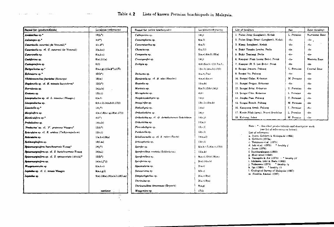
haul list (produetidioida)
A..-....Unuop.•
Aal...teyoosp.•
C~llo eoncrini (de Verueuil)"
CGIIn'itarllo sp. d. C. conerini (de Vern•uil)
C~IDop.
Cutiforint~ op.
Ct~Miqiraq•ro op.
Diefr«k>•tu op. •
E<Amtia...U op. •
f&:Ainoconchru fut:iotw (Kutorga)
H~IID op. d. H. minuta Sarytch•va•
Horridonio •P·
Krolovio sp.
~uctru op. d. L. linealu.o IWaag•n)
Lirwprodaetw op.
LioM>klla ep. •
llarginifrrG op.
IIMJJkalif•ru op."
I'Yol>oliolino op.
"Protlvctu' sp. cf. ·p ·. gratio.sw Waagt~n•
IU~io sp. cf. R. urolieo (Tl!Cbyerny•chew)
~tiosp.
~rraop.
~raqeru bftcJaou,.,..U Huang•
~rra op. d. S. lcaeichowmou Huang
~eru op. d. S. qinocollala (Abich) 0
~eruop.
W_.,hoop
Lql»du op. d. L. tenuu W&a«en
Le,llldtu ap.
Table 4. 2 Lit~ts of known Pt!rmiau brachiopod:-; in Malaysia.
~------------------------~-------------Loca.liti .. (mereuc .. )
12(h0 )
4(d")
4(c.d")
S(a.b.c)
6(a.b.c)
6(a).ll(a)
6(f)
8(a.c.g).(l5a.k*).( 171)
12(b0 )
18(m)
12(b 0)
14(a.b)
lJ(c.l)
G(a.f)
6( b.c .1),14( a,b.l ).17(1)
14(j")
4(a.c ),8(a.c,g).9(a).l7(1)
4(d 0)
14(a,b)
12(b")
13(c.l)
l(a,b.e).9(a)
18(1.m)
14(j*)
14(a.i)
12(h0 )
14(a.iJ",I)
6(a.b,c)
8(a..c.gJ)
9(a).IO(a).l4(a.bJ),l8(1.m)
continue .. ,
Fauna.! list (oth•~r brar:hluJuul!tl
Call~pirina ttp.
Comrrophoria sp.
Cam~rotMthia ap.
Chonetr• op.
Comp.uita "P·
CrtnUpiri/t!r !i1p.
D<rl>via op.
Dir.lumoap.
Haml<t.lk. sp. d. H. alta I Hamlet I
Hwt.dia op.
Martini asp.
Microphrlia Hp.
N<ophricodothyru •P·
Nro•pirifr.r ep.
Nothothvna •r>·
Orthoth<thina •P·
Orthotichia 1p. d. 0. doraAhamrnsu SokulNita..Va
OrtAotir:hio Mp.
Phricodolhvria sp.
Purdont"/lo ap.
Schellwlt·n~lla ~p. d. S. robrr I Frt~f:h)
Sl'hi:ophona ~p.
Spirijer •P·
Spirifr~llino rri•taio ( Schlnt hd111)
Spirifrr<llina •V·
SpirifPritlG Hp.
Squamularia Np.
Stt!no6riJtma sp.
st ... ptorhpch!U< •J>·
UnrinuJru 11p.
Uncinu,.ellinG limo,..,...;. ( Bryrirh)
W011geniteo op.
L()(';llilit·!'ltr••ff'ff'Ul"('f"j
141jl
6ia.li
6(a.f)
6ia.f.ll
21•.• ).6(a..f).l6ia.)
141jl
41rl ).6(a.h.d.l).7ia.l 1.
13tc.ll.l41a.b}.l7111
5~ a..r J, 71 al
4{a.r}.~(a.e)
15(a.k)
Gta.f i.l2ih ).14(j)
4trll
14(jJ
13ir.ll.l~la.k)
141•1
141jl
Hlcjl
H(a.ll
13tr.l)
13idl
141a.i.ll
131...11
6(a.b.dl.81u}.l7(1}
15(a.k)
8ta.c).12ihJ.I8(ml
91a).l41".hl
2(a.e)
6(b.c)
2(a.•·).9ial
Jla.r).9ia)
8(a.jii)
1711)
List of loca.litit•,. Age Zont i\ocl\lity}
1. Pulau Jong (Laugkawi}. Kedah L. P~rruinu N urt lowest Zon• I 2. Pulau Singa 8f.AAr (L.,ngkawi). Kedalo -do- -do-
I 3. Kiaap. LAngkawi. K<dah ·do- -do·
4. Bukit Thn~ku L..mhu. P•rli• ·do- -do·
5. Bukit Trmiaog. Porli• -do· ·d<>-
6. Kampar INam Loong 8<-ds). Perak -d<>- Wetitern Zone
7. K&ropar (H. S. L..., B••dsl. Porak ·do- ·do-I
8. Sunga.i J.-m•uu. Pahan~ U. Permian Central Zoue I
9. Sunga.i Yu. Pahang ·On- -do-
10. Sung.U GaJM. 1\rlantan M. Pt>nnJa.n ·do·
11. Sung&i P'"rgau. Kdn.ntan ·rlo- ·do·
12. Sungai Rrl.U. Krlantan U. P~rmlau ·do-
13. Sungai C'iku. Krlanta.n L. Pf·rmia.n -do-
14. Jt>n~ka Patul, Pi\haug U. Prrmian ·do-
Ia. Sungai Remih. Pahang M. Prnuia.n -do·
16 . .Kampung Awa.h. Pa.ha.ng ll. PPrmian ·do-
17. Kuala Pilab (ar~a). Nt·~~·ri $f•mbilau L. Pf•rmian -do-
18. Kuhau,; . .Johm M. Pt'nui.L.n -do·
Note : • - ci..snihrd pru(hwtidiuids and d1·~nipt.ivr work (!'let> lisl of rl•fprt~lu:ew·t~ij ht'low):
List of rt!(t'rPnrt·~:
a. Jon•:!!. Guhbrtt .\.: I\1Jhaya.'jhi ( 19GGJ b. Gnhb••tt (1973a) c. Toriya.ma •·t.aJ. (1975) d. bobi et.al. ( 1972) • !orality. •· Jon.,. ( 1976) f. Suntloara.lingan• (19GB I g. M uir·wood il94S) b. Yanagida & Aw 1197~1 -· • loralitu 12 i. Ichikawa. bhii .It Hada 119G6) j. N akazawa 11973) ' localil u 1 • k. I go ( 1964) · • ltH'alrt.v IS I. G.ologiraJ Survey of Malay•ia (1987) m. lhrahirn Arunan IIV87)

because there are many differences of opinion on fossil identifications given by later
workers for the same localities. Even Gobbett himself ( Gobbett, 1973b) has identified
different faunal elements and revised faunal lists for some of the localities which he
listed in Jones et al. ( 1966).
Current research has failed to locate the collection of fossils identified and sum
marized in Jones et al. (1966) for the purpose of re-identification. For this reason the
list of the Permian brachiopods in table 4.2 is not updated and a more reliable list
of Permian brachiopod faunas that have been studied by the author is given in table
4.3.
Current research and material studied
The purpose of this research is to identify and determine the distribution of
Malaysian Permian productidinid brachiopods and to study aspects of their ecology
and evolution. During this research material from the collection of the Malaysian
Geological Survey Department and the Geology Department of the University of
Malaya were studied besides a large collection made by the author before and during
the tenure of this research.
The Malaysian Geological Survey Collection (GSD collection).
Without their own palaeontologist to work on the local fossil collections, the
Malaysian Geological Survey Department usually sends fossils overseas to be identi
fied. Some of these fossils were described by foreign experts as a matter of personal
interest. The identified and described collection is carefully labelled and well stored.
With this excercise and with limited funds available, normally only part of the fos
sils collected are identified. Those which are not well preserved and those which are
difficult to transport remain unidentified. Most of the unidentified material in the
Malaysian Geological Survey collection comes from the states of Pahang, Kelantan
and Negeri Sembilan.
184
During this research, with cooperation from the Malaysian Geological Survey
officials, their geologists (En. Khoo Han Peng and En. Ibrahim Amnan) and their
senior laboratory assistant En. Amran N ali, these collections were made available for a
brief study. Owing to limited time available, attention was foc.used . on material which
came from remote localities which have not been visited during the current research.
The good records kept by the Malaysian Geological Survey Department allow the
excact location and origin of the large unidentified collection to be determined.
The University of Malaya collection (UMcollection)
A substantial collection of Permian fossils including some brachiopods was made
over the last few decades by various collectors in the Department of Geology (Univer
sity of Malaya), mostly undergraduates who studied a particular region for their final
year thesis. Of all those collections, the Permian brachiopods from Kampar are the
most interesting. This is partly because of the abundance and very diverse composi
tion and partly because this particular collecting locality, which was once the floor of
an open cast tin mine, is no longer available for further research due to the flooding
of the site in 1966 (Runnegar & Cobbett, 1975)~. By consent of the keeper of this
particular collection (Dr. ldris Mohamad) this material was brought to Durham for
more detailed study. The record of the collector is not very clear, but I beleived that
this material was gathered together by Suntharaligam, Toong and Cobbett before the
pit was flooded.
Current Research collection (UKM-MSL collection)
Owing to limited time and funds available collecting work for this research was
restricted only to those easily accessible Permian brachiopod loca li. ties in Perlis,
Pahang and N egeri Sembilan. Part of the collection has also come from field work
made during the tenure of the University Kebangsaan Malaysia's Research Grants
Nos 23/85 and 10/86 for the study of the general geology of the Kuala Lipis area and
the Dada Kering - Gua M usang highway.
185

The current research collection comes from Bukit Tungku Lembu and Bukit Temi
ang in Perlis, from Kuala Lipis, Merapoh and Maran areas in Pahang and from
Kampung Juaseh and Kampung Baru Kepis in the vicinity of Kuala Pilah, Negeri
Sembilan. For further details on the distribution of these localities see text-figure 4.1,
page 177. The geology of these collecting localities can be summarised as follows;
PERLIS AREA
Locality 1. Bukit Tungku Lembu, Perlis (GR. QN 247509)
About 40 meters above the base of the western foothill of Bukit Tungku Lembu
the highly fossiliferous transitional or passage beds from the K u bang Pasu Formation
to the Chuping Formation are exposed. At this point, the thickly bedded to massive
quartzitic sandstones of Kubang Pasu Formation are seen to change gradually
to recrystallized pure calcitic limestones of the Chuping Formation. The sandstone
at the base of this exposure is about 20 to 30cm thick and it is overlain by about
3.5m of medium to thickly bedded brownish calcareous sandstone which in places
contains small-scale, moderate to high angle cross-bedding of possible ripple origin.
The top 1m of this bed is occasionally interbedded with shelly layers of Cancrinella
cancrini (de Koninck). This calcareous sandstone is overlain by about 1.2m thin
to moderate, irregularly bedded impure bioclastic limestones. The bottom 1m of
this bed is dominated by Cancrinella cancrini with very few other accessory elements
while the top of it is made of up to 20cm thick bed of very fragmented crinoid ossicles.
This bioclastic limestone bed is followed by about 2m of eroded beds of softer rocks
probably equivalent to the grey-green calcareous flaggy siltstone and shale beds of the
Bukit Temiang succession (see Gobbett, 1973b), Above this eroded sequence are
few meters of fine and irregularly bedded limestone interbedded with argillaceous and
cherty laminae. Isolated fossils of unidentified gastropods, bivalves and brachiopods
can be seen in these thin bedded limestones. These thinly bedded limestones gradually
pass through to a more massive fine grain recrystallized limestone of the Chuping
Formation. Here, gradual changes in colour from dark grey to light milky colour is
seen from the bottom to the top of this limestone succesion. For further details, see
186

Siltstone and Bhtlle
LifY!estone
Sanc:blone
&rachiopoda
Crinoidea.
--
.. ·--- ~ ..
-·~ ""\ I ] \
I :'\ I I )
. .~, ·-.r.-v ••loll1 ~ "' !Wid' I . ·.I .IC7, ·'Of . . .J
.. ~- . ~-. I
.-.·-•··-r·~ • f·.. ..
-I-- --1: ~L~~J.
-~- ~ . ~-
:T--·1·~· . . . _, . ,_. ----l.-; . :. . I .' . I ~+·-~-.·<'I . :-:--~; --~_\ -·· --~---_1.-,-q
.. -~ ._,.:J,·. }t :.-: .... ·, . '· . : ·: . . :\
::.·:_.-_:~;>':8 ·.:--.·~:-.:.::-~ .-:-.·.:- _.··,~:·.-·~
··~· .. -:~) . .-... ·-.,;....L. -: .... '·_: . .. ·j
CHUPING LIMESTONE FORMATION
-------------
CRINOIDAL LIMESTONE
CANCRINELLA BEDS
.- _. ~--:-: ·.-·i· ;r_ r.' :; . __ _
... ··· :_ · .. : ..... KUBANG PASU FORMATION . · :: .. ·.· ....
.. . ...
Text-figure 4.2. Stratigraphic column of the transitional beds at Bukit Tungku Lembu, Perlis.
187

text-figure 4.2.
Fossils and their preservation. The fauna of these transitional beds are mainly made
up of brachiopods C. cancrini and fragmentary crinoid oss-icles with very few accessory
elements (see table 3 for the full faunal list). Most of the fossils are found in either
the C. cancrini or the crinoid beds, some C. cancrini are also found in shelly layers
on top of the calcareous sandstone beds and even on the flank of the crossbedded
sandstones.
Most of the brachiopods and the bivalves are disarticulated and most of the
productaceans have lost all their spines, while the crinoids and the bryozoans are very
fragmentary in nature. This shows that the fauna has been subjected to a considerable
degree of transportation before being laid down. In terms of preservation, some of the
original shell material is still preserved showing good internal and external details,
while some others are only preserved in the forms of moulds. '· Dissolution
of the original shell material probably occuJTed partly during diagenesis and partly
during weathering of the rocks after being exposed at the surface.
Locality 2. Bukit Temiang, Perlis (GR. QN 224508)
Very similar transitional beds has been reported by Cobbett (1973b) from Bukit
Temiang, about 2.4 Km south of Bukit Tungku Lembu. From an isolated exposure
of C. cancrini beds a few fossils mostly C. cancrini itself have been collected.
KUALA LIP IS AREA, PAHANG
The Gua Sai and Gua Barna limestones in the vicinity of Padang Tengku (in Kuala
Lipis Map Sheet) has been considered to be of Carboniferous age since the work of
Newton (1913), Procter (1975) (see also all the Geological Survey Annual Reports and
Maps). Unfortunately, no further discovery offossils has ever been reported to confirm
Newton's findings. Procter (1975) divided his Raub Group into three formations in
which he introduced the Padang Tengku Formation as the oldest formation, consisting
188

the two limestone hills mentioned above and the surrounding non-calcareous rocks. He
also generalized that his Raub Group is more or less younging westwards and assumed
that his Triassic Lipis Group immediately east of Padang Tengku is unconformable
over the Padang Tengku Formation. During my research on this supposed Lipis
Group, I have failed to traced any unconformity between the non-calcareous rocks
surrounding these two limestone hills and the overlying Triassic rocks. Furthermore,
I also found that the limestone at Gua Sai pass laterally through to the non-calcareous
rocks to the west in which a Leptodus shale is found; Leptodus bearing rocks in this
area are known to be of Upper Permian in age (Muir-wood 1 1948; Jones et al., 1966 _;,
Gobbett, 1973b ). Three Leptodus bearing fossil localities are reported for the first
time in this thesis and they come from Kampung Gua, Kampung Garok and north
Terenggun.
Note: Metcalfe (personal communication) suggests that the conodont assemblages
of Gua Sai limestones are of Permian in age. The use of the term Raub Group at
present is restricted to the Triassic formations and the term Lipis Group is no longer
in use. The Permian rocks in Kuala Lipis area have much in common with the Gua
Musang Formation found to the northwest of Kuala Lipis Map Sheet. Therefore, in
this thesis the author employ.sthe term Gua Musang Formation for the Permian rock
succession in Kuala Lipis area (see also Mohd Shafeea Leman, 1987a).
Locality 3. Kampung Gua (GR. VE 65654355)
On the road (track) cut west of the Gua Sai limestone hill, thin to moderately
bedded silty shales with a few fossils are exposed. About 30m south of it, on the track
surface the shale becomes much finer and yields a much richer fauna with Leptodus
present. The shale is reddish in colour and very highly weathered. The exact position
of this shale is not known due to its highly weathered nature but it is estimated to
be about 1 or 2m below the silty shales mentioned earlier. The fauna is preserved in
the form of moulds probably as a result of weathering. The brachiopods,
bivalves and solitary corals are normally preserved whole and scattered within the
sediment body.
189
Locality 4. Kampung Garuk (GR. VE 642344)
About 2.5 km south of Kampung Gua exposure, another track exposure revealed a
slightly different fauna in a different kind of lithology. Here, fossils including Leptodus
are found in a highly weathered tuffaceous siltstone and shale sequence which overlie
a wedge of massive agglomerate and lappili tuff. The fauna in these highly weathered
tuffaceous rocks are not well preserved. They are all preserved in form of moulds >:~
: with traces of sulphide dissolution salt, indicating that the shell has probably
been replaced by pyrite or other sulphide minerals prior to weathering process. The
Kampung Garok fauna are also found as whole specimens and scattered within the
sediment body.
Locality 5. North Terenggun (GR. VE 62454530)
About 0.9.km north-northeast of Terenggun Dam, lapilli tuff that is similar to
those of the dam site is exposed. This lapilli tuff is overlain by tuffaceous silty
shale which contains a few isolated brachiopods. The tuffaceous silty shale gradually
becomes finer upward and at around GR. VE 62454530, which is about 5m above the
lapilli tuff bed, it yields an interestingly rich fauna of dwarf brachiopods associated
with some larger lyttoniids. This fauna is contained in a very fine tuff which is
highly fractured and cleaved so that the large lyttoniids (mainly Leptodus) cannot
be collected unbroken. Some of the fossils from this locality still have their original
shell material preserved because the rock sequence was exposed only a short time
before the collecting work was done. As in previous localities, most of the fossils are
preserved in form of whole shells, scattered within the rock mass.
DADA KERING - GUA MUSANG HIGHWAY (MERAPOH MAP SHEET)
The Dada Kering - Gua Musang highway which connects Northwest Pahang
and Southwest Kelantan passes through two major rock formations, the Permian -
?Lower Triassic Gua Musang Formation and the younger Bukit Tujuh granite which
190

~ Permo- Triassic limestones
I L .,..,- - --"'
KUALA LIPJS
km
Text-figure 4.3. Distribution of localities 6 to 16 along the Dada Kering - Gua Musang Highway, Northwest of Pahang.

baked part of the Gua Musang Formation during its intrusion (Mohd Shafeea Le
man, 1987b ). In between Bukit Tujuh and Merapoh, highway cuttings expose several
Permian fossiliferous localities (see localities 6 to 16 below).
Locality 6 - 15. Gua Panjang - Sungai Yu halt
More than ten fossiliferous localities can be seen in several road cuts between Gua
Panjang (Merapoh) and Sungai Yu halt (Km 12 to 16.2 Merapoh- Kuala Lumpur).
An isolated fossiliferous locality is also found on the road cut immediately north of
Sungai Toh (about Km 31.2 Merapoh- K. Lumpur). Details on lithology and fossil
contents show that all these fossiliferous localities represent a singular fossil horizon
within the Upper Permian of Gua Musang Formation. The most important faunal
element in this horizon is Leptodus or related lyttoniids which can be found in almost
all fossiliferous localities. Therefore this horizon can be tentatively related to the
Leptodus shale of Muir-wood (1948).
Nearly all fossils from these localities are found in form of moulds - which
sometimes reveals accurate internal and external morphological details. The major
problem faced in identifying these faunas is that they are usually strongly deformed
by lateral compression in additon to the burial compression.
Locality 6- CR. QZ 08174470. In between two mogotes of horizontal thinly bedded
limestones ( Gua Panjang hill in the west and unnamed hill in the east) lies a small
exposure of highly weathered tuffaceous pebbly sandstones overlain by highly fossil
iferous weathered slates. The fauna of these slates either accumulated in nearly hori
zontal layers or sometimes lie scattered within the sediments. In either case, they are
generally highly deformed by lateral compression. This is shown by re-arrangement
of fossil lineations in~o an almost vertical axis striking north-south, parallel to the
secondary cleavage within this rocks. The fauna is made of brachiopods (mainly the
spiriferids), bivalves, bryozoans and some remains of trilobite pygidia.
Locality 7 (GR. QZ 07854485) is an extension of the slates from locality 6, but the
rocks here are much more weathered than that of the previous locality. However,
192

a few remains of fossils including some brachiopods are still recognisable from these
highly weathered rocks.
At locality 8 (C.R. QZ 07254485) weathered indurated mudstones of less compressed
nature than the slate from locality 6 are exposed in a drainage trench west of the
highway. This slightly weathered indurated mudstones yields numerous brachiopods
including some productidinids, gastropods and some bivalves. Accumulation of fossils
into one particular layer is rather less common, but whenever this occurs, fragmented
shells are conspicious.
At locality 9. (GR. QZ 069448) the highway cuts about 35m of very hard yellowish
grey slates which are rapidly softened and altered to reddish in colour by weathering.
The highly weathered slates from this locality are very similar in appearance with
that of locality 6 and 7. The fauna of this locality commonly accumulated on laterally
compressed bedding planes, so that the fossils are highly distorted.
Locality 10 - GR. QZ 065447. The surface exposure afforded by an abandoned con
struction vehicles station at this locality reveals dark grey to reddish tuffaceous shales
rich in brachiopods (especially large orthids ), bivalves and gastropods. Also found
here are bryozoans, trilobites and plant remains. Most of the fossils are well preserved
with slight compression to the original shape following the shaly cleavage. Some of
the fossils still have their original shell preserved.
Locality 11 - 14.- ~GR. QZ 06084442, 05804435, 05604430 and 04804478. Weathered
indurated mudstones similar to that of locality 8 are exposed at several places between
locality 9 and Sungai Yu halt. Faunal elements of these localities vary from one
another, but in general the fauna is dominated by brachiopods with Leptodus or
related lyttoniids forming an essential element.
Locality 15- Sungai Yu Halt.-. GR. QZ 04204405. A large road cut east of Sungai Yu
halt exposes reddish weathered slates overlying less weathered dark grey calcareous
slates with thin lenses of marl and oolitic limestone. Fossils are found in several layers
within the weathered slates, calcareous slates and limestones. They are normally
affected by a quite strong lateral compression, similar to that of locality 6 and 9.
193

Most of the brachiopods are found in the weathered slates and the less calcareous
slates.
Locality 16. North Sungai Toh road cut , '(c;R. QZ 95204508)
An isolated fossiliferous locality is also found in the road cut immediately north
of Sungai Toh (about Km 31.2 Merapoh- K. Lumpur). At this locality, the highway
cuts through about 230m of the western flank of folded tuffaceous pebbly sandstones,
sandstones, siltstones, shales and indurated mudstones. The southern half of the
exposure forms a normal open fold while toward the north end of the cutting it changes
to an isoclinal fold. Fossils are found on both flanks of the open fold which are exposed
on both sides of the highway. This open fold shows very interesting lithological and
biotic changes from the bottom (south) to the top (north). The core of the fold
is made of unfossiliferous tuffaceous pebbly sandstones and non-pebbly sandstones
which are uniformly moderate to thickly bedded. This sandstone sequence is followed
by an irregularly bedded tuffaceous sandstone, siltstones and mudstones commonly
grading one into another, with occasional beds of coarser pebbly sandstones. At the
top of this sequence, the coarser tuffaceous sandstones and pebbly sandstones occur
as small scale channel lag deposits or in the form of small lenses within finer tuffaceous
rocks. At these horizons the tuffaceous sandstone - siltstone - mudstone sequences are
frequently interbedded with laminae of plant debris, some of which are replaced by
recently developed iron pan. Above this sequence the flora becomes less fragmented
and individual specimens are recognisable, while the sand grains become finer. The
first fauna appears when the sediments becomes finer and regularly bedded about
6m above the first horizon of plant debris. At first, both fauna and flora are found
together, but after about 0.5m the flora vanishes and is succeded by a succession
about 7.4m thick containing an Upper Permian fauna. The sediments enclosing this
fauna are made of tuffaceous siltstones or very fine sansdtones. They fine upwards
continuously upwards until they finally become a very fine indurated white tuffaceous
mudstone at the top of the whole succession. Within this faunal succession, several
fragmentary shelly layers are traceable within the first 4.5m, while above it the fauna
becomes scarce as the sediments become very fine. Most of the fauna are preserved
194

r=-=-=1 Tuffaceous ~ mudstone
~ T1.1ffaceow L::..::...::..: Sande'l:one
~Pebbly ~ Sandsione
/9' Plan{ rE'mail) rem
. Srachiopocla
* l I
:·:.: =·; 7.: :·,.~7.: :.;-~.~< ·: :;: :. ......... ,.. .... ... . .· :/:i :~:\::~~- \~~·~; .. :
c- - -
-_--~ -_-
- --~--- - -·-~.:...-:-.:a... - -:._: -:,._---:_ -:·~~-. - .-.-.. - ~ .. ;·:-:. .··. - -· .. -. ~ . , :
·-.::-:~~-~-,;._ - ~. 7~ .. ·,;.. - .-.
Text-figure 4.4. Stratigraphic column at locality 16, north of Sungai Toh, ;., ''· Merapoh, Pahang.
195
in the form of moulds.
illustrated in text-figure 4.4.
Details on the rock section from this locality is
MARAN AREA, PAHANG (EAST) (MAP SHEET 71, 89 &: 90)
Several isolated Permian outcrops have been reported from this area including
the well documented Jengka pass Permian. In this thesis, faunas from two other
fossiliferous localities are introduced beside the Jengka Pass fauna. They are fauna
from Bukit Jaya road cut and from Maran Oil Palm mill.
Locality 17. Jengka Pass (C,R. WF 91651445)- Sheet 89
The Jengka Pass exposure and its celebrated unconformity in between the Up
per Permian and the Upper Triassic has been illustrated in great detail by Gobbett
(1973b), Kon'no & Asama (1970) and Nakazawa (1973), while its fauna and flora has
been mentioned in so many publications and well described in Fontaine et al. (1988),
Ichikawa et al. (1966), Ishii (1966), Kon'no & Asama (1970) and Nakazawa (1973).
Permian brachiopods have recorded from both the limestones (Gobbett, 1973b),
the carbonaceous shales and the tuffaceous sandstones (Ichikawa et al., 1966 and
Nakamura, 1973). The fauna in my collection comes from the carbonaceous shales
formerly assigned to bed M40A by Kon'no & Asama (1970) and Yanagida (1973).
They consist of productidinid and spiriferid brachiopods, bivalves, bryozoans and
fusulinid forams and are preserved as moulds.
Locality 18. Bukit Jaya Road Cut {GR. WA 431967} -Sheet 71
Bukit Jaya road cut is mainly made of carbonaceous and pebbly slates, silt
stones, mudstones and sandstones and cherts with a lot of down slope movement
structures. At the eastern end of the road cut the sedimentary bedding becomes
more uniform and at a locality around GR. WA 431967 thinly bedded pebbly chias
tolitic slates interbedded with pebbly carbonaceous sandstones yield scattered fossils
196

mostly fusulinid fotams with a few bivalves and brachiopods. The brachiopods are
generally poorly preserved and strongly flattened.
Locality 19. Timor oil palm mill (CR. WA 00753240)- Sheet 90
At the northeastern corner of the Timor Oil Palm mill, another exposure shows
carbonaceous, cherty slates and siltstones interbedded with thickly bedded sandstones
and faulted against a massive meta-arenite. The siltstones and slates are fossiliferous
in places with the fauna dominated by fusulinid forams and a few brachiopods and
bivalves.
KUALA PILAH AREA, NEGERI SEMBILAN
The Permian Kepis Bed is well exposed in Kuala Pilah area in between Kuala
Pilah and Bahau. Several fossiliferous localities are found in Batu Kikir, Kampung
Juaseh and Kampung Baru Kepis, but most of these fossiliferous exposures only yield
Permian bivalves. Permian brachiopods are found at one locality in Kampung Juaseh
and several localities in Kampung Baru Kepis.
Locality 20. Kampung Juaseh (C;R. VK 35502295)
At the southern road cut of this locality, a wedge of polymictic conglomerate
overlain by moderate to thickly bedded indurated mudstones are exposed. The mud
stones yield a rich fauna of brachiopods and bivalves with very few other fossils. The
fauna is preserved in the form of moulds ·
deformed and fractured by joint planes.
197
and the specimens are commonly

Locality 21. Kampung Baru Kepis (GR. VK 27853055)
A terrace cutting on a small hill east of Kampung Baru Kepis reveals a rather
chaotic sedimentary sequence where angular blocks of various origins (sometimes
reaching more than one metre in diameter) are randomly distributed in mudstones
and siltstones matrices. Fossils are occasionally found in these blocks, but some
brachiopods are also found in the matrix.
The Malaysian Permian brachiopods studied in this thesis
During this study, 23 species of productidinids are identified and described, the
detailed of which comprise Chapter 5 of this thesis. All the studied species are listed
in table 4.3 (below). Owing to shortage of time and material, less exact treatment is
given to the Malaysian Permian Productidina as compared to those of Britain. This
inevitably has left a number of uncertain species in the faunal list, partly due to lack
of complete specimens and partly due to insufficient comparative material available.
More detailed studies on some of the important faunas are suggested as a project to
be pursued after the completion of this thesis, and this will be done by the author in
Malaysia in the very near future, when access to more material will be possible (p.
241).
198

Table 4.3. List of the studied Malaysian Permian Productidina
Productidinid species
Antiquatonia sp. A
Cancrinella cancrini (de Koninck)
Cancrinella sp. cf. C. cancriniformis Tscher.
Cancrinella sp. A
Costispinifera sp. A
Craspedalosia sp. A
Dictyoclostus? gratiosu.~ (Waagen)
Dictyoclostus sp. indet.
Echinauris sp. indet.
Echinauris? sp. A
Echinoconchus sp. A
Institella sp. A
Linoproductus sp. cf. L ?. sinosus Huang
Liosotella nakamurai n. sp.
Marginifera? sp. A
Paucispinifera sp. A
Reticulatia sp.indet.
Retimarginifera lipisensis n. sp.
Striatifero sp. A
Stepanoviella sp. cf. S. flexuosa Waterhouse
Strophalosia sp. A
Waagenoconcha sp. cf. W. abichi (Waagen)
Waagenoconcha sp. indet.
Localities*
10,20,21,26
1,2
20
23
11,17,27
23
5,8.10,11,14,16,25
19,28
10,11,29
23
23
22
6,7,9,10,13
17.18
20
29
5,8,14,16,19
3,4.5,8,12,15,25
23
24
25
23
16
Age ranges
L. - U. Permian
L. Permian
L. Permian
L. Permian
U. Permian
L. Permian
U. Permian
U. Permian
U. Permian
L. Permian
L. Permian
L. Permian
U. Permian
U. Permian
U. Permian
U. Permian
U. Permian
U. Permian
L. Permian
L. Permian
U. Permian
L. Permian
U. Permian
Localities* - Localities 1 - 21 are described earlier in this thesis where the collection was made personally by the author. Localities 22 - 29 are localities in which the collection was made by other researchers, and the specimens are made available for this study by the Geological Survey Department and the Department of Geology, University of Malaya.
Locality 22 - Pulau Jong, Langkawi, Kedah.
Locality 23 - H. S. Lee mine pit no 8, Kampar. Perak.
Locality 24- Kampung Baru, Kepis, Negeri Sembilan.
Locality 25 - Railway cut adjacent to Sungai Jeleteh. Merapoh, Pahang.
Locality 26 - Sungai Sepia, Kuala Lipis, Pahang.
Locality 27 - Ulu Lepar, Maran. Pahang.
Locality 28 - Sungai Galas, Gua Musang, Kelantan.
Locality 29 - Sungai Paloh, Kuala Krai, Kelautan.
199
CHAPTER 5
SYSTEMATICS OF THE MALAYSIAN PERMIAN PRODUCTIDINA
Suborder PRODUCTIDINA Waagen
Superfamily STROPHALOSIACEA Schuchert
Family STROPHALOSIIDAE Schuchert
Subfamily STROPHALOSIINAE Schuchert
Genus STROPHALOSIA King 1844
STROPHALOSIA sp. A
(Text-figure 5.1A, Plate 15, figure 1)
Material. A single poorly preserved specimen no. LF 176 ( GSD collection) from
railway cut adjacent to Sg. Jeleteh, Merapoh, Pahang shows the dorsal view of the
internal mould of an undetermined species of Strophalosia.
Horizon and Age. Upper Permian Leptodus Bed.
Description. The shell is 13mm long and ' 10mm wide with the maximum width
acquired at the anterior quarter of the shell. The shell plan outline is subtriangu
lar in shape. The dorsal valve is weakly concave with the interior reflecting a few
concentric growth rugae. The adductor muscle platform is roughly pear shaped and
the median septum extends to about half the length of the valve. The interarea of
the ventral valve is high and wide with triangular shape and sharply pointed tip.
The delthyrium is poorly preserved, but posterior excavation of the interarea exposed
a pair of large teeth at the base of the broken pseudodeltidium. Several randomly
arranged posteriorly directed spines probably belong to the ventral valve are seen in
the matrix posterior to the shell. Other details of the specimen are unknown.
Discussion. The large teeth and high ventral interarea shown by this specimen sug
gests that it belongs to the genus Strophalosia. However, until more details are known
it is impossible to assign this specimen to any particular species or genus with absolute
certainty.
200

Family STROPHALOSIIDAE Schuchert
Subfamily HETERALOSIINAE Muir-Wood & Cooper
Genus CRASPEDALOSIA Muir-Wood & Cooper 1960
CRASPEDALOSIA sp. A
(Plate 12, figures 1,2)
Material. One incomplete dorsal valve - specimen no 2727 (labelled as Echinostegi
nae?) collected by D. J. Gobbett and C. T. Tan from H. S. Lee Mine no 8, near
Kampar, Perak (UM collection).
Horizon and Age. N am Loong Bed, Lower Permian.
Dimensions. The dorsal valve of specimen 2727 is more than 22mm in length and
13mm in width.
Description. The profile of the dorsal valve is convex posteriorly and flat anteriorly.
The dorsal valve shows an abraded rugate dorsal surface, which left the umbo smooth.
The shaft of the cardinal process is long (2.7mm in length), and posteroventrally
pointed. The cardinal process is buttressed by a strong median ridge which extends
anteriorly a.s the median septum. The median septum is highly elevated at its anterior
extremity. The adductor muscle platform is quite large in size and highly elevated
anteriorly. Other details are unknown.
Discussion. Unlike fhat of other aulostegacean, the long shafted cardinal process without
alveolus in Craspedalosia sp. A is genuinely a strophalosiacean feature. The non
spinose rugate and possibly squamose dorsal valve and long shafted cardinal process
in the described specimen is characteristic of the genus Craspedalosia. As compared
to other species of Craspedalosia, Craspedalosia sp. A has a dorsal valve with larger
convex area, larger size adductor muscle platform and highly elevated median septum.
Unfortunately, other details are not available for further discussion and possibly for
designation of a new species.
201

Suborder PRODUCTIDINA Waagen
Superfamily AULOSTEGACEA Muir-Wood & Cooper
Family AULOSTEGIDAE? Muir-Wood & Cooper
Subfamily INSTITELLINAE Muir-Wood & Cooper
Genus INSTITELLA Cooper 1942
INSTITELLA sp. A
(Plate 12, figure 3)
Material. Specimen no 1353 (labelled as Reticulatia sp.) collected by D. J. Gobbett
from Pulau Jong, Langkawi, Kedah (UM collection). This specimen might be the one
refered as Reticulatia sp. by Gobbett (1973b), Jones (1976) and Jones et al. (1966).
Horizon and Age. Chuping Formation, late Lower Permian.
Description. The maximum shell width is about 60mm, positioned slightly anterior to
the hinge line. The ventral valve is weakly convex with moderately developed median
sulcus, small incurved umbo and large flattened auricle. The lateral trail is conjoined
with the auricle, but it is slightly swollen. The umbo and the visceral disc are strongly
reticulate, but the intensity of reticulation is fainter towards the anterior and lateral
margins. The posterior margin is strongly wrinkled and weakly reticulate. Several
large laterally directed spines are developed on the auricle. Marginal spines, when
preserved are large, posteriorly directed and rhizoid in nature. The ventral interarea
is very low, but wide, hidden under the in curved umbo medianly. The dorsal valve
is weakly concave with depressed umbo and weakly developed median sulcus. The
visceral cavity is limited in size. Apart from the absence of dorsal spines, the dorsal
ornamentation is similar to that of the ventral valve. Other details are unknown.
Discussion. The very narrow visceral cavity and the development of flat trail dis
miss the possibility of this specimen being associated with the genus Reticulatia.
The presence of rhizoid spines and a very low ventral interarea indicate that it is
an aulostegacean, while the nature of the trail suggests that it belongs to the genus
Institella. lnstitella sp. A resembles the type species, /. leonardensis Cooper from
202

the Permian of Texas, in its reticulate ornament, but I. leonardensis shows a more
complete recurving of the trail and more numerous rhizoid cementing spines as com
pared to Institella sp. A. Until more specimens are available, the author will reserve
opinion on the specific status of this material.
Synonymy:
Suborder PRODUCTIDINA Waagen
Superfamily PRODUCTACEA Gray
Family DICTYOCLOSTIDAE Stehli
Subfamily DICTYOCLOSTINAE Stehli
Genus DICTYOCLOSTUS Muir-Wood 1930
DICTYOCLOSTUS? GRATIOSUS (Waagen) 1884
(Text-figure 5.1C-F, Plate 12, figures 4-9, Plate 15, figure 12)
1884 Productus gratiosus Waagen, p. 691, pl. 72, fig. 3-7. 1916 Productus gratiosus Waagen; Broili, p. 12-13, pl. 66, fig. 4-5, 7-13. 1927 Productus gratiosus Waagen; Chao, p. 44, pl. 4, fig. 6-10. 1932 Productus {Dictyoclostus) aff. gratiosus Waagen; Huang, p. 32-33, pl. 2, fig. 3. 1933 Productus {Dictyoclostus) cf. gratiosus Waagen; Huang, p. 33-34, pl. 2, fig. 4-5. 1944 Marginifera gratiosa (Waagen); Reed, p. 98-99, pl. 19, fig. 6-7. 1979 Productus sp. cf. "P". gratiosus Waagen, Yanagida & Aw, p. 132-133, pl. 28,
fig. 18.
Localities and Material. Locality 8 (2 specimens), locality 10 (6 specimens), locality
11 ( 4 specimens), locality 14 ( 1 specimen) and locality 16 ( 6 specimens), near Mer
apoh, and north Terenggun (6 specimens), near Kuala Lipis, Pahang (UKM-MSL
collection).
Horizon and Age. Upper Permian Leptodus Bed.
Description. The shell is small in size, transversely subtrapezoidal to squarish in
shape with the whole surface heavily costate and reticulate. The ornamentation is
so strong that even the interior shows its reflection. The ventral valve weakly to
strongly convex with short or long geniculated trail and well developed median sulcus
203

Table 5.1. Dictyoclostus? gratiosus (Waagen) - dimensions in mm
Specimen no length curved length
MSL10.1a 10.4 na
MSL10.1b 9.5 na
MSL10.1c 9.1 na
MSL16.1a 10.0 e13.7
MSL16.lb 11.4 e14.4
MSL16.1c 9.9 e17.0
dimensions= refer to text-figure 1.1 na = not available e = estimated
hinge width
e11.4
e13.5
e16.2
e17.0
e17.8
e15.6
height
na
e2.5
e2.5
4.6
e3.5
7.5
dividing the visceral disc into two halves anteriorly. The median sulcus is shallower
towards the posterior and becomes obsolete at the umbo. The very strong reticulation
gives the ventral valve a rather grated appearence, especially at the umbonal region.
The reticulation is weaker towards the anterior, but usually persists through to the
anterior margin. The costae are coarse, bifurcate and radiate from two separate
nuclei on each half of the visceral disc, thus the costae usually converge into the
median sulcus medianly. The number of costae at the anterior margin of each half of
the visceral disc ranges from 8 to 12.
The auricle is moderate to well developed, triangular in shape, and flat or slightly
swollen in profile. It is provided with a fine reticulate radial costae and strong wrinkles
at the posterior margin. The hinge line is wide and represents the maximum width
of the shell. The auricular spines comprise a row of two or more spines (probably
halteroid) at the base of the convex visceral disc. No other spines is known.
The dorsal valve is moderately concave and closely follows the curvature of the
ventral valve, leaving a very limited visceral cavity between them. The spines are not
known from the dorsal valve, but other surface ornamentation is very similar to that
of the ventral valve. Other details are unknown.
Discussion. When Waagen (1884) described P. gratiosus, his figures seems to show
two rather separate varieties, one with strongly convex ventral valve, long trail, small
204
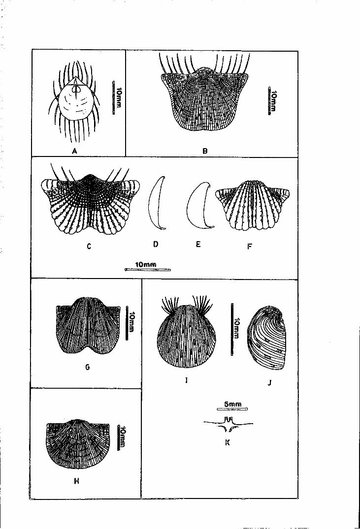
lr
A B
c D E F
10mm q . .., ~'···- h ·•
G
J
5mm

Text-figure 5.1. Reconstructions of Malaysian Permian
Prod uctidina I
Figure A - Strophalosia sp. A - interior of dorsal valve and ventral interarea.
Figure B - Antiquatonia sp. A; ventral exterior.
Figure C- Dictyoclostus'? gratiosus (Waagen); ventral exterior of variety II.
Figure D - lateral profile of C.
Figure E - lateral profile of variety I of the same species.
Figure F - ventral exterior of E.
Figure G - Linoproductus sp. cf. L '? sinosus Huang; ventral exterior.
Figure H - Stepanoviella sp. cf. S. flexuosa Waterhouse; ventral exterior.
Figures I, J - ventral and lateral exterior of Cancrinella cancrini (de Koninck).
Figure K - cardinal process of the same species.
206
auricle and more spines on the ventral valve (see Waagen 1884, pl. 72, fig. 3-5),
while the other has less convex ventral valve, short trail, large auricle and hardly
any spines on the ventral valve (Waagen 1884, pl. 72, fig. 6-7). The generic status
of this species was debated by later authors and some avoided discussion of this
species in their general taxonomic classification. This is probably because the original
description is incomplete and lacks features of high taxonomic value. P. gratiosus has
been associated with the genus Dictyoclostus by Huang ( 1932, 1933) and Muir-Wood
& Oakley (1941), and to the genus Marginifera by Licharev (1936) and Reed (1944),
while other authors maintain its questionable relationship to the genus Productus.
The first variety of Waagen's (1884) species P. gratiosus seems to belongs to the
family Dictyoclostidae, even though most dictyoclostids do not have a very strong
median sulcus. It might belong to a separate new genus in this family, but until
better and more complete specimens are available it is not possible for the author to
place this species precisely in any particular genus. This variety is not very common
in the Permian of Malaysia, and it is illustrated in text-figures 5.1E,F and plate 13,
figure 9 under Dictyoclostus? gratiosus (Waagen) variety A.
The second variety of Waagen 's ( 1884) P. gratiosus might represent an incomplete
part of the first variety, but many more complete specimens similar to it figured by
later authors seem to give it a rather separate identity from the first variety (see
Broili (1916, pl. 66, fig. 5, 9-12), Huang (1932), Reed (1944) and Yanagida & Aw
(1979)). The low visceral cavity in this variety is not a characteristic of the genus
Dictyoclostus, but there are no other features which might associate this variety with
other productacean families. Although Reed (1944) tried to illustrate the presence of
a marginal ridge in this variety, his figures are not very convincing. If Reed's (1944)
postulation is accepted, this variety should have its own specific identity, under a new
genus very close to the paucispiniferinid genus Reticulatia Waterhouse. This variety is
more common in the Permian of Malaysia, and it is illustrated in text-figures 5.1C,D
and figured in plate 13, figures 4-8 and plate 16, figure 12.
Forms associated with Waagen's (1884) P. gratiosus, have been recorded from
several Upper Permian localities in Asia, and it becomes an important element in
207

the Upper Permian (Wuchiapingian) of South China. The Malaysian occurrences
also seem to be restricted to those horizons associated with llytoniids, except that
from South Central Kelantan which was dated as late Upper Permian by Aw et
al. (1977) and Yanagida & Aw (1979). Because of its stratigraphic importance,
Dictyoclostus'? gratiosus must be studied in more detail in the future for clarification
of its taxonomic status.
DICTYOCLOSTUS'? sp. indet.
(Plate 15, figures 2,3)
Material. Several specimens in 3 rock slabs no FPhN 19 from Sungai Galas, South
Kelantan (GSD collection) and 1 specimen from Bukit Jaya, near Maran, Pahang
(UKM-MSL collection).
Horizon and Age. Upper Permian Leptodus Bed and unnamed formation of ?Upper
Permian age.
Description. The ventral valve is very strongly deformed and not representing the
true shape and curvature of the shell. Therefore, the dimensions of the shell are not
easy to measure. Some of the large flattened specimens have a maximum shell width
of about 20mm or more. The umbo and the visceral region is finely reticulate while
the trail is broadly costate. The median sulcus is poorly defined. The auricle is small
and often weakly wrinkled. No traces of body spines or auricular spines are known.
The dorsal valve is unknown.
Discussion. Owing to a poor preservation, it is difficult to assign these specimens
to their proper genus and species with absolute certainty. These specimens resemble
Antiquatonia sp. A in their reticulate and costate ornamentation, but the absence
of body spines differentiate this species from them. The fine body spines which
characterise the genus Dictyoclostus might have been destroyed during fossilization.
208

Genus ANTIQUATONIA Miloradovich 1945
ANTJQUATONIA sp. A.
(Text-figure 5.1B, Plate 12, figures 10-13, Plate 15, figures 4-6)
Synonym: 1964 Dictyoclostus sp. A, lgo, p. 67, text-fig. 6. 1964 Dictyoclostus sp. B, Igo, p. 67-8. 1964 Dictyoclostus sp. C, lgo, p. 68.
Material. Specimens no L 1096 (26, 29a,b, 30, 31 and 32a,b) from Sg. Sepia, near
Kuala Lipis, Pahang (GSD collection), 5 specimens from locality 10, near Merapoh,
Pahang and 8 incomplete specimens from Kg. Juaseh, near Kuala Pilah, Negeri
Sembilan (UKM-MSL collection)
Horizon and Age. The age ranges from late Lower Permian (Kepis Bed) to Upper
Permian (Leptodus Bed).
Table 5.2. Antiquatonia sp. A- dimensions in mm
Specimen no length curved length
MSL10.2a e10.4
MSL10.2b na
L1096(26) 10.0
dimensions = refer -to text-f'igure 1 .1
e = estimated na = not available
17.8
e18.7
na
hinge width
19.0
26.7
13.9
Description. The shell is medium to large in size with maximum dimensions reaching
50mm. The plan outline is variable in shape due to deformation. The ventral valve
is weakly convex with long and geniculated trail, moderately developed auricle and
small umbo. The median sulcus is fairly deep at the convex part of the visceral disc,
shallower towards the umbo and the anterior margin. The umbo and the posterior
part of the visceral disc is furnished with reticulate ornament. The reticulation of
the shell is fainter towards the anterior trail, where it is replaced by 40 to 60 well
developed radial costae which sometimes bifurcate before reaching the shell margin.
The auricle is strongly wrinkled, and the wrinkles are often crenulated. The body
209

spines consist of less than ten large randomly arranged halteroid spines at the trail
and a few smaller spines on the posterior part of the visceral disc. Five to six pairs
of posteriorly directed marginal spines are developed on the posterior margin. Two
rows of two to five auricular spine bases are seen at the base of the visceral disc, but
the nature of the spines are not known. The ventral adductor muscle platform is
moderately elevated and the muscle impressions are dendritic.
The dorsal valve is only depressed at the umbo, while the rest of the visceral disc
is fiat with strongly geniculated trail. The ornamentation of the dorsal valve is similar
to that of the ventral valve, except that the dorsal valve has no spines. Interiorly, the
cardinal process is large and sessile with the bilobed or quadrilobed myophore facing
dorsally. Other details are unknown.
Discussion. The large body spines randomly set on the crest of the costae put this
species into the genus Antiquatonia, instead of Dictyoclostus as it was previously as
signed by Igo (1964). The genus Antiquatonia is wideapread during the Carboniferous
time, while only a few species of Antiquatonia are known from the Permian. Antiqua
tonia sp. A resembles the Upper Permian species A. planumbona Stehli from Word
Formation of Texas, in its reticulate ornamentation, but the spines of Antiquatonia
sp. A are slightly coarser and less in number as compared to A. planumbona.
Genus RETICULATIA Muir-Wood & Cooper 1960
RETICULATIA sp. indet.
(Plate 12, figures 16-18)
Material. Three incomplete external moulds (one of which contains broken internal
mould of the cardinal process) from locality 16, near Merapoh, and one incomplete
cast and/or mould of the exterior of ventral valve each from locality 8 and 14 near
Merapoh, north Terenggun near Kuala Lipis and Bukit Jaya near Maran, all in Pa
hang (UKM-MSL collection).
Horizon and Age. Leptodus Bed (Upper Permian) and unnamed formation of Upper
Permian age.
210

Remarks. The ventral valve is strongly reticulate, stronger at the umbonal region and
slightly weaker towards the anterior and at the lateral posterior margin. The ventral
valve is strongly convex with weak median sulcus, short smoothly curved trail, small
incurved umbo and poorly defined auricle. The dorsal valve is weakly concave with
poorly defined median fold. The cardinal process is short and sessile with ?bilobed or
?quadrilobed myophores, and dorsally facing muscle attachment base. Reticulatia sp.
differs from the strongly reticulated Dictyoclostus '? gratiosus in its poorly developed
median sulcus and auricle. No attempt can be made to relate this uncertain species
with any known species of Reticulatia, until a better collection of more complete
specimens is available. [MSL16.2a- estimated dimensions; length = 17.2mm, width
= 32.5mm].
Family ECHINOCONCHIDAE Stehli
Subfamily ECHINOCONCHINAE Stehli
Genus ECHINOCONCHUS Weller 1914
ECHINOCONCHUSsp. A
(Plate 12, figures 19-23)
Material. 18 unnumbered specimens from H. S. Lee Mine no 8, near Kampar, Perak
(UM collection).
Horizon and Age. N am Loong Bed, Lower Permian.
Description. The shell is small to medium in size, with posteriorly tapered subtrape
zoidal to subtriangular shell plan. The ventral valve is strongly convex with poorly
defined median sulcus and small and poorly defined auricle. The umbo is large and
strongly incurved. The ventral surface is covered by a series of concentric bands on
which are set several concentric rows of spines. In smaller individuals, only one con
centric row of spines are usually preserved on each band, while in larger individuals
each band usually carries one row of larger spines and two to three rows of much finer
spines. The spines are very fine in size and are suberect in nature. The dorsal valve
211
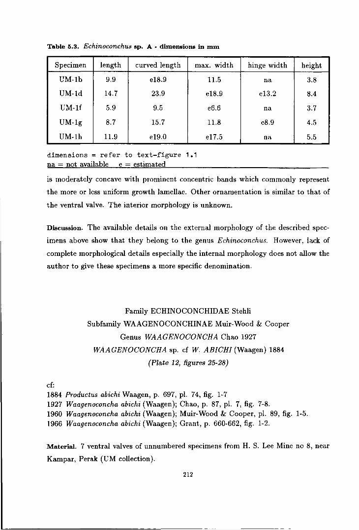
Table 5.3. Echinoconchus sp. A - dimensions in mm
Specimen length curved length max. width
UM-lb 9.9 e18.9
UM-ld 14.7 23.9
UM-lf 5.9 9.5
UM-1g 8.7 15.7
UM-1h 11.9 e19.0
dimensions= refer to text-rigure 1.1 na = not available e = estimated
11.5
e18.9
e6.6
11.8
e17.5
hinge width height
na 3.8
e13.2 8.4
na 3.7
e8.9 4.5
na 5.5
is moderately concave with prominent concentric bands which commonly represent
the more or less uniform growth lamellae. Other ornamentation is similar to that of
the ventral valve. The interior morphology is unknown.
Discussion. The available details on the external morphology of the described spec
imens above show that they belong to the genus Echinoconchus. However, lack of
complete morphological details especially the internal morphology does not allow the
author to give these specimens a more specific denomination.
cf:
Family ECHINOCONCHIDAE Stehli
Subfamily WAAGENOCONCHINAE Muir-Wood & Cooper
Genus WAAGENOCONCHA Chao 1927
WAAGENOCONCHA sp. cf W. ABICHI (Waagen) 1884
(Plate 12, figures 25-28)
1884 Productus abichi Waagen, p. 697, pl. 74, fig. 1-7 1927 Waagenoconcha abichi (Waagen); Chao, p. 87, pl. 7, fig. 7-8. 1960 Waagenoconcha abichi (Waagen); Muir-Wood & Cooper, pl. 89, fig. 1-5. 1966 Waagenoconcha abichi (Waagen); Grant, p. 660-662, fig. 1-2.
Material. 7 ventral valves of unnumbered specimens from H. S. Lee Mine no 8, near
Kampar, Perak (UM collection).
212
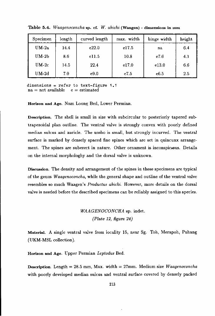
Table 5.4. Waagenoconcha sp. cf. W. abichi {Waagen)- dimensions in mm
Specimen length curved length
UM-2a 14.4 e22.0
UM-2b 8.6 el1.5
UM-2c 14.5 22.4
UM-2d 7.0 e9.0
dimensions = refer to text-figure 1 .1 na = not available e = estimated
max. width
e17.5
10.8
e17.0
e7.5
Horizon and Age. N am Loong Bed, Lower Permian.
hinge width
na
e7.6
e13.0
e6.5
height
6.4
4.1
6.6
2 .. 5
Description. The shell is small in size with subcircular to posteriorly tapered sub
trapezoidal plan outline. The ventral valve is strongly convex with poorly defined
median sulcus and auricle. The umbo is small, but strongly incurved. The ventral
surface is marked by densely spaced fine spines which are set in quincunx arrange
ment. The spines are suberect in nature. Other ornament is inconspicuous. Details
on the internal morphologhy and the dorsal valve is unknown.
Discussion. The density and arrangement of the spines in these specimens are typical
of the genus Waagenoconcha, while the general shape and outline of the ventral valve
resembles so much Waagen's Productus abichi. However, more details on the dorsal
valve is needed before the described specimens can be reliably assigned to this species.
WAAGENOCONCHA sp. indet.
(Plate 12, figure 24)
Material. A single ventral valve from locality 15, near Sg. Toh, Merapoh, Pahang
(UKM-MSL collection).
Horizon and Age. Upper Permian Leptodus Bed.
Description. Length = 28.5 mm, Max. width = 27mm. Medium size Waagenoconcha
with poorly developed median sulcus and ventral surface covered by densely packed
213
fine recumbent to suberect spines, set in quincunx arrangement.
Discussion. The specimen discussed is slightly compressed. If not compressed the
length should be slightly shorter than the maximum width, and the specimens would
appear to be more like those of the Lower Permian of Kampar. However, compared
to the Kampar specimens the above specimen appears to be slightly larger in size.
cf:
Family LINOPRODUCTIDAE Stehli
Subfamily LINOPRODUCTINAE Stehli
Genus LINOPRODUCTUS Chao 1927
LINOPRODUCTUS sp. A, cf. L? SIN OS US Huang 1932
(Text-figure 5.1G, Plate 12, figures 29-31)
1932 Linoproductus sinosus Huang, p. 43-44, pl. 2, fig. 15-16.
Material. Ten specimens from localities 9, 2 specimens each from locality 10 and 13
and 1 specimen each from localities 6,, 7 and 14, all from the road cut in between Gua
Panjang and Sungai Yu halt, near Merapoh, Pahang, and 1 specimen from Bukit
Jaya, near Maran, Pahang (UKM-MSL collection).
Horizon and Age. Upper Permian Leptodus Bed and unnamed formation of ?Upper
Permian age.
Description. The shell is small to medium in size with strongly deformed shell plan
outline and profile. The largest individual (MSL9.1a) has an estimated maximum
shell width of about 30mm. The ventral valve is strongly convex with fine costae and
costellae and occassional growth lamellae. The median sulcus is strongly developed
anteriorly and weaker towards the umbo. The trail is long, linear and geniculate with
the costae slightly coarser than that on the visceral disc. The umbo is large and
strongly incurved. The hinge line is straight and possibly represents the maximum
width of the shell. The auricle is moderately developed, flat and triangular in shape
214

with prominent wrinkles and faint radial costae. Other details are unknown.
Discussion. Although the described specimens are all deformed in one direction or
another, the essentially deep median sulcus, wide hinge line and strongly incurved
umbo in Linoproductus sp. A closely resembles that of Linoproductus sino sus Huang.
This species, however, has been assigned by Nakazawa (1973) to the genus Anidan
thus. Unfortunately, the available specimens of Linoproductus sp. A do not show any
convincing features which could associate them with the later genus. For this reason,
the author will tentatively assign ' the studied specimens into the genus Linoproduc
tus until further comparison with the Chinese type specimens of L. sinosus is made.
The essential features outlined above distinguish Linoproductus sp. A from other
Malaysian species of Linoproductus.
cf:
Genus STEPANOVIELLA Zavodowsky 1960
STEPANOVIELLA sp. A, cf. S. FLEXUOSA Waterhouse 1970
(Text-figure 5.1H, Plate 15, figures 7-9)
1970 Stepanoviella flexuosa Waterhouse, p. 45-48, pl. 14, fig. 1-8, 15-16.
Material. Seventeen · moulds of disarticulated valves on 6 rock slabs no. KPF (1,
2, 3, 3a., 3b and 5) and some unnumbered specimens from Kuala Pilah area, Negeri
Sembilan (GSD collection).
Horizon and Age. Kepis Bed, late Lower Permian.
Description. The shell is asymmetrically transversely elongated to subcircular in plan
outline. The maximum shell width is at about the mid shell length. The ventral
valve is weakly to moderately convex with short, broad and weakly incurved umbo,
short and indistinct trail, small auricle and no median sulcus. The umbo is usually
flattened by small rough surface of cicatrix in younger individuals, but the cicatrix
is less pronounced in the larger individuals. The hinge line is inequal in length, and
215
the umbo is usually pointing towards the shorter hinge line. The auricle is developed
as a narrow flattened or slightly swollen strip along the hinge line. The exterior
of the shell is furnished with broad flexuous costellae, irregularly intercepted by a
series of discontinuous growth lamellae or occasional rugae. Concentric bands are
also commonly developed. The posterior margin is usually strongly wrinkled.
The dorsal valve is weakly concave with very short trail, flattened auricle and
no median fold. The curvature of the dorsal valve closely follows that of the ventral
valve, thus the visceral cavity is narrow. The outline of the dorsal valve is usually
transversely elongated. The surface ornamentation of the dorsal valve is very similar
to that of the ventral valve. No spines are known in either ventral or dorsal valves.
The interior details are unknown.
Discussion. The short trail and flexuous costellae distinguished Stepanoviella sp. A
from the genus Linoproductus. Stepanoviella sp. A resembles S. flexuosa Water
house in its transverse shape, broad umbo, concave dorsal valve and coarse costellae.
Stepanoviella sp. A differs from S. flexuosa in the development of a cicatrix in younger
individuals. S. fiexuosa is reported to be restricted to the Stage 3 of Waterhouse's
(1970a) Permian brachiopod zonation, equivalent to the Sakmarian-Artinskian Stage
of the Russian platform.
Synonym:
Genus CANCRINELLA Fredericks 1928
CANCRINELLA CANCRINI (de Koninck) 1842
(Text-figure 5.1I-K, Plate 13, figures 2-10)
1847 Productus cancrini de Koninck; de Koninck, p. 208-211, pl. 11, fig. 3a-f. 1973 Cancrinella cancrini (de Koninck); Ishii et al., p. 69-70, pl. 2, fig. 2, 4-7.
Material. Hundreds of complete and partly complete disarticulated ventral and dorsal
valves from Bukit Tungku Lembu and about 20 specimens of similar nature from Bukit
Temiang, Pedis (UKM-MSL collection). These specimens are preserved in form of
moulds, · but occasionally the original shell at the cardinal area of the dorsal
216

valve is preserved.
Horizon and Age. Chuping Formation, late Lower Permian.
Table 5.5. Cancrinella cancrini (de Koninck) - dimensions in mm
Specimen length curved length max. width hinge width height
MSL1.1a(1) 14.0 24.8 e14.0 na 8.2
MSLl.lf 9.1 14.6 e9.5 na 4.1
MSL1.1g 8.7 14.7 9.0 na 6.5
MSL1.1a(2)* 11.5 na 14.5 12.8 na
MSL1.1b(1)* e14.3 na 16.8 e15.0 na
MSL1.1b(2)* e7.9 na e9.3 na na
MSL1.1c(1)* e11.4 na e14.7 e12.8 na
MSL1.1d(1)* 10.5 na 12.4 e10.6 na
MSL1.1d(2)* 7.9 na 9.0 e7.4 na
dimensions= refer to text-figure 1.1
na =not available e = estimated * = dorsal valve
Description. Small sized Cancrinella with maximum dimension reaching about 17
mm. The shell is roughly subcircular in its plan outline. The maximum width of the
shell is at about the mid shell length. The ventral valve is moderately to strongly
convex with no median sulcus, very short trail and small flat auricle. The umbo is
large and strongly overhanging and incurved beyond the hinge line. The exterior of
the ventral valve is covered by regular fine costae which sometimes bifurcate anteriorly,
with little or no concentric bands or growth lamellae. Suberect spines are disposed
at irregular intervals on the crest of the costae, without any systematic pattern of
arrangement. The fully grown individual has about 20 to 30 body spines on the
ventral valve. The erect and laterally directed auricular spines are concentrated in
two or three rows at the base of the visceral disc. The auricle is wrinkled, with the
strongest wrinkles developed at the posterior margin. The posterior margin is straight
and provided with a row of posteriorly directed marginal spines.
217

The dorsal valve is weakly concave with depressed umbo, no median sulcus and
short geniculate trail. The auricle is narrow, transversely elongated triangular in
shape and flat in profile. The posterior margin is strongly wrinkled laterally. The
dorsal surface is finely costate with occasional growth lamellae, and is sometimes
wavy in nature. No traces of spines are observed. The cardinal process has a very
short shaft, and is supported by a pair of strong lateral ridges. The myophore is
bilobed with each lobe well separated and each tends to split into two smaller lobes.
The muscle attachment bases face the dorsal. Other details are unknown.
Discussion. At Bukit Temiang and Bukit Tungku Lembu, C. cancrini forms cocquina
beds with virtually no other faunal elements, within the passage beds between the
arenaceous Kubang Pasu Formation and the Chuping Limestone Formation. As in
many other brachiopod species which dominate their fauna (e.g. S. excavata and S.
lewisiana from the British Permian), individuals of the species seem to show wide
variation.
The Malaysian C. cancrini shows considerable variation in the shape and convex
ity of its ventral and dorsal valves. The ventral valve varies in its plan outline from
subcircular to a slightly elongated pear shape, and in its strong to very strong convex
ity, out maintains its non-sulcate form with large incurved umbo. Furthermore, the
ventral valve remains more or less smoothly curved with very little or no fluctuation,
except for the auricular wrinkles. These general features, as well as the ornamenta
tion of the shell which characterised the C. cancrini described from the Permian of
Russia, distinguish the Malaysian forms from other species of Cancrinella. The thin
shell at the auricle was probably damaged during the fossilization, thus the ventral
valve seldom shows a well developed auricle.
The dorsal valve, on the other hand, shows a greater variation from longitudinally
to transversely elongated shape and from moderately concave to nearly flat profile.
The flatter forms are usually associated with longitudinally elongated shell with less
geniculated trail. Since the ventral valve and the dorsal valve are always separated,
it is very difficult to prove whether the strongly varied shape and profile of the dorsal
valve belongs to an extreme variation of the species or merely a result of deformation.
218

It is also difficult to prove whether the fluctuate dorsal valve in some specimens belongs
to another species of Cancrinella or is simply deformed shells. Although the degree
of deformation is generally very low in the rock formations containing this fauna, it
is still possible for individuals of these thin shelled brachiopods to be crushed upon
one another. More detailEd rludy on the taphonomy of this interesting fauna is needed
before a better conclusion can be achieved on the variation of the morphology within
the species.
cf:
CANCRINELLA sp. cf. C. CANCRINIFORMIS (Tschernyschew) 1902
(Plate 13, figure 11)
1916 Productus cancriniformis Tschernyschew; Broili, p. 13-14, pl. 66, fig. 6.
Material. Five incomplete moulds from Kg. Juaseh, near Kuala Pilah,
Negeri Sembilan (UKM-MSL collection)
Horizon and Age. Kepis Bed, late Lower Permian.
Description. The shell is small to medium in size. The ventral valve is moderately
convex with no median sulcus, small, narrow and weakly incurved umbo and poorly
developed auricle. The surface of the ventral valve is covered with fine costae and
several suberect body and auricular spines. The spines are small in diameter and dis
tributed at irregular intervals at least 3 mm apart. The shell is concentrically banded
with broad to sharp ridges and shallow to deep grooves. The ridges and grooves are
often discontinuous. The grooves are sometimes overlapped by occasionally devel
oped growth lamellae or rugae. The dorsal valve is flat or weakly concave with fine
radial costae and irregular concentric bands. Other details are unknown. [MSL20.2a
-estimated dimensions; length= 19.5mm, width= 22mm].
Discussion. The specimens described above are similar to some extend to C. cancrini
formis from Timor, but incomplete preservation does not allow these specimens to be
placed into the named species with confidence. The Juaseh specimenf differs from the
C. cancrini from Perlis in their larger size, smaller umbo and stronger fluctuation of
219

their shell, although the shell fluctuation might be exaggerated by the deformation
of the Kepis Bed which seems to be stronger compared to the Chuping Limestone
Formation.
CANCRINELLA sp. A
(Plate 13, figures 12-13)
Material. 4 casts and 1 mould of unnumbered specimens from H. S. Lee Mine no 8,
near Kampar, Perak (UM collection).
Horizon and Age. Nam Loong Bed, Lower Permian.
Description. The ventral valve is medium in size, moderately convex with small
slightly pointed umbo, small flattened auricle and no median sulcus. Apart from the
umbo, the whole ventral surface is covered with strong radial costae and concentric
bands of ridges with sharp crest and profound grooves. Several large, distant suberect
spines are arranged in radial as well as concentric rows on the crest of the costae.
The auricular spines~renot known. The auricle is strongly wrinkled and the posterior
margin is provided with a row of posteriorly directed marginal spines. Other details
are unknown. (UM-3a- dimensions; length = 22.5mm, width = 2l.Omm].
Remarks. As compared to other Malaysian species of Cancrinella, Cancrinella sp.
A is distinguished by its stronger radial costae and concentric bands, and larger and
more systematically arranged body spines. The ornamentation in Cancrinella sp. A
resembles the New Zealand species C. halli Waterhouse, but like Cancrinella sp. A,
the New Zealand species was described from incomplete specimens, some features are
thus unavailable for comparison (see Waterhouse, 1982). Therefore, the author will
delay naming this species until a better collection of specimens is available.
220

Family LINOPRODUCTIDAE Stehli
Subfamily STRIATIFERINAE Muir-Wood & Coopper
Genus STRIATIFERA Chao 1927
STRIATIFERA sp. A
(Plate 13, figure 1)
Material. Two incomplete external moulds of dorsal valve from H. S. Lee Mine no 8,
near Kampar, Perak (UM collection).
Horizon and Age. Nam Loong Bed, Lower Permian.
Remarks. The maximum shell width of large flattened specimen (UM-4a) is more
than 60mm. The dorsal valve is large in size and partly impressed upon the contour
of the rock. In general the dorsal valve is moderately concave with the maximum
concavity situated at the umbo. The auricle is small and flat, while the hinge line is
short and straight. The surface of the shell is covered by fine costae and costellae and
occassional weak wrinkles. The costae and costellae bifurcate at various positions
near the anterior margin. The wrinkles are stronger at the lateral posterior margin.
Other details are unknown.
The incomplete preservation of the described specimens make comparison with
any known species of Striatifera impossible.
Family MARGINIFERIDAE Stehli
Subfamily MARGINIFERINAE Stehli
Genus LIOSOTELLA Cooper 1953
LIOSOTELLA NAKAMURA! new species
Text-figure 5.2A-E, Plate 14, figures 6-15
Named after Nakamura who in 1973, described this species under Spinomarginifera
kueichowensis Huang and Liosotella sp.
Synonym; 1973 Spinomarginifera kueichowensis Nakamura (not-Huang), p. 292-3, pl. 31, fig.
9. 1973 Liosotella sp. indet. Nakamura, p. 293, pl. 31, fig. 10-11. 1973 ?Spinomarginifera sp. indet. Nakamura, p. 293, pl. 31, fig. 8.
221

Diagnosis. Medium sized Liosotella with 14 to 16 coarse costae, a row of 4 to 5 pairs of
large halteroid auricular spines, rare smaller body spines including two concentric rows
near the anterior margin where each row of spines is set on alternate costae. Ventral
valve moderately to strongly convex with short trail, small smooth umbo, smooth
auricle and weakly developed median sulcus. Dorsal valve weakly concave with no
median fold, smooth umbo, costate anterior and very short trail. Marginal ridges
of spinomarginiferid type. Cardinal process small with posteriorly pointed shaft.
Adductor muscle platform subtriangular in shape. Brachial impression moderate in
SIZe.
Material. Paratypes: Specimens no. MSL17.1a, MSL17.Ib, MSL17.1c, MSL17.1d
and MSL17.1e (UKM-MSL collection) from carbonaceous shale exposed at the Jengka
Pass road cut, are chosen to represent a series of paratypes of Liosotella nakamurai
n. sp., in the absence of a complete specimen. The following description also covers
observations made on another 80 specimens from the type locality and 12 specimens
from the Maran Oil Palm Mill exposure.
Type locality. The Jengka Pass road cut (GR. WF 91651445, Sheet 89) is chosen as
the type locality of Liosotella nakamurai n. sp. because of the numerous occurreaces
of the species in this locality, though the newly cited locality, i.e. the Maran Oil
Palm Mill produces a few well preserved specimens. The Jengka Pass road cut, in
addition, is the locality in which Nakamura (1973) described this species under S.
kueichowensis and Liosotella sp.
Horizon and Age. Unnamed formation of Upper Permian (Midian) age.
Description. The shell is transversely subtrapezoidal in shape, as inferred from the
less distorted disarticulated dorsal valve. Owing to substantial deformation the plan
outline of the ventral valve varies from transverse to elongate subelliptical to subtri
angular shaped, and often twisted. The maximum shell width is at the hinge line,
but the ventral valve often shows a hinge line shorter than the maximum width due
to deformation.
The ventral valve is strongly convex, although it often shows a great variation in
222
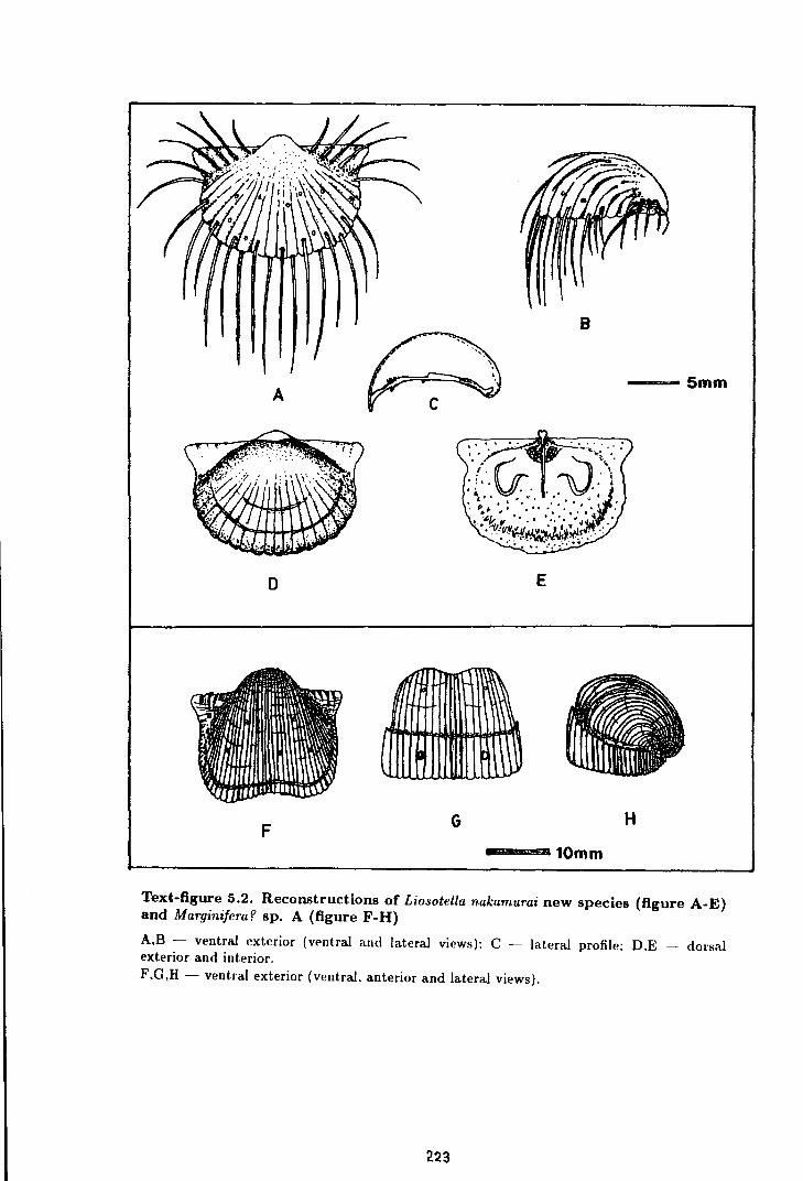
B
A ---5mm
D E
F G H
---10mm
Text-figure 5.2. Reconstructions of Liosotella nakamurai new species (figure A-E) and Marginifera? sp. A (figure F-H)
A,B - ventral exterior (vPntral and lateral views): C - laten1.l profile: D.E - dorsal exterior and interior. F,G,H- ventral exterior (ventral, anterior and lateral views).
2.23
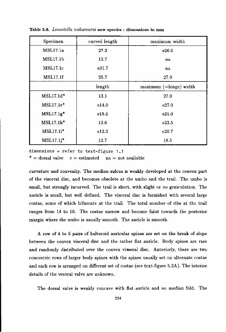
Table 5.6. Liosotella nakamurai new species - dimensions in mm
Specimen curved length maximum width
MSL17.1a 27.3 e26.0
MSL17.1b 13.7 na
MSL17.1c e31.7 na
MSL17.1f 25.7 27.9
length maximum (=hinge) width
MSL17.1d* 13.1 27.0
MSL17.1e* e14.0 e27.0
MSL17.1g* e15.0 e21.0
MSL17.lh* 13.6 e23.5
MSL17.1i* e12.3 e20.7
MSL17.1j* 13.7 18.5
dimensions= refer to text-figure 1.1
* = dorsal valve e = estimated na = not available
curvature and convexity. The median sulcus is weakly developed at the convex part
of the visceral disc, and becomes obsolete at the umbo and the trail. The umbo is
small, but strongly incurved. The trail is short, with slight or no geniculation. The
auricle is small, but well defined. The visceral disc is furnished with several large
costae, some of which bifurcate at the trail. The total number of ribs at the trail
ranges from 14 to 16. The costae narrow and become faint towards the posterior
margin where the umbo is usually smooth. The auricle is smooth.
A row of 4 to 5 pairs of halteroid auricular spines are set on the break of slope
between the convex visceral disc and the rather flat auricle. Body spines are rare
and randomly distributed over the convex visceral disc. Anteriorly, there are two
concentric rows of larger body spines with the spines usually set on alternate costae
and each row is arranged on different set of costae (see text-figure 5.2A). The interior
details of the ventral valve are unknown.
The dorsal valve is weakly concave with flat auricle and no median fold. The
224

costae are weakly developed on the dorsal surface. The dorsal exterior is spineless.
The external marginal ridge is sometimes developed as a narrow rim, a short distant
from the anterior and anterolateral margins. The narrow trail beyond the external
marginal ridge maintains the exterior curvature and morphology of the dorsal valve.
Interiorly, significant change can be seen in between the trail and the visceral
cavity. At the visceral region, the dorsal interior is furnished with fine marginally
pointed endospines. The interior marginal ridge is not very well developed. It is only
slightly raised and marked by a rim of coarser strainer endospines. Anterior to the
marginal ridge, the interior surface becomes smoother and the curve becomes weaker
as such the anterior margin of the dorsal valve meets the ventral valve at a rather
wide angle.
The cardinal process is small with posteriorly pointed shaft and bilobed myophore.
The lateral ridges are weakly developed. The adductor muscle platform is subtrian
gular in shape with the lobate scars obscurely divided into the posterior and anterior
sections. The brachial impressions are moderate in size with the inner ridges bent
anteromedianly towards the edge of the median septum. The median septum is nar
row and low, appears at the middle of the adductor muscle platform and extends to
about the mid shell length.
Discussion. Although the actual shell is not preserved, the reconstruction based on
the external and internal moulds of the dorsal valve shows that the marginal ridge of
Liosotella nakamurai is similar to that figured by Huang (1931) for the genus Spino
marginifera. Under the present scheme of classification, however, the strongly costate
ventral and dorsal valves with smooth umbo, and the absence of spines from the dor
sal valve should placed this species in the genus Liosotella, not Spinomarginifera as
was suggested by Nakamura (1973).
The marginal ridge of L. nakamurai is unique and has not been found in any
other species of Liosotella. L. nakamurai resembles L. grandicosta (Chao) and L.
magnirugosa Cooper in terms of the size of their costae. L. nakamurai, however,
differs from the other two species in its smaller auricle, less profound median sulcus
and the systematically arranged spines on its trail.
225
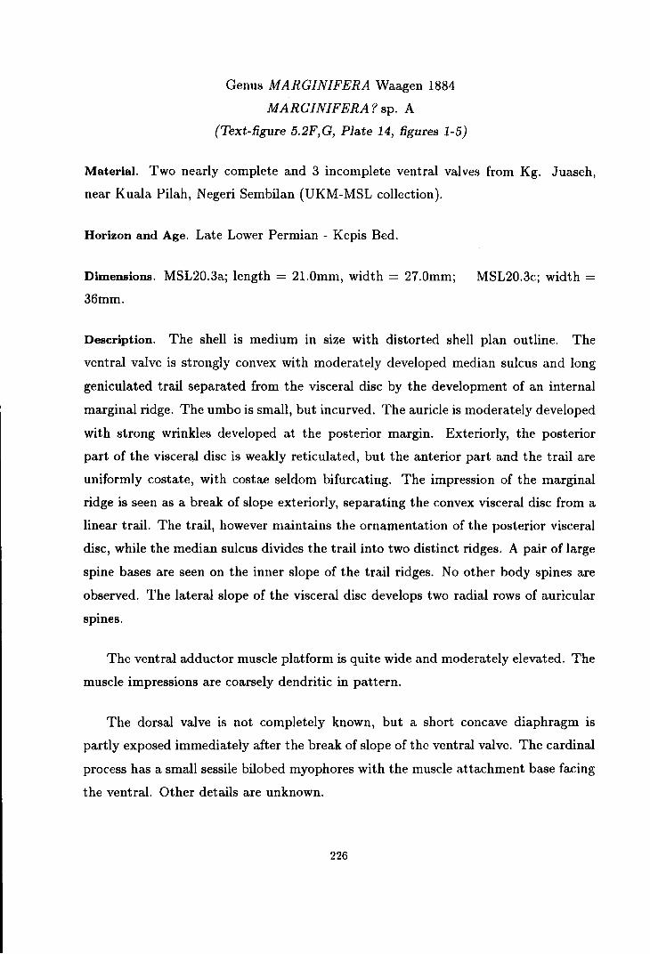
Genus MARGINIFERA Waagen 1884
MARGINIFERA? sp. A
(Text-figure 5.2F,G, Plate 14, figures 1-5)
Material. Two nearly complete and 3 incomplete ventral valves from Kg. Juaseh,
near Kuala Pilah, Negeri Sembilan (UKM-MSL collection).
Horizon and Age. Late Lower Permian - Kepis Bed.
Dimensions. MSL20.3a; length = 2l.Omm, width = 27.0mm;
36mm.
MSL20.3c; width =
Description. The shell is medium in size with distorted shell plan outline. The
ventral valve is strongly convex with moderately developed median sulcus and long
geniculated trail separated from the visceral disc by the development of an internal
marginal ridge. The umbo is small, but incurved. The auricle is moderately developed
with strong wrinkles developed at the posterior margin. Exteriorly, the posterior
part of the visceral disc is weakly reticulated, but the anterior part and the trail are
uniformly costate, with costae seldom bifurcating. The impression of the marginal
ridge is seen as a break of slope exteriorly, separating the convex visceral disc from a
linear trail. The trail, however maintains the ornamentation of the posterior visceral
disc, while the median sulcus divides the trail into two distinct ridges. A pair of large
spine bases are seen on the inner slope of the trail ridges. No other body spines are
observed. The lateral slope of the visceral disc develops two radial rows of auricular
spines.
The ventral adductor muscle platform is quite wide and moderately elevated. The
muscle impressions are coarsely dendritic in pattern.
The dorsal valve is not completely known, but a short concave diaphragm is
partly exposed immediately after the break of slope of the ventral valve. The cardinal
process has a small sessile bilobed myophores with the muscle attachment base facing
the ventral. Other details are unknown.
226
Discussion. Although the very rare, large probably halteroid body spines in these
specimens might characterise the subfamily Paucispiniferinae, the numerous auricu
lar spines place them into the subfamily Marginiferinae. The rarity in body spines
and the strong deflection of the trail in ventral valve allies these specimens with a
marginiferinid genus Alifera Muir-Wood & Cooper, but the smaller auricle and very
long trail in these specimens does not agree with the description of the genus Alifera.
Despite the lack of body spines and the strongly deflected trail of the ventral valve,
these specimens resembles other species of M arginifera in their general ornamenta
tion, the auricular spines and the general shape. More better preserved and complete
specimens are needed before these specimens can be placed into their proper genus
and species with certainty.
Synonym:
Family MARGINIFERIDAE Stehli
Subfamily PAUCISPINIFERINAE Muir-Wood & Cooper
Genus PAUCISPINIFERA Muir-Wood & Cooper 1960
PA UCISPINIFERA sp. A
(Plate 15, figure 14)
1979 Anidanthus sp. Yanagida & Aw, p.131-132, pl.28, fig. ?5, 15-16.
Material Specimens no. GS(F) 5, ?51, 52 and 53 from locality FSKN 73, Sg. Paloh,
tributary of Sg. Relai in South Central Kelantan (GSD collection).
Horizon and Age. Late Upper Permian Aring Formation.
Dimensions. GS(F)5; length= llmm, width= 24mm; GS(F)52; length= 16mm.
Description. The shell is small to medium. in size with fusiform shape, with the
maximum shell width slightly posterior to the hinge line and more than twice greater
than the shell length. The ventral valve is strongly curved longitudinally, with poorly
developed median sulcus, broad strongly incurved umbo and large flat or slightly
swollen auricle. The trail is short and strongly curved at the anterior margin, but at
227

the lateral margin it is flattened and coalesces with the auricle. The ventral surface
is weakly costate, smoother at the trail. The auricle is smooth. A few (3 or 4 pairs)
large spine bases are seen at the lateral and anterior slope of the visceral disc. No
other spines are known. Specimen no 52 shows an interior mould with large radially
striated diductor muscle scars and strong anterior marginal ridges which bend towards
the posterior inside the median sulcus. Other details are unknown.
Discussion. The presence of marginal ridges and rare large body spines suggest
that the described specimens belong to the marginiferid subfamily Paucispiniferi
nae, not to Anidanthus or other linoproductids. Although the marginal ridges of the
described specimen resembles that of Retimarginifera lipisensis n. sp., the lack of
reticulate ornament suggests that it belongs to the genus Paucispinifera. At present,
the genus Paucispinifera is only known from the Upper Permian of North America.
Paucispinifera sp. A resembles the type species, P. auriculata Muir-Wood & Cooper
from Word Formation of Texas, in its general shape and convex auricle, but the spines
of Paucispinifera sp. A are not systematically arranged as they are in P. auriculata.
Genus RETIMARGINIFERA Waterhouse 1973
RETIMARGINIFERA LIPISENSIS new species
(Text-figure 5.3A-F, Plate 14, figures 16-26, Plate 15, figure 13)
Named after Kuala Lipis District in which all the described specimens came from.
Diagnosis. Retimarginifera with finely reticulate umbonal region, moderately devel
oped median sulcus and auricle. Ventral valve with three pairs of halteroid marginal
spines and three pairs of halteroid body spines following the outline ot the interior
marginal ridge. The marginal ridges are well developed in both ventral and dorsal
valve, stronger on the ventral valves especially anteriorly.
Type specimens. Specimens no MSL15.la, MSL15.1b and MSL15.1c from Sungai Yu
halt road cut, near Merapoh, Pahang are chosen as a series of paratypes of Reticulatia
lipisensis, representing various morphology of the species.
228

Type Locality. The Sungai Yu halt road cut (GR. QZ 04204405), near Merapoh,
Pahang is chosen for type locality of the species, because some of the best preserved
specimens of this species came from here.
Other material and localities. For the following description, reference are also made
on several other specimens in UKM-MSL collection including 4 other specimens from
the type locality, 5 specimens from locality 8 and 2 specimens from locality 12, near
Merapoh; 5 specimens from Kg. Gua, 4 specimens from Kg. Garok and 1 specimen
from north Terengun, near Kuala Lipis, all of which from the Kuala Lipis District in
Northwest Pahang. Also studied are several specimens in GSD collection including 4
specimens from railway cut adjacent to Sungai Jeleteh, near Merapoh.
Wahid Abdul Rahman (1987) reported the occurrences of Retimarginifera sp. from
Labis area in Northwest Johor, hut the author failed to locate these specimens during
the recent visit to the Geological Survey Department.
Horizon and Age. Upper Permian Leptodus Bed.
Table 5.7. Retimarginifera lipisensis new species- dimensions in mm
Specimen length curved length
MSL15.1a e23.8 e32.0
MSL15.1h e25.0 na
MSL15.1c na na
MSL8.3a 19.5 e37.5
MSL8.3b e4.5 na
MSL3.1a e17.4 na
dimensions= refer to text-figure 1.1 e = estimated na = not available
maximum (=hinge) width
34.1
24.0
e35.0
28.5
7.8
e21.0
Description. The shell is medium in size with the largest individual reaching 35mm
in its maximum width at the hinge line. The shell plan outline is transversely sub
trapezoidal in shape with the length of the shell about two thirds of its hinge width.
The ventral valve is moderately convex with the anterior half is more convex than
229
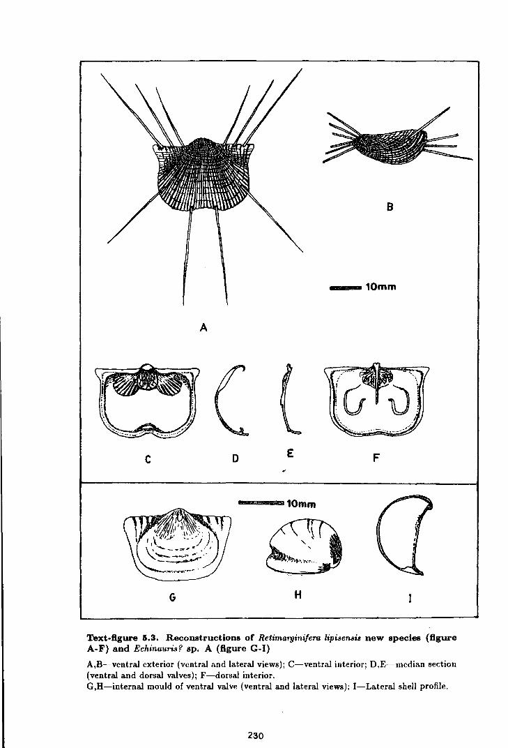
8
--10mm
A
c D E F
--=-10mm
G H
Text-figure 6.3. Reconstructions of Retimargini/era lipisensis new species (figure A-F) and Echinauris? sp. A (figure G-1)
A,B-ventral exterior (ventral and lateral views); C-ventral interior; D.E-median section (ventral and dorsal valves); F-dorsal interior. G,H-internal mould of ventral valve (ventral and lateral views); I-Lateral shell profile.
230

its posterior. From the mid shell length, the ventral valve curves gently towards the
small umbo. The median sulcus is moderately developed, more profound anteriorly
than posteriorly. The umbo is small, weakly inflated and incurved. The auricle is
small and slightly swollen. The trail is short and poorly defined exteriorly.
The umbo and the central part of the visceral disc are furnished with fine reticulate
ornament. The reticulate ornament fades away towards the margin where the visceral
disc becomes regularly costate peripherally. The costae are about 0.5 to lmm apart
and never bifurcate, they fade towards the margin. The auricle is wrinkled. There
are only twelve halteroid spines on the ventral valve. These spines are made of
three pairs of marginal spines and three pairs of body spines. The marginal spines
increases its diameter laterally, and they are posterolaterally pointed. Two pairs of
large body spines are arranged in the same concentric row with the outermost pair
of the marginal spines. These two pairs of body spines are anterolaterally pointed,
forming one horizon of spines with the marginal spines. The anterior pair is the
largest pair of spines with their diameter reaching lmm. A pair of smaller spine
bases are developed on the lateral slope of the visceral disc, and this pair seems to be
formed on the same concentric row with the second pair of the the marginal ridges
(see text-figure 5. 3A).
The ventral adductor muscle platform is weakly to moderately elevated with
coarsely dendritic muscle impressions. The adductor muscle platform is undivided,
and is surrounded by a large pair of low radially striated diductor muscle platforms.
The lateral ridges are moderatately developed, but the marginal ridge is well devel
oped, especially anteriorly. The anterior and anterolateral marginal ridges are thick
with their crest inclined towards the central visceral cavity. Medianly, the anterior
marginal ridge curves towards the posterior and then towards the ventral (see text
figure 5.3D). The interior surface of the shell is endospinose.
The dorsal valve is weakly concave with weakly developed median sulcus and
small auricle. Apart from the absence of spines, the surface ornamentation of the
dorsal valve is similar to that of the ventral valve. The lateral ridges are well de
veloped, broader laterally and become crenulated at the point it meets the lateral
231

marginal ridge. The lateral marginal ridge is narrower and sharper towards the an
terior marginal ridge. The cardinal process is small with the shaft pointing to the
posterior. The adductor muscle platform is low to moderately elevated, subtriangular
in shape with dendritic muscle impressions. The brachial impressions are medium in
SIZe.
Discussion. The genus Retimarginifera is not a very well known genus, and the number
of species described in the past is small and their distribution is limited to the Permian
of Asia and Russia. R. lipisensis differs from the type species R. perforata Waterhouse
in its finer and more regular reticulate ornament and costae and less profound median
sulcus. The interior details of the type species is not known and cannot be compared
with this species. The spines arrangement is similar between R. lipisensis and R.
perforata, although Waterhouse did not described the relationship between the body
spines and the marginal spines.
Family MARGINIFERIDAE Stehli
Subfamily COSTISPINIFERINAE Muir-Wood & Cooper
Genus COSTISPINIFERA Muir-Wood & Cooper
COSTISPINIFERAsp. A
(Plate 13, figures 14-15, Plate 15, figures 10-11)
Material. Four incomplete specimens from locality 11, near. Merapoh and 5 speci
mens from Jengka Pass road cut, near Maran, Pahang (UKM-MSL collection); and
more than 20 fragmentary specimens from Ulu Lepar, near Maran, Pahang (GSD
collection).
Horizon and Age. Upper Permian Leptodus Bed and unnamed formation of ?Upper
Permian age.
Description. Small to medium size Costispinifera with strongly deformed and frag
mented shell. The large specimens sometime have a maximum width of more than
20mm. The median sulcus is poorly developed. The umbo is strongly incurved,
232

crenulated or sometimes costate. The ventral valve is furnished with wide crenulated
costae and numerous spines. The auricle is small, smooth or wrinkled with three
radial rows of auricular spines. The ornament of the dorsal valve is similar to that of
the ventral valve, but very rare spines are preserved. No spines are observed on the
dorsal auricle. Other details are unknown.
Discussion. The roughly crenulated costae and numerous spines placed these spec
imens into the genus Costispinifera. However, the shells are often distorted and
incomplete, thus specific nomination of these specimens is difficult to attempt.
Synonym;
Genus ECHINAURIS Muir-Wood & Cooper 1960
ECHINAURIS sp. indet.
(Plate 13, figure 16)
1979 Echinauris sp. Yanagida & Aw, p. 129-130, pl. 28, fig. 7(not 6).
Material. Specimen no GS(F)54a,b from locality FSKN73, Sungai Paloh in South
Central Kelantan and one specimen each from localities 10 and 11, near Merapoh,
Pahang.
Horizon and Age. Upper Permian Leptodus Bed of Gua M usang Formation and A ring
Formation.
Remarks. The shell is very small in size (usually less than 10mm in dimension) with
subcircular plan outline. The ventral valve is strongly convex with small auricle,
narrow, but incurved umbo, no median sulcus and short and indistinct trail. The
ventral surface is smooth, with several large halteroid spines developed on the convex
part of the visceral disc and on the auricle. Other details are unknown. The incom
plete preservation of the described specimens prevent the author from describing and
discussing more details about this species. It is quite possible that these specimens
could be confused with the juveniles of other species with smooth shell surface, but
such species have not been reported from the same horizon in which Echinauris sp.
is found.
233
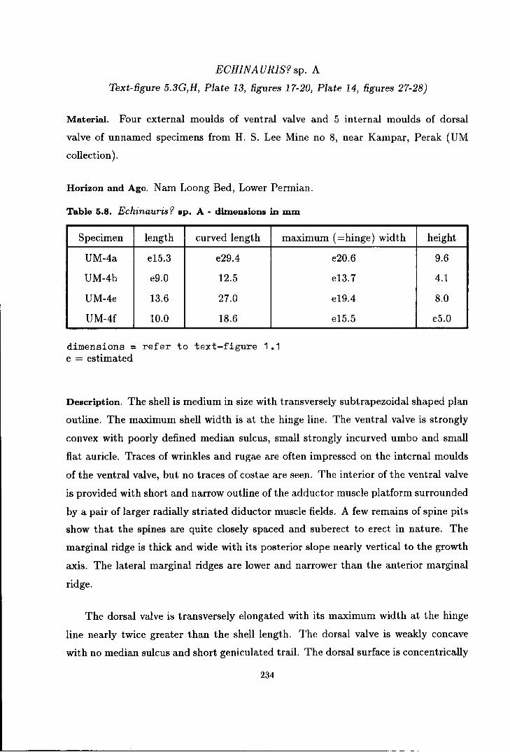
ECHINA URIS? sp. A
Text-figure 5.3G,H, Plate 13, figures 17-20, Plate 14, figures 27-28)
Material. Four external moulds of ventral valve and 5 internal moulds of dorsal
valve of unnamed specimens from H. S. Lee Mine no 8, near Kampar, Perak (UM
collection).
Horizon and Age. N am Loong Bed, Lower Permian.
Table 5.8. Echinauris '? sp. A - dimensions in mm
Specimen length curved length maximum (=hinge) width
UM-4a e15.3 e29.4
UM-4b e9.0 12.5
UM-4e 13.6 27.0
UM-4f 10.0 18.6
dimensions= refer to text-figure 1.1 e = estimated
e20.6
e13.7
e19.4
e15.5
height
9.6
4.1
8.0
e5.0
Description. The shell is medium in size with transversely subtrapezoidal shaped plan
outline. The maximum shell width is at the hinge line. The ventral valve is strongly
convex with poorly defined median sulcus, small strongly incurved umbo and small
flat auricle. Traces of wrinkles and rugae are often impressed on the internal moulds
of the ventral valve, but no traces of costae are seen. The interior of the ventral valve
is provided with short and narrow outline of the adductor muscle platform surrounded
by a pair of larger radially striated diductor muscle fields. A few remains of spine pits
show that the spines are quite closely spaced and suberect to erect in nature. The
marginal ridge is thick and wide with its posterior slope nearly vertical to the growth
axis. The lateral marginal ridges are lower and narrower than the anterior marginal
ridge.
The dorsal valve is transversely elongated with its maximum width at the hinge
line nearly twice greater than the shell length. The dorsal valve is weakly concave
with no median sulcus and short geniculated trail. The dorsal surface is concentrically
234

lamellose and provided with numerous erect spines which are set in a more or less
concentric rows or randomly distributed.
Discussion. These specimens are tentatively assigned to the genus Echinauris because
of their smooth, but quite heavily spinose shells, although the marginal ridge is quite
distinct from any other species of this genus. The thick ventral marginal ridge is
known in costispiniferinid genus Rudinia, but this genus has a weakly costate shell
with rare spines. Lack of better preserved and more complete specimens, prevents
the author from describing these specimens further.
235

CHAPTER 6
SUMMARY AND CONCLUSIONS
Summary of the British Permian Productidina
The British Permian Productidina comprise 4 species of Strophalosia, 1 species
of Eostrophalosia, 6 species of Heteralosia, 3 species of Craspedalosia, 2 species of
Howseia, 4 species of Horridonia and 2 species of Spinohorridonia (see table 6.1).
These productidinids are thought to be carried in with the marine incursion from
the Boreal Ocean to the north, during a possible high eustatic sea-level at the end
of the Lower Permian period (Ziegler, 1982; Taylor, 1984). A closely related
fauna found in East Greenland and Spitzbergen might have been derived from similar
source, but settled down there before reaching the Zechstein depression.
The earliest British Permian productidinid recorded is H orridonia horrida from
the Marl Slate at Eldon Hill (Mills & Hull, 1976) and Middridge (Bellset al., 1979).
Fresh water influx (Turner & Magaritz, 1986) and the locally developed anoxic envi
ronment (Smith, 1988) during the early stages of the development of the Zechstein
Sea, were not suitable for a breeding ground for exclusively marine articulate bra
chiopods like the Productidina.
As soon as the marine environment was fully established, the productidinids be
gan to dominate the Zechstein fauna. The Raisby Formation (EZla Ca- Durham
Province), in particular, is in places rich in Horridonia (including the peculiarly ori
entated H. horrida) and various species of Strophalosiidae. Interestingly, the early
British strophalosiids are significantly different from those found in the east Zechstein
(East Germany, Poland and Lithuania). Strophalosia goldfussi and Craspedalosia
lamellosa which used their vermiform spines for attachment are abundant in the east
Zechstein, but rare in the English (west) Zechstein. On the contrary, the large-cicatrix
bearing strophalosiids H eteralosia aycliffensis and C. langtonensis are abundant in
the English Zechstein, but are not known from the east Zechstein. This phenomenon
236

is probably related to the relative abundance of dissolved carbonate in the sea water.
Warm tropical currents circulated the carbonate better to the west than to the east
of the confined Zechstein Sea.
In the Yorkshire Province, the productidinids are only known from boreholes.
The fauna seems to be very similar to that of Durham Province. The early attempt
of colonization of the Zechstein Sea was unsuccessful, and towards the top of the EZla
Ca, the productidinids as well as other fauna decline significantly. This is probably
the result of contemporary increase of salinity as the confined basin evolved.
However, immediately before the EZl Ca reef is developed, the productidinids
as well as other faunal elements returned to the shallow margins of the Zechstein
Sea. The productidinids as well as other brachiopods, molluscs and frame building
bryozoans are responsible in forming the foundation of the reef by developing several
shell or coquina banks on the edge of the Zechstein Sea shelf. This new fauna shows
the first appearances of many productidinid genera including Howseia, Eostrophalosia
and Spinohorridonia as well as many new species. Among the most notable, is the
d i'Sapearance of H. horrida and the appearance of the new species H. dunelmensis, S.
excavata and S. lewisiana which later dominate the benthic fauna of EZlb Ca. The
genus Heteralosia which was re-introduced by Heteralosia morrisiana in the basal
coquina, later becomes very specialized, with many new species appearing in the
different reef sub-facies.
These rich and diverse productidinid groups, however, did not persist through
the life of the reef, except for S. excavata and its stunted variety that lived up to
the reef-flat and reef-crest at many localities. No brachiopods are known to survive
the hypersaline water conditions which eliminated the Durham Permian reef and its
fauna before depositing the EZl anhydrite.
237
Biostratigraphic significance of British Permian Productidina
Interestingly, most of the British Permian Productidina have a very short vertical
distribution, but no formal biostratigrahic subdivision has ever been erected for the
Permian of Britain. In the east Zechstein, however, Karwowski & Klapcinski (1986)
have introduce a biostratigraphy of the Polish Zechstein, in which the first two bio
zones were based on the brachiopods Lingula credneri and H orridonia horrida. It
is not known how these Polish authors classified their H. horrida, but current study
shows that the British H. horrida became extinct much earlier than is suggested in
Poland.
In Britain, H. horrida did not persist beyond EZ1a Ca and it was replaced by H.
dunelmensis in the EZlb Ca reef. Therefore, a tripartite zonation of the EZl seems
to be appropriate and should be introduced as follows:
i) The Lingula. credneri Biozone - commencing with the Marl Slate to the basal
Raisby /Cadeby Formation. This biozone was proposed by Karwowski & Klapcin
ski (1986) for a zone equivalent to the White sandstone and K uperschiefer of
Poland.
ii) The Horridonia. horrida. Biozone- covering the main part of the Raisby Formation
and Wetherby Member of Cadeby Formation (EZ1a Ca), except for the usually
barren lowermost part and the coquina bed at the top of these formations. This
biozone was originally assigned by the Polish authors to cover the Zechsteinkalk
of Poland which is equivalent to the whole EZ1 Ca of England.
iii) The Stropha.losia. exca.va.ta. Biozone- effective from the reef foundation (topmost
EZ1a Ca) to the top of the reef (EZ1b Ca). This will include most of the Ford
Formation and Sprotborough Member of Cadeby Formation. This biozone is
introduced to cover the highly fossiliferous period of Zechstein beyond range of
the English H. horrida. S. excavata is the most prolific species within this period
and it is the last productidinid to become extinct in Britain.
238

---
en tr:l tr:l ~ N
tr:l tr:l 0. N N (") §' ~
N (") .... .... N ~ '< =-01 0"' .... 0. ~ ..,
(") > (") t:l ... (') ~ .... ~
01 P> P>
~ s·
(') ::0 0
a:: ~ ~ > ::rl t: 0 ~ t:l "tt ~
~ ... ~ ... ~
t:l ~ 8 (") ... ... El El =- ...
~ = '< ;:,- ~ ~ ~ ... P> .., P> ... .., ... =-en 0. .... ... g. i:i" Pi' .... 0"' .... 0. ::1. "0 0 0 P> c;· '< c;· 0 l:l 0 (") !3 .... .... g. t:l
§. ~ ~ t:l l:l ~ ~
~ .... ~
c;; Q a:: a:: ~ a:: 'C ~ > ~ tr:l ~ P>
... ~ t:l ::rl e: "tt ~ ~ 0 ... ...
::!. ... =- ~ El ~ ...
El .... !3 0"' ~ '< s· ~ en 0"' =- 0"' 0 0. 01 oq =· Pi' ~ ~
~ ... ... ::1. 0 ... ... =-... 0 !3 t:l c;· 0 (") ::;· ~ ~
... .... ... - = = t:l ~ ~ ~ P> ~
CJ'l ... =- 0 =
--- OUfJ<Jdf11J[ '$ ·p ·ds D~Uop~01(0U~d$
ouv~~~u~:~6 v~uop~ovou~dS
,..._ .... --- v ·ds v~uop~on
'?ruaw1aunp v~uop~on
- vuv~addo1( v~uopJ.UOH
.. -- vpJ.UO'l v~uopJ.UOH
vJowuoqwn v~asmon
OJWJBW~JDJ fJ~<Jri(JI.O H
-·-- V ·ds v~tlOtvpadtrru:J ._.., ___ ~riU<JUOJ6UOJ fJ~BOJDpadtrru:J
~--- fJriOll<lWDI D~llOlDP<Jdtrru:J
~ -~- ~r1U<JUOJ6U~vnb J fJ~riOJW<l1<JH
~--· ~ruaJJ.aplio o~fiOJD.l<lf<J H --- ~riU<JWDfJD<Jrl fJ~IIOJD.l<1J<J H - rl~riU<JUOJJii'l fJ~BOJD.l<li<J H ....._ ~suauop1qwnv o~sorruap H
• ouo~~ow fJ~fiOJD.l<l1<JH
--- fJUD~UU<Jd fJ~IIOJD'/do.lJfiO{l - va.md J fJ~BOJDfJd0.11$
·-- ~- - ~smfpto6 D~BOJDfJdWJ$
o~oao:JZ<J fJ~IlO/D'/dWJS
DUD~s~m<JJ D~BOJD'/dWJ$
t-< t:l:l ..... b
~ en ~ c·
01; <'!> ;:; "' ....
N 'Z c ::::- >< .g -9 t-< =- ...
~ - 0 ... ~· 0 .... ;1: ... <"l ~ lb
~ :»
3. 0.: Ill :::- !3 o- ... .... ol ('\ ~ !:... <I> ::r oti' ~ c.. c c ..... ~ c;· ... &;'
Q. Ill b p; 5; Ill Ill Cl) Ill ::r ..... II> 'iii' = b Ill ..... 'C lb Ill - ::r ::t (::;'
t-< ~
t:::J ~ Ill ..... ~ c;· 1::1 t:t: Q..
~ Q) lb N 0:
:::- c ,Q t-<
... ... - 0 ... = .... ~ ~ 0 ... i Ill ::: !:!. ~
lb 10 ~ 1:1 Ill
o- e: ~ ...
('\ -· Q. lb 00 "0 Oi:i' ;· ~ Q. c
~ ..... 01 /') ~ c;· ... ::r Ill b Ill
Q. ..... Ill s· 0 = Ill b "' "' ~ 'C lb er. Cl) ::r :::!. er. ;:;·
The Lebeia squamosa biozone is used as it is defined by the Polish author, i.e. to
cover the EZ2 Ca of both sides of the Zechstein.
The stratigraphic range of other British Permian Productidina is illustrated in
table 5.1
Summary of Malaysian Permian Productidina
The Malaysian Permian Productidina is dominated by the Productacea with 20
species of 16 genera, while the Strophalosiacea and Aulostegacea are only represented
by 3 isolated specimens. The productidinids occur in a wide stratigraphic range from
the lowermost to uppermost Permian. They are usually asociated with the late Lower
Permian (Artinskian) fusulines, and the Upper Permian fusulines as well as lyttoniids.
Table 4.3 on p. 199 shows the distribution of the Malaysian Permian Productidina.
It is very difficult to trace the origin of the Permian Productidina in Malaysia
owing to the mainly unfossiliferous Upper Carboniferous and Lower Permian strata
of the region. The Lower Permian Productidinids are widespread in West Penin
sular Malaysia, dominated by the linoproductid genus Cancrinella. In general, the
fauna resembles the Lower Permian cold water fauna from southern Tethys in some
respects (Metcalfe, 1988j Waterhouse, 1975, 1982b; Yanagida, 1967,1976),
but elsewhere it resembles to some extent the Salt Range fauna. The Lower Permian
genus Stepanoviella from Negeri Sembilan is exclusive to the Southern Gondwana
(Waterhouse, 1970a).
The Middle Permian fauna is not well known, but the Upper Permian productid
ina are widespread in Central Peninsular Malaysia. The dictyoclostid genus Liosotella
and its associated flora from Maran Area appear to belong to the northern Cathaysian
elements, while the widespread lyttoniid fauna during the late Permian indicates the
spreading of warm tropical condition in Malaysia where the fauna closely resem
bles those of the Salt Range and Southwestern China. Among the best described
240
productidinid species are Dictyoclostus? gratiosus and Retimarginifera lipisensis. D?
gratiosus and other productidinids from South Central Kelantan, the youngest known
productidinids in Malaysia, nearly reached the Permo-Triassic boundary.
Some of the Malaysian Permian Productidina seem to have a short stratigraphic
ranges, but owing to poorly known lithostratigraphic correlation no biostratigraphic
zonation can be tabulated in this thesis.
Comparison between British and Malaysian Permian fauna
Being restricted to a confined basin, the British (or the Zechstein) fauna has no
fully comparable equivalents outside its own basin. The fauna itself is unique and
its development within the basin was not influenced from outside, similarly it did
not affect others outside the basin. Nevertheless, the fauna was still dynamic, with
several diversification periods attempted before increasing salinity of the sea caused
the extinction of Zechstein brachiopods.
The Malaysian fauna, on the other hand, was part of a huge open realm, where
many elements could be introduced or withdrawn from time to time. Furthermore,
Malaysia was situated on the unstable and tectonically active periphery of the Gond
wana super-continent. Volcanic activity features widely in the Malaysian Permian
and thick sequences of ash are widespread. For these reasons, the Malaysian Permian
Productidina belong to a special facies that cannot easily be correlated with neigh
bouring areas. From time to time, unexpected elements are found in the Malaysian
fauna, but in general it is not greatly different from other Southern Tethyan faunas.
Further Research
Owing to the establishment of this field of study for over 150 years in Britain,
little can be suggested for future research on British Permian Productidina. However,
241
more detailed study on stratigraphical as well as paleogeographical distribution of
the Productidina might produce new findings which would not necessarily support
the conclusions of this thesis. Detailed paleoecological study of the British Permian
Productidina, undoubtedly has much interest and would certainly produce a huge
volume of results, especially if details of all variants of each species are taken into
consideration.
The scale of study carried out on the Malaysian Permian Productidina clearly is
only provisional and it cannot be compared with study of British forms. However, for
a start to this field of study in Malaysia, the author has incorporated a short account
in this thesis. The study of the Malaysian Permian Productidina will be caried out
in more detail in the future and it is anticipated that these studies will solve some
of the problems faced in this research concerning uncertain fossil identifications and
stratigraphic horizons. Detailed geological mapping and more detailed stratigraphic
correlation of the widespread Permian rocks of Malaysia should be tackled first in
order to understand the distribution of its fauna. Other faunal elements besides
the Productidina also need urgent attention. Only when all these various aspects of
geology are better understood, will the Malaysian Permian Productidina be ready for
detailed descriptive study.
242

EXPLANATION OF PLATE 1
Strophalosia lewisiana (de Koninck)
Reef Core of Ford Formation, Humbleton Hill
1 -- Internal morphology of the dorsal valve, MSLHH3 2-5 - Plan view of the ventral and dorsal valves, and posterior and lateral views of
the ventral valve, MSLHHl. 6 --Ventral exterior with posteriorly directed spines, MSLHH2
Reef base of Ford Formation, Hylton Castle
7 --Internal mould of the dorsal valve, MSLHCl.
Patch reef of Ford Formation, High Newport
8- Closely spaced hair-like spines, MSLNH3- preserved in life position. 9 -- Ventral exterior with recumbent to suberect spines, MSLHN2. 10- A juvenile with some of the posterior spines erect and recurved to the posterior,
MSLHNl.
Reef core of Ford Formation, Tunstall Hill old trench
11 - Part of the interior morphology of the dorsal valve and some spines from the ventral valve (dotted line represent the margin of the broken shell), MSLot 1.
12 - Ventral valve, MSLot2a (showing plan view) and MSLot2b (showing anterior view).
Strophalosia excavata ( Geinitz)
Reef core of Ford Formation, Beacon Hill railway cut
13-14- Ventral and dorsal exterior, MSLBH8. 15-17- Ventral, dorsal and lateral exterior, MSLBH3. 18-19- Ventral and dorsal exterior, MSLBH9.
{All specimens are coated with ammonium chloride).
(specimen numbers = refer to appendix)
243

PLATE 1
Smm
244

J
EXPLANATION OF PLATE 2
Strophalosia lewisiana (de Koninck)
Reef core of Ford Formation, Tunstall Hill old trench 1 - Internal morphology of the dorsal valve, MSLot3
Strophalosia excavata ( Geinitz)
Reef core of Ford Formation, Tunstall Hill old trench 2 -- A hollow cardinal process, teeth and sockets, and other internal morphology
of dorsal valve, MSLot4. 4 --Dorsal valve interior, MSLot5. 5-6- Ventral and dorsal interior, MSLot6.
Reef core of Ford Formation, Beacon Hill 3- Sharply pointed umbo and irregular development of growth rugae, MSLBHl.
Strophalosia goldfussi (Munster)
Lower part of Raisby Formation, Old Quarrington Quarry 7- Part of ventral valve exterior with posteriorly directed vermiform spines, MSLOQQ2.
Zechsteinkalk, Gera, E. Germany 8 - Two series of hair-like spines at posterior region and coarser erect and posteri
orly directed vermiform spines at anterior region, G88.44a (Hancock Museum collection).
Lower part of Raisby Formation, East Thickley Quarry 9 --Various mode of attachment shown by specimens MSLETQla,b,c. 10- Enlargement of MSLETQ1a showing centrally directed dorsal body spines and
posteriorly directed ventral body spines. The ventral body spines are cemented to the ventral valve of Horridonia horrida (Sowerby) which is preserved in life position. This suggest that S. goldfussi in figures 9 and 10 might have been attached on a living H. horrida.
Lower part of Raisby Formation, Old Town (=White) Quarry 11 - Open interareas, centrally directed dorsal body spines and posteriorly directed
vermiform ventral body spines clasping a tubular object, probably a spine of H. horrida, MSLWQl
Reef core of Ford For!Ilation, Tunstall Hill old trench 12- Internal nl.oul9. of the dorsal valve, MSLot7. 13 - Enlargement of rubber latex cast of the same specimen showing a strongly
shafted trilobed cardinal process and a pair of thick socket ridges .
.fAll specimens are coated with ammonium chloride}
(specimen numbers = refer to appendix)
245

PLATE 2
5mm
-smm 5mm
24-6
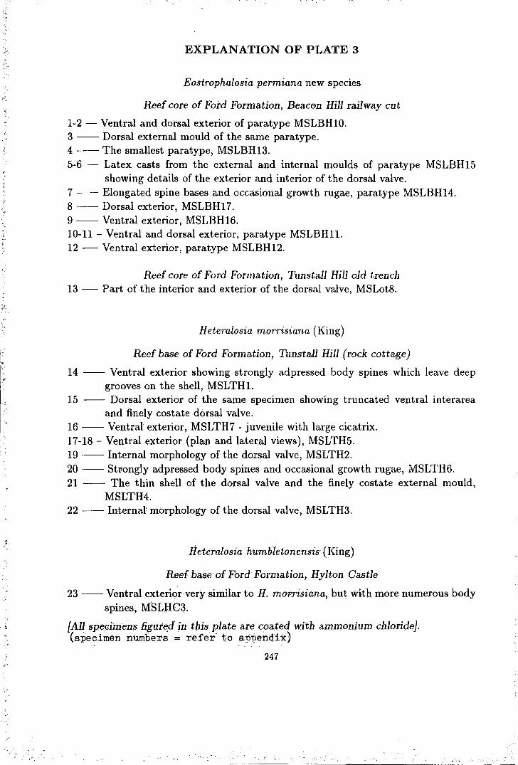
EXPLANATION OF PLATE 3
Eostrophalosia permiana new species
Reef core of Ford Formation, Beacon Hill railway cut
1-2 - Ventral and dorsal exterior of paratype MSLBH10. 3 -- Dorsal external mould of the same paratype. 4 --The smallest paratype, MSLBH13. 5-6 - Latex casts from the external and internal moulds of paratype MSLBH15
showing details of the exterior and interior of the dorsal valve. 7 --Elongated spine bases and occasional growth rugae, paratype MSLBH14. 8 --Dorsal exterior, MSLBH17. 9 --Ventral exterior, MSLBH16. 10-11 -Ventral and dorsal exterior, paratype MSLBHll. 12 - Ventral exterior, paratype MSLBH12.
Reef core of Ford Formation, Tunstall Hill old trench 13- Part of the interior and exterior of the dorsal valve, MSLot8.
Heteralosia morrisiana (King)
Reef base of Ford Formation, Tunstall Hill (rock cottage)
14 -- Ventral exterior showing strongly adpressed body spines which leave deep grooves on the shell, MSLTHl.
15 -- Dorsal exterior of the same specimen showing truncated ventral interarea and finely costate dorsal valve.
16 --Ventral exterior, MSLTH7- juvenile with large cicatrix. 17-18- Ventral exterior (plan and lateral views), MSLTH5. 19 --Internal morphology of the dorsal valve, MSLTH2. 20 --Strongly adpressed body spines and occasional growth rugae, MSLTH6. 21 -- The thin shell of the dorsal valve and the finely costate external mould,
MSLTH4. 22 --Internal morphology of the dorsal valve, MSLTH3.
Heteralosia humbletonensis (King)
Reef base of Ford Formation, Hylton Castle
23 --Ventral exterior very similar to H. morrisiana, but with more numerous body spines, MSLHC3.
{All spedmens figured in this plate are coated with ammonium chloride). (specimen numbers = refer to appendix)
247

PLATE 3
-smm
-smm
248

EXPLANATION OF PLATE 4
Heteralosia humbletonensis (King)
Reef core of Ford Formation, Humbleton Hill
1-3- Ventral exterior (ventral, posterior and lateral views), G88.56a. 4 --Latex cast froin the internal mould of the dorsal valve, G88.56d. 5 --Concentric rows of spine bases developed on the growth rugae, G88.56c. 6 -- Ventral exterior, G88.56b. 7-- Dorsal interior, G88.56e. 8 -- Ventral exterior from latex cast showing adpressed body spines and bases of
erect body spines, MSLHH3. 9-10- Ventral exterior and dorsal interior, MSLHH4. 11- Ventral exterior younger specimen, MSLHH5.
H eteralosia aycliffensis new species
Lower part of Ra.isby Formation, Old Town (=White) Quarry
12 -Several specimens on slab MSLWQ7 with dorsal exterior of paratype MSLWQ7a (marked as a).
13 - Deformed specimen with few very long fine hair-like spines, paratype MSLWQ5. 14- Ventral exterior with elongate spine bases and fine costae, paratype MSLWQ6. 15 - Juvenile with large cicatrix of attachment, paratype MSLWQ8. 16- Ventral exterior, paratype MSLWQ4. 17- Overgrown cicatrix, MSLWQ10. 18- Dorsal exterior with impression of large cicatrix, MSLWQlla. 19- Ventral exterior, l\1SLWQ9.
Lower part of Ra.isby Formation, East Thickley Quarry
20- Ventral exterior, MSLETQ2. 21- Ventral exterior, MSLETQ3.
Lower part of Raisby Formation, Raisby Hill Quarry
22- Ventral interior with a pair of teeth, MSLRHQl.
{Specimen$ in figures 1-3, 5, 9-10 and 22 are not coated; other specimens are coated with ammonium chloride} (specimen numbers = refer to appendix)
249

PLATE 4
' ...
~ . . (' ' '
I '
18
-smm
250

I-
I
EXPLANATION OF PLATE 5
H eteralosia hyltonensis new species
Reef base of Ford Formation, Hylton Castle
1-2 - Ventral exterior (plan and right lateral views), paratype MSLHC3. 3 --Ventral exterior with deep grooves on the trail, paratype MSLHC5a. 4-5- Ventral exterior (plan and posterior views), paratype MSLHC4. 6 --Ventral exterior, paratype MSLHC8. 7 -- Latex cast from internal mould of the same specimen. 8 -- Ventral exterior, MSLHC10. 9-- Ventral exterior (posterior view) with several rhizoid spines, paratype MSLHC6. 10-11 - Ventral and dorsal exterior, paratype MSLHC7. 12- Latex cast of the ventral exterior with strongly adpressed spines, MSLHC9. 13- Dorsal exterior with fine radial costae, MSLHC11.
Heteralosia seahamensis new species
Reef core of Ford Formation, Beac011 Hill railway cut
14-- Ventral exterior with concentrically directed marginal spines, paratype MSLBH22. 15 --Dorsal interior, MSLBH25. 16-17- Ventral and dorsal interior, paratype MSLBH19. 18 -- Latex cast from internal mould (figure 17) of the same paratype showing
details of the internal morphology of the dorsal valve. 19-20- Ventral interior (ventral and posterior views), MSLBH27. 21 -- Ventral exterior of younger individual, MSLBH20b. 22 --Ventral interior, paratype MSLBH20a. 23-24- Dorsal interior and lateral profile, MSLBH26. 25 -- External mould of ·ventral interarea and dorsal exterior, MSLBH24. 26 --Ventral interior, paratype MSLBH21. 27 --Ventral exterior, paratypes MSLBH23.
- - - - - - - reconstructed shell ll1argin {All specimens are coated wi~b ammonium chloride} (specimen numbers = refer to appendix)
251
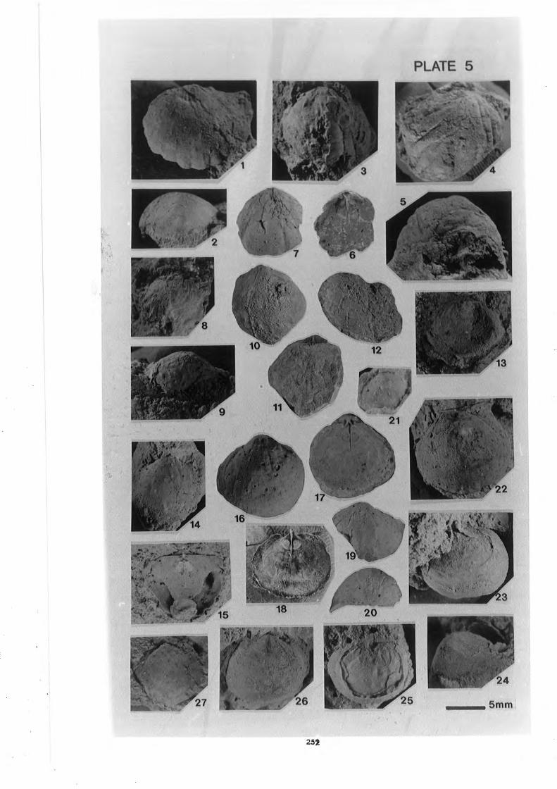
PLATE 5
252

EXPLANATION OF PLATE 6
Craspedalosia langtonensis new species
Lower part of Raisby Formation, Hobgate (=Langton) Quarry
1-2- Ventral interarea and very thin visceral cavity, paratype MSLLQl. 3 -- Strong impression of cicatrix upon the dorsal valve, paratype MSLLQ2. 4 -- Ventral exterior of small individual with large cicatrix covered by matrix,
paratype MSLLQ6. 5 -- Dorsal exterior with strong growth rugae, paratype MSLLQ3. 6-- Small individual attached to a spine of Horridonia horrida (Sowerby), paratype
MSLLQ5. 7 --Dorsal interior, paratype MSLLQ4. 8 -- Ventral exterior with large cicatrix, MSLLQ8. 9 --Dorsal exterior showing squamose growth rugae and fine radial costae, MSLLQ9. 10- Ventral exterior, MSLLQ11.
Craspedalosia lamellosa ( Geinitz)
Cadeby Formation, Nottinghamshire (BGS specimens)
11-12- Ventral and dorsal exterior, YPF 4168, Barnby Moore borehole. 13-14- Ventral and dorsal exterior, BN8223i, Mattersey borehole.
Craspedalosia sp: A
Ford Formation, Hylton Castle (BGS specimens)
15-16- Internal moulds of ventral and dorsal valves, YEF 2367Da. 17 -- External mould of the same specimen showing squamose dorsal growth rugae. 18 --Ventral exterior of two specimens, YEF 2367Dd,e. 19 -- Ventral interior with sharply pointed umbo, YEF2367Dd. 20 --Ventral interior, YEF 2367Df.
[All specimens are coated witl1 ammonium chloride) specimen numbers = refer to appendix)
253

PLATE 6
15 17
--smm
254

EXPLANATION OF PLATE 7
H eteralosia? quarringtonensis new species
Lower part of Raisby Formation, Old Quarrington Quarry
1- Ventral exterior, paratype MSLOQQ3a. 2- Ventral exterior, paratype MSLOQQ4. 3- Internal mould of dorsal valve, paratype MSLOQQ5a (marked as a) with two
other specimens, MSLOQQ5b,c. 4 - Ventral exterior, paratype MSLOQQ6. 5 - Interior of dorsal valve, MSLOQQ8. 6- External mould of dorsal valve and ventral interarea, MSLOQQ3b. 7 - Latex cast of ventral exterior, MSLOQQ3c.
Lower part of Raisby Formation, Sherburn Hill Quarry
8- Latex cast of ventral exterior, MSLSHQ3a. 9- Ventral exterior with elongate spine bases, MSLSHQl. 10- Ventral exterior, MSLSHQ3b. 11 - Latex cast of ventral exterior, MSLSHQ2.
Strophalosia? parva (King)
Reef core of Ford Formation, Humbleton Hill
12 - Ventral valve attached on the exterior umbo of Horridonia dunelmemsis new species, MSLHH8 - this latex reproduction shows the interior of ventral valve with large strongly elevated adductor muscle platform.
13 - Syntype of S?. parva (King collection in University College of Galway) showing large cicatrix cemented on the interior of H.dunelmemsis. this specimen shows numerous spines creeping on the host.
Howseia latirostrata (Howse)
Patch reef of Ford Formation, Gilleylaw builder's trench (Dr. G. A. L. Johnson's specimens)
14 - Latex cast of dorsal exterior showing part of the ventral interarea and some rhizoid spines, GALJ7.
15 - Latex cast of dorsal interior, showing the internal morphology of the dorsal valve and some rhizoid spines from the ventral valve, GALJ8.
16 - Latex cast of speci111ens GALJ9a,b,c showing auricular spines of specimen marked•b clasping ono the shell-of specifnen marked a.
{All specimens are coated w-ith ammonium chloride].
(specimen numbers = re!er to appendix) 255

PLATE 7
2mm - - smm
256

EXPLANATION OF PLATE~8
Howseia latirostrata (Howse)
Reef base of Ford Formation, Tunstall Hill (rock cottage)
1 -- Ventral exterior, MSLTH12. 2 --Ventral exterior, MSLTH-13. 3-4- Ventral and dorsal exterior of a male dimorph, G88.57a. 5 --Ventral exterior of a male dimorph, MSLTH14. 6 --Ventral exterior of a female dimorph, G88.57b. 7 --Ventral exterior of a female dimorph, MSLTH15. 8 --Ventral exterior of a senile female dimorph, G88.57c. 9 --Ventral interior, MSLTH18. 10- Dorsal interior, MSLTH17. 11 -Dorsal exterior and ventral interarea, MSLTH16.
Howseia umbonillata (King)
Reef base of Ford Formation, Tunstall Hill (rock cottage)
12 - Dorsal exterior (male) with lophidium inserted into triangular opening of ventral interarea, MSLTH21.
13- Ventral interior (female), MSLTH22. 24 - Ventral exterior of a juvenile, MSLTH25. 25- Ventral exterior (female), MSLTH23.
Ford Formation, Dalton-le-Dale (HM collection)
14 - Dorsal interior (mould) and ventral interarea, G88.53b. 19- Ventral interior with shelly material (male dimorph), G88.53a.
Reef patch of Ford Formation, Gilleylaw builder's trench (Dr. G. A. L. Johnson's collection)
15 -- Ventral exterior of juvenile, GALJl. 16 -- Ventral exterior of juvenile, GALJ2. 17 -- Ventral exterior of juvenile, GALJ3. 18 -- Ventral exterior of male dimorph, GALJ4. 20-21 - Dorsal interior and lateral view of ventral interior of female dimorph, GALJ6. 22-23- Dorsal interior and lateral view of ventral interior of female dimorph, GALJ5.
{All specimens (except specimens in figures 14 and 19) are coated with ammonium chloride}
(specimen numbers = refer to apl)endix)
257

PLATE 8
15
10mm
258

EXPLANATION OF PLATE 9
Horridonia horrida (Sowerby)
Lower part of Raisby Formation, Old Town (=White) Quarry 1- Ventral exterior (posterior view) showing unique spines arrangement, MSLWQ12. 2- Ventral exterior (posterior view) showing unique spines arrangement, MSLWQ13. 3- Ventral exterior of juvenile, MSLWQ14. 4 - Ventral exterior of juvenile, MSLWQ15. 5 - Ventral exterior, MSLWQ16. 6 -- Ventral interior, MSLWQ17. 7 -- Ventral exterior, MSLWQ18. 8- Ventral exterior, MSLWQ19. 10 - Ventral exterior in life position, seen from the top, MSLWQ20 - exposed by
natural erosion. 11- Natural cross section, seen from the top, MSLWQ 23 -preserved in life position. 12 - Dorsal interior, MSLWQ21. 13- Ventral exterior (posterior view) showing unique spines arrangement, MSLWQ22. 14- Enlargement of figure 3.
Lower part of Raisby Formation, East Thickley Quarry
9 -- Dorsal exterior, MSLETQ4 (the two large spines have been repositioned during diagenesis).
H orridonia species A
Lower part of Raisby Formation, Old Town (=White) Quarry
15- Ventral exterior, MSLWQ26.
Reef core of Ford Formation, Tunstall hill old trench
16- Ventral exterior, MSLot12.
{All specimen are coated with ammonium chloride, except specimen in figure 2 which is immersed in water} (specimen numbers = refer to appendix)
259

PLATE 9
14
10mm 5mm -
260

= : - ; ~ -
EXPLANATION OF PLATE 10
Horridonia hoppeiana (Eisel)
Lower part of Raisby Formation, Hobgate (=Langton) Quarry
1-3- Ventral exterior (posterior, lateral and plan views), MSLLQ11.
Lower part of Raisby Formation, Old Town (=White) Quarry
4-5- Lateral profile ventral exterior, MSLWQ24. 10-11- Lateral and anteroventral views of ventral interior, MSLWQ25.
Patch reef of Ford Formation, High Newport old railway cut
6 --Ventral exterior, MSLHN4. 7-- Ventral exterior, MSLHN5. 8-9- Ventral exterior (posterior and plan views), MSLHN6.
Spinohorridonia species cf. S. laspeana (Eisel)
Reef core of Ford Formation, Humbleton Hill
12- Dorsal exterior (external mould), MSLHH21. 13- Dorsal exterior (external mould), MSLHH19. 14- Dorsal exterior (latex cast), MSLHH20.
?Reef core of Ford Formation, Ryhope old railway cut
15-16- Latex cast of dorsal and ventral exterior, MSLR2.
{All specimens are coated with ammonium chloride} (specimen numbers = refer to appendix)
261

PLATE 10
262

=..o ~-.· • - .
EXPLANATION OF PLATE 11
H orridonia dunelmensis new species
Reef core of Ford Formation, Humbleton Hill
1-8 - Internal mould of ventral valves from a series of ontogenic development, MSLHH12-16,9,17-18. (MSLfiH9 shows numerous spines set on a horizontal plane).
9 - Ventral internal mould, MSLHH18. 10-11- Latex cast of dorsal and ventral exteriors, MSLHH8. (ventral exterior carries
numerous body spines while dorsal exterior has no body spines). 12 - Dorsal interior showing a row of marginal spine apertures, latex cast from
MSLHH10. 13- Ventral interior showing a ridge with coarse endospines matching the apertures
in figure 12, latex cast from MSLHH11.
Reef core of Ford Formation, Beacon Hill railway cut
14- Ventral exterior (lateral view), MSLBH29b. 15 - Dorsal interior with quadrilobed myophore, MSLBH29a. 16 - Posterior view, latex cast of MSLBH30.
Reef talus of Ford Formation, Ryhope old railway cut
17 - Ventral internal mould with impression of spine bases, MSLRl.
Reef core of Ford Formation, Tunstall Hill old trench
18- Ventral internal mould with patches of shell remain, MSLotll.
Spinohorridonia geinitziana (de Kouinck)
Reef base of Ford Formation, Hylton Castle
19-20, 22- Ventral exterior (posterior, anterior and lateral views), MSLHC13. 21 - Dorsal exterior of the same specimen. 23- Parts of dorsal interior, MSLHC16.
Reef core of Ford Formation, Tunstall Hill old trench
24- Ventral internal mould, MSLot14. 25 - Parts of dors(:l.l interior, MSLotl5. 26 - Posterior region frotn ventr~l ititerna:l mould, MSLot13.
{All specimens figured in this plate are coated with ammonium chloride}. (specimen numbers = refer to appendix)
263
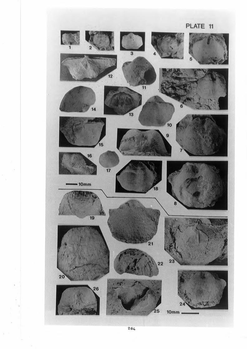
PLATE 11
264

EXPLANATION OF PLATE 12
Craspedalosia.species A
Na:m Loong Bed o{Kinta Limestone, H. S. Lee mine no 8 1-2- Exterior and interior of dorsal valve, 02727 (UM collection).
Institella species A
~ase of Chuping Limestone Formation, Pulau Jong 3 -- Ventral exterior ~ith large rhizoid marginal spine and smooth lateral margin, '!
1353 (UM collection).
Dictyoclostus.'? gratiosus (Waagen)
Leptodus Bed of Gua, Musang Formation Dada Kering -(;ua.Musailg highway [UKM~MSL collection}
4 -- Ventral exterior, MSL10.1a (Ioca.lity 10). 5-- Dors~ interior, MS:t19.1b (locality 10). 6 -- Ventra.l exterior, M:SL8.1a (locality 8). 7 --Ventral exterior, MSL16.la(locality 16). 8- Dor5al interior, MSL16.1b (locality 16). 9 -- Strongly .convex ventral :va.hr.e :With.Jorig .trail,.MSL16.lc (locality 16). _
Antiquatonia species A
Leptodus Bed of Gua Musang Formation Dada Keting- Gua lrfu~ang highway{UKM"MSL collection)
10- Ext~rnal: moul~ of open ventral and donial valves, MSL10.2a (locality 10). 11- Interna.J. mould ventral' valve with a row of marginal spines, MSL10.2b (locality
10).
Kepis Bed, Kampung /uaseh 12- Ventral valve compressed against dorsal valve, MSL20.1a. 13- Dorsal interior, MSL20.1b. 14- Ventral external 'mquld: MSL20.lc. 15- Ventral interior; MSL20.1a.
2(;5
I;
'.,,, '< •• ", .•. ,. "'•':' ''
Reticulatia sp. indet.
Leptodus Bed of Gua Musang Formation Dada Kering- Gua Musang highway {UKM-MSL collection}
16 - Ventral exterior, MSL8.2a (locality 8). 17,18- Ventral exterior, MSL16.2a.,b (locality 16).
Echinoconchus species A
Nam Loong Bed of Kinta Limestone, H. S. Lee mine no 8 19,23- Dorsal external moulds, UM-1a,e (UM collection). 20,21,23- Ventral exteriors, UM-1b,c,d (UM collection).
W aagenoconcha sp. indet.
Leptodus Bed of Gua Musang Formation Dada Kering- Gua Musang highway {UKM-MSL collection)
24 -- Ventral exterior, MSL16.3 (locality 16)
Waagenoconcha sp. cf. W. abichi (Waagen)
Nam Loong Bed of Kinta Limestone, H. S. Lee mine no 8 25,26,27,28- Casts of ventral exteriors, Um~2a.;b,c,d (UM collection).
Linoproductus sp. cf. L '?. sinosus Huang
Leptodus Bed of Gua Musang Formation Dada Kering - Gua Musang highway {UKM-MSL collection}
29 -- Cast of ventral exterior, MSL10.3 (locality 10). 30 --Cast of ventral exterior (postero-lateral view), MSL9.la. (locality 9). 31 -- Several specimen from rock slab MSL9.1b (locality 9).
{All specimens figured in this plate are coated with ammonium chloride}
:I '.'·! 1111
'l'
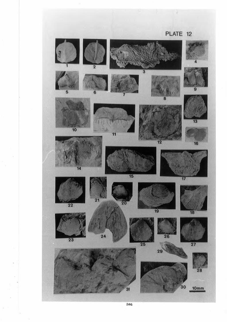
. ~-~ .... ,;.
,j_• s . ~ -,..,
6
PLATE 12
)t'• . ·ll'
·-.~~- ... ,~ ...
9
10mm
266 -oooo:------~----~-~.....:1
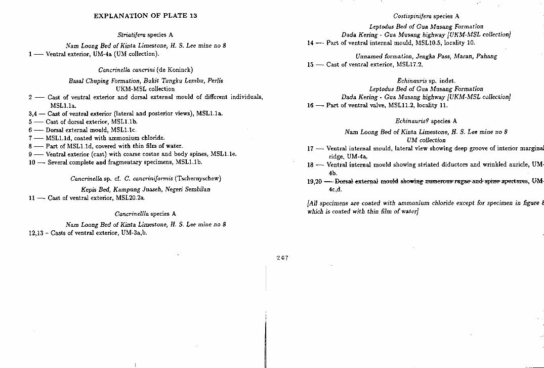
EXPLANATION OF PLATE 13
Striatifera species A
Nam Loong Bed of Kinta Limestone, H. S. Lee mine no 8 1 - Ventral exterior, UM-4a (UM collection).
Cancrinella cancrini (de Koninck)
Basal Chuping Formation, Bukit Tungku Lembu, Perlis UKM-MSL collection
2 - Cast of ventral exterior and dorsal external mould of different individuals, MSLl.la.
3,4- Cast of ventral exterior (lateral and posterior views), MSL1.1a. 5- Cast of dorsal exterior, MSLl.lb. 6- Dorsal external mould, MSL1.1c. 7- MSLl.ld, coated with ammonium chloride. 8- Part of MSL1.1d, covered with thin film of water. 9- Ventral exterior (cast) with coarse costae and body spines, MSLl.le. 10- Several complete and fragmentary specimens, MSL1.1b.
Cancrinella sp. cf. C. cancriniformis (Tschernyschew)
Kepis Bed, Kampung Juaseh, Negeri Sembilan 11 - Cast of ventral exterior, MSL20.2a.
Cancrinellla species A
Nam Loong Bed of Kinta Limestone, H. S. Lee mine no 8 12,13- Casts of ventral exterior, UM-3a,b.
ZG7
Costispinifera species A
Leptodus Bed of Gua Musang Formation Dada Kering - Gua Musang highway [UKM-MSL collection}
14 -- Part of ventral internal mould, MSL10.5, locality 10.
Unnamed formation, Jengka Pass, Maran, Pahang 15 - Cast of ventral exterior, MSL17.2.
Echinauris sp. indet. Leptodus Bed of Gua Musang Formation
Dada Kering- Gua Musang highway [UKM-MSL collection} 16- Part of ventral valve, MSL11.2, locality 11.
Echinauris? species A
Nam Loong Bed of Kinta Limestone, H. S. Lee mine no 8 UM collection
17 - Ventral internal mould, lateral view showing deep groove of interior marginal ridge, UM-4a.
18 - Ventral internal mould showing striated diductors and wrinkled auricle, UM-4b.
19,20 --Dorsal· e-xternal mould showing· numerons·l'l!lgae' amhprne"'<tpe-rtnres, UM· 4c,d.
{All specimens are coated with ammonium chloride except for specimen in figure 8 which is coated with thin film of water}
16
268
I f
''
/
(.f;l
PLATE 13
17
---10mm
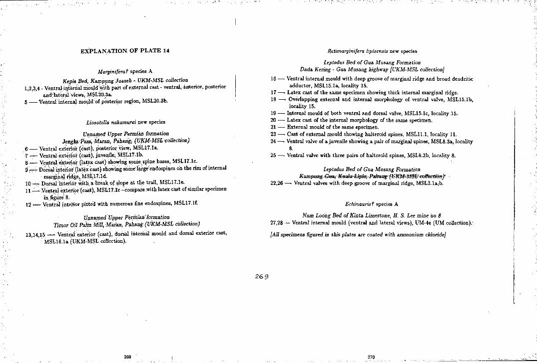
''d,
EXPLANATION OF PLATE 14
Marginifera? species A
Kepis Bed, Kampung Juaseb - UKM-MSL collection 1;2,3,4 - Ventral i~t'errial mould ~th part of external cast - ventral, anterior, posterior
a:nd·la.t~i:al views, MSL20.3i. 5- Ventral internal ~ould'of p~sterior region, MSL20.3b.
' ' .. .
Liosotella nakamurai new species
Unnamed Upper Permian formation Jengka P~, ·Maran; Pabiing, (UKM-MSL collection)
6- Ventral exte:rior (cast), posterior \iew, MSL17.la. 7-. ·-Ventral exterior (cast), juvenile; MSL17.lb. ~ .- Ve~~ral exterio~ (l~tex c;asi) showing some spine bases, MSL17.lc. 9 -. -. Do~al·interior (latex cast) shqwing s01ne llu·ge endospines on the rim of internal
mar~a1. rid&e. MS~17;ld. 10 '--:- Dors~ interior, With. a. break of slope at the trail, MSL17.1e. 11 -· - Ve~t~al e~enor (c~t), MSLi 'l.l'c- compare with latex cast of similar specimen
· in figure 8: 12- VentraJinteiiorpitted with numerous fine endospines; MSL17.1f.
Unnilmed Upper Permi'an ·formation TiiJ?or Oil Palm Min, Maran, Pab.ang (UKM-MSL collection)
13,14,15 -. -. Ventx:al e~enot (cast), dorsal interna.I mould and dorsal exterior cast, · MSL18.1a: (UKMcMSL collection).
269
269
.. .' .. ···• !
Retimarginifera lipisensis new species
Leptodus Bed ofGua Musang Formation Dada Kering- Gua Musang highway {UKM-MSL collection}
16- Ventral internal mould with deep groove of marginal ridge and broad dendritic adductor, MSL15.1a, locality 15.
17 - Latex cast of the same specimen showing thick internal marginal ridge. 18 - Overlapping external and internal morphology of ventral valve, MSL15.Ib,
locality 15. 19- Internal mould of both ventral and dorsal valve, MSL15.1c, locality 15. 20 - Latex cast of the internal morphology of the sallie specimen. 21 - External mould of the same specimen. 23- Cast of external mould showing halteroid spines, MSL11.1, locality 11. 24- Ventral valve of a juvenile sho.;nng a pair of marginal spines, MSL8.3a, locality
8. 25 - Ventral valve with three pairs of halteroid spines, MSL8.3b, locality 8.
Leptodus Bed of Gua Musang Formation KampGng,·GlJ.a.;··Kualwi;ipisj-Paltarrg.(liHM~MS1tco11ectiorrr
22,26- Ventral valves with deep groove of marginal ridge, MSL3.1a;b.
Echinauris? species A
Nam Loong Bed of Kinta Limestone, H. S. Lee mine no 8 27,28- Ventral internal mould (ventral and lateral views), UM-4e (UM collection):
{All specimens figured in this plates are coated with ammonium chloride}
270
'!-> ,_r i
·I I ·, d

PLATE 14
25 --·10mm
270

EXPLANATION OF PLATE 15 (Geological Survey Department (GSD) colection)
Strophalosia species A Leptodus Bed of Gua Musang Formation
Railway cut adjacent to Sungai Jeleteh, Merapoh, Pahang 1 - Internal mould of dorsal valve with ventral interarea, LF 176.
Dictyoclostus sp. indet. Leptodus Bed of Gua Musang Formation
Sungai Galas, Gua Musang, Kelantan 2,3 - Several undetermined specimens showing coarse costae and reticulate umbo,
FPhN19a,b.
Antiquatonia species A ?Gua Musang Formation, Sungai Sepia, Kuala Lipis, Pahang
4,6- Casts of ventral exterior, L1096(31,26). 5 --Dorsal external mould, L1096(29b).
Stepanoviella sp. cf. S. flexuosa Waterhouse Kepis Bed, Kampung Baru Kepis, Kuala Pilah, Negeri Sembilan
7 - Dorsal interior, KPF3b. 8- Dorsal external mould, KPF3a. 9- Ventral exterior (cast), unnumbered specimen.
Costispinifera species A Unnamed Upper Permian formation, Ulu Lepar, Maran, Pahang
10,11 -- Several fragmentary cast showing coarse corrugated costae with numerous spines especially on lateral slope of visceral disc, AN29a,b.
Dictyoclostus? gratiosus (Waagen) Leptodus Bed of Gua M usang Formation
Railway cut adjacent to Sungai Jeleteh, Merapoh, Pahang 12 - Ventral exterior, 172/9c.
Retimarginifera lipisensis new species Leptodus Bed of Gua. Musang Fotmation
Railway cut adjacent to Sungai Jel~~eh, Merapoh, Pahang 13 - Superimposed internal and external morphology of both ventral and dorsal
valve, 172/7.
Paucispinifera species A Upper Permicm Aring F6rma.tion, South Central Kelantan
14 - Ventral internal mo-ulgs of three specimens GS{F)5,~52,53.
{All specimens are not coated}
271
_--;- --·· "'- ~--~:. ; ~

PLATE 15
10mm
---10mm
12
272.

LIST OF REFERENCES
Aw, P. C. et al. 1977. On Palaeofusulina- Colaniella fauna from the Upper Permian of Kelantan, Malaysia. Transactions and Proceedings of the Palaeontological Society of Japan, (new series), 104, 407-417, pl 43.
Ball, H. W. 1975. British Palaeozoic fossils, (fourth edition). British Museum (Natural History), London.
Batten, R. L. 1972. Permian gastropods and chitons from Perak, Malaysia, Pt. L Chitons, bellerophontids, euomphalids and pleurotomarians. Bulletin of American Museum Natural History, 147, 1-44.
Bell$,J. et al. 1979. A new exposure of the Marl Slate and basal Permian Breccia at Middridge, County Durham. Proceedings of the Yorkshire Geological Society, 42, 439-460.
Branson, C. C. 1948. Bibliographic index of Permian invertebrates. Memoirs of the Geological Society of America, 26, 1049 pp.
Broili, F. 1916. Die Permischen Brachiopoden von Timor. Palaeontologie von Timor, 7, 104 pp, 13 pls.
Brunton, C. H. C. 1966. Silicified productoids from the Visean of County Fermanagh. Bulletin of British Museum (Natural History), Geology, 12, 173-243.
Brunton, C. H. C. & Mundy, D. J. C. 1988, Strophalosiacean and aulostegacean productoids (Brachiopoda) from the Craven Reef Belt (late Visean) of North Yorkshire. Proceedings of the Yorkshire Geological Society, 47(1), 55-88, fig 1-14.
Bryant, D. L. 1955. Index, type species and bibliography of productid genera. Journal of Paleontology, 29, 283-94.
Chao, Y. T. 1928. Productidae of China, Part II, PalaeontologiaSinica, 5(3), 75 pp, 5 pls.
Clark, R. G. 1974. Growth lines in invertebrate skeletons. Annals and Review of Earth and Planetary Sciences, 2, 77-79.
Cocks, L. R. M. 1985. Brachiopoda in Murray, J. W. (ed.). Atlas of invertebrate macrofossils. The Paleontological Association. Longman, London.
Coleman, P. J. 1957. Permian Productacea of Western Australia. Bulletin of Bureau
273

of Mineral Resources, Geololgy and Geophysics, 40, 1-189, 21 pls.
Cooper, G. A. 1957. Permian brachiopods from Central Oregon. Smithsonian Miscelaneous Collections, 134(12), 1-79, pl 1-12.
Cooper, G. A. & Grant, R. E. 1969. New Permian brachiopods from West Texas. Smithsonian Contribution to Paleobiology, 1, 20 pp.
' ---- 1975a. Permian brachiopods from West Texas III. Smithsonian Contribu-
tion to Paleobiology, 19(1), 795-1921.
---- 1975b. Permian brachiopods from West Texas IIIb. Smithsonian Contribution to Paleobiology, 19(2), pl 192-502.
Cooper, G. A. & Stehli, F. G. 1955. New genera of Permian brachiopods from West Texas. Journal of Paleontology, 29(3), 469-474, pl 52-54.
Cooper. G. A. et al. 1953. Permian faunas at el Antimonio, Western Sonora, Mexico, Smithsonian Miscelaneous Collections, 119(2), 1-106, pl 1-25.
Davidson, T. 1853. A monograph of the British fossil Brachiopoda, Vol. I, Pt. I, The general introduction. 1-136, pl 1-9, Palaeontographical Society, London.
---1858, A monograph of the British fossil Brachiopoda, Vol. II, Permian species. 1-51, pl 1-4, Palaeontographical Society, London.
--- 1880, A monograph of the British fossil Brachiopoda, Vol. IV, Pt. IV, A supplement to the British Permian Brachiopoda. 243-8, Palaeontographical Society, London.
Dunbar, C. 0. 1955. Permian brachiopods faunas of Central East Greenland. Meddelelser om Gronland, 110(3), 1-169, pl 1-32.
---- 1962. Faunas and correlation of the late Paleozoic rocks of Northeast Greenland, pt. III, Brachiopoda. Meddelelser om Gronland, 167(6), 1-14, pl 1-2.
Dunbar, C. 0. & Condra, G. E. 1932, Brachiopoda of the Pennsylvanian System in Nebraska. Nebraska Geological Survey Bulletin, (Series 2), 5, 377 pp, 44pls.
Eden, R. A., Stevenson, S. P. & Edwards, W. 1957. Geology of the country around Sheffield, Memoirs of the Geological Survey of Great Britain, Sheet 100.
Edwards. W. N. 1951. Geology of the concealed coalfield in Nottinghamshire and Yorkshire, Memoirs of the Geological Survey of Great Britain.
----1967. Geology of the country around Ollerton, Memoirs of the Geological Survey of Great Britain, Sheet 113.
274

Eisel, R. 1909. Varianten des Productus horridus SOW. bei Gera. Geselllschaft von Freundin der Naturwissenschaft in Gera, Jahr, 49-50, 33,.37, pl 1.
Fagerstrom, J. A. & Boellstorff, J.D. 1964. Taxonomic criteria in the classification of the Pennsylvanian productoid Juresania nebrascensis. Paleontology; 7{1), 23-8, pl2.
Ferguson, L. 1969. Possible brood pouches and sexual dimorphism in the productid brachiopod Megousia Muir-Wood & Cooper. in Westermann, G. E. G. (ed.) Sexual dimorphism in fossil Metazoa and taxonomic implications. International Union of Geological Science, (series A), no. 1, Stuttgart.
Fontaine, H. 1986. The Permian of Southeast Asia. Technical Bulletin - United Nations ESCAP, CCOP, 18, 171 pp.
Fontaine, H. & Khoo, H. P. 1988. Palaeontological research in Terengganu State, Peninsular Malaysia. CCOP Newsletter, 13{1), 25-30.
Fontaine, H., Lys, M. & Tien, N. D. 1988. Some Permian corals from East Peninsular Malaysia; associated microfossils, palaeogeographic significance. Journal of Southeast Asian Earth Sciences, 2{2), 65-78, 7 pls.
Frebold, H. 1931a. Das Marine Oberkarbon Ostgronlands. Meddelelser om Gmnland, 84(2), 1-85, pl 1-8.
Frebold, H. 1931b. Unterer Mariner Zechstein in Ostgronland und das alter der Depot Island Formation. Meddelelser om Gronland, 84{3), 1-38, pl. 1-2.
Frebold, H. 1933. Weitere Beitrage Zur Kenntnis des oberen Palaozoikums Ostgronlands. Meddelelser om Gronland, 84{7), 1-61, pl. 1-6.
Geinitz, H. B. 1842. Uber einege Petrefakte des Zechsteins und Muschelkalk. Neues Jahrbuch fur Mineralogie. Geognosie und Geologie Petrefakten-Kunde Abhand-lungen, 576-9, pl 10. ·
----·- 1847. Orthothrix Geinitz. Soc. Imp. Nat. Moscow Bulletin, 20{2), 84-86.
----- 1848. Die Versteinerungen des deutschen Zechsteingebirges. Arnoldische Buchhandlung. 26 pp, 8 pls, Dresden, Leipzig.
----- 1857. Euber zwei neue Versteinerungen und de Strophalosien des Zechsteins. Neues Jahrbuch fur Mineralogie. Geologie und Palaontologie, 9, 207-10, pl 11.
1861. Dyas oder Zechstein/ormation und das Rothliegende. 2 vols, Leipzig.
Geological Survey of Malaysia, 1987. Annual Report Geological Survey of Malaysia
275

1986. National Printing Department, Kuala Lumpur.
-----, 1988. Annual Report Geological Survey of Malaysia 1987. National Printing Department, Sarawak.
Cobbett, D. J. 1961. The Permian brachiopod genus Horridonia CHAO. Palaeontology, 4{1), 42-53, pls 3-5.
--- 1963. Carboniferous and Permian brachiopods of Svalbard. Norsk Polarinstitutt, Oslo, 201 pp, 25 pls.
--- 1973a. Carboniferous and Permian correlation in Southeast Asia. Bulletin of the Geological Society of Malaysia, 6, 131-142.
1973b. Upper Palaeozoic, in Cobbett, D. J. & Hutchinson, C. J. (eds.), Geology of the Malay Peninsula. Wiley Interscience, New York.
Grant, R. E. 1968. Structural adaptation in two Permian brachiopod genera, Salt Range, West Pakistan. Journal of Paleontology, 42(1), 1-32, pl 1-9.
Hall, J. & Clarke, J. M. 1892. Introduction to the study of the genera of Palaeozoic brachiopod, Pt 1. Geolological Survey of New York, Paleontology, 8{1), 1-367, pl 1-20.
Hill, D. 1950. The Productinae of the Artinskian Cracow Fauna of Queensland. Papers for the Department of Geology, University of Queensland, 3{2), 26 pp, 9 pls.
Hinchey, N. & Ray, L. L. 1935. New Mississippian species of Strophalosia from Missouri. Journal of Paleontology, 9{3), 247-50, pl 25.
Hollingworth, N. T. J. 1987. Palaeoecology of the Upper Permian Zechstein Cycle I Reef of N. E. England, Unpublished PhD thesis, University of Durham.
Hollingworth, N. T. J. & Pettigrew, T. 1988. Field guide to fossils, No 3; Zechstein reef fossils and their palaeoecology. The Palaeontological Association, London, 75 pp.
Howse, R. 1848. A Catalogue of the fossils of the Permian Systems of the Counties of Nothumberland and Durham drawn up at the request of the Tyneside Naturalists Field Club. Transactions of the Tyneside Naturalists Field Club, 1(3), 219-264.
-.-- 1857. Notes on the Permian System of Nothumberland and Durham. Annals and Magazine of Natural History, (Ser. 2), 19, 33-52, 1 pl.
-- 1858. Notes on the Permian System of Nothumberland and Durham, Transactions of the Tyneside Naturalists Field Club, 3(4), 1-53, 1 pl.
276

Huang, T. K. 1932. Late Permian Brachiopoda of Southwestern China. Palaeontologia Sinica, (series B), 9(1), 107 pp, 9 pls.
1933. Late Permian Brachiopoda of Southwestern China Part 2. Palaeontologia Sinica, (series B), 9(2), 131 pp, 11 pls.
Ibrahim Amnan. 1988. Progress report of fossil indexing for Johor, p 147-155, in Gelogical Survey Department, Annual Report Geological Survey of Malaysia 1987. National Printing Department, Sarawak.
Igo, H. 1964. Permian fossils from Northern Pahang, Malaya. Japanese Journal of Geology and Geography, 35(1), 57-71, pl 2.
Ichikawa, K., Ishii, K. & Hada, S. 1966. On the remarkable uncomformity at the Jengka Pass, Pahang, Malaysia. Journal of Geosciences, Osaka City University, 9, 123-130.
Ishii, K. 1966. Preliminary notes of the Permian fusulinids of H. S. Lee mine no 8 limestone, near Kampar, Malaya. Journal of Sciences, Osaka City University, 9, 145.
Ishii, K. et al. 1972. Permian brachiopods from Bukit Tungku Lembu, Pedis, Malaysia. Journal of Geosciences, Osaka City University, 15, 65-76, 2 pls.
International Commision on Zoological Nomenclature, 1961. International Code of Zoological Nomenclature. International Trust for Zoological Nomenclature, London, 176pp.
Johnson, G. A. L. 1981. Geographical evolution form Lauresia to Pangea. Proceedings of the Yorkshire Geological Society, 43(3), 221-252.
Johnson, G. A. L. 1982. Geographical changes in Britain during the Carboniferous period. ProcBedings of the Yorkshire Geological Society, 44(2), 181-203.
Jones, C. R., Gobbett, D. J. & Kobayashi, T. 1966. Summary of fossil record in Malaya and Singapore 1900-1965. Geology and Palaeontology of Southeast Asia, 2, 309-359.
Karwowski, L. & Klapcinski, J. 1986. Macrofauna of Polish Zechstein: its occurence and stratigraphy. in Harwood, G. M. & Smith, D. B. (eds.) The English Zechstein and related topics. Geological Society Special Publication, 22, 211-216.
Kato, M. & Ezaki, Y. 1986. Permian corals from Pahang and Terengganu, Malaysia. Journal of Faculty Sciences, Hokkaido University, (series IV), 21(4), 645-668.
King, R. H. 1938. New Chonetidae and Productidae from Pennsylvanian and Permian Strata of North-Central Texas. Journal of Paleontology, 12(3), 257-79, pl36-39.
277

King, W. 1844. On a new genus of Palaeozoic shells. Annals and Magazine of Natural History, 14, 313-317.
-- 1846. Remarks on certain genera belonging to the class Palliobranchiata. Annals and Magazine of Natural History, 18, 26-42 & 83-94.
--- 1848. A catalogue of the organic remains of the Permian rocks of N othumberland and Durham. Newcastle upon Tyne.
--- 1850. A monograph of the Permian fossils of England. Palaeontographical Society, London, 258 pp, 28 pls.
-- 1856. · Notes on Permian fossils:- Palliobranchiata. Annals and Magazine of Natural History, (series 2), 17, 258-269 & 333-341, pl 12.
de Koninck, L. 1846. Monographie du genre Productus. Memoir de la Society Royale des Sciences des Liege, 4, 73-278, pl 1-17.
- 1847. Recherches sur les Animaux Fossiles, Pt I. Monographie des genres Productus et Chonetes. H. Dessain, Liege, 246 pp, 20 pls.
Konno, E. & Asama, K. 1970. Some Permian plants from the Jengka Pass, Pahang, West Malaysia. Geology and Paleontology of Southeast Asia, 8, 97-132, pl 17-24.
Land, D. H. 1974. Geology of the Tynemouth District. Memoirs of the Geological Survey of Great Britain, Sheet 15.
Licharew, B. K. & Einor, 0. L. 1939. Paleontology of the Soviet Arctic, Pt. 4, Contribution to the knowledge of the Upper Paleozoic fauna of Novaya Zemlya. Brachiopoda. Transactions of Arctic Institut Leningrad, 27,. 1-245, 28 pls.
Logan, A. 1962. A revision of the palaeontology of the Permian limestones of County Durham. Unpublished PhD thesis, University of Durham (King's College).
-- 1963. The new brachiopod genus Howseia from the Permian magnesian limestone of Durham. Palaeontology, 6( 4), 754-61, pl 108.
--1966. The Upper Palaeozoic productoid brachiopod Horridonia timanica (Stuckenberg) and its close relatives. Transactions of Leeds Geological Association, 7, 193-210, fig 1-2.
Logan, A. & Mcgugan, A. 1967. Biostratography and faunas of the Permian lshbel Group, Canadian Rocky Mountains. Journal of Paleontology, 42(5), 1123-1139, pl 141-144.
Malzahn, E. 1937. Die Deutschen Zechstein Brachiopoden. Abhandlungen Preussen Geologische Landesanst., 185, 1-67, pl 1-4.
278

Manson, R.T. 1899. Outlines of the geology of South Durham. Darlington Halfholiday Guide, Darlington and Teesdale Naturalists Field Club, Wm Dresser & Sons, Darlington.
Maxwell, W. H. G. 1954. Strophalosia in the Permian of Queensland. Journal of Paleontology, 28( 5), 533-559.
Metcalfe, I. 1981. Permian and early Triassic conodonts from Northwest Peninsular Malaysia. Bulletin of the Geological Society of Malaysia, 14, 119-126.
1988. Origin and assembly of Southeast Asian continental terranes. m Audley-Charles, M. G. & Hallam, A. (eds.) Gondwana and Tethys. The Geological Society Special Publication no 37, 101-118.
Mills, D. A. C. & Hull, J. H. 1976. Geology of the country around Barnard Castle. Memoir of the Geological Survey of Great Britain, Sheet 27.
Mohd Shafeea Leman. 1987a. Geologi batuan Trias di sekitar Kuala Lipis, Pahang. Unpublished report UKM Research Grant No 23/85.
Mohd Shafeea Leman. 1987b. Geologi am lebuhraya Dada Kering -Gua Musang. Unpublished report UKM Research Grant No 10/86.
Muenster, G. C. 1839. Beitrage zur Petrefakten-kunde. Bayreuth, 7 vols.
Muir-Wood, H. 1948. Malayan Lower Carboniferous fossils (and their bearing on the Visean Palaeogeography of Asia. British Museum (Natural) History, London, 118 pp, 17 pls.
----- 1965. Productidina: in Moore, R. C. ( ed. ), Treatise on Invertebrate Paleontology, Part H- Brachiopoda. 1, 439-510.
Muir-Wood, H. & Cooper, G. A. 1960. Morphology, classification and life habit of the Productoidea (Brachiopoda). Memoirs of the Geological Society of America, 81, 447 pp, 112 pls.
Mundy, D. J. C. & Brunton, C. H. C. 1985. Morphological similarities in some British Dinantian and Texas Permian reef brachiopods. in Compte Rendu du Congres International de Stratigraphie et de Geologie de Carbonifere 9eme (Urbana, 1979}, 5, 225-232.
Nakamura, K. 1973. Phylum Brachiopoda in Nakazawa, K. On the Permian fossils from the Jengka Pass, Pahang, Malay Peninsula. Tohuku University Sciences Reports, (series 2), 6, 291-294, pl 31.
Nakazawa, K. 1973. On the Permian fossils from the Jengka Pass, Pahang, Malay peninsula. Tohuku University Sciences Reports, 6, 277-296, pl 30-31.
279

Nelson, S. J. 1961. Horridonid brachiopod as horizon indicators, Permo-Pennsylvanian of the Yukon Territory. Journal of Alberta Society of Petroleum Geologists, 10(4), 192-197, pl 1-3.
Nelson, S. J. & Johnson, C. E. 1968. Permo-Pennsylvanian Brachythyrid and Horridonid brachiopods from the Yukon Territory, Canada. Journal of Paleontology, 42(3), 715-746, pl 90-96.
Newton, R. B. 1926. On fusuline and other organisms in a partially calcareous quartzite from near the Malayan-Siamese frontier. Annals and Magazine of Natural History, 9(7), 49-64.
Norton, H. 1988. The development of growth lines on articulate brachiopods. Lethaia, 21, 177-188.
Pattison, J. 1970. A review of the marine fossils from the Upper Permian rocks of Northern Ireland and Northwest of England. Bulletin of the Geological Survey of Great Britain, 32, 123-165.
--- 1977. Catalogue of the type, figured and cited specimens in the King collection of Permian fossils. Bulletin of the Geological Survey of Great Britain, 62, 33-44.
, Smith, D. B. & Warrington, G. 1973. A review of late Permian biostratigraphy in the British Isles in Logans, A & Mills, L. V. (Eds.), The Permian and Triassic Systems and their mutual boundary. Memoirs of Canadian Society of Petroleum Geologists, 2, 220-260.
Pettigrew, T. H. 1980. Geology in Dunn, T. C. (Ed.), The Magnesian Limestone of County Durham, Durham County Conservation Trust, 77 pp.
Prendergast, K. L. 1943. Permian Productinae and Strophalosiinae of Western Australia. Journal and Proceedings of the Royal Society of Western Australia, 28, 1-73, pl 1-6.
Procter, W. D. 1975. The geology and mineral resources of the neighbourhood of Benta, Pahang. Geological Survey of Malaysia Map Bulletin 1.
Quendstedt, F. A. 1871. Petrefactenkunde Deutschlands. Pt 1, Vol 2, Die Brachiopoden. 748 pp, pl 37-61, Leipzig.
Reed, F. R. C. 1944. Brachiopoda and Mollusca from the Productus Limestone of the Salt Range. Palaeontologia Indica, (new series), 23, 1-596, pl 1-65.
Richardson, J. A. 1939. The geology and mineral resources of the neighbourhood of Raub, Pahang, Federated Malay State with account on geology of the Raub Australian gold mine. Memoirs of the Geological Survey of Malaya no 3.
280

--- 1950. The geology and mineral resources of the neighbourhood of Chegar Perah and Merapoh, Pahang. Memoirs of the Geological Survey of Malaya no 4.
Runegar, B. & Gobbett, D. J. 1975. Tanchintongia gen. nov., a bizzare Permian myalinid bivalve from West Malaysia .and Japan, Palaeontology, 18(2), 315-322, pl 45-46.
Schuchert, C. 1913. Brachiopoda in von Zittel, K. Textbook of Palaeontology. (translated by C. R. Eastman), MacMillan & Co. Ltd, London, 355-420.
Schuchert, C. & le Vene, C. M. 1929. Brachiopoda. Fossilium Catalogus I- Animalia. 42, 140 pp. Berlin.
Scrivenor, J. B. 1926. The palaeontology of British Malaya. Journal of Malayan Branch of the Royal Asiatic Society, 4, 173-184.
- 1931. Geology of Malaya, London.
Smith, D. B. 1958. Observations on the Magnesian Limestone reefs of Northeastern Durham. Bulletin of the Geological Survey of Great Britain, 15, 71-84.
--- 1970a. The Permian and Trias. in Johnson, G. A. L. (ed.). Geology of Durham County. Transactions of the Natural History Society of Nothumbria, 41.
--- 1970b. Submarine slumping and sliding in the Lower Magnesian Limestone of N uthemberland and Durham. Proceedings of the Yorkshire Geological Society, 38, 1-36.
---1972. The Permian and Trias (revised). in Johnson, G. A. L. (ed.). Geology of Durham County (2nd edition). Transactions of the Natural History Society of Nothumbria, 41.
--- 1980a. Permian and Triassic rocks in Robson, D. A. (ed.). The geology of Northeast England. Special Publication of the Natural History Society of Nothumbria, Newcastle Upon Tyne.
1980b. The evolution of the English Zechstein basin. Zechstein Symposium 3.
--- 1981. The Magnesian Limestone (Upper Permian) reef complex of norteastern England. Special Publication of the Society of Economy, Paleontology and Mineralogy, 30, 161-186.
--- 1986. The Trow Point Bed- a deposit of Upper Permian marine oncoids, peloids and columnar stromatolites in the Zechstein of Northeast England. in Harwood, G. M. & Smith, D. B. (eds.). The English Zechstein and related topics.
281

Geological Society Special Publication, 22, 113-125
---1989. The late Permian palaeogeography of Northeast England. Proceedings of the Yorkshire Geological Society, 47(4), 285-312.
(in publication). The geology of the country around Sunderland, Memoirs of the Geological Survey of Great Britain, Sheet 21.
Smith, D. B. & Francis, E. A. 1967. The geology of the country between Durham and West Hartlepool, Memoirs of the Geological Survey of Great Britain, Sheet 27.
Smith, D. B. et al. 1974. A correlation of the Permian rocks in the British Isles. Journal of the Geological Society of London, 130, 1-45.
Smith, D.B. et al. 1986. A revised nomenclature for Upper Permian strata in eastern England. in Harwood, G. M. & Smith, D. B. (eds.). The English Zechstein and related topics. Geological Society Special Publication, 22, 9-17.
Smith, E. G., Rhys, G. H. & Goossens, R. F. 1973. Geology of the Country around East Ret ford, W orksop and Gains borough. Memoir of the Geological Survey of Great Britain, Sheet 101.
Stainbrook, M. A. 1943. Strophomenacea of the Cedar Valley Limestone of Iowa. Journal of Paleontology, 17(1), 39-59, pl6-7.
----194 7. Brachiopoda of the Percha Shale of New M.exico and Arizona. Journal of Paleontology, 21( 4), 297-328, pl 44-46.
1950. Brachiopoda and Stratigraphy of the Applington Formations of Northern Iowa. Journal of Paleontology, 24(3), 365-85, pl 53-54.
Stepanov, D. L. 1936. Contribution to the knowledge of the brachiopod fauna of the Upper Paleozoic of Spitsbergen. Annals of Leningrad University, GeologyGeography series, 2, 114-128, pl 1-5.
Stepanov, D. L. 1937. Permian Brachiopods of Spitzbergen. Transactions of Arctic lnstitut, Leningrad, 76, 105-192, pl 1-9.
Stepanov, D. L. 1959. Permian Brachiopods of the Lithuanian Zechstein. Annals of Leningrad University, Geolology-Geography series, 268{10), 190-207, pl 1-3.
Stuckenberg, A. 1875. Otschet geologischeskogo putetschestvija Petchorskii kraii Timansky jo Thndry, Russ. - kaiserliichen Min. Gessel.zu St. Petersburg, Materialen zur Geologie Russlands, 6, 1-126.
Suntharalingam, T. 1968. Upper Palaeozoic stratigraphy of the area west of Kampar, Perak. Bulletin of Geological Society of Malaysia, 1, 1-15.
282

Sutton, A. H. 1938. Taxonomy of Mississippian Productidae.,Journal of Paleontology, 12(6), 537-69, pl 62-66.
Taylor, J. C. M. 1984. Late Permian Zechstein in Glennie, K. W. (Ed.) Introduction to the Petroleum Geology of the North Sea. Blackwell Scientific Publications, Oxford, 236 pp.
Toong, K. E. 1965. 1964 progress report of the Geological Survey work done in the Kinta Valley in topographical Sheet 2N /9. Professional Papers, Geological Survey of Malaysia (Malayan Region), E-65.2-G, 16-19.
Toriyama, R. et al. 1975. The Carboniferous and Permian Systems in Thailand and Malaysia. Geology and Palaeontology Southeast Asia, 15, 39-76.
Toula, F. 1875, Permo-Karbon-Fossilien von der Westkuste von Spitzbergen. Neues Jahrbuch fur Mineralogie. Geologie und Palaontologie, 225-264, pl 5-10.
Trechmann, C. T. 1913. On a mass of anhydrite in the Magnesian Limestone at Hartlepool and on the Permian of South-Eastern Durham. Quarterly Journal of the Geological Society of London, 69, 184-218.
---- 1921. Some remarkably preserved brachiopods from the Lower Magnesian Limestones of Durham. Geological Magazine, 58, 535-538.
1945. On some new Permian fossils from the Magnesian Limestone near Sunderland. Quarterly Journal of the Geological Society of London, 100, 333-354.
1954. Thrusting and other movements in the Durham Permian, Geological Magazine, 91(3), 193-208.
Turek, V., Marek, J. & Benes, J. 1988. Fossils of the world. Hamlyn, London, 495 pp.
Turner, P. & Magaritz, M. 1986. Chemical and isotopic studies of a core of Marl Slate from NE England: influence of freshwater influx into the Zechstein Sea. in Harwood, G. M. & Smith, D. B. (eds.). The English Zechstein and related topics. Geological Society Special Publication, 22, 19-29.
Wahid Abdul Rahman, 1987. Unit batuan berusia Perm di bahagian barat dan baratlaut Johor. in Annual Report Geological Survey ~f Malaysia 1986. National Printing Department, Kuala Lumpur.
Waterhouse, J. B. 1967. Proposal of Series and Stages for the Permian of New Zealand. Transactions of the Royal Society of New Zealand Geology, 5(6), 161-180.
283

- 1969. Permian Strophalosiidae (Brachiopoda) from the Arctic Archipelago. Journal of Paleontology, 43(1), 28-40, pl 7-10.
1970a. Gondwanan occurrences of the Upper Paleozoic brachiopod Stepanoviella Zavodowsky. Journal of Paleontology, 44, 37-50, pl 14.
1970b. Permian brachiopod Retimarginifera n. gen. from the Byro Group of Carnavon Basin, Western Australia. Journal and Proceedings of the Royal Society of Western Australia, 53(4), 120-128, fig 1-3.
----1973. Permian brachiopod correlations for Southeast Asia. Bulletin of the Geological Society Malaysia, 6, 187-212.
----1975. Global distribution and character of Permian biomes based on brachiopod assemblages. Canadian Journal of Earth Sciences, 12(7), 1085-1146.
----1982a. New Zealand Permian Brachiopod Systematics, Zonation and Paleoecology. Geological Survey of New Zealand, Paleontological Bulletin, 48, 1-159, pl1-23.
----·- 1982b. An early Permian cool-water fauna from pebbly mudstones in South Thailand. Geological Magazine, 119, 337-354.
Waterhouse, J. B. & Piyasin, S. 1970. Mid-Permian brachiopods from Khao Phrik, Thailand. Palaeontographica, (series A), 135(3-6), 83-197, pl 14-32.
Waterhouse, J. B. & Shah, S. C. 1966. Costalosia, a new strophalosiid ge,nus (Brachiopoda) from the Permian of South Asia. Transactions of the Royal Society of New Zealand Geology, 4(12), 229-234, pl 1-2.
WilliamtA. & Rowell, A. J. 1965. Morphology, in Moore, R. C. (ed.). Treatise on invertebrate paleontology, pt H, Brachiopoda, vol I. Geological Society of America, Kansas University Press.
Yanagida, J. 1964. Permian brachiopods from Central Thailand. Memoirs of Faculty of Science, Kyoto University, (series D), 15(1), 1-22, pl 1-3.
---- 1967, Early Permian brachiopods from north-central Thailand. Geology and Paleontology of Southeast Asia, 3, 46-97, pl 11-23.
---- 1970, Permian brachiopods from Khao Phrik, near Ratburi, Thailand. Geology and Paleontology of Southeast Asia, 8, 69-96, pl 14-16.
---- 1976, Palaeobiogeographical consideration on the Late Carboniferous and Early Permian brachiopods of central north Thailand. Geology and Paleontology of Southeast Asia, 17, 173-189.
284
---- 1984, Carboniferous and Permian brachiopods of Thailand and Malaysia with a brief note on the Mesozoic. Geology and Paleontology of Southeast Asia, 25, 187-194.
---- & Aw, P. C. 1979. Upper Carboniferous, Upper Permian and Triassic brachiopods from Kelantan, Malaysia. Geology and Paleontology of Southeast Asia, 20, 119-141, pl 27-29.
Ziegler, P. A. 1982. Geological Atlas of Western and Central Europe. Shell International Petroleum Maatschappij B. V. The Hague.
Additional references
Gaudry, A. 1883. Les enchainements du monde animal dans les temps geologiques, fossiles primaires, Paris, 317pp.
Hollingworth, N. T. J. & Tucker, M. E. 1987. The Upper Permian (Zechstein) Tunstall Reef of North East England: palaeoecology and early diagenesis. pp 23-51. in Peryt, T. M. (ed.), Lecture notes in earth sciences 10, the Zechstein Facies in Europe. Springer-Verlag, Berlin.
Koken, E. 1896. Die Leitfossilien, Leipzig, 848pp.
Muir-wood, H. 1955. A history of the classification of the Phylum Brachiopoda, British Museum (Natural History), London, 124pp.
Quendstedt, F. A. 1885. Handbuch der Petrefactenkunde, Tubingen, 1247pp, 100pls.
Rudwick, M. J. S. 1970. Living and fossils brachiopod, Hutchinson University Library, London, 199pp.
Sowerby, J. de C. 1823. The mineral conchology of Great Britain, London.
Stehli, F. G. 1954. Lower Leonardian Brachiopoda of the Sierra Diablo, Bulletin of the American Museum Natural History, 105{3), 263-358, pls 17-27.
Waagen, W. 1884. Salt Range Fossils I. Productus Limestone Fossils, Pt. 4, Fasc. 3 - Brachiopoda. Palaeontologia Indica, 13(3).
285
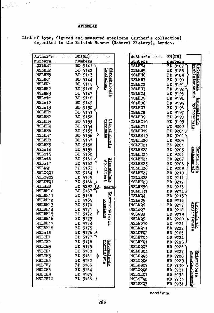
APPmDIX
List of type, figured and meaaured specimens (author's collection) deposited in the ~ritish Museum (Natural Histor,y), London.
Author.'s : numbers
~M NH · . numbers
Author's ~- '"''"' . n1m1ber_s. frlSLHH4 MSLHH5 MSLHH6 MSLHH7 MSLHC2 MSLHC3 MSLHC4 MSLHC5 MSLHC6
l3M{NH) numbers
BD 9.187} ~D 9188 BD §189 :BD 9190 BD 9191. BD 9192 "'~ ~D 9193 ~D 9194 BD 9195
I~ If: e+ e: tD
B tD e+ g 0
Ql
~ .... I»
Ql .... 01
!:I!
~ tD e+ tD
1-'
MSLHH1 MSLHH2 MSLHH3 MSLHC1 MSLHN1 MSLHN2 MSLD3 MSLot1 MSLot2 MSLot3 MSL:BH1 MSLBH2 MSLBH3 MSLBH4 MSLBH6 MSLBH7 MSLBH8 MSLBH9 MSLot4 MSLot5 MSLot6
BD 9141 . ~D 9142 ~D 9143 BD 9144 ~D 9145 ~D> 9146 BD 9147 BD 9148 ~D 9149 ~D 9150 ~D 9151-BD 9152
MSLHC7 MSLHC8
BD 9196 li BD 9197 I" e+ I»
. MSLot7 MSLWQ1 MSLOQQ1 MSLOQQ2 MSLETQ1 MSLHH8 MSLBH10 MSLBH11 MSLBH12 MSLBH13 MSLBH14 MSL~H15 MSLBH16 MSLBH17 MSLBH18 MSLot8 MSLTH1 MSLTH2 MSLTH3 MSLTH4 MSLTH5 MSLTH6 MSLTH7 MSLTH8 MSLTH9 MSLTH10
BD 9153 B'D 9154 ~D 9155 ~D 9156 ~D 9157 BD 9158 ~D 9159 BD 9160 BD 9161
BD 9162} ~D 9163 BD 9164 BD 9165 BD 9166 BD 9292 }~. arv BD 9167 BD 9168 ~D 9169 BD 9170 ~D 9171 BD 9172 BD 9173 ~D 9174 ~D 9175 ~D 9176 BD 9177 ~D 9178 ~D 9179 BD 9180 BD 9181 BD 9182 ~D 9183 BD 9184 ~D 9185 BD 9186
286
MSLHC9 MSLHC10 MSLHC11 MSLHC12 MSLBH19 MSLBH20 MSLBH21 MSLBH22 MSLBH23 MS~H24 Iv1SLBH25 MSLBH26 MSLBH27 MSLBH28 fo'I.SLBH29 MSLBH30 MSLBH31 MSLWQ4 MSLWQ5 MSLWQ6 MSLWQ7 MSLWQ8 MSLWQ9 MSLWQ10 MSLWQ11 ~ISLETQ2 MSLETQ3 MSLRHQ1 MSLOQQ3 MSLOQQ4 MSLOQQ5 MSLOQQ6 MSLOQQ7 MSLOQQ8 MSLSHQ1 MSLSHQ2 MSLSHQ3
BD 9198 BD 9199 BD 9200 BD 9201 J
BD 9202 "'~
BD 9203 :BD 9204 BD 9205 BD 9206 BD 9207 BD 9208 BD 9209 BD 9210 BD 9211 BD 9212 BD 9213 BD 9214, BD 9215"' BD 9216
. BD 9217 BD 9218 BD 9219 ~D 9220 ~D 9221 ~D 9222 BD 9223 BD 9224 ~D 9225 ~ BD 9226"1 BD 9227 BD 9228 BD 9229 BD 9230 BD 9231 ~D 9232 BD 9233 BD 9234
eontinue
g 1-' 0
Ill Ql
;~ .... Ql
Ql !:I! CD Ill I» e+
f Ill li I»
tD 1-' 't Jj 0
Ql C/l .... .... Ql I»
~if ll e+ 1-'111 '"'"li 1-1)111
~ ...., 1-'
~ 0 ())
Ql .... t-'tl» Ql
~if I» e+ li Ill li li
> ~e. ~~ Ot-'t ::s I» tD
ll -· "'

Author•s :BM(NH) numbers numbers MSLLQ1 :BD 9235 .. MSLLQ2 :BD 9236 MSLLQ3 BD 9237 MSLLQ4 :BD 9238 MSLLQ5 BD 9239
~ MSLLQ6 :BD 9240 MSLLQ7 BD 9241 MSLLQB BD 9242 MSLLQ9 BD 924, MSLLQ10 BD 9244 MSLLQ11 BD 9245 " MSLTH12 BD:. 9246 ~ MSLTH13 BD 9247 MSLTH14 BD 9248 MSLTH15 BD 9249 MSLTH16 BD 9250 KSLTH17 BD 9251 ). MSLTH18 BD 9252 MSLTH19 :BD 9253 MSLTH20 BD 9254 GALJ7 BD 9255 GALJ8 BD 9256 GALJ9 BD 9257 ..-GALJ1 BD 9258 "" GALJ2 BD 9259 GALJ3 BD 9260 GALJ4 BD 9261 GALJ5 BD 9262 GALJ6 BD 9263 > MSLTH21 BD 9264 MSLTH22 BD 9265 MSLTH23 BD 9266 MSLTH24 BD 9267 MSLTH25 BD 9268 .J
MSLWQ12 BD 9269 "" MSLWQ13 BD 9270 MSLWQ14 BD 9271 MSLWQ15 BD 9272 MSLWQ16 BD 9273 MSLWQ17 BD 9274 MSLWQ18 :BD 9275 > MSLWQ19 BD 9276 MSLWQ20 BD 9277 MSLWQ21 BD 9278 MSLWQ22 BD 9279 MSLWQ23 BD 9280 MSLETQ4 BD 9281 ...
lt-'0
~ 11 ID
..... ~ g CD ~
CD e ~ OJ 0 .... D) CD ....
ID
...... ~ ID c+ I( .... CD 11 CD 0 .... CD ID c+ 11 Ill c+ Ill
§f g' rn ::S CD ........ ...... f-1 Ill c+ ID
,~ ~ :;:~
g ~
287
... Author 1 s numbers MSLHN4 MSLHN5 MSLBN6 MSLHN7 MSLWQ24 MSLWQ25 MSLLQ12 MSLLQ13 MSLot16 MSLot17 MSLHH8 MSLBH9 MSLHH10 MSLHH11 MSLHH12 MSLHH13 MSLHH14 MSLHH15 MSLHH16 MSLHH17 li.SLHH18 MSLot10 MSLot11 MSLot12 MSLBH32 .ft'.SLBH33 MSLR1 MSLWQ26 MSLot13 MSLLQ14 MSLBC13 MSLHC14 MSLHC15 MSLHC16 MSLHC17 MSLHC18 MSLot14 MSLot15 MSLot16 MSLR2 MSLR3 MSLHH19 MSLHH20 MSLHH21
BM(NH) numbers · BD 9282 "' BD 9283 BD 9284 BD 9285 BD 9286 BD 9287 BD 9288 BD 9289 BD 9290 BD 9291 BD 9292 ~ BD 9293 § ~ :BD 92 94 CD 11 BD 9295 ....., ....
a~ BD 9296 ~ § BD 9297 CD .... BD 9298 > !;t» BD 9299 BD 9300 BD 9301 BD 9302 BD 9303 BD 9304 BD 9305 BD 9306 BD 9307 BD 9308 ~ BD 9309}Horrido BD 9310 nia sp.~ BD 9311 BD 9312 ""~ Dll Cll
BD 9313 I~ iS: BD 9314 ;:!_ 5 BD 9315 ~ g' BD 9316 ~ i ~ Bll 9317 111 ~ BD 9318 § BD 9319 .... BD 9320 t»
BD 9321 "'} CD Cll BD 9322 Cll;J ~~ BD 9323 • o 5 BD 9324 ~~ S BD 9325 ~ ~
CD ....
~ ~ [» ::s
'
111111111111111 ~~i1~illf~~~~~~~llfli~ii~ 11111111111111
3 0104 00566027 3