Periodontal Disease and Overall Health: A Clinician's Guide
331
Periodontal Disease and Overall Health: A Clinician’s Guide Editors Robert J. Genco Ray C. Williams Supported through an educational grant from
-
Upload
khangminh22 -
Category
Documents
-
view
0 -
download
0
Transcript of Periodontal Disease and Overall Health: A Clinician's Guide

Periodontal Disease and Overall Health: A Clinician’s Guide
EditorsRobert J. GencoRay C. Williams
Supported through an educational grant from
Periodontal Disease and Overall Health: A Clinician’s Guide
Robert J. Genco, DDS, PhDDistinguished Professor of Oral Biology and Microbiology
Schools of Dental Medicine and Medicine and Biomedical SciencesVice Provost, Office of Science,
Technology Transfer and Economic OutreachDirector, Clinical Research Center of the Buffalo Clinical and
Translational Research CenterState University of New York at Buffalo
Buffalo, NY, USA
Ray C. Williams, DMDProfessor and Dean, School of Dental Medicine
Stony Brook UniversityStony Brook, NY, USA
PROFESSIONAL AUDIENCE COMMUNICATIONS, INC.Yardley, Pennsylvania, USA

Periodontal Disease and Overall Health: A Clinician’s Guide
Copyright © 2010 by the Colgate-Palmolive Company. All rights reserved.
No part of this publication may be used or reproduced in any form or by any means, or
stored in a database or retrieval system, without prior written permission of the Colgate-
Palmolive Company. Making copies of any part of this book for any purpose other than
your own personal use is a violation of United States copyright laws.
ISBN-13: 978-0-6152-8508-5
ISBN-10: 0-6152-8508-2
Published by …
Professional Audience Communications, Inc.
PO Box 243
Yardley, Pennsylvania 19067 USA
Editorial Quality Control: Teri S. Siegel
Copyediting/Proofreading: Michelle Rizzo
Layout and Design: E. Allen Downs
Cover Design: Horizons Graphic Design
Indexing: Allegheny Writing & Publishing Services, LLC
Publisher: Stephen M. Siegel
Printed in the United States of America
Last digit is the print number: 9 8 7 6 5 4
ii

Silvana P. Barros, DDS, MS, PhD Research�Associate�Professor
Center�for�Oral�and�Systemic�Diseases
University�of�North�Carolina�School�of�Dentistry
Department�of�Periodontology
Chapel�Hill,�NC,�USA
Peter Mark Bartold, BDS, DDSc, PhD, FRACDS (Perio)Director,�Colgate�Australian�Clinical�Dental
Research�Centre
Professor�of�Periodontics
University�of�Adelaide
Department�of�Dentistry
Adelaide,�Australia
Yiorgos A. Bobetsis, DDS, PhDLecturer,�Department�of�Periodontology
University�of�Athens�School�of�Dentistry�
Athens,�Greece
Wenche Sylling Borgnakke, DDS, MPH, PhDAssistant�Research�Scientist
Department�of�Cariology,�Restorative�Sciences
and�Endodontics
University�of�Michigan�School�of�Dentistry
Ann�Arbor,�MI,�USA
Dawn J. Caster, MDNephrology�Fellow
Division�of�Nephrology
Department�of�Internal�Medicine
University�of�Louisville�School�of�Medicine
Louisville,�KY,�USA
Noel M. Claffey BDS, MDent Sc, FDS, FFD, FTCDProfessor�of�Periodontology
Dental�School�and�Hospital
Trinity�College�Dublin
Dublin,�Ireland
Robert J. Genco, DDS, PhDDistinguished�Professor�of�Oral�Biology
and�Microbiology
Schools�of�Dental�Medicine�and�Medicine
and�Biomedical�Sciences
Vice�Provost,�Office�of�Science,�Technology
Transfer�and�Economic�Outreach
Director,�Clinical�Research�Center�of�the�Buffalo
Clinical�and�Translational�Research�Center
State�University�of�New�York�at�Buffalo
Buffalo,�NY,�USA
William V. Giannobile, DDS, DMedScNajjar�Professor�of�Dentistry
Michigan�Center�for�Oral�Health�Research
Department�of�Periodontics�and�Oral�Medicine
University�of�Michigan�School�of�Dentistry
Ann�Arbor,�MI,�USA
Ricardo A. Gómez, MDAssociate�Professor
Department�of�Obstetrics�and�Gynecology
P.�Universidad�Católica�de�Chile
Hospital�Sótero�del�Río
Clínica�Santa�María
Santiago,�Chile
Dana T. Graves, DDS, DMScProfessor�and�Chair
Department�of�Periodontics�
New�Jersey�Dental�School�(UMDNJ)
Newark,�NJ,�USA
Ying Gu, DDS, PhDAssistant�Professor
Department�of�General�Dentistry
Stony�Brook�University�School�of�Dental
Medicine
Stony�Brook,�NY,�USA
Casey Hein, BSDH, MBAAssistant�Professor;�Division�of�Periodontics
Director�of�Education,�International�Centre
on�Oral-Systemic�Health
Faculty�of�Dentistry
University�of�Manitoba
Winnipeg,�Manitoba,�Canada
William C. Hsu, MDSenior�Physician
Medical�Director,�Asian�Clinic
Joslin�Diabetes�Center
Assistant�Professor�of�Medicine
Harvard�Medical�School
Boston,�MA,�USA
Heather L. Jared, RDH, MS, BSAdjunct�Assistant�Professor
University�of�North�Carolina�School�of
Dentistry
Department�of�Dental�Ecology
Chapel�Hill,�NC,�USA
CONTRIBUTORS
iii
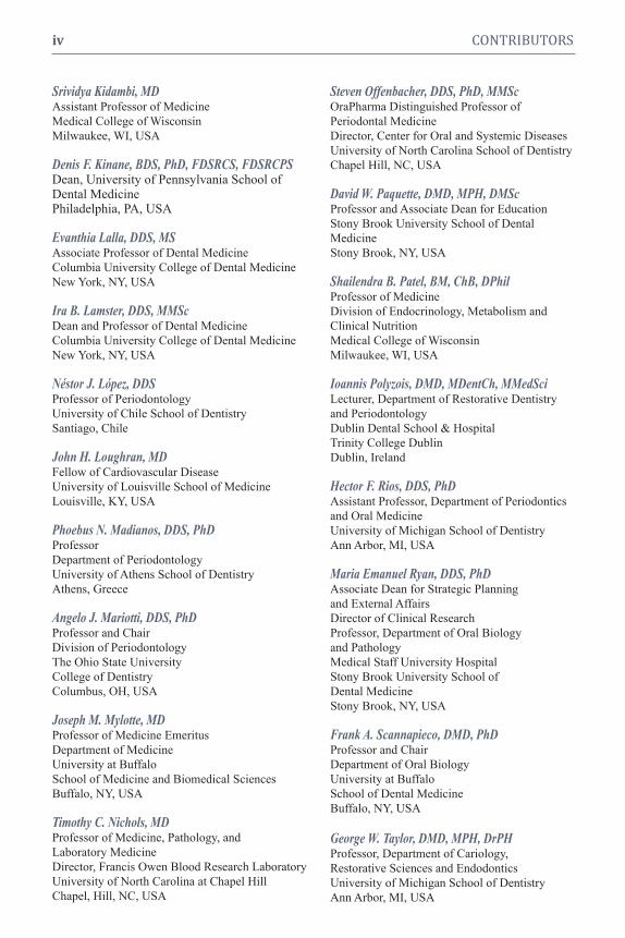
Srividya Kidambi, MDAssistant�Professor�of�Medicine
Medical�College�of�Wisconsin
Milwaukee,�WI,�USA
Denis F. Kinane, BDS, PhD, FDSRCS, FDSRCPSDean,�University�of�Pennsylvania�School�ofDental�MedicinePhiladelphia,�PA,�USA
Evanthia Lalla, DDS, MSAssociate�Professor�of�Dental�Medicine�
Columbia�University�College�of�Dental�Medicine
New�York,�NY,�USA
Ira B. Lamster, DDS, MMScDean�and�Professor�of�Dental�Medicine
Columbia�University�College�of�Dental�Medicine
New�York,�NY,�USA
Néstor J. López, DDSProfessor�of�Periodontology
University�of�Chile�School�of�Dentistry
Santiago,�Chile
John H. Loughran, MDFellow�of�Cardiovascular�Disease
University�of�Louisville�School�of�Medicine
Louisville,�KY,�USA
Phoebus N. Madianos, DDS, PhDProfessor
Department�of�Periodontology
University�of�Athens�School�of�Dentistry
Athens,�Greece
Angelo J. Mariotti, DDS, PhDProfessor�and�Chair
Division�of�Periodontology
The�Ohio�State�University
College�of�Dentistry
Columbus,�OH,�USA
Joseph M. Mylotte, MDProfessor�of�Medicine�Emeritus
Department�of�Medicine
University�at�Buffalo
School�of�Medicine�and�Biomedical�Sciences
Buffalo,�NY,�USA
Timothy C. Nichols, MDProfessor�of�Medicine,�Pathology,�and
Laboratory�Medicine
Director,�Francis�Owen�Blood�Research�Laboratory
University�of�North�Carolina�at�Chapel�Hill
Chapel,�Hill,�NC,�USA
Steven Offenbacher, DDS, PhD, MMScOraPharma�Distinguished�Professor�of
Periodontal�Medicine
Director,�Center�for�Oral�and�Systemic�Diseases
University�of�North�Carolina�School�of�Dentistry
Chapel�Hill,�NC,�USA
David W. Paquette, DMD, MPH, DMScProfessor�and�Associate�Dean�for�Education�
Stony�Brook�University�School�of�Dental
Medicine�
Stony�Brook,�NY,�USA
Shailendra B. Patel, BM, ChB, DPhilProfessor�of�Medicine
Division�of�Endocrinology,�Metabolism�and
Clinical�Nutrition
Medical�College�of�Wisconsin
Milwaukee,�WI,�USA
Ioannis Polyzois, DMD, MDentCh, MMedSci Lecturer,�Department�of�Restorative�Dentistry
and�Periodontology�
Dublin�Dental�School�&�Hospital
Trinity�College�Dublin�
Dublin,�Ireland
Hector F. Rios, DDS, PhDAssistant�Professor,�Department�of�Periodontics
and�Oral�Medicine
University�of�Michigan�School�of�Dentistry
Ann�Arbor,�MI,�USA
Maria Emanuel Ryan, DDS, PhDAssociate�Dean�for�Strategic�Planning
and�External�Affairs
Director�of�Clinical�Research
Professor,�Department�of�Oral�Biology
and�Pathology
Medical�Staff�University�Hospital
Stony�Brook�University�School�of
Dental�Medicine
Stony�Brook,�NY,�USA
Frank A. Scannapieco, DMD, PhDProfessor�and�Chair
Department�of�Oral�Biology
University�at�Buffalo
School�of�Dental�Medicine
Buffalo,�NY,�USA
George W. Taylor, DMD, MPH, DrPHProfessor,�Department�of�Cariology,
Restorative�Sciences�and�Endodontics
University�of�Michigan�School�of�Dentistry
Ann�Arbor,�MI,�USA
iv CONTRIBUTORS

Thomas E. Van Dyke, DDS, PhDProfessor,�Periodontology�and�Oral�Biology
Director,�Clinical�Research�Center
Boston�University�
Henry�M.�Goldman�School�of�Dental�Medicine
Boston,�MA,�USA
Stanley S. Wang, MD, JD, MPHClinical�Cardiologist�and�Director�of
Legislative�Affairs,�Austin�Heart
Adjunct�Assistant�Professor�of�Medicine
University�of�North�Carolina
Chapel�Hill,�NC,�USA
Ray C. Williams, DMDProfessor�and�Dean,�School�of�Dental�Medicine
Stony�Brook�University
Stony�Brook,�NY,�USA
CONTRIBUTORS v

CHAPTER 1
From the Editors
Robert J. Genco, Ray C. WilliamsDear Colleagues:
We are very pleased to have had the privilege of assembling and editing this textbook,
Periodontal Disease and Overall Health: A Clinician’s Guide.
The relationship of oral disease to overall disease is certainly not a new concept. For centuries,
the role of oral infection and inflammation in contributing to diseases elsewhere in the body has
been studied and reported. Going back to ancient times in Greece, we learn that Hippocrates
treated two patients suffering from joint pain by removal of teeth. Clearly, this was an early
example of oral disease being associated with afflictions elsewhere in the body. Then, moving
forward in time from 1912 to around 1950, the era of “focal infection” dominated our thinking.
Reports by individuals such as WD Miller, William Hunter, and Frank Billings noted that in their
opinion many of the diseases of humans could be traced to specific foci of infection elsewhere
in the body, such as the teeth and gums, the tonsils, or the sinuses. While these observations
were not supported by sound scientific evidence, and in fact led to largely incorrect practices,
they nonetheless brought attention to the effect of the mouth on the rest of the body.
Then in 1989, with a series of intriguing reports from Finland, the current interest in the role of
oral health and disease on contributing to general health and systemic conditions was launched.
Kimmo Mattila and his coworkers reported that individuals presenting to the emergency room
with a myocardial infarction were overwhelmingly likely to have periodontal disease. Might
periodontal disease be a risk factor for cardiovascular disease? Since then, a phenomenal body
of work has been directed at understanding how periodontal disease might affect distant sites
and organs, and thus have an effect on overall health. Renowned clinicians and scientists
worldwide have studied the relationship of periodontal disease to overall health and disease,
and along the way several conferences and workshops have been convened to examine the
evidence to date for the relationship between periodontal disease and the risk for systemic
conditions. At one of those conferences, in January 2008, we discussed the need for a textbook
that would summarize and put into context the current information on periodontal disease and
systemic disease together for students of dentistry and medicine. Happily for us, Foti Panagakos,
Sheila Hopkins, and their team at the Colgate-Palmolive Company agreed to support, through
an educational grant to the publisher, the undertaking of this textbook. We were fortunate to
have assembled a group of respected and scholarly clinicians and scientists who, in eighteen
chapters, provide a current and thoughtful perspective on the relationship of periodontal disease
to systemic conditions.
It is a pleasure to present this textbook. We hope you find it useful and that you enjoy it.
Sincerely,
Robert J. Genco, DDS, PhD Ray C. Williams, DMD

COMPANY

CHAPTER 1
Overview 1
Robert J. Genco, Ray C. Williams
CHAPTER 2
Overview of Periodontal Disease:
Causes, Pathogenesis, and Characteristics 5
Ying Gu, Maria E. Ryan
CHAPTER 3
Infection and Inflammation 24
Phoebus N. Madianos, Yiorgos A. Bobetsis, Thomas E. Van Dyke
CHAPTER 4
History of the Oral-Systemic Relationship 42
Noel M. Claffey, Ioannis N. Polyzois, Ray C. Williams
CHAPTER 5
Diabetes Mellitus: A Medical Overview 55
Srividya Kidambi, Shailendra B. Patel
CHAPTER 6
Association Between Periodontal Diseases and Diabetes Mellitus 83
George W. Taylor, Wenche S. Borgnakke, Dana T. Graves
CHAPTER 7
Atherosclerosis: A Pervasive Disease Affecting Global Populations 105
Stanley S. Wang
CHAPTER 8
Association Between Periodontal Disease and Atheromatous Diseases 112
David W. Paquette, Robert J. Genco
CHAPTER 9
Periodontal Disease and Pregnancy Complications 132
Silvana P. Barros, Heather L. Jared, Steven Offenbacher
CHAPTER 10
Oral Health and Diseases of the Respiratory Tract 147
Frank A. Scannapieco, Joseph M. Mylotte
viii
CONTENTS

ix CONTENTS
CHAPTER 11
Periodontal Disease and Osteoporosis 162
Hector F. Rios, William V. Giannobile
CHAPTER 12
Association Between Periodontitis and Rheumatoid Arthritis 179
P. Mark Bartold, Angelo J. Mariotti
CHAPTER 13
Oral Health, Periodontitis, and Cancer 196
P. Mark Bartold, Angelo J. Mariotti
CHAPTER 14
Dental and Medical Comanagement of Patients with Diabetes 216
Evanthia Lalla, William C. Hsu, Ira B. Lamster
CHAPTER 15
Dental and Medical Comanagement of Cardiovascular Disease 237
Timothy C. Nichols, David W. Paquette
CHAPTER 16
Dental and Medical Comanagement of Pregnancy 250
Néstor J. López, Ricardo A. Gómez
CHAPTER 17
Dental and Medical Comanagement of Osteoporosis,
Kidney Disease, and Cancer 270
Dawn J. Caster, John H. Loughran, Denis F. Kinane
CHAPTER 18
The Role of the Professional in Educating the Public
About the Importance of Oral Health 288
Casey Hein
INDEX 305

“A person can’t have good general health without good oral health.”
—Former US Surgeon General C. Everett Koop

INTRODUCTION
Periodontaldiseaseisone ofthemost
commondiseasesofmanandisresponsible
formostofthetoothlossinadults.Thisoral
diseasehasreceivedconsiderableattentionin
thepastseveraldecadesandanewunder-
standingof it is emerging.Themicrobial
causes of periodontal disease, themecha-
nismsthroughwhich periodontaltissuesare
destroyed, the effect of thehost onperio-
dontal disease expression, and the impact
periodontal diseasehas on overall health
havebeensubjectsofintensestudy.Under-
standing the complex interactionbetween
chronicinfections,suchasperiodontaldis-
ease,andsystemicconditionssuchascardio-
vascular disease, has led to a newwayof
thinkingabouttheimportanceofperiodon-
taldiseaseinoverallhealth.
Periodontal Disease as an
Integral Link to Systemic Disease
According to theNationalCenter for
HealthStatistics, thesix leadingcausesof
deathintheUnitedStatesin2005wereheart
disease(652,091),cancer(559,312),stroke/
cerebrovasculardiseases(143,579),chronic
lower respiratorydisease (130,933), unin-
tentionalaccidentalinjuries(117,809),and
diabetes(75,119).1 Fiveofthesechronic dis-
easesarerelatedtoperiodontaldisease. By
successfullymeeting the challenge to im-
proveoral health and themanagement of
periodontaldisease, generalhealthwillalso
beadvanced through sharedapproaches
targetingcommonriskfactors.Tobestaddress
thecommon risk factors and interactions
betweenoral and systemicdisease, it is
importanttounderstandtheextenttowhich
periodontal disease is related to certain
systemicdiseases,thehistoricalfoundations
ofcurrent therapeuticapproaches, the role
of inflammation, and thepossibilities for
intervention.
THREE HISTORICAL ERAS OF
PERIODONTAL DISEASE RESEARCH
Inthelast50years,therehasbeencon-
siderable progress in understanding the
etiologyandpathogenesisofperiodontaldis-
easeanditsinteractionswiththehost.The
studies and concepts canbedescribed as
havingoccurredinthreephasesoreras:the
etiopathologic(orhost-parasite)era,therisk
factorera,andmostrecently,theperiodon-
taldisease-systemicdiseaseera.
Etiopathologic Era
The etiopathologic era included land-
markinvestigationsintothemicrobialetiol-
ogyandpathogenesisofperiodontaldisease.
Theroleofbacteriaasacauseofperiodontal
diseasewasdemonstratedby a series of
seminalstudiesconductedfromthe1960sto
the1980s.ClassicstudiesbyLöeandcol-
leaguesclearlydemonstratedthatmicrobial
plaquebuildupontheteethwasassociated
withtheonsetofgingivitis,andthatthere-
moval ofmicrobial plaque resulted in the
resolutionofgingivitis.2,3 Thesestudiespro-
vided unarguable evidence thatmicrobial
dentalplaquebuildup,ratherthanothersus-
pectedagentssuchascalculus,wasrespon-
sibleforgingivitis.
CHAPTER 1
Overview
Robert J. Genco, Ray C. Williams
“A person can’t have good general health without good oral health.”
—Former US Surgeon General C. Everett Koop

Inthe1970sand1980s,Socranskyand
coworkersconductedstudies showing that
specificorganismswereassociatedwithperio-
dontaldisease(forreviewseeSocranskyand
Haffajee,2005).4These studies identified
severalcategoriesoforganisms,rangingfrom
earlycolonizers,whicharecommensaland
relativelynonvirulent,tomoderatelyvirulent
organisms,whichbridgedtheearlycolonizers
andinterconnectedthemtospecificpathogens
suchasPorphyromonas gingivalis, Tannerella
forsythensis, and Treponema denticola.Re-
searchfrommanyinvestigatorsfoundthatthe
specificpathogens, incombinationwith the
earlycolonizersandmoderatelyvirulentorgan-
isms, form acomplexmicroflorathatexistsas
abiofilmwithintheperiodontalpocket.
Other investigators began to explain
thepathogenesisofperiodontaldisease,de-
scribinghowthehostinfactwasresponsible
for tissuedestruction.Wecame to under-
standthattheinitialresponsetothebacteria
onthetoothandsubgingivallyisaseriesof
immunopathologicalactions.Antibodiesto
thesebacteriaareformed,whichincombi-
nationwithneutrophils,provide important
protection.5,6 Itwasseenthatifneutrophils
aresuppressed,moresevereperiodontaldis-
easeoccurs.Soonthereaftertheroleofthe
macrophagewasunderstood. Thisimportant
cell invades thegingival tissue andupon
triggeringbybacterial products such as
endotoxin,producespro-inflammatorycyto-
kines andmatrixmetalloproteinases that
destroytheconnectivetissuesoftheperio-
dontium. Inflammatorymediators such as
prostaglandinE2and interleukin-1 induce
alveolarboneresorption.Astheroleofthe
host becomesmoreunderstood, it is clear
that inflammation and the inflammatory
response can explainmuchof the tissue
destructioncausedbyperiodontaldisease.7,8
Risk Factor Era
Theseconderaofdiscoverybroughtthe
identificationofriskfactors whichinfluence
ormodulatetheexpressionofperiodontaldis-
ease.Epidemiologicstudiesreportedthatthe
risk factors inandof themselveswerenot
etiologic,butrathermodifiedorexaggerated
theetiopathologicprocesses setintomotion
bythecausativebacteria.Theseriskfactors
wereidentifiedinthelate1980sandearly
1990sandincludegeneticelements,behav-
iors such as smoking, and acquireddisor-
derssuchasdiabetesmellitus.9,10 Theconcept
ofmodifyingriskfactorsaspartoftheman-
agementofperiodontaldiseaseisnowwell
established.
Periodontal Disease-
Systemic Disease Era
Theunderstandingof periodontal dis-
ease isnowfocusedontherelationshipof
periodontaldiseaseasariskforcertainsys-
temicdiseases.Robuststudieshaveshown
thatperiodontaldiseaseissignificantlyasso-
ciatedwith certain systemic diseases such
as cardiovascular disease,11,12 diabetes and
complicationsofdiabetes,13-15adversepreg-
nancyoutcomes,16andrespiratoryinfections.17
Theperiodontaldisease-systemicdisease
concepthasamassedenoughevidenceand
supportthatitisnowbelievedthatfindings
about this inter-relationship shouldbe in-
corporatedintothecurriculuminschoolsfor
healthprofessionals,andshouldalsobemade
availabletoenhancetheknowledgebaseof
currentlypracticinghealthcareprofessionals.
Theassociationofperiodontaldisease
with several systemic conditions, such as
diabetesandatheroscleroticdisease,islikely
relatedtotheinflammatoryresponseassoci-
atedwithperiodontaldisease.C-reactivepro-
tein is an importantmarkerof the inflam-
matoryresponse andiselevatedinsubjects
withperiodontaldisease;itslevelsinperiph-
eral blood are reducedwhenperiodontal
diseaseistreated. Anotherindicationofthe
systemicinflammatoryresponseassociated
withperiodontaldiseaseisthepresenceof
cytokines, including tumornecrosis factor
2 Periodontal Disease and Overall Health: A Clinician's Guide

alphaandinterleukins1and6,oftenfound
in the circulationof patientswithperio-
dontal disease.There are other conditions
thatalsocontributetoasystemicinflamma-
toryresponseincludingrheumatoidarthritis,
psoriasis,andobesity. Thischronicsystemic
inflammatory response in turn increases
theriskforatheroscleroticdisease,diabetes
andcomplicationsofdiabetes, adversepreg-
nancyoutcomes,andpossiblysomecancers.
Theresearchsupportingtheseassociations
willbediscussedindetailinthefollowing
chapters.
GOALS FOR THIS TEXTBOOK
Much research is focusedonunder-
standinghowperiodontaldiseaseincreases
theriskforsystemicdiseases.It isnotyet
clearwhatimpactthebiofilmintheoralcav-
itymighthave ondistantsitesandorgans;
likewise the role of the inflammatory re-
sponseisnotfullyunderstood.Someofthe
chaptersinthistextbookwillreviewthebio-
logic plausibilityforperiodontaldiseaseasa
risk for systemic conditions.Mechanisms
throughwhichperiodontaldiseasecancon-
ferthisriskwillalsobepresented.
Theoverallgoalofthistextbookisto
present the emerging and compelling evi-
dencethatperiodontaldiseaseisariskfor
severalsystemicconditionsandtolookatthe
roleoforalhealthincontributingtooverall
health.Thisbookalsoseekstoprovidethe
readerwithaguidetopatientmanagementin
whichdentistryandmedicineworktogether.
Textbook Organization
Thechaptersinthisbook areorganized
inthefollowingmanner: Theinitialchapters
followingthisoneoutlinethebasicsofun-
derstandingperiodontaldiseaseanditsinter-
relationshipwithsystemicdisease:Chapter
2discussesthecausesandpathogenesisof
periodontaldisease;theroleofinfectionand
inflammation inperiodontal disease is ex-
aminedinChapter3;andthehistoryofthe
oraldisease-systemicdiseaserelationshipis
explainedinChapter4.
Anoverviewof diabetes (Chapter 5)
andatheroscleroticdiseases(Chapter7)are
followedbychaptersthatdescribetherela-
tionshipofperiodontaldiseasetothesecon-
ditions(Chapters6and8,respectively).The
nextchaptersexaminetheevidenceforperio-
dontal disease as a risk for adversepreg-
nancyoutcomes(Chapter9),respiratorydis-
eases (Chapter 10), osteoporosis (Chapter
11),rheumatoidarthritis(Chapter12),and
cancer(Chapter13).
The final sectionof the textbook dis-
cusses comanagementofperiodontaldisease
indiabetes(Chapter14),cardiovasculardis-
ease(Chapter15),pregnancy(Chapter16),
andotherconditions thatareassociatedwith
periodontaldisease (Chapter17).Finally,
Chapter18describestheroleofdentalpro-
fessionalsintheeducationofthepublicand
other health professionals about theoral
health-generalhealthinter-relationship.
Our Hope for This Textbook
Itisthehopeoftheauthorsandeditors
thatthistextbookwillprovideanup-to-date
understandingoftheinformationthatdetails
therelationshipofperiodontaldiseasetosys-
temicdisease,witheachchapteroutlininga
state-of-the-art understandingoftheoptimal
managementofpatients.Thistextbookhas
beenpreparedasaresourcefordentalstu-
dents,dentalhygienestudents,facultymem-
bersofdentaleducational institutions,and
fordentalprofessionalsingeneral.Wealso
believethisresourcewillprovevaluableto
students aswell as practicingmembersof
otherhealthprofessionsinthemedicalcom-
munity.Theintegrationofmedicineandden-
tistrygrowsdaily,andacommonresource
suchasthistextbookcouldserveasacon-
structive tool to help the twodisciplines
workcollaboratively.
Theeditorswouldliketothanktheau-
thorsandcoauthorsfortheir roleinpreparing
CHAPTER 1 Overview 3

andpresentingcurrentinformationinacom-
plete,yetconciseand readablemanner.We
arehopefulthatthistextbookwillfindbroad
readershipandwillbeusefultothedentaland
medicalcommunity.
REFERENCES1. HeronM,HoyertDL,MurphySL,XuJ,Kochanek
KD,Tejada-VeraB.Deaths: final data for 2006.
Natl Vital Stat Rep 2009;57:1–34.
2. LöeH,TheiladeE,JensenSB.Experimentalgin-
givitisinman.J Periodontol 1965;36:177–187.
3. TheiladeE,WrightWH, JensenSB,LöeH.Ex-
perimentalgingivitisinman.II.Alongitudinalclin-
icalandbacteriologicalinvestigation.J Periodon-
tal Res 1966;1:1–13.
4. SocranskySS,HaffajeeAD.Periodontalmicrobial
ecology.Periodontol 2000 2005;38:135–187.
5. GencoRJ,SlotsJ,MoutonC,MurrayP.Systemic
immuneresponsestooralanaerobicorganisms.In:
Anaerobic Bacteria: Selected Topics, LambeDW
Jr,GencoRJ,Mayberry-CarsonKJ,eds.,Plenum
PublishingCorp.,NewYork,277,1980.
6. EbersoleJL,TaubmanMA,SmithDJ,GencoRJ,
FreyDE.Humanimmuneresponsestooralmicro-
organisms. I.Associationof localized juvenile
periodontitis(LJP)withserumantibodyresponses
toActinobacillus actinomycetemcomitans.Clin Exp
Immunol 1982;47:43–52.
7. GencoRJ.Hostresponsesinperiodontaldiseases:
Currentconcepts.J Periodontol 1992;63(Suppl):
338–355.
8. PageRC,OffenbacherS,SchroederHE,Seymour
GJ,KornmanKS.Advancesinthepathogenesisof
periodontitis:Summaryofdevelopments,clinical
implications and future directions.Periodontol
2000 1997;14:216–248.
9. GencoRJ,LöeH.Theroleofsystemicconditions
anddisordersinperiodontaldisease.Periodontol
2000 1993;2:98–116.
10. GrossiSG,GencoRJ,MachteiEE,HoAW,KochG,
Dunford R, Zambon JJ, Hausmann E.As-
sessmentof riskforperiodontaldisease. II.Risk
indicators for alveolar bone loss.J Periodontol
1995;66:23–29.
11. MattilaK,NieminenM,ValtonenV,RasiVP,
KesäniemiYA,SyrjäläSL,JungellPS,IsoluomaM,
HietaniemiK, JokinenMJ.Associationbetween
dentalhealthandacutemyocardialinfarction.BMJ
1989;298:779–781.
12. DeStefanoF,AndaRF,KahnHS,WilliamsonDF,
RussellCM.Dentaldiseaseandriskofcoronary
heart disease andmortality.BMJ 1993;306:688–
691.
13. TaylorGW,BurtBA,BeckerMP,GencoRJ,Shloss-
man M, Knowler WC, Pettitt DJ. Severe
periodontitisandriskforpoorglycemiccontrolin
patientswithnon-insulin-dependentdiabetesmel-
litus.J Periodontol 1996;67:1085–1093.
14. GrossiSG,GencoRJ.Periodontal disease and
diabetesmellitus:A two-way relationship.Ann
Periodontol 1998;3:52–61.
15. TaylorGW,BorgnakkeWS.Periodontal disease:
Associationswithdiabetes,glycemiccontroland
complications.Oral Dis 2008;14:191–203.
16. OffenbacherS,KatzV,FertikG,CollinsJ,BoydD,
MaynorG,McKaigR,BeckJ.Periodontalinfec-
tionasapossibleriskfactorforpretermlowbirth
weight.J Periodontol 1996;67:1103–1113.
17. ScannapiecoFA.Roleoforalbacteriainrespiratory
infection.J Periodontol 1999;70:793–802.
4 Periodontal Disease and Overall Health: A Clinician's Guide

Periodontitis has been defined as the pres-
ence of gingival inflammation at sites where
there has been a pathological detachment of
collagen fibers from cementum, the junc-
tional epithelium has migrated apically, and
bone loss can be detected radiographically.
The inflammatory events associated with
connective tissue attachment loss lead to
the resorption of coronal portions of tooth
supporting alveolar bone.2 The understand-
ing of periodontal disease is continuously
changing as new research evidence emerges.
There fore, the classification of periodontal
disease has changed since the system devel-
oped at the 1989 World Workshop in Clini-
cal Periodontics. The classification presented
in this chapter is based on the results devel-
oped at the 1999 International Workshop or-
ganized by the American Academy of Perio -
dontology (AAP).
The classification of periodontal dis-
eases now includes eight general types3:
1. Gingivitis
2. Chronic periodontitis
3. Aggressive periodontitis
4. Periodontitis as a manifestation of
systemic diseases
5. Necrotizing periodontal diseases
6. Abscesses of the periodontium
7. Periodontitis associated with endo -
dontic lesions
8. Developmental or acquired deformi-
ties and conditions
The overall classification system is
presented in Table 1.3 In addition, the above
classification is different from case types
INTRODUCTION
Periodontal diseases are serious chronic
infections that involve destruction of the
tooth-supporting apparatus, including the
gingiva, the periodontal ligament, and alve-
olar bone. These diseases are initiated by a
local accumulation of bacteria adjacent to
the tooth. Periodontal diseases, including
gingivitis and periodontitis, can affect one
tooth or many teeth and, if left untreated,
can lead to tooth loss, particularly in adults.
It is the most common dental condition in
adults, and is also one of the most common
chronic inflammatory diseases affecting a
majority of the population throughout the
world. Although plaque is essential for the
initiation of periodontal diseases, the major-
ity of the destructive processes associated
with these diseases are due to an excessive
host response to the bacterial challenge.
Therefore, periodontal disease is a multifac-
torial, complex disease. The purpose of this
chapter is to provide a general overview of
the types of periodontal disease, risk factors
associated with them, and the etiology,
pathogenesis, and management of periodon-
tal diseases.
TYPES OF PERIODONTAL DISEASE
Periodontal diseases include two general
categories based on whether there is attach-
ment or bone loss: gingivitis and periodon-
titis. Gingivitis is considered a reversible
form of the disease, and generally involves
inflammation of the gingival tissues with-
out loss of connective tissue attachment.1
CHAPTER 2
Overview of Periodontal Disease: Causes, Pathogenesis, andCharacteristics
Ying Gu, Maria E. Ryan

previously developed by the AAP.4,5 The
current case types for periodontal diseases
include:
• Gingivitis (Case Type I)
• Mild periodontitis (Case Type II)
• Moderate periodontitis (Case Type III)
• Advanced periodontitis (Case Type IV)
• Refractory periodontitis (Case Type V)
Gingival Diseases
Gingival disease is further characterized
into plaque-induced and nonplaque-induced
categories.3
Plaque-Induced Gingival Diseases
Gingivitis is gingival inflammation as-
so ciated with plaque and calculus accumu-
lation. It is the most common form of gin -
gival disease. Gingivitis may or may not
progress to periodontitis, in which clinical
attachment and alveolar bone loss will
develop. Gingivitis can occur on teeth with
no attachment loss; it also occurs in the gin-
giva of teeth previously treated for peri-
odontitis with no further attachment loss.
Dental Plaque Only: Gingivitis is ini-
tiated by local accumulation of bacteria (i.e.,
dental plaque) adjacent to the tooth.6 The
bacterial antigens and their metabolic prod-
ucts (e.g., endotoxin) stimulate epithelial and
connective tissue cells to produce inflam-
matory mediators that result in a localized
inflammatory response recruiting polymor-
phonuclear leukocytes (PMNLs or neutro -
phils) to the site. An antibody response to
these bacterial antigens is also mounted. In-
flammatory cells and their products (e.g.,
cytokines, enzymes, and antigens) are pres-
ent at the site of inflammation. Thus, a host
immuno-inflammatory response is estab-
lished in the gingival tissues and the clinical
signs of gingivitis develop, including red-
ness, swelling, and bleeding. The plaque-
host interaction can be altered by the effects
of local factors, systemic factors, or both.
Systemic Factors: Systemic hormonal
changes associated with puberty, menstrual
cycle, or pregnancy, as well as with chronic
diseases such as diabetes, can alter the host
response to dental plaque.1,7 Hormonal
changes and certain diseases can upregulate
systemic cellular and immunologic function
resulting in local severe gingival inflamma-
tion, even in the presence of minimal plaque
or with an equivalent bacterial bioburden to
a person who does not have these systemic
challenges. This is commonly seen in preg-
nant women who have not had adequate oral
hygiene before becoming pregnant. Blood
dyscrasias such as leukemia may also alter
immune function by decreasing normal im-
munological function. Patients usually pres-
ent with gingival enlargement and bleeding
associated with spongy gingival tissues and
excessive vascularity.
Medications: Medications such as an-
ticonvulsant drugs (e.g., dilantin), immuno-
6 Periodontal Disease and Overall Health: A Clinician's Guide
Table 1. Periodontal Diseases
VIII. Gingival Diseases
Dental plaque-induced gingival diseases
Nonplaque-induced gingival lesions
IV.II. Chronic Periodontitis
Localized
Generalized
VIII. Aggressive Periodontitis
Localized
Generalized
IIIV. Periodontitis as a Manifestation of
IV. Systemic Diseases
IIIV. Necrotizing Periodontal Diseases
Necrotizing ulcerative gingivitis
Necrotizing ulcerative periodontitis
IIVI. Abscesses of the Periodontium
Gingival abscess
Periodonal abscess
Pericoronal abscess
IVII. Periodontitis Associated with
Endodontic Lesions
VIII. Developmental or Acquired Deformities
and Conditions
Adapted from: Ann Periodontol 1999;4:1–6.3

CHAPTER 2 Overview of Periodontal Disease:
CHAPTER 2 Causes, Pathogenesis, and Characteristics 7
suppressive drugs (e.g., cyclosporine), and
calcium channel blockers (e.g., diltiazem)
can cause severe gingival enlargement and
pseudo-periodontal pocketing (i.e., increased
probing depths with no associated attach-
ment or bone loss).8 Medication-associated
gingival conditions are often reversed after
discontinuation of the offending agents.
Malnutrition: The host immune sys-
tem can be diminished when malnutrition
develops, resulting in excessive gingival in-
flammation. Severe ascorbic acid (vitamin C)
deficiencies (i.e., scurvy) can produce bright
red, swollen, and bleeding gingival tissues.1 In
the case of vitamin C deficiency, gingivitis is
associated with a suppressed synthesis of
both connective tissue collagens (e.g., Types
I and III) and basement membrane collagen
(Type IV). Treatment with vitamin C sup-
plements can reverse this condition.
Nonplaque-Induced Gingival Lesions
These types of lesions usually are rare
and mainly due to systemic conditions. Bac-
teria, viruses, or fungi can cause these types
of gingival lesions. Sexually transmitted dis-
eases such as gonorrhea (Neisseria gonor-
rhoeae) and syphilis (Treponema pallidum)
can cause lesions in the tissues of the peri-
odontium.9 Primary streptococcal gingivitis
is an acute inflammation of the oral mucosa.
It is associated with pain and fever, as well
as red swollen gingival tissues with bleeding
or abscess formation, and can be treated with
routine periodontal scaling and root plan ing
in addition to antibiotic therapy. Herpes
simplex virus Type I is a common virus that
can cause gingival lesions.10 In children and
young adults, herpes infections can be pri-
mary and usually without symptoms, but in
some cases pain and fever are reported. In
these cases, the gingival tissues appear red
and swollen, and are followed by the for-
mation of small blisters, which eventually
break down to form shallow, painful ulcers.
These lesions are usually self-limiting and
heal within one to two weeks. After a pri-
mary infection, the herpes virus becomes la-
tent and will be preserved in the ganglion of
the trigeminal nerve. The virus may be re-
activated later in life by reduced immune
function or stress, resulting in recurrent her-
pes labialis, gingivitis, and stomatitis. Gin-
gival lesions of fungal origin usually occur in
people with diabetes or other immunocom-
promised states. A shift in the normal oral
flora related to the long-term use of system-
ically administered antibiotics can also lead
to lesions of fungal origin.11 The most com-
mon fungal infection is candidiasis, caused
by Candida albicans, often seen in patients
wearing removable prosthetic devices (e.g.,
dentures) and in patients with dry mouth
due to multiple medications or salivary gland
dysfunction. Clinical manifestations include
white patches on the gingiva, tongue, or oral
mucous membranes that can be removed
with a cotton swab or gauze, leaving behind
a bright red bleeding surface. Treatment with
antifungal agents is often necessary to re-
solve these conditions.
Gingival lesions can also be caused by
genetic systemic mucocutaneous disorders,
allergic reactions, trauma, or foreign-body
reactions. One of the most common genetic
conditions associated with gingival lesions
is autosomal-dominant hereditary gingival
fibromatosis.12 It is a benign condition af-
fecting both arches. The gingival tissues are
enlarged and asymptomatic. It may be an
isolated finding or associated with other
syndromes. Treatment is gingivectomy and
recurrence is possible. Systemic conditions
such as pemphigoid, pemphigus vulgaris,
erythema multiforma, and lupus erythe-
matosus can cause desquamative lesions and
ulceration.10,13 Gingival changes due to al-
lergic reactions to certain restorative ma -
terials, dentifrices, or mouthrinses are rare,
but have been observed.10 Traumatic lesions
are usually produced unintentionally.10 Ag-
gressive tooth brushing and flossing can

cause gingival damage. Hot foods and drinks
can cause minor burns of the gingival tissues.
Traumatic lesions can also be iatrogenically
induced by healthcare professionals during
oral examinations or dental care. Eating
crunchy foods or foods with small particles
that can be lodged in the interproximal areas
and directly into the gingival tissues can
cause these types of lesions as well. Gin-
gival tissues can also develop localized in-
flammation when exposed to foreign ma -
terials. The most common example is the
amalgam remaining in gingival tissues dur-
ing the placement of restorations or surgical
pro cedures, eventually producing amalgam
tattoos.10
PERIODONTITIS
Periodontitis is a chronic infection in-
volving destruction of the tooth-supporting
apparatus, including the periodontal ligament
and alveolar socket support of the teeth.
Gingivitis may or may not progress to
periodontitis, which is associated with at-
tachment and alveolar bone loss. Periodon-
tal disease is initiated by a local accumula-
tion of bacteria (i.e., dental plaque adjacent
to the tooth) and their metabolic products
(e.g., endotoxin), that stimulate the junc-
tional epithelium to proliferate and produce
tissue-destructive proteinases that degrade
the basement membrane and allow for the
apical migration of the junctional epithelium
along the root surface of the tooth, thus deep-
ening the gingival crevice to produce peri-
odontal pockets and associated attachment
loss, which is the hallmark lesion of peri-
odontal disease. Some of the clinical signs
include bleeding on probing, deep pockets,
recession, and tooth mobility. Often, this de-
structive process is silent and continues for
long periods of time without being identified.
Eventually, teeth can become loose and may
be lost on their own or deemed hopeless,
requiring extraction. There are many forms
of periodontitis.
Chronic Periodontitis
Chronic periodontitis (CP) is the most
common form of periodontitis and is char-
acterized by pockets with associated attach-
ment loss and/or recession of the gingival tis-
sues. It is common in adults but can occur at
any age. Progression of attachment loss usu-
ally occurs slowly, but periods of exacerba-
tion with rapid progression, or periods of re-
mission can occur. Several studies have
addressed the “episodic” nature of perio -
dontitis.14 The rate of disease progression
may be influenced by local and/or systemic
conditions that alter the normal host response
to bacterial plaque. Local factors such as
subgingivally placed fillings or crowns that
violate biological width can promote gingi-
val inflammation and clinical attachment
loss. Systemic factors such as diabetes can
decrease host defenses to bacterial infection.
Environmental factors such as smoking and
stress can also decrease host immune func-
tion, resulting in increased susceptibility to
disease. CP can occur as a localized form in
which < 30% of the sites are involved, or as
a more generalized form in which >30% of
existing sites demonstrate increased pocket
depth, attachment and bone loss.4 As men-
tioned previously, the severity of disease can
be described as slight, moderate, or severe,
based on the level of destruction.
Aggressive Periodontitis
This form of periodontitis was previ-
ously categorized as Juvenile Periodontitis.
Common features include rapid attachment
loss and bone destruction in the absence of
significant accumulations of plaque and cal-
culus.15 These forms of periodontitis usually
affect young individuals, often during pu-
berty, from 10 to 30 years of age, with a ge-
netic predisposition. The bacteria most often
associated with aggressive periodontitis are
Aggregatibacter actinomycetemcomitans
(previously Actinobacillus actinomycetem-
comitans). Individuals present with hyper-
8 Periodontal Disease and Overall Health: A Clinician's Guide

active inflammatory cells producing high
levels of cytokines and enzymes causing
rapid, aggressive destruction of periodontal
tissues. Aggressive periodontitis can be fur-
ther characterized as localized and general-
ized forms. The localized form usually
affects first molar and incisor sites. The gen-
eralized form usually involves at least three
teeth other than first molars and incisors.
Periodontitis as a Manifestation
of Systemic Diseases
Systemic conditions such as diabetes
are associated with this form of periodonti-
tis.16 Several hematologic and genetic disor-
ders have also been associated with the de-
velopment of periodontitis such as acquired,
familial, and cyclic neutropenias, leukemias,
Down’s syndrome, certain types of Ehlers-
Danlos syndrome, Papillon-Lefevre syn-
drome, Cohen syndrome, and hypophospha -
tasia. The mechanisms by which all of these
disorders affect the health of the periodon-
tium are not fully understood and continue to
be investigated by many basic and clinical re-
searchers. It is speculated that these diseases
can alter host defense mechanisms and up-
regulate inflammatory responses, resulting
in progressive periodontal destruction.
Necrotizing Periodontal Diseases
These lesions are most commonly ob-
served in individuals with systemic condi-
tions, such as human immunodeficiency virus
infection, malnutrition, and immunosup-
pression. Necrotizing periodontal diseases
are further divided into two forms: necrotiz-
ing ulcerative gingivitis (NUG) and necro-
tizing ulcerative periodontitis (NUP). These
two diseases have the same etiology and
clinical signs, except NUP involves clinical
attachment and alveolar bone loss.17
Abscesses of the Periodontium
Periodontal abscess is a localized puru-
lent infection of the periodontal tissues.18
Periodontal abscesses usually develop in
periodontitis patients who may have food
debris lodged in a pocket, or deep deposits
of calculus where drainage from a pocket
becomes blocked. Iatrogenic abscess forma-
tion can be precipitated after inadequate scal-
ing and root planing, leading to a tightening
of the coronal epithelial cuff with continued
subgingival calculus driving inflammation.
Abscesses can also occur in healthy peri-
odontal tissues due to the presence of foreign
objects lodged in the gingival crevice, such
as a toothbrush bristle or a popcorn kernel
being tightly packed into the interproximal
spaces or between the tooth and the tissues.
A pericoronal abscess is an infection of the
gingiva around a partially erupted tooth lead-
ing to pericoronitis. A small flap of tissue
may cover a partially erupted tooth surface,
serving as a nidus for food and debris to ac-
cumulate and become trapped beneath the
tissue flap. Patients usually find it very dif-
ficult to keep these areas clean, and can de-
velop inflammation and infection. In addi-
tion, trauma due to constant contact between
the tissue flap and a tooth in the opposing
arch can also lead to a pericoronal abscess.
The areas most commonly affected are as-
sociated with mandibular third molars. Pain,
swelling, redness, and suppuration are asso-
ciated with periodontal abscess. Treatment
may include incision and drainage, use of
antibiotics, and removal of the offending
source.
EPIDEMIOLOGY AND
RISK FACTORS
Epidemiology of Gingivitis
Gingivitis can occur in early childhood,
becomes more prevalent during teenage
years, and decreases in older individuals.19 In
1986-1987, the National Institute of Dental
Research conducted a nationwide survey of
oral health in US school children20 and
reported that approximately 60% of chil -
dren 14 to 17 years of age were found to
CHAPTER 2 Overview of Periodontal Disease:
CHAPTER 2 Causes, Pathogenesis, and Characteristics 9

10 Periodontal Disease and Overall Health: A Clinician's Guide
have gingivitis. In 1960–1962, the first US
national survey of periodontal disease in
adults reported that 85% of men and 79% of
women have some degree of gingivitis.21 In
the most recent National Health and Nutri-
tion Examination Survey (NHANES III)
conducted from 1988 to 1994,22 more than
50% of adults had gingivitis on an average of
three or four teeth, while 63% of 13- to 17-
year-old teenagers had gingival bleeding.
Both surveys assessed gingival bleeding by
a gingival sweep method.21,22
Epidemiology of Periodontitis
Basic clinical measurements for perio -
dontitis are gingival bleeding on probing,
clinical attachment loss, and pocket depths
accompanied by radiographic bone loss.
These types of clinical measurements may
be somewhat subjective. As our knowledge
of the pathogenesis of periodontitis im-
proves, new diagnostic markers for the dis-
ease may emerge to help better diagnose it.
Inflammatory cytokines, enzymes, and bone
breakdown products released into gingival
crevicular fluid reflect the host response to
the bacterial challenge. These biochemical
markers may be good candidates for new
diagnostic or prognostic markers of disease.
A number of cytokines have been asso ciated
with active disease, including prostaglandin
E2 (PGE2), tumor necrosis factor-alpha
(TNF-α), interleukin-1 beta (IL-1β), and
others.23,24 Enzymes such as matrix metal-
loproteinases (MMPs) and breakdown prod-
ucts such as the collagen telopeptide have
been studied as well. To date, these bio-
chemical markers in gingival crevicular
fluid are still being investigated. It will be
helpful to both clinicians and researchers
if one or more of these markers can be de-
veloped as a more objective chairside tool to
measure active periodontitis. The devel -
opment of these markers will also help
to facilitate screening for periodontal
diseases by medical professionals or even
self- assessment by patients, thereby prompt-
ing referrals to the dental office for clinical
assessment.
The national data suggest that the milder
forms of periodontitis are close to uni -
versal.25 The more severe forms are less
prevalent. According to a review of the lit-
erature by Brown and Löe26 focused on a
number of epidemiologic studies resulting
from a 1981 national probability survey, the
prevalence of CP is about 36% for the adult
US population as assessed by pocket depth
measurements. The prevalence of periodon-
titis increases with age; 29% in those aged 19
to 44 had CP; this rate increased to 50% for
people 45 years or older. In general, moder-
ate periodontitis occurred in 28% of all peo-
ple while 8% had advanced disease. How-
ever, the prevalence of moderate and severe
periodontitis increased to 44% in the popu-
lation older than 45. Based on the presence
of periodontal pockets ≥ 4 mm, it was de-
termined that 30% of the population has
periodontitis on an average of three or four
teeth. Severe pockets of ≥ 6 mm were found
in less than 5% of the population.22 The
prevalence of aggressive periodontitis was
low with less than 1%.27 More recently,
NHANES III (1988–1994) reported the
prevalence of periodontitis for adults 30 to
90 years old.28 Attachment loss and probing
depths were assessed at two sites per tooth.
When assessed by the level of attachment
loss, 53% of the population was found to
have ≥ 3 mm attachment loss. The preva-
lence of attachment loss increased with age,
from approximately 35% for the 30-year-
old participants to 89% for the 80-year-old
participants. When assessed by probing
depth, approximately 64% of the population
had probing depths of ≥ 3 mm. The preva-
lence of periodontitis increases with age and
was found to be more prevalent in males
than females, and in African-Americans and
Mexican-Americans than in non-Hispanic
Caucasians.

Risk Factors
There are a number of risk factors asso-
ciated with periodontal diseases.29-34 Deter-
mining risk is helpful in developing recom-
mendations for prevention and in
determining strategies for the overall man-
agement of periodontitis. It has been recog-
nized that the severity and progression of
periodontal disease varies from individual
to individual. Bacteria are essential for the
initiation of the disease, but it is the host re-
sponse to the bacterial challenge that deter-
mines the severity and rate of progression of
the periodontitis. Therefore, it is the host’s
immunologic reaction that determines sus-
ceptibility to the disease.
General categories of risk factors asso-
ciated with the development of periodontitis
include genetic, environmental (e.g., tobacco
use), and acquired risk factors (e.g., systemic
disease). Risk factors (Table 2) and risk
reduction strategies (Table 3) should be con-
sidered when assessing each patient.35 Some
risk factors can be modified to reduce a
patient’s susceptibility. Environmental fac-
tors such as tobacco use and stress can be
managed with smoking cessation and stress
management; for acquired factors such as
systemic diseases, medications usually
prescribed by the physician can be used to
help in the management and control of
chronic disorders (Table 3). The use of
chemotherapeutic agents specifically de-
signed to improve the clinical outcomes of
mechanical treatments for periodontal dis-
eases may be particularly useful in the man-
agement of those individuals with single or
multiple risk factors. Risk assessment can
CHAPTER 2 Overview of Periodontal Disease:
CHAPTER 2 Causes, Pathogenesis, and Characteristics 11
Table 2. Risk Assessment for Periodontitis
1. Heredity as determined by genetic testing and family history
2. Smoking, including frequency, current use, and history
3. Hormonal variations such as those seen in:
a. Pregnancy, in which there are increased levels of estradiol and progesterone that may change the
environment and permit virulent organisms to become more destructive
b. Menopause, in which the reduction in estrogen levels leads to osteopenia and eventually osteoporosis
4. Systemic diseases such as:
a. Diabetes (the duration and level of control are important)
b. Osteoporosis
c. Immune system disorders such as HIV
d. Hematologic disorders such as neutropenias
e. Connective tissue disorders such as Marfan’s and Ehlers-Danlos syndromes
5. Stress as reported by the patient
6. Nutritional deficiencies and obesity that may require a dietary analysis
7. Medications such as:
a. Calcium channel blockers
b. Immunomodulatory agents
c. Anticonvulsants
d. Those known to cause dry mouth or xerostomia
8. Faulty dentistry such as overhangs and subgingival margins
9. Poor oral hygiene resulting in excessive plaque and calculus
10. History of periodontal disease
Sources: J Periodontol 1994;65:260–267.29 J Periodontol 1995;66:23–29.30 J Periodontol 1999;70:711–723.31
J Periodontol 2000;71:1057–1066.32 J Periodontol 2000;71:1215–1223.33 J Periodontol 2000;71:1492–1498.34

12 Periodontal Disease and Overall Health: A Clinician's Guide
help the practitioner to establish an accurate
diagnosis, provide an optimal treatment plan,
and determine appropriate maintenance pro-
grams. In patients with multiple risk factors,
the practitioner may aggressively use phar-
macologic adjuncts such as antimicrobials
and host-modulatory therapy in addition to
mechanical therapy. It is also important to
update and assess risk factors for each patient
on a regular basis as some of these factors
are subject to change throughout life.
ETIOLOGY AND PATHOGENESIS
OF PERIODONTAL DISEASE
Initially, periodontal disease was
thought to be related to aging and was there-
fore uniformly distributed in the population,
with disease severity being directly corre-
lated with plaque levels. Now as a result of
extensive research, it has been shown that
periodontal disease is initiated by plaque,
but the severity and progression of the dis-
ease is determined by the host response to
the bacterial biofilm. People with severe
plaque and calculus accumulation will have
gingivitis, but not necessarily periodontitis.
On the other hand, certain individuals, de-
spite maintaining adequate oral hygiene, find
themselves susceptible to aggressive forms
of periodontitis, with deep pocketing, tooth
mobility, and early tooth loss. Clearly, the re-
sponse of the periodontal tissues to plaque is
different in these two different scenarios. Pe-
riodontal disease does not appear to behave
as a classic infection, but more as an oppor-
tunistic infection.35 These observations led
researchers to realize that the host response
to the bacterial challenge, presented by sub-
gingival plaque, is the important determinant
of disease severity. Although plaque bacteria
are capable of causing direct damage to the
periodontal tissues, it is now recognized that
Table 3. Risk Reduction Strategies
1. More frequent visits for those with a genetic predisposition; use of pharmacotherapeutics for the
2.management of periodontitis
2. Smoking cessation using one or more of the six approved regimens; these regimens are rarely
2. successful as sole therapies (multiple forms of therapy often are used in combination with
2. counseling to achieve success)
3. Hormonal variations such as those seen in:
a. Pregnancy, which requires good oral care before conception to prevent complications during
a. pregnancy; treatment during pregnancy may be necessary to prevent adverse pregnancy outcomes
b. Menopause, which may require hormonal supplements, calcium, and other medications and
a. supplements prescribed by the physician to prevent osteopenia
4. Systemic diseases that require consultation with the physician include:
a. Diabetes (for improved glycemic control)
b. Osteoporosis (requiring calcium supplements, bisphosphonates)
c. Immune system and hematologic disorders
d. Connective tissue disorders
5. Stress management; possible referral to a psychologist or psychiatrist
6. Nutritional supplementation and weight reduction; possible referral to a nutritionist
7. Medications can be changed in consultation with the physician
8. Corrective dentistry
9. Improved oral hygiene (brushing, flossing, use of antiseptics)
10. Occlusal adjustments
Source: Dent Clin North Am 2005;49:611–636.35
CHAPTER 2 Overview of Periodontal Disease:
CHAPTER 2 Causes, Pathogenesis, and Characteristics 13
the host immuno-inflammatory response to
plaque bacteria produces destructive cy-
tokines and enzymes resulting in periodon-
tal tissue destruction. The host response is es-
sentially protective by intent but can also
result in tissue damage, including the break-
down of connective tissue fibers in the peri-
odontal ligament and the resorption of alve-
olar bone. The host response to the plaque
biofilm is modified by genetic factors (help-
ing to explain why aggressive periodontitis
tends to have a familial aggregation), as well
as systemic and environmental factors (e.g.,
diabetes, stress, smoking).
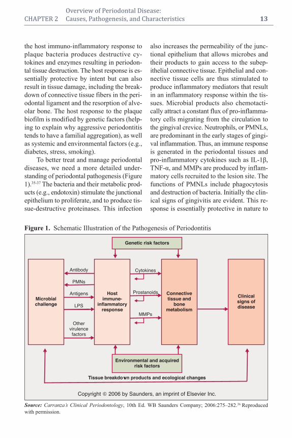
To better treat and manage periodontal
diseases, we need a more detailed under-
standing of periodontal pathogenesis (Figure
1).35-37 The bacteria and their metabolic prod-
ucts (e.g., endotoxin) stimulate the junctional
epithelium to proliferate, and to produce tis-
sue-destructive proteinases. This infection
also increases the permeability of the junc-
tional epithelium that allows microbes and
their products to gain access to the subep-
ithelial connective tissue. Epithelial and con-
nective tissue cells are thus stimulated to
produce inflammatory mediators that result
in an inflammatory response within the tis-
sues. Microbial products also chemotacti-
cally attract a constant flux of pro-inflamma -
tory cells migrating from the circulation to
the gingival crevice. Neutrophils, or PMNLs,
are predominant in the early stages of gingi-
val inflammation. Thus, an immune response
is generated in the periodontal tissues and
pro-inflammatory cytokines such as IL-1β,
TNF-α, and MMPs are produced by inflam-
matory cells recruited to the lesion site. The
functions of PMNLs include phagocytosis
and destruction of bacteria. Initially the clin-
ical signs of gingivitis are evident. This re-
sponse is essentially protective in nature to
Figure 1. Schematic Illustration of the Pathogenesis of Periodontitis
Source: Carranza’s Clinical Periodontology, 10th Ed. WB Saunders Company; 2006:275–282.36 Reproduced
with permission.
Genetic risk factors
Copyright � 2006 by Saunders, an imprint of Elsevier Inc.
Tissue breakdown products and ecological changes
Antibody Cytokines
Prostanoids
MMPs
PMNs
Antigens
LPS
Othervirulencefactors
Microbialchallenge
Hostimmune-
inflammatoryresponse
Connectivetissue and
bonemetabolism
Clinicalsigns ofdisease
Environmental and acquiredrisk factors

control bacterial infection. In persons who
are not susceptible to periodontitis, the pri-
mary defense mechanisms control the infec-
tion, and chronic inflammation (i.e., chronic
gingivitis) may persist. However, in indi-
viduals susceptible to periodontitis, the
above inflammatory process will eventually
extend apically and laterally to involve
deeper connective tissues and alveolar bone,
recruiting monocytes and lymphocytes to
the site of infection at these later stages.
These monocytes and macrophages are
activated by the bacterial endotoxins lead-
ing to the production of high levels of
prostaglandins (e.g., PGE2), interleukins
(e.g., IL-1α, IL-1β, IL-6), TNF-α, and MMPs
by the host cells. The MMPs break down
collagen fibers, disrupting the normal
anatomy of the gingival tissues, resulting in
destruction of the periodontal apparatus. If
left untreated, the inflammation continues
to extend apically, and osteoclasts are stim-
ulated to resorb alveolar bone triggered by
the high levels of PGs, ILs, and TNF-α in the
tissues. The elevated levels of pro-inflam-
matory mediators and MMPs are counter-
balanced by a protective response in the host
with elevations in anti-inflammatory media-
tors such as the cytokines IL-4 and IL-10, as
well as other mediators such as IL-1ra (re-
ceptor antagonist) and tissue inhibitors of
MMPs (TIMPs; Figure 2).36,37 Under normal,
healthy conditions, the anti-inflammatory
mediators are balanced with inflammatory
mediators, thereby controlling tissue destruc -
tion. If an imbalance is seen, with excessive
levels of the pro-inflammatory mediators,
upregulated MMP expression and activity,
and insufficient levels of protective anti-
inflammatory mediators, loss of periodontal
connective tissue and bone will occur.
Thus, plaque bacteria initiate an in-
flammatory response by the host, resulting in
excessive levels of pro-inflammatory medi-
ators and enzymes, leading to the destruction
of periodontal tissues. If this inflammation
14 Periodontal Disease and Overall Health: A Clinician's Guide
Figure 2. The Periodontal Balance
Source: Carranza’s Clinical Periodontology, 10th Ed. WB Saunders Company; 2006:275–282.36 Reproduced
with permission.
Risk factors (e.g., genetics,smoking, diabetes)
Overproduction of proinflammatory ordestructive mediators and enzymes (e.g.,
IL-1, IL-6, PGE2, TNF-�, MMPs
Underactivity or overactivityof aspects of host response
Poor compliancePoor plaque control
DISEASE
Subgingivalbioburden
Reduction ofrisk factors
Expression of host-derived antiinflammatoryor protective mediators
(e.g., IL-4, IL-10, IL-1ra, TIMPs)
Host modulatorytherapy
Resolution ofinflammation
OHI, SRP, surgery, antiseptics, antibioticsto reduce bacterial challenge
Copyright � 2006 by Saunders, an imprint of Elsevier Inc.
HEALTH
�� �

continues and extends further apically, more
bone is resorbed, and more periodontal tissue
is broken down, leading to deeper and deeper
pockets and associated attachment and bone
loss revealed as the clinical and radiographic
signs of periodontitis. In people with peri-
odontitis, these inflammatory mediators
(e.g., prostanoids and cytokines) and local
oral bacteria will eventually enter into the
circulation, stimulating the liver to produce
acute-phase proteins (notably C-reactive pro-
tein, but also fibrinogen, haptoglobin, etc.)
which are “biomarkers” of a systemic in-
flammatory response. The ever-expanding
data supporting the fact that this systemic
inflammatory response driven by the chronic
infection and inflammation associated with
periodontitis will eventually increase an in-
dividual’s risk for developing a number of
systemic diseases, including cardiovascular
diseases, adverse pregnancy outcomes, and
diabetic complications.
MANAGEMENT OF
PERIODONTAL DISEASES
Periodontal management includes a
complete assessment of each individual pa-
tient. Medical and dental history, clinical and
radiographic examination, as well as an as-
sessment of risk factors are all important to
making an accurate diagnosis, prognosis,
and developing an optimal treatment plan.
There are many treatment options available
for the management of periodontal diseases,
and review of treatment outcomes or re-eval-
uation is key to successful management and
long-term maintenance. In the past, treat-
ments that focused on reduction of the mi-
crobial load were basically the sole consid-
eration for all periodontal therapy. Currently,
due to a better understanding of the host re-
sponse, host-modulation therapies have been
used as adjunctive approaches to both non-
surgical and surgical treatments to aid in
reducing probing depths, increasing clinical
attachment levels, and in regeneration of
the lost attachment apparatus. It is likely that
the most effective therapeutic approaches
will include multiple, synergistic host-
modulation therapies combined with treat-
ments that target the microbial etiology.
In addition to reducing the bacterial
challenge and modulating the host response,
reduction of risk is also a key treatment
stra tegy when managing periodontitis. For
example, it is known that smoking can
contribute to the development of periodontal
disease and make management of the dis-
ease more difficult;38,39 therefore smoking
ces sation would benefit all patients with
periodontitis. Smoking cessation can be
undertaken in the dental office (if staff
is appropriately trained) or in a medical
setting. There are a variety of medications to
aid with smoking cessation, counseling is
important as well, and alternative medicine
such as acupuncture may be used. Systemic
diseases such as diabetes will increase pa-
tients’ risk for periodontitis when poorly
controlled.40 When treating people with dia-
betes, knowing the patient’s level of diabetic
control is important to assessing risk, and
collaborating with medical colleagues to
improve control of diabetes is essential to
assure successful periodontal treatment.
Perio dontitis is also prevalent in patients
with cardiovascular disease, and periodontal
therapy may have a positive impact on the
overall health status of these individuals.
The management of patients with peri-
o dontitis can therefore involve the following
complementary treatment strategies:41
• Patient education, including oral hy-
giene instruction and explanation of
the rationale for any adjunctive treat-
ments
• Risk factor modification and risk re-
duction
• Reduction of bacterial burden with
traditional scaling and root planing
• Intensive periodontal treatment with
local delivery systems or general
CHAPTER 2 Overview of Periodontal Disease:
CHAPTER 2 Causes, Pathogenesis, and Characteristics 15

antimicrobial therapy with oral ad-
ministration of antibiotics
• Host-modulation therapy
• Periodontal surgery
It is the responsibility of the dentist to
provide appropriate treatments on an indi-
vidualized basis. A combination of treatment
approaches (discussed below) for each pa-
tient as listed in Figure 3 will provide the
ultimate periodontal treatment and result in
a better prognosis.41
The Antimicrobial Approach
Traditional periodontal therapy based
on the antimicrobial approach consists of
mechanical nonsurgical and surgical thera-
pies that may or may not be supplemented by
local antiseptics and/or local or systemic
antibiotics.
Mechanical Therapy
A regimen of brushing and flossing,
combined with the use of dentifrices and/or
mouthrinses containing antiseptics is the
most basic approach to microbial reduction
and control. Good oral hygiene can effec-
tively reduce bacterial loads to prevent gin-
givitis and aid in the treatment and manage-
ment of periodontitis. This simple approach
relies on an individual’s knowledge of the
correct techniques and compliance with home
care instructions. Unfortunately, many patients
are not compliant, lose motiva tion, and do not
spend a sufficient amount of time brushing or
flossing on a daily basis.42 It is for this reason
that dentifrices and mouth rinses containing
antiseptics have been devel oped. Antiseptics
have been found to improve plaque reduc-
tion, as well as reduce gingival inflammation
16 Periodontal Disease and Overall Health: A Clinician's Guide
Figure 3. Complementary Treatment Strategies in Periodontitis
Adapted from: Carranza’s Clinical Periodontology, 10th Ed. WB Saunders Company; 2006:813–827.41 Reproduced
with permission.
Reduction of
bacterial burden:
SRP, local or
systemic antimicrobials
surgical pocket
reduction
Risk factor
modification:
cessation therapy,
Diabetes control
Host modulation therapy:
subantimicrobial dose therapy,
enamel matrix proteins, rhPDGF, BMP-2
Best chance
for clinical
improvement

seen with brushing and flossing alone. There-
fore, antiseptic-containing dentifrices and
mouth rinses have been accepted as adjuncts
to the mechanical approach of brushing and
flossing.
Routine tooth scaling every six months
by the dental care provider is also a key com-
ponent in treating and preventing gingivitis.
Scaling and root planing is the traditional
nonsurgical treatment of periodontitis, with
multiple clinical studies demonstrating that
it effectively reduces the microbial load and
leads to reductions in bleeding on probing
and probing depths, and allows for gains in
clinical attachment.43 However, this proce-
dure can be very time-consuming and is op-
erator-dependent.44 Surgical procedures can
be used to visualize the remaining subgingival
calculus, and through resective or regenerative
procedures will also lead to decreased prob-
ing depths that are more manageable for the
long-term maintenance of patients with peri-
odontitis. Although nonsurgical and surgical
procedures aimed at reducing the bacterial
load and restoring the attachment apparatus
continue to be the most widely used methods
of treating periodontitis, these strategies alone
may be insufficient at reducing the bacterial
load as significant numbers of micro-organ-
isms may be left behind. In addition, many of
the putative pathogens will remain within the
oral cavity at distant sites allowing for re-
population in the future. Therefore, the need
for the development of chemotherapeutic
agents as adjuncts to mechanical debride-
ment was deemed necessary.
Mouthrinses and Dentifrices
Antiseptic Mouthrinses
Antiseptic mouthrinses have been used to
reduce plaque levels and gingivitis. Two clin-
ically proven American Dental Association-
accepted antiseptic mouthrinses are chlorhex-
idine gluconate (Peridex®) and the four
essential oils in Listerine®. An association
CHAPTER 2 Overview of Periodontal Disease:
CHAPTER 2 Causes, Pathogenesis, and Characteristics 17
between oral conditions such as periodontal
disease and several respiratory conditions
such as pneumonia and chronic obstructive
pulmonary disease has been noted. The
plaque surrounding the teeth is an excellent
harbor for respiratory pathogens. Studies have
shown that using a chlorhexidine oral rinse
can reduce the risk of pneumonia in institu-
tionalized patients with poor oral hygiene.45
Locally Applied Antiseptics
Periochip® contains the active ingredi-
ent chlorhexidine gluconate (2.5 mg) that is
released into the pocket over a period of
seven to 10 days. It has been found to sup-
press the bacteria in the pocket for up to 11
weeks post-application.46 Periochip is the
only locally applied antiseptic that is ap-
proved by the Food and Drug Administration
(FDA) for use as an adjunct to scaling and
root planing procedures to aid in the reduc-
tion of pocket depths. Other locally applied
antimicrobials are antibiotics.
Dentifrices
Major improvements in the oral health
of populations in developed countries have
been seen over the last 50 years. Most of this
resulted from the reduction in the caries rate
of about 50%, and the principle reason for
this is thought to be the addition of fluoride
to dentifrices. Modern, commercially avail-
able dentifrices, in addition to providing anti -
caries effects of fluoride, also contribute to
the reduction of plaque, gingivitis, cal culus
formation, relief of dentin hypersenitivity,
and tooth stain. They also reduce halitosis and
result in a clean, fresh mouth feel. Two den ti-
frices available in the US that are approved by
the FDA for their effects on reduction of gin-
givitis include a stannous fluoride/sodium
hexametaphosphate den ti frice and a triclosan/
copolymer/sodium fluoride dentifrice.
There is a large amount of literature on
these and other dentifrices containing chlor -
hexidine and other agents in the control of

gingivitis. A review of the clinical efficacy
and safety of a triclosan/copolymer/sodium
fluoride dentifrice was carried out by Blink -
horn and colleagues.47 They found about 200
articles dating from 1998 to 2008 relating to
this dentifrice and concluded that twice daily
use of this dentifrice will result in clinically
significant improvement in plaque control
and gingivitis and slower progression of
perio dontal disease. Further long-term stud-
ies extending over several years with these
dentifrices are needed to establish whether or
not short-term effects seen will be sustained
over the long term, and indeed result in pre-
venting the initiation of perio dontitis and
slowing the progression of already existing
periodontitis.
It should be noted that the antiplaque
and antigingivitis effects of dentifrices during
a tooth brushing regimen are mainly on the
occlusal and smooth surfaces of the teeth,
and that interproximal plaque and gingivitis
control is not optimally reduced with tooth
brushing alone, with or without a dentifrice.
Interproximal aids such as flossing, inter-
proximal brushing, and to some extent, flush-
ing with effective mouthrinses is often needed
for full plaque control on interproximal sur-
faces of the teeth. As periodontal disease is
often initiated and progresses more rapidly in
interproximal spaces, it is clear that inter-
proximal cleansing is an important adjunct to
toothbrushing with dentifrices.
Locally Delivered Antimicrobials
Atridox
Atridox® is an FDA-approved locally
delivered tetracycline system. It comes with
a 10% formulation of doxycycline in a bio -
absorbable, “flowable” poly-DL lactide and
N-methyl-2-pyrrolidone mixture delivery
system that allows for controlled release over
seven days. This system is applied subgin -
givally to the base of the pocket through a
cannula. Atridox is a resorbable site-specific
locally delivered antibiotic proven to promote
clinical attachment gains and reduce pocket
depths, bleeding on probing, and levels of
pathogenic bacteria for up to six months post-
placement.35 Periodontal disease has been
linked to systemic diseases such as diabetes.
Research has shown that periodontal treat-
ment with locally delivered doxycycline 10
mg in periodontal pockets produced favor-
able clinical results in diabetic patients.48
Arestin
Arestin® is an FDA-approved minocy-
cline microsphere system that is bioadhesive
and bioresorbable, allowing for sustained re-
lease of 1 mg of minocycline up to 19 days.
Arestin can be used as an adjunct to scaling
and root planing procedures for reduction of
pocket depth in patients with adult perio -
dontitis. Arestin is delivered to sites of 5 mm
or greater. Periodontitis has been associated
with increased systemic inflammation, which
is directly linked to diabetes and cardiovas-
cular diseases. Recent research has shown
that periodontal therapy with local Arestin
administration resulted in decreased HbA1c
levels in diabetic subjects49 and significant
reductions in systemic inflammatory bio-
markers that are risk factors for cardiovas-
cular disease.50
Systemic Antimicrobials
Systemic antimicrobial therapy is usu-
ally reserved for advanced cases of peri-
odontitis: 1) for sites that have not responded
to treatment, so-called “refractory periodon-
titis,” and 2) for patients demonstrating pro-
gressive periodontal destruction.35 Systemic
antibiotics can be used as adjuncts to con-
ventional mechanical therapy as strong evi-
dence for their use as a monotherapy has not
been developed. For these special situations,
randomized double-blinded clinical trials and
longitudinal assessments of patients indicate
that systemic antimicrobials may be useful
in slowing disease progression.51 Metroni -
18 Periodontal Disease and Overall Health: A Clinician's Guide

dazole can be used to treat acute necrotizing
ulcerative gingivitis (NUG),52 and metron-
idazole/amoxicillin combination therapy can
be used to treat aggressive adolescent peri-
odontitis associated with A. actinomycetem-
comitans.53 Systemic antibiotic therapy has
the advantage of simple, easy administration
of drugs to multiple periodontal sites. How-
ever, patient compliance needs to be consid-
ered, inability to achieve adequate concen-
trations at the site of infection, adverse drug
reactions, and the develop ment of antibiotic
resistance can be issues.54 Common antibiotic
therapies for the treatment of periodontitis
include metronidazole, clindamycin, doxy-
cycline or minocycline, ciprofloxacin,
azithromycin, metronidazole/amoxicillin,
and metronidazole/cipro floxacin.55 For adult
patients with acute periodontal abscesses,
amoxicillin is used as an adjunct to incision
and drainage. For patients with allergies to
beta-lactam drugs (e.g., amoxicillin), azith -
romycin or clindamycin would be the choice.
Researchers have shown that periodon-
tal treatment can benefit some systemic dis-
eases known to be associated with periodon-
titis, such as diabetes and preterm delivery.
Grossi and colleagues reported that diabetic
patients receiving scaling and root planing
with systemic doxycycline showed signifi-
cant reductions in mean HbA1c.56 Effective
treatment of periodontal infection and reduc -
tion of periodontal inflammation is associ-
ated with a reduction in levels of glycated
hemo globin. Clothier and colleagues also
showed that performing scaling and root plan-
ing in pregnant women with periodontitis
may reduce preterm delivery.57 However, ad -
junc tive metronidazole therapy did not im -
prove pregnancy outcomes. Two recent stud-
ies have not shown improvements in adverse
pregnancy outcomes with scaling and root
planing.58 However, the level of perio dontal
treatment provided may have been very
inadequate. More studies are needed in this
field to determine the effect of periodontal
treatment on the outcomes of adverse preg-
nancy and the extent of therapy that may
need to be provided in order to have a sig-
nificant impact.
Host-Modulation Therapy
Bacteria and the host are two essential
factors to the development of periodontitis.
Reduction of bacterial load is the con -
ventional approach for the management of
periodontal diseases. More recently, peri-
odontal treatment strategies have included
host-modulation therapy as an adjunctive
treatment option. Host-modulation therapy is
treating the host response to either reduce the
excess production of cytokines and destruc-
tive enzymes so there is less damage to the
periodontal tissues, or to stimulate the re-
gen erative process, allowing for the restora-
tion of connective tissue attachment and
bone formation to occur.
Host modulation was first introduced to
dentistry by Williams59 and Golub and col-
leagues.60 Williams stated: “There are com-
pelling data from studies in animals and hu-
man trials indicating that pharmacologic
agents that modulate the host responses be-
lieved to be involved in the pathogenesis of
periodontal destruction may be efficacious in
slowing the progression of periodontitis.”59
Golub and colleagues discussed “host mod-
ulation with tetracyclines and their chemi-
cally modified analogues.”60 A variety of dif-
ferent drug classes have been evaluated as
host-modulation agents, including the non -
steroidal anti-inflammatory drugs, bisphos-
phonates, tetracyclines (Figure 4),36 enamel
matrix proteins, growth factors, and bone
morphogenetic proteins.
Systemically Administered Agents
Subantimicrobial-Dose Doxycycline
Subantimicrobial-dose doxycycline (SDD)
is the only FDA-approved MMP inhibitor
and systemic host-modulation therapy for the
CHAPTER 2 Overview of Periodontal Disease:
CHAPTER 2 Causes, Pathogenesis, and Characteristics 19

management of periodontitis. SDD is a 20-mg
dose of doxycycline (Periostat®) taken twice
daily for three months and used in multicen-
ter clinical trials for up to a maximum of 24
months of continuous dosing. SDD is used as
an adjunct to scaling and root planing in the
treatment of CP. The host-modulation effects
of SDD include enzyme inhibition, cytokine
reductions, and effects on osteoclast function.
Since perio dontitis is associated with many
systemic diseases (e.g., osteoporosis, diabetes,
cardiovascular disease), researchers have in-
vestigated the effect of SDD on these system -
ic conditions. Studies have shown that SDD:
• Can effectively reduce the levels of
localized and systemic inflammatory
mediators in osteopenic patients, in
addition to improving on the clinical
measurements of periodontitis61
• Has been shown to reduce systemic
inflammatory biomarkers in CVD
patients62
• Decreases HbA1c in patients who
are taking normally prescribed hypo-
glycemic agents63
The impact of SDD therapy on peri-
odontitis may be amplified by an independ-
ent benefit for other inflammatory diseases;
additional studies are being conducted to in-
vestigate the impact of this host-modulation
therapy.
Locally Administered Agents
Enamel Matrix Proteins, Growth Factors,
and Bone Morphogenetic Proteins
A number of local host-modulation
agents have been investigated for potential
use as adjuncts to surgical procedures to im-
prove periodontal health. These have in-
cluded enamel matrix proteins (Emdogain®),
bone morphogenetic proteins, and platelet-
derived growth factors (PDGF). The initial
local host-modulation agent approved by the
FDA for adjunctive use during surgery to
assist with clinical attachment gain and
wound healing was Emdogain; this has been
followed by PDGF combined with a re-
sorbable synthetic bone matrix growth-fac-
tor enhanced matrix (GEM 21S) to assist in
20 Periodontal Disease and Overall Health: A Clinician's Guide
Figure 4. Potential Adjunctive Therapeutic Approaches
Source: Carranza’s Clinical Periodontology, 10th Ed. WB Saunders Company; 2006:275–282.36 Reproduced
with permission.
Antimicrobials
Host response component
Osteoclasts
Prostaglandins
Cytokines(IL-1, IL-6, TNF)
Periostat
MMPs
NSAIDSPeriostat
Bacterialproducts
PocketsandCAL
Toothmobilityandkiss
Bacterialcomponent
+ = Clinicalsequelae
BisphosphonatesPeriostat
Hostcells
Boneresorption
Connectivetissuebreakdown

regenerative procedures approved recently
by the FDA, as well as recombinant human
bone morphogenetic protein-2 (rhBMP-2)
contained within an absorbable collagen
sponge to assist with ridge and sinus aug-
mentation. The technology behind GEM 21S
has already been marketed for use in wound
healing, particularly in people with diabetes,
and rhBMP-2 has been used for quite some
time by the orthopedic community for the
healing of fractures.
The findings discussed with regard to the
use of host-modulation therapy to better man-
age chronic periodontal disease may have ap-
plications to other chronic systemic diseases
such as arthritis, diabetes, osteoporosis, and
cardiovascular disease. In addition, studies
utilizing locally delivered antimicrobials as
part of an intensive periodontal therapy reg-
imen have shown very promising results.
Future studies may demonstrate that in ad-
dition to our current standard therapies, in-
tensive periodontal therapy with adjunctive
antibiotics and/or host modulation for the
management of periodontal disease may
have profound positive effects on the over-
all health status of high-risk patients. The
proper management of local infection and
inflammation (periodontitis) will have a sig-
nificant impact on general overall health of
the population.
Acknowledgments
The authors would like to acknowledge Dr.
Hsi-ming Lee, René Martin, and Laura
Bertolotti for their assistance in the organi-
zation of this manuscript.
Supplemental Readings
Williams RC. Periodontal disease. N Engl J Med 1990;
322:373-382.
Page RC, Offenbacher S, Schroeder HE, Seymour GJ,
Kornman KS. Advances in the pathogenesis of perio -
dontitis: summary of developments, clinical implica-
tions and future directions. Periodontol 2000 1997;14:
216-248.
Brown LJ, Löe H. Prevalence, extent, severity and pro-
gression of periodontal disease. Periodontol 2000 1993;
2:57-71.
Burt B. Research, Science and Therapy Committee of the
American Academy of Periodontology. Position paper:
Epidemiology of periodontal diseases. J Periodontol
2005;76:1406-1419.
Ryan EM. Nonsurgical approaches for the treatment of
periodontal diseases. Dent Clin North Am 2005;49:611–
636.
Ryan ME, Preshaw PM. Host modulation. In: Newman
MG, Takei HH, Klokkevold PR, Carranza FA, eds.
Carranza’s Clinical Periodontology, Edition 10. WB
Saunders Co. 2006; pp. 275-282.
Preshaw PM, Ryan ME, Giannobile WV. Host modula-
tion agents. In: Newman MG, Takei HH, Klokkevold
PR, Carranza FA, eds. Carranza’s Clinical Periodon-
tology, Edition 10. WB Saunders Co. 2006; pp. 813-827.
REFERENCES1. Mariotti A. Dental plaque-induced gingival diseases.
Ann Periodontol 1999;4:7–19.
2. Van Dyke TE. The management of inflammation in
periodontal disease. J Periodontol 2008;79(8 Suppl):
1601–1608.
3. Armitage GC. Development of a classification sys-
tem for periodontal diseases and conditions. Ann
Periodontol 1999;4:1–6.
4. Novak MJ. Classification of diseases and condi-
tions affecting the periodontium. In: Newman MG,
Takei HH, Carranza FA, eds. Carranza’s Clinical
Periodontology, 9th Ed. WB Saunders Company;
2002:64–73.
5. Flemmig TF. Periodontitis. Ann Periodontol 1999;
4:32–38.
6. Armitage GC. Periodontal diseases: diagnosis. Ann
Periodontol 1996;1:37–215.
7. Kinane DF. Blood and lymphoreticular disorders.
Periodontol 2000 1999;21:84–93.
8. Rees TD. Drugs and oral disorders. Periodontol
2000 1998;18:21–36.
9. Scully C, Monteil R, Sposto MR. Infectious and
tropical diseases affecting the human mouth. Peri-
odontol 2000 1998;18:47–70.
10. Holmstrup P. Non-plaque induced gingival lesions.
Ann Periodontol 1999;4:20–31.
11. Stanford TW, Rivera-Hidalgo F. Oral mucosal le-
sions caused by infective microorganisms II. Fungi
& parasites. Periodontol 2000 1999;21:125–144.
12. Aldred MJ, Bartold PM. Genetic disorders of the
gingivae and periodontium. Periodontol 2000 1998;
18:7–20.
CHAPTER 2 Overview of Periodontal Disease:
CHAPTER 2 Causes, Pathogenesis, and Characteristics 21

13. Scully C, Laskaris G. Mucocutaneous disorders.
Periodontol 2000 1998;18:81–94.
14. Page RC, Offenbacher S, Schroeder HE, Seymour
GJ, Kornman KS. Advances in the pathogenesis
of periodontitis: summary of developments, clini-
cal implications and future directions. Periodontol
2000 1997;14:216–248.
15. Tonetti MS, Mombell A. Early onset periodontitis.
Ann Periodontol 1999;4:39–53.
16. Kinane DF. Periodontitis modified by systemic fac-
tors. Ann Periodontol 1999; 4:54–64.
17. Novak MJ. Necrotizing ulcerative periodontitis.
Ann Periodontol 1999;4:74–78.
18. Meng HX. Periodontal abscess. Ann Periodontol
1999;4:79–83.
19. Stamm JW. Epidemiology of gingivitis. J Clin
Perio dontol 1986;13:360–370.
20. Bhat M. Periodontal health of 14- to 17-year-old
US schoolchildren. J Public Health Dent 1991;51:
5–11.
21. U.S. Public Health Service, National Center for
Health Statistics. Periodontal Disease in Adults.
United States 1960–1962. Washington, DC: Govern -
ment Printing Office; 1965.
22. Oliver RC, Brown LJ, Löe H. Periodontal diseases
in the United States population. J Periodontol 1998;
69:269–278.
23. Page RC. Host response tests for diagnosing perio -
dontal diseases. J Periodontol 1992;63(Suppl.):
356–366.
24. Offenbacher S, Collins JG, Yalda B, Haradon G.
Role of prostaglandins in high-risk periodontitis
patients. In: Genco R, Hamada S, Lehner T,
McGhee J. Mergenhagen S, eds. Molecular Patho-
genesis of Periodontal Disease. Washington DC:
American Society for Microbiology;1994:203–
213.
25. Burt B. Research, Science and Therapy Committee
of the American Academy of Periodontology. Po-
sition paper: Epidemiology of periodontal diseases.
J Periodontol 2005;76:1406–1419.
26. Brown LJ, Löe H. Prevalence, extent, severity and
progression of periodontal disease. Periodontol
2000 1993;2:57–71.
27. Löe H, Brown LJ. Early onset periodontitis in the
United States of America. J Periodontol 1991;62:
608–616.
28. Albandar JM, Brunelle JA, Kingman A. Destructive
periodontal disease in adults 30 years of age and
older in the United States, 1988–1994. J Perio -
dontol 1999;70:13–29.
29. Grossi SG, Zambon JJ, Ho AW, Koch G, Dunford
RG, Machtei EE, Norderyd OM, Genco RJ. As-
sessment of risk for periodontal disease. I. Risk
indicators for attachment loss. J Periodontol 1994;
65:260–267.
30. Grossi SG, Genco RJ, Machtei EE, How AW, Koch
G, Dunford R, Zambon JJ, Hausmann E. Assess-
ment of risk for periodontal disease. II. Risk indi-
cators for alveolar bone loss. J Periodontol 1995;
66:23–29.
31. Genco RJ, Ho AW, Grossi SG, Dunford RG,
Tedesco LA. Relationship of stress, distress and
inadequate coping behaviors to periodontal dis-
ease. J Periodontol 1999;70:711–723.
32. Nishida M, Grossi SG, Dunford RG, Ho AW, Tre-
visan M, Genco RJ. Calcium and the risk for peri-
odontal disease. J Periodontol 2000;71:1057–1066.
33. Nishida M, Grossi SG, Dunford RG, Ho AW, Tre-
visan M, Genco RJ. Dietary vitamin C and the risk
for periodontal disease. J Periodontol 2000;71:
1215–1223.
34. Tezal M, Wactawski-Wende J, Grossi SG, Ho AW,
Dunford R, Genco RJ. The relationship between
bone mineral density and periodontitis in post-
menopausal women. J Periodontol 2000;71:1492–
1498.
35. Ryan ME. Nonsurgical approaches for the treat-
ment of periodontal diseases. Dent Clin North Am
2005;49:611–636.
36. Ryan ME, Preshaw PM. Host modulation. In: New-
man MG, Takei HH, Klokkevold PR, Carranza FA,
eds. Carranza’s Clinical Periodontology, 10th Ed.
WB Saunders Company; 2006:275–282.
37. Page RC, Kornman KS. The pathogenesis of hu-
man periodontitis: an introduction, Periodontol
2000 1997;14:9–11.
38. Grossi SG, Zambon J, Machtei EE, Schifferle R,
Andreana S, Genco RJ, Cummins D, Harrap G.
Effects of smoking and smoking cessation on heal-
ing after mechanical periodontal therapy. J Am Dent
Assoc 1997;128:599–607.
39. Kinane DF, Chestnutt IG. Smoking and periodontal
disease. Crit Rev Oral Biol Med 2000;11:356–365.
40. Mealey B. Diabetes and periodontal diseases. J
Perio dontol 2000;71:664–678.
41. Preshaw PM, Ryan ME, Giannobile WV. Host
modulation agents. In: Newman MG, Takei HH,
Klokkevold PR, Carranza FA, eds. Carranza’s Clin-
ical Periodontology, 10th Ed. WB Saunders Com-
pany; 2006:813–827.
42. Bader HI. Floss or die: implications for dental pro-
fessionals. Dent Today 1998;17:76–82.
43. Cobb CM. Non-surgical pocket therapy: mechani-
cal. Ann Periodontol 1996;1:443–490.
44. Greenstein G. Periodontal response to mechanical
non-surgical therapy: a review. J Periodontol 1992;
63:118–130.
45. Nesse W, Spijkervet FK, Abbas F, Vissink A. Links
between periodontal disease and general health. 1.
22 Periodontal Disease and Overall Health: A Clinician's Guide

Pneumonia and cardiovascular disease. Ned Tijd-
schr Tandheelkd. 2006;113:186–190.
46. Stabholz A, Sela MN, Friedman M, Golomb G,
Soskolne A. Clinical and microbiological effects of
sustained release chlorhexidine in periodontal pock-
ets. J Clin Periodontol 1986;13:783–788.
47. Blinkhorn A, Bartold PM, Cullinan MP, Madden
TE, Marshall RI, Raphael SL, Seymour GJ. Is there
a role for triclosan/copolymer toothpaste in the
management of periodontal disease? Br Dent J
2009;207:117–125.
48. Martorelli de Lima AF, Cury CC, Palioto DB, Duro
AM, da Silva RC, Wolff LF. Therapy with adjunc-
tive doxycycline local delivery in patients with type
1 diabetes mellitus and periodontitis. J Clin Perio -
dontol 2004;31:648–653.
49. Skaleric U, Schara R, Medvescek M, Hanlon A,
Doherty F, Lessem J. Periodontal treatment by
Arestin and its effects on glycemic control in type
1 diabetes patients. J Int Acad Periodontol 2004;
64(Suppl.):160–165.
50. D’Aiuto F, Nibali L, Parkar M, Suvan J, Tonetti
MS. Short-term effects of intensive periodontal
therapy on serum inflammatory markers and cho-
lesterol. J Dent Res 2005;84:269–273.
51. Haffajee AD, Socransky SS, Dzink JL, Taubman
MA, Ebersole JL. Clinical, microbiological and
immunological features of subjects with refractory
periodontal diseases. J Clin Periodontol 1988;15:
390–398.
52. Duckworth R, Waterhouse JP, Britton DE, Nuki K,
Sheiham A, Winter R, Blake GC. Acute ulcerative
gingivitis. A double-blind controlled clinical trial of
metronidazole. Br Dent J 1966;120:599–602.
53. van Winkelhoff AJ, Rodenburg JP, Goene RJ, Ab-
bas F, Winkel EG, de Graaff J. Metronidazole plus
amoxicillin in the treatment of Actinobacillus actin-
omycetemcomitans associated periodontitis. J Clin
Periodontol 1989;16:128–131.
54. Slots J. Research, Science and Therapy Committee.
Systemic antibiotics in periodontics. J Periodontol
2004;75:1553–1565.
55. Slots J, van Winkelhoff AJ. Antimicrobial therapy
in periodontics. J Calif Dent Assoc 1993;21:51–56.
56. Grossi SG, Skrepcinski FB, DeCaro T, Robertson
DC, Ho AW, Dunford RG, Genco RJ. Treatment of
periodontal disease in diabetics reduces glycated
hemoglobin. J Periodontol 1997;68:713–719.
57. Clothier B, Stringer M, Jeffcoat MK. Periodontal
disease and pregnancy outcomes: exposure, risk
and intervention. Best Pract Res Clin Obstet Gy-
naecol 2007;21:451–466.
58. Michalowicz BS, Hodges JS, DiAngelis AJ, Lupo
VR, Novak MJ, Ferguson JE, Buchanan W, Bofill
J, Papapanou PN, Mitchell DA, Matseoane S,
Tschida PA. OPT Study. Treatment of periodontal
disease and the risk of preterm birth. N Engl J Med
2006;355:1885–1894.
59. Williams RC. Periodontal disease. N Engl J Med
1990;322:373–382.
60. Golub LM, Suomalainen K, Sorsa T. Host modu-
lation with tetracyclines and their chemically mod-
ified analogues. Curr Opin Dent 1992;2:80–90.
61. Golub LM, Lee HM, Stoner JA, Sorsa T, Reinhardt
RA, Wolff MS, Ryan ME, Nummikoski PV, Payne
JB. Subantimicrobial-dose doxycycline modulates
gingival crevicular fluid biomarkers of periodonti-
tis in postmenopausal osteopenic women. J Perio -
dontol 2008;79:1409–1418.
62. Brown DL, Desai KK, Vakili BA, Nouneh C, Lee
HM, Golub LM. Clinical and biochemical results of
the metalloproteinase inhibition with subanti -
microbial doses of doxycycline to prevent acute
coronary syndromes (MIDAS) pilot trial. Arterio -
scler Thromb Vasc Biol 2004;24:733–738.
63. Engebretson SP, Hey-Hadavi J, Celenti R, Lam-
ster IB. Low-dose doxycycline treatment reduces
glycosylated hemoglobin in patients with type 2
diabetes: a randomized controlled trial. J Dent Res
2003;82(Spec Iss):Abstract #1445.
CHAPTER 2 Overview of Periodontal Disease:
CHAPTER 2 Causes, Pathogenesis, and Characteristics 23

CHAPTER 3
Infection and Inflammation
Phoebus N. Madianos, Yiorgos A. Bobetsis, Thomas E. Van Dyke
INTRODUCTIONPeriodontal diseases (gingivitis and perio-
dontitis) are destructive inflammatory diseasesof the gingiva and the supporting structures ofthe teeth, induced by a microbial biofilmcommonly called dental plaque. The funda-mental principle of the bacterial etiology ofgingivitis was first established in a landmarkstudy by Löe et al. in 1965.1 Using a novel,now classic, experimental design, it wasdemonstrated that when students withhealthy gingiva abstained from oral hygienepractices for 10–21 days, marginal inflam-mation of the gingiva (gingivitis) developedas a result of plaque accumulation. Onceoral hygiene was reinstated, gingival healthreturned. Today, in vitro and in vivo experi-ments, along with histological assessmentsof inflamed and healthy gingiva, have pro-vided a clearer understanding of the nature ofthe interactions between bacteria and hostcells. However, current understanding of theetiology and pathogenesis of the periodontaldiseases is far from complete.
Periodontal bacteria possess a plethoraof virulence factors that, upon interactionwith host cells, induce production of in-flammatory mediators at the gingival level.These mediators are thought to be importantfor the initiation and progression of an in-flammatory response, which although in-tended to eliminate the bacterial challenge,inevitably results in tissue damage when thebacterial challenge persists. It is also impor-tant to note that inflammation is not confinedonly to periodontal tissues. Bacteria and in-flam matory mediators may enter blood circu -lation to induce systemic inflammation. Thereis increasing evidence that cardiovasculardisease,2 adverse pregnancy outcomes,3 and
diabetes mellitus4 are associated with elevatedsystemic inflammation, suggesting a commonpathway in the pathogenesis of a number ofinflammatory diseases.
Depending upon the effectiveness ofthe innate immune response, bacterial infec-tion may persist and lead to perpetuation ofinflammation, which may become chronicwith development of acquired immunity.However, if the infection is cleared, then res-olution of inflammation occurs with the re-turn of tissue homeostasis without perma-nent damage. Recent discoveries have alteredour understanding of inflammation resolu-tion and return of tissue homeostasis. Wenow understand that resolution of inflam-mation is an active process, not the passivedecrease of pro-inflammatory signals as oncethought. The ability to manipulate theseprocesses may provide a new treatment par-adigm for both local and systemic inflam-matory diseases (see Serhan et al.).5
Chapter GoalsThis chapter is structured to: (a) provide
background information regarding the initi-ation and orchestration of inflammation atthe gingival level after the interaction of thebiofilm with host cells; (b) examine the evi-dence for periodontal disease influencingsystemic inflammation and describe the pos-sible biological pathways, as well as the cel-lular and molecular events that may occur;(c) explore the idea that systemic inflamma-tion may be the link that associates peri-odontal with other systemic diseases focus-ing on the potential mechanisms of action;(d) address the role of resolution of inflam-mation in the pathogenesis of inflammatorydiseases; and (e) introduce new strategies

CHAPTER 3 Infection and Inflammation 25
directed at mechanisms of inflammation resolution that may be used in treating in -flammatory diseases.
PART I: INFLAMMATIONAT THE GINGIVAL LEVEL
Periodontal disease is an inflammatorydisorder of the gingiva initiated by bacteriathat leads to the destruction of the supportingtissues of the teeth in a susceptible host. Bac-teria in the oral cavity colonize the teeth, thegingival sulcus, and eventually the perio -dontal pocket, forming an organized biofilm.Depending upon the stage of maturation, thebiofilm may consist of several hundred dif-ferent bacterial species, many of which haveyet to be identified.6 Some of these speciesare associated with health, whereas othersare associated with pathology.7 However,which organisms actually initiate disease re-mains unknown.
Bacterial ComponentsThe formation of organized biofilms
enhances the ability of bacteria to survive.Bacteria have also evolved a variety of viru-lence factors to further enhance their sur-vival, such as toxins, proteases, and glyco -sidases. Virulence factors are presumablyintended to hide the bacteria from host de-tection as well as to provide essential mole-cules for nourishment. Conversely, the hosthas evolved mechanisms for detection ofbacteria through the recognition of structuralcomponents of the bacterial surface, such aslipopolysaccharide (LPS), peptidoglycan(PGN), and other cell surface componentssuch as fimbriae that perform essential phys-iologic functions for the bacteria. Variationsof these bacterial components may be seenbetween various species, or even betweendifferent strains of the same species. Despitetheir structural heterogeneity, most of thesemolecules have conserved motifs known aspathogen-associated molecular patterns(PAMPs) that are recognized by host cell
receptors called pattern recognition receptors(PRRs). These highly conserved innate im-mune receptors evolved for detection of in-vading bacteria. Binding of PAMPs by PRRsactivates specific signaling pathways in hostcells that are important for the initiation of aninflammatory response. Although this re-sponse is intended to eliminate the microbialchallenge, the inflammatory mediators thatare secreted may lead to further tissue damageif bacterial clearance is not achieved.
Today, the most studied bacterial factorsinclude LPS, PGN, lipoteichoic acids (LTAs),fimbriae, proteases, heat-shock proteins(HSPs), formyl-methionyl-leucyl-phenyl -alanine (fMLP) and toxins. Host PRRs in-clude the Toll-like receptors (TLRs) and theG-protein-coupled receptors (GPCRs). Table1 presents a summary of the results by ac-tions of various bacterial factors after inter -action with specific host cells.8
Bacteria and GI EquilibriumThe oral cavity, as part of the gastro -
intestinal tract, is naturally colonized by awide variety of bacteria. This is a physiologicsituation that does not always result inpathology. The tooth-gingival interface is thesite of a variety of natural, innate host defensemechanisms, including the regular sheddingof epithelial cells, the washing effect of thesaliva and the gingival crevicular fluid (GCF),and most importantly, the phagocytic actionof neutrophils that migrate continuouslythrough the junctional epithelium into thegingival sulcus. However, if this equilibriumis disturbed, pathogenic bacteria may over-grow, initiating the pathogenesis of gingivi-tis and possibly periodontitis. Current under -standing of the steps leading to overgrowthof pathogens includes periodontal bacteriaattaching to epithelial cells using their fim-briae and PRR recognition of PAMPs, in-ducing epithelial cell secretion of pro-in-flammatory cytokines (TNF-, IL-1, IL-6)and the chemokine IL-8 in the connective
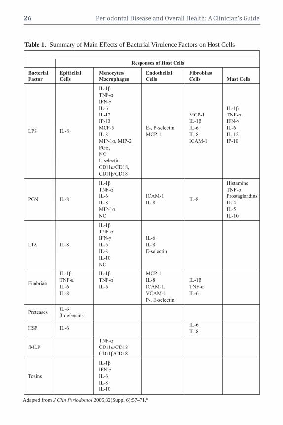
26 Periodontal Disease and Overall Health: A Clinician's Guide
Adapted from J Clin Periodontol 2005;32(Suppl 6):57–71.8
Table 1. Summary of Main Effects of Bacterial Virulence Factors on Host Cells
Responses of Host Cells
Bacterial Epithelial Monocytes/ Endothelial Fibroblast
Factor Cells Macrophages Cells Cells Mast Cells
IL-1β
TNF-α
IFN-γ
IL-6 IL-1β
IL-12 MCP-1 TNF-α
IP-10 IL-1β IFN-γ
MCP-5 E-, P-selectin IL-6 IL-6 LPS IL-8
IL-8 MCP-1 IL-8 IL-12
MIP-1α, MIP-2 ICAM-1 IP-10
PGE2
NO
L-selectin
CD11α/CD18,
CD11β/CD18
IL-1β Histamine
TNF-α TNF-α
PGN IL-8IL-6 ICAM-1
IL-8Prostaglandins
IL-8 IL-8 IL-4
MIP-1α IL-5
NO IL-10
IL-1β
TNF-α
IFN-γ IL-6
LTA IL-8 IL-6 IL-8
IL-8 E-selectin
IL-10
NO
IL-1β IL-1β MCP-1
FimbriaeTNF-α TNF-α IL-8 IL-1β
IL-6 IL-6 ICAM-1, TNF-α
IL-8 VCAM-1 IL-6
P-, E-selectin
ProteasesIL-6
β-defensins
HSP IL-6IL-6
IL-8
TNF-α
fMLP CD11α/CD18
CD11β/CD18
IL-1β
IFN-γ
Toxins IL-6
IL-8
IL-10

CHAPTER 3 Infection and Inflammation 27
tissue. Normally, the intact sulcular and junc-tional epithelium serves as an effective nat-ural barrier that keeps the bacteria from en-tering host tissues. However, severalperiodontopathogens (e.g., P. gingivalis, A.
actinomycetemcomitans) have been shownto invade and transverse epithelial cells togain access to the connective tissue. More-over, bacterial components (e.g., LPS, PGN)and products (e.g., proteases, toxins) that areeither shed or secreted can also diffusethrough the epithelial junctions to the con-nective tissue.9
Bacteria in Connective TissueBacteria and/or their virulence factors
found in the connective tissue directly stim-ulate host cells residing in this area, includ-ing leukocytes, fibroblasts, mast cells, endo -thelial cells, dendritic cells and lymphocytes.Neutrophils, macrophages, fibroblasts andmast cells release more proinflammatory cy-tokines (TNF-, IL-1, IL-6, IL-12), chemo-attractants (IL-8, MIP-1-�, MIP-2, MCP-1,MCP-5) and PGE2 in the connective tissue.In addition, degranulation of mast cells re-sults in the secretion of histamine andleukotrienes further amplifying the inflam-matory cascade.10,11
Mediators that are secreted from acti-vated host cells (e.g., IL-1, TNF-, PGE2,and histamine) will further assist bacterialvirulence factors in the activation of endo -thelial cells. This leads to secretion of morechemokines (IL-8, MCP-1) and expressionof adhesion molecules on the surface of en-dothelial cells, which are important forleukocyte extravasation (P- and E-selectinsas wells as ICAM-1 and -2).12 Specifically,P- and E-selectins interact with glyco proteinson leukocytes allowing the cells to adhere re-versibly to the vessel wall, causing circulat-ing leukocytes to appear to “roll” along theactivated endothelium. Then, IL-8 and otherchemokines, bound to proteoglycans on thesurface of leukocytes, trigger a conforma-
tional change of integrins (LFA-1, CD11b:CD18). As a result, adhesive properties in-crease dramatically and leukocytes attachfirmly to ICAM-1 expressed on endothelialcells. TNF-, PGE2 and histamine increasevascular permeability, allowing leukocytesto squeeze between the endo thelial cells,thereby entering the connective tissue ina process known as diapedesis. Finally,chemokines, such as IL-8 that are producedat the site of infection and bind to proteo-glycans of the extracellular matrix, alongwith bacterial chemo-attractants (fMLP, fimbriae) form a concentration gradient thatguides the leukocytes to migrate to the focusof infection.
The Inflammatory CascadeNeutrophils are the first leukocytes to
arrive followed by mononuclear phagocytesthat subsequently differentiate into macro -phages. The interaction of these cells withbacterial virulence factors induces further ac-tivation, which enhances their phagocytic ac-tivity by increasing the production of nitricoxide (NO) and the expression of comple-ment receptors (CR3). If the innate immuneresponse is successful, the bacteria are elim-inated and resolution of inflammation fol-lows. However, persistence of bacteria leadsto a chronic response characterized by extra-celluar release of neutrophil granule contents,including degradative enzymes and reactiveoxygen species that spill into the extracellularmilieu, leading to local tissue damage andamplification of acute inflammatory signals.13
Pro-inflammatory cytokines (TNF-,IL-1, IL-6) from the site of inflammationenter the circulation and reach the liverwhere they activate hepatocytes. This leads,among other events, to the synthesis ofplasma proteins known as acute-phase pro-teins, including LPS binding protein (LBP)and CD14, which are important for therecognition of bacterial virulence factors.Complement proteins and C-reactive protein

28 Periodontal Disease and Overall Health: A Clinician's Guide
(CRP), contribute by opsonizing bacteria,thereby aiding in recognition for phagocyto-sis. These products enter the circulation andbecause of increased vascular permeability,diffuse into the inflamed gingival tissues.Figure 1 illustrates the initiation of inflam-mation at the gingiva.
The Immune ResponseIf the infection persists, the acquired
immune response is initiated and the “es- tablished lesion” is created as described by
Page and Schroeder.14 Briefly, dendritic cellswithin the epithelium take up bacterial anti-gens and migrate to the peripheral lymphnodes. The antigens are processed into aform that is recognizable by the immune sys-tem, i.e., the antigenic peptide binds to aClass II major histocompatibility complex(MHC) receptor, and consequently “present”the antigen. As a result, antigen-specific ef-fector T cells and antibody-secreting B cellsare generated by clonal expansion and dif-ferentiation over the course of several days,
Figure 1. Initiation of Inflammation at the Gingival Level
Neutrophils in the GCF and epithelial cells comprise the first line of defense to prevent bacteria from invading thehost. The interaction of the bacterial biofilm with epithelial cells leads to activation and secretion of pro-inflam-matory cytokines (green). Bacteria and their virulence factors (red) may penetrate the epithelial lining and enterthe connective tissue. In this compartment they may interact with host cells, such as macrophages, fibroblasts, andmast cells to stimulate these cells to release more pro-inflammatory mediators such as TNF-α, IL-1β, IL-8,LTB-4, and histamine. These mediators, along with bacteria/virulence factors, may activate endothelial cells to attractcirculating leukocytes in the connective tissues. In this compartment, phagocytic cells take up bacteria and theirantigenic molecules. This process, if further enhanced by acute-phase response proteins, such as CRP, that are produced from activated hepatocytes, enter the connective tissue via circulation due to increased vascular permeability. If the noxious agents are eliminated, resolution of inflammation follows. However, if the bacterialchallenge persists, the more efficient adaptive immune response takes over. Adapted from J Clin Periodontol 2005;32(Suppl 6):57–71.8

CHAPTER 3 Infection and Inflammation 29
during which time the induced responses ofinnate immunity continue to function. Even-tually, antigen-specific T cells and then an-tibodies are released into the blood to targetthe infection site.15 Macrophages that engulfbacteria at the site of infection express co-stimulatory molecules (MHC-II) and presentbacterial antigens on their surface. Antigen-specific T cells “see” the antigens and acti-vate the macrophages, enabling them to de-stroy intracellular bacteria more efficiently.In addition, secreted antibodies protect thehost from infection by: (a) inhibiting thetoxic effects or infectivity of pathogens bybinding (neutralization); (b) opsonizing thepathogens and promoting phagocytosis; and(c) activating the complement system.Failureto clear the infection at this point leads to fur-ther tissue damage. Activated macrophagesproduce oxygen radicals, NO, and proteasesin the gingival tissues that are toxic to thehost cells. Moreover, recent work on a mousemodel revealed that the induction of an adaptive immune response to colonizingpathogens results in receptor activator of nu-clear factor-kappaB ligand-dependent perio -dontal bone loss.16
Summary of Part IThe trigger that causes the shift from
tissue homeostasis to pathology remains un-clear. The logical extension of Löe’s obser-vation is that this is caused by specific bac-teria and indeed, a large body of evidencesuggests that certain bacteria are associatedwith progressive disease. However, studies ofthe microbiota of the periodontal lesion arecross-sectional and definitive cause/effectrelationships have not been demonstrated.Recently, a longitudinal study of periodontaldisease progression failed to implicate anysingle organism or group of organisms inthe initiation of periodontal attachmentloss.17 In addition, recent animal studies sug-gest that the level of host inflammation hasa major impact on the composition of the
biofilm. Interestingly, inflammation is astronger predictor of periodontal attachmentloss than the composition or quantity of theoral biofilm.17 Clearly, the etiology andpathogenesis of periodontitis requires fur-ther study. It is also apparent that “tradi-tional” periodontal pathogens (Socransky’s“red complex”) contribute to and acceleratedisease when they overgrow in the perio -dontal environment. However, the role of in-flammation and the host immune responsehas taken on a new perspective, potentiallydetermining susceptibility and providing anovel therapeutic target.
PART II: SYSTEMIC INFLAMMATIONDUE TO PERIODONTAL INFECTION
Despite the localized nature of perio -dontal disease, infection of the sulcus/perio -dontal pocket with periodontopathogens maybe responsible for inflammatory responsesthat develop beyond the periodontium. Todate, several biological pathways have beenrecognized that present reasonable hypothe-ses for periodontal disease induction of sys-temic inflammation.
Inflammatory PathwaysIn health, the sulcular epithelium along
with innate immune molecules acts as a nat-ural barrier system that inhibits and elimi-nates penetrating bacteria. Hence, only asmall number of bacteria, mostly facultative,manage to enter the gingival tissues and thebloodstream. However, in cases of perio -dontal disease, the inflamed and ulceratedpocket epithelium is vulnerable to bacterialpenetration and forms an easy port of entryfor the bacteria. This leads to an increase inthe number of periodontopathogens, mainlyanaerobic Gram-negative, in the gingival tis-sues and consequently in the circulation.Bacteremia can be initiated after mechanicalirritation of the inflamed gingiva during toothbrushing, chewing, oral examination, andscaling and root planing.18 The microorgan-

30 Periodontal Disease and Overall Health: A Clinician's Guide
isms that gain access to the blood and circu-late throughout the body are usually elimi-nated by the reticulo-endothelial systemwithin minutes (transient bacteremia) andusually there are no other clinical symptomsother than possibly a slight increase in bodytemperature.19 However, if the disseminatedbacteria find favorable conditions, they maycolonize distant sites and form ectopic foci ofinfection. Similarly, bacterial virulence fac-tors that are secreted or shed in the gingivaltissues may also disseminate via the circula-tion and stimulate remote tissues.20 Bacteriaand bacterial antigens that are systemicallydispersed can trigger significant systemic inflammation. Leukocytes as well as endo -thelial cells and hepatocytes respond to bacteria/virulence factors, producing pro- inflammatory immune mediators. Moreover,soluble antigens may react with circulatingspecific antibodies, forming macromolec ularcomplexes. These immune complexes mayfurther amplify inflammatory reactions atsites of deposition.21
Pro-Inflammatory MediatorsA different biological pathway that may
explain the systemic inflammation inducedby periodontal disease involves pro-inflamma -tory mediators, such as IL-1, IL-6, TNF-and PGE2 that are produced by host cells inthe inflamed gingival tissues. These media-tors are secreted locally in response to bac-terial challenge, but may “spill” into the cir-culation and exert distant or systemic effects.
Specifically, cytokines may reach dis-tant sites and further activate endothelialcells leading, in some cases, to endothelialdysfunction.22 Moreover, the circulating me-diators, due to the increased vascular per-meability at the sites of inflammation, mayenter inflamed tissues and exacerbate the in-flammatory processes. However, the mostimportant impact of these circulating medi-ators is systemic. Pro-inflammatory cyto -kines may induce leukocytosis, which is an
increase in circulating neutrophils. More-over, IL-1, TNF-, and especially IL-6 mayreach the liver and activate hepatocytes toproduce acute-phase proteins. The most im-portant acute-phase reactants include CRP,serum amyloid A (SAA) protein, fibrinogen,plasminogen activator inhibitor 1 (PAI-1),complement proteins, LBP, and solubleCD14. These proteins are released in theplasma and possess a wide variety of func-tions, such as multiple pro-inflammatory ac-tivities and stimulation of tissue repair mech-anisms. The production of these proteins ispart of an acute-phase response that is char-acterized by fever, increased vascular per-meability, and a general elevation of meta-bolic processes. An acute-phase responsestarts within hours or days of most forms ofacute tissue damage or inflammation and de-spite its name, persists with chronic inflam-mation. As acute-phase reactants enter thecirculation, they may return to the inflamedgingival tissues. However, since they circu-late throughout the body they can affect ec-topic sites, causing inflammation or exacer-bation of existing inflammatory processes.This concept takes on new meaning in lightof the recent implication of CRP in thepathogenesis of cardiovascular disease.23
Because there is to date no consensuson the mechanisms that induce systemic in-flammation from periodontal disease, any ofthe above pathways (bacteremia, systemicspilling of cytokines, and activation of theacute-phase response) must be considered acandidate for the generation of systemic in-flammation. It is also possible that depend-ing upon the severity of periodontal disease,any of these mechanisms may occur alone orin combination, leading to variations of in-duced systemic inflammation.
Acute-Phase ProteinsCRP is produced mainly by the liver,
but it may also be synthesized locally at sitesof inflammation. CRP opsonizes different

CHAPTER 3 Infection and Inflammation 31
bacteria by binding to phosphorylcholinefound on the surface, thereby assisting inbacterial uptake by phagocytes.24 Opsoniza-tion and phagocytosis are further enhancedby activation of the complement system byCRP. Other pro-inflammatory activities ofCRP include the up-regulation of the expres -sion of adhesion molecules, such as ICAM-1 and E-selectin on endothelial cells and theinduction of IL-6, IL-1, and TNF-, and ofthe chemokines IL-8 and MCP-1. Otherproperties of CRP that may not be of obviousimportance in periodontal disease but maysignificantly affect other systemic inflam-matory diseases (e.g., atherosclerotic le-sions), include thrombosis due to the pro-coagulant activity and reduction offibrinolysis by inducing an increase in theexpression of PAI-1, the main inhibitor offibrinolysis.25 Finally, CRP mediates prolif-eration and activation of smooth muscle cells(SMCs) and decreases the expression of en-dothelial nitric oxide synthase (eNOS). CRPmay also have anti-inflammatory propertiesand hence its primary role is likely to be theregulation of acute inflammation.
SAASAA proteins are a family of apolipo -
proteins associated with high-densitylipoprotein in plasma. They have several pro-inflammatory functions, such as the recruit-ment of immune cells to inflammatory sitesand the induction of enzymes that degradeextracellular matrix. Also, SAA proteinstransport cholesterol to the liver for secretioninto the bile.
FibrinogenFibrinogen is a soluble plasma glyco-
protein. Processes in the coagulation cascadeactivate prothrombin to thrombin, which isresponsible for converting fibrinogen into fib-rin. Fibrin is then cross-linked by factor XIIIto form a clot. Thus, fibrinogen is involved inblood coagulation and platelet activation.
PAI-1PAI-1 is produced by the liver and en-
dothelial cells. It inhibits the serine proteasestPA and uPA/urokinase, and therefore is aninhibitor of fibrinolysis, the physiologicalprocess that degrades blood clots.
Complement ProteinsThese proteins take part in a triggered-
enzyme cascade that activates the comple-ment system. There are three ways by whichcomplement is involved in inflammatoryprocesses. First, activated complement pro-teins may bind covalently to pathogens asopsonins for engulfment by phagocytes bear-ing receptors for complement. Second, thesmall fragments of some complement pro-teins act as chemo-attractants to recruit moreleukocytes to the site of complement activa-tion. Third, terminal complement compo-nents damage certain bacteria by creatingpores in the bacterial membrane.26
LBP and Soluble CD14 These proteins play an important role in
transferring LPS and PGN to the Toll-like re-ceptors. Hence, their presence is critical forinitiating and organizing an inflammatoryimmune response after bacterial challenge.
Systemic Cellular and Molecular Markers of Inflammation
Periodontal infection may induce an in-flammatory response that is not limited to thetissues surrounding the teeth, but is also ex-tended systemically. The main cellular andmolecular markers of systemic inflamma-tion induced by periodontal disease includethe increased number of peripheral leuko-cytes, the higher concentrations of serumanti bodies against periodontopathogens,and the elevated levels of circulating pro- inflammatory cytokines and acute-phase pro-teins. With the exception of serum antibodiesagainst periodontopathogens, these markersare not specific for periodontal disease, but

32 Periodontal Disease and Overall Health: A Clinician's Guide
could be shared with distant inflammatoryprocesses that have systemic effects. As such,these markers can be affected by other in-flammatory diseases that could occur con-comitantly. The following systemic markershave been associated with the presence of pe-rio dontal disease and are usually affected bythe severity of inflammation in the gingiva.
Peripheral Blood LeukocytesIn periodontitis patients, leukocyte
counts have been shown to be slightly ele-vated compared to healthy subjects, althoughnot always significantly.27 The elevated levelof circulating leukocytes depends largely onthe extent and severity of periodontal dis-ease. Periodontal therapy may lead to a re-duction in the number of peripheral leuko-cytes.28 PMNs are the main leukocytes thatare increased, and it is possible that these cellsare recruited at higher levels during episodesof bacteremia and leakage of bacterial viru-lence factors during periodontal disease.
Serum Antibodies Against Periodontopathogens
In chronic periodontal disease, in whichthe adaptive immune response has been ac-tivated, local and systemic exposure to peri-odontopathogens leads to an increase in thelevels of circulating antibodies against thepathogenic antigens. Treatment of disease isfollowed by a reduction in antibody levels.
Serum Pro-Inflammatory Cytokines In healthy subjects, the levels of circu-
lating pro-inflammatory cytokines are verylow or nondetectable. However, in perio -dontitis patients, several pro-inflammatorycytokines may “spill” into the bloodstreamand increase the concentration in the plasma.Of the pro-inflammatory mediators studied,only the levels of IL-6 have been consistentlyshown to be elevated in the serum. This increase is related to the extent and severityof inflammation in periodontal tissues.29
However, controversial reports have beenpublished on the impact of periodontal ther-apy on IL-6 levels suggesting the need forfurther research on the topic. Finally, most ofthe studies looking at the levels of serumIL-1 and TNF-α� among healthy and perio -dontitis patients failed to report any differ-ences, and in most cases cytokine levels werenot measurable.30
Acute-Phase ProteinsThe levels of several acute-phase reac-
tants, such as CRP, fibrinogen, LBP and sol-uble CD14 have been studied and have beenshown to be elevated in patients with perio -dontal disease. However, the acute-phaseproteins that have received most attentionand are consistent markers of systemic in-flammation in periodontal disease are CRPand fibrinogen. A large number of studies,both in animal models and humans, have re-vealed a positive association between peri-odontal disease and circulating CRP levels,while a recent meta-analysis limited to hu-man studies has confirmed that plasma CRPis elevated in patients with periodontitis com-pared to healthy individuals.31 Moreover, thisincrease was proportional to the extent andseverity of the disease. Several studies reporta decrease of plasma CRP after periodontalintervention, but there is modest evidencethat periodontal therapy lowers the levels ofthis protein. Finally, in several studies, thelevels of fibrinogen have also been found tobe elevated in periodontitis patients com-pared to healthy individuals.32 However, todate there is no available evidence to supportthat periodontal therapy actually reduces theamount of circulating fibrinogen.
Possible Role of Systemic Inflammationin Various Disorders
During the late nineteenth and earlytwentieth centuries, the “focal infection” the-ory dominated the medical world.33 This the-ory held that foci of sepsis were responsible

CHAPTER 3 Infection and Inflammation 33
for the initiation and progression of a varietyof inflammatory diseases, including arthritis, peptic ulcers, and appendicitis. As a result,therapeutic full-mouth extractions became acommon dental practice. However, many teethwere extracted without evidence of infection.When it was finally realized that there wasno therapeutic benefit, the theory was finallydiscredited and the practice abandoned. Dur-ing the final two decades of the twentiethcentury—as our knowledge concerning theinflammatory component of systemic dis-eases was enriched and our understanding ofthe relationship of periodontal disease to systemic inflammation increased—the ideathat periodontal infection may affect the progression of systemic disorders such as cardiovascular disease, adverse pregnancycomplications, diabetes mellitus, and otherdiseases re-emerged.
In this possible association, systemicinflammation seems to play a key role.Specifically, on one hand, periodontal dis-ease may induce systemic inflammation andon the other, there is increasing evidencesuggesting that elevated levels of the mark-ers of systemic inflammation are associatedwith an increased risk for systemic diseases.
Cardiovascular Disease (CVD)There is now abundant clinical evidence
demonstrating that many biomarkers of in-flammation are elevated years in advance offirst-ever myocardial infarction (MI) orthrombotic stroke, and that these same bio-markers are highly predictive of recurrentMI, recurrent stroke, and death due to CVD.2
Moreover, studies demonstrate that serumIL-6 levels were significantly elevated insubjects who subsequently experienced anMI compared to age-matched controls.34
Similarly, plasma levels of soluble P-selectin,soluble CD40L, and macrophage-inhibitorycytokine-1 were all significantly increased inhealthy subjects who subsequently developedCVD events compared to matched controls.35
Elevated plasma concentrations of TNF-α�have also been associated with CVD, andspecifically with recurrent nonfatal MI orother CVD events. Moreover, TNF-α levelswere persistently higher among post-MI patients at increased risk for recurrent coro-nary events.
Besides these pro-inflammatory cyto -kines, several acute-phase reactants have alsobeen associated with CVD. One of the fac-tors with the strongest evidence as a bio-marker for predicting CVD events is CRP(specifically, high sensitivity CRP, hsCRP).When measured in the blood, hsCRP provedto be a strong, independent predictor of fu-ture MI and stroke among apparently healthyasymptomatic men. Also, the relative riskfor first MI and ischemic stroke increasedsignificantly with each increasing quartileof baseline concentrations of CRP.36 As de-scribed already, CRP may contribute to theinitiation and development of atherothrom-botic lesions not only by up-regulating theexpression of pro-inflammatory cytokines,but also by mediating proliferation and acti-vation of SMCs and by activating the pro- coagulant system. This last property may befurther enhanced by another acute-phase pro-tein, fibrinogen, which is often found to beelevated in CVD patients.
Adverse Pregnancy OutcomesSystemic inflammation has also been
implicated in adverse pregnancy outcomes,since elevated concentrations of CRP in earlypregnancy are associated with an increasedrisk of preterm birth and very-preterm birth.
Diabetes MellitusFinally, systemic inflammation has
been associated with both Type 1 and Type2 diabetes mellitus. Recent studies suggestthat in Type 1 diabetes, the levels of sys-temic markers of inflammation, such as CRP,do not differ between healthy individualsand subjects for which Type 1 diabetes has

34 Periodontal Disease and Overall Health: A Clinician's Guide
been just diagnosed. However, the levels ofcirculating CRP are significantly higher inindividuals with long-term diabetes.37 It isalso believed that inflammatory processesmay have a more pronounced effect on thedevelopment of complications of Type 1 di-abetes. Thus, elevated levels of plasma CRPand of the pro-inflammatory soluble adhe-sion molecule, vascular cell adhesion mole-cule-1 (VCAM-1), have been found in pa-tients with microvascular disease comparedto those without.
In Type 2 diabetes, inflammatoryprocesses are more strongly associated withthe development of the disease. Systemicmarkers of inflammation are found to be in-creased in healthy individuals who developType 2 diabetes later in their lives. AmongPima Indians, a population with a high preva-lence of Type 2 diabetes, subjects with whiteblood cell counts within the highest tertilewere more likely to develop Type 2 diabetesover a period of 20 years compared to those inthe lowest tertile. Moreover, in two other stud-ies, healthy individuals demonstrating serum levels of CRP and IL-6 within the highestquartiles were more likely to develop Type 2diabetes in the next four to seven years com-pared to subjects in the lowest quartile.4 Sim-ilar results were found with increased levelsof another acute-phase protein, PAI-1.
Insulin resistance, which is associatedwith Type 2 diabetes and usually procedesthe development of frank diabetes, may alsobe affected by pre-existing systemic inflam-mation since several pro-inflammatory andacute-phase proteins, such as TNF-, IL-6,MCP-1, PAI-1, and SAA are associated withthe induction of insulin resistance.38
Summary of Part IIBased on available evidence, it is pos-
sible that systemic inflammation may actu-ally be the link that associates periodontaldisease with other systemic diseases. Detailsof the plausible biological mechanisms that
may associate periodontal disease with var-ious systemic diseases are further analyzed inother chapters of this book.
PART III: RESOLUTION OFINFLAMMATION IN PERIODONTITIS AND OTHER SYSTEMIC DISEASES
Inflammation is thought to play a cen-tral role in the progression of periodontaland a number of systemic diseases. Experi-ments in animal models and in man havedemonstrated that periodontal destruction ismediated primarily by the inflammatory re-sponse, although periodontal pathogens area necessary etiologic factor.20,39,40 Geneticpolymorphisms and other factors may alsobe responsible for a “hyperinflammatoryphenotype” that may further affect the sus-ceptibility of the host to periodontal diseaseand tissue destruction. Currently, it is be-lieved that in chronic periodontal disease, de-struction does not follow a linear pattern withtime, but occurs in “random bursts” with pe-riods of remission and exacerbation. How-ever, the reasons behind this random pro-gression are not fully understood. Diseaseprogression becomes even more enigmaticconsidering that it is not always clear why achronic inflammation of the gingiva may re-main as gingivitis in some patients andprogress to periodontitis in others. Irrespec-tive of the nature of periodontal disease pro-gression, the perpetuation of the inflamma-tory process in the gingiva may lead to achronic low-grade systemic inflammatory re-sponse, which in turn potentially contributesto the progression of systemic diseases.
The Process of Inflammatory ResolutionThe landmark events during inflamma-
tion include the accumulation of leukocytesin the infected area and phagocytosis of thebacteria and/or their virulence factors. Aspart of the inflammatory process, activationof neutrophil lysosomal phospholipase re-leases free arachidonic acid from membrane

CHAPTER 3 Infection and Inflammation 35
phospholipids. Once free arachidonic acidis available, two different pathways can beinitiated: (a) the cyclo-oxygenase (COX) pathway that leads to the production ofprostaglandins (e.g., PGE2), and (b) the lipoxy-genase (LO) pathways that lead to the pro-duction of a series of hydroxyl acids charac-terized by the 5-LO products, the leukotrienes(e.g., LTB4). There are three cell type-specificLOs; the 5-LO from myeloid cells, the 12-LOfrom platelets, and the 15-LO of epithelial andendothelial cells. PGE2 is a potent activator ofosteoclast-mediated bone resorption, and withother eicosanoids mediates inflammation andperiodontal tissue destruction. LTB4 attractsneutrophils, stimulates the release of granule-associated enzymes from neutro phils, and con-tributes to pro-inflammatory processes and tofurther tissue damage.
Returning to HomeostasisOnce the bacteria have been removed
by phagocytosis, resolution of inflammationoccurs with the reduction or removal ofleukocytes and debris from inflamed siteswith a return to homeostasis.5 Until recently,resolution of inflammation was considered tobe a passive process in which the lack ofbacterial stimuli decreased the production ofinflammatory mediators, which in turn re-duced the inflammatory response, therebyreturning to normal function. New data sug-gest that resolution of inflammation is an ac-tive biochemical and metabolic process thatis initiated by a newly identified class of re-ceptor agonists that emerge temporally asthe inflammatory lesion matures.5 Althoughprostaglandins and leukotrienes secreted byneutrophils have pro-inflammatory proper-ties, as inflammation proceeds the sameprostaglandins (PGE2 and PGD2) may pro-mote expression of the 15-LO gene, leadingto a switch in the expression of biosyntheticenzymes by infiltrating neutrophils (Figure2). Binding of lipoxin A4 to neutrophils leadsto a phenotypic change, stopping all pro-
inflammatory activity of neutrophils andleading to apoptosis. As a result, they stop secreting the chemo-attractant LTB4 and several cellular pathways are activated, pro-ducing, at a local level, other dual-actinganti- inflammatory and proresolution lipidmediators, including resolvins and protectins.
Mechanisms of Inflammation ResolutionResolvins and protectins provide potent
signals that orchestrate and accelerate mech-anisms that promote resolution of inflam-mation and homeostasis. Specifically, as de-picted in Figure 3, pro-resolution mediatorsstop neutrophil infiltration and drive neu-trophils to apoptosis, while at the same timeattracting monocytes to the lesion.41 Lipoxin-stimulated monocytes/macrophages obtaina nonphlogistic phenotype, which resultsin phagocytosis of apoptotic neutrophilsand enhanced mucosal clearance of bacteriawithout concomitant secretion of pro- inflam -matory mediators that could contribute totissue damage.42 Moreover, pro-resolutionlipid molecules increase the exit of phago-cytes from the inflamed site through the lym-phatics. Finally, some of these moleculesmay also stimulate the uptake and clearanceof local cytokines by apoptotic neutrophils.After neutrophils and debris are removed,homeostasis returns and repair mechanismsare initiated; lipoxins are antifibrotic and allow for complete tissue healing withoutscarring.
Hence, it can be argued that the per-sistence of an inflammatory disease, such asperiodontal disease, may be caused by toomuch pro-inflammatory signal or not enoughproresolution signal. In other words, a “hy-perinflammatory phenotype” due to a par-ticular genetic background of the host mayresult in oversecretion of inflammatory me-diators in response to bacterial stimuli, whichin turn contributes to periodontal disease sus-ceptibility, or a failure of resolution pathways.As high levels of inflammatory cyto kines are

36 Periodontal Disease and Overall Health: A Clinician's Guide
Figure 2. Progression and Biosynthesis of Lipid Mediators During Inflammation Resolution
Chemical mediators involved in the initiation of acute inflammation, such as prostaglandins (PGs) and leukotrienes(LTs), induce “class switching” toward pro-resolving lipid mediators. The pro-resolving mediators include -6PUFAs, AA- derived LXs, ATLs, -3 PUFA EPA-derived RvEs, docosahexanoic acid (DHA)-derived RvDs, andprotectins (PDs) (or neuroprotectins in neural tissues). Reprinted by permission from Wiley-Blackwell: Br J Pharmacol 2008;153(Suppl)S200–S215.
Figure 3. Dual Anti-Inflammatory and Pro-Resolution Actions of Specific Lipoxins, Figure 3. Resolvins and Protectins
Reprinted by permission from Macmillan Publishers Ltd: Nat Rev Immunol 2008;8:349-361.5

CHAPTER 3 Infection and Inflammation 37
maintained, tissue destruction continues andinflammation persists. If pro-resolution sig-nals are weak, neutrophils are not removedand monocytes/macrophages maintain aphlogistic phenotype. This results in furtherproduction of inflammatory cytokines andperpetuation of the inflamed state.
New Treatment ParadigmsIt is reasonable to suggest that the un-
derstanding and ability to manipulate reso-lution of inflammation may provide a newtreatment paradigm for inflammatory dis-eases, local and systemic. Although humandata are not yet available, there is a growingand promising literature from in vitro workand animal models that supports the bene -ficial actions of resolution agonists both onperio dontal and other systemic diseases.5
The Role of Pro-Resolution MediatorsExamples of the actions of therapeutic
pro-resolution mediators in periodontal dis-ease include over-expression of lipoxin A4 intransgenic rabbits protecting against perio -dontitis and atherosclerosis.43 In anotherstudy, topical treatment with resolvins (-3fatty acid derived resolution agonists, vide
infra) prevented more than 95% of alveolarbone destruction in rabbits. Moreover, his-tological analysis revealed few, if any, neu-tro phils in the tissue and little tissue damage.At the same time, the numbers of osteoclastswere also found to be reduced. In addition,treatment of periodontitis with resolvins sys-temically reversed the observed increase inCRP and IL-1 levels. Finally, in establishedperiodontal disease, resolvins prevented fur-ther tissue destruction, and both gingival andosseous tissues that were lost during diseasewere regenerated.44
Resolvins, lipoxins, and protectins havealso been shown in animal models to havebeneficial impact on a variety of other in-flammatory diseases. For example, lipoxinsstopped neutrophil recruitment and promoted
lymphatic removal of phagocytes in perio -tonitis.42 Moreover, in cystic fibrosis, lipoxinsdecreased neutrophil inflammation, pulmo -nary bacterial burden, and disease severity.45
Resolvins in a colitis model in mice decreasedneutrophil recruitment and pro-inflammatorygene expression, improved survival, and reduced weight loss.46 In addition, resolvinsprotected against neovascularization inretinopathy.47 Finally, in an asthma model,protectins protected against lung damage,airway inflammation, and airway hyper- responsiveness.48 Table 2 lists the impact oflipoxins, resolvins, and protectins on variousinflammatory disease models.
It is conceivable that the use of pro-resolution mediators in managing periodon-tal and other inflammatory diseases mayprove to be beneficial in humans as well.Mechanical debridement, which aims at thereduction of the bacterial load in the gingivalpocket, may help the host/patient to clearthe infection. In addition, it is possible thatthe use of locally applied pro-resolution me-diators could prevent further tissue damage,enhance the resolution of inflammation(which would lead to healthy gingiva), andideally result in periodontal tissue regenera-tion rather than scarring and repair. More-over, resolution of inflammation at the gingi -val level may minimize systemic inflamma-tion induced by periodontal disease, thereby attenuating the possible negative effects ofperio dontal disease on systemic diseases.
Origins of Pro-Resolution MediatorsIn order to manipulate resolution of
inflammation more effectively, it is imperativeto understand the biological origin of the pro-resolution mediators. Lipoxins (e.g., lipoxinA4) derive from arachidonic acid after acti -vation of the 12-/5-LO or the 15-/5-LO path-ways. Resolvins and protectins are bio synthe-sized from omega-3 essential poly -unsaturatedfatty acids (-3 PUFAs), such as eicosapen-tanoic acid (EPA) and docosahexanoic acid

38 Periodontal Disease and Overall Health: A Clinician's Guide
Disease Model Species Action(s)Lipoxin A4/ATL
Periodontitis Rabbit –Reduces neutrophil infiltration–Prevents connective tissue and bone loss
Peritonitis Mouse Stops neutrophil recruitment and lymphatic removal of phagocytesDorsal air pouch Mouse Stops neutrophil recruitmentDermal inflammation Mouse Stops neutrophil recruitment and vascular leakageColitis Mouse –Attenuates pro-inflammatory gene expression
–Reduces severity of colitis–Inhibits weight loss, inflammation, pulmonary dysfunction
Asthma Mouse Inhibits airway hyper-responsiveness and pulmonary inflammationCystic fibrosis Mouse Decreases neutrophilic inflammation, pulmonary bacterial burden, and disease severityIschemia-reperfusion injury Mouse –Attenuates hind-limb ischemia-reperfusion lung injury
–Causes detachment of adherent leukocytes in mesenteric ischemia-reperfusion injuryCorneal disorders Mouse –Accelerates cornea re-epithilialization
–Limits sequelae of thermal injury (such as neovascularization and opacity)–Promotes host defense
Angiogenesis Mouse Reduces angiogenic phenotype: endothelial-cell proliferation and migrationBone-marrow transplant Mouse Protects against bone-marrow-transplant-induced graft-versus-host diseasesGlomerulonephritis Mouse –Reduces leukocyte rolling and adherence
–Decreases neutrophil recruitmentHyperalgesia Rat –Prolongs paw withdraw latency and reduces hyperalgesic index
–Reduces paw oedemaPleuritis Rat Shortens the duration of pleural exudation
Resolvin E1
Periodontitis Rabbit –Reduces neutrophil infiltration–Prevents connective tissue and bone loss–Promotes healing of diseased tissues–Regenerates lost soft tissue and bone
Peritonitis Mouse –Stops neutrophil recruitment–Regulates chemokine and/or cytokine production–Promotes lymphatic removal of phagocytes
Dorsal air pouch Mouse Stops neutrophil recruitmentRetinopathy Mouse Protects against neovascularizationColitis Mouse –Decreases neutrophil recruitment and pro-inflammatory gene expression
–Improves survival–Reduces weight loss
Resolvin D1
Peritonitis Mouse Stops neutrophil recruitmentDorsal skin air pouch Mouse Stops neutrophil recruitmentKidney ischemia- Mouse –Protects from ischemia-reperfusion kidney damage and loss of function
reperfusion injury Mouse Regulates macrophageRetinopathy Mouse Protects against neovascularization
Protectin D1
Peritonitis Mouse –Inhibits neutrophil recruitment–Regulates chemokine and/or cytokine production–Promotes lymphatic removal of phagocytes–Regulates T-cell migration
Asthma Mouse Protects from lung damage, airway inflammation, and airway hyper-responsivenessAsthma Human Protectin D1 is generated in humans and appears to be diminished in asthmatics Kidney ischemia- Mouse Protects from ischemia-reperfusion kidney damage and loss of function
reperfusion injury Regulates macrophages functionRetinopathy Mouse Protects against neovascularizationIschemic stroke Rat –Stops leukocyte infiltration
–Inhibits nuclear factor-kB and cyclo-oxygenase-2 inductionAlzheimer’s disease Human Diminishes protecting D1 production in human Alzheimer’s disease
Table 2. Impact of Lipoxins, Resolvins, and Protectins on Various Inflammatory Disease Models

CHAPTER 3 Infection and Inflammation 39
(DHA). EPA and DHA can be metabolizedby aspirin-modified COX-2 pathways toform resolvins, while DHA can be convertedto protectins via an LO-mediated pathway(Figure 4).
Another aspect of current anti-inflam-matory strategies was the discovery that dis-ruption of biosynthesis of these pro-resolu-tion mediators by either COX-2 or LOinhibitors may lead to a “resolution deficit”phenotype, which is characterized by im-paired phagocyte removal, delayed resolu-tion, and prolonged inflammation. This mayexplain why several anti-inflammatory agents,such as selective COX-2 inhibitors and cer-tain LO inhibitors, have been shown to impairresolution of inflammation and lead to sys-temic inflammatory complications.
Summary of Part IIITheoretically, combining pro- resolution
mediators and anti-inflammatory agents suchas aspirin and statins—agents that decreasethe extent of inflammation without inter -fering with the endogenous pro- resolu tionprocesses—may be a useful strategy to control
excessive inflammation and restore homeo -stasis. More research is necessary to obtainsolid information on the efficacy and safetyof these interventions in humans. However,it is possible that in the future we can expectthat new treatment strategies will be avail-able for the treatment of periodontal diseaseand its systemic complications.
Supplemental Readings
Serhan CN, Chiang N, Van Dyke TE. Resolving in-flammation: dual anti-inflammatory and pro-resolutionlipid mediators. Nat Rev Immunol 2008;8(5):349–61.
Madianos PN, Bobetsis YA, Kinane DF. Generation ofinflammatory stimuli: how bacteria set up inflammatoryresponses in the gingiva. J Clin Periodontol 2005;32(Suppl 6):57–71.
Loos BG. Systemic markers of inflammation in perio -dontitis. J Periodontol 2005;76(Suppl 11):2106–2115.
Ridker PM, Silvertown JD. Inflammation, C-reactiveprotein, and atherothrombosis. J Periodontol 2008;79(Suppl 8):1544–1551.
REFERENCES1. Löe H, Theilade E, Jensen SB. Experimental gin-
givitis in man. J Periodontol 1965;36:177–187.
Figure 4. Schematic Illustration of Lipid-Mediated Pro-Inflammatory and Figure 4. Pro-Resolution Pathways

40 Periodontal Disease and Overall Health: A Clinician's Guide
2. Ridker PM, Silvertown JD. Inflammation, C-reac-tive protein, and atherothrombosis. J Periodontol
2008;79:1544–1551.3. Lohsoonthorn V, Qiu C, Williams MA. Maternal
serum C-reactive protein concentrations in earlypregnancy and subsequent risk of preterm deliv-ery. Clin Biochem 2007;40:330–335.
4. Pradhan AD, Manson JE, Rifai N, Buring JE, Ridker PM. C-reactive protein, interleukin 6, andrisk of developing type 2 diabetes mellitus. JAMA
2001;286:327–334.5. Serhan CN, Chiang N, Van Dyke TE. Resolving
inflammation: dual anti-inflammatory and pro- resolution lipid mediators. Nat Rev Immunol 2008;8:349–361.
6. Paster BJ, Olsen I, Aas JA, Dewhirst FE. Thebreadth of bacterial diversity in the human perio -dontal pocket and other oral sites. Periodontol 2000
2006;42:80–87.7. Haffajee AD, Cugini MA, Tanner A, Pollack RP,
Smith C, Kent RL Jr, Socransky SS. Subgingivalmicrobiota in healthy, well-maintained elder andperiodontitis subjects. J Clin Periodontol 1998;25:346–353.
8. Madianos PN, Bobetsis YA, Kinane DF. Generationof inflammatory stimuli: How bacteria set up in-flammatory responses in the gingiva. J Clin Perio -
dontol 2005;32(Suppl 6):57–71.9. Sandros J, Papapanou PN, Nannmark U, Dahlén G.
Porphyromonas gingivalis invades human pocketepithelium in vitro. J Periodontal Res 1994;29:62–69.
10. Takada H, Mihara J, Morisaki I, Hamada S. Induc-tion of interleukin-1 and -6 in human gingival fibro -blast cultures stimulated with Bacteroides lipopoly -saccharides. Infect Immun 1991;59:295–301.
11. Supajatura V, Ushio H, Nakao A, Akira S, OkumuraK, Ra C, Ogawa H. Differential responses of mastcell Toll-like receptors 2 and 4 in allergy and innateimmunity. J Clin Invest 2002;109:1351–1359.
12. Darveau RP, Cunningham MD, Bailey T, SeachordC, Ratcliffe K, Bainbridge B, Dietsch M, Page RC,Aruffo A. Ability of bacteria associated with chronicinflammatory disease to stimulate E- selectin ex-pression and promote neutrophil adhesion. Infect
Immun 1995;63:1311–1317.13. Weissmann G, Smolen JE, Korchak HM. Release of
inflammatory mediators from stimulated neutro -phils. N Engl J Med 1980;303:27–34.
14. Page RC, Schroeder HE. Pathogenesis of inflam-matory periodontal disease. A summary of currentwork. Lab Invest 1976;34:235–249.
15. Kinane DF, Karim SN, Garioch JJ, al Badri AT,Moughal N, Goudie RB. Heterogeneity and selec-tive localisation of T cell clones in human skin and
gingival mucosa. J Periodontal Res 1993;28:497–499.
16. Kawai T, Paster BJ, Komatsuzawa H, Ernst CW,Goncalves RB, Sasaki H, Ouhara K, Stashenko PP,Sugai M, Taubman MA. Cross-reactive adaptiveimmune response to oral commensal bacteria re-sults in an induction of receptor activator of nuclearfactor-kappaB ligand (RANKL)-dependent perio -dontal bone resorption in a mouse model. Oral
Micro biol Immunol 2007;22:208–215.17. Tanner AC, Kent R Jr, Kanasi E, Lu SC, Paster BJ,
Sonis ST, Murray LA, Van Dyke TE. Clinical characteristics and microbiota of progressing slightchronic periodontitis in adults. J Clin Periodontol
2007;34:917–930.18. Kinane DF, Riggio MP, Walker KF, MacKenzie D,
Shearer B. Bacteraemia following periodontal pro-cedures. J Clin Periodontol 2005;32:708–713.
19. Li X, Kolltveit KM, Tronstad L, Olsen I. Systemicdiseases caused by oral infection. Clin Microbiol
Rev 2000;13:547–558.20. Jain A, Batista EL Jr, Serhan C, Stahl GL, Van
Dyke TE. Role for periodontitis in the progressionof lipid deposition in an animal model. Infect
Immun 2003;71:6012–6018.21. Thoden van Velzen SK, Abraham-Inpijn L, Moorer
WR. Plaque and systemic disease: A reappraisalof the focal infection concept. J Clin Periodontol
1984;11:209–220.22. Amar S, Gokce N, Morgan S, Loukideli M, Van
Dyke TE, Vita JA. Periodontal disease is associatedwith brachial artery endothelial dysfunction andsystemic inflammation. Arterioscler Thromb Vasc
Biol 2003;23:1245–1249.23. Ridker PM, Danielson E, Fonseca FA, Genest J,
Gotto AM Jr, Kastelein JJ, Koenig W, Libby P,Lorenzatti AJ, MacFadyen JG, Nordestgaard BG,Shepherd J, Willerson JT, Glynn RJ; JUPITERStudy Group. Rosuvastatin to prevent vascularevents in men and women with elevated C-reactiveprotein. N Engl J Med 2008;359:2195–2207.
24. Casey R, Newcombe J, McFadden J, Bodman-Smith KB. The acute-phase reactant C-reactive protein binds to phosphorylcholine-expressingNeisseria meningitidis and increases uptake by human phagocytes. Infect Immun 2008;76:1298–1304.
25. Nakakuki T, Ito M, Iwasaki H, Kureishi Y, OkamotoR, Moriki N, Kongo M, Kato S, Yamada N, IsakaN, Nakano T. Rho/Rho-kinase pathway contributesto C-reactive protein-induced plasminogen acti -vator inhibitor-1 expression in endothelial cells. Arterioscler Thromb Vasc Biol 2005;25:2088–2093.
26. Janeway C., Travers P., Walport M., Shlomchik M.Immunobiology. The immune system in health and

CHAPTER 3 Infection and Inflammation 41
disease. 5th Edition. Garland Publishing, NewYork; 2001:44.
27. Loos BG. Systemic markers of inflammation in periodontitis. J Periodontol 2005;76:2106–2115.
28. Christan C, Dietrich T, Hägewald S, Kage A, Bern-imoulin JP. White blood cell count in generalizedaggressive periodontitis after non-surgical therapy.J Clin Periodontol 2002;29:201–206.
29. Mengel R, Bacher M, Flores-De-Jacoby L. Inter-actions between stress, interleukin-1beta, inter-leukin-6 and cortisol in periodontally diseased patients. J Clin Periodontol 2002;29:1012–1022.
30. Meyle J. Neutrophil chemotaxis and serum con-centration of tumor-necrosis-factor-alpha (TNF-).J Periodontal Res 1993;28(Pt 2):491–493.
31. Paraskevas S, Huizinga JD, Loos BG. A systematicreview and meta-analyses on C-reactive protein inrelation to periodontitis. J Clin Periodontol 2008;35:277–290.
32. Kweider M, Lowe GD, Murray GD, Kinane DF,McGowan DA. Dental disease, fibrinogen andwhite cell count; links with myocardial infarction?Scott Med J 1993;38:73–74.
33. Scannapieco FA. Position paper of The AmericanAcademy of Periodontology: periodontal disease asa potential risk factor for systemic diseases. J Pe-
rio dontol 1998;69:841–850.34. Ridker PM, Rifai N, Pfeffer M, Sacks F, Lepage S,
Braunwald E. Elevation of tumor necrosis factor-alpha and increased risk of recurrent coronaryevents after myocardial infarction. Circulation
2000;101:2149–2153.35. Ridker PM, Buring JE, Rifai N. Soluble P-selectin
and the risk of future cardiovascular events. Circu-
la tion 2001;103:491–495.36. Ridker PM, Cushman M, Stampfer MJ, Tracy RP,
Hennekens CH. Inflammation, aspirin, and the riskof cardiovascular disease in apparently healthymen. N Engl J Med 1997;336:973–979.
37. Treszl A, Szereday L, Doria A, King GL, Orban T.Elevated C-reactive protein levels do not corre-spond to autoimmunity in type 1 diabetes. Dia-
betes Care 2004;27:2769–2770.38. Shoelson SE, Lee J, Goldfine AB. Inflammation
and insulin resistance. J Clin Invest 2006;116:1793–1801.
39. Serhan CN, Jain A, Marleau S, Clish C, Kantarci A,Behbehani B, Colgan SP, Stahl GL, Merched A,Petasis NA, Chan L, Van Dyke TE. Reduced in-
flammation and tissue damage in transgenic rabbitsoverexpressing 15-lipoxygenase and endogenousanti-inflammatory lipid mediators. J Immunol
2003;171:6856–6865.40. Williams RC, Jeffcoat MK, Howell TH, Reddy MS,
Johnson HG, Hall CM, Goldhaber P. Topical flur-biprofen treatment of periodontitis in beagles. J Periodontal Res 1988;23:166-169.
41. Ariel A, Fredman G, Sun YP, Kantarci A, Van DykeTE, Luster AD, Serhan CN. Apoptotic neutrophilsand T cells sequester chemokines during immuneresponse resolution through modulation of CCR5expression. Nat Immunol 2006;7:1209–1216.
42. Schwab JM, Chiang N, Arita M, Serhan CN. Re-solvin E1 and protectin D1 activate inflammation-resolution programmes. Nature 2007;447:869–874.
43. Shen J, Herderick E, Cornhill JF, Zsigmond E, KimHS, Kühn H, Guevara NV, Chan L. Macrophage-mediated 15-lipoxygenase expression protectsagainst atherosclerosis development. J Clin Invest
1996;98:2201–2208.44. Hasturk H, Kantarci A, Ohira T, Arita M, Ebrahimi
N, Chiang N, Petasis NA, Levy BD, Serhan CN,Van Dyke TE. RvE1 protects from local inflam-mation and osteoclast-mediated bone destruction inperiodontitis. FASEB J 2006;20:401–403.
45. Karp CL, Flick LM, Park KW, Softic S, Greer TM,Keledjian R, Yang R, Uddin J, Guggino WB, Ata-bani SF, Belkaid Y, Xu Y, Whitsett JA, Accurso FJ,Wills-Karp M, Petasis NA. Defective lipoxin- mediated anti-inflammatory activity in the cystic fibrosis airway. Nat Immunol 2004;5:388–392.
46. Hudert CA, Weylandt KH, Lu Y, Wang J, Hong S,Dignass A, Serhan CN, Kang JX. Transgenic micerich in endogenous omega-3 fatty acids are pro-tected from colitis. Proc Natl Acad Sci USA 2006;103:11276–11281.
47. Connor KM, SanGiovanni JP, Lofqvist C, Ader-man CM, Chen J, Higuchi A, Hong S, Pravda EA,Majchrzak S, Carper D, Hellstrom A, KangJX, Chew EY, Salem N Jr, Serhan CN, SmithLE. Increased dietary intake of omega-3-poly - un saturated fatty acids reduces pathological retinalangiogenesis. Nat Med 2007;13:868–873.
48. Levy BD, Bonnans C, Silverman ES, Palmer LJ,Marigowda G, Israel E. Severe Asthma ResearchProgram, National Heart, Lung, and Blood Institute.Diminished lipoxin biosynthesis in severe asthma.Am J Respir Crit Care Med 2005;172:824–830.

CHAPTER 4
History of the Oral-Systemic Relationship
Noel M. Claffey, Ioannis N. Polyzois, Ray C. Williams
INTRODUCTION
In the last decade, the possible association
between oral and systemic health has been
highlighted in numerous reports. The focus of
attention is mainly periodontitis and its impact
on certain conditions. Periodontitis is an infec -
tious disease associated with a number of pre -
dominantly gram-negative bacteria, and it is now
recognized that for the initiation and progression
of this disease, a susceptible host is also required.
It is also well documented that certain systemic
conditions can modify the host’s susceptibility to
periodontitis, but it is only recently that evidence
surfaced about the possi bility of a two-way rela -
tionship. Speci fically, perio dontitis has been impli -
cated as a potential risk factor for cardio vas cular
diseases, respiratory diseases, diabetes mellitus,
preterm labor, low birth weight, and renal disease.
Interest in the relationship of oral health/
periodontal disease to general health is not new,
but more of a resurgence in the old and dis credited
con cept of focal infection. Focal infec tion theory
be came popular in the begin ning of the twentieth
century because it explained a number of con -
ditions for which there was no sci entific explana -
tion at the time. It eventually fell in to disrepute
because of a lack of scientific evidence.
This chapter examines the history of the
hypothesis that micro-organisms would localize
from the source focus to the distant, systemic
focus and follows, step by step, concepts of the
oral-systemic relationship that have evolved
over the years.
ANCIENT CIVILIZATIONS
AND THE MIDDLE AGES
Throughout recorded history, many
theories have been put forward to explain
human illness. One area of the body that has
been repeatedly implicated in the origin of
human diseases is the oral cavity. Writings as
far back as from the ancient Egyptians (2100
BC) mention tooth pain associated with
women’s reproductive system diseases.1 In
Assyria, the physician of King Ashurbanipal
(669–626 BC) wrote about the troubles of
his king: “The pains in his head, arms, and
feet are caused by his teeth and must be
removed.”2 In ancient Greece, Hippocrates
(400 BC) recorded two cases in which erad-
ication of the infections of the mouth ap-
peared to relieve patients of rheumatic-like
troubles of the joints.3 Aristotle, perhaps the
first dental anatomist—especially from the
standpoint of comparative anatomy—stated
that “those persons who have the most teeth
are the longest lived.”4 In his book, On Hy-
giene, the Roman physician Galen (166–201
AD) emphasized the inter-relationship be-
tween the oral cavity and other illnesses.5
From the end of the Roman Empire
until the middle ages, all sciences fell into
abeyance, and had it not been for the Arabs
(who had access to the learning and science
contained in Greek manuscripts brought to
their country by Nestorian exiles from By -
zantium and Greeks who settled in southern
Italy), the bulk of science and knowledge
accumulated to that date might have been
lost.4 The next notable advance in dentistry
probably occurred in Italy in the 1400s when
a physician named Giovanni d’Arcoli began
filling decayed teeth with gold leaf; an
admirably progressive step for that time. He
is further credited with stating that for cases
of severe dental pain, early intervention was

CHAPTER 4 History of the Oral-Systemic Relationship 43
advisable because “such violent pains are
followed by syncope or epilepsy, through
injury communicated to the heart or brain.”6
In 1548, Ryff wrote a monograph that
dealt exclusively with dental afflictions. In
his pamphlet titled Useful Instructions on
the Way to Keep Healthy, to Strengthen and
Re-invigorate the Eyes and the Sight. With
Further Instructions of the Way of Keeping
the Mouth Fresh, the Teeth Clean and the
Gums Firm he wrote, “The eyes and teeth
have an extraordinary affinity or reciprocal
relation to one another, by which they easily
communicate to each other their defects and
diseases, so that one cannot be perfectly
healthy without the other being so too.”5
In 1768, Berdmore in A Treatise on the
Disorders and Deformities of the Teeth and
Gums described the relationship between
the teeth and the entire body as one leading to
the most “excruciating pains and dangerous
inflammations and sometimes deep seated
abscesses which destroy neighbouring parts
and affect the whole system by sympathy, or
by infecting the blood with corrupted matter.”7
In 1818, one of the most famous physicians in
America, Benjamin Rush, reported the course
of a disease in which a woman who was
suffering from rheumatism of long standing
had an aching tooth extracted and “she re -
covered in just a few days.”7
All of these statements over the course
of history were made without sufficient sup-
porting evidence, yet they were current be-
liefs at the time. These conclusions were
usually drawn by repeated observation of a
number of patients with similar symptoms
and outcomes. Today these ancient theories
—especially those related to oral systemic
conditions—cannot be considered to be any-
thing more than guess work based on simple
observation. However, it is of great interest
to see that historically there existed a sus -
picion or hunch that an inter-relationship
existed between oral disease and systemic
conditions.
ORAL SEPSIS AS A
CAUSE FOR DISEASE
The importance of oral hygiene in rela-
tion to bacteriology was first detailed by
Dutch scientist Antonie von Leeuwenhoek in
1683. However, it was with the discoveries
of the late 1800s that the centuries-old debate
about the influence of the mouth on the rest
of the body began. One of the main reasons
for interest in the area was due to strides
made in the study of microbiology. Major
contributors to advances in microbiology in-
cluded Pasteur, Lister, and Koch. Koch was
a physician working as a District Medical
Officer in Wöllstein, a small city in what is
now Germany. During the Franco-Prussian
war, he began to study the disease anthrax,
which was prevalent among farm animals
in the community. Earlier, the anthrax bacil-
lus had been discovered by Pollande, Royer,
and Davine. Through a series of experi-
ments, Koch demonstrated that pure cultures
of the anthrax bacillus could cause the
anthrax disease. His work was published
in 1876 and the “germ theory of disease
causation” was introduced to the world.
Soon, scientists around the globe became in-
terested in bacteria and their role in disease
etiology.
An American dentist working at Koch’s
Institute for Infectious Diseases, Miller was
convinced that the bacteria residing in the
mouth could explain most illnesses. In 1880,
to support his theory, Miller published a
book with the title The Microorganisms of
the Human Mouth: The Local and General
Diseases Which are Caused by Them. In
1891, Miller published a classic article in
the Dental Cosmos journal.8 The title of the
article was “The Human Mouth as a Focus of
Infection.” This article aimed to “call atten-
tion to the various local and general diseases
which have been found to result from the
action of microorganisms which have col-
lected in the mouth and to various channels
through which these microorganisms or their

44 Periodontal Disease and Overall Health: A Clinician's Guide
waste products may obtain entrance to parts
of the body adjacent to or remote from the
mouth.” It also aimed to “establish the great
importance of thorough understanding on
the part of the physician, no less than of the
dentist, of mouth germs as a factor in the
production of disease.” The article was pre-
sented under three headings/sections:
• Diseases of the human body which have
been traced to the action of mouth
bacteria
• The pathogenic mouth bacteria
• Prophylactic measures
The diseases he felt could be traced to
bacteria colonizing the mouth included osti-
tis, osteomyelitis, septicemia, pyemia menin-
gitis, disturbance of alimentary tract, pneu-
monia, gangrene of the lungs, Ludwig’s
angina, diseases of the maxillary sinus, actin-
omycosis, noma, diphtheria, tuberculosis,
syphilis, and thrush. He described 149 cases,
many of which he ascribed to a dental origin,
such as fistulae that opened on the neck,
shoulder, arm, or breast. Thus was devel-
oped the concept of focus of infection, with
organisms in the oral cavity being implicated
in diseases of the body remote from the
mouth. Although he did not mandate re-
moval of teeth as a method of eradication of
foci of infection, he sometimes suggested
that the “treatment and filling of root canals”
could serve this purpose. In Miller’s opinion,
local collection of disease-producing organ-
isms could produce “a metastic abscess
wherever a point of diminished resistance
existed.” Moreover, he postulated that teeth
were not the only source of aggregation of
such bacteria but that foci in other organs, such
as the tonsils and uterus, could be implicated.5,8
The next important figure in the his-
tory of oral sepsis as a cause of disease was
the English physician, Hunter. At the time of
Miller’s paper presentation, which Hunter
attended, he was the senior assistant physi-
cian at the London Fever Hospital and his at-
tention was already drawn to the mouth as a
possible source of infection. In 1900, he
wrote an article titled “Oral Sepsis as a
Cause of Disease,” which was published in
the British Medical Journal.9 Hunter impli-
cated poor oral hygiene, together with iatro-
genic conservative dentistry, as causes of
the multitude of diseases attributed to focal
infection. He advocated oral antisepsis meas-
ures to diseased teeth or inflamed gums, the
removal of “tooth stumps,” the boiling of
every “tooth plate” worn, and the avoidance
of restorations such as bridges, which can’t
be cleanly maintained.5,9
In 1900, Godlee described how the
signs and symptoms of other conditions,
such as pleurisy, could be attributed to
pyorrhea alveolaris, and how all the signs
and symptoms disappeared after careful re-
moval of all calculus and regular syringing
of the pockets with a hydrogen peroxide so-
lution.5,10 In 1902, Colyer described the res-
olution of irregular heart beat, gastric effects
and “general debility” after the treatment of
any oral sepsis present. He also suggested a
good maxim with which a dentist should work
was “better no teeth than septic ones.”5,11
In an article published by Wilcox in
1903, antral disease was put forward as an
important sequelae of oral sepsis.12 It was
believed that prolonged antral suppuration
could lead to extreme mental depression, of-
ten ending in suicidal tendency.5,12 Other re-
lationships that were put forward were those
between oral sepsis and migraine headaches,
laryngeal pain and spasm (which could in-
duce cough, loss of voice, and wasting),
blindness, and deafness, all of which Wilcox
hypothesized could be cured with treatment
of the oral sepsis.5 As the concept of oral
sepsis became more popular, theories were
put forward as to which organs were most
susceptible to different types of oral sepsis,
and how the treatment of oral sepsis could
lead to recovery from tonsillitis, tuberculosis,
and diabetes. It was also believed that oral
sepsis could be transmitted by the licking of

CHAPTER 4 History of the Oral-Systemic Relationship 45
envelopes, use of contaminated telephone
receivers, and men with beards.
In 1908, Merritt published an article in
Dental Cosmos with the title “Mouth Infec-
tion: the Cause of Systemic Disease.”13 In
this article he stated that “there is a general
disposition on the part of the medical and
dental professions to underestimate the
relations which exist between an unclean
mouth and many local and systemic disorders
of grave nature.” He felt that in many cases
of malnutrition, the sole cause was a “filthy
mouth” and that “no greater good could
come to humanity than the full recognition of
the dangers from this insidious, prolific and
virulent infection in the human mouth.” He
also stated that “the adoption of proper oral
hygiene practices would result in immediate
and marked improvement to general health
and notable increase in the average duration
of human life.”
On October 3, 1910, Hunter was invited
to McGill University in Montreal, Canada, to
give the keynote address at the dedication of
the Strathcona Medical Building. The title
of his address was “The Role of Sepsis and
Antisepsis in Medicine.” In his address, he
blamed “oral sepsis” as the cause of a great
many diseases, and made an attack on con-
servative dentistry, or as he called it “septic
dentistry.”5 His address was published in
The Lancet, which was the leading British
medical journal at the time, as well as in the
Dental Register.14,15 Hunter is best remem-
bered for the following statement in The
Lancet report: “No one has probably had
more reason than I have had to admire the
sheer ingenuity and mechanical skill con-
stantly displayed by the dental surgeon. And
no one has had more reason to appreciate
the ghastly tragedies of oral sepsis which his
misplaced ingenuity so often carries in its
train. Gold fillings, crowns and bridges,
fixed dentures, built on and about diseased
tooth roots form a veritable mausoleum of
gold over a mass of sepsis to which there is
no parallel in the whole realm of medicine.”
He continued with “The worst cases of
anaemia, gastritis, obscure fever, nervous
disturbances of all kinds from mental
depression to actual lesions of the cord,
chronic rheumatic infections, kidney dis -
eases, all those which owe their origin to, or
are gravely complicated by the oral sepsis
produced by these gold traps of sepsis. Time
and again I have traced the very first onset of
the whole trouble to the period within a
month or two of their insertion.” It appears
that Hunter’s condemnation of conservative
dentistry was based primarily on its poor
standard. It was fashionable in London at the
time to mimic complicated American
dentistry. However, in many cases the results
were often of substandard quality. Some well-
respected dentists at the time, such as Edward
Cameron Kirk, the editor of Dental Cosmos,
recognized the potential systemic effects of
oral sepsis, but felt that Hunter’s criticism of
dentistry was unfair as Hunter’s observations
were primarily based on the disastrous effects
of a very low-standard dentistry.5
In 1911, Billings, the long-serving
Dean of Medicine at the University of
Chicago and head of the focal infection re-
search team at Rush Medical College and
Presbyterian Hospital, replaced the term
“oral sepsis” with “focal infection.” Soon
after that, he was honored with being asked
to give the annual Lane Memorial Lecture
at Stanford University in 1915. There, he
defined a focus of infection as a “circum-
scribed area of tissue infected with patho-
genic organisms” and said that the term focal
infection implied that: (1) such a focus or
lesion of infection existed, (2) the infection
was bacterial in nature, and (3) it was ca pa-
ble of dissemination, resulting in systemic
infection of other contiguous or noncon-
tiguous parts. The teeth, tonsils, adenoids,
and mastoids were thought to be the usual
sources of bacteremia, and certain bacteria,
such as streptococcus and pneumococcus,

46 Periodontal Disease and Overall Health: A Clinician's Guide
had special affinities for target organs like
the heart and lungs.5,16 Billings advocated
that chronic infectious arthritis was often as-
sociated with remote foci of streptococcus,
gonococcus, or tuberculosis organisms, and
suggested the removal of all foci of infection
and the improvement in patients’ immunity
by absolute rest and improvement of popu-
lation-wide and individual oral hygiene.17,18
One of the first studies measuring the clini-
cal benefit of removing focal infection in
1917 confirmed his suggestions. The study
was conducted as a retrospective postal sur-
vey, and 23% reported a cure for their arthri-
tis following removal of infective foci. An
additional 46% reported experiencing some
improvement in symptoms.5,19
One of Billings’ research associates was
Rosenow. He was a graduate of the Rush
Medical College where he had been a student
of Billings. He utilized special methods for
culturing material from various foci of
infection. He obtained a number of pathogenic
bacteria from patients, including streptococci
and gonococci, which he injected in animals.
He then tested whether these organisms would
provoke lesions similar to the secondary
manifestations noted in the patients from
whom the foci had been removed. He used the
term “elective localization” to note that certain
strains of pathogenic bacteria (mostly
streptococci) isolated from the oral cavity of
the patients had localized to the joints, cardiac
valves, or other areas of the animals.5
Physicians like Billings and Rosenow
were prominent and convincing. More and more
articles were published and many other physi -
cians, such as Barker and Cecil, embraced the
concept of focal infection. Cecil, best known
for his textbook of medicine, reported in 1933
that “the keystone of the modern treatment of
rheumatoid arthritis is the elimination of the
infected foci.”20 In an article in 1938, he quotes
Rosenow who said “the prevention of oral
sepsis in the future, with the view to lessen-
ing the incidence of systemic diseases, should
hence forth take precedence in dental practice
over the preservation of the teeth almost
wholly for mechanical or cos metic pur -
poses.”21 Other leading members of the medical
community, such as Mayo, also advocated the
focal infection theory. He stated that “in
children the tonsils and mouth probably carry
eighty percent of the infective diseases that
cause so much trouble in later life.” He went
on to write “teeth with putrescent pulps may
harbour green-producing streptococci and
even though they show no redness at the gums
they may be very dangerous to keep in the
mouth.”3-5
What followed in dentistry as the result
of the “theory of focal infection” was an un-
precedented wave of tooth extractions and
the avoidance of conservative dentistry.5,22
All teeth that were endodontically or peri-
odontally involved were extracted to avoid a
possible focus of infection. This approach
came to be known as the “hundred per-
center.” The leading spokesperson for this
radical approach was the physiologist Fisher.
He regarded a tooth with a root filling as a
dead organ that needed to be extracted.5
As biomedical research evolved in the
early 1930s, it also started evaluating con-
cepts on a scientific basis. It was then that the
strong belief in the theory of focal infection
began to decline. What stimulated this de-
cline was the work of Holman, who noted
that Rosenow’s work was fraught with con-
tamination and that his data were incon -
sistent.22,23 Others noted that Rosenow
in oculated animals with such high counts
of bacteria that it was inevitable that every
organ or joint would be affected.24, 25 Gross-
man, in his book Root Canal Therapy, noted
that Rosenow’s technique “so devastates the
laboratory animal that lesions are sometimes
produced in almost every tissue and organ of
the body.” The fact that Rosenow’s work in
animal models could not be reproduced by
other investigators heavily discredited his
theories.25

CHAPTER 4 History of the Oral-Systemic Relationship 47
Cecil, a great proponent of the focal in-
fection theory, together with the rest of the
medical community, started to re-evaluate
his approach. He and Angevine published
an article in 1938 that reported a follow-up
study of 156 patients with rheumatoid arthri-
tis who had teeth and/or tonsils removed be-
cause of foci of infection. They concluded
that chronic focal infection was relatively
unimportant in rheumatoid arthritis because
of the 52 patients who had teeth removed, 47
did not get any better and three got worse. In
their own words they concluded that “focal
infection is a splendid example of a plausi-
ble medical theory which is in danger of be-
ing converted by its enthusiastic supporters
into the status of an accepted fact.”21
In 1940, Reiman and Havens wrote a
critical review of the theory of focal infection
in the Journal of the American Medical As-
sociation.26 They reviewed the literature in
detail and ended the report with the follow-
ing paragraph. “It may be said, therefore,
that: (a) The theory of focal infection, in the
sense of the term used here, has not been
proved, (b) the infectious agents involved
are unknown, (c) large groups of persons
whose tonsils are present are no worse than
those whose tonsils are out, (d) patients
whose teeth or tonsils are removed often
continue to suffer from the original disease
for which they were removed, (e) beneficial
effects can seldom be ascribed to surgical
procedures alone, (f) measures are often out-
weighed by harmful effects or no effect at all,
and (g) many suggestive foci of infection
heal after recovery from systemic disease, or
when the general health is improved with
hygienic and dietary measures.”
In 1951, a review by Williams and
Burket27 concluded the following “There is
no good scientific evidence to support the
theory that removal of these infected teeth
would relieve or cure arthritis, rheumatic
heart disease, and kidney, eye, sin, or other
disorders.” The very strongly worded review
by Reiman and Havens, as well as over-
whelming new evidence, brought the “era
of focal infection” to an end.26 An editorial in
the Journal of the American Medical Asso-
ciation in 195228 stated that this happened
because “many patients with diseases pre-
sumably caused by foci of infection have
not been relieved of their symptoms by re-
moval of the foci. Many patients with these
same systemic diseases have no evident fo-
cus of infection, and also foci of infection
are, according to statistical studies, as com-
mon in apparently healthy persons as those
with disease.” In looking back, the theory of
focal infection not only was an easy way to
explain the cause of many diseases, but also
advocated treatment that was available to
the patients at the time.5 According to Gib-
bons,22 the role of economics in the spread-
ing of the focal infection theory should not
be underestimated. It is easy to understand
that as the era of focal infection came to an
end the lucrative business of extracting teeth,
removing tonsils, and treating sinuses as a
way of treating human diseases gradually
diminished. In his article “Germs, Dr. Billings
and the Theory of Focal Infection,” Gibbons
quotes one bacteriologist of the focal infec-
tion period saying “The age of specialization
stimulates surgery. Operations carry the
best fees with them, and without intimating
that economics play a role in the specialist’s
decision, nevertheless it is only reasonable to
regard him as human—if he is the proud
possessor of surgical skill, he is more prone
to use it.” 22
In dentistry, for almost 50 years (1940–
1989), there was little interest in the effect
of the mouth on the rest of the body. However,
throughout the second half of the twentieth
century, there were dental scientists who
continued to question whether oral infection
(and inflammation) might in some way
contribute to a person’s overall health, but
the reasons given were mostly speculative.
They continued to suggest that bacteria and

48 Periodontal Disease and Overall Health: A Clinician's Guide
bacterial products found in the mouth could
enter the bloodstream and could in some way
be harmful to the body as a whole.29 It was
not until the last decade of the twentieth cen-
tury that dentistry and medicine started again
to consider the relationship of oral diseases,
such as periodontal disease, as a contributor
to risk factors for certain systemic diseases.
Oral-Systemic Relationship Revisited
The late 1980s saw an increasing num-
ber of publications implicating an association
between periodontopathogenic bacteria and
certain systemic conditions such as coronary
artery disease, stroke, and preterm/low birth
weight babies. Such insinuations had also
been made early in the twentieth century, but
this time reports were judged with a more
measured response.30 According to Barnett30
this response was a result of several factors:
(a) greater analytical and statistical knowl-
edge, and a better understanding of the con -
strains of epidemiological research in “estab -
lishing disease causalty”; (b) increased aware -
ness of the etiology and pathogenesis of oral
diseases; (c) increased awareness of the
etiology and pathogenesis of associated sys-
temic diseases; (d) modern advances in the
treatment of oral conditions; (e) realization
that bacteria could in some way be impli-
cated in the development of diseases that as
yet have an undetermined etiology.
In 1989, Mattila and coworkers31 in
Finland conducted a case-control study on
100 patients who had suffered an acute my-
ocardial infarction. They compared these pa-
tients to 102 control subjects selected from
the community. A full dental examination
was performed on all of the subjects studied.
Additionally, a dental index was computed.
This index computed the sum of scores from
the number of missing teeth, carious lesions,
peri-apical lesions, probing depths, and the
presence or absence of pericoronitis. It was
found that dental health was significantly
worse in patients with a history of acute
myocardial infarction than in control sub-
jects. This association remained valid even
after adjustment for age, social class, smok-
ing, serum lipid concentrations, and the pres-
ence of diabetes. It was mainly this study
that renewed the interest of physicians and
dentists in the relationship of oral to sys-
temic disease.
In retrospect, it is now clear that the
advent of reports by Mattila and colleagues—
followed soon thereafter by DeStefano et
al.32 and Offenbacher et al.33—was the be-
ginning of a new era of understanding the
impact of oral health and disease on overall
health and disease.25,30 By 1996, the term
“periodontal medicine” would emerge as
scientists and clinicians in dentistry and
medicine began to appreciate the tremen-
dous effect that oral disease can have on the
body.34
PERIODONTAL/ORAL DISEASE
AS A RISK FACTOR FOR
SYSTEMIC DISEASE
Despite many years of history demon-
strating the influence of oral status on general
health, recent decades have seen an acceler-
ated effort for the prevention and manage-
ment of these conditions through ground-
breaking advances. Specifically, periodontitis,
a chronic infectious and inflammatory dis-
ease of the gums and supporting tissues, has
been associated with systemic conditions
such as coronary heart disease and stroke,
higher risk for preterm, low-birth-weight
babies, and certain cancers. It has also been
suggested that it might pose threats to those
with chronic disease, e.g., diabetes, respira-
tory diseases, and osteoporosis.35-38 Perio -
dontal diseases are infections that are caused
by micro-organisms that colonize the tooth
surface at or below the gingival margin.
These infections affect the gingival tissues
and can cause damage to the supporting
connective tissue and bone. Periodontal dis-
ease can be caused by specific bacteria (such

CHAPTER 4 History of the Oral-Systemic Relationship 49
as Porphyromonas gingivalis) from the bio -
film within the periodontal pocket. Several
different pathways for the passage of perio -
dontal pathogens and their products into the
circulation have been suggested and are cur-
rently the subject of intensive research.
The focal infection theory, as proposed
and defended the first time around, was
mainly based on anecdotal evidence and the
occasional case report. In order for the hy-
pothesis not to fall into disrepute the second
time around, different levels of evidence must
be examined in order to establish a relation-
ship between the periodontal condition and
systemic health of the patient. Since not all
scientific evidence is given the same weight,
the stronger the evidence, the more likely
it is that a true relationship exists between
these conditions.
Case reports provide us with very weak
evidence and can only suggest a link, but
not a relationship. Case-control studies are
mainly used to identify factors that may con-
tribute to a medical condition by comparing
subjects who have the condition with pa-
tients who don’t, but are otherwise similar.
These studies may lead to cross-sectional
analyses. Observational studies are used to
examine associations between exposures and
disease. These are relatively inexpensive and
frequently used for epidemiological studies.
However, the fact that they are retrospective
and not randomized limits their validity.
Cross-sectional analysis studies the rela -
tionship between different variables at a point
in time. These type of data can be used to assess
the prevalence of acute or chronic conditions
in a population. However, since exposure and
disease status are measured at the same point
in time, it may not always be possible to
distinguish whether the exposure preceded or
followed the disease. Stronger evidence is
provided with a longitudinal study, in which
subject populations are examined over time.
A longitudinal study is often undertaken to
obtain evidence to try to refute the existence
of a suspected association between cause
and disease; failure to refute a hypothesis
strengthens confidence in it. Longitudinal
studies with controls are much stronger than
the ones without. The same applies for inter -
vention trials that provide the strongest form of
evidence. Unfortunately, not only are these
difficult to conduct, they are expensive and
involve many ethical considerations.
What Is Risk?
Risk is the statistical likelihood that cer-
tain factors are associated with the develop-
ment of disease. It can be divided into
absolute risk, which is the likelihood of ac-
quiring a certain disease, and relative risk,
which is the likelihood of acquiring a disease
if certain factors are modified, compared to
the same likelihood if they are not. It is easy
to understand that if true risk factors are
identified, then intervention for those at risk
can be planned and implemented.
The strength of association between pu-
tative risk factors and a disease state can be
expressed in odds ratios. An odds ratio of one
indicates an equal chance as to whether or
not an association will occur. An odds ratio
of two indicates a two-fold chance of an as-
sociation being present. Care should be ex-
ercised inferring causation from odds ratios.
Association does not, in itself, infer causa-
tion. In the interpretation of odds ratios, it is
important that the confidence interval of the
odds ratio not traverse one. If it does, the
odds ratio—regardless of magnitude—can-
not be relied upon.
There has long been an interest in the
role of systemic factors as they affect perio -
dontal disease. A series of studies were car-
ried out looking at systemic risk factors for
perio dontal disease and were summarized
by Genco in 1996.39 In this review, it was
pointed out that in addition to pre-existing
dis eases, sys temic factors have been identified.
These in clude reduced neutrophil function,
stress and coping behaviors, osteopenia, age,

50 Periodontal Disease and Overall Health: A Clinician's Guide
gender (with more disease seen in males),
hereditary factors, infection with periodontal
patho gens, cigarette smoking, and diabetes.
It should be noted that these are risk factors
com mon to many chronic, noncommunica-
ble diseases, such as heart disease, stroke,
and diabetes, all of which are associated with
periodontitis.39
Periodontitis/Oral Health
as a Risk for Specific Diseases:
Evidence for an Association
Cardiovascular
There are at least three possible mech-
anisms by which oral infections may con-
tribute to cardiovascular disease:40
1. Direct effect of infectious agent in
atheroma formation
2. Indirect or host-mediated responses
3. Common genetic predisposition
Bahekar35 and colleagues recently con-
ducted a systematic review of the literature
in order to evaluate if such an association ex-
ists. This review revealed five prospective
cohort studies involving 86,092 patients for
at least six years. The authors considered
that three out of the five prospective studies
were of good quality, and both the incidence
and prevalence of coronary heart disease
were increased in subjects with periodontal
disease after adjustments for other variables
known to increase the risk of coronary heart
disease. Furthermore, five case-control stud-
ies involving 1,423 patients and five cross-
sectional studies involving 17,724 patients
were also evaluated. All supported a sig -
nificant relationship between periodontal
disease and coronary heart disease. More
prospective studies are needed, however, to
prove the assumption that periodontitis may
be a risk factor for coronary heart disease and
to evaluate risk reduction with the treatment
of periodontitis.
In planning prospective studies, it is
important to remember that patients with pe-
riodontal disease share many of the same
risk factors as patients with cardiovascular
disease. These risk factors include age, gen-
der, lower socioeconomic status, stress, and
smoking.41 Additionally, a large number of
patients with periodontal disease also ex-
hibit cardiovascular disease; this could be
an indication that periodontal disease and
atherosclerosis share similar or common eti-
ologic pathways.42 The literature also sug-
gests that a number of pathogens, antigens,
endotoxins, and cytokines of periodontitis
might be significant contributing factors.43,44
According to Williams et al.,45 controlling for
such confounding factors when carrying out
epidemiological and observational studies re-
quires large numbers of subjects to be en-
rolled and these subjects need to be followed
over a long period of time. Common perio -
don tal pathogens such as Porphyromonas gingi -
valis and Streptococcus sanguis have been found
in arterial plaques from carotid en darterec tomy
samples. Furthermore, perio dontal disease has
been associated with elevated levels of in-
flammatory markers, such as C-reactive pro-
tein. Although there is growing evidence to
support a role for C-reactive protein as a pre-
dictive, pathogenic factor for vascular risk, it
is recognized that more research is needed.35
There is a need for large-scale prospec-
tive intervention studies to assess whether or
not periodontitis can be considered an effec -
tive modifiable risk factor in the prevention of
cardiovascular disease.
Adverse Pregnancy Outcomes
Several studies on laboratory animals that
took place in the 1970s and 1980s re vealed
that bacterial endotoxin (a cell wall com ponent
isolated from E. coli) is capable of pro ducing
spontaneous abortion, low fetal weight, and
malformations.46 Collins and col leagues suc -
cessfully demonstrated that oral anaerobes
such as P. gingivalis had similar effects. 47,48
In 1996, Offenbacher and colleagues
constructed a case-control study with the
title “Periodontal Infection as a Possible Risk

CHAPTER 4 History of the Oral-Systemic Relationship 51
Factor for Preterm Low Birth Weight.”33 In
this investigation, they sought to determine
whether or not the prevalence of maternal
periodontal infection could be associated
with preterm low birth weight, while control -
ling for known risk factors such as smoking
and poor nutrition. Results observed from
the 124 pregnant or postpartum mothers who
took part in this study indicated that perio -
dontal disease represents a clinically signif-
icant risk factor for preterm low birth weight.
This landmark report by Offenbacher and
colleagues was the first of this kind.
In the last seven years, there has been
an explosion of data released from case-
control studies, cohort studies, and clinical
trials, as well as from systematic reviews.
Many studies have reported a positive asso-
ciation, but it must be concluded that due
to different study designs, heterogeneity in
the way adverse pregnancy outcomes were
measured, as well as a lack of adequate analysis
for confounders, there is still no consistent
evidence for or against this association.
There is a need for large-scale prospec-
tive intervention studies in which adverse
pregnancy outcomes and the severity of
periodontal disease can be clearly defined.
Diabetes: A Two-Way Relationship
It is clear from epidemiologic studies
that diabetes mellitus increases the risk for
periodontal disease.49,50 The available litera-
ture highlights the importance of oral health
in subjects with diabetes, and demonstrates
an increased prevalence of periodontitis
among patients with poorly controlled dia-
betes.45 Patients with controlled diabetes
show periodontal conditions similar to those
of the healthy population.
The current literature does not provide us
with conclusive evidence to support a causal
relationship between periodontal disease and
risk for Type 2 diabetes. There is evidence
that there is an increased risk of periodontitis
in patients with diabetes, but Taylor and
coworkers also showed that patients with Type
2 diabetes who suffer from periodontitis have
worse glycemic control, suggesting that not
only does diabetes affect periodontitis, but
once a diabetic has periodontitis, it leads to
worsening diabetes or glycemic control.51 This
was followed by a paper by Grossi and Genco
in which periodontal disease and diabetes
mellitus was presented as a two-way relation -
ship.52 This began a long line of investigation
in which treatment of periodontal disease in
diabetes was found to contribute to glycemic
control, with one of the first studies reported
by Grossi and colleagues.53 Recently, a meta-
analysis of nine control studies on the subject
confirmed that the reduction of glycated
hemoglobin with periodontal therapy can be
significant, comparable to other attempts to
control glycated hemoglobin.45 Researchers
tried to evaluate the effects of periodontal
therapy on systemic inflammatory markers
and on glycemic control.54 Several randomized
control trials and a number of longitudinal and
observational studies pro vided some evidence
to support the concept that periodontitis can
adversely affect glycemic man agement.
Overall though, it is incon clusive that
periodontal treatment results in improvement
of metabolic control and of markers of
systemic inflammation.
There is emerging evidence to suggest
that periodontitis predicts the development
of overt nephropathy and endstage renal dis-
ease in patients with Type 2 diabetes.37,55 A
prospective study by Shultis37 and colleagues
was conducted exclusively in individuals with
diabetes. It also included a proportionally
large number of individuals with kidney dis-
ease. Whether or not treatment of periodon-
titis will reduce the risk of diabetic kidney
disease has not yet been determined, but this
study provides a rationale for further inves-
tigation into the connections between perio -
dontal disease and diabetic progression.
There is a need for large-scale prospec-
tive intervention studies, mainly in specific

52 Periodontal Disease and Overall Health: A Clinician's Guide
high-risk groups because, according to
Williams45 and colleagues, these groups
can provide more immediate answers than
studies with a more heterogeneous diabetic
population.
Respiratory Infections
Scannapieco56 describes four possible
mechanisms of the presence of oral bacteria
in the pathogenesis of respiratory infections:
1. The oral cavity might be a reservoir
for micro-organisms that contaminate
saliva and is then aspirated into the
lungs.
2. Periodontal disease-associated en-
zymes in saliva may facilitate the
adherence of respiratory pathogens
in the mucosal surfaces.
3. Periodontal disease-associated en-
zymes may destroy protective sali-
vary pellicles, resulting in fewer
nonspecific host defense mecha-
nisms in high-risk patients.
4. Cytokines and other molecules orig-
inating from untreated periodontal
tissues are continuously released in
saliva. Aspiration of these may alter
respiratory epithelium and promote
respiratory pathogen colonization.
A systematic review published in 2006
by Azarpazhooh and Leake investigated ev-
idence for a possible etiological association
between oral health and pneumonia or other
respiratory diseases.57 They concluded that
there is fair evidence of an association of
pneumonia with oral health, and poor evi-
dence of an association of chronic obstruc-
tive pulmonary disease with oral health.
Additionally, there is good evidence that
implementation of high-quality and frequent
oral healthcare decreases the occurrence and
progression of respiratory diseases among
elderly hospitalized or institutionalized
individuals.57
There is a need for large-scale prospec-
tive intervention studies targeting high-risk
people of the community, nursing homes,
and intensive care units.
Osteoporosis
Over the last decade, it has been specu -
lated that by decreasing the patient’s alveolar
bone mass, osteoporosis makes teeth more
susceptible to resorption by the periodontal
inflammatory reaction. Human studies have
addressed this relationship, and several large-
scale studies showed there is an association
between osteoporosis and reduced alveolar
crestal height in postmenopausal women.58
In another study, osteoporosis and perio -
dontal infection were found to be independ-
ent risk factors for oral bone loss.59 Other
studies, especially longitudinal studies, are
necessary to determine the temporal nature
of this relationship and to further evaluate it.
Some studies investigated the effect of
hormone replacement therapy or vitamin D
intake on tooth loss.60 In almost all studies,
there was a positive correlation between the
number of teeth retained and medical treat-
ment, but it must be kept in mind that con-
founding factors such as age, smoking, socio -
economic status, and many others may have
affected the results.40
There is a need for large-scale pro s-
pec tive studies with as many confounding
factors as possible to be factored into these
investigations.
CONCLUSIONS
There is a long-standing and well-
accepted principle that good oral health is an
integral component of good general health.
In recent years, there has been an attempt to
tie oral conditions to systemic diseases in a
causal relationship, but existing data support
only an association. Evidence for this rela-
tionship is growing, and a scientifically based
understanding of how oral health may pose
a risk for certain systemic diseases is devel-
oping. Certain linkages are stronger than
others, but until there are a number of well-

CHAPTER 4 History of the Oral-Systemic Relationship 53
constructed, controlled intervention studies
providing “hard” evidence, treatment rec-
ommendations need to be guarded.
Supplemental Readings
O’Reilly PG, Claffey NM. A history of oral sepsis as a
cause of disease. Periodontol 2000 2000;23:13–18.
Gibbons RV. Germs, Dr. Billings and the theory of focal
infection. Clin Infect Dis 1998;27:629–633.
Mattila K, Nieminen M, Valtonen V, Rasi VP, Kesäni-
emi YA, Syrjälä SL, Jungell PS, Isoluoma M, Hietani-
emi K, Jokinen MJ. Association between dental health
and acute myocardial infarction. Br Med J
1989;298:779–782.
Offenbacher S, Katz V, Fertik G, Collins J, Boyd D,
Maynor G, McKaig R, Beck J. Periodontal infection as
a possible risk factor for preterm low birth weight. J
Periodontol 1996;67:1103–1113.
Williams RC, Barnett AH, Claffey N, Davis M, Gadsby
R, Kellett M, Lip GY, Thackray S. The potential impact
of periodontal disease on general health: A consensus
view. Curr Med Res Opin 2008;24:1635–1643.
REFERENCES1. Gold SI. Periodontics. The past. Part (I) Early
sources. J Clin Periodontol 1985;12:79–97.
2. Francke OC. William Hunters’ “oral sepsis” and
American odontology. Bull Hist Dent 1973;21:
73–79.
3. Mayo CH. Focal infection of dental origin. Dental
Cosmos 1922; 64:1206–1208.
4. Lindsay LL. A short history of dentistry. London:
John Bale, Sons and Danielsson, Ltd; 1933:14–17.
5. O’Reilly PG, Claffey NM. A history of oral sepsis
as a cause of disease. Periodontology 2000 2000;
23:13–18.
6. Arculani J. Comenteria, Venetiis. Cap. XIViii. De
Dolore Dentium 1542:192.
7. Rush ME. An account of the core of several dis-
eases by the extraction of decayed teeth.1818:5th
ed.:197–201.
8. Miller WD. The human mouth as a focus of infec-
tion. Dental Cosmos 1891;33:689–706.
9. Hunter WD. Oral sepsis as a cause of disease. Br
Med J 1900;2:215–216.
10. Godlee RJ. On some of the medical and surgical
complications of pyorrhea alveolaris. Dent Rec
1900;20:337–347.
11. Colyer S. Oral sepsis: and some of its effects. Dent
Rec 1902;20:200–206.
12. Wilcox R. Some immediate and remote effects of
suppuration in the mouth and jaws. Br Dent J
1903;24:733–736.
13. Merritt AH. Mouth infection: the cause of systemic
disease. Dental Cosmos 1908;50:344–348.
14. Hunter WD. The role of sepsis and antisepsis in
medicine. Lancet 1911;1:79–86.
15. Hunter W. The role of sepsis and antisepsis in med-
icine and the importance of oral sepsis as its chief
cause. Dental Register 1911;44:579–611.
16. Billings FA. Focal infection: its broader application
in the etiology of general disease. JAMA 1914;
63:899–903.
17. Billings FA. Chronic focal infections and their
etiologic relations to arthritis and nephritis. Arch Int
Med 1912;9:484–498.
18. Billings FA. Chronic focal infection as a causative
factor in chronic arthritis. JAMA 1913;61:819–823.
19. Hughes RA. Focal infection revisited. Br J Rheu -
matol 1994;33:370–377.
20. Woods AC. Focal infection. Am J Opthalmol 1942;
25:1423–1444.
21. Cecil RL, Angevine DM. Clinical and experimen-
tal observations on focal infection with an analysis
of 200 cases of rheumatoid arthritis. Ann Int Med
1938:12:577–584.
22. Gibbons RV. Germs, Dr. Billings and the theory of
focal infection. Clin Infect Diseases 1998;27:627–633.
23. Holman WL. Focal infection and selective localiza -
tion: a critical review. Arch Path Lab Med 1928;5:
68–136.
24. Beeson PB. Fashions in pathogenetic concepts dur-
ing the present century: autointoxication, focal in-
fection, psychosomatic disease and autoimmunity.
Perspect Biol Med 1992;36:13–23.
25. Pallasch TJ, Wahl MJ. Focal infection: new age or
ancient history. Endod Topics 2003;4:32–45.
26. Reiman HA, Havens WP. Focal infections and
systemic disease: a critical appraisal. JAMA 1940;
114:1–6.
27. Williams NB, Burkett LW. Focal infection—a
review. Philadelphia Med 1951;46:1509.
28. Editorial. JAMA 1952;150:490.
29. Thoden van Velzen T, Abraham-Inpijn L, Moorer
WR. Plaque and systemic disease: A reappraisal of
the focal infection concept. J Clin Periodontol
1984;11:209–220.
30. Barnett ML. The oral systemic disease connection.
An update for the practicing dentist. JADA 2006;
137:5S–6S.
31. Mattila K, Nieminen M, Valtonen V, Rasi VP, Ke-
säniemi YA, Syrjälä SL, Jungell PS, Isoluoma M,
Hietaniemi K, Jokinen MJ. Association between
dental health and acute myocardial infarction. Br
Med J 1989;298:779–781.

54 Periodontal Disease and Overall Health: A Clinician's Guide
32. DeStefano F, Anda RF, Kahn HS, Williamson DF,
Russell CM. Dental disease and risk of coronary
heart disease and mortality. Br Med J 1993;306:
688–691.
33. Offenbacher S, Katz V, Fertik G, Collins J, Boyd D,
Maynor G, McKaig R, Beck J. Periodontal infec-
tion as a possible risk factor for preterm low birth
weight. J Periodontol 1996;67:1103–1113.
34. Rose LF, Genco RJ, Cohen DW, Mealey BL., Eds.,
Periodontal Medicine. BC Decker, Inc., Hamilton,
Ontario, 2000.
35. Bahekar AA, Singh S, Saha S, Molnar J, Arora R.
The prevalence and incidence of coronary heart
disease is significantly increased in periodontitis:
a meta-analysis. Am Heart J 2007;154:830–837.
36. Grau AJ, Becher H, Ziegler CM, Lichy C, Buggle
F, Kaiser C, Lutz R, Bültmann S, Preusch M, Dörfer
CE. Periodontal disease as a risk factor for ischemic
stroke. Stroke 2004;35:496–501.
37. Shultis WA, Weil EJ, Looker HC, Curtis JM, Shloss-
man M, Genco RJ, Knowler WC, Nelson RG. Ef-
fect of periodontitis on overt nephropathy and end
stage renal disease in type 2 diabetes. Diabetes
Care 2007;30:306–311.
38. Bobetsis YA, Barros SP, Offenbacher S. Exploring
the relationship between periodontal disease and
pregnancy complications. JADA 2006;137:7s–13s.
39. Genco RJ. Current view of the risk factors for perio -
dontal diseases. J Periodontol 1996;67:1041-1049.
40. Renvert S. Destructive periodontal disease in rela-
tion to diabetes mellitus, cardiovascular diseases,
osteoporosis and respiratory diseases. Oral Health
Prev Dent. 2003;1 Suppl:341–357.
41. Beck JD, Offenbacher S, Williams RC, Gibbs P,
Garcia R. Periodontitis: A risk factor for coronary
heart disease? Ann Periodontol 1998;3:127–141.
42. Umino M, Nagao M. Systemic diseases in elderly
dental patients. Int Dent J 1993;43:213–218.
43. Beck JD, Eke P, Lin D, Madianos P, Couper D,
Moss K, Elter J, Heiss G, Offenbacher S. Asso -
ciations between IgG antibody to oral organisms
and carotid intima-medial thickness in community-
dwelling adults Atherosclerosis 2005;183:342–348.
44. Kozarov EV, Dorn BR, Shelburne CE, Dunn WA Jr,
Progulske-Fox A. Human atherosclerotic plaque
contains viable invasive Actinobacillus actino-
mycetemcomitans and Porphyromonas gingivalis.
Arterioscler Thromb Vasc Biol 2005; 25:17–18.
45. Williams RC, Barnett AH, Claffey N, Davis M,
Gadsby R, Kellett M, Lip GY, Thackray S. The po-
tential impact of periodontal disease on general
health: A consensus view. Curr Med Res Opin
2008;24:1635–1643.
46. Lanning JC, Hilbelink DR, Chen LT. Teratogenic ef-
fects of endotoxin on the golden hamster. Teratog
Carcinog Mutagen 1983;3:145–149.
47. Collins JG, Windley HW 3rd, Arnold RR, Offen-
bacher S. Effects of a Porphyromonas gingivalis
infection on inflammatory mediator response and
pregnancy outcomes in hamsters. Infect Immun
1994;62:4356–4361.
48. Collins JG, Smith MA, Arnold RR, Offenbacher S.
Effects of Escherichia coli and Porphyromonas
gingivalis lipopolysaccharide on pregnancy outcome
in the golden hamster. Infect Immun 1994;62:4652–
4655.
49. Papapanou PN. Periodontal diseases: epidemiol-
ogy. Ann Periodontol;1996:1:1–36.
50. Taylor GW, Manz MC, Borgnakke WS. Diabetes,
periodontal diseases, dental caries and tooth loss: A
review of the literature. Compend Contin Educ
Dent 2004;25:179–184.
51. Taylor GW, Burt BA, Becker MP, Genco RJ,
Shlossman M, Knowler WC, Pettitt DJ. Severe
periodontitis and risk for poor glycemic control in
patients with non-insulin-dependent diabetes mel-
litus. J Periodontol 1996;67:1085-1093.
52. Grossi SG, Genco RJ. Periodontal disease and
diabetes mellitus: A two-way relationship. Ann
Perio dontol 1998;3(1):52–61
53. Grossi SG, Skrepcinski FB, DeCaro T, Robertson
DC, Ho AW, Dunford RG, Genco RJ. Treatment of
periodontal disease in diabetics reduces glycated
haemoglobin. J Periodontol 1997;68:713–719.
54. Taylor GW, Borgnakke WS. Periodontal disease:
Associations with diabetes, glycemic control and
complications. Oral Dis 2008;14:191–203.
55. Saremi A, Nelson RG, Tulloch-Reid M, Hanson
RL, Sievers ML, Taylor GW, Shlossman M,
Bennett PH, Genco R, Knowler WC. Periodontal
disease and mortality in type 2 diabetes. Diabetes
Care 2005;28:27–32.
56. Scannapieco FA. Role of oral bacteria in respiratory
infection. J Periodontol 1999;70:793–802.
57. Azarpazhooh A, Leake J. Systematic review of
the association between respiratory diseases and
oral health. J Periodontol 2006;77:1465–1482.
58. Wactawski-Wende J, Hausmann E, Hovey K,
Trevisan M, Grossi S, Genco RJ. The association
between oteoporosis and alveolar crestal height in
postmenopausal women. J Evid Based Dent Pract
2006;6:289–290.
59. Brennan-Calanan RM, Genco RJ, Wilding GE,
Hovey KM, Trevisan M, Wactawski-Wende J.
Osteoporosis and oral infection: Independent risk
factors for oral bone loss. J Dent Res 2008;87:232–
237.
60. Grodstein F, Colditz GA, Stampfer MJ. Post-
menopausal hormone use and tooth loss: a pros -
pective study. JADA 1996;127:370–377.

CHAPTER 5
Diabetes Mellitus: A Medical Overview
Srividya Kidambi, Shailendra B. Patel
INTRODUCTION
Diabetes mellitus (DM) is a quintes-
sential metabolic disease in which the char-
acteristic phenotype is loss of control of glu-
cose homeostasis, but the pathophysiology
also affects fat and protein metabolism. Re-
sulting hyperglycemia is associated with
both short- and long-term complications,
making early diagnosis and treatment of this
condition essential. The key hormonal dis-
turbance causing DM can be either a defect
in insulin secretion, insulin action, or both.
Several pathogenic mechanisms have been
proposed for the disease, and more than one
mechanism may be at play for the disease to
become clinically evident. This chapter
describes the classification, epidemiology,
pathogenesis/pathophysiology, clinical pre-
sentations, complications, and diagnosis of
the disease, as well as a brief overview of
treatment options.
Key educational objectives are to under -
stand that
• diabetes is a true metabolic disorder
caused by disrupting insulin action.
• both genetic and environmental fac-
tors are involved in causing diabetes.
• there are two main forms, Type 1 and
Type 2, distinguished upon abso-
lute and relative insulin deficiency,
respectively.
• insulin action is intimately tied to many
other counter-regulatory actions.
• long-term complications of diabetes
affect every organ in the body.
• controlling glycemia levels in addi-
tion to cardiovascular risk factors is
important in preventing, delaying, or
ameliorating disabling or life-threaten -
ing complications.
CLASSIFICATION OF
DIABETES MELLITUS
DM is classified into several subtypes,
based upon etiology, which can help explain
clinical manifestations and provide a ration-
ale for various treatments (Table 1). The ma-
jority of patients with DM have Type 2 dis-
ease (85%–90%) marked by defective
insulin action as well as relative deficiency
in insulin secretion. Another 5%–10% of pa-
tients have Type 1 disease (absolute defect in
insulin secretion). The remaining sub-types
are rare (Table1). This chapter will focus on
the major subgroups: Type 1 DM, Type 2
DM, and gestational DM (GDM); the latter
affects fetal and maternal health and is a risk
factor for later development of Type 2 DM.
EPIDEMIOLOGY
According to 2007 estimates, 23.6 million
people (or 7.8% of the population) in the United
States have DM.1 About 6 million of these
individuals do not know they have this dis-
ease and present to healthcare providers after
a point of no return in preventing compli -
cations. Prevalence increases with advancing
age, affecting almost 24% of individuals
over the age of 60. An epidemic of Type 2
DM is underway in both the developed and
developing world, but the brunt is being felt
sharply in developing countries.2-4 Globally,
the number of people with diabetes is ex-
pected to rise from the current estimate of
150 million to 220 million in 2010 and 300
million in 2025, making the cost of treating
DM and its complications a pressing eco-
nomic as well as clinical concern.5
Patients with DM are at a two- to four-
fold higher risk for developing heart disease
and stroke compared to people without DM.

In addition, DM is the leading cause of new
cases of blindness and kidney failure among
adults ages 20 to 74.1 Gingivitis and perio -
dontitis are also more common in people
with DM. Almost one-third of people with
DM have severe periodontal disease with
loss of attachment of gums to teeth measur-
ing five millimeters or more.1
In addition to enormous morbidity asso -
ciated with DM, this disease was the seventh
leading cause of death listed on US death
certificates in 2006. Overall, the risk for
death among people with DM is about twice
that of people without DM of similar age.1
PATHOPHYSIOLOGY
The pathophysiology of DM revolves
around impairment of insulin secretion, in-
sulin resistance, or both, resulting in reduced
utilization of glucose, hyperglycemia, and
impairment of fatty acid metabolism. Symp-
toms and complications of DM are due to
hyperglycemia as well as lack of adequate
insulin action.
Glucose Metabolism
Carbohydrates, broken down mainly into
glucose, are an important source of energy in
humans. Consideration of glucose and insulin
metabolic pathways is crucial to understand-
ing the pathophysiology of DM (Figure 1).
Glucose is derived from three sources:
in tes tinal absorption following digestion of
dietary carbohydrates; glycogenolysis, the
breakdown of glycogen, which is the poly-
merized storage form of glucose; and gluco-
neogenesis, the formation of glucose from
precursors including lactate (and pyruvate),
amino acids (especially alanine and gluta-
mine), and to a lesser extent, glycerol.5 Only
the liver and kidneys are capable of releasing
glucose into circulation by glycogenolysis
and gluconeogenesis. All tissues can utilize
glucose as a substrate for energy production,
but only the brain is wholly dependent upon
glucose as its main energy source. Thus,
56 Periodontal Disease and Overall Health: A Clinician's Guide
Note: Patients with any form of diabetes may re-quire insulin treatment at some stage of their disease.Such use of insulin does not, of itself, classify the patient.Adapted from the American Diabetes Association. Diabetes Care 2009;31(Suppl 1).37
Table 1. Etiologic ClassificationTable 1. of Diabetes Mellitus
III. Type 1 diabetes (b-cell destruction, usually leading to absolute insulin deficiency)A. Immune medicatedB. Idiopathic
III. Type 2 diabetes (may range from predominantly insulin resistance with relative insulin deficiencyto a predominantly secretory defect with insulin resistance)
III. Other specific typesA. Genetic defects of b-cell function
1. Chromosome 20, HNF-4a (MODY1)2. Chromosome 7, glucokinase (MODY2)3. Chromosome 12, HNF-1a (MODY3)4. Chromosome 13, insulin promoter factor-1
(IPF-1; MODY4)5. Chromosome 17, HNF-1b (MODY5)6. Chromosome 2, NeuroD1 (MODY6)7. Mitochondrial DNA8. Others
B. Genetic defects in insulin action1. Lipodystrophic syndromes2. Type A insulin resistance3. Leprechaunism4. Rabson-Mendenhall syndrome5. Others
C. Diseases of the exocrine pancreas1. Pancreatitis2. Trauma/pancreatectomy3. Neoplasia4. Cystic fibrosis5. Hemochromatosis6. Fibrocalculous pancreatopathy
D. Endocrinopathies1. Cushing’s syndrome 2. Acromegaly3. Glucagonoma4. Pheochromocytoma5. Others
E. Drug- or chemical-induced1. Glucocorticoids2. Atypical Antipsychotics3. Pentamidine4. Diazoxide5. a-Interferon6. Others
.F. Infections1. Congenital rubella
G. Uncommon forms of immune-mediated diabetes1. “Stiff-man” syndrome2. Anti-insulin receptor antibodies3. Others
H. Other genetic syndromes sometimes associatedwith diabetes1. Down’s syndrome2. Turner’s syndrome3. Wolfram’s syndrome4. Laurence-Moon-Biedl syndrome5. Prader-Willi syndrome6. Others
.IV. Gestational diabetes mellitus (GDM)

mechanisms to maintain a steady-state supply
of glucose to the central nervous system are
integral to metabolic control.
Both insulin-dependent and non- insulin-
independent pathways can determine whole
body clearance of glucose.6 Glucose is trans-
ported into the cells by specific transporters,7
activated by phosphorylation to glucose-6-
phosphate by the tissue-specific enzymes
hexokinase or glucokinase, allowing it to
enter metabolic pathways such as the glycol-
ysis, glycogen synthesis, hexosamine biosyn-
thesis (alternative pathway to glycolysis), or
pentose phosphate pathways.6 These path-
ways are subject to regulation by insulin,
as well as glucagon. It is important to note
that entry of glucose into different tissues is
regulated by expression of different glucose
transporters; in muscle and fat, glucose entry
is allowed only via an insulin-dependent
Chapter 5 Diabetes Mellitus: A Medical Overview 57
Figure 1. Action of Insulin & Glucagon Under Feeding & Fasting Conditions
Feeding, satiety, and the neurohumoral response to feeding are integrated by the brain, especially the hypothal-
amus (1A). This consists of the vagal system, incretin hormone secretion, and gut motility hormones, among other
mechanisms. Upon feeding (1A), the neurohormonal response, as well as direct glucose stimulation of the pancreas,
results in activation of pathways that will lead to efficient insulin secretion as well as a decrease in glucagon secretion
from the Islets of Langerhans in the pancreas into the portal tract. This results in increased liver uptake of glucose,
inhibition of hepatic gluconeogenesis, increased fatty acid synthesis, and VLDL secretion and increased glycogen
storage. Although the majority of insulin is cleared by the liver, it also reaches the central circulation where in the
fat, it increases glucose uptake and triglyceride storage, and inhibits free fatty acid release. In muscle, insulin
increases glucose uptake and glycogen storage; in the kidney it inhibits gluconeogenesis.
Under fasting conditions (1B), the neurohumoral response is switched to maintenance of glucose levels, resulting
in decreased insulin and increased glucagon secretion, with the resultant opposite effects on the above-described
target organs. In the liver, gluconeogenesis, glycogenolysis, and fatty acid breakdown is stimulated. In adipose
tissues, fat is mobilized with increased lipolysis and free-acid release. In muscle, decreased glucose uptake and
increase fatty acid catabolism take place. All of these actions are tightly regulated and coordinated to account for
all physiological processes, ranging from short-term energy expenditure (such as exercise) to both short- and long-
term fasting. In addition, many other hormones (e.g., cortisol, growth hormones, catecholamines) are involved, but
are not described here.

translocation of the glucose transporter
(GLUT)-4, to the cell surface, whereas in
the brain, GLUT-1 is constitutively active
and not dependent upon insulin action.
Insulin regulates glucose uptake, inhibits
glycogen breakdown and gluco neogenesis,
whereas glucagon has the opposite effects.8
Hence, absolute deficiency of insulin (as in
Type 1 DM), or relative (as in Type 2 DM),
is associated with decreased clearance of
blood glucose from the body and leads to
hyperglycemia (Figure 1).
Role of Insulin in the Body
Insulin is secreted by b-cells of Islets of
Langerhans, an endocrine organ, present in
the pancreas. The pancreatic islet comprises
a group of cells, termed a-, b-, and d-cells,
surrounded by exocrine pancreas. These
islets synthesize and release a number of
hormones, the classic ones being insulin and
amylin from the b-cell, glucagon from the
�-cell, somatostatin from the d-cell, as well
as a number of other bioactive polypeptides.
Insulin is synthesized as a pro-hormone,
transported to granules where it is processed
by a pro-protein convertase, resulting in ma-
ture insulin, C-peptide (by product of pro-
insulin processing), and amylin.9,10 These are
stored in these mature granules until released
upon stimulation of the b-cell. Insulin pro-
duction usually exceeds the need, so the un-
released granules are stored or destroyed in
the lysosomal compartment of the b-cell.
Glucose is the primary stimulant of insulin
secretion, and oxidative metabolism of glu-
cose is required for glucose to stimulate
granule exocytosis.11-15 A number of other
secretagogues, including hormones, gut pep-
tides, and amino acids also have the ability
to provoke insulin secretion.
Insulin’s primary physiologic function
in the body can be described as anabolic, re-
sulting in storage of fuels from ingested car-
bo hydrate and fat and regulating catabolism
of stored fuel. Its main target tissues are liver,
skeletal muscle, and adipose tissue and its
action on these tissues (or lack thereof) is re-
sponsible for the systemic effects of insulin.16
If insulin is the “Yin,” a group of hormones
such as glucagon, cortisol, and growth hor-
mone comprise the “Yang” to counteract and
keep the metabolism in balance for energy
needs. Under feeding conditions, with entry
of nutrients, insulin increases and glucagon
decreases, resulting in storage of the incom-
ing nutrients. Under fasting conditions, in-
sulin decreases, glucagon increases, resulting
in increased lipolysis from fat to allow fatty
acids to be transported to the liver and other
tissues, and increased gluconeogenesis from
the liver (and kidney) to maintain blood glu-
cose, and some glycogenolysis. Under pro-
longed starvation, fatty acids are metabo-
lized to ketone bodies to supply the central
nervous system with fuel, in addition to the
glucose.
Insulin exerts its action by binding to a
cell-surface receptor, the insulin receptor (IR),
which has an extracellular and intracellular
domain.17 Intracellular domain possesses
tyrosine-specific protein kinase activity,
which activated by insulin binding, phospho-
rylates several intra-cellular proteins, specif-
ically insulin receptor substrates (IRS) -1, -2,
-3, and -4 (Figure 2). These phosphorylated
IRSs lead to activation of multiple down-
stream signaling pathways and ultimately to
activation of metabolic pathways, including
increased uptake of glucose by muscle and fat,
activation of glycogen synthesis, and sup-
pression of gluco neogenesis by liver and
lipolysis by fat.17 In addition to carbohydrate
metabolism, insulin has several other actions.
Its principle effect on adipose tissue is to sup-
press lipolysis, increase uptake of fatty acids,
and synthesize triglycerides, thus keeping
circulating free fatty acid levels in check.18,19
Elevated free fatty acids inhibit glucose uti-
lization by peripheral tissues20 and also in-
crease hepatic gluconeogenesis. Disordered
metabolism of fatty acids has been proposed
58 Periodontal Disease and Overall Health: A Clinician's Guide
Chapter 5 Diabetes Mellitus: A Medical Overview 59
Figure 2. Insulin Signaling Pathways
Insulin signaling occurs via many pathways and leads to the various actions of insulin. These signaling pathwaysinteract with many other pathways that are not depicted (e.g., cortisol, epinephrine, glucagon) and the concept ofcritical nodes has been evoked to explain some key interactions. Critical nodes form an important part of the signaling network that functions downstream of the insulin receptor (IR—black arrows) and the insulin growth factor-1 receptor (IGF1R—blue arrows). Signaling pathways that are activated by cytokines such as tumor necro-sis factor-α (TNF-α), interleukin-6 (IL-6), and leptin interfere with insulin signaling through crosstalk (orange andred arrows). Three important nodes in the insulin pathway are the IR, the IR substrates (IRS 1-4—light blue box),the phosphatidylinositol 3-kinase (PI3K) with its several regulatory and catalytic subunits (light green box), andthe three AKT/protein kinase B (PKB) isoforms (pink box). Downstream or intermediate effectors as well as modulators of these critical nodes include:
• Akt substrate of 160 kDa (AS160)• atypical protein kinase C (aPKC)• Cas-Br-M (murine)• Cbl-associated protein (CAP)• Cotropic retroviral transforming sequence homo logue (Cbl)• Cell-division cycle 42 (CDC42)• c-Jun-N-terminal kinase (JNK)• Extracellular signal-regulated kinase 1 and 2 (ERK1 and ERK2)• Forkhead box O1 (FOXO1)• Glycogen synthase kinase 3 (GSK3)• Janus kinase (JAK)• mammalian target of rapamycin (mTOR)• p90 ribosomal protein S6 kinase (p90RSK)• Phosphatase and tensin homologue (PTEN)• Phosphoinositide-dependent kinase 1 and 2 (PDK1 and 2)• Protein tyrosine phosphatase-1B (PTP1B)• Rac• Ras• Ras homologue gene family, member Q (ARHQ; also called TC10)• Signal transducer and activator of transcription (STAT)• Src-homology-2-containing protein (Shc)• Suppressor of cytokine signaling (SOCS)
Note: Dashed arrows represent an activation process with less intensity. Reproduced with permission from Nat RevMol Cell Biol 2006;7:85–96.17

as having a major effect on pathophysiology
in diabetes.21 Insulin’s effect on adipose tis-
sue appears to be as important as its effects
on carbohydrate metabolism.
The development of DM thus involves
not only pancreatic b-islet cell dysfunc-
tion/destruction, but also involves action of
insulin in the periphery. Although attention is
usually focused on insulin, it is important to
acknowledge the role counter-regulatory hor-
mones (the “Yang”) play to fully understand
the pathophysiology of DM.
Type 1 DM
In the majority of patients, Type 1 DM is
an autoimmune disorder with selective de-
struction of the b-cells in the pancreatic islets,
resulting in absolute insulin deficiency.22-24
Autoantibodies are detected as epiphenomenon
in 85%–90% of individuals in the beginning
stages of the disease, but the immune dam-
age is cell-mediated, involving CD4+ T cell
dysfunction. Autoantibodies are directed
against insulin, glutamic acid decarboxylase
(GAD65), and tyrosine phosphatases. In a
few patients, autoantibodies may have dissi-
pated by the time of diagnosis, or an alternate
pathway for destruction of pancreatic b cells
is present. Although there are no good mark-
ers to predict impending development of
Type I DM, the presence of these auto -
antibodies have been used to identify high-
risk individuals.
Genetics of Type 1
Type 1 DM develops in the background
of genetic susceptibility in the context of
poorly defined environmental factors. Sus-
ceptibility to Type 1 DM seems to be deter-
mined by multiple genetic loci, and recent ad-
vances in genome-wide scanning have begun
to elucidate many of these genes.25,26 Although
the prevalence of Type 1 DM in Western pop-
ulations is approximately 0.3%,27 it increases
to 6% if the father is affected and 3% if the
mother is affected.28 Although the risk of DM
is greater for the relatives of patients with
Type 1 DM, > 85% of people who develop
Type 1 DM do not have a first-degree relative
with the disease.29 The risk for an identical
twin is estimated to be 30%–50%, suggesting
a strong environmental effect.30-33 These en-
vironmental factors have not been identified.
There is a robust association between Type 1
DM and the major histocompatibility com-
plex on chromosome 6, especially, with poly-
morphisms in Class II immune response
genes (HLA-DR and HLA-DQ).34,35 Specific
alleles of both HLA-DR and HLA-DQ can
either increase or decrease the risk of Type 1
DM. The function of these HLA molecules is
to present antigens to the immune cells,
specifically T-lymphocytes, thereby sensitiz-
ing them to auto-antigens. Patients with Type
1 DM are also prone to other autoimmune
diseases such as Hashimoto’s thyroiditis
(hypo thyroidism), vitiligo (loss of skin
pigmentation), Addison’s disease (cortisol
and aldosterone deficiency), and pernicious
anemia (vitamin B12 deficiency).36
Type 2 DM
Type 2 DM, also known as non-insulin
dependent DM or adult-onset DM, is associ-
ated with impairments in both insulin action
(insulin resistance) and insulin secretion.37
Insulin resistance requires elevated levels of
insulin for glucose and lipid homeostasis; a
person can maintain normoglycemia as long
as his or her pancreas can increase the pro-
duction of insulin. However, in predisposed
individuals, over time the b-cells are unable
to increase insulin production to the levels re-
quired to compensate for insulin resistance, a
condition occasionally called “b-cell exhaus-
tion,” resulting in increasing hyperglycemia
and finally, diabetes.
Insulin Resistance
It is likely that several processes, such as
elevations in free fatty acid levels, increases in
inflammation of tissues, and rising counter-
60 Periodontal Disease and Overall Health: A Clinician's Guide

regulatory hormones lead to the development
of insulin resistance.
Obesity can predispose a person to de-
velop insulin resistance, and thereby Type 2
DM, by causing insulin resistance via several
mechanisms, though many of these mecha-
nisms remain to be elucidated.38 One of the
mechanisms attributed to increased insulin
resistance in obesity was decreased insulin
receptor tyrosine kinase activity in the skele-
tal muscle39 and possibly other tissues.40,41
Weight loss improves the activity of insulin
receptor tyrosine kinase.42 This and other
studies provide evidence that insulin resist-
ance does not always have to be genetically
determined, but can be acquired and reversed
to some extent.43,44 Another hypothesis posits
that substances secreted by adipose tissue
cause tissues to become insulin resistant.
These substances include fatty acids, as well
as pro- and anti-inflammatory adipocyto kines
such as leptin, tumor necrosis factor-a
(TNF-a), adiponectin, etc.45 Some products,
such as free fatty acids, may create a vicious
cycle whereby insulin resistance increases
free fatty acid levels, which in turn increase
insulin resistance. A break in the cycle of pro-
gressively increasing free fatty acids, either by
decrease in weight (thus reducing the insulin
resistance) or by treatment with insulin or in-
sulin sensitizer, may alleviate the degree of in-
sulin resistance. At least some of the defects
related to obesity seem to be acquired and
partially reversible.46 Adipose tissue may also
secrete factors that are beneficial for insulin
sensitivity, such as adiponectin, and the fall in
their levels may be contributory.
Glucose toxicity results from chronic
exposure of the pancreatic islet cells to sup-
raphysiological concentrations of glucose in
the blood leading to cellular dysfunction.
b-cell toxicity, which is to some extent
reversible, may become irreversible over
time.47 In addition, in animals, hyper-
glycemia is also associated with insulin
resistance by decreasing glucose transport
into the cells.48,49 Thus, high glucose con-
centrations can cause secretory defects as
well as increase insulin resistance, but with
timely treatment, this could be reversible.
The relative secretory defect in Type 2
DM is not associated with immune-medi-
ated destruction of b-cells, but amyloid de-
posits in the islet that are associated with re-
duction in islet cell mass.50-52 The protein
component of these deposits was found to be
the b-cell secretory product amylin.53,54 How-
ever, the mechanism for the initiation of amy-
loid formation and deposition in the islets is
not yet clear. It may have both gene tic and
environmental etiology.55 A number of types
of insulin secretory abnormalities have been
described in Type 2 DM, including the
timing and intensity of response to glucose
challenge and other secretagogues.56-64
Genetics of Type 2
Type 2 DM has a stronger genetic pre-
disposition than Type I DM,26,65 with super-
imposed environmental influences.66-69 Sev-
eral aspects of development of Type 2 DM
are under genetic control, including intra-
abdominal obesity, insulin resistance, and
insulin secretory defects.70-78 Studies on
monozygotic twins show an extremely high
concordance rate of close to 90%, which is
much higher than that of Type 1 DM, sug-
gesting higher heritability.79 Although envi-
ronmental issues are also important, unlike
Type 1 DM, more information about some of
the environmental influences is available.
These include increased centripetal obesity,
development of hypertension and dyslipi-
demia, and a sedentary lifestyle.
CLINICAL PRESENTATION
In general, Type 1 DM patients present
in a more acute and dramatic fashion than do
Type 2 DM patients, which is usually more
insidious in presentation. In both cases, the
commonest symptoms are polyuria (and
new-onset nocturia), polydypsia, and weight
Chapter 5 Diabetes Mellitus: A Medical Overview 61

loss. However, Type 2 DM is an insidious
disease and may fester for years before a di-
agnosis is made.
Type 1 DM
Type 1 DM, although commonly known
as juvenile diabetes, can occur at any age, but
typically presents around the age of puberty.
There is a short prodromal phase with fatigue,
weight loss, polyuria, and polydipsia. In
young children, new-onset bedwetting may be
a presenting feature. If these symptoms go
unrecognized, this could progress to ketoaci-
dosis with tachypnea, tachycardia, hypoten-
sion—a condition called diabetic ketoacido-
sis (DKA)—and may lead to an altered mental
state and coma. Frequently, an intercurrent
illness or infection may be a trigger event.
Because there is an absolute deficiency of in-
sulin, the presentation is acute (days) and
needs urgent medical attention. In adults who
have autoimmune diseases, or have had dam-
age (typically caused by alcohol) or surgery to
the pancreas gland, a diagnosis of diabetes
should be considered for failure to gain
weight, or unexplained weight loss.
Type 2 DM
Type 2 DM has long been considered
an adult-onset disease, although increasing
numbers of children and young adults are be-
ing diagnosed with this disease. The disease
can evolve over several years and the natural
history may differ between patients. Many
patients with Type 2 DM are older than 40,
overweight or obese, with an increased waist
circumference (a surrogate marker for vis-
ceral fat accumulation).80 The presence of
metabolic syndrome markers (e.g., increased
waist-to-hip ratio, hypertension, low levels
of high-density lipoprotein [HDL] choles-
terol, impaired fasting glucose) is strongly
predictive of development of Type 2 DM.81
Although insulin resistance is the primary
cause for Type 2 DM, it is not sufficient to
cause DM as long as the pancreas can secrete
enough insulin to compensate. Presentation is
usually insidious and goes unrecognized for
years, since mild-to-moderate hyperglycemia
may not cause noticeable symptoms. How-
ever, this mild hyperglycemia and associated
lipid and blood pressure abnormalities are not
entirely benign as they increase the risk of
macro- and microvascular complications; up
to 20% of newly diagnosed Type 2 DM
patients manifest signs of diabetic complica-
tions. With worsening hyperglycemia, symp-
toms of polyuria, weight loss, etc., may result
in the condition coming to light. It is not un-
common for an intercurrent illness, such as
stroke, myocardial infarction or infection, to
also uncover diabetes. Rarely, acute severe
hyperglycemia (over 1–2 days) can lead to a
hyperosmolar, hyperglycemic, nonketotic
state (HHS), with altered mental state and
coma as an acute presenting feature. Hence,
screening in high-risk individuals is essential
to minimize the risk of these complications.
Presentation with ketoacidosis occurs rarely
but may appear in conjunction with other ill-
nesses, such as acute myocardial infarction.
Type 2 DM could be considered to evolve in
two stages; Stage 1 is development of impaired
glucose tolerance or impaired fasting glucose
(also called as prediabetes) and Stage 2 is the
development of overt diabetes (Table 2).
Gestational Diabetes
GDM is a form of DM that has its ini-
tial onset during the later stage of the second
trimester of pregnancy and resolves at the
end of pregnancy. As many as 4% of preg-
nancies in the United States are complicated
by the development of GDM. During preg-
nancy, GDM increases both fetal growth
(macrosomia) as well as obstetric compli-
cations. Although screening guidelines differ
among various groups of physicians, most
agree that screening between 24 and 28
weeks of pregnancy in women who are obese,
have a first-degree relative with diabetes, are
older than 25, and come from a higher-risk
62 Periodontal Disease and Overall Health: A Clinician's Guide

ethnic group (Hispanic, African American,
or Native American) should be undertaken.
Many of the risk factors and the pathophysi-
ological processes for GDM are similar to
those of Type 2 DM.82 With increase in in-
sulin resistance during the pregnancy, women
who are susceptible to develop Type 2 DM due
to a concomitant secretory defect may develop
Type 2 DM. Although the risk of fetal devel-
op mental abnormalities is higher in women
who are diabetic before becoming pregnant,
with GDM, the diabetes develops at the end
of the second trimester, a time when organo-
genesis has already been completed. Most
women revert to normal glucose tolerance
postpartum. Approximately 50% of patients
who have had GDM will have a recurrence in
future pregnancies, as well as a 30%–60% risk
of developing established diabetes long term.83
GDM provides a unique opportunity to edu-
cate women about the need for lifestyle
changes (weight loss, exercise, and improved
diet) to prevent overt DM in the future.
Acute Complications
DKA and HHS are potentially fatal, but
largely preventable acute complications of
untreated DM.84 DKA is most common in
patients with Type 1 DM, although it can
occur in patients with Type 2 DM as well;
HHS is more common in patients with Type
2 DM. Although these conditions are dis-
cussed as separate entities, they are part of
the same spectrum of diseases characterized
by inadequate insulin. DKA results when
there is absolute insulin deficiency, while
HHS develops with relative insulin defi-
ciency. However, they do differ in the degree
of dehydration, ketosis, and metabolic aci-
dosis. For both disorders, precipitating fac-
tors could be the same, including missed
insulin doses, dehydration, infection, certain
medications, or other major illnesses.85,86
However, restricted water intake resulting
from ill health or immobilization, com-
pounded by altered thirst response, may
contribute to dehydration and HHS in elderly
patients with Type 2 DM.
Diabetic Ketoacidosis
DKA is characterized by varying degrees
of hyperglycemia, ketonemia, and metabolic
acidosis.87 When combined, these elements
give rise to the clinical syndrome of DKA,
which is associated with dehydration, elec-
trolyte abnormalities, and altered sensorium.
Due to severe insulin deficiency, there
is unchecked gluconeogenesis, glycogenol-
ysis, and decreased glucose utilization re-
sulting in hyperglycemia.88 Gluconeogenesis
is also fueled by increased availability of
amino acids that are a result of increased
catabolism, again due to decreased insulin.
Elevated blood glucose results in increased
plasma osmolality (hyperosmolar state) lev-
els causing a shift of water from the intra-
cellular space and intracellular dehydration.
Increased plasma osmolality promotes os-
motic diuresis, resulting in net loss of body
water, thus initiating a vicious cycle of in-
creased plasma osmolality and progressive
dehydration. The progressive dehydration also
leads to reduced tissue perfusion, hypoxia,
and thus the generation of lactic acid, which
Chapter 5 Diabetes Mellitus: A Medical Overview 63
a Fasting defined as no caloric intake for 8 hours.b Oral glucose tolerance test involves measurement of
plasma glucose at specified time after consuming a
glucose load of 75 or 100 g dissolved in water.c Random plasma glucose defined as any time of the day
without any temporal association to caloric intake.d Symptoms of hyperglycemia include polyuria, poly -
phagia, polydypsia, and unexplained weight loss.
Table 2. Diagnostic Criteria forTable 1. Diabetes Mellitus
Pre- Overt
Diabetes Diabetes
Test Criteria Mellitus Mellitus
Fasting plasma glucosea ≥ 100 mg/dL ≥ 126 mg/dL
Plasma glucose post 140–199 mg/dL 2 hours: 75 g oral glucose ≥ 200 mg/dLtolerance testb
Random plasma glucosec — ≥ 200 mg/dLwith symptoms ofhyperglycemiad

will contribute to acidosis. Decreased blood
pH leads to reduced insulin action, setting up
a vicious cycle. Osmotic diuresis also pro-
motes loss of multiple minerals and elec-
trolytes, including sodium, potassium, phos-
phate, calcium, magnesium, and chloride,
all of which need to be replaced.
Loss of insulin action results in an in-
crease in lipolysis and free fatty acid pro-
duction; these fatty acids are metabolized by
the liver into ketone bodies (acetone, ace-
toacetate, and b-hydroxy butyrate, a source
of energy for the heart and brain used during
starvation).89 Initially, the ketonemia may be
mild as the body tries to metabolize these
fatty acids.90,91 As the acidosis from tissue
hypoxia, with further reduced insulin action
occurs, excessive numbers of ketone bodies
are produced. Acidosis promotes movement
of potassium from intracellular to extracel-
lular space, from where it can be excreted in
urine along with osmotic diuresis, resulting
in hypokalemia. Ketones may be detected
clinically in the breath, as these are volatile.
Laboratory abnormalities helpful in making
the diagnosis are hyperglycemia, an in -
creased anion gap (indicative of acidosis), a
low arterial blood pH (< 7.4 to as low as
6.9), a low bicarbonate level, elevated lactic
acid levels, and increased ketone bodies.
Since ketone body production is part of a
normal response to starvation, their detec-
tion, per se, is not indicative of DKA.
In addition, DKA may be associated
with increased levels of proinflammatory
cyto kines and procoagulant factors. These,
along with dehydration, create a fertile
ground for a prothrombotic state.92
Hyperglycemic, Hyperosmolar,
Nonketotic State
This condition, HHS, is similar to
DKA, but differs due to the presence of suf-
ficient insulin to prevent lipolysis and thus
acidosis is not a major factor. The levels of
insulin required to inhibit lipolysis are much
lower than required to increase tissue up-
take of glucose or inhibit gluconeogenesis.
Since patients with Type 2 DM have this
residual insulin, they are less likely to de-
velop DKA but are prone to develop severe
hyperglycemia and dehydration.93 Other fac-
tors such as diuretic use, febrile illness, di-
arrhea, nausea, and vomiting can contribute
to excessive volume losses. Mortality from
HHS is higher in the presence of older age,
comorbid conditions, and the severity of de-
hydration. While DKA begins rapidly within
a few hours of precipitating events, HHS
can be insidious in onset and may not be ap-
parent until it is very severe.
Laboratory abnormalities include a
significant hyperglycemia (typically > 500
mg/dL), but no significant alteration in blood
pH, increased blood osmolality, and either
normo- or hypernatremia. Hypokalemia may
be less severe and the goals of therapy in
these patients are to restore circulatory vol-
ume and tissue perfusion, gradually reduce
serum glucose (as rapid reduction may result
in cerebral edema), and correction of elec-
trolyte abnormalities. Precipitating causes
such as infection should be identified and
treated. Intravenous fluids and insulin infu-
sion are the mainstay of treatment, along
with frequent monitoring of clinical and
laboratory parameters and adjustment of
treatment.
COMPLICATIONS OF
DIABETES MELLITUS
Diabetes is a true metabolic disease and
can affect many organ systems over the
course of the disease. The following com-
plications are responsible for considerable
morbidity and mortality. These conditions
can be divided into microvascular, macro -
vascular, and nonvascular and are summa-
rized in Table 3. The risk of these complications
increases as a function of hyper glycemia and
duration of DM, and are usually first seen ten
years after diagnosis.
64 Periodontal Disease and Overall Health: A Clinician's Guide

Microvascular Complications
Large, randomized clinical trials of in -
di vid uals with Type 1 or Type 2 DM, such as
the Diabetes Control and Complications Trial
(DCCT)94 and the UK Prospective Diabetes
Study (UKPDS),95 have conclu sively demon-
strated that reducing chronic hyperglycemia pre-
vents or delays these complications (Table 3).
Chapter 5 Diabetes Mellitus: A Medical Overview 65
Table 3. Chronic Complications of Diabetes Mellitus
Chronic Complication Salient Features
Eye disease: Annual screening should begin at the time of diagnosis in patients with
• Retinopathy Type 2 DM and 5 years after the onset of Type 1 DM
• Macular edema
• Cataracts & glaucoma Retinopathy progresses in two stages: Nonproliferative and proliferative.
Laser photocoagulation of retina is the major modality of treatment to
prevent further loss
These conditions are more common in patients with DM, occurring at a
younger age and progressing faster than usual
Kidney disease: Annual screening of urine for microalbumin
• Nephropathy Microalbuminuria (30–300 mg/24 hours), potentially reversible
• Renal FailureOvert proteinuria (> 300 mg/24 hours) irreversible
Major cause of renal failure and dialysis requirement
Generalized symmetric Annual screening for neuropathy by examination of feet using
neuropathy: monofilament testing
• Acute sensory
• Chronic sensorimotor Podiatry referral for custom footwear should be considered in all
• Autonomic patients with neuropathy
Focal or multifocal neuropathy: Pharmacotherapy management of pain remains a challenge
• Cranial, truncal
• Diabetic amyotrophy
Coronary artery, Cardiovascular disease leading cause of premature death in Type 2 DM
peripheral arterial, and
cerebrovascular diseaseWell-developed treatment guidelines and medications for lipid and
blood pressure management
Smoking cessation is vital
Infections Strict glycemic control reduces the chance for infections
Skin changes Most are self-limiting
Skin infections can be prevented with good glycemic control
Moisturizing can prevent dryness of the skin
Necrobiosis lipoidica seen occasionally in Type 1 DM patients
Gum and periodontal Poor oral health associated with increased cardiovascular
disease, tooth loss morbidity/mortality
Hearing loss Increased sensorineural hearing loss
Osteoporosis No evidence-based guidelines for management, risk of bone loss
increased by DM
Musculoskeletal Stiff joints and arthralgias much more common than previously
complications suspected
Diabetic cheiro-arthropathy, flexor tenosynovitis, Dupytren’s contracture,
and Charcot’s joints (secondary to neurological deficits) require some
intervention

It must be noted that not all individuals with
poor glycemic control develop these com-
plications, suggesting that other factors, such
as genetics or environment, may modulate
their development.
Ophthalmic Complications
Diabetic retinopathy is the leading
cause of blindness in individuals between
the ages of 20 and 74. Nonproliferative
retinopathy is characterized by retinal vas-
cular microaneurysms, blot hemorrhages, in-
traretinal microvascular abnormalities with
increased retinal vascular permeability, and
alterations in retinal blood flow, all of which
lead to retinal ischemia. It is present in al-
most all individuals who have had DM for
more than 20 years. Neovascularization in
response to retinal hypoxia is characteristic
of proliferative retinopathy. These newly
formed blood vessels lack sufficient integ -
rity, rupture easily, resulting in intra-vitreous
hemorrhage, and subsequently lead to fibro-
sis and retinal detachment.
Intensive glucose control from the time
of DM diagnosis is necessary for the pre-
vention of onset and progression of retinopa-
thy. An annual exam for early detection and
laser photocoagulation (panretinal or focal)
can delay or prevent blindness.
Renal Complications
DM is the most common cause of renal
failure in United States. The earliest patho-
logical changes alter microcirculation, caus-
ing structural changes in the glomerular ma-
trix, thereby increasing glomerular filtration
rate (GFR). This increases excretion of small
amounts of albumin in the urine (microal-
buminuria 30–300 mg/day). Approximately
50% of these individuals will progress to
overt proteinuria (300 mg to 1 g/day) or
nephrotic syndrome (urine protein > 1 g/day).
Urinary protein excretion is considerably
exacerbated by uncontrolled hypertension,
and the combination of diabetes and hyper-
tension is the most frequent predisposing
factor for endstage renal disease.
In addition to nephropathy, Type IV
renal tubular acidosis (and resulting hyper-
kalemia) and predisposition to radiocontrast-
induced nephrotoxicity are also seen in pa-
tients with DM. Intensive control of blood
glucose, dyslipidemia, and blood pressure is
recommended to prevent the onset of micro -
albuminuria and to prevent progression to
overt proteinuria or renal failure.
Neurological Complications
Approximately half of patients with
Type 1 or Type 2 DM will develop neurop -
athy. It is usually a symmetrical sensory poly -
neuropathy but may also be a focal mono -
neuropathy and/or an autonomic neuropathy,
with loss of both myelinated and unmyeli-
nated nerve fibers. Up to 50% of individuals
with neuropathy may not be symptomatic.96
Generalized symmetrical polyneuropa-
thy is the most common manifestation and
can involve sensorimotor somatic nerves;
however, distal nerves of hands and feet are
most commonly affected in a “glove-and-
stocking” distribution. Symptoms include
pain, tingling, and numbness, and physical
exam reveals loss of fine touch, vibration,
and position sense. This may later progress
to wasting of small muscles of the hands and
feet, resulting in Charcot’s joints and sensory
ataxia. Mononeuropathy is associated with
pain and weakness in one nerve distribution
that is self-limiting. Diabetic amyotrophy,
seen mainly in elderly Type 2 patients, is
associated with pain in the lower limbs
followed by muscle weakness in major mus-
cles of the buttocks and thighs. It is a self-
limiting condition, resolves in 12 to 24
months, and treatment is supportive. Auto-
nomic neuropathy, though not well diag-
nosed, can affect all the major organ sys-
tems. Multiple cardiovascular abnormalities
are noted, including resting tachycardia, di-
minished heart rate variability, reduced heart
66 Periodontal Disease and Overall Health: A Clinician's Guide

rate and blood pressure response to exercise,
and development of postural hypotension.
The typical pain response to cardiac is-
chemia is lost, leading to “silent myocardial
ischemia/infarction.” Altered small- and
large-bowel motility (constipation or diar-
rhea) and gastroparesis (anorexia, nausea,
vomiting, and bloating) are frequent mani-
festations of gastrointestinal system involve -
ment. Genitourinary systems are also in-
volved and present with bladder hypotonia
(incomplete emptying, dribbling, and over-
flow incontinence), erectile dysfunction, and
female sexual dysfunction. Abnormal sweat
production may result in xerosis, cracking of
skin, and decreased sweating in response to
hypoglycemia. In addition, entrapment syn-
dromes (e.g., carpal tunnel syndrome) are
also common among diabetics.
Tight glycemic control from the time of
onset of DM is necessary to prevent diabetic
neuropathy. Treatment is difficult once neu-
ropathy affects various organ systems. For
symptomatic treatment of painful neuropa-
thy, several therapies have been tried with
limited efficacy and include tricyclic antide-
pressants, anticonvulsants, selective sero-
tonin and norepinephrine reuptake inhibitors,
and topical therapies. Treatment for organ-
specific complications of autonomic neu-
ropathy is symptomatic.
Macrovascular Complications
Association of macrovascular compli-
cations with hyperglycemia is less conclu-
sive and appears to be related more to co -
existing conditions such as dyslipidemia and
hypertension.
Cardiovascular disease risk is increased
in individuals with Type 2 DM by two-fold
and four-fold in women. It is unclear if pa-
tients with Type 1 DM are also at high risk
of cardiovascular complications. The pres-
ence of Type 2 DM is now counted by the
American Heart Association and the Ameri-
can Diabetes Association as a major cardio-
vascular risk factor, the equivalent of having
had a cardiovascular event in the absence of
diabetes. Additionally, some patients with
DM may suffer silent myocardial ischemia
or infarction, which often does not manifest
itself by chest pain or other symptoms that
are typical of coronary artery disease. The
presence of insulin resistance is associated
with an increased risk of cardiovascular
complications in individuals both with and
without DM. Individuals with insulin resist-
ance and Type 2 DM have elevated levels of
plasminogen activator and fibrinogen, which
enhance the coagulation process and impair
fibrinolysis, thus favoring the development
of thrombosis. Cardiovascular risk is in-
creased when either retinopathy, nephropa-
thy, hypertension, or dyslipidemia coexist
with DM.
Dyslipidemia frequently coexists with
DM, especially Type 2 DM, with a typical
pattern of increased triglycerides, decreased
HDL cholesterol, and increased small-dense
low-density lipoprotein (LDL) cholesterol
particles. The goals of therapy are to reduce
LDL cholesterol to < 100 mg/dL and triglyc-
erides to < 150 mg/dL.
Hypertension is frequently associated
with DM and can hasten the onset of com-
plications, especially for cardiovascular dis-
ease and nephropathy. Management includes
aggressive control of blood pressure and may
require multiple medications.
As a preventive measure, aspirin should
be prescribed for all Type 2 DM patients,
unless contraindicated.
Miscellaneous Complications
Elaborated below are several other
complications of DM.
Infections
DM is associated with greater fre-
quency and severity of common infections;
in addition, some infections are seen exclu-
sively in patients with DM. Hyperglycemia
Chapter 5 Diabetes Mellitus: A Medical Overview 67

results in impaired white blood cell function
and enhances the colonization and growth of
various bacterial and fungal species. Bacterial
infections such as styes, boils, and carbun-
cles are most common. Frequently seen fun-
gal infections include intertrigo caused by
Candida albicans, and ringworm infections.
Dermatologic Manifestations
Dermatologic problems are common in
diabetes, with approximately 30% of patients
experiencing some cutaneous involvement
during the course of illness. Several skin
conditions are more common among patients
with DM, including bacterial infections, fun-
gal infections, and itching, but these are not
exclusive to patients with DM. Skin mani-
festations generally appear during the course
of the disease in patients known to have
diabetes but they may also be the first
presenting sign of diabetes; they may even
precede DM diagnosis by several years.97
Some conditions occur exclusively in
patients with DM. Diabetic dermopathy is an
asymptomatic, benign condition that arises
due to small vessel changes and presents
as oval or circular pigmented and scaly
patches on the legs. Necrobiosis lipoidica
diabeticorum is another disease caused by
changes in blood vessels. This rare disorder
begins in the pretibial region as painful ery-
thematous papules that enlarge into glossy
plaques with irregular borders and atrophic
centers. These patches are deeper than der-
mopathy, can be itchy and painful, and can
ulcerate. There are no proven treatments, but
occlusive steroid dressings have been used to
prevent ulceration and breakdown of the skin.
Bullous diabeticorum is a condition in which
blisters can occur on the backs of hands and
feet, and are self-limiting. A third of people
who have Type 1 DM can develop tight,
thick, and waxy skin on the backs of their
hands, known as digital sclerosis. No spe-
cific treatment exists for this condition other
than to achieve adequate glycemic control.
Fungal skin infections, as well as vitiligo,
etc., occur more frequently in patients with
diabetes.97
Complications Related to Bones
Osteoporosis is the most underappre-
ciated complication of DM.98 The association
of osteoporosis and fracture risk with Type 1
DM is relatively noncontroversial.99 For sub-
jects with Type 2 DM, general bone density
seems to be preserved. However, there is an
increased risk of fracture that has been at-
tributed to several causes, including falls fol-
lowing hypoglycemia, poor balance due to
diabetic neuropathy, foot ulcers and ampu-
tations, poor vision due to cataracts and
retinopathy, and orthostatic hypotension.
Periodontal Disease
Oral health is now increasingly recog-
nized as an important part of diabetes man-
agement; there is a mutually reciprocal rela-
tionship between glycemic control and oral
health. Periodontal infection worsens meta-
bolic control of DM while improving oral
hygiene enhances glycemic control.100,101
People with poorly controlled DM are more
likely to develop periodontal disease, dry
mouth, dental abscess and fungal infections,
and slow wound healing.102
The Diabetic Foot
The key causes of diabetic foot prob-
lems are neuropathy, vasculopathy, or both,
usually on a background of poor glycemic
control. Other factors that exacerbate this
are hypertension and smoking. Approxi-
mately 15% of diabetic patients may experi-
ence foot ulceration in their lifetime. In
addition, foot deformities from muscle
weakness and visual defects add to the pre-
disposing factors for foot injury. Preventive
care is essential and should include not only
professional care (Table 4), but also care by
the individual on a day-to-day basis. Daily
inspection of the feet and wearing clean, soft
68 Periodontal Disease and Overall Health: A Clinician's Guide

socks and properly fitting shoes are among
many precautions one could take to prevent
foot ulceration. Once the ulcer is formed,
antibiotics, relief of pressure to the ulcer-
ated area, control of blood glucose, and
efforts to improve blood flow form the main-
stay of treatment.
At present, there is no unifying mech-
anism to account for the pathogenesis of di-
abetic complications. It seems likely that
several different mechanisms are involved.
DIAGNOSIS OF
DIABETES MELLITUS
Criteria for diagnosis of DM are based
on plasma glucose levels and symptomatol-
ogy. The threshold values for plasma glu-
cose were not chosen arbitrarily, but due to
their ability to predict retinopathy.103,104 The
diagnosis of diabetes is met if fasting plasma
glucose level is > 126 mg/dL, 2-hour post-
oral glucose tolerance test is 200 mg/dL, or
random blood glucose is > 200mg/dL with
clinical symptoms in an otherwise stable
subject. Although these cutoff values are use-
ful for clinical management, it should be
pointed out that there is a continuous pro-
gression in the level of glucose from nor-
mal to impaired glucose to diabetes. Sub-
jects with impaired glucose levels have been
shown to have increased cardiovascular risk,
thus these biochemical definitions are likely
to undergo revision in the future.
MANAGEMENT OF
DIABETES MELLITUS
The goals of managing DM should be
to achieve near-normal glucose homeostasis,
prevent progression of the disease, and avoid
long-term complications. Treatment goals
are listed in Table 4 and require a multidis-
ciplinary approach.
Diabetes Education
Every patient with DM should be
taught diabetes self-management skills.
Education needs to include teaching good
dietary habits and nutrition, weight loss and
Chapter 5 Diabetes Mellitus: A Medical Overview 69
Table 4. Comprehensive Principles for Preventing DM Complications
Lifestyle changes:
• Diet: Sugar-free diet for Type 2 DM; no restrictions for Type 1 DM Fat 20%–35% of total calorie intake; saturated fat < 7% of calories, < 200 mg/dL dietary cholesterol
• Exercise: At least 150 minutes/week of aerobic physical activity
• Optimum weight (BMI < 25)
• Smoking cessation
Modifiable risk factors:
• Glycemic control: Glycosylated hemoglobin—measured at least twice a year (HbA1c < 7%) Self-monitoring of blood glucose (SMBG)—no recommended optimal frequency, but probably should be
measured 3–4 times a day in patients who are insulin-treated (pre-prandial plasma glucose 90–130
mg/dL, 2-hour postprandia plasma glucose of < 180 mg/dL)
• Lipid management (LDL < 100mg/dL, optimal goal < 70mg/dL; HDL > 45mg/dL in men and > 55 mg/dL
in women; triglycerides < 150mg/dL)
• Blood pressure control (< 130/80 mmHg; < 125/75 mmHg preferred in those with nephropathy)
• Aspirin prophylaxis (primary prevention in diabetics over the age of 40)
Preventive care:
• Annual screening Screening for microalbuminuria Annual eye exam Podiatric exam
• Annual influenza vaccination
• Pneumococcal vaccination

exercise, self-monitoring of blood glucose
(SMBG), awareness of acute and chronic
complications and how to prevent them,
treatment options, and insulin administra-
tion (if applicable). Patients also need to be
taught to recognize and treat hypoglycemia
and modify lifestyle choices, such as smok-
ing cessation.
Lifestyle Modification
Medical nutrition therapy (MNT) and
exercise are central components to the man-
agement of DM. Patient education regarding
these aspects should be continuous and re-
inforced at every healthcare visit. Although
the role of diet in managing Type 1 DM is
not as critical as it is for Type 2 DM, atten-
tion to calorie intake and a heart-healthy diet
is still important.
MNT is important to prevent onset of
Type 2 DM by promoting healthy eating
habits and weight loss in those with obesity
and prediabetes (primary prevention), man-
aging existing Type 1 and Type 2 DM (sec-
ondary prevention), and preventing compli-
cations of DM (tertiary prevention). Goals of
MNT should be individualized, and diets
should be balanced with respect to fat, protein,
and carbohydrate content, and should include
fruits, vegetables, and fiber. For Type 2 DM
patients, avoidance of dietary sugar is manda-
tory, whereas for Type 1, this needs to be tem-
pered as well as matched with insulin deliv-
ery. Monitoring carbohydrate intake, through
carbohydrate counting or food exchanges, is
essential for achieving glycemic control. Mixed
meals (those with protein/carbohydrate/fat
combination) decrease postprandial excursion
of blood glucoses; low-glycemic index foods
(associated with lower postprandial rise in
blood glucoses) may improve glycemic con-
trol. Patients who are on insulin should learn
to estimate carbohydrates in their diet and
match insulin dose to carbohydrates.
In overweight and obese individuals,
even modest weight loss of 5%–10% is
associated with improvement in insulin re-
sistance, and will prevent development of DM
in individuals with prediabetes or improve
treatment requirements in those who already
have DM. Increase in physical activity is
also an essential component, especially to
maintain the weight loss achieved by dietary
modification. Weight loss medications may
be considered as these can help achieve
weight loss of 5%–10% as recommended,
but the effects may be short-lived, especially
if lifestyle changes are not implemented.
Exercise is essential to maintain weight loss,
improve physical fitness, and reduce plasma
glucose concentrations. Bariatric surgery is
growing as a choice and has some scientific
support for efficacy.105 Older diabetics start-
ing a new exercise program may need to have
medical clearance due to a high prevalence
of asymptomatic cardiovascular disease.
Certain dietary restrictions may be nec -
essary once complications set in (e.g., limit-
ing protein intake to 0.8 g/kg of body weight
per day may be essential in patients with overt
diabetic nephropathy with proteinuria).
Monitoring the Level
of Glycemic Control
Long-term assessment (every three
months) of glycemic control is made by
measuring levels of glycosylated hemoglobin
(HbA1c) in a clinical setting. Blood glucose
leads to a nonenzymatic glycosylation of
hemoglobin. There is a relationship between
HbA1c and average blood glucose levels that
allows for the overall level of glycemic con-
trol to be assessed clinically. Since the mean
erythrocyte (and its hemoglobin) life span is
120 days, HbA1c levels are taken to represent
the mean glycemic control over the past three
months. In general, target HbA1c should be
< 7% (mean blood glucose of ~150 mg/dL),
but it may be individualized with both lower
and higher goals being acceptable in some
patients. SMBG with the help of glucometers
(long the standard of care for Type 1 DM
70 Periodontal Disease and Overall Health: A Clinician's Guide

patients) is important when insulin is used
as daily treatment to help with insulin dosing
and to prevent hypoglycemia. Continuous
glucose monitoring systems are a new method
that can be used to monitor glycemic control.
These systems measure glucose in intersti-
tial fluid that is in equilibrium with blood
glucose.
Pharmacologic Treatment
Insulin is the required treatment for
Type 1 DM (oral agents are not indicated
for Type 1 DM), whereas patients with Type
2 DM can be managed with diet alone or
may require oral hypoglycemic agents and/or
insulin. For the latter, multiple-agent use is
common and includes pharmacotherapy for
cardiovascular risk factor management. Oral
agents, summarized in Table 5, can work to
stimulate b-cells to secrete insulin (sulfo -
nylureas, miglinitides); sensitize response to
insulin (thiazolidinediones, biguanides); in-
hibit intestinal glucose absorption (alpha-
glucosidase inhibitors); or improve incretin
function that results in increased insulin and
decreased glucagon secretion (gliptins or
incretin-mimetics). These agents and their
major effects can have other benefits as dis-
cussed below. For insulin therapy, there are
now many types of insulins that allow dif-
fering onset, peak, and duration-of-action
times as well as delivery systems. All of
the insulins used currently are synthetic with
animal-derived insulins being rarely used.
Oral Antidiabetic Agents
Sulfonylureas: These time-honored
drugs have been available since the 1950s,
and although some older-generation agents
(e.g., glyburide) continue to be used, the
more common first-line agents are glipizide
and glimepiride. Unlike glyburide, which is
excreted primarily via the kidneys, glipizide
and glimepiride are metabolized in the liver
and are safer for patients with deteriorating
renal function. The sulfonylureas, dosed
once or twice a day, result in increased insulin
secretion from b-cells by binding to the
sulfonylurea receptor. The major side effect
of these agents is hypoglycemia, especially
when used in combination with other agents,
or if there is extant renal or liver disease.
These agents (along with metformin, see
below) are the most frequently prescribed
agents for Type 2 DM and are now generic.
Miglitinides: These agents (repaglin-
ide, nateglinide) also stimulate the sulfo -
nylurea receptor, but have very different
pharmacokinetics; they bind, stimulate, and
have a very short action time, allowing these
agents to be used as “real-time” agents.
These drugs are particularly useful in the
elderly or in subjects for whom inconsistent
eating patterns are an issue.
Insulin Sensitizers: These drugs act
by improving the action of insulin on target
tissues (e.g., liver, skeletal muscle, adipose
tissues). This group includes the biguanide
metformin (the action of which is to inhibit
hepatic gluconeogenesis, along with im-
proving skeletal muscle uptake of glucose)
as well as the thiazolidinediones pioglita-
zone and rosiglitazone (which act primarily
on adipose and skeletal muscle to improve
insulin action). Metformin is one of the most
commonly used oral agents because it pro-
motes weight loss or weight maintenance
(an issue for most Type 2 DM patients).
However, the major side effect of GI upset
(pain and/or diarrhea) can limit its use. Ad-
ditionally, a potential side effect is lactic aci-
dosis, an effect that can occur in the context
of significant major organ (kidney, liver or
heart) failure. These adverse effects, how-
ever, are almost negligible when metformin
is used per prescribing guidelines. Because
metformin is excreted via the kidneys, it is
not used in subjects with renal impairment.
Its use is suspended when intravenous-
contrast agents need to be used for radio-
logic purposes as contrast nephropathy can
result.
Chapter 5 Diabetes Mellitus: A Medical Overview 71

72 Periodontal Disease and Overall Health: A Clinician's Guide
Table 5. Oral Hypoglycemic Agents
Agent Characteristics Adverse Effects
Insulin Secretagogues
Sulfonylureas: • Bind and activate the sulfonylurea receptor on β-cells • Hypoglycemia
—Glipizide • Duration of action and daily doses vary by agent • Weight gain
—Glyburide • May be excreted by liver (glimepiride, glipizide)
—Glimepiride • or kidneys (glyburide)
Meglitinides: • Bind and activate the sulfonylurea receptor on β-cells • Generally none, but
—Repaglinide • Short duration of action, quick onset of action, can be • possible hypoglycemia
—Nateglinide • taken 15 min before meals to target postprandial
• hyperglycemia
• Contraindicated in those with gastroparesis and chronic
• renal insufficiency
Insulin Sensitizers
Biguanide: • Exact mechanism not characterized, likely is a low- • GI distress with diarrhea,
—Metformin • grade mitochondrial poison and leads to activation • nausea, crampy abdominal
• of AMP kinase • pains, and dysgeusia
• Decreases hepatic gluconeogenesis and increases • Rarely, lactic acidosis
• peripheral glucose uptake• • can develop in patients
• Contraindicated in renal insufficiency • with renal insufficiency•
• (GFR < 40 mL/min), age over 80, heart failure, • Can cause contrast
• hepatic failure, alcohol abuse, acute illness • nephropathy
• Promotes modest weight loss and has a low risk
• for hypoglycemia
Thiazolidinediones: • Activate nuclear transcription factor peroxisome • Weight gain
—Pioglitazone • proliferator-activated receptor-gamma (PPAR-γ) • Fluid retention, may
—Rosiglitazone • Improve peripheral glucose uptake in skeletal muscle • precipitate congestive
• and fat and reduce free fatty acid levels thus reducing • heart failure in susceptible
• insulin resistance • individuals
• Take up to 6–12 weeks to attain optimal therapeutic effect • Possible increase in risk
• Contraindicated in those with heart failure • of bone loss
• No significant risk of hypoglycemia
Agents that Decrease Glucose Absorption
Alpha-glucosidase • Inhibit alpha-glucosidase in the gut and thus prevent • Bloating, diarrhea, and
inhibitors: • breakdown of some complex carbohydrates into simple • flatulence due to action
—Acarbose • sugars, which then cannot be absorbed • of colonic bacteria on
—Miglitol • Prevent post-prandial glucose excursions • undigested carbohydrates
• Possible angioedema in
• in rare cases
Agents that Augment Incretin Pathways
Dipeptidyl • Inhibit degradation of native GLP-1 and GIP, enhancing • Well tolerated and safe
peptidase • incretin effect • in renal failure
IV inhibitor: • Clinical experience with this agent is limited • Possible angioedema
—Sitagliptin
GLP-receptor • Injectable only • Rash, skin reactions
agonist: • Acts by mimicking incretins • Nausea
—Exenatide • Can act centrally as well as peripherally • Pancreatitis has been
• Causes satiety and leads to weight loss • reported•

Thiazolidinediones act in adipose and
muscle tissues to sensitize the action of in-
sulin. Like metformin, these agents have
been shown to delay and/or prevent the on-
set of diabetes when used in prediabetic sub-
jects in clinical trials (thiazolidinediones are
not indicated for this use). The main side ef-
fect of these agents is weight gain (due to
fluid retention as well as adipose tissue in-
crease), and they have been associated with
potential bone loss, limiting widespread use.
However, in subjects with considerable in-
sulin resistance, they are particularly useful
in limiting the need for insulin therapy.
Alpha-Glucosidase Inhibitors: These
act by decreasing glucose absorption from
the intestine. Major side effects include in-
testinal upset (e.g., diarrhea and flatulence);
high cost also limits their use.
Incretins and Incretin-Mimetics: In-
cretins are hormones that upon dietary chal-
lenge, amplify glucose-stimulated insulin
secretion, decrease glucagon action, and may
improve islet cell health (the latter has been
demonstrated only in animal models).106
There are two known incretins; glucagon-like-
peptide-1 (GLP-1) and gastric- inhibitory-
peptide (GIP). Incretins are produced in
intestinal L cells (GLP-1) or K cells. In re-
sponse to nutrients, these stimulate insulin
secretion in a glucose-dependent fashion,
thereby inhibiting glucagon secretion, slow-
ing gastric emptying, reducing appetite, and
improving satiety. They act by binding to
their cognate receptors and are inactivated
in the blood stream by dipeptidyl peptidase
IV (DPP-IV). One FDA-approved inhibitor
to DDP-IV activity is the drug sitagliptin;
others (e.g., vildagliptin, alogliptin) are likely
to be approved in the near future. Because
the mechanism of action is related, one
injectable agent, exenatide, is discussed here
because it also uses the incretin pathway.
This drug was identified as the active ingre-
dient that causes hypoglycemia in the saliva
of the Gila monster. Exenatide binds and
activates the GLP-1 receptor in the hypo-
thalamus to suppress feeding and increase
satiety, in addition to action in the periphery.
Sitagliptin is generally well tolerated but has
not been on the market long enough to con-
clude it is generally safe. Exenatide is also
generally well tolerated; nausea, vomiting,
and diarrhea are common but may be transi-
tory with continued use. Rarely, pancreatitis
has been reported with exenatide.
Combination Therapy: It is common
(often necessary) to use combination therapy
in the management of Type 2 DM. Using
combination therapy is predicated on ensur-
ing that each of the components acts in a
different pathway, thereby ensuring syner-
gism of action. The most common combi -
nations are a sulfonylurea agent and an
insulin sensitizer, but triple therapies with
a sulfonylurea, insulin sensitizer, and DDP-
IV inhibitor are used. Metformin and
thiazolidinediones can be used together as
they target different tissues to improve in-
sulin sensitivity and act via different path-
ways. With all combination therapies, the
risk of hypoglycemia is increased and may
require appropriate dose titration. In gen-
eral, once insulin therapy is initiated, most
oral agents, except for insulin sensitizers,
are discontinued.
Insulin Therapy
For Type 1 DM patients, insulin is the
mainstay and in the majority of patients,
frequent multiple dosing (basal and bolus) is
common. Insulin should also be considered
as the initial therapy in certain individuals
with Type 2 DM, such as those with renal or
hepatic disease, and any acute illness; it can
also be used as the sole therapy for Type 2
DM patients. Many patients with Type 2 DM
will need insulin as the disease progresses
and the oral hypoglycemic agents fail to
achieve good control of blood sugars. Al-
though early insulins were extracted from
bovine or ovine pancreata, all of the currently
Chapter 5 Diabetes Mellitus: A Medical Overview 73

used insulins are recombinant and range
from generic regular and isophane (NPH), to
the more expensive analogs (Table 6). Each
of these insulins has a relatively predictable
onset, peak, and duration of action. The
choice of insulin and method of administra-
tion is based on patient characteristics, costs,
and health insurance. The type and regimen
used is individualized to control blood sugar
and minimize hypoglycemia. No currently
available insulin regimen can mimic the way
the endogenous pancreas releases insulin. A
favored regimen is a basal insulin, supple-
mented with bolus insulin, to cover meals.
The newer long-acting insulins, such as
once-daily administered glargine and de-
temir, allow for a basal profile, though NPH
given twice a day can also accomplish this
task. Mealtime insulins are given with each
meal and typically use the newer analogs
(aspart, lispro or glulisine), which are fast-
acting and have a short duration of action
(< 3 hrs), but regular insulin can also be
used. Insulins are self-administered with a
calibrated syringe and ultra-fine needles, al-
though devices, such as a pen or continuous
pump are available. Continuous subcuta-
neous insulin infusion (also known as in-
sulin pump therapy) delivers insulin through
a tiny, soft tube attached to a cannula that is
implanted under the skin through a needle
stick (infusion set). The tubing on the other
end is connected to an external device con-
taining a computer chip, an insulin reservoir
that can hold up to 300 units of rapid-acting
insulin, and a pumping system. The infusion
set needs to be changed once every three
days. This system is probably the closest of
all available technologies to mimicking the
secretion of insulin by the pancreas. The rate
of infusion can be set automatically for
several segments throughout the day, and
the pump can be programmed for multiple
basal rates during the 24-hour cycle, based
on exercise and activity level. The pump is
an open-loop system, requiring the pump-
wearer to decide how much basal or bolus
dose to be given. In addition to eliminating
74 Periodontal Disease and Overall Health: A Clinician's Guide
Table 6. Different Types of Insulin and Their Profiles
Type of Insulin Characteristics Action in Hours
Rapid-acting insulin: • Analog insulins Onset of action: < 1 hr
—Lispro • Altered amino acid sequence promotes insulin Peak action: 1–2 hrs
—Aspart • monomers that are rapidly absorbed Duration of action: 2–3 hrs
—Glulisine • Injected shortly before meals
• Shorter duration of action results in fewer
• hypoglycemic episodes
Short-acting insulin: • Soluble human insulin, generic Onset of action: 0.50–1 hr
—Regular • Injected 30–60 minutes before meals for optimal action, Peak action: 2–4 hrs
• failing to do so results in postprandial hyperglycemia Duration of action: 4–6 hrs
• Less convenient than rapid-acting analogs
Intermediate-acting • Formed by adding protamine to human insulin, generic Onset of action: 2– 3 hrs
insulin: • Acts as both basal and bolus insulin due to a peak at Peak action: 4–6 hrs
—NPH (isophane • 4–6 hrs Duration of action: 6–8 hrs
suspension • Hypoglycemia a problem due to these peaks •
Long-acting insulin: • Insulin analogs Onset of action: 1– 4 hrs
—Glargine • Lesser incidence of hypoglycemia than NPH insulin Peak action: none
—Detemir • Glargine: Provides consistent level in plasma over Duration of action: up to
• long duration 22 hrs
• Detemir: Binds to albumin via fatty acid chain, hence
• slower absorption and consistent levels, shorter
• duration than glargine

multiple daily injections, it can deliver small
amounts of insulin with great accuracy,
thereby reduc ing the risk of hypoglycemia.
However, like all regimens, its success is
user-dependent and requires some self-
discipline.
Other Therapies
Pramlintide: Amylin is a hormone that
is usually cosecreted with insulin in response
to glucose by pancreatic beta cells. Its effects
are mostly on the gut and include suppres-
sion of glucagon secretion, slowing of gas-
tric emptying, and promotion of satiety, com-
plementing insulin’s action in establishing
glucose homeostasis. Patients with Type 1
and Type 2 DM have been shown to have a
deficiency of amylin as well as insulin.
Pramlintide, a synthetic amylin analogue, is
currently approved for use along with in-
sulin in patients with Type 1 and Type 2 DM.
It is given before meals, usually in conjunc-
tion with prandial insulin, but in a separate
subcutaneous injection. Side effects include
nausea and vomiting, especially at higher
doses. A frequent dosing schedule (3 times
daily) also makes it inconvenient for many
patients. Because of its effect on promoting
weight loss, pramlintide seems to be most at-
tractive for patients who are overweight. In-
sulin dose may need to be reduced due to in-
creased risk of hypoglycemia.
Exenatide: See Incretins, above.
Transplantation: Transplanting the
whole pancreas (common) or isolated islet
cells (experimental) is a treatment option for
patients with Type 1 DM. Solid-organ pan-
creas transplantation is usually carried out in
conjunction with renal transplant (thus most
patients are those who have renal failure), as
the treatment necessitates taking antirejection
medications that have potential side effects.
Both forms of transplantation eliminate or
reduce the need for intensive insulin therapy,
which has been associated with severe hy-
poglycemia, to attain near normal glycemic
control.107 Whole pancreas transplantation,
either performed alone or in combination
with kidney transplantation, is limited by or-
gan availability, graft failure, surgical com-
plications, and morbidity associated with im-
munosuppressive therapy.108 Improvements
in surgical techniques and immunosuppres-
sive therapy regimens have helped reduce
morbidity and mortality, making this a viable
therapeutic alternative for treating DM.109
The greatest promise of islet cell trans-
plantation is the possibility of immuno -
suppression-free transplantation, allowing
patients to avoid the high side effects asso-
ciated with antirejection medi cations. Cur-
rently, this procedure is experimental. Major
potential adverse effects from islet cell trans-
plantation include failure from rejection, as
well as side effects associated with anti -
rejection drugs (e.g., increased risk of long-
term immune suppression, steroid-induced
bone and skin changes, certain malignancies,
atherosclerotic disease).
Pregnancy and DM
Maternal hyperglycemia at the time of
conception from either Type 1 or Type 2 DM
greatly increases the risk of spontaneous
miscarriage and elevates risk or morbidity
and mortality to both mother and fetus.110,111
In addition, DM in pregnancy is an inde-
pendent risk factor for pregnancy-induced
hypertension and pre-eclampsia.82 Uncon-
trolled blood glucose in the last trimester is
associated with fetal macrosomia (due to
high blood glucoses reaching the fetus and
stimulating fetal insulin production, which
increases fetal growth), polyhydramnios
(increased amniotic fluid due to high blood
glucoses), and sudden fetal death. Intensive
control of DM from the prepregnancy period
throughout the pregnancy, along with ag-
gressive fetal surveillance and perinatal care
can improve outcomes.82 Control of blood
glucose during this period is now regarded
as a standard of care. Insulin is the preferred
Chapter 5 Diabetes Mellitus: A Medical Overview 75

treatment for managing DM during preg-
nancy, not only in Type 1 DM, but also in
those with Type 2 DM or GDM in which
diet therapy alone does not result in good
control. Insulin requirements drop precipi-
tously in the immediate postpartum period,
so necessary adjustments should be made.
Although there is evidence from clinical tri-
als that oral hypoglycemic agents may be
used under some circumstances in pregnant
women with Type 2 DM, these are not FDA-
approved at this time.
Hypoglycemia
This is the most common daily compli-
cation that can occur in patients with Type 1
or Type 2 diabetes. Hypoglycemia results
when blood glucose levels fall below 65
mg/dL, though the onset of symptoms can
vary. Almost all hypoglycemia events in a di-
abetic subject are a result of medication; rel-
ative nondeliberate insulin overdose is the
most frequently cited reason. For example,
taking the morning breakfast-time insulin
but not eating enough carbohydrates can re-
sult in a falling blood glucose within 1 or 2
hours. For Type 2 DM patients, the medica-
tions most commonly associated with hypo-
glycemia are sulfonylurea agents. In a well-
controlled patient, an event such as skip-
ping a meal or over-exercising are common
reasons. Another iatrogenic cause sometimes
occurs when patients who are on insulin are
asked to fast (usually for laboratory tests),
but nonetheless take their usual insulin
doses.
Other risk factors for hypoglycemia in-
clude exercise (causing increased utilization
of glucose), alcoholism (which interferes
with gluconeogenesis), and renal failure (de-
creased insulin clearance). All subjects are
educated to recognize these symptoms, con-
firm with a glucometer, if available, and to
treat promptly with an appropriate snack.
When blood glucose levels reach a
threshold of approximately 70–80 mg/dL,
counter-regulatory hormones such as glu -
cagon, epinephrine, growth hormone, and
cortisol increase. These hormones stimulate
glycogenolysis and gluconeogenesis (Figure
1). In individuals with DM, these thresholds
may not be the same as for healthy individ-
uals. Blood glucose levels at which counter-
regulatory mechanisms get initiated change
to a higher level in those with poorly con-
trolled DM (who often have symptoms of
hypoglycemia at normal glucose levels)
and to lower levels in people with recurrent
hypoglycemia. Hypoglycemia is “sensed”
centrally, resulting in activation of the sym-
pathetic nervous system, as well as release of
catecholamines from the adrenal glands.
Auto nomic symptoms and signs include pal-
pitations, tremors, anxiety, sweating, intense
craving for food, and a rise in blood pressure
and pulse rate. With continued glucose,
neuro glycopenic symptoms can result in
behavioral changes, confusion, fatigue or
weakness, visual changes, seizure, loss of
consciousness, and rarely—if hypoglycemia
is severe and prolonged—permanent neuro-
logical damage or death. Once neurogly-
copenia (otherwise known as severe hypo-
glycemia) develops, patients are no longer
able to take care of themselves and need
assistance. It is important to minimize the
frequency of hypoglycemic reactions be-
cause glucose can drop significantly close to
levels that can compromise mental function
without the person being aware. While in-
tensive control of DM is recommended to
prevent microvascular complications, it is
important to recognize the limitations of
such intensive therapy. Goals of treatment
should be individualized based on age, co-
morbidities, and financial restrictions. Most
episodes are mild to moderate (with or with-
out symptoms) and can be effectively treated
by oral glucose supplementation and par-
enteral treatment if necessary in patients who
are unwilling or unable to take oral carbo-
hydrates (Table 7).
76 Periodontal Disease and Overall Health: A Clinician's Guide

CONCLUSION
DM is an ancient disease that is be-
coming more prevalent throughout the
world. Although there is a strong genetic
predisposition toward developing Type 1 or
Type 2 diabetes, environmental factors are a
critical component because the rise in inci-
dence is occurring at a much faster pace than
genetics alone can produce. The key patho-
physiologic disturbance is centered on in-
sulin action, which is intimately involved
with almost every metabolic process and en-
do crine pathway. Understanding the patho-
physiology necessitates a comprehension of
all disrupted metabolic pathways. For ex-
ample, insulin regulates not only glucose
metabolism, but also fatty acid, cholesterol,
and amino acid metabolism in cell growth
and cell division, acting as a balance to other
hormones that perform the opposite function.
Long-term disturbances of these path-
ways are responsible for devastating com-
plications: diabetes is a major cause of
retinopathy and blindness, renal failure and
dialysis-requirement, premature cardio -
vascular disease (heart attacks, stroke and
periph eral vascular disease), and loss of limbs.
Many other organs or systems are also now
recognized to be affected by diabetes, such
as all aspects of oral health. Although dia-
betes is diagnosed with well- defined bio-
chemical criteria, it is increasingly appreci-
ated that any level of elevated glycemia (an
indication of reduced insulin action) leads to
Chapter 5 Diabetes Mellitus: A Medical Overview 77
Table 7. Identification and Treatment of Hypoglycemia
Symptoms of Hypoglycemia Signs of Hypoglycemia
• Shakiness • Tremors
• Anxiety • Tachycardia
• Palpitations • Sweating
• Increased sweating • Confusion, inappropriate behavior
General Principles:
• Treatment should be initiated as soon as possible
• All patients should be given instructions as to how to treat hypoglycemia
• When in a healthcare institution, staff should not wait for lab results or wait for response from a physician
• If blood glucose is extremely low on the glucometer, e.g., < 40 mg/dL, blood should be drawn and sent to
• the lab for accurate blood glucose level as the precision of glucometers is low at extremely low blood
• glucose levels
Treating the Conscious Hypoglycemic Patient:
• Treat with ~15 gm of simple carbohydrates orally ½ can of regular soda 4 oz of regular fruit juice, or 3–4 glucose tablets
• Repeat finger stick glucose in 15 minutes
• If blood glucose is < 60 mg/dL, repeat 15 gm of simple carbohydrates and check blood glucose in
• 15 minutes; continue this protocol until blood glucose is > 60 and then follow with a mixed snack
• Ascertain cause and if hypoglycemia not likely to recur, ask patient to discuss the hypoglycemia with his
• or her physician
Treating the Unconscious Hypoglycemic Patient or Otherwise Unable to Consume Oral Carbohydrate:
• With IV access:
25 to 50 gm of 50% dextrose can be given immediately
• Without IV access:
Glucose gel can be applied to the mouth or the rectum in the semi-obtunded patient
Treat with 1 mg of glucagon intramuscularly or subcutaneously; patient should regain consciousness
in 15 to 20 minutes.
Repeat the blood glucose in 15 minutes

greater cardiovascular morbidity and mor-
tality. An increasingly sophis ticated group
of therapies are available to manage this dis-
order to improve the health of the patient.
Supplemental Readings
Buse JB, Polonsky KS, Burant CF. Type 2 diabetes
mellitus. In: Kronenberg HM, Melmed S, Polonsky KS,
Larsen RP, eds. Kronenberg: Williams Textbook of
Endocrinology, 11th edition. St. Louis: W.B. Saunders
Company; 2008:1329–1330.
Taylor SI. Insulin action, insulin resistance, and type 2
diabetes mellitus. In: Valle D, ed. The Online Metabolic
& Molecular Bases of Inherited Disease, Part 7; Mc-
Graw-Hill, 2006: http://www.ommbid.com/OMMBID/
the_online_metabolic_and_molecular_bases_of_inher-
ited_disease/b/fulltext/part7/ch68.
McGarry JD. What if Minkowski had been ageusic?
An alternative angle on diabetes. Science 1992;258:
766–770.
Hales CN, Barker DJ. Type 2 (non-insulin-dependent)
diabetes mellitus: The thrifty phenotype hypothesis.
Diabetologia 1992;35:595-601.
Kitabachi AE, Fisher JN, Murphy MB, Rumbak MJ.
Diabetic ketoacidosis and the hyperglycemic hyper -
osmolar nonketotic state. In: Kahn CR, Weir GC, eds.
Joslin’s Diabetes Mellitus, 13th edition. Philadelphia;
Lea & Febiger; 1994:738–770.
McCabe LR. Understanding the pathology and mecha-
nisms of Type I diabetic bone loss. J Biochem 2007;102:
1343–1357.
Baggio LL, Drucker DJ. Biology of incretins: GLP-1
and GIP. Gastroenterology 2007;132:2131–2157.
REFERENCES1. National Institute of Diabetes, and Digestive and
Kidney Diseases (NIDDK) National Diabetes
Statistics, 2007; Vol 2008: National Diabetes In-
for mation Clearinghouse. Available at: http://dia-
betes.niddk.nih.gov/dm/pubs/statistics/index.htm.
Accessed Dec 28, 2008.
2. Boyko EJ, de Courten M, Zimmet PZ, Chitson P,
Tuomilehto J, Alberti KG. Features of the meta-
bolic syndrome predict higher risk of diabetes and
impaired glucose tolerance: a prospective study in
Mauritius. Diabetes Care 2000;23:1242–1248.
3. Ramachandran A, Snehalatha C, Latha E, Vijay V,
Viswanathan M. Rising prevalence of NIDDM in
an urban population in India. Diabetologia 1997;
40:232–237.
4. O’Dea K. Westernisation, insulin resistance and
diabetes in Australian aborigines. Med J Aust
1991;155:258–264.
5. Buse JB, Polonsky KS, Burant CF. Type 2 diabetes
mellitus. In: Kronenberg HM, Melmed S, Polonsky
KS, Larsen RP, eds. Kronenberg: Williams Text-
book of Endocrinology, 11th edition. St. Louis:
W.B. Saunders Company; 2008:1329–1330.
6. Cryer P.E. Glucose homeostasis and hypoglycemia.
In: Kronenberg HM, Melmed S, Polonsky KS,
Larsen RP, eds. Kronenberg: Williams Textbook of
Endocrinology, 11th edition. St. Louis: W.B. Saun-
ders Company; 2008:1503–1508.
7. Brown GK. Glucose transporters: Structure, func-
tion and consequences of deficiency. J Inherit
Metab Dis 2000;23:237–246.
8. Bouché C, Serdy S, Kahn CR, Goldfine AB. The
cellular fate of glucose and its relevance in Type 2
diabetes. Endocr Rev 2004;25:807–830.
9. Halban PA, Irminger JC. Sorting and processing of
secretory proteins. Biochem J 1994;299 (Pt 1):1–18.
10. Badman MK, Shennan KI, Jerman JL, Docherty K,
Clark A. Processing of pro-islet amyloid polypep-
tide (proIAPP) by the prohormone convertase PC2.
FEBS Lett 1996;378:227–231.
11. Matschinsky FM. Glucokinase as glucose sensor
and metabolic signal generator in pancreatic beta-
cells and hepatocytes. Diabetes 1990;39:647–652.
12. Matschinsky F, Liang Y, Kesavan P, Wang L, Froguel
P, Velho G, Cohen D, Permutt MA, Tanizawa Y,
Jetton TL, et al. Glucokinase as pancreatic beta
cell glucose sensor and diabetes gene. J Clin Invest
1993;92:2092–2098.
13. Cook DL, Hales CN. Intracellular ATP directly
blocks K+ channels in pancreatic B-cells. Nature
1984;311:271–273.
14. Yalow RS, Berson SA. Immunoassay of endo -
genous plasma insulin in man. J Clin Invest 1960;
39:1157–1175.
15. Karam JH, Grodsky GM, Forsham PH. Excessive
insulin response to glucose in obese subjects as
measured by immunochemical assay. Diabetes
1963;12:197–204.
16. Taylor SI. Insulin action, insulin resistance, and
type 2 diabetes mellitus. In: Valle D, ed. The Online
Metabolic & Molecular Bases of Inherited Disease,
Part 7; McGraw-Hill, 2006: http://www.omm-
bid.com/OMMBID/the_online_metabolic_and_mo
lecular_bases_of_inherited_disease/b/fulltext/part7/
ch68.
17. Taniguchi CM, Emanuelli B, Kahn CR. Critical
nodes in signaling pathways: insights into insulin
action. Nat Rev Mol Cell Biol 2006;7:85–96.
18. Swislocki AL, Chen YD, Golay A, Chang MO,
Reaven GM. Insulin suppression of plasma-free
78 Periodontal Disease and Overall Health: A Clinician's Guide

fatty acid concentration in normal individuals and
patients with type 2 (non-insulin-dependent) dia-
betes. Diabetologia 1987;30:622–626.
19. Chen YD, Golay A, Swislocki AL, Reaven GM.
Resistance to insulin suppression of plasma free
fatty acid concentrations and insulin stimulation of
glucose uptake in noninsulin-dependent diabetes
mellitus. J Clin Endocrinol Metab 1987;64:17–21.
20. Randle PJ, Kerbey AL, Espinal J. Mechanisms
decreasing glucose oxidation in diabetes and star-
vation: Role of lipid fuels and hormones. Diabetes
Metab Rev 1988;4:623–638.
21. McGarry JD. What if Minkowski had been ageusic?
An alternative angle on diabetes. Science 1992;
258:766–770.
22. Pipeleers D, Ling Z. Pancreatic beta cells in insu -
lin-dependent diabetes. Diabetes Metab Rev 1992;
8:209–227.
23. Doniach I, Morgan AG. Islets of Langerhans in
juvenile diabetes mellitus. Clin Endocrinol (Oxf)
1973;2:233–248.
24. Gepts W, Lecompte PM. The pancreatic islets in
diabetes. Am J Med 1981;70:105–115.
25. Todd JA, Walker NM, Cooper JD, Smyth DJ,
Downes K, Plagnol V, Bailey R, Nejentsev S, Field
SF, Payne F, Lowe CE, et al. Robust associations of
four new chromosome regions from genome-wide
analyses of type 1 diabetes. Nat Genet 2007;39:
857–864.
26. Rich SS, Norris JM, Rotter JI. Genes associated
with risk of type 2 diabetes identified by a candi-
date-wide association scan: As a trickle becomes a
flood. Diabetes 2008;57:2915–2917.
27. Rewers M, Norris J, Dabelea D. Epidemiology of
type 1 diabetes mellitus. Adv Exp Med Biol
2004;552:219–246.
28. el-Hashimy M, Angelico MC, Martin BC,
Krolewski AS, Warram JH. Factors modifying the
risk of IDDM in offspring of an IDDM parent.
Diabetes 1995;44:295–299.
29. Eisenbarth GS, Polonsky KS, Buse JB. Type 1 di-
abetes mellitus. In: Kronenberg HM, Melmed S,
Polonsky KS, Larsen RP, eds. Kronenberg: Williams
Textbook of Endocrinology, 11th edition. St. Louis:
W.B. Saunders Company; 2008:1393–1396.
30. Kaprio J, Tuomilehto J, Koskenvuo M, Romanov
K, Reunanen A, Eriksson J, Stengård J, Kesäniemi
YA. Concordance for type 1 (insulin-dependent)
and type 2 (non-insulin-dependent) diabetes melli-
tus in a population-based cohort of twins in Finland.
Diabetologia 1992;35:1060-1067.
31. Leslie RD, Pyke DA. Escaping insulin dependent
diabetes. Br Med J 1991;302:1103–1104.
32. Kumar D, Gemayel NS, Deapen D, Kapadia D,
Yamashita PH, Lee M, Dwyer JH, Roy-Burman P,
Bray GA, Mack TM. North-American twins with
IDDM. Genetic, etiological, and clinical signifi-
cance of disease concordance according to age, zy-
gosity, and the interval after diagnosis in first twin.
Diabetes 1993;42:1351–1363.
33. Lo SS, Tun RY, Hawa M, Leslie RD. Studies of di-
abetic twins. Diabetes Metab Rev 1991;7:223–238.
34. Erlich HA, Zeidler A, Chang J, Shaw S, Raffel LJ,
Klitz W, Beshkov Y, Costin G, Pressman S,
Bugawan T, et al. HLA class II alleles and suscep-
tibility and resistance to insulin dependent diabetes
mellitus in Mexican-American families. Nat Genet
1993;3:358–364.
35. Noble JA, Valdes AM, Cook M, Klitz W, Thomson
G, Erlich HA. The role of HLA class II genes in
insulin-dependent diabetes mellitus: Molecular
analysis of 180 Caucasian, multiplex families. Am
J Hum Genet 1996;59:1134–1148.
36. Thorsby E. Invited anniversary review: HLA asso-
ciated diseases. Hum Immunol 1997;53:1–11.
37. American Diabetes Association. Diagnosis and
classification of diabetes mellitus. Diabetes Care
2008;31:S55–60.
38. Kolterman OG, Scarlett JA, Olefsky JM. Insulin re-
sistance in non-insulin-dependent, type II diabetes
mellitus. Clin Endocrinol Metab 1982;11:363–388.
39. Caro JF, Sinha MK, Raju SM, Ittoop O, Pories WJ,
Flickinger EG, Meelheim D, Dohm GL. Insulin re-
ceptor kinase in human skeletal muscle from obese
subjects with and without noninsulin dependent
diabetes. J Clin Invest 1987;79: 1330–1337.
40. Caro JF, Ittoop O, Pories WJ, Meelheim D,
Flickinger EG, Thomas F, Jenquin M, Silverman JF,
Khazanie PG, Sinha MK. Studies on the mecha-
nism of insulin resistance in the liver from humans
with noninsulin-dependent diabetes. Insulin action
and binding in isolated hepatocytes, insulin recep-
tor structure, and kinase activity. J Clin Invest
1986;78: 249–258.
41. Comi RJ, Grunberger G, Gorden P. Relationship of
insulin binding and insulin-stimulated tyrosine
kinase activity is altered in type II diabetes. J Clin
Invest 1987;79:453–462.
42. Freidenberg GR, Reichart D, Olefsky JM, Henry
RR. Reversibility of defective adipocyte insulin
receptor kinase activity in non-insulin-dependent
diabetes mellitus. Effect of weight loss. J Clin
Invest 1988;82:1398–1406.
43. Cama A, Patterson AP, Kadowaki T, Kadowaki H,
Siegel G, D’Ambrosio D, Lillioja S, Roth J, Taylor
SI. The amino acid sequence of the insulin recep-
tor is normal in an insulin-resistant Pima Indian. J
Clin Endocrinol Metab 1990;70:1155–1161.
44. Moller DE, Yokota A, Flier JS. Normal insulin-
receptor cDNA sequence in Pima Indians with
Chapter 5 Diabetes Mellitus: A Medical Overview 79

NIDDM. Diabetes 1989;38:1496–1500.
45. Rasouli N, Kern PA. Adipocytokines and the meta-
bolic complications of obesity. J Clin Endocrinol
Metab 2008;93:S64–73.
46. Brillon DJ, Freidenberg GR, Henry RR, Olefsky
JM. Mechanism of defective insulin-receptor kinase
activity in NIDDM. Evidence for two receptor pop-
ulations. Diabetes 1989;38:397–403.
47. Robertson RP, Harmon J, Tran PO, Tanaka Y, Taka-
hashi H. Glucose toxicity in ß-cells: Type 2 dia-
betes, good radicals gone bad, and the glutathione
connection. Diabetes 2003;52:581–587.
48. Kahn BB, Shulman GI, DeFronzo RA, Cushman
SW, Rossetti L. Normalization of blood glucose
in diabetic rats with phlorizin treatment reverses
insulin-resistant glucose transport in adipose cells
without restoring glucose transporter gene expres-
sion. J Clin Invest 1991;87:561–570.
49. Rossetti L, Smith D, Shulman GI, Papachristou D,
DeFronzo RA. Correction of hyperglycemia with
phlorizin normalizes tissue sensitivity to insulin in
diabetic rats. J Clin Invest 1987;79:1510–1515.
50. Westermark P, Wilander E. The influence of amyloid
deposits on the islet volume in maturity onset dia-
betes mellitus. Diabetologia 1978;15:417–421.
51. Clark A, Wells CA, Buley ID, Cruickshank JK,
Vanhegan RI, Matthews DR, Cooper GJ, Holman
RR, Turner RC. Islet amyloid, increased A-cells,
reduced B-cells and exocrine fibrosis: Quantitative
changes in the pancreas in type 2 diabetes. Diabetes
Res 1988;9:151–159.
52. Clark A, Saad MF, Nezzer T, Uren C, Knowler WC,
Bennett PH, Turner RC. Islet amyloid polypeptide
in diabetic and non-diabetic Pima Indians. Dia-
betologia 1990:33:285–289.
53. Cooper GJ, Willis AC, Clark A, Turner RC, Sim RB,
Reid KB. Purification and characterization of a
peptide from amyloid-rich pancreases of type 2 di-
abetic patients. Proc Natl Acad Sci USA 1987;84:
8628–8632.
54. Bertelli A, Breschi MC, Mazzanti L, Schinetti ML.
Protective action of some drugs in amanitin and
phalloidin intoxication. Curr Probl Clin Biochem
1977;7:75–85.
55. Sakagashira S, Sanke T, Hanabusa T, Shimomura H,
Ohagi S, Kumagaye KY, Nakajima K, Nanjo K.
Missense mutation of amylin gene (S20G) in Japan-
ese NIDDM patients. Diabetes 1996;45:1279–
1281.
56. Seltzer HS, Allen EW, Herron AL Jr, Brennan MT.
Insulin secretion in response to glycemic stimulus:
Relation of delayed initial release to carbohydrate
intolerance in mild diabetes mellitus. J Clin Invest
1967;46:323–335.
57. Perley MJ, Kipnis DM. Plasma insulin responses to
oral and intravenous glucose: Studies in normal
and diabetic subjects. J Clin Invest 1967;46:1954–
1962.
58. Cerasi E, Luft R. The plasma insulin response to
glucose infusion in healthy subjects and in diabetes
mellitus. Acta Endocrinol (Copenh) 1967;55:278–
304.
59. Pfeifer MA, Halter JB, Porte D Jr. Insulin secretion
in diabetes mellitus. Am J Med 1981;70:579–588.
60. Metz SA, Halter JB, Robertson RP. Paradoxical in-
hibition of insulin secretion by glucose in human
diabetes mellitus. J Clin Endocrinol Metab 1979;
48:827–835.
61. Ferrone JD Jr. Congenital deformities about the
knee. Orthop Clin North Am 1976;7:323–330.
62. Ward WK, Bolgiano DC, McKnight B, Halter JB,
Porte D Jr. Diminished B cell secretory capacity in
patients with noninsulin-dependent diabetes melli-
tus. J Clin Invest 1984;74: 1318–1328.
63. Roder ME, Porte D Jr, Schwartz RS, Kahn SE.
Disproportionately elevated proinsulin levels reflect
the degree of impaired B cell secretory capacity in
patients with noninsulin-dependent diabetes mellitus.
J Clin Endocrinol Metab 1998;83:604–608.
64. Halter JB, Graf RJ, Porte D Jr. Potentiation of in-
sulin secretory responses by plasma glucose levels
in man: Evidence that hyperglycemia in diabetes
compensates for impaired glucose potentiation.
J Clin Endocrinol Metab 1979;48:946–954.
65. Zeggini E, Scott LJ, Saxena R, Voight BF, Marchini
JL, Hu T, de Bakker PI, Abecasis GR, Almgren P,
Andersen G, et al. Meta-analysis of genome-wide
association data and large-scale replication identifies
additional susceptibility loci for type 2 diabetes.
Nat Genet 2008;40:638–664.
66. Hamman RF. Genetic and environmental determi-
nants of non-insulin-dependent diabetes mellitus
(NIDDM). Diabetes Metab Rev 1992;8:287–338.
67. Kahn CR. Banting Lecture. Insulin action, diabeto -
genes, and the cause of type II diabetes. Diabetes
1994;43:1066–1084.
68. Bouchard C. Genetics and the metabolic syndrome.
Int J Obes Relat Metab Disord 1995;19:S52–59.
69. Hales CN, Barker DJ. Type 2 (non-insulin-
dependent) diabetes mellitus: The thrifty pheno-
type hypothesis. Diabetologia 1992;35:595–601.
70. Carey DG, Jenkins AB, Campbell LV, Freund J,
Chisholm DJ. Abdominal fat and insulin resistance
in normal and overweight women: Direct meas-
urements reveal a strong relationship in subjects at
both low and high risk of NIDDM. Diabetes 1996;
45:633–638.
71. Lillioja S, Mott DM, Spraul M, Ferraro R, Foley JE,
Ravussin E, Knowler WC, Bennett PH, Bogardus
C. Insulin resistance and insulin secretory dysfunc -
80 Periodontal Disease and Overall Health: A Clinician's Guide

tion as precursors of non-insulin-dependent dia-
betes mellitus. Prospective studies of Pima Indians.
N Engl J Med 1993;329:1988–1992.
72. Martin BC, Warram JH, Rosner B, Rich SS, Soeld-
ner JS, Krolewski AS. Familial clustering of in -
sulin sensitivity. Diabetes 1992;41:850–854.
73. DeFronzo RA, Bonadonna RC, Ferrannini E.
Pathogenesis of NIDDM. A balanced overview.
Diabetes Care 1992;15:318–368.
74. Kissebah AH, Freedman DS, Peiris AN. Health
risks of obesity. Med Clin North Am 1989;73:111–
138.
75. Bogardus C, Lillioja S, Nyomba BL, Zurlo F, Swin-
burn B, Esposito-Del Puente A, Knowler WC,
Ravussin E, Mott DM, Bennett PH. Distribution of
in vivo insulin action in Pima Indians as mixture of
three normal distributions. Diabetes 1989;38:1423–
1432.
76. Bjorntorp P. Abdominal obesity and the develop-
ment of noninsulin-dependent diabetes mellitus.
Diabetes Metab Rev 1988;4:615–622.
77. Iselius L, Lindsten J, Morton NE, Efendić S, Cerasi
E, Haegermark A, Luft R. Genetic regulation of
the kinetics of glucose-induced insulin release in
man. Studies in families with diabetic and non-
diabetic probands. Clin Genet 1985;28:8–15.
78. Yki-Jarvinen H. Role of insulin resistance in the
pathogenesis of NIDDM. Diabetologia 1995;38:
1378–1388.
79. Bowden DW, Sale M, Howard TD, Qadri A, Spray
BJ, Rothschild CB, Akots G, Rich SS, Freedman
BI. Linkage of genetic markers on human chromo -
somes 20 and 12 to NIDDM in Caucasian sib pairs
with a history of diabetic nephropathy. Diabetes
1997;46:882–886.
80. Report of the Expert Committee on the Diagnosis
and Classification of Diabetes Mellitus. Diabetes
Care 1997;20:1183–1197.
81. Sattar N, McConnachie A, Shaper AG, Blauw GJ,
Buckley BM, de Craen AJ, Ford I, Forouhi NG,
Freeman DJ, Jukema JW, Lennon L, Macfarlane
PW, Murphy MB, Packard CJ, Stott DJ, Westen-
dorp RG, Whincup PH, Shepherd J, Wannamethee
SG. Can metabolic syndrome usefully predict cardio -
vascular disease and diabetes? Outcome data from
two prospective studies. Lancet 2008;371:1927–
1935.
82. Miodovnik M, Lavin JP, Knowles HC, Holroyde J,
Stys SJ. Spontaneous abortion among insulin-
dependent diabetic women. Am J Obstet Gynecol
1984;150:372–376.
83. Powers AC. Diabetes Mellitus. In: Fauci AS,
Braunwald E, Kasper DL, Hauser SL, Longo DL,
Jameson JL, Loscalzo J, eds. Harrison’s Principles
of Internal Medicine, 17th edition. McGraw-Hill,
2006;http://www.accessmedicine.com/content.aspx
?aID=2891108.
84. Kitabachi AE, Fisher JN, Murphy MB, Rumbak
MJ. Diabetic ketoacidosis and the hyperglycemic
hyperosmolar nonketotic state. In: Kahn CR, Weir
GC, eds. Joslin’s Diabetes Mellitus, 13th edition.
Philadelphia; Lea & Febiger; 1994:738–770.
85. Wachtel TJ, Silliman RA, Lamberton P. Predispos-
ing factors for the diabetic hyperosmolar state. Arch
Intern Med 1987;147:499–501.
86. Kitabachi AE, Umipierrez GE, Murphy MB. Dia-
betic ketoacidosis and hyperglycemic hyperosmo-
lar state. In: DeFronzo RA, Ferrannini E, Keen H,
Zimmet P, eds. International Textbook of Diabetes
Mellitus, 3rd edition. Chichester (UK); John Wiley
& Sons; 2004:1101–1119.
87. Kitabachi, AE, Fisher JN. Clinical studies in
medical biochemistry. In: Glew RA, Peters SP, eds.
Clinical Studies in Medical Biochemistry, 3rd
edition. New York; Oxford University Press; 1987:
102–117.
88. Exton JH. Mechanisms of hormonal regulation of
hepatic glucose metabolism. Diabetes Metab Rev
1987;3:163–183.
89. McGarry JD, Woeltje KF, Kuwajima M, Foster
DW. Regulation of ketogenesis and the renaissance
of carnitine palmitoyltransferase. Diabetes Metab
Rev 1989;5:271–284.
90. Reichard GA Jr, Skutches CL, Hoeldtke RD, Owen
OE. Acetone metabolism in humans during dia-
betic ketoacidosis. Diabetes 1986;35:668–674.
91. Balasse EO, Féry F. Ketone body production and
disposal: Effects of fasting, diabetes, and exercise.
Diabetes Metab Rev 1989;5:247–270.
92. Stentz FB, Umpierrez GE, Cuervo R, Kitabchi AE.
Proinflammatory cytokines, markers of cardiovas-
cular risks, oxidative stress, and lipid peroxidation
in patients with hyperglycemic crises. Diabetes
2004;53:2079–2086.
93. Chupin M, Charbonnel B, Chupin F. C-peptide
blood levels in keto-acidosis and in hyperosmolar
nonketotic diabetic coma. Acta Diabetol Lat 1981;
18:123–128.
94. The Diabetes Control and Complications Trial Re-
search Group. The effect of intensive treatment of
diabetes on the development and progression of long-
term complications in insulin-dependent diabetes
mellitus. N Engl J Med 1993;329:977–986.
95. UK Prospective Diabetes Study (UKPDS) Group.
Intensive blood-glucose control with sulphony-
lureas or insulin compared with conventional treat-
ment and risk of complications in patients with type
2 diabetes (UKPDS 33). Lancet 1998;352:837–853.
96. Kles KA, Vinik AI. Pathophysiology and treatment
of diabetic peripheral neuropathy: The case for
Chapter 5 Diabetes Mellitus: A Medical Overview 81

diabetic neurovascular function as an essential
component. Curr Diabetes Rev 2006;2:131–145.
97. Van Hattem S, Bootsma AH, Thio HB. Skin mani-
festations of diabetes. Cleve Clin J Med 2008;75:
772, 774, 776–777.
98. Saller A, Maggi S, Romanato G, Tonin P, Crepaldi
G. Diabetes and osteoporosis. Aging Clin Exp Res
2008;20:280–289.
99. McCabe LR. Understanding the pathology and
mechanisms of type I diabetic bone loss. J Cell
Biochem 2007;102:1343–1357.
00. Grossi SG, Skrepcinski FB, DeCaro T, Robertson
DC, Ho AW, Dunford RG, Genco RJ. Treatment of
periodontal disease in diabetics reduces glycated
hemoglobin. J Periodontol 1997;68:713–719.
01. Demmer RT, Jacobs DR Jr, Desvarieux M. Perio -
dontal disease and incident type 2 diabetes: Re-
sults from the First National Health and Nutrition
Examination Survey and Its Epidemiologic Follow-
Up Study. Diabetes Care 2008;31:1373–1379.
02. Tsai C, Hayes C, Taylor GW. Glycemic control of
type 2 diabetes and severe periodontal disease in the
US adult population. Community Dent Oral Epi-
demiol 2002;30:182–192.
03. Engelgau MM, Thompson TJ, Herman WH, Boyle
JP, Aubert RE, Kenny SJ, Badran A, Sous ES, Ali
MA. Comparison of fasting and 2-hour glucose
and HbA1c levels for diagnosing diabetes. Diag-
nostic criteria and performance revisited. Diabetes
Care 1997;20:785–791.
04. McCance DR, Hanson RL, Charles MA, Jacobsson
LT, Pettitt DJ, Bennett PH, Knowler WC. Com-
parison of tests for glycated haemoglobin and fast-
ing and two hour plasma glucose concentrations as
diagnostic methods for diabetes. British Med J 1994;
308:1323–1328.
05. Pories WJ. Bariatric surgery: Risks and rewards. J
Clin Endocrinol Metab 2008;93:S89–96.
06. Baggio LL, Drucker DJ. Biology of incretins: GLP-
1 and GIP. Gastroenterology 2007;132: 2131–2157.
07. Uwaifo GI, Ratner RE. Novel pharmacologic
agents for type 2 diabetes. Endocrinol Metab Clin
North Am 2005;34:155–197.
08. Meloche RM. Transplantation for the treatment of
type 1 diabetes. World J Gastroenterol 2007;13:
6347–6355.
09. Sutherland DE, Gruessner RW, Dunn DL, Matas
AJ, Humar A, Kandaswamy R, Mauer SM,
Kennedy WR, Goetz FC, Robertson RP, Gruessner
AC, Najarian JS. Lessons learned from more than
1,000 pancreas transplants at a single institution.
Ann Surg 2001;233:463–501.
10. Greene MF. Spontaneous abortions and major mal-
formations in women with diabetes mellitus. Semin
Reprod Endocrinol 1999;17:127–136.
11. Greene MF, Solomon CG. Gestational diabetes
mellitus—time to treat. N Engl J Med 2005;352:
2544–2546.
82 Periodontal Disease and Overall Health: A Clinician's Guide
1
1
1
1
1
1
1
1
1
1
1
1

CHAPTER 6
Association Between PeriodontalDiseases and Diabetes Mellitus
George W. Taylor, Wenche S. Borgnakke, Dana T. Graves
INTRODUCTION
Both diabetes and periodontal diseases
are common chronic disorders in many parts
of the world. This chapter will provide a
description of the evidence supporting a bi-
directional relationship between diabetes and
periodontal disease. This bi-directional rela-
tion ship is one in which we recognize that dia -
betes adversely affects periodontal health, and
periodontal infection (or periodontal disease)
adversely affects diabetes by contributing to
poorer glycemic control, increasing the risk for
certain diabetes complications, and possibly
increasing the risk for the development of dia-
betes. It is important for not only dental health
professionals, but for all healthcare profes-
sionals, to understand the role and importance
of oral health in managing diabetes patients.
With a relatively small but concerted effort,
a potentially large gain might be obtained in
improving the quality of life for individuals
with diabetes, while reducing the immense bur -
den of diabetes on the patients, their families
and employers, and on society as a whole.
The following educational objectives
provide an overview of the topics in this chapter:
1. Effect of Diabetes on Periodontal Health
• Describe in general terms the types
and strength of empirical evidence,
e.g., studies conducted on humans,
that support the concept that diabetes
has an adverse effect on perio dontal
health.
• Describe the mechanisms by which
diabetes is thought to contribute to
poor er periodontal health, e.g., tissue,
cellular, and molecular dy namics or
interactions.
2. Effect of Periodontal Infection on Gly -
cemic Control in Diabetes
• Describe the mechanisms by which
periodontal infection is thought to
contribute to poorer glycemic control
in people with diabetes.
• Describe the current state of empirical
evidence derived from observational
studies regarding perio dontal infection
having an adverse effect on glycemic
control.
• Describe the current state of empiri cal
evidence from treatment studies that
show periodontal therapy has a bene-
ficial effect on improving gly cemic
control.
3. Periodontal Infection and Development
of Complications of Diabetes and Possi -
bly Diabetes Itself
• Describe the role blood glucose con-
trol plays in preventing the occurrence
and/or progression of diabetes com-
plications and the definitive evidence
from clinical trials supporting this
concept.
• Describe the current state of the evi -
dence regarding the effect of se vere
periodontal disease on the risk of
diabetes complications and the de -
velopment of Type 2 diabetes.
ADVERSE EFFECTS OF DIABETES
ON PERIODONTAL HEALTH
Evidence from Epidemiologic and
Other Clinical Studies
The evidence that diabetes adversely
affects periodontal health comes from studies
conducted in different parts of the world.

Figure 1 shows the countries from which
published reports investigating the role of
diabetes on periodontal health originate.
Only studies that include people both with
and without diabetes are displayed. The
colors represent the relative number of
reports coming from each of the countries
shown globally.
The variety in the body of evidence
comes not only from the geographic origins
of the reports, but is also determined by the
designs and methods of conducting the stud-
ies because these factors help determine con-
clusions about causality that can be inferred
from the results. Causality factors relevant in
human health issues can be defined as follows:
a factor is a cause of a disease or health-
related condition if its operation increases
the frequency of the disease or condition.1
In studying inter-relationships between
diabetes and periodontal disease, our princi-
pal interests are determining whether dia betes
causes (or contributes to) poorer perio dontal
health, whether periodontal infection con-
tributes to poorer glycemic control or com-
plications of diabetes, and finally, whether
periodontal infection contributes to the de-
velopment or pathogenesis of Type 2 dia-
betes. Because the inter-relationship between
diabetes and periodontal disease is being
evaluated in both directions, it is called a bi-
directional (two-way) relationship. Depend-
ing on the type of question asked, the causal
factor can be related to either diabetes or
periodontal disease.
Research Study Designs and Evidence
to Support Causal Relationships
Research study designs differ in the
strength with which they support causal re-
lationships. The following section about
study designs and causal relationships draws
from a clear and comprehensive treatment of
these topics by Elwood.1 The simplest, most
84 Periodontal Disease and Overall Health: A Clinician's Guide
Figure 1. Effect of Diabetes on Periodontal Health
The 89 studies, which include a nondiabetes control group (Table 1), were conducted in 30 countries from six
continents.

direct test of causation in health is an exper-
imental study in which there are two groups
of people who are as similar as possible in all
of their relevant characteristics, but the sus-
pected (i.e., putative) causal factor is applied
to only one group. Relevant characteristics
are those factors, other than the one being
studied, that could affect the frequency or
severity of occurrence of the disease or con-
dition. In studies of human health, this type
of experiment is called a randomized con-
trolled trial (RCT). The RCT has been used
most extensively in comparing methods of
treatment, in which groups receiving differ-
ent treatments are selected by chance as-
signment (i.e., random allocation) and, when
possible, neither the participants nor the in-
vestigators are aware of which treatment is
given (double-blinding or masking). By
“blinding” both the investigators and the
study participants, the observations of the
outcomes can be made the same way for all
study participants, without knowledge of the
treatment affecting the assessment by the in-
vestigators or perception of the effects by
the study participants. The double-blind RCT
is the most definitive way to test a causal re-
lationship in human health. In appropriate
circumstances, RCTs are considered the
“gold standard” or best possible method for
causal evidence.
However, for practical and ethical rea-
sons, RCTs are not always appropriate. RCTs
can be used only to assess methods of treat-
ment that are likely to be beneficial. In
studies of human health, many important re-
lationships are those in which we are con-
cerned about harmful, rather than beneficial,
effects or exposures. This is the case in con-
ducting studies to establish whether diabetes
has an adverse effect on periodontal health.
On the other hand, RCTs involving perio -
dontal treat ment are appropriate in providing
evidence to demonstrate that treating peri-
odontal infection contributes to improved
glycemic control or perhaps reduces the risk
of development of diabetes or its compli -
cations. However, in studying the role of
periodontal disease in the pathogenesis of
diabetes or its complications, the RCT might
be impractical because of the length of time
required to observe complications or to
diagnose diabetes, or the large number of
participants required to demonstrate such a
causal relationship.
Analytical Observational Study Designs:
Cohort, Case-Control, and
Cross-Sectional Studies
Because of the ethical, methodological,
and practical limitations of experimental stud-
ies (i.e., RCTs) for answering questions about
causation for many types of health-related is-
sues, other study designs are also used. The
three main types of analytical, nonexperi-
mental study designs used to support or ex-
plore causal relationships in human health are
the cohort, case-control, and cross-sectional
study designs. These three study designs are
considered “observational” studies that dif-
fer in the way study participants are chosen
and the relationship in time between the oc-
currence of exposure to the putative causal
factor and the occurrence of the disease or
condition, referred to as the “outcome.”
Cohort Studies
These studies compare groups of peo-
ple exposed to a putative causal factor with
those who are not exposed. The exposures
are identified at a point in time when none of
the people in either group have the outcome
of interest. The groups are observed over a
period of time to compare differences be-
tween the groups in the occurrence of the
outcome. If a greater number of people in the
group exposed to the putative causal factor
develop the outcome, and other relevant
characteristics have been considered, then it
can be inferred that the putative causal fac-
tor is likely to be a cause or risk factor for the
outcome.
Chapter 6 Association Between Periodontal Disease and Diabetes Mellitus 85

Case-Control Studies
These studies compare a group of peo-
ple who have already experienced the out-
come (cases) with a group of similar people
without the outcome (controls). Investiga-
tors then look backward in time, or retro-
spectively, to assess the frequency with which
the cases and controls were exposed to the
putative causal factor in the time period prior
to the diagnosis of the outcome. If the cases
had a higher frequency of exposure to the pu-
tative causal factor, then one can make an in-
ference about causality after accounting for
other relevant characteristics.
Cross-Sectional Studies
This study design selects participants
who comprise a population or are consid-
ered a sample of people representative of a
population. All data on the putative causal
factor (or exposure) and the outcome are col-
lected at the same point in time, i.e., a cross-
section in time. One is able to assess whether
there is an association between the putative
causal factor and the outcome by comparing
differences in the frequency of the outcome
in those who have the putative causal factor
with people who do not. Although a greater
frequency of the outcome may be observed
in the people exposed to the putative causal
factor, the ability to make causal inferences
about the relationship is limited because the
time sequence of events cannot be estab-
lished. That is to say it cannot be determined
if the exposure to the putative causal factor
occurred before the disease or condition or
vice versa. The cross-sectional study design
does allow testing hypotheses regarding how
strongly associated a putative causal factor
may be with an outcome.
To address the question of whether
diabetes adversely affects periodontal health,
it is not ethical to use an RCT in humans, be-
cause doing so would require researchers to
cause some individuals to develop diabetes.
(However, a section later in this chapter will
describe experimental evidence in rodent
models). Therefore, the evidence about
whether or not diabetes adversely affects pe-
riodontal health must come from observa-
tional studies.
Adverse Effects of Diabetes on
Periodontal Health: Evidence from
Observational Studies
As might be expected, reports in the
literature provide answers to different kinds
of questions and vary in their ability to help
us understand how diabetes can be consid-
ered a cause of poorer periodontal health
and establish how strong that relationship
is. The majority of the reports are cross-sec-
tional studies that provide information about
the association between diabetes and the
prevalence of periodontal diseases. Studies
of prevalence allow us to compare the dif-
ferences in percentages or proportions of in-
dividuals with periodontal disease between
those with and without diabetes. The vast
majority of studies reporting on the preva-
lence of periodontal disease conclude that
the prevalence is greater in people who have
diabetes.
Some of the studies provide informa-
tion about the extent or severity of peri-
odontal disease in people with diabetes. The
studies reporting on the extent of periodon-
tal disease assess the number of teeth or sites
affected, while studies of severity assess the
amount of periodontal destruction by con-
sidering the extent of pocket depth or at-
tachment loss. People with diabetes are more
likely to have deeper periodontal pockets
and greater attachment loss than people who
do not have diabetes.
The strongest type of evidence sup-
porting the role of diabetes contributing to
poorer periodontal health comes from stud-
ies of incidence and progression of perio -
dontal disease. These types of studies must
follow people over time and hence can allow
for conclusions to contribute to establishing
86 Periodontal Disease and Overall Health: A Clinician's Guide

a causal relationship between diabetes and
poorer periodontal health. Incidence is a
measure of the rate of new cases of peri-
odontal disease over time. Progression of
periodontal disease is a measure of the wors-
ening of periodontal status over time.
Cross-sectional studies provide infor-
mation on the prevalence, extent, and sever-
ity of periodontal disease in people with di-
abetes at a single point in time. Studies
following individuals with diabetes over
time (i.e., prospective, cohort, or longitudi-
nal studies) allow quantification of the de-
gree to which diabetes increases the risk for
periodontal disease incidence, severity, or
progression.
Table 1 summarizes the conclusions of
studies reported in the literature that address
the question of whether or not diabetes ad-
versely affects periodontal health. To inter-
pret the contents of Table 1, first look at the
column headings and then the row headings.
The column headings indicate the type of
diabetes included in the study and the row
headings indicate the type of study. Notice
that there are both cohort and cross-sectional
studies that have addressed the question of
whether diabetes has an adverse effect on
periodontal health. It is important to keep in
mind that RCTs would not be appropriate
for addressing the question summarized
in Table 1, i.e., if diabetes adversely affects
perio dontal health, because it would be
unethical to use an intervention in humans
that caused diabetes in an experimental
group. Therefore, the highest level of evi-
dence useful in addressing this question
would come from the results of cohort
studies.
In each category (group of studies)
within Table 1, the numerator is the number
of studies confirming that diabetes adversely
affects one or more of several measures of
periodontal health (e.g., gingivitis, probing
pocket depth, attachment loss, radiographic
bone loss). The denominator represents the
total number of studies of that particular kind
(e.g., cross-sectional studies with participants
having Type 2 diabetes). For example, all
four of the reports from cohort studies that
included people with Type 2 diabetes con-
cluded that periodontal health was worse in
people who had diabetes than in those with-
out diabetes. Similarly, all three of the re-
ports investigating gestational diabetes mel-
litus (GDM) concluded that women with
GDM were more likely to have poorer perio -
dontal health than pregnant women without
GDM. In the bottom row there are roughly
equal numbers of studies reporting on Type
1 diabetes (26) and Type 2 diabetes (27),
exclusively; only three reports address
GDM. From the information presented in
Table 1 we can see the vast majority of re-
ports, namely 79 of 89 studies as shown in
the extreme bottom right corner, provide evi -
dence that diabetes adversely affects perio -
dontal health.
Chapter 6 Association Between Periodontal Disease and Diabetes Mellitus 87
Table 1. Effects of Diabetes on Periodontal Health: Conclusions of the 89 Studies that
Table 1. Include a Nondiabetes Control Group (Figure 1)
Note: The numerator represents the number of studies reporting diabetes having an adverse effect on periodontal
health; the denominator represents the total number of studies in each group: # Studies with Effect/Total # Studies.
Diabetes Mellitus Type
Type 1 Gestational Type Not Total Number of Studies
Study Design Type 1 Type 2 or 2 (GDM) Reported (# with Effect /# All Studies)
Cohort 3/3 4/4 0/0 0/0 0/0 7/7
Cross-sectional,
Descriptive22/23 20/23 15/18 3/3 11/15 72/83
Total: 25/26 24/27 15/18 3/3 11/15 79/89

Another set of studies in the literature
addresses the question of whether the de-
gree of glycemic control of diabetes is asso-
ciated with poorer periodontal health. The
degree to which diabetes is controlled or
managed is usually assessed by measuring
the amount of hemoglobin A1c (HbA1c) in
the blood, called glycosylated (or glycated)
hemoglobin. HbA1c is a measure of how
much glucose has been present in the blood
and has bound to the hemoglobin in the red
blood cells over the lifetime of those cells.
Blood from a simple finger stick can be an-
alyzed, and the result indicates the level of
control of glycemia for the previous 60 to
90 days, measured as a percent of HbA1c.
The current therapeutic target for HbA1c is to
achieve a level less than 7%. People without
diabetes usually have a level of 4.5%–6%.
Table 2 summarizes the conclusions of
studies that provided information compar-
ing the effects of better or poorer glycemic
control on periodontal health in people with
diabetes. Similarly to Table 1, the evidence
comes from both cohort and cross-sectional
studies, as indicated in the row titles of the
table; the column headings indicate the type
of diabetes of the participants in the studies.
The majority of the studies in Table 2 con-
cluded that people with poorer glycemic con-
trol had worse periodontal health than those
with better glycemic control (42 of the 61
studies). The stronger evidence comes from
the set of cohort studies that followed people
over time, allowing for more definitive con-
clusions regarding causality. Eight of the 10
cohort studies supported the conclusion that
poorer glycemic control leads to poorer pe-
riodontal health over time.
Although the majority of studies re-
porting on the adverse effects of diabetes are
cross-sectional and involve convenience
samples of patients, principally from hospi-
tals and clinics, a smaller subset of longitu-
dinal and population-based studies provide
additional support for the association be-
tween diabetes and periodontal disease. The
studies were conducted in different settings
and different countries, with different ethnic
populations and age mixes, and with a vari-
ety of measures of periodontal disease status
(e.g., gingival inflammation, pathologic
probing pocket depth, loss of periodontal
attachment, or radiographic evidence of
alveolar bone loss). The studies used differ-
ent parameters to assess periodontal disease
occurrence (e.g., prevalence, incidence, ex-
tent, severity, or progression). This inevit able
variation in methodology and study popula-
tions limits the possibility that the same
biases or confounding factors apply in all
the studies and provides support for con-
cluding that diabetes is a risk factor for perio -
dontal disease incidence, progression, and
severity. In addition, there is substantial evi -
dence to support a “dose-response” relation -
88 Periodontal Disease and Overall Health: A Clinician's Guide
Table 2. Effects of Degree of Glycemic Control on Periodontal Health:
Table 1. Conclusions of the Studies
Note: The numerator represents the number of studies reporting increasing degree of glycemic control having
decreasing adverse effect on periodontal health; the denominator represents the total number of studies in each group:
# Studies with Effect/Total # Studies.
Diabetes Mellitus Type
Type Not Total Number of Studies
Study Design Type 1 Type 2 Type 1 or 2 Reported (# with Effect /# All Studies)
Cohort 4/4 3/3 1/3 0/0 8/10
Cross-sectional,
Descriptive11/18 15/17 7/11 1/5 34/61
Total: 15/22 18/20 8/14 1/5 42/61

ship, i.e., as glycemic control worsens, the
adverse effects of diabetes on periodontal
health become greater. Finally, there are no
studies with superior design features to refute
this conclusion. Examples of comprehensive
reviews of the studies in this body of litera-
ture are presented in articles by Mealey and
Ocampo,2 Lamster et al.,3 and Taylor and
Borgnakke.4, 5
ADVERSE EFFECTS OF DIABETES
ON PERIODONTAL HEALTH:
EVIDENCE FROM IN VITRO
AND ANIMAL STUDIES
This next section provides insight into
mechanisms of diabetes-enhanced peri-
odontal disease from animal and in vitro
studies. Studies in people with diabetes
clearly indicate that diabetes causes an in-
crease in the inflammatory response. This
has significant impact on periodontal dis-
eases, which largely result from inflamma-
tory changes triggered by bacterial invasion
into connective tissue.6 Diabetic rats have
increased inflammation of the periodontal
epithelium and connective tissues, along
with increased degradation of connective tis-
sue and increased bone loss.7,8 Similarly,
diabetic mice have been shown to have in-
creased inflammation, oxidative stress, and
bone loss.9 Furthermore, diabetes may in-
crease the systemic response to periodontal
bacteria or their products.10
Heightened Inflammatory Response:
Cellular and Molecular Events
Insight into mechanisms of diabetes-en-
hanced periodontitis come from in vitro and
animal studies that established cause-and-
effect relationships between specific cellu-
lar and molecular events that cannot be per-
formed easily in human studies.11 Cell culture
studies have demonstrated that lipopolysac-
charide (LPS)-stimulated monocytes isolated
from individuals with diabetes exhibit greater
production of tumor necrosis factor-alpha
(TNF-α) than in individuals who do not have
diabetes.12 Neutrophils from individuals with
diabetes have increased production of super-
oxides, which enhance oxidative stress.13 In
an animal model, injection of Porphyromonas
gingivalis, an established periodontal pathogen,
into connective tissue was shown to stimulate
more prolonged formation of an inflamma-
tory infiltrate with higher levels of inflam-
matory cytokines.14 This was linked to higher
levels of TNF-α since a specific TNF in-
hibitor reversed the increased inflammation
in the diabetic mice stimulated by P. gingi-
valis.15 In another animal model, Type 2 dia-
betes was shown to significantly increase pe-
riodontal inflammation induced by the onset
of periodontal disease compared to matched
healthy rats.16 Diabetic rats had significantly
more prolonged gingival inflammation as
well as greater osteoclast numbers and in-
creased bone loss.
Uncoupling of Bone Resorption
and Bone Formation
A second mechanism is proposed to
contribute to greater bone loss in the dia-
betic animals, namely an uncoupling of bone
resorption and bone formation.16 Once bone
is resorbed, it is repaired by new bone for-
mation by a process referred to as coupling.17
Thus, the amount of bone lost is equal to
the amount of bone resorption minus the
amount of reparative new bone formation.
The diabetic animals had significantly less
coupling so that there was less reparative
bone formation following an episode of re-
sorption, which contributes to the net bone
loss. A potential mechanism was indicated
by decreased numbers of bone-lining and
perio dontal ligament cells, which could be
explained by higher levels of cell death
(apoptosis).16 This interpretation is supported
by studies in which a cause-and-effect rela-
tionship between diabetes-enhanced apop-
tosis and the ability to form bone after an
episode of resorption,18 since inhibition of
Chapter 6 Association Between Periodontal Disease and Diabetes Mellitus 89

apoptosis with a caspase inhibitor signifi-
cantly improves the amount of reparative
bone formation in diabetic animals following
bone resorption induced by P. gingivalis. In-
flammation could also affect bone-lining
cells by reducing proliferation or interfering
with differentiation to osteoblasts.6 A
schematic of this process is shown in Figure
2. TNF dysregulation was also suggested as
a mechanism since a TNF inhibitor signifi-
cantly reduced cell death and improved bone
formation.19 Interestingly, TNF inhibition
significantly improved these parameters in
diabetic animals, but not in normal animals,
suggesting that the level of TNF-α in the di-
abetic group is especially problematic.
Advanced Glycation
End-product Formation
Enhanced inflammation through ad-
vanced glycation end-products (AGEs) has
been implicated in P. gingivalis-induced bone
loss in a murine periodontal model.20 AGEs
are long-lived molecules formed by the ir re-
versible binding of glucose to protein and
lipids in the plasma and tissues dur ing
per sist ing hyperglycemia. In these studies, a
soluble receptor for advanced glycation
end-products (sRAGE) was used to prevent
the binding of AGEs to cell surface AGE
receptors in mice. Treatment with sRAGE
decreased the levels of TNF-α and inter-
leukin-6 (IL-6, an inflammatory mediator)
90 Periodontal Disease and Overall Health: A Clinician's Guide
Figure 2. Impact of Diabetes on Periodontal Bone Loss
Diabetes enhances the inflammatory response to oral bacteria. Increased inflammation could affect alveolar bone
by increasing resorption as well as inhibiting bone formation, resulting in uncoupling and greater net bone loss.
One of the mechanisms of diminished bone formation is through reduced numbers of osteoblasts caused by the
impact of inflammation on apoptosis, proliferation, or differentiation of bone-lining cells.

in gingival tissue and suppressed alveolar
bone loss. These findings link AGEs with
an exaggerated inflammatory response and
increased bone loss in diabetes-enhanced
perio dontal disease.
Other Mechanisms
Several other studies have provided
insight into mechanisms by which diabetes
increases periodontal bone loss in animal
models. In one example, treatment with N-
acetylcysteine significantly inhibited al -
veolar bone loss in a murine model.21 Since
N-acetylcysteine reduces inflammation and
oxidative stress, these studies suggest that ele -
vated levels of inflammation and oxidation
in diabetic animals promote greater perio -
dontal tissue loss. Mahamed and colleagues
have demonstrated that increased produc-
tion of receptor-activator of nuclear factor
kappaB (NF-B) ligand (RANKL) occurs
in the periodontium of diabetic mice.22 A
cause-and-effect relationship was shown
between diabetes-increased RANKL and
accelerated bone loss by successful reduc-
tion in bone loss via treatment with a RANKL-
inhibitor, osteoprotegerin.
Increased bone loss in diabetic rats has
also been linked to increased matrix metal-
loproteinase (MMP) activity.23 MMPs are a
group of enzymes that can break down pro-
teins, such as collagen, that are normally
found in the spaces between cells in tissues
(i.e., extracellular matrix proteins). When
MMP activity was inhibited, there was sig-
nificantly reduced bone loss in diabetic rats.23
Paradoxically, an enhanced inflammatory re-
sponse may also be associated with a less ef-
fective reaction that results in deficits in bac-
terial killing.24 Decreased bacterial killing
may potentially enhance the growth of
pathogens and increase the possibility that
opportunistic bacteria will cause periodontal
tissue loss. An altered host response to bac-
teria can lead to greater or prolonged ex-
pression of cytokines that stimulate bone re-
sorption and may limit bone repair. Similar
alterations are noted in other diabetic com-
plications, such as retinopathy, nephropathy,
and impaired fracture healing.
Lipid Dysregulation and
Impaired Wound Healing
Three reviews provide discussion of
investigations providing additional per spec-
tive on metabolic dysregulation in diabetes
and the effects of hyperlipidemia on mono-
cyte/macrophage function in wound signal-
ing.25-27 The monocyte/macrophage is con-
sidered the major mediator of the inflam-
matory phase in wound healing, having pri-
mary roles in wound signal transduction and
in the initiation of the transition of healing
from the inflammatory to the granulation
phase. One hypothesized effect of hyper-
lipidemia occurs through fatty acid interac-
tion with the monocyte cell membrane, caus-
ing impaired function of membrane-bound
receptors and enzyme systems. This leads to
impaired amplification and transduction of
the wound signal. Another postulated path-
way leading to impaired monocyte function
in diabetes and wound signaling is via the
nonenzymatic glycosylation of lipids and
triglycerides in addition to proteins. These
AGEs are thought to affect normal differen-
tiation and maturation of specific monocyte
phenotypes throughout the different stages of
wound healing. The net result of both of
these pathways is exacerbated host- mediated
inflammatory responses and tissue de -
struction. In impairing monocyte function,
diabetes-associated lipid dysregulation, lead-
ing to high levels of low-density lipopro-
teins and triglycerides, may be a major factor
in the incidence and severity of perio dontal
disease.
Molecular Dysregulation in Diabetes
that Enhances Inflammation
A number of metabolic pathways are
affected by diabetes. In many cases they
Chapter 6 Association Between Periodontal Disease and Diabetes Mellitus 91

have been linked to hyperglycemia, although
the potential contribution of hypoinsulinemia
should also be considered. One of the
changes that occurs is increased shunting
through the polyol pathway that leads to en-
hanced aldose reductase activity and greater
production of the sugars sorbitol and fruc-
tose. As a result, there is increased formation
of AGEs and enhanced formation of reactive
oxygen species (ROS) and nitric oxide (NO).
AGEs, ROS, and NO are pro-inflammatory.
That this pathway is important in diabetes
has been shown by the use of aldose reduc-
tase inhibitors. When aldose reductase is
inhibited, there is reduced protein kinase C
(PKC) activation, less nuclear translocation
of NF-κB, and reduced expression of mark-
ers of inflammation.28,29 Inhibiting aldose
reductase also decreases the production of
ROS.28,29 In addition, bacterial killing by
neutrophils, which is reduced in individuals
with diabetes, is improved by use of an
aldose reductase inhibitor.30 This may have
implications for periodontal disease as dia-
betes reduces the capacity of neutrophils to
kill periodontal pathogens such as P. gingi-
valis.24 As indicated above, oxidative stress
is significantly increased by diabetes and is
associated with both enhanced formation of
ROS and decreased antioxidant capacity.6,7
One of the mechanisms by which this occurs
is through overloading the electron transport
chains in mitochondria, which leads to es-
cape of electrons that react with oxygen-pro-
ducing superoxides. ROS cause cell dam-
age and also stimulate the production of
inflammatory cytokines. The importance of
oxidative stress in diabetic conditions has been
demonstrated by improvements of diabetic
complications by treatment of diabetic ani-
mals with antioxidants.31
Diabetic complications have been linked
to elevated activation of PKC. Diabetes
increases glycolysis, which in turn causes
in creased levels of dihydroxyacetone phos-
phate that may be converted to diacylglyc-
erol. PKC is activated by diacylglycerol and
by an increased ratio of nicotinamide ade-
nine dinucleotide (reduced form) to nico -
tinamide adenine dinucleotide (NADH/NAD+
ratio) associated with diabetes.32 Increased
PKC activity then stimulates formation of
ROS and inflammation. PKC-α/PKC-β
inhibitors reverse this increase. Hyperglyce -
mia is also linked to increased formation of
advanced glycation end-products, which are
pro-inflammatory.20,39 AGEs also possess
pro-apoptotic activity.33-37 Inhibition of one
of the receptors for AGEs, RAGE, reduces in -
flammation that is enhanced by diabetes.20,38
The use of a RAGE inhibitor in a periodon tal
model was discussed in a previous section.
ADVERSE EFFECTS OF
PERIODONTAL INFECTION
ON GLyCEMIC CONTROL
This next section assesses the evidence
for an effect going in the opposite direc-
tion, namely periodontal infection adversely
affect ing glycemic control in people with
diabetes.
In addition to the substantial evidence
demonstrating diabetes as a risk factor for
poorer periodontal health, there is a growing
body of evidence supporting the long-held
clinical observation that periodontal infec-
tion adversely affects glycemic control.
There is also increasing evidence that peri-
odontal infection contributes to greater risk
for diabetes complications. Diabetes compli -
cations are the conditions or diseases that
people with diabetes often develop due to
their diabetic status, such as increased risk
of coronary heart disease (CHD), stroke,
myocardial infarction, and other cardio -
vascular events; nephropathy (diseases of the
kidney, ultimately leading to end-stage renal
disease [ESRD], requiring renal dialysis for
the patient to survive); neuropathy (diseases
of the peripheral and autonomic nerves);
retinopathy (diseases of the retina, possibly
leading to blindness); extremely decreased
92 Periodontal Disease and Overall Health: A Clinician's Guide

wound healing; and amputations due to one
or more of the complications mentioned.
Evidence supporting the biologic plau-
sibility of periodontal infection contributing
to poorer glycemic control, complications
of diabetes, and perhaps the development of
Type 2 diabetes, comes from integrating sev-
eral converging perspectives. This biologic
plausibility is grounded in periodontitis be-
ing a chronic inflammatory response to a
predominantly Gram-negative bacterial in-
fection and the emerging evidence linking
chronic infection and inflammation, insulin
resistance, and the development of diabetes
and its complications.39
Periodontal Infection: Contribution to a
Chronic Systemic Inflammatory Burden
Periodontal infection, if untreated, can
lead to extensive destruction of local soft
connective tissue and alveolar bone, and is a
major cause of tooth loss.40 Periodontal
infection elicits a local chronic inflammatory
response and is considered a contributing
source for the total chronic, systemic,
inflammatory burden.41 The highly vascular,
inflamed, and ulcerated periodontal pocket
epithelium provides a ready portal of entry
for periodontal pathogens from the dental
plaque biofilm, many of which are Gram-
negative and obligate anaerobes. Bacteremia,
a situation in which bacteria have penetrated
into the blood stream and therefore are
circulated throughout the body, is recognized
to occur in periodontitis. Periodontal patho -
gens have been identified in atheromatous
plaques and shown to invade aortic and
coronary endothelial cells.42 Bacterial com -
ponents (e.g., LPS) are also disseminated into
the systemic circulation. The bacteremia and
subsequent systemic dis persal of periodontal
pathogens and bac terial components may
relocate antigenic stimuli at sites such as
endo thelial cells and hepato cytes, eliciting
their production of pro-inflam matory immune
mediators. Additionally, the pro-inflammatory
mediators, such as IL-1β, TNF-α, IL-6,
prostaglandins E2
(PGE2), and thromboxane
B2, produced locally at the periodontal lesion,
may also enter the systemic circulation and
produce distant systemic pro-inflammatory
effects at target tissues and organs.41,42 Hence,
the inflamed periodontal tissue behaves as a
potential endocrine-like source for increased
chronic systemic inflammatory challenge.
TNF-α, IL-6, and IL-1β, all mediators
important in periodontal inflammation, have
been shown to have important effects on glu-
cose and lipid metabolism as well as insulin
action, following an acute infectious chal-
lenge or trauma.43-47 Additionally, chronic,
low-grade inflammation, in which these
same mediators are involved, has been
shown to be associated with the develop-
ment of insulin resistance, diabetes, and its
complications.39,48
The mechanisms by which periodontal
infection could contribute to insulin resist-
ance, poorer glycemic control and compli-
cations of diabetes, and possibly the devel-
opment of Type 2 diabetes, are perhaps best
understood by considering the emerging ev-
idence regarding the systemic effects of obe-
sity. Adipose tissue is now recognized as im-
portant in chronically activating the innate
immune system and contributing to a chronic,
low-grade systemic inflammatory burden.
The adverse effects of obesity-related acti-
vation of systemic inflammation also include
development of insulin resistance, glucose
intolerance, and increased risk for the de-
velopment of Type 2 diabetes.49
Adipose tissue secretes several bio -
active proteins, known as adipokines, that
have local autocrine/paracrine effects as well
as systemic hormonal effects. Several of the
adipokines are pro-inflammatory mediators
that can contribute to a state of chronic, low-
grade systemic inflammation.50 As previ-
ously mentioned, a subset of the adipokines
produced by the adipose tissue are also
among the set of inflammatory mediators
Chapter 6 Association Between Periodontal Disease and Diabetes Mellitus 93

produced locally in periodontal tissue as part
of the host response to periodontal infection.
These include TNF-α, IL-1β, and IL-6.
These mediators have direct effects in acti-
vat ing the innate immune system and its sys-
temic component, the acute-phase response;
inhibiting the action of insulin; and contribut -
ing to insulin resistance; all leading to glucose
dysregulation and hyperglycemia. Figure 3
illustrates a conceptual model integrating the
clinical picture of periodontitis and obesity
with the hypothesized role of inflammation
in the pathogenesis of diabetes, poor glycemic
control, and CHD.
There is a growing body of compelling
evidence from cohort and cross-sectional
epidemiologic studies and experimental
studies in humans and animal models link-
ing obesity, chronic inflammation, insulin
resistance, and the development of Type 2
94 Periodontal Disease and Overall Health: A Clinician's Guide
Figure 3. Conceptual Model Integrating the Clinical Pictures of Periodontitis and Obesity
Figure 3. with the Hypothesized Role of Inflammation in the Pathogenesis of Diabetes,
Figure 3. Poor Glycemic Control, and Coronary Heart Disease
Adapted from Donahue RP, Wu T. Ann Periodontol 2001;6:119-124.107 This conceptual model integrates the clinical
pictures of periodontitis and obesity with the hypothesized role of inflammation in the pathogenesis of diabetes, poor
glycemic control, and coronary heart disease (CHD). The model depicts CHD because of the evidence supporting
inflammation as a major factor in the pathogenesis of CHD, and the recognition of diabetes as a risk factor for this
condition. In this model, sources of chronic inflammation, such as periodontitis and visceral obesity, produce a gen-
eralized pro-inflammatory state that includes chronic overexpression of cytokines and other inflammatory mediators.
As shown here, a prevailing linking hypothesis is that the host response to periodontal infection can be an important
site of chronic, low-grade inflammation, and a source for the systemic burden of pro-inflammatory cytokines. How-
ever, it should be noted that the metabolic disorders associated with diabetes affect many pathways, some of which
are linked to inflammation, while others may aggravate pathologic processes in a cytokine-independent manner.
Il-6, TNF-α, and Il-1β are among the important pro-inflammatory mediators that are produced in periodontitis. These
mediators have been reported to be important in pathways for the pathogenesis of insulin resistance, CHD, and more
recently, diabetes itself. In one hypothesized pathway, Il-6 and TNF-α stimulate the acute-phase response in the liver,
resulting in the production of acute-phase reactants such as C-reactive protein (CRP), fibrinogen, and plasminogen
activator inhibitor-1 (PAI-1). These acute-phase reactants have been reported to contribute to insulin resistance, as well
as to be risk indicators and risk factors for the prevalence and incidence of both CHD and diabetes in epidemiologic
studies. Il-6, TNF-α, and Il-1β have also been shown to disrupt insulin signaling, leading to reduced uptake of glucose
by the cells. Further, insulin resistance, as a component of the metabolic syndrome, is recognized as a risk factor in
the pathogenesis of diabetes and its complications, as well as CHD. Not included in this diagram are all of the
adipokines and other factors that are thought to significantly contribute to insulin resistance.
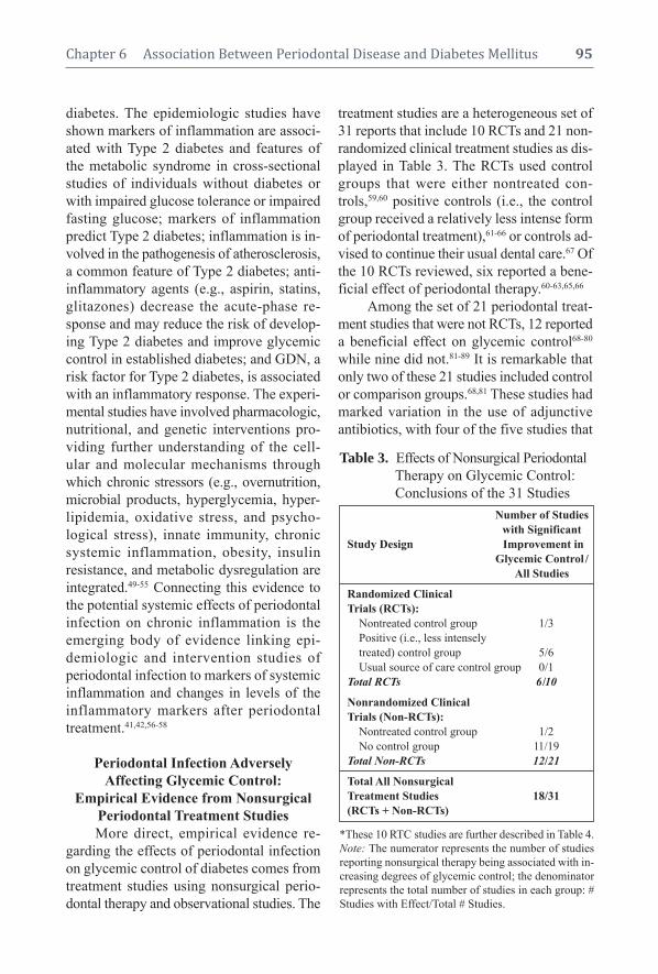
diabetes. The epidemiologic studies have
shown markers of inflammation are associ-
ated with Type 2 diabetes and features of
the metabolic syndrome in cross-sectional
studies of individuals without diabetes or
with impaired glucose tolerance or impaired
fasting glucose; markers of inflammation
predict Type 2 diabetes; inflammation is in-
volved in the pathogenesis of atherosclerosis,
a common feature of Type 2 diabetes; anti-
inflammatory agents (e.g., aspirin, statins,
glitazones) decrease the acute-phase re-
sponse and may reduce the risk of develop-
ing Type 2 diabetes and improve glycemic
control in established diabetes; and GDN, a
risk factor for Type 2 diabetes, is associated
with an inflammatory response. The experi-
mental studies have involved pharmacologic,
nutritional, and genetic interventions pro-
viding further understanding of the cell-
ular and molecular mechanisms through
which chronic stressors (e.g., overnutrition,
microbial products, hyperglycemia, hyper-
lipidemia, oxidative stress, and psycho -
logical stress), innate immunity, chronic
systemic inflammation, obesity, insulin
resistance, and metabolic dysregulation are
integrated.49-55 Connecting this evidence to
the potential systemic effects of periodontal
infection on chronic inflammation is the
emerging body of evidence linking epi-
demiologic and intervention studies of
perio dontal infection to markers of systemic
inflammation and changes in levels of the
inflammatory markers after periodontal
treatment.41,42,56-58
Periodontal Infection Adversely
Affecting Glycemic Control:
Empirical Evidence from Nonsurgical
Periodontal Treatment Studies
More direct, empirical evidence re-
garding the effects of periodontal infection
on glycemic control of diabetes comes from
treatment studies using nonsurgical perio -
dontal therapy and observational studies. The
treatment studies are a heterogeneous set of
31 reports that include 10 RCTs and 21 non-
randomized clinical treatment studies as dis-
played in Table 3. The RCTs used control
groups that were either nontreated con-
trols,59,60 positive controls (i.e., the control
group received a relatively less intense form
of periodontal treatment),61-66 or controls ad-
vised to continue their usual dental care.67 Of
the 10 RCTs reviewed, six reported a bene-
ficial effect of periodontal therapy.60-63,65,66
Among the set of 21 periodontal treat-
ment studies that were not RCTs, 12 reported
a beneficial effect on glycemic control68-80
while nine did not.81-89 It is remarkable that
only two of these 21 studies included control
or comparison groups.68,81 These studies had
marked variation in the use of adjunctive
anti biotics, with four of the five studies that
Chapter 6 Association Between Periodontal Disease and Diabetes Mellitus 95
Table 3. Effects of Nonsurgical Periodontal
Table 1. Therapy on Glycemic Control:
Table 1. Conclusions of the 31 Studies
*These 10 RTC studies are further described in Table 4.
Note: The numerator represents the number of studies
reporting nonsurgical therapy being associated with in-
creasing degrees of glycemic control; the denominator
represents the total number of studies in each group: #
Studies with Effect/Total # Studies.
Number of Studies
with Significant
Study Design Improvement in
Glycemic Control /
All Studies
Randomized Clinical
Trials (RCTs):
Nontreated control group 1/3
Positive (i.e., less intensely
treated) control group 5/6
Usual source of care control group 0/1
Total RCTs 6/10
Nonrandomized Clinical
Trials (Non-RCTs):
Nontreated control group 1/2
No control group 11/19
Total Non-RCTs 12/21
Total All Nonsurgical
Treatment Studies 18/31
(RCTs + Non-RCTs)

used systemic antibiotics reporting a bene -
ficial effect on glycemic control71,74,76,79 and
one finding a numerical decrease in the
HbA1c level that did not reach statistical
significance.81
For the body of literature consisting of
both RCTs and other types of studies, there
is marked hetereogeneity in the studies’
designs, geographic locations, source popu-
lations, conduct, length of follow-up for
glycemic control assessment, types of par-
ticipants and their baseline periodontal dis-
ease and glycemic control status, inclusion
of control groups, periodontal treatment pro-
tocols, sample size and power to detect dif-
ferences in periodontal and metabolic re-
sponse, and specific hypotheses tested.90 The
details of the variation in this body of lit -
erature have been extensively described in
several reviews.43,91,92
Table 4 provides additional informa-
tion about the RCTs summarized in Table 3.
For each study, information on the number of
participants and the type of diabetes included
96 Periodontal Disease and Overall Health: A Clinician's Guide
is given, along with indication of whether
antibiotics were used in conjunction with the
treatment. The types of antibiotics used and
information on their dispensing mode ( local,
systemic, rinse) can be seen in the table’s foot -
note marked with one asterisk (*). Finally,
in the far right column is the outcome
achieved, indicating whether the study treat-
ment resulted in improved glycemic control
as measured by a decrease in the level of
HbA1c.
One remarkable observation is the rel-
atively small numbers of participants in
some of the studies. This is mostly due to the
immense resources required for conducting
clinical trials, especially those that have a
long follow-up period. Also, it is difficult to
identify individuals meeting inclusion crite-
ria and those free of exclusion criteria, and to
recruit, enroll, and keep participants return-
ing for all study visits that typically are of
long duration due to their many components
(e.g., interview, clinical examination, blood
draw, radiographic exposure, and initial
Table 4. Effects of Nonsurgical Periodontal Therapy on Glycemic Control (HbA1c):Table 1. Descriptions and Conclusions of the 10 Randomized Clinical Trials (RTCs)Table 1. Summarized in Table 3
*Adjunctive antibiotic types: a) systemic doxycycline, b) amoxicillin and augmentin, c) minocycline, locally delivered.**The group not receiving antibiotic showed greater improvement.
Adjunctive Statistically Significant
Clinical Study Design by Sample Diabetes Antibiotics Beneficial Effect
Type of Control Group Size Type Used* on HbA1c
RCT with Nontreated Control Group:
Aldridge et al. study 1 (1995)59 31 1 No No
Aldridge et al. study 2 (1995)59 22 1 No No
Kiran et al. study 1 (2005)60 44 2 No Yes
RCT with Positive (i.e., Less
Intensely Treated) Control Group:
Grossi et al. (1997)61.1130 2 aYesa Yes
Al-Mubarak et al. (2002)64 78 1,2 No No
Rodrigues et al. (2003)62 30 2 aYesb **Yes**
Skaleric et al. (2004)63 20 1 aYesc Yes
Yun et al. (2007)66 46 2 aYesa Yes
O’Connell et al. (2008)65 30 2 aYesa Yes
RCT with Usual Source
of Care-Control Group:
Jones et al. (2007)67 1650 2 aYesa No

therapy and maintenance visits). In addition,
clinical studies typically last several months
or years.
An important source of variation in the
RCTs is the use of adjunctive antibiotics with
the nonsurgical periodontal therapy. Among
the six RCTs that included adjunctive anti -
biotics, five used the antibiotics systemi-
cally61,62,65-67 and one was locally delivered.63
Five of the six RCTs using antibiotics showed
beneficial effects on glycemic control.61-63,65,66
However, it is important to note the signifi-
cant improvement for one study was in the
positive control group that did not receive the
systemic antibiotic.62 Also, one of the six
RCTs reporting a beneficial effect did not
use any antibiotics.60 Hence, to date there is
suggestive, but not clear-cut evidence to sup-
port the use of antibiotics in combination
with nonsurgical periodontal treatment in
order to observe an improvement in glycemic
control.
Periodontal Infection Adversely
Affecting Glycemic Control: Empirical
Evidence from Observational Studies
Additional evidence to support the
effect of periodontitis on increased risk for
poorer glycemic control comes from a small
number of observational studies. A longitu-
dinal epidemiologic study of the Pima Indi-
ans in Arizona found at baseline that subjects
who had Type 2 diabetes, that was in good to
moderate control, but also had severe perio -
dontitis, were approximately six times more
likely to have poor glycemic control at a
two-year follow-up than those who did not
have severe periodontitis at baseline.93 In an-
other observational study of 25 adults ages
58 to 77 with Type 2 diabetes, Collin et al.
also reported an association between ad-
vanced periodontal disease and impaired
metabolic control.94
Finally, two cross-sectional studies
report findings consistent with poorer peri-
odontal health associated with poorer
Chapter 6 Association Between Periodontal Disease and Diabetes Mellitus 97
glycemic control. One of these studies,
which included 127 pregnant women with
diabetes, reported that those with perio -
dontitis were more likely to have poorer
glycemic control, after controlling for pres-
ence of a urinary tract infection and/or cer-
vicovaginal infection and a measure of com-
pliance to recommended medical treatment
for the diabetes.95 The other study, analyzing
the association between number of bleed-
ing sites and degree of glycemic control
(HbA1c), reported a statistically significant
association, i.e., the HbA1c values increased
(poorer control with higher HbA1c) as the
number of bleeding sites increased. How-
ever, the authors determined this result
would have no clinical significance96 be-
cause the measure of association between
the number of bleeding sites and glycemic
control was very small.
Periodontal Infection as a Potential Risk
Factor for Diabetes Complications:
Empirical Evidence from
Observational Studies
It is well recognized that poor glycemic
control is a major determinant for the devel-
opment of the chronic complications of dia-
betes. Results from the landmark Diabetes
Control and Complications Trial (involving
Type 1 diabetes) and the UK Prospective Di-
abetes Study (UKPDS, involving Type 2 di-
abetes) demonstrated that attaining and
maintaining good glycemic control could re-
duce the risk for and slow the progression of
microvascular complications in patients with
Type 1 and Type 2 diabetes.97-99 Additionally,
the UKPDS observed a 16% reduction
(p = 0.052) in the risk of combined fatal or
nonfatal myocardial infarction and sudden
death. Further epidemiologic analysis from
the UKPDS showed a continuous association
between the risk of cardiovascular compli-
cations and glycemia; every percentage point
decrease in HbAlc (e.g., 9% to 8%) was
associated with a 25% reduction in diabetes-

related deaths, 7% reduction in all-cause
mortality, and 18% reduction in combined
fatal and nonfatal myocardial infarction.100
There is emerging evidence from ob -
ser vational studies regarding the association
between periodontal disease and the risk for
diabetes complications. Thorstensson and
colleagues101 studied 39 case-control pairs
of individuals with Type 1 and Type 2 dia-
betes for six years’ median follow-up time in
Jönköping, Sweden. In each pair, the cases
had severe alveolar bone loss and controls
had gingivitis or minor alveolar bone loss.
They found that cases were significantly
more likely to have prevalent proteinuria
and cardiovascular complications, including
stroke, transient ischemic attacks, angina,
myo cardial infarction, and intermittent clau-
dication than controls at their follow-up
medical assessments.
Two reports from the ongoing longitu-
dinal study of diabetes and its complications
in the Gila River Indian Community in Ari-
zona, conducted by the National Institute of
Diabetes and Digestive and Kidney Dis-
eases, address nephropathy and cardiovas-
cular disease. Saremi and colleagues102 stud-
ied a cohort of 628 individuals for a median
follow-up time of 11 years. Individuals with
severe periodontal disease had 3.2 times
greater risk for cardio-renal mortality (i.e.,
ischemic heart disease and diabetic nephrop -
athy combined) than those with no, mild, or
moderate periodontal disease. This estimate
of significantly greater risk persisted while
controlling for several major risk factors of
cardio-renal mortality, including age, sex,
diabetes duration, HbA1c, body mass index,
hypertension, blood glucose, cholesterol,
electrocardiographic abnormalities, macro -
albuminuria, and smoking.
In the second report, Shultis and col-
leagues103 investigated the effect of perio -
dontitis on risk for development of overt
nephropathy (macroalbuminuria) and ESRD
in a group of 529 Gila River Indian Com-
munity adults with Type 2 diabetes. Their
proportional hazards model analyses, adjusted
for age, sex, diabetes duration, body mass
index, and smoking, indicated periodontitis
and edentulism were significantly associated
with the risk of developing overt nephrop -
athy and ESRD. The incidence of macroal-
buminuria was 2.0, 2.1, and 2.6 times greater
in individuals with moderate or severe perio -
dontitis or in those who were edentulous,
respectively, than those with none/mild perio -
dontitis. The incidence of ESRD was also
2.3, 3.5, and 4.9 times greater for individu-
als with moderate or severe periodontitis or
for those who were edentulous, respectively,
than those with none/mild periodontitis.
Periodontal Infection as a Potential Risk
Factor for Development of Diabetes:
Empirical Evidence from
Observational Studies
In addition to evidence supporting pe-
riodontal disease as a potential risk factor
for developing diabetes complications, there
is also evidence emerging that periodontal
disease may be a risk factor for the develop-
ment of Type 2 diabetes and possibly GDM.
Demmer and colleagues104 investigated the
association between periodontal disease and
the development (i.e., incidence) of new di-
abetes cases in a representative sample of
the US population, analyzing data from the
first National Health and Nutrition Exami-
nation Survey (NHANES I) and its Epi-
demiologic Follow-up Study (NHEFS). The
average follow-up period for the 9,296
individuals in the analysis was 17 years, for
the period between 1971 to 1992. The
study used a cohort study design because
information on the presence or absence of
perio dontal disease (i.e., the hypothesized
causal factor) was known at the time the
study began, and the outcome (development
of diabetes) was assessed subsequently. This
study concluded that having periodontal
disease was significantly associated with a
98 Periodontal Disease and Overall Health: A Clinician's Guide

50–100% greater risk for developing Type 2
diabetes, after controlling for other estab-
lished risk factors for diabetes. The greater
risk for diabetes was also consistent with
previous research using NHANES I and
NHEFS, in which risk factors for diabetes
that did not include perio dontal disease, for
example, measures of adiposity (body mass
index and subscapular skinfold thickness),
having hypertension, and increased age, were
also statistically significant.105
Finally, Dasanayake and colleagues106
investigated whether or not pregnant women
who develop GDM, compared to pregnant
women who do not develop GDM, had
poorer clinical perio dontal health and/or
demonstrated higher levels of other bio -
logical markers of perio dontal disease ap -
proximately two months before their GDM
diagnosis. The other biological markers
included bacterio logical (in dental plaque and
cervicovaginal samples), immunological, and
periodontitis-related inflammatory mediator
analytes. This study found that women who
had higher levels of Tannerella forsythia (T.
forsythia), a recognized perio dontal pathogen,
in the vaginal flora were statistically signifi -
cantly more likely to develop GDM than those
women with lower levels. The Dasanayake
et al. study concluded that T. forsythia in the
vaginal flora is a potential risk factor for GDM.
SUMMARy AND CONCLUSIONS
The evidence reviewed in this chapter
supports the conclusion that a bi-directional
association exists between diabetes mellitus
and periodontal health: Diabetes is associ-
ated with increased development and pro-
gression of periodontitis, and periodontal
infection is associated with poorer glycemic
control in people with diabetes.
There is also evidence emerging that
gestational diabetes may adversely affect
periodontal health.
Additionally, evidence is emerging to
suggest that periodontal disease is asso ciated
with increased risk for diabetes complica-
tions, the development of Type 2 diabetes,
and perhaps the development of GDM.
While treating periodontal infection in
people with diabetes is clearly an important
component in maintaining oral health, it may
also play an important role in establishing
and maintaining glycemic control, and pos-
sibly in delaying the onset or progression of
diabetes and its complications.
Therefore, dental health professionals
might fulfill an important role in maintaining
or improving the health, and ultimately the
quality of life, of individuals with diabetes
and GDM, as well as aiding in lessening the
immense burden of diabetes and periodontal
diseases on our society in general.
Supplemental Readings
Darre L, Vergnes JN, Gourdy P, Sixou M. Efficacy of
periodontal treatment on glycaemic control in diabetic
patients: A meta-analysis of interventional studies.
Diabetes Metab 2008;34:497-506.
Demmer RT, Jacobs DR Jr, Desvarieux M. Periodontal
disease and incident Type 2 diabetes: results from the
First National Health and Nutrition Examination Survey
and its epidemiologic follow-up study. Diabetes Care
2008;31:1373-1379.
Graves DT, Liu R, Oates TW. Diabetes-enhanced in-
flammation and apoptosis: impact on periodontal patho-
sis. Periodontology 2000 2007;45:128-137.
King GL. The role of inflammatory cytokines in dia-
betes and its complications. J Periodontol 2008;79:
1527-1534.
Mealey BL, Ocampo GL. Diabetes mellitus and perio -
dontal disease. Periodontol 2000 2007;44:127-153.
Taylor GW, Borgnakke WS. Periodontal disease: asso-
ciations with diabetes, glycemic control and complica-
tions. Oral Dis 2008;14:191-203.
REFERENCES1. Elwood J. Critical Appraisal of Epidemiological
Studies and Clinical Trials. New York: Oxford Uni-
versity Press; 1998.
2. Mealey BL, Ocampo GL. Diabetes mellitus and
periodontal disease. Periodontol 2000 2007;44:
127-153.
Chapter 6 Association Between Periodontal Disease and Diabetes Mellitus 99

3. Lamster IB, Lalla E, Borgnakke WS, Taylor GW.
The relationship between oral health and diabetes
mellitus. JADA 2008;139(Suppl):19S–24S.
4. Taylor GW, Borgnakke WS. Periodontal disease:
associations with diabetes, glycemic control and
complications. Oral Dis 2008;14:191–203.
5. Taylor GW, Borgnakke WS. Treatment of estab-
lished complications: periodontal disease. In:
Herman W, Kinmouth AL, Wareham NJ, Williams
R, eds. The Evidence Base in Diabetes Care, 2nd
edition. Chichester, The United Kingdom: John
Wiley & Sons, 2009.
6. Graves DT, Liu R, Alikhani M, Al-Mashat H,
Trackman PC. Diabetes-enhanced inflammation
and apoptosis—impact on periodontal pathology. J
Dent Res 2006;85:15–21.
7. Silva JA, Lorencini M, Reis JR, Carvalho HF,
Cagnon VH, Stach-Machado DR. The influence of
type I diabetes mellitus in periodontal disease in-
duced changes of the gingival epithelium and con-
nective tissue. Tissue Cell 2008;40:283–292.
8. Bissada NF, Schaffer EM, Lazarow A. Effect of
alloxan diabetes and local irritating factors on the
periodontal structures of the rat. Periodontics 1966;
4:233–240.
9. Gyurko R, Siqueira CC, Caldon N, Gao L, Kantarci
A, Van Dyke TE. Chronic hyperglycemia predis-
poses to exaggerated inflammatory response and
leukocyte dysfunction in Akita mice. J Immunol
2006;177:7250–7256.
10. Lu H, Raptis M, Black E, Stan M, Amar S, Graves
DT. Influence of diabetes on the exacerbation of an
inflammatory response in cardiovascular tissue.
Endocrinology 2004;145:4934–4939.
11. Graves DT, Fine D, Teng YT, Van Dyke TE, Hajish -
engallis G. The use of rodent models to investigate
host-bacteria interactions related to perio dontal dis-
eases. J Clin Periodontol 2008;35:89–105.
12. Salvi GE, Collins JG, Yalda B, Arnold RR, Lang
NP, Offenbacher S. Monocytic TNF alpha secretion
patterns in IDDM patients with periodontal dis-
eases. J Clin Periodontol 1997;24:8–16.
13. Omori K, Ohira T, Uchida Y, Ayilavarapu S, Batista
EL Jr, Yagi M, Iwata T, Liu H, Hasturk H, Kantarci
A, Van Dyke TE. Priming of neutrophil oxidative
burst in diabetes requires preassembly of the
NADPH oxidase. J Leukoc Biol 2008;84:292–301.
14. Graves DT, Naguib G, Lu H, Leone C, Hsue H,
Krall E. Inflammation is more persistent in type 1
diabetic mice. J Dent Res 2005;84:324–328.
15. Naguib G, Al-Mashat H, Desta T, Graves DT. Dia-
betes prolongs the inflammatory response to a bac-
terial stimulus through cytokine dysregulation. J In-
vest Dermatol 2004;123:87–92.
16. Liu R, Bal HS, Desta T, Krothapalli N, Alyassi M,
Luan Q, Graves DT. Diabetes enhances periodon-
tal bone loss through enhanced resorption and
diminished bone formation. J Dent Res 2006;85:
510–514.
17. Parfitt AM. The coupling of bone formation to bone
resorption: a critical analysis of the concept and of
its relevance to the pathogenesis of osteoporosis.
Metab Bone Dis Relat Res 1982;4:1–6.
18. Al-Mashat HA, Kandru S, Liu R, Behl Y, Desta T,
Graves DT. Diabetes enhances mRNA levels of
proapoptotic genes and caspase activity, which con-
tribute to impaired healing. Diabetes 2006;55:
487–495.
19. Liu R, Bal HS, Desta T, Behl Y, Graves DT. Tumor
necrosis factor-alpha mediates diabetes-enhanced
apoptosis of matrix-producing cells and impairs
diabetic healing. Am J Pathol 2006;168:757–764.
20. Lalla E, Lamster IB, Feit M, Huang L, Spessot A,
Qu W, Kislinger T, Lu Y, Stern DM, Schmidt AM.
Blockade of RAGE suppresses periodontitis-asso-
ciated bone loss in diabetic mice. J Clin Invest
2000;105:1117–1124.
21. Ohnishi T, Bandow K, Kakimoto K, Machigashira
M, Matsuyama T, Matsuguchi T. Oxidative stress
causes alveolar bone loss in metabolic syndrome
model mice with type 2 diabetes. J Periodontal
Res 2009;44:43-51.
22. Mahamed DA, Marleau A, Alnaeeli M, Singh B,
Zhang X, Penninger JM, Teng YT. G(-) anaerobes-
reactive CD4+ T-cells trigger RANKL-mediated
enhanced alveolar bone loss in diabetic NOD mice.
Diabetes 2005;54:1477-1486.
23. Ryan ME, Ramamurthy NS, Sorsa T, Golub LM.
MMP-mediated events in diabetes. Ann NY Acad
Sci 1999;878:311-334.
24. Cutler CW, Eke P, Arnold RR, Van Dyke TE. De-
fective neutrophil function in an insulin-dependent
diabetes mellitus patient. A case report. J Perio -
dontol 1991;62:394-401.
25. Iacopino AM. Diabetic periodontitis: possible lipid-
induced defect in tissue repair through alteration of
macrophage phenotype and function. Oral Dis
1995;1:214–229.
26. Iacopino AM. Periodontitis and diabetes interre -
lationships: role of inflammation. Ann Periodontol
2001;6:125–137.
27. Cutler CW, Iacopino AM. Periodontal disease: links
with serum lipid/ triglyceride levels? Review and
new data. J Int Acad Periodontol 2003;5:47–51.
28. Ihm SH, Yoo HJ, Park SW, Park CJ. Effect of tol-
restat, an aldose reductase inhibitor, on neutrophil
respiratory burst activity in diabetic patients.
Metabolism 1997;46:634-638.
29. Tebbs SE, Lumbwe CM, Tesfaye S, Gonzalez AM,
Wilson RM. The influence of aldose reductase on
100 Periodontal Disease and Overall Health: A Clinician's Guide

the oxidative burst in diabetic neutrophils. Dia-
betes Res Clin Pract 1992;15:121–129.
30. Boland OM, Blackwell CC, Clarke BF, Ewing DJ.
Effects of ponalrestat, an aldose reductase inhibitor,
on neutrophil killing of Escherichia coli and auto-
nomic function in patients with diabetes mellitus.
Diabetes 1993;42:336–340.
31. Stosic-Grujicic SD, Miljkovic DM, Cvetkovic ID,
Maksimovic-Ivanic DD, Trajkovic V. Immuno -
suppressive and anti-inflammatory action of anti -
oxidants in rat autoimmune diabetes. J Autoimmun
2004;22:267–276.
32. Koya D, King G. Protein kinase C activation and
the development of diabetic complications. Dia-
betes 1998;47:859–866.
33. Hiramatsu Y, Sekiguchi N, Hayashi M, Isshiki K,
Yokota T, King GL, Loeken MR. Diacylglycerol
production and protein kinase C activity are in-
creased in a mouse model of diabetic embryopathy.
Diabetes 2002;51:2804-2810.
34. Cohen MP, Shea E, Chen S, Shearman CW. Gly-
cated albumin increases oxidative stress, activates
NF-kappa B and extracellular signal-regulated
kinase (ERK), and stimulates ERK-dependent
transforming growth factor-beta 1 production in
macrophage RAW cells. J Lab Clin Med 2003;
141:242–249.
35. Rashid G, Luzon AA, Korzets Z, Klein O, Zeltzer
E, Bernheim J. The effect of advanced glycation
end-products and aminoguanidine on TNF alpha
production by rat peritoneal macrophages. Perit
Dial Int 2001;21:122–129.
36. Alikhani Z, Alikhani M, Boyd CM, Nagao K,
Trackman PC, Graves DT. Advanced glycation end
products enhance expression of pro-apoptotic genes
and stimulate fibroblast apoptosis through cyto-
plasmic and mitochondrial pathways. J Biol Chem
2005;280:12087–12095.
37. Alikhani M, Maclellan CM, Raptis M, Vora S,
Trackman PC, Graves DT. Advanced glycation end
products induce apoptosis in fibroblasts through
activation of ROS, MAP kinases, and the FOXO1
transcription factor. Am J Physiol Cell Physiol
2007;292:C850–856.
38. Goova MT, Li J, Kislinger T, Qu W, Lu Y, Buccia-
relli LG, Nowygrod S, Wolf BM, Caliste X, Yan SF,
Stern DM, Schmidt AM. Blockade of receptor for
advanced glycation end-products restores effective
wound healing in diabetic mice. Am J Pathol
2001;159:513–525.
39. King GL. The role of inflammatory cytokines in
diabetes and its complications. J Periodontol 2008;
79:1527–1534.
40. Phipps KR, Stevens VJ. Relative contribution of
caries and periodontal disease in adult tooth loss for
an HMO dental population. J Public Health Dent
1995;55:250–252.
41. Loos BG. Systemic markers of inflammation in
periodontitis. J Periodontol 2005;76 (Suppl):2106–
2115.
42. Kshirsagar AV, Moss KL, Elter JR, Beck JD, Offen -
bacher S, Falk RJ. Periodontal disease is associated
with renal insufficiency in the Atherosclerosis Risk
In Communities (ARIC) study. Am J Kidney Dis
2005;45:650–657.
43. Grossi SG, Genco RJ. Periodontal disease and
diabetes mellitus: a two-way relationship. Ann
Perio dontol 1998;3:51–61.
44. Feingold KR, Soued M, Serio MK, Moser AH,
Dinarello CA, Grunfeld C. Multiple cytokines stim-
ulate hepatic lipid synthesis in vivo. Endocrinology
1989;125:267–274.
45. Ling PR, Istfan NW, Colon E, Bistrian BR. Differ-
ential effects of interleukin-1 receptor antagonist in
cytokine- and endotoxin-treated rats. Am J Physiol
1995;268:E255–E261.
46. Feingold KR, Grunfeld C. Role of cytokines in in-
ducing hyperlipidemia. Diabetes 1992;41 (Suppl):
97–101.
47. Grunfeld C, Soued M, Adi S, Moser AH, Dinarello
CA, Feingold KR. Evidence for two classes of
cytokines that stimulate hepatic lipogenesis: rela-
tionships among tumor necrosis factor, interleukin-1
and interferon-alpha. Endocrinology 1990;127:
46–54.
48. Shoelson SE, Lee J, Goldfine AB. Inflammation
and insulin resistance. J Clin Invest 2006;116:
1793–1801.
49. Tataranni PA, Ortega E. A burning question: does
an adipokine-induced activation of the immune
system mediate the effect of overnutrition on type
2 diabetes? Diabetes 2005;54:917–927.
50. Shoelson SE, Herrero L, Naaz A. Obesity, inflam-
mation, and insulin resistance. Gastroenterology
2007;132:2169–2180.
51. Duncan BB, Schmidt MI. The epidemiology of
low-grade chronic systemic inflammation and type
2 diabetes. Diabetes Technol Ther 2006;8:7–17.
52. Pickup JC. Inflammation and activated innate im-
munity in the pathogenesis of type 2 diabetes.
Diabetes Care 2004;27:813–823.
53. Wellen KE, Hotamisligil GS. Inflammation, stress,
and diabetes. J Clin Invest 2005;115:1111–1119.
54. Hotamisligil GS. Inflammation and metabolic dis-
orders. Nature 2006;444:860–867.
55. Pradhan A. Obesity, metabolic syndrome, and type
2 diabetes: inflammatory basis of glucose meta-
bolic disorders. Nutr Rev 2007;65:S152–S156.
56. Beck JD, Offenbacher S. Systemic effects of perio -
dontitis: epidemiology of periodontal disease and
Chapter 6 Association Between Periodontal Disease and Diabetes Mellitus 101

cardiovascular disease. J Periodontol 2005;76
(Suppl):2089–2100.
57. Demmer RT, Desvarieux M. Periodontal infections
and cardiovascular disease: the heart of the matter.
JADA 2006;137(Suppl):14S–20S.
58. Tonetti MS, D’Aiuto F, Nibali L, Donald A, Storry
C, Parkar M, Suvan J, Hingorani AD, Vallance P,
Deanfield J. Treatment of periodontitis and en-
dothelial function. New Engl J Med 2007;356:
911–920.
59. Aldridge JP, Lester V, Watts TL, Collins A, Viberti
G, Wilson RF. Single-blind studies of the effects of
improved periodontal health on metabolic control
in type 1 diabetes mellitus. J Clin Periodontol
1995;22:271–275.
60. Kiran M, Arpak N, Unsal E, Erdoğan MF. The ef-
fect of improved periodontal health on metabolic
control in type 2 diabetes mellitus. J Clin Perio -
dontol 2005;32:266–272.
61. Grossi SG, Skrepcinski FB, DeCaro T, Robertson
DC, Ho AW, Dunford RG, Genco RJ. Treatment of
periodontal disease in diabetics reduces glycated
hemoglobin. J Periodontol 1997;68:713–719.
62. Rodrigues DC, Taba MJ, Novaes AB, Souza SL,
Grisi MF. Effect of non-surgical periodontal ther-
apy on glycemic control in patients with type 2 dia -
betes mellitus. J Periodontol 2003;74:1361–1367.
63. Skaleric U, Schara R, Medvescek M, Hanlon A,
Doherty F, Lessem J. Periodontal treatment by
Arestin and its effects on glycemic control in type
1 diabetes patients. J Int Acad Periodontol 2004;
6(Suppl):160-165.
64. Al-Mubarak S, Ciancio S, Aljada A, Mohanty P,
Ross C, Dandona P. Comparative evaluation of ad-
junctive oral irrigation in diabetics. J Clin Perio -
dontol 2002;29:295-300.
65. O’Connell PA, Taba M, Nomizo A, Foss Freitas
MC, Suaid FA, Uyemura SA, Trevisan GL, No-
vaes AB, Souza SL, Palioto DB, Grisi MF. Effects
of periodontal therapy on glycemic control and in-
flammatory markers. J Periodontol 2008;79:774–
783.
66. Yun F, Firkova EI, Jun-Qi L, Xun H. Effect of non-
surgical periodontal therapy on patients with type
2 diabetes mellitus. Folia Med (Plovdiv) 2007;49:
32–36.
67. Jones JA, Miller DR, Wehler CJ, Rich SE, Krall-
Kaye EA, McCoy LC, Christiansen CL, Rothendler
JA, Garcia RI. Does periodontal care improve
glycemic control? The Department of Veterans
Affairs Dental Diabetes Study. J Clin Periodontol
2007;34:46–52.
68. Stewart JE, Wager KA, Friedlander AH, Zadeh
HH. The effect of periodontal treatment on glycemic
control in patients with type 2 diabetes mellitus. J
Clin Periodontol 2001;28:306–310.
69. Faria-Almeida R, Navarro A, Bascones A. Clinical
and metabolic changes after conventional treat-
ment of type 2 diabetic patients with chronic perio -
dontitis. J Periodontol 2006;77:591–598.
70. Iwamoto Y, Nishimura F, Nakagawa M, Sugimoto
H, Shikata K, Makino H, Fukuda T, Tsuji T,
Iwamoto M, Murayama Y. The effect of antimi-
crobial periodontal treatment on circulating tumor
necrosis factor-alpha and glycated hemoglobin
level in patients with type 2 diabetes. J Periodon-
tol 2001;72:774–778.
71. Miller LS, Manwell MA, Newbold D, Reding ME,
Rasheed A, Blodgett J, Kornman KS. The rela-
tionship between reduction in periodontal inflam-
mation and diabetes control: a report of 9 cases. J
Periodontol 1992;63:843–848.
72. Seppälä B, Ainamo J. A site-by-site follow-up study
on the effect of controlled versus poorly controlled
insulin-dependent diabetes mellitus. J Clin Perio -
dontol 1994;21:161–165.
73. Seppälä B, Seppälä M, Ainamo J. A longitudinal
study on insulin-dependent diabetes mellitus and
periodontal disease. J Clin Periodontol 1993;20:
161–165.
74. Williams RC, Jr, Mahan CJ. Periodontal disease
and diabetes in young adults. JAMA 1960;172:776–
778.
75. Wolf J. Dental and periodontal conditions in dia-
betes mellitus. A clinical and radiographic study.
Proc Finn Dent Soc 1977;73 (Suppl):1–56.
76. Fu Y, Ling J, Deng Y. [The effect of non-surgical
periodontal therapy on blood sugar level of non-in-
sulin dependent diabetes mellitus diabetics with
periodontitis]. Zhonghua Kou Qiang Yi Xue Za Zhi
2000;35:444–446.
77. Guo YH, Zhu BL. [The effect of initial therapy on
periodontal status and saccharified Hb (HbAIc) of
patients with type II diabetes mellitus]. Shanghai
Kou Qiang Yi Xue 2004;13:150–151.
78. Navarro-Sanchez AB, Faria-Almeida R, Bascones-
Martinez A. Effect of non-surgical periodontal ther-
apy on clinical and immunological response and
glycaemic control in type 2 diabetic patients with
moderate periodontitis. J Clin Periodontol 2007;
34:835–843.
79. Yang PS, Wang Y, Qi XM, Ren JM, Ge SH. [The
effect of periodontal initial therapy on circulating
TNF-alpha and HbA1C in type 2 diabetes patients
with periodontitis]. Zhonghua Kou Qiang Yi Xue Za
Zhi 2003;38:364–366.
80. Villa AA. [Clinical, biochemical and histopatho log-
ical correlation in diabetic patients with perio dontal
disease]. Rev Fac Cien Med Univ Nac Cordoba 2006;
63 (Suppl):50–55.
102 Periodontal Disease and Overall Health: A Clinician's Guide

81. Promsudthi A, Pimapansri S, Deerochanawong C,
Kanchanavasita W. The effect of periodontal ther-
apy on uncontrolled type 2 diabetes mellitus in
older subjects. Oral Dis 2005;11:293–298.
82. Christgau M, Palitzsch KD, Schmalz G, Kreiner U,
Frenzel S. Healing response to non-surgical perio -
dontal therapy in patients with diabetes mellitus:
clinical, microbiological, and immunologic results.
J Clin Periodontol 1998;25:112–124.
83. Smith GT, Greenbaum CJ, Johnson BD, Persson
GR. Short-term responses to periodontal therapy in
insulin-dependent diabetic patients. J Periodontol
1996;67:794–802.
84. Talbert J, Elter J, Jared HL, Offenbacher S, Souther-
land J, Wilder RS. The effect of periodontal therapy
on TNF-alpha, IL-6 and metabolic control in type
2 diabetics. J Dent Hyg 2006;80:7.
85. Westfelt E, Rylander H, Blohmé G, Jonasson P,
Lindhe J. The effect of periodontal therapy in dia-
betics. Results after 5 years. J Clin Periodontol
1996;23:92–100.
86. Campus G, Salem A, Sacco G, Maida C, Cagetti
MG, Tonolo G. Clinical effects of mechanical
periodontal therapy in type 2 diabetic patients.
Diabetes Res Clin Pract 2007;75:368–369.
87. da Cruz GA, de Toledo S, Sallum EA, Sallum AW,
Ambrosano GM, de Cássia Orlandi Sardi J, da Cruz
SE, Gonçalves RB. Clinical and laboratory evalu-
ations of non-surgical periodontal treatment in sub-
jects with diabetes mellitus. J Periodontol 2008;79:
1150–1157.
88. Makiura N, Ojima M, Kou Y, Furuta N, Okahashi
N, Shizukuishi S, Amano A. Relationship of Por-
phyromonas gingivalis with glycemic level in pa-
tients with type 2 diabetes following periodontal
treatment. Oral Microbiol Immunol 2008;23:
348–351.
89. Schara R, Medvescek M, Skaleric U. Periodontal
disease and diabetes metabolic control: a full-mouth
disinfection approach. J Int Acad Periodontol 2006;
8:61–66.
90. Taylor GW. The effects of periodontal treatment on
diabetes. JADA 2003;134 (Spec Iss):41S–48S.
91. Darre L, Vergnes JN, Gourdy P, Sixou M. Efficacy
of periodontal treatment on glycaemic control in
diabetic patients: A meta-analysis of interventional
studies. Diabetes Metab 2008;34:497–506.
92. Janket SJ, Wightman A, Baird AE, Van Dyke TE,
Jones JA. Does periodontal treatment improve
glycemic control in diabetic patients? A meta-
analysis of intervention studies. J Dent Res 2005;
84:1154–1159.
93. Taylor GW, Burt BA, Becker MP, Genco RJ, Shloss-
man M, Knowler WC, Pettitt DJ. Severe periodon-
titis and risk for poor glycemic control in patients
with non-insulin-dependent diabetes mellitus. J Pe-
riodontol 1996;67(Suppl):1085–1093.
94. Collin HL, Uusitupa M, Niskanen L, Kontturi-
Närhi V, Markkanen H, Koivisto AM, Meurman
JH. Periodontal findings in elderly patients with
non-insulin dependent diabetes mellitus. J Perio -
dontol 1998;69:962–966.
95. Diaz-Romero RM, Casanova-Román G, Beltrán-
Zuñiga M, Belmont-Padilla J, Méndez JD, Avila-
Rosas H. Oral infections and glycemic control in
pregnant type 2 diabetics. Arch Med Res 2005;36:
42–48.
96. Lal S, Cheng B, Kaplan S, Softness B, Greenberg
E, Goland RS, Lalla E, Lamster IB. Gingival bleed-
ing in 6- to 13-year-old children with diabetes mel-
litus. Pediatr Dent 2007;29:426–430.
97. Anonymous. The effect of intensive treatment of
dia betes on the development and progression of
long-term complications in insulin-dependent dia-
betes mellitus. The Diabetes Control and Compli-
cations Trial Research Group. N Engl J Med
1993;329:977–986.
98. Anonymous. Intensive blood-glucose control with
sulphonylureas or insulin compared with con -
ventional treatment and risk of complications in
patients with type 2 diabetes (UKPDS 33). UK
Prospective Diabetes Study (UKPDS) Group.
Lancet 1998;352:837–853.
99. Anonymous. Effect of intensive blood-glucose con-
trol with metformin on complications in overweight
patients with type 2 diabetes (UKPDS 34). UK
Prospective Diabetes Study (UKPDS) Group.
Lancet 1998;352:854–865.
00. Genuth S, Eastman R, Kahn R, Klein R, Lachin J,
Lebovitz H, Nathan D, Vinicor F; American Dia-
betes Association. Implications of the United King-
dom prospective diabetes study. Diabetes Care
2003;26 (Suppl):S28–S32.
01. Thorstensson H, Kuylenstierna J, Hugoson A. Med-
ical status and complications in relation to perio -
dontal disease experience in insulin-dependent
diabetics. J Clin Periodontol 1996;23:194–202.
02. Saremi A, Nelson RG, Tulloch-Reid M, Hanson
RL, Sievers ML, Taylor GW, Shlossman M,
Bennett PH, Genco R, Knowler WC. Periodontal
disease and mortality in type 2 diabetes. Diabetes
Care 2005;28:27–32.
03. Shultis WA, Weil EJ, Looker HC, Curtis JM,
Shlossman M, Genco RJ, Knowler WC, Nelson
RG. Effect of periodontitis on overt nephropathy
and end-stage renal disease in type 2 diabetes. Dia -
betes Care 2007;30:306–311.
04. Demmer RT, Jacobs DR, Jr., Desvarieux M. Perio -
dontal disease and incident type 2 diabetes: results
from the First National Health and Nutrition Ex-
Chapter 6 Association Between Periodontal Disease and Diabetes Mellitus 103
1
1
1
1
1

amination Survey and its epidemiologic follow-up
study. Diabetes Care 2008;31:1373–1379.
05. Lipton RB, Liao Y, Cao G, Cooper RS, McGee D.
Determinants of incident non-insulin-dependent
diabetes mellitus among blacks and whites in a
national sample. The NHANES I Epidemiologic
Follow-up Study. Am J Epidemiol 1993;138:
826–839.
06. Dasanayake AP, Chhun N, Tanner AC, Craig RG,
Lee MJ, Moore AF, Norman RG. Periodontal
pathogens and gestational diabetes mellitus. J Dent
Res 2008;87:328–333.
07. Donahue RP, Wu T. Insulin resistance and perio -
dontal disease. An epidemiologic overview of
research needs and future directions. Ann Perio -
dontol 2001;6:119–124.
104 Periodontal Disease and Overall Health: A Clinician's Guide
1
1
1
coronary atherosclerosis range from stable
angina, resulting from ischemia caused by
stable coronary arterial plaques that reduce
luminal cross-sectional area and restrict
blood flow, to acute coronary syndrome
(ACS) characterized by unstable, ruptured
coronary atherosclerotic plaques. ACS can
be further subdivided into non-ST elevation
ACS—such as unstable angina—or ST-
elevation and non-ST elevation myocardial
infarction, depending on the degree of
luminal occlusion following plaque rupture
and intracoronary thrombosis.
Another location for atherosclerotic
disease is in the cerebrovascular bed. As with
coronary atherosclerosis, clinical manifes-
tations of cerebrovascular atherosclerosis
vary depending on the stability of the arterial
plaques. Stable disease may lead to chronic
symptoms of dementia, whereas unstable
atherosclerosis may lead to transient ischemic
attacks (often called “ministrokes”) and
strokes.
Atherosclerotic disease also occurs in
the aorta, manifesting as aortic atheroscle-
rosis and aneurysms, as well as in the periph -
eral arteries. In addition, atherosclerosis is
thought to contribute to valvular heart dis-
eases such as aortic stenosis.2,3
MECHANISMS OF
ATHEROMA FORMATION
Atherosclerotic disease is thought to pro -
gress from microscopic endothelial events
that eventually lead to plaque development,
growth, and rupture. Multiple complex mech-
anisms appear to contribute to the formation
of atheromatous plaques. The initiating
INTRODUCTION
Atherosclerotic disease results from the
pathological formation of plaques in arterial
walls. Plaque development, or atherosclero-
sis, is a complex process with many under-
lying inflammatory molecules participating
in increasingly well-understood pathways.
The consequences include a wide variety
of clinical syndromes involving different or-
gans with a common underlying deficiency
—inadequate blood supply due to reduction
of blood flow to the target organ.
This chapter will review the clinical
manifestations of atherosclerosis and explore
its underlying molecular pathophysiology.
Understanding the underpinnings of athero-
sclerosis is crucial to appreciating the po-
tential interplay between atherosclerosis and
periodontal disease, which is discussed in
the next chapter.
TYPES OF
ATHEROSCLEROTIC DISEASE
Atherosclerosis may occur in any arte-
rial bed with the most clinically relevant
ones being the coronary, cerebrovascular,
and peripheral arterial circulation. Athero-
sclerotic plaques may be stable or unstable,
based on whether or not the plaque has rup-
tured. More recently, studies have shed ad-
ditional light on “vulnerable” atherosclerotic
plaques, which are stable plaques with a
propensity to rupture.
Coronary heart disease is the most clin-
ically evident form of atherosclerotic disease,
affecting over 17 million Americans accord-
ing to the American Heart Association’s
2009 statistical update.1 Manifestations of
CHAPTER 7
Atherosclerosis: A Pervasive Disease Affecting Global Populations
Stanley S. Wang

process involves endothelial injury, which may
occur due to mechanical, biochemical, or
immunological factors. At the cellular level,
the process begins with the recruitment of
monocytes into the arterial wall through the
effect of specific molecules, including the
chemo -attrac tants interleukin (IL)-1 and
tumor necrosis factor-alpha (TNF-).4
Selectins and anti gens on the leukocyte
surface induce move ment of the monocytes.5
Endothelial cellular adhesion molecules (es-
pecially vascular cell adhesion molecule 1
and intercellular adhesion molecule 1) in-
teract with leukocyte integrins, causing the
monocytes to adhere to the endothelium and
subsequently migrate across it.6 After they
move into in a subendo thelial position,
monocytes transform into macro phages and
begin to secrete many inflammatory cyto-
kines, proteases, and metallo proteinases.
Subsequently, the macrophages accu-
mulate and oxidize low-density-lipoprotein
cholesterol (LDLC), becoming the foam
cells that are the precursors of atherosclerotic
plaque.7 C-reactive protein (CRP) appears
to stimulate the process of LDLC uptake by
macrophages,5 which may explain why statin
treatment of patients with elevated CRP lev-
els appears to be clinically beneficial.8 On-
going LDLC deposition and inflammation
leads to plaque growth, and hemodynamic
mechanisms appear to play an important role
as well.9
Plaque rupture may occur at any point in
the process, producing significant clinical
events. It has been estimated that in about
two-thirds of patients with acute coronary
syndromes, the inciting event is rupture of a
coronary plaque that had been less than
50%–70% occlusive.10 Ruptured plaques
have distinctive histopathological features,
including having a fibrous cap that is defi-
cient in collagen and smooth muscle con-
tent, a rich lipid core, and an inflammatory
infiltrate of macrophages and other inflam-
matory cells.11
Role of Inflammation
in Atherosclerotic Disease
Although the precise mechanisms of
atherosclerosis are not completely under-
stood, at the molecular level atherosclerosis
appears to arise from an inflammatory reac-
tion to cardiovascular risk factors.12,13 Chronic
inflammation underlies the processes of plaque
formation and progression, while acute in-
flammatory processes are likely involved in
plaque rupture.
On a molecular level, many mediators
of inflammation have been identified and
more than 50 have been classified by struc-
ture and function. Some of these cytokines or
“protein cell regulators”14 are pro-inflam-
matory, including IL-1, IL-12, IL-18, inter-
feron gamma, and TNF-. Others are anti-
inflammatory, including IL-4, IL-10, IL-13,
and transforming growth factor-beta.
In many cellular mediators of athero-
sclerosis, including macrophages and en-
dothelial cells, pro-inflammatory cytokines
stimulate the nuclear factor-kappa-beta (kb)
pathway, leading to the increased produc-
tion of cellular adhesion molecules, chemo -
kines, growth factors, and other enzymes
such as cyclo-oxygenases and nitric oxide
synthase.15 Enhanced activity of the nuclear
factor-kb pathway appears to correlate with
an increased risk of developing atheroscle-
rotic plaques.
RISK FACTORS FOR
CARDIOVASCULAR AND
CEREBROVASCULAR DISEASES
Traditional risk factors for athero -
sclerotic disease seem to have an associa-
tion with inflammation in common. These
risk factors include dyslipidemia, hyper-
tension, diabetes mellitus, tobacco use, and
adiposity.
Dyslipidemia
Dyslipidemia appears to contribute di-
rectly to atherosclerotic disease, with the risk
106 Periodontal Disease and Overall Health: A Clinician's Guide

CHAPTER 7 Atherosclerosis: A Pervasive Disease Affecting Global Populations 107
of cardiac events rising in direct proportion
to plasma cholesterol levels,16 especially cho-
lesterol particles containing apolipoprotein b
(including LDLC).17 The primacy of choles-
terol in initiating atherosclerosis has been
shown in many molecular studies, but may
be most evident in studies of fetal aortas, in
which quantitative and qualitative analyses
of fatty streaks have shown that native
LDLC uptake in the intima precedes LDLC
oxidation and subsequent endothelial acti-
vation and monocyte recruitment.18 Oxidized
LDLC appears to increase endothelial ex-
pression of cellular adhesion molecules, at-
tracting monocytes and promoting their dif-
ferentiation into macrophages. LDLC also
stimulates the release of pro-inflammatory
cytokines from macrophages. Apolipopro-
tein B particles containing the lipoprotein(a)
moiety appear to be particularly athero-
genic.19,20 In addition, the ratio of apolipopro-
tein B to apolipoprotein A holds promise in
the prognostication of cardiovascular dis-
ease across multiple populations.21
Hypertension
Systemic hypertension contributes by
enhancing monocyte preactivation, increas-
ing production of IL-1 and TNF-,22 and rais-
ing circulating levels of cellular adhesion
molecules.23 Studies have shown a clear re-
lationship between elevated high-sensitivity
CRP levels in normotensive patients and risk
of developing incident hypertension, under-
scoring its inflammatory nature.24
Diabetes Mellitus
Diabetes mellitus appears to be associ-
ated with inflammation, which may explain
its close relationship to atherosclerosis.
Studies have shown that adipose tissue and
macro phages recruited into adipose tissue
release cytokines, including IL-1, IL-6, and
TNF-. These cytokines act on: 1) the liver
to promote dyslipidemia and dysfibrinogen-
emia, 2) adipose tissue to stimulate leptin
secretion, and 3) the pituitary gland to in-
crease secre tion of adrenocorticotropic hor-
mone.25 Moreover, serum levels of various
inflammatory markers and mediators (CRP,
fibrinogen, IL-6, plasminogen activator in-
hibitor 1, and serum amyloid A) correlate
with the degree of hyperglycemia.26 Evi-
dence is accruing to suggest that the forma-
tion and accumulation of advanced glycation
endproducts (AGEs) may be a pathophys -
iologic link between diabetes and cardio -
vascular disease, mediated in part by the
receptor for AGEs (RAGE).27 RAGE, an
immunoglobulin that binds multiple ligands
and spurs production of toxic reactive
oxygen species, is emerging as a potential
therapeutic target for antagonists designed to
reduce atherosclerosis and ischemia-
reperfusion injury.28 Another potential ther-
apeutic target that is attracting interest is pro-
tein kinase C (PKC) beta, an intracellular
signaling molecule that plays a key role in
the development of diabetic complications.29
Experimental PKC inhibitors have been
shown to delay or even stop the progression
of microvascular complications; one such
agent has been submitted for regulatory
clearance for the treatment of diabetic
retinopathy.30
Tobacco Use
Cigarette smoking contributes to coro-
nary heart disease through at least four
pathophysiological pathways,31 including in-
duction of a hypercoagulable state through
increased plasma levels of factor VII and
thromboxane A2, reduction in oxygen deliv-
ery as a result of carbon monoxide generation,
vasoconstriction of the coronary arteries
(which is at least partially mediated by alpha-
adrenergic stimulation), and direct hemo -
dynamic effects of nicotine. Cigarette use
has gradually declined in the United States in
the past 40 years, but remains a public health
problem with approximately 20% of the pop-
ulation continuing to smoke.32

108 Periodontal Disease and Overall Health: A Clinician's Guide
Of note, cocaine use also causes hemo-
dynamic effects as well as significant in-
flammation. Studies in mice suggest that
cocaine increases the expression of cellular
adhesion molecules and promotes greater
neutrophil adhesion to the coronary endo -
thelium.33 In human cells, cocaine exposure
appears to increase the expression of gene
coding for numerous inflammatory markers,
including IL-1, IL-6, and TNF-.34
Obesity
Obesity with visceral adiposity is an-
other emerging atherosclerotic risk factor.
Adipose tissue acts as a metabolically ac-
tive and dynamic endocrine organ with pro-
inflammatory actions. Adipocytes secrete
many inflammatory cytokines, including
IL-6 and TNF-.35 Although cytokine levels
vary in proportion to overall adiposity, visceral
adipose tissue appears to contribute to inflam -
mation to a greater degree than subcutaneous
fat.36 There is an inverse relationship between
visceral adiposity and plasma levels of the
anti-inflammatory adipocyte-derived pro-
tein, adiponectin.37 Lower adiponectin levels
correlate with reduced insulin sensitivity
and a higher risk of having diabetes mellitus,38
as well as endothelial dysfunction, increased
inflammation, and clinically significant
atherosclerotic events such as myocardial
infarction.39
EPIDEMIOLOGY OF
HEART DISEASE AND STROKE
Although the overall death rate from
cardiovascular diseases has declined by
29.2% from 1996 to 2006, cardiovascular
diseases remain the number one cause of
mortality in the United States, accounting
for 34.3% of the 2,425,900 deaths (or 1 of
every 2.9 deaths) in the United States in
2006.1 Based on 2004 International Classi-
fication of Disease-10 data from the Cen-
ters for Disease Control and Prevention,
cerebrovascular diseases (including stroke)
account for an additional 5.7% of deaths.40
The American Heart Association esti-
mates that 785,000 Americans will suffer a
new heart attack in 2010 and another 470,000
will experience a recurrent heart attack.1
Furthermore, an additional 195,000 will have
a silent first heart attack. Although less lethal,
strokes are expected to be highly prevalent in
2009, with an anticipated 610,000 new and
185,000 recurrent cases.
Despite these numbers, overall choles-
terol screening and treatment remain sub -
optimal. Data from the 2005–2006 National
Health and Nutrition Examination Survey
suggested that 30% or more adults failed to
undergo cholesterol screening in the prior
five years, and 16% of adults had total cho-
lesterols of 240 mg/dL or greater. Similarly,
achieving adequate blood pressure control
has proven to be challenging. In 2003 and
2004, only 44% of blood pressure recordings
in approximately 176 million individuals
demonstrated readings lower than 140/90
mmHg.41 Almost two-thirds of the popula-
tion in the United States is overweight or
obese, and more than one-fourth of the pop-
ulation meet diagnostic criteria for metabolic
syndrome, a multiplex cardiovascular risk
factor that arises from the interplay between
adiposity and inflammation.42
TRENDS IN DISEASE PATTERNS
IN DEVELOPED AND
DEVELOPING WORLDS
Globally, cardiovascular disease (pre-
dominantly heart disease and stroke) is the
leading chronic disease, accounting for 17
million deaths.43 More than one billion in -
dividuals meet the medical criteria for being
overweight or obese, but the rates of athero -
sclerotic disease and risk factors vary between
countries.44.45 Studies have confirmed the con-
sistent impact of established cardiovascular
risk factors (hypertension, dyslipidemia,
obesity, smoking) on the risk of athero -
sclerotic disease across multiple populations.

The INTERHEART study found that incre-
mental changes in these risk factors, such as
a 5 mm HG elevation of blood pressure or a
10 mg/dL increase in LDLC, raise the risk of
atherosclerotic disease by a similar amount
across people of different nationalities and
ethnicities.46 The central implication is that
variations in cardiovascular risk between
populations likely reflect differences in total
exposure to risk factors rather than biological
differences in sensitivity to these risk fac-
tors.47 When comparing developed and
developing worlds, data indicate that low-
income countries have the highest burden of
high blood pressure and related sequelae,48
including stroke mortality and morbidity.49
CONCLUSION
It is clear that more study is needed on
the development, progression, treatment, and
expression of atherosclerotic diseases at all
levels—from molecular to global perspec-
tives. Great opportunity remains to reduce
the impact of atherosclerosis—a largely pre-
ventable disease. Moving forward, interna-
tional coalitions such as that initiated by the
World Heart Federation are likely to play a
crucial role in promoting improvements in
global cardiovascular health.50
Supplemental Readings
Shah PK. Inflammation and plaque vulnerability. Car-
dio vasc Drugs Ther. 2009;23(1):31–40.
Ross R. Atherosclerosis—an inflammatory disease. N
Engl J Med 1999;340:115–126.
Wang SS, Smith SC Jr. Role of the Inflammatory
Process in Atherosclerosis and Vascular Disease. In:
Pollock’s Textbook of Cardiovascular Disease and Re-
habilitation, 1st Edition, Durstine JL, Moore GE, La-
Monte MJ, Franklin BA, eds. Human Kinetics, 2008.
Lloyd-Jones D, Adams RJ, Brown TM, Carnethon M,
Dai S, De Simone G, Ferguson TB, Ford E, Furie K,
Gillespie C, Go A, Greenlund K, Haase N, Hailpern S,
Ho PM, Howard V, Kissela B, Kittner S, Lackland D,
Lisabeth L, Marelli A, McDermott MM, Meigs J,
Mozaffarian D, Mussolino M, Nichol G, Roger V, Rosa-
mond W, Sacco R, Sorlie P, Stafford R, Thom T,
Wasserthiel-Smoller S, Wong ND, Wylie-Rosett J; on
behalf of the American Heart Association Statistics
Committee and Stroke Statistics Subcommittee. Heart
disease and stroke statistics—2010 update: a report
from the American Heart Association. Circulation 2010;
121:e1–e170.
Libby P, Ridker PM, Hansson GK; Leducq Trans atlantic
Network on Atherothrombosis. Inflammation in athero -
sclerosis: from pathophysiology to practice. J Am Coll
Cardiol 2009;54:2129–2138.
REFERENCES1. Lloyd-Jones D, Adams RJ, Brown TM, Carnethon
M, Dai S, De Simone G, Ferguson TB, Ford E,
Furie K, Gillespie C, Go A, Greenlund K, Haase
N, Hailpern S, Ho PM, Howard V, Kissela B, Kit-
tner S, Lackland D, Lisabeth L, Marelli A, Mc-
Dermott MM, Meigs J, Mozaffarian D, Mussolino
M, Nichol G, Roger V, Rosamond W, Sacco R,
Sorlie P, Stafford R, Thom T, Wasserthiel-Smoller
S, Wong ND, Wylie-Rosett J; on behalf of the
American Heart Association Statistics Committee
and Stroke Statistics Subcommittee. Heart disease
and stroke statistics—2010 update: a report from
the American Heart Association. Circulation 2010;
121:e1–e170.
2. Helske S, Kupari M, Lindstedt KA, Kovanen PT.
Aortic valve stenosis: an active atheroinflammatory
process. Curr Opin Lipidol 2007;18:483–491.
3. Goldbarg SH, Elmariah S, Miller MA, Fuster V.
Insights into degenerative aortic valve disease. J Am
Coll Cardiol 2007;50:1205–1213.
4. Nilsson J. Cytokines and smooth muscle cells in
atherosclerosis. Cardiovasc Res 1993;27:1184–
1190.
5. Tousoulis D, Charakida M, Stefanadis C. Endo -
thelial function and inflammation in coronary artery
disease. Heart 2006;92:441–444.
6. Blankenberg S, Barbaux S, Tiret L. Adhesion mol-
ecules and atherosclerosis. Atherosclerosis 2003;
170:191–203.
7. Bobryshev YV. Monocyte recruitment and foam
cell formation in atherosclerosis. Micron 2006;37:
208–222.
8. Ridker PM, Danielson E, Fonseca FA, Genest J,
Gotto AM Jr, Kastelein JJ, Koenig W, Libby P,
Lorenzatti AJ, MacFadyen JG, Nordestgaard BG,
Shepherd J, Willerson JT, Glynn RJ; JUPITER
Study Group. Rosuvastatin to prevent vascular
events in men and women with elevated C-reactive
protein. N Engl J Med 2008;359:2195–2207.
9. Katritsis DG, Pantos J, Efstathopoulos E. Hemo -
dynamic factors and atheromatic plaque rupture in
the coronary arteries: from vulnerable plaque to
CHAPTER 7 Atherosclerosis: A Pervasive Disease Affecting Global Populations 109

110 Periodontal Disease and Overall Health: A Clinician's Guide
vulner able coronary segment. Coron Artery Dis 2007;
18:229–237.
10. Hackett D, Davies G, Maseri A. Pre-existing coro-
nary stenoses in patients with first myocardial
infarction are not necessarily severe. Eur Heart J
1988;9:1317–1323.
11. Shah PK. Inflammation and plaque vulnerability.
Cardiovasc Drugs Ther. 2009;23(1):31–40.
12. Ross R. Atherosclerosis—an inflammatory disease.
N Engl J Med 1999;340:115–126.
13. Lamon BD, Hajjar DP. Inflammation at the molec-
ular interface of atherogenesis: an anthropological
journey. Am J Pathol 2008;173:1253–1264.
14. Balkwill FR, Burke F. The cytokine network. Im-
munol Today 1989;10:299–304.
15. De Martin R, Hoeth M, Hofer-Warbinek R, Schmid
JA. The transcription factor NF-kappa B and the
regulation of vascular cell function. Arterioscler
Thromb Vasc Biol 2000;20:E83–88.
16. Stamler J, Wentworth D, Neaton JD. Is relationship
between serum cholesterol and risk of premature
death from coronary heart disease continuous and
graded? Findings in 356,222 primary screenees of
the Multiple Risk Factor Intervention Trial (MR-
FIT). JAMA 1986;256:2823–2828.
17. Carmena R, Duriez P, Fruchart JC. Atherogenic
lipoprotein particles in atherosclerosis. Circulation
2004;109(Suppl 1):1112–1117.
18. Napoli C, D’Armiento FP, Mancini FP, Postiglione
A, Witztum JL, Palumbo G, Palinski W. Fatty streak
formation occurs in human fetal aortas and is
greatly enhanced by maternal hypercholes-
terolemia. Intimal accumulation of low density
lipoprotein and its oxidation precede monocyte re-
cruitment into early atherosclerotic lesions. J Clin
Invest 1997;100:2680–2690.
19. Marcovina SM, Koschinsky ML, Albers JJ, Skar-
latos S. Report of the National Heart, Lung, and
Blood Institute Workshop on Lipoprotein(a) and
Cardiovascular Disease: recent advances and future
directions. Clin Chem 2003;49:1785–1796.
20. Anuurad E, Boffa MB, Koschinsky ML, Berglund
L. Lipoprotein(a): a unique risk factor for cardio-
vascular disease. Clin Lab Med 2006;26:751–772.
21. McQueen MJ, Hawken S, Wang X, Ounpuu S, Sni-
derman A, Probstfield J, Steyn K, Sanderson JE,
Hasani M, Volkova E, Kazmi K, Yusuf S; INTER-
HEART study investigators. Lipids, lipoproteins,
and apolipoproteins as risk markers of myocardial
infarction in 52 countries (the INTERHEART
study): a case-control study. Lancet 2008;372:224–
233.
22. Dörffel Y, Lätsch C, Stuhlmüller B, Schreiber S,
Scholze S, Burmester GR, Scholze J. Preactivated
peripheral blood monocytes in patients with essen-
tial hypertension. Hypertension 1999;34:113–117.
23. Boulbou MS, Koukoulis GN, Makri ED, Petinaki
EA, Gourgoulianis KI, Germenis AE. Circulating
adhesion molecule levels in type 2 diabetes melli-
tus and hypertension. Int J Cardiol 2005;98:39–
44.
24. Sesso HD, Buring JE, Rifai N, Blake GJ, Gaziano
JM, Ridker PM. C-reactive protein and the risk of
developing hypertension. JAMA 2003;290:2945–
2951.
25. Pickup JC, Crook MA. Is type II diabetes a disease
of the innate immune system? Diabetologica 1998;
41:1241–1248.
26. Sjöholm A, Nyström T. Inflammation and the eti-
ology of type 2 diabetes. Diabetes Metab Res Rev
2006;22:4–10.
27. Yan SF, Ramasamy R, Schmidt AM. Mechanisms of
disease: advanced glycation end-products and their
receptor in inflammation and diabetes complica-
tions. Nat Clin Pract Endocrinol Metab 2008;4:
285–293.
28. Yan SF, Ramasamy R, Schmidt AM. The receptor
for advanced glycation endproducts (RAGE) and
cardiovascular disease. Expert Rev Mol Med 2009;
11:e9.
29. Clarke M, Dodson PM. PKC inhibition and dia-
betic microvascular complications. Best Pract Res
Clin Endocrinol Metab 2007;21:573–586.
30. Budhiraja S, Singh J. Protein kinase C beta in-
hibitors: a new therapeutic target for diabetic
nephropathy and vascular complications. Fundam
Clin Pharmacol 2008;22:231–240.
31. Ludvig J, Miner B, Eisenberg MJ. Smoking cessa-
tion in patients with coronary artery disease. Am
Heart J 2005;149:565–572.
32. Centers for Disease Control and Prevention (CDC).
Cigarette smoking among adults and trends in
smoking cessation—United States, 2008. MMWR
Weekly, 2009 Nov 13;54(44):1227–1232.
33. Chen Y, Ke Q, Xiao YF, Wu G, Kaplan E, Hampton
TG, Malek S, Min JY, Amende I, Morgan JP.
Cocaine and catecholamines enhance inflammatory
cell retention in the coronary circulation of mice by
upregulation of adhesion molecules. Am J Physiol
Heart Circ Physiol 2005;288:H2323–2331.
34. Crawford FC, Wood ML, Wilson SE, Mathura VS,
Hollen TR, Geall F, Kolippakkam DN, Mullan MJ.
Cocaine induced inflammatory response in human
neuronal progenitor cells. J Neurochem 2006;97:
662–674.
35. Lyon CJ, Law RE, Hsueh WA. Minireview: Adi-
posity, inflammation, and atherogenesis. Endo -
crinology 2003;144:2195–2200.
36. Dusserre E, Moulin P, Vidal H. Differences in
mRNA expression of the proteins secreted by the

CHAPTER 7 Atherosclerosis: A Pervasive Disease Affecting Global Populations 111
adipocytes in human subcutaneous and visceral
adipose tissues. Biochim Biophys Acta 2000;1500:
88-96.
37. Cnop M, Havel PJ, Utzschneider KM, Carr DB,
Sinha MK, Boyko EJ, Retzlaff BM, Knopp RH,
Brunzell JD, Kahn SE. Relationship of adiponectin
to body fat distribution, insulin sensitivity and
plasma lipoproteins: evidence for independent roles
of age and sex. Diabetologia 2003;46:459-469.
38. Bajaj M, Suraamornkul S, Piper P, Hardies LJ,
Glass L, Cersosimo E, Pratipanawatr T, Miyazaki Y,
DeFronzo RA. Decreased plasma adiponectin con-
centrations are closely related to hepatic fat content
and hepatic insulin resistance in pioglitazone-
treated type 2 diabetic patients. J Clin Endo crinol
Metab 2004;89:200–206.
39. Pischon T, Girman CJ, Hotamisligil GS, Rifai N,
Hu FB, Rimm EB. Plasma adiponectin levels and
risk of myocardial infarction in men. JAMA 2004;
291:1730–1737.
40. Heron M, Hoyert DL, Murphy SL, Xu J, Kochanek
KD, Tejada-Vera B. Deaths: final data for 2006.
Natl Vital Stat Rep 2009;57:1–80.
41. Fang J, Alderman MH, Keenan NL, Ayala C, Croft
JB. Hypertension control at physicians’ offices in the
United States. Am J Hypertens 2008;21:136–142.
42. Grundy SM. Metabolic syndrome pandemic. Arte-
rioscler Thromb Vasc Biol 2008;28:629–636.
43. Beaglehole R, Reddy S, Leeder SR. Poverty and
human development: the global implications of
cardiovascular disease. Circulation 2007;116:
1871–1873.
44. Gaziano TA. Reducing the growing burden of
cardiovascular disease in the developing world.
Health Aff (Millwood) 2007;26:13–24.
45. Levi F, Chatenoud L, Bertuccio P, Lucchini F, Negri
E, Vecchia CL. Mortality from cardiovascular and
cerebrovascular diseases in Europe and other areas
of the world: an update. Eur J Cardiovasc Prev
Rehabil 2009;125:666 –673.
46. Yusuf S, Hawken S, Ounpuu S, Dans T, Avezum A,
Lanas F, McQueen M, Budaj A, Pais P, Varigos J,
Lisheng L; INTERHEART Study Investigators. Ef-
fect of potentially modifiable risk factors associated
with myocardial infarction in 52 countries (the
INTERHEART study): case-control study. Lancet
2004;364:937-952.
47. Ezzati M. How can cross-country research on health
risks strengthen interventions? Lessons from IN-
TER HEART. Lancet 2004;364:912–914.
48. Lawes CM, Vander Hoorn S, Rodgers A; Interna-
tional Society of Hypertension. Global burden of
blood-pressure-related disease, 2001. Lancet 2008;
371:1513–1518.
49. Johnston SC, Mendis S, Mathers CD. Global vari-
ation in stroke burden and mortality: estimates from
monitoring, surveillance, and modelling. Lancet
Neurol 2009;8:345-354. (Comments: Lancet Neu-
rol 2009;8:306-307; Lancet Neurol 2009;8:308–
309; Lancet Neurol 2009;8:700; author reply 700.)
50. Bayés de Luna A, Tse TF, de Figueiredo MB,
Maranhão M, Voûte J, Nishtar S, Fuster V, Poole-
Wilson P; World Heart Federation. World Heart
Day: A World Heart Federation enterprise promot-
ing the prevention of heart disease and stroke across
the world. Circulation 2003;108:1038–1040.

112 Periodontal Disease and Overall Health: A Clinician's Guide
INTRODUCTION
Atheromatous diseases constitute a broad
set of chronic vascular conditions, including
cardiovascular disease (CVD), which en-
compasses coronary artery disease (CAD),
cerebrovascular disease (i.e., stroke), and
peripheral arterial disease (PAD). These dis-
eases are major causes of morbidity and mor-
tality in human populations worldwide. For
example, in the United States, atherosclero-
sis affects one in four persons and contri -
butes to 39% of deaths annually.1 CVD
accounts for 29% of deaths worldwide and
ranks as the second leading cause of death
after infectious and parasitic diseases.2
In atherosclerosis, large- to medium-
sized muscular arteries and large elastic
arteries become occluded with fibro-lipid
lesions or “atheromas.” End-stage compli-
cations or events associated with athero-
sclerosis include coronary thrombosis, acute
myo cardial infarction (MI) and cerebral vas-
cular accident or stroke. As presented in the
preceding chapter, traditional risk factors re-
lated to behaviors, diet, lifestyle, and family
history do not appear to fully account for
the development of atherosclerosis. Further-
more, despite continued preventive efforts
addressing modifiable risk factors, mortality
rates from CVD have remained virtually un-
changed over the past decade in developed
countries.
The Role of Inflammation
Currently, clinicians and investigators
appreciate that inflammation appears to play
a pivotal role in the pathogenesis of athero-
sclerosis, from the development of the “fatty
streak” to plaque rupture. This appreciation
has intensified the search for chronic expo-
sures or infections that potentially cause
inflammation in vessels. Putative infections
that may at least exacerbate atherosclerosis
include cytomegalovirus, herpes simplex virus,
Chlamydia pneumoniae, Helicobacter pylori,
and periodontal disease.3 Over the last two
decades, observational studies in human pop-
ulations have consistently shown a modest,
but significant association between perio -
dontal disease and atheromatous diseases. In
addition, animal models chronically exposed
to periodontal infective agents have demon-
strated more rapid progression of athero-
sclerosis. Lastly, early intervention studies in
human subjects suggest that periodontal dis-
ease treatment may improve surrogate mark-
ers associated with atherosclerosis and CVD.
The objective of this chapter is to provide the
reader with a strong understanding of the
evidence implicating a relationship between
periodontal disease and atheromatous diseases.
EVIDENCE FROM HUMAN
OBSERVATIONAL STUDIES
Patients with periodontal disease share
many of the same risk factors as patients
with atheromatous diseases, including age,
gender (predominantly male), lower socio -
economic status, stress, and smoking.4 Ad-
ditionally, a large proportion of patients with
periodontal disease also exhibit atheroscle-
rosis or CVD.5 These observations suggest
that periodontal disease and atherosclerosis
share similar or common etiologic pathways.
Several recent systematic reviews of the avail-
able evidence support an association between
CHAPTER 8
Association between PeriodontalDisease and Atheromatous Diseases
David W. Paquette, Robert J. Genco

CHAPTER 8 Association between Periodontal and Atheromatous Diseases 113
periodontal disease and atheromatous dis-
eases. In one systematic review, Scannapieco
and colleagues asked the focused question,
“Does periodontal disease influence the
initiation/progression of atherosclerosis and
therefore CVD, stroke, and peripheral vas-
cular disease?”6 The investigators identified
31 human studies. Although the authors did
not perform a meta-analysis due to differ-
ences in reported outcomes, they noted
relative (not absolute) consistency and con-
cluded, “Periodontal disease may be mod-
estly associated with atherosclerosis, myo -
cardial infarction, and cardiovascular events.”
A recent Editors’ Consensus Report pub-
lished in the American Journal of Cardiology
and the Journal of Periodontology makes
recommendations for patient information,
medical and dental evaluations, and risk fac-
tor treatment for patients with periodontitis
who are at risk for atherosclerotic disease.7
Meta-Analyses
In the interval between this initial review
and the recent consensus report, at least three
other meta-analyses on the atherosclerosis-
periodontal disease asso ciation have been con-
ducted and published. Meurman and colleagues
reported a 20% increase in the risk for CVD
among patients with periodontal disease (95%
CI:1.08– 1.32), and an even higher risk ratio
for cerebrovascular disease or stroke, varying
from 2.85 (95% CI:1.78–4.56) to 1.74 (95%
CI: 1.08–2.81).8 Similarly, Khader et al. and
Vettore reported relative risk estimates of
1.19 (95% CI:1.08–1.32) and 1.15 (95%
CI:1.06–1.25), respectively.9,10 These meta-
analyses of the available observational human
data lend credence to the hypothesis that peri-
odontal and atheromatous diseases are associ-
ated with one another in various populations.
Case-Control Studies
Accordingly, case-control studies, which
select subjects with the dependent disease
and assess exposure (risk), have consistently
supported a positive association between pe-
riodontal disease and atheromatous diseases
(Table 1). Matilla et al. first reported that
poor oral health (including periodontal dis-
ease) was a predictor for MI among a Finnish
population of 100 cases and 102 control sub-
jects.11 Using a Dental Severity Index score
as a measure of periodontal and endodontic
infections as well as dental caries, the in-
vestigators found that individuals with evi-
dence of oral infection were 30% more likely
to present with MI compared to subjects
without oral infections. This association was
significant after adjusting for known risk
factors such as age, total cholesterol levels,
hypertension, body mass index, and cigarette
smoking. In a follow-up publication in the
same population, these investigators docu-
mented a significant and specific associa-
tion between dental infections and severe
coronary atheromatosis in males.12 More
recently, Arbes et al. evaluated available
cross-sectional data from the Third National
Health and Nutrition Survey (NHANES
III).13 Accordingly, for cases with severe clin-
ical attachment loss and periodontitis, the
odds ratio for MI was 3.8 (95% CI:1.5–9.7)
compared to periodontally healthy controls.
In addition, the probability for a coronary
event rose with increasing periodontitis
severity. While the discussed case-control
studies do not provide information on the
chronicity of exposure and disease, they
do indicate that the association has relative
strength, is specific, and follows an exposure-
response relationship.
Cohort Studies
Longitudinal cohort studies, in contrast,
provide a higher level of evidence since they
properly document that the periodontitis
exposure precedes the CVD (Table 2). Beck
and colleagues evaluated 1,147 males ages
21–80 who were participants in the Normative
Aging Study and who were free of CAD at base-
line.14 Periodontal status for this population

114 Periodontal Disease and Overall Health: A Clinician's Guide
ABI = ankle-brachial pressure index; CAL = clinical attachment loss; EKG = electrocardiogram; IMT = intima-media wall thick-ness; MI = myocardial infarction; NHANES = National Health and Nutrition Survey; OR = odds ratio; PAD = peripheral arterialdisease; PD = probing depth
Table 1. Summary of Evidence from Case-Control Studies Investigating an AssociationTable 1. Between Periodontal and Atheromatous Diseases in Human Populations
Periodontal Outcome Cardiovascular Findings andReference Study Design Population or Exposure Outcome Conclusions
Matilla et al. 198911 Case-control 100 cases and Dental Severity Index Evidence of MI Dental health signifi-102 controls (sum of scores for caries, from EKG and cantly worse in patients
periodontal disease, elevated enzyme with MI versus controlsperiapical pathosis, levels (creatinine after adjusting forand pericoronitis) phosphokinase smoking, serum lipids,
isoenzymes) and diabetes
Matilla et al. 199312 Case-control 100 cases Dental Severity Index Clinical Significant associationdiagnosis or between dentalradiographically infections and severeconfirmed MI coronary atheromatosis
in males (but not females)
Arbes et al. 199913 Case-control 5,564 subjects Percent attachment loss Self-reported MI Positive association(NHANES III) of all teeth (> 3 mm) between periodontal
and categorized disease and CHD according to four levels (OR= 3.8 for severe
attachment loss) and after adjusting for age, gender, race, etc.
Söder et al. 200528 Case-control 82 cases and Clinical periodontitis Carotid IMT Significant association31 controls defined as having values between periodontal
1 site with a pocket disease and carotid depth 5 mm atherosclerosis
(OR = 4.64)
Andriankaja et al. 200626 Population- 537 cases and Various including Incident MI Significant associationbased case- 800 controls continuous CAL, PD, between periodontalcontrol and missing teeth; also disease and incident
various case definitions MI regardless of measurement or definition of periodontal disease used, after adjusting for multiple potential confounders
Andriankaja et al. 200727 Population- 574 cases and Mean CAL Incident MI Significant associationbased case- 887 controls between periodontalcontrol disease and incident
MI in both genders, and in nonsmokers as well as smokers, after adjusting for multiple potential confounders
Lu et al. 200832 Case-control 3,585 subjects Percent attachment loss PAD defined Significant association(NHANES III) of all teeth (>3 mm) and ABI < 0.9 between periodontal
categorized according to disease and PAD four levels (OR = 2.25 for severe
attachment loss) after adjusting for age, gender, race, poverty, and traditional risk factors for PAD
Chen et al. 200833 Case-control 25 PAD cases Clinical periodontitis PAD diagnosed Significant association32 controls defined as having at via clinical between periodontitis
least one probing site symptoms, ABI, and PAD (OR = 5.45)with PD 4 mm or and angiographic after adjusting for age, CAL 4 mm in each finding gender, diabetes, and quadrant smoking

CHAPTER 8 Association between Periodontal and Atheromatous Diseases 115
Table 2. Summary of Evidence from Cohort Studies Investigating an AssociationTable 1. Between Periodontal and Atheromatous Diseases in Human Populations
Periodontal Outcome CardiovascularReference Population or Exposure Outcome Findings and Conclusions
Beck et al. 199614 1,147 males Percent radiographic Incidence of total and Periodontal disease is associated
(Normative alveolar bone loss fatal CAD and stroke with moderate risk for CAD
Aging Study) (OR = 1.5–1.9) and stroke after
adjusting for age and CVD risk
factors (OR = 2.9)
DeStefano et al. 199315 9,760 subjects Subjects classified with Hospital admission or Periodontitis is associated with
(NHANES I) no periodontal disease, death due to CAD small increased risk for CAD
with gingivitis, (RR = 1.7) among males
periodontitis ( 4
probing depth), or
edentulous
Wu et al. 200016 9,962 subjects Subjects classified with Incident cases of Compared to periodontal health,
(NHANES I no periodontal disease, stroke relative risk for stroke with
and follow-up) with gingivitis, periodontitis was 2.1 and
periodontitis ( 4 teeth significant
with overt pocketing),
or edentulous
Hujoel et al. 200017 8,032 dentate Periodontal pocketing Death or hospitalization Periodontitis was not associated
adults and attachment loss due to CAD or with a significant risk for CAD
(NHANES I) revascularization
obtained from medical
records
Howell et al. 200118 22,037 male Self-reported presence Incident fatal and No significant associaton
subjects or absence of nonfatal MI or stroke between self-reported
(Physician’s periodontal disease periodontal disease and CVD
Health Study I) at baseline events
Elter et al. 200419 8,363 subjects Severe periodontitis Prevalent CAD Significant associations for
(ARIC Study) defined as clinical attachment loss (OR = 1.5) with
attachment loss 3 mm prevalent CAD
at 10% of sites or
tooth loss (< 17
remaining teeth)
Beck et al. 200120 6,017 subjects Severe periodontitis Carotid artery IMT Periodontitis may influence
(ARIC Study) defined as clinical 1 mm atheroma formation (OR = 1.3)
attachment loss 3 mm
at 30% of sites
Beck et al. 200521 15,792 subjects Serum antibodies to IMT 1 mm Presence of antibody to C. rectus
(ARIC Study) periodontal pathogens was associated with carotid
atherosclerosis (OR = 2.3)
Hung et al. 200422 41,407 males Self-reported tooth loss Incident fatal and For males with tooth loss, the
from the HPFS at baseline nonfatal MI or stroke relative risk for coronary heart
and 58,974 disease was 1.36; for females
females from with bone loss, the relative risk
the NHS was 1.64
Joshipura et al. 200423 468 males Self-reported Serum CRP, fibrinogen, Self-reported periodontal disease
from the HPFS “periodontal disease factor VII, tPA, LDL was associated with significantly
with bone loss” at cholesterol, von higher levels of CRP, tPA,
baseline Willebrand factor, and and LDL cholesterol after
soluble TNF receptors controlling for age, cigarette
1 and 2 smoking, alcohol intake, physical
activity, and aspirin intake

was assessed via percent alveolar bone loss
using dental radiographs. Over an 18-year
follow-up period, 207 men developed CAD,
59 died from CAD, and 40 experienced
strokes. Odds ratios (OR) adjusted for age
and established cardiovascular risk factors
were 1.5 (95% CI:1.04–2.14), 1.9 (95%
CI:1.10–3.43) and 2.8 (95% CI:1.45–5.48)
for periodontal bone loss and total coronary
heart disease, fatal coronary heart disease,
and stroke, respectively. When Beck and col-
leagues graphed the cumulative incidence
of coronary heart disease or events versus
baseline mean alveolar bone loss, they noted
a linear relationship such that increasing
severities of periodontitis were accom-
panied by increasing occurrences of CVD
(Figure 1).14
Another cohort study conducted by
DeStefano et al. assessed this risk relation-
ship among 9,760 adults followed for 14
years in NHANES I.15 Several potentially
confounding variables were also examined,
including age, gender, race, education, mar-
ital status, systolic blood pressure, total cho-
lesterol levels, body mass index, diabetes,
physical activity, alcohol consumption,
poverty, and cigarette smoking. Accordingly,
individuals with pre-existing clinical signs of
periodontitis were 25% more likely to de-
velop CAD relative to those with minimal
periodontal disease after adjusting for other
known risk factors or confounders. Males
younger than 50 with periodontitis in this
study were 72% more likely to develop CAD
compared to their periodontally healthy
counterparts. Similarly, Wu et al. evaluated
the potential contribution of periodontitis to
stroke risk within this same NHANES I pop-
ulation (n = 9,962 adults).16 The investigators
ARIC: Atherosclerosis Risk in Communities; CAD: coronary artery disease; CRP: C-reactive protein; CVD: cardiovascular disease; HPFS: Health Professional Follow-up Study; IMT: intima-media wall thickness; INVEST: Oral Infections and VascularDisease Epidemiology Study; LDL: low density lipoprotein; MI: myocardial infarction; NHANES: National Health and NutritionExamination Survey; NHS: Nurses Health Study; OR: odds ratio; RR: relative risk; tPA: tissue plasminogen activator
Table 2. Cont’d Summary of Evidence from Cohort Studies Investigating an AssociationTable 1. Between Periodontal and Atheromatous Diseases in Human Populations
Periodontal Outcome CardiovascularReference Population or Exposure Outcome Findings and Conclusions
Engebretson et al. 200524 203 subjects Radiographic alveolar Carotid plaque Severe periodontal bone loss was
from INVEST bone loss thickness via independently associated with
ultrasonography carotid atherosclerosis
(OR = 3.64)
Desvarieux et al. 200525 1,056 subjects Subgingival bacterial Carotid artery IMT Severe periodontal bone loss was
from INVEST burden 1 mm independently associated with
carotid atherosclerosis
(OR = 3.64)
Pussinen et al. 200429 6,950 Finnish Serum antibodies to Incident fatal or Seropositive subjects had an OR
subjects in the P. gingivalis or A. nonfatal stroke of 2.6 for stroke
Mobile Clinic actinomycetemcomitans
Health Survey
Pussinen et al. 200530 1,023 males Serum antibodies to A. Incident MI or CAD Subjects with the highest tertile
in the Kuopio actinomycetemcomitans death of A. actinomycetemcomitans
Ischemic Heart antibodies were two times more
Disease Study likely to suffer MI or CAD death
(RR = 2.0) compared with those
with lowest tertile of antibody
levels
Abnet et al. 200531 29,584 rural Tooth loss Incidence of fatal MI Tooth loss was associated with an
Chinese or stroke increased odds for death from MI
subjects (RR = 1.29) and stroke (RR = 1.12)
116 Periodontal Disease and Overall Health: A Clinician's Guide

CHAPTER 8 Association between Periodontal and Atheromatous Diseases 117
Figure 1. Linear Relationship Between Cumulative Coronary Artery Disease (%)Figure 1. Versus Bone Loss Among Male Participants in the Normative Aging Study
Source: J Periodontol 1996;67:1123–1137.14 Reproduced with permission.
Population Studies
Different findings from several large,
worldwide population studies provide sup-
portive and positive evidence for an associ-
ation. These studies include Atherosclerosis
Risk in Communities Study (ARIC), the
Health Professional Follow-up Study
(HPFS), the Nurses Health Study (NHS), the
Oral Infections and Vascular Disease Epi-
demiology Study (INVEST), and the West-
ern New York MI/Perio Studies conducted in
the United States. Other studies have in-
volved populations from Sweden, Finland,
and China.
Beck and colleagues collected perio -
dontal clinical data on 6,017 persons ages 52–
75, who participated in the ARIC study.19-21
These investigators assessed both the pres-
ence of clinical CAD (as manifested by MI
or revascularization procedure) and subclin-
ical atherosclerosis (carotid artery intima-
media wall thickness [IMT] using B-mode
ultrasound) as dependent variables in the
population. Individuals with both high at-
tachment loss ( 10% of sites with attach-
ment loss > 3 mm) and high tooth loss (< 17
remaining teeth) exhibited elevated odds of
reported that the presence of clinical perio -
dontitis significantly increased the risk for
fatal and nonfatal strokes two-fold. Increased
relative risks (RR) for total and nonhem -
orrhagic strokes were observed for both gen -
ders, Caucasians, and African Americans with
periodontitis.
In contrast, two prominent cohort studies
failed to detect any significant association be-
tween periodontitis and atheromatous diseases
in primary or secondary analyses. Hujoel et al.
re-examined data from the NHANES I pop-
ulation over a longer, 21-year follow-up
period, and reported a nonsignificant hazard
ratio of 1.14 (95% CI:0.96–1.36) for perio -
dontitis and CAD.17 In addition, Howell et al.
found no association between self- reported
periodontal disease and CVD events for
22,037 participants in the Physicians’ Health
Study I.18 Accordingly, the relative risk for
physicians with self-reported periodontal dis-
ease and subsequent CVD events over a mean
12.3 years was 1.13 (95% CI:0.99–1.28). Op-
ponents have criticized these studies for their
over-adjustment of confounders or misclassi-
fication of exposures with the long follow-up
or with self-reporting methods.

118 Periodontal Disease and Overall Health: A Clinician's Guide
significant association between severe peri-
odontitis and thickened carotid arties after
adjusting for covariates such as age, gender,
diabetes, lipids, hypertension, and smoking
(Figure 3).20 Accordingly, the odds ratio for
prevalent CAD compared to individuals with
low attachment loss and low tooth loss
(OR = 1.5, 95% CI:1.1–2.0 and OR=1.8,
CI:1.4–2.4, respectively; Figure 2).19 A sec-
ond logistic regression analysis indicated a
Note: IMT was significantly higher among periodontitis cases versus noncases. Source: Arterioscler Thromb
Vasc Biol 2001;21:1816–1822.20 Reproduced with permission.
Figure 3. Percent of Subjects with Carotid Atherosclerosis or Intima-Media Wall Figure 1. Thickness (IMT) Versus Severity of Clinical Attachment Loss (CAL)
Figure 2. Percent of Subjects with Coronary Artery Disease (CAD) Versus Severity of Figure 1. Clinical Attachment Loss (CAL)
Note: Prevalent CAD was significantly higher among periodontitis cases versus noncases. Source: J Periodontol
2004;75:782–790.19 Reproduced with permission.

CHAPTER 8 Association between Periodontal and Atheromatous Diseases 119
severe periodontitis (i.e., 30% or more of
sites with 3 mm clinical attachment loss)
and subclinical carotid atherosclerosis was
1.31 (95% CI:1.03–1.66). In a third report,
these investigators quantified serum IgG
antibody levels specific for 17 periodontal
organisms using a whole bacterial checker-
board immunoblotting technique.21 Ana lyzing
mean carotid IMT ( 1 mm) as the outcome
and serum antibody levels as exposures for
this same population, the investigators noted
the presence of antibody to Campylobacter
rectus increased the risk for subclinical ath-
erosclerosis two-fold (OR = 2.3, 95% CI:1.83,
2.84). In particular, individuals with both high
C. rectus and Pepto streptococcus micros anti -
body titers had al most twice the prevalence of
carotid athero sclerosis than did those with
only a high C. rectus antibody (8.3% versus
16.3%). Stratification by smoking indicated
that all microbial models significant for smok-
ers were also significant for subjects who had
never smoked, except for Porphyromonas gin-
givalis. Thus, clinical signs of periodontitis are
associated with CAD and subclinical athero-
sclerosis in the ARIC population, and expo-
sures to specific periodontal pathogens sig-
nificantly increase the risk for atherosclerosis
in smoking and nonsmoking subjects.
Hung and Joshipura and colleagues as-
sessed self-reported periodontal disease out-
comes and incident CVD in two extant data-
bases, HPFS (n = 41,407 males followed for
12 years) and NHS (n = 58,974 females
followed for 6 years).22 After controlling for
important cardiovascular risk factors, males
with a low number of reported teeth ( 10 at
baseline) had a significantly higher risk of
CAD (RR = 1.36, 95% CI:1.11–1.67) com-
pared to males with a high number of teeth
(25 or more). For females with the same re-
ported extent of tooth loss, the relative risk
for CAD was 1.64 (95% CI:1.31–2.05) com-
pared to women with at least 25 teeth. The
relative risks for fatal CAD events increased
to 1.79 (95% CI:1.34, 2.40) for males and
1.65 (95% CI:1.11–2.46) for females with
tooth loss, respectively. In a second report,
the investigators evaluated the association
between self-reported periodontal disease
and serum elevations in CVD biomarkers
cross-sectionally in a subset of HPFS par-
ticipants (n = 468 males).23 Serum biomark-
ers included C-reactive protein (CRP), fib-
rinogen, factor VII, tissue plasminogen
activator (tPA), low density lipoprotein cho-
lesterol (LDLC), von Willebrand factor, and
soluble tumor necrosis factor receptors 1 and
2. In multivariate regression models con-
trolling for age, cigarette smoking, alcohol
intake, physical activity, and aspirin intake,
self-reported perio dontal disease was as -
sociated with significantly higher levels of
CRP (30% higher among periodontal cases
compared with noncases), tPA (11% higher),
and LDLC (11% higher). These analyses re-
veal significant associations between self-
reported number of teeth at baseline and risk
of CAD, and be tween self-reported perio -
dontal disease and serum biomarkers of
endothelial dysfunction and dyslipidemia.
The INVEST Study
The INVEST population study was
planned a priori and conducted exclusively
to evaluate the association between CVD
and periodontal outcomes in a cohort popu-
lation. Engebretson and colleagues reported
that for a group of 203 stroke-free subjects
(ages 54–94) at baseline, mean carotid plaque
thickness (measured with B-mode ultrasound)
was significantly greater among dentate sub-
jects with severe periodontal bone loss ( 50%
measured radiographically) than among
those with less bone loss (< 50%).24 Indeed,
the group noted a clear dose-response rela-
tionship when they graphed subject tertiles
of periodontal bone loss versus carotid plaque
thickness. The investigators next collected
subgingival plaque from 1,056 subjects and
tested for the presence of 11 known peri-
odontal bacteria using DNA techniques.25

120 Periodontal Disease and Overall Health: A Clinician's Guide
The investigators found that cumulative pe-
riodontal bacterial burden was significantly
related to carotid IMT after adjusting for
CVD risk factors. Whereas mean IMT values
were similar across burden tertiles for pu ta-
tive and health-associated bacteria, IMT values
rose with each tertile of etiologic bac terial
burden (Aggregatibacter actinomy cetem-
comitans, P. gingivalis, Treponema denticola
and Tannerella forsythensis; Figure 4). Simi-
larly, white blood cell values (but not serum
CRP) increased across these burden tertiles.
These data from INVEST provide evidence
of a direct relationship between periodontal
microbiology and subclinical atherosclerosis
independent of CRP.
Western New York Population Study
A population-based, case-control study
of MI and periodontal disease was conducted
in Western New York that included approxi -
mately 1,485 patients ages 35–69.26 Five-
hundred thirty-seven cases were survivors
of incident MI, and controls were randomly
selected from residents in the same region.
The observed association between perio dontal
disease and incident MI was consistent across
different measurements and/or case defini-
tions of periodontal disease, including attach-
ment loss, pocket depth, alveolar crestal height,
and number of missing teeth, respectively.
Odds ratios ranged from 2.19 to 1.04, the low-
est associated with missing teeth. All asso-
ciations were statistically significant at the
p < 0.05 level. In a second study from the
same group, 27 the association between perio -
dontal disease and incident MI was found in
both genders, with an odds ratio of 2.08
(95% CI:1.47–2.94) in women and 1.34 (95%
CI:1.15–1.57) in men. An important finding
from this study is that the association between
periodontal disease and incident MI was in-
dependent from the possible confounding ef-
fects of smoking, since it was found in both cig-
a rette smokers (OR = 1.49, 95% CI:1.26–
1.77) as well as nonsmokers (OR = 1.40, 95%
CI:1.06–1.86), after adjusting for age, gender,
body mass index, physical activity, hyper-
Figure 4. Mean Carotid Intima-Media Wall Thickness (IMT) Across Tertiles of Bacteria
Figure 4. Burden (Etiologic, Putative, and Health-Associated) for Participants in the
Figure 4. INVEST Trial
Adjusted for age, BMI, smoking, systolic blood pressure, race/ethnicity, gender, diabetes, education, LDL choles -
terol, and HDL cholesterol. Source: Circulation 2005;111:576–582.25 Reproduced with permission.
P for linear trend: etiologic = 0.05, putative and health associated > 0.50
Etiologic
0.875
0.87
0.865
0.86
0.855
0.85
0.845
0.84
0.835
0.83
Me
an
IM
T (
mm
)
Putative Health Associated
Tertile I Tertile II Tertile IIIBacterial Burden Tertiles

CHAPTER 8 Association between Periodontal and Atheromatous Diseases 121
tension, cholesterol, diabetes, and total pack-
years of cigarette smoking. This is an im-
portant finding because there is an active de-
bate among authors suggesting smoking is a
causal confounding factor between perio -
dontal disease and CVD. The positive asso-
ciation of periodontal disease and MI in non-
smokers provides greater evidence that
periodontal disease could affect coronary
heart disease independent of smoking.
Population Studies in Europe and Asia
Consistent associations between peri-
odontal outcomes and atherosclerosis have
been demonstrated among populations in
Europe and Asia. For 131 adult Swedes,
mean carotid IMT values were significantly
higher in subjects with clinical and/or radio -
graphic evidence of periodontal disease com-
pared to periodontally healthy controls.28
Multiple logistic regression analysis identi-
fied periodontal disease as a principal inde-
pendent predictor of carotid atherosclerosis
with an odds ratio of 4.64 (95% CI:1.64–
13.10). Pussinen et al. monitored antibody
responses for A. actinomycetemcomitans and
P. gingivalis among 6,950 Finnish subjects
for whom CVD outcomes over 13 years were
available (Mobile Clinic Health Survey).29
Compared with the subjects who were sero -
negative for these pathogens, seropositive
subjects had an odds ratio of 2.6 (95% CI:
1.0-7.0) for a secondary stroke. In another
report on 1,023 males (Kuopio Ischemic
Heart Disease Study), Pussinen and col-
leagues observed that cases with MI or CAD
death were more often seropositive for A.
actinomycetemcomitans than those controls
who remained healthy (15.5% versus 10.2%).30
In the highest tertile of A. actinomycetem-
comitans antibodies, the relative risk for MI or
CAD death was 2.0 (95% CI:1.2–3.3) com-
pared with the lowest tertile. For P. gingivalis
antibody responses, the relative risk was
2.1 (95% CI:1.3–3.4). Abnet and colleagues
have published findings from a cohort study
of 29,584 healthy, rural Chinese adults mon-
itored for up to 15 years.31 Tooth loss was eval-
uated as an exposure outcome for perio dontal
disease, and mortality from heart disease or
stroke were modeled as dependent variables.
Individuals with greater than the age-specific
median number of teeth lost exhibited a sig-
nificantly increased risk of death from MI (RR
= 1.28, 95% CI:1.17–1.40) and stroke (RR =
1.12, 95% CI:1.02–1.23). These elevated risks
were present in males and females irrespective
of smoking status. Collectively, these findings
indicate consistent associations for perio dontal
disease and patho genic exposures with CVD.
Observational Studies
Two recent observational studies have
focused on the relationship between perio -
dontal disease and PAD. The study popula-
tion for the first of these reports consisted of
3,585 participants who were 40 or older in
NHANES III during 1999–2002.32 PAD was
defined as an ankle-brachial pressure index
of < 0.9, and the presence of periodontal dis-
ease was based on clinical attachment sever-
ity scores (i.e., 0%, 1–15%, 6–33% and more
than 33% of sites with clinical attachment
loss 3 mm). While 4.8% of subjects were
recognized as PAD cases, multiple logistic
regression analyses indicated that periodon-
tal disease was significantly associated with
PAD (OR = 2.25, 95% CI:1.20–4.22) after
adjusting for age, gender, race, poverty, tra-
ditional risk factors of PAD, and other po-
tential confounding factors. Systemic mark-
ers of inflammation (CRP, white blood cell
count, fibrinogen) were also associated with
PAD, and were significantly associated with
clinical attachment loss or periodontitis. A sec-
ond case control study included 25 patients
diagnosed with PAD (aorto-iliac and/or
femoro-popliteal occlusive disease), and 32
generally healthy control subjects.33 Poly-
merase chain reaction was used to identify P.
gingivalis, T. denticola, A. actinomycetem-
comitans, Prevotella intermedia, Cytomegalo -

122 Periodontal Disease and Overall Health: A Clinician's Guide
virus (CMV), Chlamydia pneumoniae, and
H. pylori in tissue specimens obtained at the
time of bypass grafting. After adjusting for
age, gender, diabetes, and smoking, perio -
dontitis increased five-fold the risk of having
PAD (OR = 5.45, 95% CI:1.57−18.89). In
addition, periodontopathic bacteria were
detected in approximately half of the ather-
osclerotic specimens. In contrast, CMV or C.
pneumoniae was detected in only 4% of
specimens, and H. pylori was detected in
none of the specimens. These early observa-
tional data suggest a higher likelihood of
PAD among subjects with periodontal dis-
ease, and that DNA sequences specific to
periodontal pathogens may be present in
PAD lesions.
BIOLOGICAL PLAUSIBILTY AND
EVIDENCE FROM ANIMAL MODELS
Since periodontal infections result in
low-grade bacteremias and endotoxemias in
affected patients,34,35 systemic effects on vas-
cular physiology via these exposures appear
biologically plausible. Four specific path-
ways have been proposed to explain the
plausibility of a link between CVD and
periodontal infection. These pathways (act-
ing independently or collectively) include:
1) direct bacterial effects on platelets; 2)
auto immune responses; 3) invasion and/or
uptake of bacteria in endothelial cells and
macrophages; and 4) endocrine-like effects
of pro-inflammatory mediators.36 In sup-
port of the first pathway, two oral bacteria, P.
gingivalis and Streptococcus sanguis, ex-
press virulence factors called “collagen-like
platelet aggregation-associated proteins” that
induce platelet aggregation in vitro and in
vivo.37,38 Secondly, autoimmune mechanisms
may play a role since antibodies that cross-
react with periodontal bacteria and human
heat shock proteins have been identified
(Figure 5).39,40 Deshpande and colleagues
have thirdly demonstrated that P. gingivalis
can invade aortic and heart endothelial cells
via fimbriae (Figure 6).41 Several investi -
gative groups have independently identi -
fied specific oral pathogens in atheromatous
tissues.42,43 In addition, macrophages incu-
Figure 5. Infection-Induced Stimulation of Accelerated Atherosclerosis by AutoimmuneFigure 5. Responses or “Molecular Mimicry”
Note: Specific antibodies directed toward bacterial heat shock proteins cross-react with human heat shock proteins,
leading to endothelial cell damage, monocyte recruitment, elevated circulating lipids such as oxidized LDL (ox-
LDL), and atherogenesis. Source: J Dent Res 2006;85:106–121.36 Reproduced with permission.

CHAPTER 8 Association between Periodontal and Atheromatous Diseases 123
bated in vitro with P. gingivalis and LDL
uptake the bacteria intracelluarly and trans-
form into foam cells.44 In the last potential
pathway, systemic pro-inflammatory media-
tors are up-regulated for endocrine-like ef-
fects in vascular tissues, and studies consis-
tently demonstrate elevations in CRP and
fibrinogen among periodontally diseased
subjects (Figure 7).16,45
Murine Model
Experiments with animal models dem -
onstrate that specific infections with perio -
dontal pathogens can actually accelerate
atherogenesis. For example, inbred hetero -
zygous and homozygous apolipoprotein E
(apoE)-deficient mice exhibit increased
aortic atherosclerosis when challenged orally
or intravenously with invasive strains of P.
gingivalis.46-49 In one of these experiments,
Figure 6. Invasion of the Vascular Endothelium by Pathogenic Bacteria Results in theFigure 6. Induction of Local Inflammatory Responses
Note: This includes expression of cell adhesion molecules (CAMs), toll-like receptors (TLRs), chemokines, and
cytokines. These events culminate in monocyte recruitment, elevations in oxidized LDL (ox-LDL), and accelerated
atherogenesis. Source: J Dent Res 2006;85:106–121.36 Reproduced with permission.

124 Periodontal Disease and Overall Health: A Clinician's Guide
Lalla and colleagues randomized 50 male
homozygous apoE-null mice to either topical
inoculation with P. gingivalis (strain 381 both
orally and anally because of the coprophagic
nature of the animals) over three weeks, or
antibiotics plus vehicle (sterile phosphate-
buffered saline) on the same schedule.48
Accordingly, P. gingivalis-infected animals
displayed evidence of local periodontal in-
fection and marked alveolar bone loss. In-
fected animals also exhibited serum IgG
responses to P. gingivalis, elevated serum
levels of interleukin-6, and increased aortic
expression of vascular cell adhesion mole-
cule-1 and tissue factor. P. gingivalis DNA
was localized in the aortic tissue from a lim-
ited number of infected mice, but not in any
noninfected controls. Most importantly,
morphometric analyses revealed a statis -
tically significant 40% increase in mean
atherosclerotic lesion area for P. gingivalis-
infected animals compared with controls.
Porcine Model
Similarly, Brodala and colleagues have
demonstrated accelerated atherogenesis in a
Figure 7. Persistent Periodontal Infection May Promote Atherosclerosis via ChronicFigure 7. Up-Regulation of Inflammatory Cascades
Note: These include tumor necrosis factor-alpha (TNF-), interleukin-1 (IL-1), interferon (IFN), IL-8, 1monocyte
chemo-attractant protein-1 (MCP-1), and C-reactive protein (CRP). Source: J Dent Res 2006;85:106–121.36
Reproduced with permission.

CHAPTER 8 Association between Periodontal and Atheromatous Diseases 125
pig bacteremia model.50 Accordingly, the in-
vestigators allocated pigs (n = 36) to either
low- or high-fat diets. They sensitized some
animals with heat-killed P. gingivalis (109
organisms), and then challenged them with
live P. gingivalis (106–107 organisms) in-
jected intravenously three times weekly over
a five-month period. Other animals that were
sensitized with P. gingivalis (no live chal-
lenge) or that were treated with saline served
as controls. Results indicated that P. gingi-
valis-challenged pigs developed significantly
more coronary and aortic atherosclerosis
than controls in the low-fat (normocholes-
terolemic) group, and nearly as significant
in the high-fat (hypercholesterolemic) group
(Figure 8). P. gingivalis was detected by
polymerase chain reaction in arteries from
most (94%) of the challenged pigs, but not
controls. This finding suggests that P. gingi-
valis bacteremia may exert an atherogenic
stimulus independent of high cholesterol
levels in pigs.
Animal Studies Summary
It is worth noting that a wide range of
P. gingivalis doses was used in these murine
and porcine studies. While the clinically rele -
vant dose for human subjects is unknown at
present, it probably varies greatly.51-53 Impor -
tantly, P. gingivalis challenges enhanced
atherosclerosis despite different routes of
administration and dosing regimens in both
species. While P. gingivalis’ 16 ribosomal
DNA could be detected in atheromas from
some but not all of the animals, these exper-
iments suggest that both the host response
and the virulence of specific P. gingivalis
strains appear to be important variables in
these infection-atherogenesis models.
EVIDENCE FROM HUMAN
INTERVENTION TRIALS
Human evidence demonstrating benefi-
cial effects of periodontal therapy on athero-
matous disease outcomes is limited and
indirect at present. In an initial intervention
Figure 8. Histology of Right Coronary Artery Atherosclerosis from Study Pigs
A: Coronary artery from a P. gingivalis-sensitized and challenged pig (Group 1) fed low-fat diet (immunohisto-
chemical staining); smooth muscle cells comprise the majority of cells in the lesion. B: Higher magnification of
rectangle in A. C: Coronary artery from a pig fed a high-fat diet and injected with saline control (Group 6). D: Coronary
artery from a P. gingivalis–challenged pig fed a high-fat diet (Group 5). E: Coronary artery from a P. gingivalis-
sensitized pig fed low-fat diet (Group 3). AP indicates atherosclerotic plaque; L, lumen. Source: Arterioscler Thromb
Vasc Biol 2005;25:1446–1451.50 Reproduced with permission.

126 Periodontal Disease and Overall Health: A Clinician's Guide
trial, D’Auito and colleagues demonstrated
that periodontitis patients treated with scal-
ing and root planing exhibited significant
serum reductions in the CVD biomarkers,
CRP, and interleukin-6.54 In particular, pa-
tients who clinically responded to periodon-
tal therapy in terms of pocket depth reduc-
tions were four times more likely to exhibit
serum decreases in CRP relative to patients
with a poor clinical periodontal response.
The PAVE Study
In 2009, Offenbacher and collaborators
published results from the Periodontitis and
Vascular Events (PAVE) pilot study, which
was conducted to investigate the feasibility
of a randomized secondary-prevention
trial.55 Accordingly, five clinical centers re-
cruited participants who had documented CAD
( 50% blockage of one coronary artery or
previous coronary event including MI, coro-
nary artery bypass graft surgery, or coronary
transluminal angioplasty three to 36 months
prior to enrollment) and who met study cri-
teria for periodontal disease ( 3 teeth with
probing depths 4 mm, 2 teeth with in-
terproximal clinical attachment loss 2 mm,
and 10% of sites with bleeding on prob-
ing). Three-hundred and three eligible par-
ticipants were enrolled, and all subjects re-
ceived extractions for hopeless teeth. There-
after, subjects were randomized to receive
either periodontal therapy (“intensive treat-
ment” group consisting of scaling and root
planing at baseline) or community-based
dental care (control). Serum samples ob-
tained at baseline (prior to randomization)
and at six months were analyzed for levels
of high-sensitivity (hs) CRP. While the in -
tensive treatment protocol significantly im-
proved periodontal probing parameters over
six months, the treatment did not signifi-
cantly improve mean serum hsCRP levels in
the overall population. Interestingly, in sec-
ondary analyses it was noted that 48% of
the community care control group received
some form of preventive or periodontal
therapy over the six-month study period
(e.g., dental prophylaxis, scaling and root
planing, and/or periodontal surgery). When
the investigators stratified for any treatment
and obesity, they detected a treatment effect
on hsCRP levels clustered among the non-
obese subjects (Figure 9). Among non-obese
individuals in the community who received
no treatment, 43.5% had elevated hsCRP
(> 3 mg/L) at six months. In contrast, 18% of
subjects receiving any treatment exhibited
elevated hsCRP values. Logistical regres-
sion modeling indicated that any treatment
among all subjects resulted in a statistically
significant 2.38-fold lower odds for high
hsCRP (OR = 0.42, 95% CI:0.18–0.99). The
effects were even stronger among the non-
obese subjects, with a 3.85-fold lower odds
for having high hsCRP at six months with
any treatment (OR = 0.26, 95% CI:0.09–
0.72). These data suggest that crossover from
the control arm may be high within inter-
vention studies, and that any periodontal dis-
ease treatment effect on hsCRP levels may
be exaggerated among non-obese patients.
The data summarized in Figure 9 also sug-
gests that even a dental prophylaxis, which
includes supragingival removal of plaque
and oral hygiene instruction, has an effect on
systemic inflammation. From Figure 9 it can
be seen that those in the Community Screen-
ing Group receiving prophylaxis who were
non-obese had a significant reduction in
serum CRP levels (p = 0.009). It is reason-
able, then, to recommend good oral hygiene
practices in cardiovascular patients, including
tooth brushing using a toothpaste containing
active ingredients that have antigingivitis,
as well as antiplaque effects.
Studies of Endothelial Function
At least three other human intervention
trials have evaluated the effects of perio -
dontal disease interventions on endothelial
function, another surrogate outcome for

CHAPTER 8 Association between Periodontal and Atheromatous Diseases 127
atherosclerosis. Elter and colleagues treated
22 periodontitis patients with “complete
mouth disinfection” (scaling and root plan-
ing, periodontal flap surgery, and extraction
of hopeless teeth within a two-week interval)
and observed significant improvements in
endothelial function (flow-mediated dilation
of the brachial artery) and serum biomarkers
such as interleukin-6.56 Similarly, Seinost
and colleagues tested endothelial function
in 30 patients with severe periodontitis ver-
sus 31 periodontally healthy control sub-
jects.57 Prior to interventions, flow-mediated
dilation was significantly lower in patients
with periodontitis than in control subjects.
Periodontitis patients with favorable clinical
responses to nonsurgical periodontal therapy
(i.e., scaling and root planing, topical and
peroral antimicrobials plus mechanical re-
treatment) exhibited concomitant improve -
ments in flow-mediated dilation of the
brachial artery and serum hsCRP concentra-
tions. In a larger third trial, Tonetti and col-
leagues documented endothelial responses
for 120 medically healthy patients with severe
perio dontitis.58 Subjects in this trial were
randomized to either community-based
periodontal care (control) or intensive perio -
dontal treatment (extraction of hopeless
teeth, scaling and root planing, plus locally
administered minocycline microspheres). At
24 hours post-treatment, intensive flow-
mediated dilation was significantly lower in
the intensive-treatment group than in the
control-treatment group (Figure 10), and
levels of hsCRP, interleukin-6, E-selectin
and von Willebrand factor were significantly
higher. However by 60 and 180 days, subjects
receiving the intensive treatment exhibited sig -
nificantly improved flow-mediated dilation
and the plasma levels of soluble E-selectin
compared to control subjects.
While the effects of periodontal therapy
on endstage events in patients with athero-
matous diseases have yet to be determined,
the available evidence suggests that perio -
dontal interventions may be associated with
three- to six-month improvements in serum
Figure 9. Percent of Subjects with Elevated Serum High Sensitivity C-Reactive ProteinFigure 9. (hsCRP) > 3 mg/L at Six Months by Treatment Group and Stratified byFigure 9. Obesity for the Periodontitis and Vascular Events (PAVE) Pilot Study
*p = 0.009; †p = 0.03; ‡p = 0.04; §p = 0.006; Tx = treatment; Prophy = prophylaxis; SRP = scaling and root planing.
Vertical dashed line designates that “Any Tx” is a composite of the three treatment groups. Source: J Periodontal
2009;80:190–2001.55 Reproduced with permission.

128 Periodontal Disease and Overall Health: A Clinician's Guide
inflammatory biomarkers and endothelial
function that are predictive of atheromatous
diseases.
CONCLUSION
Human observational studies and experi -
mental animal models continue to implicate
periodontal infection as a systemic exposure
that may perpetuate inflammatory events in
vessels, and that may contribute to the pro-
gression of atheromatous diseases. Although
treatments aimed at decreasing periodontal
infection can reduce serum inflammatory
biomarkers predictive of atheromatous dis-
eases and improve vascular responses, the
clinical relevance of these surrogate changes
Note: Monitored over a six-month period for periodontitis subjects treated with intensive therapy vs. community
controls. Source: N Engl J Med 2007;356:911–920.58 Reproduced with permission.
Figure 10. Flow-Mediated Dilation and Nitroglycerin-Mediated Dilation Over
Figure 10. Six Months for Periodontitis Subjects Treated with Intensive Therapy
Figure 10. Versus Community ControlsF
low
-Med
iate
d D
ilata
tio
n (
%)
Study Period
Study Period
B
A
•
• ••
•*
*
•••
•Intensive-treatment group
Intensive-treatment group
Control-treatment group
Control-treatment group
Baseline Day 1 Day 2 1 Mo 2 Mo 6 Mo
Baseline Day 1 Day 2 1 Mo 2 Mo 6 Mo
10–
9–
8–
7–
6–
5–
4–
0–
20–
15–
10–
5–
0–
Nit
rog
lyceri
n-M
ed
iate
d D
ilata
tio
n (
%)
••
•

CHAPTER 8 Association between Periodontal and Atheromatous Diseases 129
to reduced risk for MI or stroke are not
known at this time. Nevertheless, clinicians
and patients should be knowledgeable about
this consistent association and the potential pre-
ventive benefits of periodontal interventions.
Supplemental Readings
O’Connor S, Taylor C, Campbell LA, Epstein S, Libby
P. Potential infectious etiologies of atherosclerosis: a
multifactorial perspective. Emerg Infect Dis 2001; 7:
780–787.
DeStefano F, Anda RF, Kahn HS, Williamson DF,
Russell CM. Dental disease and risk of coronary heart
disease and mortality. Br Med J 1993; 306: 688–691.
Wu T, Trevisan M, Genco RJ, Falkner KL, Dorn JP,
Sempos CT. An examination of the relation between pe-
riodontal health status and cardiovascular risk factors.
Serum total and HDL cholesterol, C-reactive protein,
and plasma fibrinogen. Am J Epidemiol 2000; 85:
180–189.
Andriankaja OM, Genco RJ, Dorn J, Dmochowski J,
Hovey K, Falkner KL, Trevisan M. Periodontal disease
and risk of myocardial infarction: the role of gender and
smoking. Eur J Epidemiol 2007;22(10):699–705. Epub
2007 Sep 8.
Haraszthy VI, Zambon JJ, Trevisan M, Zeid M, Genco
RJ. Identification of periodontal pathogens in athero-
matous plaques. J Periodontol 2000; 71: 1554–1560.
Slade GD. Offenbacher S. Beck JD. Heiss G. Pankow
JS. Acute-phase inflammatory response to periodontal
disease in the US population. J Dent Res 2000; 79:49–
57.
Offenbacher S, Beck JD, Moss K, Mendoza L, Paquette
DW, Barrow DA, Couper DJ, Stewart DD, Falkner KL,
Graham SP, Grossi S, Gunsolley JC, Madden T,
Maupome G, Trevisan M, Van Dyke TE, Genco RJ.
Results from the Periodontitis and Vascular Events
(PAVE) Study: a pilot multicentered, randomized, con-
trolled trial to study effects of periodontal therapy in a
secondary prevention model of cardiovascular disease.
J Perio dontol 2009; 80: 190–201.
REFERENCES1. American Heart Association. Heart, Disease and
Stroke Statistics—2004 Update. Dallas, TX: Amer-
ican Heart Association; 2003.
2. World Health Organization. The World Health Re-
port 1997 Geneva: World Health Organization:
1997.
3. O’Connor S, Taylor C, Campbell LA, Epstein S,
Libby P. Potential infectious etiologies of athero-
sclerosis: a multifactorial perspective. Emerg Infect
Dis 2001;7:780–787.
4. Beck JD, Offenbacher S, Williams RC, Gibbs P,
Garcia K. Periodontitis: a risk factor for coronary
heart disease? Ann Periodontol 1998;3:127–141.
5. Umino M, Nagao M. Systemic diseases in elderly
dental patients. Int Dent J 1993; 43:213–218.
6. Scannapieco FA, Bush RB, Paju S. Associations
between periodontal disease and risk for athero-
sclerosis, cardiovascular disease, and stroke. A sys-
tematic review. Ann Periodontol 2003;8:38–53.
7. Friedewald VE, Kornman KS, Beck JD, Genco R,
Goldfine A, Libby P, Offenbacher S, Ridker PM,
Van Dyke TE, Roberts WC. The American Journal
of Cardiology and Journal of Periodontology Edi-
tors’ Consensus: Periodontitis and Atherosclerotic
Cardiovascular Disease. J Periodontol 2009;80:
1021–1032. Published simultaneously in Am J
Cardiol 2009;104:59-68.
8. Meurman JH, Sanz M, Janket SJ. Oral health, ath-
erosclerosis and cardiovascular disease. Crit Rev
Oral Biol Med 2004;15:403–413.
9. Khader YS, Albashaireh ZS, Alomari MA. Perio -
dontal diseases and the risk of coronary heart and
cerebrovascular diseases: a meta-analysis. J Perio -
dontol 2004;75:1046–1053.
10. Vettore MV. Periodontal disease and cardiovascu-
lar disease. Evid Based Dent 2004;5:69.
11. Matilla K, Nieminen M, Valtonen V, Rasi V,
Kesaniemi Y, Syrjala S, Jungul P, Isoluoma M,
Hietaniemi K, Jokinen M, Huttunen J. Association
between dental health and acute myocardial in-
farction. Br Med J 1989;298:779–782.
12. Matilla KJ, Valle MS, Nieminen MS, ValtonenVV,
Hietaniemi KL. Dental infections and coronary ath-
erosclerosis. Atherosclerosis 1993;103:205–211.
13. Arbes SJ, Slade GD, Beck JD. Association between
extent of periodontal attachment loss and self-
reported history of heart attack: An analysis of
NHANES III Data. J Dent Res 1999;78:1777–
1782.
14. Beck JD, Garcia R, Heiss G, Vokonas P, Offen-
bacher S. Periodontal disease and cardiovascular
disease. J Periodontol 1996;67:1123–1137.
15. DeStefano F, Anda RF, Kahn HS, Williamson DF,
Russell CM. Dental disease and risk of coronary
heart disease and mortality. Br Med J 1993;306:
688–691.
16. Wu T, Trevisan M, Genco RJ, Falkner KL, Dorn JP,
Sempos CT. An examination of the relation be-
tween periodontal health status and cardiovascular
risk factors. Serum total and HDL cholesterol, C-
reactive protein, and plasma fibrinogen. Am J Epi-
demiol 2000;85:180–189.

130 Periodontal Disease and Overall Health: A Clinician's Guide
17. Hujoel PP, Drangsholt M, Spiekerman C, DeRouen
TA. Periodontal disease and coronary heart disease
risk. JAMA 2000;284:1406–1410.
18. Howell TH, Ridker PM, Ajani UA, Hennekens CH,
Christen WG. Periodontal disease and risk of sub-
sequent cardiovascular disease in U.S. male physi-
cians. J Am Coll Cardiol 2001;37:445–450.
19. Elter JR, Champagne CM, Offenbacher S, Beck
JD. Relationship of periodontal disease and tooth
loss to prevalence of coronary heart disease. J
Periodontol 2004;75:782–790.
20. Beck J, Elter J, Heiss G, Couper D, Mauriello S,
Offenbacher S. Relationship of periodontal disease
to carotid artery intima–media wall thickness: the
Atherosclerosis Risk in Communities (ARIC)
Study. Arterioscler Thromb Vasc Biol 2001;21:
1816–1822.
21. Beck JD, Eke P, Lin D, Madianos P, Couper D,
Moss K, Elter J, Heiss G, Offenbacher S. Associa-
tions between IgG antibody to oral organisms and
carotid intima-medial thickness in community-
dwelling adults. Atherosclerosis 2005;183:342–348.
22. Hung HC, Joshipura KJ, Colditz G, Manson JE,
Rimm EB, Speizer FE, Willet WC. The association
between tooth loss and coronary heart disease in
men and women. J Public Health Dent 2004;64:
209–215.
23. Joshipura KJ, Wand HC, Merchant AT, Rimm EB.
Periodontal disease and biomarkers related to car-
diovascular disease. J Dent Res 2004;83:151–155.
24. Engebretson SP, Lamster IB, Elkind MS, Rundek T,
Serman NJ, Demmer RT, Sacco RL, Papapanou
PN, Desvarieux M. Radiographic measures of
chronic periodontitis and carotid artery plaque.
Stroke 2005;36:561–566.
25. Desvarieux M, Demmer RT, Rundek T, Boden-
Albala B, Jacobs DR Jr, Sacco RL, Papapanou PN.
Periodontal microbiota and carotid intima-media
thickness: the Oral Infections and Vascular Dis-
ease Epidemiology Study (INVEST). Circulation
2005;111:576–582.
26. Andriankaja OM, Genco RJ, Dorn J, Dmochowski
J, Hovey K, Falkner KL, Scannapieco F, Trevisan
M. The use of different measurements and defini-
tions of periodontal disease in the study of the
association between periodontal disease and risk
of myocardial infarction. J Periodontol 2006;77:
1067–1073.
27. Andriankaja OM, Genco RJ, Dorn J, Dmochowski
J, Hovey K, Falkner KL, Trevisan M. Periodontal
disease and risk of myocardial infarction: the role
of gender and smoking. Eur J Epidemiol 2007;
22:699–705. Epub 2007 Sep 8.
28. Söder PO, Söder B, Nowak J, Jogestrand T. Early
carotid atherosclerosis in subjects with periodontal
diseases. Stroke 2005;36:1195–1200.
29. Pussinen PJ, Alfthan G, Rissanen H, Reunanen A,
Asikainen S, Knekt P. Antibodies to periodontal
pathogens and stroke risk. Stroke 2004;35:2020–
2023.
30. Pussinen PJ, Nyyssonen K, Alfthan G, Salonen R,
Laukkanen JA, Salonen JT. Serum antibody levels
to Actinobacillus actinomycetemcomitans predict
the risk for coronary heart disease. Arterioscler
Thromb Vasc Biol 2005;25:833–838.
31. Abnet CC, Qiao YL, Dawsey SM, Dong ZW, Tay-
lor PR, Mark SD. Tooth loss is associated with in-
creased risk of total death and death from upper
gastrointestinal cancer, heart disease, and stroke in
a Chinese population-based cohort. Int J Epidemiol
2005;34:467–474.
32. Lu B, Parker D, Eaton CB. Relationship of perio -
dontal attachment loss to peripheral vascular dis-
ease: an analysis of NHANES 1999-2002 data.
Atherosclerosis 2008;200:199–205.
33. Chen YW, Umeda M, Nagasawa T, Takeuchi Y,
Huang Y, Inoue Y, Iwai T, Izumi Y, Ishikawa I.
Periodontitis may increase the risk of peripheral
arterial disease. Eur J Vasc Endovasc Surg 2008;35:
153–158.
34. Sconyers JR, Crawford JJ, Moriarty JD. Relation-
ship of bacteremia to tooth brushing in patients
with periodontitis. J Am Dent Assoc 1973;87:616–
622.
35. Silver JG, Martin AW, McBride BC. Experimental
transient bacteremias in human subjects with vary-
ing degrees of plaque accumulation and gingival in-
flammation. J Clin Periodontol 1980;4:92–99.
36. Gibson FC 3rd, Yumoto H, Takahashi Y, Chou HH,
Genco CA. Innate immune signaling and Porphy-
romonas gingivalis-accelerated atherosclerosis. J
Dent Res 2006;85:106–121.
37. Herzberg MC, Myer MW. Effects of oral flora on
platelets: possible consequences in cardiovascular
disease. J Periodontol 1996;67:1138–1142.
38. Herzberg ME, Meyer MW. Dental plaque, platelets
and cardiovascular diseases. Ann Periodontol 1998;
3:152–160.
39. Hinode D, Nakamura R, Grenier D, Mayrand D.
Cross-reactivity of specific antibodies directed to
heat shock proteins from periodontopathogenic
bacteria and of human origin. Oral Microbiol Im-
munol 1998;13:55–58.
40. Sims TJ, Lernmark A, Mancl LA, Schifferle RE,
Page RC, Persson GR. Serum IgG to heat shock
proteins and Porphyromonas gingivalis antigens in
diabetic patients with periodontitis. J Clin Perio -
dontol 2002;29:551–562.
41. Deshpande RG, Khan MB, Genco CA. Invasion of
aortic and heart endothelial cells by Porphyromonas
CHAPTER 8 Association between Periodontal and Atheromatous Diseases 131
gingivalis. Infect Immun 1998;66:5337–5343.
42. Chiu B. Multiple infections in carotid athero -
sclerotic plaques. Am Heart J 1999; 138:534–536.
43. Haraszthy VI, Zambon JJ, Trevisan M, Zeid M,
Genco RJ. Identification of periodontal pathogens
in atheromatous plaques. J Periodontol 2000;71:
1554–1560.
44. Giacona MB, Pappanou PN, Lamster IB, Rong LL,
D’Agati VD, Schmidt AM, Lalla E. Porphyro -
monas gingivalis induces uptake by human macro -
phages and promotes foam cell formation in vitro.
FEMS Microbiol Lett 2004;241:95–101.
45. Slade GD, Offenbacher S, Beck JD, Heiss G,
Pankow JS. Acute-phase inflammatory response to
periodontal disease in the US population. J Dent
Res 2000;79:49–57.
46. Chi H, Messas E, Levine RA, Graves DT, Amar S.
Interleukin-1 receptor signaling mediates athero-
sclerosis associated with bacterial exposure and/or
a high-fat diet in a murine apolipoprotein E hetero -
zygote model: pharmacotherapeutic implications.
Circulation 2004;110:1678–1685.
47. Gibson FC 3rd, Hong C, Chou HH, Yumoto H,
Chen J, Lien E, Wong J, Genco CA. Innate im-
mune recognition of invasive bacteria accelerates
atherosclerosis in apolipoprotein E-deficient mice.
Circulation 2004;109:2801–2806.
48. Lalla E, Lamster IB, Hofmann MA, Bucciarelli L,
Jerud AP, Tucker S, Lu Y, Papapanou PN, Schmidt
AM. Oral infection with a periodontal pathogen
accelerates early atherosclerosis in apolipoprotein
E-null mice. Arterioscler Thromb Vasc Biol 2003;
23:1405–1411.
49. Li L, Messas E, Batista EL, Jr., Levine RA, Amar
S. Porphyromonas gingivalis infection accelerates
the progression of atherosclerosis in a hetero zygous
apolipoprotein E-deficient murine model. Circula-
tion 2002;105:861–867.
50. Brodala N, Merricks EP, Bellinger DA, Damrongsri
D, Offenbacher S, Beck J, Madianos P, Sotres D,
Chang YL, Koch G, Nichols TC. Porphyromonas
gingivalis bacteremia induces coronary and aortic
atherosclerosis in normocholesterolemic and hyper -
cholesterolemic pigs. Arterioscler Thromb Vasc Biol
2005;25:1446–1451.
51. Daly CG, Mitchell DH, Highfield JE, Grossberg
DE, Stewart D. Bacteremia due to periodontal prob-
ing: a clinical and microbiological investigation. J
Periodontol 2001;72:210–214.
52. Haynes WG, Stanford C. Periodontal disease and
atherosclerosis: from dental to arterial plaque. Ar-
terioscler Thromb Vasc Biol 2003;23:1309–1311.
53. Ide M, Jagdev D, Coward PY, Crook M, Barclay
GR, Wilson RF. The short-term effects of treat-
ment of chronic periodontitis on circulating levels
of endotoxin, C-reactive protein, tumor necrosis
factor-alpha, and interleukin-6. J Periodontol 2004;
75:420–428.
54. D’Aiuto F, Ready D, Tonetti MS. Periodontal dis-
ease and C-reactive protein-associated cardiovas-
cular risk. J Periodontal Res 2004;39:236–241.
55. Offenbacher S, Beck JD, Moss K, Mendoza L,
Paquette DW, Barrow DA, Couper DJ, Stewart
DD, Falkner KL, Graham SP, Grossi S, Gunsolley
JC, Madden T, Maupome G, Trevisan M, Van Dyke
TE, Genco RJ. Results from the Periodontitis and
Vascular Events (PAVE) Study: a pilot multicen-
tered, randomized, controlled trial to study effects
of periodontal therapy in a secondary prevention
model of cardiovascular disease. J Periodontol
2009;80:190–201.
56. Elter JR, Hinderliter AL, Offenbacher S, Beck JD,
Caughey M, Brodala N, Madianos PN. The effects
of periodontal therapy on vascular endothelial func-
tion: a pilot trial. Am Heart J 2006;151:47.
57. Seinost G, Wimmer G, Skerget M, Thaller E, Brod-
mann M, Gasser R, Bratschko RO, Pilger E. Peri-
o dontal treatment improves endothelial dysfunction
in patients with severe periodontitis. Am Heart J
2005;149:1050–1054.
58. Tonetti MS, D’Aiuto F, Nibali L, Donald A, Storry
C, Parkar M, Suvan J, Hingorani AD, Vallance P,
Deanfield J. Treatment of periodontitis and endo -
thelial function. N Engl J Med 2007;356:911– 920.
132 Periodontal Disease and Overall Health: A Clinician's Guide
INTRODUCTION
Since the first publication that linked perio -
dontal disease with pregnancy complications
in 1996, there have been numerous studies
exploring the association.1 These include
case-control, longitudinal, and intervention
studies in humans, as well as models of ex-
peri mental periodontal infections in pregnant
rodents, rabbits, and monkeys. In the last
decade, evidence has continued to support
the concept that periodontal infection can
serve as a distant site of infection, affecting
prematurity and fetal growth. Although preg-
nancy complications are clearly multifactor-
ial in nature and involve a complex molecu-
lar and biologic interplay of mother and fetus,
many studies suggest that periodontal infec-
tions may be potentially one of the causes of
adverse pregnancy outcomes.
Furthermore, in women who experience
preterm delivery, there is direct biological
evidence that periodontal organisms are as-
sociated with amniotic fluid inflammation,
intrauterine fetal exposure, and fetal inflam-
mation. Despite the evidence supporting an
association and the biological data suggesting
causality, the evidence supporting the ability
of maternal periodontal treatment to prevent
prematurity is still equivocal. The associa-
tion remains strong between the two condi-
tions, but not all treatment studies show re-
duction in preterm deliveries. In other words,
the findings suggest that managing perio -
dontal disease in pregnancy is difficult, and
that treatment to control periodontal disease
may not disrupt the biological basis of oral
infection-mediated pregnancy complications.
Some studies suggest that the treatments are
ineffective in reducing prematurity, de pend-
ing on the nature and timing of the treatment
provided. These findings suggests that if
perio dontal disease is a bona fide cause of
prematurity, it may be a nonmodifiable
cause of disease, or at least our treatment
protocols are not optimized to yield potential
benefits.
As researchers begin to better understand
the linkages between periodontal infec tion
and systemic health and to examine inter -
vention studies, the concept has emerged that
the treatments used to control oral manifes-
tations of periodontal disease may not be
sufficient to halt the systemic effects of oral
infection. In spite of the tremendous amount
of association data gathered from studies
around the world, as well as the biological
plausibility and mechanistic incrimina-
tion of causality, we have not yet identified
how to treat or prevent periodontal infection
from having deleterious effects on the fetal-
placental unit.
This chapter will discuss the evidence
linking periodontal diseases to pregnancy
and neonatal development abnormalities, in-
cluding prematurity, fetal growth deviations,
and obstetric complications. The condition of
preeclampsia is quite different in clinical
presentation and pathogenesis, and is dis-
cussed separately. Animal model data as well
as population studies in humans will be high-
lighted. The data linking periodontal disease
to fetal demise and stillbirth are quite limited
and are included under both human and an-
imal discussions. Recently published meta-
analyses and reviews will be cited as back-
ground information to focus on recent trials
CHAPTER 9
Periodontal Disease andPregnancy Complications
Silvana P. Barros, Heather L. Jared, Steven Offenbacher

CHAPTER 9 Periodontal Disease and Pregnancy Complications 133
and the interpretation of findings that im-
pact clinical practice and healthcare policy.
Key educational objectives include ac-
quiring the ability to:
1. understand the types of obstetric com-
plications that have been associated with
periodontal disease;
2. learn the evidence linking periodontal
disease to obstetric complications, as it
relates to other risk factors for adverse
pregnancy outcomes;
3. gain insight into the current models of
pathogenesis that establish biological
plausibility and the evidence from hu-
man and animal models that support
these mechanisms; and
4. appreciate how this information impacts
our thinking regarding the clinical man-
agement of the pregnant mother and the
potential implications for maternal-child
health.
ASSOCIATION OF PREGNANCY
WITH PERIODONTAL DISEASE
Epidemiologic and longitudinal studies
have clearly shown that pregnancy is asso-
ciated with an increase in gingival inflam-
mation and a worsening of periodontal sta-
tus. Periodontal diseases, including gingivitis
and periodontitis, affect approximately three
out of four pregnant women.1 This increased
susceptibility during pregnancy is thought
to be attributable to changes in gingival tis-
sue structure, the nature and quality of the
host response, and alterations in the oral
biofilm composition. Pregnancy provides a
special opportunity for the emergence of
biofilm pathogens. Increases in serum pro -
gesterone serve to amplify gingival crevicu-
lar fluid flow rate, altering conditions within
the subgingival flora, and leading to elevated
levels of Porphyromonas species. There is
an overgrowth of the Socransky Red and
Orange complexes. This contributes to an
increased prevalence of gingivitis and sever-
ity of periodontal disease during pregnancy.
The gingival tissues themselves are affected
by the hormonal increases that lead to in-
creased synthesis of hyaluronic acid and gly-
cosaminoglycan aggregates, which osmoti-
cally induce tissue edema and gingival
enlargement. These changes lead to greater
clinical inflammation.
The typical inflammatory appearance
demonstrating the changes in gingival tis-
sue architecture has been well described and
emphasizes that the neovascularization
within the gingival tissues increases during
pregnancy, as the gingival changes almost
mirror that of the uterus during this hormonal
barrage. Finally, the maternal immune re-
sponse changes during pregnancy and can be
best characterized as a relative state of im-
munotolerance, which serves to protect the
fetus from host versus graft immunorejec-
tion. Thus, alteration in the maternal immune
response likely contributes to the increase
in gingival inflammation that occurs during
pregnancy.
TYPES OF OBSTETRIC ADVERSE
EVENTS ASSOCIATED WITH
PERIODONTAL DISEASE
Maternal infections have long been rec-
ognized as increasing risk for pregnancy
complications, such as preterm birth, fetal
growth restriction, preeclampsia, fetal loss
(spontaneous abortions and fetal demise),
and stillbirths. Although these obstetric com-
plications differ in clinical presentation and
outcomes, there are similarities, in terms of
pathogenesis, reflecting common inflamma-
tory effector pathways that result in disease.
Oral infection in the mother appears to be
one potential stimulus for this inflammatory
effector mechanism, but it is not the only
cause. Rather, it appears that oral infection is
just one factor triggering inflammatory
events that result in a variety of obstetric
complications. Furthermore, there is not
sufficient evidence to suggest that the ef-
fects of periodontal infection are limited to

134 Periodontal Disease and Overall Health: A Clinician's Guide
one particular obstetric complication. Thus,
the lack of specificity is likely attributable to
a commonality of pathological signals or
the presence of an unknown underlying
inflammatory trait that places the mother at
risk for both periodontal disease and obstet-
ric complications. It is possible that mothers
who have obstetric complications have a
genetic trait that creates an abnormal hyper-
inflammatory response that can result in
pregnancy complications and simultaneously
be associated with more severe periodontal
disease. Indeed, obstetric complications ex-
hibit familial aggregation, suggesting a ge-
netic component. However, such “hyper -
inflammatory” traits may create the genomic
structure that enables infectious agents to
disseminate and cause pathology of the fetal-
placental unit. An underlying inflammatory
factor that places a mother at risk for both
conditions would create the possibility for
oral infections, or any infection, to increase
the risk for obstetric complications. Thus,
the importance of oral infection is even more
relevant to obstetric risk in the presence of a
hyperinflammatory trait.
One might consider that the presence of
a genetic predisposition might diminish the
importance of oral infection as a mitigating
factor in abnormal pregnancy outcomes, but
in fact it increases oral infection’s relevance
to managing maternal-child health outcomes,
since the effects of oral infection would be
exaggerated in an individual with this trait.
More specifically, preeclampsia has been
conceptually characterized as a hyperin-
flammatory state, which is linked to the fact
that more than 30% of the leukocytic cells in
the placental decidual layer in normal preg-
nancy are macrophages. Macrophages at the
placental tissue are important cytokine pro-
ducers and may be critical regulatory cells
for controlling trophoblast cell invasion in
the maternal vascular system. Macrophages
may have cytolytic effects, presumably by
the production of cytotoxic cytokines, and
the overactivation may be linked to placen-
tation defects and pregnancy complications.
PRETERM BIRTH AND FETAL
GROWTH RESTRICTION
In the United States, preterm birth
(PTB), defined as birth of an infant born be-
fore 37 weeks completed gestation or before
259 days of gestational age,2 is occurring at
an increasing rate in the population, with a
rise of 13% for the period of 1981–1989,
and 16% for 1990–2002.3,4 Furthermore, in
2004, low birth weight (LBW; < 2,500 gm)
affected 8.1% of live births, an increase from
7.6% in 1998. Although weight at birth and
gestational age are highly correlated, in
many countries the determination of gesta-
tional age by date of last menstrual period
and ultrasound are not routinely performed,
so LBW becomes a worldwide reporting
standard. The increased rate of prematurity is
due to three factors: an inability to identify
all causal risk factors; the inability to ade-
quately control known risk factors; and an
improved ability to support survival of the
smallest and most premature babies. As a
result, the incidence of PTB and LBW
deliveries has actually increased in most
industrialized nations.5-7 With prematurity
remaining the leading cause of perinatal mor-
bidity and mortality worldwide, and with
widespread recognition that inflammation is
responsible for a substantial fraction of
PTBs,8-10 maternal periodontal disease has
gained prominence as a potentially modifi-
able risk factor for adverse pregnancy out-
comes. Systemic dissemination of oral
pathogens with subsequent maternal, fetal,
and/or placental inflammatory responses has
been associated with pregnancy complica-
tions.11,12 Furthermore, periodontal disease
and the systemic bacteremias caused by
periodontal pathogens are significant con-
tributors to a systemic maternal inflamma-
tory response during pregnancy, involving
cytokinemia and the release of acute-phase

CHAPTER 9 Periodontal Disease and Pregnancy Complications 135
proteins by the liver, such as C-reactive pro-
tein. Inflammation, in turn, can serve as an
independent biochemical threat to the fetal-
placental unit by inducing labor, rupture
of membranes, and early parturition. These
inflammatory processes can extend to the
developing fetus to also threaten the health of
the neonate, an effect that may persist into
childhood and even into adulthood by early
intrauterine exposures that permanently
affect neurological and metabolic systems.
Most of these concepts have been demon-
strated in animal models and reflected in
human data.13-15
Normal-term deliveries occur at 40
weeks. Very PTB are babies that are born at
less than 32 weeks completed gestation. The
risk of serious postnatal complications, dis-
ability, and mortality increases significantly
at earlier gestational ages. In Figure 1, rep-
resentative statistics from the state of North
Carolina demonstrate the national trend.
About one half of the preterm deliveries oc-
cur near term between 35–37 weeks gestation,
and the rate of morbidity and mortality in
this group is relatively low compared to ear-
lier gestations. Figure 1 shows that the rate of
mortality increases steeply at earlier gesta-
tions, with about 16% mortality at 32 weeks.
Understanding the potential causes of
prematurity is important because it is the
leading cause of death in the first month; up
to 70% of all perinatal deaths.16 Even late
premature infants, those born between 34
Figure 1. Cumulative Percent Infant Death Rate and Births by Gestational Age
Vital statistics from years 1999–2000 (single births only), N = 349,688.
136 Periodontal Disease and Overall Health: A Clinician's Guide
and 36 weeks,17 have a greater risk of feed-
ing difficulties, thermal instability, respira-
tory distress syndrome, jaundice, and de-
layed brain development.15 Prematurity is
responsible for almost 50% of all neurolog-
ical complications in newborns, leading to
lifelong complications in health that include,
but are not limited to visual problems, devel -
opmental delays, gross and fine motor delays,
deafness, and poor coping skills. The increase
in morbidity among survivors also increases
markedly at earlier gestational age.
In many ways, the fetus physiologically
functions as a foreign body with parasitic
properties. The placenta is an invasive fetal-
derived tissue and is efficient in its ability to
take nutrients from the mother. If the mother
is nutritionally deprived, the mother’s reser-
voirs are depleted first. All the placental nu-
trient exchange molecules have higher bind-
ing affinities than those of the mother to
favor a uni-directional nutrient and vitamin
exchange from the mother to the fetus. For
example, if the mother is starving, the ma-
ternal liver will shrink to one-third its normal
size during pregnancy and the fetus will con-
tinue to grow. Thus, any impairment in fetal
growth is believed to be attributable to im-
pairments in nutrient exchange or impaired
growth factor secretion via placental damage
rather than maternal-based nutrient impair-
ments. Prematurity, on the other hand, is due
to maternal responses that involve uterine
smooth muscle contraction and rupture of
the membranes. This is a maternal tissue
response triggered by an inflammatory bio-
chemical cascade. Infection can be one
important source of inflammation, but fetal
stress can be another source of inflam ma -
tion that targets the maternal uterine smooth
muscle and/or the membrane integrity.
Either or both can be involved in pregnancy
complications.
About one-third of preterm delivery
occurs as a consequence of preterm pre -
mature rupture of membranes, one-third due
to preterm labor, and one third from every-
thing else, including preeclampsia, as well as
medical and fetal indications for delivery.
Inflammation can extend beyond the mater-
nal membranes and uterus to promote pre-
maturity, but can also reach the placenta and
fetus to impair growth and damage fetal tis-
sues. Impaired growth or growth restriction
can occur at any gestational age, even full-
term babies can be small for gestational age
(SGA), typically defined as birth weight
lower than the 10th percentile of weight for
gestational age. Many of the molecular and
cellular inflammatory effector pathways that
underlie the pathogenesis of PTB are also in-
volved in growth restriction and develop-
mental problems, ranging from respiratory
distress to cognitive and learning disabilities.
For example, fetal neurologic tissues are es-
pecially susceptible to damage via cyto kines,
such as interferon gamma (IFN-), which
induce apoptosis and impair development of
synapse connections among embryologic
neurons.18
Although there have been advances in
technology to help save those infants who
are born premature or LBW, the lifelong
problems associated with these conditions
have not declined.
Potential risks to a baby born with fetal
growth restriction include:
• Increased risk for motor and neuro-
logical disabilities
• Chromosomal abnormalities
• Hypoglycemia
• Increased risk for hypoxia
• Decreased oxygen levels
Selected neonatal complications follow -
ing PTB include:
• Chronic lung disease
• Developmental delay
• Growth impairment
• Periventricular leukomalacia
• Respiratory distress syndrome
• Hearing impairment
• Intraventricular hemorrhage

CHAPTER 9 Periodontal Disease and Pregnancy Complications 137
ASSOCIATION OF PTB AND FETAL
GROWTH RESTRICTION WITH
PERIODONTAL DISEASE
The human data supporting an associa-
tion between maternal periodontal disease
and preterm delivery has recently been re-
viewed in a meta-analysis by Vergnes and
Sixou.19 The authors conducted a meta-
analysis of 17 peer-reviewed studies and
found an overall odds ratio of 2.83 (95%
CI:1.95–4.10, p < 0.0001) was significant
(n = 17 studies: 7,150 patients), concluding
that periodontal disease may be an inde-
pendent risk factor for preterm and LBW
deliveries. In Figure 2, the Forest plot shows
the results of these clinical investigations
that have been conducted around the world.
Thus, the consensus of the available
data support the concept that maternal peri-
odontal disease is associated with prematu-
rity. The specifics of maternal infection have
been studied in a few publications and some
trends appear. First, antenatal maternal dis-
ease status or periodontal progression during
pregnancy is most strongly associated with
earlier deliveries. That is, periodontal infec-
tion as an exposure appears to confer greater
risk to the pregnancy early in gestation. This
is illustrated in Figure 3. As the exposure
occurs closer to term, the risk diminishes, but
remains statistically significantly greater
than 1.0. For example, the hazards ratio of
delivery at 32 weeks gestational age for
moderate-severe periodontal disease is about
Figure 2. Meta-Analysis of Preterm LBW and Maternal Periodontal Status
Source: Am J Obstet Gynecol 2007;196:135.e1–7.19 Reproduced with permission.
Study
Dortbudak et al.
Glesse et al.
Goepfert et al.
Jarjoura et al.
Jeffcoat et al.
Konopka et al.
Louro et al.
Lunardelli et al.
Marin et al.
Mokeem et al.
Moliterno et al.
Moore et al.
Noack et al.
Offenbacher et al.
Radnai et al.
Rajapakse et al.
Robles et al.
Overall
OR %
(95% CI) Weight
20.00 (1.95, 201.70) 2.15
2.73 (1.33, 5.59) 8.84
2.50 (0.90, 7.40) 6.19
2.75 (1.01, 7.54) 6.68
4.45 (2.16, 9.18) 8.78
3.90 (0.93, 19.14) 3.85
7.20 (0.40, 125.40) 1.49
1.50 (0.50, 4.40) 6.25
1.97 (0.40, 9.20) 4.02
4.21 (1.99, 8.93) 8.56
3.48 (1.17, 10.36) 6.16
1.04 (0.70, 1.56) 11.410
0.73 (0.13, 4.19) 3.35
7.92 (1.52, 41.40) 3.64
5.46 (1.72, 17.32) 5.79
1.90 (0.70, 5.40) 6.45
4.06 (1.41, 11.65) 6.69
2.83 (1.95, 4.10)
0.1 0.5 1.0 5.0 10.0 50.0
Odds Ratioooooooooooooooooo
Forest plot of ORs (Random Effect model). Square bracket indicates that interval represented in theplot does not contain its endpoint.

138 Periodontal Disease and Overall Health: A Clinician's Guide
3.7, but less than 2.0 by 35 weeks (Figure 3).
A similar relationship can be seen with bac-
terial vaginosis, which is a vaginal infection
that increases risk of prematurity. Thus, as
the fetal-placental unit nears delivery, it ap-
pears less susceptible to infectious and in-
flammatory challenge.
Other, less common, adverse pregnancy
outcomes (e.g., diabetes, SGA birth weight,
miscarriage) may also be associated with
maternal periodontal infection. Infants who
are SGA have significantly higher neonatal
mortality rates compared to appropriate- and
large-for-gestational-age infants.2 In 2006,
Boggess and coworkers reported in a study
with 1,017 women, analyzing periodontal
disease status and pregnancy complications,
that from the 67 women who delivered an
SGA infant, the SGA rate was higher among
women with moderate or severe periodontal
disease compared to those with health or
mild disease (13.8% versus 3.2% versus
6.5%, p < 0.001).20 Moderate or severe peri-
odontal disease was associated with an SGA
infant, at a risk ratio of 2.3 (1.1 to 4.7), ad-
justed for age, smoking, drugs, marital and
insurance status, and preeclampsia, con-
cluding that moderate or severe periodontal
disease early in pregnancy is associated with
delivery of SGA infants (Figure 4).
PREECLAMPSIA: RISK FACTORS
AND COMPLICATIONS
Preeclampsia, a pregnancy complication
recognized by new-onset gestational hyper-
tension and proteinuria, is considered one
of the most significant health problems in
human pregnancy, and it complicates 8%
to 10% of all pregnancies.21 The disorder
affects both mothers and their infants with
Figure 3. Hazards Ratio for Preterm Delivery at Various Gestational Ages
Figure 3. Based on Maternal Periodontal Status
Source: Offenbacher S, Beck J. Has periodontal treatment failed to reduce adverse pregnancy outcomes? The
answer may be premature. J Periodontol 2007;78:195–197. Reproduced with permission.

CHAPTER 9 Periodontal Disease and Pregnancy Complications 139
considerable fetal mortality and morbidity
due to the effects of the disease on the fetus,
as well as prematurity. The induced delivery
of women, to prevent the progression of
preeclampsia, is responsible for 15% of all
PTBs.20 In the last two decades, appreciation
that preeclampsia is a multi-systemic syn-
drome characterized by vasoconstriction,
metabolic changes, endothelial dysfunction,
activation of the coagulation cascade, and
increased inflammatory response, has redi-
rected research. Intravascular inflammation
and endothelial cell dysfunction with altered
placental vascular development is believed
to be central to the pathogenesis of
preeclampsia.21 Cardiovascular, central nerv-
ous, renal, respiratory, hepatic, and coagula-
tion systems are affected to variable extents,
increasing maternal blood pressure with pro-
teinuria during pregnancy. Risk factors for
preeclampsia include obesity, diabetes, and
inflammation.22
Women with preeclampsia are three to
four times more likely to deliver an SGA
infant than normal women. Conservatively
estimated, 20,000 PTBs at less than 34 weeks
occur annually in the United States due to
complications of preeclampsia. Potential
mechanisms associated with preeclampsia
include direct local effects of infectious
agents on endothelium (on vascular smooth
muscle cells and/or on macrophages within
the atherosclerotic lesion) or amplification
of the systemic inflammatory response.23
There are epidemiologic data supporting the
premise that chronic infection could link
preeclampsia with later atherosclerosis, es-
pecially given the increased susceptibility to
chronic infection due to reduced cell-medi-
ated immunity in pregnancy, which is the
outcome of trophoblastic activity in the pla-
centa protecting the fetus from maternal im-
mune attack by reducing cell- mediated im-
munity.24 Fetal cells contribute to the process
by producing immunosuppressive cytokines,
chemokines, and prostaglan dins that dampen
T lymphocyte proliferation and export high
levels of immune suppressive hormones such
as progesterone. Case reports have linked
gastrointestinal, urinary, and lower genital
tract infections with the development of
preeclampsia.25
Figure 4. Rate of Delivery of an SGA Infant by Maternal Periodontal Disease Category
Figure 4. and C-Reactive Protein Quartile
Categories: Health versus mild and moderate/severe. C-reactive protein quartiles: Q1, less than 25th percentile;
Q2, 25th to 49th percentile or greater; Q3, 50th to 74th percentile or greater; Q4, 75th percentile or greater.
Source: Am J Obstet Gynecol 2006;194:1316–1322.20 Reproduced with permission.
SG
A (
%)
20
15
10
5
0Health Mld Moderate/Severe
Q1
Q2
Q3
Q4

140 Periodontal Disease and Overall Health: A Clinician's Guide
Maternal clinical periodontal disease at
delivery has been associated with an in-
creased risk for the development of
preeclampsia, independent of the effects of
maternal age, race, smoking, gestational age
at delivery, and insurance status. In addition,
clinically active disease, as measured by the
presence of periodontal disease progression,
was also associated with an increased risk for
preeclampsia.26
ASSOCIATION OF PREECLAMPSIA
WITH PERIODONTAL DISEASE
Boggess and colleagues were the first
investigators to report an association be-
tween maternal clinical periodontal infec-
tion and the development of preeclampsia.27
In a longitudinal study of more than 1,000
women, the presence of periodontal infection
at delivery, or disease worsening during the
course of pregnancy, was associated with a
two-fold increased risk for preeclampsia
compared to women without periodontal in-
fection or progression. Since that report, sev-
eral other investigators have demonstrated an
association between maternal periodontal in-
fection and preeclampsia. Canakci and col-
leagues reported that women with preeclamp-
sia were three times more likely to have
periodontal infection than healthy normoten-
sive women and that periodontal disease also
affects the severity of preeclampsia.26 In an-
other case-control study, Barak and cowork-
ers also found that women with preeclampsia
had more severe periodontal disease in com-
parison to healthy controls, with significant
elevation in gingival crevicular fluid levels of
PGE-2, interleukin (IL)-1, and tumor necro-
sis factor alpha (TNF-).28 In another study,
women with preeclampsia were also found to
have worse periodontal infection than normal
healthy women. In addition, two “red com-
plex” micro-organisms, Porphyromonas gin-
givalis and Tannerella forsythensis, were
more prevalent in the oral plaque among
preeclamptic women than among controls.29
All of these studies raise the question as to
whether periodontal treatment may be a po-
tential preventive intervention therapy for
preeclampsia through periodontal infection
control.
HUMAN STUDIES ON THE ASSOCIATION
OF PERIODONTAL DISEASE AND
ADVERSE PREGNANCY EVENTS
Several studies suggest a significant as-
sociation between maternal periodontal dis-
ease and pregnancy complications, including
premature delivery and preeclampsia.27,28,30
Since the first reported case-control study
in humans published in 1996,1 showing that
mothers with premature LBW babies had
more severe periodontal disease independent
of other traditional obstetric risk factors, many
other studies have explored the potential asso -
ciation between maternal periodontal disease
and prematurity and LBW. These studies
have been generally, but not universally, sup-
portive of an association.
Moderate-to-severe periodontal disease
(defined as 15 or more sites with five or
more mm of probing depth) is highly preva-
lent among pregnant women, with about 15%
affected during the first trimester and over-
all about 25% showing worsening perio -
dontal progression during pregnancy.11,31,32
Both antenatal periodontal disease and pro-
gression during pregnancy appear to confer
risk for preterm delivery, and the strength of
the association increases at earlier gestational
deliveries. Periodontal disease is twice as
prevalent among African-Americans, and it
has been suggested that the difference in
perio dontal disease prevalence may, in
part, explain the observed increased risk in
preterm delivery and fetal growth restriction
among African-Americans.33 Studies explor -
ing the connection between maternal in -
fection with specific periodontopathogenic
organisms and periodontal disease progres-
sion in relation to fetal immune response
to oral pathogens have, in general, supported

CHAPTER 9 Periodontal Disease and Pregnancy Complications 141
the notion that periodontitis is independently
associated with PTB and LBW.
In 2001, Madianos33 and colleagues an-
alyzed clinical data from the first 812 deliv-
eries from a cohort study of pregnant moth-
ers titled Oral Conditions and Pregnancy.
This study demonstrated that both antepar-
tum maternal periodontal disease and inci-
dence/progression of periodontal disease are
associated with PTB and growth restriction
after adjusting for traditional obstetric risk
factors. The high prevalence of elevated fe-
tal immunoglobulin (Ig)M to Campylobacter
rectus among premature infants raised the
possibility that this specific maternal oral
pathogen contributed as a primary fetal in-
fectious agent eliciting prematurity.
With the objective to determine whether
oral bacteria can be found in the amniotic
cavity, Bearfield and colleagues collected
samples, including dental plaque and am niotic
fluid, from 48 women attending for elective
caesarean section.12 Data analysis indicated
that Streptococcus species and Fuso bacterium
nucleatum in the amniotic fluid may have an
oral origin. Han and colleagues also demon-
strated that in certain cases of chorioam-
nionitis-associated preterm birth, the same
clonal type of organism was found in both
the maternal plaque and the amniotic fluid.34
Later, this same group reported a term still-
birth caused by oral Fusobacterium nuclea-
tum in a woman with pregnancy-associated
gingivitis and an upper respiratory tract in-
fection at term. The linkage was confirmed
by examination of different microbial floras
from the mother identifying the same clone
in the placenta, fetus, and subgingival plaque;
but not in supragingival plaque, vagina, or
rectum. The authors also suggested that the
translocation of the oral bacteria to placenta
was facilitated by the weakened maternal
immune system.35
In 2007, Lin and coworkers explored the
underlying microbial and antibody responses
associated with oral infection with complexes
most often linked with periodontitis, Socransky
Orange and Red.36 They found that high levels
of periodontal pathogens and low maternal IgG
antibody response to periodontal bacteria dur-
ing pregnancy are associated with an increased
risk for preterm delivery, with higher levels of
periodontal pathogens, measured antepartum,
in the preterm compared to the full-term deliv-
eries. Overall, the data from meta-analyses that
combine information from several studies con-
tinue to demonstrate a significant association
between periodontal disease severity and ab-
normal pregnancy outcomes.
ANIMAL STUDIES
Early studies in animal models using
oral organisms as a challenge in the 1980s
demonstrated there is a dose-response re-
lated to obstetric outcomes. At low microbial
challenges, there is a mild systemic inflam-
matory response which is associated with
transient increases in circulating cytokines,
such as TNF-α, and increases in the activa-
tion of the hepatic acute phase response as
evidenced, for example, in mice by increases
in serum amyloid A (SAA). In rodents, the
major acute-phase reactant is SAA, as com-
pared to C-reactive protein in humans. Not
only is there a mild systemic inflammatory
response at low levels of challenge, there is
also mild inflammation of maternal amni-
otic membranes, increase of inflammatory
mediators within the amniotic fluid, and uter-
ine smooth-muscle irritability. The mem-
branes and the uterus are maternal tissues
that become inflamed at low levels of mi-
crobial challenge. In humans, this inflam-
mation would be associated with earlier rup-
ture of membranes and uterine contraction,
leading to preterm delivery. However, there
are no animal models of preterm delivery.
For example, in rodents there are changes in
inflammatory mediators and histologic evi-
dence of inflammation, but only primates
and humans have preterm deliveries. At
moderate dosages of bacterial challenge

142 Periodontal Disease and Overall Health: A Clinician's Guide
there are enhanced maternal membrane and
uterine inflammatory changes compared to
those seen at lower dosages, but now there
are also exposures that reach the fetal tis-
sues, beginning with the placenta.
Placental inflammation is associated
with alterations in placental architecture that
cause the incomplete development of the
labyrinth zone. This is the portion of the pla-
centa that exchanges nutrients from the ma-
ternal side to the fetal side, and its incomplete
development is associated with impaired fetal
growth. In rodents, one can see there is clear
linkage between placental exposure of the
organisms and fetal outcomes. For exam ple,
a pregnant mouse would typically have seven
or eight fetal pups, each with their own pla-
centa and membranes. A maternal challenge
of 107 colony forming units/mL of P. gingivalis
during pregnancy, as an example, would cause
three of the eight fetuses to have impaired
growth, and these would be runted. Analysis
of placentas from all eight fetuses shows that
when P. gingivalis is found within the pla-
centa the fetus is runted, whereas P. gingi-
valis-negative placentas would have normal-
sized fetuses. Increasing the concentration to
3 � 10,7 one sees three normal fetuses, four
runts, and two that have been resorbed. That
means that the fetus is nonviable and is anal-
ogous to fetal demise in humans or fetal loss.
At even higher bacterial concentrations there
are more resorptions and more runts, with
some runts having congenital anomalies.
Thus, the higher the dose of microbial chal-
lenge, the more severe the effect on fetal
development and the possibility of birth
defects among survivors. This suggests that
many of these infection-mediated complica-
tions appear as part of a continuum, begin-
ning with prematurity to very preterm to
growth restriction to fetal loss and anom-
alies. Thus, when considering the human
condition, the level and the timing of the ex-
posure likely have a major influence on the
type of obstetric complication observed.
Early work with pregnant rodent mod-
els exploring the role of maternal periodon-
titis as a potential maternal-fetal stressor
demonstrated that low-grade challenges with
oral organisms during pregnancy resulted in
impaired fetal growth that was demonstrated
using a chronic subcutaneous infection
model and challenge with P. gingivalis.37
Later, using the same distant chronic infec-
tion model in mice and challenging with P.
gingivalis and C. rectus, it was also demon-
strated that low-grade infections with oral
organisms were associated with dissemination
to the fetal unit. These studies have shown
the ability of the oral organisms to translo-
cate hematogenously to the placental tissues
to cause growth restriction.37-39 The impor-
tance of placental dissemination was con-
vincingly demonstrated when it was shown
that placentas that harbored oral organisms
had growth-restricted fetuses, whereas pla-
centas from normal-sized littermates from
the same mother had noninfected placentas.
Thus, once at the site of the placenta, P. gin-
givalis has been shown to modulate both fe-
tal growth and the local T helper (TH)-1 and
TH-2 immune response.39 Evidence from
pregnant murine infection models indicates
that maternal challenges with P. gingivalis
that resulted in growth restriction were also
associated with increases in maternal TNF-
and a suppression of IL-10 within the serum.38
This was accompanied by an increase in pla-
cental mRNA expression of IFN- and IL-2,
as well as a decrease in IL-10, IL-4, and
tumor growth factor-. Thus, P. gingivalis
challenge was associated with an overall in-
crease in the placental TH-1/TH-2 ratio, con-
sistent with the observed shifts seen in hu-
man growth restriction.
In rodents and humans, there is no
blood-brain barrier early in gestation and or-
ganisms that cross the placenta can also
reach the brain. In rodent models, a distant
maternal challenge with C. rectus resulted in
C. rectus infecting the fetal brain. This brain

CHAPTER 9 Periodontal Disease and Pregnancy Complications 143
tropism is analogous to that seen during a
maternal syphilis infection in humans. In the
rodent, this challenge was associated with
increasing fetal brain levels of TNF-,
mRNA, and attendant growth restriction.
This was accompanied by deviations in neuro -
development, with altered myelination and
white matter damage in the hippocampus.38
In 2007, Bobetsis and colleagues re-
ported that in addition to the inflammatory
placental response triggered by maternal in-
fections, there were also structural ab -
normalities in placental development with
impaired formation of the placental labyrinth
zone.40 This zone is the point of maternal-
fetal vascular anastomoses that regulate nu-
trient and oxygen exchange, and is rich in
spongiotrophoblasts which secrete growth
factors such as insulin-like growth factor
(IGF) that stimulate fetal growth and devel-
opment. In humans, impairment of placental
IGF-2 expression is associated with intra -
uterine growth restriction (IUGR). Bobetsis
et al. examined whether the alterations in
placental structure seen in the pregnant
mouse infection model were related to IGF-
2 suppression in murine IUGR.40 Not only
did the investigators demonstrate that IUGR
was associated with low IGF-2 placental ex-
pression, but that this suppression was due to
alterations in the placental chromatin struc-
ture. This alteration in chromatin structure
specifically involved altered DNA methyla-
tion of the IGF-2 promoter, which is termed
an “epigenetic modification,” because it does
not involve a sequence change, but does re-
sult in a change in gene expression that can
persist even following gene replication.
These investigators reported that the bacterial
infection induced epigenetic modification of
placental tissues represented by hypermethy-
l ation of the IGF-2 gene, with consequent
down-regulation of this gene that plays a
critical role in fetal growth and development
programming.41 With these findings, the
investigators proposed a new mechanism
linking an environmental infectious and in-
flammatory insult to now include epigenetic
modifications.
Epigenetic modifications carry impor-
tant consequences for development as they
can be permanently retained in the genome.
These potentially permanent alterations may
in part explain the poor prognosis of the in-
fant born SGA, since the modifications that
occur in utero due to alterations in methyla-
tion patterns may persist for the entire life of
the offspring. This was proposed as a new hy-
pothesis underlying the linkages found be-
tween preterm delivery and diseases of the
offspring that account for a wide spectrum of
adult-onset diseases that include neurological
impairments and adult-onset conditions, such
as diabetes and cardiovascular disease.42,43
This concept raises the possibility that in-
trauterine exposure to oral organisms of ma-
ternal origin may have more permanent ef-
fects that extend beyond the perinatal period.
INTERVENTION TRIALS
Early clinical trials that have provided
periodontal treatments with scaling and root
planing have shown promise for preventing
PTB. A landmark study by López et al. sug-
gested that periodontal treatment may re-
duce the rate of preterm deliveries five-
fold.44 However, many additional clinical
trials are still in progress. The data are
encouraging, but not conclusive. A recent
meta-analysis conducted by Polyzos and
colleagues summarized the available data
from seven randomized trials and reported
that overall treatment reduced prematurity.45
The overall reported odds ratio was 0.48
(0.23 – < 1.0, p = 0.049).45 Thus, there is
marked consistency between the two meta-
analyses in that periodontal disease increases
the risk 2.8-fold and treatment potentially
decreases the risk two-fold. Furthermore, the
treatment provided does not appear to in-
crease the rate of adverse events, suggesting
that periodontal treatment during pregnancy

144 Periodontal Disease and Overall Health: A Clinician's Guide
may be safe. Nonetheless, one large, multi-
centered study conducted by Michalowicz
and colleagues that was included in this treat-
ment meta-analysis failed to show any ob-
stetric benefits,46 suggesting that additional
treatment studies need to be conducted to
better under stand the potential risks and ben-
efits of periodontal care. Thus, it remains to
be proven whether periodontal disease is a
reversible cause of PTB or pregnancy com-
plications. Furthermore, the linkage with
neonatal health and the recent discovery of
intrauterine epigenetic influences raise im-
portant questions for future studies to deter-
mine the impact of maternal infection on the
health of the baby from birth through
adulthood.
FUTURE STUDIES
Clearly, additional research is needed
to understand the effects of periodontal treat-
ment on pregnancy outcomes. Many clini-
cians and scientists continue to debate when
the findings from association studies and
treatment studies enable us to infer causality.
Some suggest that the Bradford Hill criteria
of causality (strength, consistency, speci-
ficity, temporality, biological gradient, bio-
logical plausibility) need to be applied before
we consider public health consequences.
However, not all causes of disease are mod-
ifiable. For example, bacterial vaginosis is
generally believed to be a cause of prematu-
rity, and yet most intervention trial studies
fail to show any benefits of treatment. This
suggests that the treatments either confer ad-
ditional risk or they fail to modify the com-
ponents of the vaginal infection. Importantly,
maternal periodontal health is in itself an ex-
tremely important outcome irrespective of
the potential influence on pregnancy. Thus,
as long as periodontal care can be provided
safely, it is difficult to imagine a downside to
improving maternal oral health. Ideally, pre-
vention would be the public health measure
of choice.
Additional studies to understand the bi-
ological processes that underlie the associa-
tion between maternal periodontal disease
and pregnancy complications, as well as the
effects of treatments, will provide greater
insight into pathogenesis. In other words,
there needs to be a better understanding of
the cellular and molecular events that un-
derlie the association between periodontal
progression and fetal exposure, and how
treatments, whether successful or not, mod-
ify these biological components. For exam-
ple, Lin and coworkers reported that al-
though periodontal treatments reduced the
risk of PTB among treated mothers, there
was a significant persistence of high levels of
oral P. gingivalis infection.36 This suggested
that the treatment provided did not ade-
quately control infection in all mothers, and
was therefore insufficient to improve ob-
stetric outcomes.
Understanding the mechanism of patho-
genesis is the cornerstone of knowledge that
enables us to identify who is at risk, so we
under stand who needs to be treated, and how
we should treat to show maximum benefit
and minimized risks. These are critical ques-
tions that await further study.
Supplemental Readings
Han YW, Fardini Y, Chen C, Iacampo KG, Peraino VA,
Shamonki JM, Redline RW. Term stillbirth caused by
oral fusobacterium nucleatum. Obstet Gynecol 2010;115
(2, Part 2):442–445.
Hollenbeck AR, Smith RF, Edens ES, Scanlon JW.
Early trimester anesthetic exposure: incidence rates in
an urban hospital population. Child Psychiatry Hum
Dev 1985;16(2):126–134.
Armitage GC. Periodontal disease and pregnancy: dis-
cussion, conclusions, and recommendations. Ann Perio -
dontol 2001;6(1):189–192.
Radnai M, Pal A, Novak T, Urban E, Eller J, Gorzo I.
Benefits of periodontal therapy when preterm birth
threatens. J Dent Res 2009;88:280–284.
CHAPTER 9 Periodontal Disease and Pregnancy Complications 145
REFERENCES1. Offenbacher S, Katz V, Fertik G, Collins J, Boyd D,
Maynor G, McKaig R, Beck J. Periodontal infec-
tion as a possible risk factor for preterm low birth
weight. J Periodontol 1996;67(10 Suppl):1103–
1113.
2. World Health Organization. The prevention of peri-
natal mortality and morbidity. WHO technical re-
port series (Report 457), 1970. WHO, Geneva.
3. Martin JA. United States vital statistics and the
measurement of gestational age. Paediatr Perinat
Epidemiol 2007;21(Suppl 2):13–21.
4. Martin JA, Hamilton BE, Sutton PD, Ventura SJ,
Menacker F, Munson ML. 2003 Births: final data
for 2002. National Vital Statistics Reports; 52:1–
113. Hyattsville, MD: National Center for Health
Statistics. http://www.cdc.gov/nchs/data/nvsr/
nvsr52/nvsr52_10.pdf.
5. US Department of Health and Human Services.
Healthy People 2010, 2nd ed. With Understanding
and Improving Health and Objectives for Improv-
ing Health, 2 Volumes. Washington, DC: US Gov-
ernment Printing Office, 2000.
6. Wyatt JS. Mechanisms of brain injury in the new-
born. Eye 2007;21:1261–1263.
7. Hack M, Fanaroff AA. Outcomes of extremely im-
mature children—a perinatal dilemma. N Engl J
Med 1993;329:1649–1650.
8. Goldenberg RL, Hauth JC, Andrews WW. In-
trauterine infection and preterm delivery. N Engl J
Med 2000;342:1500–1507.
9. Steinborn A, Kühnert M, Halberstadt E. Immun-
modulating cytokines induce term and preterm par-
turition. J Perinat Med 1996;24:381–390.
10. Romero R, Espinoza J, Goncalves LF, Kusanovic
JP, Friel LA, Nien JK. Inflammation in preterm
and term labour and delivery. Semin Fetal Neona-
tal Med 2006;11:317–326.
11. Offenbacher S, Jared HL, O’Reilly PG, Wells SR,
Salvi GE, Lawrence HP, Socransky SS, Beck JD.
Potential pathogenic mechanisms of periodontitis
associated pregnancy complications. Ann Perio -
dontol 1998;3:233–250.
12. Bearfield C, Davenport ES, Sivapathasundaram V,
Allaker RP. Possible association between amniotic
fluid micro-organism infection and microflora in
the mouth. BJOG 2002;109:527–533.
13. Goncalves LF, Chaiworapongsa T, Romeo R. Intra -
uterine infection and prematurity. Ment Retard Dev
Disabil Res Rev 2002;8:3–13.
14. Adams-Chapman I. Neurodevelopmental outcome
of the late preterm infant. Clin Perinatol 2006;33:
947–964.
15. Hagberg H, Mallard C, Jacobsson B. Role of cyto -
kines in preterm labour and brain injury. BJOG
2005;112(Suppl 1):16–18.
16. D’Aiuto F, Parkar M, Andreou G, Suvan J, Brett
PM, Ready D, Tonetti MS. Periodontitis and sys-
temic inflammation: control of the local infection is
associated with a reduction in serum inflammatory
markers. J Dent Res 2004;83:156–160.
17. Slade GD, Offenbacher S, Beck JD, Heiss G,
Pankow JS. Acute-phase inflammatory response to
periodontal disease in the US population. J Dent
Res 2000;79:49–57.
18. Hagberg H, Mallard C. Effect of inflammation on
central nervous system development and vulnera-
bility.Curr Opin Neurol 2005;18:117–123.
19. Vergnes JN, Sixou M. Preterm low birth weight
and maternal periodontal status: a meta-analysis.
Am J Obstet Gynecol 2007;196:135.e1–7.
20. Boggess KA, Beck JD, Murtha AP, Moss K, Of-
fenbacher S. Maternal periodontal disease in early
pregnancy and risk for a small-for-gestational-age
infant. Am J Obstet Gynecol 2006;194:1316–1322.
21. Roberts JM, Pearson G, Cutler J, Lindheimer M.
Summary of the NHLBI Working Group on Re-
search on Hypertension During Pregnancy. Hyper-
tension 2003;41:437–445.
22. Roberts JM, Cooper DW. Pathogenesis and genet-
ics of preeclampsia. Lancet 2001;357:53–56.
23. Conrad KP, Benyo DF. Placental cytokines and the
pathogenesis of preeclampsia. Am J Reprod Im-
munol 1997;37:240–249.
24. Petroff MG, Chen L, Phillips TA, Azzola D, Sedl-
mayr P, Hunt JS. B7 family molecules are favorably
positioned at the human maternal-fetal interface.
Biol Reprod 2003;68:1496–1504.
25. Rustveld LO, Kelsey SF, Sharma R. Association be-
tween maternal infections and preeclampsia: a sys-
tematic review of epidemiologic studies. Matern
Child Health J 2008;12:223–242.
26. Canakci V, Canakci CF, Yildirim A, Ingec M, Eltas
A, Erturk A. Periodontal disease increases the risk
of severe preeclampsia among pregnant women. J
Clin Periodontol 2007;34:639–645.
27. Boggess KA, Lieff S, Murtha AP, Moss K, Beck J,
Offenbacher S. Maternal periodontal disease is as-
sociated with an increased risk for preeclampsia.
Obstet Gynecol 2003;101:227–231.
28. Barak S, Oettinger-Barak O, Machtei EE, Sprecher
H, Ohel G. Evidence of periopathogenic microor-
ganisms in placentas of women with preeclampsia.
J Periodontol 2007;78:670–676.
29. Contreras A, Herrera JA, Soto JE, Arce RM,
Jaramillo A, Botero JE. Periodontitis is associated
with preeclampsia in pregnant women. J Perio -
dontol 2006;77:182–188.
30. Gervasi MT, Chaiworapongsa T, Naccasha N,
Pacora P, Berman S, Maymon E, Kim JC, Kim

146 Periodontal Disease and Overall Health: A Clinician's Guide
YM, Yoshimatsu J, Espinoza J, Romero R. Mater-
nal intravascular inflammation in preterm prema-
ture rupture of membranes. J Matern Fetal Neona-
tal Med 2002;11:171–175.
31. Offenbacher S, Lieff S, Boggess KA, Murtha AP,
Madianos PN, Champagne CM, McKaig RG, Jared
HL, Mauriello SM, Auten RL Jr, Herbert WN, Beck
JD. Maternal periodontitis and prematurity. Part I:
Obstetric outcome of prematurity and growth re-
striction. Ann Periodontol 2001;6:164–174.
32. Lieff S, Boggess KA, Murtha AP, Jared H, Madianos
PN, Moss K, Beck J, Offenbacher S. The oral con-
ditions and pregnancy study: periodontal status of
a cohort of pregnant women. J Periodontol 2004;
75:116–126.
33. Madianos PN, Lieff S, Murtha AP, Boggess KA,
Auten RL Jr, Beck JD, Offenbacher S. Maternal
perio dontitis and prematurity. Part II: Maternal
infection and fetal exposure. Ann Periodontol
2001;6:175–182.
34. Han YW, Ikegami A, Bissada NF, Herbst M, Red-
line RW, Ashmead GG. Transmission of an uncul-
tivated Bergeyella strain from the oral cavity to
amniotic fluid in a case of preterm birth. J Clin
Microbiol 2006;44:1475–1483.
35. Han YW, Fardini Y, Chen C, Iacampo KG, Peraino
VA, Shamonki JM, Redline RW. Term stillbirth
caused by oral fusobacterium nucleatum. Obstet
Gynecol 2010;115(2, Part 2):442–445.
36. Lin D, Moss K, Beck JD, Hefti A, Offenbacher S.
Persistently high levels of periodontal pathogens
as sociated with preterm pregnancy outcome. J Pe-
riodontol 2007;78:833–841.
37. Lin D, Smith MA, Champagne C, Elter J, Beck J,
Offenbacher S. Porphyromonas gingivalis infec-
tion during pregnancy increases maternal tumor
necrosis factor alpha, suppresses maternal inter-
leukin-10, and enhances fetal growth restriction
and resorption in mice. Infect Immun 2003;71:
5156–5162.
38. Offenbacher S, Riché EL, Barros SP, Bobetsis YA,
Lin D, Beck JD. Effects of maternal Campylobac-
ter rectus infection on murine placenta, fetal and
neonatal survival, and brain development. J Perio -
dontol 2005;76(11 Suppl):2133–2143.
39. Lin D, Smith MA, Elter J, Champagne C, Downey
CL, Beck J, Offenbacher S. Porphyromonas gingi-
valis infection in pregnant mice is associated with
placental dissemination, an increase in the placen-
tal Th1/Th2 cytokine ratio, and fetal growth re-
striction. Infect Immun 2003;71:5163–5168.
40. Bobetsis YA, Barros SP, Lin DM, Weidman JR,
Dolinoy DC, Jirtle RL, Boggess KA, Beck JD, Of-
fenbacher S. Bacterial infection promotes DNA hy-
permethylation. J Dent Res 2007;86:169–174.
41. Constancia M, Hemberger M, Hughes J, Dean W,
Ferguson-Smith A, Fundele R, Stewart F, Kelsey G,
Fowden A, Sibley C, Reik W. Placental-specific
IGF-II is a major modulator of placental and fetal
growth. Nature 2002;417:945–948.
42. Allin M, Rooney M, Griffiths T, Cuddy M, Wyatt
J, Rifkin L, Murray R. Neurological abnormalities
in young adults born preterm. J Neurol Neurosurg
Psychiatry 2006;77:495–499.
43. Mericq V. Prematurity and insulin sensitivity. Horm
Res 2006;65(Suppl 3):131–136.
44. López NJ, Da Silva I, Ipinza J, Gutiérrez J. Peri-
odontal therapy reduces the rate of preterm low
birth weight in women with pregnancy-associated
gingivitis. J Periodontol 2005;76(11 Suppl):2144–
2153.
45. Polyzos NP, Polyzos IP, Mauri D, Tzioras S, Tsappi
M, Cortinovis I, Casazza G. Effect of periodontal
disease treatment during pregnancy on preterm
birth incidence: a metaanalysis of randomized tri-
als. Am J Obstet Gynecol 2009;200:225–232.
46. Michalowicz BS, Hodges JS, DiAngelis AJ, Lupo
VR, Novak MJ, Ferguson JE, Buchanan W, Bofill
J, Papapanou PN, Mitchell DA, Matseoane S,
Tschida PA. OPT Study. Treatment of periodontal
disease and the risk of preterm birth. N Engl J Med
2006;355:1885–1894.

INTRODUCTION
Because the surfaces of the oral cavity
are contiguous with those of the trachea and
lower airway, pathogenic bacteria that colo-
nize the oral cavity can be aspirated into the
lower airway to cause infection. These bac-
teria could be exogenous pathogens that are
not normal members of the oral flora, or en-
do genous, opportunistic commensal organ-
isms. In addition, oral inflammation, for
example, in the form of periodontal disease,
results in the release of biologically active
inflam matory mediators and hydrolytic en-
zymes into the oral fluids that may also be
aspirated into the airway to incite inflamma -
tion and increase susceptibility to infection.
Recent evidence suggests that oral bacteria
and oral inflammation are associated with
respiratory diseases and conditions that have
significant morbidity and mortality. Further -
more, some respiratory illnesses (such as
asthma) may have an effect on orofacial
morphology or even on dentition. This chap-
ter discusses several important respiratory
diseases that may be influenced by oral
micro flora or oral inflammation. Much of
the material presented has been previously
reviewed and discussed.1-5
PNEUMONIA
Pneumonia is an infection of the lungs
caused by bacteria, mycoplasma, viruses,
fungi, or parasites. Bacterial pneumonia is a
common and significant cause of mortality
and morbidity in human populations. Pneu-
monia, together with influenza, is an impor-
tant cause of death throughout the world.
Pneumonia also contributes to morbidity and
decline in quality of life, as well as increased
medical costs. Bacterial pneumonia is com–
prised of several subtypes: community-
acquired pneumonia, aspiration pneumonia,
hospital-acquired (nosocomial) pneumonia,
ventilator-associated pneumonia, and nurs-
ing home-associated pneumonia. In all cases,
correlations have been made with oral health
status.
Community-Acquired Pneumonia
Community-acquired pneumonia (CAP)
is an important cause of morbidity and mor-
tality. Bacterial pneumonia is often preceded
by viral infection or Mycoplasma pneumoniae
infections that diminish the cough reflex,
interrupt mucociliary clearance, and enhance
pathogenic bacterial adherence to the respi-
ratory mucosa to foster the chain of events
that may eventually lead to CAP.6
The major etiologic agents of CAP are
viruses; for example, respiratory syncytial
virus or rhinovirus. Early in life, bacterial
causes of CAP include group B streptococci
or gram-negative enteric bacteria, as well as
Streptococcus pneumoniae, while S. pneu-
moniae and H. influenzae are often the cause
of CAP in adults.
CAP EpidemiologyAbout 4 million CAP cases occur in the
United States each year.7 Most of these pa-
tients are treated outside of the hospital. For
example, a recent large, population-based
cohort study of 46,237 seniors 65 years of
age or older were observed over a three-year
period.8 The overall rate of community-
acquired pneumonia ranged from 18.2 cases
CHAPTER 10
Oral Health andDiseases of the Respiratory Tract
Frank A. Scannapieco, Joseph M. Mylotte

per 1,000 person-years among those who
were 65 to 69 years old, to 52.3 cases per
1,000 person-years among those who were
85 or older. In this population, 59.3% of all
pneumonia episodes were treated on an out-
patient basis. Overall, CAP results in more
than 600,000 hospitalizations, 64 million
days of restricted activity, and 45,000 deaths
annually.
CAP Risk FactorsRisk factors for CAP include age, male
sex, chronic obstructive pulmonary disease
(COPD), asthma, diabetes mellitus, conges-
tive heart failure, and smoking.8 In a study of
1,336 patients with CAP and 1,326 controls
for risk factors, multivariate analysis found
cigarette smoking, usual contact with chil-
dren, sudden changes of temperature at
work, inhalation therapy (particularly those
containing steroids), oxygen therapy, asthma,
and chronic bronchitis to all be independent
risk factors for CAP.9 Interestingly, this study
also showed that a visit to a dentist in the pre-
vious month was an independent protective
factor for CAP, presumably by encouraging
improvements in oral hygiene, which could
limit colonization by respiratory pathogens.
CAP Symptoms, Diagnosis, andResistant Strains
Common clinical symptoms of CAP in-
clude cough, fever, chills, fatigue, dyspnea,
rigors, and pleuritic chest pain.7 Depending
on the pathogen, a patient’s cough may be
persistent and dry, or it may produce sputum.
Other presentations may include headache
and myalgia. Certain bacteria, such as Legion -
ella may induce gastrointestinal symptoms.
The chest radiography is a critical tool
for the diagnosis of pneumonia. A typical
positive chest radiograph shows consolida-
tion within the lung lobe, or a more diffuse
infiltration.7 However, chest radiography
per formed early in the course of the disease
could be found to be negative.
A worrisome recent development is the
emergence of community-acquired methicil -
lin resistant Staphylococcus aureus (MRSA)
infections, including CAP.10 While still a
rare infection, a 2008 analysis of the medical
literature published in the previous two years
found that the median age of MRSA patients,
four of whom died, was 21 years, and usually
a short interval occurred between the devel-
opment of respiratory symptoms and the
detection of disease.10
CAP Treatments and OutcomesAntibiotics are the cornerstone of CAP
treatment. Typical antibiotic regimens include
the use of oral azithromycin (Zithromax®),
clarithromycin (Biaxin®), erythromycin (var-
ious brand names) or doxycycline (various
brand names) in otherwise healthy patients,
or oral moxifloxacin (Avelox®), gemifloxacin
(Factiv®), or levofloxacin (Levaquin®) in
patients having other comorbidities.
While the focus for CAP is typically on
short-term outcomes, it is becoming more
apparent that there are sometimes long-term
negative consequences of CAP, particularly
in the elderly.11 For example, a large study of
the Medicare database used a matched case-
control design to evaluate the one-year mor-
tality rate of 158,960 older CAP patients to
794,333 control subjects hospitalized for
reasons other than CAP.12 The single-year
mortality rate for CAP patients exceeded that
of control subjects (40.9% versus 29.1%,
respectively); the differences could not be
explained by the types of underlying disease.
These findings suggest that the consequences
of CAP in the elderly are important to long-
term survival, and thus should be prevented.11
Aspiration Pneumonia
Aspiration pneumonia is an infectious pro -
cess caused by the inhalation of oropharyn-
geal secretions colonized by pathogenic bac-
teria.13 This is differentiated from aspiration
pneumonitis, which is typically caused by
148 Periodontal Disease and Overall Health: A Clinician's Guide

chemical injury following inhalation of sterile
gastric contents. Aspiration pneumonia is often
caused by anaerobic organisms derived from
the oral cavity (gingival crevice), and often de-
velops in patients with elevated risk of aspira-
tion of oral contents into the lung, such as those
with dysphagia or depressed consciousness, and
is very common in the nursing home setting.
Most adults inhale small amounts of
oropharyngeal secretions during sleep. How-
ever, the small number and typically aviru-
lent nature of the commensal microflora, as
well as defense mechanisms such as cough-
ing, ciliary action, and normal immune mech -
anisms all work together to prevent onset of
infection. However, circumstances that in-
crease the volume of aspirate, especially the
number of organisms in the aspirate, will in-
crease the risk of pneumonia. The risk of as-
piration pneumonia is lower in patients with-
out teeth as well as in patients who receive
aggressive oral care (explained in further
detail below). However, there is little infor-
mation available regarding the effect of perio -
dontal therapy in the prevention of aspiration
pneumonia in vulnerable populations.
Nosocomial Pneumonia
Hospital-acquired pneumonia (HAP)
was originally defined as pneumonia occur-
ring with onset > 48 hours after admission to
the hospital. This classification scheme was
straightforward and easy to apply. However,
over the past decade there has been a shift in
delivery of medical care from the hospital to
the outpatient setting for delivery of services
such as antibiotic therapy, cancer chemo -
therapy, wound management, outpatient dial-
ysis centers, and short-term rehabilitation.
As a result, the classification scheme for HAP
pneumonia has changed. This shift in care from
the hospital to the outpatient, home setting,
or nursing home leads to cases of pneumonia
that occur outside the hospital setting, but
clearly within other healthcare delivery set-
tings. Thus, such pneumonia has been referred
to as “healthcare-associated pneumonia.”14
Nursing-home associated pneumonia
(NHAP) is the most important common in-
fection affecting nursing home residents be-
cause of its high morbidity and mortality.15
Pneumonia is also a common reason for
transfer of residents from the nursing home
to the hospital.16 HAP is a common infection
in the hospital, often causing considerable
morbidity and mortality, as well as extending
the length of stay and increasing the cost of
hospital care. HAP can be further divided
into two subtypes: ventilator-associated pneu-
monia (VAP) and nonventilator-associated
pneumonia. Pneumonia is the most common
infection in the intensive-care unit (ICU)
setting, accounting for 10% of infections.17
VAP is the second most common hospi-
tal-acquired infection.18,19 It is a leading cause
of death in critically-ill patients in the ICU,
with estimated prevalence rates of 10%– 65%
and mortality rates of 25%–60% depend ing
on the study, patient populations, and medical/
surgical conditions involved.17,20-25 VAP and
other forms of HAP are independent risk
factors for mortality in hospitalized patients,
irrespective of the severity and type of un-
derlying illness.26 An episode of HAP adds
approximately 5–6 days to the length of hos-
pital stay and thousands of dollars in medical
care costs.20-25 The risk of developing VAP in
the medical and surgical ICU varies from 5–
21 per 1,000 ventilator days.27 The onset of
pneumonia easily can double the length of
the patient’s hospital stay, and the cost of
VAP treatment has been estimated to average
as high as $40,000 per case.28 Pneumonia is
also prevalent in nursing homes, comprising
13%–48% of all infections.29 The mortality
rate of nosocomial pneumonia can be as high
as 25%.28
The Oral Cavity as a Reservoir
of Respiratory Infection
The oral cavity may be an important
source of bacteria that cause infections of
CHAPTER 10 Oral Health and Diseases of the Respiratory Tract 149
the lungs. Dental plaque, a tooth-borne
biofilm that initiates periodontal disease and
dental caries, may host bacterial species as
part of the normal flora that are capable of
causing respiratory infection, or may become
colonized by exogenous respiratory
pathogens. Oral pathogens may then be shed
from the oral biofilm and released into the
oral secretions, which are then aspirated into
the respiratory tract.
Mechanisms Causing Pulmonary InfectionBacteria causing CAP are species such
as S. pneumoniae, Haemophilus influenzae,
and M. pneumonia that normally colonize
the oropharynx. Nosocomial pneumonia
is, in contrast, often caused by bacteria
that are not common members of the oro -
pharyngeal flora, such as Pseudo monas
aeruginosa, S. aureus, and enteric gram-
negative bac teria. These organisms colo-
nize the oral cavity in certain settings,
for example in institutionalized subjects
and in people living in areas served by
unsanitary water supplies.1 Respiratory
pathogens, such as S. aureus, P. aeruginosa,
and Escherichia coli, are found to be present
in substantial numbers on the teeth in both
institutionalized elders30 and intensive care
patients.31
One cubic millimeter of dental plaque
contains about 100 million bacteria and may
serve as a persistent reservoir for potential
pathogens, both oral and respiratory bacteria.
It is likely that oral and respiratory bacteria
in the dental plaque are shed into the saliva
and are then aspirated into the lower respi-
ratory tract and the lungs to cause infec-
tion.1,31 Indeed, the commensal, or normal,
microflora of the oral cavity, especially pe-
riodontal disease-associated anaerobic bac-
teria that reside in the subgingival space, of-
ten cause aspiration pneumonia in patients
who have high risk for aspiration, such as
those with dysphagia or neurologic impair-
ment affecting the swallowing apparatus.
Cytokines and enzymes induced from
the periodontally inflamed tissues by the oral
biofilm may also be aspirated into the lungs
where they may stimulate local inflamma-
tory processes preceding colonization of
pathogens and the actual lung infection.1,31
Other possible mechanisms to explain pul-
monary infection are inhalation of airborne
pathogens or translocation of bacteria from
local infections via bacteremia.
Patients at Increased RiskIn a healthy subject, the respiratory tract
is able to defend itself against aspirated bac-
teria. Patients with diminished salivary flow,
decreased cough reflex, swallowing disorders,
poor ability to perform good oral hygiene, or
other physical disabilities have a high risk for
pulmonary infections. Mechanically venti-
lated patients in ICUs with no ability to clear
oral secretions by swallowing or by coughing,
are at particularly high risk for VAP, espe-
cially if the ventilation lasts for more than 48
hours.32 Oral bacterial load increases during
intubation and higher dental plaque scores
predict risk of pneumonia.33 Anaerobic bac-
teria are frequently found to colonize the
lower respiratory tract in mechanically venti -
lated patients.32 Colonization of bacteria in the
digestive tract has been suggested to be a
source for nosocomial pneumonia, but recently
oral and dental bacterial colonization has
been proposed to be the major source of bac-
teria implicated in the etiology of VAP.34 It is
likely that bacteria that first colonize the den-
tal plaque are shed before subsequently at-
taching to the tubing that passses through
the oral cavity into the lung (Figure 1).
In the institutionalized elderly, the aspi -
ration of saliva seems to be the main route by
which bacteria enter the lungs to cause aspi-
ration pneumonia. Dysphagia seems to be an
important risk factor, even a predictor, for as-
piration pneumonia.35 For example, the ma-
jor oral and dental risk factors for aspiration
pneumonia in veteran residents of nursing
150 Periodontal Disease and Overall Health: A Clinician's Guide

homes were number of decayed teeth, perio -
dontitis, oral S. aureus colonization, and re-
quirement of help with feeding.36 In another
study of 613 elderly nursing-home patients,
inadequate oral care and swallowing difficul -
ties were also associated with pneumonia.37
Recent systematic reviews of the literature
substantiated the link between poor oral health
and pneumonia,38-40 but more studies on the
possible role of periodontitis are needed. Den -
tate status may be a risk factor for pneumonia
and respiratory tract infections; patients with
natural teeth develop aspiration pneu monia
more often than edentulous subjects.41,42 The
presence of cariogenic bacteria and periodon-
tal pathogens in saliva or dental plaque have
also been shown to be risk factors for aspira-
tion pneumonia in nursing home patients.35,36
It is well known that the teeth and gingival
margin are places that favor bacterial colo-
nization, and periodontal pockets may serve as
reservoirs for potential respiratory pathogens.
Previous studies have shown enteric bacteria
colonize periodontal pockets.43,44 Periodontitis,
CHAPTER 10 Oral Health and Diseases of the Respiratory Tract 151
Figure 1. Bacteria Associated with Dental Plaque
Bacteria associated with dental plaque are shed from the biofilm to attach to the tubing, which facilitates entry
of the bacteria into the lower airway. Source: Inside Dentistry 2007;3(Special Issue 1):12–16. Reproduced with
permission.

along with abundant dental plaque, may to-
gether facilitate colonization of dental plaque
by respiratory pathogens and therefore pro-
mote pneumonia.
Bacterial Strains Genetically IdentifiedSeveral recently published studies have
clearly demonstrated the genetic identity of
bacterial strains from dental plaques with
isolates from the lower airway from me-
chanically ventilated patients with suspected
pneumonia. For example, strains of potential
respiratory pathogens recovered from lung
fluid were compared by pulse-field gel elec-
trophoresis to isolates of the same species
from the dental plaque of critically ill
residents of long-term care facilities trans-
ferred to an ICU.45 Of 13 isolates recovered
from bronchoalveolar lavage fluid, nine
respiratory pathogens appeared genetically
identical to isolates of the same species
recovered from the corresponding dental
plaque. A subsequent study also assessed the
genetic relationship between strains of res-
piratory pathogens first isolated from the
oral cavity and later isolated from bron-
choalveolar lavage fluid from patients ad-
mitted to a trauma critical care unit under -
going mechanical ventilation with suspected
VAP.46
Pulse-field gel electrophoresis and mul-
tilocus sequence typing were used to de -
termine the genetic relatedness of strains
obtained from oral, tracheal, and broncho -
alveolar lavage samples. Isolates of S. aureus,
P. aeruginosa, Acinetobacter species, and
enteric species recovered from plaque from
most patients were indistinguishable from
isolates recovered from bronchoalveolar
lavage fluid (Figure 2). These studies suggest
152 Periodontal Disease and Overall Health: A Clinician's Guide
Figure 2. Pulse-Field Gel Electrophoresis Patterns with Dendogram for P. aeruginosa Isolates
These results demonstrate that the bacterial isolates from the supragingival dental plaque (SG), tracheal secretions
(TS) and bronchoalveolar lavage (BAL) from the same subject with suspected pneumonia are genetically identical.
Source: Clin Infect Dis 2008;47:1562–1570. Reproduced with permission.
A Pt ID Source Day
88 SG 12
88 TS 12
88 BAL 12
89 SG 4
89 SG 6
89 TS 8
89 BAL 8
89 TS 12
89 BAL 12
86 SG 8
86 BAL 8
86 TS 8
PAOI
Pt ID Source Day
21 SG 6
21 TS 6
21 BAL 4
50 SG 4
50 TS 4
50 BAL 4
59 SG 2
59 TS 2
59 BAL 2
PAOI
B
60
70
80
90
100
80
90
100
Pseudomonas aeruginosa

CHAPTER 10 Oral Health and Diseases of the Respiratory Tract 153
that respiratory pathogens isolated from the
lung are often genetically indistinguishable
from strains of the same species isolated
from the oral cavity in patients who receive
mechanical ventilation who are admitted to
the hospital from both nursing homes and the
community. Thus, dental plaque is an impor -
tant reservoir for VAP infection.
Oral Interventions to ReducePulmonary Infections
Oral interventions to reduce pulmonary
infections have been examined in both me-
chanically ventilated ICU patients and non-
ventilated elderly patients.38-40 These studies
included chemical intervention using topical
antimicrobial agents and traditional oral me-
chanical hygiene performed by a profes-
sional. The use of oral topical chlorhexidine
(CHX) reduces pneumonia in mechanically
ventilated patients, and may even decrease
the need of systemic intravenous antibiotics
or shorten the duration of mechanical venti-
lation in the ICU.47-50 Also, oral application
of CHX in the early post-intubation period
lowers the numbers of cultivable oral bacte-
ria and may delay the development of VAP.51
Studies validating the effectiveness of oral
CHX on reducing pneumonia are not unan-
imous. For example, gingival decontamina-
tion with CHX gel significantly decreased
the prevalence of oropharyngeal coloniza-
tion by pathogenic bacteria in ventilated pa-
tients, but this was not sufficient to reduce
the incidence of respiratory infections.52 An-
other study reported that a significant re-
duction in pneumonia using CHX rinse in
ICU patients was achieved only after 24
hours of intubation.53 However, the efficacy
of oral CHX decontamination to reduce VAP
needs further investigation since no clear re-
duction in mortality rate has been demon-
strated. In addition to CHX, other antiplaque
agents have been investigated. The use of
anti microbial gels, including polymyxin B
sulfate, neomycin sulfate, and vancomycin
hydrochloride54 or gentamicin/colistin/van-
co mycin55 also reduce VAP. Recently, the
first study showing that mechanical oral care
in combination with povidone iodine signif-
icantly decreases pneumonia in ventilated
ICU patients was published.56 This suggests
that tooth brushing combined with a topical
antimicrobial agent is a promising method
for oral cleansing of mechanically ventilated
patients.
Institutionalized, but nonventilated pa-
tients, mainly elders living in nursing homes,
appear to also benefit from improved oral
care by showing lower levels of oral bacte-
ria and fewer pneumonia episodes and
febrile days. Daily tooth brushing and topical
oral swabbing with povidone iodine signifi-
cantly decreased pneumonia among residents
in long-term care facilities.57-59 However, in
an earlier study by the same research group,
oral care with both brushing and antimicro-
bial gargling had an effect only on febrile
days but not on incidence of pneumonia.60
Professional cleaning by a dental hygienist
once a week significantly reduced the preva-
lence of fever and fatal pneumonia in 141
elderly patients in nursing homes.61 Similar
once-a-week professional oral cleaning sig-
nificantly reduced influenza infections in an
elderly population.62 Dental plaque is known
to form clearly visible masses on the teeth in
a few days, but these studies suggest that
improved oral care, even without chemical
agents and even if not performed daily, not
only reduces the oral bacterial, viral, and
fungal load, but may have an effect on re-
ducing the risk of pneumonia. More studies
are needed to find the easiest oral deconta-
mination methods to reduce pulmonary in-
fections in elderly nursing home patients.
Oral cleansing reduces pneumonia in both
edentulous and dentate subjects, suggesting
that oral colonization of bacteria contributes
to nosocomial pneumonia to a greater ex-
tent than periodontitis. However, interven-
tion studies of the treatment of periodontitis

on the incidence of pneumonia have not been
performed due to the complexities required
in investigating ICU or bed-bound nursing
home patients. In edentulous people, den-
tures may easily serve as a similar reservoir
as natural teeth for oral and respiratory bac-
teria if not cleaned properly and daily.
Suggestions for Oral Care of
Hospitalized and Nursing Home
Residents to Prevent Pneumonia
Many studies demonstrate that im-
proved oral hygiene can reduce the risk of
pneumonia in vulnerable patients. This raises
the question as to what is the present status
of oral hygiene practice in hospitals and
nursing homes. Traditional nursing teaching
recommends that toothbrushing be per-
formed twice a day together with swabbing
of the mouth with glycerine and lemon
swabs for comfort. In light of the recent find-
ings described above, routine nursing prac-
tice needs to include more rigorous oral care
procedures.
A recent survey assessed the type and fre-
quency of oral care provided in ICUs in the
United States, as well as the attitudes, beliefs,
and knowledge of healthcare personnel.63
The findings showed that while 512 (92%) of
556 respondents perceived oral care to be a
high priority, primary oral care procedures
involved the use of foam swabs, moisturizers,
and mouthwash. Interventions thought to re-
duce oral colonization by respiratory patho-
gens, such as tooth brushing and the use of
antiseptic rinses such as CHX, appear to be
used infrequently in critical care settings.64
While no official guidelines promul-
gated by professional societies or regulatory
agencies have been published to date, a num-
ber of prudent actions may be considered
when caring for the vulnerable patient:
1. Remove all dental appliances upon ad-
mission to the critical care unit.
2. Conduct oral examination initially and
daily by a registered nurse.
3. Brush teeth two or three times per day;
also floss if possible.
4. Rinse all oral surfaces with antimicro-
bial rinses.
5. Perform frequent deep suction of oral
and pharyngeal secretions as needed,
as well as prior to repositioning the tube
or deflating the cuff.
6. Remove hard deposits (e.g., tartar/
calculus) from the teeth, if possible.
7. Request that teeth be professionally
cleaned before admission to the hospi-
tal for elective procedures.
Several suggestions can also be made to
advise healthcare providers on the proper
oral hygiene techniques to reduce risk of oral
colonization by respiratory pathogens in ven-
tilated patients:
1. Place the patient’s head to the side or
place in semi-fowlers (semi-reclined)
body position.
2. Provide deep suction, as needed, in in-
tubated patients to remove oropharyn-
geal secretions that can migrate down
the tube and settle on top of the cuff.
3. Brush teeth using a wet soft toothbrush
for approximately one to two minutes.
4. Brush tongue and vestibular surfaces.
5. Apply mouth moisturizer inside mouth
and lip balm if needed to reduce risk of
oral ulceration.
CHRONIC OBSTRUCTIVE
PULMONARY DISEASE
Patients with COPD have chronic airflow
obstruction due to narrowing of the airways,
with excess production of sputum resulting
from chronic bronchitis and/or emphysema.65
Chronic bronchitis is defined as the result of
irritation to the bronchial airway and excessive
secretion of mucus sufficient to cause cough
with expectoration for at least three months
of the year over two consecutive years.66
Emphysema results from the distention of
the air spaces distal to the terminal bron -
chiole with destruction of the alveolar septa.
154 Periodontal Disease and Overall Health: A Clinician's Guide

CHAPTER 10 Oral Health and Diseases of the Respiratory Tract 155
Although this condition is associated with
certain symptoms, the definitive diagnosis of
emphysema can only be made histologically.
Epidemiology and Costs of COPD
Chronic bronchitis is quite prevalent,
with 20%–30% of all adults over the age of
45 years having some history of this condi-
tion,67 typically as a sequela of smoking. The
incidence of emphysema is less well known
since the main tool for noninvasive diagno-
sis (CT scanning) cannot be practically ap-
plied in population studies. While it is rare to
find lungs completely free of emphysema
postmortem, the majority of individuals do
not show well-defined histologic evidence of
emphysema and do not have clinical symp-
toms of the disease.
The most significant risk factor for COPD
is prolonged cigarette smoking. Other envi-
ronmental risk factors include chronic ex-
posure to toxic atmospheric pollutants (e.g.,
second-hand smoke). Possible genetic risk fac-
tors include a defective alpha-1 antitrypsin
gene, variant alpha-1 antichymotrypsin,
alpha-2 macroglobulin, vitamin D-binding
protein, and blood group antigen genes.68
These genetic defects account for only a
small percentage of individuals with COPD.
Worldwide, the prevalence of physio -
logically defined COPD in adults ages 40 or
older is approximately 9%–10%.69 In 2001,
approximately 12.1 million adults older than
25 were diagnosed with COPD in the United
States, and another 24 million showed im-
paired lung function.70 It is likely that COPD
in the community remains underreported due
to difficulties in diagnosing this disorder.
COPD is the fourth leading cause of morbid-
ity and mortality in the United States, and is
projected to become the fifth most common
cause for morbidity and the third most fre-
quent cause of mortality worldwide by the
year 2020. It has been estimated that the mean
excess costs of COPD, after adjustment for
sociodemographic factors and smoking status,
is $4,932 per patient.71 Inpatient costs are
greater than outpatient and emergency costs
($8.3 versus $7.8 billion) and hospital and
medication costs account for most resources
spent.72
Pathogenesis of COPD
COPD is the result of chronic airflow
limitation resulting from an inflammatory
response to inhaled particles and gases
in the lung, in most cases delivered from
tobacco smoking.73 Smoking is related to
macrophage-predominant inflammation and
airspace enlargement. High concentrations of
reactive oxygen species in tobacco smoke
results in oxidative stress. Resulting recruit-
ment of macrophages leads to release of pro-
teases such as macrophage elastase (matrix
metalloproteinase [MMP]-12), which seems
to be a key pathogenic factor in emphysema.
For example, an MMP-inhibitor in devel -
opment, AZ11557272, prevented smoke-
induced increases in lung inflammatory cells,
lavage desmosine (a marker of elastin break-
down), and serum tumor necrosis factor-
alpha (TNF-) in a guinea pig model of
cigarette smoke-induced COPD.74
COPD is a complex disease that is in-
fluenced by a variety of environmental and
genetic factors. Several environmental fac-
tors, for example cigarette smoking and air
pollution, have been strongly associated with
the initiation and progression of the disease.
It is clear that not all smokers develop COPD.
Thus other factors, likely genetic, may help
explain why some people develop COPD
while others do not.75,76 It is well known that
COPD is related to alpha-1 anti trypsin defi-
ciency,77,78 although severity of disease is af-
fected by other risk factors, such as gender,
history of asthma, chronic bronchitis, and
pneumonia. Some evidence has been pre-
sented demonstrating that COPD sometimes
clusters in families. Genetic factors may also
influence susceptibility to respiratory infec-
tions leading to acute COPD exacer bations.

It appears that, to date, attempts to associate
COPD experience with specific genetic poly-
morphisms, for example targeting cytokine
or global promoter genes, have proven in-
conclusive.76
A major complication of COPD is the
occurrence of periodic “exacerbations” or
periods of aggravation of disease symptoms.
Exacerbations have recently been associated
with bacterial infection,79,80 typically by non-
typable H. influenzae, S. pneumonia, and M.
catarrhalis. Viral infection has also been im-
plicated in initiating this process.81 Acute ex-
acerbations of COPD are thus often treated
using empiric antibiotic therapy. The cost of
therapy for this group of patients is extra -
ordinarily high. Treatment failure from rou-
tine antimicrobial therapy can lead to hos -
pitalization, respiratory failure, and death.
Antibiotic therapy for exacerbations of
COPD can also lead to emergence of bacte-
rial antibiotic resistance and increased costs.
Treatment and Management of COPD
A mainstay of therapy is the use of
inhaled drug therapy. In severe cases, lung
volume reduction surgery has been shown to
reduce mortality, increase exercise capacity,
and improve quality of life. Supplemental
oxygen during exercise reduces exertional
breathlessness and improves exercise toler-
ance of the hypoxemic patient. Noninvasive
ventilation has been used as a palliative treat-
ment to reduce dyspnea.
A recent systematic review of the liter-
ature concluded that antibiotics effectively
reduce treatment failure and mortality rates
in COPD patients with severe exacerba-
tions.82 However, antibiotics may not be gen-
erally indicated for patients with mild-to-
moderate exacerbations.
COPD and Oral Health
Associations between respiratory dis-
eases and oral health in community-dwelling
populations were initially assessed by analysis
of the National Health and Nutrition Exam-
ination Survey (NHANES) I data.83 This
database contains information on the gen-
eral health status of 23,808 people. Of these,
365 individuals reported a respiratory con-
dition, categorized as confirmed chronic
respiratory disease (chronic bronchitis or
emphysema), or acute respiratory disease
(influenza, pneumonia, acute bronchitis).
After controlling for gender, age, and race,
subjects with a confirmed chronic respiratory
disease had a significantly greater oral hy-
giene index than subjects without respiratory
disease. Further, subjects with acute disease
tended to have more decayed teeth than those
without disease. No other statistical associ-
ations were noted between any of the other
measures of oral health and acute respiratory
disease. Also, no associations were noted
between the periodontal index and either
acute or chronic diseases.
Another study found that periodontal
disease, measured as alveolar bone loss that
occurred between baseline and later meas-
urements from periapical radiographs, was
an independent risk factor for COPD in adult
males enrolled in the VA Normative Aging
study.84
These results were supported by a sub -
sequent study that measured associations be-
tween poor oral health and chronic lung dis-
ease, and was able to carefully control for a
number of potentially confounding variables.
Data from NHANES III, which documented
the general health and nutritional status of
randomly selected US subjects from 1988 to
1994, were analyzed.85 This cross-sectional,
retrospective study of the NHANES III data-
base included a study population of 13,792
subjects ≥ 20 years of age having at least
six natural teeth. A history of bronchitis
and/or emphysema was recorded from the
medical questionnaire. Lung function was
estimated by calculation of the ratio of forced
expiratory volume after 1 sec/forced vital
capacity. Oral health status was deduced
156 Periodontal Disease and Overall Health: A Clinician's Guide

from the Decayed Missing Filled System
index (summary of cumulative caries ex -
perience), gingival bleeding, gingival reces-
sion, gingival pocket depth, and periodontal
attachment level.
Subjects with COPD had, on average,
more clinical attachment loss (CAL;1.48 ±
1.35 – mean ± SD) than those without COPD
(mean CAL:1.17 ± 1.09). To simultaneously
control for multiple variables that may con-
found statistical analysis, gender, age, race,
education, income, dental treatment history,
alcohol consumption, diabetes status, and
smoking status were considered in a logistic
regression model against a history of COPD.
The risk for COPD appeared to be signifi-
cantly elevated when mean attachment loss
(MAL) was found to be severe (MAL ≥ 2.0
mm) compared to periodontal health (< 2.0
mm MAL; odds ratio 1.35, 95% CI:1.07–
1.71). Furthermore, the odds ratio was 1.45
(95% CI:1.02–2.05) for those who had ≥ 3.0
mm MAL. A trend was also noted in that
lung function appeared to diminish as the
amount of attachment loss increased. No
such trend was apparent when gingival
bleeding was considered. No other statistical
associations were noted between any of the
measures of oral health and acute respiratory
diseases, such as influenza or pneumonia.
Chronic Lung Disease inHospitalized Patients
Dental plaque may serve as a reservoir
of respiratory pathogen colonization in hos-
pitalized patients with chronic lung dis-
eases.86 Using a checkerboard DNA-DNA
hybridization technique to determine preva-
lence of eight respiratory pathogens and eight
oral pathogens, species such as S. aureus,
P. aeruginosa, Acinetobacter baumannii, and
Enterobacter cloacae were detected in plaque
from 29 of the 34 (85.3%) hospitalized
patients, while only 12 of 31 (38.7%) non-
hospitalized subjects were colonized. These
results indicate that dental plaque may serve
as a reservoir of infection in hospitalized pa-
tients with chronic lung diseases.
Another recent analysis examined the
relationship between airway obstruction
and periodontal disease in a cohort of 860
community-dwelling elders enrolled in the
Health, Aging, and Body Composition Study.87
Results showed that after stratification by
smoking status and adjustment for age, race,
gender, center, and pack-years, those with
normal pulmonary function had significantly
better gingival index (p = 0.036) and loss of
attachment (p = 0.0003) scores than those
with airway obstruction. Thus, a significant
association between periodontal disease and
airway obstruction was noted, especially in
former smokers.
Finally, an association between chronic
periodontitis and severe COPD was supported
by a recent study that demonstrated a greater
prevalence of chronic periodontitis in 130
patients with very severe COPD than in 50
patients with other very severe respiratory
diseases.88 It was found that prevalence of
periodontitis was 44% in the COPD group
versus 7.3% in the non-COPD group, and this
difference was significant after adjustment
for age, gender, and pack-years smoked.
CONCLUSION
Recent research points to possible as-
sociations between oral health, especially
dental plaque and periodontal disease, and
respiratory diseases such as community-
acquired and nosocomial pneumonia and
COPD. Further research into these associa-
tions may allow development and routine
implementation of simple and effective
strategies to prevent respiratory disease in
vulnerable populations.
Supplemental Readings
Coulthwaite L, Verran J. Potential pathogenic aspects of
denture plaque. Br J Biomed Sci 2007;64:180–189.
Scannapieco FA, Yu J, Raghavendran K, Vacanti A,
Owens SI, Wood K, Mylotte JM.A randomized trial
CHAPTER 10 Oral Health and Diseases of the Respiratory Tract 157

of chlorhexidine gluconate on oral bacterial pathogens
in mechanically ventilated patients. Crit Care 2009;13:
R117.
Sona CS, Zack JE, Schallom ME, McSweeney M,
McMullen K, Thomas J, Coopersmith CM, Boyle WA,
Buchman TG, Mazuski JE, Schuerer DJ.The impact
of a simple, low-cost oral care protocol on ventilator-
associated pneumonia rates in a surgical intensive care
unit. J Intensive Care Med 2009;24:54–62.
Wang Z, Zhou X, Zhang J, Zhang L, Song Y, Hu FB,
Wang C. Periodontal health, oral health behaviours, and
chronic obstructive pulmonary disease. J Clin Perio -
dontol 2009;36:750-755.
REFERENCES1. Scannapieco FA. Role of oral bacteria in respiratory
infection. J Periodontol 1999;70:793–802.
2. Shay K, Scannapieco FA, Terpenning MS, Smith
BJ, Taylor GW. Nosocomial pneumonia and oral
health. Spec Care Dentist 2005;25:179–187.
3. Scannapieco FA. Pneumonia in nonambulatory pa-
tients. The role of oral bacteria and oral hygiene.
JADA 2006;137(Suppl):21S–25S.
4. Paju S, Scannapieco FA. Oral biofilms, periodon-
titis, and pulmonary infections. Oral Dis 2007;
13:508–512.
5. Raghavendran K, Mylotte JM, Scannapieco FA.
Nursing home-associated pneumonia, hospital-
acquired pneumonia and ventilator-associated
pneumonia: the contribution of dental biofilms and
periodontal inflammation. Periodontol 2000 2007;
44:164–177.
6. Stein RT, Marostica PJ. Community-acquired pneu-
monia. Paediatr Respir Rev 2006;7 (Suppl 1):S136–
S137.
7. Lutfiyya MN, Henley E, Chang LF, Reyburn SW.
Diagnosis and treatment of community-acquired
pneumonia. Am Fam Physician 2006;73:442–450.
8. Jackson ML, Neuzil KM, Thompson WW, Shay
DK, Yu O, Hanson CA, Jackson LA. The burden of
community-acquired pneumonia in seniors: results
of a population-based study. Clin Infect Dis 2004;
39:1642–1650.
9. Almirall J, Bolibar I, Serra-Prat M, Roig J, Hospi-
tal I, Carandell E, Agustí M, Ayuso P, Estela A, Tor-
res A; Community-Acquired Pneumonia in Catalan
Countries (PACAP) Study Group. New evidence of
risk factors for community-acquired pneumonia: a
population-based study. Eur Respir J 2008;31:
1274–1284.
10. Durrington HJ, Summers C. Recent changes in the
management of community acquired pneumonia
in adults. BMJ 2008;336:1429–1433.
11. Niederman MS. Recent advances in community-ac-
quired pneumonia: inpatient and outpatient. Chest
2007;131:1205–1215.
12. Kaplan V, Clermont G, Griffin MF, Kasal J, Watson
RS, Linde-Zwirble WT, Angus DC. Pneumonia:
still the old man’s friend? Arch Intern Med 2003;
163:317–323.
13. Marik PE. Aspiration pneumonitis and aspiration
pneumonia. N Engl J Med 2001;344:665–671.
14. Tablan OC, Anderson LJ, Besser R, Bridges C, Ha-
jjeh R; CDE; Healthcare Infection Control Practices
Advisory Committee. Guidelines for preventing
health-care-associated pneumonia, 2003: recom-
mendations of CDC and the Healthcare Infection
Control Practices Advisory Committee. MMWR
Recomm Rep 2004;53(RR-3):1–36.
15. Mylotte JM. Nursing home-acquired pneumonia.
Clin Infect Dis 2002;35:1205–1211.
16. Muder RR. Pneumonia in residents of long-term
care facilities: epidemiology, etiology, manage-
ment, and prevention. Am J Med 1998;105:319–
330.
17. Vincent JL, Bihari DJ, Suter PM, Bruining HA,
White J, Nicolas-Chanoin MH, Wolff M, Spencer
RC, Hemmer M. The prevalence of nosocomial in-
fection in intensive care units in Europe. Results of
the European Prevalence of Infection in Intensive
Care (EPIC) Study. EPIC International Advisory
Committee. JAMA 1995;274:639–644.
18. Richards MJ, Edwards JR, Culver DH, Gaynes RP.
Nosocomial infections in medical intensive care
units in the United States. National Nosocomial
Infections Surveillance System. Crit Care Med 1999;
27:887–892.
19. Arozullah AM, Khuri SF, Henderson WG, Daley J.
Participants in the National Veterans Affairs Surgi-
cal Quality Improvement Program. Development
and validation of a multifactorial risk index for
predicting postoperative pneumonia after major
noncardiac surgery. Ann Intern Med 2001;135:
847–857.
20. Control CFD. National Nosocomial Infections
Study Report. Annual Summary. MMWR 1984;35:
17SS–29SS.
21. Craven DE, Barber TW, Steger KA, Montecalvo
MA. Nosocomial pneumonia in the 1990s: update
of epidemiology and risk factors. Semin Respir In-
fect 1990;5:157–172.
22. Craven DE, Steger KA, Barber TW. Preventing
nosocomial pneumonia: state of the art and per-
spectives for the 1990s. Am J Med 1991;91:44S–
53S.
23. Craven DE, Steger KA. Epidemiology of nosoco-
mial pneumonia. New perspectives on an old dis-
ease. Chest 1995;108(2 Suppl):1S–16S.
158 Periodontal Disease and Overall Health: A Clinician's Guide

24. Kollef MH. The identification of ICU-specific out-
come predictors: a comparison of medical, surgical,
and cardiothoracic ICUs from a single institution.
Heart Lung 1995;24:60–66.
25. Kollef MH. Prevention of hospital-associated pneu-
monia and ventilator-associated pneumonia. Crit
Care Med 2004;32:1396–1405.
26. Fagon JY, Chastre J, Hance AJ, Montravers P,
Novara A, Gibert C. Nosocomial pneumonia in venti -
lated patients: a cohort study evaluating attri butable mor-
tality and hospital stay. Am J Med 1993;94:281–288.
27. Lynch J, Lama V. Diagnosis and therapy of noso-
comial ventilator associated pneumonia. AFC 2000;
4:19–26.
28. Rello J, Ollendorf DA, Oster G, Vera-Llonch M,
Bellm L, Redman R, Kollef MH; VAP Outcomes
Scientific Advisory Group. Epidemiology and out-
comes of ventilator-associated pneumonia in a large
US database. Chest 2002;122:2115–2121.
29. Crossley KB, Thurn JR. Nursing home-acquired
pneumonia. Semin Respir Infect 1989;4:64–72.
30. Russell SL, Boylan RJ, Kaslick RS, Scannapieco
FA, Katz RV. Respiratory pathogen colonization of
the dental plaque of institutionalized elders. Spec
Care Dent 1999;19:128–134.
31. Scannapieco FA, Wang B, Shiau HJ. Oral bacteria
and respiratory infection: effects on respiratory
pathogen adhesion and epithelial cell proinflam-
matory cytokine production. Ann Periodontol 2001;
6:78–86.
32. Estes RJ, Meduri GU. The pathogenesis of venti-
lator-associated pneumonia: Mechanisms of bacte-
rial translocation and airway inoculation. Intensive
Care Med 1995;21:365–383.
33. Munro CL, Grap MJ, Elswick RK Jr, McKinney J,
Sessler CN, Hummel RS 3rd. Oral health status and
development of ventilator-associated pneumonia: a
descriptive study. Am J Crit Care 2006;15:453–460.
34. Garcia R. A review of the possible role of oral and
dental colonization on the occurrence of health care-
associated pneumonia: underappreciated risk and a
call for interventions. Am J Infect Control 2005;33:
527–541.
35. Langmore SE, Terpenning MS, Schork A, Chen Y,
Murray JT, Lopatin D, Loesche WJ. Predictors of
aspiration pneumonia: how important is dyspha-
gia? Dysphagia 1998;13:69–81.
36. Terpenning MS, Taylor GW, Lopatin DE, Kerr CK,
Dominguez BL, Loesche WJ. Aspiration pneumo-
nia: dental and oral risk factors in an older veteran
population. J Am Geriatr Soc 2001;49:557–563.
37. Quagliarello V, Ginter S, Han L, Van Ness P, Allore
H, Tinetti M. Modifiable risk factors for nursing
home-acquired pneumonia. Clin Infect Dis 2005;
40:1–6.
38. Scannapieco FA, Bush RB, Paju S. Associations be-
tween periodontal disease and risk for nosocomial
bacterial pneumonia and chronic obstructive pul-
monary disease. A systematic review. Ann Perio -
dontol 2003;8:54–69.
39. Azarpazhooh A, Leake JL. Systematic review of the
association between respiratory diseases and oral
health. J Periodontol 2006;77:1465–1482.
40. Chan EY, Ruest A, Meade MO, Cook DJ. Oral de-
contamination for prevention of pneumonia in me-
chanically ventilated adults: systematic review and
meta-analysis. BMJ 2007;334:889.
41. Terpenning M, Bretz W, Lopatin D, Langmore S,
Dominguez B, Loesche W. Bacterial colonization
of saliva and plaque in the elderly. Clin Infect Dis
1993;16:314–316.
42. Mojon P, Budtz-Jørgensen E, Michel JP, Limeback
H. Oral health and history of respiratory tract in-
fection in frail institutionalised elders. Gerodon-
tology 1997;14:9–16.
43. Rams TE, Babalola OO, Slots J. Subgingival oc-
curence of enteric rods, yeasts and staphylococci af-
ter systemic doxycycline therapy. Oral Microbiol
Immunol 1990;5:166–168.
44. Slots J, Rams TE, Listgarten MA. Yeasts, enteric
rods and psuedomonads in the subgingival flora of
severe adult periodontitis. Oral Microbiol Immunol
1988;3:47–52.
45. El-Solh AA, Pietrantoni C, Bhat A, Okada M, Zam-
bon J, Aquilina A, Berbary E. Colonization of den-
tal plaques: a reservoir of respiratory pathogens for
hospital-acquired pneumonia in institutionalized
elders. Chest 2004;126:1575–1582.
46. Heo SM, Haase EM, Lesse AJ, Gill SR, Scanna-
pieco FA. Genetic relationships between respiratory
pathogens isolated from dental plaque and bron-
choalveolar lavage fluid from patients in the inten-
sive care unit undergoing mechanical ventilation.
Clin Infect Dis 2008;47:1562–1570.
47. DeRiso AJ 2nd, Ladowski JS, Dillon TA, Justice
JW, Peterson AC. Chlorhexidine gluconate 0.12%
oral rinse reduces the incidence of total nosocomial
respiratory infection and nonprophylactic systemic
antibiotic use in patients undergoing heart surgery.
Chest 1996;109:1556–1561.
48. Genuit T, Bochicchio G, Napolitano LM, McCarter
RJ, Roghman MC. Prophylactic chlorhexidine oral
rinse decreases ventilator-associated pneumonia in
surgical ICU patients. Surg Infect 2001;2:5–18.
49. Fourrier F, Cau-Pottier E, Boutigny H, Roussel-
Delvallez M, Jourdain M, Chopin C. Effects of den-
tal plaque antiseptic decontamination on bacterial
colonization and nosocomial infections in critically
ill patients. Intensive Care Med 2000;26:1239–
1247.
CHAPTER 10 Oral Health and Diseases of the Respiratory Tract 159

50. Koeman M, van der Ven AJ, Hak E, Joore HC,
Kaasjager K, de Smet AG, Ramsay G, Dormans
TP, Aarts LP, de Bel EE, Hustinx WN, van der
Tweel I, Hoepelman AM, Bonten MJ. Oral decon-
tamination with chlorhexidine reduces the inci-
dence of ventilator-associated pneumonia. Am J
Respir Crit Care Med 2006;173:1348–1355.
51. Grap MJ, Munro CL, Elswick RK Jr, Sessler CN,
Ward KR. Duration of action of a single, early oral
application of chlorhexidine on oral microbial flora
in mechanically ventilated patients: a pilot study.
Heart Lung 2004;33:83–91.
52. Fourrier F, Dubois D, Pronnier P, Herbecq P, Leroy
O, Desmettre T, Pottier-Cau E, Boutigny H,
Di Pompéo C, Durocher A, Roussel-Delvallez M;
PIRAD Study Group. Effect of gingival and dental
plaque antiseptic decontamination on nosocomial
infections acquired in the intensive care unit: a
double-blind placebo-controlled multicenter study.
Crit Care Med 2005;33:1728–1735.
53. Houston S, Hougland P, Anderson JJ, LaRocco M,
Kennedy V, Gentry LO. Effectiveness of 0.12%
chlorhexidine gluconate oral rinse in reducing
prevalence of nosocomial pneumonia in patients un-
der going heart surgery. Am J Crit Care 2002;11:
567–570.
54. Pugin J, Auckenthaler R, Lew DP, Suter PM.
Oropharyngeal decontamination decreases inci-
dence of ventilator-associated pneumonia. A ran-
domized, placebo-controlled, double-blind clinical
trial. JAMA 1991;265:2704–2710.
55. Bergmans DC, Bonten MJ, Gaillard CA, Paling
JC, van Der Geest S, van Tiel FH, Beysens AJ, de
Leeuw PW, Stobberingh EE. Prevention of venti-
lator-associated pneumonia by oral decontamina-
tion. A prospective, randomized, double-blind,
placebo-controlled study. Am J Respir Crit Care
Med 2001;164:382–388.
56. Mori H, Hirasawa H, Oda S, Shiga H, Matsuda K,
Nakamura M. Oral care reduces incidence of ven-
tilator-associated pneumonia in ICU populations.
Intensive Care Med 2006;32:230–236.
57. Yoshida M, Yoneyama T, Akagawa Y. [Oral care re-
duces pneumonia of elderly patients in nursing
homes, irrespective of dentate or edentate sta-
tus].[Article in Japanese]. Nippon Ronen Igakkai
Zasshi 2001;38:481–483.
58. Yoneyama T, Yoshida M, Matsui T, Sasaki H. Oral
care and pneumonia. Oral Care Working Group.
Lancet 1999;354:515.
59. Yoneyama T, Yoshida M, Ohrui T, Mukaiyama H,
Okamoto H, Hoshiba K, Ihara S, Yanagisawa S,
Ariumi S, Morita T, Mizuno Y, Ohsawa T, Aka-
gawa Y, Hashimoto K, Sasaki H; Oral Care Work-
ing Group. Oral care reduces pneumonia in older
patients in nursing homes. J Am Geriatr Soc 2002;
50:430–433.
60. Yoneyama T, Hashimoto K, Fukuda H, Ishida M,
Arai H, Sekizawa K, Yamaya M, Sasaki H. Oral hy-
giene reduces respiratory infections in elderly bed-
bound nursing home patients. Arch Gerontol Geri-
atr 1996;22:11–19.
61. Adachi M, Ishihara K, Abe S, Okuda K, Ishikawa
T. Effect of professional oral health care on the
elderly living in nursing homes. Oral Surg Oral
Med Oral Pathol Oral Radiol Endod 2002;94:191–
195.
62. Molloy J, Wolff LF, Lopez-Guzman A, Hodges JS.
The association of periodontal disease parameters
with systemic medical conditions and tobacco use.
J Clin Periodontol 2004;31:625–632.
63. Binkley C, Furr LA, Carrico R, McCurren C. Sur-
vey of oral care practices in US intensive care units.
Am J Infect Control 2004;32:161–169.
64. Grap MJ, Munro CL, Ashtiani B, Bryant S. Oral
care interventions in critical care: frequency and
documentation. Am J Crit Care 2003;12:113–118.
65. Ingram RH. Chronic bronchitis, emphysema, and
airways obstruction. In: Isselbacher KJ, Braunwald
E, Wilson JD, Martin JB, Fauci AS, Kasper DL,
eds. Harrison’s Principles of Internal Medicine.
McGraw-Hill: New York 1994:1197–1206.
66. Society AT. Standards for the diagnosis and care of
patients with chronic obstructive pulmonary dis-
ease. Am J Respir Crit Care Med 1995;152:S77–
S121.
67. Renwick DS, Connolly MJ. Prevalence and treat-
ment of chronic airways obstruction in adults over
the age of 45. Thorax 1996;51:164–168.
68. Sandford AJ, Weir TD, Paré PD. Genetic risk fac-
tors for chronic obstructive pulmonary disease. Eur
Respir J 1997;10:1380–1391.
69. Halbert RJ, Natoli JL, Gano A, Badamgarav E,
Buist AS, Mannino DM. Global burden of COPD:
systematic review and meta-analysis. Eur Respir J
2006;28:523–532.
70. Skrepnek GH, Skrepnek SV. Epidemiology, clini-
cal and economic burden, and natural history of
chronic obstructive pulmonary disease and asthma.
Am J Manag Care 2004;10 (Suppl):S129–S138.
71. Miller JD, Foster T, Boulanger L, Chace M, Russell
MW, Marton JP, Manzin J. Direct costs of COPD
in the U.S.: an analysis of Medical Expenditure
Panel Survey (MEPS) data. COPD 2005;2:311–
318.
72. Wilson L, Devine EB, So K. Direct medical costs
of chronic obstructive pulmonary disease: chronic
bronchitis and emphysema. Respir Med 2000;94:
204–213.
73. Fujita M, Nakanishi Y. The pathogenesis of COPD:
160 Periodontal Disease and Overall Health: A Clinician's Guide
lessons learned from in vivo animal models. Med
Sci Monit 2007;13:RA19–24.
74. Churg A, Wang R, Wang X, Onnervik PO, Thim K,
Wright JL. Effect of an MMP-9/MMP-12 inhibitor
on smoke-induced emphysema and airway remod-
el ing in guinea pigs. Thorax 2007;62:706–713.
75. Silverman EK. Genetics of chronic obstructive pul-
monary disease. Novartis Found Symp 2001;234:
45–58.
76. Molfino NA. Current thinking on genetics of
chronic obstructive pulmonary disease. Curr Opin
Pulm Med 2007;13:107–113.
77. Snider GL. Two decades of research in the patho-
genesis of emphysema. Schweiz Med Wochenschr
1984;114:898–906.
78. Demeo DL, Sandhaus RA, Barker AF, Brantly ML,
Eden E, McElvaney NG, Rennard S, Burchard E,
Stocks JM, Stoller JK, Strange C, Turino GM,
Campbell EJ, Silverman EK. Determinants of air-
flow obstruction in severe alpha 1-antitrypsin defi-
ciency. Thorax 2007;62:706–713.
79. Sethi S, Evans N, Grant BJ, Murphy TF. New
strains of bacteria and exacerbations of chronic ob-
structive pulmonary disease. N Engl J Med 2002;
347:465–471.
80. Sethi S, Murphy TF. Acute exacerbations of chronic
bronchitis: new developments concerning microbi-
ology and pathophysiology—impact on approaches
to risk stratification and therapy. Infect Dis Clin
North Am 2004;18:861–882.
81. Papi A, Bellettato CM, Braccioni F, Romagnoli M,
Casolari P, Caramori G, Fabbri LM, Johnston SL.
Infections and airway inflammation in chronic ob-
structive pulmonary disease severe exacerbations.
Am J Respir Crit Care Med 2006;173:1114–1121.
82. Puhan MA, Vollenweider D, Latshang T, Steurer J,
Steurer-Stey C. Exacerbations of chronic obstruc-
tive pulmonary disease: when are antibiotics indi-
cated? A systematic review. Respir Res 2007;8:30.
83. Scannapieco FA, Papandonatos GD, Dunford RG.
Associations between oral conditions and respira-
tory disease in a national sample survey population.
Ann Periodontol 1998;3:251–256.
84. Hayes C, Sparrow D, Cohen M, Vokonas PS, Gar-
cia RI. The association between alveolar bone loss
and pulmonary function: the VA Dental Longitudi-
nal Study. Ann Periodontol 1998;3:257–261.
85. Scannapieco FA, Ho AW. Potential associations
between chronic respiratory disease and periodon-
tal disease: analysis of National Health and Nutri-
tion Examination Survey III. J Periodontol 2001;
72:50–56.
86. Didilescu AC, Skaug N, Marica C, Didilescu C.
Respiratory pathogens in dental plaque of hospi-
talized patients with chronic lung diseases. Clin
Oral Investig 2005;9:141–147.
87. Katancik JA, Kritchevsky S, Weyant RJ, Corby P,
Bretz W, Crapo RO, Jensen R, Waterer G, Rubin
SM, Newman AB. Periodontitis and airway
obstruction. J Periodontol 2005;76(Suppl):2161–
2167.
88. Leuckfeld I, Obregon-Whittle MV, Lund MB,
Geiran O, Bjørtuft Ø, Olsen I. Severe chronic ob-
structive pulmonary disease: association with mar-
ginal bone loss in periodontitis. Respir Med 2008;
102:488–494.
CHAPTER 10 Oral Health and Diseases of the Respiratory Tract 161

162 Periodontal Disease and Overall Health: A Clinician's Guide
INTRODUCTION
Bone as a tissue represents a highly dy-
namic biologic system that comprises a se-
ries of tightly regulated and synergistic ana-
bolic and catabolic events that lead to proper
metabolic and skeletal structural homeo -
stasis. Multiple factors may negatively in-
fluence these processes, leading to reduced
bone mass, decreased density, altered micro-
architecture, and increased bone fragility.
The term “osteoporosis” has been collec-
tively used to refer to conditions in which the
ability of the skeletal tissue to respond and
adapt to environmental and physiological
challenges is compromised. Due to the above-
mentioned factors, microdamage accu mu -
lation and increased fracture susceptibility
can occur. Within this context, numerous pro-
inflammatory cytokines have been identi-
fied as important determinants of bone loss.1-7
A significantly increased production of pro-
inflammatory cytokines occurs in conditions
such as periodontitis, a disease initiated by
bacterial plaque biofilms. Understanding the
impact of osteoporosis on host susceptibility
to periodontal breakdown is a developing
area.8-11 This chapter will review and eval -
uate the available literature regarding the as-
sociation between these two complex multi-
factorial conditions, their effect on disease
extent and severity, and the coexisting mech-
anisms by which they may affect overall
bone structural integrity and homeostasis.
OsTeOpOROsIs aND
bONe ReMODeLING
Bone is a highly dynamic tissue that has
the capacity to adapt based on physiological
needs. Hence, bone adjusts its biologic prop-
erties according to metabolic and mechanical
requirements.12-14 The skeletal adaptation mech-
anism is primarily executed by processes of
bone resorption and bone formation, and re-
ferred to collectively as “bone remodeling”
(Figure 1). Bone is resorbed by osteoclasts,
after which new bone is deposited by osteo -
blastic cells.15 From the perspective of bone
remodeling, it has been proposed that osteo-
clasts recognize and target skeletal sites of
compromised mechanical integrity, thereby
initiating the bone remodeling process for
the purpose of generating new bone that is
mechanically competent.16
The remodeling process takes place in
bone multicellular units (BMUs; Figure 2).
A BMU comprises: 1) a front of osteoclasts
residing on a surface of newly resorbed bone
referred to as the “resorption front;” 2) a
compartment containing vessels and peri-
cytes; and 3) a layer of osteoblasts present on
a newly formed organic matrix known as the
“deposition front.” In Figure 2, the resorption
front is clearly visualized by the cells stained
for tartrate-resistant acid phosphatase. The
number of new and active BMUs is regu-
lated by a variety of hormones and cytokines,
which dictates the spaciotemporal synchro-
nization and coupling of anabolic and cata-
bolic remodeling events.
One of the best-studied remodeling cou-
pling mechanisms is the receptor activator
for nuclear factor κB ligand (RANKL)-
mediated activation of osteoclasts (Figure
3). RANKL is a cytokine produced by osteo -
blasts and other cells (e.g., lymphocytes); it
resides on the surface of osteoblast-like cells.
CHAPTER 11
Periodontal Disease andOsteoporosis
Hector F. Rios, William V. Giannobile

CHAPTER 11 Periodontal Disease and Osteoporosis 163
Figure 1. Bone Remodeling
The bone remodeling cycle involves a complex series of sequential steps that are highly regulated. The “activa-
tion” phase of remodeling is dependent on the effects of local and systemic factors of mesenchymal cells of the
osteoblast lineage. These cells interact with hematopoietic precursors to form osteoclasts in the “resorption”
phase. Subsequently, there is a “reversal” phase during which mononuclear cells are present on the bone surface.
They may complete the resorption process and produce the signals that initiate formation. Finally, successive waves
of mesenchymal cells differentiate into functional osteoblasts, which lay down matrix in the “formation” phase.
Source: McCauley LK, Nohutcu RM. Mediators of periodontal osseous destruction and remodeling: principles and
implications for diagnosis and therapy. J Periodontol 2002;73:1377–1391. Reproduced with permission.
Figure 2. Bone Multicellular Units (BMUs)
Bone remodeling occurs in local groups of osteoblasts and osteoclasts called BMUs; each unit is organized into areabsorbing front of osteoclasts, followed by a trail of osteoblasts reforming the bone to fill the defect left by os-teoclasts. The red staining (tartrate acid phosphatase) highlights the resorption front. Notice the increased numberof multinucleated osteoclasts in this area.

164 Periodontal Disease and Overall Health: A Clinician's Guide
These cells produce RANKL in response to
systemic hormones (e.g., 1,25-dihydroxy -
vitamin D3) and cytokines (e.g., interleukin
[IL] -6). Cell contact between cells express-
ing RANKL and osteoclast precursors ex-
pressing receptor activator of nuclear factor
B (RANK) induces osteoclast differentia-
tion, fusion, and activation. Modulation of
this coupling mechanism occurs through a
molecule known as osteoprotegerin (OPG).
OPG binds to RANKL before it has an op-
portunity to bind to RANK. Therefore, OPG
suppresses the capacity to increase bone
resorption.
In addition, hormonal factors have a
major impact on the rate of bone resorption;
lack of estrogen increases bone resorption, as
well as decreasing the formation of new
bone. Osteocyte apoptosis has been also doc-
umented in estrogen deficiency.17,18 In addi-
tion to estrogen, calcium metabolism plays a
significant role in bone turnover; deficiency
of calcium and vitamin D leads to impaired
bone deposition (Figure 4). It is also well
known that the parathyroid glands react to
low calcium levels by secreting parathyroid
hormone (PTH), which increases bone re-
sorption to ensure sufficient calcium in the
blood.
In postmenopausal osteoporosis, a lack
of estrogen leads to increased numbers of
BMUs and altered remodeling due to un-
coupling of bone formation and bone re-
sorption. This process results in too little
bone being laid down by osteoblasts com-
pared with the amount of bone being re-
sorbed by osteoclasts.19 Such disruption in
normal remodeling events compromises the
ability to adapt to external and internal de-
mands. Without an efficient coupling mech-
anism, each remodeling event results in a net
loss of bone, which ultimately compromises
bone strength due to altered architecture
(Figure 5).
Figure 3. Bone Formation/Resorption Coupling
Bone formation and resorption processes are mutually and intimately linked. The osteoblastic/stromal cells pro-vide an osteoclastogenic microenvironment by the presentation of RANKL to the osteoclast precursor, triggeringtheir further differentiation and fusion, and leading to the formation of multinucleated and active osteoclasts. Thisprocess is modulated by inhibitors of these interactions, such as the osteoprotegerin (OPG) molecule. In addition,the bone formation by osteoblasts depends on the preceding resorption by osteoclasts.

CHAPTER 11 Periodontal Disease and Osteoporosis 165
Figure 4. Calcium and Bone Metabolism
Calcium homeostasis is of major importance for many physiological processes necessary to maintain health. The
balance of serum ionized calcium blood concentrations results from a complex interaction between parathyroid
hormone (PTH), vitamin D, and calcitonin. The figure reflects how input from the diet and the bones, and excretion
via the gastrointestinal tract and urine maintain homeostasis. Vitamin D is involved in the absorption of calcium,
while PTH stimulates calcium release from the bone, reduces its excretion from the kidney, and assists in the con-
version of vitamin D into its biologically active form (1,25-dihydroxycholecalciferol). Decreased intake of calcium
and vitamin D and estrogen deficiency may also contribute to calcium deficiency.
Figure 5. Altered Cortical and Trabecular Architecture in Osteoporosis
In osteoporosis, there is decreased cortical thickness, in addition to a marked decrease in trabecular number and
connectivity. As this process continues over time, there is further deterioration of the internal architecture with a
significant impact on the ability of the bone to sustain compressive forces without failure.

166 Periodontal Disease and Overall Health: A Clinician's Guide
The affected skeletal integrity in osteo-
porosis is characterized by cortical and tra-
becular thinning, as well as through the loss
of trabecular connectivity. In addition, the
number of osteons per unit volume of bone
decreases, offering less resistance to bone-
crack initiation. The diminished bone qual-
ity in osteoporosis is also reflected by a de-
crease in cell density. The decreased osteo-
cytic density in interstitial bone degrades the
ability to detect damage. If bone formation is
lessened in the BMU due to decreased num-
bers of osteoblasts, then impaired synthesis
of osteocytes may contribute to the deficit in
this cell type, which is considered the or-
chestrator of bone remodeling. Reduced
periosteal bone formation in adulthood con-
tributes to skeletal fragility because endo-
cortical resorption is not compensated for,
creating failure to offset cortical thinning
and failure to shift the cortical bone outward
from the neutral axis—a change that in-
creases resistance to bending.
In summary, the interaction of genetic
and environmental factors on bone loss un-
derlies the development of fragile bone tis-
sue, decreasing the inherent capacity of the
skeleton to adapt and respond adequately to
structural needs.
OsTeOpOROsIs aND
bONe MINeRaL DeNsITy
Osteoporosis severely compromises
skeletal integrity. However, fractures tend
to occur late in the disease process. Today, it
is generally accepted that bone mineral
density (BMD) measurement is the most
valuable parameter to identify patients who
are more susceptible to, or at greater risk
for, fractures. The widespread availability
and popularity of bone densitometry has led
to a widely accepted definition proposed
by the World Health Organization (WHO)
in 1994, based on BMD measurements in
standard deviation units called “T-scores”
(Figure 6).20
Figure 6. Bone Mineral Density (BMD)
Dual-energy x-ray absorptiometry (DEXA), is considered the preferred technique for measurement of BMD. The
sites most often used for DEXA measurement of BMD are the spine, femoral neck, and forearm. The World Health
Organization defines osteoporosis based on “T-scores.” T-scores refer to the number of standard deviations above
or below the mean for a healthy 30-year-old adult of the same sex as the patient.
Young adult standard deviation
Measured BMD – young adult mean BMDT-Score

CHAPTER 11 Periodontal Disease and Osteoporosis 167
Type I osteoporosis (postmenopausal
osteoporosis) generally develops in women
after menopause when the amount of estro-
gen in the body greatly decreases. This
process leads to an increase in the resorption
of bone (the bones lose substance). Type I
osteoporosis occurs in 5% to 20% of women,
most often between the ages of 50 and 75,
because of the sudden postmenopausal de-
crease in estrogen levels, which results in a
rapid depletion of calcium from the skeleton.
It is associated with fractures that occur
when the vertebrae compress together, caus-
ing a collapse of the spine, and with fractures
of the hip, wrist, or forearm caused by falls
or minor accidents. Type I accounts for the
significantly greater risk of osteoporosis in
women versus men.
Type II osteoporosis (senile osteoporo-
sis) typically happens after the age of 70 and
affects women twice as frequently as men.
Type II osteoporosis results when the process
of resorption and formation of bone are no
longer coordinated, and bone breakdown
overcomes bone building. This occurs with
age in everyone to some degree. Type II af-
fects trabecular and cortical bone, often re-
sulting in fractures of the femoral neck, ver-
tebrae, proximal humerus, proximal tibia,
and pelvis. It may result from age-related
reduction in vitamin D synthesis or resist-
ance to vitamin D activity (possibly medi-
ated by decreased or unresponsive vitamin D
receptors in some patients). In older women,
Types I and II often occur together.
Secondary Osteoporosis
Secondary osteoporosis is caused by other
conditions, such as hormonal imbalances, cer-
tain diseases, or medications (such as corti-
co steroids). Secondary osteoporosis accounts
for < 5% of osteoporosis cases. Causes include
endocrine disease (e.g., glucocorticoid excess,
hyperparathyroidism, hyper thyroidism, hy-
pogonadism, hyperprolacti nemia, diabetes
mellitus), drugs (e.g., glucocorticosteroids,
Essentially, the T-score indicates the dif-
ference between an individual’s BMD and
normal BMD. As illustrated in Figure 6,
WHO defines four main levels of osteo-
porosis risk assessment based on t-scores:
• Normal bone mineral density is found
when the T-score is –1
• Osteopenia refers to a low BMD
T-score between –1.0 and –2.5
• Osteoporosis is diagnosed if the indi-
vidual has a T-score –2.5
• Established osteoporosis refers to
those individuals with one or more
fragility fractures in addition to a
T-score –2.5
Types of Osteoporosis
The clinical presentation of osteoporosis
includes several characteristics that facilitate
the diagnosis. However, the primary etiol-
ogy that leads to the appearance of the con-
dition may vary considerably. The ability to
recognize and classify etiologic differences
among conditions that may appear clinically
similar will impact the ability to successfully
treat these patients. Primary osteoporosis is
simply the form seen in older individuals and
women past menopause in which bone loss is
accelerated over that predicted for age and
sex. Secondary osteoporosis results from a
variety of identifiable conditions.
Primary Osteoporosis
There are two forms of primary osteo-
porosis: Type I and Type II. The determining
factor for the actual existence of osteoporo-
sis, whether Type I or Type II, is the amount
of calcium remaining in the skeleton and
whether it places a person at risk of frac-
ture. Someone who has exceptionally dense
bones to begin with will probably never lose
enough calcium to reach the point at which
osteoporosis occurs, whereas a person who
has low bone density could easily develop
osteoporosis despite losing only a relatively
small amount of calcium.

168 Periodontal Disease and Overall Health: A Clinician's Guide
and is associated with systemic overproduc-
tion of pro-inflammatory mediators, such as
cytokines, nitric oxide, and prostaglandins. In
patients with perio dontal disease and con-
comitant postmeno pausal osteoporosis, the
possibility exists that the lack of estrogen in-
fluences the activities of bone cells and im-
mune cells in such a way that the progression
of alveolar bone loss will be enhanced.
aLVeOLaR bMD
Vs. skeLeTaL bMD
Systemic factors can lead to loss of
BMD throughout the body, including bone
loss in the maxilla and the mandible. The
resulting local reduction of BMD in the jaw
bones could set the stage for more rapid loss
of alveolar crestal height because a compa-
rable challenge of bacteria-derived bone-
resorbing factors could be expected to re-
sult in greater alveolar bone destruction than
in an individual with normal bone mass. In
addition, there are systemic risk factors, such
as smoking, diabetes, diet, and hormone lev-
els that affect systemic bone levels that may
also affect periodontitis.
Oral bone loss has been shown to be as-
sociated with osteoporosis and low skeletal
BMD. In their search for oral radiographic
changes associated with osteoporosis, most
investigators have focused on measures of
jaw bone mass or morphology. The com-
monly used assessment of oral bone includes
radiographic measures of loss of alveolar
crestal height, measures of resorption of the
residual ridge after tooth loss, and assess-
ment of oral BMD. Tools used to measure
bone mass include single and dual photon
absorptiometry, dual-energy x-ray absorp-
tiometry (DEXA) quantitative computed
tomography, and film densitometry.
Mandibular mineral content is reduced
in subjects with osteoporotic fractures.21
Further, the BMD of buccal (but not
tra becular) mandibular bone correlates
with osteo porosis (low skeletal BMD).22,23
ethanol, dilantin, tobacco, barbiturates,
heparin), and other conditions (e.g., immo-
bilization, chronic renal failure, liver dis-
ease, malabsorption syndromes, chronic ob-
structive lung disease, rheumatoid arthritis,
sarcoidosis, malignancy, and prolonged
weightlessness as found in space flight).
OsTeOpOROsIs
aND INFLaMMaTION
Emerging clinical and molecular evi-
dence suggests that inflammation also exerts
significant influence on bone turnover, in-
ducing osteoporosis. Numerous pro- inflam -
matory cytokines have been implicated in the
regulation of osteoblasts and osteoclasts, and
a shift toward an activated immune profile
has been hypothesized as an important risk
factor.1 Chronic inflammation and the im-
mune system remodeling characteristic of ag-
ing, as well as of other pathological conditions
commonly associated with osteoporosis, may
be determinant pathogenetic factors.6
The cellular and molecular pathogenetic
mechanisms in inflammation-induced os-
teolysis and sclerosis have been explored by
different investigators. There is certainly
substantial evidence that bone remodeling
is a tightly regulated, finely balanced process
influenced by subtle changes in pro-inflam-
matory and inhibitory cytokines, as well as
hormones and cellular components that act
primarily. but not exclusively, through the
RANK/RANKL/osteoprotegerin system.
Therefore, an acute or chronic imbalance in
the system due to infection or inflammation
could contribute to systemic (or local) bone
loss and increase the risk of fracture.15
Generalized osteoporosis and an in-
creased risk of fracture are commonly ob-
served in chronic inflammatory diseases, such
as rheumatoid arthritis, ankylosing spondyli-
tis, and inflammatory bowel disease. Current
evidence suggests that the osteoporosis de -
veloped during chronic inflammation may
result from the inhibition of bone formation,

CHAPTER 11 Periodontal Disease and Osteoporosis 169
Mandibular density (measured with a DEXA
scan) also correlates with skeletal BMD.24
Using film densitometry, most investi-
gators have found that the optical density of
the mandible is decreased in subjects with
osteoporosis compared with controls. Further,
mandibular radiographic optical density
correlates with vertebral BMD in osteo-
porotic women,25 control (nonosteoporotic)
women,26 and in women with a history of
vertebral fracture.27,28 Reduction in cortical
and subcortical alveolar bone density has
also been reported to correlate with osteo-
porosis in longitudinal studies.29-31 As re-
ported by Hildebolt in 1997, the preponder-
ance of the evidence indicates that the jaws
of subjects with osteoporosis show reduced
bone mass.32 Table 1 summarizes the avail-
able data regarding the relationship between
systemic and oral bone loss.
assOCIaTION beTweeN
OsTeOpeNIa aND INCReaseD
seVeRITy OF aLVeOLaR
bONe LOss aND TOOTh LOss
It is hypothesized that periodontitis re-
sults from bacteria that produce factors that
cause loss of collagenous support of the teeth,
as well as loss of alveolar bone. Systemic
factors can lead to loss of BMD throughout
the body, including bone loss in the maxilla
and mandible. The resulting local reduction
of BMD in the jawbone could set the stage
for more rapid marginal bone loss because
a comparable challenge of bacterial bone-
resorbing factors could be expected to result
in greater alveolar crestal bone resorption. In
addition to this finding, there are systemic
risk factors, such as smoking, diabetes, diet,
and hormone levels, that affect systemic
bone level and may also affect periodontitis.
Although periodontal disease has historically
been thought to be the result of a local in-
fectious process, others have suggested that
periodontal disease may be an early mani-
festation of generalized osteo penia.33
Several potential mechanisms by which
osteoporosis or systemic bone loss may be
associated with periodontal attachment loss,
reduction of alveolar bone height or density,
and tooth loss have been proposed. One of
these mechanisms states that low BMD or
loss of BMD may lead to more rapid resorp -
tion of alveolar bone after insult by perio -
dontal bacteria. With less dense oral bone to
start, loss of bone surrounding the teeth may
occur more rapidly. Another mechanism the-
ory proposes that systemic factors affecting
bone remodeling may also modify the local
tissue response to periodontal infection. Per-
sons with generalized bone loss are known to
have increased systemic production of cyto -
kines (i.e., IL-1 and IL-6) that may have
effects on bone throughout the body, includ-
ing the bones of the oral cavity. Periodontal
infection has been shown to increase local
cytokine production that, in turn, increases
local osteoclast activity resulting in increased
bone resorption. A third mechanism would
be related to genetic factors that predispose
an individual to systemic bone loss, and that
would also influence or predispose an indi-
vidual to periodontal destruction. Also, cer-
tain lifestyle factors such as cigarette smok-
ing and suboptimal calcium intake, among
others, may put individuals at risk for de-
velopment of both systemic osteo penia and
oral bone loss.
The ROLe OF
CaLCIUM IN
MODeRaTING
The ReLaTIONshIps
beTweeN OsTeOpOROsIs
aND peRIODONTaL DIsease
Osteoporosis and osteopenia may in-
fluence periodontal disease and tooth loss.34
Furthermore, it appears that low dietary cal-
cium results in more serious periodontal dis-
ease.35 Although many studies suggest that
in elderly men and women, maintenance of
normal bone mineral density is associated

170 Periodontal Disease and Overall Health: A Clinician's Guide
with improved tooth retention, the evidence
is still inconclusive. Hormone replacement
therapy and calcium and vitamin D supple-
ments that are used to prevent or treat
osteoporosis appear to have beneficial ef-
fects on tooth retention as well.36 Future
prospective studies, including randomized
clinical trials, are needed to confirm these
findings.
ThRee ChaLLeNGes TO
peRIODONTaL INTeGRITy
The integrity of the periodontium in os-
teoporotic patients faces multiple coexisting
challenges. Local and systemic factors im-
pact the ability of the host to maintain the
homeostasis within these tissues. Hypo -
thetically, three different challenges may be
influencing the periodontal integrity in
BMD = bone mineral density; CAL = clinical attachment loss.
a. Earnshaw SA, Keating N, Hosking DJ, Chilvers CE, Ravn P, McClung M, Wasnich RD. Tooth counts do not
predict bone mineral density in early postmenopausal Caucasian women. EPIC study group. Int J Epidemiol
1998;27:479–483.
b. Elders PJ, Habets LL, Netelenbos JC, van der Linden LW, van der Stelt PF. The relation between periodontitis
and systemic bone mass in women between 46 and 55 years of age. J Clin Periodontol 1992;19:492–496.
c. Krall EA, Garcia RI, Dawson-Hughes B. Increased risk of tooth loss is related to bone loss at the whole body,
hip, and spine. Calcif Tissue Int 1996;59:433–437.
d. Jeffcoat MK, Lewis CE, Reddy MS, Wang CY, Redford M. Post-menopausal bone loss and its relationship to
oral bone loss. Periodontol 2000 2000;23:94–102.
e. Wactawski-Wende J, Hausmann E, Hovey K, Trevisan M, Grossi S, Genco RJ. The association between osteo-
porosis and alveolar crestal height in postmenopausal women. J Periodontol 2005;76(11 Suppl):2116–2124.
f. Payne JB, Reinhardt RA, Nummikoski PV, Dunning DG, Patil KD. The association of cigarette smoking with
alveolar bone loss in postmenopausal females. J Clin Periodontol 2000;27:658–664.
g. Yoshihara A, Seida Y, Hanada N, Miyazaki H. A longitudinal study of the relationship between periodontal disease
and bone mineral density in community-dwelling older adults. J Clin Periodontol 2004;31:680–684.
Table 1. Studies on the Relationship Between Systemic and Oral Bone Loss
study Type
studies Oral systemic Cross-sectional Longitudinal Correlation
Earnshaw et al.a Tooth Count Lumbar BMD NO
Elders et al.b Bone Height/ Lumbar BMD
Tooth Count NO
Klemetti et al.23 Bone Height/ Skeletal BMD
Tooth Count YES
Krall et al.c Tooth Loss Skeletal BMD YES
Jeffcoat et al.d Mandibular BMD Femoral Neck BMD YES
Hildebolt32 CAL Lumbar/ YES
Femoral Neck BMD
Kribbs25 CAL Normal NO
Osteoporosis
Wactawski-Wende et al.e Bone Height Skeletal BMD YES
von Wowern et al.21 CAL Forearm BMD YES
Payne et al.f Bone Height/ Normal
Bone Density Osteopenia YES
Osteoporosis
Yoshihara et al.g CAL Normal NO
Osteopenia

CHAPTER 11 Periodontal Disease and Osteoporosis 171
osteoporotic patients, thereby increasing
perio dontal disease susceptibility and the
aggravation of the local signs of disease
(Figure 7).
In the context of systemic and local
reduced bone mass due to systemic osteo-
porosis, it is possible that superimposed
inflammation-induced bone resorption may
lead to enhanced progression of bone loss.
This is particularly true with respect to the
roles of the pro-inflammatory cytokines
(e.g., IL-1, tumor necrosis factor-alpha
[TNF-a], and IL-6 ) and the osteoclas togenic
cytokine RANKL.14 In primary osteoporosis,
the modulatory effect of estrogen in the
expression of the bone-resorbing cytokines
IL-1, TNF-a, and IL-6 is absent. There are
greater levels of these molecules produced in
an inflammatory process in postmenopausal
women with estrogen deficiency, compared
with an inflammatory process in women
with normal estrogen levels.14,37 In addition,
in the presence of a mechanically chal-
lenging environment such as that manifested
by the oral cavity, the amount of micro -
cracks accumulating in the alveolar bone
may further signal osteoclasts in an attempt
to repair the already compromised osseous
architecture.12,13
OsTeOpOROsIs aND
ORaL IMpLaNTs
The impact of osteoporosis in alveolar
bone quality and its potential impact in the
implant therapy outcome has been evaluated
by several groups.38-43 Several studies in
humans have reported successful implant
placement in osteoporotic individuals.43-46
No correlation between DEXA scores and
implant failure has been found, as shown by
case-control studies.45 A retrospective study
analyzing sixteen osteoporotic patients who
received implant therapy showed an overall
implant survival rate of 97% in the maxilla
and 97.3% in the mandible with a follow-up
time of six months to eleven years.47
Implant success in osteoporotic patients
as it relates to the presence of marginal bone
loss around implants has also been reported.
Von Wowern and collaborators reported no
implant failures in any of the osteoporotic
and healthy patients, although marginal bone
loss increased around the implants placed in
patients with osteoporosis.48
Figure 7. Proposed Mechanism of Action
Multiple coexisting challenges may influence periodontal integrity in the osteoporotic patient. A microbial, in-flammatory, and mechanical front may create an overwhelming situation for the host to maintain the integrity ofthe attachment apparatus. Systemically, estrogen deficiency may enhance the progression of marginal periodonti-tis, either by causing increased expression of osteotropic cytokines, or by decreasing the amount of alveolar bone.Locally, the microbial by-products, an increased number of pro-inflammatory cytokines, and a structurally alteredalveolar bone due to a significant reduction in bone mass may increase the susceptibility of tissue breakdown, therebyfacilitating the progression of periodontal disease.

172 Periodontal Disease and Overall Health: A Clinician's Guide
Bone-to-implant contact (BIC) has con-
sistently been shown to be altered in osteo-
porotic conditions.49,50 BIC is significantly
decreased in osteoporosis. In an animal study
by Cho and collaborators, the greatest de-
crease in BIC was noted when an osteoporo -
tic state was induced after osseo- integration
had occurred. BIC in the osteoporosis group
was reported as 50%, compared with 79% in
the control group.50 The results of these stud-
ies imply that although osseo-integration of
implants in osteoporotic bone is possible,
the long-term stability of the implants may
be compromised by the disease.
The literature supports dental implants
as a viable treatment option for patients with
osteoporosis. However, it is important to un-
derstand that due to the altered bone metabo-
lism, less BIC may occur with a higher risk of
marginal bone loss. However, more studies
are needed to determine the long-term effects
of osteoporosis in this patient population.
phaRMaCOLOGIC MeChaNIsMs
OF TheRapeUTICs
There are several therapeutic drug op-
tions available to treat osteoporosis. They
impact the bone mineral density by targeting
different cell populations involved in the
bone remodeling process (Figure 8).
bisphosphonates
In confirmed osteoporosis, bisphospho-
nate drugs are considered the first-line treat-
ment in women. The most often prescribed
bisphosphonates are presently sodium alen-
dronate (Fosamax®) 10 mg a day or 70 mg
once a week, risedronate (Actonel®) 5 mg a
day or 35 mg once a week, and/or iban-
dronate (Boniva®) once a month.
A 2007 manufacturer-supported study
suggested that in patients who had suffered
a low-impact hip fracture, yearly infusion of
5 mg zoledronic acid (Zometa®) reduced risk
of any fracture by 35% (from 13.9% to
8.6%), vertebral fracture risk from 3.8% to
1.7%, and nonvertebral fracture risk from
10.7% to 7.6%. This study also found a mor-
tality benefit of 28% (9.6% in the study
group had died of any cause after 1.9 years,
as opposed to 13.3% of the control group.)
Oral bisphosphonates are relatively
poorly absorbed and must therefore be taken
on an empty stomach with no food or drink
Figure 8. Pharmacologics to Treat Bone Loss
In general, two groups can be distinguished among all of the known pharmacologic agents: 1) antiresorptive agents;
and 2) anabolic agents. These exert beneficial effects in the treatment of the osteoporotic patient by targeting distinct
cell populations or by modulating the interaction of certain cells. Source: Periodontology 2000 2007;43:294–315.73
Reproduced with permission.
CHAPTER 11 Periodontal Disease and Osteoporosis 173
to follow for the next 30 minutes. They are
associated with esophagitis and are there-
fore sometimes poorly tolerated; weekly or
monthly administration (depending on the
preparation) decreases the likelihood of
esophagitis, and is now the standard regi-
men. Although intermittent dosing with the
intravenous formulations such as zoledronic
acid avoids oral tolerance problems, these
agents are implicated at higher rates in a rare
but debilitating oral affliction called osteo -
necrosis of the jaw (ONJ). The American
Society of Bone and Mineral Research de-
fines ONJ-confirmed lesions as areas of ex-
posed bone in the maxillofacial region that
have not healed within 8 weeks after identi-
fication by a healthcare provider, in a pa-
tient who received bisphosphonate treatment
and was not exposed to radiation therapy in
the craniofacial region.51,52
Based on the review of both published
and unpublished data, the risk of ONJ asso-
ciated with oral bisphosphonate therapy for
osteoporosis seems to be low, estimated
between 1 in 10,000 patients and < 1 in
100,000 patient-treatment years. However,
the incidence of ONJ is rapidly evolving and
the true incidence may be higher. The risk of
ONJ in patients who have cancer treated with
high doses of intravenous bisphosphonates is
clearly higher, in the range of 1–10 per 100
patients (depending on duration of therapy).
For this reason, oral bisphosphonate therapy
is probably to be preferred, and prescribing
advice now recommends any remedial den-
tal work to be carried out prior to com-
mencing treatment.
Teriparatide
PTH is a potent hormone with catabolic
and anabolic effects in bone. Teriparatide
(Forteo®, recombinant human PTH) has been
shown to be effective in osteoporosis man-
agement.53,54 It stimulates osteoblasts, thus
increasing their activity. Currently, it is used
mostly for patients with established osteo-
porosis who have particularly low BMD,
several risk factors for fracture, or who can-
not tolerate oral bisphosphonates. It is given
as a daily injection with the use of a pen-type
injection device.
The response of the alveolar bone to
PTH has been evaluated by several investi-
gators.55-57 In an animal study by Miller et al.,
PTH significantly increased crestal bone lev-
els in the mandibles of ovariectomized rats.56
Furthermore, Barros and collaborators, using
a periodontitis animal model, showed de-
creased bone resorption when treating the
animals with PTH.55
PTH is not currently used to treat oral
bone loss. However, systemic administration
may have positive benefits on the oral cavity.
estrogen
Estrogen deficiency in menopause is a
major cause of osteoporosis in women. It
acts to maintain bone mass and its with-
drawal leads to accelerated bone resorption.
Estrogen protects bone by inducing a para -
crine signal originating in osteoblasts and
leading to the death of pre-osteoclasts.58 This
establishes an appropriate ratio between bone-
forming osteoblasts and bone-resorbing
osteoclasts, in part through the induction of
osteoclast apoptosis.
Estrogen replacement therapy remains a
good treatment option for the prevention of
osteoporosis, and is being studied as a way
of preventing oral bone loss. Hormone re-
placement therapy, along with calcium and
vitamin D supplements, appears to have ben-
eficial effects on tooth retention.59
In a cohort of 488 elderly women, Krall
and collaborators found an association between
postmenopausal hormone replacement ther-
apy (HRT) and tooth retention. There was also
an association between duration of HRT and
tooth retention, with the odds of being edentu-
lous reduced by 6% for each year of HRT ther-
apy. This study suggests that postmenopausal
HRT reduces the risk of edentulism.60

174 Periodontal Disease and Overall Health: A Clinician's Guide
selective estrogen Receptor Modulators
Selective estrogen receptor modulators
(SERMs) selectively bind to estrogen re-
ceptors and inhibit bone resorption and
turnover. This is possible because they lack
the steroid structure of estrogens, but possess
a tertiary structure that allows them to bind
to the estrogen receptor.61
Unlike estrogens, which are uniformly
agonists, the SERMs exert selective agonist or
antagonist effects on various estrogen target
tissues. The mechanism of mixed agonism/
antagonism may differ depending on the
chemical structure of the SERM, but in gen-
eral, it appears to be related to the ratio of
coactivator to corepressor proteins in differ-
ent cell types and the conformation of the
estrogen receptor induced by drug binding,
which in turn determines how strongly the
drug/receptor complex recruits coactivators
relative to corepressors.
Clinically, the benefits of SERMs
therapy on bone are well-established. In
postmenopausal women with osteoporosis,
SERMs treatment decreases markers of bone
turnover by 30–40% after one year, and in-
creases bone density 2–3 % after three years.
It also decreased the incidence of vertebral
fractures by 30–50%.62-65
Although control studies evaluating oral
bone loss are needed, SERMs appear to have
excellent therapeutic potential for minimiz-
ing the local and systemic consequences of
postmenopausal osteoporosis.
strontium Ranelate
Oral strontium ranelate (Protelos®) be-
longs to a class of drugs called “dual-action
bone agents.” It stimulates calcium-sensing
receptors and leads to the differentiation of
pre-osteoblasts to osteoblasts which in creases
bone formation. In addition, it en hances the
secretion of osteoprotegerin by osteoblasts,
thereby inhibiting osteoclast differentiation
in relation to the RANKL system, which leads
to the decrease in bone resorption.66
Strontium ranelate is prescribed for the
treatment of osteoporosis to prevent vertebral
and hip fracture. In postmenopausal women
with osteoporosis, strontium ranelate 2 g/day
increased BMD.67 This treatment was asso-
ciated with vertebral fracture reductions of
49% relative to control groups.68 Strontium
ranelate has also shown significant efficacy
against peripheral and hip fractures.69
Although strontium ranelate appears to
be protective against fractures in the osteo-
porotic patient and increases BMD values, it
is not yet FDA approved for therapeutic use
in the United States.
Denosumab
Denosumab is a neutralizing human
monoclonal antibody to RANKL, mimicking
the biological function of OPG.70 Using an in-
termittent regimen either every three or every
six months, studies of the clinical efficacy of
denosumab demonstrated a strong inhibi-
tion of bone resorption as observed by
reductions of up to 88% in the levels of
serum C-telopeptide, an indicator of bone
remodeling, with a rapid onset of action
three days after administration and with a sus -
tained, but reversible, antiresorptive effect.71,72
At one year, denosumab treatment signifi-
cantly increased bone mineral density, espe-
cially in the lumbar spine and, to a lesser
extent, in the total hip and distal radius. The
clinical benefits of denosumab were similar to
or exceeded those induced by alendronate and
revealed dose-dependency.72 Apart from the
established increment of dyspepsia during
bisphosphonate treatment, no significant dif-
ferences in adverse events were observed be-
tween treatment groups. In summary, the re-
port by McClung and colleagues introduced a
promising and presumably safe anti resorptive
agent to the drug armamentarium for the treat-
ment of osteopenia and osteoporosis in post-
menopausal women.72 Denosumab has been
in evaluation for consideration of clinical use
and may be available in 2010.

CHAPTER 11 Periodontal Disease and Osteoporosis 175
CONCLUsION
Although the causality between sys-
temic bone loss and oral bone loss has not
been fully elucidated, current evidence
demonstrates a plausible association between
the two disease entities. Study results imply
that individuals with either systemic or oral
bone loss should be closely managed with a
clinical protocol that minimizes further de-
terioration of systemic or oral bony struc-
tures. Additional randomized, controlled
clinical trials are needed to clarify the causal-
ity and/or association between systemic and
oral bone loss.
acknowledgement
This work has been supported by NIH/
NIDCR DE 13397. The authors thank Mr.
Chris Jung for his assistance with the figures.
Supplemental Readings
Cohen MM Jr. The new bone biology: pathologic, mo-
lecular, and clinical correlates. Am J Med Genet A 2006;
140:2646–2706.
Hardy R, Cooper MS. Bone loss in inflammatory dis-
orders. J Endocrinol 2009;3:309–320.
Seeman E, Delmas PD (2006). Bone quality–the mate-
rial and structural basis of bone strength and fragility.
N Engl J Med 2006;354:2250–2261.
Skerry TM. The response of bone to mechanical load-
ing and disuse: fundamental principles and influences
on osteoblast/osteocyte homeostasis. Arch Bio chem Bio-
phys 2008;473:117-123.
Solomon DH, Rekedal L, Cadarette SM. Osteoporosis
treatments and adverse events. Curr Opin Rheumatol
2009;21:363–368.
ReFeReNCes1. Galliera E, Locati M, Mantovani A, Corsi MM.
Chemokines and bone remodeling. Int J Immuno -
pathol Pharmacol 2008;21:485–491.
2. Tilg H, Moschen AR, Kaser A, Pines A, Dotan I.
Gut, inflammation and osteoporosis: Basic and clin-
ical concepts. Gut 2008;57:684–694.
3. Mundy GR. Osteoporosis and inflammation. Nutr
Rev 2007;65:S147–151.
4. Romas E, Gillespie MT. Inflammation-induced
bone loss: Can it be prevented? Rheum Dis Clin
North Am 2006;32:759–773.
5. De Martinis M, Di Benedetto MC, Mengoli LP, Gi-
naldi L. Senile osteoporosis: Is it an immune-me-
diated disease? Inflamm Res 2006;55:399–404.
6. Clowes JA, Riggs BL, Khosla S. The role of the im-
mune system in the pathophysiology of osteoporo-
sis. Immunol Rev 2005;208:207–227.
7. Ginaldi L, Di Benedetto MC, De Martinis M. Os-
teoporosis, inflammation and ageing. Immun Age-
ing 2005;2:14.
8. Seymour GJ, Ford PJ, Cullinan MP, Leishman S,
Yamazaki K. Relationship between periodontal in-
fections and systemic disease. Clin Microbiol Infect
2007;13(Suppl 4):3–10.
9. Serhan CN. Clues for new therapeutics in osteo-
porosis and periodontal disease: New roles for
lipoxygenases? Expert Opin Ther Targets 2004;
8:643–652.
10. Reinhardt RA, Payne JB, Maze CA, Patil KD, Gal-
lagher SJ, Mattson JS. Influence of estrogen and
osteopenia/osteoporosis on clinical periodontitis in
postmenopausal women. J Periodontol 1999;70:
823–828.
11. Wactawski-Wende J, Grossi SG, Trevisan M, Genco
RJ, Tezal M, Dunford RG, Ho AW, Hausmann E,
Hreshchyshyn MM. The role of osteopenia in oral
bone loss and periodontal disease. J Perio dontol
1996;67:1076–1084.
12. Burr DB, Martin RB, Schaffler MB, Radin EL.
Bone remodeling in response to in vivo fatigue
micro damage. J Biomech 1985;18:189–200.
13. Mori S, Burr DB. Increased intracortical remodel-
ing following fatigue damage. Bone 1993;14:103–
109.
14. Lerner UH. Inflammation-induced bone remodel-
ing in periodontal disease and the influence of post-
menopausal osteoporosis. J Dent Res 2006;85:596–
607.
15. Raisz LG. Pathogenesis of osteoporosis: Concepts,
conflicts, and prospects. J Clin Invest 2005;115:
3318–3325.
16. Parfitt AM. Targeted and nontargeted bone remod-
eling: Relationship to basic multicellular unit orig-
ination and progression. Bone 2002;30:5–7.
17. Tomkinson A, Gevers EF, Wit JM, Reeve J, Noble
BS. The role of estrogen in the control of rat os-
teocyte apoptosis. J Bone Miner Res 1998;13:
1243–1250.
18. Tomkinson A, Reeve J, Shaw RW, Noble BS. The
death of osteocytes via apoptosis accompanies
estrogen withdrawal in human bone. J Clin Endo -
crinol Metab 1997;82:3128–3135.
19. Frost HM. Treatment of osteoporoses by manipu-

176 Periodontal Disease and Overall Health: A Clinician's Guide
lation of coherent bone cell populations. Clin Or-
thop Relat Res 1979;143:227–244.
20. Kanis JA. Assessment of fracture risk and its ap-
plication to screening for postmenopausal osteo-
porosis: Synopsis of a WHO report. WHO Study
Group. Osteoporos Int 1994;4:368–381.
21. von Wowern N, Klausen B, Kollerup G. Osteo-
porosis: A risk factor in periodontal disease. J Pe-
riodontol 1994;65:1134–1138.
22. Taguchi A, Tanimoto K, Suei Y, Ohama K, Wada T.
Relationship between the mandibular and lumbar
vertebral bone mineral density at different post-
menopausal stages. Dentomaxillofac Radiol 1996;
25:130–135.
23. Klemetti E, Collin HL, Forss H, Markkanen H,
Lassila V. Mineral status of skeleton and advanced
periodontal disease. J Clin Periodontol 1994;21:
184–188.
24. Horner K, Devlin H, Alsop CW, Hodgkinson IM,
Adams JE. Mandibular bone mineral density as a
predictor of skeletal osteoporosis. Br J Radiol
1996;69:1019–1025.
25. Kribbs PJ. Comparison of mandibular bone in nor-
mal and osteoporotic women. J Prosthet Dent 1990;
63:218–222.
26. Kribbs PJ, Chesnut CH, 3rd, Ott SM, Kilcoyne RF.
Relationships between mandibular and skeletal
bone in a population of normal women. J Prosthet
Dent 1990;63:86–89.
27. Kribbs PJ, Chesnut CH 3rd, Ott SM, Kilcoyne RF.
Relationships between mandibular and skeletal
bone in an osteoporotic population. J Prosthet Dent
1989;62:703–707.
28. Law AN, Bollen AM, Chen SK. Detecting osteo-
porosis using dental radiographs: A comparison of
four methods. J Am Dent Assoc 1996;127:1734–
1742.
29. Civitelli R, Pilgram TK, Dotson M, Muckerman J,
Lewandowski N, Armamento-Villareal R,
Yokoyama-Crothers N, Kardaris EE, Hauser J,
Cohen S, Hildebolt CF. Alveolar and postcranial
bone density in postmenopausal women receiving
hormone/estrogen replacement therapy: A random-
ized, double-blind, placebo-controlled trial. Arch Int
Med 2002;162:1409–1415.
30. Payne JB, Reinhardt RA, Nummikoski PV, Patil
KD. Longitudinal alveolar bone loss in post-
menopausal osteoporotic/osteopenic women. Os-
teo poros Int 1999;10:34–40.
31. Payne JB, Zachs NR, Reinhardt RA, Nummikoski
PV, Patil K. The association between estrogen sta-
tus and alveolar bone density changes in post-
menopausal women with a history of periodontitis.
J Periodontol 1997;68:24–31.
32. Hildebolt CF. Osteoporosis and oral bone loss. Den-
tomaxillofac Radiol 1997;26:3–15.
33. Whalen JP, Krook L. Periodontal disease as the
early manifestation of osteoporosis. Nutrition
1996;12:53–54.
34. Kaye EK. Bone health and oral health. J Am Dent
Assoc 2007;138:616–619.
35. Nishida M, Grossi SG, Dunford RG, Ho AW,
Trevisan M, Genco RJ. Calcium and the risk for
periodontal disease. J Periodontol 2000;71:1057–
1066.
36. Dervis E. Oral implications of osteoporosis. Oral
Surg Oral Med Oral Pathol Oral Radiol Endod
2005;100:349–356.
37. Krejci CB, Bissada NF. Women’s health issues and
their relationship to periodontitis. J Am Dent Assoc
2002;133:323–329.
38. Neukam FW, Flemmig TF. Local and systemic con-
ditions potentially compromising osseointegration.
Consensus report of Working Group 3. Clin Oral
Implants Res 2006;17 Suppl 2:160–162.
39. von Wowern N. General and oral aspects of osteo-
porosis: A review. Clinical Oral Investig 2001;5:
71–82.
40. Weber RL, Wiesen MJ, Iacono VJ, Baer PN. Os-
teoporosis: A risk factor for dental implants and in
the prognosis of periodontal therapy. Periodontal
Clin Investig 1997;19:5–8.
41. Shibli JA, Aguiar KC, Melo L, Ferrari DS, d’Avila
S, Iezzi G, Piattelli A. Histologic analysis of human
peri-implant bone in type 1 osteoporosis. J Oral Im-
plantol 2008;34:12–16.
42. Shibli JA, Grande PA, d’Avila S, Iezzi G, Piattelli
A. Evaluation of human bone around a dental im-
plant retrieved from a subject with osteoporosis.
Gen Dent 2008;56:64–67.
43. Fujimoto T, Niimi A, Nakai H, Ueda M. Osseoin-
tegrated implants in a patient with osteoporosis: A
case report. Int J Oral Maxillofac Implants 1996;11:
539–542.
44. Degidi M, Piattelli A. Immediately loaded bar-con-
nected implants with an anodized surface inserted
in the anterior mandible in a patient treated with
diphosphonates for osteoporosis: A case report with
a 12-month follow-up. Clin Implant Dent Relat Res
2003;5:269–272.
45. Becker W, Hujoel PP, Becker BE, Willingham H.
Osteoporosis and implant failure: An exploratory
case-control study. J Periodontol 2000;71:625 –631.
46. Friberg B. Treatment with dental implants in pa-
tients with severe osteoporosis: A case report. Int J
Periodontics Restorative Dent 1994;14:348–353.
47. Friberg B, Ekestubbe A, Mellström D, Sennerby L.
Brånemark implants and osteoporosis: A clinical
exploratory study. Clin Implant Dent Relat Res
2001;3:50–56.
CHAPTER 11 Periodontal Disease and Osteoporosis 177
48. von Wowern N, Gotfredsen K. Implant-supported
overdentures, a prevention of bone loss in edentu-
lous mandibles? A 5-year follow-up study. Clin
Oral Implants Res 2001;12:19–25.
49. Keller JC, Stewart M, Roehm M, Schneider GB.
Osteoporosis-like bone conditions affect osseoin-
tegration of implants. Int J Oral Maxillofac Im-
plants 2004;19:687–694.
50. Cho P, Schneider GB, Krizan K, Keller JC. Exam-
ination of the bone-implant interface in experi-
mentally induced osteoporotic bone. Implant Dent
2004;13:79–87.
51. Burr DB. Summary of ASBMR Task Force on ONJ.
J Musculoskelet Neuronal Interact 2007;7:
354–355.
52. Khosla S, Burr D, Cauley J, Dempster DW, Ebel-
ing PR, Felsenberg D, Gagel RF, Gilsanz V, Guise
T, Koka S, McCauley LK, McGowan J, McKee
MD, Mohla S, Pendrys DG, Raisz LG, Ruggiero
SL, Shafer DM, Shum L, Silverman SL, Van Poznak
CH, Watts N, Woo SB, Shane E; American Society
for Bone and Mineral Research. Bisphosphonate-
associated osteonecrosis of the jaw: Report of a task
force of the American Society for Bone and Mineral
Research. J Bone Miner Res 2007;22:1479–1491.
53. Neer RM, Arnaud CD, Zanchetta JR, Prince R,
Gaich GA, Reginster JY, Hodsman AB, Eriksen EF,
Ish-Shalom S, Genant HK, Wang O, Mitlak BH. Ef-
fect of parathyroid hormone (1–34) on fractures
and bone mineral density in postmenopausal
women with osteoporosis. N Engl J Med 2001;344:
1434–1441.
54. Misof BM, Roschger P, Cosman F, Kurland ES,
Tesch W, Messmer P, Dempster DW, Nieves J,
Shane E, Fratzl P, Klaushofer K, Bilezikian J, Lind-
say R. Effects of intermittent parathyroid hormone
administration on bone mineralization density in
iliac crest biopsies from patients with osteoporosis:
A paired study before and after treatment. J Clin
Endocrinol Metab 2003;88:1150–1156.
55. Barros SP, Silva MA, Somerman MJ, Nociti FH Jr.
Parathyroid hormone protects against periodontitis-
associated bone loss. J Dent Res 2003;82:791–795.
56. Miller SC, Hunziker J, Mecham M, Wronski TJ.
Intermittent parathyroid hormone administration
stimulates bone formation in the mandibles of aged
ovariectomized rats. J Dent Res 1997;76:1471–
1476.
57. Padbury AD Jr, Tözüm TF, Taba M Jr, Ealba EL,
West BT, Burney RE, Gauger PG, Giannobile WV,
McCauley LK. The impact of primary hyperpara -
thyroidism on the oral cavity. J Clin Endocrinol
Metab 2006;91:3439–3445.
58. Krum SA, Miranda-Carboni GA, Hauschka PV,
Carroll JS, Lane TF, Freedman LP, Brown M.
Estrogen protects bone by inducing Fas ligand in
osteoblasts to regulate osteoclast survival. EMBO
J 2008;27:535–545.
59. Krall EA. Osteoporosis and the risk of tooth loss.
Clin Calcium 2006;16:287–290.
60. Krall EA, Dawson-Hughes B, Hannan MT, Kiel
DP. Postmenopausal estrogen replacement and
tooth retention. Compend Contin Educ Dent Suppl
1998:S17–22.
61. Riggs BL, Hartmann LC. Selective estrogen-
receptor modulators—mechanisms of action and
application to clinical practice. N Engl J Med 2003;
348:618–629.
62. Delmas PD, Bjarnason NH, Mitlak BH, Ravoux
AC, Shah AS, Huster WJ, Draper M, Christiansen
C. Effects of raloxifene on bone mineral density,
serum cholesterol concentrations, and uterine endo -
metrium in postmenopausal women. N Engl J Med
1997;337:1641–1647.
63. Lufkin EG, Whitaker MD, Nickelsen T, Argueta R,
Caplan RH, Knickerbocker RK, Riggs BL. Treat-
ment of established postmenopausal osteoporosis
with raloxifene: A randomized trial. J Bone Miner
Res 1998;13:1747–1754.
64. Ettinger B, Black DM, Mitlak BH, Knickerbocker
RK, Nickelsen T, Genant HK, Christiansen C, Del-
mas PD, Zanchetta JR, Stakkestad J, Glüer CC,
Krueger K, Cohen FJ, Eckert S, Ensrud KE, Avioli
LV, Lips P, Cummings SR. Reduction of vertebral
fracture risk in postmenopausal women with os-
teoporosis treated with raloxifene: Results from a 3-
year randomized clinical trial. Multiple Outcomes
of Raloxifene Evaluation (MORE) Investigators.
JAMA 1999;282:637–645.
65. Johnston CC Jr, Bjarnason NH, Cohen FJ, Shah A,
Lindsay R, Mitlak BH, Huster W, Draper MW,
Harper KD, Heath H 3rd, Gennari C, Christiansen
C, Arnaud CD, Delmas PD. Long-term effects of
raloxifene on bone mineral density, bone turnover,
and serum lipid levels in early postmenopausal
women: Three-year data from 2 double-blind, ran-
domized, placebo-controlled trials. Arch Int Med
2000;160:3444–3450.
66. Meunier PJ, Roux C, Ortolani S, Diaz-Curiel M,
Compston J, Marquis P, Cormier C, Isaia G,
Badurski J, Wark JD, Collette J, Reginster JY. Effects
of long-term strontium ranelate treatment on verte-
bral fracture risk in postmenopausal women with
osteoporosis. Osteoporos Int 2009;20:1663–1673.
67. Meunier PJ, Slosman DO, Delmas PD, Sebert JL,
Brandi ML, Albanese C, Lorenc R, Pors-Nielsen S,
De Vernejoul MC, Roces A, Reginster JY. Strontium
ranelate: dose-dependent effects in established post-
menopausal vertebral osteoporosis—A 2-year ran-
domized placebo controlled trial. J Clin Endo crinol

178 Periodontal Disease and Overall Health: A Clinician's Guide
Metab 2002;87:2060–2066.
68. Meunier PJ, Roux C, Seeman E, Ortolani S,
Badurski JE, Spector TD, Cannata J, Balogh A,
Lemmel EM, Pors-Nielsen S, Rizzoli R, Genant
HK, Reginster JY. The effects of strontium ranelate
on the risk of vertebral fracture in women with
postmenopausal osteoporosis. N Engl J Med 2004;
350:459–468.
69. Reginster JY, Seeman E, De Vernejoul MC, Adami
S, Compston J, Phenekos C, Devogel/aer JP, Curiel
MD, Sawicki A, Goemaere S, Sorensen OH,
Felsenberg D, Meunier PJ. Strontium ranelate re-
duces the risk of nonvertebral fractures in post-
menopausal women with osteoporosis: Treatment
of Peripheral Osteoporosis (TROPOS) study. J Clin
Endocrinol Metab 2005;90:2816–2822.
70. Lewiecki EM. RANK ligand inhibition with deno-
sumab for the management of osteoporosis. Expert
Opin Biol Ther 2006;6:1041–1050.
71. Miller PD, Bolognese MA, Lewiecki EM, Mc-
Clung MR, Ding B, Austin M, Liu Y, San Martin J,
Amg Bone Loss Study Group. Effect of denosumab
on bone density and turnover in postmenopausal
women with low bone mass after long-term con-
tinued, discontinued, and restarting of therapy: A
randomized blinded phase 2 clinical trial. Bone
2008;43:222–229.
72. McClung MR, Lewiecki EM, Cohen SB, Bolog-
nese MA, Woodson GC, Moffett AH, Peacock M,
Miller PD, Lederman SN, Chesnut CH, Lain D,
Kivitz AJ, Holloway DL, Zhang C, Peterson MC,
Bekker PJ; AMG 162 Bone Loss Study Group.
Denosumab in postmenopausal women with low
bone mineral density. N Engl J Med 2006;354:821-
831.
73. Kirkwood KL, Cirelli JA, Rogers JE, Giannobile
WV. Novel host response therapeutic approaches to
treat periodontal diseases. Periodontol 2000 2007;
43:294-315.

CHAPTER 12
Association Between Periodontitisand Rheumatoid Arthritis
P. Mark Bartold, Angelo J. Mariotti
introdUCtion
Two of the most common chronic in-
flammatory diseases affecting humans are
periodontitis and rheumatoid arthritis. Both
of these conditions are characterized by an
exuberant inflammatory reaction in the local
tissues associated with significant soft and
hard tissue destruction. Furthermore, these
conditions have similar patterns of natural
history and their pathogenesis, orchestrated
by immunogenetics, cellular infiltration,
enzymes, and cytokines, is similar. Not sur-prisingly, the treatment and management implications of both periodontitis andrheumatoid arthritis include treatment ofthe clinical symptoms, modulation of the inflammatory response, and surgical options.While the onset of inflammation in perio -
dontitis is related to host responses to bac -
teria within the subgingival biofilm, the
offending stimulus in rheumatoid arthritis
remains unknown. Nonetheless, given the
very similar pathologic processes, when
periodontitis and rheu ma toid arthritis co-
exist in the same patient, the plausibility of
a common under lying pathogenic mech-
anism (not etiology) is worthy of further
consideration.
In recent years, the number of reports
confirming an association between these
two diseases has increased and numerous
models for such an association have been
proposed. Therefore, the aim of this chapter
is to review the similarities between these
two chronic inflammatory diseases and
consider the evidence to support an associa -
tion between periodontitis and rheumatoid
arthritis.
The educational objectives for this
chapter are:
1. Understand the potential associations
between rheumatoid arthritis and periodontitis.
2. Explore the various hypotheses under -
lying the association between oral health,
periodontal disease, and rheumatoid arthritis.
3. Understand the clinical relevance of
such an association.
Periodontal diseases
Although the periodontal diseases man-ifest as a wide variety of inherited and ac-quired conditions affecting the periodontium,gingival diseases and destructive periodontaldiseases (e.g., chronic periodontitis) comprisethe majority of periodontal conditions.1 Plaque-induced gingivitis, as its name suggests, is confined to the gingival tissues, whereas the various forms of periodontitis affect all of thecomponents of the periodontium (gingiva,alveolar bone periodontal ligament, and ce-mentum). In general, both conditions demon-strate all of the classic signs and symptoms ofchronic inflammation, including redness and
swelling of the tissues, loss of architectural
form, and reduced function (Figure 1). If the
inflammatory response is not contained by the
host, or is left untreated, inflammatory destruc -
tion can be so severe as to put the teeth at risk
and tooth loss can be the ultimate outcome of
periodontal disease. The diagnosis of gingi -
vitis or periodontitis is largely based on the
results of a dental and medical history and
clinical examination investigating param eters
such as pocket depth, gingival inflammation,
clinical attachment loss, furcation involvement,
tooth mobility, radio graphic evidence of

bone loss, and tooth loss. While a plethora of
laboratory diagnostic tests have been pro-
posed, none have proven to be particularly
useful. Currently, periodontitis is considered to
be a family of related diseases that may differ
in their natural history, cause, rate and pattern
of progression, and response to treatment.2
Etiological, genetic, and environmental factors
are thought to account for this variability. The
critical factor for periodontitis is the develop-
ment of a subgingival bacterial biofilm. How-
ever, it must be noted that while the bacterial
infection is necessary, it is not sufficient for the
disease to develop and there are many other
factors that must be present in order for overt
perio dontitis to become clinically evident.
rheUmatoid arthritis
Rheumatoid arthritis is also a chronicinflammatory disease, and like periodontaldisease it demonstrates the classic signs ofinflammation, including swelling, pain, heat,
and loss of function. Clinically, this conditionis characterized by joint swelling, joint ten-derness to palpation, morning stiffness, severe motion impairment, progressive de-generation of synovial-lined joints, and radio -graphic evidence of joint changes (Figure1).3 The onset of rheumatoid arthritis can beacute or subacute and can include a palin-dromic onset, monoarticular presentation(both slow and acute forms), extra-articularsynovitis (tenosynovitis, bursitis), polymyal-gic-like onset, as well as general symptoms(malaise, fatigue, weight loss, fever).A num-ber of laboratory tests are used to assist in thediagnosis of rheumatoid arthritis, includingmeasurement of erythrocyte sedimentationrate, acute-phase proteins, plasma viscosity,and measurement of citrullinated proteins.Of these, erythrocyte sedimentation rateand serum C-reactive protein levels providethe best information about the acute-phaseresponse. C-reactive protein levels correlate
180 Periodontal Disease and Overall Health: A Clinician's Guide
Figure 1. Clinical and Radiographic Appearance of Periodontitis and Rheumatoid Arthritis
A. Clinical appearance of chronic periodontitis C. Clinical appearance of rheumatoid arthritis
B. Radiographic appearance of chronic periodontitis D. Radiographic appearance of rheumatoid arthritis

well with clinical assessment and radio -graphic changes. Recently, it has been estab -lished that a positive test for citrullinatedpeptides, with its high sensitivity and speci-ficity, is very close to a diagnostic test forrheumatoid arthritis.4 Historically, the extentof anatomic changes occurring in joints ofrheumatoid arthritis patients has been as-sessed by radiography. More recently, ultra-sonography and magnetic resonance imaginghave gained acceptance for studying joint,tendon, and bursal involvement in rheuma-toid arthritis. The clinical course of rheuma-toid arthritis fluctuates and the prognosis forthis disease is unpredictable. In most situa-tions rheumatoid arthritis is considered amultifactorial disease resulting from a com-bination of host, environmental, and geneticinfluences.
tyPes oF rheUmatoid
arthritis and Periodontitis
In general, rheumatoid arthritis can be
classified into three different types depend-
ing on its manifestation and response to treat-
ment. These are termed self-limited, easily
controlled, and progressive.5 It seems that
for the majority of people diagnosed with
rheumatoid arthritis, progression is in-
evitable. Even following management with
second-line medications, only a very small
proportion will undergo remission of longer
than 3 years. Most patients continue to have
progression of the disease even while taking
these medications.6 In general, these patients
have a number of poor prognostic indica-
tors, implying that they have significant sys-
temic impairment of the inflammatory and
immune responses that would normally be
protective against such a disease.
Similar to rheumatoid arthritis, perio -
dontitis also manifests in three general forms:
rapid progression, moderate progression, and
no progression of periodontal disease.7 More
recently, these have been termed aggressive
or chronic, and may exist in either stable or
active forms of the disease.1
Periodontitis and
rheUmatoid arthritis
are inFlammatory diseases
In both rheumatoid arthritis and peri-
odontitis, inflammation is likely initiated by
antigen stimulation (in the form of peptide or
virulence factors), and the subsequent cas-
cade of acute and chronic inflammation leads
to a vicious cycle of continuous release of
pro-inflammatory mediators perpetuated by
Chapter 12 Association Between Periodontitis and Rheumatoid Arthritis 181
Table 1. Disease Activity in Periodontitis and Rheumatoid Arthritis
Adapted from J Clin Periodontol 2003;30:761-772.22
Periodontitis rheumatoid arthritis
Well-Maintained Periodontitis Self-Limiting Rheumatoid Arthritis
Periodontitis commences, but by following simple Following commencement of the disease, it does not
treatment it can be contained and with appropriate progress to more severe forms
maintenance, very little progression occurs
Downhill Periodontitis Easily Controlled Rheumatoid Arthritis
Established periodontitis can be largely controlled Once the disease becomes established, its progression
with a combination of simple and complex treatments. can largely be controlled through simple treatments
In some cases, some further progression may occur including “first line” medications
but this is usually only small
Extreme Downhill Periodontitis Progressive Rheumatoid Arthritis
Once established, despite various treatment protocols, Following establishment, the disease continues to
the disease progresses over time (not necessarily in a progress causing significant joint damage. Advanced
linear manner) and may even result in tooth loss treatments including “second line” medications and
disease-modifying antirheumatic drugs are of little
help in stopping disease progression
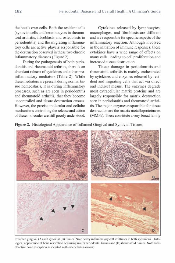
the host’s own cells. Both the resident cells
(synovial cells and keratinocytes in rheuma-
toid arthritis, fibroblasts and osteoblasts in
periodontitis) and the migrating inflamma-
tory cells are active players responsible for
the destruction observed in these two chronic
inflammatory diseases (Figure 2).
During the pathogenesis of both perio -
dontitis and rheumatoid arthritis, there is an
abundant release of cytokines and other pro-
inflammatory mediators (Table 2). While
these mediators are present during normal tis-
sue homeostasis, it is during inflammatory
processes, such as are seen in periodontitis
and rheumatoid arthritis, that they become
uncontrolled and tissue destruction ensues.
However, the precise molecular and cellular
mechanisms controlling the release and action
of these molecules are still poorly understood.
Cytokines released by lymphocytes,
macrophages, and fibroblasts are different
and are responsible for specific aspects of the
inflammatory reaction. Although involved
in the initiation of immune responses, these
cytokines have a wide range of effects on
many cells, leading to cell proliferation and
increased tissue destruction.
Tissue damage in periodontitis and
rheumatoid arthritis is mainly orchestrated
by cytokines and enzymes released by resi-
dent and migrating cells that act via direct
and indirect means. The enzymes degrade
most extracellular matrix proteins and are
largely responsible for matrix destruction
seen in periodontitis and rheumatoid arthri-
tis. The major enzymes responsible for tissue
destruction are the matrix metalloproteinases
(MMPs). These constitute a very broad family
182 Periodontal Disease and Overall Health: A Clinician's Guide
Figure 2. Histological Appearance of Inflamed Gingival and Synovial Tissues
Inflamed gingival (A) and synovial (B) tissues. Note heavy inflammatory cell infiltrates in both specimens. Histo -
logical appearance of bone resorption occurring in (C) periodontal tissues and (D) rheumatoid tissues. Note areas
of active bone resorption associated with osteoclasts (arrows).

Chapter 12 Association Between Periodontitis and Rheumatoid Arthritis 183
Table 2. Cytokines Involved in Periodontitis and Rheumatoid Arthritis
Cytokine Properties
Chemokines Produced by: macrophages, endothelium, fibroblasts, platelets, T cell
Functions:
Leukocyte chemotaxis, activation
IFN-α and IFN-β Produced by: macrophages (α), fibroblasts (β)
Enhanced by: bacterial endotoxin, TNF-α, Il-1
Functions:
All cells: antiviral state, increased class I MHC expression
natural killer (NK) cells: activation
IFN-γ Produced by: NK cell, T cells
Enhanced by: IL-2
Functions:
Macrophages: activation of microbicidal function
Stimulation of some antibody responses
IL-1 Produced by: macrophages, endothelium, epithelium
Induced by: bacterial endotoxin, leukotrienes, C5a, TNF, Il-6
Functions:
Endothelial cell activation: inflammation, coagulation
PMN: activation
Hypothalamus: fever
IL-10 Produced by: macrophages, T cells
Functions:
Macrophages: inhibition of IL-12 production
Reduced expression of costimulators and Class II MHC molecules
IL-12 Produced by: macrophages, dendritic cells
Functions:
NK and T cells: IFN-γ synthesis, increased cytolytic activity
T cells: Th1 differentiation
IL-15 Produced by: macrophages
Functions:
NK cells: proliferation
T cells: proliferation
IL-18 Produced by: macrophages
Functions:
NK and T cells: synthesis of IFN-γ
IL-6 Produced by: macrophages, endothelium, T cells
Induced by: bacterial endotoxin, leukotrienes, C5a, TNF, IL-6
Functions:
Liver: acute-phase proteins
B cells: proliferation of antibody producing cells
TNF Produced by: macrophages, T cells
Induced by: bacterial endotoxin, viruses and protozoa, C5a, immune complexes,
substance P, IL-1, IL-2, TNF-α
Functions:
Endothelial cell activation: inflammation, coagulation
PMN: activation
Hypothalamus: fever
Liver: acute-phase proteins
Muscle, fat: catabolism

of enzymes with a variety of substrates and
functions (Table 3). The cells that release
MMPs in periodontitis and rheumatoid
arthritis are polymorphonucleotides (PMNs),
monocytic phagocytes, and fibroblasts. Both
IL-1 and TNF-α may induce the production
of collagenase and other neutral proteases
by most of these cells.
For both rheumatoid arthritis and peri-
odontitis, disease progression occurs as a re-
sult of very high tissue levels of IL-1β and
TNF-α, and low levels of cytokines such
as IL-10 and transforming growth Factor β,
which suppress the immunoinflammatory
response. In addition, an imbalance between
tissue inhibitors of metalloproteinases
(TIMPs) and MMPs secreted by macro -
phages, fibroblasts, and other resident and
inflammatory cells is characteristic of the
active phases of both rheumatoid arthritis
and periodontitis.
Bone destruction is a common finding
in periodontitis and rheumatoid arthritis.
This occurs as a result of disruption to the
normally balanced processes of bone re-
sorption and bone formation. The major me-
diators of bone resorption are PGE2, IL-1,
TNF-α, and IL-6.
In both periodontitis and rheumatoid
arthritis the immune response is regulated
via genes that control T cell responses to
foreign antigens. In this way the nature of the
protective antibody response, as well as the
magnitude of tissue destructive inflamma-
tory responses, are determined.
role oF GenetiCs in
rheUmatoid arthritis
and Periodontitis
The role of genetics in periodontitis and
rheumatoid arthritis is of considerable im-
portance. For both chronic periodontitis and
rheumatoid arthritis there is considerable
variance in the clinical manifestation of the
disease. Much of this variance can be ac-
counted for by genetic factors.8 Many of these
differences in disease manifestation can be
related to the overexpression or underex-
pression of numerous cytokines and other
inflammatory mediators. Of these, IL-1,
TNF-α, and PGE2
have been found to be un-
der quite strong genetic control. Numerous
studies have been published concerning the
so-called “hyper-responsive monocyte ge-
netic traits,” which have common genetic
regions that influence the susceptibility to
inflammatory diseases. Typical of this trait is
the overproduction of pro-inflammatory me-
diators such as IL-1β, TNF-α, and PGE2, and
is typically seen in patients with aggressive
periodontitis.9 The concept of a monocytic
hypersecretory state has also been described
for rheumatoid arthritis patients.10
Many of the genes that regulate the
cytokine profiles and responses of mono-
cytes have been mapped to the human leuko-
cyte antigen D related (HLA-DR) region of
chromosome 5 in the area of the TNF-β
genes.11 Since both rheumatoid arthritis and
periodontitis have been associated with this
HLA complex, a common genetic basis ex-
ists for the observed monocyte trait, linking
rheumatoid arthritis and periodontitis.
role oF environmental
FaCtors on Periodontitis
and rheUmatoid arthritis
It has been recognized for some time
that the clinical manifestation of chronic dis-
eases such as periodontitis and rheumatoid
arthritis are significantly influenced by en-
vironmental or “modifying” factors. These
factors are anything that might modify or
alter the host inflammatory response, and
include demographic, socioeconomic, life -
style, diet, hormonal, and psychological vari-
ables. Since genetic factors may account for
up to 50% of the risk for both periodontitis
and rheumatoid arthritis,8,12 other factors, in-
clud ing environmental influences and gene-
environment interactions, must explain the
rest.
184 Periodontal Disease and Overall Health: A Clinician's Guide

Chapter 12 Association Between Periodontitis and Rheumatoid Arthritis 185
Table 3. Human Matrix Metalloproteinases and Their Substrates*
Protein Collagenous noncollagenous nonstructural eCm
name substrates eCm substrates Component substrates
MMP-1 Collagen Types I, II, III, Aggrecan, casein, nidogen, α1-antichymotrypsin, α
1-antitrypsin/
VII, VIII, X, and gelatin serpins, versican, perlecan, α1-proteinase inhibitor, IGFBP-3, IGFBP-5,
proteoglycan link protein, IL-1β, L-selectin, ovostatin, recombinant
and tenascin-C TNF-α peptide, and SDF-1
MMP-2 Collagen Types I, IV, V, Aggrecan, elastin, fibronectin, Active MMP-9, active MMP-13, FGF R1,
VII, X, XI, XIV, and laminin, nidogen, proteoglycan IGF-BP3, IGF-BP5, IL-1β, recombinant
gelatin link protein, and versican TNF-α peptide, and TGF-β
MMP-3 Collagen Types II, IV, IX, Aggrecan, casein, decorin, α1-antichymotrypsin, α
1-proteinase fibrinogen,
X, and gelatin elastin, fibronectin, laminin, IGF-BP3, L-selectin, ovostatin, pro-HB-EGF,
nidogen, perlecan, proteoglycan, pro-IL-β, pro-MMP-1, pro-MMP8,
link protein, and versican pro-MMP-9, pro-TNF-α, and SDF-1
MMP-7 Collagen Types I, II, III, IV, Aggrecan, casein, elastin, β4 integrin, decorin, defensin, E-cadherin,
V, X, and XI enactin, laminin, and Fas-L, plasminogen, pro-MMP-2, pro-
proteoglycan link protein MMP-7, pro-TNF-α, transferrin, and syndecan
MMP-8 Collagen Types I, II, III, V, Aggrecan, laminin, and α2-antiplasmin and pro-MMP-8
VII, VIII, X, and gelatin nidogen
MMP-9 Collagen Types IV, V, VII, Fibronectin, laminin, nidogen, CXCL5, IL-1β, IL2-R, plasminogen,
X, XIV, and XI proteoglycan link protein, and pro-TNF-α, SDF-1, and TGF-β
versican
MMP-10 Collagen Types III, IV, V, Fibronectin, laminin, and Pro-MMP-1, pro-MMP-8, and pro-MMP-10
and gelatin nidogen
MMP-11 Laminin α1-antitrypsin, α
1-proteinase inhibitor, and
IGFBP-1
MMP-12 Elastin Plasminogen
MMP-13 Collagen Types I, II, III, IV, Aggrecan, fibronectin, laminin, Plasminogen activator 2, pro-MMP-9,
V, IX, X, XI, and gelatin perlecan, and tenascin pro-MMP-13, and SDF-1
MMP-14 Collagen Types I, II, III, Aggrecan, dermatan sulphate, αvβ
3 integrin, CD44, gC1qR, pro-MMP2,
and gelatin proteoglycan, fibrin, pro-MMP-13, pro-TNF-α, SDF-1, and
fibronectin, laminin, nidogen, tissue transglutaminase
perlecan, tenascin, and
vitronectin
MMP-15 Collagen Types I, II, III, Aggrecan, fibronectin, laminin, Pro-MMP-2, pro-MMP-13, and tissue
and gelatin nidogen, perlecan, tenascin, transglutaminase
and vitronectin
MMP-16 Collagen Types I, III, and Aggrecan, casein, fibronectin, Pro-MMP-2 and pro-MMP-13
gelatin laminin, perlecan, and
vitronectin
MMP-17 Gelatin Fibrin and fibronectin
MMP-19 Collagen Types I, IV, and Aggrecan, casein, fibronectin,
gelatin laminin, nidogen, and tenascin
MMP-20 Aggrecan, amelogenin, and
cartilage
MMP-21 α1-antitrypsin
MMP-23 Gelatin
MMP-24 Gelatin Chondroitin sulfate, dermatin Pro-MMP-2 and pro-MMP-13
sulfate, and fibronectin
MMP-25 Collagen Type IV and gelatin Fibrin and fibronectin Pro-MMP-2
MMP-26 Collagen Type IV and gelatin Casein, fibrinogen, and β1-proteinase inhibitor
fibronectin
MMP-28 Casein
*Note:Many of these substrates are found in both the periodontium and synovium. Adapted from Somerville RPT,Oblander SA, Apte SS. Matrix metalloproteinases: old dogs with new tricks. Genome Biol 2003:4:216-226.

Of the environmental risk factors iden-
tified to date, smoking is probably the single
most important environmental factor im-
pacting many chronic diseases. Through its
adverse effects on the cardiovascular sys-
tem, immune cell function, and general tissue
physiology, it is not surprising that smoking
is considered a major risk factor for the de-
velopment of both rheumatoid arthritis and
periodontitis.13,14
Another environmental factor of inter-
est is socioeconomic status. This may be a
significant factor in the outcome of many
chronic diseases, including both rheumatoid
arthritis and periodontitis.15,16 The reasons
for this are not entirely clear, although sug-
gestions have been made that individuals
from lower socioeconomic status may be
less compliant with regard to their general
healthcare and have less access to the full
range of healthcare. However, these expla-
nations do not fully explain this relationship.
In this regard, allostatic load has also been
proposed to describe dysregulation of phys-
iological adaptive processes, including im-
mune function, that in health maintain sta-
bility to stressors. It has been proposed that
allostatic load may follow a socioeconomic
gradient that is, in part, responsible for in-
equalities in chronic diseases such as peri-
odontitis and rheumatoid arthritis. In this
model, low socioeconomic status results in
increased exposure to psychosocial stress,
which activates primary allostatic mediators,
including a wide range of inflammatory me-
diators. Under these conditions dysregulation
of a range of immunomodulatory functions
occurs, possibly leading to alterations in the
extent and severity of chronic diseases.
Periodontitis and rheUmatoid
arthritis Can be Classed as
“ComPlex” or “eCoGenetiC”
diseases
In recent times, periodontitis andrheumatoid arthritis have been considered
“complex” or “ecogenetic” diseases.17,18 Acomplex disease is defined as a disorder witha genetic component but does not follow thesimple single-gene dominant or single-generecessive Mendelian law (Table 4). Since
both inherited genetic variations and envi-ronmental factors, such as smoking, hygiene,and pathogenic bacteria interact to determinean individual’s risk for the development ofperiodontitis and rheumatoid arthritis, thesetwo diseases fit this definition of complexdiseases. Hence in these diseases, in re-sponse to an initiating event, environmentalagents interact with genetic factors to influ-ence disease susceptibility (Figure 3). Suchinteractions initiate and regulate immuno-inflammatory reactions that ultimately man-ifest as the clinical signs of rheumatoidarthritis or periodontitis. While understand-ing the role of dental plaque as being pivotalin the initiating events for periodontal diseaseis well understood, the initiating events forrheumatoid arthritis are less clear.
Interactions between environmental riskfactors and genetics are now providingus with valuable clues as to the nature of complex diseases such as periodontitisand rheumatoid arthritis. One of the best-
186 Periodontal Disease and Overall Health: A Clinician's Guide
Table 4. Features of Complex DiseasesTable 4. or Ecogenetic Diseases
• The diseases interact with ecology, molecular
genetics, toxicology, public health medicine, and
environmental epidemiology.
• These are common chronic diseases with adult
onset that show familial aggregation, but do not
follow Mendelian family patterns.
• These diseases appear to be caused by an un-
known number of multiple genes, which interact
with environmental factors.
• In these diseases, only a small proportion (1%–
7%) of affected individuals show a single mutant
gene transmitted by Mendelian inheritance with
characteristic transmission.
• These genetically exceptional families often have
an earlier age of onset and have more severe
clinical manifestations.

recognized environmental risk factors forboth diseases is smoking. A relationship be-tween smoking and HLA-DR shared epitopegenes, the main genetic risk factors forrheumatoid arthritis and periodontitis, hasbeen shown to impart increased risk for thedevelopment of both rheumatoid arthritis andperiodontitis. From these types of studies itwill be possible to define the various en -vironmental risk factors that, in certain genetic contexts, will initiate adverse im-mune reactions, ultimately leading to clinicalsymptoms in various subsets of susceptible patients.
In recent years there has been consid-erable interest in identifying single nucleo -tide polymorphisms (SNPs) and statisticalgenetic strategies, such as haplotype map-ping, to identify genes that may be of im-portance in complex diseases such asrheumatoid arthritis and periodontitis. Eventhough simple one-to-one mapping between
disease gene and disease phenotype does notoccur in complex diseases, this should not reduce the importance of trying to identifygenes that determine individual differencesin disease susceptibility for rheumatoidarthritis and periodontitis. An understandingand identification of gene mutations in com-plex diseases, using genome-wide associa-tion studies, may account for genetic sourcesof variation in disease risk and should enablea better understanding of environmental effects.
Periodontal disease and
rheUmatoid arthritis
have siGniFiCant
inter-relationshiPs with
other systemiC Conditions
The focus of this volume is the associ-
ation of periodontal diseases with a large
number of systemic conditions, including
diabetes, preterm low birth weight infants,
Chapter 12 Association Between Periodontitis and Rheumatoid Arthritis 187
Figure 3. Disease Interactions Between Periodontitis and Rheumatoid Arthritis
The periodontal diseases and rheumatoid arthritis are classified as ecogenetic or complex diseases involving
intricate interactions between host responses, environmental factors, etiologic factors, and genetics.
HOSTRESPONSESENVIRONMENT
DISEASE
ETIOLOGY
GENETICS

cardiovascular disease, pulmonary disease,
obesity, and osteoporosis. In the context of
this chapter, which considers the potential in-
teraction between periodontitis and rheuma-
toid arthritis, it is of interest to note that all
of these diseases have also been associated
with individuals suffering from rheumatoid
arthritis (Table 5). Even after accounting for
variable confounders, including various
medications, many studies have been able
to demonstrate significant relationships be-
tween rheumatoid arthritis and systemic con-
ditions.19 The underlying feature in these re-
lationships seems to be dysregulated chronic
inflammation in both periodontitis and
rheumatoid arthritis.
what is the evidenCe For
a relationshiP between
Periodontitis and
rheUmatoid arthritis?
For more than 80 years there have been
reports in the literature concerning a possi-
ble relationship between rheumatoid arthritis
and periodontitis. A number of reviews
and com mentaries on this topic have been
published.20-24
One of the earliest reports concerning
this relationship was written by Sachs, who
commented in 1926 that the relationship
between “gelenkrheumatismus and para -
dentose” was a question of “a constitution
with a predisposition to both of these condi-
tions” (quoted from Helminen-Pakkala).25
Since then, a number of studies considered
the relationship between periodontal disease
and rheumatoid arthritis with little agree-
ment being reached as to whether this was
indeed possible.
Over the last 30 years, a steady stream
of papers that question this relationship have
been published. For example, an increase in
any dental disease, including periodontal dis-
ease, was not seen in a group of South
African patients with advanced joint condi-
tions.26 Similarly, a study of Japanese
rheumatoid arthritis patients found no evi-
dence of elevated levels of serum antibodies
against several periodontal pathogens.27 A
study that considered clinical and immuno-
logical features of periodontitis reported that
rheumatoid arthritis patients were not a risk
group for advanced periodontal conditions.28
Furthermore, a case-control study in a group
of Scandinavian patients (204 cases and 204
controls) reported a tendency toward better
periodontal conditions for the rheumatoid
arthritis patients.29 A study of Norwegian
rheumatoid arthritis patients between the
ages of 44 and 56 found no correlation be-
tween tooth loss and rheumatoid arthritis.30
In a study using epidemiological data, 523
individuals, initially seen during the first
National Health and Nutrition Examination
Survey (NHANES I) of 1971–75, exhibited
no correlation with self-reported arthritis and
the number of missing teeth.31
Despite the above investigations, which
reported a negative correlation between pe-
riodontitis and rheumatoid arthritis, there
have been a larger number of studies that
have supported such a relationship. While
perhaps the least convincing in terms of sci-
entific design, simple pilot studies analyzing
data obtained from self-reported illnesses
have indicated a higher incidence of rheuma-
toid arthritis in patients with periodontitis.32-35
A number of case-control studies have
also been carried out. All of these indicate
that rheumatoid arthritis patients have a
significantly higher incidence of periodon -
tal disease. For these studies, periodontal
188 Periodontal Disease and Overall Health: A Clinician's Guide
Table 5. Systemic Conditions ReportedTable 5. .to be Associated with Both Perio -Table 5. .dontitis and Rheumatoid Arthritis
Cardiovascular Disease
Diabetes
Obesity
Obstetric Problems
Pulmonary Conditions
Renal Conditions
Cancer

conditions were variously measured as num-
ber of teeth missing, gingival bleeding, at -
tachment loss, probing pocket depth, and
radiographic bone loss.32,36-44
As a result of these findings, studies
have been carried out to investigate a number
of clinical and laboratory param eters to
further ascertain if a relationship between
periodontitis and rheumatoid arthritis exists.
These have included investigations into cyto -
kine profiles,45-50 inflammatory mediators,51
HLA-DR antigens,52-54 and hormones.55 All
of these studies have supported the notion
that periodontitis and rheumatoid arthritis
are inter-related, and have led various
authors to conclude that inflammation
(and its dys regulation) may be the central
link between periodontitis and rheuma-
toid arthritis.23 At least two studies have
concluded that perio dontitis may be a risk
factor or severity factor for rheumatoid
arthritis.32,56
As early as 1985, a case report was
published describing remission of rheuma-
toid arthritis following periodontal treat-
ment.57 This study remained largely forgot-
ten until the publication of two additional
studies that indicated periodontal treatment
could reduce the severity of rheumatoid
arthri tis.56,58 While these studies have in-
volved relatively low numbers of sub-
jects, they highlight the potential for impor-
tant clinical ramifications for the relation-
ship between periodontitis and rheumatoid
arthritis.
Further support for a significant rela-
tionship between periodontitis and rheuma-
toid arthritis has been highlighted in a re-
cent animal study. Moreover, the induction
of adjuvant arthritis in rats resulted in an as-
sociated periodontal breakdown evidenced
by alveolar bone loss and increased matrix
metalloproteinase activity in adjacent gingi-
val tissues.59
Careful assessment of the data reported
from many of these studies allows us to
make some important observations. First,
few of these studies support the commonly
held tenet that rheumatoid arthritis patients
have impaired oral hygiene (judged by
plaque and bleeding scores) because of their
“disability.” It is variously reported that oral
hygiene is no different between rheumatoid
arthritis patients with periodontitis and non-
rheumatoid arthritis patients with periodon-
titis.44,60 Another important observation from
recently published studies is that individuals
with severe rheumatoid arthritis are more
likely to suffer from advanced periodontitis
and vice versa. However, the association
seems to be in favor of rheumatoid arthritis
impacting periodontitis (Relative Risk 4.1)
rather than periodontitis impacting rheuma-
toid arthritis (Relative Risk 1.5).60 Also, it is
important that longitudinal studies be car-
ried out from this association to establish
the temporal sequence. Finally, from the
studies published to date, it is interesting to
consider the influence of medications that
rheumatoid arthritis patients may be taking,
on periodontitis. Many, if not all, of these
medications are anti-inflammatory agents
and therefore have the potential to suppress
periodontal inflammation and affect peri-
odontal disease progression. Nonetheless,
reports indicate that significant periodontal
destruction can still be seen in patients who
may be taking anti-inflammatory medica-
tions for rheumatoid arthritis.40,60 It has been
proposed that before development of
rheumatoid arthritis symptoms, the peri-
odontitis was most likely also developing
and not detected.23 Thus, in the analysis of
the association between rheumatoid arthritis
and periodontitis, consideration of disease
duration (for both periodontal and rheuma-
toid) will be a critical factor. Therefore, fu-
ture studies concerning the relationship be-
tween periodontitis and rheumatoid arthritis
should address and document the disease
with regard to severity and duration of both
diseases.
Chapter 12 Association Between Periodontitis and Rheumatoid Arthritis 189

ProPosed meChanisms For
a relationshiP between
Periodontitis and
rheUmatoid arthritis
A number of mechanisms for a rela-
tionship between periodontitis and rheuma-
toid arthritis have been proposed. The prin-
cipal mechanisms may involve changes to
blood vessels or infection of host tissues.
vascular alterations
Recent investigations into the relation-
ship between osteoclast activation and vas-
cular damage have suggested a common
pathway in the development of periodontitis
and rheumatoid arthritis. It has been hy-pothesized that both rheumatoid arthritis andperiodontitis share common molecular path-ways within the receptor activator of nuclear
factor Kappa β (RANK)/osteoprotegerin
(OPG)/tumor necrosis factor-related apop-
tosis-inducing ligand (TRAIL) axis, wherebya decrease in OPG leads to reduced vascularprotection.23 In addition, increases in RANKand TRAIL levels within inflamed tissues
may result in not only the possible develop-
ment of vascular damage, but also activation
of osteoclasts and subsequent bone resorption.
Another vascular model proposes that
microvascular involvement is one of the first
stages of a number of chronic diseases, such
as periodontitis and rheumatoid arthritis.61
In this model, reduced caliber of capillaries,
along with greater numbers of and elongated
capillaries, are noted in both periodontal tis-
sues and rheumatoid synovium.
bacterial infection
Data from a number of animal models
demonstrate that arthritis can develop sec-
ondarily to several different stimuli and
through several effector pathways, includ-
ing exogenous infections. If the observations
in animal models are also applicable to hu-
man rheumatoid arthritis, we might anticipate
that different types of infections, as well as
other environmental exposures with capacity
to induce excessive pro-inflammatory cyto -
kines in genetically susceptible individuals,
may contribute to disease either together
with some autoimmune reaction or by them-
selves.
Many periodontal pathogens exhibit
similar characteristics to those micro-organ-
isms suspected to induce rheumatoid arthritisin a genetically susceptible host. Periodontal
pathogens incite a chronic continuous in-
flammation within the periodontal tissues
and also serve as an abundant supply of
lipopolysaccharide. Thus, the possibility that
an ongoing periodontitis can trigger or ex-
acerbate rheumatoid arthritis in genetically
susceptible individuals is biologically plau-
sible. A number of studies have reported
elevated serum antibodies to a number of
periodontopathic bacteria including Por-
phyromonas gingivalis, Prevotella interme-
dia, Prevotella melaninogenica, Bacteroides
forsythus, and Aggregatibacter actino-
mycetemcomitans.27,62-64 Elevated antibodies
to B. forsythus and P. intermedia have also
been found in synovial fluid. Further evi-
dence for a role of periodontal pathogens in
joint disease have come from a study re-
porting DNA for P. gingivalis, T. forsythen-
sis and P. intermedia in synovial fluid of
rheumatoid patients.65 No DNA for these
bacteria were found in the synovial fluid of
nonrheumatoid patients.
A novel hypothesis for the development
of rheumatoid arthritis via the humoral re-
sponse to oral bacteria found in periodontitis
has been proposed.66 In this model, an auto -
immune disease to proteins partially altered by
bacterial enzymes in genetically sus ceptible
individuals may develop (Figure 4). Central
to this hypothesis is the appearance of rheu -
matoid factors and anticyclic, citrullinated
pep tide autoantibodies during the develop-
ment of rheumatoid arthritis. The production
of deimination enzymes by perio dontal path-
ogenic bacteria, such as P. gingivalis, can
190 Periodontal Disease and Overall Health: A Clinician's Guide

induce autoantibodies, allowing a link between
periodontal infection and development of
rheumatoid arthritis.
viral infection
Viruses have been implicated in the
pathogenesis of both periodontitis and rheu -
matoid arthritis.67,68 A number of reports have
implicated Epstein-Barr virus in the devel-
opment of rheumatoid arthritis. Although the
mechanisms involved for viruses in the
development of rheumatoid arthritis are
unclear, it has been proposed that they can
act as an adjuvant in the development of
autoimmunity, nonspecifically stimulating
innate immune responses in genetically sus-
ceptible individuals. Epstein-Barr virus may
cause periodontal disease via direct in fection
and replication, or through impairment of host
immune responses.68 Thus, it is possible that
a common link between these two diseases
could be a viral infection leading to impaired
immune function, resulting in uncontrolled
inflammatory reactions at sites such as the
periodontium and synovium (Figure 5). At
present, this model has not been proven. It
does, however, present a perspective that
needs to be investigated further.
CliniCal relevanCe oF
an assoCiation between
Periodontitis and
rheUmatoid arthritis
The association between advanced rheu -matoid arthritis and advanced perio dontaldestruction has significant clinical implica-tions with respect to the management of rheu -matoid arthritis patients who are at risk ofalso having periodontitis. Even though mostclinical protocols for rheumatoid arthritis pa-tients include an assessment for oral pathol-ogy, they do not routinely include a full perio -dontal assessment. Since many of the signsand symptoms of periodontitis are painlessand subtle and may advance rapidly withoutthe patient being aware of the problem, this
Chapter 12 Association Between Periodontitis and Rheumatoid Arthritis 191
Figure 4. The Humoral Immune ResponseFigure 4. to Oral Bacteria Provides aFigure 4. Stimulus for Development Figure 4. of Rheumatoid Arthritis
Periodontal infection
▼Citrullination of proteins
by P. gingivalis
▼Production of autoantibodies
to citrullinated proteins
▼Cross reactivity with
cartilage components
▼Auto-immuninity to self-antigens
▼Rheumatoid arthritis
Figure 5. A Possible Mechanism for a Link
Figure 5. Between Viral Infection in Perio -
Figure 5. dontitis and Rheumatoid Arthritis
tissue inflammation
Influx of inflammatory cells with latent herpes virus
▼herpes virus activation
Immunosuppression, bacterial infection
Hormones, stress
▼tissue destruction
Uncontrolled release of cytokines,
further immunosuppression
Cytotoxicity, exacerbation of infection
▼Clinical manifestation
Destructive periodontitis
Destructive rheumatoid arthritis
herpes virus infected individual
▼

aspect of clinical assessment has been over-looked. In light of the current data, individ-uals suffering from rheumatoid arthritis seemto be at higher risk of developing periodon-tal problems. Further, early evidence has sug-gested that treatment of periodontitis can re-duce the clinical symptoms of rheumatoidarthritis. Therefore, early intervention to pre-vent periodontal destruction occurring in in-dividuals with rheumatoid arthritis shouldbe considered to reduce the impact ofrheumatoid arthritis.
ConClUsions
For several decades it has been sus-
pected that because periodontitis and rheuma-
toid arthritis share many common pathologic
features, they may be clinically related or
ass ociated diseases. It is now becoming ap-
parent, mainly from case-control studies, that
disease severity and extent may be related
for individuals suffering from both rheuma-
toid arthritis and periodontitis. While causal-
ity between the two diseases is very unlikely,
the fact that both diseases can impact each
other is becoming apparent. Thus, it is pro-
posed that these two diseases can exist as a
manifestation of generalized dysregulation
of the immune and inflammatory responses.
Hence, both periodontitis and rheumatoid
arthritis may be considered clinical mani-
festations of the same disease process pre-
senting in different parts of the body.69
Supplemental Readings
Mercado FB, Marshall RI, Bartold PM. Inter-relation-
ships between rheumatoid arthritis and periodontal dis-
ease. A review. J Clin Periodontol 2003;30:761–772.
Bartold PM, Marshall RI, Haynes DR. Periodontitis
and rheumatoid arthritis: A review. J Periodontol 2005;
76(11 Suppl):2066-2074.
Golub LM, Payne JB, Reinhardt RA, Nieman G. Can
systemic diseases co-induce (not just exacerbate) perio -
dontitis? A hypothetical "two-hit" model. J Dent Res
2006;85:102-105.
Al-Katma MK, Bissada NF, Bordeaux JM, Sue J, Askari
192 Periodontal Disease and Overall Health: A Clinician's Guide
AD. Control of periodontal infection reduces the severity
of active rheumatoid arthritis. J Clin Rheumatol 2007;
13:134–137.
Rosenstein ED, Greenwald RA, Kushner LJ, Weiss-
mann G. Hypothesis: The humoral immune response to
oral bacteria provides a stimulus for the development of
rheumatoid arthritis. Inflammation 2004;28:311–318.
de Pablo P, Chapple IL, Buckley CD, Dietrich T. Perio -
d ontitis in systemic rheumatic diseases. Nat Rev
Rheumatol 2009;5:218–224.
reFerenCes1. Armitage GC. Development of a classification sys-
tem for periodontal diseases and conditions. Ann
Periodontol 1999;4:1–6.
2. Pihlstrom BL, Michalowicz BS, Johnson NW. Pe-
riodontal diseases. Lancet 2005;366:1809–1820.
3. Persselin JE. Diagnosis of rheumatoid arthritis.
Medical and laboratory aspects. Clin Orthop Rel
Res 1991;265:73–82.
4. Avouac J, Gossec L, Dougados M. Diagnostic and
predictive value of anti-cyclic citrullinated protein
antibodies in rheumatoid arthritis: a systematic lit-
erature review. Ann Rheum Dis 2006;65:845–851.
5. Arnett FC, Edworthy SM, Bloch DA, McShane
DJ, Fries JF, Cooper NS, Healey LA, Kaplan SR,
Liang MH, Luthra HS, et al. The American Rheu -
matism Association 1987 revised criteria for the
classification of rheumatoid arthritis. Arthritis
Rheum 1988;31:315–324.
6. Wolfe F, Cathey MA. Analysis of methotrexate
treatment effect in a longitudinal observational study:
Utility of cluster analysis. J Rheumatol 1991;18:
672–677.
7. Hirschfeld L, Wasserman B. A long-term survey of
tooth loss in 600 treated periodontal patients. J
Perio dontol 1978;49:225–237.
8. Michalowicz BS. Genetic and heritable risk fac-
tors in periodontal disease. J Periodontol 1994;65
(5 Suppl):479–488.
9. Salvi GE, Yalda B, Collins JG, Jones BH, Smith
FW, Arnold RR, Offenbacher S. Inflammatory
mediator response as a potential risk marker for
periodontal diseases in insulin-dependent diabetes
mellitus patients. J Periodontol 1997;68:127–135.
10. Ollier W, Thomson W. Population genetics of
rheumatoid arthritis. Rheum Dis Clin North Am
1992;18:741–759.
11. Bendtzen K, Morling N, Fomsgaard A, Svenson
M, Jakobsen B, Odum N, Svejgaard A. Association
between HLA-DR2 and production of tumour
necrosis factor alpha and interleukin 1 by mononu-
clear cells activated by lipopolysaccharide. Scand
J Immunol 1988;28:599–606.

12. MacGregor AJ, Snieder H, Rigby AS, Koskenvuo
M, Kaprio J, Aho K, Silman AJ. Characterizing the
quantitative genetic contribution to rheumatoid
arthritis using data from twins. Arthritis Rheum
2000;43:30–37.
13. Wilson K, Goldsmith CH. 1999. Does smoking cause
rheumatoid arthritis? J Rheumatol 1999;26:1–3.
14. Bergström J, Preber H. Tobacco use as a risk fac-
tor. J Periodontol 1994;65(5 Suppl):545–550.
15. Callahan LF, Pincus T. Formal education level as a
significant marker of clinical status in rheumatoid
arthritis. Arthritis Rheum 1988;31:1346–1357.
16. Borrell LN, Burt BA, Warren RC, Neighbors HW.
The role of individual and neighborhood social fac-
tors on periodontitis: The third National Health and
Nutrition Examination Survey. J Periodontol 2006;
77:444–453.
17. Michalowicz BS, Diehl SR, Gunsolley JC, Sparks
BS, Brooks CN, Koertge TE, Califano JV, Burm-
eister JA, Schenkein HA. Evidence of a substantial
genetic basis for risk of adult periodontitis. J Peri-
odontol 2000;71:1699–1707.
18. Kobayashi S, Momohara S, Kamatani N, Okamoto
H. Molecular aspects of rheumatoid arthritis:
role of environmental factors. FEBS J 2008;275:
4456–4462.
19. Moreland LW, Curtis JR. Systemic nonarticular
manifestations of rheumatoid arthritis: Focus on
inflammatory mechanisms. Semin Arthritis Rheum
2008; Epub ahead of print.
20. Snyderman R, McCarty GA. Analogous mecha-
nisms of tissue destruction in rheumatoid arthritis
and periodontal disease. In: Genco RJ, Mergen-
hagen SE eds. Host-Parasite Interactions in Perio -
dontal Diseases. American Society for Micro -
biology: Washington, DC; 1982:354–362.
21. Okuda K, Ebihara Y. Relationships between chronic
oral infectious diseases and systemic diseases. Bull
Tokyo Dent Coll 1998;39:165–174.
22. Mercado FB, Marshall RI, Bartold PM. Inter-rela-
tionships between rheumatoid arthritis and perio -
dontal disease. A review. J Clin Periodontol
2003;30:761–772.
23. Bartold PM, Marshall RI, Haynes DR. Periodonti-
tis and rheumatoid arthritis: A review. J Periodon-
tol 2005;76(11 Suppl):2066-2074.
24. Golub LM, Payne JB, Reinhardt RA, Nieman G.Can systemic diseases co-induce (not just exacer-bate) periodontitis? A hypothetical “two-hit” model.J Dent Res 2006;85:102–105.
25. Helminen-Pakkala E. Periodontal conditions in
rheumatoid arthritis: A clinical and roentgenologi-
cal investigation. Part 2: The study in rheumatoids
based on a hospital material. Suomen hammas -
lääkäriseuran toimituksia. Supplementum (Proc
Chapter 12 Association Between Periodontitis and Rheumatoid Arthritis 193
Finnish Dental Assoc Supplement) 1971; Suppl IV;
1–108.
26. Blair GS, Chalmers IM. The dental status of a
chronically disabled section of the community: A
study of 139 patients suffering from rheumatic dis-
eases. J Dent Assoc S Afr 1976;31:329–336.
27. Yusof Z, Porter SR, Greenman J, Scully C. Levels
of serum IgG against Porphyromonas gingivalis in
patients with rapidly progressive periodontitis,
rheumatoid arthritis and adult periodontitis. J Nihon
Univ Sch Dent 1995;37:197–200.
28. Yavuzyilmaz E, Yamalik N, Calgüner M, Ersoy F,
Baykara M, Yeniay I. Clinical and immunological
characteristics of patients with rheumatoid arthritis
and periodontal disease. J Nihon Univ Sch Dent
1992;34:89–95.
29. Sjöström L, Laurell L, Hugoson A, Håkansson JP.
Periodontal conditions in adults with rheumatoid
arthritis. Comm Dent Oral Epidemiol 1989;17:
234–236.
30. Arneberg P, Bjertness E, Storhaug K, Glennås A,
Bjerkhoel F. Remaining teeth, oral dryness and
dental health habits in middle aged Norwegian
rheumatoid arthritis patients. Comm Dent Oral Epi-
demiol 1992;20:292–296.
31. Eklund SA, Burt BA. Risk factors for total tooth
loss in the United States: longitudinal analysis of
national data. J Public Health Dent 1994;54:5–14.
32. Mercado F, Marshall RI, Klestov AC, Bartold PM.
Is there a relationship between rheumatoid arthritis
and periodontal disease? J Clin Periodontol 2000;
27:267–272.
33. Lagervall M, Jansson L, Bergström J. Systemic
disorders with periodontal disease. J Clin Perio -
dontol 2003;30:293–299.
34. Georgiou TO, Marshall RI, Bartold PM. Preva-
lence of systemic diseases in Brisbane general and
periodontal practice patients. Aust Dent J 2004;49:
177–184.
35. Dumitrescu AL. Occurrence of self-reported sys-
temic medical conditions in patients with perio -
dontal disease. Rom J Intern Med 2006;44:35–48.
36. Malmström M, Calonius PE. Teeth loss and the in-
flammation of teeth-supporting tissues in rheuma-
toid disease. Scand J Rheumatol 1975;4:49–55.
37. Albandar JM. Some predictors of radiographic
alveolar bone height reduction over 6 years. J Perio -
dontal Res 1990;25:186–192.
38. Tolo K, Jorkjend L. Serum antibodies and loss of
periodontal bone in patients with rheumatoid arthri-
tis. J Clin Periodontol 1990;17:288–291.
39. Kässer UR, Gleissner C, Dehne F, Michel A, Wil-
lershausen-Zönnchen B, Bolten WW. Risk for pe-
riodontal disease in patients with longstanding
rheumatoid arthritis. Arthritis Rheum 1997;40:

2248–2251.
40. Gleissner C, Willershausen B, Kässer U, Bolten
WW. The role of risk factors for periodontal disease
in patients with rheumatoid arthritis. Eur J Med
Res 1998;3:387–392.
41. Novo E, Garcia-MacGregor E, Viera N, Chaparro
N, Crozzoli Y. Periodontitis and anti-neutrophil
cytoplasmic antibodies in systemic lupus erythe-
matosus and rheumatoid arthritis: a comparative
study. J Periodontol 1999;70:185–188.
42. Zhang DZ, Zhong DY, Deng J, Wang JB. Relati-
onship between periodontal disease and rheumatoid
arthritis. Hua Xi Kuo Qiang Yi Xue Za Zhi
2005;23:498–501.
43. Ishi Ede P, Bertolo MB, Rossa C Jr, Kirkwood KL,
Onofre MA. Periodontal condition in patients with
rheumatoid arthritis. Braz Oral Res 2008;22:72–77.
44. Pischon N, Pischon T, Kröger J, Gülmez E, Kleber
BM, Bernimoulin JP, Landau H, Brinkmann PG,
Schlattmann P, Zernicke J, Buttgereit F, Detert J.
Association among rheumatoid arthritis, oral hy-
giene, and periodontitis. J Periodontol 2008;79:
979–986.
45. Bozkurt FY, Berker E, Akkus S, Bulut S. Relation-
ship between interleukin-6 levels in gingival crevic-
ular fluid and periodontal status in patients with
rheumatoid arthritis and adult periodontitis. J Perio -
dontol 2000;71:1756–1760.
46. Havemose-Poulsen A, Sørensen LK, Stoltze K,
Bendtzen K, Holmstrup P. Cytokine profiles in
peripheral blood and whole blood cell cultures
associated with aggressive periodontitis, juvenile
idiopathic arthritis, and rheumatoid arthritis. J Perio -
dontol 2005;76:2276–2285.
47. Pers JO, Saraux A, Pierre R, Youinou P. Anti-TNF-
alpha immunotherapy is associated with increased
gingival inflammation without clinical attachment
loss in subjects with rheumatoid arthritis. J Perio -
dontol 2008;79:1645–1651.
48. Bozkurt FY, Yetkin Ay Z, Berker E, Tepe E, Akkuş
S. Anti-inflammatory cytokines in gingival crevic-
ular fluid in patients with periodontitis and rheuma-
toid arthritis: A preliminary report. Cytokine 2006;
35:180–185.
49. Havemose-Poulsen A, Westergaard J, Stoltze K,
Skjødt H, Danneskiold-Samsøe B, Locht H,
Bendtzen K, Holmstrup P. Periodontal and hema -
tological characteristics associated with aggressive
periodontitis, juvenile idiopathic arthritis, and
rheumatoid arthritis. J Periodontol 2006;77:280–
288.
50. Nilsson M, Kopp S. Gingivitis and periodontitis
are related to repeated high levels of circulating
tumor necrosis factor-alpha in patients with rheu -
matoid arthritis. J Periodontol 2008;79:1689–1696.
51. Biyikoğlu B, Buduneli N, Kardeşler L, Aksu K,
Oder G, Kütükçüler N. Evaluation of t-PA, PAI-2,
IL-1beta and PGE(2) in gingival crevicular fluid of
rheumatoid arthritis patients with periodontal dis-
ease. J Clin Periodontol 2006;33:605–611.
52. Katz J, Goultschin J, Benoliel R, Brautbar C. Hu-
man leukocyte antigen (HLA) DR4. Positive asso-
ciation with rapidly progressing periodontitis. J
Periodontol 1987;58:607–610.
53. Bonfil JJ, Dillier FL, Mercier P, Reviron D, Foti B,
Sambuc R, Brodeur JM, Sedarat C. A “case control”
study on the role of HLA DR4 in severe perio don -
titis and rapidly progressive periodontitis. Identifi-
ca tion of types and subtypes using molecular biol-
ogy (PCR.SSO). J Clin Periodontol 1999;26:77–84.
54. Marotte H, Farge P, Gaudin P, Alexandre C, Mou-
gin B, Miossec P. The association between perio -
dontal disease and joint destruction in rheumatoid
arthritis extends the link between the HLA-DR
shared epitope and severity of bone destruction.
Ann Rheum Dis 2006;65:905–909.
55. Soory M. Hormone mediation of immune responses
in the progression of diabetes, rheumatoid arthritis
and periodontal diseases. Curr Drug Targets Im-
mune Endocr Metabol Disord 2002;2:13–25.
56. Ribeiro J, Leão A, Novaes AB. Periodontal infec-
tion as a possible severity factor for rheumatoid
arthritis. J Clin Periodontol 2005;32:412–416.
57. Iida M, Yamaguchi Y. Remission of rheumatoid
arthritis following periodontal treatment. A case
report. Nippon Shishubyo Gakkai Kaishi 1985;27:
234–238.
58. Al-Katma MK, Bissada NF, Bordeaux JM, Sue J,
Askari AD. Control of periodontal infection re-
duces the severity of active rheumatoid arthritis. J
Clin Rheumatol 2007;13:134–137.
59. Ramamurthy NS, Greenwald RA, Celiker MY, Shi
EY. Experimental arthritis in rats induces bio-
markers of periodontitis which are ameliorated by
gene therapy with tissue inhibitor of matrix metal-
loproteinases. J Periodontol 2005;76:229–233.
60. Mercado FB, Marshall RI, Klestov AC, Bartold
PM. Relationship between rheumatoid arthritis and
periodontitis. J Periodontol 2001;72:779–787.
61. Scardina GA, Messina P. Microvascular periodon-
tal alterations: A possible relationship between
perio dontitis and rheumatoid arthritis. Clin Hemor -
heol Microcirc 2007;37:229–235.
62. Yoshida A, Nakano Y, Yamashita Y, Oho T, Ito H,
Kondo M, Ohishi M, Koga T. Immunodominant re-
gion of Actinobacillus actinomycetemcomitans 40-
kilodalton heat shock protein in patients with
rheumatoid arthritis. J Dent Res 2001;80:346–350.
63. Moen K, Brun JG, Madland TM, Tynning T, Jons-
son R. Immunoglobulin G and A antibody responses
194 Periodontal Disease and Overall Health: A Clinician's Guide

to Bacteroides forsythus and Prevotella inter media in
sera and synovial fluids of arthritis patients. Clin
Diagn Lab Immunol 2003;10:1043–1050.
64. Ogrendik M, Kokino S, Ozdemir F, Bird PS,
Hamlet S. Serum antibodies to oral anaerobic bac-
teria in patients with rheumatoid arthritis. Med-
scape Gen Med 2005;7(2):2
65. Moen K, Brun JG, Valen M, Skartveit L, Eribe EK,
Olsen I, Jonsson R. Synovial inflammation in active
rheumatoid arthritis and psoriatic arthritis facili-
tates trapping of a variety of oral bacterial DNAs.
Clin Exp Rheumatol 2006;24:656–663.
66. Rosenstein ED, Greenwald RA, Kushner LJ, Weis-
smann G. Hypothesis: The humoral immune re-
sponse to oral bacteria provides a stimulus for the
development of rheumatoid arthritis. Inflammation
2004;28:311–318.
67. Costenbader KH, Karlson EW. Epstein-Barr virus
and rheumatoid arthritis: Is there a link? Arthritis
Res Therapy 2006;8(1):204. Epub 2006 Jan 16.
68. Slots J. Herpes viruses in periodontal diseases.
Perio dontol 2000 2005;38:33–62.
69. Greenwald RA, Kirkwood K. Adult periodontitis as
a model for rheumatoid arthritis (with emphasis on
treatment strategies). J Rheumatol 1999;26:1650–
1653.
Chapter 12 Association Between Periodontitis and Rheumatoid Arthritis 195

196 Periodontal Disease and Overall Health: A Clinician's GuideCHAPTER 13
Oral Health, Periodontitis, and Cancer
P. Mark Bartold, Angelo J. Mariotti
INTRODUCTION
Periodontitis is a chronic immuno- inflammatory reaction to bacteria that residewithin the subgingival plaque biofilm. In ad-dition to pathogenic microorganisms in thebiofilm, genetic and environmental factorscontribute to the pathogenesis of this dis-ease, which results in the destruction of theperiodontal tissues and alveolar bone sup-porting the teeth. During these responsesthere is a potential for complications or otherinfluences to impact systemic health via bac-teremia or dissemination of locally producedinflammatory mediators. Bacteremia has thepotential to result in a general systemic in-flammatory response. Moreover, locally pro-duced inflammatory mediators disseminatedinto the circulation can result in increasedlevels of inflammatory mediators such as tumor necrosis factor alpha (TNF-a), inter-leukins 1 beta and 6 (IL-1 and IL-6), andprostaglandin E2 (PGE2), as well as acute-phase proteins such as C-reactive protein.This can result in a chronic inflammatoryburden on distant organ systems.
Recent studies have demonstrated as-sociations between periodontal disease andseveral systemic diseases, including cardio-vascular disease, diabetes mellitus, adversepregnancy outcomes, respiratory diseases,osteoporosis, and rheumatoid arthritis.
With increasing attention being focusedon oral/systemic interactions, studies havesuggested that periodontal disease may be as-sociated with increased cancer risk.1 Interestfor such an association stemmed from earlystudies that investigated the association ofpoor oral health and missing teeth on bothoral cancer and cancer of other systemic or-gans. While tooth loss may be a poor indicator
of periodontal disease, it has been used as amajor surrogate marker for this disease inolder individuals.2 Indeed, there are many rea-sons for tooth loss and while it can be a resultof either dental caries or periodontal diseases,age can also be a major contributing factor forthese two conditions. Thus, in early studies,tooth loss in older individuals was assumed tomore likely be a result of periodontal diseasethan caries. In more recent times, such asser-tions would almost certainly be challenged.Current understanding would indicate thattooth loss in older individuals may be a resultof perio dontal disease, but this associationmay not always be particularly strong. Thisbeing the case, it is surprising that very fewstudies investigating the role of oral healthand cancer have undertaken specific perio -dontal assessments.
The potential interaction between can-cer and periodontal disease is important, andmany studies imply that a specific associa-tion between periodontitis and cancer (bothoral and general) is feasible. However,larger, more defined studies are needed todetermine whether or not the association canbe confirmed and how this might impact ourunderstanding of the etiology of various can-cers, their prevention, and control.
In this review, studies included ad-dressed the association of general oral con-dition (e.g., oral hygiene, restorations, pros-theses, tooth brushing), tooth loss, andperiodontal disease with both oral cancerand cancers of other organs.
The educational objectives for thischap ter are:
1. Understand the potential associationsbetween oral health, periodontal disease, andvarious cancers.

Chapter 13 Oral Health, Periodontitis, and Cancer 197
2. Recognize the limitations of thesestudies with regard to confounding factors inrisk assessment.
3. Explore the various hypotheses under -lying the association between oral health,periodontal disease, and cancer.
ORAL HeALTH, PeRIODONTITIS,
AND ORAL CANCeR
Since one of the earliest case-controlreports on oral cancer was published morethan 50 years ago,3 there have been numer-
ous studies investigating the role of oralhealth and oral cancer (Table 1). While al-cohol and smoking are considered two ofthe most important risk factors for oral can-cer, poor oral hygiene and poor dental statushave also been reported to carry a significantrisk for development of oral cancer.
Studies of the Relationship Between
General Oral Health and Oral Cancer
An early study to investigate the role ofdentition, diet, tobacco, and alcohol on risk
Table 1. Summary of Findings from Studies Published to Date Concerning Table 1. Relationships Between Oral Conditions and Oral Cancer
Irregular
Study Tooth Poor Oral Gingival Poor Oral Dental Periodontal
Author(s) Design Loss Hygiene Bleeding Condition Check-ups Condition
Graham et al., Case-control Yes Yes — — — —19774
Zheng et al., Case-control Yes Yes — — — —19905
Winn et al., Case-control No No No No — —19916
Marshall et al., Case-control Yes Not — — — —19927 SignificantBundgaard et al., Case-control Yes — — — Yes —19958
Schildt et al., Case-control — — — Yes—for — —19989 recurrent
infectionsVelly, 199810 Case-control — Yes — Yes—for — —
denture-related
oral soresTalamini et al., Case-control — — Yes Yes — —200011
Moreno-López, Case control — Yes — — No —200012
Garrote et al., Case-control Yes — — Yes — —200113
Campisi and Male Yes Yes — — — —Margiotta, 2001 populationBalaram et al., Case-control Yes — Yes Yes — —200214
Lissowska et al., Case-control Yes Yes — Yes Yes —200315
Tezal et al., NHANES — — — — — Yes200516
Rosenquist et al., Case-control Yes Yes — — — Yes200517
Tezal et al., Case-control — — — — — Yes200718
Rezende, 200819 Case-control No — — — — Yes

198 Periodontal Disease and Overall Health: A Clinician's Guide
for oral cancer was a case study by Grahamet al.4 In this study the cases consisted of584 males with cancer of the oral cavityat the Roswell Park Memorial Institute, Buffalo, New York. The controls con-sisted of 1,222 males with no neoplastic dis-eases at the same institute. Interviews werecarried out to obtain information regard-ing dentition, diet, tobacco, and alcoholconsumption.
From this study it was reported that ahigher risk of developing cancer was notedin heavy smokers and heavy drinkers. Poororal hygiene was also associated with in-creased risk for oral cancer. When controlledfor other factors, each of these three factorsdemonstrated a higher risk. When combined,heavy smokers and heavy drinkers with apoor dentition had a risk 7.7 times that ofmen with none of these features.
In another case-control study carriedout in Beijing, Zheng et al.5 investigated thedentition and oral hygiene status for risk oforal cancer. The cases consisted of 404 pa-tients with histologically confirmed oral can-cer and a similar number of control patientswhose hospitalizations were for minor con-ditions. Subjects were interviewed to obtaininformation regarding alcohol, tobacco use,dentition, and oral hygiene. An oral exam -ination included recording the total numberof teeth, jagged teeth, filled teeth, decayedteeth, and presence of gingivitis or perio -dontal disease. After adjustment for tobaccosmoking, alcohol intake, years of education,gender, and age, males who had lost teethhad an increased risk for oral cancer withan odds ratio (OR) of 2.4 (95% confidenceinterval [CI]:1.3–4.5) if they had replace-ment teeth, and an OR of 3.7 (CI: 2.2–6.4)if they had no tooth replacement. The datafor females showed an even stronger ef-fect of tooth loss on increased risk for oralcancer, with an OR of 5.6 (CI:12.2–14.5)if they had replacement teeth and an ORof 8.3 (CI:3.5–19.6) if they had no tooth
replacement. When oral hygiene was as-sessed, according to whether the teeth werebrushed or not, men had an adjusted OR of6.9 (CI: 2.5 –19.4) and women an adjustedOR of 2.5 (CI: 0.9–7.5) for increased risk oforal cancer if they did not brush their teeth.The findings were inter preted to indicate thatmissing teeth and poor oral hygiene were riskfactors for oral cancer independent of theknown risks associated with smoking andalcohol consumption.
In a study aimed primarily at investi-gating the effect of mouthwash use on oralcancer, Winn et al.6 studied 1,114 oral cancerpatients from four population-based registriesin the US. Control subjects (n = 1,268) werenoncancerous individuals selected by ran-dom dialing to select individuals of suitableage and gender-matched status. Interviewswere carried out to obtain information regard -ing tobacco use, alcohol use, diet, occupation,and oral health status. The oral health param -eters included the number of teeth, use ofdentures, tooth brushing frequency, andbleeding gingiva. The presence of other oraldiseases as well as the frequency, intensity,duration, and reason for use of mouthwasheswere recorded. A highly significant relation-ship between mouthwashes of high alcoholcontent and oral cancer was noted for bothmales (OR:1.5; CI:1.1–2.1) and females(OR: 2.0; CI:1.3–3.1). In contrast to mostother studies, this study found no relation-ship between oral/dental conditions and oralcancer.
In another US investigation, Marshall etal.7 carried out a case-control study in threewestern New York counties to investigatethe contribution of alcohol, dentition, anddiet to oral cancer. The cohort consisted of290 pathologically confirmed cases of oralcancer selected from hospital records, whilematched controls were obtained throughneighborhood matching. Cases and controlswere interviewed to gather information con-cerning smoking and tobacco use, alcohol

Chapter 13 Oral Health, Periodontitis, and Cancer 199
consumption, dental history (i.e., tooth loss,tooth replacement, oral hygiene, and dentalcheck-up practices), and diet. Compared toindividuals who had not lost any teeth, an in-creased risk for oral cancer was noted forthose individuals who had lost more than 11teeth (OR: 3.9; CI:1.3–11.3). When furtheranalyzed, individuals who smoked cigarettes,drank alcohol, and had lost teeth without having them replaced had an increased riskfor oral cancer (OR:12.8; CI: 4.9–33.8). Theeffect of oral hygiene in this study was deter-mined to be insignificant.
A population-based case-control studyon a Danish population has examined if therisk of oral squamous cell carcinoma couldbe related to occupation, marital status, den-tal status, and consumption of coffee, tea,alcohol, and tobacco (Bundgaard et al.).8 Inthis study, the cases consisted of 161 con-secutively admitted patients with histologi-cally verified intra-oral squamous cell car-cinoma. Four-hundred age- and gender-matched controls were selected from theneighborhood. Information was gathered byinterview and no clinical dental examinationwas carried out. After correcting for alcoholand tobacco consumption, dental status wasfound to be a significant factor associatedwith oral squamous cell carcinoma manifes-tation. Individuals with fewer than five teethhad an OR of 2.4 (CI:1.3–4.1) compared tothose with 15 or more teeth. Furthermore,those individuals who had irregular dentalcheck-ups had an OR of 2.1 (CI:1.3–3.3)compared to those who had regular dentalcheck-ups. While significant, these findingswere determined to be of less importancethan tobacco or alcohol use with regard torisk for oral squamous cell carcinoma.
In a case-control study of a Swedishpopulation, Schildt et al.9 investigated therelationship of oral infections and other den-tal factors for risk of oral cancer. For thisstudy, 410 cases of oral cancer and 410matched controls were sampled. All subjects
received a mailed questionnaire concerningdifferent exposure factors of interest for oralcancer including oral infections, dental pros-theses, radiographic exposure, restorations,tooth loss, and presence of calculus. Recur-rent oral infection was found to be associatedwith increased risk of oral cancer (OR:3.8;CI:2.1–6.9). Other dental factors such asrestorations, dentures, and dental radiographswere of no significance.
In a study investigating the relationshipbetween dental factors and risk of upper di-gestive tract cancer, Velly et al.10 studied 717patients who had newly diagnosed carcino-mas of the tongue, gingiva, floor of mouth,and other parts of the oral cavity from threecenters in Sao Paulo. Controls (noncancer-ous patients) were selected from the same in-stitutions and data were collected from twocontrols matched to each case on the basis ofgender, five-year age group, trimester of hos-pital admission, and study site. Informationcollected by interview included informationon socio-economic variables, health condi-tions, environmental and occupational ex-posures, tobacco and alcohol consumption,and diet and oral hygiene. The dental healthinformation was obtained only by interviewand included information concerning bro-ken teeth, use of dentures and sores causedby dentures, and frequency of tooth brush-ing. The association between cancer and den-tal factors was assessed using a number ofadjustments for a priori and empirical con-founders, including tobacco and alcohol con-sumption, diet, and socio-economic vari-ables. The risk for all oral cancer in generalwas significantly associated with dentures(OR:0.7; CI:0.52–0.96), history of oral sorescaused by dentures (OR: 0.91; CI: 0.6–1.3), broken teeth (OR:1.42; CI:1.1–1.9) andinfrequent tooth brushing (OR: 2.2; CI:1.6–3.1). Following adjustment of severalconfounders and for all dental factors, onlythe association with tooth brushing fre-quency was significant (OR:1.8; CI:1.2–2.8

200 Periodontal Disease and Overall Health: A Clinician's Guide
and OR:1.7; CI:1.1–2.8, respectively). Whenassessed on a subsite basis, only less-than-daily tooth brushing was a risk for tonguecancer (OR:1.3; CI:0.6–3.0) and other partsof the mouth, including the gingiva (OR:2.4; CI:1.3–4.4). For laryngeal cancer, bro-ken teeth (OR:1.8; CI:1.3–2.7) and infre-quent tooth brushing (OR:1.9; CI:1.2–2.9)were the only significant risk markers. Forpharyngeal cancer, only infrequent toothbrushing (OR:1.5; CI:1.0–2.2) was deter-mined to be a significant risk factor. The au-thors concluded that poor oral hygiene, dueto infrequent tooth brushing and denture- related oral sores, were significant risk fac-tors for cancer of the mouth and upper diges -tive tract, and that these associations werenot due to insufficient controlling for con-founding factors.
Talamini et al.11 carried out a case- control study on an Italian population inves-tigating the effect of oral hygiene and denti-tion status on oral cancer risk. The cohortconsisted of 132 first-incident cases of oralcancer identified in three northern Italy hos-pitals. One-hundred and forty-eight hospital-based control subjects, who had been ad-mitted for acute conditions unrelated tosmoking or drinking habits, were also re-cruited for this study. Cases and controlswere interviewed to obtain information relating to sociodemographic characteristics,smoking and drinking habits, as well as dental information related to oral hygiene,gingival bleeding, mouthwash usage, wear-ing of dentures, and dental check-up history.A visual examination determined number ofmissing teeth, presence of calculus, decayedteeth, and mucosal condition. The presenceof gingival bleeding was found to be sig -nificant (OR:3.9; CI:1.2–12.6) when com-pared to those whose gingiva did not bleed atall. When the general oral condition was assessed as being poor on the basis of cal-culus, decayed teeth, and mucosal irritation,the risk for oral cancer had an OR of 4.5
(CI: 1.8–10.9) compared to those who hadgood oral condition. Interestingly, and incontrast to previous studies, the number ofmissing teeth was not found to be a signifi-cant factor.
The role of tobacco, alcohol, and oralhygiene in the appearance of oral cancerwas investigated in a case-control study byMoreno-López et al.12 For this study, thecases consisted of 75 histologically con-firmed oral squamous cell carcinomas. Thecontrol group consisted of age- and gender-matched individuals in the same healthcarecenter who did not suffer from cancer anddid not have any medical or oral disease ororal manifestation of any systemic disease.An interview was used to obtain informationrelated to demographic variables, tobacco use,alcohol consumption, frequency of dentalcheck-ups, and level of tooth brushing. Nointra-oral examination was carried out.While no statistical significance could befound for dental visits, a significant relation -ship for tooth brushing was found that indi-cated this to be a protective factor (OR: 0.31;CI:0.18–0.56).
In a case-control study on a Cuban pop-ulation, the effect of smoking, alcohol, food,oral hygiene, and sexually transmitted dis-eases on risk for oral cancer was evaluated.Garrote et al.13 compared 200 cases of can-cer of the oral cavity and pharynx with 200frequency-matched age and gender controlsfrom the hospital. Interviews were held toobtain information regarding sociodemo-graphic characteristics, smoking and alcoholuse, prior occurrence of sexually transmittedinfections, family history of cancer, and di-etary information. Indicators of oral hygienewere self-reported via nine specific ques-tions, while the number of missing teeth thathad not been replaced and the general oralcondition with regard to presence of calculus,decayed teeth, and mucosal irritation wereevaluated visually by the interviewing den-tist. After allowance for confounding factors

Chapter 13 Oral Health, Periodontitis, and Cancer 201
such as education, smoking, and drinkinghabits, individuals with greater than 16 miss-ing teeth had a higher risk of having oralcancer (OR: 2.7: CI:1.2–6.1). In addition,poor general oral condition was more fre-quent among cancer cases than controls (OR:2.6: CI:1.2–5.2).
In an Indian population derived fromthree regions (Bangalore, Madras, andTrivandrum), Balaram et al.14 investigatedthe role of smoking, paan chewing, and oralhygiene on risk for oral cancer. In this study,591 incident cases of oral cancer and 582 hos -pital controls that were frequency matchedby age and gender were studied. Informationregarding smoking habits, paan chewing, andoral hygiene habits was obtained by interview.Visual oral inspection allowed assessmentof missing teeth and general oral conditionbased on the presence of calculus, decayedteeth, and mucosal irritation. Regular dentalcheck-ups were found to be protective forwomen but not men (OR: 0.4; CI: 0.19–0.87). Significantly elevated risk for oralcancer for both genders was noted for gin-gival bleeding (men = OR:2.8; CI:1.7–4.7and women = OR:3.4; CI:1.8–6.1), havingsix or more missing teeth (men = OR:3.9;CI: 2,5–6.1 and women = OR:7.6; CI: 3.9–14.9), and interviewer-reported poor generaloral condition (men = OR: 4.9; CI:3.1–7.8and women = OR:6.0; CI:3.00–12.00).
In a European case-control investiga-tion, Lissowska et al.15 studied a Polish pop-ulation to investigate the effect of smoking,alcohol, diet, dentition, and sexual practiceson risk for oral cancer. The study populationconsisted of 122 patients with histologicallyconfirmed cancer of the oral cavity and phar-ynx. The controls consisted of 124 age- andgender-matched patients admitted to the hos-pital for non-neoplastic conditions unrelatedto tobacco and alcohol. The subjects wereinterviewed to obtain information regardingdemographics, smoking, alcohol consump tion,family history of cancer, and oral hygiene.
After adjusting for smoking and alcohol, andpoor dentition as assessed by number of miss-ing teeth (OR: 9.8; CI:2.3–42.8), the fre-quency of dental check-ups (OR:11.9; CI:3.3–42.5) and of tooth brushing (OR: 3.2;CI:1.2–8.5) were found to be the most sig -nificant risk factors for oral cancer. It wascon cluded that poor oral hygiene may bean independent risk factor for oral and oro -pharyngeal cancer.
Studies of the Relationship Between
Periodontitis and Oral Cancer
In the first study in which the perio -dontium was assessed, Tezal et al.16 used across-sectional analysis of data extractedfrom the Third National Health and NutritionExamination Survey (NHANES III; NationalCenter for Health Statistics 1994). For thisstudy, individuals who were 20 and olderand had at least six natural teeth were in-cluded. Subjects requiring antibiotics beforea dental examination were excluded. Perio -dontal measurements included assessmentof clinical attachment loss, while other oralassessments included number of missingteeth, caries, restorations, and the presence ofpartial or full prostheses. Following adjust-ment for age, gender, race, ethnicity, educa-tion, tobacco use, alcohol consumption, andoccupational hazard, clinical attachment losswas significantly associated with the pres-ence of oral tumors (OR: 4.6; CI: 2.3–9.3).Additional analyses considering the interac-tions between clinical attachment levels(CAL) and smoking indicated that CALwas a significant risk for tumor (OR: 21.76;CI:3.6–131.63) in current smokers, sug-gesting that it is a risk modifier. This conceptis strengthened by the observation that CALhad no effect on tumor risk for former smokers or people who never smoked andhence, is probably not an independent riskfactor.
Shortly following the Tezal et al.16
study, Rosenquist et al.17 published results
202 Periodontal Disease and Overall Health: A Clinician's Guide
from a study that also used a comprehen-sive periodontal assessment. In this case-control study of a Swedish population, alcohol consumption, tobacco use, oral hy-giene, dental status, and dental radiographicstatus were evaluated for increasing risk fororal cancer. The cases consisted of 132 oraland oropharyngeal cancer patients who wereselected from a population residing in thesouthern healthcare region of Sweden. Age-and gender-matched controls were selectedfrom the same region with no previous can-cer diagnosis (except for skin cancer). Theoral condition was assessed via interviewfor frequency of dental check-ups, visual as-sessment of plaque score, modified gingivalbleeding index, number of missing teeth, defective teeth, tooth mobility, furcation in-volvement, and presence of dentures. A radio - graphic examination of the dentition evalu-ated marginal bone levels, loss of bone alongroot surfaces, angular bony defects, and fur-cation defects. A mucosal assessment wasalso provided. In an unadjusted analysis, individuals with average (OR:3.0; CI:1.7–5.1) or poor (OR:10.0; CI:5.1–20.1) oral hygiene, as assessed by plaque scores, weresignificantly at risk for oral cancer. After ad-justing for smoking and alcohol use, indi-viduals with an average plaque score had anOR of 2.0 (CI:1.1–3.6), while a poor plaquescore had an OR of 5.3 (CI:2.5–11.3). Thenumber of missing teeth was also found to bea significant risk factor, with more than 20missing teeth being statistically significant inunadjusted (OR:6.1; CI:2.7–14.0) and ad-justed analyses (OR:3.4; CI:1.4–8.5). Thosewith more than five missing teeth also hadsignificant risk in both unadjusted (OR: 4.8;CI:2.0–11.4) and adjusted (OR:3.1; CI: 1.2–8.2) analyses. Upon radiographic assess-ment, a high level of marginal bone wasnoted to have an increased risk for oral cancer in unadjusted analyses (OR: 3.00;CI:1.0–8.7); however, this failed to reach significance in adjusted analyses. Regular
dental check-ups were noted to be associatedwith a decreased risk of oral cancer in ad-justed analyses (OR: 0.4; CI:0.2–0.6).
In a subsequent study, Tezal et al.18 car-ried out a case-control study of pre-existingdata for patients seen at the Roswell ParkCancer Center (1999–2005) to assess the roleof periodontitis for risk of tongue cancer.The cases consisted of 54 non-Hispanic Cau-casian males with primary squamous cellcarcinoma of the tongue. Age- and gender-matched non-Hispanic Caucasian men seenin the same hospital department but not diagnosed with any cancer or oral dysplasiaserved as the controls (n = 54). The perio -dontal assessment consisted of evaluation ofalveolar bone loss from panoramic radio -graphs. Other dental information includingcaries, restorations, and endodontic treat-ment was determined from the radiographs.Analyses following adjustments for the con -founders of age, smoking habit, and numberof missing teeth indicated that for every millimeter of alveolar bone loss, there was a5.2-fold increase in the risk of tongue cancer(OR:5.2; CI:2.6–10.4). Other variables stud-ied, including caries, restorations, and rootcanal treatment, failed to show any signifi-cant association with tongue cancer.
The most recent published study as-sessing the association between oral hygiene,periodontal disease, and oropharyngeal andoral cancer was a cross-sectional prospectivecase-control study.19 In this study, 50 caseswith untreated oral and oropharyngeal squa-mous cell carcinoma were compared to5,009 cancer-free subjects matched forage and gender. An oral health questionnairewas used to assess tooth brushing as wellas use of mouthrinses, dental floss, andother oral hygiene aids. An oral examinationwas carried out to determine CommunityPeriodontal Index of Treatment Needs(CPITN) scores, missing teeth, caries,restorations, and prostheses, but there was noconsideration given to smoking status or

Chapter 13 Oral Health, Periodontitis, and Cancer 203
alcohol consumption. Following very sim -plistic statistical analyses, the authors reported that advanced perio dontal diseasewas greater in the subjects with oral andoropharyngeal cancer. Up to 76% of the cancer subjects had periodontal probingpockets greater than 6 mm compared to 20%of the patients without cancer. No statisti-cally significant differences could be foundfor caries, missing teeth, restorations, orprostheses.
Summary of the Relationship
Between Oral Health and Oral Cancer
It is clear that a number of oral condi-tions, including tooth loss, poor oral hygiene,poor oral condition, and general periodontalcondition are significant risk factors for oralcancer (Table 1). Due to great variability instatistical analyses, it is difficult to deter-mine the real significance of many of thesestudies. Nonetheless, several studies havetried to remove confounding influences andhave been able to demonstrate that many ofthese oral conditions remain significant riskfactors. Perhaps the main confounding fac-tors are smoking and alcohol consumption.When considered together (smoking, alco-hol, and oral conditions), risk for oral cancerseemed to increase significantly. When twoof these confounders (smoking and alcohol)were removed, oral conditions remainedhighly significant risk factors. The interplaybetween oral condition and oral cancer, already induced by recognized risk factorssuch as alcohol and tobacco, needs to be further investigated. It has only been in re-cent years that an evaluation of periodontalcondition has been assessed as a potentialrisk factor. While the early data indicate a putative role for periodontal disease, thereis considerable scope for further studies toinvestigate in more detail specific periodon-tal parameters, as well as types of periodon-titis in the periodontal diseases-oral canceraxis.
ORAL CONDITIONS AND
VARIOUS TYPeS OF CANCeR
Oral Conditions and Upper
Gastrointestinal (GI) Cancer
One of the first reports to suggest an association between oral condition and GIcancer was a case-control study carried outin Germany.20 The cases included stomach cancer patients (n = 257) and healthy, non-cancerous control subjects (n = 766). Infor-mation was obtained from patient interviewsand 20 variables were found to be signifi-cantly associated with gastric cancer. Ofthese, early tooth loss was identified as aprominent variable.
During the 1990s, several case-controlstudies were conducted to investigate the as-sociation of oral health and upper GI cancer.Demirer et al.21 studied a Turkish popula-tion, principally to investigate the relationshipbetween diet and stomach cancer, but usedsome oral measurements as well. The cancercases (n = 100) had histologically provenadenocarcinoma of the stomach, and age,gender, and residential area-matched subjectswith no gastrointestinal disease were usedas controls (n = 100). Information was ob-tained by interview with regard to food andbeverage intake, frequency of tooth brushing,and num ber of missing teeth. In this study,patients with gastric cancer brushed theirteeth less frequently (p < 0.0001) and hadmore missing teeth (p < 0.0001). The relativerisk and confidence intervals for these datawere not reported. A case-control study onChinese populations from three areas inShanxi province (North-Central China) wascarried out to determine the influence of diet,smoking, drinking habits, sociopsychologi-cal factors, and family history on the etiologyof esophageal cancer.22 As part of this study,information concerning dental hygiene habitswas obtained. The cases (n = 326) had beendiagnosed previously with histologicallyconfirmed esophageal cancer and controls

204 Periodontal Disease and Overall Health: A Clinician's Guide
(n = 396) were matched by age, gender, andresidence location. Demographic, social, andmedical information was gathered by inter-view and included dental hygiene habits. Ofthe parameters evaluated, frequency of toothbrushing was found to be associated withreduced risk for esophageal cancer (OR: 0.2;CI:0.1–0.5). In another case-control study,Watabe et al.23 studied a Japanese populationto investigate the etiological relation betweengastric cancer and lifestyle. The cases ofgastric cancer (n = 242) and controls (n = 484)were matched for age, gender, and place ofresidence. Oral condition was determinedaccording to number of teeth present. The re-sults from this study indicated that toothnumber was inversely associated with a highodds ratio for development of gastric cancer.After correcting for some confounders, thenumber of missing teeth was still found to besignificantly associated with gastric cancer.
A number of recent case-control, co-hort, and cross-sectional studies have beencarried out to ascertain the relationship between oral health and gastric cancer. Ina large case-control study of a Chinesepop ulation, Abnet et al.24 investigated there lationship between tooth loss and riskof developing esophageal squamous cell carcinoma, gastric cardiac adenocarcinoma,or gastric noncardiac adenocarcinoma. Thecases had been diagnosed previously throughhistological confirmation of upper gastro -intestinal cancers (n = 2,204). The controls(n = 27,715) were cancer-free, came from theLinxian area of China, and were part of theLinxian General Population Trial cohort in1985. Tooth loss was assessed from subjectinterview and also visual inspection. Toothloss was high in this population with 74% ofparticipants having lost at least one perma-nent tooth. The median number of teeth lostwas six and median age for first tooth lostwas 39. Further analyses indicated that toothloss was significantly (p < 0.01) associatedwith each of the three cancer sites studied.
When assessed for each cancer site, toothloss was associated with a relative risk (RR)of 1.3 (CI:1.1–1.6) in the esophagus, a RR of1.3 (CI:1.0–1.6) for the gastric cardiac, anda RR of 1.8 (CI:1.1–3.0) for the gastric non-cardiac. Additional analyses indicated thatthe increased risk was strongest for the firstteeth lost in younger individuals.
In a similar study, Abnet et al.25 carriedout a prospective cohort study to determinewhether tooth loss was associated with increased risk of gastric noncardiac adeno -carcinoma in a cohort of Finnish smokers.The study population comprised 29,124 sub-jects, which included 49 esophageal squamouscell carcinomas, 66 esophageal/gastric car-diac adenocarcinomas, and 179 gastric non -cardiac adenocarcinomas. Interviews en-abled information to be collected on generalbackground characteristics, smoking, and dietary history. The dentition was assessedby interview and related to number of miss-ing teeth. Tooth loss was found to be signif-icantly associated with an increased hazardratio (HR) for gastric noncardiac cancer,whereby the HR for edentulous people versus those with < 10 teeth lost was 1.65(CI:1.1–2.5). For esophageal squamous cellcarci noma and esophageal/gastric cardiacadeno carcinoma, there were no statisticallysignificant associations with tooth loss.
In another cross-sectional study of a ru-ral Chinese population, Wei et al.26 investi-gated the risk factors for oropharyngealsquamous dysplasia. The study population(Linzhou, formerly Linxian, China) was cho-sen because of a very high prevalence ofesophageal squamous cell carcinoma andgastric cardiac adenocarcinoma. A screeningstudy of 724 adults who were apparentlyhealthy was carried out. An interview wasconducted to obtain general informationon personal characteristics, smoking and al-cohol use, and living conditions. Dental ex-aminations followed the NHANES III pro-tocol and included a tooth count. Of the 720

Chapter 13 Oral Health, Periodontitis, and Cancer 205
subjects, 230 people had a prevalent squa-mous dysplasia. Subjects who had lost be-tween 12 and 31 teeth had higher odds fordysplasia (OR: 1.91; CI: 1.2–3.2). Thesefindings were similar to the earlier report by Abnet and colleagues.25
A cross-sectional study by Dye et al.27
investigated the oral health of a non-representative sample of adult participantsin an esophageal cancer study. In this study,subjects (n = 718) were recruited from three regions within Linzhou, China. They wereexamined by esophageal cytology and inter-viewed to obtain information regardinghealth history and risk behavior. A dentalexamination was also executed according tothe NHANES III protocol, which includedassessment of the number of teeth present.The periodontal examination consisted ofassessing gingival inflammation, clinical at-tachment levels, and bleeding on probing. Asnoted earlier,24 tooth loss was prevalent with17% of the study population being edentu-lous. In an unadjusted model, individualswho had 12–31 teeth had an increased riskfor esophageal cancer (OR:1.7; CI:1.03–2.83). Poor oral health was derived fromboth periodontal status and caries experi-ence and was found to be associated withincreased risk of esophageal cancer (OR:1.58; CI:1.0–6-2.7), and when adjusted fornonsignificant covariates, the OR was 1.59(CI:1.06–2.39). No associations were notedwhen all covariates were considered. Theauthors interpreted this to indicate that theextent and severity of poor oral health couldbe an important contributing factor to theprevalence of esophageal cancer.
Of the previous eight studies mentionedin this section, four have been published bythe same group using essentially similarstudy designs and similar population groups.Nonetheless, from all of the studies pub-lished to date concerning oral conditions andupper GI cancer, evidence has accrued tosuggest that tooth loss may be an important
risk factor for GI cancer. How well toothloss correlates with periodontal conditions isstill open to question. It has, however, beenused as a surrogate marker for periodontaldisease and poor oral health. With only onestudy seriously considering periodontal pa-rameters, it is too early to determine whetherperiodontal health is also a risk factor forthis condition.
Oral Conditions and Lung Cancer
There are very few reports in the liter-ature concerning the relationship betweenoral disease and lung cancer. The most widelyquoted study is population-based and derivedfrom data obtained from the NHANES IEpidemiologic Follow-up Study.28 Data from11,328 adults between the ages of 25–74 un-derwent a medical examination, a standard-ized medical history, and standardized dentalexamination in 1971–1975. The individualswere followed with a 96.2% success rate un-til 1992. Death certificates of deceased indi-viduals were obtained and the analysis con-sidered those who had died from malignantneoplasms including lung and bronchus,pancreas, colon, gingiva, oral cavity, and anyother cancer. The dental examinations in-cluded measuring the extent of gingival in-flammation and the size of perio dontal pock-ets. A periodontal score was obtained usingthe Russell Index. Associations between can-cer types and periodontal status were exam-ined controlling for age and gender. Theseassociations were described using odds ratiosand their 95% confidence intervals. Moredetailed analyses included using Cox pro-portional-hazards models to determinewhether individuals with gingivitis, perio -dontitis, or edentulism at the commencementof the study were at higher risk for develop-ing fatal neoplasms during the study periodto 1992. Following these analyses, it wasdetermined that while periodontitis patientshad an elevated risk of death from cancer,it was significant only for lung cancer

206 Periodontal Disease and Overall Health: A Clinician's Guide
(OR:1.94; CI:1.16–3.26). After a Cox pro-portional hazards analysis adjusting for demo -graphic factors, the hazard ratio was deter -mined to be 2.14 (CI:1.30–3.53). With furtheradjustment for socio-economic status, smok -ing status, alcohol consumption, and the in-take of vitamins A and C, the hazard ratio re-duced to 1.73 (CI:1.01–2.97). No associationbetween periodontitis and lung cancer wasdetected if the analyses were limited to peo-ple who had never smoked. However, if theanalysis was restricted to smokers, then perio -dontitis became significantly associated withlung cancer (HR:1.94; CI:1.14–3.30). Theauthors interpreted these findings to implythat an association between periodontitis andlung cancer, after adjustment for known riskfactors, could be demon strated. However, theycautioned that this periodontitis-cancer asso-ciation could be spurious.
The only other published report inwhich periodontitis and lung cancer wasstudied also does not support a link. In thisstudy, associations between tooth loss andmortality patterns in a cohort from Glasgowwere studied29 in 223 individuals (medianage at baseline was 19) who were followedfor up to 57 years. The cause of death wasrecorded and related to dental data includingmissing teeth, decayed teeth, and restoredteeth. Missing teeth were used as the indexof oral health. Following extensive statisticalanalyses, the authors concluded there wasno association between external causes ofdeath and tooth loss as a continuous (HR:0.97; CI:0.92 –1.03) or categorical variablefor missing five to eight teeth (HR:0.74; CI:0.45–1.21) or missing nine or more teeth(HR:0.89; CI:0.42–1.88). In addition, no evi -dence of an association between lung cancerand tooth loss was found, with or without adjustment for smoking.
While the literature is scant on thistopic, to date it does not seem to support anyassociation between periodontal conditionand lung cancer.
Oral Conditions and Pancreatic Cancer
In light of earlier observations that oralhygiene and tooth loss could be associatedwith increased risk for upper GI cancers,Stolzenberg-Solomon et al.30 hypothesizedthat tooth loss may be associated with pancreatic cancer. This was a cohort studyof Finnish men, ages 50–69, who smokedmore than five cigarettes per day and hadno history of any malignancy apart fromnonmelanoma of the skin or carcinoma insitu. Baseline information obtained by in-terview included medical, dental (number ofteeth), smoking, and dietary history. Out ofthe 29,104 participants, 174 developed pan-creatic cancer. Cox proportional hazard mod-els were used to account for age, smoking, education, urban living, and height. In thisstudy, tooth loss, as accounted for by totaledentulism, was associated with pancreaticcancer when compared to individuals missing10 or fewer teeth (HR:1.63; CI:1.09–2.46).However, for people missing 11–31 teeththis association was not significant (HR:1.23;CI:0.82–1.85). The authors concluded thatfurther studies were needed to fully evaluatethe association between tooth loss and pan-creatic cancer.
Hujoel et al.28 in their study utilizingthe NHANES I data to investigate the asso-ciation between periodontitis and variouscancers found no association for pancreaticcancer.
A subsequent study by Michaud et al.2
investigated the association of periodontitisin 216 males diagnosed with pancreatic can-cer from a larger cohort of 48,375 men parti - cipating in the Health Professionals Follow-up Study in the US. The study period was 16years. At baseline, participants reported thenumber of natural teeth and this was updatedevery two years. It was reported that a peri-odontal disease analysis was carried out atbaseline and every two years thereafter.However, no details were provided as to thenature of these analyses. Individuals who

Chapter 13 Oral Health, Periodontitis, and Cancer 207
were assessed to have periodontal diseaseat baseline had an increased risk of having pancreatic cancer (RR:1.83; CI:1.36–2.45).When adjusted for age, smoking, profession,race, geographic location, physical activity,diabetes, body mass index, height, chole-cystectomy, nonsteroidal anti-inflammatorydrug use, multivitamin use, dietary factors,and total calories, the RR was 1.64 (CI:1.19–2.26). Most of this attenuation could beaccounted for by smoking. The number ofteeth present at baseline was not significantlyassociated with pancreatic cancer. However,in a joint analysis, tooth loss in conjunctionwith periodontal disease resulted in a 2.7-foldincrease (RR:2.71; CI:1.70–4.32) in pan-creatic cancer when compared to either noperiodontal disease or no recent tooth loss.Additional analyses indicated that the influ-ence of periodontal disease was stronger inpeople who had never smoked (RR:2.09;CI:1.18–3.71). Furthermore, the influenceof periodontal disease was also stronger inindividuals with a body mass index of lessthan 25 kg/m2 (RR:2.2; CI:1.34–3.61). Theauthors concluded that this indicated thatsmoking and obesity were unlikely to ex-plain the association between periodontaldisease and pancreatic cancer. Nonetheless,they concluded that if the association is to beproven, additional studies are required.
In an interesting follow-up to theMichaud et al.2 publication, Taguchi,31 ina “Letter to the Editor,” commented thatMichaud and colleagues did not adjust forthe effects of passive exposure to cigarettesmoke, which could have negated their find-ings. In addition, Taguchi suggested that tobetter understand the relationship betweenperiodontal disease and pancreatic cancer,it would be helpful to demonstrate an asso-ciation between duration and grade of peri-o dontal disease and pancreatic cancer risk. Inresponse to these comments Michaud et al.2
argued that not withstanding the lack of dataconcerning environmental tobacco smoke,
controlling for passive smoking in theirstudy may have attenuated their findings butnot eliminated the association between peri-odontal disease and pancreatic cancer. It wasnoted that the two-fold increase in risk forpancreatic cancer in people with periodontaldisease who had never smoked is greaterthan the reported association between pas-sive smoking and pancreatic cancer. Fur-thermore, it was pointed out this two-foldincrease in risk for pancreatic cancer amongpatients with periodontal disease who hadnever smoked is of a similar magnitude tothe association between current smoking andpancreatic cancer. With regard to the need forassessment of duration and severity of peri-odontal disease, Michaud was in agreement.Indeed, this should be a requirement for allfuture studies investigating the associationbetween periodontal disease and any cancer.
PeRIODONTAL DISeASe,
CANCeR, AND MORTALITY
To date, very few studies have investi-gated the association between periodontaldisease and cancer by assessment of the clin-ical parameters of periodontal status. By far,the majority of studies have reported on theassociation between tooth loss and cancerrisk. Such approaches may be flawed sincetooth loss may also result from trauma or,more commonly, caries. However, thesestudies claim that because teeth lost at anolder age are more likely due to periodontaldisease compared to those lost at younger ages(which may be due more to dental caries),tooth loss in older individuals can be a goodsurrogate marker of periodontal disease. Onthe basis of that, it seems assessment of toothloss may provide an insight into the overallrole of oral health and its effect on cancerrisk. The cumulative influence of age ontooth loss and its relationship to periodontaldisease can be seen in the study by Michaud,et al.2 In this study, which ran more than 16years, the number of teeth lost at baseline

208 Periodontal Disease and Overall Health: A Clinician's Guide
was not related to risk of pancreatic cancer.However, by the end of the study, it wasnoted that tooth loss within the previous fouryears was a predictor of pancreatic cancerrisk. When both periodontal disease and re-cent tooth loss were assessed jointly, the riskof pancreatic cancer increased significantlycompared to individuals who did not haveperiodontal disease and had not experiencedany recent tooth loss. These results suggestthat in this population, recent tooth loss maybe a marker for severity of periodontal dis-ease, whereas baseline tooth loss may reflectloss due to factors other than periodontitis.
Another complicating factor in inter-preting many of the published studies is theinfluence of known risk factors which in-clude smoking, alcohol consumption, andsocio-economic status. Smoking is a well-recognized risk factor for oral and lung can-cer but is also a recognized risk factor for periodontal disease and tooth loss. Thus,some authors have questioned any reportedassociation between tooth loss and canceras being due to confounding factors ratherthan a real risk factor.28,29 While this may betrue for lung cancer, other cancers appear tobe less influenced by smoking and indeed,tooth loss persists as a significant risk factorfor both gastric and pancreatic cancer afteradjusting for smoking status.2,27 Similarly,alcohol consumption can be a significantrisk factor for cancer and is also a possiblerisk factor for periodontitis. Thus, alcoholconsumption must be accounted for wheninvestigating the effect of tooth loss on cancer risk. In cancers of the oral cavity, thisis particularly relevant since alcohol is a significant risk factor for oral cancer. A num-ber of studies have adjusted for alcohol intake and found that tooth loss persists as a significant risk factor for this cancer.15-17
Socio-economic status is also considered animportant risk factor for periodontitis andhence there is further potential for socio-economic status to be a confounding issue in
studies considering the effect of tooth loss oncancer. While most studies have includedsocio-economic status in their question-naires, adjustment for this component hasnot always been a prominent feature of thestatistical modeling.
Although smoking, alcohol consump-tion, and socio-economic status may be thethree commonly recognized confounders formany studies concerning cancer risk and perio dontal disease, it is highly likely thatmany other, hitherto unidentified confound-ing factors could be at play and need to beidentified before these associations can beconfidently accepted.
With these caveats in mind, there areseveral scientific reports that attempt to eval-uate the relationship between periodontaldisease, cancer, and mortality. One of thefirst to report that periodontitis was posi-tively associated with cancer was Hujoel etal.28 in 2003. This study has been describedin more detail in the previous section dealingwith lung cancer. Briefly, Hujoel and col-leagues followed 11,328 individuals over a10-year period and compared periodontalstatus to fatal cancer. Associations betweencancer types and periodontal status were ex-amined controlling for age and gender. Ofthe six fatal cancers studied as the main out-come measures, only lung cancer was foundto have a significant association with perio -dontitis. It was noted that the association be-tween periodontitis and lung cancer mortal-ity could be found even after adjusting forknown risk factors for lung cancer such assmoking (OR:1.94; CI:1.16–3.26).
Further prospects for a relationship be-tween oral health and increased risk of totaldeath and death from cancer have been madefrom a cohort study on rural Chinese.32 Thiswas a follow up to the Abnet et al.24 studyon a Chinese population in Linxian, China,in which 29,584 rural Chinese participatedover a 10-year period. Tooth loss was used asthe measure of oral health, and mortality

Chapter 13 Oral Health, Periodontitis, and Cancer 209
outcomes were studied as well as total death,upper GI cancer death, other cancer death,heart disease death, and fatal stroke. It wasfound that individuals with greater than theage-specific median number of teeth lost hadstatistically increased risk of total death (RR:1.13; CI:1.09–1.18) and death from upper GIcancer (RR:1.35; CI:1.14–1.59). After ac-counting for the confounding effect of smok-ing, these associations were generally stillsignificant. Risk from death at other cancersites showed no significant associations withtooth loss. It was concluded that tooth losswas significantly associated with increasedrisk for total death from cancer and from up-per GI cancer.
In contrast to the above findings, Cabreraet al.,33 in a study investigating the relationshipbetween tooth loss and chronic disease, foundno associations between tooth loss and totalcancer mortality after adjusting for knownconfounders (RR:1.16; CI:0.90–1.49). Thiswas a prospective study of females residing inGothenburg, Sweden over 24 years. The dentalexamination consisted of determining toothnumber; mortality outcomes were death fromcardiovascular disease and all-site cancer.Despite no association between tooth numberand all-site cancer mortality, no assessmentof site- specific cancers was made. Similarfindings were noted by Tu et al.29 in thepreviously described Glasgow cohort study.Moreover, after adjusting for a variety of con -founders, no association was found betweenall-cause mortality for each additional missingtooth (HR:1.01; CI:1.00–1.02) or cancermortality (HR:1.00; CI: 0.98–1.02). From thisstudy, it appeared that any relationshipbetween tooth loss and cancer mortality couldbe explained by other causal or confoundingmechanisms.
Tramini et al.34 investigated tooth loss andassociated factors in elderly patients in Francewho had been institutionalized long term. Thiswas a cross-sectional study of 321 elderlypatients in which socio-economic, behavioral,
medical, and oral information was recorded.Multivariate logistic regression analyses werecarried out to test the associations betweenthese covariates and tooth loss. The resultsindicated that “cancerous disease” was themost significant condition associated withpartial tooth loss. The type of “cancerousdisease” was not qualified. From these data,the authors concluded that the number ofremaining teeth has a strong effect on oralhealth-related quality of life.
Söder et al.35 published the results froma 16-year longitudinal study investigatingperiodontitis and premature death. In thisstudy the causes of death for 3,273 individ-uals were recorded and subsequently relatedto dental findings. The dental assessment atbaseline included recording missing teeth,gingival inflammation, oral hygiene status,calculus scores, and periodontal probingpocket depth. An individual was consideredto have periodontitis if he or she had at leastone tooth with a probing pocket depth of 5mm or greater. After logistic relation analy-sis of being dead (dependent variable) andseveral independent variables including age,gender, education, income, smoking, dentalvisits, dental plaque, gingival inflammation,missing teeth, and missing molars, the totalnumber of individuals who died from neo-plasms was significantly higher in the perio -dontitis group who had missing molar teeth(OR:3.62; CI:1.28–10.16). It was concludedthat young periodontitis patients with miss-ing molars were at higher risk for prematuredeath by neoplasm than their more healthycounterparts.
In another case-control study, Hiraki etal.36 examined the relationship between toothloss and the risk of 14 types of cancers ina Japanese population. The cohort consistedof 5,240 cancer subjects and 10,480 non-cancer controls who were age- and gender-matched. Information on lifestyle, smoking,alcohol consumption, diet, exercise, andnumber of teeth present was collected. Of the
210 Periodontal Disease and Overall Health: A Clinician's Guide
14 cancers studied, tooth loss was found tobe associated with esophageal (OR:2.36;CI:1.17–4.75) and lung cancer (OR:1.54;CI:1.05–2.27). After adjusting for age, theseassociations remained significant but weredecreased. These findings are in agreementwith the more focused studies on upper GIcancer and lung cancer.
In a detailed study, Michaud et al.37
analyzed periodontal disease, tooth loss, andcancer risk in a male health professional co-hort. This prospective study was carried outon the same Health Professionals Follow-up Study as described in the above section onpancreatic cancer. Commenced in 1986,51,529 (97% male) participants answered aquestionnaire on lifestyle, smoking history,alcohol consumption, physical activity, diet,and medical history. Follow-up question-naires were completed every two years until2002. Dental assessments were also carriedout and these consisted of self-reported ex-perience of periodontal disease and toothloss. Cancer experience was recorded by theparticipants who were required to report anynew cancer diagnosis on the biennial ques-tionnaires. The data were analyzed and mul-tivariate hazards ratios and 95% confidenceintervals were calculated by Cox propor-tional hazard models for periodontal diseaseexperience and number of missing teeth atthe baseline measurement. From this study,the five main cancers experienced by thiscohort were colorectal, melanoma of theskin, lung, bladder, and prostate. Followingadjustment for known cancer risk factorssuch as smoking history and diet, comparedto individuals with no reported history ofperiodontal disease, individuals with a self-reported history of periodontal diseasedemonstrated an increased risk for total cancer (HR:1.14; CI:1.07–1.22). For specificcancers, a past history of periodontal diseasewas associated with increased risk for lung(HR:1.36; CI:1.15–1.60), kidney (HR:1.49;CI:1.12–1.97), pancreas (HR:1.54; CI:1.16–
2.04) and hematological cancers (HR:1.30;CI:1.11–1.53). These findings for lung andpancreas were in agreement with previouslypublished studies. The findings for kidneyand hematological cancers were new andhave not been reported previously. In con-trast to previous studies, the association foresophageal cancer, while increased, was notsignificant after adjusting for smoking status.Missing teeth, which was also noted to be associated with smoking status, was foundto be associated with increased risk forlung cancer only (HR:1.7; CI:1.37–2.11).The associations were strongest for peri-odontal disease and missing teeth whensmoking was not considered a covariate; thisindicates that smoking was a strong con-founder for these associations. Interestingly,for pancreatic and kidney cancers, the asso-ciations remained strong even after control-ling for smoking. For lung cancer, smokingwas found to be a very strong confounderand was probably largely responsible for riskof this cancer. Removal of confounding fac-tors for kidney and pancreatic cancers suchas diabetes and obesity did not significantlychange the associations, indicating that thesetwo known risk factors were not likely tobe responsible for the noted association of perio dontal disease with pancreatic and kid-ney cancers. Overall, the authors concludedthat periodontal disease appeared to be as-sociated with a small but nonetheless sig-nificant risk for cancer in general. Some in-fluence of smoking was noted in smokersbut the associations persisted in people whohad never smoked. Whether some of theseassociations were due to direct effects of pe-riodontal disease on cancer or the result ofbeing more like a surrogate marker requiresfurther investigation.
PeRIODONTITIS, VIRUSeS,
AND ORAL CANCeR
In recent years, several reports havesuggested that viruses may be associated

Chapter 13 Oral Health, Periodontitis, and Cancer 211
with various forms of periodontitis. In par-ticular, Epstein-Barr Virus (EBV) has beenimplicated in the pathogenesis of advancedand aggressive forms of periodontitis.38 Ithas been hypothesized that EBV proteinsmay lead to an up-regulation of growth factors and cytokines involved in celltransformation of EBV-associated oral malig nancies.39 While this is an interesting theory, considerably more research isneeded to determine the exact role, if any,that viruses play in periodontitis and oralmalignancies.
ORAL CONDITIONS, Helicobacter
pylori, AND CANCeR
Helicobacter pylori is associated withchronic gastritis, duodenal ulcers, and in-creased risk of developing gastric adenocar-cinoma.40,41 Since H. pylori can be isolated inthe oral cavity, especially in individuals withperiodontitis who have the bacterium in theirgastrointestinal tract,42 it has been proposedthat the oral cavity may act as a reservoir forH. pylori-associated gastric cancer. While ithas been suggested that H. pylori cannot sur-vive in the oral cavity, there are studies thatsupport the notion that H. pylori can be foundin dental plaque and periodontal pockets.43,44
Nonetheless, it is generally accepted that thepresence of H. pylori in the oral cavity maybe independent of infection status of thestomach45 and no good evidence exists forthe presence of periodontal disease, oralH. pylori, and gastric cancer.30
POSSIBLe MeCHANISMS FOR THe
ReLATIONSHIP BeTweeN ORAL
CONDITIONS AND CANCeR
A number of hypotheses have been pro-posed to explain the observed relationshipsbetween periodontal disease and cancer including poor diet, mechanical irritation,chronic infection, systemic inflammation,and immune suppression, as well as increasedexposure to carcinogens.1,32
Diet and Mechanical Irritation
The role of poor oral condition andtooth loss with trauma has been well dis-cussed for both oral and upper gastroin -testinal cancer. For decades, an associationbetween poor restorative dentistry and ill-fitting prostheses and oral cancer has beenrecognized.46 However, more recently it hasbeen proposed that tooth loss may alter di-etary patterns and this may be a contributoryfactor to the development of upper GI can-cer.32 In addition, it has been suggested thattooth loss may result in inadequate mastica-tion and the resulting poorly chewed foodbolus could have an irritating effect on theesophagus, leading to increased risk of can-cer through mechanical irritation.47 To datethese hypotheses have not been proven. Inlight of findings that tooth loss and chewingefficiency are not related and that tooth lossis associated with increased risk for GI cancer, the fact that the GI system is a sitethat is unlikely to be affected by food bolussize mitigates against mechanical-trauma hypotheses.32
Inflammation
Inflammation appears to play an im-portant role in carcinogenesis and the pres-ence of inflammation may enhance cellularproliferation and mutagenesis, reduce adapta -tion to oxidative stress, promote angiogenesis,inhibit apoptosis, and increase secretion ofinflammatory mediators.48 This is demon -strated with chronic pancreatitis being asso-ciated with an increased risk of pancreaticcancer.49 Indeed, inflammation has beenshown, at least in animal studies, to be asso-ciated with the progression of liver and coloncancer.50 Since periodontal disease is an inflammatory disease in which there are elevated levels of circulating inflammatorycytokines, a suggestion has been made thatthis could be a plausible link leading to thebreakdown of normal cell growth controland potential carcinogenesis.1 Thus, the host

212 Periodontal Disease and Overall Health: A Clinician's Guide
response in periodontal disease may lead to asystemic exposure to pro-inflammatory cyto -kines, which in turn may lead to increasedrisk of neoplastic transformation at distantsites. However, the situation may not be assimple as this since most studies investigat-ing the link between cancer and inflamma-tion consider the effect of local inflammationat the site of the cancer rather than systemicelevation of inflammatory mediators. Whileit is possible that elevated systemic levels ofinflammatory cytokines may encourage sub-threshold neoplastic states to become neo-plastic, local inflammation and local releaseof inflammatory mediators at a site of po-tential neoplastic transformation seems morelikely. Alternatively, it has been suggestedthat individuals who suffer from both peri-odontal disease and cancer may share simi-lar gene polymorphisms in genes encodinginflammatory cytokines; thus periodontitismay merely be a marker of an underlying ge-netic predisposing factor rather than a truerisk factor for cancer.
Infection
Chronic infections have been associ-ated with increased cancer risk. For example,bacterial infections such as H. pylori havebeen implicated in gastric cancer as well asHepatitis B and C viral infections implicatedin hepatocellular carcinoma.40,41
Since periodontitis is a chronic infec-tion, it has been postulated that periodontalbacteria within the subgingival plaquebiofilm may be associated with carcinogen-esis through the release of a multitude oftoxic products (endotoxins, enzymes, hy-drogen sulfide, ammonia) leading to cell mu-tations in tumor suppressor genes and proto-oncogenes or alter signaling pathways thataffect cell proliferation or cell survival.18 Inaddition, chronic inflammation induced byperiodontal pathogens results in chronic re-lease of pro-inflammatory cytokines, chemo -kines, prostaglandins, growth factors, and
enzymes that may have indirect effects oncarcinogenesis by deregulating physiologicalcell turnover and cell growth.
In another hypothesis, it has been pro-posed that periodontal pathogens may in-crease the level of certain carcinogens such asnitrosamines.32 The formation of endogenousnitrosamines in the oral cavity by nitrate-reducing bacteria is promoted by poor oralhygiene as well as by tobacco use and certaindietary factors.51 Increased production of car-cinogenic nitrosamines by oral bacteria hasbeen suggested as a possible mechanism foran increased risk of pancreatic cancer in in-dividuals with reported periodontal disease.2
Immunity
Periodontitis in susceptible patientsmay reflect a failure in the interaction be-tween the innate and adaptive immune re-sponse to clear the bacterial challenge withinthe periodontal pocket. Deregulation of theimmune response may also place an indi-vidual at risk of inadequate cellular surveil-lance for tumor growth. In particular, thestable periodontal lesion consists of a pre-dominantly T helper cell 1 (Th l) response52
and is associated with high levels of inter-feron- (IFN-), an important cytokine incell-mediated immunity and tumor surveil-lance.53 The progressive periodontitis lesionconsists predominantly of a Th 2 responsewith lower levels of IFN- and a poor innateimmune response.52 Hence, periodontitis couldmerely be a marker of immune dysfunctionrather than a true risk factor for cancer.
CONCLUSION
To date, only a limited number of studieshave investigated the association betweenperiodontal disease and cancer risk, althoughmany reports have been published concern-ing the association between cancer risk andoral condition, oral hygiene, and tooth loss.Positive associations have been demonstratedeven after controlling for known risk factors

Chapter 13 Oral Health, Periodontitis, and Cancer 213
alcohol in the epidemiology of oral cancer. J Natl
Cancer Inst 1977;59:1611–1618.5. Zheng TZ, Boyle P, Hu HF, Duan J, Jian PJ, Ma DQ,
Shui LP, Niu SR, Scully C, MacMahon B. Denti-tion, oral hygiene, and risk of oral cancer: A case-control study in Beijing, People’s Republic ofChina. Cancer Causes Control 1990;1:235–241.
6. Winn DM, Blot WJ, McLaughlin JK, Austin DF,Greenberg RS, Preston-Martin S, Schoenberg JB,Fraumeni JF Jr. Mouthwash use and oral conditionsin the risk of oral and pharyngeal cancer. Cancer
Res 1991;51:3044–3047.7. Marshall JR, Graham S, Haughey BP, Shedd D,
O’Shea R, Brasure J, Wilkinson GS, West D.Smoking, alcohol, dentition and diet in the epidemi -ology of oral cancer. Eur J Cancer. Part B. Oral
Oncology 1992;28B:9–15.8. Bundgaard T, Wildt J, Frydenberg M, Elbrønd O,
Nielsen JE. Case-control study of squamous cellcancer of the oral cavity in Denmark. Cancer
Causes Control 1995;6:57–67.9. Schildt EB, Eriksson M, Hardell L, Magnuson A.
Oral infections and dental factors in relation to oralcancer: a Swedish case-control study. Eur J Cancer
Prev 1998;7:201–206.10. Velly AM, Franco EL, Schlecht N, Pintos J,
Kowalski LP, Oliveira BV, Curado MP. Relation-ship between dental factors and risk of upper aero -digestive tract cancer. Oral Oncol 1998;34:284–291.
11. Talamini R, Vaccarella S, Barbone F, Tavani A, LaVecchia C, Herrero R, Muñoz N, Franceschi S.Oral hygiene, dentition, sexual habits and risk oforal cancer. Br J Cancer 2000;83:1238–1242.
12. Moreno-López LA, Esparza-Gómez GC, González-Navarro A, Cerero-Lapiedra R, González-Hernán-dez MJ, Domínguez-Rojas V. Risk of oral cancer as-sociated with tobacco smoking, alcoholconsumption and oral hygiene: a case-control studyin Madrid, Spain. Oral Oncol 2000;36:170–174.
13. Garrote LF, Herrero R, Reyes RM, Vaccarella S,Anta JL, Ferbeye L, Muñoz, N, Franceschi S. Riskfactors for cancer of the oral cavity and oro-pharynxin Cuba. Br J Cancer 2001;85:46–54.
14. Balaram P, Sridhar H, Rajkumar T, Vaccarella S,Herrero R, Nandakumar A, Ravichandran K, Ram-das K, Sankaranarayanan R, Gajalakshmi V, MuñozN, Franceschi S. Oral cancer in southern India: theinfluence of smoking, drinking, paan-chewing andoral hygiene. Int J Cancer 2002;98:440–445.
15. Lissowska J, Pilarska A, Pilarski P, Samolczyk-Wanyura D, Piekarczyk J, Bardin-Mikollajczak A,Zatonski W, Herrero R, Muñoz N, Franceschi S.Smoking, alcohol, diet, dentition and sexual prac-tices in the epidemiology of oral cancer in Poland.Eur J Cancer Prev 2003;12:25–33.
such as smoking or when analyses are re-stricted to nonsmokers. However, these find-ings need to be interpreted with caution asthere may be additional confounding factorsthat researchers are unaware of and that havenot been included in the analyses for adjust-ment. There is a need for more studies ofappropriate statistical power using appropri-ate markers for periodontal disease, appro-priate consideration of the different types ofperiodontal disease, as well as the appropri-ate consideration of confounding factors.
Supplemental Readings
Meyer MS, Joshipura KJ, Giovannucci E, Michaud DS.A review of the relationship between tooth loss, peri-odontal disease and cancer. Cancer Causes Control2008;19:895–907.
Michaud DS, Joshipura K, Giovannucci E, Fuchs CS. Aprospective studyof periodontal disease and pancreaticcancer in US male health professionals. J Nat CancerInst 2007;17:171–175.
Hujoel PP, Drangsholt M, Spiekerman C, Weiss NS.An exploration of the periodontitis-cancer association.Ann Epidemiol 2003;13:312–316.
Michaud DS, Liu Y, Meyer M, Giovannucci E, Jo-shipura K. Periodontal disease, tooth loss, and cancerrisk in male health professionals: A prospective cohortstudy. Lancet Oncol 2008;9:550–558.
Coussens LM, Werb Z. Inflammation and cancer. Nature2002;420:860-867.
Karin M, Greten FR. NF-Kappaβ: linking inflammationand immunity to cancer development and progression.Nat Rev Immunol 2005;5:749–759.
ReFeReNCeS1. Meyer MS, Joshipura KJ, Giovannucci E, Michaud
DS. A review of the relationship between toothloss, periodontal disease and cancer. Cancer Causes
Control 2008;19:895–907.2. Michaud DS, Joshipura K, Giovannucci E, Fuchs
CS. A prospective study of periodontal disease andpancreatic cancer in US male health professionals.J Nat Cancer Inst 2007;17:171–175.
3. Wynder EL, Bross IJ, Feldman RM. A study of theetiological factors in cancer of the mouth. Cancer
1957;10:1300–1323.4. Graham S, Dayal H, Rohrer T, Swanson M, Sultz H,
Shedd D, Fischman S. Dentition, diet, tobacco, and

214 Periodontal Disease and Overall Health: A Clinician's Guide
16. Tezal M, Grossi SG, Genco RJ. Is periodontitis as-sociated with oral neoplasms? J Periodontol 2005;76:406–410.
17. Rosenquist K, Wennerberg J, Schildt EB, Blad-strom A, Goran Hansson B, Andersson G. Oral status, oral infections and some lifestyle factors asrisk factors for oral and oropharyngeal squamouscell carcinoma. A population-based case-controlstudy in southern Sweden. Acta Otolaryngologica
2005;125:1327–1336. 18. Tezal M, Sullivan MA, Reid ME, Marshall JR,
Hyland A, Loree T, Lillis C, Hauck L, Wactawski-Wende J, Scannapieco FA. Chronic periodontitisand the risk of tongue cancer. Arch Otolaryngol
Head Neck Surg 2007;133:450–454.19. Rezende CP, Ramos MB, Daguíla CH, Dedivitis
RA, Rapoport A. Oral health changes in with oraland oropharyngeal cancer. Braz J Otorhinolaryngol
2008;74:596–600.20. Wolff G, Läuter J. On epidemiology of gastric can-
cer (author’s transl) Arch Geschwulstforsch 1976;46:1–14.
21. Demirer T, Icli F, Uzunalimoglu O, Kucuk O. Dietand stomach cancer incidence. A case-control studyin Turkey. Cancer 1990;65:2344–2348.
22. Wang YP, Han XY, Su W, Wang YL, Zhu YW,Sasaba T, Nakachi K, Hoshiyama Y, Tagashira Y.Esophageal cancer in Shanxi Province, People’sRepublic of China: a case-control study in high andmoderate risk areas. Cancer Causes Control 1992;3:107–113.
23. Watabe K, Nishi M, Miyake H, Hirata K. Lifestyleand gastric cancer: a case-control study. Oncol Rep1998;5:1191–1194.
24. Abnet CC, Qiao YL, Mark SD, Dong ZW, TaylorPR, Dawsey SM. Prospective study of tooth lossand incident esophageal and gastric cancers inChina. Cancer Causes Control 2001;12:847–854.
25. Abnet CC, Kamangar F, Dawsey SM, Stolzenberg-Solomon RZ, Albanes D, Pietinen P, Virtamo J, Tay-lor PR. Tooth loss is associated with increased riskof gastric non-cardiac adenocarcinoma in a cohortof Finnish smokers. Scand J Gastroenterol
2005;40:681–687.26. Wei WQ, Abnet CC, Lu N, Roth MJ, Wang GQ,
Dye BA, Dong ZW, Taylor PR, Albert P, Qiao YL,Dawsey SM. Risk factors for oesophageal squa-mous dysplasia in adult inhabitants of a high risk region of China. Gut 2005;54:759–763.
27. Dye BA, Wang R, Lashley R, Wei W, Abnet CC,Wang G, Dawsey SM, Cong W, Roth MJ, Li X,Qiao Y. Using NHANES oral health examinationprotocols as part of an esophageal cancer screeningstudy conducted in a high-risk region of China.BMC Oral Health 2007;7:10.
28. Hujoel PP, Drangsholt M, Spiekerman C, WeissNS. An exploration of the periodontitis-cancer association. Ann Epidemiol 2003;13:312–316.
29. Tu YK, Galobardes B, Smith GD, McCarron P, Jeffreys M, Gilthorpe MS. Associations betweentooth loss and mortality patterns in the GlasgowAlumni Cohort. Heart 2007;93:1098–1103.
30. Stolzenberg-Solomon RZ, Dodd KW, Blaser MJ,Virtamo J, Taylor PR, Albanes D. Tooth loss, pan-creatic cancer, and Helicobacter pylori. Am J ClinNutr 2003;78:176–181.
31. Taguchi A. Re: A prospective study of periodontaldisease and pancreatic cancer in US male healthprofessionals. J Natl Cancer Inst 2007;99:738–739; author reply 739.
32. Abnet CC, Qiao YL, Dawsey SM, Dong ZW, Tay-lor PR, Mark SD. Tooth loss is associated with increased risk of total death, and death from uppergastrointestinal cancer, heart disease, and stroke ina Chinese population-based cohort. Int J Epidemiol
2005;34:467–474.33. Cabrera C, Hakeberg M, Ahlqwist M, Wedel H,
Björkelund C, Bengtsson C, Lissner L. Can the relation between tooth loss and chronic disease beexplained by socio-economic status? A 24-year follow-up from the population study of women inGothenburg, Sweden. Eur J Epidemiol 2005;20:229–236.
34. Tramini P, Montal S, Valcarcel J. Tooth loss and associated factors in long-term institutionalised elderly patients. Gerodontology 2007;24:196–203.
35. Söder B, Jin LJ, Klinge B, Söder PO. Periodontitisand premature death: a 16-year longitudinal studyin a Swedish urban population J Periodontal Res
2007;42:361–366.36. Hiraki A, Matsuo K, Suzuki T, Kawase T, Tajima K.
Teeth loss and risk of cancer at 14 common sites inJapanese. Cancer Epidemiol Biomarkers Prev 2008;17:1222–1227.
37. Michaud DS, Liu Y, Meyer M, Giovannucci E, Joshipura K. Periodontal disease, tooth loss, andcancer risk in male health professionals: a prospec-tive cohort study. Lancet Oncol 2008;9:550–558.
38. Rotola A, Cassai E, Farina R, Caselli E, Gentili V,Lazzarotto T, Trombelli L. Human herpesvirus 7,Epstein-Barr virus and human cytomegalovirus inperiodontal tissues of periodontally diseased andhealthy subjects. J Clin Periodontol 2008;35:831–837.
39. Slots J, Saygun I, Sabeti M, Kubar A. Epstein-Barrvirus in oral diseases. J Periodontal Res 2006;41:235–244.
40. Dubois A. Spiral bacteria in the human stomach.The gastric helicobacters. Emerg Infect Dis 1995;1:79–85.

Chapter 13 Oral Health, Periodontitis, and Cancer 215
41. Forman D. Helicobacter pylori infection and can-cer. Br Med Bull 1998;54:71–78.
42. Umeda M, Kobayashi H, Takeuchi Y, Hayashi J,Morotome-Hayashi Y, Yano K, Aoki A, Ohkusa T,Ishikawa I. High prevalence of Helicobacter pyloridetected by PCR in the oral cavities of periodontitispatients. J Periodontol 2003;74:129–134.
43. Anand PS, Nandakumar K, Shenoy KT. Are dentalplaque, poor oral hygiene, and periodontal diseaseassociated with Helicobacter pylori infection? JPeriodontol 2006;77:692–698.
44. Souto R, Colombo AP. Detection of Helicobacterpylori by polymerase chain reaction in the sub -gingival biofilm and saliva of non-dyspeptic perio -dontal patients. J Periodontol 2008;79:97–103.
45. Cześnikiewicz-Guzik M, Karczewska E, BielanskiW, Guzik TJ, Kapera P, Targosz A, Konturek SJ,Loster B. Association of the presence of Helico -bacter pylori in the oral cavity and in the stomach.J Physiol Pharmacol 2004;55(Suppl 2):105–115.
46. Budtz-Jørgensen E. Oral mucosal lesions associatedwith the wearing of removable dentures. J Oral
Pathol 1981;10:65–80.
47. Yang CS. Research on esophageal cancer in China:A review. Cancer Res 1980;40:2633–2644.
48. Coussens LM, Werb Z. Inflammation and cancer.Nature 2002;420:860–867.
49. Lowenfels AB, Maisonneuve P, Lankisch PG.Chronic pancreatitis and other risk factors for pan-creatic cancer. Gastroenterol Clin North Am 1999;28:673–685.
50. Karin M, Greten FR. NF-kappa B: linking inflam-mation and immunity to cancer development andprogression. Nat Rev Immunol 2005;5:749–759.
51. Nair J, Ohshima H, Nair UJ, Bartsch H. Endo -genous formation of nitrosamines and oxidativeDNA-damaging agents in tobacco users. Crit Rev
Toxicol 1996;26:149 –161.52. Seymour GJ, Gemmell E, Reinhardt RA, Eastcott
J, Taubman MA. Immunopathogenesis of chronic inflammatory periodontal disease: cellular and mo-lecular mechanisms. J Periodontal Res 1993;28:478–486.
53. Karin M, Lawrence T, Nizet V. Innate immunitygone awry: linking microbial infections to chronic ininflammation and cancer. Cell 2006;124:823–835.

DIABETES AND PERIODONTITIS:
PREVALENT AND INTERRELATED
CHRONIC DISEASES
Diabetes and periodontitis share many
similar epidemiologic and clinical features.
Both are very prevalent, easily screened, and
interconnected by important pathophys -
iologic links. The successful treatment for
either condition depends heavily on intensive
intervention, active maintenance, and life -
style modifications. Managing issues such
as acute hypoglycemia, oral infection, and
smoking cessation in diabetic patients is clin-
ically relevant for dental as well as medical
professionals. As the incidence of diabetes
continues to rise and the understanding of the
relationship between diabetes and perio -
dontitis deepens, the comanagement of pa-
tients with diabetes is expected to become
the standard model of care.
Diabetes
Diabetes is one of the most common
chronic illnesses, affecting approximately
24 million people in the United States and
246 million throughout the world.1,2 In the
decade from 1996 to 2006, the prevalence of
diagnosed diabetes nearly doubled in the
US,1 presenting significant challenges for a
wide range of healthcare professionals. The
incidence of the disease is rapidly growing
across all age and socio economic strata, but
the highest expansion is seen among the eld-
erly and minority populations.1 Despite be-
ing the leading cause of blindness, kidney
failure, and amputations not related to acci-
dent or injury in the US, nearly one quarter
of those with diabetes are unaware that they
INTRODUCTION
This chapter discusses the comanage-
ment of diabetic patients by oral and
medical healthcare providers. The need
for comanagement of patients becomes
evident when one considers the prevalence
and chronic nature of diabetes and perio -
dontitis (its major oral complication), and
the link between the two diseases. Both
topics are briefly reviewed below. The
role of dental and medical professionals
in this setting is then reviewed separately
and in detail. The chapter concludes by
sum marizing the principles related to a
patient-centered, team approach to diabetes
care.
Educational Objectives
After reading this chapter the reader
should be able to:
• Appreciate the importance of dental
and medical comanagement of the
patient with diabetes.
• Understand the responsibilities of the
dental team towards a patient with
known diabetes and a patient who may
have diabetes (or prediabetes) but is
unaware of it.
• Describe specific procedures required
to manage such patients in practice.
• Identify ways to prevent and treat
emergencies related to diabetes in the
dental office.
• Understand the ways medical and den-
tal professionals can work together
to provide better care for their mutual
patients with diabetes.
CHAPTER 14
Dental and Medical Comanagement of Patients with Diabetes
Evanthia Lalla, William C. Hsu, Ira B. Lamster

CHAPTER 14 Dental and Medical Comanagement of Patients with Diabetes 217
have this disease.1,3 Because the symptoms
are neither specific to the disease nor accu-
rately reflective of blood glucose concentra-
tion, the diagnosis is frequently not made un-
til severe symptoms or complications appear.
Once diagnosed, the clinical sequelae of
diabetes can be prevented or delayed with
strict metabolic control. Thus, the patient
can play a critical role in how the disease
progresses by committing to self-care;
healthcare providers (beyond the treating
physician) can contribute to the better man-
agement of affected individuals by reinforc-
ing the need for good metabolic control.
Prediabetes
According to the 2010 Standards of
Medical Care in Diabetes by the American
Diabetes Association,4 the term prediabetes
applies to individuals with glycemic levels
too high to be considered normal, but not
meeting criteria for diabetes. These indi -
viduals are identified based on a hemoglobin
A1c (HbA1c) result between 5.7% and 6.4%,
or a blood glucose level following an
overnight fast between 100 and 125 mg/dl
(impaired fasting glucose [IFG]), or a blood
glucose level following a two-hour oral glu-
cose tolerance test between 140 and 199
mg/dl (impaired glucose tolerance [IGT]).
Prediabetes is a condition that has received
little medical attention in the past, but has
important public health implications. Predi-
abetes affects an estimated 57 million Amer-
icans ages 20 or older, more than twice the
number of diabetic cases, and totaling 20%
of the adult population. People with predia-
betes have a strong risk for developing type
2 diabetes and are already at an increased risk
for heart disease, stroke, and microvascular
diseases typical of individuals with fully de-
veloped diabetes.5 There is strong evidence
indicating that people with prediabetes who
lose weight and increase their physical ac-
tivity can prevent or delay diabetes and re-
turn their blood glucose levels to normal.6 As
with diabetes, the paramount challenge is
early detection and intervention.
Periodontal Diseases
In a similar sense, periodontal diseases
are common chronic disorders and are
broadly grouped into gingivitis and perio -
dontitis. Gingivitis includes inflammatory
disorders of the nonmineralized tissues sur-
rounding the teeth, and there is no evidence
of loss of support around the teeth (referred
to as clinical attachment loss or CAL) or
loss of alveolar bone surrounding the teeth.
Perio dontitis is associated with loss of at-
tachment or loss of supporting alveolar bone
and loss of teeth. The persistent inflamma-
tion and infection associated with periodon-
titis has been linked to increased risk for
many disorders, including cardiovascular
and cerebrovascular diseases, diabetes com-
plications, adverse pregnancy outcomes,
respiratory disease, and kidney disease. Pe-
riodontitis generally takes many years to
develop, and more advanced disease is more
common with advancing age. Once diag-
nosed, attendant morbidity (abscess forma-
tion, alveolar bone and tooth loss) can be
reduced by strict adherence to a rigorous
self-administered and professional oral hy-
giene regimen.
Defining the prevalence of periodontitis
has been challenging because there has not
been a generally accepted definition of peri-
odontitis. When the definition includes any
evidence of periodontal destruction (e.g., two
mm of CAL is generally considered the lower
limit of detection, and this must occur on at
least one tooth surface) the majority of adults
will be identified as affected.7 It is clear, how-
ever, that this very mild form of periodon-
titis does not affect function or place a tooth
at risk for being lost. In contrast, advanced
forms of periodontitis affect 5% to 15% of
different populations.8 An interesting trend
observed over the last 30 years in developed
countries is increased tooth retention. Data
218 Periodontal Disease and Overall Health: A Clinician's Guide
from Sweden indicate there has been a reduc -
tion in the percent of the population that is af-
fected by gingivitis and mild-to-moderate
periodontitis, and a cor responding increase
in the percent of the population that has a
healthy periodontium.9 As examples, the per-
cent of periodontally healthy individuals in
1983, 1993, and 2003 was 23%, 22%, and
44%, respectively. However, the percentage
of individuals with severe disease remained
essentially unchanged during this time (13%,
13%, and 11%).
Therefore, both diabetes and periodon-
titis are common and present for years before
clinical symptoms are evident. In addition,
proper management of both disorders re-
quires affected individuals to be involved in
their own care. For diabetes mellitus, that
means careful control of carbohydrate con-
sumption, weight control, and following
other aspects of a healthy lifestyle. For perio -
dontitis, that means a focus on performance
of proper oral hygiene. Appropriate profes-
sional care is also critical, and patients play
an active role by keeping to their schedule of
regular visits to their physician or dentist.
Underdiagnosis of Diabetes and
Difficulties in Achieving Optimal
Metabolic Control in Diagnosed Patients
A significant percentage of patients with
diabetes remain undiagnosed, indicating the
need for screening at multiple healthcare lo-
cations. Fortunately, some progress has been
made over the past few years in this arena.
Increased public attention and enhanced pub-
lic health measures have led to a drop in the
percentage of undiagnosed patients with
diabetes from 30% in 2005 to 24% in 2007.3
Further improvement in the detection of
diabetes can be achieved by expanding the
number of contact points undiagnosed indi-
viduals have with a wide range of health-
care providers. Currently in the US, there
are only 15,000 certified dia betes educators
and 4,000 endocrinologists10 whose primary
focus is to provide clinical care, numbers
that pale in comparison to the escalating
disease burden. Therefore, the brunt of the
responsibility for diabetes screening and
management has fallen on primary care
physicians. A shrinkage in the numbers of
primary care physicians in the workforce has
generated new interest in enlisting more
healthcare providers such as dentists to ex-
pand the diabetes screening effort. National
data suggest that approximately 70% of
Americans have visited a dental office in the
preceding year,11,12 pointing to the potential
for dental professionals to be involved in the
identification of individuals unaware of their
diabetic status.
Despite greater understanding of the
disease and the ever-expanding options for
medical therapy, diabetes remains an incur-
able and difficult-to-control disease. Land-
mark studies such as the Diabetes Control
and Complications Trial (DCCT)13 and the
United Kingdom Prospective Diabetes Study
(UKPDS)14 have convincingly demonstrated
that lowering HbA1c levels was associated
with significant reductions in risk for com-
plications. Although progression to diabetes
complications is not inevitable with intensive
glycemic control, national data from 200415
suggest that only 56.8% of the diabetic pop-
ulation in the US achieved glycemic goal,
an HbA1c of less than 7% (Figure 1). Con-
sequently in 2007, diabetes costs were over
$174 billion in the US, most of which were
related to care for diabetes complications.1
The percentage of diabetic patients achieving
the HbA1c goal of 7% in 2004 appears sig-
nificantly increased when compared to ear-
lier national data (36.9% in 1999–2000 and
49.4% in 2001–2002), but remains an un-
satisfactory outcome.15 The underlying rea-
sons are not entirely clear, but it is evident
that the daily challenges of managing dia-
betes are inseparable from the complexities
of life. Managing diabetes often becomes a
daily struggle balancing glycemic control

CHAPTER 14 Dental and Medical Comanagement of Patients with Diabetes 219
with quality of life. While the adoption of ad-
vanced medical regimens and de vices has
opened up unprecedented possi bilities for
improved diabetes care, successful outcomes
can only come about by proficient applica-
tion of diabetes self- management skills and
concrete lifestyle changes. Particular areas of
lifestyle modification, such as maintaining oral
hygiene and smoking cessation, are critical
for the management of periodontitis and dia-
betes, providing strong rationale for members
of the various healthcare disciplines to work
together to improve health outcomes for
patients.
Moreover, optimal diabetes management
requires a delicate balance. Theoretically, reach-
ing glycemic target should not be difficult as
long as sufficient medications or enough
insulin dosages are given. However, the
practical challenge is that intensive diabetes
therapy potentiates the risk for frequent and
serious hypoglycemic events that in turn
limits the intensity of treatment.16 Any in-
terruptions such as fasting or stress factors
will have significant impact on glycemic lev-
els. All healthcare providers who routinely
take care of diabetic patients should have a
basic understanding of diabetic medications
and the skills to manage acute hypo- and
hyper glycemic emergencies.
Underdiagnosis of Periodontitis and
Complexities in Achieving Optimal
Oral Hygiene Among Diabetic Patients
Unrecognized oral changes and difficul -
ties by diabetic patients in achiev ing optimal
oral hygiene and in receiving professional
dental care highlight the need for support by
all healthcare providers.
There are important changes in the oral
cavity associated with diabetes mellitus17
that may often go unrecognized and there-
fore untreated. Diabetes is an established
risk factor for periodontitis, and is the only
disease that has been shown to independ-
ently and significantly increase the risk for
periodontitis.18 Other oral diseases and dis-
orders that may be linked to diabetes include
Candida infections, reduced salivary flow,
dental caries, and certain oral mucosal dis-
orders (e.g., lichen planus, burning mouth
syndrome). Periodontitis does not always
manifest with symptoms that are obvious to
the patient. Further, patients with diabetes
are often unaware of their risk for periodon-
tal disease, medical professionals may not
discuss the link between diabetes and perio -
dontitis with their patients, and oral care is
often overlooked when trying to control
other problems associated with diabetes.
Thus periodontal changes in diabetic patients
may often go undiagnosed for several years.
Periodontitis is a classic chronic disorder
and as such, once a patient is diagnosed,
management is dependent upon patient com-
pliance for the most successful treatment
outcomes. Patients are required to perform
effective oral hygiene, which requires a daily
Figure 1. Glycemic Control in US Adults
Figure 1. with Diabetes (NHANES Data)
Although, levels of glycemic control in individuals with
diagnosed diabetes in the US have improved in recent
years, only 56.8% of diabetic patients are at the treatment
goal of HbA1c < 7%. Poor control (HbA1c > 9%) was
less prevalent in 2003–2004 compared to earlier years,
but remains a significant concern. Further improvement
in the glycemic control of diabetic patients can be
achieved by expanding the number of contact points
these individuals have with a wide range of health care
providers who educate and support them in their efforts
to reach glycemic goals and reduce the risk for diabetes-
related complications. Source: Diabetes Care 2008;31:
81– 86.15
HbA1c < 7%
HbA1c > 9%
Perc
en
tag
e o
f P
ati
en
ts

220 Periodontal Disease and Overall Health: A Clinician's Guide
commitment. Further, maintaining regular
appointments with their dentist for oral pro-
phylaxis visits and examinations to deter-
mine if the periodontal condition is stable
requires a continuous decision to schedule
and keep appointments. As is the case for di-
abetes, patients with periodontitis are chal-
lenged on a daily basis to adhere to these
regimens.19 In both cases, the majority of
patients are unable to maintain this commit-
ment and need the support of all health pro-
fessionals involved in their care.
Effect of Periodontal Infections
on the Diabetic State
The relationship of diabetes mellitus and
periodontal disease is bi-directional. In ad-
dition to the well-documented increased
prevalence and severity of periodontal dis-
ease in patients with diabetes, evidence sug-
gests that periodontitis may adversely affect
metabolic control. A recent report20 included
a review of how the presence of periodontal
disease could affect metabolic management
of diabetes, as well as the development of
clinical complications of the disease.
The effect of periodontal disease on
both metabolic control and ultimately on
complications of diabetes is believed to be
due to the effect of pro-inflammatory cy-
tokines and other inflammatory mediators
produced in the highly vascular periodontal
tissue when periodontitis is present.21 Tumor
necrosis factor-alpha as well as interleukins
-1 and -6 are three of the many inflammatory
mediators produced by the periodontal
tissues; if these mediators gain entry to the
systemic circulation, important adverse
affects may result when diabetes is present.
Of primary importance, these mediators may
act as insulin antagonists.22,23
Older reports have indicated that the
presence of periodontitis in patients with
diabetes is associated with subsequent
development of clinical complications in
those patients. A number of recent studies
have defined these associations. Saremi and
colleagues24 examined a cohort of 628 mem -
bers of the Gila River Indian Community in
Arizona, a group with a very high prev elance
of type 2 diabetes. Following this cohort
for a minimum of 11 years revealed that
individuals with severe periodontitis at
baseline (versus a healthy periodontium or
mild or moderate periodontitis) were at 3.2
times the risk of dying from cardiac or renal
disease. In addition, another report examining
the same community found that compared to
no disease or mild periodontitits, individuals
with moderate or severe periodontitis or who
were edentulous were at 2.0 to 2.6 times in -
creased risk for the development of nephrop -
athy.25 The chance of developing endstage
renal disease was even higher for those
individuals with moderate or severe perio -
dontitis, or those who were edentulous. In
both studies, the models were fully adjusted
for potentially confounding variables.
More recently, Demmer and colleagues26
asked an intriguing question: Could the
presence of periodontal disease predict the
subsequent development of diabetes
mellitus? Nearly 9,300 individuals were
included in this study, specifically those
people who were part of the first US Na-
tional Health and Nutrition Examination Sur-
vey (NHANES I, 1971–1976), had received
a dental examination, and were seen at least
one other time (1982–1992). Periodontal
status was determined by the Periodontal
Index, and patients were graded on a zero to
five scale, with zero being periodontal health
and all others grouped into quintiles by
severity of periodontal disease. Diabetes was
determined by evaluation of the death
certificate (ICD-9 Code for diabetes), use of
diabetes medication as reported by the
patient, and/or a stay at a healthcare facility
necesitated by diabetes as determined by the
discharge code. Odds ratios were calculated
to assess the relationship of periodontal dis -
ease to subsequent development of dia betes.

CHAPTER 14 Dental and Medical Comanagement of Patients with Diabetes 221
Relative to periodontal health (score of zero),
the risk of developing diabetes mellitus was
not raised for individuals with scores of one
or two. In contrast, the odds ratios of
developing diabetes were elevated in groups
with scores of 3 (2.26, confidence interval
[CI] 1.56 to 3.27), 4 (1.71, CI:1.0 to 2.69),
and 5 (1.5, CI:0.99 to 2.27). For individuals
without teeth, the odds ratio was 1.3 (CI:1.0
to 1.7). Since the investigators used logistic
modeling to account for the effect of other
variables, these data suggest that perio–
dontitis is an independent risk factor for the
development of diabetes.
Another approach to examining the
effect of periodontitis on the diabetic state is
via studies of the effect of periodontal ther-
apy on metabolic control. As reviewed by
Taylor and Borgnakke,20 a total of 20 studies
were identified that examined the effect of
nonsurgical periodontal therapy on meta-
bolic control. Seven of the studies were ran-
domized controlled trials (RCTs) and 13
were not (non-RCTs). Of the RCTs, four of
seven studies included the use of adjunctive
antibiotics, and three of those studies demon-
strated a positive effect of treatment on meta-
bolic control. Of the 13 non-RCTs, eight re-
ports were associated with an improvement
in metabolic control. Five of the 13 reports
included the use of adjunctive antibiotics,
and three of these studies demonstrated a
positive effect. There is, however, great het-
erogeneity in terms of the design of the RCTs
and non-RCTs. It was concluded that the use
of antibiotics as part of the approach in-
volving periodontal therapy to improve
metabolic management in patients with dia-
betes has not been proven. Periodontal ther-
apy will improve oral health for patients with
diabetes mellitus, and this therapy may also
result in improved metabolic control. It is
clear that additional clinical research is
needed to better understand this finding.
All the above emphasize the importance
and significant implications of the link
between diabetes mellitus and periodontal
diseases. An emphasis must be placed on in-
creasing professional and patient awareness
of this relationship and of the need for
medical-dental comanagement of affected
individuals.
THE ROLE OF DENTAL
PROFESSIONALS
The above information strongly sug-
gests a greater need for dental professionals
(e.g., general dentists, periodontists, and
hygienists) to assume a role in the manage-
ment of the patient with diabetes.
Diagnosis and treatment of diabetes is
clearly within the realm of the physician.
However, dental professionals can evaluate
signs and symptoms indicative of poor meta-
bolic control in patients with known dia-
betes, and seek to identify patients who may
remain undiagnosed and refer such patients
to physicians for proper evaluation and treat-
ment. A number of characteristics of dental
practice are consistent with dentists assum-
ing such a role: they treat large numbers of
patients each year and often provide primary
and preventive care. Dentists frequently see
patients on a regular basis, and most visits
are nonemergent in nature.
Managing the needs of patients with
diabetes is not new to dentistry. The associ-
ation between diabetes and periodontal dis-
ease, the possible other oral manifestations
of this metabolic disorder, treatment guide-
lines, and special considerations with regard
to the management of these patients in a
dental setting have all been discussed in the
dental literature, promoted by professional
associations, such as the American Dental
Association and the American Academy of
Periodontology, and taught in dental and
dental hygiene schools for many decades.
Similarly to what has been shown within
the medical profession, however, efforts to
translate research into primary care have
been met with resistance27 and such a gap

222 Periodontal Disease and Overall Health: A Clinician's Guide
between knowledge and practice appears to
exist in the dental profession.
Are Dental Professionals Involved?
The first reports to document the extent
of US dentists’ practice activities with re-
spect to the management of patients with di-
abetes28,29 demonstrated that a clear majority
of general dental practitioners did not feel
they had mastery of the knowledge involved,
viewed such activities as peripheral to their
role as caregivers, and did not believe that
colleagues or patients expected them to per-
form such activities. Although periodontists
generally performed risk identification and
management for patients with diabetes more
frequently than general practitioners, both
groups tended to engage in activities that in-
quire and discuss, and rates of proactive pa-
tient management activities were quite low
for both groups of clinicians. A subsequent
study of general dentists in New Zealand30
showed striking similarities in attitudes and
orientations compared to those identified in
the US study.
These data suggest there is a need to in-
crease dentists’ involvement in the active
management of the diabetic patient. Such
actions can be expected to result in improved
periodontal and general health outcomes.
The evidence suggests, however, that ap-
proaches to changing dentists’ behavior
should aim not only at increasing knowl-
edge, but also at overcoming attitudes and
orientations associated with actively man-
aging patients who have diabetes. Basic
views of the dentist’s role as a primary and
preventive care provider need to be changed
to facilitate the desired behavioral changes.
Interestingly, when looking at predictors of
active management of the diabetic patient,31
these appear different for general dentists
versus periodontists. For the latter, varia-bles
that reflected feelings of confidence,
involvement with colleagues and medical
experts, and viewing active management of
the diabetic patient as belonging in their
sphere of professional responsibility were
influential. Variables pertaining to patient
relations, such as discussion with patients,
patient expectations, and the Medicaid status
of their patients were influential predictors
for general dentists. These findings provide
the initial step toward identifying the com-
ponents of targeted interventions aimed at in-
creasing specialists’ and general dentists’
level of involvement in the management of
the diabetic patient, thereby contributing to
the improvement of the dental patient’s oral
and systemic health.
How Can Dental Professionals
Be More Involved?
In order for dental professionals to pro-
vide safe and effective oral care to patients
with diabetes, to be able to contribute to the
patients’ better overall management, and
help in the identification of those with pre-
diabetes or even those with frank diabetes
who remain undiagnosed, it is essential that
they have a thorough knowledge of certain
aspects of this complex disorder. Dental pro-
fessionals need to be aware of and appreci-
ate the multiple risk factors involved in the
development of diabetes, the types of treat-
ments diabetic patients may be receiving,
the risk for emergency episodes, the diffi-
culties and everyday challenges that diabetic
patients are faced with, and the constant sup-
port and reinforcement these patients need to
properly manage their chronic condition.
Every dental care setting should have
clinical protocols in place to provide for the
dental needs of a patient with diabetes. These
should include:
• criteria assessed and risk factors con-
sidered when screening patients with
potentially unrecognized (pre) diabetes;
• evaluation of every new diabetic
patient;
• routine care of a diabetic patient based
on their level of metabolic control;

CHAPTER 14 Dental and Medical Comanagement of Patients with Diabetes 223
and
• appropriate equipment, supplies, and
training in order to prevent and/or
manage a diabetic emergency during
or after a dental appointment.
In addition, guidelines should be in
place to determine:
• the need for a medical consultation,
referral, or follow up;
• how to perform risk assessment for
oral/periodontal diseases;
• the type of dental and/or periodontal
therapy and the frequency of follow-
up care; and
• the need to refer to a dental specialist.
Taking a complete medical history is
something that all dental practitioners are
required to do every time they see a new
patient, and updates should be performed at
each maintenance/recall visit. However, once
a patient identifies as having diabetes, the
dentist should gather and record additional
detailed information, including:
• time since diagnosis;
• type of treatment/medications the pa-
tient is receiving;
• level of the patient’s metabolic control,
including recent HbA1c values;
• presence of any diabetic complications
or other associated conditions, i.e.,
hyper tension, hypercholesterol emia,
etc.; and
• frequency of prior hypoglycemic epi -
sodes and precipitating factors.
One very important next step is that
the dentist establish communication with the
treating physician. This allows the dentist to
confirm answers to the questions above, es-
pecially if the patient is a poor historian. The
dentist can then inform the physician about
his/her dental treatment plan, discuss any
concerns, and get advice about potential
changes in the management of the patient if
the plan includes any extensive and/or stress-
ful procedures. Communication should be
ongoing, especially if the planned dental
treatment is extensive and the patient is
poorly controlled.
An essential part of the oral disease risk
assessment for patients with known diabetes
is a detailed clinical evaluation. This should
include:
• a thorough intraoral exam for oral mu-
cosal lesions (e.g., lichen planus, apt-
hous stomatitis);
• identification of signs and/or symp-
toms of opportunistic infections (e.g.,
oral Candidiasis);
• evaluation of salivary flow;
• assessment of taste disturbances and
signs/symptoms of burning mouth
syndrome;
• dental caries assessment; and
• a complete periodontal evaluation with
whole mouth probing depth and at-
tachment loss measurements, assess-
ment of the level of plaque and gin -
gival inflammation, and radio graphic
evaluation of bone levels, as needed.
Managing the dental care of diabetic
patients should not be a significant chal-
lenge in the large majority of cases. Any ac-
tive infection must be immediately treated
as it may also have a significant adverse
impact on the diabetic state, especially on
the level of glycemic control. The patient
with diabetes who is under good medical
care and maintains good glycemic control
generally can receive any indicated dental
treatment.
Recommendations for proper home care
are very important for patients with diabetes
and must be discussed in detail prior to any
therapy and reviewed at follow-up visits.
The oral hygiene regimen should include the
use of an over-the-counter toothpaste and/or
mouthrinse with antibacterial properties
to help manage supragingival plaque and
gingival inflammation. Patients must be en-
couraged to brush and floss after each meal,
conduct self-examinations regularly, and
contact the dentist or hygienist if they see

224 Periodontal Disease and Overall Health: A Clinician's Guide
signs of infection, such as edematous, bleed-
ing gingiva or other oral changes, such as
ulcers, burning mouth, or reduced salivary
flow.
In patients with known diabetes, dentists
should not only aggressively screen for, but
also carefully treat periodontal infections.
Important points for consideration follow:
• If periodontal or other oral surgery is
needed, the level of glycemic control
may determine healing and response
to treatment.
• Elective therapy may be postponed
until the patient demonstrates im-
proved metabolic control.
• The response to initial periodontal
therapy (scaling and root planing)
should be closely monitored as it may
help the dentist to better assign prog-
nosis and predict outcomes of further
treatment, including response to and
healing capacity following perio dontal
surgery, extractions, implant surgery,
or regenerative procedures.
• Following active dental and/or peri-
odontal therapy, patients should be
scheduled for frequent recall appoint-
ments to prevent and monitor bacter-
ial recolonization, reinforce proper
oral hygiene, and treat any disease
reactivation.
• There is no need for antibiotic pre-
medication, but antibiotics may be
considered pre-/postoperatively or
in conjunction with periodontal ther-
apy, especially if an overt infection is
apparent.
• Since diabetes affects the host re-
sponse to infection, adjunct therapies,
such as locally delivered antimicro-
bials, systemic antibiotics, or a sub-
anti microbial dose of doxycycline may
be considered.
• The patient’s physician should be con-
sulted about dietary recommendations
and any modification to the type and
dose of medications both pre- and
postoperatively
• Typically, diabetic patients should
receive morning appointments when
endogenous corticosteroids are at high
levels (better stress management).
• Vital signs, blood pressure, and glu-
cose levels should be assessed preop-
eratively and as discussed in detail be-
low.
• Appointments should be kept as atrau-
matic, short, and stress-free as possi-
ble, as endogenous epinephrine re-
lease in response to stress and pain
can antagonize insulin action and pro-
mote hyperglycemia.
• Epinephrine should be used in the
dental anesthetics to ensure long last-
ing and profound anesthesia.
• Post-operative analgesics should be
provided to ensure that the patient is
pain-free following tooth extraction,
periodontal surgery, or any other in-
vasive procedure.
Prevention and Proper Management
of Diabetes- Related Emergencies
in the Dental Office
Extreme glycemic variability is one of
the most frequently encountered medical
emergencies in dental offices. All dental
professionals should be trained to prevent,
recognize, and properly manage both hypo-
and hyperglycemic episodes.
Hypoglycemia is defined as plasma glu-
cose level below 70 mg/dL and confirmed
when symptoms are relieved after eating.32 It
is important to note that diabetic patients
may complain of symptoms suggestive of
hypoglycemia at blood glucose levels higher
than 70 mg/dL, if they have had chronically
elevated blood glucose. Hypoglycemia is
commonly caused by skipped or delayed
meals while taking medication, alcohol con-
sumption, excessive physical activity, or a
combination of these factors. Some of the

CHAPTER 14 Dental and Medical Comanagement of Patients with Diabetes 225
important questions to ask patients at the be-
ginning of the office visit may include: “Did
you miss or delay your meal?” “Did you
exercise without snacking?” or, “Did you
adjust your medication and how?”
The classic symptoms of hypoglycemia
include hunger, shakiness, nervousness,
sweating, or weakness.32 However, as the
duration of diabetes and the frequency of
hypoglycemic events increase, individuals
with diabetes gradually lose these obvious
adrenergic symptoms. The deficient release
of counter-regulatory hormones and the
blunted autonomic responses eventually re-
sult in a state of hypoglycemia unawareness.
At this point, the focus for the patient and of-
fice staff education should be on identifying
a distinct set of less obvious neuroglycopenic
symptoms, such as slow cognitive response,
light-headedness, sleepiness, confusion, dif-
ficulty speaking, and anxiety.
The steps for intervention when hypo-
glycemia is suspected are outlined in Box 1.
The immediate treatment for hypoglycemia
is to give glucose or carbohydrates that easily
break down to glucose, such as glucose
tablets, fruit juice, nondiet soda, or honey.
Complex carbohydrates or food that con-
tains fat may delay the recovery process and
are not recommended as first-line treatment.
A commonly recommended treatment algo-
rithm for hypoglycemia, also known as the
15-15 rule, includes: 1) consume 15 g of
simple carbohydrates; 2) wait 15 minutes to
recheck blood glucose; and 3) repeat 15 g of
carbohydrates if glucose level is still below
target (90 mg/dL). If the initial glucose is
below 50 mg/dL, then consumption of 30 g
of simple carbohydrates is indicated. Shortly
after the immediate treatment, the patient
should follow with a meal or snack. In
practice, food such as ½ peanut butter sand-
wich, 6 saltine crackers, or 3 graham cracker
squares provides complex carbohydrates and
protein to prevent further hypoglycemia. Oc-
casionally, blood glucose can plunge into the
hypoglycemic range again after the return to
a normal level. Therefore, further glucose
monitoring may be necessary before leaving
the dental office, especially prior to operating
a motor vehicle. There always exists the temp-
tation to overtreat hypogly cemia with a large
amount of carbohydrates due to the urgency
and the discomfort associated with the symp-
toms. Yielding to this practice will lead to ex-
cessive rebound hyperglycemia, thereby gen-
erating vicious cycles of glycemic instability.
In the event of severe hypoglycemia where
the individual is unconscious or too confused
to ingest carbohydrates, trained personnel in
Box 1. Steps for Intervention When Suspecting Hypoglycemia
1. Check blood glucose to confirm hypoglycemia (blood glucose < 70 mg/dL).
2. If patient is conscious, give 15 g of simple carbohydrates orally as immediate treatment.
Options include 4 oz of fruit juice, 5–6 oz regular soda, 1 tablespoon of table sugar or
honey, 7–8 Lifesaver candies, 3 tablespoons of jelly, 2 tablespoons of raisins, or 4 –5
glucose tablets. If initial blood glucose is less than 50 mg/dL, give 30 g of simple
carbohydrates.
3. Recheck blood glucose after 10–15 minutes. If blood glucose is less than 70 mg/dL repeat
the treatment (step 2) until blood glucose returns to at least 90 mg/dL.
4. Follow with a meal or snack such as 6 saltine crackers, 3 graham cracker squares, or 1⁄2
peanut butter sandwich. Further glucose monitoring may be necessary.
5. If patient is unconscious, activate 911. Inject glucagon intramuscularly.
6. When patient is alert enough to swallow, give fruit or soda immediately and follow
steps 2 to 4.

226 Periodontal Disease and Overall Health: A Clinician's Guide
addition to activating the emergency medical
service may intramuscularly inject glucagon,
which is packaged as a 1 mg ampoule of
glucagon with diluents and a syringe. The
glucagon injection is expected to restore the
patient to consciousness within 10-15 min-
utes, but the effect may be short-lived. The
second line of treatment should include in-
gestion of juice or soda, followed by a snack
of solid food. Every dental office should
have staff capable of using a glucose moni-
tor and glucagon. In addition, care should be
taken to ensure that dental offices are
equipped with glucose monitors, unexpired
glucose testing strips, glucagon kits, and ap-
propriate food/drink for treatment of a hypo -
glycemic episode.
Prevention and early recognition of hypo -
glycemia is obviously best and an important
component in the planning for a dental pro ce-
dure. Most office-based dental procedures
do not necessitate an adjustment of diabetic
medications. When fasting or sedation is re-
quired, proper medication adjustment should
be made in advance to prevent an in-office di-
abetic emergency. Close communication
among the healthcare providers should be a
priority. Placing diabetic patients early in the
appointment schedule can prevent hypo-
glycemic episodes associated with prolonged
fasting or delayed/skipped meals.
Not all diabetic medications cause severe
hypoglycemia. For the purpose of classifying
drugs according to their risk for hypoglycemia,
diabetes medications can be divided into either
antihyperglycemic or hypoglycemic (Table
1). Technically, the antihyperglycemic class
of medications includes agents that can lower
glucose from the hyperglycemic range to near
normal range without the risk of driving the
glucose concentration into the hypoglycemic
range. Most of these agents work via mech-
anisms distinct from direct stimulation of in-
sulin production. For others, the stimulation
Table 1. Classification of Diabetes Agents According to Their Potential toTable 1. Lower Glucose Below Physiologic Range
Hypoglycemic Agents Antihyperglycemic Agents
Sulfonylureas Biguanide
Glyburide (Diabeta®, Micronase®) Metformin (Glucophage®)Glipizide (Glucotrol®, Glucotrol XL®) ThiazoladinedionesGlimepiride (Amaryl®) Pioglitazone (Actos®)
Short-acting secretagogues Rosiglitazone (Avandia®)Repaglinide (Prandin®) Alpha-glucosidase inhibitorsNateglinide (Starlix®) Acarbose (Precose®)
Insulin Miglitol (Glyset®)Basal/intermediate to long-acting insulin Incretins
Detemir (Levemir®) Exenatide (Byetta®)Glargine (Lantus®) Sitagliptin (Januvia®)NPH (Novolin N®, Humulin N®)
Amylin analogShort-acting insulinAmylin (Symlin®)Regular human insulin (Novolin R®, Humulin R®)
Bile acid binderUltra-short-acting insulin Colesevelam (Welchol®)Aspart (NovoLog®)
Lispro (Humalog®)Glulisine (Apidra®)
Mixed insulin Aspart 70/30 (NovoLog mix 70/30)Lispro 75/25 (Humalog mix 75/25)Lispro 50/50 (Humalog mix 50/50)Regular human mix 70/30 (Humulin 70/30, Novolin 70/30)Regular human mix 50/50 (Humulin 50/50)

CHAPTER 14 Dental and Medical Comanagement of Patients with Diabetes 227
of insulin release occurs in a glucose-de-
pendent manner. In other words, the glu-
cose-lowering effect of these drugs moder-
ates as glucose levels normalize. These drugs
include biguanide, thiazoladinediones, al-
pha-glucosidase inhibitors, incretins, and bile
acid-binders. The hypoglycemic class of
medications lowers glucose levels either by
insulin replacement or direct insulin stimu-
lation. Sulfonylureas, short-acting insulin
secretagogues, and the various insulin for-
mulations have the highest hypoglycemic
potential. Insulin-requiring patients are sub -
ject to hypoglycemic events and hypogly -
cemia unawareness, which can severely
disrupt quality of life and compromise the
ability to tighten glucose control. A common
misconception is that all patients on insulin
have type 1 diabetes. While it is correct that
individuals with type 1 diabetes must rely on
insulin for survival, many individuals with
type 2 diabetes also require insulin at later
stages of the disease. It is important to em-
phasize that antihyperglycemic agents are
associated with a profound risk of causing
hypoglycemia when combined with drugs
from the hypoglycemic class.
Generally, there is no need for adjust-
ment of the medication or insulin regimen
prior to or on the day of the dental appoint-
ment. If the patient is asked to fast overnight
prior to the office visit and basal insulin is
used, then either the same dose or no less
than 75% of its dose should be given the
night before. If neutral protamine hagedorn
(NPH) insulin or premix insulin is generally
taken at night, then no dose adjustment is re-
quired the night before the procedure. On
the day of the dental visit, the fasted patient
should be instructed not to take any antidia-
betic oral medications or fast-acting insulin
in the morning. If basal insulin is usually
taken in the morning, either the same dose or
no less than 75% of its usual dose should be
given. For those who are on NPH or premix
insulin in the morning, they should take only
one third to one half of the usual dose. A
finger stick glucose check should be per-
formed prior to the procedure upon arrival in
the office. During a prolonged procedure,
periodic glucose monitoring may be neces-
sary. The regular medication regimen can be
resumed upon returning to normal diet after
the procedure is finished. In general, and es-
pecially for long and stressful procedures,
the dentist should consult with the treating
physician if there is any concern or he/she
thinks that a change in the patient’s diabetic
regimen may be necessary.
It is well known that chronically ele-
vated glucose levels impair wound healing
and predispose patients to infections.33 In
contrast, transient hyperglycemia at levels
below 300 mg/dL during an office appoint-
ment generally does not pose an immediate
danger to most patients with type 2 diabetes,
nor does it necessitate cancellation of the
dental procedure as long as hyperglycemia is
corrected shortly. Acute hyperglycemia can
occur in the setting of pain, stress, or under-
dosing of diabetic medications related to the
dental procedure. Reviewing a recent HbA1c
test result can help determine if it reflects the
underlying glycemic trend. However, for pa-
tients with type 1 diabetes, significant keto -
acidosis (also known as diabetic ketoacidosis
or DKA) can occur at glucose concentrations
above 250 mg/dL in which additional insulin
administration and hydration are urgently in-
dicated.34 The telltale signs and symptoms of
DKA include excessive thirst, fatigue, rapid
breathing, fruity breath, nausea, and vomit-
ing. While most patients with type 1 diabetes
have the skills to manage hyperglycemia and
mild DKA by aggressive rehydration and in-
sulin administration, a consultation with their
medical provider is necessary if the symp-
toms become severe. When the patient is se-
verely nauseated or unable to keep down flu-
ids with blood glucose above 250 mg/dL, the
patient should be transferred to a hospital
Emergency Room for medical intervention.

228 Periodontal Disease and Overall Health: A Clinician's Guide
Screening for Undiagnosed Diabetes
in the Dental Office
Early identification of diabetes and, in
diagnosed patients, achieving and maintain-
ing glycemic levels as close to normal as
possible have been the focus of efforts from
the American Diabetes Association and the
medical and public health communities for
many years. With respect to screening for
undiagnosed diabetes, the American Dia-
betes Association has made recommenda-
tions for routine screening in adults 45 or
older, and has defined the high-risk cate-
gories in which screening is advisable more
often or in younger individuals (Box 2). The
increased risk is associated with certain de-
mographic characteristics (minority, race-
ethnicity status, family history of diabetes),
clinical characteristics (obesity, physical
inactivity, hypertension, dyslipidemia), and
prior evidence of abnormal glucose values
(gestational diabetes, IFG, IGT).4
Historically, the primary method used to
diagnose diabetes mellitus has been the fast-
ing plasma glucose test. While valuable for
making a diagnosis, this tends to be highly
dependent on patient compliance, and is
meaningful only for the immediate time pe-
riod prior to when the test is administered.
The HbA1c assay is based on the knowl-
edge that blood glucose can bind to hemo-
globin molecules. This reaction is not enzy-
matically driven and therefore is a measure
of the exposure to glucose in the blood.
Based on the 2010 revisions of the Standards
of Medical Care in Diabetes by the American
Diabetes Association,4 the HbA1c assay is
also now accepted as a test to diagnose dia-
betes (with a cut point of ≥ 6.5%). In addi-
tion, it remains very valuable for monitoring
glycemic levels and response to treatment,
and can be an excellent screening tool that
does not rely on patient compliance, does
not require fasting, and gives an indication of
glucose levels over an extended period of
time.
The importance of early diagnosis of
diabetes cannot be overstated and it clearly
cannot be the sole responsibility of the med-
ical community or of any single group of
healthcare providers. Survey data from the
American Dental Association in 2007 show
that 68.5% of adults had visited a dentist in
the previous year,12 and data from the Be-
havioral Risk Factor Surveillance System
show an even higher percentage.11 Insurance
utilization patterns indicate that individuals
tend to seek routine and preventive oral
Box 2. Screening for Diabetes in Asymptomatic Adults
All 45 years of age—if normal, repeat every 3 years. Test at younger ages or more
frequently if patient is overweight or obese (body mass index 25 kg/m2 for most, but not all
racial/ethnic groups) and having one or more of the following risk factors:
� Family history of diabetes (parent or sibling)
� High-risk race/ethnicity (African-American, Hispanic/Latino, Alaska Native, American
Indian, Asian American, or Pacific Islander)
� Habitual physical inactivity
� Delivery of infant > 9 lbs or history of gestational diabetes
� Polycystic ovarian syndrome
� Blood pressure 140/90 mm Hg
� High-density lipoprotein cholesterol < 35 mg/dL or triclycerides > 250 mg/dL
� Impaired glucose tolerance or impaired fasting glucose
� History of vascular disease or other diabetes-associated conditions
Adapted from the American Diabetes Association “Standards of Medical Care in Diabetes—2010.”
Diabetes Care 2010;33(Suppl1):S11–S61.4

CHAPTER 14 Dental and Medical Comanagement of Patients with Diabetes 229
healthcare on a more frequent basis than rou-
tine and preventive medical care.35 These
facts allow dentists and dental hygienists to
be at the front line of screening interven-
tions and risk-reduction strategies.36 Yet can
this happen in real world practice?
Previous studies have examined the per-
formance of predictive models for diabetes
screening in medical settings using a mix of
self-reported and objective characteristics.37
A recent report explored for the first time a
predictive model for undiagnosed diabetes38
that included measures of periodontal dis-
ease using national data from the third US
NHANES study. Findings revealed that, for
example, a 45-year-old person, with self-
reported family history of diabetes, self-
reported hypertension, high cholesterol
levels, and clinical evidence of periodontal
disease bears a probability of having dia-
betes (and being unaware of it) between 27%
and 53%, with Mexican-American men ex-
hibiting the highest and white women the
lowest.38 These probabilities increase among
individuals 60 years of age to between ap-
proximately 48% and 74%. These findings,
coupled with emerging supportive data by
others,39 demonstrate that simple pieces of
information from a patient’s medical history
and an oral examination can be used effec-
tively to identify patients at risk for undiag-
nosed diabetes in a dental care setting. The
results of this novel approach are not defin-
itive, but afford us the opportunity to test
and validate such a model in the clinic.
Participating in the Management
of Patients with Unrecognized
or Known Diabetes
Based on the above, the dentist or dental
hygienist should assess the presence of risk
factors for diabetes in their patients (Figure 2).
A risk calculator is available from the Amer-
ican Diabetes Association and dental profes-
sionals can use it to assess (and discuss) lev-
els of risk for diabetes in their patients.40 If
they identify a patient at risk, then they can
either use a screening blood test in the office
or refer to a physician for diag nostic testing.
Irrespective of the strategy used and the result
of any testing, the concerns and findings need
to be discussed with the patient and in the
case of a medical referral, the dental profes-
sionals need to follow up on the outcome.
Similarly, dental professionals should be
involved in the ongoing efforts of patients
with known diabetes (or prediabetes) to
achieve appropriate glycemic control and
modify behavior and habits, such as smoking,
lack of physical activity and unhealthy diet—
all risk factors that may exacerbate diabetes-
associated complications. Dentists and den-
tal hygienists can help their patients by:
• evaluating and managing risks for oral
complications;
• providing guidance in goal setting;
• helping with strategies to achieve
goals and overcome barriers; and
• providing continuous education, re -
inforcement, and support.
Dental practices should establish a sys-
tem of referrals for routine preventive care as
well as for urgent needs. Dentists and hy-
gienists should not just tell the person with
a potentially serious problem to consult a
health specialist right away, they should con-
tact primary care and specialty providers to
discuss criteria for referral and ensure that
procedures are in place for seeing a person
who is referred for care or on an urgent ba-
sis. A list of providers, case managers, phone
numbers, and other contact information can
be very useful for quick reference. The den-
tal team should also consider giving indi-
viduals handouts with referral information,
or calling clinics directly. Multidisciplinary
team care is key to both successful diabetes
recognition and management.
Patient Education
A recent report from the United King-
dom41 assessed the knowledge diabetic
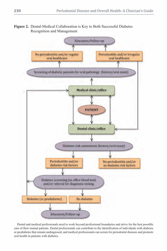
230 Periodontal Disease and Overall Health: A Clinician's Guide
Figure 2. Dental-Medical Collaboration is Key to Both Successful Diabetes
Figure 2. Recognition and Management
Dental and medical professionals need to work beyond professional boundaries and strive for the best possible
care of their mutual patients. Dental professionals can contribute to the identification of individuals with diabetes
or prediabetes that remain undiagnosed, and medical professionals can screen for periodontal diseases and promote
oral health in patients with diabetes.

CHAPTER 14 Dental and Medical Comanagement of Patients with Diabetes 231
patients have of their risk for periodontal
disease and their attitudes towards oral
health. Only a third of the 101 patients who
participated in the study were aware of their
increased risk for periodontitis as opposed to
their knowledge of the risk for other diabetic
complications, which ranged from 84% to
99%. History of prior dental care was spo-
radic, with 43% reporting seeing a dentist
within the past year. An earlier study from
Sweden42 had reported that 83% of diabetic
patients were unaware of the link between
diabetes and oral health, and that 48% be-
lieved that their dentist/dental hygienist were
unaware that they even had diabetes. Oral
health does not appear to be a priority for pa-
tients with diabetes.11,43-45 Tomar and Lester
reported that diabetic individuals were less
likely to visit a dentist than a nondiabetic
individual in the preceding 12 months and,
interestingly, the leading reason for not see-
ing a dentist was “lack of perceived need.”11
Competing financial and time commitments
may explain the inadequacy of routine den-
tal care in patients with diabetes.46
Dental professionals have an opportu-
nity and the responsibility to educate their
patients about the diabetes-oral health link
and promote good oral and overall health
behaviors. They should play a supporting
role in modifying patient behavior and habits
related to risk factors that may exacerbate di-
abetes-associated complications. Specifi-
cally, as part of oral health education, dental
practitioners and their teams can reinforce
the need for regular dental visits and proper
oral hygiene, but also the need for proper
nutrition, exercise, smoking cessation, ad-
herence to medication regimens, regular
monitoring of blood glucose levels, and reg-
ular medical follow ups, as indicated by the
patient’s physician. Patients should be en-
couraged to achieve the best glycemic con-
trol possible, as good control can improve
oral health and lead to better and more pre-
dictable perio dontal treatment outcomes. To
this end, patients need to know that they are
at greater risk for increased prevalence,
severity, and progression of periodontitis,
and that perio dontitis has been recognized
as a condition often found in patients with
diabetes. Indeed, the American Diabetes
As sociation Standards of Medical Care in
Diabetes recognize that every patient with
diabetes needs to see a dentist for appropri-
ate evaluation and treatment of oral dis-
eases.4 Patients need to be informed that
proper control of periodontal infections may
even have a beneficial effect on their level of
metabolic control and systemic inflamma-
tion, as well as the risk for vascular and kid-
ney complications.20
Patients must comprehend the overar-
ching principle that medical and dental pro-
fessionals have common goals: providing
the best possible care to their patients and
helping them avoid complications (Box 3).
Multi factorial, complex diseases such as dia -
betes and periodontitis interrelate and can
am plify one another, creating an imperative
for a comanagement model that has the po-
tential to improve patient outcomes.
THE ROLE OF MEDICAL
PROFESSIONALS
Members of the medical and dental ac-
ademic communities—dentists, hygienists,
physicians, nurses, representatives from den-
tal and diabetes professional societies—as
well as representatives from dental/medical
insurance carriers have convened a number
of workshops over the past few years to ad-
dress oral-systemic links and discuss issues
related to communication among different
healthcare professionals and patients.
Recently, two such symposia and their
subsequent reports highlighted these issues
and offered recommendations. Following the
“Scottsdale Project” meeting in April 2007,
a panel of experts presented a consensus
report that stated that “it is appropriate
to develop guidelines to assist medical

232 Periodontal Disease and Overall Health: A Clinician's Guide
providers in identifying patients who are at
risk for periodontal disease or screening
patients who may have undiagnosed perio -
dontal disease.”47 Similarly the report follow -
ing the July 2007 “Oral-Systemic Diseases:
From Bench to Chair—Putting Information
into Practice” symposium stated that “there
is a need for coordination and cooperation
between dental and medical health profes-
sionals with regard to screening for and
diag nosing diseases or conditions that affect
patients who traditionally have been cared
for by other healthcare providers. Therefore,
medical practitioners need to be aware of
oral diseases and make appropriate recom-
mendations and referrals.”48
The concepts emphasized above are espe -
cially important for medical professionals
who treat patients with diabetes (e.g., intern -
ists, diabetologists, nurses, diabetes educa-
tors) as these patients are more likely to have
perio dontal disease and more likely to have
less regular dental care. Medical care pro vid -
ers need to discuss with their diabetic pa-
tients the importance of oral health and its
relationship to the diabetic state; about the
potential sequelae of long-standing, un-
treated oral infections; and to provide edu-
cational brochures and other relevant mate-
rial. By simply asking patients if they have
a dentist and when was the last time they
visited him/her, physicians can send a power -
ful message and play a significant role in
promoting oral health and preventing oral
complications in patients with diabetes.
Screening for Periodontal Changes
and Key Questions to Ask
The guidelines for diagnosis of perio -
dontal diseases include a detailed periodon-
tal evaluation, including probing depth meas-
urements and intraoral radiographs, which
are not feasible in most medical settings.
Nevertheless, medical providers can screen
for periodontal diseases (Figure 2) based on
patient history, symptoms (sore, bleeding
gums, sensitive teeth, history of abscesses)
and a visual assessment of the patient’s
mouth for relevant signs, such as:
• food debris or plaque around teeth;
Box 3. Key Messages All Healthcare Providers Can Reinforce
� Emphasize the importance of good control (HbA1c, blood pressure, cholesterol) for
complication prevention
� Promote a healthy lifestyle
� Reinforce self-exams
� Explain the benefits of comprehensive multidisciplinary care and emphasize the
importance of regular appointments with medical and oral healthcare providers
How Can a Busy Healthcare Provider Find the Time to Give Key Messages?
� Do not give every message at one appointment
� Customize and prioritize messages according to the patient’s needs
� Provide patient with a computer-generated reminder of key messages discussed
� Document what is accomplished at each appointment and the patient’s response
� Create pamphlets for office or use materials available through national diabetes or
dental organizations and professional societies
� Include key messages in office newsletters
Source: Adapted from the 2007 National Diabetes Education Program publication “Working together
to manage diabetes: a guide for pharmacists. podiatrists, optometrists, and dental professionals.” Atlanta,
GA. US Department of Health and Human Services, Public Health Service, Centers for Disease Control
and Prevention, National Center for Chronic Disease Prevention and Health Promotion, 2007.51

CHAPTER 14 Dental and Medical Comanagement of Patients with Diabetes 233
• red, swollen, receding, or bleeding
gums;
• loose teeth, separation of teeth;
• oral abscesses;
• missing teeth; or
• halitosis.
If diabetic patients tell a member of the
medical team that they have not seen a dentist
in the last year, they should be immediately
referred to one. If a patient has seen a dentist
in the past year, but presents with detectable
signs or symptoms of oral/periodontal infec-
tions, he or she should again be referred to a
dentist. Finally, physicians should advise all
poorly controlled diabetic patients to see a
dentist/periodontist for evaluation and treat-
ment on a regular, ongoing basis. Medical care
providers should also facilitate communica-
tion with treating dental practitioners by offer -
ing information on the patients’ medical back-
ground, level of glycemic control, presence of
complications, and comorbidities. Further-
more, they should be available to offer advice
on medical management modifications that
may be necessary, and be open to a meaning-
ful professional interaction in order to assure
that patients receive the best possible care.
PATIENT-CENTERED TEAM CARE
Finally, the concept of a “syndemic
approach to diabetes management” as intro-
duced and discussed in the dental literature
by Hein and Small36 deserves some men-
tion. “Syndemic” is a term originally used to
describe a set of two or more linked health
problems, which synergistically contribute to
excess burden in a population.49 A syndemic
orientation has the potential to provide a
framework that can guide more efficient and
effective initiatives because healthcare pro -
viders will not approach diseases like dia-
betes and periodontitis as discrete problems,
and will be prompted to collaborate across
and beyond professional boundaries.
The model of “working together” has
been discussed in the diabetes literature
extensively. In 2001, the National Diabetes
Education Program (NDEP), a joint program
of the National Institutes of Health and the
Centers for Disease Control and Prevention,
published a report titled “Team Care: Com -
prehensive Lifetime Management for Dia-
betes.”50 This report was created to help or-
ganizational leaders of healthcare systems
and purchasers of healthcare to implement
multidisciplinary team care for people with
diabetes in all clinical settings, and set forth
an analysis of the evidence that supports
team care as an effective method for chronic
disease management.
The executive summary of this report
stated that although primary care physicians
currently provide 80% to 95% of diabetes
care in the US, they cannot do all that is re-
quired and often are discouraged that the cur-
rent medical system does not function well for
people with diabetes.50 The challenge is to
find a way to meet the needs of patients with
diabetes by broadening the opportunities for
delivery of care. Team care meets this chal-
lenge by integrating the skills of different
healthcare professionals with those of the pa-
tient and family members to create a com-
prehensive lifetime diabetes management pro-
gram. The report highlights that if diabetes
care is to achieve the health benefits that mod-
ern science has made possible, it must be:
• continuous, not episodic;
• proactive, not reactive;
• planned, not sporadic;
• patient-centered rather than provider-
centered; and
• population-based, as well as individual-
based.
CONCLUSION
Although the model of multidiscipli-
nary, patient-centered care presents many
challenges, all healthcare providers should
strive to participate in it. There is no doubt
that by changing their thinking and trying to
adopt these concepts into everyday practice,

234 Periodontal Disease and Overall Health: A Clinician's Guide
healthcare providers can render better health-
care and more predictable therapeutic out-
comes, maximize their success in combating
the diabetes epidemic, and play a significant
role in promoting the oral and overall health
of patients. In the “Working Together to
Manage Diabetes” 2007 publication by the
NDEP, all healthcare providers are called
upon to play a role in diabetes primary pre-
vention and in diabetes control.51 Among
others, dentists and dental hygienists can
make a difference in primary prevention and
management because patients are seen on a
regular basis by them, patients trust them,
and a few words from them can have a ma-
jor impact on patients’ healthcare behavior.
Supplemental Readings
American Diabetes Association Position Statement. Di-
agnosis and classification of diabetes mellitus. Diabetes
Care 2010;33(Suppl 1):S62–S69.
Borrell LN, Kunzel C, Lamster I, Lalla E. Diabetes in
the dental office: using NHANES III to estimate the
probability of undiagnosed disease. J Periodontal Res
2007;42:559–565.
Hein C, Small D. Combating diabetes, obesity, peri-
odontal disease and interrelated inflammatory condi-
tions with a syndemic approach. Grand Rounds in Oral-
Systemic Medicine 2006;2:36–47.
Kunzel C, Lalla E, Lamster IB. Management of the pa-
tient who smokes and the diabetic patient in the dental
office. J Periodontol 2006;77:331–340.
Working together to manage diabetes: a guide for phar-
macists, podiatrists, optometrists, and dental profes-
sionals. Atlanta, GA: U.S. Department of Health and
Human Services, Public Health Service, Centers for
Disease Control and Prevention, National Center for
Chronic Disease Prevention and Health Promotion,
2007.
REFERENCES1. Centers for Disease Control and Prevention. Na-
tional diabetes fact sheet: general information and
national estimates on diabetes in the United States,
2007, Atlanta, GA: U.S. Department of Health and
Human Services, Centers for Disease Control and
Prevention, 2008.
2. International Diabetes Federation. Diabetes preva-
lence. Available at: http://www.idf.org/home. Ac-
cessed: January 24, 2010.
3. American Diabetes Association. Diabetes statis-
tics. Available at: http://www.diabetes.org/diabetes-
statistics. jsp. Accessed: January 24, 2010.
4. American Diabetes Association Position Statement.
Standards of medical care in diabetes—2010. Dia-
betes Care 2010;33(Suppl 1):S11–S61.
5. Diabetes Prevention Program Research Group. The
prevalence of retinopathy in impaired glucose tol-
erance and recent-onset diabetes in the Diabetes
Prevention Program. Diabet Med 2007;24:137–144.
6. Knowler WC, Barrett-Connor E, Fowler SE, Ham-
man RF, Lachin JM, Walker EA, Nathan DM;
Diabetes Prevention Program Research Group.
Reduction in the incidence of type 2 diabetes with
lifestyle intervention or metformin. N Engl J Med
2002;346:393–403.
7. Albandar JM. Periodontal diseases in North Amer-
ica. Periodontol 2000 2002;29:31–69.
8. Papapanou PN. Periodontal diseases: epidemiol-
ogy. Ann Periodontol 1996;1:1–36.
9. Hugoson A, Sjodin B, Norderyd O. Trends over
30 years, 1973–2003, in the prevalence and sever-
ity of periodontal disease. J Clin Periodontol 2008;
35:405–414.
10. American Association of Diabetes Educators.
Dia betes Education Fact Sheet. Available at:
http://www.diabeteseducator.org/export/sites/aade/_
resources/pdf/Diabetes_Fact_Sheet.pdf Accessed:
January 24, 2010.
11. Tomar SL, Lester A. Dental and other health care
visits among U.S. adults with diabetes. Diabetes
Care 2000;23:1505–1510.
12. American Dental Association. 2007 Public Opinion
Survey. American Dental Association Survey Cen-
ter, 2008.
13. The Diabetes Control and Complications Trial Re-
search Group. The effect of intensive treatment of
diabetes on the development and progression of
long-term complications in insulin-dependent dia-
betes mellitus. N Engl J Med 1993;329:977–986.
14. Turner RC, Cull CA, Frighi V, Holman RR.
Glycemic control with diet, sulfonylurea, met-
formin, or insulin in patients with type 2 diabetes
mellitus: progressive requirement for multiple ther-
apies (UKPDS 49). UK Prospective Diabetes Study
(UKPDS) Group. JAMA 1999;281:2005–2012.
15. Hoerger TJ, Segel JE, Gregg EW, Saaddine JB. Is
glycemic control improving in U.S. adults? Dia-
betes Care 2008;31:81–86.
16. Cryer PE. Hypoglycemia: still the limiting factor in
the glycemic management of diabetes. Endocr Pract
2008;14:750–756.

CHAPTER 14 Dental and Medical Comanagement of Patients with Diabetes 235
17. Lamster IB, Lalla E, Borgnakke WS, Taylor GW.
The relationship between oral health and diabetes
mellitus. J Am Dent Assoc 2008;139(Suppl):19S–
24S.
18. Mealey BL, Oates TW; American Academy of
Periodontology. Diabetes mellitus and periodontal
diseases. J Periodontol 2006;77:1289–1303.
19. Wilson TG Jr. Compliance. A review of the litera-
ture with possible applications to periodontics. J
Periodontol 1987;58:706–714.
20. Taylor GW, Borgnakke WS. Periodontal disease:
associations with diabetes, glycemic control and
complications. Oral Dis 2008;14:191–203.
21. Genco RJ, Grossi SG, Ho A, Nishimura F, Mura -
yama Y. A proposed model linking inflammation
to obesity, diabetes, and periodontal infections. J
Perio dontol 2005;76:2075–2084.
22. Grunfeld C, Soued M, Adi S, Moser AH, Dinarello
CA, Feingold KR. Evidence for two classes of cyto -
kines that stimulate hepatic lipogenesis: relation-
ships among tumor necrosis factor, interleukin-1
and interferon-alpha. Endocrinology 1990;127:46–
54.
23. Pickup JC, Mattock MB, Chusney GD, Burt D.
NIDDM as a disease of the innate immune sys-
tem: association of acute-phase reactants and inter -
leukin-6 with metabolic syndrome X. Diabetologia
1997;40:1286–1292.
24. Saremi A, Nelson RG, Tulloch-Reid M, Hanson
RL, Sievers ML, Taylor GW, Shlossman M, Ben-
nett PH, Genco R, Knowler WC. Periodontal dis-
ease and mortality in type 2 diabetes. Diabetes
Care 2005;28:27–32.
25. Shultis WA, Weil EJ, Looker HC, Curtis JM,
Shlossman M, Genco RJ, Knowler WC, Nelson
RG. Effect of periodontitis on overt nephropathy
and end-stage renal disease in type 2 diabetes.
Diabetes Care 2007;30:306–311.
26. Demmer RT, Jacobs DR Jr., Desvarieux M. Perio -
dontal disease and incident type 2 diabetes: results
from the First National Health and Nutrition Ex-
amination Survey and its epidemiologic followup
study. Diabetes Care 2008;31:1373–1379.
27. Berwick DM. Disseminating innovations in health
care. JAMA 2003;289:1969–1975.
28. Kunzel C, Lalla E, Albert DA, Yin H, Lamster IB.
On the primary care frontlines: the role of the gen-
eral practitioner in smoking-cessation activities and
diabetes management. J Am Dent Assoc 2005;136:
1144–1153.
29. Kunzel C, Lalla E, Lamster IB. Management of
the patient who smokes and the diabetic patient in
the dental office. J Periodontol 2006;77:331–340.
30. Forbes K, Thomson WM, Kunzel C, Lalla E, Lam-
ster IB. Management of patients with diabetes by
general dentists in New Zealand. J Periodontol
2008;79:1401–1408.
31. Kunzel C, Lalla E, Lamster I. Dentists’ manage-
ment of the diabetic patient: contrasting generalists
and specialists. Am J Public Health 2007;97:725–
730.
32. National Diabetes Information Clearinghouse, a
service of the National Institute of Diabetes and Di-
gestive and Kidney Diseases, NIH. Hypoglycemia.
Available at: http://diabetes.niddk.nih.gov/dm/pubs/
hypoglycemia/. Accessed: January 24, 2010.
33. Smiley DD, Umpierrez GE. Perioperative glucose
control in the diabetic or nondiabetic patient. South
Med J 2006;99:580–589.
34. Kitabchi AE, Wall BM. Management of diabetic
ketoacidosis. Am Fam Physician 1999;60:455–464.
35. Glick M, Greenberg BL. The potential role of den-
tists in identifying patients’ risk of experiencing
coronary heart disease events. J Am Dent Assoc
2005;136:1541–1546.
36. Hein C, Small D. Combating diabetes, obesity,
perio dontal disease and interrelated inflammatory
conditions with a syndemic approach. Grand Rounds
Oral-Systemic Med 2006;2:36–47.
37. Tabaei BP, Herman WH. A multivariate logistic
regression equation to screen for diabetes: develop -
ment and validation. Diabetes Care 2002;25:1999–
2003.
38. Borrell LN, Kunzel C, Lamster I, Lalla E. Diabetes
in the dental office: using NHANES III to estimate
the probability of undiagnosed disease. J Perio -
dontal Res 2007;42:559–565.
39. Strauss SM, Russell S, Wheeler A, Norman R, Bor-
rell LN, Rindskopf D. The dental office visit as a
potential opportunity for diabetes screening: an
analysis using NHANES 2003–2004 data. J Public
Health Dent 2009, Epub ahead of print; doi: 10.1111/
j.1752–7325.2009.00157.x.
40. American Diabetes Association. Diabetes Risk
Calculator. Available at: http://www.diabetes.org/
risktest.jsp. Accessed: January 24, 2010.
41. Allen EM, Ziada HM, O’Halloran D, Clerehugh V,
Allen PF. Attitudes, awareness and oral health-
related quality of life in patients with diabetes. J
Oral Rehabil 2008;35:218–223.
42. Sandberg GE, Sundberg HE, Wikblad KF. A con-
trolled study of oral self-care and self-perceived
oral health in type 2 diabetic patients. Acta Odon-
tol Scand 2001;59:28–33.
43. Karikoski A, Ilanne-Parikka P, Murtomaa H. Oral
self-care among adults with diabetes in Finland.
Community Dent Oral Epidemiol 2002;30:216–
223.
44. Thorstensson H, Falk H, Hugoson A, Kuylenstierna
J. Dental care habits and knowledge of oral health

236 Periodontal Disease and Overall Health: A Clinician's Guide
in insulin-dependent diabetics. Scand J Dent Res
1989;97:207–215.
45. Mayfield JA, Rith-Najarian SJ, Acton KJ, Schraer
CD, Stahn RM, Johnson MH, Gohdes D. Assess-
ment of diabetes care by medical record review.
The Indian Health Service model. Diabetes Care
1994;17:918–923.
46. Moore PA, Orchard T, Guggenheimer J, Weyant
RJ. Diabetes and oral health promotion: a survey of
disease prevention behaviors. J Am Dent Assoc 2000;
131:1333–1341.
47. Hein C, Cobb C, Iacopino A. Report of the inde-
pendent panel of experts of the Scottsdale Project.
Grand Rounds Oral-Systemic Med 2007;Suppl:6–
27.
48. Lamster IB, DePaola DP, Oppermann RV, Papa-
panou PN, Wilder RS. The relationship of perio -
dontal disease to diseases and disorders at distant
sites: communication to health care professionals
and patients. J Am Dent Assoc 2008;139:1389–1397.
49. Singer M. AIDS and the health crisis of the U.S.
urban poor; the perspective of critical medical an-
thropology. Soc Sci Med 1994;39:931–948.
50. Team care: Comprehensive lifetime management
for diabetes. Atlanta, GA: U.S. Department of
Health and Human Services, Public Health Service,
Centers for Disease Control and Prevention, Na-
tional Center for Chronic Disease Prevention and
Health Promotion, 2001.
51. Working together to manage diabetes: A guide for
pharmacists, podiatrists, optometrists, and dental
professionals. Atlanta, GA: U.S. Department of
Health and Human Services, Public Health Service,
Centers for Disease Control and Prevention, Na-
tional Center for Chronic Disease Prevention and
Health Promotion, 2007.

1. Define the pathogenesis of coronary
atherosclerosis and recognize its acute
and chronic clinical presentations
2. Comprehend the scientific basis that
identifies risk factors for CVD
3. Explain the rationale that justifies
intervention by risk factor reduction
4. Describe commonly used medica-
tions for patients with CVD and
their impact on the delivery of oral
and dental care
5. Discuss principles of comanagement
of patients with cardiovascular dis-
ease and periodontal disease
AThEROsClEROTIC
CARDIOvAsCUlAR DIsEAsE
Atherosclerosis has been defined as a
progressive disease process that involves the
large- to medium-sized muscular and large
elastic arteries. Coronary atherosclerosis may
obstruct blood flow and result in ischemia to
the myocardium tissue that is dependent
on the blood supply of the diseased artery.
Lesions caused by atherosclerosis may also
rupture, and the resulting thrombus may be
clinically silent or may cause a fatal myo -
cardial infarction (MI, aka “heart attack”).
Approximately 40% of deaths in the United
States are attributed to the complications of
atherosclerosis; about half of these sequelae
are represented by coronary atherosclerosis
complicated by thrombosis and MI.4
Pathogenesis
Atherosclerosis generally begins in
childhood, and manifests as a flat fatty streak
usually detected only as an incidental finding
INTRODUCTION
Cardiovascular disease (CVD) accounts
for 29% of deaths worldwide, ranks as the
leading cause of death, and poses the great-
est threat in low-income and middle-income
countries.1 Atherosclerosis, which is a major
component of cardiovascular disease, affects
one in four persons and contributes to ~ 40%
of deaths annually in the United States.2
The estimated cost of treating CVD in
2008 was $448.5 billion, a 20% increase in
recent years (http://www.americanheart.org/
presenter.jhtml?identifier=4475, accessed
2/20/2010). The pervasiveness of cardio -
vascular disease makes it a natural target for
prevention by all healthcare professionals. If
prevention programs could reduce the acute-
treatment costs and associated morbidity of
CVD by 20% per year, the savings would be
at least $80 billion per year in healthcare
costs. By comparison, the cost of Medicare
Part D was estimated to be ~ $70 billion in
2008 (http://www.cbo.gov/ftpdoc.cfm?index
=6076&type=0#table1, accessed 2/20/2010).
Several studies point to the possibility that
periodontal care in patients with cardio -
vascular disease significantly reduced med-
ical care costs.3 This chapter reviews the
known risk factors for CVD that are targets
for disease prevention, and discusses a cur-
rent and future rationale for oral, dental, and
medical healthcare practitioners to work
together to implement optimal CVD risk fac-
tor reduction.
Educational Objectives
After reading this chapter, the reader
should be able to:
CHAPTER 15
Dental and Medical Comanagement of Cardiovascular Disease
Timothy C. Nichols, David W. Paquette

at an autopsy that is performed for other rea-
sons.5,6 The advanced raised lesion is called
an “atheroma,” which consists of elevated
focal intimal plaques with a central core con-
taining necrotic cells, cholesterol ester crys-
tals, lipid-laden foam cells, and plasma pro-
teins, including fibrin and fibrinogen. This
central core is also associated with a cellular
infiltrate comprised of hypertrophic smooth
muscle cells, macrophages, and sparse T-lym-
phocytes.One theory of atherogenesis is that
the atherosclerotic plaque develops as a re-
sponse to injury to the vascular endothelium,
and that the endothelial injury is the primary
event in atherogenesis. When the endothe-
lium is even minimally injured, platelets and
monocytes accumulate and attach to the dam-
aged wall. As platelets aggregate around the
injury, they release thromboxane, promoting
further platelet aggregation and coronary
vasoconstriction. Monocytes invade the in-
tima, and scavenge for lipids and other extra -
cellular materials. These cells release vari-
ous growth factors that attract more smooth
muscle cells from the media of the artery with
resultant intima hyperplasia. As the lesion
progresses, fibrosis, lipid deposition, necrosis,
and calcification may ensue to yield a com-
plicated plaque. Inflammation, oxidative, and
mechanical stress through high blood pres-
sure can also induce primary injury of the
arterial endothelium by which the pathogen-
esis of atherosclerosis can be initiated and
propagated.
Risk Factors for the Development of
Coronary Atherosclerosis
Traditional major risk factors for athero -
sclerotic cardiovascular disease include:
• Cigarette smoking
• Hypertension (> 140/90 mmHg)
• High levels of low-density lipo protein
cholesterol (LDLC, defined as > 100
mg/dL)
• Low levels of high-density lipo protein
cholesterol (HDLC, defined as < 40
mg/dL for men or < 35 mg/dL for
women)
• Insulin resistance
• Diabetes mellitus
• Family history of premature coronary
heart disease (occurring in a parent
age 45)
• Age (men 45 years, women 55
years)
• Obesity (body mass index > 30 kg/m2)
• Physical inactivity and an atherogenic
diet (Figure 1)
It is also recognized that these factors
can interact with each other to increase the
risk of CVD.7 For example, the Framingham
Heart Study involved more than 3,000 pa-
tients and showed that for total cholesterol
levels between 185 mg/dL and 335 mg/dL,
cardiovascular risk was elevated further with
the addition of each of the following risk fac-
tors: glucose intolerance; elevated systolic
blood pressure; cigarette smoking; and left
ventricular hypertrophy on electrocardiogra-
phy.8 Data from the Framingham Heart Study
and two other large prospective cohort studies,
the Chicago Heart Association Detection
Project in Industry (n = 35,642) and the
Multiple Risk Factor Intervention Trial
(n = 347,978), indicated that the majority of
patients with fatal coronary artery athero-
sclerosis or nonfatal MI present with at least
one of four risk factors: cigarette smoking;
diabetes mellitus; hyperlipidemia; and hyper -
tension.9 For fatal MI due to coronary artery
atherosclerosis, exposure to at least one risk
factor ranged from 87% to 100% for all three
cohort studies. For nonfatal MI in the Fram-
ingham Heart Study, prior exposure to at least
one risk factor was found in 92% of men and
87% of women ages 40 to 59 at baseline.
Furthermore, another recent analysis involv-
ing 14 international randomized clinical trials
(n = 122,458) showed that one of these four
conventional risk factors was present in 84.6%
of men and 80.6% of women with coronary
artery disease.10
238 Periodontal Disease and Overall Health: A Clinician's Guide

The Role of Inflammatory Markers
Recent attention has focused on ele-
vated serum C-reactive protein (CRP) as a
strong and independent risk factor or predic-
tor of events due to coronary artery athero-
sclerosis, such as MI or sudden death.11 CRP
CHAPTER 15 Dental and Medical Comanagement of Cardiovascular Disease 239
Figure 1. Cardiac Risk Factors for Coronary Atherosclerosis Recognition and Management
Multiple risk factors are shown, many of which are known to increase risk in an additive fashion when pres-
ent concurrently. A cross section of an artery is shown with a raised atherosclerotic plaque that obstructs a portion
of the lumen. From Netter's Cardiology. Reproduced with permission.

is an acute-phase reactant primarily produced
by the liver in response to infection or
trauma. Other tissues may be involved in its
synthesis including smooth muscle cells
from normal coronary arteries and diseased
coronary artery bypass grafts.12,13 CRP ap-
pears to be directly involved in augmenting
the innate inflammatory response via induc-
tion of prothrombotic factors (e.g., plas-
minogen activator inhibitor-1, pro-inflam-
matory adhesion molecules, and monocyte
chemoattractant protein-1) and interference
with endothelial nitric oxide synthase.14 In
the Physicians’ Health Study, an epidemio-
logic investigation of more than 22,000
healthy middle-aged men with no clinical
evidence of disease, increasing levels of
serum high-sensitivity CRP at study entry
were associated with up to a three-fold in-
crease in the risk of incident MI and a two-
fold increase in risk of ischemic stroke.15
When compared with other potential serum
biomarkers, such as homocysteine, lipopro-
tein(a), interleukin-6, intracellular adhesion
molecule-1, serum amyloid A, and standard
lipid measures, CRP proved to be the single
strongest predictor of cardiovascular risk in
apparently healthy participants in the
Women’s Health Study (n = 28,263).16,17 Ac-
cordingly, the relative risk ratio for the high-
est versus lowest quartile of serum CRP con-
centrations was 4.4 (95% CI:1.7–11.3).
Moreover, the addition of serum CRP to tra-
ditional cholesterol screening enhanced car-
diovascular risk prediction and proved to be
independent of LDLC. The poorest event-
free survival in women was among those
with high LDLC and high CRP levels, and
the best event-free survival was among those
with low LDLC and low CRP levels. No-
tably, individuals with low LDLC levels but
high CRP levels were at higher risk than
those with high LDLC levels but low CRP
levels.
Very recent data show that treatment with
HMG-CoA reductase inhibitors (“statins”)
for asymptomatic individuals with elevated
CRP levels but normal cholesterol levels re-
duces risk for future cardiovascular events.11
These provocative findings have triggered
considerable debate that may fundamentally
alter our approach to primary prevention of
coronary atherosclerosis.
Prevention of Coronary Atherosclerosis
by Risk Factor Modification
Since atherosclerosis continues to in-
crease in prevalence in developed countries,
it is counterintuitive that death rates from
cardiovascular diseases overall have de-
creased by more than a third in the past two
decades. The explanations most often given
for this apparent paradox include success of
primary- and secondary-prevention strate-
gies, improvements in patient care, and re-
habilitation. These findings underscore the
importance of recognizing risk factors (Fig-
ure 1) and optimizing strategies for risk fac-
tor reduction (Figure 2). These strategies
constitute an opportunity for medical and
dental professionals to work for a common
goal of continued improvement in cardio-
vascular health, but there are formidable
challenges to overcome in order to achieve
this goal.18
First is to recognize that we are pro -
viding encouragement for lifestyle changes
that patients may find very difficult, includ-
ing smoking cessation, weight reduction,
dietary changes, regular exercise, and com-
pliance with prescribed medications for
elevated cholesterol, blood pressure, and
diabetes mellitus. While both dental and
medical healthcare providers can fairly
encourage all of these, continued patient
support over time is essential. Second is to
remember that atherosclerosis may be pres-
ent but not clinically evident for years,19
underscoring the need for sustained efforts
at risk factor reduction even in otherwise
healthy individuals.
240 Periodontal Disease and Overall Health: A Clinician's Guide

CHAPTER 15 Dental and Medical Comanagement of Cardiovascular Disease 241
Figure 2. Non-drug Therapy for Prevention of Coronary Atherosclerosis
Risk factors that can be addressed by all healthcare practitioners are shown. A standardized approach that pro-
vides patient education about these risk factors and support for adhering to these lifestyle changes is the foundation
of risk factor reduction. Other risk factors are highly likely to be identified over time, and this strategy will be
continually updated accordingly. From Netter's Cardiology. Reproduced with permission.
Clinical Presentation of
Cardiovascular Disease
Recognition of the symptoms of coro-
nary atherosclerosis is essential and not al-
ways straightforward. Three classical clinical
presentations are possible. Angina pectoris
or chest pain is usually retrosternal pressure,
tightness, heaviness, or discomfort that radi-
ates to the left arm, jaw, or back, and is often
associated with dyspnea, diaphoresis, nau-
sea, and a feeling of impending doom (Figure
3). Angina with exercise or exertion, large
meals, or emotional stress is usually pre-
dictable and relieved by rest, a pattern usually
called “stable angina.” More ominous is chest
pain at rest or a sudden increase in frequency
or ease of onset of angina, a pattern often
termed “unstable or accelerating angina.”
Obtaining a history of angina requires time
and patience and the recognition that there are
several causes of chest pain.
In addition, women and patients with dia -
betes mellitus may have a completely different
pattern to their angina or may have “anginal
equivalents,” such as dyspnea alone or ab-
dominal discomfort. A high index of clinical
suspicion in patients with risk factors for coro-
nary atherosclerosis is of paramount impor-
tance for recognition of anginal equivalents.
The second classic presentation of coro-
nary atherosclerosis is MI. Patients experi-
encing an MI usually complain of prolonged
and sustained (> 5 minutes) chest pain not re-
lieved by rest or nitroglycerin. Associated
symptoms may also be present, such as nau-
sea and vomiting, and palpitations signaling
irregular heart rhythms. Also alarming is the
presence of heart failure symptoms, including
weakness and dyspnea. The interval between
treatment and long-term prognosis is directly
proportional; prompt recognition and early
treatment are associated with marked im-
provement in outcomes.
The third manifestation of coronary
atherosclerosis is sudden cardiac death
when the heart has an irregular rhythm that
is unable to support blood pressure, usually
ventricular fibrillation. Tragically, coronary
atherosclerosis first presents as sudden car-
diac death in approximately 25% of patients.
The availability of personnel trained to rec-
ognize and treat ventricular fibrillation and
administer cardiopulmonary resuscitation is
the primary determinant of outcome. Both
community-based efforts and the presence of
automatic external defibrillators have been
important factors in improved survival for
patients experiencing sudden cardiac death.
MEDICAl MANAGEMENT OF
CORONARY AThEROsClEROsIs
At present, 80 million Americans are
thought to exhibit some form of cardiovas-
cular disease.20 Thus, many patients pre-
senting to oral and dental healthcare
providers will have coronary atherosclerosis
and will be receiving therapy. It will be im-
portant, therefore, to understand the basic
classes of drugs that are used in patients with
heart disease, especially the ones that might
have a direct impact on oral health or may
complicate oral and dental procedures.
Antiplatelet Therapy
All patients with atherosclerosis should
be on some form of antiplatelet therapy. The
minimal cost and relatively profound effec-
tiveness of aspirin make it the treatment of
choice in all patients who can tolerate taking
this medication. Another currently available
antiplatelet drug is the thienopyridine clopi-
dogrel, and often patients are on both aspirin
and clopidogrel. The primary concern for both
of these drugs is bleeding, especially with
procedures. If the procedure requires tem-
porary interruption of use of either of these
two drugs, this should be done in consulta-
tion with the patient’s regular physician. This
is especially important for patients who are
on clopidogrel to prevent clotting on drug-
eluting stents that have been implanted inside
one or more coronary arteries to open athero -
242 Periodontal Disease and Overall Health: A Clinician's Guide

CHAPTER 15 Dental and Medical Comanagement of Cardiovascular Disease 243
Figure 3. Angina Pectoris
Classic angina pectoris is retrosternal, radiates to the left arm or jaw, and feels like a heavy or constricting “vice-
like” discomfort. This illustration shows common precipitating factors. From Netter's Cardiology. Reproduced with
permission.

sclerotic obstructions. Abrupt cessation of
clopidogrel during the first year after place-
ment of a drug-eluting stent can be associ-
ated with acute thrombosis resulting in an
otherwise preventable heart attack.
Several investigational antiplatelet drugs
are close to FDA approval. Likely, bleeding
will be the major concern for all new anti -
platelet drugs. The challenge to all who de-
liver healthcare will be to maintain a current
understanding of the uses and side effects of
these novel therapies.
Beta Blockade
As with aspirin, beta blockade is rec-
ommended for all patients with cardiovas-
cular disease unless they have a contraindi-
cation, such as untreated conduction disease
in the heart with bradycardia, severe asthma,
difficult-to-control diabetes mellitus, and in
some cases, severe atherosclerosis in arteries
in the legs. In general, patients already re-
ceiving beta blockers should not experience
any complications during oral procedures.
In addition, cardiologists may recommend
beta blockers for select patients with heart
disease undergoing general anesthesia for
oral or dental procedures. The American
Heart Association provides guidelines for
such situations and these guidelines are con-
stantly being revised as new data become
available.21 Considerable planning and dis-
cussion between the cardiologist and den-
tist would be required in these cases.
ACE Inhibitors and ARBs
Patients with coronary atherosclerosis
and depressed heart function are generally
encouraged to take angiotensin-converting
enzyme (ACE) inhibitors. If they are intol-
erant to ACE inhibitors due to cough or other
issues, they may be prescribed angiotensin II
recep tor blockers (ARBs). Mortality rates
have repeatedly been shown to be lower in
post-MI infarction patients who are taking
ACE inhibitors. Both ACEs and ARBs are
powerful antihypertensive agents. For dental
patients on stable doses, these drugs are not
recognized to interfere with oral procedures
or dental care.
Nitrates
Both short- (less than 10 minutes) and
long- (several hours) acting nitrates are fre-
quently used to relieve and prevent, respec-
tively, myocardial ischemia. Nitrates may
also be used to treat heart failure, especially
in African-Americans, as well as hyper -
tension. A “nitrate-free” interval is recom-
mended on a daily basis to prevent tachy-
phylaxis. For dental patients on stable doses,
these drugs are not recognized to interfere
with oral procedures or dental care.
lipid-lowering Therapy
The current National Cholesterol Edu-
cation Program Guidelines recommend an
LDLC level of < 100 mg/dL for patients with
known cardiovascular disease and a total cho-
lesterol of < 200 mg/dL. Secondary causes of
hyerlipidemia include diabetes mellitus, liver
disease, and renal failure. Regardless of the
cause, all patients who have values above
these levels should receive dietary counsel-
ing, suggestions for weight reduction, and
encouragement to increase physical activity.
The most commonly used drugs for hyper-
lipidemia are the statins, which inhibit cho-
lesterol synthesis. Pharmacologic interven-
tion with the statin class of drugs is used to
further reduce serum lipids and the likeli-
hood of cardiovascular events, even in those
with average LDL concentrations.
Numerous clinical trials have consis-
tently demonstrated that statin drugs reduce
cardiovascular events by at least 25%.22 In
contrast, the effect of statins and other lipid-
lowering therapies on reducing the size of
atherosclerotic plaques is much smaller.23 This
finding has been the basis for seeking alter-
native explanations for why statins improve
outcomes so profoundly. For example, statins
244 Periodontal Disease and Overall Health: A Clinician's Guide
may have secondary anti-inflammatory ef-
fects. Indeed, CRP concentrations decrease
15%–50% with statin therapy.11 Thus, the
pleotropic effects of these drugs appear to im-
prove outcomes and markers of atheroscle-
rotic cardiovascular disease. Fibrates reduce
lipoprotein lipase activity and nicotinic acid
reduces tissue lipase activity, and very-low-
density lipoprotein synthesis. Both fibrates
and nicotinic acid reduce triglyceride levels
effectively. The cholesterol absorption in-
hibitor ezetimibe and bile acid reabsorption
inhibitors (e.g., cholestyramine) are often used
in combination with other drugs to achieve
target goals. Lipid-lowering drugs should not
impact the delivery of oral or dental care.
lDl Apheresis
In rare cases, patients with familial hy-
percholesterolemia do not respond to drug
therapy and require apheresis. In this case, the
patient’s blood is perfused over a column that
binds and thereby reduces their LDLC levels.
Patients with familial hypercholesterolemia
frequently develop coronary atherosclerosis
by the second decade of life and should be
carefully evaluated for all procedures.
Coronary Angioplasty and
Bypass surgery
For patients with severe symptomatic
coronary atherosclerosis, interventions involve
physically expanding stenotic vessels via an-
gioplasty (with or without stenting), versus
revascularization via coronarybypass surgery.
As discussed above, these patients are likely to
be on medications that could increase the like-
lihood of bleeding during dental procedures.
ClINICAl RECOMMENDATIONs FOR
PATIENTs WITh PERIODONTITIs
AND AThEROsClEROTIC
CARDIOvAsCUlAR DIsEAsE
Current knowledge about the close asso-
ciation between periodontitis and atheroscle-
rotic cardiovascular disease is summarized in
Chapter 8. It is clear that although we do not
have ultimate proof, the balance of evidence
is sufficiently strong such that recommen-
dations for close cooperation among dental
and medical professionals in the manage-
ment of patients with periodontal disease
and cardiovascular disease are reasonable.
Recently there was an Editors’ Consensus
Report published simultaneously in the
American Journal of Cardiology and the
Journal of Periodontology that makes clini-
cal recommendations for patients with peri-
odontitis and patients with atherosclerotic
cardiovascular disease.18 The potential ben-
efits of applying current understanding of
the association between periodontal disease
and cardiovascular disease outweighs any neg-
ative effects of premature application of this
information. Four major recommendations are
made that provide guidance for physicians and
dentists who often care for patients either suf-
fering from, or at risk for, periodontal disease
and cardiovascular diseases.
These recommendations include:
1. Patients with periodontitis should be
informed that they may be at in-
creased risk for atherosclerotic car-
diovascular disease. Further, patients
with periodontitis who have one or
more risk factors for cardiovascular
disease should be specifically and
clearly referred for medical evalua-
tion if they have not been evaluated
in the past 12 months.
2. Patients with periodontitis should
have their risk for future cardiovas-
cu lar events assessed using validated
tools such as the Reynolds Risk Score
(http://www.reynoldsriskscore.org,
accessed 2/20/2010) or the National
Cholesterol Education Program Risk
Calculator (http://hp2010.nhlbihin.
net/atpiii/calculator.asp, accessed
2/20/2010). Those patients at high
risk should have a complete physical
examination by a physician.
CHAPTER 15 Dental and Medical Comanagement of Cardiovascular Disease 245

3. Patients with periodontitis who have
cardiovascular risk factors includ-
ing abnormal lipids, smoke ciga-
rettes, have hypertension, or suffer
from metabolic syndrome should
have their risk factors moderated
and be comanaged by the dentist as
well as the physician. It is especially
important for the dental profession
to be involved in cooperating with
the medical team in reducing ciga-
rette smoking and controlling diabetes
and weight, and modifying diet in
such patients. It is currently well rec-
ognized that dentists can play a sig-
nificant role in assisting patients in
smoking cessation programs. We are
proposing that dentists extend their
efforts to include assistance in weight
and diet control, and in reinforcing
compliance with blood pressure med-
ications in hypertensive patients.
4. Patients with atherosclerotic cardio-
vascular disease who do not have a
previous diagnosis of periodontal
disease should be examined for signs
of periodontal disease, including lost
teeth, gum recession, or inflamed
gingivae. They should be referred by
the medical team for a periodontal
evaluation. Furthermore, if perio -
dontitis is newly diagnosed or exists
in patients with atherosclerotic cardio -
vascular disease, periodontal therapy
should be carried out and a proactive
secondary prevention and mainte-
nance program should be instituted.
Physicians and dentists should em-
phasize the importance of timely
management of periodontal disease
in patients with cardiovascular dis-
ease to decrease their inflammatory
burden.
5. Patients with both periodontitis and
cardiovascular disease should have
members of the dental and medical
team work closely together to help
reduce risk factors, especially those
common to both diseases, such as
uncontrolled diabetes, smoking, and
obesity.
The Use of sugar
The dental profession has long been in-
volved in advising patients to reduce their
sugar intake to reduce the risk for dental
caries. It is reasonable for both dental and
medical professionals to make the recom-
mendations of maximum intake of added
sugars, especially in cardiovascular patients,
both for reduction of risk for cardiovascular
disease as well as risk for dental caries.
For example, a recent scientific state-
ment from the American Heart Association
addresses dietary sugars and cardiovascular
health. It is recommended that patients re-
duce the intake of sugars added to foods to
100 calories per day for women, and 150
calories per day for men. This is about 5–6
teaspoons of sugar or sugar equivalents per
day for the average adult woman, and 9 tea-
spoons per day for the average adult man. It
is realized that reduction of dietary carbo-
hydrates requires a multifaceted approach,
and that one, perhaps, easily implemented
approach is to reduce energy intake from
added sugars to levels suggested above.
Finally, it should be recognized that
there is no evidence that suggests that pa-
tients with periodontitis and atherosclerotic
cardiovascular disease should receive dif-
ferent periodontal treatment from other pa-
tients with periodontitis. In fact, recent stud-
ies suggest that standard treatments of
periodontitis in patients with cardiovascular
disease are effective and do not lead to ad-
verse cardiovascular outcomes.24 As with any
patient, the daily management of plaque and
gingival inflammation should be encouraged.
Oral hygiene instruction should be provided,
and the use of an antibacterial toothpaste and
mouthrinse and interproximal cleaning should
246 Periodontal Disease and Overall Health: A Clinician's Guide

be recommended with each patient and re-
inforced at each visit.
CONClUsIONs AND
FUTURE DIRECTIONs
Dental and medical healthcare providers
need to work together closely to provide op-
timal care for their patients with established
cardiovascular disease. A basic understand-
ing of the pathogenesis of cardiovascular
disease and commonly used medications will
provide a basis for preventing complications
during the delivery of dental and oral health-
care in patients with CVD.
The role of the oral and dental health
practitioner in the prevention of heart disease
has great potential. Such practitioners are
uniquely poised to discuss risk factor reduc-
tion with their patients that is paramount for
both oral and cardiovascular health. Preven-
tive interventions (primary or secondary) for
coronary atherosclerosis focus on recogni-
tion and reduction of modifiable risk factors
in patients (Figures 1 and 2). These ap-
proaches include blood pressure screening,
weight reduction, exercise, smoking cessa-
tion, diet modification, medication compli-
ance (especially with treatments for blood
pressure and diabetes), patient counseling,
and education. Initiating and maintaining
these lifestyle changes are not easy tasks,
but more likely to be adopted by patients
who receive consistent advice and encour-
agement from all of their healthcare providers.
For the present, the role of oral health in
atherosclerosis remains an association and
not a risk factor. Human trials that directly
address the role of periodontitis and athero-
sclerosis have been limited to the Periodon-
titis and Vascular Events study. This pilot
study was designed to determine how many
patients with combined symptomatic ather-
osclerosis and periodontitis would be needed
to document the fact that successful treat-
ment of periodontitis reduces sub sequent
coronary and carotid events.24 If perio dontitis
is proven to be a risk factor for cardiovascu-
lar disease, cardiologists and oral and dental
health practitioners will need to work even
more closely to provide optimal care for their
patients.
Supplemental Readings
Tricoci P, Allen JM, Kramer JM, Califf RM, Smith SC,
Jr. Scientific evidence underlying the ACC/AHA clini-
cal practice guidelines. JAMA 2009;301:831–841.
Fleisher LA, Beckman JA, Brown KA, Calkins H,
Chaikof E, Fleischmann KE, Freeman WK, Froehlich
JB, Kasper EK, Kersten JR, Riegel B, Robb JF, Smith
SC, Jr., Jacobs AK, Adams CD, Anderson JL, Antman
EM, Buller CE, Creager MA, Ettinger SM, Faxon DP,
Fuster V, Halperin JL, Hiratzka LF, Hunt SA, Lytle BW,
Nishimura R, Ornato JP, Page RL, Tarkington LG,
Yancy CW. ACC/AHA 2007 guidelines on periopera-
tive cardiovascular evaluation and care for noncardiac
surgery: executive summary: a report of the American
College of Cardiology/American Heart Association
Task Force on Practice Guidelines (Writing Committee
to Revise the 2002 Guidelines on Perioperative Cardio -
vascular Evaluation for Noncardiac Surgery). Anesth
Analg 2008;106:685–712.
McFarlane SI, Castro J, Kaur J, Shin JJ, Kelling D, Jr.,
Farag A, Simon N, El-Atat F, Sacerdote A, Basta E,
Flack J, Bakris G, Sowers JR. Control of blood pres-
sure and other cardiovascular risk factors at different
practice settings: outcomes of care provided to diabetic
women compared to men. J Clin Hypertens (Greenwich
2005;7:73–80.
Fleisher LA, Beckman JA, Brown KA, Calkins H,
Chaikof EL, Fleischmann KE, Freeman WK, Froehlich
JB, Kasper EK, Kersten JR, Riegel B, Robb JF. 2009
ACCF/AHA focused update on perioperative beta
blockade incorporated into the ACC/AHA 2007 guide-
lines on perioperative cardiovascular evaluation and
care for noncardiac surgery. J Am Coll Cardiol 2009;
54:e13–e118.
Hunt SA, Abraham WT, Chin MH, Feldman AM, Fran-
cis GS, Ganiats TG, Jessup M, Konstam MA, Mancini
DM, Michl K, Oates JA, Rahko PS, Silver MA, Steven-
son LW, Yancy CW. 2009 focused update incorporated
into the ACC/AHA 2005 Guidelines for the Diagnosis
and Management of Heart Failure in Adults: a report of
the American College of Cardiology Foundation/Amer-
ican Heart Association Task Force on Practice Guide-
lines: developed in collaboration with the International
Society for Heart and Lung Transplantation. Circulation
2009;119:e391–479.
CHAPTER 15 Dental and Medical Comanagement of Cardiovascular Disease 247
Runge MS, Stouffer G, Patterson C, eds. Netter's
Cardiology (Netter Clinical Sciences). Teterboro, New
Jersey: Icon Learning Systems LLC; 2010 (Second
edition anticipated publication release July 2010).
REFERENCEs1. Fuster V, Vedanthan R. Cardiovascular disease and
the UN Millennium Development Goals: Time to
move forward. Nat Clin Pract Cardiovasc Med
2008;5:593.
2. Thom T, Haase N, Rosamond W, Howard VJ,
Rumsfeld J, Manolio T, Zheng ZJ, Flegal K,
O’Donnell C, Kittner S, Lloyd-Jones D, Goff DC
Jr, Hong Y, Adams R, Friday G, Furie K, Gorelick
P, Kissela B, Marler J, Meigs J, Roger V, Sidney S,
Sorlie P, Steinberger J, Wasserthiel-Smoller S, Wil-
son M, Wolf P; American Heart Association Sta-
tistics Committee and Stroke Statistics Subcom-
mittee. Heart disease and stroke statistics—2006
update: A report from the American Heart Associ-
ation Statistics Committee and Stroke Statistics
Subcommittee. Circulation 2006;113:e85–151.
3. Albert DA, Sadowsky D, Papapanou P, Conicella
ML, Ward A. An examination of periodontal treat-
ment and per member per month (PMPM) medical
costs in an insured population. BMC Health Serv
Res 2006;6:103.
4. Mackman N. Triggers, targets and treatments for
thrombosis. Nature 2008;451:914–918.
5. Rader DJ, Daugherty A. Translating molecular dis-
coveries into new therapies for atherosclerosis. Na-
ture 2008;451:904–913.
6. Sanz J, Fayad ZA. Imaging of atherosclerotic car-
diovascular disease. Nature 2008;451:953–957.
7. Smith SC Jr. Current and future directions of car-
diovascular risk prediction. Am J Cardiol 2006;
97:28A–32A.
8. Kannel WB. High-density lipoproteins: epidemio-
logic profile and risks of coronary artery disease.
Am J Cardiol 1983;52:9B–12B.
9. Greenland P, Knoll MD, Stamler J, Neaton JD,
Dyer AR, Garside DB, Wilson PW. Major risk fac-
tors as antecedents of fatal and nonfatal coronary
heart disease events. JAMA 2003;290:891–897.
10. Khot UN, Khot MB, Bajzer CT, Sapp SK, Ohman
EM, Brener SJ, Ellis SG, Lincoff AM, Topol EJ. Preva-
lence of conventional risk factors in patients with
coronary heart disease. JAMA 2003;290:898–904.
11. Ridker PM, Danielson E, Fonseca FA, Genest J,
Gotto AM Jr, Kastelein JJ, Koenig W, Libby P,
Lorenzatti AJ, MacFadyen JG, Nordestgaard BG,
Shepherd J, Willerson JT, Glynn RJ; JUPITER
Study Group. Rosuvastatin to prevent vascular
events in men and women with elevated C-reactive
protein. N Engl J Med 2008;359:2195–2207.
12. Calabro P, Willerson JT, Yeh ET. Inflammatory cy-
tokines stimulated C-reactive protein production
by human coronary artery smooth muscle cells.
Circulation 2003;108:1930–1932.
13. Jabs WJ, Theissing E, Nitschke M, Bechtel JF,
Duchrow M, Mohamed S, Jahrbeck B, Sievers HH,
Steinhoff J, Bartels C. Local generation of C-reac-
tive protein in diseased coronary artery venous by-
pass grafts and normal vascular tissue. Circulation
2003;108:1428–1431.
14. Devaraj S, Xu DY, Jialal I. C-reactive protein in-
creases plasminogen activator inhibitor-1 expres-
sion and activity in human aortic endothelial cells:
Implications for the metabolic syndrome and
atherothrombosis. Circulation 2003;107:398–404.
15. Ridker PM, Cushman M, Stampfer MJ, Tracy RP,
Hennekens CH. Inflammation, aspirin, and the risk
of cardiovascular disease in apparently healthy
men. N Engl J Med 1997;336:973–979.
16. Ridker PM, Hennekens CH, Buring JE, Rifai N. C-
reactive protein and other markers of inflamma-
tion in the prediction of cardiovascular disease in
women. N Engl J Med 2000;342:836–843.
17. Ridker PM, Rifai N, Rose L, Buring JE, Cook NR.
Comparison of C-reactive protein and low-density
lipoprotein cholesterol levels in the prediction of
first cardiovascular events. N Engl J Med 2002;347:
1557–1565.
18. Friedewald VE, Kornman KS, Beck JD, Genco R,
Goldfine A, Libby P, Offenbacher S, Ridker PM,
Van Dyke TE, Roberts WC. Editors’ Consensus:
Periodontitis and Atherosclerotic Cardiovascular
Disease. Am J Cardiol 2009;104:59–68.
19. Berry JD, Liu K, Folsom AR, Lewis CE, Carr JJ,
Polak JF, Shea S, Sidney S, O’Leary DH, Chan C,
Lloyd-Jones DM. Prevalence and progression of
sub clinical atherosclerosis in younger adults with
low short-term but high lifetime estimated risk for
cardiovascular disease: the coronary artery risk de-
velopment in young adults study and multi-ethnic
study of atherosclerosis. Circulation 2009;119:382–
389.
20. Lloyd-Jones D, Adams R, Carnethon M, De
Simone G, Ferguson TB, Flegal K, Ford E, Furie
K, Go A, Greenlund K, Haase N, Hailpern S, Ho
M, Howard V, Kissela B, Kittner S, Lackland D,
Lisabeth L, Marelli A, McDermott M, Meigs J,
Mozaffarian D, Nichol G, O’Donnell C, Roger V,
Rosamond W, Sacco R, Sorlie P, Stafford R, Stein-
berger J, Thom T, Wasserthiel-Smoller S, Wong N,
Wylie-Rosett J, Hong Y. Heart disease and stroke
statistics—2009 update: a report from the American
Heart Association Statistics Committee and Stroke
Statistics Subcommittee. Circulation 2009;119:
480–486.
248 Periodontal Disease and Overall Health: A Clinician's Guide

21. Fleisher LA, Beckman JA, Brown KA, Calkins H,
Chaikof E, Fleischmann KE, Freeman WK,
Froehlich JB, Kasper EK, Kersten JR, Riegel B,
Robb JF, Smith SC Jr, Jacobs AK, Adams CD, An-
derson JL, Antman EM, Buller CE, Creager MA,
Ettinger SM, Faxon DP, Fuster V, Halperin JL,
Hiratzka LF, Hunt SA, Lytle BW, Nishimura R,
Ornato JP, Page RL, Riegel B, Tarkington LG,
Yancy CW. ACC/AHA 2007 guidelines on periop-
erative cardiovascular evaluation and care for non-
cardiac surgery: executive summary: a report of
the American College of Cardiology/American
Heart Association Task Force on Practice Guide-
lines (Writing Committee to Revise the 2002 Guide-
lines on Perioperative Cardiovascular Evaluation
for Noncardiac Surgery). Anesth Analg 2008;
106:685–712.
22. Sparrow CP, Burton CA, Hernandez M, Mundt S,
Hassing H, Patel S, Rosa R, Hermanowski-Vosatka
A, Wang PR, Zhang D, Peterson L, Detmers PA,
Chao YS, Wright SD. Simvastatin has anti-inflam-
matory and antiatherosclerotic activities independ-
ent of plasma cholesterol lowering. Arterioscler
Thromb Vasc Biol 2001;21:115–121.
23. Brown BG, Zhao XQ, Chait A, Fisher LD, Cheung
MC, Morse JS, Dowdy AA, Marino EK, Bolson
EL, Alaupovic P, Frohlich J, Albers JJ. Simvastatin
and niacin, antioxidant vitamins, or the combination
for the prevention of coronary disease. N Engl J
Med 2001;345:1583–1592.
24. Offenbacher S, Beck JD, Moss K, Mendoza L, Pa-
quette DW, Barrow D, Couper DJ, Stewart DD,
Falkner KL, Graham SP, Grossi S, Gunsolley JC,
Madden T, Maupone G, Trevisan M, Van Dyke TE,
Genco RJ. Results from the Periodontitis and Vas-
cular Events (PAVE) Study: a pilot multicentered,
randomized, controlled trial to study effects of pe-
riodontal therapy in a secondary prevention model
of cardiovascular disease. J Periodontol 2009;80:
190–201.
CHAPTER 15 Dental and Medical Comanagement of Cardiovascular Disease 249

2. Identify the effects that pregnancy’s
physiologic changes may have on
oral health.
3. Enumerate the most frequent risk
factors for pregnancy complications.
4. Explain the considerations that have to
be applied to provide dental treatment
to women with normal pregnancy.
5. List strategies for the prevention of
medical and dental complications in
pregnant women.
6. Describe the initiatives that can be
taken by dentists and obstetricians in
comanaging patients with oral dis-
eases and who are at risk for adverse
pregnancy outcomes.
PHYSIOLOGICAL CHANGES
IN PREGNANCY AND THEIR
RELATIONSHIP TO ORAL HEALTH
Pregnancy is characterized by dramatic
endocrine changes. Placental tissues produce
a significant increase in progesterone and
estrogen concentrations, which in turn in-
fluence physiological changes in systemic
and oral tissues. The most important physi-
ological changes associated with pregnancy
that have dental relevance are addressed
below.
Gingival Hyperplasia and Edema
Vasodilatation, increased vascular per-
meability, and cell proliferation elicited by
pregnancy hormones result in the swelling of
gingiva and desquamation of cells.1,2
Changes described in the microcircula-
tion of pregnant women include swelling of
endothelial cells, adherence of granulocytes
INTRODUCTION
Pregnancy involves complex physio-
logic, physical, and psychological changes
mediated by female sex hormones that have
a profound impact on even healthy women.
Physiologic changes during pregnancy can
exacerbate already existing oral pathologic
conditions, such as gingivitis, periodontitis,
and caries lesions. Moreover, oral infections
during pregnancy may be associated with
adverse pregnancy outcomes. Prenatal care
is an essential part of a successful pregnancy,
and oral health assessment must be part of
prenatal care. Changes produced by preg-
nancy present a number of unique manage-
ment problems in dental treatment. The best
approach to avoid pregnancy complications
and adverse pregnancy outcomes is to apply
preventive strategies. The most important
objective in planning dental care for the
pregnant woman is establishing a healthy
oral environment that is free of inflammation
and infection.
This chapter reviews the physiologic
changes that occur during normal gestation,
and the management of dental treatment in
women with normal pregnancy, as well as
women at risk for adverse pregnancy out-
comes.
Educational Objectives
By the end of this chapter, the reader
should be able to:
1. Recognize the most important phys-
iological, physical, and psychologi-
cal changes mediated by female sex
hormones that have an impact on
healthy pregnant women.
CHAPTER 16
Dental and Medical Comanagement of Pregnancy
Néstor J. López, Ricardo A. Gómez

CHAPTER 16 Dental and Medical Comanagement of Pregnancy 251
to vessel walls, generation of microthrombi,
disruption of perivascular mast cells, and
vascular proliferation.1,2 A concomitant shift
in oral microbiota promotes the growth of
bacteria associated with gingivitis.3 Immune
adaptations that occur during pregnancy may
further facilitate infections of oral tissues.
For example, gingival fibroblasts exposed
to progesterone down-regulate the produc-
tion of interleukin-6 and a variety of matrix
metalloproteinases, making the gingiva more
susceptible to inflammatory challenges
elicited by bacteria.4,5
Upper Gastrointestinal Changes
Beginning in the first trimester, en-
docrine changes induce a reduction in
smooth muscle tissue tone and frequency of
contractions. This affects gastric emptying
and the functionality of the gastro-esophagic
sphincter, facilitating reflux of stomach con-
tent towards the esophagus and mouth. Psy-
chological changes appear early in preg-
nancy and contribute to the nausea and
vomiting syndrome called “morning sick-
ness.” A small fraction (1% to 3%) of these
patients progress to hyperemesis gravid -
arium, which is associated with weight loss,
electrolyte imbalance, dehydration, and,
eventually, ketonemia. The persistence of
these symptoms despite treatment obligates
one to rule out other disorders, such as pan-
creatitis, cholecystitis, hepatitis, psychiatric
illness, and hyperthyroidism.
Salivary changes during pregnancy in-
clude an increase in volume that is dependant
on oral-esophagic content delay rather than
salivary flow rate. Rarely, patients may lose
more than one liter of saliva per day, a dis-
order known as ptyalism. Additional changes
comprise a decreased salivary pH and ele-
vation of protein and estrogen concentra-
tions. Estrogens act locally by increasing the
proliferation and desquamation of the oral
mucosa, setting the conditions for bacterial
growth. Recommendations for managing
nausea and vomiting in the dental setting are
provided in the section titled “Pregnancy
Complications” below.
Cardiovascular Changes and Establishment
of Uteroplacental Circulation
As the placenta and fetus develop, flow
through the uterine and placental arteries in-
creases notably. Changes in the microcir -
culation and within the intervillous space
(mimicking an arteriovenous shunt) decrease
arterial resistance. Elevations in blood vol-
ume (especially at the expense of maternal
plasma) and heart rate compensate the
changes in vascular resistance. As a conse-
quence, cardiac output increases by 30% to
40% throughout pregnancy. Maternal blood
pressure tends to lower during the first and
second trimester, reaching baseline levels
early in the third trimester. The growing
uterus may compress the inferior vena cava,
impairing the venous return to the heart and
therefore the stroke volume. Compensatory
mechanisms are set in action, leading to
symptoms such as palpitations (due to tachy-
cardia) nausea, hypotension, and dizziness.
This chain of events is frequently observed
during the second half of pregnancy when
pregnant women are in the supine position.
Dentists may reduce the likelihood of this
supine hypotensive syndrome by elevating
the right hip of the patient with a pillow or
folded sheet, or rolling the patient to the left
in order to alleviate vena cava obstruction.
Respiratory Changes
The most important physiological adap-
tations at the respiratory level are derived from
the pressure that the pregnant uterus imposes
on the abdominal side of the diaphragm, re-
ducing the height and increasing the trans-
verse diameter of the thorax. A progesterone-
driven hyperventilation compensates for the
lessened residual capacity of the lungs. Dys-
pnea is not an uncommon sign during the
third trimester, especially in patients with

252 Periodontal Disease and Overall Health: A Clinician's Guide
twin gestation, large fetuses, and/or poli -
hydramnios. As stated before, avoiding the
supine position is central to the management
of these patients.
Hematologic Changes
Red blood cells, leukocytes, and most
coagulation factors are increased during preg-
nancy. Plasma volume increases above that of
red blood cells, leading to the condition known
as physiologic anemia of pregnancy. Due to
this hematologic adaptation, pregnant women
are diagnosed with anemia only if the hema -
tocrit falls below 33%. On the other hand,
leukocytosis during pregnancy is established if
the white blood cell count is above 15,000
cells/mm3. This is important when evaluating
laboratory tests in the setting of oral infec-
tions. Clotting factor production by the liver is
stimulated by gestational hormones, leading to
a “hypercoagulable state” that predisposes to
thromboembolism. Several disorders (e.g., an-
tiphospholipid syndrome) aggravate this con-
dition and may require the utilization of aspirin
or heparin. These patients should receive close
treatment surveillance to determine if dental
procedures may be performed without the risk
of excessive bleeding.
Endocrine Changes
Most of the physiological adaptations
described above are the result of primary en-
docrine changes during pregnancy. More-
over, elevated levels of estrogens, proges-
terone, cortisol, and placental lactogen
mobilize the patient’s metabolic resources
in order to secure fetal nutrition. As a result,
a “diabetogenic state” develops, especially
during the second half of pregnancy, in
which insulin resistance increases, eventu-
ally leading to gestational diabetes in 4% to
10% of patients. The resulting glucose avail-
ability in oral secretions of patients with
poorly controlled diabetes provides a suit-
able environment for the development of
dental infections.
DENTAL MANAGEMENT OF WOMEN
WITH NORMAL PREGNANCY
Utilization of dental care during preg-
nancy is lower than expected. Data from sur-
veys indicate that 30% to 50% of women do
not receive dental care during pregnancy.6,7
Indeed, only about 10% of dentists provide
complete treatment for conditions consid-
ered necessary during the gestation period,
delaying most of them for the postpartum
period.8 Pregnant women are also less likely
to request dental treatment even in the con-
text of free health services, such as those
provided through the National Health Ser -
vice in the United Kingdom.9 Moreover, only
about one-quarter of patients are referred to
a dental examination by health providers
during pregnancy.9 Collectively, these data
indicate that both health professionals and
patients tend to postpone dental treatments
until after delivery.
Attitudes of Dentists, Physicians, and
Patients Towards Dental Treatment
During Pregnancy
The reluctance of dental practitioners
to provide dental care to pregnant women10
may explain, among other reasons, the low
percentage of women who receive dental
care during pregnancy. Lydon-Rochelle and
colleagues11 found that 58% of pregnant
women in Seattle, Washington, received no
dental treatment during pregnancy. Only
22% to 34% of women in the United States
consult a dentist during pregnancy. Even
when an oral problem occurs, only half of
pregnant women attend to it.12
A common concern of dental practi-
tioners is the timing for necessary proce-
dures. The evaluation and management of
pregnant women may require special con-
sideration, but pregnancy does not preclude
them from necessary dental care. There is no
evidence that dental or periodontal treatment
is damaging to the pregnant woman or her
developing fetus. However, the American

CHAPTER 16 Dental and Medical Comanagement of Pregnancy 253
Dental Association suggests that elective
dental care should be avoided, if possible,
during the first trimester and the last one-half
of the third trimester.13 The recommenda-
tion for not doing procedures during the first
trimester is because the developing fetus is at
greatest risk of teratogenicity during the em-
bryologic development (between the 2nd and
8th week after conception) and because the
highest rate of spontaneous abortion occurs
during the first trimester. Thus, there is con-
cern that the patient may perceive that the
cause of an eventual birth defect or sponta-
neous abortion is the dental procedure per-
formed during that period. The last weeks of
the third trimester are associated with greater
discomfort and risk of supine hypotension
syndrome, due to the large uterus and its
content.
Concern about the maternal and fetal ef -
fects of pharmacologic agents commonly used
in dentistry offers another reason to explain the
attitudes of dentists and women toward den-
tal treatment during pregnancy. How ever, most
drugs used during dental treatment are safe
and listed likewise by the Food and Drug
Administration (Table 1). The recommen -
dation is based more on fear of litigation than
on evidence of harm. It is there fore remark-
able that there is no evidence linking dental
treatments and adverse pregnancy outcomes.
On the contrary, there is a growing body of
evidence, although controversial, that treat-
ment of periodontal infection may reduce the
rate of certain pregnancy complications.14-16
Another factor that prevents dentists
from performing treatment in pregnant
women is the belief that dental procedures
may initiate an inflammatory cascade lead-
ing to uterine response, preterm labor, and/or
fetal loss. Although transient bacteremia is
recognized as part of the pathophysiology
following dental invasive procedures, the
specific association between these proce-
dures and pregnancy complications has not
been demonstrated.15-17
Safety of Dental Diagnostic and
Therapeutic Procedures During Pregnancy
Dental treatments are preferably per-
formed during the second trimester. Emer-
gency dental procedures can be performed at
any gestational age. During the first and sec-
ond trimester, no specific positional require-
ment needs to be satisfied. On the contrary,
patients with a large uterus (such as those in
the third trimester, twin gestation, or poli-
hydramnios) are at risk for supine hypoten-
sion due to vena cava compression. There-
fore, propping patients to their left side and
frequent repositioning are necessary during
the dental procedure.
A general principle used for all drugs
and diagnostic tests during pregnancy is that
the period before 12 weeks’ gestation is con-
sidered vulnerable for the embryo organo-
genesis. Specific drugs and tests may be ad-
ministered during the first trimester only if
the potential benefit surpasses the risks.
Radiography
Dental radiographs for acute diagnostic
purposes are associated with minimal fetal
exposure to ionizing effects. The exam
should be performed using a lead apron and
thyroid shield; high-speed film and retakes
should be avoided. Efforts to avoid the period
of embryo organogenesis (up to 12 weeks’
gestation) are desirable, but emergency den-
tal treatments that rely on radiographic diag-
nosis usually incline the risk-benefit balance
towards having the exam performed.
Pharmacologic Agents
Drugs used in dental treatment are fairly
safe during pregnancy (Table 1). Local anes-
thetics such as lidocaine and prilocaine are
considered safe by the Food and Drug Ad-
ministration (category B). When possible,
co-adjuvant epinephrine should be avoided,
since it may impair uterine blood flow
through the placenta. Epinephrine is contra -
indicated in patients with pre-eclampsia and

254 Periodontal Disease and Overall Health: A Clinician's Guide
chronic hypertension. Acetaminophen is ap-
propriate to treat dental pain at any gesta-
tional age (category B), as well as ibuprofen
in the first and second trimesters (category
B). The latter drug should be avoided during
the third trimester due to known effects on
both fetal ductus arteriosus and renal function.
For severe pain, narcotics such as o xyco done
may be used for a limited time during the first
and second trimester. Avoiding use of these
drugs during both the third trimester and
women with impending delivery prevents
breathing/withdrawal complications in the
newborn. If a dental procedure requires seda-
tion, a safe choice is premedication with an
antihistaminic such as doxylamine (category
B), which also has an antiemetic effect. Ben-
zodiazepines should be avoided throughout
pregnancy unless a specific indication arises
(mostly of psychiatric nature). Nitrous oxide
is a sedative-analgesic gas and short-term use
is considered safe during the second and third
trimesters of gestation. It is contraindicated in
patients with chronic obstructive pulmo nary
disease and drug-related dependencies.
Antibiotics
Antibiotics used for oral infections are
generally safe for the mother and fetus. Peni-
cillin-family agents are frequently used as
co- adjuvants in the treatment of periodontal
disease, dental abscesses, and cellulitis.
Cephalosporins fall in the same category. Ex-
cept for patients with hypersensitivity, both anti -
microbial agents are classified as safe by the
Food and Drug Administration (category B).
Erythromycin (with the exception of the esto-
late form, which may produce cholestatic hep-
atitis), and clindamycin (category B) are
alternatives for penicillin-allergic patients.
Table 1. Drugs Frequently Used by the Dental Professional During PregnancyDrugs FDA Category Comments/Suggestions
Analgesics and anesthetics
Acetaminophen B Safe throughout pregnancy
Aspirin C Avoid after 34 weeks
Ibuprofen B Do not use after 28 weeks
Codeine C Use with caution if benefit outweighs risks
Oxycodone B Avoid in proximity of labor
Morphine B Avoid in proximity of labor
Meperidine B Avoid in proximity of labor
Lidocaine B Safe throughout pregnancy
Mepivacaine C Use with caution if benefit outweighs risks
Prilocaina B Safe throughout pregnancy
Antibiotics/Antifungals
Ampicillin/Amoxicillin B Safe throughout pregnancy
Cephalosporins B Safe throughout pregnancy
Chlorhexidine B Safe throughout pregnancy
Clindamycin B Safe throughout pregnancy
Clotrimazole B Safe throughout pregnancy
Erythromycin B Safe throughout pregnancy. Do not use estolate
Fluconazole C Safe when used in single dose
Metronidazole B Avoid before 12 weeks
Penicillin B Safe throughout pregnancy
Others
Benzodiazepines D Do not use unless benefit outweighs risks
Corticosteroids B Prednisone, betamethasone, and dexamethasone,
safe throughout pregnancy
Doxylamine B Safe throughout pregnancy
Nitrous oxide — Avoid before 12 weeks

CHAPTER 16 Dental and Medical Comanagement of Pregnancy 255
Metronidazole, an alternative for clindamycin
in severe infections, may be administered as
well during the second and third trimester (cat-
egory B). Tetracyclines are contraindicated
during pregnancy.
Antibiotic prophylaxis for infective endo -
carditis should be administered only to those
women with cardiac conditions associated
with a major risk for endocarditis. A recent
committee opinion from the American Col-
lege of Obstetrics and Gynecology provided
up-to-date indications for pregnant women
with cardiac conditions who require certain
dental procedures.18
• Prosthetic cardiac valve or prosthetic
material used for cardiac valve repair
• Previous infective endocarditis
• Specific congenital heart diseases: un-
repaired cyanotic cardiac disease (in-
cluding palliative shunts and con-
ducts), completely repaired cardiac
anomalies with prosthetic material or
device (whether placed by surgery or
catheter intervention) during the first
6 months after procedure, and repaired
cardiac disease with residual defects at
the site or adjacent to the site of a
prosthetic patch or prosthetic device
(which inhibit endothelialization)
Dental procedures eligible for prophy-
laxis are those involving manipulation of the
gingival tissue or periapical region of the
teeth. No prophylaxis is required in cases of
general dental cleaning, cavity filling, taking
of radiographs, adjusting orthodontic appli-
ances, or when injecting anesthetic into non-
infected tissue. Appropriate antibiotics for
infective endocarditis prophylaxis should be
administered 30–60 minutes before the den-
tal procedure and include the following:
• Ampicillin 2 g intravenously
• Cefazolin or ceftriaxone 1 g intra-
venously
• Amoxicillin 2 g oral
In patients allergic to penicillin, clin-
damycin 600 mg intravenously may be used.
It should be noted that drugs other than
ampicillin or amoxicillin do not cover ente-
rococcus. If enterococcus involvement is
suspected, vancomycin 1 g intravenously
over one hour is the choice. Table 2 summa-
rizes the principles guiding dental treatment
during pregnancy.19
Treatment of Specific Dental Conditions
During Pregnancy
During pregnancy, dental or periodontal
care may be modified according to the indi-
vidual characteristics of each patient and the
conditions of the pregnancy, but there is no
need to withhold treatment for any oral in-
fectious condition. Extensive dental treat-
ments, such as crowns and reconstructive
procedures that need long appointments,
should not be performed during pregnancy.
The most important objective in plan-
ning dental care for the pregnant woman is
establishing previous to pregnancy, or early
during pregnancy, a healthy oral environ-
ment, free of inflammation and infection.
There is evidence that oral health influ-
ences systemic health and well-being.20 Ma-
ternal oral health has important implications
Table 2. Principles Guiding Dental Table 2. Treatment During Pregnancy
1. Women should be advised to seek oral healthcare
prior to becoming pregnant and throughout gestation.
2. Oral healthcare is safe and effective during pregnancy.
3. First trimester diagnosis (including necessary dental
x-rays with adequate shielding) is safe.
4. Acute infection, abscess, and conditions predisposing
to bacteremia and sepsis require prompt intervention
regardless of the stage of pregnancy.
5. Necessary treatment can be provided throughout
pregnancy. However, the period between the 12th
and 22nd weeks represents the best time to provide
oral services, especially scaling and root planing.
6. Elective treatment for conditions considered not
progressive may be deferred until after delivery.
7. Delay in necessary treatment could result in signif -
icant risk to the mother and the fetus.

256 Periodontal Disease and Overall Health: A Clinician's Guide
for birth outcomes and infant oral health.
The most prevalent oral diseases, dental
caries and periodontal disease, influence ma-
ternal health status and may increase the risk
of other diseases. Periodontal disease may
increase the risk of atherosclerosis,21 dia-
betes,22 rheumatoid arthritis,23 and adverse
pregnancy outcomes.24
Maternal dental caries can also increase
the risk of early development of caries in
children.25 Dental caries and periodontal dis-
ease are both preventable conditions. How-
ever, they have a high prevalence in women
of childbearing age, especially among low
socio-economic level populations. Both
caries and periodontal disease are chronic
diseases with few or no symptoms, making
it difficult for the patient to be aware of the
disease. The characteristics of both diseases,
in addition to their high prevalence and in-
sufficient treatment rates, induced the US
Surgeon General to characterize dental and
oral diseases as a “silent epidemic.”26
Dental Caries and Pregnancy
One-fourth of women of reproductive
age in the US have dental caries27 and the
prevalence of dental caries may reach 76% in
young women of low socio-economic status
in developing countries.28 There are no defi-
nite data indicating whether the incidence of
dental caries increases during pregnancy. As
dental caries usually takes more than the 40
weeks of pregnancy to develop, it is difficult
to determine the pregnancy-related incidence
of caries. The main bacteria that produce
caries is Streptococcus mutans which is usu-
ally acquired by young children through di-
rect salivary contact from their mothers.29
Since ma ternal oral flora is the strongest pre-
dictor of infant oral flora,30 maternal health
status is critical to children’s oral health. Ma-
ternal dental educational and behavioral in-
terventions that reduce caries activity through
use of fluorides, control of cariogenic diet,
chlorhexidine mouthwashes, and varnishes
can decrease caries activity and the associated
oral flora, thus improving women’s oral
health and reducing bacterial transmission to
their children.31
Pregnancy-Associated Gingivitis
Plaque-induced gingivitis is an inflam-
mation of the gingiva resulting from bacter-
ial infection, and is one of the most common
oral diseases in pregnant women.32,33 Preg-
nant women have more gingivitis than non-
pregnant women, with a prevalence ranging
from 30% to 75%.13,34 During pregnancy, the
severity of gingivitis has been reported to
be elevated, yet unrelated to the amount of
dental plaque present.35,36 Approximately one
of two women with pre-existing gingivitis
has significant exacerbation during preg-
nancy.37 Gingivitis is usually more evident in
the second month of pregnancy and reaches
a maximal level during the eighth month.
The severity of gingivitis is correlated with
sex steroid hormone levels during preg-
nancy.36 The characteristics of pregnancy-
associated gingivitis are similar to plaque-
induced gingivitis, but there is a tendency to
more severe inflammation.35,36
The factors that have been associated
with higher gingival inflammation in preg-
nancy are increased levels of estrogen and
progesterone,38 changes in oral flora,3 and a
decreased immune response.39,40 Aggravation
of gingival inflammatory symptoms during
pregnancy is also associated with low con-
centrations of plasminogen activator inhibitor
type-2 (PAI-2) in gingival fluid. PAI-2, pro-
duced by macrophages, is an important in-
hibitor of tissue proteolysis, and has multiple
other functions. Women showing a low in-
flammatory response to plaque have high
concentrations of PAI-2, which probably pro-
tects connective tissue from excessive break-
down.41 Changes in subgingival flora may
occur during pregnancy. An increased ratio of
subgingival anaerobic to aerobic bacteria has
been found. The proportion of Prevotella

CHAPTER 16 Dental and Medical Comanagement of Pregnancy 257
intermedia increases during pregnancy,3 but
the relation of P. intermedia to the clinical
signs of gingivitis has rendered controver-
sial results.42
Pregnancy epulis or pregnancy tumor
is a pyogenic granuloma that appears in no
more than 5% of pregnant women. It is a
pedunculated, soft, erythematous lesion that
grows from an interdental papilla, and is
associated with inflammation due to dental
plaque and calculus accumulation. The lesion
usually arises during the second trimester,
shows rapid growth, bleeds easily, and tends
to diminish after pregnancy. The lesion can
be removed under local anesthesia and some-
times there is a risk of excessive hemorrhage
due to its high vascularity.
There is some basis to support the hy-
pothesis that gingivitis may be a potential
risk factor for preterm birth. One of the hy-
potheses to explain the association between
periodontal disease and preterm birth is that
periodontal infection is a source of bacteria
and bacterial products that may spread from
the infected periodontium to the systemic
circulation and, eventually, the amniotic cav-
ity, similar to transient bacteremia occurring
in patients with periodontitis.43 It has been
shown that bacteremia commonly occurs in
patients with gingivitis,44 and bacteria or their
products may conceivably reach the placen-
tal tissues, providing an inflammatory setting
for the onset of labor (Figure 1).45 There is
evidence that some periodontal pathogens
can cross the placental barrier and produce
infection in the fetal membranes. The preva-
lence of P. intermedia has been found to be
significantly higher in preterm than in full-
term neonates. The fetal antibody seroposi-
tivity for P. intermedia, as indexed by cord
blood immunoglobulin M, suggests in utero
exposure of the fetus to this bacterium or its
products.43 Fusobacterium nucleatum is one
of the most commonly recovered micro-
organisms from sites with gingivitis,46 and
is frequently isolated from amniotic fluid
cultures obtained from pregnant women with
premature labor and intact placental mem-
branes.47 A study in pregnant mice showed
that F. nucleatum can cross the placenta and
spread to the amniotic fluid, producing pre-
mature delivery and stillbirths.48 The associ-
ation between pregnancy-associated gin-
givitis with preterm birth was explored in a
randomized controlled trial.49 Women with
gingivitis who received periodontal therapy
before 28 weeks of gestation had a signifi-
cantly lower incidence of preterm low-birth-
weight than women who did not receive
perio dontal therapy.
Periodontitis and Pregnancy
Periodontitis, the destructive form of
periodontal disease, affects childbearing-
aged women, from 15% in developed coun-
tries50 to 45% in undeveloped countries.51 In
the US, up to 40% of pregnant women have
some form of periodontal infection.52 In
some developing countries, such as in Chile,
76% of pregnant women of low socio-eco-
nomic status have some form of periodontal
disease (López NJ, unpublished data).
Several studies have shown that ma -
ternal periodontal infection is associated with
adverse pregnancy outcomes, such as pre -
term birth,24,53 pre-eclampsia,54 gestational
diabetes,55 delivery of a small-for-ges -
tational-age infant,56 and fetal loss.57 A more
complete critical review of the studies show-
ing association between oral infections and
adverse pregnancy outcomes is presented in
Chapter 9 of this book.
Pre-Conception Oral Health
The American Academy of Periodontol-
ogy released the recommendation that “all
women who are pregnant or planning a preg-
nancy should undergo periodontal examina-
tion, and appropriate preventive or therapeu-
tic services, if indicated, should be provided.”58
There is no definitive evidence that the as -
sociation between periodontal infection and

258 Periodontal Disease and Overall Health: A Clinician's Guide
adverse pregnancy outcomes is causal or is a
surrogate for another maternal factor. How-
ever, the strong evidence of an association be-
tween periodontal disease and systemic health
means that oral healthcare should be the aim
of every person. This is especially important
for pregnant women, since there is evidence to
support the concept that maternal oral health
influences pregnancy outcome. The obvious
and best cost benefits strategy to reduce the
effect of periodontal infections on pregnancy
outcome is preconception preventive oral
healthcare to ensure a healthy oral environ-
ment throughout pregnancy.
Patient education about the effects of
oral health on systemic health and pregnancy
Figure 1. Proposed Mechanisms Involved in Preterm Birth Syndrome,Figure 1. Including Periodontal Disease
Inflammatory mediators and/or bacteria of oral origin may reach the placenta and fetus. Alternatively, preterm
labor and delivery could result from common variations in the inflammatory response to oral and cervicovaginal
micro-organisms.

CHAPTER 16 Dental and Medical Comanagement of Pregnancy 259
outcomes should be given before pregnancy
or early in pregnancy. The dentist should
inform the pregnant woman of the oral
changes she may expect during pregnancy,
and discuss how to prevent dental problems
that may arise from these changes. Preg-
nancy is a good time to obtain modifications
in lifestyle behaviors because women are
more motivated to make healthy changes
during this time.
While there are no oral diseases directly
attributable to pregnancy, the physiologic and
behavioral changes that occur during preg-
nancy can aggravate preconceptional existing
gingivitis33 or periodontitis.59 Oral hygiene
instructions to control dental plaque must be
emphasized in pregnant women, and treat-
ment of gingivitis and perio dontitis should
be performed if needed.
PREGNANCY COMPLICATIONS
Nausea and Vomiting
Pregnant patients may experience vary-
ing degrees of nausea and vomiting, espe-
cially during the first three months of gestation
(morning sickness). Changes in the ging ival
tissue elicited by the hormonal profile of
gestation, as well as a decreased use of oral
hygiene due to nausea, predispose patients to
gingival inflammation. Therefore, dental
treatment is warranted for a fraction of these
patients. Morning appointments should be
avoided. When needed, premedication with
antihistaminic agents provide significant
relief of symptoms along with a sedative ef-
fect that is desirable when performing den-
tal procedures. Doxylamine 25 mg orally is
an approved choice for patients with nausea
and vomiting during gestation, including the
first trimester.60
Threatened Abortion
Approximately 10% of pregnant women
experience uterine bleeding during the first
five months of gestation. This condition is
known as “threatened abortion” and leads to
spontaneous abortion in 10% to 20% of
cases, with 80% to 90% of women continu-
ing with their pregnancies uneventfully. A
definitive prognostic factor is the presence of
a normal embryo/fetal ultrasound examina-
tion.61 Dental treatments may be preferably
performed after bleeding stops, which usu-
ally takes a few days after diagnosis.
Preterm Labor and Preterm
Premature Rupture of Membranes
Preterm delivery occurs in 3% to 12%
of patients worldwide.62 Preterm labor and
preterm premature rupture of membranes are
the predecessors of preterm delivery in two
thirds of cases, accounting for most perinatal
mortality due to neonatal complications as-
sociated with prematurity. Both conditions
have been linked to a hetero geneous group of
etiologies (the preterm delivery syndrome,
Figure 1), including perio dontal disease. Intra -
uterine infection is asso ciated with both con -
ditions, especially preterm premature rupture
of membranes.63-66
Preterm labor is a disorder characterized
by regular uterine contractions and cervical
changes between 22 and 37 weeks of gestation.
Of all patients diagnosed with preterm labor,
only 50% will actually deliver before 37
weeks. The clinical identification of this
group is difficult at the time of admission to
the hospital.67 The most important therapeu-
tic measure is the administration of intra-
muscular corticosteroids to reduce the risk of
neonatal complications attributable to pre-
maturity. Drugs aimed at reducing uterine
contractions (tocolytics such as nifedipine,
betamimetics, atosiban) are also utilized in
order to delay delivery for at least 48 hours.
Antibiotics are not used routinely in patients
with preterm labor.68
Preterm premature rupture of mem-
branes is defined by the bursting of the
chorioamniotic membrane containing the
fetus and the amniotic fluid, occurring before
labor begins, prior to 37 weeks of gestation.

260 Periodontal Disease and Overall Health: A Clinician's Guide
Clinical presentation includes fluid leaking
from the vagina followed by uterine activity
in the majority of patients. Management
includes the administration of both cortico -
steroids and antibiotics to reduce the risk of
neonatal complications, delay delivery, and
decrease the likelihood of maternal and
neonatal infections. Antibiotics frequently
utilized are ampicillin, erythromycin, clin-
damycin, and metronidazole.69,70 Delivery is
indicated when the pregnancy has reached 32
to 34 weeks. Tocolytics are not routinely
used in patients with preterm premature rup-
ture of membranes.
For the two conditions described above,
essential dental procedures may be performed
after the patient is admitted to the hospital or
clinic, treatment is established, and uterine
quiescence obtained. Elective treatment may
be postponed until after delivery.
Pre-Eclampsia/Eclampsia
The development of both hyperten-
sion and significant amounts of protein
in urine occurring during the second half
of pregnancy and puerperium is referred to
as pre-eclampsia. The progression to seizures
or coma characterizes the presence of
eclampsia. The disorder is multisystemic
in nature and affects 3% to 7% of preg -
nancies. The etiology of pre-eclampsia is
heterogeneous, but a common terminal
pathway characterized by endothelial dys-
function is the key pathophysiologic land-
mark of the disease.71 Patients with pre-
eclampsia are at increased risk for maternal
and perinatal mortality. Maternal com -
plications include persistent hyper tensive
crisis, cerebral hemorrhage, liver/hemato-
logic dysfunction, and premature placental
separation (abruptio placentae). Fetal/neona-
tal complications comprise placental insuf-
ficiency (chronic fetal hypoxia), fetal growth
retardation, and newborn complications
derived from medically indicated premature
delivery.
The progression of the disease is inter-
rupted only by delivering the fetus and pla-
centa. However, a significant fraction of
patients with pre-eclampsia are suitable to
expectant management in order to advance in
gestational age and decrease the risk of
neonatal complications derived from early
delivery.71 Drugs commonly used in women
with pre-eclampsia are hypotensors such as
labetalol, methyldopa, and hidralazine. Mag-
nesium sulphate is used in the prophylaxis
and treatment of seizures, especially around
labor and delivery. Patients at risk for pre-
eclampsia may receive aspirin starting in the
second trimester of pregnancy in an attempt
to reduce the likelihood of developing the
disorder.
Emergency dental treatment should be
performed only after the pre-eclamptic pa-
tient is stabilized and goals regarding blood
pressure and neurologic status are reached.
The dentist should be aware that patients
with pre-eclampsia suffer from a fragile neu-
rologic condition. Informing adequately
about the procedure and pre-medication with
a sedative agent are to be considered. Epi-
nephrine is contraindicated, thus anesthetic
drugs with vasoconstrictors must be avoided.
If the patient is under aspirin prophylaxis, in-
creased bleeding time is expected. Elective
procedures should be deferred after deliv-
ery until complete resolution of the disorder.
Fetal Growth Restriction
Fetal growth restriction is a frequent di-
agnosis during the second half of pregnancy.
Diagnosis is made by ultrasound fetal biom-
etry when the estimated fetal weight falls
below the 10th percentile for a specific ges-
tational age. The disorder is associated with
increased perinatal morbidity and mortality,
although most fetuses diagnosed with the
condition are constitutionally small but oth-
erwise healthy.72 This distinction is usually
made by ultrasound velocimetry of placental
vessels and a detailed examination of fetal

CHAPTER 16 Dental and Medical Comanagement of Pregnancy 261
anatomy. The mother is generally not af-
fected, unless there is evidence of maternal
disease predisposing to poor fetal growth
(hypertension, diabetes, renal disease, etc.).
A significant fraction of fetuses reaches 37
weeks of pregnancy under periodic ultra-
sound surveillance. Recent investigations
have proposed that fetal growth restriction
is associated with adult disease (diabetes,
hypertension, and coronary heart disease).73
There are no specific medications for the
treatment of fetal growth restriction. Intra-
muscular corticosteroids may be necessary
if preterm delivery is indicated. In gen-
eral, the presence of fetal growth restric-
tion does not alter dental treatment during
pregnancy.
Fetal Death
Fetal demise or stillbirth after 22 weeks
occurs in 5 to 10 per 1,000 births. The condi-
tion is diagnosed by establishing the absence
of cardiac activity by ultrasound. The most
frequent causes of fetal death are maternal
diseases (pre-eclampsia, pre-gestational dia-
betes, renal disorders), placental insufficiency
(often associated with fetal growth restric-
tion), acute fetal hypoxia (due to pla cental
abruption, uterine rupture, or cord accidents)
congenital anomalies, chromosomal abnor-
malities, fetal infection, fetal anemia, and con-
ditions specific to multiple gestation. A sig-
nificant proportion (30% to 60%) of fetal
deaths have no evident etiology.
Of significance for the dental profes-
sional is a rare hematologic condition that
develops when the deceased fetus remains
in utero for more than three weeks. In these
cases fibrinogen levels may drop, leading
to a subclinical coagulopathy that could
progress to disseminated intravascular coag -
ulation, characterized by systemic sponta-
neous bleeding and multiple organ dysfunc-
tion. Any dental procedure should be
deferred until the condition is completely
resolved.
DENTAL MANAGEMENT OF
WOMEN AT RISK FOR ADVERSE
PREGNANCY OUTCOMES
Risk Factors for Preterm Birth
Preterm birth is the most relevant ad-
verse outcome for pregnant patients requiring
dental treatment. Preterm birth should be
thought of as a syndrome caused by multiple
mechanisms, including infection, inflam -
mation, uteroplacental ischemia, choriode-
cidual hemorrhage, uterine overdistension,
and stress (Figure 1). A growing number of
risk factors associated with these etiologic
conditions have been described. The most
important risk factors for the devel opment
of spontaneous preterm delivery include:62
1. Spontaneous preterm birth occurring
in a previous gestation
2. Intrauterine infection
3. A short uterine cervical length as
determined by ultrasound in the
midtrimester
4. Inflammatory biomarkers in cervico-
vaginal fluid or maternal urine (e.g.,
fibronectin)
5. Low prepregnancy body mass index
6. Systemic inflammation
7. Vaginal bleeding of uterine origin
during the second half of pregnancy
8. Multiple gestation
9. Cigarette smoking
10. Social vulnerability (Afro-American
race, adolescent pregnancy, low socio-
economic status)
Dental Care in Pregnant Women
at Risk for Preterm Birth
Several review studies74,75 have shown
that it is safe to provide dental care for preg-
nant women, although clinical trials support-
ing such evidence are scarce.14 The American
Academy of Periodontology recommends that
pregnant women with perio dontal disease
should receive periodontal treatment during
pregnancy.58 The notion that periodontal treat-
ment and routine dental treatment do not have

262 Periodontal Disease and Overall Health: A Clinician's Guide
special risk for the pregnant patient is based
on indirect observations derived mainly from
clinical trials aimed to determine the effect of
periodontal treatment on pregnancy out-
comes. Six randomized controlled trials of
periodontal treatment in pregnant women
have been published. 15-17,49,76,77
Treatment Before and After
28 Weeks of Gestation
López and colleagues15 performed a
randomized trial with 400 pregnant women
with moderate-to-severe periodontitis. The
study population showed several well-
known risk factors for preterm birth. All the
women were of low socio-economic level,
22% were unmarried, 4.7% had a history of
preterm birth, 15% smoked, 12% were
underweight, and 20% had begun prenatal
care after 20 weeks of gestation. In addition,
11% had urinary infections and 18% had
bacterial vaginosis during pregnancy. All
these infections were medically treated. Two-
hundred women received periodontal treat-
ment that consisted of plaque control in-
structions, scaling, and root planing under
local anesthesia without vasoconstrictor be-
fore 28 weeks of gestation. A control group
of 200 women received a periodontal ex-
amination at the time they were enrolled.
Patients were monitored every four to six
weeks during the gestational period, and
another complete periodontal examination
was performed after 28 weeks of gestation.
The control group received periodontal
treatment after delivery. Carious lesions
were treated, and all teeth indicated for
extraction were extracted from both groups.
The incidence of spontaneous abortions and
of medically indicated preterm deliveries
were similar in the treatment group and in
the group that had not received treatment
during pregnancy. No other adverse events
that could be ascribed to dental treatment
were observed among the participants of the
study.
Treatment with Metronidazole
Jeffcoat and colleagues17 did not report
safety outcomes for 366 women randomized
into a group that received a prophylaxis or
scaling and root planing, with or without
systemic metronidazole therapy. Unexpect-
edly, within this study, the lowest rate of
preterm birth occurred in women who re-
ceived scaling and root planing and placebo,
and not in the group that received metron-
idazole. Metronidazole has been shown to be
effective in controlling periodontal infection
when given as an adjunctive of root planing.
However, a later study by Carey and Kle-
banoff78 showed that oral metronidazole ther-
apy may produce changes in the vaginal flora
leading to a heavy growth of Escherichia
coli and Klebsiella pneumonia, which were
associated with an increased risk of preterm
birth. Thus, oral metronidazole, used as the
only antimicrobial for periodontal infection
in pregnancy should be used with caution.
Treatment of Extensive Pregnancy-
Associated Gingivitis
In another randomized trial,49 570 wo -
men with extensive pregnancy-associated
gingivitis received periodontal treatment be-
fore 28 weeks of gestation. Periodontal treat-
ment consisted of plaque-control instruc-
tions, subgingival scaling, crown polishing,
and mouth rinsing with 0.12% chlorhexidine
once a day. Timing in which women received
periodontal treatment is shown in Table 3.
The results of the study showed that
treatment of gingivitis reduced preterm birth
Table 3. Timing in Which Pregnant WomenTable 3. with Gingivitis Received
Table 3. Periodontal Treatment
Received Treatment
Week of Gestation n %
Between 7 and 12 weeks 131 23
Between 13 and 20 weeks 376 66
Between 21 and 28 weeks 63 11
Total 470 1000

CHAPTER 16 Dental and Medical Comanagement of Pregnancy 263
and low-birth-weight infant rates by 68%. No
adverse events ascribed to dental treatment
were identified in the treatment groups during
pregnancy, and no significant differences were
observed when women of the treatment group
were compared to the group that did not
receive periodontal treatment. The results
of this study show that periodontal treatment
administered between 7 and 28 weeks of ges-
tation is not associated with an increased risk
of serious adverse events in women despite
the presence of other risk factors associated
with adverse pregnancy outcomes, such as
low socio-economic status, history of preterm
birth, smoking, and genito-urinary infections
during pregnancy.
Treatment of Periodontitis
Offenbacher and colleagues76 random-
ized 74 women with mild periodontitis, 40 of
who received periodontal treatment early in
the second trimester of pregnancy. Perio -
dontal treatment consisted of plaque control
instructions, scaling and root planing, and
crown polishing; 34 women received only
supragingival debridement. This study pop-
ulation represented a high-risk group of
preterm birth because 60% of the partici-
pants were African-American, tending to be
economically disadvantaged, and 75% had
previously experienced pre term births. The
findings of this study indicate that the inter-
vention was successful in treating periodon-
tal disease, and no serious adverse events
occurred in terms of either obstetric or perio -
dontal outcomes that were attributed to pe-
riodontal treatment. The authors reported
two cases of fetal demise during the study;
however, neither the timing nor the group
to which the women with these events be-
longed was specified.
Tarannum and Faizuddin77 evaluated the
effect of periodontal therapy on pregnancy
outcome in a randomized trial with 200
women with periodontitis. The treatment
group received plaque-control instructions
and scaling and root planing under local
anesthesia, as well as mouth rinse twice daily
with 0.2% chlorhexidine. The control group
received tooth brushing instructions only.
A significantly higher incidence of preterm
birth was observed in the control group com-
pared to the treatment group (76.4% versus
53.5%, p < 0.001). No adverse effects due to
periodontal treatment were reported.
Treatment of Slight-to-Moderate
Periodontitis
Michalowicz and colleagues16 con-
ducted a multicenter, randomized trial to de-
termine if periodontal therapy reduces the
risk of preterm delivery. They concluded that
treatment of periodontitis in pregnant women
improves periodontal disease and is safe, but
does not significantly alter rates of preterm
birth. The data from this study were also
used to investigate safety outcomes related to
the provision of dental care in pregnant
women.14 Participants in the study had gen-
eralized, slight-to-moderate periodontitis,
and belonged to minority and underserved
groups who were at an elevated risk of ad-
verse pregnancy outcomes. The population
study consisted of African-American women
(45%), Hispanic women (42%), and women
who had a history of preterm birth deliveries
(9.3%). The authors randomized 413 preg-
nant women with periodontitis to a group
that received scaling and root planing be-
tween 13 and 21 weeks of gestation. Dentists
provided periodontal treatment over one to
four visits, and topical or locally injected
anesthetics were administered as needed. A
control group of 410 women were monitored
during pregnancy and treated after delivery.
Women of both groups were evaluated for
essential dental treatment. The necessity for
“essential” dental treatment was defined as
the presence of one or more of the follow-
ing: odontogenic abscesses, decayed teeth
judged likely to become symptomatic during
pregnancy if left untreated, and fractured or

264 Periodontal Disease and Overall Health: A Clinician's Guide
decayed teeth that could adversely affect the
health of adjacent teeth. Affected teeth were
treated with temporary or permanent restora-
tions, endodontic therapy, or extraction at a
time between 13 and 21 weeks of gestation.
Four-hundred and eighty-three women
needed essential dental treatment and 72.7%
of these women completed all recommended
treatment. Serious adverse effects were
recorded and included spontaneous abortion,
stillbirth, hospitalization for more than 24
hours due to labor pains or other reasons,
fetal or congenital anomalies, or neonatal
deaths. The adjusted odds ratios for all ad-
verse outcomes related to essential dental
treatment were close to one, showing that
the dental treatments administered were not
associated with any significant increase in
risk for these outcomes.
Nonsignificant differences of adverse
events were found in women who received
essential dental treatment and who received
or who did not receive periodontal treatment.
The distribution of adverse events was not
significantly different in women who re-
ceived periodontal treatment nor in those
who did not receive treatment during preg-
nancy. Thus, in a population at high risk of
adverse pregnancy outcomes, periodontal
and dental treatments administered between
13 and 21 weeks of gestation did not in-
crease the risk of serious medical adverse
events, preterm deliveries, spontaneous
abortion or stillbirths, or fetal anomalies.
This study confirms the predominant no-
tion in the obstetric community that few
risks are associated with routine dental care
during pregnancy.79 Some experts recom-
mend that it is advisable to defer elective
dental treatment during the first 12 weeks of
gestation because of the potential vul -
nerability of the fetus.76,80 However, there is
no evidence that routine dental treatment
or periodontal treatment may have adverse
effects on fetal development or induce
malformations.
Treatment During Pregnancy Reduces
Risk of Preterm Birth
Ideally, women should begin their preg-
nancy without periodontal infections, and
preventive oral care services should be pro-
vided as early in pregnancy as possible.
However, if a periodontal or dental infec-
tion is diagnosed at any time during preg-
nancy, the treatment should be administered
as soon as possible in order to reduce the
risk of preterm birth. In women with perio -
dontal disease diagnosed late in the second or
in the third trimester of pregnancy, and who
have a high risk of preterm birth or symp-
toms of preterm labor, the administration of
systemic antibiotics to control periodontal
infection is advisable. The combination of
metronidazole (250 mg) plus amoxicillin
(500 mg) three times a day for seven days,
used in conjunction with root planing, has
been shown to be effective to control peri-
odontal infections in patients with chronic
periodontitis.81 The timing of the adminis-
tration of antibiotics in relation to the scaling
and root planing treatment is controversial,
and protocols to determine the best timing to
administer antibiotics have not been tested.
Apparently, the results of the administration
of metronidazole and amoxicillin contrast
with those of the administration of metron-
idazole alone in relation to changes in the
vaginal microflora. This combination of anti -
biotics has been widely used to treat peri-
odontal infection and no secondary effects
on the vaginal flora have been reported.
López and colleagues15 gave metronidazole
and amoxicillin to 29 pregnant women with
severe aggressive periodontitis as an adjunct
to scaling and root planing. The treatment
was administered between 16 weeks and 28
weeks of gestation; the administration of anti -
biotics began the day that scaling and root
planing was initiated. No adverse effects that
could have been attributed to antibiotic treat-
ment were observed and all the women had
normal-term parturition.

CHAPTER 16 Dental and Medical Comanagement of Pregnancy 265
Managing Periodontal Infection in the
Pregnant Adolescent Patient
Maternal age under 18 years is a risk
factor for preterm birth82 and pregnant ado-
lescents are at an increased risk for medical
complications.83 In the US, more than 6% of
adolescent females become pregnant every
year. Of these pregnancies, 51% end in live
births, 35% in induced abortion, and 14% in
miscarriages or stillbirths.84 Poor and low-in-
come adolescents make up 38% of all
women ages 15 to 19 in the US, yet they ac-
count for 73% of all pregnancies in that age
group. It is known that prevalence and sever-
ity of periodontal disease are also higher in
disadvantaged populations. Teenage mothers
are much less likely than older mothers to re-
ceive timely prenatal care and are more
likely to smoke during pregnancy. As a result
of these and other factors, babies born to
teenagers are more likely to be preterm (< 37
weeks’ gestation) and of low birth weight
(< 2,500 g) and are at greater risk of serious
and long-term illness, developmental delays,
and dying in the first year of life compared
to infants of older mothers.85 There are no
studies about the oral health status in preg-
nant adolescents, but extrapolating the in-
formation on prevalence of gingivitis and
periodontitis from studies in nonpregnant
adolescents, it can be expected that these
diseases have similar prevalence. Gingivi-
tis is common in children, reaching a peak
at puberty followed by a limited decline in
adolescence.
In order to determine the effect of peri-
odontal treatment on pregnancy outcomes,
a cohort of 164 pregnant adolescents at
high risk of preterm birth was recruited by
Mitchell-Lewis and colleagues.86 The study
population consisted of pregnant adoles-
cents, ages 14 to 19. All of them were of
low socio-economic status; 60% were African-
American and 39% Hispanic. These socio -
demographic characteristics of young age,
minority ethnicity, and low socio- economic
status are well-known risk factors for pre -
term birth.82 Periodontal examinations were
performed to assess dental plaque, calculus,
bleeding on probing, and pocket depth. Perio -
dontal treatment consisted of oral hygiene
instructions, scaling, and crown polishing.
This treatment was given to 74 women; 90
women with no treatment were used as con-
trols. Four plaque samples per subject were
obtained during pregnancy and post-partum
to study the prevalence of 12 bacterial species.
Preterm low birth weight occurred in 13.5%
of women who received periodontal treat-
ment and 18.9% of women who did not re-
ceive treatment. The difference was not sta-
tistically significant. However, preterm birth
mothers had significantly higher levels of
Tannerella forsythia and Campylobacter rectus
and consistently higher levels of the other
species studied. The reduction of 28.6% in
the incidence of preterm low birth weight,
even though not statistically significant,
shows that periodontal intervention may
reduce adverse outcome in pregnant ado les-
cents with periodontal infection. No adverse
effects due to periodontal treatment were
reported.
Pregnant adolescents are at high risk for
adverse pregnancy outcomes, and for those
who have gingivitis or periodontitis, the risk
may increase. The principles for medical and
dental management of these patients are not
very different from those used for adult
women, but the higher incidence of complica-
tions coupled with more serious social and le-
gal issues if they are under the age of 18, make
their overall management more complex.
CONCLUSION
In summary, pregnant women with gin-
givitis or periodontitis should receive perio -
dontal treatment as soon as the periodontal
condition is diagnosed. In women at risk for
adverse pregnancy outcomes and periodontal
infection, collaboration between the obste-
trician and dentist is essential to determine

266 Periodontal Disease and Overall Health: A Clinician's Guide
the timing and the characteristics of perio -
dontal treatment that should be administered.
Supplemental Readings
Goldenberg RL, Culhane JF, Iams JD, Romero R. Epi-
demiology and causes of preterm birth. Lancet 2008;
371:75–84.
Gómez R, Romero R, Edwin SS, David C. Pathogene-
sis of preterm labor and preterm premature rupture of
membranes associated with intraamniotic infection.
Infect Dis Clin North Am 1997;11:135–176.
Iams JD, Romero R, Culhane JF, Goldenberg RL. Pri-
mary, secondary, and tertiary interventions to reduce
the morbidity and mortality of preterm birth. Lancet
2008;371:164–175.
López NJ, Da Silva I, Ipinza J, Gutierrez J. Periodontal
therapy reduces the rate of preterm low birth weight in
women with pregnancy-associated gingivitis. J Perio -
dontol 2005;76:2144–2153.
López NJ, Smith PC, Gutierrez J. Periodontal therapy
may reduce the risk of preterm low birth weight in
women with periodontal disease: a randomized con-
trolled trial. J Periodontol 2002;73:911–924.
Michalowicz BS, DiAngelis AJ, Novak MJ, et al. Ex-
amining the safety of dental treatment in pregnant
women. J Am Dent Assoc 2008;139:685–695.
REFERENCES1. Zachariasen RD. The effect of elevated ovarian
hormones on periodontal health: oral contracep-
tives and pregnancy. Women Health 1993;20:21–
30.
2. Krejci CB, Bissada NF. Women’s health issues and
their relationship to periodontitis. J Am Dent Assoc
2002;133:323–329.
3. Kornman KS, Loesche WJ. The subgingival mi-
crobial flora during pregnancy. J Periodontal Res
1980;15:111–122.
4. Lapp CA, Thomas ME, Lewis JB. Modulation by
progesterone of interleukin-6 production by gingi-
val fibroblasts. J Periodontol 1995;66:279–284.
5. Lapp CA, Lohse JE, Lewis JB, et al. The effects of
progesterone on matrix metalloproteinases in cul-
tured human gingival fibroblasts. J Periodontol
2003;74:277–288.
6. Gaffield ML, Gilbert BJ, Malvitz DM, Romaguera
R. Oral health during pregnancy: an analysis of in-
formation collected by the pregnancy risk assess-
ment monitoring system. J Am Dent Assoc 2001;
132:1009–1016.
7. Mangskau KA, Arrindell B. Pregnancy and oral
health: utilization of the oral health care system by
pregnant women in North Dakota. Northwest Dent
1996;75:23–28.
8. Pistorius J, Kraft J, Willershausen B. Dental treat-
ment concepts for pregnant patients—results of a
survey. Eur J Med Res 2003;8:241–246.
9. Lindow SW, Nixon C, Hill N, Pullan AM. The in-
cidence of maternal dental treatment during preg-
nancy. J Obstet Gynaecol 1999;19:130–131.
10. Shrout MK, Pooter BJ, Comer RW, Powell BJ.
Treatment of the pregnant dental patient: a survey
of general dental practitioners. Gen Dent 1994;
42:164–167.
11. Lydon-Rochelle MT, Krakowiak P, Hujoel PP,
Peters RM. Dental care use and self -reported den-
tal problems in relation to pregnancy. Am J Public
Health 2004;94:765–771.
12. Gaffield ML, Gilbert BJ, Malvitz DM, Romaguera
R. Oral health during pregnancy: an analysis of
information collected by the pregnancy risk assess -
ment monitoring system. J Am Dent Assoc 2001;
132:1009–1016.
13. American Dental Association Council on Access,
Prevention and Interprofessional Relations. Wo -
men´s oral health issues. American Dental As so -
ciation. 2006. http:www.ada.org/sections/profes-
sionalResources/pdfs/healthcare_womens.pdf.
14. Michalowicz BS, DiAngelis AJ, Novak MJ, et al.
Examining the safety of dental treatment in preg-
nant women. J Am Dent Assoc 2008;139:685–695.
15. López NJ, Smith PC, Gutierrez J. Periodontal ther-
apy may reduce the risk of preterm low birth weight
in women with periodontal disease: a randomized
controlled trial. J Periodontol 2002;73:911–924.
16. Michalowicz BS, Hodges JS, DiAngelis AJ, et al.
Treatment of periodontal disease and the risk of
preterm birth. N Engl J Med 2006;355:1885–1894.
17. Jeffcoat MK, Hauth JC, Geurs NC, et al. Perio -
dontal disease and preterm birth: results of a pilot
intervention study. J Periodontol 2003;74:1214–
1218.
18. ACOG. ACOG Committee Opinion No. 421, No-
vember 2008: antibiotic prophylaxis for infective
endocarditis. Obstet Gynecol 2008;112:1193–1194.
19. Brown A. Access to oral health care during the
perinatal period: A policy brief. National Maternal
and Child Oral Health Resource Center 2008.
20. Editorial. Oral health: prevention is key. Lancet
2009;373:1.
21. Beck JD, Pankow J, Tyroler HA, Offenbacher S.
Dental infections and atherosclerosis. Am Heart J.
1999;138:528–533.
22. Shlossman M, Knowler WC, Pettit DJ, Genco RJ.
Type 2 diabetes mellitus and periodontal disease.

CHAPTER 16 Dental and Medical Comanagement of Pregnancy 267
J Am Dent Assoc 1990;121:532–536.
23. Mercado, Marshall RI, Klestov AC, Bartold PM. Is
there a relationship between rheumatoid arthritis
and periodontal disease? J Clin Periodontol 2000;
27:267 –272.
24. Offenbacher S, Katz V, Fertik G, Collins J, Boyd D,
Maynor G, et al. Periodontal infection as a possible
risk factor for preterm low birth weight. J Perio -
dontol 1996;67:1103–1113.
25. Köhler B, Andréen I. Influence of caries-preventive
measures in mothers on cariogenic bacteria and
caries experience in their children. Arch Oral Biol
1994;39:907–911.
26. Oral Health in America: A Report of the Surgeon
General. In: Services USDoHAH, ed. U.S. De-
partment of Health and Human Services, National
Institute of Dental and Craniofacial Research,
National Institute of Health, Rockville, MD: U.S.
Department of Health and Human Services, 2000.
27. U.S. Department of Health and Human Services.
National Institute of Dental and Craniofacial
Research. Oral Health in America: A report of the
Surgeon General. NIH publication no.00-4713.
Rockville, Md.: U.S. Public Health Service, Dept.
of Health and Human Services;2000.
28. Umesi-Koleoso DC, Ayanbadejo PO, Oremosu OA.
Dental caries trend among adolescents in Lagos,
South West Nigeria. West Afr J Med 2007;26:201–
205.
29. Caufield PW, Cutter GR, Dasanayake AP. Initial
acquisition of mutans streptococci by infants: evi-
dence for a discrete window of infectivity. J Dent
Res 1993;72:37–45.
30. Berkowitz RJ. Mutans streptococci: acquisition and
transmission. Pediatr Dent 2006;28:106–109.
31. Kohler B, Bratthall D, Krasse B. Preventive meas-
ures in mothers influence the establishment of the
bacterium Streptococcus mutans in their infants.
Arch Oral Biol 1983;28:225–231.
32. Löe H. Periodontal changes in pregnancy. J Perio -
dontol 1965;36:209–217.
33. Arafat AH. Periodontal status during pregnancy. J
Periodontol 1974;45:641–643.
34. Löe H, Sillness J. Periodontal disease in pregnancy.
I. Prevalence and severity. Acta Odont Scand 1963;
21;533–551.
35. Hugoson A. Gingivitis in pregnant women. A lon-
gitudinal clinical study. Odont Revy 1971;22:65–68.
36. Raber-Durlacher JE, Leene W, Palmer-Bouva CC,
Raber J, Abraham-Inpijn L. Experimental gingivitis
during pregnancy and postpartum: Immunohisto -
chemical aspects. J Periodontol 1993;64:211–218.
37. Hey-Hadavi JH. Women´s oral health issues: sex
differences and clinical implications. Women´s
Health Prim Care 2002;5:189–199.
38. Mariotti A. Sex steroid hormones and cell dynam-
ics in the periodontium. Crit Rev Oral Biol Med
1994;5:27–53.
39. Priddy KD. Immunologic adaptations during preg-
nancy. J Obstet Gynecol Nursing 1997;263:388–
394.
40. Taylor DD, Sullivan SA, Eblen AC, Gercel-Taylor
C. Modulation of T-cell CD3-zeta chain expres-
sion during normal pregnancy J Reproduct Im-
munol 2002;5415–5431.
41. Kunnby B, Matsson L, Astedt B. Aggravation of
gingival inflammatory symptoms during pregnancy
associated with the concentration of plasminogen
activator inhibitor type-2 in gingival fluid. J Peri-
o dont Res 1996;31:271–277.
42. Jonsson R, Howland BE, Bowden GHW. Rela-
tionships between periodontal health, salivary
steroids, and Bacteroides intermedius in males,
pregnant and non-pregnant women. J Dent Res
1988;67:1062–1069.
43. Madianos PN, Lieff S, Murtha AP, et al. Maternal
periodontitis and prematurity. Part II: Maternal
infection and fetal exposure. Ann Periodontol
2001;6:175–182.
44. Ness PM, Perkins HA. Transient bacteremia after
dental procedures and other minor manipulations.
Transfusion 1980;20:82–85.
45. Offenbacher S, Jared HL, O´Reilly PG, et al.
Potential pathogenic mechanisms of periodontitis-
associated pregnancy complications. Ann Peri-
odontol 1998;3:233–250.
46. Moore WEC, Moore LVH. The bacteria of peri-
odontal diseases. Periodontol 2000 1994;5:66–77.
47. Hill GB. Preterm birth: Associations with genital
and possibly oral microflora. Ann Periodontol
1998;3:222–232.
48. Han YW, Redline EW, Li M, Yin L, Hill GB, Mc-
Cormick TS. Fusobacterium nucleatum induces
premature and term stillbirths in pregnant mice:
Implication of oral bacteria in preterm birth. Infect
Immun 2004;74:2272–2279.
49. López NJ, Da Silva I, Ipinza J, Gutierrez J. Perio -
dontal therapy reduces the rate of preterm low birth
weight in women with pregnancy-associated gin-
givitis. J Periodontol 2005;76:2144–2153.
50. Davenport ES, Williams CE, Sterne JA, Sivap-
athasundram V, Fearne JM, Curtis MA. The east
London study of maternal chronic periodontal dis-
ease and preterm low birth weight infants: study de-
sign and prevalence data. Ann Periodontol 1998;
3:213–221.
51. Baelum V, Scheutz F. Periodontal disease in Africa.
Periodontology 2000 2000;29:79–103.
52. Lief S, Boggess KA, Murtha AP, Jared H, Madi-
anos PN, Moss K, et al. The oral conditions and

268 Periodontal Disease and Overall Health: A Clinician's Guide
pregnancy study: periodontal status of a cohort of
pregnant women. J Periodontol 2004;75:116–126.
53. Xiong X, Buekens P, Fraser WD, Beck J, Offen-
bacher S. Periodontal disease and adverse preg-
nancy outcomes: a systematic review. BJOG 2006;
113–143.
54. Bogges KA, Lieff S, Murtha AP, Moss K, Beck J,
Offenbacher S. Maternal periodontal disease is as-
sociated with an increased risk for preeclampsia.
Obstet Gynecol 2003;101:227–231.
55. Xiong X, Buekens P, Vastardis S, Pridjian G. Perio -
dontal disease and gestational diabetes mellitus.
Am J Obstet Gynecol 2006;195:1086–1089.
56. Borgess KA, Beck JD, Murtha AP, Moss K, Of-
fenbacher S. Maternal periodontal disease in early
pregnancy and risk for a small-for-gestational-age
infant. Am J Obstet Gynecol 2006;194:1316–1322.
57. Moore S, Ide M, Coward PY, Randhawa M,
Borkowska E, Baylis R, et al. A prospective study
to investigate the relationship between periodontal
disease and adverse pregnancy outcome. Br Dent J
2004;197:251–258.
58. Task Force on Periodontal Treatment of Pregnant
Women, American Academy of Periodontology.
American Academy of Periodontology statement
regarding periodontal management of the pregnant
patient. J Periodontol 2004;75:495.
59. Moss KL, Beck JD, Offenbacher S. Clinical risk
factors associated with incidence and progression of
periodontal conditions in pregnant women. J Clin
Periodontol 2005;32:492–498.
60. Koren G, Maltepe C. Pre-emptive therapy for se-
vere nausea and vomiting of pregnancy and hyper-
emesis gravidarum. J Obstet Gynaecol 2004;24:
530–533.
61. Schauberger CW, Mathiason MA, Rooney BL.
Ultrasound assessment of first-trimester bleeding.
Obstet Gynecol 2005;105:333–338.
62. Goldenberg RL, Culhane JF, Iams JD, Romero R.
Epidemiology and causes of preterm birth. Lancet
2008;371:75–84.
63. Gómez R, Ghezzi F, Romero R, Munoz H, Tolosa
JE, Rojas I. Premature labor and intra-amniotic in-
fection. Clinical aspects and role of the cytokines in
diagnosis and pathophysiology. Clin Perinatol
1995;22:281–342.
64. Gómez R, Romero R, Ghezzi F, Yoon BH, Mazor
M, Berry SM. The fetal inflammatory response
syndrome. Am J Obstet Gynecol 1998;179:194–
202.
65. Gómez R, Romero R, Edwin SS, David C. Patho-
genesis of preterm labor and preterm premature
rupture of membranes associated with intraamniotic
infection. Infect Dis Clin North Am 1997;11:135–
176.
66. Romero R, Mazor M, Munoz H, Gómez R, Galasso
M, Sherer DM. The preterm labor syndrome. Ann
N Y Acad Sci 1994;734:414–429.
67. Gómez R, Romero R, Medina L, et al. Cervico -
vaginal fibronectin improves the prediction of pre -
term delivery based on sonographic cervical length
in patients with preterm uterine contractions and
intact membranes. Am J Obstet Gynecol 2005;192:
350–359.
68. Iams JD, Romero R, Culhane JF, Goldenberg RL.
Primary, secondary, and tertiary interventions to
reduce the morbidity and mortality of preterm birth.
Lancet 2008;371:164–175.
69. Mercer BM. Preterm premature rupture of the
membranes: current approaches to evaluation and
management. Obstet Gynecol Clin North Am 2005;
32:411–428.
70. Ilekis JV, Reddy UM, Roberts JM. Preeclampsia—
a pressing problem: an executive summary of a
National Institute of Child Health and Human De-
velopment workshop. Reprod Sci 2007;14:508–
523.
71. Haddad B, Sibai BM. Expectant management of se-
vere preeclampsia: proper candidates and preg-
nancy outcome. Clin Obstet Gynecol 2005;48:430–
440.
72. Garite TJ, Clark R, Thorp JA. Intrauterine growth
restriction increases morbidity and mortality among
premature neonates. Am J Obstet Gynecol 2004;
191:481–487.
73. Barker DJ. Adult consequences of fetal growth re-
striction. Clin Obstet Gynecol 2006;49:270–283.
74. Lakshmanan S, Radfar L. Medical management
update: Pregnancy and lactation. Oral Surg Oral
Med Oral Pathol Oral Radiol Endod 2004;97:672–
682.
75. Hilger KK, Douglass J, Mathieu GP. Adolescent
pregnancy: a review of dental treatment guidelines.
Pediatr Dent 2003;25:459–467.
76. Offenbacher S, Lin D, Strauss R, et al. Effects of
periodontal therapy during pregnancy on perio -
dontal status, biologic parameters, and pregnancy
outcomes: a pilot study. J Periodontol 2006;
77:2011–2024.
77. Tarannum F, Faizuddin M. Effect of periodontal
therapy on pregnancy outcome in women affected
by periodontitis. J Periodontol 2007;78:2095–
2103.
78. Carey JC, Klebanoff MA. Is a change in the vagi-
nal flora associated with an increased risk of
preterm birth? Am J Obstet Gynecol 2005;192:
1341–1347.
79. Shrout MK, Comer RW, Powell BJ. Treatment of
the pregnant dental patient: four basic rules ad-
dressed. J Am Dent Assoc 1992;123:75–80.

CHAPTER 16 Dental and Medical Comanagement of Pregnancy 269
80. Mishkin DJ, Johnson KE, Javed T. Dental diseases
In: Gleicher N (ed). Principles and Practice of
Medical Therapy in Pregnancy. Stamford, Con-
necticut: Appleton / Lange, 1998:1093–1095.
81. Winkel EG, Van Winkelhoff AJ, Timmerman MF,
Van der Velden U, Van der Weijden GA. Amoxi-
cillin plus metronidazole in the treatment of adult
periodontitis patients. A double-blind placebo-con-
trolled study. J Clin Periodontol 2001;28:296–305.
82. Berkowitz GS, Papiernik E. Epidemiology of
preterm birth. Epidemiol Rev 1993;15:414–444.
83. Fraser AM, Brocker JE, Ward RH. Association of
young maternal age with adverse reproductive out-
comes. N Eng J Med 1995:332:1113–1117.
84. Committee on Adolescence. Adolescent pregnancy-
current trend and issues: 1998. Pediatrics 1999;
103:516–520.
85. Ventura SJ, Abma JC, Mosher WD, Henshaw SK.
Recent trends in teenage pregnancy in the United
States, 1990–2002. NCHS Health E-Stat. 2006.
Available at:http://www.cdc.gov/nchs/products/
pubs/pubd/hestat/teenpreg1990–2002/teenpreg
1990–2002.htm.
86. Mitchell-Lewis D, Engebretson SP, Chen J, Lam-
ster IB, Papapanou PN. Periodontal infections and
pre-term birth: early findings from a cohort of
young minority women in New York. Eur J Oral
Sci 2001;109:34–39.

Approximately 300,000 hip fractures occur
each year requiring hospital admission and,
ultimately, surgical correction.2 Considering
the increasing size of the elderly population
as the “baby boomers” rapidly approach their
sixth and seventh decades of life, it is easy to
see how this disease process can and will
affect the healthcare system of the United
States and worldwide.
Osteoporosis is diagnosed on the basis
of a low-impact or fragility fracture or low
bone mineral density (BMD), which is best
assessed by central dual-energy x-ray ab -
sorp tiometry (DEXA).3 By World Health
Organization guidelines, a diagnosis of os-
teoporosis is made by DEXA scan demon-
stration of a BMD which is 2.5 standard de-
viations below the young adult reference
mean based on gender. Classifications for
osteoporosis are broken down into primary
and secondary etiologies. Secondary causes
of osteoporosis are many, and although they
are less commonly seen in clinical practice,
there are a few, such as diabetes mellitus and
chronic obstructive pulmonary disease, that
are seen quite often in osteoporosis patients.
Paget’s Disease
Paget’s disease is a disorder character-
ized by excessive resorption of bone. Sub-
sequent to this resorption, new bone is de-
posited in a haphazard fashion to compensate
for the rapid bone loss. This creates the “mo-
saic” pattern commonly associated with the
disease process that describes the disorgan-
ized trabecular bone formed instead of the
INTRODUCTION
Before addressing any therapeutic man-
agement, it needs to be determined whether
or not there are indeed associations, mecha-
nistic links, or shared risk factors for dental
diseases and osteoporosis, kidney disease,
and cancer. Fortunately, these are addressed
in previous chapters; the goal of this chapter
is to consider the management issues that
may arise in dealing with the comorbid state.
Can we modify treatment? Do we need to
prescribe the routine drugs or are there al-
ternatives? How should the dentist or physi-
cian manage these patients presenting with
both oral and systemic conditions? First we
need to consider the diseases and the char-
acteristics that might complicate their dual
management. Each section concludes with
suggestions for both the physician and den-
tist for treating these patients.
OSTEOPOROSIS AND
OTHER BONE DISEASES
Osteoporosis
Osteoporosis is the most common bone
disease in humans. Disease prevalence has
been reported by some sources as 3% to
6% in men and up to 13% to 18% in women
(approximately 8 million women and 2 mil-
lion men), with a significantly higher preva-
lence reported for those meeting diagnostic
criteria for osteopenia.1 The risk of devel -
oping osteoporosis increases with age, es -
pecially in women, due to loss of ovarian
function that precipitates rapid bone loss.2
CHAPTER 17
Dental and Medical Comanagement of Osteoporosis, Kidney Disease,and Cancer
Dawn J. Caster, John H. Loughran, Denis F. Kinane

CHAPTER 17 Dental and Medical Comanagement of Osteoporosis,CHAPTER 17 Kidney Disease, and Cancer 271
normal pattern of lamellar bone. This disor-
dered bone deposition is weak and prone to
deformities as well as fracture. Incidence of
Paget’s disease is not well reported, and the
current incidence of 3.0% to 3.7% is based
on autopsies and radiographs of patients over
40 years of age.4 Etiology of the disease is
unknown, although there are several proposed
theories, including viral and genetic factors.
Careful evaluation of history and physical
examination help to delineate Paget’s dis-
ease from other possible diagnoses (e.g.,
metastatic bone disease). Certain serologi-
cal and radiographic tests aid in making the
diagnosis. Although there are several clinical
manifestations of the disease including com-
plaints of upper dentures not fitting anymore,
it is the skeletal sequelae that are most ger-
mane to the current discussion.
Metastatic Bone Disease
Metastatic bone disease is most com-
monly associated with breast and prostate can-
cer, but is frequently seen in many advanced
cases of malignancy. Tumor cells express sev-
eral chemical and genetic factors that make
bone a preferred site for localization and
growth. There is much discussion in the med-
ical literature regarding the propensity of cer-
tain malignancies to express osteoblastic ver-
sus osteolytic bone lesions. However, most
patients with bone metastases have evidence of
both lesions. Clinical manifestations of meta -
static bone disease include pain, fracture, and
possibly spinal cord compression. Cord com-
pression is a medical emergency requiring im-
mediate intervention to avoid permanent neu-
rological dysfunction. Although antineoplastic
and analgesic therapies are the mainstay of
treatment for most metastatic bone lesions,
there are alter native strategies that have been
gaining favor, such as bisphosphonate therapy.
Pharmacology of the Bisphosphonates
Bisphosphonates are synthetic ana-
logues of inorganic pyrophosphate. They
were initially developed in the 1800s and
have industrial uses such as softening water
for irrigation systems. The compound’s abil-
ity to soften water is due to the inhibition of
calcium carbonate crystal formation, and it
was later found that bisphosphonates can
also inhibit calcium pyrophosphate crystal
formation. Bisphosphonates are classified
into two groups based on whether or not they
contain an amino group. Mechanism of ac-
tion differs between the groups. Aminobis-
phosphonates (zolendronate, alendronate,
pamidronate, ibandronate, and risedronate)
disrupt the pathway involving metabolism
of mevalonic acid. They also promote
abnormalities in cytoskeleton production,
inducing apoptosis of osteoclasts that retard
bone resorption. Bisphosphonates that
do not contain amino groups (etidronate,
clodronate, and tiludronate) act by disrupting
ATP formation after being metabolized
within osteoclasts, also promoting apoptosis.
There is, unfortunately, a secondary effect
that bisphosphonates exhibit—that is, their
ability to inhibit bone mineralization, there-
by causing osteomalacia. Once again we see
a difference among the classes. Whereas
etidronate has been shown to inhibit resorp-
tion and mineralization at similar concen-
trations, alendronate has been shown to
have a markedly favorable therapeutic in-
dex (i.e., better resorption inhibition than
defective mineralization) up to 36 months
from the initiation of therapy.5 The overall
higher potency and lower toxicity of amino-
bis phosphonates are likely the reasons they
are used more often in clinical practice than
other bisphosphonates.
Although less than 10% of orally in-
gested doses of bisphosphonates are ab-
sorbed, between 20% and 50% of the ab-
sorbed dose accumulates in bone depending
on the rate of bone turnover. The remainder
of the dose is excreted in urine. The half-life
of these drugs varies considerably, but in the
case of alendronate (one of the more common

272 Periodontal Disease and Overall Health: A Clinician's Guide
pharmaceutical agents used in the treatment
of disorders of bone metabolism), it is as long
as 10 years. One point of contention is the
timeframe for which the compound persists
within the bone, which according to some
sources can be the lifetime of the patient.6
This is an important factor to bear in mind
when considering interruption of therapy
prior to surgical procedures.
Osteonecrosis of the Jaw
Osteonecrosis of the jaw (ONJ) has at-
tracted increased interest for both medical
and dental practitioners in recent years. In
the early part of the last century, the term
“phossy jaw” had been utilized to describe
the condition that linked white phosphorus
exposure with the disease process of os-
teonecrosis. Radiation and chemotherapy
have also been implicated as possible etiolo-
gies of ONJ. The most recent debate in the
current literature and to be addressed in this
chapter is bisphosphonate-associated ONJ.
Bisphosphonates have taken on a vital role in
the management of chronic disease processes
such as osteoporosis and Paget’s disease, as
well as the prevention of skeletal complica-
tions in patients with bone metastases. Bis-
phosphonate-associated ONJ was first re-
ported in 2003, and since then many more
cases have come to light, propelling the
American Society for Bone and Mineral Re-
search (ASBMR) to appoint a task force to
review the literature and make recommen-
dations for future diagnosis and management.
The case definition of bisphosphonate-
associated ONJ is an area of exposed bone in
the maxillofacial region that does not heal
within eight weeks after identification by a
healthcare provider, in a patient who was re-
ceiving or had been exposed to a bisphos-
phonate and had not had radiation therapy to
the craniofacial region.7 The reporting of
many cases involving ONJ associated with
bisphosphonate use was made prior to the
accepted ASBMR task force definition.
Therefore, the quality of evidence regard-
ing the true incidence of ONJ is in question,
as is the true causal relationship between
bisphosphonates and ONJ. Information pre-
sented here is based on the most current facts,
but this is an expanding field and the reader
should update regularly on this topic.
The known epidemiologic data for bis-
phosphonate-associated ONJ varies greatly
based on the disease processes for which the
drug is utilized (Figures 1-3). Therefore, this
chapter will consider the osteoporosis and
Paget’s disease patient subsets and those re-
ceiving bisphosphonates for skeletal compli -
Figure 1. Clinical Photograph of an
Figure 1. Upper Jaw Exhibiting Marked
Figure 1. Osteonecrosis in a Patient on
Figure 1. Bisphosphonate Therapy
Courtesy of Dr. George M. Kushner, University of
Louisville Dental School.
Figure 2. Orthopantomograph of
Figure 1. Osteonecrosis in the Lower
Figure 1. Border of the Lower Jaw
Courtesy of Dr. George M. Kushner, University of
Louisville Dental School.

CHAPTER 17 Dental and Medical Comanagement of Osteoporosis,CHAPTER 17 Kidney Disease, and Cancer 273
cations of malignancy as separate entities.
Patients receiving bisphosphonate therapy for
osteoporosis and Paget’s disease are mostly
treated with oral agents, whereas those with
malignancy complications generally receive
intravenous therapy. There is conflicting
evidence regarding the incidence of ONJ in
patients receiving bisphosphonates for osteo -
porosis. One study estimated the prevalence
to be < 1 in 250,000.8 The incidence of ONJ
in patients receiving bisphosphonate ther-
apy for complications of malignancy ranges
between 1% and 10%.9 A prospective study
by Bamias and colleagues of cancer patients
receiving bisphosphonate therapy estimated
that the risk of developing ONJ increased
with length of exposure to the drug and was
dependent on the bisphosphonate used. Al-
though the data supporting these claims are
limited, it is generally accepted that the risk
of developing ONJ is higher in patients re-
ceiving treatment for metastatic bone dis-
ease. Whether or not these higher incidence
rates are secondary to the higher doses re-
ceived by patients with malignancy com-
pared to those receiving therapy for osteo-
porosis or Paget’s disease remains to be seen.
The pathogenesis of ONJ remains un-
known. Patients with the aforementioned
disease processes requiring bisphosphonate
therapy have many areas of poor bone
health. These areas are posited to be at high
risk for developing ONJ. The antiangiogenic
effects attributed to bisphosphonates are pur-
ported to leave areas such as these in a rela-
tively ischemic condition. Ischemic regions
with infarcted bone will not properly re-
model because of the antiresorptive proper-
ties of these medications. Areas such as these
are susceptible to further necrosis after
trauma such as oral surgery. Furthermore,
these areas are a perfect nidus for infection
and a wide range of bacterial infections is
found in these situations. Once again, these
are only proposed mechanisms and further
clinical research is necessary to elicit the
pathophysiology of the disorder, particularly
regarding the issue of predisposing factors
such as corticosteroid use or alcohol abuse.
A currently investigated topic is the
early identification of osteonecrotic bone
prior to initiation of bisphosphonate therapy
or oral surgery. This is extremely difficult,
however, as most readily available radi-
ographic techniques cannot identify defects
of cancellous bone until advanced stages.
Current recommendations by the task force
call for development of noninvasive diag-
nostic and imaging techniques to further
characterize the disorder.
Management
Conditions requiring bisphosphonate
therapy are common and may (especially os-
teoporosis) become more prevalent in the
near future. Morbidity secondary to disease
progression such as ONJ negatively impacts
the healthcare system financially, and has
considerable emotional and physical ramifi-
cations for patients. Thus, ongoing preventive
measures are necessary in the management
of these medical conditions. Until more ef-
fective therapies with fewer adverse effects
Figure 3. Computer-Assisted Tomographic
Figure 1. View of the Same Subject Seen
Figure 1. in Figure 2 Demonstrating
Figure 1. Extensive Osteonecrosis of the
Figure 1. Lower Border of the Lower Jaw
Courtesy of Dr. George M. Kushner, University of
Louisville Dental School.
274 Periodontal Disease and Overall Health: A Clinician's Guide
are available, the use of bisphosphonates
will continue. The heightened awareness of
ONJ associated with these medications, com-
bined with stronger guidelines outlining case
definitions and a call to responsible reporting,
has already resulted in an increased number
of cases reported over the past two years.
Management of bisphosphonate-associated
ONJ will require an interdisciplinary ap-
proach, with open communication between
medical and dental practitioners and patients
alike. Since the incidence and risks of de-
veloping this complication are different be-
tween patient subsets, this chapter will out-
line the recommended management strategies
separately for patients with osteoporosis/
Paget’s disease versus those treated for com-
plications of malignancy. These recommen-
dations for management are adapted from
the guidelines of the 2007 ASBMR Task
Force.7
As previously mentioned, free commu-
nication between medical and dental practi-
tioners is necessary to ensure proper conti-
nuity of care. Full disclosure concerning the
risks and benefits of medical therapy is the
responsibility of the medical practitioner for
any patients initiating treatment with bis-
phosphonates. Reducing the risk of devel-
oping ONJ includes observing strict mainte-
nance of the patient’s oral hygiene and
regular follow-ups with a dental practitioner,
which should be an integral part of the med-
ical care for all patients taking bisphospho-
nates. Patients are to be instructed that any
oral problems should be reported to their
physician and dentist promptly.
As previously discussed, the risk of de-
veloping ONJ for patients receiving oral
therapy for osteoporosis or Paget’s disease is
fairly low. The risk also seems to be related
to length of exposure to the medication.
Therefore, it is not necessary to have a den-
tal evaluation prior to the initiation of bis-
phosphonate therapy for these disorders. For
patients taking these medications longer than
three years, there are more detailed recom-
mendations for management. Patients on
long-term therapy should receive appropriate
nonsurgical treatment for periodontal dis-
ease (unless contraindicated by comorbid ill-
ness). Moderate bone recontouring is accep -
table if necessary. There is currently no
contraindication to dental implant surgery
in this patient subset. Endodontic treatment
is the preferred mode of therapy over extrac -
tion when at all possible. When invasive ther-
apy is necessary, it is recommended to tem-
porarily discontinue bisphosphonate therapy;
however, there is no evidence to support im-
proved dental outcomes when discontinuing
therapy. Also as previously mentioned, the
long half-life of certain bisphosphonates and
the even longer retention of the medication
in bone call into question the validity of
such strategies. Once again, quality com-
munication among practitioners and patients
is of the utmost importance in making these
decisions.
For patients receiving medical therapy
for complications of malignancy, the risks of
developing ONJ are greater, and thus man-
agement strategies are more conservative.
Dental evaluation by a qualified specialist
should be completed prior to the initiation of
therapy, with follow-up evaluations at 6- to
12-month intervals. If at all possible, inva-
sive procedures with appropriate time allot-
ted for healing should be performed prior to
the start of medical therapy. If medical ther-
apy must be initiated sooner, then concomi-
tant surgical treatment is recommended with
close follow-up. Elective procedures such
as implant placement and extraction of
asymptomatic teeth are not recommended.
Symptomatic teeth should be treated by non-
surgical means when possible, unless the
tooth is excessively mobile and presents a
risk for aspiration.
Patients with established ONJ should
be referred to a qualified dental practitioner
for management. For those with clinical

CHAPTER 17 Dental and Medical Comanagement of Osteoporosis,CHAPTER 17 Kidney Disease, and Cancer 275
evidence of infection, appropriate antimi-
crobial therapy is recommended. Surgical
intervention for ONJ should be delayed un-
less the necrotic bone has sharp edges that
may cause continued trauma to adjacent soft
tissues. Segmental jaw resection may be nec-
essary for large areas of necrosis. The decision
to discontinue bisphosphonate therapy for
those with this complication will depend on
the patient’s clinical condition, as this strat-
egy has not been established to improve
outcome. Recommendations are to ensure
maintenance of a high standard of oral
hygiene, and ensure no active disease by
employing nonsurgical and surgical therapy
where needed, as well as adjunctive anti -
microbial therapy. Anti-inflammatory drugs
should be avoided unless there is evidence
from research that such medications will not
interfere or interact with medications used
for osteoporosis. There is a possibility that
any dampening of normal inflammation may
permit the bacteria in an infectious lesion to
become more virulent, allowing greater de-
struction of bone. Alternatively, dampening
inflammation may be helpful in certain cases
of periodontal disease, but until there is ev-
idence one way or the other, anti-inflamma-
tory drugs should be avoided in this comor-
bid situation.
KIDNEY DISEASE
The number of patients with chronic
kidney disease (CKD) is growing and is pro-
jected to rise in the future. As the incidence
climbs, patients with CKD, including those
with end-stage renal disease (ESRD), will
represent a larger portion of those seek-
ing dental treatment. With this in mind, it
is important to understand the complex
interaction between CKD and periodontal
disease.
CKD is associated with many physio-
logic changes that might contribute to the
development of periodontal disease. There
are several documented physiologic changes
in oral tissues that have been associated with
CKD. These include xerostomia, decreased
salivary pH levels, decreased mineralization,
and loss of the lamina dura.10 Additionally,
some of the medications commonly pre-
scribed to CKD patients may increase the
risk of developing periodontal disease.
Both CKD and periodontal disease have
been implicated as sources of chronic in-
flammation. Thus, periodontal disease may
represent a modifiable contributor to the
already high inflammatory burden in pa-
tients with CKD, especially in those with
diabetes. Treatment of periodontal disease
in these patients could decrease the overall
chronic inflammatory burden and its seque-
lae. A collaborative effort between dental
and medical professionals is necessary to
ensure that patients get appropriate advice
and treatment.
Overview of Kidney Function
The principle function of the kidneys is
to remove waste products of metabolism, as
well as maintain fluid and electrolyte bal-
ance. The kidneys also play a vital role in
blood pressure regulation through the release
of renin. Erythropoietin, a potent stimulator
of red blood cell production, is also made
by the kidneys. Additionally, the kidneys
play an important role in bone health by pro-
viding the final step in the conversion of vi-
tamin D to its active form. Decreased kidney
function can affect each of these areas and
has far-reaching consequences on overall
health.
The waste products removed by the kid-
ney include blood urea nitrogen, a by-product
of protein metabolism, and creatinine, a by-
product of muscle breakdown. Blood levels
of these compounds are commonly used in
lab testing to measure kidney function. More
than 100 additional uremic solutes have been
identified, many of which are thought to be
toxic. As kidney function deteriorates, these
solutes can build up, contributing to the

276 Periodontal Disease and Overall Health: A Clinician's Guide
uremic syndrome. The uremic syndrome has
been associated with an increase in fatigue,
anorexia, and mental status changes, and has
been shown to cause leukocyte dysfunction,
insulin resistance, and decreased platelet
function.
Decreased kidney perfusion causes the
release of renin by granular cells in the juxta -
glomerular apparatus. This release contrib -
utes to the renin-angiotensin-system, leading
to multiple local and systemic effects, in-
cluding vasoconstriction, sodium reabsorp-
tion, and fluid retention. There are multiple
antihypertensive agents targeting this sys-
tem, including angiotensin converting en-
zyme (ACE) inhibitors, angiotensin II re-
ceptor blockers (ARBs), and a new class of
direct renin inhibitors (DRIs).
Patients with CKD and ESRD are at risk
for developing bone disease secondary to
the electrolyte and endocrine derangements
that occur with decreased kidney function.
As kidney disease progresses, phosphate
excretion is impaired. Additionally, there is
decreased production of active vitamin D.
Vitamin D is either synthesized in the skin af-
ter exposure to ultraviolet light or absorbed
from dietary sources. However, vitamin D
from ultra violet light or dietary sources is not
active. It must undergo two hydroxylation re-
actions to be activated in the body. The first
hydroxylation reaction occurs in the liver
and the final hydroxylation reaction occurs
in the kidney. Decreased active vitamin D
levels in combination with decreased phos-
phorous excretion leads to hypocalcemia,
hyperphosphatemia, and secondary hyper-
parathyroidism. Long-standing derangements
in calcium and phosphorus homeo stasis
eventually lead to renal osteodystrophy,
which is associated with impaired bone min-
eralization, increased risk of fractures, and
calcification.
Overview of CKD
CKD is a broad term used to encom-
pass patients with evidence of permanent
kidney damage and/or progressive decrease
in kidney function as defined by glomerular
filtration rate (GFR).11 It is estimated that 31
million Americans suffer from CKD and
millions of others are at risk (from National
Health and Nutrition Examination Survey
data 1999-2006). The most common causes
of CKD include diabetes, hypertension, and
glomerulonephritis.
The National Kidney Foundation has
published staging guidelines for adult pa-
tients with CKD. These guidelines are based
on estimated GFR, which is calculated us-
ing the widely accepted Modification of Diet
in Renal Disease (MDRD) equation. The
Table 1. Classification of Chronic Kidney DiseaseGFR Action Required
Disease Stage mL/min/1.73 m2 (Additional to Previous Stage)
Patient with risk factors for CKD ≥ 90 Reduce risk factors
1. Kidney damage with normal or increased GFR ≥ 90 Diagnose and treat
comorbidities and
CVD risk reduction
2. Kidney damage with slight GFR decrease 60–89 Estimate progression
3. Kidney damage with moderate GFR decrease 30–59 Treat complications
4. Kidney damage with severe GFR decrease ≥ 15–29 Consider kidney replacement
5. Kidney failure < 15 or Replace kidneydialysis
CKD is defined as either kidney damage (pathological abnormalities in blood, urine, or imaging) or a GFR less than
60 for three months or longer. (Table modified from KDOQI 2002.11)

CHAPTER 17 Dental and Medical Comanagement of Osteoporosis,CHAPTER 17 Kidney Disease, and Cancer 277
equation uses serum creatinine level com-
bined with the variables of age, sex, and race
in the estimation of GFR. (MDRD equation:
GFR [mL/min/1.73 m2] = 186 � [Scr]-1.154 �
[age]-0.203 � [0.742 if female] � [1.212 if
African-American] [conventional units]).
This equation provides a much better esti-
mate of kidney function than creatinine
alone.11 Creatinine is a byproduct of muscle
and thus creatinine levels vary with muscle
mass. Two individuals with the same creati-
nine can have striking differences in GFR
because of differences in muscle mass. For
example, an 80-year-old Caucasian female
with a creatinine of 1 mg/dL has a decreased
estimated GFR of 57 mL/min/1.73 m2, while
a 30-year-old African-American male with a
creatinine of 1 mg/dL has a normal estimated
GFR of 123 mL/min/1.73 m2.
Renal Replacement Therapy
CKD patients with a GFR of less than
15 mL/min/1.73m2 are considered to be in
kidney failure. Most patients at this level of
kidney function present with symptoms of
uremia, and renal replacement therapy must
be initiated to sustain life. Occasionally, a pa-
tient will have symptoms of uremia requiring
renal replacement therapy prior to reaching
a calculated GFR of < 15. Any patient with
a GFR of < 15 or who is on dialysis is con-
sidered Stage V CKD.
Renal replacement therapies include he-
modialysis, peritoneal dialysis, and kidney
transplantation. Dialysis provides a mecha-
nism for filtration of waste products, removal
of excess fluid, and titration of electrolytes.
Dialysis does not replace the endocrine func-
tions of the kidney. Therefore, many dialysis
patients rely on exogenous sources of erythro -
poietin and vitamin D as part of their treat-
ment regimen. Patients undergoing trans-
plantation recover complete kidney function.
However, they must remain on lifelong
imuno suppressive therapy to prevent allo-
graft rejection.
Medications Used for CKD
Antihypertensive Agents
Many patients with CKD have hyper-
tension and require multiple medications to
reach adequate blood pressure control. Fur-
thermore, the target blood pressure for pa-
tients with CKD, at 130/80, is lower than it
is for the general population (according to
the Joint National Committee on Hyperten-
sion 7). Major classes of antihypertensive
agents include diuretics, beta blockers, ACE
inhibitors, ARBs, and calcium channel
blockers. Direct vasodilators, alpha blockers,
and centrally acting agents represent less fre-
quently used agents. Finally, the newest cat-
egory of antihypertensives, DRIs, just be-
came available. Of these agents, calcium
channel blockers have been implicated as a
source of gingival hyperplasia.
Calcium channel blockers consist of di-
hydropyridines and non-dihydropyridines.
The dihydropyridines include amlodipine,
felodipine, isradipine, nicardipine, nifedip-
ine, and nisoldipine. The non-dihydropy-
ridines include diltiazem and verapamil. Both
classes of calcium channel blockers reduce
blood pressure by relaxing arteriole smooth
muscle and reducing systemic vascular re-
sistance. The non-dihydropyridines also have
a significant effect on heart rate through a
direct negative chronotropic effect. Calcium
channel blockers have been documented to
cause gingival hyperplasia in multiple reports
(Figure 4). It is a potential adverse effect with
all classes of calcium channel blockers, but is
thought to occur more often with the dihy-
dropyridine agents.10 Gingival hyperplasia
usually occurs within months after the initi-
ation of therapy and resolves within months
of discontinuing therapy.12 Clinicians may
consider discontinuing these medications in
patients with calcium channel blocker-in-
duced gingival hyperplasia. However, care
should be made to find an alternative antihy-
pertensive to help maintain adequate blood
pressure control.

278 Periodontal Disease and Overall Health: A Clinician's Guide
Immunosuppressive Therapy
Patients who have undergone kidney
transplantation require immunosuppressive
therapy to prevent rejection. There are both
cell-mediated and humeral components to
transplant rejection. Transplant medications
target both aspects of transplant rejection.
Most regimens include calcineurin inhibitors
to decrease the T-cell mediated response.
The calcineurin inhibitors include cy-
closporine and tacrolimus. Cyclosporine has
been documented to cause gingival hyper-
plasia in renal transplant patients in multiple
studies.13 Additionally, this effect is thought
to be augmented when cyclosporine is used
in combination with a calcium channel
blocker.14 Tacrolimus has shown much
lower rates of gingival hyperplasia and may
be a safe alternative for a patient experi-
encing significant cyclosporine-induced hy-
perplasia.14,15 Any change in transplant med-
ications should be made by a transplant
specialist to ensure efficacy and safety. An-
other approach to treating gingival hyper-
plasia might be the combination of a stan-
dard oral hygiene program and azithromycin
therapy, which has been shown in at least
one study to reduce both symptoms and ob-
jective measures of cyclosporine-induced
gingival hyperplasia.16
Inflammatory State
The relationship of kidney disease to
periodontal disease is a complex one that re-
quires further study. Periodontal disease con-
tributes to a chronic inflammatory state that
has been linked to multiple systemic ill-
nesses. Two recent cross-sectional studies
identified periodontal disease as an inde-
pendent risk factor for chronic kidney dis-
ease. However, the temporal relationship be-
tween the two is unknown and no
conclusions can be made on causality.17-19
Patients with CKD, especially those on
dialysis, have exceedingly high mortality
rates. Recent data from the US Renal Data
System (USRDS) show a mortality rate of
84 deaths per 1,000 patient-years among
dialysis patients ages 20 to 44, and 174
deaths per 1,000 patient-years among those
45 to 64 years of age. These rates represent
an eight-fold increase from the general pop-
ulation. The leading cause of morbidity and
mortality of CKD patients is cardiovascular
disease.
The increased prevalence of cardiovas-
cular disease among CKD patients is thought
to be multifactorial. Many CKD patients
have well-known risk factors associated with
cardiovascular disease, such as hypertension
and dyslipidemia. However, chronic inflam-
mation is a potential risk factor for cardio-
vascular disease in CKD patients. Reduc-
tion in kidney function is associated with
increased serum levels of inflammatory cy-
tokines and C-reactive protein (CRP) and
decreased levels of albumin. This inflam-
matory state appears to accelerate the pro-
gression of vascular disease.
Furthermore, periodontal disease may
add to this inflammatory burden in patients
with CKD. Periodontal disease is common
in CKD patients, often more severe than in
the general population, and is frequently
overlooked.20 Death from diabetic nephropa-
thy, and ESRD are significantly greater in
diabetic patients with periodontal disease
Figure 4. Gross Gingival Hyperplasia in
Figure 4. the Upper Anterior Region of a
Figure 4. Patient with Hypertension on
Figure 4. Treatment with a Calcium
Figure 4. Antagonist (Dihydropyridine)

CHAPTER 17 Dental and Medical Comanagement of Osteoporosis,CHAPTER 17 Kidney Disease, and Cancer 279
compared to those with diabetes and little or
no periodontal disease.21,22 Multiple studies
have shown the association of inflammatory
markers to periodontal disease in dialysis
patients.23,24 Treatment of periodontal dis-
ease has been shown to reduce inflammatory
markers in non-CKD patients.25 Further-
more, in a study of CKD patients, treatment
of periodontal disease was shown to signif-
icantly decrease CRP levels.24 Measures that
decrease periodontal disease in the CKD
population may ultimately reduce the in-
flammatory burden of the CKD patient, thus
decreasing the mortality from cardiovascular
disease, but no such study has been performed.
Another potential benefit from decreas-
ing inflammatory markers in CKD patients is
decreased use of erythropoietin-stimulating
agents. Elevated CRP levels in patients on
dialysis are associated with higher doses of
erythropoietin.23 Furthermore, the most re-
cent information from the USRDS shows
that Medicare spent $1.9 billion on erythro-
poietin-stimulating agents in one year alone.
Decreasing CRP might result in lower ery-
thropoietin doses and thus have a large and
positive financial impact.
Treatment of Periodontal Disease
in the CKD Patient
The management of periodontal disease
frequently requires significant instrumenta-
tion, pharmacotherapy, and sometimes sur-
gery. Some clinicians recommend antibiotic
prophylaxis prior to dental procedures in pa-
tients with arterial venous grafts because of
the risk of infective endocarditis.26 Multiple
antibiotic and analgesic regimens exist. For
all antibiotics, it is important to adjust dose
based on GFR and avoid nephrotoxic agents
in patients who are not yet on dialysis. Non-
steroidal anti-inflammatory drugs (NSAIDs)
can decrease GFR and are best avoided in
patients with CKD. Patients with CKD may
be at increased bleeding risk secondary to
platelet dysfunction, as well as anticoagu-
lants received on hemodialysis. One might
try to implement a procedure on the day fol-
lowing dialysis to decrease the risk.10 Most
renal transplant protocols include a dental
workup prior to transplantation to treat any
potential problems once immunosuppressive
therapy is initiated. Extra caution is neces-
sary in renal transplant patients as they are
more susceptible to infection.
CANCER AND
PERIODONTAL DISEASE
Almost 11 million people in the US are
living with cancer or have a history of cancer,
with approximately 1.4 million new cases oc-
curring yearly.27 “Cancer” is a broad term
used to describe a group of illnesses defined
by uncontrolled growth of abnormal cells that
can occur anywhere in the body. Many envi-
ronmental and intrinsic factors have been im-
plicated in the development of various forms
of cancer. Environmental factors include to-
bacco, chemicals, radiation, and infection.
Intrinsic factors include gene mutations, hor-
mones, and immune conditions. Many can-
cers are likely caused from a combination of
environmental and intrinsic factors.27
Patients with cancer represent a unique
segment of the dental population. Many pa-
tients with cancer have pre-existing peri-
odontal disease at the time of cancer diag-
nosis. Additionally, several cancer treatments
are toxic to oral tissues and can worsen un-
derlying oral disease or result in the devel-
opment of new periodontal disease. Cancer
treatments include chemotherapy, radiation
therapy, surgery, hormone therapy, biologic
therapy, and targeted therapy. Furthermore,
some cancers may involve the oral cavity
and have a local effect on oral tissues.
In recent years, periodontal disease has
been shown to have an association with many
chronic diseases, including cardio vascular
disease and diabetes. Much of this association
is thought to be secondary to the chronic in-
flammatory state.28 Recent studies have shown

280 Periodontal Disease and Overall Health: A Clinician's Guide
a small but significant increase in cancer risk
in patients with periodontal disease.29,30
Chemotherapy
Chemotherapeutic regimens were de-
veloped to target rapidly dividing cells such
as tumor cells. With that concept in mind, it
is logical to draw an association between
these therapies and side effects impacting
the gastrointestinal tract. Oral toxicity asso-
ciated with chemotherapy in cancer patients
is a common side effect of these medical
regimens, which can affect the entire ali-
mentary tract. Symptoms are numerous, in-
cluding lesions of the oropharynx, dyspha-
gia, gastritis, and diarrhea.
Mucositis is a term used to describe in-
flammation of the mucous membranes lining
the oral cavity and digestive tract. Oral mu-
cositis is commonly reported and is esti-
mated to be found in 35% to 40% of patients
receiving cytotoxic chemotherapy, and a higher
prevalence in those undergoing hemato-
poietic stem cell transplantation (HCT).31
Multiple factors contribute to the develop-
ment of mucositis. Tissue damage/cell death,
stimulation of a pro-inflammatory state, and
interference with normal tissue healing are
direct and indirect effects of the medications.
This final chapter section focuses on com-
plications of chemotherapeutic regimens and
HCT involving the oropharynx, pretreatment
considerations, and management of these
issues.
Chemotherapeutic Effects
There are numerous chemotherapy reg-
imens available depending on the type of
cancer being treated. Some of the more com-
mon agents associated with oral toxicity in-
clude alkylating agents, anthracyclines, anti -
metabolites, antitumor antibiotics, taxanes,
and topoisomerase inhibitors. There are
many more anticancer drugs associated with
oral toxicity and a more comprehensive list
can be seen in Table 2. While the individual
mechanism of action of these medications is
not essential to this discussion, it is important
to understand how they exert their effects
on tissues. Reactive oxygen species cause
damage to the DNA of tumor cells as well as
healthy tissue. Damaged cells undergo apop-
tosis, setting into motion the body’s normal
response to cell death, which includes in-
creased activity of the immune system. Ac-
tivation of the immune system increases the
concentration of pro-inflammatory mole-
cules in the internal milieu, such as cytokines
and biologically active proteins. Normal
healing is compromised by the persistence of
the offending agent.
Oral Mucositis
Ulcerative oral mucositis is one of the
more common side effects associated with
chemotherapy. As previously mentioned,
prevalence among cancer patients treated
with chemotherapeutic regimens can be as
high as 40% or even higher in patients under -
Table 2. Chemotherapeutic Agents Associated with Oral Toxicity and MucositisDrug Category Chemotherapeutic Drug Names (Generic)
Alkylating agents bisulfan, carboplatin, cisplatin, cyclophosphamide, ifofamide, mechloethamine, melphalan, procarbazine, thiotepa
Anthracyclines daunorubicin, doxorubicin, iadarubicin, epirubicin, mitoxantrone
Antimetabolites capecitabine, cytarabine, fluorouracil, fludarabine, gemcitabine, hydroxyurea, methotexate, 6-mercaptopurine, pemetrexed, 6-thioguanine
Antitumor antibiotics dactinomycin, bleomycin, mitomycin
Taxanes docteaxel, paclitaxel
Topoisomerase inhibitors etoposide, topotecan, irinotecan, teniposide
Adapted from UpToDate, 2010.

CHAPTER 17 Dental and Medical Comanagement of Osteoporosis,CHAPTER 17 Kidney Disease, and Cancer 281
going HCT. Intensive chemotherapy can
cause ulcerative mucositis that emerges ap-
proximately two weeks after initiation of
high-dose chemotherapy.32 Risk factors for
development of mucositis include younger
age, quality of dental hygiene, and level of
immunosuppression prior to the initiation of
therapy. The ulcerations associated with oral
mucositis can be extremely painful and may
interfere with the patient’s capacity for re-
quired nutritional intake. Subsequent infec-
tion is another noted problem associated with
these lesions. Considering the level of im-
munosuppression linked to chemotherapy,
these are clinical findings that need to be
identified and addressed in a timely fashion
by medical and dental practitioners. If symp-
toms are severe, modification of the
chemotherapeutic agents as well as dosing
may be necessary. Mucositis is considered to
be self-limited and usually resolves within
14 days of cessation of chemotherapy. This
may coincide with the recovery of granulo-
cytes, but has not been shown to have a lin-
ear relationship. Other less common side ef-
fects include xerostomia, hemorrhage, and
neuropathy.
Preventing Oral Mucositis
There is limited objective data to sup-
port the concept of dental therapy prior to the
initiation of treatment for cancer. While
many feel that aggressive preventive dental
care limits the extent of oral complications
associated with such medical therapy, one
study proposed that such measures had no
impact on overall outcome.33 Practice guide-
lines concerning oral prophylaxis for mu-
cositis were published in 2007 by the Mu-
cositis Study Section of the Multinational
Association of Supportive Care in Cancer
and the International Society for Oral On-
cology (MSSMA/SCCISO).34 For patients
undergoing standard chemotherapy, evalua-
tion by a dental practitioner is encouraged
prior to the initiation of therapy. Interval
assessment is recommended to evaluate oral
cavity health, including the use of validated
tools such as the National Cancer Institute
Common Toxicity Criteria or the University
of Nebraska Oral Assessment Score to as-
certain the severity and clinical course of
mucositis.
In those patients undergoing high-dose
chemotherapy plus HCT, preventive meas-
ures have recently been developed and are
currently recommended for routine use. Pal-
ifermin is a keratinocyte growth factor-1
stimulator. It accelerates growth of epidermal
cells including those of the gastrointestinal
tract. In a double-blinded randomized control
trial, it was shown to reduce the incidence of
severe mucositis compared with the placebo
group.35 The use of granulocyte-macrophage
colony-stimulating factor mouthwashes are
also recommended for the prevention of mu-
cositis in this patient population. Low-level
laser therapy has been recommended in clin-
ical guidelines as part of pretreatment for
HCT patients. However, this therapy is ex-
pensive and not widely available, and there
is limited objective evidence to support its
efficacy. The use of cryotherapy is also rec-
ommended for the prevention of oral muco -
sitis. It is important to remember that many
of these guidelines are based on expert opin-
ion; further clinical research is necessary to
validate the use of these protocols.
Treating Oral Mucositis
The treatment of established oral muco -
sitis is supportive. Soft diets are a good choice
to reduce the incidence of trauma to already
friable tissue. Practitioners need to encour-
age sound oral hygiene practice as this will re-
duce the incidence of secondary infection and
promote timely healing. Soft toothbrushes,
non-irritating oral rinses, and removal of den-
tures should all be encouraged as a part of rou-
tine care. The use of mucosal coating agents
has been employed, although the data to sup-
port the efficacy of such agents are weak.

282 Periodontal Disease and Overall Health: A Clinician's Guide
Oral solutions including lidocaine, diphenhy-
dramine, and morphine sulfate have all been
employed as analgesic control for oral mu-
cositis. The MSSMA/SCCISO panel recom-
mended systemic morphine as the treatment
of choice for HCT patients with oral pain
associated with severe oral mucositis.34
Cautious evaluation of the neutropenic
patient is critical. These patients may present
with reduced signs and symptoms secondary
to myelosuppression. Antimicrobial therapy
early in the course of infection is required to
avert potentially catastrophic complications.
Studies have shown that oral and periodon-
tal assessment and management reduces the
risk of infection and fever associated with
oral conditions.36 Studies have also shown
that pretreatment oral care and oral care dur-
ing therapy results in reduced oral compli-
cations with no increase in risk of fever or
bacteremia.36
In summary, the complication of oral
mucositis has a very high prevalence among
cancer patients treated with chemotherapy.
Clinical guidelines were published in 2007
outlining recommendations for the pretreat-
ment of cancer patients at risk for develop-
ing oral mucositis. While preventive meas-
ures such as palifermin and cryotherapy are
recommended for high-dose chemotherapy
patients, the basis of care is sound oral hy-
giene and regular assessment by a dental
practitioner. Although oral mucositis is con-
sidered to be a self-limiting phenomenon,
supportive care is necessary to ameliorate
the invasive symptoms associated with this
complication. Special attention needs to be
given to the neutropenic patient as infection
in this patient population is potentially life-
threatening.
Leukemia and the Oral Tissues
Leukemia, a disease of the bone marrow
and blood, is characterized by the malignant
proliferation of white blood cells. Leukemias
are further categorized based on the cell type
that is involved; these are divided into myelo -
genous and lymphocytic. Futhermore, leuke -
mias are categorized as acute or chronic.
Chronic leukemias occur more commonly
in older individuals and are characterized by
the excessive proliferation of relatively ma-
ture, abnormal white blood cells. Typically,
these leukemias progress over a period of
months to years. Acute leukemias are char-
acterized by a rapid proliferation of im -
mature white blood cells. Acute leukemias
are the most common form of leukemia in
children, but can also affect adults. The four
major classifications of leukemia are acute
lympho cytic leukemia, acute myelogenous
leukemia, chronic lymphocytic leukemia,
and chronic myelogenous leukemia. Within
these main categories exist several subtypes.
Additionally, there are some less-common
forms of leukemia that do not fit well into
any of these categories.
In all forms of leukemia, bone marrow
function is impaired. Anemia, thrombocy-
topenia, and impaired immunity often result.
These changes can result in gingival hemor-
rhage, oral ulcerations, and increased oral
infections.37,38 The use of chemotherapy
and stem cell transplantation can contribute
further to this by causing increased bone
marrow suppression as well as toxic effects
on oral tissues.
When evaluating a leukemic patient
with gingival lesions, it is often difficult to
distinguish between changes due to the dis-
ease process and those brought on by treat-
ment. In order to better characterize gingival
lesions in patients with leukemia, a classi -
fication system has been proposed. This
classification system consists of four major
categories: direct infiltration, direct drug
toxicity, graft-versus-host disease, and bone
marrow/lymphoid tissue suppression.39
It is important for dental practitioners to
recognize that a patient may present with
oral lesions prior to the diagnosis of leuke -
mia. Case reports have described gingival

CHAPTER 17 Dental and Medical Comanagement of Osteoporosis,CHAPTER 17 Kidney Disease, and Cancer 283
hyperplasia, rapidly progressive periodon-
tal disease, prolonged post-extraction hem-
orrhage, and gingival pain as presenting
symptoms that led to various leukemia
diagnoses.37,40
Radiation Therapy in
Head and Neck Cancer
Head and neck cancers include cancer
of the oral cavity, the pharynx, the larynx,
salivary glands, nasal cavity, paranasal si-
nuses, and neck lymph nodes. The majority
of head and neck cancers are squamous cell
carcinomas. Patients undergoing head and
neck radiation treatments are at risk for a
variety of oral complications. These com-
plications include mucositis, dysgeusia (al-
tered sense of taste), xerostomia (dry mouth),
dental caries, periodontal disease, and osteo -
radionecrosis. Collaboration among physi-
cians and dental professionals is necessary to
provide optimal care.
Preradiation oral assessment and inter-
vention, followed by the implementation of
an oral care program prior to and during ra-
diation, is essential to improve outcomes in
patients undergoing radiation. A recent sur-
vey of healthcare professionals reported a
75% referral rate for oral and dental assess-
ment prior to head and neck radiation. The
same survey also reported that integrated
dental and medical services were available at
only 25% of institutions.41
Mucositis is a common side effect of
radiation therapy and has been reported in up
to 80% of patients receiving radiotherapy
for head and neck cancer.42 Radiation dis-
rupts DNA replication in the basal layer of
the oral epithelium. This leads to thinning of
the epithelium and eventual ulceration of
oral tissues. The ulcerative phase is wors-
ened by local bacterial colonization.42
As in patients with chemotherapy-in-
duced mucositis, the cornerstone of therapy
in patients with radiation-induced mucositis
includes adequate pain management and
maintenance of oral hygiene. Recent guide-
lines specific to radiation-induced mucositis
support the use of midline radiation blocks
and 3-dimensional radiation therapy to min-
imize mucosal damage. The guidelines also
recommend the use of benzydamine, a locally
acting NSAID, for mucositis prevention in
patients exposed to moderate doses of radi-
ation. Because of a lack of clinical benefit,
the guidelines recommend against routine
use of chlorhexidine rinses and antimicrobial
lozenges to prevent radiation-induced oral
mucositis. They also recommend against the
use of sucralfate in the treatment of radiation-
induced mucositis.34
Dysgeusia and xerostomia are common
side effects of radiation. Radiation therapy
can damage taste buds, and in some cases
lead to permanent taste loss. Radiation leads
to atrophy, vascular damage, and connective
tissue fibrosis of the salivary glands. The
result is both dose- and location-dependent.
Higher radiation doses and involvement of
large areas of salivary tissue will result in
more severe cases of xerostomia. Patients
with significant xerostomia are at much
higher risk for developing dental caries. For
these patients, daily fluoride treatment and
meticulous oral hygiene is recommended for
the prevention of dental decay.
Radiation can lead to alterations in vas-
cularity of soft tissue and bone, reduced con-
nective tissue cellularity, and increased tissue
fibrosis. The vascular changes result in de-
creased blood flow to tissues, with con-
comitant tissue hypoxia and reduction in tis-
sue cellularity. This can have a deleterious
effect on bone and soft tissue in the oral cav-
ity. High-dose radiation has been shown to
contribute to tooth loss and greater perio -
dontal attachment loss. Furthermore, perio -
dontal attachment loss has the potential to
lead to osteoradionecrosis.43
Osteoradionecrosis is a less common, but
potentially devastating side effect of radiation
that primarily occurs in the mandible, and

284 Periodontal Disease and Overall Health: A Clinician's Guide
is a condition defined by exposed bone in
areas of radiation injury. A recent retrospec-
tive study of 207 patients who received
radiation therapy showed osteoradio-
necrosis in 5.5% of patients.44 This com -
plication of radiation therapy occurs as a
result of decreased wound healing. It can
occur spon taneously, but more frequently
occurs after tissue trauma resulting in
exposed bone, especially dental extraction.
Preradiation assessment for potential prob-
lems and appropriate preradiation extrac-
tions can help limit postradiation dental
extractions and the potential development
of osteo radio necrosis.
Surgery
Surgical resection is an important treat-
ment modality for head and neck cancers.
Unfortunately, these surgeries are frequently
disfiguring and debilitating. Furthermore,
infection of the oral cavity can lead to sig-
nificant setbacks in recovery, as well as de-
lay adjunctive chemotherapy or radiation.
Thorough preoperative oral and dental eval-
uation can help improve outcomes. Patients
who undergo significant resections often re-
quire removable prostheses to maintain func-
tion and may also undergo skin grafting as
part of the surgical procedure. There are in-
tra-oral prostheses that can aid in speech and
nutrition, while extra-oral prostheses can
help to reduce disfiguration. Regardless of
the type of prosthesis, a preoperative meet-
ing with the patient and family can help them
know what to anticipate postoperatively.
Close postoperative monitoring of the surgi-
cal site is essential. When applicable, the
skin graft site should be monitored for via-
bility. During the initial postoperative eval-
uation, the patient can be instructed in an
oral care regimen, as well as oral opening
exercises to aid in a recovery of function.
There are several commercially available
mechanical devices that can aid in oral open-
ing exercises.45,46
Periodontal Disease and Cancer Risk
This chapter section has focused on the
effect of cancer treatments on oral tissues. A
multidisciplinary approach, with involve-
ment of medical and dental professionals, is
necessary to optimize oral care in cancer pa-
tients. However, it must be noted that poor
oral health may be a risk factor for the de-
velopment of cancer. Multiple studies have
demonstrated the inflammatory effects of
periodontal disease, and this inflammatory
state might have an effect on the develop-
ment of cancer. There appears to be a rela-
tionship between tooth loss and head and
neck cancer that is independent of alcohol
and tobacco use. Furthermore, tooth loss has
been shown to be a risk factor for the develop -
ment of esophageal, gastric, and pancreatic
cancers. Additionally, periodontal disease
has been associated with a small, but signif-
icant increase in overall cancer risk.29,30
Recommendations for Cancer and
Periodontal Disease Management
Patients with cancer represent a unique
segment of the dental population. Many can-
cer treatments are toxic to oral tissues. On
the other hand, chronic oral infectious and
inflammatory conditions such as periodontal
disease and endodontic lesions may con-
tribute to cancer risk, and if they persist or
exacerbate during cancer therapy, they could
be a source of life-threatening infection. Pre-
treatment dental evaluation of the cancer
patient is highly recommended and can help
identify potential problems and facilitate the
management of anticipated side effects of
therapy.
Dentists and physicians need to work
together to plan care for their patients; in
particular, there should be:
A pretreatment oral evaluation for any
existing periodontal, carious, or endodontic
problems that may be a future source of
chronic infection or may be exacerbated
by cancer treatment or if cancer therapy
CHAPTER 17 Dental and Medical Comanagement of Osteoporosis,CHAPTER 17 Kidney Disease, and Cancer 285
involves treatments that reduce resistance to
infection.
Treatments that may induce xerostomia,
such as radiation therapy, should be per-
formed only with the understanding that the
reduced salivary flow may result in rampant
caries and oral mucosal problems that need
to be regularly checked for and treated.
Supplemental Readings
Khosla S, Burr D, Cauley J, Dempster DW, Ebeling PR,
Felsenberg D, Gagel RF, Gilsanz V, Guise T, Koka S,
McCauley LK, McGowan J, McKee MD, Mohla S,
Pendrys DG, Raisz LG, Ruggiero SL, Shafer DM,
Shum L, Silverman SL, Van Poznak CH, Watts N, Woo
SB, Shane E. Bisphosphonate-associated osteonecrosis
of the jaw: report of a task force of the American Soci-
ety for Bone and Mineral Research. J Bone Miner Res
2007;22:1479 –1491.
Craig RG. Interactions between chronic renal disease
and periodontal disease. Oral Dis 2008;14:1–7.
Kadiroglu AK, Kadiroglu ET, Sit D, Dag A, Yilmaz
ME. Periodontitis is an important and occult source of
inflammation in hemodialysis patients. Blood Purif
2006;24:400–404.
Williams RC, Barnett AH, Claffey N, Davis M, Gadsby
R, Kellett M, Lip GY, Thackray S. The potential impact
of periodontal disease on general health: a consensus
view. Curr Med Res Opin 2008;24:1635–1643.
Epstein JB, Stevenson-Moore P. Periodontal disease
and periodontal management in patients with cancer.
Oral Oncol 2001;37:613–619.
Jham BC, Reis PM, Miranda EL, Lopes RC, Carvalho
AL, Scheper MA, Freire AR. Oral health status of 207
head and neck cancer patients before, during and after
radiotherapy. Clin Oral Investig 2008;12:19–24.
Toth BB, Chambers MS, Fleming TJ, Lemon JC,
Martin JW. Minimizing oral complications of cancer
treatment. Oncology (Williston Park) 1995;9:851–858;
discussion 858, 863–866
REFERENCES1. Mauck KF, Clarke BL. Diagnosis, screening, pre-
vention, and treatment of osteoporosis. Mayo Clin
Proc 2006;81:662–672.
2. Ettinger MP. Aging bone and osteoporosis: strate-
gies for preventing fractures in the elderly. Arch In-
tern Med 2003;163:2237–2246.
3. Qaseem A, Snow V, Shekelle P, Hopkins R Jr,
Forciea MA, Owens DK. Screening for osteo -
porosis in men: a clinical practice guideline from
the American College of Physicians. Ann Intern
Med 2008;148:680–684.
4. Polednak AP. Rates of Paget’s disease of bone
among hospital discharges, by age and sex. J Am
Geriatr Soc 1987;35:550–553.
5. Chavassieux PM, Arlot ME, Reda C, Wei L, Yates
AJ, Meunier PJ. Histomorphometric assessment of
the long-term effects of alendronate on bone qual-
ity and remodeling in patients with osteoporosis.
J Clin Invest 1997;100:1475–1480.
6. Fleisch H. Bisphosphonates: mechanisms of action.
Endocr Rev 1998;19:80–100.
7. Khosla S, Burr D, Cauley J, Dempster DW, Ebeling
PR, Felsenberg D, Gagel RF, Gilsanz V, Guise T,
Koka S, McCauley LK, McGowan J, McKee MD,
Mohla S, Pendrys DG, Raisz LG, Ruggiero SL,
Shafer DM, Shum L, Silverman SL, Van Poznak
CH, Watts N, Woo SB, Shane E. Bisphosphonate-
associated osteonecrosis of the jaw: report of a task
force of the American Society for Bone and Mineral
Research. J Bone Miner Res 2007;22:1479–
1491.
8. Sambrook P, Olver I, Goss A. Bisphosphonates and
osteonecrosis of the jaw. Aust Fam Physician 2006;
35:801–803.
9. Woo SB, Hellstein JW, Kalmar JR. Narrative [cor-
rected] review: bisphosphonates and osteonecrosis
of the jaws. Ann Intern Med 2006;144:753–761.
10. Proctor R, Kumar N, Stein A, Moles D, Porter S.
Oral and dental aspects of chronic renal failure. J
Dent Res 2005;84:199–208.
11. National Kidney Foundation. K/DOQI Clinical
Practice Guidelines for Chronic Kidney Disease:
Evaluation, classification and stratification. Guide-
lines K. Part 4. Definition and classification of
stages of chronic kidney disease Am J Kidney Dis
2002;39:S46–S75.
12. Missouris GG, Kalaitzidis RG, Cappuccio FP, Mac-
Gregor GA. Gingival hyperplasia caused by cal-
cium channel blockers. J Hum Hypertens 2000;14:
155–156.
13. Craig RG. Interactions between chronic renal dis-
ease and periodontal disease. Oral Dis 2008;14:
1–7.
14. Spolidorio LC, Spolidorio DM, Massucato EM,
Neppelenbroek KH, Campanha NH, Sanches MH.
Oral health in renal transplant recipients adminis-
tered cyclosporin A or tacrolimus. Oral Dis 2006;
12:309–314.
15. Ellis JS, Seymour RA, Taylor JJ, Thomason JM.
Prevalence of gingival overgrowth in transplant
patients immunosuppressed with tacrolimus. J Clin

286 Periodontal Disease and Overall Health: A Clinician's Guide
Periodontol 2004;31:126–131.
16. Ramalho VL, Ramalho HJ, Cipullo JP, Azoubel R,
Burdmann EA. Comparison of azithromycin and
oral hygiene program in the treatment of cyclo -
sporine-induced gingival hyperplasia. Ren Fail
2007;29:265–270.
17. Fisher MA, Taylor GW, Papapanou PN, Rahman
M, Debanne SM. Clinical and serologic markers of
periodontal infection and chronic kidney disease.
J Periodontol 2008;79:1670–1678.
18. Fisher MA, Taylor GW, Shelton BJ, Jamerson KA,
Rahman M, Ojo AO, Sehgal AR. Periodontal dis-
ease and other nontraditional risk factors for CKD.
Am J Kidney Dis 2008;51:45–52.
19. Kshirsagar AV, Moss KL, Elter JR, Beck JD,
Offenbacher S, Falk RJ. Periodontal disease is as-
sociated with renal insufficiency in the Athero-
sclerosis Risk In Communities (ARIC) study. Am J
Kidney Dis 2005;45:650–657.
20. Borawski J, Wilczyńska-Borawska M, Stokowska
W, Myśliwiec M. The periodontal status of pre-
dialysis chronic kidney disease and maintenance
dialysis patients. Nephrol Dial Transplant 2007;
22:457–464.
21. Saremi A, Nelson RG, Tulloch-Reid M, Hanson
RL, Sievers ML, Taylor GW, Shlossman M, Ben-
nett PH, Genco R, Knowler WC. Periodontal dis-
ease and mortality in type 2 diabetes. Diabetes
Care 2005;28:27–32.
22. Shultis WA, Weil EJ, Looker HC, Curtis JM,
Shlossman M, Genco RJ, Knowler WC, Nelson
RG. Effect of periodontitis on overt nephropathy
and end-stage renal disease in type 2 diabetes.
Diabetes Care 2007;30:306–311.
23. Rahmati MA, Craig RG, Homel P, Kaysen GA,
Levin NW. Serum markers of periodontal disease
status and inflammation in hemodialysis patients.
Am J Kidney Dis 2002;40:983–989.
24. Kadiroglu AK, Kadiroglu ET, Sit D, Dag A, Yilmaz
ME. Periodontitis is an important and occult source
of inflammation in hemodialysis patients. Blood
Purif 2006;24:400–404.
25. Kotanko P. Chronic inflammation in dialysis pa-
tients—periodontal disease, the new kid on the
block. Oral Dis 2008;14:8–9.
26. Tong DC, Rothwell BR. Antibiotic prophylaxis in
dentistry: a review and practice recommendations.
J Am Dent Assoc 2000;131:366–374.
27. American Cancer Society. Cancer Facts and Fig-
ures. American Cancer Society 2008.
28. Williams RC, Barnett AH, Claffey N, Davis M,
Gadsby R, Kellett M, Lip GY, Thackray S. The
potential impact of periodontal disease on general
health: a consensus view. Curr Med Res Opin 2008;
24:1635–1643.
29. Michaud DS, Liu Y, Meyer M, Giovannucci E,
Joshipura K. Periodontal disease, tooth loss, and
cancer risk in male health professionals: a prospec-
tive cohort study. Lancet Oncol 2008;9:550–558.
30. Tezal M, Sullivan MA, Reid ME, Marshall JR, Hy-
land A, Loree T, Lillis C, Hauck L, Wactawski-
Wende J, Scannapieco FA. Chronic periodontitis
and the risk of tongue cancer. Arch Orolaryngol
Head Neck Surg 2007;133:450–454.
31. Sonis ST. The pathobiology of mucositis. Nat Rev
Cancer 2004;4:277–284.
32. Anonymous. Oral Complications of Chemother-
apy and Head/Neck Radiation. Retrieved 3/14/09
from NCI web page: http://www.cancer.gov/can-
certopics/pdq/ supportivecare/oralcomplications/
HealthProfessional/page3/print 2008.
33. Melkos AB, Massenkeil G, Arnold R, Reichart PA.
Dental treatment prior to stem cell transplantation
and its influence on the posttransplantation out-
come. Clin Oral Investig 2003;7:113–115.
34. Keefe DM, Schubert MM, Elting LS, Sonis ST,
Epstein JB, Raber-Durlacher JE, Migliorati CA,
McGuire DB, Hutchins RD, Peterson DE. Updated
clinical practice guidelines for the prevention and
treatment of mucositis. Cancer 2007;109:820–831.
35. Spielberger R, Stiff P, Bensinger W, Gentile T,
Weisdorf D, Kewalramani T, Shea T, Yanovich S,
Hansen K, Noga S, McCarty J, LeMaistre CF, Sung
EC, Blazar BR, Elhardt D, Chen MG, Em-
manouilides C. Palifermin for oral mucositis after
intensive therapy for hematologic cancers. N Engl
J Med 2004;351:2590–2598.
36. Epstein JB, Stevenson-Moore P. Periodontal dis-
ease and periodontal management in patients with
cancer. Oral Oncol 2001;37:613–619.
37. Cousin GC. Oral manifestations of leukemia. Dent
Update 1997;24:67–70.
38. Lynch MA, Ship II. Initial oral manifestations of
leukemia. J Am Dent Assoc 1967;75:932 –940.
39. Barrett AP. Gingival Lesions in Leukemia: A Clas-
sification. J Periodontol 1984;55:585–588.
40. Sydney SB, Serio F. Acute monocytic leukemia di-
agnosed in a patient referred because of gingival
pain. J Am Dent Assoc 1981;103:886–887.
41. Barker GJ, Epstein JB, Williams KB, Gorsky M,
Raber-Durlacher JE. Current practice and knowl-
edge of oral care for cancer patients: a survey of
supportive health care providers. Support Care
Cancer 2005;13:32–41.
42. Rubenstein EB, Peterson DE, Schubert M, Keefe D,
McGuire D, Epstein J, Elting LS, Fox PC, Cooksley
C, Sonis ST. Clinical practice guidelines for the
prevention and treatment of cancer therapy-induced
oral and gastrointestinal mucositis. Cancer 2004;
100:2026–2046.

CHAPTER 17 Dental and Medical Comanagement of Osteoporosis,CHAPTER 17 Kidney Disease, and Cancer 287
43. Epstein JB, Lunn R, Le N, Stevenson-Moore P. Pe-
rio dontal attachment loss in patients after head and
neck radiation therapy. Oral Surg Oral Med Oral
Pathol Oral Radiol Endod 1998;86:673–677.
44. Jham BC, Reis PM, Miranda EL, Lopes RC, Carvalho
AL, Scheper MA, Freire AR. Oral health status of 207
head and neck cancer patients before, during and af-
ter radiotherapy. Clin Oral Investig 2008;12:19–24.
45. Toth BB, Chambers MS, Fleming TJ, Lemon JC,
Martin JW. Minimizing oral complications of can-
cer treatment. Oncology (Williston Park) 1995;9:
851–858; discussion 858, 863–856.
46. Chambers MS, Toth BB, Martin JW, Fleming TJ,
Lemon JC. Oral and dental management of the can-
cer patient: prevention and treatment of complica-
tions. Support Care Cancer 1995;3:168–175.

288 Periodontal Disease and Overall Health: A Clinician's GuideCHAPTER 18
The Role of the Professionalin Educating the Public About the Importance of Oral Health
Casey Hein
INTRODUCTION
It has been recognized for some time
that health outcomes are, in part, a function
of health literacy and education of the public.1
The level of health literacy, or level of knowl-
edge necessary to guide healthy living within
a population, is one of the strongest social
determinants of health within a society. Lim-
ited healthcare literacy has been implicated in
undermining the public’s ability to fully ben-
efit from what healthcare systems have to of-
fer. Lack of oral health knowledge presents
an obstacle to better oral healthcare in the
United States;2,3 almost half of all Americans
lack adequate oral health skills, which may
account for billions of dollars in added
healthcare costs each year.2,3 It is important
to consider whether or not lack of adequate
practitioner-to-patient communication may
be implicated in the challenges we face re-
garding oral health literacy.
Oral diseases are often a source of over-
looked infection and systemic inflammation
that has the potential to affect overall health.
As such, oral diseases have been termed a
“silent epidemic.”4 Given the strength of
evidence that supports inter-relationships
between oral and overall health, educating
consumer-patients about the threat that oral
infections may pose to general health can
no longer be considered optional. Both den-
tal and nondental healthcare practitioners,
such as physicians, nurses, and allied health-
care providers, share in the responsibility to
educate the public regarding the significance
of oral health in achieving and sustaining
whole-body health. Codes of professional
conduct convey a responsibility of healthcare
practitioners to educate patients. Health out-
comes—beyond the oral cavity—may be
positively influenced by effective patient ed-
u cation and health literacy campaigns tar-
geting oral health.
Information about the relationship be-
tween oral and systemic health started to be
disseminated to the public from a number of
sources beginning in the 1990s. Various pub-
lic relations campaigns have increased the
awareness of the connection between oral
and systemic health among a broad audience
of consumer-patients. Information from the
lay press, mainstream radio and television,
university- and government-sponsored pub-
lic health outreach to local communities, in-
surance industry campaigns, and commercial
advertising associated with oral care prod-
ucts have provided highly visible and effec-
tive mechanisms for educating the public
about the significance of oral health. How-
ever, nothing can be as powerful as practi-
tioner-to-patient education. The time dentists
and dental hygienists spend with in di vidual
patients presents a valuable opportunity to
communicate credible findings of research
related to systemic inflammation associated
with oral infections. In addition, as point-
of-care providers, dental practitioners may
be uniquely positioned to identify patients
who may be at risk for chronic diseases, such
as diabetes and atherosclerotic diseases,
which share risk factors common to oral
diseases.
Chapter 18
Chapter 18 The Role of the Professional in Educating the Public About the Importance of Oral Health 289
As the depth and breadth of evidence to
support a relationship between periodontal
diseases and several common systemic in-
flammatory diseases continues to expand,
the preponderance of evidence suggests that
dental providers have a responsibility to ap-
propriately and effectively communicate this
information to patients.
In the case of diabetes, evidence from
pilot trials shows that treatment of perio -
dontal disease may improve metabolic con-
trol of diabetes.5-7 However, large-scale, de-
finitive trials are still needed to establish the
efficacy of periodontal intervention. Simi-
larly, in the case of pregnancy outcomes,
several pilot trials have shown a reduction of
adverse pregnancy outcomes associated with
periodontal treatment.8,9 However, a large
study failed to show a reduction in adverse
pregnancy outcomes associated with treat-
ment of periodontal disease.10 Several stud-
ies have assessed the effect of periodontal
therapy on cardiovascular outcomes and
have shown improvement in endothelial
function.11-13 However, large, randomized,
controlled trials are needed to look at the ef-
fects of perio dontal therapy on cardiovascu-
lar outcomes. Even though there are not yet
definitive data on the effects of periodontal
treatment on several conditions associated
with perio dontal disease, association data
are strong. Hence, educating patients about
oral-systemic relationships is appropriate to
ensure that their treatment decisions are well
informed.
The medical profession has responded
to emerging evidence of periodontal-sys-
temic relationships with a number of arti-
cles that call attention to the likelihood that
periodontal disease is an often overlooked
and unrecognized source of infection with the
potential to evoke a systemic inflammatory
response.14-16 As these kinds of evidence-
based, authoritative statements are circulated
within the medical professions, it is reason-
able to assume that more and more phy si-
cians and allied healthcare providers will
acknowledge the significance of oral health
in achieving and sustaining overall health. As
a result, they will begin to screen for oral dis-
eases, educate patients about oral-systemic
connections, and pursue collaborative rela-
tionships with dental practitioners in the co-
management of inflammatory-driven disease
states. Already, informally gathered informa-
tion from physicians in specialties such as en-
docrinology, cardiology, obstetrics, rheuma-
tol ogy, pulmonology, and nephrology, among
others, substantiate that medical practitioners
are beginning to incorporate findings of cred-
ible research of oral-systemic relationships
into their practices. Simultaneously, medical
protocols that include periodontal evaluation,
treatment, and monitoring of clinical out-
comes related to the care of patients who may
be at greater risk for cardiovascular disease
(CVD) and diabetes may also begin to emerge.
Educational Objectives
After reviewing the information pre-
sented in this chapter, readers should be able to:
• Discuss how the limitations in oral
health literacy present a barrier to ef-
fective prevention, diagnosis, and
treatment of oral diseases.
• Describe various sources of infor-
mation and statistics about the rela-
tionship between oral and systemic
health that suggest consumer-patients
are aware of the importance of oral
health in achieving and maintaining
overall health.
• Identify various hurdles dental prac-
titioners face in effectively educating
patients about oral-systemic health
and describe ways to address these
obstacles.
• Describe the responsibility of dental
professionals in ensuring that only
scientifically supported evidence of
oral-systemic relationships is com-
municated to patients.

290 Periodontal Disease and Overall Health: A Clinician's Guide
• Elaborate upon the professional de-
velopment process that will distin-
guish individual dentists and dental hy -
gienists as authoritative experts in evi -
dence of oral-systemic relationships.
• Identify ways in which dentists and
dental hygienists can influence the
public’s perception of the importance
of oral health outside the practice
setting.
THE PROCESS OF CHANGE
IN INFLUENCING THE
PUBLIC’S PERCEPTION OF THE
IMPORTANCE OF ORAL HEALTH
“Because oral diseases in general are
treatable and usually not life threatening,
they have been erroneously perceived as
having little relationship to other aspects of
health, often being viewed as of minor im-
portance in the social and economic con-
text.”17 This opinion reflects the all-too-real
disconnect between oral and overall health
and therefore has far-reaching implications.
Segregation of the oral cavity from the rest
of the body—and consequently the historical
schism between dentistry and medicine—
has helped contribute to the disparities that
currently exist in oral health among Ameri-
cans.4 The failure to recognize oral health as
integral and essential to general health has
also adversely impacted healthcare policy.18
In 2009, greater appreciation of the signifi-
cance of inflammation in prevention and
management of chronic diseases and mount-
ing evidence in support of oral-systemic inter-
relationships at genetic and molecular levels
are changing nondental healthcare providers’
perceptions of the importance of oral health.
Indeed, the shift from an infection model to
an inflammation model relative to the threat
that perio dontal disease poses to overall health,
has gar nered the attention of the medical com-
munity. Medical journals—including some of
the most prestigious—are reporting evidence
of oral-systemic relationships with increased
frequency. In addition, governmental reports,4,19
educational institutions,20,21 and professional
associations22 have called for educational re-
form that would increase nondental health-
care providers’ knowledge of oral health,
and collaborative models of care that would
bring together dental and nondental health-
care providers to focus on interprofessional,
comprehensive chronic disease management
that includes oral care.
The insurance industry has investigated
the potential cost savings associated with
treatment of periodontal disease, and found
that medical costs associated with chronic
diseases such as CVD and diabetes may be
significantly reduced when patients are
treated for periodontal disease.23,24 Although
these studies do not prove cause and effect,
they are sufficient for insurance companies
to be more liberal in their coverage for peri-
odontal therapy and maintenance for their
clients with diabetes and CVD. In addition,
various guidelines created by health depart-
ments of state agencies have begun to ad-
dress the importance of oral health in the
overall health of their citizenry.25,26
Changes in public policy, increased in-
surance reimbursement, and improved med-
ical/dental education undoubtedly will facil-
itate interprofessional collaboration between
the healthcare professions; however, this
magnitude of change is unlikely to happen in
the short term. In the interim, educating the
public through commercially supported me-
dia campaigns, outreach from professional
organizations and universities, and individ-
ual practitioner-to-patient education are es-
sential in helping the public reprioritize the
importance of oral health and its implica-
tion to overall health.
The answers to the following four
questions provide a reference point to guide
practitioner-to-patient communication and
articulate messages that are essential to
successful patient education in oral-systemic
health:

Chapter 18
Chapter 18 The Role of the Professional in Educating the Public About the Importance of Oral Health 291
1. How much do consumer-patients
know about the threat inflammation
poses to whole body health and oral-
systemic relationships?
2. How important do consumer-patients
believe oral health is to achieving
and sustaining overall health?
3. How well are dental practitioners do-
ing regarding educating patients
about oral-systemic relationships and
what are the hurdles to effectively
communicating this information to
consumer-patients?
4. What research should be credibly com-
municated to patients about the rela-
tionship between periodontal disease
and inflammatory-driven disease
states, such as coronary heart dis-
ease, stroke, diabetes, and adverse
pregnancy outcomes?
5. In addition to individual practitioner-
to-patient communication, what types
of activities could dental practitioners
pursue to change the perception of
nondental healthcare providers about
the importance of oral health, thereby
increasing the public’s awareness of
oral-systemic relationships?
CONSUMER-PATIENTS’
KNOWLEDGE ABOUT
ORAL-SYSTEMIC LINKS, THE
SIGNIFICANCE OF ORAL HEALTH,
AND THE THREAT INFLAMMATION
POSES TO GENERAL HEALTH
Help from Mainstream Media
Over the last decade there have been
numerous sources of information from main-
stream media about the relationship between
oral and systemic health, including lay pub-
lications,27,28 television,29 and radio.30 In
2004, Time Magazine27 committed an entire
issue, “The Secret Killer,” to help readers ex-
plore the link between inflammation and var-
ious life-threatening conditions such as heart
disease. The article introduced readers to
some fairly sophisticated scientific concepts
that describe how the body’s efforts to heal
the damage produced by infection and in-
flammation often end up causing permanent
damage to certain organs and increasing the
risk for various systemic diseases. The arti-
cle specifically discussed the potential of pe-
riodontal disease to elicit such a cascade of
events: “It appears that some people are more
sensitive to plaques and tangles than others.
Perhaps they have a genetic predisposition.
Or perhaps a long-running, low-grade bac-
terial infection, like gum disease, keeps the
internal fires burning and tips the balance
toward chronic infection.”27 If readers can
comprehend such sophisticated information,
it is reasonable to assume the public is becom -
ing increasingly aware of the relationship be -
tween periodontal disease and inflammatory-
driven disease states.
CNN News jump-started the year 2009
with a segment on “How to Live Longer,”
which aired on January 2 and was hosted by
Dr. Sanjay Gupta, CNN’s Chief Medical Cor-
respondent.29 Gupta discussed several simple
modifications to lifestyle that he proposed
would net increased longevity. He cited using
dental floss as the number one recommenda-
tion, explaining that oral care could reduce in-
flammation, a known contributor to increased
risk for heart disease. These are only a few of
the many examples of information on oral-
systemic health that have been generated
through mainstream media sources.
A Snapshot of What the Public Knows
Piecing together data compiled by vari-
ous professional and nonprofit organizations
and the insurance industry provide a snap-
shot of how well the consumer-patient public
understands the importance of oral health in
achieving and maintaining overall health.
Findings of a survey conducted in the year
2000 by the American Dental Association
indicated that the vast majority of consumer-

292 Periodontal Disease and Overall Health: A Clinician's Guide
patients are aware that there is a link between
periodontal disease and systemic conse-
quences, and more than 99% recognize that
prevention of periodontal disease is an im-
portant step in maintaining oral health.31
Other data indicate that 85% of Amer-
icans believe there is a strong connection
between oral health and general health.32 The
large majority (77%) of Americans believe
that personal maintenance of their oral health
is very important to their own overall
health,33 and 80% of Americans agree that
taking care of one’s mouth, teeth, and gums
is “absolutely needed.”2 When asked
whether “you take dental health into account
when rating your overall health,” 78% of re-
spondents of a randomly selected nationally
representative survey of US adults indicated
that they did.34 A correlational analysis of
the same data showed that the public’s rating
of oral and overall health were strongly re-
lated (r = .46, p < .001). These data suggest
that oral health and general health status are
clearly connected in the consciousness of
Americans.34
Patients’ Concerns About
Periodontal Disease
Of 1,000 subjects from a randomly
selected, nationally representative survey of
US adults, 85% reported that it was “very im-
portant” for dentists to examine their mouths
for periodontal disease.34 Other data confirm
that patients want to be evaluated for peri-
odontal disease because they are concerned
about the systemic implication of periodontal
disease. When briefly educated about the risk
of systemic consequences related to peri-
odontal disease, and asked what kind of treat-
ment they would prefer when they next visit
the dentist, two out of three consumer-pa-
tients from a nationally representative sample
opted for periodontal examinations instead of
routine prophylaxis.35 The survey question
and results of the consumer-patient responses
are included in Figure 1.
The Strength of Patient
Education Campaigns
Various professional organizations such
as the American Academy of Periodontology
(http://perio.org/consumer/index.html), the
oral care industry (http://www.colgate.com/
app/ColgateTotal/US/EN/MBHC.cvsp), and
nonprofit organizations such as the American
Diabetes Association (http://www.diabetes.org)
among others, have mounted impressive web-
based patient education campaigns targeting
oral-systemic health.
The contribution of medical providers
in educating patients about the potential of
periodontal disease to elicit systemic in-
flammation and increased risk for chronic
disease states cannot be overlooked. In a
February 2008 issue of the Journal of the
American Medical Association,36 a patient
education page (Figure 2) was dedicated to
a discussion of periodontal disease and its
potential association to heart disease, stroke,
and premature birth. The article briefly de -
fined the causes, signs and symptoms, preven -
tion, and treatment of periodontal disease,
and offered other resources for additional
information.
Summary Points
1. Consumer-patients are very aware of
the connection between oral health
and general health.
2. A number of publications from the
lay press and mainstream radio and
television have done an excellent job
of educating consumer-patients on
the threat that inflammation poses to
whole body health and the potential
of periodontal disease to incite an
inflammatory response.
3. Consumer-patients seem to be aware
that prevention of periodontal disease
is important in maintaining overall
health, and subsequently want to be
evaluated for periodontal disease be-
cause they are concerned about the

Chapter 18
Chapter 18 The Role of the Professional in Educating the Public About the Importance of Oral Health 293
systemic implication of perio dontal
disease.
4. There are numerous sources of infor-
mation available for educating pa-
tients in oral-systemic health, includ-
ing websites designed for direct access
of consumer-patients and printed ma-
terials supplied by professional or-
gan izations that can be disseminated
by healthcare practitioners.
DEVELOPING EFFECTIVE
COMMUNICATION WITH PATIENTS
ABOUT ORAL-SYSTEMIC HEALTH
The American Dental Association (ADA)
Weighs in on Oral Health Literacy
The ADA has defined oral health liter-
acy as “the degree to which individuals have
the capacity to obtain, process, and under-
stand basic health information and services
needed to make appropriate oral health de-
cisions.”3 It is recognized that people with
low oral health literacy are often less likely
to seek preventive care, comply with pre-
scribed treatment, and maintain self-care reg-
imens; as such, limited oral health literacy is
a potential barrier to effective prevention,
diagnosis, and treatment of oral disease.3
Addressing Oral Health Literacy
Within the Dental and Dental
Hygiene Professions
In a 2004 National Institutes of Medi-
cine report, it was estimated that 90 million
adult Americans have difficulty in obtaining,
processing, and understanding basic health
information and services needed to make ap-
propriate health decisions.1 This calls atten-
tion to the importance of healthcare providers
Figure 1. Question on Consumer-Patient Survey Conducted in 2005.
Adapted from Hein C et al. Presented at 83rd Annual Session of the American Dental Hygienists’ Association;
Orlando, Florida; June 2006.35
The survey question first provided a very brief overview of the risk “gum disease”may pose in increasing the risk for serious systemic diseases. Then consumer-patientswere asked to respond to how important it is to be examined for periodontal disease attheir next check-up visit. Results follow.
It is estimated that 50%–80% of adults have some level of gum disease. More important though
is that over the last 10 years there has been increasing evidence that periodontal disease may be
associated with serious systemic consequences. This includes the potential for increased risk for
heart disease and stroke, and for pregnant women with periodontal disease an increased risk of
delivering preterm, low birth weight infants. Individuals with impaired immune systems and
periodontal disease may have an increased risk for certain respiratory diseases. In addition,
diabetics have an increased risk for developing periodontal disease and periodontal disease in
diabetics often makes metabolic control of blood sugar levels very difficult. For this reason, it is
very important that diabetics have thorough periodontal evaluations.
Question: Given the evidence that periodontal disease may be linked to these kinds of serious
whole body diseases/conditions, at your next visit to the dentist’s office, would you rather be
examined for periodontal disease or have your teeth cleaned?
� Examined for periodontal disease � Have my teeth cleaned
Results: There were 1,415 responses to this question. 945 (66.78%) of consumer-patients who
responded answered that given the evidence that periodontal disease may be linked to serious
whole body diseases/conditions, they would rather be examined for periodontal disease instead of
having their teeth cleaned at their next visit to the dentist’s office.

294 Periodontal Disease and Overall Health: A Clinician's Guide
to improve their communications skills and
deliver patient education in such a way that
it can be readily understood and acted upon
by patients.
Given such strong data that suggest pa-
tients understand the correlation between oral
and systemic health, it seems reasonable to as-
sume that dental providers are conveying and
Figure 2. Patient Education Article About Periodontal Disease Published by the
Figure 2. Journal of the American Medical Association.
As a public service of JAMA, the organization has permitted the article to be photocopied
noncommercially by physicians and other healthcare professionals to share with patients.
Reprinted with permission from Torpy JM, Burke AE, Glass RM. JAMA 2008;299:5.36

Chapter 18
Chapter 18 The Role of the Professional in Educating the Public About the Importance of Oral Health 295
reinforcing this information. However, evi-
dence that dental practitioners are doing an
adequate job in educating their patients is
not readily apparent when searching the pro-
fessional literature, or reviewing survey data
generated by professional organizations. Al-
though more than 75% of 1,000 subjects in
a randomly selected, nationally representa-
tive survey of US adults believe oral health
is integral to overall health, it is disconcert-
ing to find that in the same survey, only 51%
responded that their dentist discussed the re-
lationship between oral and overall health.34
It has been estimated that 33% of den-
tal patients may not know that periodontal
disease needs to be treated and should not be
left alone;2 another 33% believe that a little
bleeding from brushing is normal.2 While
83% of US adults may say their dentist is
their primary source of information on oral
care practices, a significant portion of these
adults also report that they have not dis-
cussed their oral health issues with a dental
professional.2 This is especially troubling
when considering that more than half of
the adults living in the US experience one
or more oral health conditions.2
These types of responses from con-
sumer-patients mirror the disturbing find-
ings reported by various researchers when
they studied dental providers’ track records
in providing smoking cessation counseling.
For instance, it has been estimated that only
30%–50% of dentists and 25% of dental
hygienists in the US ask their patients about
smoking,37,38 and the cessation advice pro-
vided in dental offices has been described as
“rather ad hoc and somewhat superficial.”39
Another study found that when comparing
tobacco-use cessation services provided by
various types of healthcare providers, inter-
ventions by dental providers ranked lowest
(compared with physicians, mental health
counselors, and social workers) in terms of
both quantity and quality of services.40 Lack
of training and incentives were most often
cited to explain the reluctance of dentists
and dental hygienists to provide tobacco ces-
sation interventions.41
Dismantling the Hurdles to
Effectively Educate Patients
About Oral-Systemic Health
By virtue of the frequency by which
people visit dentists for checkups and routine
prophylaxis, dentists and dental hygienists
are in a unique position to deliver a pivotally
important message to patients about oral-
systemic health. However, this opportunity is
often forfeited. One of the greatest hurdles to
effectively communicating information
about issues related to oral-systemic health is
that dentists are often reluctant to discuss is-
sues that patients may perceive as unpopular.
However, evidence that patients are con-
cerned about periodontal disease suggests
that the opposite may be true.34,35 Some den-
tists may believe that their involvement in
greater systemic sequelae of oral infections
and inflammation falls beyond their scope of
practice. However, data suggest a growing
trend that patients are starting to view den-
tists as overall healthcare providers.34 Ac-
cordingly, it is crucial that dentists lead the
charge in conveying to patients the impor-
tance of maintaining both oral and overall
health.34 Other hurdles associated with ef-
fectively communicating important infor-
mation about oral-systemic health include
the barriers listed below.
Lack of Training in Oral-Systemic Science
Oral-systemic science may not have
been emphasized during a dentist’s or dental
hygienist’s formal education and training.
Many are unclear about the credibility of the
science or strength of evidence; others are
uncertain about the etiological mechanisms
that have been implicated in many oral-sys-
temic relationships and are uncomfortable
with how these inter-relationships should be
explained to patients.

296 Periodontal Disease and Overall Health: A Clinician's Guide
Organized dentistry has recently taken
on the monumental task of planning for edu-
cational reform, much of which is related to
revision of curricula to include more com-
prehensive education in oral-systemic rela-
tionships, immunology, genetics, and molec-
ular biology.21,42-44 For professionals already in
practice, there exists numerous opportunities
for continuing education in oral-systemic
science, and a routinely conducted literature
search of studies related to oral-systemic re-
lationships, that includes both medical and
dental journals, will provide practitioners with
the most up-to-date information.
Ineffective Communication Skills
A dentist or dental hygienist may have
inadequate communication skills.
Practitioner-to-patient counseling is the
most effective way to increase a consumer-
patients’ understanding of the significance
of oral health, assuming practitioners have
adequate communication skills. It is unclear
whether or not patients believe their dentists
are as concerned with their overall health
as they are with their oral health.34 As such,
dentists and dental hygienists must start to
consider the liability associated with a lim-
ited view of their responsibility to ensure
patients’ oral-systemic health, become pro -
active in advocating for comprehensive
education of patients, and master effective
communication skills.
The first step to increasing patients’ un-
derstanding of oral-systemic relationships is
to provide the right kind of training to ensure
that all the members of the dental team are
able to effectively communicate key mes-
sages related to oral-systemic health, and be
prepared to appropriately answer patients’
questions. The ADA has passed a resolution
to seek external funding to support the de-
sign and execution of a comprehensive oral
health literacy awareness and education strat-
egy targeting the entire dental team.3 A re-
lated resolution encourages the development
of oral health literacy continuing education
programs to train dentists and allied dental
team members to communicate effectively
with patients who have limited literacy skills.3
Scheduling Limitations
Scheduling often does not allow for
time to counsel patients; likewise, there are
no incentive or reimbursement mechanisms
available for patient education/counseling.
Regardless of whether or not there is an
incentive to educate and counsel patients,
dental providers are increasingly expected
to perform these important services. A survey
of the public’s perception of dentistry indi-
cates that consumer-patients may see the
dentist’s role as much larger than the prac-
ticing dentist sees it, and that patients may
see their dentist more as a physician than
dentists themselves do.34 Patients expect den-
tists to discuss serious health issues they might
be confronting and not just discuss the tra-
ditional expectations of dental services.34
A well-recognized practice management
expert45 noted that compared to previous
decades, more dental patients are “shopping
around” for dental care and changing dental
practices. More comprehensive service of-
ferings was cited as an important factor in
patients’ selection of dentists, and delivering
exceptional customer service, advocating pa-
tient education, and developing customized
home care regimens were cited as key in
developing long-term patient retention. The
author concluded, “By demonstrating a
strong commitment to customer service, ed-
ucation, and home care, patients recognize
that oral healthcare pro viders are interested in
their well-being rather than simply treating
problems.”45 It seems clear that the public
wants a different approach to dental care,
and those practitioners who provide effec-
tive patient education and risk counseling
services will be well positioned to grow their
practices, even during economically chal-
lenging times.
Chapter 18
Chapter 18 The Role of the Professional in Educating the Public About the Importance of Oral Health 297
Summary Points
1. Because of a profound limitation in
oral health literacy within the US, a
large portion of the public is not able
to obtain, process, and make appro-
priate decisions about their oral
health. This disparity contributes to
avoidable healthcare spending in the
magnitude of billions of dollars each
year. The American Dental Associa-
tion recognizes the severity of the
lack of oral health literacy in the US
and is taking steps to address this
disparity.
2. It is unclear whether or not the ma-
jority of dentists and dental hygien-
ists are proactively educating patients
about the relationship between oral
and systemic health; however, this is
an important factor to consider in de-
termining why oral health literacy is
so limited within the US.
3. There are a number of hurdles that
prevent dental practitioners from ef-
fectively communicating information
about issues related to oral-systemic
health. Dentists and dental hygienists
must address issues related to inade-
quate education in oral-systemic re-
lationships, philosophies of practice
that may be outdated or preclude pro-
viding this level of patient education,
and concerns relative to the lack of
compensation related to providing
patient education. Failure to provide
patient education services has both
ethical and legal implications.
KEY AND CREDIBLE
INFORMATION TO COMMUNICATE
TO PATIENTS ABOUT THE LINK
BETWEEN PERIODONTAL DISEASE
AND SYSTEMIC DISEASES
Information about the relationship
between oral and systemic health originates
from numerous sources, including consumer
publications, television, radio, continuing
education programs, and insurance industry
campaigns. Other important sources of in-
formation include professional and not-for-
profit organizations, such as the American
Academy of Periodontology and the Amer-
ican Diabetes Association, as well as from
health professionals and industries marketing
oral care products. It should be noted that the
source that patients often put the greatest
weight on is information coming from their
dentist or dental hygienist. It is therefore of
prime importance that the dental community
and dental paraprofessionals stay current
with emerging research about oral-systemic
connections. Oral health professionals are
responsible for filtering second-hand sources
of information on oral-systemic health
(sources other than well-respected scientific
literature) to ensure that what is communi-
cated to patients is scientifically sound.
The process of professional develop-
ment that will prepare individual dentists
and dental hygienists to become authoritative
experts in the evidence of oral-systemic re-
lationships is illustrated in Figure 3. As prac-
titioners proceed through this process—from
surveillance to clinical application and pub-
lic outreach—confidence in how to commu-
nicate this information to patients becomes a
natural by-product of the self-learning that
occurs throughout the process.
Key and Credible Sound Bites
for Patient Education
What evidence of oral-systemic rela-
tionships should we confidently communi-
cate to patients? The following statements,
communicated in layman’s terms, are well
supported by scientific evidence, and easily
understood by patients. These statements
provide an explanation of the potential for
periodontal pathogens and their endotoxins
to gain access to the vasculature and incite
inflammation and a cascade of pathological
events in distant organs. This information is

298 Periodontal Disease and Overall Health: A Clinician's Guide
applicable when describing the etiological
mechanisms that have been implicated in
most of the oral-systemic relationships under
investigation. Each involve describing the
inter-relationship between the following:
Periodontal Infection and
Systemic Inflammation
• Today we know that infection from
gum disease is not contained simply
within the oral cavity.
• Bacteria from gum infection cause
inflammation.
• Bacteria and their products enter the
blood stream from the gum pockets,
leading to systemic inflammation.
• Bacteria and toxins from gum dis-
ease can move through blood vessels
to distant sites in the body, including
the heart, kidneys, lungs, brain, and
developing fetuses in infected preg-
nant women.
• This inflammatory process has been
linked to a number of serious dis-
eases and conditions such as heart
disease, stroke, pneumonia, preterm
birth of low birth weight babies, com-
plications of diabetes, and chronic
kidney disease. It is therefore impor-
tant that any potential source of in-
fection and inflammation be treated.
• Gum disease (periodontal disease) is
an often overlooked source of infec-
tion and inflammation and it is very
important that patients be examined.
If gum disease is diagnosed, it must
be treated to reduce the risk for sys-
temic inflammation that is associ-
ated with many of these diseases and
conditions.
Diabetes and Periodontal Disease
• Diabetes increases the risk of infec-
tion from any source. Gum disease is
an infection and a complication of
diabetes that is often unrecognized.
• People with poorly controlled dia-
betes are much more susceptible to
gum disease and may be two to
four times more likely to develop
gum disease than people without
diabetes.
Figure 3. The Process of Ensuring Scientific Integrity of Information Related to Figure 3. Oral-Systemic Relationships and Appropriate Integration into Patient Education,Figure 3. Clinical Practice, and Public Outreach Activities to Increase Nondental HealthcareFigure 3. Providers’ and Consumer-Patients’ Awareness of the Importance of Oral Health.

Chapter 18
Chapter 18 The Role of the Professional in Educating the Public About the Importance of Oral Health 299
• The presence of gum disease in-
creases the risk of worsening glycemic
control over time.
• Research suggests gum disease
causes inflammation throughout the
body, making it more difficult for pa-
tients with diabetes to utilize insulin.
This may cause hyperglycemia and
make it difficult for patients and their
physicians to regulate blood sugar
levels. This increases the risk for
coronary heart disease.
• Good glycemic control, an HbA1c
value of less than 6% for most patients,
significantly reduces the risk for the
serious complications of diabetes, in-
cluding gum disease.
• Although more research needs to be
conducted, studies that have meas-
ured the difference in HbA1c after
treatment of gum disease report im-
provements in blood glucose control
over time.
• Patients with poor blood sugar control
may have more rapid recurrence of deep
pockets and less favorable long-term
response to treatment of gum disease.
• When gum disease goes untreated in
patients with diabetes, they are put at
greater risk for developing long-term
complications associated with diabetes,
such as CVD and kidney disease.
• Patients should be counseled to com-
ply with their healthcare provider’s
recommendations for HbA1c testing
at least every three months, and to re-
quest that physicians forward copies
of test results to their dentists. This
allows the dental provider to monitor
blood sugar levels and the health of
their patients’ gums.
Periodontal Disease and Increased Risk
for CVD
• Accumulated evidence suggests that
individuals with gum disease may
have a moderately increased risk for
coronary heart disease and stroke.
• It is important to identify those indi-
viduals who may be at greater risk
for heart disease or stroke and who
have gum disease.
• It is important to understand how gum
disease and increased risk for heart
disease and stroke may be related.
• When there is inflammation within
heart tissues, arteries become less
elastic while the lumen of affected
arteries become narrower and more
restricted.
• When arteries become more nar-
rowed, blood clots may form and
small particles of clots may break
off, accumulate, and clog arteries,
impeding blood flow. This can result
in a heart attack, stroke, or pul-
monary embolism, depending on the
location of the blood clot.
• It is known that damage from infec-
tion and inflammation can accumulate
over a lifetime, increasing the cumu-
lative risk for heart disease and stroke.
• There is some early evidence sug-
gesting that treatment of gum dis-
ease may improve the flow of blood
to the coronary arteries; however,
more research is needed before it is
known for certain how gum treat-
ment affects the heart. In the mean-
time, the American Academy of
Perio dontology has determined that
treatment of gum disease may prevent
the onset or progression of athero -
sclerosis-induced diseases.
Periodontal Disease and Increased Risk
for Adverse Pregnancy Outcomes
• Infection from any source increases
the risk of complications during preg-
nancy. Gum disease may be one of the
infections that poses a threat to healthy
pregnancy.

300 Periodontal Disease and Overall Health: A Clinician's Guide
• It is estimated that approximately
40% of pregnant women have some
form of gum disease.
• Evidence suggests that in some pop-
ulations, pregnant women who have
gum disease may be at two- to five-
times greater risk for various preg-
nancy complications, including pre -
term birth, pre-eclampsia, gestational
diabetes, and delivery of low birth
weight infants.
• Now that oral healthcare providers
and obstetricians recognize there
might be a link between inflamma-
tion in the body and problems during
pregnancy, the goal is to eliminate
all oral inflammation before and dur-
ing pregnancy.
• Oral health before and during preg-
nancy may be important for prevent-
ing adverse pregnancy events, how-
ever, this has yet to be well established.
• Research has confirmed that gum
treatment during pregnancy is safe
and improves maternal oral health.
Periodontal Disease and Increased Risk
for Respiratory Infection
• Research suggests that institutional-
ized elderly people and patients in
intensive care units who have poor
oral hygiene may be at greater risk
for pneumonia and other respiratory
infections.
• Oral pharyngeal surfaces, including
the teeth, can serve as a reservoir for
pathogenic bacteria that are known to
cause pneumonia. These bacteria can
be aspirated into the lungs where
they may cause respiratory infections
—many of which can be fatal.
• Respiratory infections related to poor
oral hygiene in institutionalized pa -
tients in intensive care units and nurs-
ing homes can be reduced by effec-
tive oral plaque control measures.
• Cytokines are a type of chemical nor-
mally produced by the body to de-
fend itself against inflammation.
When produced in gum tissue as a re-
sult of infection, cytokines may cause
inflammation of the lower respira-
tory airway following aspiration of
bacteria known to cause pneumonia.
This causes the lining of the airways
to become more vulnerable to in-
vading bacteria. Therefore, it is im-
portant to identify elderly individuals
who may be at greater risk for respi-
ratory problems because of undiag-
nosed and untreated gum disease.
General Advice to Patients
• It has become increasingly clear that
prevention, diagnosis, and treatment
of periodontal disease are very im-
portant in maintaining overall health
during the aging process.
• Patients should be advised to come to
each dental appointment with an up-
to-date list of prescribed and over-the-
counter medications they are taking so
the dentist or dental hygienist can be
aware of any agents that may affect
the oral cavity or be a contraindication
for certain types of dental treatment.
• Up-to-date information regarding the
status of the patient’s overall sys-
temic health needs to be related to
the oral healthcare provider.
• Patients need to be counseled to pro-
vide information about oral health—
especially when gum disease has been
diagnosed—to their other medical
providers.
• Oral healthcare providers should con-
tinually reinforce good oral hygiene
and home care. Inclusion of an anti-
bacterial toothpaste or mouth rinse
in the home care regimen can help
reduce dental plaque build-up and
gingivitis.
Chapter 18
Chapter 18 The Role of the Professional in Educating the Public About the Importance of Oral Health 301
Summary Points
Not only do oral healthcare providers
have an ethical obligation to educate patients
on the relationship between oral health and
general health, dentists and dental hygienists
are responsible for ensuring that what is
communicated to patients and the public at
large is scientifically supported.
1. Given the increasing preponderance
of evidence about oral-systemic rela-
tionships generated from second-hand
sources, the task of ensuring scien-
tific integrity of information can be
challenging. If practitioners systemize
the process of updating their knowl-
edge base through reading peer-
reviewed articles on a routine basis,
this will provide an excellent screen
through which to filter information
from the domain of second-hand, of-
ten unreliable, sources of information.
2. Although there is much that is still in-
conclusive about certain oral-systemic
relationships, there does exist suffi-
cient evidence of the relationship be-
tween periodontal disease and its role
in amplifying systemic inflammation
and increased risk for heart disease,
stroke, adverse pregnancy outcomes,
complications of diabetes, and in-
creased risk for respiratory infections
in institutionalized patients. Effective
communication of this information is
a responsibility of all dentists and
dental hygienists.
OUTREACH ACTIVITIES TO
INFLUENCE THE PUBLIC’S
PERCEPTION OF THE
IMPORTANCE OF ORAL HEALTH
The number of things that dentists and
dental hygienists can do to reach out to the
community to create greater awareness of oral-
systemic health is limited only by individual
initiative and a commitment to change the
perceptions of nondental healthcare providers
and the public regarding the importance of
oral health. Table 1 lists a number of outreach
activities that oral healthcare providers have
reported as being successful in increasing the
awareness of oral-systemic relationships in
physician communities and the public at large.
Beyond the practice setting, dentists
and dental hygienists have the opportunity to
engage in novel outreach activities that have
the potential to increase awareness of perio -
dontal-systemic relationships. These types
of endeavors are valuable in bringing about
improvement in oral health literacy and they
provide excellent opportunities for practi-
tioners to build interpersonal collaboration
and enhance their practices.
CONCLUSIONS
Oral diseases are an often overlooked
source of infection that have the potential to
Table 1. Outreach Activities for Dental Professionals to Influence Nondental Healthcare Providers’
Table 1. .and the Public’s Perception of the Importance of Oral Health Outside the Practice Setting
• Volunteer to deliver a presentation at the local hospital’s rounds
• Take physicians and nurses to lunch to discuss building a collaborative relationship and systems of triage
• Routinely visit physicians’ offices to supply educational materials for patients
• Dental hygiene organizations may partner with nursing organizations to conduct a health fair in which
nurses screen for oral diseases/conditions, and hygienists screen for CVD and diabetes
• Volunteer to present information at local meetings of civic organizations, hospital programs for the public,
churches, etc.
• Volunteer to write a column about oral-systemic relationships in community newspapers
• Invite medical colleagues to a study club when information on oral-systemic medicine is being presented
• Provide volunteer in-service training in oral healthcare for nursing assistants at nursing home facilities
• Use referral letters to simultaneously educate physicians about oral-systemic relationships

302 Periodontal Disease and Overall Health: A Clinician's Guide
compromise overall health, especially in in-
dividuals who have an amplified inflamma-
tory response to bacterial infections such as
periodontal disease. Evidence to support a
relationship between periodontal disease and
increased risk for heart disease, stroke, wors-
ened glycemic control in individuals with
diabetes, adverse pregnancy outcomes, res-
piratory conditions, chronic kidney disease,
and complications of diabetes is emerging as
a relatively new body of knowledge that
dental and dental hygiene professionals are
ethically bound to share with their patients.
Mainstream media, university- and govern-
mental-sponsored public health outreach,
insurance industry campaigns, professional
and nonprofit organizations, nondental
healthcare practitioners and commercial ad-
vertising associated with oral care products
have contributed greatly to increasing oral
healthcare literacy. However, it must be rec-
ognized that second-hand information about
oral-systemic health must be filtered by prac-
titioners to ensure that what is being com-
municated to patients and the public at large
is scientifically supported.
Lack of health literacy has been cited as
a significant factor in undermining the effec -
tiveness of our current healthcare delivery
system, and may account for billions of
dollars in added healthcare costs each year.
There are a number of reasons why dental
providers may be reluctant to become in-
volved with counseling patients about oral-
systemic health. However, given the strength
of evidence that supports inter-relationships
between oral diseases and systemic sequelae,
providing effective patient education pro-
grams is no longer optional.
Beyond practice-based patient educa-
tion strategies, forward thinking oral health-
care providers must pursue opportunities to
increase the awareness of the importance of
oral health within nondental healthcare
provider communities and the consumer
public. Consumer-patients are increasingly
expecting dental practitioners to reconnect
the mouth to the rest of the body, and are an-
ticipating that dentists and dental hygienists
will move beyond a preoccupation with pro-
viding traditional dental procedures exclu-
sively. Finally, if dentists, dental hygienists,
and nondental providers are effective in com-
municating how integral oral health is to over-
all health, this heightened oral health literacy
may prompt changes in public policy.
Supplemental ReadingsAmerican Academy of Periodontology. Parameter on
Systemic Conditions Affected by Periodontal Diseases.
J Periodontol 2000;71:880–883. http://perio.org/resources-
products/pdf/880.pdf.
Mealey BL, Oates TW. Diabetes Mellitus and Perio -
dontal Disease. J Periodontol 2006;77:1289–1303.
http://perio.org/resources-products/pdf/lr-diabetes.pdf.
Friedewald VE, Kornman KS, Beck JD, et al. The
American Journal of Cardiology and Journal of Perio -
dontology Editors’ Consensus: Periodontitis and Athero -
sclerotic Cardiovascular Disease. J Periodontol 2009;
Vol. 80, No. 7, Pages 1021–1032. http://www.jopon-
line.org/doi/pdf/10.1902/jop.2009.097001?cook-
ieSet=1.
Hein C, Cobb C, Iacopino A. Report of the Independ-
ent Panel of Experts of ‘The Scottsdale Project’. Pub-
lished as a Special Supplement to Grand Rounds in Oral-
Sys Med 2007;3. Access through www.caseyhein.com.
Hein C. Proceedings and consensus opinion from the
glo bal oral and systemic health summit; present evidence
and future directions. Grand Rounds in Oral-Sys Med
2007;(Suppl):1. Access through www.caseyhein.com.
REFERENCES1. Nielsen-Bohlman L, Panzer AM, Kindig DA, eds.
Health Literacy: A Prescription to End Confusion.
National Institutes of Medicine, Washington, DC,
National Academies Press, 2004.
2. American Dental Association. The public speaks up
on oral health care. October 2008. Available at
http://ada.org/public/media/presskits/publics-
peaks/index.asp. Accessed October 8, 2009.
3. Crozier S. ADA House passes resolutions on oral
health literacy. Available at http://www.ada.org/
prof/resources/pubs/adanews/adanewsarticle.asp?ar
ticleid=2236. Accessed October 8, 2009.
4. US Department of Health and Human Services.

Chapter 18
Chapter 18 The Role of the Professional in Educating the Public About the Importance of Oral Health 303
Oral Health in America: A Report of the Surgeon
General. Rockville, MD: US Department of Health
and Human Services, National Institute of Dental
and Craniofaccial Research, National Institutes of
Health, 2000.
5. Taylor GW, Burt BA, Becker MP, Genco RJ,
Shlossman M, Knowler WC, Pettitt DJ. Severe pe-
riodontitis and risk for poor glycemic control in
patients with non-insulin-dependent diabetes mel-
litus. J Periodontol 1996;67:1085–1093.
6. Mealey BL, Oates TW. American Academy of Pe-
riodontology. Diabetes mellitus and periodontal
diseases. J Periodontol 2006;77:1289–1303.
7. Taylor GW, Borgnakke WS. Periodontal disease:
associations with diabetes, glycemic control and
complications. Oral Dis 2008;14:191–203.
8. López NJ, Smith PC, Gutiérrez J. Periodontal ther-
apy may reduce the risk of preterm low birth weight
in women with periodontal disease: a randomized
controlled trial. J Periodontol 2002;73:911–924.
9. López NJ, DaSilva I, Ipinza J, Gutiérrez J. Perio -
dontal therapy reduces the rate of preterm low birth
weight in women with pregnancy-associated gin-
givitis. J Perio dontol 2005;76(Suppl):2144–2153.
10. Michalowicz BS, Hodges JS, DiAngelis AJ, Lupo
VR, Novak MJ, Ferguson JE, Buchanan W, Bofill
J, Papapanou PN, Mitchell DA, Matseoane S,
Tschida PA. Treatment of periodontal disease and
the risk of preterm birth. N Engl J Med 2006;355:
1885–1894.
11. Tonetti M, D’Aiuto F, Nibali L, Donald A, Storry
C, Parkar M, Suvan J, Hingorani AD, Vallance P,
Deanfield J. Treatment of periodontitis and endo -
thelial function. N Engl J Med 2007;356:911–920.
12. Elter JR, Hinderliter AL, Offenbacher S, Beck JD,
Caughey M, Brodala N, Madianos PN. The effects
of periodontal therapy on vascular endothelial func-
tion: a pilot trial. Am Heart J 2006;151:47.
13. D’Aiuto F, Parkar M, Tonetti MS. Acute effects of
periodontal therapy on bio-markers of vascular
health. J Clin Periodontol 2007;34:124–129.
14. Hein C, Cobb C, Iacopino A. Report of the inde-
pendent panel of experts of the Scottsdale project.
Grand Rounds Oral-Sys Med 2007;3(Suppl).
15. Pearson TA, Mensah GA, Alexander RW, Anderson
JL, Cannon RO 3rd, Criqui M, Fadl YY, Fortmann
SP, Hong Y, Myers GL, Rifai N, Smith SC Jr,
Taubert K, Tracy RP, Vinicor F; Centers for Disease
Control and Prevention; American Heart Associa-
tion. Markers of inflammation and cardiovascular
disease: Application to clinical and public health
practice: A statement for healthcare professionals
from the Centers for Disease Control and Preven-
tion and the American Heart Association. Circula-
tion 2003;107:499–511.
16. Friedewald VE, Kornman KS, Beck JD, Genco R,
Goldfine A, Libby P, Offenbacher S, Ridker PM,
Van Dyke TE, Roberts WC. The American Journal
of Cardiology and Journal of Periodontology Edi-
tors’ Consensus: Periodontitis and atherosclerotic
cardiovascular disease. J Periodontol 2009;80:
1021-1032.
17. Barnett ML. Synopsis of oral health, systemic
health, and quality of life; presented by PE Pe-
tersen at Coordination Meeting on Oral Health and
Systemic Health Periodontal Medicine: Health
Policy Implications; Geneva, Switzerland, Dec 5, 6,
2002. J Periodontol 2003;74,1081–1086.
18. Nash DA. Oral physician redux: Theses for a (ma-
jor) reformation of dental education. The Necessity
for Major Reform in Dental Education; Santa Fe
Group Planning Conference; August 29–30, 2004.
19. Hobdell M, Petersen PE, Clarkson J, Johnson N.
Global goals for oral health 2020. Int Dent J
2003;53:285–288.
20. Report IX; Contemporary Issues in Medicine: Oral
Health Education for Medical and Dental Students.
Medical School Objectives Project; American As-
sociation of Medical Colleges; June 2008.
21. American Dental Education Association. Curricu-
lum and Clinical Training in Oral Health for Physi-
cians and Dentists: Report of a Panel of the Macy
Study, 2008. Available at http://www.adea.org/pub-
lications/Documents/MACY%20RE-
PORT%202.pdf. Accessed October 8, 2009.
22. Touger-Decker R, Mobley CC: American Dietetic
Association. Position of the American Dietetic
Asso ciation: Oral Health and Nutrition. J Am Diet
Assoc 2007;107:1418–1428.
23. Albert DA, Sadowsky D, Papapanou P, Conicella
ML, Ward A. An examination of periodontal treat-
ment and per member per month (PMPM) medical
costs in an insured population. BMC Health Serv
Res 2006;6:103–109.
24. Press Release Blue Cross Blue Shield of Michigan.
University of Michigan. Blue Care Network study
quantifies health care savings of regular dental
care for patients with diabetes. Available at
http://bcbsm.com/pr/pr_12-09-2008_11079.shtml.
Accessed October 8, 2009.
25. New York State Department of Health. Oral
health care during pregnancy and early childhood;
practice guidelines, August 2006. Available at http://
www.health.state.ny.us/publications/0824.pdf. Ac-
cessed October 8, 2009.
26. Wisconsin Department of Health Services. Wiscon-
sin Diabetes Mellitus Essential Care Guidelines;
Section 9: Oral Care. Available at http://dhs.wis-
consin.gov/health/diabetes/PDFs/GL09.pdf. Ac-
cessed October 8, 2009.

304 Periodontal Disease and Overall Health: A Clinician's Guide
27. Gorman G, Park A. The fires within. Time Maga-
zine February 23, 2004;39–46.
28. Rennie J, ed. Oral and whole body health. 2006;
Scientific American.
29. Gupta S. How to live longer. CNN broadcast aired
on January 2, 2009. Available at http://www.cnn.com/
video/#/video/health/2009/01/02/gupta.live.longer.
flossing.cnn. Accessed October 8, 2009.
30. Oz M, Levine J. Dental Health. Oprah and Friends
Radio. Available at http://www.oprah.com/arti-
cle/oprahandfriends/moz/moz_20070122. Accessed
October 8, 2009.
31. The American Dental Association. 2000 Public
Opinion Survey. Oral Health of the US Population.
2001;15,16.
32. MetLife. MetLife Oral Health Insights Study: Con-
sumers Perceive a Strong Link between Oral and
Medical Health. 2006 US MetLife.
33. Research! America. Americans Speak Out on Oral
Health Research. 2003; US. Research! America.
34. Siperstein GN, Romano N, Glick GC, et al. A
national survey of public perceptions of dentistry.
2007; University of Massachusetts, Boston.
35. Hein C, Kunselman B, Frese P, Kellar K. Prelimi-
nary findings of consumer-patients’ perception of
dental hygienists’ scope of practice/qualifications
and the level of care being rendered. Presented at
83rd Annual Session of the American Dental Hy-
gienists’ Association; Orlando, Florida; June 2006.
36. Torpy JM, Burke AE, Glass RM. Periodontal dis-
ease. JAMA 2008;299:5.
37. Dolan TA, McGorray SP, Grinstead-Skigen CL,
Mecklenburg R. Tobacco control activities in
U.S. dental practices. J Am Dent Assoc 1997;128:
1669–1679.
38. Jones RB, Pomrehn PR, Mecklenburg RE, Lindsay
EA, Manley M, Ockene JK. The COMMIT dental
model: tobacco control practices and attitudes. J Am
Dent Assoc 1993;124:92–104.
39. Hastreiter RJ, Bakdash B, Roesch MH, Walseth J.
Use of tobacco prevention and cessation strategies
and techniques in the dental office. J Am Dent As-
soc 1994;125:1475–1484.
40. Secker-Walker RH, Solomon LJ, Flynn BS, Dana
GS. Comparisons of the smoking cessation coun-
seling activities of six types of health professionals.
Prev Med 1994;23:800–808.
41. Albert DA, Anluwalia KP, Ward A, Sadowsky D.
The use of academic detailing to promote tobacco-
use cessation counseling in dental offices. J Am Dent
Assoc 2004;135:1700–1706.
42. Wilder RS, Thomas KM, Jared H. Periodontal-
systemic disease education in United States dental
hygiene programs. J Dent Educ 2008;72:669–679.
43. Wilder RS, O’Donnell JA, Barry JM, Galli DM,
Hakim FF, Holyfield LJ, Robbins MR. Is dentistry
at risk? A case for interprofessional education. J
Dent Educ 2008;72:1231–1237.
44. Global News Nexus. Santa Fe Group Salon Special
Report. The necessity for major reform in dental
education. Fall 2004. NYU College of Dentistry.
Available at http://www.nyu.edu/dental/nexus/
issues/fall2004/santafe.html. Accessed October 8,
2009.
45. Levin RP. Developing lifetime relationships with
patients: strategies to improve patient care and
build your practice. J Contemp Dent Pract 2008;9:
105–112.

Tables, figures, and boxes are indicated by an italic t,
f, or b.
AA Treatise on the Disorders and Deformities of the
Teeth and Gums (Berdmore), 43
abscesses of periodontium, 9
Acarbose (alpha-glucosidase inhibitor), 72t, 73
ACE. see angiotensin converting enzyme
acetaminophen, in pregnancy, 254, 254t
Acinetobacter sp., in lung disease, 152, 157
acquired neutropenia, 9
Actinobacillus actinomycetemcomitans. see
Aggregatibacter actinomycetemcomitans
Actonel (risedronate), 172
acute coronary syndrome (ACS), 105
acute leukemia, 282–283
acute necrotizing NUG, 18–19
acute-phase proteins
activity of, 30–31
in atherosclerotic disease, 239–240
and cardiovascular disease, 33
in inflammatory response, 27
in pregnancy, 134–135
as systemic cellular markers, 32
in systemic inflammation, 30
see also C-reactive protein
addiction. see drug-related dependencies
Addison’s disease, in DM, 60
adipokines
in insulin resistance, 61
role in inflammation, 93–94
adiponectin
in DM, 108
in insulin resistance, 61
adipose tissue
in DM, 58
and insulin resistance, 61
role in inflammation, 93–94, 107, 108
adolescent patients, pregnancy in, 265
adult-onset diabetes mellitus (DM), 60, 62
advanced glycation end-products (AGEs), 90, 107
African-Americans
adolescent pregnancy, 265
DM risk in, 228, 228b
gestational diabetes mellitus, 62–63
glomerular filtration calculation, 277
nitrate treatment, 244
periodontal disease, 10
preterm birth risk, 140, 263
stroke risk, 117
Aggregatibacter actinomycetemcomitans
in aggressive periodontitis, 8
in connective tissue invasion, 27
metronidazole/amoxicillin, 18–19
in RA, 190
aggressive periodontitis, 8–9
alcohol use, as risk factor for oral cancer, 197–201
alendronate, 271–272
alkylating agents, 280, 280t
allostatic mediators, in RA, 186
alpha blockers, in CKD treatment, 277
alpha-glucosidase inhibitors, 72t, 73, 227
alveolar bone loss
in COPD, 156
vs. coronary heart disease, 116, 117t
early research on, 2
in osteoporosis, 52
in postmenopausal osteoporosis, 168
as risk factor for oral cancer, 202
vs. skeletal bone loss, 168–169, 170t
and teriparatide (PTH) treatment, 173
see also bone loss
amalgam tattoos, 8
Amaryl (glimepiride), 226t
American Academy of Periodontology
patient education campaign, 292
preconception recommendations, 257–258
pregnancy recommendations, 261
American College of Obstetrics and Gynecology,
cardiac conditions guidelines, 255
American Dental Association (ADA)
on oral health literacy, 293
pregnancy guidelines, 252–253
American Diabetes Association, patient education
campaign, 292
American Heart Association, statement on dietary
sugars, 246
American Journal of Cardiology, Editors’ Consensus
Report, 245
American Society of Bone and Mineral Research
(ASBMR)
ONJ lesions defined, 173
ONJ task force, 272
ampicillin/amoxicillin
in periodontitis, 19
in pregnancy, 254t, 255, 264
in premature rupture of membranes, 259–260
amylin (Pramlintide), 75
amyloid formation, in DM, 61
analgesics, in pregnancy, 254, 254t
anesthetics, in pre-eclampsia, 260
INDEX
305

aneurysms, 105
angina pectoris, 105, 242, 243f
angioplasty, 245
angiotensin converting enzyme (ACE) inhibitors
in atherosclerotic disease, 244
in kidney disease, 276, 277
anthracyclines, 280, 280t
antibiotic prophylaxis
in DM, 224
for Enterococcus sp., 255
in kidney disease, 279
in pregnancy, 255
antibiotics. see antibiotic prophylaxis; antimicrobial
therapies; pharmacologic treatment; individual
names
antiemetics, in pregnancy, 254
antihistamines, in pregnancy, 259
antihyperglycemic medications, 226–227, 226t
antihypertensive agents, in kidney disease, 277
anti-inflammatory drugs
in host-modulation therapy, 19
in kidney disease, 279
in ONJ, 275
antimetabolites, 280, 280t
antimicrobial therapies
antitumor antibiotics, 280, 280t
in community-acquired pneumonia, 148
for COPD, 156
effect on glycemic control, 96t
effect on metabolic control, 97, 221
in gingival disease, 7
in hospital-acquired pneumonia, 149
locally applied, 17
lozenges in radiation therapy, 283
in oral mucositis treatment, 282
in osteonecrosis of the jaw, 275
in pregnancy, 254–255, 254t, 262
in preterm labor, 259–260, 264
prophylaxis in DM, 224
prophylaxis in kidney disease, 279
rinses, 154
systemic vs. locally applied, 19
in ventilator-associated pneumonia, 153
see also pharmacologic treatment
antiplatelet therapy, in atherosclerotic disease, 242–244
antitumor antibiotics, 280, 280t
aortic atherosclerosis, 105
Apidra (glulisine), 226t
apoptosis
bisphosphonates and, 271
in bone remodeling, 89–90, 90f, 164
in chemotherapy, 280
in estrogen deficiency, 164, 173
in inflammation, 35, 211
in preterm birth, 136
Arestin (minocycline), 18
arterial plaque. see atheromatous plaques
ASBMR. see American Society of Bone and Mineral
Research
aspart (NovoLog), 74, 74t, 226t
aspiration pneumonia, 148–150
aspirin
in atherosclerotic disease, 242
in DM, 67, 69, 69t, 95
in pre-eclampsia, 260
in pregnancy, 252, 254t, 260
in treatment of inflammation, 39
asthma, protectins effect on, 37, 38t
atherogenesis, theory of, 238
atheromatous diseases linked to periodontal disease
animal models, 122–125
case-controlled studies, 113, 114t
cohort studies, 113–116, 115t–116t
endothelial function studies, 126–128
meta-analyses of, 113
observational studies, 121–122
PAVE study, 126, 127f
population studies, 117–118, 121
role of inflammation in, 112
Western New York MI/Perio Studies, 120–121
atheromatous plaques
vs. clinical attachment loss, 118t
CRP in, 33
formation, 105–106, 238
and periodontal disease, 50
role of inflammation in, 112
and statin treatment, 244–245
atherosclerotic disease
epidemiology, 108–109
mechanisms, 105–106
periodontal disease management in, 246–247
risk factors, 106–108
Atridox (tetracycline), 18
autoantibodies, in DM, 60
Avelox (moxifloxacin), in community-acquired
pneumonia, 148
azithromycin (Zithromax)
in community-acquired pneumonia, 148
in kidney disease, 278
in periodontitis, 19
Bbacteremia
animal studies, 124–125
following mechanical irritation, 29–30
in inflammation, 30, 93
in neutropenic patients, 282
in pregnancy, 255t, 257, 258f
in pulmonary infections, 150
sources of, 45
transient, 253
bacterial biofilm. see biofilms
306 Periodontal Disease and Overall Health: A Clinician's Guide

bacterial burden, and atherosclerosis, 120
bacterial infections, as trigger for RA, 190
bacterial virulence
effect on host cells, 26t
role in disease etiology, 43
Bacteroides forsythus, in RA, 190
-cell exhaustion, in DM, 60
benzodiazepines, in pregnancy, 254, 254t
benzydamine, in radiation therapy, 283
Berdmore, Thomas, 43
beta blockers, 244, 277
Biaxin (clarithromycin), in community-acquired
pneumonia, 148
BIC. see bone-to-implant contact
bi-directional relationship, DM and periodontal
disease, 83, 220–221
biguanide, 71–73, 72t, 227
bile acid-binders, 227
Billings, Frank, 45
biofilms
in carcinogenesis, 212
composition of, 29
early research on, 2
in gingival inflammation, 25, 28f
host response to, 12–13
impact on systemic health, 3
in pregnancy, 133
in RA, 179, 180
in respiratory tract diseases, 150, 151f
in systemic inflammation, 98
see also dental plaque
biomarkers
in cardiovascular disease, 33, 239–240
decreased with Arestin treatment, 18
in DM, 34
in pregnancy, 261
subantimicrobial-dose therapy, 20
types of, 15
bisphosphonates
in host-modulation therapy, 19
and ONJ, 272–273
in osteoporosis, 172–173, 172f
pharmacology of, 271–272
blood urea nitrogen (BUN), 275
blood-brain barrier, in pregnancy, 142–143
BMD. see bone mineral density
BMUs. see bone multicellular units
bone disease
bone loss, 91
metastatic, 271
secondary to kidney disease, 276
see also alveolar bone loss; osteonecrosis of the
jaw (ONJ)
bone mineral density (BMD), 166–169, 166f, 170t, 270
bone morphogenetic proteins, 19, 20–21
bone multicellular units (BMUs), 162, 163f
bone remodeling
in animal studies, 89, 90f
process of, 162–166, 163f, 164f
role of PGE2, 35
bone resorption/formation. see bone remodeling
bone-sparing drugs, 172–174
bone-to-implant contact (BIC), in osteoporosis, 172
Boniva (ibandronate), 172
brushing. see mechanical therapy
BUN. see blood urea nitrogen
bypass surgery, 245
Ccalcineurin inhibitors, in kidney disease, 278
calcium
in bone remodeling, 164, 165f
in periodontal disease, 169–170
calcium channel blockers, in kidney disease, 277, 278f
Campylobacter rectus
in animal studies, 142–143
as indicator of atherosclerosis, 119
in pregnancy, 141
in pregnant adolescents, 265
cancer, 279–285
bisphosphonate therapy and ONJ in, 273
chemotherapy, 280, 280t
comanagement, 284–285
epidemiology, 279
metastatic bone disease in, 271
oral mucositis, 280–282
and osteonecrosis of the jaw, 173
radiation therapy, 283–284
surgery, 284
see also leukemia
cancer and periodontal disease
Helicobacter pylori, 211, 212
lung cancer, 205–206
mechanisms of relationship, 211–212
mortality in, 207–210
pancreatic cancer, 206–207
periodontal disease association, 201–203, 284
studies showing relationship, 197–201, 197t
upper GI cancer, 203–205
viruses, 210–211
Candida sp., 7, 68, 219
CAP. see community acquired pneumonia
cardiac guidelines, American College of Obstetrics
and Gynecology, 255
cardiovascular abnormalities
in DM, 66–67
in pregnancy, 251
cardiovascular disease (CVD), 237–247
biomarker reduction with subantimicrobial-dose
therapy, 20
clinical presentation, 242, 243f
comanagement with periodontal disease, 245–247
INDEX 307
in DM, 65t
epidemiology, 105–109, 237
global trends, 108
inflammatory markers, 33, 239–240
management, 242–245
pathogenesis, 237–238
patient education “sound bites,” 299
periodontal disease association, 15, 48, 50, 116,
119
prevention, 240–241, 240f
prognostication, 107
related to kidney disease, 278
risk factors, 105–109, 238–239, 239f
and systemic inflammation, 24
Type 2 DM as a risk factor, 67
see also atheromatous diseases
causality factors
Bradford Hill criteria, 144
defined, 84
study methods demonstrating relationships, 84–85
see also risk factor assessment/reduction
CD14, 27, 30, 31, 32
cephalosporins, in pregnancy, 254–255, 254t
cerebrovascular disease
in atherosclerotic disease, 105, 108
in DM, 65t
linked to periodontal disease, 217
risk factors for, 106
Charcot’s joints, 66
chemotherapeutic agents
in leukemia, 282–283
oral toxicity and, 280–282, 280t
in periodontal disease, 11, 17
Chicago Heart Association Detection Project in
Industry, risk factors for atherosclerotic disease, 238
Chlamydia pneumoniae, in atherosclerosis, 112
chlorhexidine (CHX), in ventilator-associated
pneumonia, 153
chlorhexidine gluconate (Peridex/PerioChip)
in periodontal disease, 17
in pregnancy, 254t, 262–263
in radiation therapy, 283
chronic kidney disease (CKD). see kidney disease
chronic leukemia, 282–283
chronic obstructive pulmonary disease (COPD), 17,
52, 154–157, 254
chronic periodontitis (CP), 8
ciprofloxacin, in periodontitis, 19
citrullinated peptide autoantibodies, in RA, 181–182,
190, 191f
CKD (chronic kidney disease). see kidney disease
clarithromycin (Biaxin), in community-acquired
pneumonia, 148
clindamycin
in periodontitis, 19
in pregnancy, 254–255, 254t
in premature rupture of membranes, 259–260
clinical attachment loss (CAL)
vs. arterial plaque, 118t
vs. CAD, 116–117, 118t
in COPD, 157
in osteoporosis, 170t
in radiation therapy, 283
as risk factor for oral cancer, 201
clinical protocols for diabetic patients, 222–224, 229
clodronate, 271–272
clotrimazole, in pregnancy, 254t
CNN News, 291
cocaine use, hemodynamic effects of, 108
codeine, in pregnancy, 254t
Cohen syndrome, and periodontal disease, 9
Colgate-Palmolive Company, patient education, 292
colitis, resolvins effect on, 37, 38t
Colyer, J. F., 44
comanagement
of cancer, 270–285
of DM, 216–234
of kidney disease, 270–285
of osteoporosis, 270–285
of pregnancy complications, 261–265
see also team-care
community acquired pneumonia (CAP), 147–148, 150
complement proteins, role in inflammation, 31
complex diseases, RA as, 186, 186t, 187f
congenital heart disease, in pregnancy, 255
connective tissue, in inflammatory response, 27
Consumer-Patient Survey (2005), 292, 293f
coronary artery disease (CAD), vs. clinical
attachment loss, 116–117, 117t
coronary atherosclerosis, 237, 240–241, 240f
coronary heart disease, 105–109
corticosteroids
in pregnancy, 254t
in premature rupture of membranes, 259–260
in secondary osteoporosis, 167
cortisol, in DM, 58
coupling process, in bone reformation, 89
C-reactive protein (CRP)
in atherosclerotic disease, 239–240
as biomarker, 15
in hypertension, 107
in inflammation, 2–3, 30–31, 32, 33
in kidney disease, 278–279
in PAVE study, 127f
in plaque formation, 106
as predictor of cardiovascular disease, 50
in pregnancy, 134–135, 139f
in RA, 181–182
creatinine, 275, 276–277
critical nodes, in insulin signaling pathways, 58–60,
59f
crowns
308 Periodontal Disease and Overall Health: A Clinician's Guide

in chronic periodontitis, 8
implicated in systemic disease, 45
in pregnancy, 255
cryotherapy, in mucositis prevention, 281, 282
CVD. see cardiovascular disease
cyclosporine, in kidney disease, 278
cystic fibrosis, lipoxins effect on, 37, 38t
cytokines
in bone remodeling, 162–164
in chemotherapy, 280
in DKA, 64
early research on, 2
and EBV, 211
in inflammatory response, 2–3
in insulin signaling pathways, 58–60, 59f
in kidney disease, 278–279
and neoplastic formation, 211–212
in osteoporosis, 168
and periodontal disease, 10, 13
in plaque formation, 106, 107
in pregnancy, 134, 136
in RA, 182, 183t
in respiratory tract diseases, 150
in systemic inflammation, 30, 32
cytomegalovirus, affecting atherosclerosis, 112
Dd’Arcoli, Giovanni, 42–43
DCCT. see Diabetes Control and Complications Trial
dementia, 105
denosumab, 174
dental caries
maternal vs. child, 256
in pregnancy, 256
in radiation therapy, 283–284, 285
reduction with fluoride, 17, 256
dental implants, and bisphosphonate therapy, 274
dental plaque
compared to respiratory tract pathogens, 152–153
composition of, 150, 157
in gingivitis, 6
as initiator of periodontal disease, 5, 12
and ventilator-associated pneumonia, 150, 151f
dental procedures
in pre-eclampsia, 260
in pregnancy, 253, 255, 261–265
in premature rupture of membranes, 260
dental professionals
attitudes toward treatment in pregnancy, 252–253
role in DM management, 221–231
dental prophylaxis
in DM, 220
in PAVE study, 126, 127t
in pregnancy, 262
dental restorations
avoidance for hygiene, 44
Hunter on, 45
dentifrices, success of, 17–18
dentistry, American vs. English, 45
dentition
as risk factor for GI cancer, 203–205
as risk factor for oral cancer, 197–201
dermatologic manifestations in DM, 68
detemir (Levemir), 74, 74t, 226t
DEXA. see dual-energy x-ray absorptiometry
DiaBeta/Micronase (glyburide), 226t
Diabetes Control and Complications Trial (DCCT),
65, 97–98, 218
diabetes mellitus (DM)
and atherosclerotic disease, 107, 238, 239f, 242
clinical presentation, 61–64
complications, 63, 64–69, 65t, 216–234, 276,
278–279
diagnosis, 63t, 64, 69
epidemiology, 55–56, 216–217
etiology, 55, 56t
genetics of, 60, 61
inflammation in, 24, 33–34
management of, 69–76, 69t, 72t, 74t, 77t
medications for, 226–227, 226t
pathogenesis/pathophysiology, 56–61, 57f, 59f
patient education “sound bites,” 298–299
diabetes mellitus (DM) and periodontal disease
benefit of periodontal treatment, 18–19
effect of periodontal disease on glycemic control,
88t, 92–93
effects of DM on periodontal health, 83–84, 84f,
86, 87t
periodontal disease as risk factor for DM, 15, 97,
220–221
periodontal disease association, 98–99, 216–234,
217–218
periodontal disease in, 56, 65t, 84
reciprocal link to periodontal disease, 68
studies supporting relationship, 85–86, 89
two-way relationship, 51, 83
diabetes mellitus (DM) comanagement
emergencies in dental offices, 224–228, 225b
role of dental professionals, 221–224, 229, 230f
role of medical professionals, 231–234
screening in dental offices, 228–229, 228b
treatment model, 230f
underdiagnosis of DM, 218–219
underdiagnosis of periodontitis, 219–220
diabetes self-management skills, 69–70
diabetic dermopathy, 68
diabetic foot, 68–69
diabetic ketoacidosis (DKA)
during dental procedure, 227–228
vs. HHS, 63, 64
pathogenesis, 63–64
in Type 1 DM, 62
INDEX 309
diabetic retinopathy, 66
diabetogenic state, in pregnancy, 252
diapedesis, in inflammatory response, 27
dietary sugar, in DM, 70
digital sclerosis, 68
dihydropyridines, in kidney disease, 277, 278f
dipeptidyl peptidase IV inhibitors, 72t, 73
diphenhydramine, in oral mucositis, 282
direct renin inhibitors (DRIs), 276
disseminated intravascular coagulation (DIC)
in fetal death, 261
in pregnancy, 252
diuretics
in hyperglycemic/hyperosmolar state, 64
in kidney disease, 277
dose-response relationship, of glycemic control and
periodontitis, 88–89
Down’s syndrome, and periodontal disease, 9
doxycycline
Atridox (tetracycline), 18
in community-acquired pneumonia, 148
subantimicrobial-dose therapy, 19–20
doxylamine, in pregnancy, 254, 254t, 259
DRIs. see direct renin inhibitors
drug therapies
causing secondary osteoporosis, 167–168
for diabetes mellitus (DM), 71–73, 72t
in host-modulation therapy, 19
in kidney disease, 279
in ONJ, 275
drug-related dependencies
cocaine, 108
nitrous oxide use in, 254
dual-energy x-ray absorptiometry (DEXA)
and dental implants, 171–172
interpretation of, 166f, 168–169
in osteoporosis, 270
dysgeusia, in radiation therapy, 283–284
dyslipidemia
in DM screening, 228
genetics of DM, 61
in macrovascular complications of DM, 67
in renal complications, 66
as a risk factor for atherosclerotic disease, 106–
107
dyspnea, in pregnancy, 251–252
Eecogenetic diseases, RA as, 186, 186t, 187f
edema, in pregnancy, 250–251
Ehlers-Danlos syndrome, and periodontal disease, 9
emphysema, 154–155
enamel matrix proteins (Emdogain), in host-
modulation therapy, 19, 20
endocarditis
in kidney disease, 279
in pregnancy, 255
endocrine system
changes in pregnancy, 252
in secondary osteoporosis, 167
endothelial injury, in atherosclerosis, 238
end-stage renal disease (ESRD). see kidney disease
Enterobacter cloacae, in COPD, 157
Enterococcus sp., prophylaxis for, 255
environmental risk factors
in DM, 60, 61
in RA, 184–187
epigenetic modification, in animal studies, 143
epinephrine
as anesthetic in DM, 224
contraindicated in pre-eclampsia, 253, 260
in hypoglycemia, 76
in insulin signaling pathways, 59t
Epstein-Barr virus (EBV)
in aggressive periodontitis, 211
in RA, 191, 191f
erythema multiforma, in gingival disease, 7
erythromycin
in community-acquired pneumonia, 148
in pregnancy, 254–255, 254t
in premature rupture of membranes, 259–260
erythropoietin, 275, 277, 279
Escherichia coli
in nosocomial pneumonia, 150
in vaginal flora, 262
ESRD (end-stage renal disease). see kidney disease
estrogen
in bone remodeling, 164, 165f
effect on periodontal health, 250, 251, 256
in osteoporosis, 173
in primary osteoporosis, 167
etidronate, 271–272
etiopathologic era of periodontal disease research, 1–2
Exenatide (GLP-receptor agonist), 72t, 73
eye disease. see ophthalmic complications
FFactiv (gemifloxacin), in community-acquired
pneumonia, 148
fasting, in DM, 227
fatty acid metabolism, 58–60, 61, 64
fatty streaks, 107, 112, 237–238
fibrinogen
in atheromas, 238
in DM, 67, 107
elevated in cardiovascular disease, 33
in fetal death, 261
formation of, 31
in inflammation, 15, 30, 32
in periodontitis, 123
fibroblasts
cytokines released from, 182, 183t, 184
310 Periodontal Disease and Overall Health: A Clinician's Guide

gingival, 251
in pregnancy, 251
fimbriae, in host response, 25, 26t
flossing. see mechanical therapy
flow-mediated dilation therapy, 127, 128f
fluconazole, in pregnancy, 254t
fluoride treatment
caries reduction with, 17, 256
in radiation therapy, 283
fMLP. see formyl-methionyl-leucyl-phenyl-alanine
focal infection
Billings on, 45
concept origins, 44
original evidence for, 49
theory of, 48
Food and Drug Administration (FDA), pregnancy
guidelines, 253–255, 254t
foreign objects, and abscesses, 9
formyl-methionyl-leucyl-phenyl-alanine (fMLP), in
host response, 25, 26t
Forteo (parathyroid hormone), 173
Fosamax (sodium alendronate), 172
Framingham Heart Study, risk factors for
atherosclerotic disease, 238
fungal infections, 7, 68, 219
Fusobacterium nucleatum, in pregnancy, 141, 257
Ggastrointestinal (GI) tract
cancer and oral health, 203–205
chemotherapeutic toxicity, 280, 280t
Helicobacter pylori, 211
in pregnancy, 139, 251
GDM. see gestational diabetes mellitus
GEM 21S. see growth-factor enhanced matrix
gemifloxacin (Factiv), in community-acquired
pneumonia, 148
genetic disorders, and periodontal disease, 9
genetic predisposition
for COPD, 155–156
and periodontal disease, 50
in pregnancy, 134
gentamicin/colistin/vancomycin, in ventilator-
associated pneumonia, 153
germ theory of disease causation, 43
gestational diabetes mellitus (GDM)
effect on oral flora, 252
effect on periodontal health, 87
epidemiology, 62–63
periodontal disease as a risk factor for, 98–99
Gila River Indian Community, DM in, 98, 220
gingival diseases
in DM, 56
hyperplasia, 277, 277f, 278
in leukemia, 282–283
in pregnancy, 133, 250–251, 256–257
in pregnant adolescents, 265
treatment during pregnancy, 259, 262–263, 262t
glargine (Lantus), 74, 74t, 226t
glimepiride (Amaryl), 71, 72t, 226t
glipizide (Glucotrol), 71, 72t, 226t
glomerular filtration rate (GFR)
in antimicrobial prophylaxis, 279
in kidney disease classification, 276–277, 276t
glomerulonephritis, as cause of kidney disease, 276
GLP-receptor agonists, 72t, 73
glucagon, in glucose metabolism, 57–58, 57f
glucagon injections, in hypoglycemic emergencies,
225–226
glucose levels, diagnostic of DM, 69
glucose metabolism, in DM, 56–58, 57f
glucose monitoring
during dental procedure, 227
supplies, 226
glucose toxicity, in insulin resistance, 61
Glucotrol (glipizide), 226t
glulisine (Apidra), 74, 74t, 226t
glyburide (DiaBeta/Micronase), 71–73, 72t, 226t
glycated hemoglobin. see hemoglobin A1c (HbA1c)
glycemic control
affected by periodontal disease, 92–93, 95, 95t, 97,
220–221
affected by periodontal treatment, 51, 97
assessment of, 70–71
difficulty achieving, 218–219, 219f
in DM, 69t, 70–71
effect on periodontal health, 88, 88t
periodontal disease management and, 51, 95–96, 96t
see also hemoglobin A1c (HbA1c)
glycosylated hemoglobin. see hemoglobin A1c
(HbA1c)
Godlee, R. J., 44
G-protein-coupled receptors (GPCRs), in host
response, 25, 26t
granulocyte-macrophage colony-stimulating factor
(GM-CSF), in mucositis prevention, 281
growth factors, in host-modulation therapy, 19, 20
growth hormone, in DM, 58
growth-factor enhanced matrix (GEM 21S), in host-
modulation therapy, 20–21
gum disease. see periodontal disease
Gupta, Sanjay, 291
HHaemophilus influenzae
in community-acquired pneumonia, 147, 150
in COPD, 156
Hashimoto’s thyroiditis, in DM, 60
HbA1c. see hemoglobin A1c (HbA1c)
HCT. see hematopoietic stem cell transplantation
healthcare-associated pneumonia, 149
hearing loss, in DM, 65t
INDEX 311

heart disease, DM as a risk factor, 55
heat-shock proteins (HSPs), in host response, 25, 26t
Helicobacter pylori
affecting atherosclerosis, 112
association with GI cancer, 211, 212
hematologic system
changes in pregnancy, 252
and periodontal disease, 9
hematopoietic stem cell transplantation (HCT), 280,
281
hemodialysis, 277, 279
hemoglobin A1c (HbA1c)
defined, 88
as glycemic control monitor, 70–71
in prediabetes, 217
reduction with periodontal treatment, 18, 19, 20, 51
heparin therapy, in pregnancy, 252
Hepatitis B, in hepatocellular carcinoma, 212
hereditary gingival fibromatosis, in gingival disease, 7
herpes virus
affecting atherosclerosis, 112
in gingival disease, 7
in RA, 191, 191f
HHS. see hyperosmolar/hyperglycemic state
high sensitivity CRP (hsCRP), 33
high-density lipoprotein cholesterol (HDLC), 238,
239f
Hispanics
adolescent pregnancy, 265
DM risk in, 228, 228b
gestational diabetes mellitus, 62–63
preterm birth risk, 263
hormone replacement therapy (HRT)
periodontal disease management in, 52
and tooth retention, 173
hospital-acquired pneumonia (HAP), 149
host response
acute-phase proteins, 30
in chronic periodontitis, 8
in DM, 224
early research on, 2
hyperinflammatory phenotype, 34, 91
to periodontal bacteria, 24, 25, 26t, 28–29
in periodontal disease, 5, 6, 9, 10, 11, 12, 13, 50
see also hyperinflammatory phenotype; immuno-
inflammatory responses
host-modulation therapies, 15, 19–21, 20t
“How to Live Longer” (CNN News), 291
HRT. see hormone replacement therapy
HSPs. see heat-shock proteins
Humalog (lispro), 226t
human immunodeficiency virus (HIV), and
periodontal disease, 9
human leukocyte antigens
in DM, 60
in RA, 184
“Human Mouth as a Focus of Infection, The” (Miller),
43–44
Hunter, William, 44, 45
hydropyridines, in kidney disease, 278
hypercholesterolemia, LDL apheresis in, 245
hypercoagulable state
in fetal death, 261
in pregnancy, 252
hyperemesis gravidarum, 251
hyperglycemia
complications from, 64–67, 65t
diagnosis, 64
in DM, 56, 58
HHS state, 62
HHS vs. DKA, 63
and inflammatory markers, 107
hyperinflammatory phenotype
contribution to periodontal disease, 35
host response in, 34
in RA, 184
see also immuno-inflammatory responses
hyperinflammatory traits, in pregnancy, 134
hyperlipidemia. see lipid dysregulation
hyperosmolar/hyperglycemic state (HHS), 62, 63, 64
hyperplasia/edema in pregnancy, 250–251
hypertension
as atherosclerotic disease risk factor, 107, 238, 239f
comanagement of, 246
in DM, 67, 69t
in kidney disease, 276, 277
in pregnancy, 253–254
hyperventilation, in pregnancy, 251–252
hypoglycemia
in DM, 76
emergencies in dental offices, 224–227, 225b
treatment, 76
hypoglycemic medications, 71–73, 72t, 226–227, 226t
hypoinsulinemia, pro-inflammatory mediators, 92
hypokalemia, in DKA, 64
hypophosphatasia, and periodontal disease, 9
hypotension, in pregnancy, 251
Iibandronate (Boniva), 172, 271–272
ibuprofen, in pregnancy, 254, 254t
immune dysfunction, as risk factor for cancer, 212
immuno-inflammatory responses
AGEs role in, 91
animal studies in DM, 89
in connective tissue, 27
in gingivitis, 6
and periodontal disease, 9, 11, 12–13, 24
in pregnancy, 133, 256
see also host-response
immunosuppression, and periodontal disease, 9
immunosuppressive therapy, in kidney disease, 278
312 Periodontal Disease and Overall Health: A Clinician's Guide

incretins, 227
infant deaths. see pregnancy complications linked to
periodontal disease
infections
and cancer, 212
in DM, 65t, 67–69
ectopic foci, 30
inflammation
acute-phase proteins, 30–31
adipose tissue, 93–94, 107, 108
in atherosclerotic disease, 106
and cancer, 211–212, 284
cocaine use and, 108
gingival, 25–29, 28t
in kidney disease, 275, 278–279
oral-systemic relationship, 1, 32–34
in osteoporosis, 168, 171
as predictor of attachment loss, 29
in pregnancy, 136, 139, 250–266, 258f
in RA, 188, 189
resolution, 34–39, 36f
systemic, 15, 29–34
inflammatory markers
in atherosclerotic disease, 239
in kidney disease, 279
inflammatory response, in oral-systemic relationship,
2–3
influenza vaccination, in DM, 69t
insulin
adjustment prior to dental appointment, 227
signaling pathways, 58–60, 59f
treatment for DM, 71, 73–75, 74t
types of, 226t
insulin antagonists, 220–221
insulin deficiency, absolute vs. relative, 63
insulin resistance/sensitivity
and adiponectin levels, 108
in DM, 56, 60–61
in pregnancy, 63
as a risk factor for atherosclerotic disease, 238, 239f
as a risk factor for cardiovascular disease, 67
role of inflammation in, 34
interleukins
effect on metabolic control, 220–221
in inflammatory response, 2–3
in insulin signaling pathways, 58–60, 59f
in pregnancy, 251
Islets of Langerhans
amyloid formation in, 61
insulin secretion, 58
transplantation of, 75
isophane suspension (NPH), 74, 74t
JJournal of Periodontology, Editors’ Consensus
Report, 245
Journal of the American Medical Association
(JAMA), patient education, 292, 294f
juvenile diabetes, 62
Kketoacidosis, 62, 63, 227
kidney disease
classification, 276t
comanagement, 275, 279
in DM, 65t, 66, 98
ESRD, 275
kidney function, 275–277
medications, 277–278
pathogenesis, 66
periodontal disease association, 51, 278–279
replacement therapy, 277, 278
Kirk, Edward Cameron, 45
Klebsiella pneumoniae, in vaginal flora, 262
Koch, Robert, 43
LLantus (glargine), 226t
LBP. see LPS binding protein (LBP)
LBW (low birth weight). see pregnancy
complications linked to periodontal disease
LDL apheresis, in atherosclerotic disease, 245
Leeuwenhoek, Antonie von, 43
Legionella sp., in community-acquired pneumonia, 148
leptin
in insulin resistance, 61
in insulin signaling pathways, 58–60, 59f
leukemias, 6, 9, 282–283
leukocytes, as systemic cellular markers, 32
leukocytosis, in pregnancy, 252
Levaquin (levofloxacin), in community-acquired
pneumonia, 148
Levemir (detemir), 226t
lidocaine
in oral mucositis, 282
in pregnancy, 253, 253t, 254t
lifestyle modification
in DM, 55–78, 63, 69t, 70
inactivity as a risk factor for atherosclerotic
disease, 238, 239f
in pregnancy, 258–259
lipid dysregulation, 69t, 91, 244
lipid mediators, in inflammation resolution, 36f
lipid-lowering therapies, 244
lipoteichoic acids (LTAs), in host response, 25, 26t
lipoxins, 37, 38t, 39f
lispro (Humalog), 74, 74t, 226t
Löe and colleagues, plaque research, 1
low birth weight (LBW). see pregnancy
complications linked to periodontal disease
low-density lipoprotein cholesterol (LDLC), 106,
107, 238, 239f
INDEX 313

LPS binding protein (LBP), 27, 31, 32
LTAs. see lipoteichoic acids
lung cancer, related to oral health, 205–206
lupus erythematosus, in gingival disease, 7
lymphocytic leukemia, 282–283
Mmacrophages
in COPD, 155
early research on, 2
in pregnancy, 134
macrovascular complications, of DM, 67
malnutrition, and periodontal disease, 9
marginal periodontitis, 257, 263
Matilla study, 48
matrix metalloproteinases (MMPs)
in COPD, 155
defined, 91
early research on, 2
and periodontal disease, 10
in pregnancy, 251
in RA, 182–184, 185t
subantimicrobial-dose therapy, 19–20
mechanical irritation, and oral cancer, 211
mechanical therapy, in periodontal disease, 12, 16–18
medical nutrition therapy (MNT), in DM, 70
medical professionals, role in DM comanagement,
231–233
meglitinides, 71, 72t
mendelian inheritance, 186
meperidine, in pregnancy, 254t
mepivacaine, in pregnancy, 254t
Merrit, Arthur H., 45
metabolic control, affected by periodontal disease,
220–221
metabolic syndrome, 62
metastatic bone disease, 271, 273
Metformin (biguanide), 71–73, 72t
methicillin resistant Staphylococcus aureus (MRSA),
148
metronidazole
in NUG, 18–19
in periodontitis, 19
in pregnancy, 19, 254t, 255, 259–260, 262, 264
Microorganisms of the Human Mouth, The (Miller), 43
microvascular complications, 65–66, 107
Miglitol (alpha-glucosidase inhibitor), 72t, 73
Miller, W. D., 43
ministrokes, 105
minocycline (Arestin), 18, 19
MMP inhibitors, periodontal disease management, 91
MNT. see medical nutrition therapy
Modification of Diet in Renal Disease (MDRD),
kidney disease guidelines, 276–277
monocyte/macrophage function, lipid dysregulation
effect, 91
Moraxella catarrhalis, in COPD, 156
morning sickness, in pregnancy, 251
morphine sulfate
in oral mucositis, 282
in pregnancy, 254t
mosaic pattern, in Paget’s disease, 270–271
“Mouth Infection: The Cause of Systemic Disease”
(Merrit), 45
moxifloxacin (Avelox), in community-acquired
pneumonia, 148
MRSA. see methicillin resistant Staphylococcus
aureus
mucositis
and chemotherapy, 280t
mucosal coating agents, 281–282
in radiation therapy, 283–284
Mycoplasma pneumoniae, in community-acquired
pneumonia, 147, 150
myelogenous leukemia, 282–283
myelosuppression, in oral mucositis, 282
myocardial infarction (MI), 48, 67, 105, 108, 242
NN-acetylcysteine, periodontal disease management, 91
nateglinide (Starlix), 71–73, 72t, 226t
National Cholesterol Education Program, 244, 245
National Kidney Foundation, kidney disease
guidelines, 276–277
nausea and vomiting, in pregnancy, 259
necrobiosis lipoidica diabeticorum, 68
necrotizing periodontal disease, 9, 18–19
Neisseria gonorrhoeae, gingival lesions from, 7
neomycin sulfate, in ventilator-associated pneumonia,
153
neurological complications, 65t, 66–69
neutropenia
in oral mucositis, 282
and periodontal disease, 9
nitrates, in atherosclerotic disease, 244
nitrosamines, as carcinogens, 212
nitrous oxide, in pregnancy, 254, 254t
NovoLog (aspart), 226t
NPH (Novolin/Humulin), 74, 74t, 226t, 227
NUG/NUP. see necrotizing periodontal disease
nursing-home associated pneumonia (NHAP), 149
nutrition
and GI cancer, 211
in gingivitis, 7
in oral mucositis, 281
in periodontal disease, 9
in pregnancy, 136
as risk factor for oral cancer, 197–201
Oobesity
and insulin resistance, 61
314 Periodontal Disease and Overall Health: A Clinician's Guide

and periodontal disease, 94, 94f
as a risk factor for atherosclerotic disease, 108, 238,
239f
Offenbacher, S., 50–51
ONJ. see osteonecrosis of the jaw
OPG. see osteoprotegerin
ophthalmic complications, 65t, 66
opportunistic infections, periodontal disease as, 12
oral antiseptic mouthrinses
effect on oral cancer risk, 198
in periodontal diseases, 16–17
oral cancer, periodontal disease association, 196–213,
201–203
oral flora
equilibrium with host, 25, 27
humoral immune response to in RA, 190–191,
191f
in infants, 256
intrauterine exposure to, 141, 143
as pathogens in respiratory tract diseases, 52, 147–
157
in pregnancy, 251, 256
oral health
in atherosclerotic disease, 247
care of institutionalized patients, 154
in COPD, 156–157
historical improvements in, 17
in pregnancy, 250–266
relationship to oral cancer, 203
sulcular epithelium in, 29
oral health education, 288–302
consumer-patients’ knowledge, 291–293, 293f
influencing public perception, 290–291
key information, 297–300, 298f
oral health literacy, 288, 289, 293–295, 301, 302
outreach activities, 301, 301t
overcoming obstacles, 295–297
physicians’ role in, 289
responsibility for, 288, 289, 295, 296, 297
oral hygiene
in DM, 219–220
in institutionalized patients, 154
in ONJ, 274–275
as risk factor for oral cancer, 197–201
oral implants. see dental implants
Oral Infections and Vascular Disease Epidemiology
Study (INVEST), 117, 119–120, 120t
oral mucositis
and chemotherapy, 280–282, 280t
and radiation therapy, 283
oral opening exercises, in cancer resection, 284
“Oral Sepsis as a Cause of Disease” (Hunter), 44
oral sepsis as cause for disease, 44, 48
oral toxicity, and chemotherapy, 280, 280t
oral-systemic relationship
current understanding, 48
dental professionals’ understanding of, 295–296
early understanding of, 42–43
emerging recognition of importance, 1, 290
historical era of research, 2
history of, 42–53
oral sepsis as cause for disease, 43–48
patient education, 288–302
osteoblasts, in bone remodeling, 162–163, 163f, 164f
osteoclasts
in bone remodeling, 162, 163f
in RA, 190
osteonecrosis of the jaw (ONJ)
bisphosphonate associated, 173, 272–273, 272f,
273f
comanagement of, 274–275
osteopenia
and alveolar bone loss, 169
defined, 167
subantimicrobial-dose therapy, 20
osteoporosis
alveolar vs. skeletal bone mineral density, 168–169
bisphosphonate pharmacology, 271–272
BMD, 166–167
bone remodeling and skeletal integrity, 162–166
calcium and, 169–170
comanagement, 273–275
dental implants in, 171–172
in DM, 65t, 68
etiology and classification, 167–168, 270
inflammation, 168
linked to periodontal disease, 52, 169
and metastatic bone disease, 271
Paget’s disease, 270–271
periodontal integrity, 170–171, 171f
therapies for, 172–174, 172f
see also osteonecrosis of the jaw
osteoprotegerin (OPG), 164, 164f
osteoradionecrosis
in radiation therapy, 283–284
see also osteonecrosis of the jaw (ONJ)
oxycodone, in pregnancy, 254, 254t
PPaget’s disease, bisphosphonates and, 272–273
PAI. see plasminogen activator inhibitor
palifermin, in mucositis prevention, 281, 282
pamidronate, 271–272
pancreatic cancer, related to oral health, 206–207
Papillon-Lefevre syndrome, and periodontal disease, 9
parathyroid hormone, in bone remodeling, 164, 165f
parathyroid hormone (Forteo), 173
pathogen-associated molecular patterns (PAMPs), in
host response, 25, 26t
patient education
diabetes self-management skills, 69–70, 217
in DM, 219–220, 229–231
INDEX 315

key messages for medical professionals, 232, 232b
maternal dental education, 256, 258–259
see also oral health education
pattern recognition receptors (PRRs), in host
response, 25, 26t
PAVE. see Periodontitis and Vascular Events Study
PDGF. see platelet-derived growth factors
pemphigoid, in gingival disease, 7
penicillin-family agents, in pregnancy, 254–255, 254t
peptidoglycan (PGN), in host response, 25, 26t
Peptostreptococcus micros, in atherosclerosis, 119
pericoronal abscess, 9
Peridex/PerioChip (chlorhexidine gluconate), 17
periodontal disease management
antimicrobial and mechanical therapies, 16–17
antimicrobials, 18–19
antiseptics/toothpastes, 17–18
aspirin in, 39
barriers to treatment, 19
complimentary strategies, 15–16, 16f
future therapies, 21
host-modulation therapy, 19–21
periodontal diseases
abscesses, 9
classification, 5–6, 6t
clinical presentation, 6, 7, 8
epidemiology/etiology, 9–10, 12, 24
gingival diseases, 6–8
historical eras of research, 1–3
linked to systemic disease, 1, 2–3, 9, 15
necrotizing, 9, 18–19
pathogenesis, 12–15, 13f, 14f
periodontitis, 8–9
risk assessment and reduction, 11–12, 11t, 12t
periodontal health, effects of DM, 86–87
“Periodontal Infection as a Possible Risk Factor for
Preterm Low Birth Weight” (Offenbacher), 50–51
periodontal-systemic inflammation, 288–302
Periodontitis and Vascular Events Study (PAVE), 126,
127f, 247
Periostat (doxycycline), 19–20
peripheral artery complications, in DM, 65t
peritoneal dialysis, 277
peritonitis, pro-resolution mediators’ effect on, 37, 38t
pernicious anemia, in DM, 60
PGN. see peptidoglycan
pharmacologic treatment
in periodontal disease, 12
in pregnancy, 253, 253–254, 254t
“phossy jaw,” 272
physiologic anemia of pregnancy, 252
Pioglitazone (thiazolidinedione), 71–73, 72t
plaques. see atheromatous plaques; dental plaque
plasminogen activator inhibitor type-2 (PAI-2), in
pregnancy, 256
platelet-derived growth factors (PDGF), in host-
modulation therapy, 19, 20
pneumococcal vaccination, in DM, 69t
pneumonia, 17, 52, 147–149, 153–154
polymyxin B sulfate, in ventilator-associated
pneumonia, 153
Porphyromonas gingivalis
in animal studies, 89, 142
in connective tissue, 27
as indicator of atherosclerosis, 119
in pregnancy, 133, 140, 144
in RA, 190, 191f
Socransky’s research on, 2
in spontaneous abortion, 50
postmenopausal osteoporosis, 164, 167, 168
practitioner-to-patient communication, 288, 296
Pramlintide (amylin), 75
Prandin (repaglinide), 226t
prediabetic state, 62, 63t, 217
preeclampsia
clinical presentation, 138
as distinct complication, 132
epinephrine contraindicated in, 253–254
etiology, 260–261
as hyperinflammatory state, 134
pathogenesis and risk factors, 139
periodontal disease association, 140
pregnancy
comanagement of adolescent pregnancy, 265
comanagement of complications, 258f, 259–265
comanagement of DM and GDM, 75–76, 87
comanagement of normal pregnancy, 252–255,
255t
dental caries, 256
gingivitis, 6, 256–257
inflammation in, 24, 33
marginal periodontitis, 257
metronidazole treatment, 19
patient education “sound bites,” 299–300
periodontal disease management in, 132, 140
physiological changes in, 250–252
pre-conception recommendations, 257–259
pregnancy epulis, 257
pregnancy complications linked to periodontal
disease, 132–144
animal studies supporting, 141–143
association with periodontal disease, 137–138, 141
clinical presentation, 133
future research needs, 144
human studies supporting, 140–141
infant deaths from, 135–136, 135f
intervention trials, 143–144
pathogenesis, 133–134
periodontal disease connection, 15
preeclampsia, 132, 138–140
preterm and low birth weight, 134–138, 137f, 138f,
139f
316 Periodontal Disease and Overall Health: A Clinician's Guide

types of complications, 133–134
premature rupture of membranes, 259–260
preterm birth (PTB)
in adolescents, 265
effect of dental care on, 261–265
and periodontal disease, 257, 258f
periodontal disease management in, 261–265
risk factors for, 261
risk from dental procedures, 253
Prevotella intermedia
in pregnancy, 256–257
in RA, 190
prilocaine, in pregnancy, 253, 253t, 254t
procoagulant factors, DKA, 64
professional development, and patient education, 297,
297f
progesterone, effect on periodontal health, 250, 256
pro-inflammatory mediators
and cardiovascular disease, 33
DKA, 64
effect on metabolic control, 220–221
origins of, 39f
in RA, 182, 183t, 184
in systemic inflammation, 30
pro-resolution mediators, 35, 36f, 37, 39f
prostaglandin E2, early research on, 2
prostheses, and oral cancer, 211, 284
prosthetic cardiac valves, care during pregnancy, 255
protamine hagedorn (NPH) insulin, 227
proteases, in host response, 25, 26t
protectins, 35, 36f, 37, 38t, 39f
protein cell regulators, in plaque formation, 106
protein kinase C (PKC), 107
Protelos (strontium ranelate), 174
PRRs. see pattern recognition receptors
Pseudomonas aeruginosa
in COPD, 157
in nosocomial pneumonia, 150
in ventilator-associated pneumonia, 152, 152f
PTB. see preterm birth
ptyalism, in pregnancy, 251
RRA, periodontal disease management in, 189
radiation therapy, in head and neck cancer, 283–284
radiography, in pregnancy, 253
receptor activator for nuclear factor B ligand
(RANKL), 162–164, 164f
receptor agonists, in inflammation resolution, 35
receptor for AGEs (RAGE), 107
recombinant human bone morphogenetic protein-2
(rhBMP-2), 19, 20–21
refractory periodontitis, 18–19
renal disease. see kidney disease
renin, 275, 276
repaglinide (Prandin), 71–73, 72t, 226t
“resolution deficit” phenotype, 39
resolvins, 35, 36f, 37, 38t, 39f
respiratory tract, changes in pregnancy, 251–252
respiratory tract diseases, periodontal disease
association, 147–157, 156, 157
respiratory tract diseases associated with oral flora
bacterial strains involved, 152–153
COPD, 154–157
in hospitalized patients, 157
mechanisms involved, 52, 150
patient education “sound bites,” 300
and periodontal disease, 52
pneumonia, 147–149
prevention of, 153–154
risk factors, 150–151, 151f
retinopathy
PKC inhibitors, 107
predicted by plasma glucose values, 69
resolvins effect on, 37, 38t
Reynolds Risk Score, 245
rhBMP-2. see recombinant human bone
morphogenetic protein-2
rheumatoid arthritis (RA) and periodontal disease
clinical presentation, 179–181, 180f, 181t
clinical relevance of association, 191–192
cytokines in, 182, 183t
as ecogenetic diseases, 186–187, 186t, 187f
environmental risk factors, 184–186
genetic component, 184
inflammation in, 181–184, 182f
matrix metalloproteinases in, 182–184, 185t
mechanisms of relationship, 190–191
similarities, 179
studies concerning relationship, 188–189, 188t
risedronate (Actonel), 172, 271–272
risk factor assessment/reduction
atherosclerotic disease, 50, 106–108, 240–241,
245–246
common to oral and systemic disease, 1
defined, 49
historical era of research, 2
oral cancer, 197, 197–201, 197t, 203
periodontal disease, 11, 11–12, 11t, 12t, 49
see also causality factors
“Role of Sepsis and Antisepsis in Medicine, The”
(Hunter), 45
Rosiglitazone (thiazolidinedione), 71–73, 72t
Rush, Benjamin, 43
Ryff, Walter H., 43
SScottsdale Project (April 2007), 231–233
SDD. see subantimicrobial-dose therapy
“Secret Killer, The” (Time Magazine), 291
sedation, in pregnancy, 254
selective estrogen receptor modulators (SERMs), in
INDEX 317

osteoporosis, 174
self-monitoring of blood glucose (SMBG), 69–70
short-acting insulin secretagogues, 226t, 227
“silent epidemic,” 256, 288
single nucleotide polymorphisms (SNPs), in RA, 187
Sitagliptin (dipeptidyl peptidase IV inhibitor), 72t, 73
skeletal integrity
vs. alveolar bone loss, 168–169, 170t
in osteoporosis, 166
SMBG. see self-monitoring of blood glucose
smoking. see tobacco use
socioeconomic risk factors
in pregnancy, 256
in RA, 186
Socransky’s red/orange complex
contribution to periodontal disease, 2, 29
in pregnancy, 133, 140, 141
sodium alendronate (Fosamax), 172
sodium fluoride, 17–18
soluble receptor for advanced glycation end-products
(sRAGE), in animal studies, 90
“sound bites” for patient education, 297–301
spontaneous abortion, 259
squamous cell carcinomas, 283–284
sRAGE, periodontal disease management, 90–91
SSA proteins, role in inflammation, 31
stannous fluoride/sodium hexametaphosphate, 17–18
Staphylococcus aureus
in community-acquired pneumonia, 148, 150
in COPD, 157
in nosocomial pneumonia, 150
in ventilator-associated pneumonia, 151, 152
Starlix (nateglinide), 226t
statins
and CRP, 106, 240
in hyperlipidemia, 244–245
in inflammation, 39
Streptococcus agalactiae, in community-acquired
pneumonia, 147
Streptococcus mutans, maternal vs. child, 256
Streptococcus pneumoniae
in community-acquired pneumonia, 147
in COPD, 156
Streptococcus sanguis, 50
Streptococcus species, in pregnancy, 141
stress, in chronic periodontitis, 8
strokes
DM as a risk factor, 55
due to atherosclerotic disease, 105
strontium ranelate (Protelos), 174
sucralfate, in radiation therapy, 283
sudden cardiac death, in atherosclerotic disease, 242
sugar intake, in atherosclerotic disease, 246
sulfonylureas, 71–73, 72t, 226t, 227
supine hypotension syndrome, 251, 253
syndemic approach, 233
synovial fluid, periodontopathic bacteria in, 190
systemic disease, metastasis from local infections, 44
systemic inflammation
biomarkers decreased with Arestin treatment, 18
caused by periodontal disease, 24, 27
from focal infection, 42
patient education “sound bites,” 297
reduction with subantimicrobial-dose therapy, 20
see also oral-systemic relationship
Ttacrolimus, in kidney disease, 278
Tannerella forsythensis
in preeclampsia, 140
in pregnant adolescents, 265
in RA, 190
Socransky’s research on, 2
tartrate acid phosphatase, 162, 163f
taxanes, 280, 280t
team-care
in DM, 233
emerging need for, 289
see also comanagement
teriparatide (Forteo), 173
tetracyclines
contraindication in pregnancy, 255
in host-modulation therapy, 19
in periodontal disease, 18
thiazolidinediones, 71–73, 72t, 227
thienopyridine clopidogrel, 242–244
threatened abortion, 259
thromboembolism, in pregnancy, 252
tiludronate, 271–272
Time Magazine, “The Secret Killer,” 291
tissue homeostasis
disruption of, 29
early research on, 2
following inflammation resolution, 24
in inflammatory response, 27
TLRs. see Toll-like receptors
tobacco use
cessation, 15, 246, 295
in chronic periodontitis, 8
passive exposure, 207
risk factor for atherosclerotic disease, 107–108,
238, 239f
risk factor for COPD, 155
risk factor for oral cancer, 197–201
risk factor for RA, 186
tocolytics, in premature rupture of membranes, 259–
260
Toll-like receptors (TLRs)
in host response, 25, 26t
role in inflammation, 31
tooth loss
and GI cancer, 211
as indicator of periodontal disease, 207–208
318 Periodontal Disease and Overall Health: A Clinician's Guide

topoisomerase inhibitors, 280, 280t
toxins, in host response, 25, 26t
transient ischemic attacks (TIAs), 105
transplantation, 75, 277
treatment model, for DM, 230f
Treponema denticola, Socransky’s research on, 2
Treponema pallidum, gingival lesions from, 7
triclosan, 17–18
T-scores, 166–167, 166f
tumor necrosis factor- (TNF-)
in DM, 89
effect on metabolic control, 220–221
in inflammatory response, 2–3
inhibition of, 90
in insulin resistance, 61
in insulin signaling pathways, 58–60, 59f
Type 1 vs. Type 2 diabetes mellitus (DM), 55, 60–62
UUnited Kingdom Prospective Diabetes Study
(UKPDS), 65, 97–98, 218
upper digestive tract cancer, 199–200
uremic syndrome, 275–276
Useful Instructions on the Way to Keep Healthy, to
Strengthen and Re-invigorate the Eyes and the Sight
(Ryff), 43
uterine bleeding, in pregnancy, 259
Vvaginal flora, metronidazole treatment and, 264
vancomycin hydrochloride, in ventilator-associated
pneumonia, 153
vascular damage in RA, 190
vascular permeability, in pregnancy, 250–251
vasoconstrictors, in pre-eclampsia, 260
vasodilators, in kidney disease, 277
ventilator-associated pneumonia (VAP), 149, 151f,
153–154
ventricular fibrillation, in atherosclerotic disease, 242
viral infections, in gingival disease, 7
virulence factors
effect on host cells, 26t
found in connective tissue, 27
of periodontal bacteria, 24, 25
in remote tissues, 30
viruses
cancer and oral health, 210–211
in RA, 191, 191f
vitamin D
activation of, 276
in bone remodeling, 164, 165f
in kidney disease, 275
periodontal disease management in, 52
in primary osteoporosis, 167
in renal replacement therapy, 277
vitiligo, 60, 68
Wweight control, co-management of, 246
weight loss, in DM, 70
Western New York MI/Perio Studies, 117, 120–121
Wilcox, Robert, 44
Women’s Health Study, 240
World Health Organization (WHO), T-scores, 166–
167, 166f, 270
wound healing, in DM, 227
Xxerostomia, in radiation therapy, 283–284, 285
ZZithromax (azithromycin), in community-acquired
pneumonia, 148
Zometa (zoledronic acid), 172, 271–272
INDEX 319

Renowned clinicians and scientists worldwide have studied the
relationship of periodontal disease to overall health and disease.
We are fortunate to have assembled such a respected and scholarly
body of contributors who, in eighteen chapters, provide a current
and thoughtful perspective on this relationship.
—Robert J. Genco, Ray C. Williams