Periodic re-emergence of endemic strains with strong epidemic potential—A proposed explanation for...
14
Periodic re-emergence of endemic strains with strong epidemic potential—A proposed explanation for the 2004 Indonesian dengue epidemic § Swee Hoe Ong a,d , Jin Teen Yip a , Yen Liang Chen a , Wei Liu a , Syahrial Harun b , Erlin Lystiyaningsih b,c , Bambang Heriyanto b , Charmagne G. Beckett c , Wayne P. Mitchell d , Martin L. Hibberd d , Agus Suwandono b , Subhash G. Vasudevan a , Mark J. Schreiber a, * a Novartis Institute for Tropical Diseases Pte. Ltd., 10 Biopolis Road, #05-01 Chromos, Singapore 138670, Singapore b Center for Research and Program Development on Disease Control, Ministry of Health, Jl. Percetakan Negara No. 29, Jakarta Pusat 10560, Indonesia c US Naval Medical Research Unit No. 2 (US NAMRU-2), Kompleks P2M-PLP/LITBANGKES, Jl. Percetakan Negara No. 29, Jakarta Pusat 10560, Indonesia d Genome Institute of Singapore, 60 Biopolis Street, #02-01 Genome, Singapore, 138672, Singapore Received 5 October 2007; received in revised form 17 December 2007; accepted 17 December 2007 Available online 25 December 2007 Abstract Indonesia experienced a severe dengue epidemic in the first quarter of 2004 with 58,301 cases and 658 deaths reported to the WHO. All four dengue virus (DENV) serotypes were detected, with DENV-3 the predominant strain. To ascertain the molecular epidemiology of the DENV associated with the epidemic, complete genomes of 15 isolates were sequenced from patient serum collected in Jakarta during the epidemic, and two historical DENV-3 isolates from previous epidemics in 1988 and 1998 were selectively sequenced for comparative studies. Phylogenetic trees for all four serotypes indicate the viruses are endemic strains that have been circulating in Indonesia for a few decades. Whole-genome phylogeny showed the 2004 DENV-3 isolates share high similarity with those isolated in 1998 during a major epidemic in Sumatra. Together these subtype I DENV-3 strains form a Sumatran-Javan clade with demonstrated epidemic potential. No newly-acquired amino acid mutations were found while comparing genomes from the two epidemics. This suggests re-emergence of little-changed endemic strains as causative agents of the epidemic in 2004. Notably, the molecular evidence rules out change in the viral genomes as the trigger of the epidemic. # 2008 Elsevier B.V. All rights reserved. Keywords: Dengue; Epidemic; Molecular epidemiology; Genome 1. Introduction Dengue fever is a mosquito-born disease with high public health impact that is estimated to affect nearly 50 million people worldwide each year (Monath, 1994; Gubler and Clark, 1995; WHO, 2002). Dengue fever is endemic throughout most of the tropical areas of the world, coincident with the distribution pattern of its mosquito vectors. Dengue disease can manifest in the form of the mild dengue fever (DF), or the more severe and potentially fatal dengue haemorrhagic fever/ dengue shock syndrome (DHF/DSS) which has a fatality rate as high as 10–15% depending on the availability of healthcare (Gubler, 2002). Currently there is no vaccine or therapeutic agent available against dengue fever. The causative agent of dengue fever, the dengue virus (DENV), is transmitted to humans by infected females of the mosquito vectors Aedes aegypti and Aedes albopictus. DENV is a single-stranded positive-sense RNA virus of the genus Flavivirus. The 10.7 kb DENV genome encodes three structural (capsid, pre/membrane and envelope) and seven www.elsevier.com/locate/meegid Available online at www.sciencedirect.com Infection, Genetics and Evolution 8 (2008) 191–204 § The GenBank accession numbers of the sequences reported in this paper are AY858035–AY858050, AY858983, AY662691 and AY947539. * Corresponding author. Tel.: +65 6722 2973; fax: +65 6722 2910. E-mail address: [email protected] (M.J. Schreiber). 1567-1348/$ – see front matter # 2008 Elsevier B.V. All rights reserved. doi:10.1016/j.meegid.2007.12.005
-
Upload
independent -
Category
Documents
-
view
1 -
download
0
Transcript of Periodic re-emergence of endemic strains with strong epidemic potential—A proposed explanation for...

www.elsevier.com/locate/meegid
Available online at www.sciencedirect.com
Infection, Genetics and Evolution 8 (2008) 191–204
Periodic re-emergence of endemic strains with strong epidemic
potential—A proposed explanation for the 2004
Indonesian dengue epidemic§
Swee Hoe Ong a,d, Jin Teen Yip a, Yen Liang Chen a, Wei Liu a, Syahrial Harun b,Erlin Lystiyaningsih b,c, Bambang Heriyanto b, Charmagne G. Beckett c,
Wayne P. Mitchell d, Martin L. Hibberd d, Agus Suwandono b,Subhash G. Vasudevan a, Mark J. Schreiber a,*
a Novartis Institute for Tropical Diseases Pte. Ltd., 10 Biopolis Road, #05-01 Chromos, Singapore 138670, Singaporeb Center for Research and Program Development on Disease Control, Ministry of Health,
Jl. Percetakan Negara No. 29, Jakarta Pusat 10560, Indonesiac US Naval Medical Research Unit No. 2 (US NAMRU-2), Kompleks P2M-PLP/LITBANGKES,
Jl. Percetakan Negara No. 29, Jakarta Pusat 10560, Indonesiad Genome Institute of Singapore, 60 Biopolis Street, #02-01 Genome, Singapore, 138672, Singapore
Received 5 October 2007; received in revised form 17 December 2007; accepted 17 December 2007
Available online 25 December 2007
Abstract
Indonesia experienced a severe dengue epidemic in the first quarter of 2004 with 58,301 cases and 658 deaths reported to the WHO. All four
dengue virus (DENV) serotypes were detected, with DENV-3 the predominant strain. To ascertain the molecular epidemiology of the DENV
associated with the epidemic, complete genomes of 15 isolates were sequenced from patient serum collected in Jakarta during the epidemic, and
two historical DENV-3 isolates from previous epidemics in 1988 and 1998 were selectively sequenced for comparative studies. Phylogenetic trees
for all four serotypes indicate the viruses are endemic strains that have been circulating in Indonesia for a few decades. Whole-genome phylogeny
showed the 2004 DENV-3 isolates share high similarity with those isolated in 1998 during a major epidemic in Sumatra. Together these subtype I
DENV-3 strains form a Sumatran-Javan clade with demonstrated epidemic potential. No newly-acquired amino acid mutations were found while
comparing genomes from the two epidemics. This suggests re-emergence of little-changed endemic strains as causative agents of the epidemic in
2004. Notably, the molecular evidence rules out change in the viral genomes as the trigger of the epidemic.
# 2008 Elsevier B.V. All rights reserved.
Keywords: Dengue; Epidemic; Molecular epidemiology; Genome
1. Introduction
Dengue fever is a mosquito-born disease with high public
health impact that is estimated to affect nearly 50 million
people worldwide each year (Monath, 1994; Gubler and Clark,
1995; WHO, 2002). Dengue fever is endemic throughout most
of the tropical areas of the world, coincident with the
§ The GenBank accession numbers of the sequences reported in this paper are
AY858035–AY858050, AY858983, AY662691 and AY947539.
* Corresponding author. Tel.: +65 6722 2973; fax: +65 6722 2910.
E-mail address: [email protected] (M.J. Schreiber).
1567-1348/$ – see front matter # 2008 Elsevier B.V. All rights reserved.
doi:10.1016/j.meegid.2007.12.005
distribution pattern of its mosquito vectors. Dengue disease
can manifest in the form of the mild dengue fever (DF), or the
more severe and potentially fatal dengue haemorrhagic fever/
dengue shock syndrome (DHF/DSS) which has a fatality rate as
high as 10–15% depending on the availability of healthcare
(Gubler, 2002). Currently there is no vaccine or therapeutic
agent available against dengue fever.
The causative agent of dengue fever, the dengue virus
(DENV), is transmitted to humans by infected females of the
mosquito vectors Aedes aegypti and Aedes albopictus. DENV is
a single-stranded positive-sense RNA virus of the genus
Flavivirus. The �10.7 kb DENV genome encodes three
structural (capsid, pre/membrane and envelope) and seven

S.H. Ong et al. / Infection, Genetics and Evolution 8 (2008) 191–204192
non-structural proteins (NS1, NS2A, NS2B, NS3, NS4A, NS4B
and NS5) in a single open reading frame. DENV is divided into
four antigenically-related serotypes denoted as DENV-1,
DENV-2, DENV-3 and DENV-4. Each serotype is sufficiently
different that infection with one does not provide complete
cross protection for the other three. In a scheme first proposed
by Rico-Hesse (1990), DENV can be further divided into intra-
serotypic categories called interchangeably as subtypes or
genotypes based on their nucleotide sequence data. Subtype
determination via phylogenetic means are often used to infer
the phylogeny and to monitor the spread of virus strains
(Chungue et al., 1995; Rico-Hesse et al., 1997; Messer et al.,
2003).
South East Asia has been a focal point of dengue activity
since 1950s when DHF was first described. Indonesia, the
largest country in the region, has experienced periodic
outbreaks of dengue since 1968 with increasing numbers of
infections and severity (Sumarmo, 1987). Major dengue
epidemics occurred in Indonesia in 1998, during which
72,133 cases and 1414 deaths were reported, and again in
2004 with more than 58,301 cases and 658 deaths in the first 4
months of the year (WHO, May 11, 2004). In the 2004
epidemic, running from the start of January until early May,
cases were reported in all provinces across the Indonesian
archipelago. The densely populated capital city of Jakarta, with
a population of over 16 million people, was the hardest hit in
terms of reported cases and deaths.
Availability of viral genome sequence data will no doubt
contribute to a better understanding of the molecular evolution
and epidemiology of DENV, especially in a country with a long
history of dengue infections such as Indonesia. To this end, we
sequenced the genome of 15 DENV isolates collected from
hospitals around Jakarta during the 2004 epidemic. In addition,
two samples collected during previous epidemics in Jakarta in
1988 and 1998 were also sequenced to ascertain the ancestry of
the DENV associated with the 2004 epidemic. Surprisingly,
Indonesia does not seem to experience importation of DENV
strains despite its proximity to busy international waterway that
is the Malacca Strait, and to Thailand, the other country with a
long history of dengue infections. Rather, our results point to
periodic re-emergence of endemic strains with a demonstrated
epidemic potential as the most likely cause of the 2004
epidemic.
2. Methods
2.1. Virus sample collection and preparation
Sixty nine patient serum samples were collected at eight
hospitals in the Greater Jakarta area during the epidemic. Blood
samples were taken from patients showing symptoms of dengue
fever, as part of routine surveillance, after administering a
consent form designed by the Center for Research and Program
Development on Disease Control in Jakarta. The presence of
acute dengue infection was confirmed by serological tests at the
CDC in Jakarta and the serum samples were stored at �80 8Cuntil use. The two historical DENV-3 samples collected in
Jakarta from 1988 (den3_88) and 1998 (den3_98) were
provided by the US Naval Medical Research Unit No 2
(NAMRU-2).
2.2. Virus propagation, RNA extraction and virus typing
using RT-PCR
Virus samples were propagated in C6/36 Aedes albopictus
gut cells and viral RNA was extracted using QIAamp Viral
RNA Mini Kit (QIAGEN) according to manufacturer’s
protocol. Serotype of the isolates was inferred via RT-PCR
prior to sequencing. Dengue viral RNA was first reverse-
transcribed into cDNA using SuperScript III reverse tran-
scriptase (Invitrogen) and random hexamers, followed by
amplification using Taq DNA polymerase (Roche). Serotyping
was performed using a slightly modified version of the
multiplex RT-PCR protocol (Lanciotti et al., 1992) and uses
primers described by Seah et al. (1995) that distinguishes the
four dengue serotypes by PCR product size.
2.3. Primer design
Four consensus sequences, one for each serotype, were
derived from alignments of published dengue genomes in
GenBank. Known recombinant and artificially-mutated strains
were excluded. For DENV-4, partial sequences were included
in the alignment as only two complete genomes were publicly
available at the time. Forward and reverse primers were
designed using Vector NTI Suite 9 (Invitrogen Bioinformatics)
to give overlapping sequence traces. Criteria of primers design
include low number of degenerate positions, no degeneracy in
the final three bases, a Tm between 55 and 65 8C and a GC
content of 35–60%. The binding positions for the primers on the
consensus sequence are shown in Appendix A and the sequence
of the sequencing and amplification primers are shown in
Appendix B.
2.4. Viral cDNA amplification, sequencing, assembly and
annotation
cDNA templates were generated from viral RNA using five
serotype-specific priming primers at 2 pmol each with the
SuperScript III reverse transcriptase. Due to the linear nature of
the viral genome the 50 and 30 extremes of the cDNA are primer
sequence. The cDNA template was then amplified using Pfu
Turbo DNA polymerase (Stratagene) in five separate reactions
to generate five slightly overlapping PCR fragments. The PCR
fragments were separated on 1% agarose gels, excised, and the
DNA purified using the QIAquick1 PCR Purification Kit
(QIAGEN). The purified PCR fragments, ranging from 2.1 to
2.8 kb, were then sent for automated capillary sequencing using
50 serotype-specific sequencing primers. Sequencing was done
with a 3730xl DNA Analyzer (Applied Biosystems) using the
BigDye Terminator Cycle Sequencing Ready Reaction Kit
(Applied Biosystems). Contigs were assembled using SeqS-
cape version 2.5 (Applied Biosystems) and the assembled
genome sequences were aligned to other relevant DENV

S.H. Ong et al. / Infection, Genetics and Evolution 8 (2008) 191–204 193
sequences using ClustalW-MPI (Li, 2003), followed by manual
editing of the alignments.
2.5. Strain nomenclature
Details of the genomes sequenced in this study are listed in
Table 1. The first two alphabets of the strain names refer to the
identity of the hospitals from which the samples were collected.
The eight hospitals were abbreviated as follows: BA for Budi
Asih and PH for Persahabatan, two hospitals in the city (kota) of
East Jakarta. Similarly, SW (Sumber Waras) is in West Jakarta,
KJ (Kodja) and PI (Infectious Disease Sulianti Saroso) in North
Jakarta, SC (St Carolus) in Central Jakarta, while both FW
(Fatmawati) and TB (Tebet) are in South Jakarta. The genome
sequences were deposited in the GenBank database with the
accession numbers AY858035–AY858050 and AY858983.
Both AY662691, a 2003 DF-causing strain from Singapore,
and AY947539, the H241 prototype DENV-4, were previously
sequenced in 2003 at NITD. Sequences and annotation of the
reported genomes are also available at the DengueInfo database
(http://www.dengueinfo.org/) (Schreiber et al., 2007).
2.6. Phylogenetic analysis of the DENV genomes
All phylogenetic trees were constructed from aligned
nucleotide sequence data using the maximum likelihood
(ML) method implemented in PAUP* (Swofford, 2002), with
the best-fit model of nucleotide substitution (GTR+G) selected
by Akaike Information Criterion (AIC) implemented in
ModelTest (Posada and Crandall, 1998). Branch topology
was verified by generating 1000 bootstraps using TBR branch-
swapping and the scores on tree nodes represent the number of
bootstrap replicates (presented in percentage) supporting each
node. The length of the tree branches is proportional to the
number of nucleotide changes. All trees are midpoint rooted
and strains sequenced in this study are underlined for clarity.
Table 1
The GenBank accession, strain name, serotype, year of isolation, sequence length
GenBank accession Strain name Serotype Year isolated
AY858035 BA05i DENV-2 2004
AY858036 TB61i DENV-2 2004
AY858037 BA51 DENV-3 2004
AY858038 den3_88 DENV-3 1988
AY858039 den3_98 DENV-3 1998
AY858040 FW01 DENV-3 2004
AY858041 FW06 DENV-3 2004
AY858042 KJ30i DENV-3 2004
AY858043 KJ46 DENV-3 2004
AY858044 KJ71 DENV-3 2004
AY858045 PH86 DENV-3 2004
AY858046 PI64 DENV-3 2004
AY858047 TB16 DENV-3 2004
AY858048 TB55i DENV-3 2004
AY858049 SW36i DENV-4 2004
AY858050 SW38i DENV-4 2004
AY858983 SC01 DENV-1 2004
The disease severity for seven cases was not evaluated by the admitting hospital
haemorrhage fever grade I according to WHO guidelines, i.e. patients with positiv
Strain datasets used to construct the phylogenetic trees for
the purpose of subtype determination were selected to provide
good coverage of the known diversity of the four dengue
serotypes. Subsequently, published complete genomes of
DENV were collected from the DengueInfo database
(Schreiber et al., 2007) to build genome trees for each of
the serotypes. Information on the year of isolation, country of
origin, and clinical outcome of the strains (DF, DHF or DSS)
was collected, when available, from GenBank records or
through personal communication with submitters of the
records.
2.7. Clade-specific mutations of the DENV-3 isolates
Clades which are phylogenetically relevant to the Indonesian
strains and contain sufficient amount of genome sequence data
were then identified and selected for further comparative
sequence analysis. Deduced amino acid sequences for subtype
I DENV-3 genomes were divided into two groups: those
belonging to the Sumatran-Javan lineage (including the slightly
more distantly-related Timor Leste strains) and those that are not.
The x2 test was used to identify residues that are significantly
different between the two groups at the 0.05 significance level.
The identified residues were submitted to SIFT (http://
blocks.fhcrc.org/sift/SIFT.html), a tool that uses sequence
homology to predict the effects of amino acid substitutions on
protein function (Ng and Henikoff, 2001). The database used by
SIFT was SWISS-PROT 51.3 and TREMBL 34.3, with the
median conservation of sequences set at 3.00. Conservative and
non-conservative amino acid substitutions were defined accord-
ing to the BLOSUM62 matrix (Henikoff and Henikoff, 1992)
with changes having a positive or neutral value in the matrix
considered as conservative whereas those with a negative value
considered as non-conservative. Similar analyses for the other
DENV serotypes were not pursued due to a lack of closely related
genome sequences for comparison (data not shown).
and patient-related information of dengue genomes sequenced in this study
Length Severity Patient age Patient sex
10723 DF 2 M
10723 – – –
10707 DF 14 M
10707 – – –
10707 – – –
10706 DF + HM 33 M
10707 – – –
10707 – – –
10706 DHF-I 18 F
10707 – – –
10707 DF 15 M
10707 DHF-I 31 M
10707 DF 59 M
10673 – – –
10114 DHF-I 12 F
10516 DHF-I 30 F
7455 DF 7 M
s. Key: DF, dengue fever; HM, haemorrhagic manifestations; DHF-I, dengue
e tourniquet test, thrombocytopenia and plasma leakage.

S.H. Ong et al. / Infection, Genetics and Evolution 8 (2008) 191–204194
2.8. Site-specific selection pressures
Site-specific selection pressures acting on the DENV-3 coding
sequences were determined using the HyPhy package, imple-
Fig. 1. Phylogenetic tree of DENV-1 based on the complete nucleotide sequences o
number, country of isolation, and year of isolation. The dataset used is identical to the
within subtype IV of DENV-1.
mented as a Web application at http://www.datamonkey.org/
(Pond and Frost, 2005a,b), using the single likelihood ancestor
counting (SLAC) method (Pond and Frost, 2005a,b) and the
General Reversible Model nucleotide substitution model.
f the E gene (1485 bases). The leaves are labeled with the GenBank accession
one used by Goncalvez et al. (2002) with the addition of AY858983, which fall

S.H. Ong et al. / Infection, Genetics and Evolution 8 (2008) 191–204 195
3. Results
3.1. Genome sequencing of dengue isolates
The genomes of 15 DENV isolates were successfully
sequenced out of the 69 serum samples collected in Jakarta,
Fig. 2. Phylogenetic tree of DENV-2 based on the complete nucleotide sequences o
number, country of isolation, and year of isolation. The dataset used is a subset of the
and with the addition of AY858035 and AY858036. The two 2004 Indonesian strain
Indonesia during the 2004 epidemic. Ten of the genomes were
found to be DENV-3, along with two DENV-2, two DENV-4
and one DENV-1. In the remaining cases, there was insufficient
virus in the serum samples to allow viral propagation or RNA
extraction. Two historical Indonesian dengue samples collected
in 1988 and 1998 (den3_88 and den3_98) were similarly
f the E gene (1485 bases). The leaves are labeled with the GenBank accession
one used by Twiddy et al. (2002), comprising just 52 of the original 147 strains,
s sequenced in this study fall within the ‘‘Cosmopolitan’’ subtype of DENV-2.

S.H. Ong et al. / Infection, Genetics and Evolution 8 (2008) 191–204196
sequenced. The two samples were selected to represent the two
other major dengue epidemics reported in Indonesia in 1988
and 1998 (Corwin et al., 2001; WHO, May 11, 2004;
Suwandono et al., 2006).
Fig. 3. Phylogenetic tree of DENV-4 based on the complete nucleotide sequences o
number, country of isolation, and year of isolation. The dataset used is identical to th
and AY947539. The two 2004 Indonesian strains sequenced in this study fall with
The final length obtained for each genome and other relevant
details are listed in Table 1. The solitary DENV-1 genome was
sequenced apart from the last 2.7 kb which were recalcitrant to
repeated amplification attempts. Similarly, approximately 550
f the E gene (1485 bases). The leaves are labeled with the GenBank accession
e one used by Lanciotti et al. (1997) with the addition of AY858049, AY858050
in subtype II of DENV-4.

S.H. Ong et al. / Infection, Genetics and Evolution 8 (2008) 191–204 197
bases at the 50 end for SW36i could not be obtained. Due to the
small number of published DENV-4 genomes in GenBank, it
was not possible to design redundant primers that encompass
the variability typically seen within a dengue serotype. For this
reason, the two DENV-4 isolates were subsequently sequenced
using a primer walking approach.
3.2. Phylogeny of the DENV-1, DENV-2 and DENV-4
isolates
Phylogeny of the sequenced DENV-1, DENV-2 and DENV-
4 isolates were inferred based on the nucleotide sequences of
the complete envelope (E) gene. This choice is due to the
availability of a greater diversity of E gene sequences for these
serotypes compared to genome sequences.
The E gene phylogenetic tree of DENV-1 places the solitary
DENV-1 isolate in this study, SC01 (AY858983), in subtype IV
as described by Goncalvez et al. (2002) (Fig. 1). This group
Fig. 4. Phylogenetic tree of DENV-3 based on a 705-base segment covering the pre-M
The leaves are labeled with the GenBank accession number, strain name/country of
Messer et al. (2003) with the addition of AY858037-AY858048 and AY662691. A
whereas the AY662691 strain from Singapore fall within subtype III.
primarily contains isolates from Australia and the West Pacific
islands (Indonesia and the Philippines included) – a result that
suggests SC01 is an endemic strain in Indonesia.
The E gene phylogenetic tree for DENV-2 (Fig. 2) places
BA05i (AY858035) and TB61i (AY858036) in the Cosmopo-
litan subtype according to the classification scheme proposed
by Twiddy et al. (2002). This subtype has previously been
associated with DHF/DSS (Leitmeyer et al., 1999) and as the
name suggests, is a diverse lineage that contains viruses from
India, Southeast Asia, Africa, the Middle East and Australia.
This means the two DENV-2 strains are likely to be regionally
endemic strains.
The E gene phylogenetic tree for DENV-4 (Fig. 3) shows
both SW36i (AY858049) and SW38i (AY858050) as belonging
to subtype II, a genetic lineage that contains viruses from
Indonesia, the South Pacific and the Western hemisphere
(Chungue et al., 1995; Lanciotti et al., 1997). Another
Indonesian strain isolated in 1973 is also found in the clade
/M and a part of the E gene (from position 437 to 1141 of the DENV-3 genome).
isolation, and year of isolation. The dataset used is identical to the one used by
ll Indonesian strains sequenced in this study fall within subtype I of DENV-3

S.H. Ong et al. / Infection, Genetics and Evolution 8 (2008) 191–204198
that contains the two 2004 strains, thereby confirming the two
as being endemic in Indonesia.
3.3. Diversity and phylogeny of the DENV-3 isolates
Fig. 4 shows the phylogenetic tree constructed from a dataset
containing the 40 DENV-3 isolates described by Messer et al.
(2003), but with additional sequences from a DF-causing strain
isolated in 2003 in Singapore (AY662691) and the newly-
sequenced Indonesian DENV-3 isolates. This tree is based on a
705-base segment covering pre-M/M and a portion of the E
gene. All DENV-3 strains from Indonesia fall into subtype I,
according to the classification described by Lanciotti et al.
(1994), a lineage that can be traced all the way back to strains
isolated in the same region in the early 1970s. In contrast, the
strain from nearby Singapore clusters with subtype III strains
Fig. 5. Phylogenetic tree based on 30 subtype I DENV-3 whole-genome nucleotid
country of isolation, year of isolation and known clinical severity. The branch topolog
Fig. 4.
which are commonly associated with viruses found in Eastern
Africa and South Asia. The 2004 Indonesian strains can be
further divided into two distinct clades, but there is no spatial
clustering by the location of the admitting hospitals. The
amount of data available is insufficient to attempt any
correlation between genetic variation and the reported disease
severity.
After the intra-serotypic classification of the DENV-3 strains
had been established, all published DENV-3 complete genomic
sequences were collected to construct a whole-genome
phylogeny. The whole-genome phylogenetic tree for 30
subtype I genomes (Fig. 5) shows a distinct cluster that groups
strains isolated from 1988, 1998 and 2004 in Indonesia, as well
as strains isolated in a subsequent outbreak in Timor Leste in
2005 (WHO, 2005). This Sumatran-Javan clade is potentially a
viral lineage that possesses a superior level of evolutionary
e sequences. The tree leaves are labeled with the GenBank accession number,
y for the Indonesian strains sequenced in this study is similar to the prM-E tree in
S.H. Ong et al. / Infection, Genetics and Evolution 8 (2008) 191–204 199
fitness and epidemic potential, as evident by its sustained
transmission since 1970s and being implicated in major dengue
epidemics in 1988, 1998 and 2004. In contrast, the other clade
within subtype I does not have a similar epidemic-causing
history, and has not been implicated in any epidemic since the
early 1990s. The ability to cause DHF/DSS is not restricted to
the Sumatran-Javan lineage and is observed in both lineages
within subtype I (Fig. 5), suggesting that disease severity and
epidemic potential are likely to be governed by separate
discrete factors.
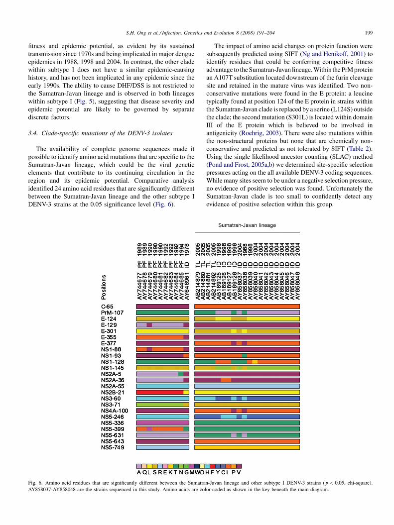
3.4. Clade-specific mutations of the DENV-3 isolates
The availability of complete genome sequences made it
possible to identify amino acid mutations that are specific to the
Sumatran-Javan lineage, which could be the viral genetic
elements that contribute to its continuing circulation in the
region and its epidemic potential. Comparative analysis
identified 24 amino acid residues that are significantly different
between the Sumatran-Javan lineage and the other subtype I
DENV-3 strains at the 0.05 significance level (Fig. 6).
Fig. 6. Amino acid residues that are significantly different between the Sumatra
AY858037-AY858048 are the strains sequenced in this study. Amino acids are co
The impact of amino acid changes on protein function were
subsequently predicted using SIFT (Ng and Henikoff, 2001) to
identify residues that could be conferring competitive fitness
advantage to the Sumatran-Javan lineage. Within the PrM protein
an A107T substitution located downstream of the furin cleavage
site and retained in the mature virus was identified. Two non-
conservative mutations were found in the E protein: a leucine
typically found at position 124 of the E protein in strains within
the Sumatran-Javan clade is replaced by a serine (L124S) outside
the clade; the second mutation (S301L) is located within domain
III of the E protein which is believed to be involved in
antigenicity (Roehrig, 2003). There were also mutations within
the non-structural proteins but none that are chemically non-
conservative and predicted as not tolerated by SIFT (Table 2).
Using the single likelihood ancestor counting (SLAC) method
(Pond and Frost, 2005a,b) we determined site-specific selection
pressures acting on the all available DENV-3 coding sequences.
While many sites seem to be under a negative selection pressure,
no evidence of positive selection was found. Unfortunately the
Sumatran-Javan clade is too small to confidently detect any
evidence of positive selection within this group.
n-Javan lineage and other subtype I DENV-3 strains ( p < 0.05, chi-square).
lor-coded as shown in the key beneath the main diagram.

Table 2
Amino acid residues that potentially confer competitive fitness advantage to the Sumatran-Javan DENV-3 lineage when compared to other subtype I DENV-3
genomes
The two most likely candidates are shaded in grey. Mutations predicted by SIFT as ‘‘Tolerated’’ are less likely to have an impact on
the fitness level of the virus. Threshold of intolerance used by SIFT is 0.05.
S.H. Ong et al. / Infection, Genetics and Evolution 8 (2008) 191–204200
A similar comparison of the deduced amino acid sequences
involving only the 16 Indonesian DENV-3 strains from the
epidemics in 1988, 1998 and 2004 did not detect any specific
mutation that could have served as the trigger of the epidemic in
2004 (Fig. 7). Only 98 sites out of 3390 (2.89%) of the DENV-3
polyprotein reported any mutation, and this number drops to 87
if den3_88 from 1988 is removed from the analysis. From the
pattern of mutations observed these differences are likely to be
the result of random mutations that confers little or no
evolutionary advantage.
4. Discussion
The genome sequences of three serotypes of DENV were
successfully obtained using a genome sequencing strategy that
can be used on patient serum samples. Success with DENV-4
sequencing using this approach is expected in the future as
more DENV-4 genomes become available for better RT-PCR
primer design. Based on the obtained sequences, we have
determined the molecular characteristics and phylogenetic
relationships of the DENV strains isolated during the 2004
epidemic in Jakarta, Indonesia.
The phylogenetic trees for each of the four serotypes show
that the viruses isolated in the 2004 epidemic cluster within
subtypes that have been circulating in South East Asia for at
least 30 years. The strongest evidence of these being endemic
strains comes from the phylogenetic data for DENV-3. All 12
Indonesian DENV-3 strains sequenced in this study fall into
subtype I which is recognized as comprising strains from
Southeast Asia and the South Pacific islands. The close

Fig. 7. Amino acid differences among the Indonesian DENV-3 strains isolated in 1988, 1998 and 2004. The sequences are sorted in chronological order followed by
accession number, starting from the left. Only 98 positions out of 3390 of the DENV-3 polyprotein reported any mutations, and none could be suggested as the trigger
for the epidemic in 2004. Amino acids are color-coded as shown in the key beneath the main diagram.
S.H. Ong et al. / Infection, Genetics and Evolution 8 (2008) 191–204 201
phylogenetic relationship observed between the strains
isolated from Sumatra in 1998 and those from Jakarta in
2004 indicates that the 2004 epidemic might have had its
origins in strains derived from viruses circulating in Sumatra in
1998, or possibly in a common ancestor of the Sumatran strains
(Fig. 5). Sumatra is one of the largest Indonesian islands and
Jakarta on the nearby island of Java is the capital city of
Indonesia, therefore frequent transmission by travelers is
likely.
The identified Sumatran-Javan clade links the newly
sequenced DENV-3 strains directly to the strains implicated
in the epidemic on the island of Java in 1988, on the island of
Sumatra 6 years earlier and those that subsequently caused an
outbreak in Timor Leste in 2005. This clearly suggests these
viruses possess an inherent epidemic potential. Since viruses
from this particular lineage have been implicated in causing
four epidemics in the past two decades, it is very likely these
viruses could yet cause another dengue epidemic in Indonesia

Appendix A. Binding positions of RT-PCR primers
used in this study
Primers for DENV-1 Binding positions (sense strand)
d1a5 8558–8577
d1a9 6551–6573
d1a13 4544–4561
d1a17 2540–2559
d1a23 10716–10735
d1s6 2201–2223
d1s10 4213–4231
d1s14 6216–6235
d1s18 8211–8232
d1s22 1–20
Primers for DENV-2 Binding positions (sense strand)
d2a6 8468–8488
d2a10 6477–6497
d2a14 4461–4484
d2a18 2455–2474
d2a23 10704–10723
d2s5 2182–2201
d2s9 4175–4197
d2s13 6193–6213
d2s16 7669–7692
d2s23 1–20
Primers for DENV-3 Binding positions (sense strand)
d3a6 8342–8361
d3a10 6339–6360
d3a14 4334–4356
d3a18 2361–2380
d3a23 10688–10707
d3s5 2035–2053
d3s8 3532–3553
d3s13 6032–6053
d3s17 8025–8046
d3s23 1–20
Primers for DENV-3Binding positions (sense strand)AF1109–
131AF51927–1949BF13629–3652CF17111–1734AR12708–
2727BR14133–4153CR17664–7687DR110606–10637
Appendix B. Sequencing and amplification primers
used in this study
Primer name Sequence Serotype
d1a1 50-ACAGCTTCCCCTGGTGTTGG-30 DENV-1
d1a2 50-DTCTTCCCAACTGGAYACATG-30 DENV-1
d1a3 50-YACRCARTCATCTCCRCTGAT-30 DENV-1
d1a4 50-CACTCCACTGAGTGAATTCTCTCT-30 DENV-1
d1a5 50-GGRATRACATCCCATGGTTT-30 DENV-1
d1a6 50-AGRACACGTAACGTTCTWCCTTC-30 DENV-1
d1a7 50-CCTACCTCCTCCTARAGATTTCA-30 DENV-1
d1a8 50-CAAGTCCCATCAATATAGCTGC-30 DENV-1
d1a9 50-CCAGTYARCACAGCTATCAAAGC-30 DENV-1
d1a10 50-TCTCTCYGGCTCAAAGAGGG-30 DENV-1
d1a11 50-CRTAGCCTGARTTCCATGATCT-30 DENV-1
S.H. Ong et al. / Infection, Genetics and Evolution 8 (2008) 191–204202
in the near future if they remained in circulation. We then
examined the clade-specific mutations of the Sumatran-Javan
DENV-3 isolates for potential viral genetic markers that
could be the trigger of the epidemics. However, comparative
study of the deduced polyprotein sequences established that
re-emergence of little-changed endemic strains, and not
newly-acquired amino acid mutations by the DENV-3 strains,
as the most likely cause of the dengue epidemic in 2004
(Fig. 7).
According to a parallel serological study involving 272
hospitalised patients, all four dengue serotypes were detected
during the 2004 epidemic with DENV-3 being the
predominant circulating serotype (Suwandono et al.,
2006). Similar serological result was reported for the 1998
epidemic in south Sumatra, and the predominance of DENV-
3 as a result of inherent sampling bias (on the premise that
DENV-3 causes more severe illness and therefore causes
more hospitalisation that facilitates sampling) has been
proposed (Corwin et al., 2001). The fact that all four
serotypes were found to be circulating in successive
epidemics adds credence to the suggestion that advantageous
amino acid mutations is not the trigger of the epidemics,
otherwise this hypothetical fitter form would then dominate
and become the sole serotype detected in subsequent
outbreaks.
The availability of genome sequence data has made possible
the attempt to identify putative viral genetic factors that
contributed to the continuing circulation of the Sumatran-Javan
lineage and its epidemic potential. Two candidate residues were
found in the E protein (Table 2) that may account for this
difference however no conclusive evidence of positive selection
was found. It is clear the genome sequencing and analysis
strategy broadens the search for novel functional mutants
beyond the scope of most previous studies which have mainly
focused on using nucleotide sequences from the three dengue
structural proteins.
The sequence data obtained in this study tells little about the
role of population dynamics of the four serotypes in causing
epidemics, but it clearly shows that the viruses that caused the
2004 epidemic in Indonesia are local strains that have been
circulating in the region for a few decades. The identified
Sumatran-Javan lineage of DENV-3 apparently is robust
enough for sustained transmissions and has demonstrated
strong epidemic potential spanning two decades.
Acknowledgements
We thank Lam Sai Kit and Amin Soebandario for
encouraging the initiation of this collaboration, Klaus Ribbe
of Novartis Indonesia for valuable assistance during site visits,
and Edward C. Holmes for his advice on the phylogenetic
analyses. We also wish to acknowledge the advice and
discussions of Lim Siew Pheng from NITD and the technical
skills of Khoo Chen Ai and Pauline Aw Poh Kim from the
Infectious Disease Group together with the Cloning and
Sequencing Group (headed by Yi Jun Ruan and Chia Lin Wei)
at the Genome Institute of Singapore. During the course of this
work S.H. Ong was an attachment student at NITD from the
Bioinformatics Institute, 30 Biopolis Street, #07-01 Matrix,
Singapore 138670, Singapore.

Appendix B (Continued )Primer name Sequence Serotype
d1a12 50-CCTCGTCCTCAATCTCTGGTAG-30 DENV-1
d1a13 50-TTCCACTTCYGGAGGGCT-30 DENV-1
d1a14 50-CCGGAAGCCATGTTGTTTT-30 DENV-1
d1a15 50-GCATYTTTCTRCTCCATCTGGATC-30 DENV-1
d1a16 50-CARCTTCCARGTYTCGTTCTT-30 DENV-1
d1a17 50-CCAATGGCYGCTGAYAGTCT-30 DENV-1
d1a18 50-AAAGGTGGYTCYGYYTCAAT-30 DENV-1
d1a19 50-GTTTGTGGACRAGCCATGATT-30 DENV-1
d1a20 50-CGTCTTCAAGAGTTCAATGTCC-30 DENV-1
d1a21 50-CATYGCAATRAGRGTGCACAT-30 DENV-1
d1a22 50-AGCTTCCGATTCGAAACTGT-30 DENV-1
d1a23 50-AGAACCTGTTGATTCAACAG-30 DENV-1
d1s1 50-TRGCTCCATCGTGGGGAT-30 DENV-1
d1s2 50-TTGCTYTCAGGCCAAGGACC-30 DENV-1
d1s3 50-AAACGTTCCGTSGCACTGGC-30 DENV-1
d1s4 50-TGTGTGTCGMCGAACGTT-30 DENV-1
d1s5 50-GCAATGCACACYGCGTTG-30 DENV-1
d1s6 50-GGYTCTATAGGAGGRGTGTTCAC-30 DENV-1
d1s7 50-GGCCCAAGGRAARAAAATG-30 DENV-1
d1s8 50-ACAAACAGCAGGGCCRTGGCA-30 DENV-1
d1s9 50-CCTAGCYYTGATGGCYACTTT-30 DENV-1
d1s10 50-RGCYGGSCCACTAATAGCT-30 DENV-1
d1s11 50-AAGAGRCTGGAACCRAGYTGGGC-30 DENV-1
d1s12 50-AAATGGCAGAGGCGCTCAAGGG-30 DENV-1
d1s13 50-ACAAAAAAYAAYGACTGGGACTAT-30 DENV-1
d1s14 50-ATGGRGAAAGGAACAACCAG-30 DENV-1
d1s15 50-GGATAGCGGCCTCYATCATACT-30 DENV-1
d1s16 50-GCAAARGCYACTAGAGAAGCTCAA DENV-1
d1s17 50-GAAACRACYAAACAYGCAGTG-30 DENV-1
d1s18 50-CCACYCATGAAATGTAYTGGGT-30 DENV-1
d1s19 50-GCCARGTGGTTATGGGGTTT-30 DENV-1
d1s20 50-GGATGATCTTCAGAATGAGGC-30 DENV-1
d1s21 50-TYATGAAGGATGGGAGGGA-30 DENV-1
d1s22 50-AGTTGTTAGTCTACGTGGAC-30 DENV-1
d2a1 50-AGGAAACGAAGGAACGCC-30 DENV-2
d2a2 50-ACGCCATGCGTACAGCTT-30 DENV-2
d2a3 50-CCGTYGTCATCCATTCATG-30 DENV-2
d2a4 50-TTTCTTCTGTGRCTGTCAGGTG-30 DENV-2
d2a5 50-TCTGCTGCCTTTTGCCTT-30 DENV-2
d2a6 50-CATGGTAWGCCCAYGTTTTGT-30 DENV-2
d2a7 50-TTCTGGCGGRRTGAAGAA-30 DENV-2
d2a8 50-TGACACYGCAATGGTAGTGTT-30 DENV-2
d2a9 50-CAATGCTATGTCTCARCATTGGTGT-30 DENV-2
d2a10 50-TACGCCCTTCCRCCTGCTTCA-30 DENV-2
d2a11 50-CCAGTGTGCACAGTCTTCATCAT-30 DENV-2
d2a12 50-ATGGRTCTCTRCTTCCCGG-30 DENV-2
d2a13 50-CACCATTACCATAAAGACCCAC-30 DENV-2
d2a14 50-GCCGTGATTGGTATTGATACAGGA-30 DENV-2
d2a15 50-GTGCAACTCACTTTCCATGC-30 DENV-2
d2a16 50-CGGCTGTGACCAAGGAGTT-30 DENV-2
d2a17 50-CCGCTGACATGAGTTTTGAGTC-30 DENV-2
d2a18 50-CCACTGCCACATTTCAGTTC-30 DENV-2
d2a19 50-GGCGRCCTAAGACATRTCTTTT-30 DENV-2
d2a20 50-GCCATARCCTGTCARTTCTGC-30 DENV-2
d2a21 50-CTGAAACCCCTTCTACAAAGTCTC-30 DENV-2
d2a22 50-TGTGGTTCTCCGTTACGTGT-30 DENV-2
d2a23 50-AGAACCTGTTGATTCAACAG-30 DENV-2
d2s1 50-GCAACAGCTGACAAAGAGATTCTC-30 DENV-2
d2s2 50-CACCACRGGAGAACAYAGAAGA-30 DENV-2
d2s3 50-CAGCCTAAAWGAAGAGCAGGA-30 DENV-2
d2s4 50-GCGAAGAAACAGGATGTTGTTG-30 DENV-2
d2s5 50-GGTGACACAGCCTGGGATTT-30 DENV-2
d2s6 50-YATGACAGGAGACATCAAAGGA-30 DENV-2
d2s7 50-WCAACACAACTAYAGACCAGGCT-30 DENV-2
d2s8 50-TGGGCGTGACTTATCTTGC-30 DENV-2
Appendix B (Continued )Primer name Sequence Serotype
d2s9 50-GCATTTTRGCCAGTTCTCTCCTA-30 DENV-2
d2s10 50-GYGCTGTYCTAATGCATAAAGG-30 DENV-2
d2s11 50-YAGAGTCGTGGCAGCTGAA-30 DENV-2
d2s12 50-GGAAGACYTTTGATTCTGAGTATGT-30 DENV-2
d2s13 50-GCAGACAGAAGGTGGTGTTTT-30 DENV-2
d2s14 50-CCACACTGGATAGCAGCTTCAATA-30 DENV-2
d2s15 50-GACTYCAAGCAAAAGCAACC-30 DENV-2
d2s16 50-CAGGAAGTGGATAGAACCTTAGCA-30 DENV-2
d2s17 50-CTCTCACGRAACTCCACACAT-30 DENV-2
d2s18 50-RGCAGAGTGGCTKTGGAAA-30 DENV-2
d2s19 50-GGGACACAAGAATCACACTAGAAG-30 DENV-2
d2s20 50-GCCYTTYTGTTCACACCATTTCCA-30 DENV-2
d2s21 50-AGGAATACACAGATTACATGCCA-30 DENV-2
d2s22 50-GGAATGGTGCTGTTGAATCAAC-30 DENV-2
d2s23 50-AGTWGTTAGTCTACGTGGAC-30 DENV-2
d3a1 50-GGTTTCTCACGCGTTTCAG-30 DENV-3
d3a2 50-TTTTAACGTCCTTGGACGG-30 DENV-3
d3a3 50-GGATGCTAGTCTRAGATCTCTTCTG-30 DENV-3
d3a4 50-CTGCCTCTTTGGTCTTTCCT-30 DENV-3
d3a5 50-CGTTCTCTGTCCACAAGTTTCC-30 DENV-3
d3a6 50-GCATTRACATGTCGRGTTCC-30 DENV-3
d3a7 50-TCCTCGCACTTCTGTRACTTT-30 DENV-3
d3a8 50-TTGAACTGCACACARAACCAG-30 DENV-3
d3a9 50-CACCTGGYTCYTTAGACATTCCTA-30 DENV-3
d3a10 50-GCYGCAAARTCCTTGAATTCCT-30 DENV-3
d3a11 50-TTGGTCCAGCCAGGATCA-30 DENV-3
d3a12 50-GTGAAATGRGCCTCATCCAT-30 DENV-3
d3a13 50-CCTGGCATGGTTTGAAAGTT-30 DENV-3
d3a14 50-ACTGTGATCATTAARTTGTGGGA-30 DENV-3
d3a15 50-CCCCARAGCRATTCCATT-30 DENV-3
d3a16 50-GGCAACACCATTCGTGTATCA-30 DENV-3
d3a17 50-CACTTGGACACTCCGGTGT-30 DENV-3
d3a18 50-GATTCCTATCGCAATGCATG-30 DENV-3
d3a19 50-GCGTTTCKGAGACTTCTTTCTTC-30 DENV-3
d3a20 50-GACGGTGTATTTGAGGTTCTCA-30 DENV-3
d3a21 50-GGCTAGTATGGTRAACCCTGG-30 DENV-3
d3a22 50-CCTTCTTGAAGCCTTTYARGACCT-30 DENV-3
d3a23 50-AGAACCTGTTGATTCAACAG-30 DENV-3
d3s1 50-CAGTTTCGACTCGGAAGCTT-30 DENV-3
d3s2 50-CAACATGTGCACACTCATAGCC-30 DENV-3
d3s3 50-GACTACCATGGCTAAGAACAAGC-30 DENV-3
d3s4 50-GAAGAACAAAGCATGGATGGTA-30 DENV-3
d3s5 50-TGAACCTCCTTTTGGGGAA-30 DENV-3
d3s6 50-CCMAAAAGATTGGCAACAGC-30 DENV-3
d3s7 50-CATGGGCTATTGGATAGAAAGC-30 DENV-3
d3s8 50-GGTGATGAGAGGAAAATTTGGG-30 DENV-3
d3s9 50-GAAAACAGATTGGCTCCCAA-30 DENV-3
d3s10 50-CCCCCCAGAGACACAGAAAG-30 DENV-3
d3s11 50-CGACACCAGAGTTGGAAGAAG-30 DENV-3
d3s12 50-GCTCATGGAATTCAGGCAAT-30 DENV-3
d3s13 50-CCAGCTCTCTTTGAACCAGAAA-30 DENV-3
d3s14 50-CTCYTGGGACTGATGATCTTGT-30 DENV-3
d3s15 50-CTGATGGGTTTRGACAAAGGA-30 DENV-3
d3s16 50-TTTTTCTATYATGAAATCAGTTGGA-30 DENV-3
d3s17 50-CAACAGTGGAAGAAAGCAGAAC-30 DENV-3
d3s18 50-ACAAAACCATGGGATGTGG-30 DENV-3
d3s19 50-CTGGTTCTCGCGTGAAAAC-30 DENV-3
d3s20 50-GGGATGATTGCGTAGTGAAA-30 DENV-3
d3s21 50-TCCAGTCACAACGTGGGAA-30 DENV-3
d3s22 50-TGTACCTCCTTGCAAAGGACTA-30 DENV-3
d3s23 50-AGTTGTTAGTCTACGTGGAC-30 DENV-3
AR1 50-CTTGCCTTYGGTCAACACCC-30 DENV-4
BR1 50-CCTCGTTAAGRGGCCARGATC-30 DENV-4
CR1 50-GGCTTCAGTCCTGTCCACTTCTAG-30 DENV-4
DR1 50-ATCCATCTTGCGGCGCTCTGTG-30 DENV-4
S.H. Ong et al. / Infection, Genetics and Evolution 8 (2008) 191–204 203

S.H. Ong et al. / Infection, Genetics and Evolution 8 (2008) 191–204204
References
Chungue, E., Cassar, O., Drouet, M.T., Guzman, M.G., Laille, M., Rosen, L.,
Deubel, V., 1995. Molecular epidemiology of dengue-1 and dengue-4
viruses. J. Gen. Virol. 76, 1877–1884.
Corwin, A.L., Larasati, R.P., Bangs, M.J., Wuryadi, S., Arjoso, S., Sukri, N.,
Listyaningsih, E., Hartati, S., Namursa, R., Anwar, Z., Chandra, S., Loho,
B., Ahmad, H., Campbell, J.R., Porter, K.R., 2001. Epidemic dengue
transmission in southern Sumatra, Indonesia. Trans. R. Soc. Trop. Med.
Hyg. 95, 257–265.
Goncalvez, A.P., Escalante, A.A., Pujol, F.H., Ludert, J.E., Tovar, D., Salas,
R.A., Liprandi, F., 2002. Diversity and evolution of the envelope gene of
dengue virus type 1. Virology 303, 110–119.
Gubler, D.J., Clark, G.G., 1995. Dengue/dengue hemorrhagic fever: the emer-
gence of a global health problem. Emer. Infec. Dis. 1, 55–57.
Gubler, D.J., 2002. Epidemic dengue/dengue haermorrhagic fever as a public
health, social and economic problem in the 21st century. Trends Microbiol.
10, 100–103.
Henikoff, S., Henikoff, J.G., 1992. Amino acid substitution matrices from
protein blocks. Proc. Natl. Acad. Sci. 89, 10915–10919.
Lanciotti, R.S., Calisher, C.H., Gubler, D.J., Chang, G.J., Vorndam, A.V., 1992.
Rapid detection and typing of dengue viruses from clinical samples by using
reverse transcriptase-polymerase chain reaction. J. Clin. Microbiol. 30,
545–551.
Lanciotti, R.S., Lewis, J.G., Gubler, D.J., Trent, D.W., 1994. Molecular
evolution and epidemiology of dengue-3 viruses. J. Gen. Virol. 75, 65–75.
Lanciotti, R.S., Gubler, D.J., Trent, D.W., 1997. Molecular evolution and
phylogeny of dengue-4 viruses. J. Gen. Virol. 78, 2279–2286.
Leitmeyer, K.C., Vaughn, D.W., Watts, D.M., Salas, R., Villalobos, I., de
Chacon, Ramos, C., Rico-Hesse, R., 1999. Dengue virus structural differ-
ences that correlate with pathogenesis. J. Virol. 73 (6), 4738–4747.
Li, K.B., 2003. ClustalW-MPI: ClustalW analysis using distributed and parallel
computing. Bioinformatics 19, 1585–1586.
Messer, W.B., Gubler, D.J., Harris, E., Sivananthan, K., de Silva, A., 2003.
Emergence and global spread of a dengue serotype 3, subtype III virus.
Emer. Infec. Dis. 9, 800–809.
Monath, T.P., 1994. Dengue: the risk to developed and developing countries.
Proc. Natl. Acad. Sci. 91, 2395–2400.
Ng, P.C., Henikoff, S., 2001. Predicting deleterious amino acid substitution.
Genome Res. 11, 863–874.
Pond, S.L., Frost, S.D., 2005a. Datamonkey: rapid detection of selective
pressure on individual sites of codon alignments. Bioinformatics 21,
2531–2533.
Pond, S.L., Frost, S.D., 2005b. Not so different after all: a comparison of
methods for detecting amino acid sites under selection. Mol. Biol. Evol. 22,
1208–1222.
Posada, D., Crandall, K.A., 1998. MODELTEST: testing the model of DNA
substitution. Bioinformatics 14, 817–818.
Rico-Hesse, R., 1990. Molecular evolution and distribution of dengue viruses
type 1 and 2 in nature. Virology 174, 479–493.
Rico-Hesse, R., Harrison, L.M., Salas, R.A., Tovar, D., Nisalak, A., Ramos, C.,
Boshell, J., de Mesa, M.T.R., Nogueira, R.M.R., da Roasa, A.T., 1997.
Origins of dengue type-2 viruses associated with increased pathogenicity in
the Americas. Virology 230, 244–251.
Roehrig, J.T., 2003. Antigenic structure of flavivirus proteins. Adv. Virus Res.
59, 141–175.
Seah, C.L., Chow, V.T., Tan, H.C., Can, Y.C., 1995. Rapid, single-step RT-PCR
typing of dengue viruses using five NS3 gene primers. J. Virol. Methods 51,
193–200.
Schreiber, M.J., Ong, S.H., Holland, R.C.G., Hibberd, M.L., Vasudevan, S.G.,
Mitchell, W.P., Holmes, E.C., 2007. DengueInfo: a web portal to dengue
information resources. Infect. Genet. Evol. 7, 540–541.
Sumarmo, 1987. Dengue haemorrhagic fever in Indonesia. Southeast Asian J.
Trop. Med. Public Health 18, 269–274.
Suwandono, A., Kosasih, H., Nurhayati, Kusriastuti, R., Harun, S., Ma’roef, C.,
Wuryadi, S., Herianto, B., Yuwono, D., Porter, K.R., Beckett, C.G., Blair,
P.J., 2006. Four dengue virus serotypes found circulating during an outbreak
of dengue fever and dengue haemorrhagic fever in Jakarta, Indonesia,
during 2004. Trans. R. Soc. Trop. Med. Hyg. 100, 855–862.
Swofford, D.L., 2002. PAUP*. Phylogenetic Analysis Using Parsimony (*and
other methods). Version 4. Sinauer Associates, Sunderland, Massachusetts.
Twiddy, S.S., Farrar, J.J., Vinh Chau, N., Wills, B., Gould, E.A., Gritsun, T., Lloyd,
G., Holmes, E.C., 2002. Phylogenetic relationships and differential selection
pressures among genotypes of dengue-2 virus. Virology 298, 63–72.
World Health Organization (WHO), 2002. Dengue and dengue haemorrhagic
fever. Fact sheet no. 117.
World Health Organization (WHO), 2004. EPR: Dengue fever in Indonesia –
update 4, May 11, 2004.
World Health Organization (WHO), 2005. Dengue haemorrhagic fever, Timor-
Leste – update. Wkly. Epidemiol. Rec. 80, 85–86.