Paxillin expression and amplification in early lung lesions of high-risk patients, lung...
9
Paxillin expression and amplification in early lung lesions of high-risk patients, lung adenocarcinoma and metastatic disease Alexander C Mackinnon, 1 Maria Tretiakova, 1 Les Henderson, 2 Rajendra G Mehta, 3 Benjamin C Yan, 1 Loren Joseph, 1 Thomas Krausz, 1 Aliya N Husain, 1 Mary E Reid, 4 Ravi Salgia 2 ABSTRACT Background Paxillin is a modular protein that localises to cell adhesion sites where it facilitates bidirectional communication between the intracellular actin cytoskeleton and the extracellular matrix. These complex and dynamic interactions are essential for cell adhesion, cell migration and cell survival. The authors have previously demonstrated that paxillin is overexpressed in lung cancer tissues and identified somatic paxillin mutations in 9% of lung cancers. A murine in vivo xenograft model of the most common paxillin mutation (A127T) showed increased cell proliferation and invasive tumour growth, establishing an important role for paxillin in the development of lung cancer. Methods The authors analysed 279 bronchoscopy-aided biopsy specimens from 92 high-risk patients. Adenocarcinoma with bronchioloalveolar features and pure bronchioloalveolar carcinoma (BAC) were analysed with fluorescence in situ hybridisation (FISH) and immunohistochemistry (IHC). Results Paxillin is overexpressed in premalignant areas of hyperplasia, squamous metaplasia and goblet cell metaplasia, as well as dysplastic lesions and carcinoma in high-risk patients. Concordance between increased paxillin gene copy number and paxillin overexpression was observed in cases of adenocarcinoma eusomic for chromosome 12. Conclusions Paxillin overexpression occurs during the earliest stages of lung cancer development. FISH and IHC analysis of lung adenocarcinoma suggests that relatively small-scale genomic rearrangements of chromosome 12 are associated with paxillin overexpression in lung adenocarcinoma. INTRODUCTION Lung cancer is the leading cause of cancer death in the USA. Most often, lung cancer is diagnosed when the disease is advanced, resulting in limited therapeutic options and corresponding poor survival times. At the same time, advances in early detection through the use of low-dose spiral CT imaging clearly enhance the ability to detect lung cancer at early stages when therapeutic interven- tion can potentially extend life. 12 Autofluorescence bronchoscopy (AFB) is a potential method for identifying high-grade dysplasia and carcinoma in situ within the central airway. 3e6 However, AFB has low positive predictive value and low speci- ficity, since biopsy interpretation is confounded by the fact that not all premalignant and preinvasive lesions progress to lung cancer, 7 and low-grade dysplasia, inflammation, hyperplasia, metaplasia and granulation tissue all have abnormal AFB patterns. 8 Hence, despite improvements in early lung cancer detection, it is not yet evident that early detection by CT or AFB results in mortality reduction. Therefore, discovering clinically impor- tant lung cancer biomarkers to identify patients at high risk of developing lung cancer is critical to improving lung cancer diagnosis and treatment. We recently identified somatic mutations in the gene paxillin 9 in lung cancer. The paxillin gene codes for a 68 kDa focal adhesion molecule which is recruited to nascent cell adhesion sites upon acti- vation of ab integrin heterodimers. 10 Paxillin serves as a scaffold for additional structural proteins and regulatory proteins that collectively promote changes in actin dynamics leading to regulation of cell adhesion, migration and survival. These proteineprotein interactions are regulated by phosphorylation of paxillin via a large number of kinases providing a mechanism for dynamic control of the assembly and disassembly of large macro- molecular complexes. 11 Tyrosine phosphorylation of paxillin indirectly activates members of the Rho family of G-proteins leading to the formation of filopodia, lamellipodia and stress fibres, which are all actin-based cellular structures localised at the leading edge of migrating cells. 12 Paxillin promotes cell survival via its interaction with Bcl-2 9 13 14 during development, and paxillin and Bcl-2 coloc- alise in lung cancer tissue. 9 Paxillin is often over- expressed in lung cancer, suggesting that it may contribute to malignancy and serve as a useful biomarker for diagnosis of lung cancer and a cellular target for lung cancer treatment. In order to further characterise the role of paxillin during lung cancer development and assess its utility as a biomarker, we examined paxillin expression and localisation in bronchial biopsy specimens from patients at high risk of developing lung cancer. We observed early and persistent overexpression of paxillin during lung cancer development in high-risk patients. As paxillin overexpression is observed in both early neoplastic lesions and lung cancer, we used fluorescence in situ hybridisation (FISH) to demonstrate that paxillin gene copy number is often amplified in lung adenocarcinoma and pure bronchioloalveolar carci- noma (BAC). We also show that increased paxillin 1 Department of Pathology, University of Chicago Medical Center, Chicago, Illinois, USA 2 Department of Medicine, University of Chicago Medical Center, Chicago, Illinois, USA 3 Carcinogenesis and Chemoprovention Division, Illinois Institute of Technology Research Institute, Chicago, Illinois, USA 4 Department of Epidemiology, Roswell Park Cancer Institute, Buffalo, New York, USA Correspondence to Dr Alexander C Mackinnon, University of Chicago Medical Center, 5841 S Maryland Avenue, MC 2115, Chicago, IL 60637, USA; alexander. [email protected] Accepted 21 September 2010 Published Online First 2 November 2010 This paper is freely available online under the BMJ Journals unlocked scheme, see http:// jcp.bmj.com/site/about/ unlocked.xhtml 16 J Clin Pathol 2011;64:16e24. doi:10.1136/jcp.2010.075853 Original article
-
Upload
hms-harvard -
Category
Documents
-
view
2 -
download
0
Transcript of Paxillin expression and amplification in early lung lesions of high-risk patients, lung...
Paxillin expression and amplification in early lunglesions of high-risk patients, lung adenocarcinomaand metastatic disease
Alexander C Mackinnon,1 Maria Tretiakova,1 Les Henderson,2 Rajendra G Mehta,3
Benjamin C Yan,1 Loren Joseph,1 Thomas Krausz,1 Aliya N Husain,1 Mary E Reid,4
Ravi Salgia2
ABSTRACTBackground Paxillin is a modular protein that localises tocell adhesion sites where it facilitates bidirectionalcommunication between the intracellular actincytoskeleton and the extracellular matrix. These complexand dynamic interactions are essential for cell adhesion,cell migration and cell survival. The authors havepreviously demonstrated that paxillin is overexpressed inlung cancer tissues and identified somatic paxillinmutations in 9% of lung cancers. A murine in vivoxenograft model of the most common paxillin mutation(A127T) showed increased cell proliferation and invasivetumour growth, establishing an important role for paxillinin the development of lung cancer.Methods The authors analysed 279 bronchoscopy-aidedbiopsy specimens from 92 high-risk patients.Adenocarcinoma with bronchioloalveolar features andpure bronchioloalveolar carcinoma (BAC) were analysedwith fluorescence in situ hybridisation (FISH) andimmunohistochemistry (IHC).Results Paxillin is overexpressed in premalignant areasof hyperplasia, squamous metaplasia and goblet cellmetaplasia, as well as dysplastic lesions and carcinomain high-risk patients. Concordance between increasedpaxillin gene copy number and paxillin overexpressionwas observed in cases of adenocarcinoma eusomic forchromosome 12.Conclusions Paxillin overexpression occurs during theearliest stages of lung cancer development. FISH and IHCanalysis of lung adenocarcinoma suggests that relativelysmall-scale genomic rearrangements of chromosome12 are associated with paxillin overexpression in lungadenocarcinoma.
INTRODUCTIONLung cancer is the leading cause of cancer death inthe USA. Most often, lung cancer is diagnosedwhen the disease is advanced, resulting in limitedtherapeutic options and corresponding poorsurvival times. At the same time, advances in earlydetection through the use of low-dose spiral CTimaging clearly enhance the ability to detect lungcancer at early stages when therapeutic interven-tion can potentially extend life.1 2 Autofluorescencebronchoscopy (AFB) is a potential method foridentifying high-grade dysplasia and carcinoma insitu within the central airway.3e6 However, AFBhas low positive predictive value and low speci-ficity, since biopsy interpretation is confounded by
the fact that not all premalignant and preinvasivelesions progress to lung cancer,7 and low-gradedysplasia, inflammation, hyperplasia, metaplasiaand granulation tissue all have abnormal AFBpatterns.8 Hence, despite improvements in earlylung cancer detection, it is not yet evident thatearly detection by CT or AFB results in mortalityreduction. Therefore, discovering clinically impor-tant lung cancer biomarkers to identify patients athigh risk of developing lung cancer is critical toimproving lung cancer diagnosis and treatment.We recently identified somatic mutations in the
gene paxillin9 in lung cancer. The paxillin genecodes for a 68 kDa focal adhesion molecule which isrecruited to nascent cell adhesion sites upon acti-vation of ab integrin heterodimers.10 Paxillin servesas a scaffold for additional structural proteins andregulatory proteins that collectively promotechanges in actin dynamics leading to regulation ofcell adhesion, migration and survival. Theseproteineprotein interactions are regulated byphosphorylation of paxillin via a large number ofkinases providing a mechanism for dynamic controlof the assembly and disassembly of large macro-molecular complexes.11 Tyrosine phosphorylationof paxillin indirectly activates members of the Rhofamily of G-proteins leading to the formation offilopodia, lamellipodia and stress fibres, which areall actin-based cellular structures localised at theleading edge of migrating cells.12 Paxillin promotescell survival via its interaction with Bcl-29 13 14
during development, and paxillin and Bcl-2 coloc-alise in lung cancer tissue.9 Paxillin is often over-expressed in lung cancer, suggesting that it maycontribute to malignancy and serve as a usefulbiomarker for diagnosis of lung cancer and a cellulartarget for lung cancer treatment.In order to further characterise the role of paxillin
during lung cancer development and assess itsutility as a biomarker, we examined paxillinexpression and localisation in bronchial biopsyspecimens from patients at high risk of developinglung cancer. We observed early and persistentoverexpression of paxillin during lung cancerdevelopment in high-risk patients. As paxillinoverexpression is observed in both early neoplasticlesions and lung cancer, we used fluorescence in situhybridisation (FISH) to demonstrate that paxillingene copy number is often amplified in lungadenocarcinoma and pure bronchioloalveolar carci-noma (BAC). We also show that increased paxillin
1Department of Pathology,University of Chicago MedicalCenter, Chicago, Illinois, USA2Department of Medicine,University of Chicago MedicalCenter, Chicago, Illinois, USA3Carcinogenesis andChemoprovention Division,Illinois Institute of TechnologyResearch Institute, Chicago,Illinois, USA4Department of Epidemiology,Roswell Park Cancer Institute,Buffalo, New York, USA
Correspondence toDr Alexander C Mackinnon,University of Chicago MedicalCenter, 5841 S MarylandAvenue, MC 2115, Chicago, IL60637, USA; [email protected]
Accepted 21 September 2010Published Online First2 November 2010
This paper is freely availableonline under the BMJ Journalsunlocked scheme, see http://jcp.bmj.com/site/about/unlocked.xhtml
16 J Clin Pathol 2011;64:16e24. doi:10.1136/jcp.2010.075853
Original article

gene copy number and paxillin protein concentrations correlatewith chromosome 12 eusomy.
METHODSPatient and sample selectionThe high-risk patients and corresponding biopsy samplesexamined in this study have been previously described.15
Patients’ clinical and pathological information was obtainedfrom a lung bronchoscopy database (Roswell Park CancerInstitute) and a lung cancer patient database (University ofChicago) according to each institutional review board protocol.The patients seen at Roswell Park did not have cancer at thetime they were evaluated with bronchoscopy. They had at leasttwo risk factors for cancer: previous history of lung cancer withno evidence of disease for >2 years before the current biopsy;diagnosis of chronic obstructive pulmonary disease; asbestos-related lung disease16; >20 pack-year cigarette smoking history.These comprehensive databases include clinical information andrisk factor data such as age, gender, smoking status, medicalhistory, occupational exposure, pathological diagnosis, tumourstage (TNM (tumour/nodes/metastasis)) and tumour grade.
Histological examinationH&E-stained slides of bronchial biopsy samples from 105patients were reviewed by two pathologist (ACM and MT) andcategorised according to World Health Organization (WHO)classifications.17 Of these, 92 contained sufficient respiratorymucosa to be evaluated. ‘Premalignant’ changes include hyper-plasia, squamous metaplasia and goblet cell metaplasia. ‘Prein-vasive’ epithelial lesions include atypical adenomatoushyperplasia (AAH), dysplasia and carcinoma in situ.
ImmunohistochemistryTissue microarray (TMA) paraffin sections were deparaffinisedin xylene, rehydrated through graded ethanol solutions todistilled water, and then washed in Tris-buffered saline.Antigen retrieval was carried out by heating sections in ETDAbuffer (pH¼9) for 15 min in a microwave oven. Endogenousperoxidase activity was quenched by incubation in 3% H2O2 inmethanol for 5 min. Non-specific binding sites were blockedusing Protein Block (Dako, Carpinteria, California, USA) for20 min. Tissue sections were incubated for 1 h at roomtemperature with the mouse monoclonal paxillin antibody(clone 5H11, dilution 1:200; Neomarkers, Freemont, California,USA) followed by 30 min incubation with goat anti-mouse IgGconjugated to a horseradish peroxidase-labelled polymer(Envision+ System; DakoCytomation, Carpinteria, California,USA). Slides were developed for 5 min with 3,39-diamino-benzidine chromogen and counterstained with haematoxylin.Negative controls were performed by replacing the primaryantibody step with non-immune mouse immunoglobulins.Immunostaining intensity in the cytoplasm was scored asfollows: 0¼no staining; 1¼weak staining; 2¼moderatestaining; and 3¼strong staining. In this study, immunohisto-chemistry (IHC) scores of 2 and 3 were both interpreted asoverexpression. For comparison between means of two groups,Student t test or c2 test was used.
KRAS and EGFR mutation analysisSections 4 mm thick were cut from formalin-fixed, paraffin-embedded tissue samples and placed on to membrane-coatedlaser capture microscopy slides (LCM; Leica, Wetzlar, Germany.).Slides were deparaffinised in xylene, rehydrated through
gradient ethanol, stained with H&E, and dehydrated in ethanol.Target lesions were identified and isolated by LCM. Tissuefragments were digested overnight in proteinase K (10 mg/ml),DNA was purified using Qiagen MinElute kits, and resuspendedin 10 mM Tris/HCl, pH 8.0.Point mutations in KRAS codons 12 and 13 were identified
using a Light Cycler 2.0 as described by Nikiforova et al.18 In brief,purified DNAwas used to generate a PCR amplicon using primers(K-rasF1: 59-AAGGCCTGCTGAAAATGACTG; K-rasR1:59-GGTCCTGCACCAGTAATATGCA) that flank the KRAScodon 12/13 mutational hotspot. Using a pair of fluorescentlylabelled anchor and sensor oligonucleotides (KRASanc1: 59-CGTCCACAAAATGATTCTGAATTAGCTGTATCGTCAAGGCAGT-fluorescein; KRASsnsr1: 59-LC Red640-TGCCTACGCCAC-CAGCTCCAA-phosphate), amplification and mutation detectionwas accomplished via fluorescence resonance energy transfer. Thesensor oligonucleotide spans the mutation site allowing the detec-tion of a codon 12 or codon 13 mutation based on the distinctmelting temperatures (Tm) of wild-type KRAS and mutant KRAS.DNA from a cell line carrying a mutation in KRAS codon 13 servedas a positive control.EGFR mutation was detected by sequencing genomic DNA
purified as described for KRAS mutation detection. Exons 19and 21 were amplified by PCR using the following exon flankingprimer pairs: 19F, 59-CCTGAGGTTCAGAGCCATGGAC,and 19R, 59-CAGCATGTGGCACCATCTCACAATTGC; and21F, 59-CTTCCCATGATGATCTGTCCCTCACAGC, and 21R,59-GGAGAGCATCCTCCCCTGCATGTGT. Amplicon sizeswere checked by agarose electrophoresis, which permitteddetection of large deletions. The amplicons were purified byethanol precipitation, sequenced using BigDye terminatorsequencing chemistry run on an ABI 3100 capillary analyser. Theresulting DNA sequences were analysed with MutationSurveyor V3.23 software.
Tissue microarrayThe study group consisted of 51 wedge resections and lobecto-mies performed at the University of Chicago Medical Centerfrom 1990 to 2008. These cases were divided into three groups:(1) invasive adenocarcinoma with BAC pattern, (2) ‘pure BAC’without invasive adenocarcinoma, and (3) adenocarcinomawithout BAC. Eleven of the pure BAC cases were non-mucinoustype (92%). There were lymph node metastases in three cases ofinvasive adenocarcinoma. A morphologically representative areaof interest within each paraffin donor block was identified bytwo pathologists (AHN and MT) under the microscope using anH&E-stained section on a glass slide as a guide. From each casea minimum of five tissue cylinders with a diameter of 1 mmwere arrayed into a recipient block using an automated tissuemicroarrayer ATA-27 (Beecher Instruments, Sun Prairie,Wisconsin, USA). In the case of adenocarcinoma with BACpattern, the cores were obtained as follows: two cores frominvasive tumour, two from BAC component, and one fromadjacent normal lung. In the case of pure BAC or invasiveadenocarcinoma without BAC, five tissue cylinders includedthree cores from the tumour and two cores from adjacentnormal lung. A total of 270 tissue cores were arrayed on to thetwo recipient TMA blocks, which were then cut into 4 mm thickserial sections and subjected to IHC analysis.
Fluorescent in situ hybridisation and analysisTwo-colour FISH analysis was performed using the BAC probeRP11-144B2, which localises to 12q24.23 and contains the full-length paxillin gene, directly labelled with Spectrum Orange
J Clin Pathol 2011;64:16e24. doi:10.1136/jcp.2010.075853 17
Original article

using a Nick Translation Kit (Abbott Molecular, Des Plaines,Ilinois, USA), together with CEP 12 directly labelled withSpectrum Green (Abbott Molecular), which hybridises to thealpha-12 centromeric region (12p11.1e12q11.1).19 Slides werepretreated using the Paraffin Pretreatment Kit (Abbott Molec-ular). At least 18 interphase nuclei were analysed for eachpatient. Only single, clearly defined nuclei were scored; over-lapping nuclei or clusters of nuclei were not analysed. Splitfluorescence signals with similar intensity and diameter seen inthe same focal plane adjacent to each other were counted as one(eg, see figure 3F). For most cases, small round diploid nucleilikely to be infiltrating lymphocytes were used as an internalcontrol.
Paxillin and chromosome 12 gene copy number were deter-mined by dividing the total number of fluorescent signals corre-sponding to paxillin (RP11e144B2) or alpha-12 (CEP 12),respectively, by the total number of nuclei analysed for eachpatient. Since paxillin gene copy number was heterogeneouswithin individual tumour samples, equivocal cases of paxillingene copy number alteration were not included in our analysis ifwe failed to observe individual nucleiwithmore than five copies ofpaxillin or if nuclei with more than two copies of paxillin repre-sent <30% of the total nuclei scored. FISH was validated byscoring 437 and 417 normal nuclei for paxillin and alpha-12,respectively. In this report, the averaged number of fluorescentsignals in normal nucleiwas 2.04 for paxillin and 1.85 for alpha-12,and these averages were used for all subsequent analysisof malignant nuclei. Unpaired Student t test was used to deter-mine statistical significance for each case: the copy number of
Table 1 Patient characteristics (n¼105)
Patient characteristic Distribution (n (%))
Gender
Male 73 (70)
Female 32 (30)
Age
#60 years 49 (47)
>60 years 56 (53)
Race
Black 3 (3)
Caucasian 102 (97)
Smoking history
Current smoker 36 (34)
Former smoker 66 (63)
Never smoked 3 (3)
Pack-years
#45 51 (49)
>45 50 (48)
Unknown 4 (4)
COPD
Yes 79 (75)
No 19 (18)
Unknown 8 (7)
Asbestos
Yes 50 (48)
No 37 (35)
Unknown 18 (17)
COPD, chronic obstructive pulmonary disease.
Figure 1 Paxillin is overexpressed inpremalignant epithelial lesions in high-riskpatients. Representative images oftransbronchial biopsy samples stained forpaxillin. Paxillin staining localises to thecytoplasm and is enriched in the basallayers of the respiratory mucosa(arrows). The bottom row of pie graphsdescribes quantitative paxillin stainingintensity in biopsy samples withhyperplasia, squamous metaplasia orgoblet cell metaplasia morphology.
18 J Clin Pathol 2011;64:16e24. doi:10.1136/jcp.2010.075853
Original article

paxillin and alpha-12 from malignant nuclei measured from anindividual casewas comparedwith the average copy number fromall normal nuclei (ie, 2.04 and 1.85 for paxillin and alpha-12,respectively); this was done for every case analysed. Whenpossible, the average gene copy number of paxillin and alpha-12from malignant nuclei measured from an individual case wascompared with the average copy number of paxillin and alpha-12from normal nuclei measured from the same corresponding case.
In all instances, both methods were in complete statisticalagreement. For paxillin, unpaired Student t test between normaland malignant nuclei with p<0.01 was interpreted as a gene copynumber alteration; for alpha-12, unpaired Student t test betweennormal and malignant nuclei with p<0.01 was interpreted aschromosome 12 polysomy. Paxillin amplification was defined asa paxillin gene copy number increase and chromosome 12 eusomy.‘Polysomy’was defined as a copy number increase in both paxillinand chromosome 12. Loss of heterozygosity was defined as adecrease in paxillin gene copy number and chromosome 12eusomy.
RESULTSHistological changes and paxillin expression in patients at highrisk of developing lung cancerFrom the initial cohort of 105 patients at high risk of developinglung cancer (table 1), biopsy material from 92 patients wassuitable for histological and paxillin analysis. This resulted ina total of 279 individual biopsy samples for analysis (table 2)with between one and six (mean number of three) biopsyspecimens analysed for each patient. No histological abnormal-ities were observed on any of the individual biopsy samples of10 patients; the remaining patients had one or more biopsyspecimens containing a portion of histologically normalappearing respiratory mucosa. Hence, most patients (89%) hadone or more foci of histologically abnormal mucosa.A total of 173 histological normal biopsy specimens were
analysed for paxillin expression by IHC. In 74 of 92 patients(80%), no paxillin expression was observed in histologicallynormal surface epithelium (see figure 2A of Jagadeeswaranet al9), whereas weak, moderate and strong paxillin expressionwas observed in 13 (14%), four (5%) and one (1%) patients,respectively (data not shown). This showed that paxillin waseither not detectable or was weakly expressed in histologically
Table 2 Distribution of histological findings by patient(n¼105) and biopsy (n¼279)
Histology Frequency (n) (%)
By patient
No sample/not analysed 13 12
Normal histology only 10 10
Hyperplasia only (H)y 16 15
Squamous metaplasia only (S)z 4 4
Goblet cell metaplasia only (G)x 5 5
H and S 12 11
H and Gx 24 23
S and G 2 2
H and S and Gx 19 18
Total 105 100
By biopsy*
Normal respiratory mucosa 173 62
Hyperplasia 168 60
Squamous metaplasia 69 25
Goblet cell metaplasia 99 35
Dysplasia 4 1.4
Carcinoma 3 1.0
*More than one histological feature is typically present on a singlebiopsy specimen.yIncludes one patient with dysplasia and two with carcinoma.zIncludes one patient with carcinoma.xIncludes one patient with dysplasia.
Figure 2 Preinvasive epithelial lesionsfrom high-risk patients overexpresspaxillin. Bronchoscopic biopsy samplesshowing dysplasia (A,B) and carcinoma(C,D). Note the large, irregular nuclei inthe upper portion of the respiratory(white arrow) and mitotic figure(arrowheads) mucosa in (A) and(B). Paxillin staining is strongestalong the basal and parabasal layers(arrows in D).
J Clin Pathol 2011;64:16e24. doi:10.1136/jcp.2010.075853 19
Original article
normal appearing respiratory mucosa in w95% of the high-riskpatients, which is similar to our previous findings.9
An increase in paxillin expression in biopsy samplescontaining areas of abnormal histology was observed (figure 1).Whereas 11% of all histologically normal appearing biopsysamples showed moderate to strong concentrations of paxillin,moderate to strong paxillin expression was observed in 78%,81% and 93% of the hyperplastic, squamous metaplastic andgoblet cell metaplastic lesions, respectively. Paxillin expressionappeared strongest in the basal layer (arrows, figure 1). Four ofthe 92 patients (4%) had areas of dysplasia (figure 2A,B) andthree (3%) had lung carcinoma (figure 2C,D). All of these patientsamples showed elevated levels of paxillin expression, andexpression appeared strongest along the basal layer at theperipheral margins of the preinvasive lesions (black arrows,figure 2D). Together, these findings indicate that, relative toadjacent normal respiratory mucosa, paxillin expression wasupregulated in non-neoplastic precursor lesions before malignantchanges were microscopically evident and can remain elevatedduring the formation of preinvasive epithelial lesions.
Dysplasia and carcinoma were associated with a long historyof smoking (table 3). On the other hand, hyperplasia, squamous
metaplasia and goblet cell metaplasia were associated witha corresponding increase in mean smoking pack-year history,indicating that hyperplastic changes occur earliest in smokers,whereas goblet cell metaplasia occurs in the setting of a longersmoking history. The highest levels of paxillin staining wereobserved in patients with the longest smoking history. Strongerpaxillin staining was visible in 69% of biopsy samples fromcurrent smokers (22/32) compared with 49% of biopsy samplesfrom former smokers (24/49) (p¼0.0395, c2 test).
Gene copy number of paxillin correlates with histologicalsubtype and KRAS mutationA pilot series of 11 human non-small cell lung cancer (NSCLC)samples representing various histological subtypes were exam-ined by FISH to determine if paxillin (12q24.23) gene copynumber alteration correlated with a specific histological subtypeof lung cancer (table 4). Paxillin gene copy number was increasedin two of four adenocarcinoma samples, whereas paxillin genecopy number changes in other histological subtypes were notobserved. EGFR and KRAS mutation status were examined onenriched tumour foci isolated using laser capture microscopy. AKRAS mutation in codon 12/13 was detected in one of the twoadenocarcinomas cases with increased paxillin gene copynumber, indicating that KRAS muation20 21 and altered paxillingene copy number are not mutually exclusive. No mutations inexon 19 or exon 21 of EGFR in any samples were detected.
Paxillin locus is amplified in adenocarcinoma including pure BACsubtypeOn the basis of the relatively high frequency with which paxillingene copy number alterations were identified in lung adenocar-cinoma in the initial small set of samples, together with our
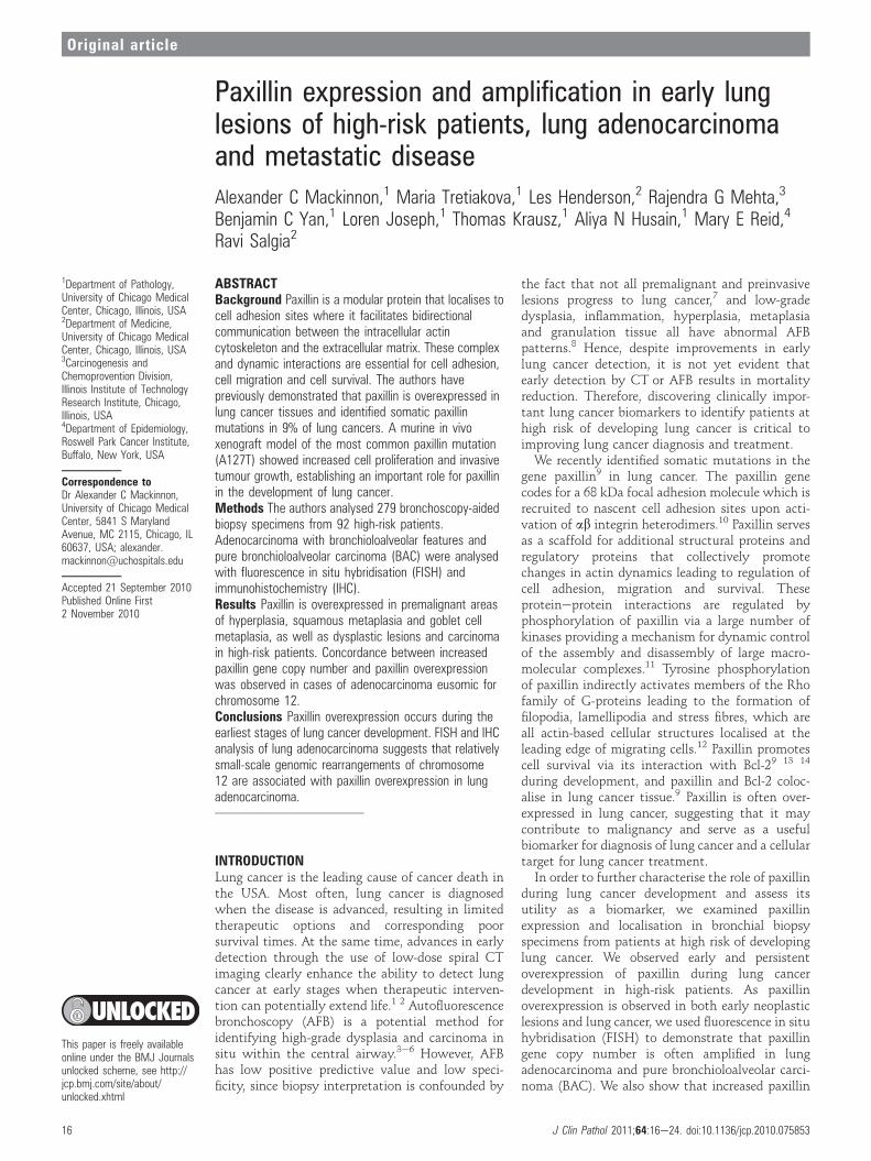
Figure 3 Paxillin gene copy number isincreased in lung adenocarcinoma andbronchioloalveolar carcinoma (BAC).Representative DAPI (4’,6-diamidino-2-phenylindole)-stained nuclei showingFISH probes for paxillin (red) and alpha-12 (green). Non-malignant nuclei exhibittwo fluorescent signals for both paxillinand alpha-12 (A). Primary lungadenocarcinoma measuring <5 mm (B),5e10 mm (C), >10 mm (D), pure BAC(E) and metastatic adenocarcinoma (F).
Table 3 Pack-years of smoking by histology and paxillinexpression
Pack-year of smoking
Histology
Hyperplasia 14.7 (27.4)
Squamous metaplasia 41.0 (26.7)
Goblet cell metaplasia 56.9 (29.5)
Dysplasia/carcinoma 79.3 (56.0)
Values are mean (SD).
20 J Clin Pathol 2011;64:16e24. doi:10.1136/jcp.2010.075853
Original article

previous finding of paxillin amplification in human lung cancercell lines,9 FISH was used to examine paxillin copy number in 39cases of adenocarcinoma comprising 27 cases of adenocarcinomawith a BAC component and 12 cases of pure BAC (figure 3).Compared with normal nuclei with two copies of paxillin(figure 3A), 13/27 cases of adenocarcinoma (48%) (table 5) and5/12 cases of pure BAC (42%) (table 6) showed altered paxillingene copy number. With the exception of two cases of pure BAC(see below), all tumours showed increased paxillin gene copynumber. Three to six copies of paxillin were typically observed(figure 3BeF); however, occasionally individual nuclei with upto 16 fluorescent signals corresponding to the paxillin locus were
observed. Two of three cases with lymph node metastasesshowed increased paxillin gene copy number similar to thecorresponding primary tumour. Occasionally, individual tumourspecimens showed heterogeneous alterations in paxillin genecopy number such that regions with increased paxillin copynumber were adjacent to regions with normal copy number.As chromosomal instability is common in NSCLC,22 23
chromosome 12 centromeric region was co-analysed to deter-mine if increased paxillin gene copy number was a non-specificconsequence of chromosome 12 polysomy (see the Methodssection). For adenocarcinoma, 8/13 cases (62%) with increasedpaxillin gene copy number showed chromosome 12 polysomy,
Table 4 Correlation of paxillin copy number (FISH) with EGFR (exon 19/21) and KRAS (codons12/13)mutation
Tumour histology Paxillin FISH EGFR exon 19 EGFR exon 21 KRAS codons 12/13
Adenocarcinoma (1.5 cm), solid andacinar types, with focal signet ringfeatures
Amplification WT WT WT
Adenocarcinoma with BAC pattern* Amplification WT WT Mutation
Adenocarcinoma (1.6 cm) Diploid WT WT WT
Adenocarcinoma (0.5 cm), acinar type Diploid WT WT WT
Large cell carcinoma (1.7 cm) Diploid WT WT WT
Squamous cell carcinoma (3.0 cm) Diploid WT WT NA
Focal squamous metaplasia withdysplasia
Diploid WT WT WT
High-grade squamous cell carcinoma(3.7 cm)
Diploid WT WT WT
Adenosquamous carcinoma (1.2 cm) Diploid WT WT WT
Adenocarinoma (3.5 cm) withsarcomatoid changes
Diploid WT WT NA
Adenosquamous carcinoma (1.2 cm) Diploid WT WT WT
*Paxillin overexpression confirmed with immunohistochemistry.BAC, bronchioloalveolar carcinoma; FISH, fluorescence in situ hybridisation; NA, KRAS PCR amplification failed; WT, wild-type.
Table 5 Correlation of increased paxillin gene copy number with chromosome 12 and paxillinexpression in lung adenocarcinoma with BAC component
Case Histological component Paxillin p Value a-12 p Value Paxillin expression*
Reference Normal 2.04 1.85
1 BAC 2.50 <0.0001 2.03 0.035
INV 2.64 <0.0001 1.85 1 Weak
2 BAC 2.70 <0.0001 2.07 0.014 Moderate
3 INV 3.87 <0.0001 3.37 <0.0001 None
4 BAC 2.97 <0.0001 2.48 <0.0001
INV 4.30 <0.0001 0.05 <0.0001 Weak
5 BAC 3.92 <0.0001 2.92 <0.0001
INV 3.64 <0.0001 2.67 <0.0001 Weak
6 BAC 2.96 <0.0001 2.00 0.025
INV 2.35 0.011 2.04 0.035 Moderate
7 INV 2.59 <0.0001 2.11 0.0037 NA
8 INV 2.40 <0.0001 1.95 0.16 High
9 BAC 2.63 <0.0001 1.79 0.51
INV 2.65 <0.0001 2.00 0.12 Moderate
10 INV 2.97 <0.0001 2.68 <0.0001
LN 4.33 <0.0001 3.17 <0.0001 Weak
11 BAC 4.58 <0.0001 2.67 <0.0001 High
12 BAC 4.24 <0.0001 3.81 <0.0001
INV 5.48 <0.0001 4.59 <0.0001 None
13 INV 3.21 <0.0001 2.95 <0.0001
LN 3.19 <0.0001 2.67 <0.0001 None
The average number of fluorescent signals per nuclei are given for paxillin and chromosome 12 (a-12).*See the Methods section for description of expression levels.BAC, bronchioloalveolar carcinoma; INV, non-BAC component; NA, immunohistochemistry for case 7 was not interpreted because oftechnical difficulties.
J Clin Pathol 2011;64:16e24. doi:10.1136/jcp.2010.075853 21
Original article
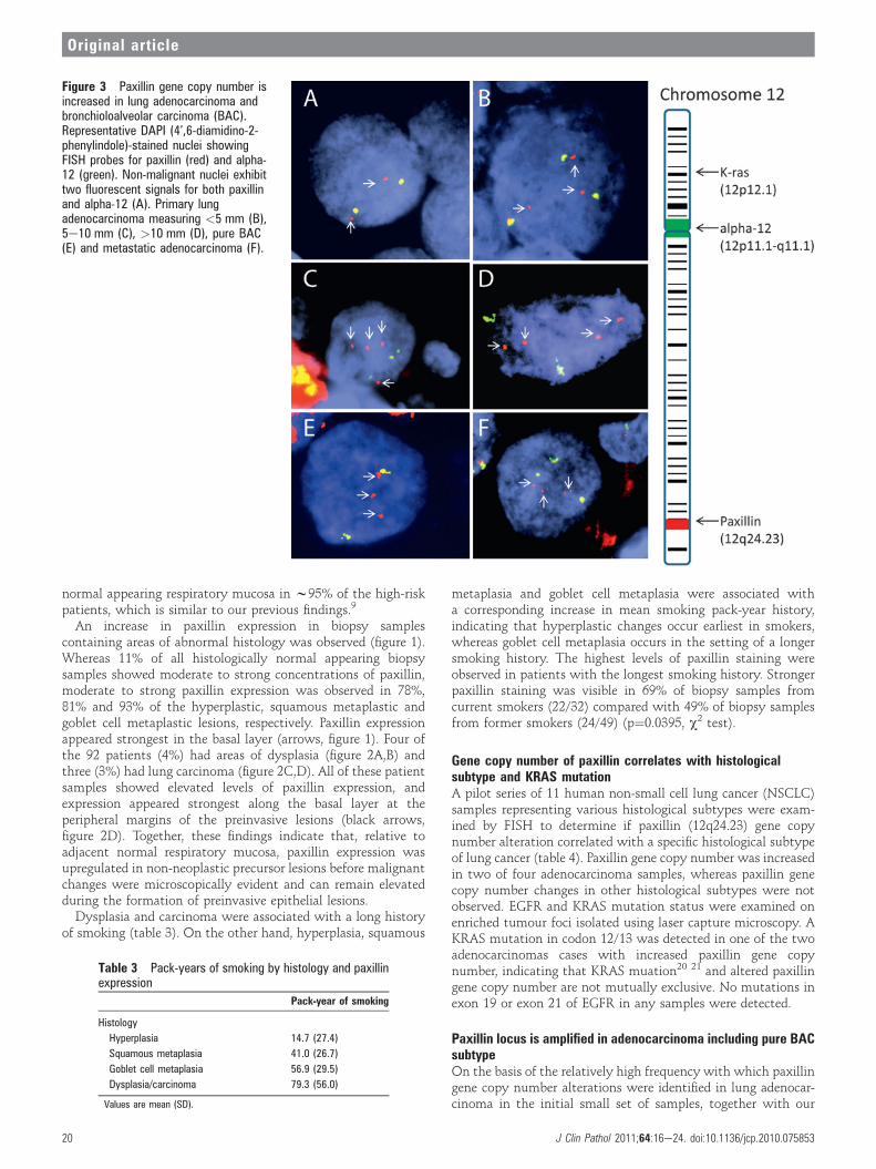
and 5/13 cases (38%) showed chromosome 12 eusomy (table 5)(p¼0.217, c2 test). Of the five cases of pure BAC with alteredpaxillin gene copy number, three showed increased paxillin genecopy number and two demonstrated loss of heterozygosity forpaxillin (table 6). Of the cases of adenocarcinoma with a BACcomponent, none showed loss of heterozygosity for paxillin. Wedid not observe any tumour specimen with multiple copies ofchromosome 12 centromeric regions and two or fewer copies ofpaxillin.
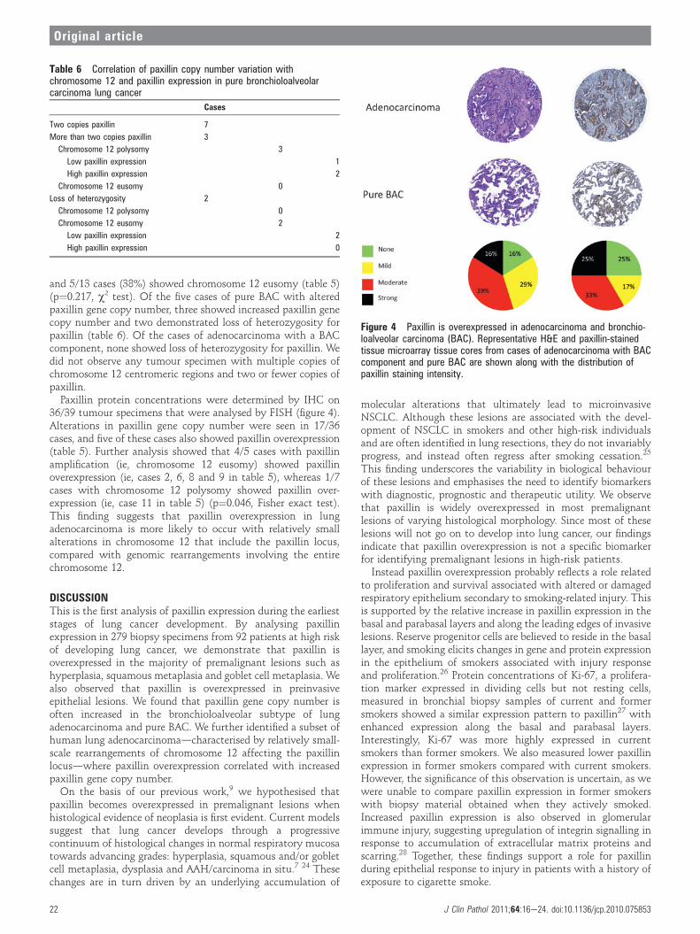
Paxillin protein concentrations were determined by IHC on36/39 tumour specimens that were analysed by FISH (figure 4).Alterations in paxillin gene copy number were seen in 17/36cases, and five of these cases also showed paxillin overexpression(table 5). Further analysis showed that 4/5 cases with paxillinamplification (ie, chromosome 12 eusomy) showed paxillinoverexpression (ie, cases 2, 6, 8 and 9 in table 5), whereas 1/7cases with chromosome 12 polysomy showed paxillin over-expression (ie, case 11 in table 5) (p¼0.046, Fisher exact test).This finding suggests that paxillin overexpression in lungadenocarcinoma is more likely to occur with relatively smallalterations in chromosome 12 that include the paxillin locus,compared with genomic rearrangements involving the entirechromosome 12.
DISCUSSIONThis is the first analysis of paxillin expression during the earlieststages of lung cancer development. By analysing paxillinexpression in 279 biopsy specimens from 92 patients at high riskof developing lung cancer, we demonstrate that paxillin isoverexpressed in the majority of premalignant lesions such ashyperplasia, squamous metaplasia and goblet cell metaplasia. Wealso observed that paxillin is overexpressed in preinvasiveepithelial lesions. We found that paxillin gene copy number isoften increased in the bronchioloalveolar subtype of lungadenocarcinoma and pure BAC. We further identified a subset ofhuman lung adenocarcinomadcharacterised by relatively small-scale rearrangements of chromosome 12 affecting the paxillinlocusdwhere paxillin overexpression correlated with increasedpaxillin gene copy number.
On the basis of our previous work,9 we hypothesised thatpaxillin becomes overexpressed in premalignant lesions whenhistological evidence of neoplasia is first evident. Current modelssuggest that lung cancer develops through a progressivecontinuum of histological changes in normal respiratory mucosatowards advancing grades: hyperplasia, squamous and/or gobletcell metaplasia, dysplasia and AAH/carcinoma in situ.7 24 Thesechanges are in turn driven by an underlying accumulation of
molecular alterations that ultimately lead to microinvasiveNSCLC. Although these lesions are associated with the devel-opment of NSCLC in smokers and other high-risk individualsand are often identified in lung resections, they do not invariablyprogress, and instead often regress after smoking cessation.25
This finding underscores the variability in biological behaviourof these lesions and emphasises the need to identify biomarkerswith diagnostic, prognostic and therapeutic utility. We observethat paxillin is widely overexpressed in most premalignantlesions of varying histological morphology. Since most of theselesions will not go on to develop into lung cancer, our findingsindicate that paxillin overexpression is not a specific biomarkerfor identifying premalignant lesions in high-risk patients.Instead paxillin overexpression probably reflects a role related
to proliferation and survival associated with altered or damagedrespiratory epithelium secondary to smoking-related injury. Thisis supported by the relative increase in paxillin expression in thebasal and parabasal layers and along the leading edges of invasivelesions. Reserve progenitor cells are believed to reside in the basallayer, and smoking elicits changes in gene and protein expressionin the epithelium of smokers associated with injury responseand proliferation.26 Protein concentrations of Ki-67, a prolifera-tion marker expressed in dividing cells but not resting cells,measured in bronchial biopsy samples of current and formersmokers showed a similar expression pattern to paxillin27 withenhanced expression along the basal and parabasal layers.Interestingly, Ki-67 was more highly expressed in currentsmokers than former smokers. We also measured lower paxillinexpression in former smokers compared with current smokers.However, the significance of this observation is uncertain, as wewere unable to compare paxillin expression in former smokerswith biopsy material obtained when they actively smoked.Increased paxillin expression is also observed in glomerularimmune injury, suggesting upregulation of integrin signalling inresponse to accumulation of extracellular matrix proteins andscarring.28 Together, these findings support a role for paxillinduring epithelial response to injury in patients with a history ofexposure to cigarette smoke.
Table 6 Correlation of paxillin copy number variation withchromosome 12 and paxillin expression in pure bronchioloalveolarcarcinoma lung cancer
Cases
Two copies paxillin 7
More than two copies paxillin 3
Chromosome 12 polysomy 3
Low paxillin expression 1
High paxillin expression 2
Chromosome 12 eusomy 0
Loss of heterozygosity 2
Chromosome 12 polysomy 0
Chromosome 12 eusomy 2
Low paxillin expression 2
High paxillin expression 0
Figure 4 Paxillin is overexpressed in adenocarcinoma and bronchio-loalveolar carcinoma (BAC). Representative H&E and paxillin-stainedtissue microarray tissue cores from cases of adenocarcinoma with BACcomponent and pure BAC are shown along with the distribution ofpaxillin staining intensity.
22 J Clin Pathol 2011;64:16e24. doi:10.1136/jcp.2010.075853
Original article
Paxillin is a scaffold protein composed of five N-terminal LDmotifs, four C-terminal LIM domains, and several SH3 domainswhich allow paxillin to form a large number of proteineproteininteractions. The LIM3 domain targets paxillin to focal adhesionsites, thereby regulating its subcellular localisation. Consequently,paxillin is able to integrate multiple signalling pathwayswithin the cell by providing docking sites for commonsignalling molecules and positioning them adjacent to acti-vated receptors such as integrin heterodimers and growthfactor receptors at the cell membrane.11 It would thereforeappear beneficial for a cell to express paxillin during woundhealing in response to injury29 as well as during malignanttransformation.30e32 For example, the terminal LD4 domain ofpaxillin binds the BH4 domain of Bcl-2, and this interactionpromotes survival and morphogenesis during kidney develop-ment.13 14 We have previously shown that paxillin colocaliseswith Bcl-2 in NSCLC.9 This raises the intriguing hypothesisthat paxillin overexpression in premalignant lesions promotessurvival via Bcl-2-mediated signalling. This interaction in turncan facilitate tissue repair, as well as tumour development.Nakanishi et al33 demonstrated increasing levels of Bcl-2expression in low-grade to high-grade AAH lesions and non-mucinous BAC. Although we did not directly examine paxillinand Bcl-2 colocalisation in the biopsy specimens, it is temptingto speculate that this interaction may be an early event inlung cancer development, and future studies can test thisprediction.
Consistent with our previous work9 and the analysispresented here, little to no paxillin expression is observed inhistologically normal respiratory epithelium. This is in markedcontrast with liver, where paxillin expression shows no statis-tically significant difference in tumour and adjacent non-tumourliver tissue,31 indicating that baseline levels of paxillin expressionin normal cells are tissue dependent. We also observed a signifi-cant correlation between paxillin overexpression and increasedpaxillin gene copy number in a subset of lung adenocarcinomawith a diploid complement of chromosome 12. This findingsuggests alternative mechanisms for paxillin overexpressiondepending on the tumour genotype. In cases of polysomy forchromosome 12, the lack of correlation between paxillin genecopy number and paxillin overexpression suggests that addi-tional mutations in other key genes develop during tumorigen-esis, at which point paxillin overexpression becomes dispensable.KRAS is located on chromosome 12 and is a known driver
mutation in lung cancer, raising the possibility that paxillinoverexpression is no longer necessary when tumours becomepolysomic for chromosome 12. Interestingly, we only observedpaxillin loss of heterozygosity in pure BAC tumours, which alsofailed to overexpress paxillin. In summary, this is the firstdescription of paxillin overexpression in the earliest morpho-logical changes leading up to the development of lung cancer. Wealso identify a subset of lung adenocarcinoma in which changesin paxillin gene copy number correlate with paxillin over-expression, and future studies will address the clinical signifi-cance of this observation.
Acknowledgements ACM is grateful to Dr Elizabeth McNally, Dr Eric Svensson,Dr Kaul and Dr Nowak for support.
Funding Supported in part by NIH/National Cancer Institute (5R01CA125541-03,3R01CA125541-03S109, 5R01CA129501-02, 3R01CA129501-02S109,5P01HL058064-140009), V-Foundation (Guy Geleerd Memorial Foundation), KateMcMullen Foundation, Respiratory Health Association of Chicago, and MesotheliomaApplied Research Foundation (Jeffrey P Hayes Memorial Grant).
Competing interests None.
Patient consent Obtained.
Ethics approval This study was conducted with the approval of the Roswell ParkCancer Institute IRB and University of Chicago IRB.
Provenance and peer review Not commissioned; externally peer reviewed.
REFERENCES1. Midthun DE, Jett JR. Update on screening for lung cancer. Semin Respir Crit Care
Med 2008;29:233e40.2. Henschke CI, Yankelevitz DF. CT screening for lung cancer: update 2007. Oncologist
2008;13:65e78.3. Arens C, Dreyer T, Malzahn K, et al. Direct and indirect autofluorescence
laryngoscopy in the diagnosis of laryngeal cancer and its precursor lesions.Otolaryngol Pol 2004;58:197e203.
4. Chhajed PN, Shibuya K, Hoshino H, et al. A comparison of video andautofluorescence bronchoscopy in patients at high risk of lung cancer. Eur Respir J2005;25:951e5.
5. Lam B, Wong MP, Fung SL, et al. The clinical value of autofluorescencebronchoscopy for the diagnosis of lung cancer. Eur Respir J 2006;28:915e19.
6. Lam S, Kennedy T, Unger M, et al. Localization of bronchial intraepithelial neoplasticlesions by fluorescence bronchoscopy. Chest 1998;113:696e702.
7. Wistuba II, Gazdar AF. Lung cancer preneoplasia. Annu Rev Pathol2006;1:331e48.
8. El-Bayoumi E, Silvestri GA. Bronchoscopy for the diagnosis and staging of lungcancer. Semin Respir Crit Care Med 2008;29:261e70.
9. Jagadeeswaran R, Surawska H, Krishnaswamy S, et al. Paxillin is a target forsomatic mutations in lung cancer: implications for cell growth and invasion. CancerRes 2008;68:132e42.
10. Hynes RO. Integrins: bidirectional, allosteric signaling machines. Cell2002;110:673e87.
11. Brown MC, Turner CE. Paxillin: adapting to change. Physiol Rev 2004;84:1315e39.12. Deakin NO, Turner CE. Paxillin comes of age. J Cell Sci 2008;121:2435e44.13. Sheibani N, Tang Y, Sorenson CM. Paxillin’s LD4 motif interacts with bcl-2. J Cell
Physiol 2008;214:655e61.14. Sorenson CM. Interaction of bcl-2 with Paxillin through its BH4 domain is important
during ureteric bud branching. J Biol Chem 2004;279:11368e74.15. Loewen G, Natarajan N, Tan D, et al. Autofluorescence bronchoscopy for lung
cancer surveillance based on risk assessment. Thorax 2007;62:335e40.16. Doll R. Mortality from lung cancer in asbestos workers. Br J Ind Med
1955;12:81e6.17. Travis WD, Brambilla E, Muller-Hermelink HK, et al. eds. Pathology and Genetics of
Tumours of the Lung, Pleura, Thymus, and Heart. Lyon: IARC Press, 2004.18. Nikiforova MN, Lynch RA, Biddinger PW, et al. RAS point mutations and PAX8-
PPAR gamma rearrangement in thyroid tumors: evidence for distinct molecularpathways in thyroid follicular carcinoma. J Clin Endocrinol Metab2003;88:2318e26.
19. Greig GM, Parikh S, George J, et al. Molecular cytogenetics of alpha satellite DNAfrom chromosome 12: fluorescence in situ hybridization and description of DNA andarray length polymorphisms. Cytogenet Cell Genet 1991;56:144e8.
20. Sagawa M, Saito Y, Fujimura S, et al. K-ras point mutation occurs in the early stageof carcinogenesis in lung cancer. Br J Cancer 1998;77:720e3.
21. Westra WH, Baas IO, Hruban RH, et al. K-ras oncogene activation in atypicalalveolar hyperplasias of the human lung. Cancer Res 1996;56:2224e8.
22. Taguchi T, Zhou JY, Feder M, et al. Detection of aneuploidy in interphase nucleifrom non-small cell lung carcinomas by fluorescence in situ hybridization using
Take-home messages
< Paxillin expression is upregulated in non-neoplastic precursorlesions before malignant changes are microscopically evident,suggesting that paxillin plays a role related to proliferation andsurvival associated with altered or damaged respiratoryepithelium.
< 48% of adenocarcinoma and 42% of pure bronchioloalveolarcarcinoma showed altered paxillin gene copy number, andcases with lymph node metastases showed increased paxillingene copy number similar to the corresponding primarytumour.
< Concordance between increased paxillin gene copy numberand paxillin overexpression was observed in a subset oflung adenocarcinoma with a diploid complement of chromo-some 12.
J Clin Pathol 2011;64:16e24. doi:10.1136/jcp.2010.075853 23
Original article

chromosome-specific repetitive DNA probes. Cancer Genet Cytogenet1996;89:120e5.
23. Weir B, Zhao X, Meyerson M. Somatic alterations in the human cancer genome.Cancer Cell 2004;6:433e8.
24. Lantuejoul S, Salameire D, Salon C, et al. Pulmonary preneoplasiaesequentialmolecular carcinogenetic events. Histopathology 2009;54:43e54.
25. Dacic S. Pulmonary preneoplasia. Arch Pathol Lab Med2008;132:1073e8.
26. Chung KF, Adcock IM. Multifaceted mechanisms in COPD: inflammation, immunity,and tissue repair and destruction. Eur Respir J 2008;31:1334e56.
27. Lee JJ, Liu D, Lee JS, et al. Long-term impact of smoking on lung epithelialproliferation in current and former smokers. J Natl Cancer Inst2001;93:1081e8.
28. Koukouritaki SB, Lianos EA. Glucocorticoid effect on human mesangial cellcytoskeletal proteins. J Lab Clin Med 1999;133:378e83.
29. Koukouritaki SB, Tamizuddin A, Lianos EA. Enhanced expression of thecytoskeleton-associated proteins paxillin and focal adhesion kinase in glomerularimmune injury. J Lab Clin Med 1999;134:173e9.
30. Jenq W, Cooper DR, Ramirez G. Integrin expression on cell adhesion function andup-regulation of P125FAK and paxillin in metastatic renal carcinoma cells. ConnectTissue Res 1996;34:161e74.
31. Li HG, Xie DR, Shen XM, et al. Clinicopathological significance of expression ofpaxillin, syndecan-1 and EMMPRIN in hepatocellular carcinoma. World JGastroenterol 2005;11:1445e51.
32. Short SM, Yoder BJ, Tarr SM, et al. The expression of the cytoskeletal focaladhesion protein paxillin in breast cancer correlates with HER2 overexpression andmay help predict response to chemotherapy: a retrospective immunohistochemicalstudy. Breast J 2007;13;130e9.
33. Nakanishi K, Kawai T, Kumaki F, et al. Survivin expression in atypical adenomatoushyperplasia of the lung. Am J Clin Pathol 2003;120:712e19.
24 J Clin Pathol 2011;64:16e24. doi:10.1136/jcp.2010.075853
Original article