Patterns of cell death induced by eluates from denture base acrylic resins in U-937 human...
11
The number of denture wearers is rapidly increas- ing as the number of elderly people is continually growing (1). Poly(methyl methacrylate) (PMMA) resins are still the most frequently used denture base materials (2). Potentially toxic substances like formaldehyde, methyl methacrylate, methacrylic acid, benzoic acid, dibutyl phthalate, phenyl benzoate, phenyl salicylate, and dicyclohexyl phthalate are eluted from these traditional denture base resins (3–9). These resins have shown various degrees of cytotoxicity in vitro (10–13). The mechanisms of cell death caused by denture base resins have not yet been elucidated. In order to control the biological responses elicited by den- tures, more knowledge is needed about the types and mechanisms of cell death induced by denture base materials. Two modes of cell death have been described: apoptosis and necrosis (14). Apoptosis is an active and physiological mode of cell death, which permits the organism to eliminate unwanted or sublethally damaged cells without triggering inflammatory reactions (14–16). It is characterized by condensa- tion of cytoplasm and nuclear chromatin followed by nuclear fragmentation. Structural integrity, most of the plasma membrane functions and the cellular organelles remain preserved until the late stages of apoptosis (17). In the latest stage of apoptosis, secondary necrosis, the integrity of the plasma membrane is lost (17). Necrosis is a passive process following exposure of the cell to gross injury. It is referred to as ‘‘cell murder’’ and is characterized by mitochondrial swelling, karyolysis, rupture of the plasma membrane, and release of the cytoplasmic constituents (16). Necrosis triggers an inflammatory reaction in the tissue (16). A recently developed method for quantification of apoptosis by flow cytometry is based on the detection of phosphatidylserine (PS) on the outer surface of the plasma membrane (18). The flip of PS from the inner to the outer membrane leaflet is an early apoptotic event (19). Another method of Patterns of cell death induced by eluates from denture base acrylic resins in U-937 human monoblastoid cells Cimpan MR, Cressey LI, Skaug N, Halstensen A, Lie SA, Gjertsen BT, Matre R: Patterns of cell death induced by eluates from denture base acrylic resins in U-937 human monoblastoid cells. Eur J Oral Sci 2000; 108: 59–69. # Eur J Oral Sci, 2000 The purpose of this study was to investigate in vitro the apoptosis- and necrosis-inducing potential of eluates from three heat-polymerized and four autopolymerized poly(methyl methacrylate)-based denture base resins. Our hypothesis was that the rate of cell death by apoptosis and/or necrosis induced by such denture base resins could be an important indicator of their cytotoxicity degree. U-937 human monoblastoid cells were exposed for 24 h and 48 h to eluates of 0.1 g/ml, 0.2 g/ml, 0.4 g/ml, and 0.8 g/ml extracted for 24 h and 48 h. The characteristics of apoptosis and necrosis were evaluated by flow cytometry and light and electron microscopy. Eluates from all resins enhanced cell death by apoptosis and necrosis in U-937 cells in a dose- and time-dependent fashion. Eluates from autopolymerized resins yielded higher percentages of apoptosis and necrosis than the heat-polymerized ones. The results support our hypothesis that eluates of poly(methyl methacrylate)-based denture base acrylic resins activate death- signaling pathways, and that the extent of this process reflects their biocompatibility degree. Mihaela Roxana Cimpan 1,7 , Lill Irene Cressey 2 , Nils Skaug 3 , Alfred Halstensen 4 , Stein Atle Lie 5 , Bjørn Tore Gjertsen 2 , Roald Matre 6 1 Institute of Odontology–Department of Dental Biomaterials, Faculty of Dentistry, University of Bergen, Bergen, 2 Department of Anatomy and Cell Biology, Faculty of Medicine, University of Bergen, Bergen, 3 Institute of Odontology– Department of Oral Microbiology, Faculty of Dentistry, University of Bergen, Bergen, 4 Medical Department B, Faculty of Medicine, University of Bergen, Bergen, 5 Division of Statistics, Faculty of Medicine, 6 Department of Microbiology and Immunology, The Gade Institute, Faculty of Medicine, University of Bergen, Bergen, 7 Centre for International Health, University of Bergen, Bergen, Norway Mihaela Roxana Cimpan, Department of Dental Biomaterials, Institute of Odontology, Faculty of Dentistry, Aarstadveien 17, Bergen 5009, Norway Telefax: +47–55974689 E-mail: [email protected] Key words: denture base; acrylic resins; apoptosis; necrosis; biocompatibility Accepted for publication October 1999 Eur J Oral Sci 2000; 108: 59–69 Printed in UK. All rights reserved
Transcript of Patterns of cell death induced by eluates from denture base acrylic resins in U-937 human...

The number of denture wearers is rapidly increas-ing as the number of elderly people is continuallygrowing (1). Poly(methyl methacrylate) (PMMA)resins are still the most frequently used denturebase materials (2). Potentially toxic substances likeformaldehyde, methyl methacrylate, methacrylicacid, benzoic acid, dibutyl phthalate, phenylbenzoate, phenyl salicylate, and dicyclohexylphthalate are eluted from these traditional denturebase resins (3±9). These resins have shown variousdegrees of cytotoxicity in vitro (10±13). Themechanisms of cell death caused by denture baseresins have not yet been elucidated. In order tocontrol the biological responses elicited by den-tures, more knowledge is needed about the typesand mechanisms of cell death induced by denturebase materials.Two modes of cell death have been described:
apoptosis and necrosis (14). Apoptosis is an activeand physiological mode of cell death, which permitsthe organism to eliminate unwanted or sublethally
damaged cells without triggering in¯ammatoryreactions (14±16). It is characterized by condensa-tion of cytoplasm and nuclear chromatin followedby nuclear fragmentation. Structural integrity,most of the plasma membrane functions and thecellular organelles remain preserved until the latestages of apoptosis (17). In the latest stage ofapoptosis, secondary necrosis, the integrity of theplasma membrane is lost (17). Necrosis is a passiveprocess following exposure of the cell to grossinjury. It is referred to as ``cell murder'' and ischaracterized by mitochondrial swelling, karyolysis,rupture of the plasma membrane, and release of thecytoplasmic constituents (16). Necrosis triggers anin¯ammatory reaction in the tissue (16).
A recently developed method for quanti®cationof apoptosis by ¯ow cytometry is based on thedetection of phosphatidylserine (PS) on the outersurface of the plasma membrane (18). The ¯ip ofPS from the inner to the outer membrane lea¯et isan early apoptotic event (19). Another method of
Patterns of cell deathinduced by eluates fromdenture base acrylicresins in U-937 humanmonoblastoid cellsCimpan MR, Cressey LI, Skaug N, Halstensen A, Lie SA, Gjertsen BT, Matre R:Patterns of cell death induced by eluates from denture base acrylic resins in U-937human monoblastoid cells. Eur J Oral Sci 2000; 108: 59±69. # Eur J Oral Sci, 2000
The purpose of this study was to investigate in vitro the apoptosis- andnecrosis-inducing potential of eluates from three heat-polymerized and fourautopolymerized poly(methyl methacrylate)-based denture base resins. Ourhypothesis was that the rate of cell death by apoptosis and/or necrosis inducedby such denture base resins could be an important indicator of their cytotoxicitydegree. U-937 humanmonoblastoid cells were exposed for 24 h and 48 h to eluatesof 0.1 g/ml, 0.2 g/ml, 0.4 g/ml, and 0.8 g/ml extracted for 24 h and 48 h. Thecharacteristics of apoptosis and necrosis were evaluated by ¯ow cytometry and lightand electron microscopy. Eluates from all resins enhanced cell death by apoptosisand necrosis in U-937 cells in a dose- and time-dependent fashion. Eluates fromautopolymerized resins yielded higher percentages of apoptosis and necrosis thanthe heat-polymerized ones. The results support our hypothesis that eluates ofpoly(methyl methacrylate)-based denture base acrylic resins activate death-signaling pathways, and that the extent of this process re¯ects their biocompatibilitydegree.
Mihaela Roxana Cimpan1,7,Lill Irene Cressey2, Nils Skaug3,Alfred Halstensen4, Stein Atle Lie5,Bjùrn Tore Gjertsen2, Roald Matre6
1Institute of Odontology±Department of DentalBiomaterials, Faculty of Dentistry, University ofBergen, Bergen, 2Department of Anatomy andCell Biology, Faculty of Medicine, University ofBergen, Bergen, 3Institute of Odontology±Department of Oral Microbiology, Faculty ofDentistry, University of Bergen, Bergen,4Medical Department B, Faculty of Medicine,University of Bergen, Bergen, 5Division ofStatistics, Faculty of Medicine, 6Department ofMicrobiology and Immunology, The GadeInstitute, Faculty of Medicine, University ofBergen, Bergen, 7Centre for InternationalHealth, University of Bergen, Bergen, Norway
Mihaela Roxana Cimpan, Department ofDental Biomaterials, Institute of Odontology,Faculty of Dentistry, Aarstadveien 17,Bergen 5009, Norway
Telefax: +47±55974689E-mail: [email protected]
Key words: denture base; acrylic resins;apoptosis; necrosis; biocompatibility
Accepted for publication October 1999
Eur J Oral Sci 2000; 108: 59±69Printed in UK. All rights reserved

quanti®cation of apoptosis by ¯ow cytometry relieson DNA content analysis (20). Extensive DNAcleavage is a characteristic feature of apoptosis. Thedegraded DNA leaks out from ®xed, permeabilizedapoptotic cells. As a consequence the apoptoticcells have a reduced DNA content. They can thusbe recognized due to their low DNA stainabilitywith propidium iodide (PI) (20).
Our hypothesis was that the rate of cell death byapoptosis and/or necrosis induced by PMMA-based denture base resins could be an importantindicator of their degree of cytotoxicity. To test ourhypothesis we identi®ed and quanti®ed the celldeath patterns induced by eluates from three heat-polymerized and four autopolymerized denturebase resins.
Material and methods
Denture base materials
The study employed test specimens of heat-polymerized and autopolymerized PMMA-baseddenture base polymers, both types in the powderand liquid version (Table 1). The currently producedSuperacryl and Duracryl are labeled cadmium free.In Superacryl and Duracryl produced before 1994,cadmium was detected by atomic absorptionspectrophotometry (CIMPAN et al., unpublished).The latter products were included in the presentstudy because dentures made before 1994 from suchmaterials are still in use. The current versions willbe referred to below as Superacryl ``New'' (SN) andDuracryl ``New'' (DN) to distinguish them fromthe previous ones Superacryl (S) and Duracryl (D).
Preparation of test specimens and eluates
The acrylic specimens (50 mm diameter, 0.5 mmthickness) were fabricated under aseptic conditions
in stainless steel moulds according to the manu-facturers' directions and the ISO standard 1567(1988) recommendations (21). The specimens werecut into 10-mm wide pieces. Eluates were preparedfollowing the ISO 10993-5: 1992, clause 4 (E)requirements (22). The extraction vehicle RPMI-1640 medium (BioWhittaker, Walkersville MD,USA) without supplements was added to thesamples in 25 cm2 Costar (Cambridge, MA, USA)polystyrene ¯asks. The following ratios between themass of the specimens and the volume of extractionmedium were used for extraction: 0.1 g/ml, 0.2 g/ml,0.4 g/ml and 0.8 g/ml (22). The extraction wasperformed with agitation (100 rpm) at 37³C for24 h and 48 h. Foetal bovine serum (FBS) andL-glutamine (BioWhittaker, Verviers, Belgium)were not added to eluates until exposure to thecells. Eluates were stored at ±20³C.
Cell culture
U-937 human histiocytic lymphoma promono-cytic cells (CRL-1593.2; American Type CultureCollection, Manassas, VA, USA) were cultured insuspension in 25 cm2 Costar ¯asks in RPMI-1640medium supplemented with 10% heat-inactivated(56³C, 30 min) FBS, 1% L-glutamine and 2% mixof penicillin/streptomycin/fungizone, and kept in afully humidi®ed atmosphere of 5% CO2 and 95%air at 37³C. The density of the cells was maintainedbetween 56105 cells/ml and 16106 cells/ml. Thedoubling time of the cells at these densities was22±24 h. The cells were routinely subcultured every2±3 d.
Exposure of cells to eluates
A number of 7.56105 U-937 cells with a viabilityhigher than 95% (tested by exclusion of 0.2%trypan blue) were cultured per well in 1.5 ml ofeluate, in 24-well Costar plates. Prior to addition
Table 1
Denture base acrylic resins used in the investigation
Batch No.
Material Powder Liquid Powder/Liquid ratio Manufacturer
Heat-polymerized1. Vertex RS (VRS) 8910254 1680671 3:1 (vol) Dentimex BV, The Netherlands2. Superacryl (S) 4010890 4601276 2:1 (g) Spofa Dental-Praha, Czech Republic3. Superacryl ``New'' (SN) 4010694 4030694 2:1 (g) Spofa Dental-Praha, Czech RepublicAutopolymerized4. Vertex SC (VSC) 9402224 GN261L07 3:1 (vol) Dentimex BV, The Netherlands5. Quick SR 3/60 Type 10 (Q) 529895 442768 3:1 (vol) Ivoclar AG, Liechtenstein6. Duracryl (D) 4020394 4010594 3:1 (vol) Spofa Dental-Praha, Czech Republic7. Duracryl ``New'' (DN) 97403 104731 3:1 (vol) Spofa Dental-Praha, Czech Republic
60 Cimpan et al.

to the cells, the eluates were enriched with thesupplements mentioned above. Cells were incub-ated in eluates for 24 h and 48 h. Cells treated with100 UI/ml of human recombinant tumor necrosisfactor alpha (TNF-a) (R&D Systems, Abingdon,UK) and 1 mg/ml cycloheximide (Cx) (SigmaAldrich) served as positive controls (23). U-937cells exposed to RPMI-1640 previously incubatedunder the same conditions as the eluates (i.e. 24 hor 48 h at 37³C, 100 rpm) provided the negativecontrol.
Annexin V-FITC/Propidium Iodide assay
The assay used has been described in detail else-where (18). Redistribution of PS to the outer lea¯etof the plasma membrane was detected by incubat-ing cells with FITC-conjugated Annexin V gener-ously provided by Prof. Chris Reutelingsperger,University of Limburg, the Netherlands, or pur-chased from NeXins Research, Kattendijke, theNetherlands (ApoptestTM V-FITC Kit A-700).Cells that lost the integrity of the plasma membrane(i.e. necrotic and secondary necrotic) were detectedby PI. At the culture endpoints cells were stainedwith Annexin V-FITC and PI according to themanufacturer's instructions. During the assay thecells were kept on ice to arrest further progressthrough the stages of life towards death. Stainedcells were analyzed by a Coulter Epics XL-MCL(Coulter Corporation, Harpender, UK) ¯ow cyto-meter, equipped with a single argon ion laser.Excitation was at 488 nm. Green (FITC) ¯uores-cence was collected between 505 nm and 545 nmand the red (PI) ¯uorescence between 605 nm and635 nm. At least 2,500 cells were analyzed persample. Quadrant settings were based on thenegative control. Each experiment run in duplicatewas repeated at least three times. Data analysis wascarried out with the Coulter Epics XL softwareversion 1.5 and with WinMDI 2.7 (24).
DNA quantification
At the culture endpoints, U-937 cells were pro-cessed essentially as described by COLIGAN et al.(25). Brie¯y, following overnight ®xation in eth-anol, cells were centrifuged (3,000 rpm, 5 min) andresuspended for 5 min in 0.5 ml of PBS containing0.2 ml of DNA extraction bu�er (192 ml of 0.2 MNa2HPO4 with 8 ml of 0.1 M citric acid; pH 7.8)(26). PI (50 mg/ml ®nal concentration), and ribo-nuclease A type I (Sigma Aldrich) (100 U/ml ®nalconcentration) were added, and after 30 minincubation at room temperature the samples wereanalyzed by ¯ow cytometry using a 488 nm laserline for excitation. Red ¯uorescence was measured
for 30,000 cells per sample (4600 nm) at forwardlight scatter. Data were gated on pulse-processed PIsignals to exclude doublets and larger aggregates.Only the objects with a fractional DNA indexgreater than 10% of that of intact G1 cells wereconsidered (20). The linear red ¯uorescence histo-grams with a 256-channel resolution were analysed.At least three experiments were made, each experi-ment performed in duplicate. Data analysis wasperformed with the Coulter Epics XL softwareversion 1.5 and with WinMDI 2.7 (24).
Light and transmission electron microscopy
The cells were ®xed by mixing with 9 volumes ofpre-warmed (37³C) glutaraldehyde (1.5% v/v) in0.1M Na-cacodylate bu�er with a pH of 7.4 andprocessed as previously reported (27). Semithinsections were stained with 2% toluidine bluesolution and viewed by light microscopy.
Statistical analysis
Statistical analysis was made using a mixed e�ectmodel considering P-values less than 5% assigni®cant (28). The day of the cell culture wasentered as a random factor to control for possiblechanges in the cell culture over time, whereas thematerial, the eluate concentration (i.e. mass/volumeratio), the time of exposure, and the method wereimplemented as ®xed factors. Data were ana-lysed and displayed on a log scale because log-transformed data ®tted a normal distribution. Thepercentages of necrotic and apoptotic cells and thedi�erences between these values were expressed asgeometric means plus and minus the standarddeviation. All statistical analysis was performedusing SPSS 8.0 (General Linear Model) (28). Theexperimental standard uncertainty was evaluatedbased on the standard deviation of the mean as:U~SD/
���np
(29).
Results
A dotplot of green ¯uorescence (Annexin V-FITC)versus red (PI) ¯uorescence showed three separateclusters: viable cells (lower left quadrant), apoptoticcells (lower right quadrant) and secondary necroticplus necrotic cells (upper right quadrant) (Fig.1A±D). In the DNA histograms of PI-stainedU-937 cells (Fig. 2A±D) the hypodiploid cells (i.e.,apoptotic) were placed in a separate `sub-G1' peak(M1) to the left of the main G0/G1 peak.
We found that the eluates of the selected materialsinduced cell death in a concentration-dependent(Fig. 3A, B) and time-dependent (Fig. 4A, B)
Patterns of cell death induced by denture base resins 61

fashion. The levels of apoptosis and/or necrosisinduced by eluates from all tested denture baseresins at all four concentrations are given in Table2. Eluates induced signi®cantly higher proportionsof cell death when compared to the controls withonly one exception: Superacryl ``New'' at 0.1 g/ml.The autopolymerized resins, in particular D andDN, induced higher percentages of apoptosis and/or necrosis than the heat-polymerized ones. Sche�eÂcontrasts showed no signi®cant di�erences betweenthe e�ects produced by the 0.2 g/ml and 0.4 g/mleluate concentrations (P~0.974). The same testrevealed signi®cant di�erences between the e�ectsof all the other concentrations (all P<0.001). Thestrongest e�ect was obtained with 0.8 g/ml.Necrosis surpassed apoptosis only for D and DNat 0.8 g/ml.
Eluates from all resins decreased the viability ofU-937 cells at all the time intervals used in thisstudy. Signi®cantly higher levels of cell death wereobserved for the 48-h eluates than for the 24-heluates for all materials (all P values<0.001), thehighest rates being given by the 48-h eluates of Dand DN. For the 24-h eluates of D and DN the
ratio between necrotic and apoptotic cells wasalmost equal to 1.
Annexin V-FITC/PI staining and DNA quanti-®cation provided concordant results regarding thepercentages of apoptotic cells for all concentrationsand time points (Figs. 3A, B and 4A, B). No stat-istically signi®cant di�erences were found betweenthe results of the two methods (P~ 0.268). Basedon the evaluation of the experimental standarduncertainty (results not shown), we consider thatthe reliability of the measurements was high.
Electron and light microscopy
U-937 cells exposed for 24 h to 0.8 g/ml VRS andD eluates extracted for 24 h showed typicalmorphological signs of apoptosis. In both treat-ments the cells rounded up and microvilli disap-peared (Figs. 5B, C and 7A, B, F). Secondarily, thenucleus became more regular in shape (Fig. 7E) andthe chromatin started to condense forming cres-cents at the nuclear periphery (Fig. 7A). The innernuclear membrane remained close to the nucleosol(Fig. 7C). In some cells treated with VRS the outermembrane was lifted due to an enlargement of thespace between the two nuclear membranes (Fig.7D). Late in the apoptotic process the followingmodi®cations were seen: decreased cell volume,further chromatin condensation and nuclearfragmentation (Figs. 5B, C and 7B, F). Small andlarge vacuoles spread throughout cytoplasmwere seen (Fig. 7A, B, F). The mitochondria,Golgi complex and endoplasmatic reticulum wereredistributed within the cytoplasm, presumablysecondary to cytoskeletal alterations. The mito-chondria tended to aggregate in the centre of thecell and seemed to be slightly condensed (Fig. 7B)when compared to normal cells (Fig. 6A). Blebbingand cellular fragmentation were not frequent. Thestructural changes seen for VRS and D were similarto those described in murine leukemia cells under-going phosphatase inhibitor-induced cell death (27,30). They did not share some of the features foundin cyclic AMP-induced cell death or those found inthe di�erentiation of the NB4 human leukemiacell line (CRESSEY, unpublished). A minor cluster ofcells treated with D was associated with char-acteristic signs of necrosis including cell swelling,decondensation of chromatin, swelling of mito-chondria and endoplasmatic reticulum and disrup-tion of cell membrane (Fig. 7G). In the untreatedcultures only a minor group of cells (<3%) wereapoptotic and their morphology resembled theone de®ned as spontaneous cell death (16, 31,32). The chromatin was hypercondensed, nuclearfragmentation was often seen and the mitochondriaappeared to be slightly decreased (Fig. 6B).
Fig. 1. E�ects of eluates on Annexin V-FITC/PI staining ofU-937 cells. Three distinct phenotypes become distinguishable:i) viable (lower left quadrant); ii) apoptotic (lower rightquadrant); and iii) necrotic and/or secondary necrotic (upperright quadrant). A) untreated cells: main group of cells aresituated in the lower left quadrant (viable cells); B) cells exposedfor 24 h to 0.8 g/ml eluate extracted from VRS for 48 h;C) positive control-cells treated for 4 h with 100 UI/ml TNF-aand 1 mg/ml Cx; D) cells exposed for 24 h to 0.8 g/ml eluateextracted from D for 48 h. Two new main groups can be seen in(C) and (D): one of Annexin V-positive cells in the lower rightquadrant (apoptotic cells) and one in the upper right quadrantof Annexin V and PI positive cells (necrotic and/or secondarynecrotic cells).
62 Cimpan et al.
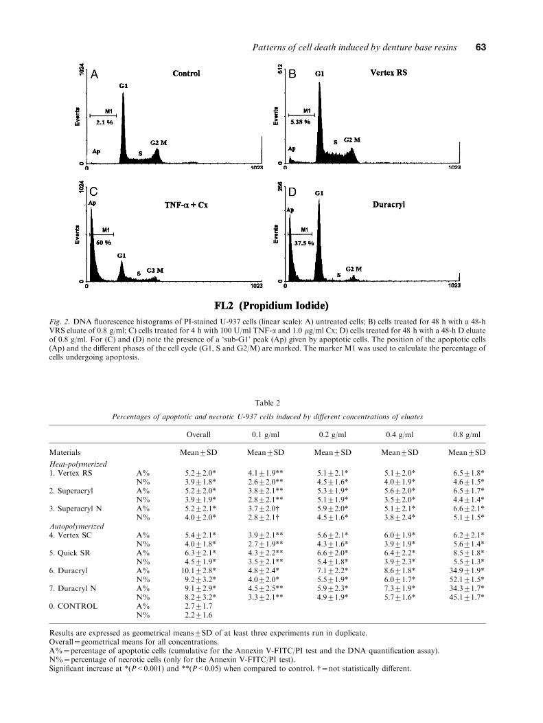
Fig. 2. DNA ¯uorescence histograms of PI-stained U-937 cells (linear scale): A) untreated cells; B) cells treated for 48 h with a 48-hVRS eluate of 0.8 g/ml; C) cells treated for 4 h with 100 U/ml TNF-a and 1.0 mg/ml Cx; D) cells treated for 48 h with a 48-h D eluateof 0.8 g/ml. For (C) and (D) note the presence of a `sub-G1' peak (Ap) given by apoptotic cells. The position of the apoptotic cells(Ap) and the di�erent phases of the cell cycle (G1, S and G2/M) are marked. The marker M1 was used to calculate the percentage ofcells undergoing apoptosis.
Table 2
Percentages of apoptotic and necrotic U-937 cells induced by di�erent concentrations of eluates
Overall 0.1 g/ml 0.2 g/ml 0.4 g/ml 0.8 g/ml
Materials Mean¡SD Mean¡SD Mean¡SD Mean¡SD Mean¡SD
Heat-polymerized1. Vertex RS A%
N%5.2¡2.0*3.9¡1.8*
4.1¡1.9**2.6¡2.0**
5.1¡2.1*4.5¡1.6*
5.1¡2.0*4.0¡1.9*
6.5¡1.8*4.6¡1.5*
2. Superacryl A%N%
5.2¡2.0*3.9¡1.9*
3.8¡2.1**2.8¡2.1**
5.3¡1.9*5.1¡1.9*
5.6¡2.0*3.5¡2.0*
6.5¡1.7*4.4¡1.4*
3. Superacryl N A%N%
5.2¡2.1*4.0¡2.0*
3.7¡2.0{2.8¡2.1{
5.9¡2.0*4.5¡1.6*
5.1¡2.1*3.8¡2.4*
6.6¡2.1*5.1¡1.5*
Autopolymerized4. Vertex SC A%
N%5.4¡2.1*4.0¡1.8*
3.9¡2.1**2.7¡1.9**
5.6¡2.1*4.3¡1.6*
6.0¡1.9*3.9¡1.9*
6.2¡2.1*5.6¡1.4*
5. Quick SR A%N%
6.3¡2.1*4.5¡1.9*
4.3¡2.2**3.5¡2.1**
6.6¡2.0*5.4¡1.8*
6.4¡2.2*3.9¡2.3*
8.5¡1.8*5.5¡1.3*
6. Duracryl A%N%
10.1¡2.8*9.2¡3.2*
4.8¡2.4*4.0¡2.0*
7.1¡2.2*5.5¡1.9*
8.6¡1.8*6.0¡1.7*
34.9¡1.9*52.1¡1.5*
7. Duracryl N A%N%
9.1¡2.9*8.2¡3.2*
4.5¡2.5**3.3¡2.1**
5.9¡2.3*4.9¡1.9*
7.3¡1.9*5.7¡1.6*
34.3¡1.7*45.1¡1.7*
0. CONTROL A%N%
2.7¡1.72.2¡1.6
Results are expressed as geometrical means¡SD of at least three experiments run in duplicate.Overall~geometrical means for all concentrations.A%~percentage of apoptotic cells (cumulative for the Annexin V-FITC/PI test and the DNA quanti®cation assay).N%~percentage of necrotic cells (only for the Annexin V-FITC/PI test).Signi®cant increase at *(P<0.001) and **(P<0.05) when compared to control. {~not statistically di�erent.
Patterns of cell death induced by denture base resins 63

Discussion
By using two separate and unrelated ¯ow cyto-metry assays for the assessment and quanti®cationof the two di�erent cell death patterns, we providedfor the ®rst time experimental evidence indicating adose- and time-dependent increase in the accumu-lation of apoptotic and necrotic cells exposed toeluates of heat-polymerized and autopolymerizedPMMA-based denture base resins. The score forapoptosis was lower in the DNA quanti®cationassay than in the Annexin V-FITC/PI assay due tothe fact that apoptotic cells in the S or G2/Mphases do not appear in the `sub-G1' shoulder.Morphological analysis con®rmed the resultsobtained by ¯ow cytometry.
We consider the identi®cation of cell deathpatterns important when evaluating the cytopatho-genic e�ects of denture base resins because apop-tosis and necrosis have di�erent biologicalsigni®cance. In vivo, apoptotic cells are removedby phagocytes and thus, an in¯ammatory responseis prevented. Necrosis on the other hand inducesin¯ammation and injuries to the surroundingtissues. Therefore, if the compounds eluted from adenture induce apoptosis, then the tissues thatcome in contact with the denture would more likelyadapt to modi®cations induced by it, whereas ifthey induce necrosis, the consequent in¯ammatoryphenomena can induce severe tissue reactions.Another aspect is that the homeostatic balance
Fig. 3. Histogram representing the proportions of apoptotic & and necrotic and/or secondary necrotic % cells induced by eluates at0.1 g/ml, 0.2 g/ml, 0.4 g/ml and 0.8 g/ml. A) results for Annexin V-FITC and PI labelling; B) results for DNA quanti®cation.0~Control, 1~VRS, 2~S, 3~SN, 4~VSC, 5~Q, 6~DN, 7~D. (See Table 1 for abbreviations.)
64 Cimpan et al.

between cell proliferation and cell death is essentialfor the development and maintenance of cells. Theonly materials that augmented necrosis morethan apoptosis were the autopolymerized D andDN at 0.8 g/ml, indicating that these materialsare more likely to induce a strong in¯ammatorytissue reaction. However, since autopolymerizedresins are used for relining and rebasing, forwhich small quantities of material are required, itis most probable that the concentration neededfor such a strong tissue reaction will not be reachedin vivo.The determination of the surface area of the
specimens was di�cult and gave higher inaccuracythan the determination of their mass. Therefore, we
chose to use the ratio between the mass of the speci-mens and the volume of the extraction medium, asrecommended by the ISO standard (22).
Due to their good reproducibility, genetic andmetabolic stability, permanent cell lines are recom-mended for toxicity screening as a ®rst approach(33). The U-937 permanent cell line was chosenbecause of its human origin and ability to performmany of the monocytes' functions. Recently, theU-937 monocytic cells were proposed as goodcandidates for normative in vitro evaluation ofbiocompatibility (34). Since apoptosis in the U-937cells has been extensively studied, a di�erentialdiagnosis between apoptosis and necrosis in thiscell line is more certain than in other types of cells
Fig. 4. Histogram representing the proportions of apoptotic & and necrotic and/or secondary necrotic % cells induced by 24-h and48-h eluates after exposure to U-937 cells for 24 h and 48 h. A) results for Annexin V-FITC and PI labelling; B) results for DNAquanti®cation. 0~Control, 1~VRS, 2~S, 3~SN, 4~VSC, 5~Q, 6~DN, 7~D. (See Table 1 for abbreviations.)
Patterns of cell death induced by denture base resins 65

like ®broblasts and epithelial cells, which do notdisplay all the typical signs of apoptosis (35). Inaddition, the U-937 cells are of monocytic lineageand alterations of their functions, caused by therelease of leachable compounds from denture baseacrylic resins, may mimic an altered local immuneresponse to acrylic dentures.
Eluates from autopolymerized resin disks yieldedhigher percentages of apoptosis and necrosis thanthe heat-polymerized ones. These ®ndings were inaccordance with earlier studies which showed thatautopolymerized resins eluted considerably morematerial (3±7, 9) and were more cytotoxic than theheat-polymerized ones (2, 7, 13). This concordancebetween our results and those of previous workssuggests that the levels of apoptosis and necrosis
re¯ect the biocompatibility degree of PMMA-baseddenture base resins.
From the autopolymerized resins only DN and Dsigni®cantly increased the level of cell death whencompared to all the other resins indicating thatsigni®cant di�erences exist between brands belong-ing to the same type of denture base material. Inorder to explain these di�erences more informationabout the polymerization system and the chemicalcomposition of each brand is needed. Futurestudies should therefore identify the individualcomponents of the eluates that are responsiblefor enhancing cell death by apoptosis and/ornecrosis.
The ®nding that 48-h eluates increased the levelsof apoptosis and necrosis more than 24-h eluates
Fig. 5. Morphological e�ects of VRS and D on human U-937monoblastoid cells. Cells were cultured in the absence of (A)or presence of 24-h eluates from 0.8 g/ml VRS (B) or D (C).24 h after the addition cell aliquots were prepared for lightmicroscopy as described in material and methods. Bars~10 mm.
Fig. 6. Ultrastructure of U-937 monoblastoid cells grown inRPMI standard medium (A). Less than 3% of these cells diedspontaneously (B). In spontaneously dead cells the chromatinand cytoplasm were condensed and the cell and mitochondriavolumes decreased. Budding and cell fragmentation wereobserved. Abbreviations: c~condensed chromatin, f~cellularfragments, m~mitochondria, n~nucleolus. Bars~2 mm.
66 Cimpan et al.

Fig. 7. Electron micrographs of VRS (A±D) and D (E±G) induced U-937 cell death. Cell nucleus is rounded up (A, E) and thechromatin condensation starts beneath the nuclear membrane (A). The nuclear pores disappeared in areas with condensed chromatin(C, D) and late in the apoptotic process the nuclear membrane was lifted and an enlarged space was observed between the twomembranes (D). Nuclear fragmentation (B, F) and gathered mitochondria (A, B) were often seen. Necrotic cell death (G) wascharacterised by cell and mitochondrial swelling and decondensation of chromatin. Abbreviations: c~condensed chromatin,m~mitochondria, n~nucleolus, arrow~nuclear membrane pore, arrowhead~outer nuclear membrane. Bars~1.5 mm.
Patterns of cell death induced by denture base resins 67

was rather unexpected. Previous reports showedthat the 24-h eluates of heat-polymerized andautopolymerized denture base resins tested by themitochondrial dehydrogenase activity (MTT assay)were more harmful than the 48-h eluates (13, 36).The di�erences between our ®ndings and those ofthe latter studies may be due to the di�erentchemical compositions of the materials, di�erentcell types analysed, and di�erent types of assaysemployed.
Previous studies have reported alterations inseveral classes of cell lipids in epithelial cellsexposed to eluates of denture base resins (11).Peroxidation of cellular lipids by benzoyl peroxide(BPO), commonly used as polymerization initiatorin denture resins, may be a major mechanism oftoxicity (37±39). It has been demonstrated that freeradicals resulting from decomposition of BPOduring polymerization are released, and that thisis a long-lasting event (40). Free radicals have ahigh reactivity with all biological molecules andproduce injurious processes to cells and tissues(40±42). These ®ndings might explain the apoptosisand necrosis inducing potential of the eluates testedin our study.
Although VRS is a heat-polymerized resin and Dis an autopolymerized one, both induced mainly thesame morphological signs of apoptosis in U-937cells, which di�ered from those of spontaneousapoptosis in the controls. This morphologicalphenotype seems similar to that observed by us inapoptosis induced by phosphatase inhibitors, whichspeci®cally alter phosphorylation pathways (27,30). This may imply that the same component ora sum of components, which alter the phosphoryla-tion pathways, may induce apoptosis in U-937 cellsexposed to VRS and D eluates.
In summary, eluates from the tested denture baseresins induced cell death by apoptosis and necrosisin U-937 cells in the following rank order ofpotency: Duracryl4Duracryl ``New''4Quick SR3/604Vertex SC4Superacryl ``New''4Superacryl4Vertex RS. The augmentation of apoptosis and/or necrosis indicates that leachable compoundsfrom PMMA-based denture base resins activatesignaling pathways involved in cell death regula-tion. The levels of apoptosis and necrosis re¯ect thecytotoxicity degree of PMMA-based denture baseresins. We consider that further studies on themechanisms of cell death patterns induced by suchresins could contribute to the development ofimproved materials and better strategies for evalu-ating tissue-material biocompatibility.
Acknowledgements ± The authors wish to express their specialgratitude toProf.ChrisReutelingsperger (UniversityofLimburg,The Netherlands), who supervised the ®rst experiments on
Annexin V labeling, for invaluable scienti®c help and forgenerously providing Annexin V-FITC. Excellent technicalassistance was provided by Steinar Sùrnes (¯ow cytometry),Bente Heggù Hansen (¯ow cytometry and cell culture) andBerit Karen Hausvik (electron microscopy). The skilfultechnical assistance of Petra Lux (University of Limburg) isalso gratefully acknowledged. The authors thank Prof. RonaldScheline (University of Bergen) for valuable linguistic help. Thiswork was supported by the Faculty of Dentistry and Faculty ofMedicine, University of Bergen, Norway, by Ole SmithHouskens fund and by Colgate Palmolive Norge A/S.
References
1. BROWN LR, FLAVIN C, FRENCH H. A new economy for anew century. In: State of the World 1999. New York: W. W.Norton & Company, 1999; 5±7.
2. ANNUSAVICE KJ. Denture base resins: Technical considera-tions and processing techniques. In: Phillips' science ofdental materials. 10th edition. Philadelphia: W.B. Saunders,1996; 237±271.
3. RUYTER IE. The release of formaldehyde from denture basepolymers. Acta Odontol Scand 1980; 38: 17±27.
4. BAKER S, BROOKS SC, WALKER DM. The release of residualmonomeric methyl methacrylate from acrylic appliances inthe human mouth: An assay for monomer in saliva. J DentRes 1988; 67: 1295±1299.
5. KODA T, TSUCHIYA H, YAMAUCHI M, HOSHINO Y, TAKAGI N,KAWANO J. High-performance liquid chromatographicestimation of eluates from denture base polymers. J Dent1989; 17: 84±89.
6. KODA T, TSUCHIYA H, YAMAUCHI M, OHTANI S, TAKAGI N,KAWANO J. Leachability of denture base acrylic resins inarti®cial saliva. Dent Mater 1990; 6: 13±16.
7. TSUCHIYA H, HOSHINO Y, TAJIMA K, TAKAGI N. Leachingand cytotoxicity of formaldehyde and methyl methacrylatefrom acrylic resin denture base materials. J Prosthet Dent1994; 71: 618±624.
8. LYGRE H, SOLHEIM E, GJERDET NR, BERG E. Leaching oforganic additives from dentures in vivo. Acta Odontol Scand1993; 51: 45±51.
9. LYGRE H, SOLHEIM E, GJERDET NR. Leaching from denturebase materials in vitro. Acta Odontol Scand 1995; 53: 75±80.
10. HENSTEN-PETTERSEN A, WICTORIN L. The cytotoxic e�ectof denture base polymers. Acta Odontol Scand 1981; 39:101±106.
11. SCHUSTER GS, LEFEBVRE CA, DIRKSEN TR, KNOERNSCHILD
KL, CAUGHMAN GB. Relationships between denturebase resins cytotoxicity and cell lipid metabolism. Int JProsthodont 1995; 8: 580±586.
12. LEFEBVRE CA, SCHUSTER GS, MARR JC, KNOERNSCHILD KL.The e�ect of pH on the cytotoxicity of eluates from denturebase resins. Int J Prosthodont 1995; 8: 122±128.
13. SHERIDAN PJ, KOKA S, EWOLDSEN NO, LEFEBVRE CA, LAVIN
MT. Cytotoxicity of denture base resins. Int J Prosthodont1997; 10: 73±77.
14. KERR JFR, WYLLIE AH, CURRIE AR. Apoptosis: a basicbiological phenomenon with wide-ranging implications intissue kinetics. Br J Cancer 1972; 26: 239±257.
15. JACOBSON MD, WEIL M, RAFF MC. Programmed cell deathin animal development. Cell 1997; 88: 347±354.
16. MAJNO G, JORIS I. Apoptosis, oncosis and necrosis. Anoverview of cell death. Am J Pathol 1995; 146: 3±15.
17. CASTEDOM,HIRSCHT, SUSHINSA,ZAMZANIN,MARCHETTIP,MACHO A, KROEMER G. Sequential acquisition of mito-chondrial and plasma membrane alterations during earlylymphocyte apoptosis. J Immunol 1996; 157: 512±521.
68 Cimpan et al.

18. VERMES I, HAANEN C, STEFFENS-NAKKEN H, REUTELINGS-
PERGER C. A novel assay for apoptosis: Flow cytometricdetection of phosphatidylserine expression on early apop-totic cells using ¯uorescein labelled annexin V. J ImmunolMethods 1995; 184: 39±51.
19. VAN ENGELAND M, NIELAND LJW, RAMAEKERS CS, SCHUTTE
B, REUTELINGSPERGER CPM. Annexin V-a�nity assay: Areview on an apoptosis detection system based on phos-phatidylserine exposure. Cytometry 1998; 31: 1±9.
20. DARZYNKIEWICZZ, JUANG,LIX,GORCZYCAW,MURAKAMIT,TRAGANOS F. Cytometry in cell necrobiology: Analysis ofapoptosis and accidental cell death (necrosis). Cytometry1997; 27: 1±20.
21. ISO (International Organization for Standardization).Dentistry±Denture base polymers. 1567 : 1988 (E). Clause7.3.2. 2nd edition. 1988-11-01; 1±9.
22. ISO (International Organization for Standardization).Biological evaluation of medical devices ± Part 5: Tests forcytotoxicity: in vitro methods. 10993-5:1992 (E). 1st edition.1992-12-15; 1±7.
23. GALEA-LAURI J, RICHARDSON AJ, LATCHMAN DS, KATZ DR.Increased heat shock protein 90 (hsp90) expression leadsto increased apoptosis in the monoblastoid cell lineU937 following induction with TNF-a and cycloheximide.J Immunol 1996; 157: 4109±4118.
24. TROTTER J.WinMDI 2.7. Build #16 software (PC/Windows).Scripps Research Institute. 1998; available from URL:http://www.uwcm.ac.uk/uwcm/hg/hoy/software.html
25. COLIGAN JE, KRUISBEEKAM,MARGULIES DH, SHEVACH EM,STROBER W, COICO R. Flow Cytometry for DNA Analysis.In: Current protocols in immunology 1995; 5.7.1±5.7.6.
26. GONG J, TRAGANOS F, DARZYNKIEWICZ Z. A selectiveprocedure for DNA extraction from apoptotic cellsapplicable for gel electrophoresis and ¯ow cytometry.Anal Biochem 1994; 218: 314±319.
27. JENSEN PH, CRESSEY LI, GJERTSEN BT, MADSEN P,MELLGREN G, HOKLAND P, GLIEMANN J, DéSKELAND SO,LANOTTE M, VINTERMYR OK. Cleaved intracellular plasmi-nogen activator inhibitor-2 in human myeloleukemia cells isa marker of apoptosis. Br J Cancer 1994; 70: 834±840.
28. SPSS base 8.0: Application Guide. Two-way analysis ofvariance. Chicago: SPSS, 1998; 281±308.
29. BIPM, IEC, IFCC, ISO, IUPAC, IUPAP, OIML. Guide tothe Expression of Uncertainty in Measurement. 1st edition.Geneva: ISO 1995; 10.
30. GJERTSEN BT, CRESSEY LI, RUCHAUD S, HOUGE G, LANOTTE
M, DéSKELAND SO. Multiple apoptotic death typestriggered through activation of separate pathwaysby cAMP and inhibitors of protein phosphatases in
one (IPC leukaemia) cell line. J Cell Sci 1994; 107:3363±3377.
31. CHAYA D, FOUGEÂ RE-DESCHATRETTE C, WEISS MC.Spontaneous apoptosis in a rat hepatoma cell line and itsinhibition by dexamethasone. Cell Death Di�er 1996; 3:97±104.
32. RINNER I, FELSNER P, HOFER P, GLOBERSON A,SCHAUENSTEIN K. Characterisation of the spontaneousapoptosis of rat thymocytes in vitro. Int Arch AllergyImmunol 1996; 111: 230±237.
33. SCHMALZ G. Use of cell cultures for toxicity testing of dentalmaterials ± advantages and limitations. J Dent 1994; 22:S6±11.
34. BLOTTIERE HM, DACULSI G, ANEGON I, POUEZAT JA,NELSON PN, PASSUTI N. Utilization of activated U937monocytic cells as a model to evaluate biocompatibility andbiodegradation of synthetic calcium phosphate.Biomaterials1995; 16: 497±503.
35. FADY C, GARDNER A, JACOBY F, BRISKIN K, TU Y, SCHMID
I, LICHTENSTEIN A. Atypical apoptotic cell death inducedin L929 targets by exposure to tumor necrosis factor.J Interferon Cytokine Res 1995; 15: 71±80.
36. LEFEBVRE CA, KNOERNSCHILD KL, SCHUSTER GS.Cytotoxicity of eluates from light-polymerized denturebase resins. J Prosthet Dent 1994; 72: 644±650.
37. EMERIT J, CHAUDIERE J. Free radicals and lipid peroxidationin cell pathology. In: MIQUEL J, QUINTANILHA AT, WEBER H,eds. Handbook of free radicals and antioxidants inbiomedicine, VI. Boca Raton, FL: CRC Press, 1984;177±185.
38. TERAKADO M, YAMAZAKI M, TSUJIMOTO Y, KAWASIMA T,NAGASHIMA K, OGAWA J, FUJITA Y, SUGIYA H, SAKAI T,FURUYAMA S. Lipid peroxidation as a possible cause ofbenzoyl peroxide toxicity in rabbit dental pulp ± Amicrosomal lipid peroxidation in vitro. J Dent Res 1984;63: 901±905.
39. LEFEBVRE CA, SCHUSTER GS, RUEGGEBERG FA, TAMARE-SELVY K, KNOERNSCHILD KL. Responses of oral epithelialcells to dental resin components. J Biomater Sci PolymerEdn 1996; 7: 965±976.
40. MOREAU MF, CHAPPARD D, LESOURD M, MONTHEÂ ARD JP,BASLEÂ MF. Free radicals and side products released duringmethylmethacrylate polymerisation are cytotoxic for osteo-blastic cells. J Biomed Mater Res 1998; 40: 124±131.
41. HALLIWELL B. Free radicals, antioxidants and humandisease: curiosity, cause or consequence? Lancet 1994;344: 721±724.
42. KEHRER JP. Free radicals as mediators of tissue injury anddisease. Crit Rev Toxicol 1993; 23: 21±48.
Patterns of cell death induced by denture base resins 69