Particle formation pathways and polymorphism of curcumin induced by ultrasound and additives during...
13
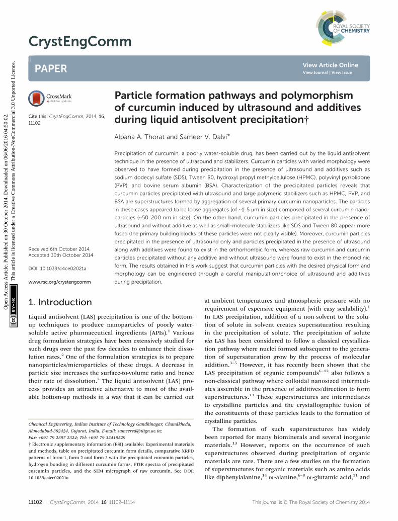
CrystEngComm PAPER Cite this: CrystEngComm, 2014, 16, 11102 Received 6th October 2014, Accepted 30th October 2014 DOI: 10.1039/c4ce02021a www.rsc.org/crystengcomm Particle formation pathways and polymorphism of curcumin induced by ultrasound and additives during liquid antisolvent precipitation† Alpana A. Thorat and Sameer V. Dalvi * Precipitation of curcumin, a poorly water-soluble drug, has been carried out by the liquid antisolvent technique in the presence of ultrasound and stabilizers. Curcumin particles with varied morphology were observed to have formed during precipitation in the presence of ultrasound and additives such as sodium dodecyl sulfate (SDS), Tween 80, hydroxyl propyl methylcellulose (HPMC), polyvinyl pyrrolidone (PVP), and bovine serum albumin (BSA). Characterization of the precipitated particles reveals that curcumin particles precipitated with ultrasound and large polymeric stabilizers such as HPMC, PVP, and BSA are superstructures formed by aggregation of several primary curcumin nanoparticles. The particles in these cases appeared to be loose aggregates (of ~1–5 μm in size) composed of several curcumin nano- particles (~50–200 nm in size). On the other hand, curcumin particles precipitated in the presence of ultrasound and without additive as well as small-molecule stabilizers like SDS and Tween 80 appear more fused (the primary building blocks of these particles were not clearly visible). Moreover, curcumin particles precipitated in the presence of ultrasound only and particles precipitated in the presence of ultrasound along with additives were found to exist in the orthorhombic form, whereas raw curcumin and curcumin particles precipitated without any additive and without ultrasound were found to exist in the monoclinic form. The results obtained in this work suggest that curcumin particles with the desired physical form and morphology can be engineered through a careful manipulation/choice of ultrasound and additives during precipitation. 1. Introduction Liquid antisolvent (LAS) precipitation is one of the bottom- up techniques to produce nanoparticles of poorly water- soluble active pharmaceutical ingredients (APIs). 1 Various drug formulation strategies have been extensively studied for such drugs over the past few decades to enhance their disso- lution rates. 2 One of the formulation strategies is to prepare nanoparticles/microparticles of these drugs. A decrease in particle size increases the surface-to-volume ratio and hence their rate of dissolution. 2 The liquid antisolvent (LAS) pro- cess provides an attractive alternative to most of the avail- able bottom-up methods in a way that it can be carried out at ambient temperatures and atmospheric pressure with no requirement of expensive equipment (with easy scalability). 1 In LAS precipitation, addition of a non-solvent to the solu- tion of solute in solvent creates supersaturation resulting in the precipitation of solute. The precipitation of solute via LAS has been considered to follow a classical crystalliza- tion pathway where nuclei formed subsequent to the genera- tion of supersaturation grow by the process of molecular addition. 3–5 However, it has recently been shown that the LAS precipitation of organic compounds 6–12 also follows a non-classical pathway where colloidal nanosized intermedi- ates assemble in the presence of additives/direction to form superstructures. 13 These superstructures are intermediates to crystalline particles and the crystallographic fusion of the constituents of these particles leads to the formation of crystalline particles. The formation of such superstructures has widely been reported for many biominerals and several inorganic materials. 13 However, reports on the occurrence of such superstructures observed during precipitation of organic materials are rare. There are a few studies on the formation of superstructures for organic materials such as amino acids like diphenylalanine, 14 DL-alanine, 6–8 DL-glutamic acid, 11 and 11102 | CrystEngComm, 2014, 16, 11102–11114 This journal is © The Royal Society of Chemistry 2014 Chemical Engineering, Indian Institute of Technology Gandhinagar, Chandkheda, Ahmedabad-382424, Gujarat, India. E-mail: [email protected]; Fax: +091 79 2397 2324; Tel: +091 79 32419529 † Electronic supplementary information (ESI) available: Experimental materials and methods, table on precipitated curcumin form details, comparative XRPD patterns of form 1, form 2 and form 3 with the precipitated curcumin particles, hydrogen bonding in different curcumin forms, FTIR spectra of precipitated curcumin particles, and the SEM micrograph of raw curcumin. See DOI: 10.1039/c4ce02021a Open Access Article. Published on 30 October 2014. Downloaded on 06/06/2016 04:50:02. This article is licensed under a Creative Commons Attribution-NonCommercial 3.0 Unported Licence. View Article Online View Journal | View Issue
-
Upload
spanalumni -
Category
Documents
-
view
8 -
download
0
Transcript of Particle formation pathways and polymorphism of curcumin induced by ultrasound and additives during...
CrystEngComm
Ope
n A
cces
s A
rtic
le. P
ublis
hed
on 3
0 O
ctob
er 2
014.
Dow
nloa
ded
on 0
6/06
/201
6 04
:50:
02.
Thi
s ar
ticle
is li
cens
ed u
nder
a C
reat
ive
Com
mon
s A
ttrib
utio
n-N
onC
omm
erci
al 3
.0 U
npor
ted
Lic
ence
.
PAPER View Article OnlineView Journal | View Issue
11102 | CrystEngComm, 2014, 16, 11102–11114 This journal is © The R
Chemical Engineering, Indian Institute of Technology Gandhinagar, Chandkheda,
Ahmedabad-382424, Gujarat, India. E-mail: [email protected];
Fax: +091 79 2397 2324; Tel: +091 79 32419529
† Electronic supplementary information (ESI) available: Experimental materialsand methods, table on precipitated curcumin form details, comparative XRPDpatterns of form 1, form 2 and form 3 with the precipitated curcumin particles,hydrogen bonding in different curcumin forms, FTIR spectra of precipitatedcurcumin particles, and the SEM micrograph of raw curcumin. See DOI:10.1039/c4ce02021a
Cite this: CrystEngComm, 2014, 16,
11102
Received 6th October 2014,Accepted 30th October 2014
DOI: 10.1039/c4ce02021a
www.rsc.org/crystengcomm
Particle formation pathways and polymorphismof curcumin induced by ultrasound and additivesduring liquid antisolvent precipitation†
Alpana A. Thorat and Sameer V. Dalvi*
Precipitation of curcumin, a poorly water-soluble drug, has been carried out by the liquid antisolvent
technique in the presence of ultrasound and stabilizers. Curcumin particles with varied morphology were
observed to have formed during precipitation in the presence of ultrasound and additives such as
sodium dodecyl sulfate (SDS), Tween 80, hydroxyl propyl methylcellulose (HPMC), polyvinyl pyrrolidone
(PVP), and bovine serum albumin (BSA). Characterization of the precipitated particles reveals that
curcumin particles precipitated with ultrasound and large polymeric stabilizers such as HPMC, PVP, and
BSA are superstructures formed by aggregation of several primary curcumin nanoparticles. The particles
in these cases appeared to be loose aggregates (of ~1–5 μm in size) composed of several curcumin nano-
particles (~50–200 nm in size). On the other hand, curcumin particles precipitated in the presence of
ultrasound and without additive as well as small-molecule stabilizers like SDS and Tween 80 appear more
fused (the primary building blocks of these particles were not clearly visible). Moreover, curcumin particles
precipitated in the presence of ultrasound only and particles precipitated in the presence of ultrasound
along with additives were found to exist in the orthorhombic form, whereas raw curcumin and curcumin
particles precipitated without any additive and without ultrasound were found to exist in the monoclinic
form. The results obtained in this work suggest that curcumin particles with the desired physical form and
morphology can be engineered through a careful manipulation/choice of ultrasound and additives
during precipitation.
1. Introduction
Liquid antisolvent (LAS) precipitation is one of the bottom-up techniques to produce nanoparticles of poorly water-soluble active pharmaceutical ingredients (APIs).1 Variousdrug formulation strategies have been extensively studied forsuch drugs over the past few decades to enhance their disso-lution rates.2 One of the formulation strategies is to preparenanoparticles/microparticles of these drugs. A decrease inparticle size increases the surface-to-volume ratio and hencetheir rate of dissolution.2 The liquid antisolvent (LAS) pro-cess provides an attractive alternative to most of the avail-able bottom-up methods in a way that it can be carried out
at ambient temperatures and atmospheric pressure with norequirement of expensive equipment (with easy scalability).1
In LAS precipitation, addition of a non-solvent to the solu-tion of solute in solvent creates supersaturation resultingin the precipitation of solute. The precipitation of solutevia LAS has been considered to follow a classical crystalliza-tion pathway where nuclei formed subsequent to the genera-tion of supersaturation grow by the process of molecularaddition.3–5 However, it has recently been shown that theLAS precipitation of organic compounds6–12 also follows anon-classical pathway where colloidal nanosized intermedi-ates assemble in the presence of additives/direction to formsuperstructures.13 These superstructures are intermediatesto crystalline particles and the crystallographic fusion ofthe constituents of these particles leads to the formation ofcrystalline particles.
The formation of such superstructures has widelybeen reported for many biominerals and several inorganicmaterials.13 However, reports on the occurrence of suchsuperstructures observed during precipitation of organicmaterials are rare. There are a few studies on the formationof superstructures for organic materials such as amino acidslike diphenylalanine,14 DL-alanine,6–8 DL-glutamic acid,11 and
oyal Society of Chemistry 2014
CrystEngComm Paper
Ope
n A
cces
s A
rtic
le. P
ublis
hed
on 3
0 O
ctob
er 2
014.
Dow
nloa
ded
on 0
6/06
/201
6 04
:50:
02.
Thi
s ar
ticle
is li
cens
ed u
nder
a C
reat
ive
Com
mon
s A
ttrib
utio
n-N
onC
omm
erci
al 3
.0 U
npor
ted
Lic
ence
.View Article Online
L-cystine12 and poorly water soluble drugs such as diclofenac9
and sodium ibuprofen dihydrate12 that have been reported.In this paper, we present an account of the formation of
superstructures of curcumin nanoparticles, precipitated fromits ethanolic solution in the presence of ultrasound and addi-tives using water as an antisolvent (experimental details arepresented in the ESI†). Curcumin is a poorly water-solubledrug found in the herbal spice turmeric (Curcumin longa). Ithas potential antioxidant, anti-inflammatory, antitumor,anti-HIV, and antimicrobial properties.15 We show that theuse of ultrasound and stabilizers during precipitation ofcurcumin particles induces morphological transformations.Curcumin exists in three forms of which form 1 is mono-clinic and the other two forms (form 2 and form 3) are ortho-rhombic.16,17 While unprocessed curcumin and curcuminparticles precipitated without ultrasound and without stabi-lizers were present in the monoclinic form (form 1), thecurcumin particles precipitated in the presence of ultrasoundand stabilizers were found to exist in the orthorhombic form(form 2 and form 3). Based on the characterization of theprecipitated particles, we propose that the non-classicalpathways of particle formation are followed during precipi-tation of curcumin particles in the presence of ultrasoundand large polymeric additives such as BSA, HPMC and PVP,where the primary curcumin nanoparticles (~50–200 nm insize) aggregate to form secondary superstructures which are1–5 μm in size.
2. Results and discussion2.1. Physical form of precipitated curcumin particles
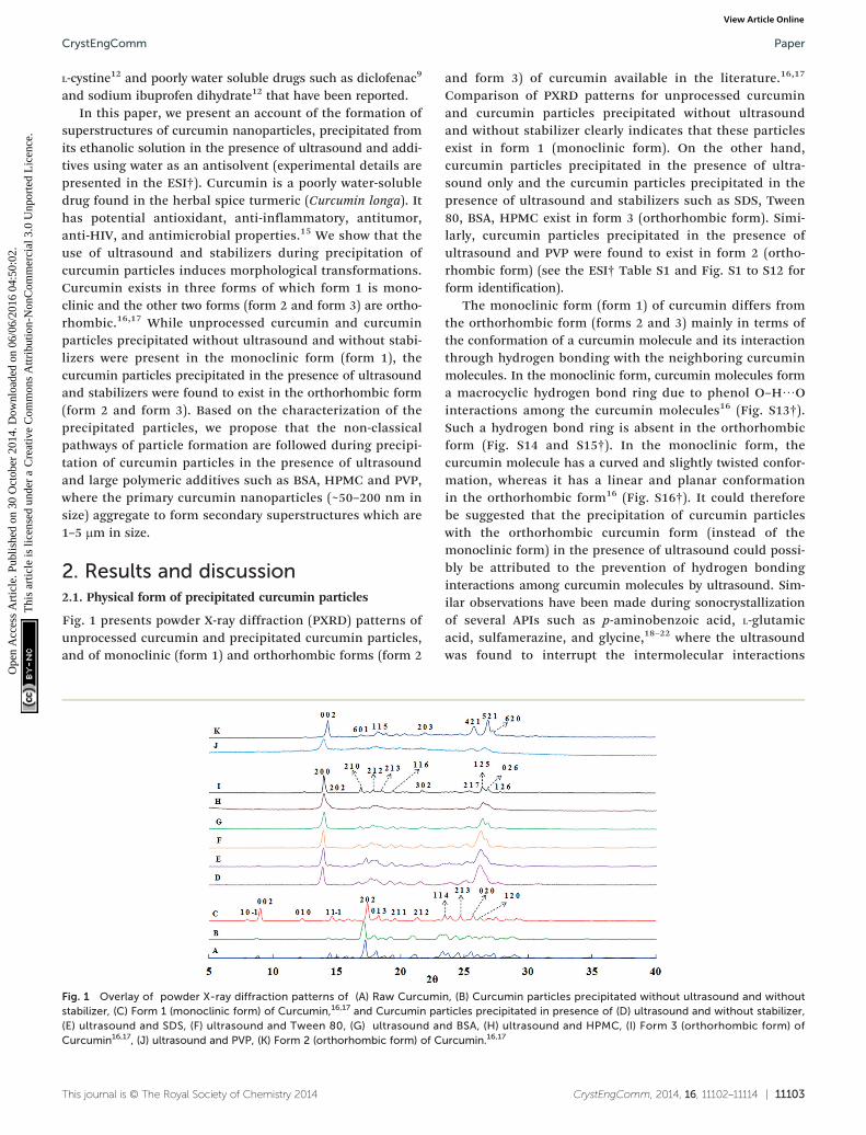
Fig. 1 presents powder X-ray diffraction (PXRD) patterns ofunprocessed curcumin and precipitated curcumin particles,and of monoclinic (form 1) and orthorhombic forms (form 2
This journal is © The Royal Society of Chemistry 2014
Fig. 1 Overlay of powder X-ray diffraction patterns of (A) Raw Curcumistabilizer, (C) Form 1 (monoclinic form) of Curcumin,16,17 and Curcumin pa(E) ultrasound and SDS, (F) ultrasound and Tween 80, (G) ultrasound anCurcumin16,17, (J) ultrasound and PVP, (K) Form 2 (orthorhombic form) of C
and form 3) of curcumin available in the literature.16,17
Comparison of PXRD patterns for unprocessed curcuminand curcumin particles precipitated without ultrasoundand without stabilizer clearly indicates that these particlesexist in form 1 (monoclinic form). On the other hand,curcumin particles precipitated in the presence of ultra-sound only and the curcumin particles precipitated in thepresence of ultrasound and stabilizers such as SDS, Tween80, BSA, HPMC exist in form 3 (orthorhombic form). Simi-larly, curcumin particles precipitated in the presence ofultrasound and PVP were found to exist in form 2 (ortho-rhombic form) (see the ESI† Table S1 and Fig. S1 to S12 forform identification).
The monoclinic form (form 1) of curcumin differs fromthe orthorhombic form (forms 2 and 3) mainly in terms ofthe conformation of a curcumin molecule and its interactionthrough hydrogen bonding with the neighboring curcuminmolecules. In the monoclinic form, curcumin molecules forma macrocyclic hydrogen bond ring due to phenol O–H⋯Ointeractions among the curcumin molecules16 (Fig. S13†).Such a hydrogen bond ring is absent in the orthorhombicform (Fig. S14 and S15†). In the monoclinic form, thecurcumin molecule has a curved and slightly twisted confor-mation, whereas it has a linear and planar conformationin the orthorhombic form16 (Fig. S16†). It could thereforebe suggested that the precipitation of curcumin particleswith the orthorhombic curcumin form (instead of themonoclinic form) in the presence of ultrasound could possi-bly be attributed to the prevention of hydrogen bondinginteractions among curcumin molecules by ultrasound. Sim-ilar observations have been made during sonocrystallizationof several APIs such as p-aminobenzoic acid, L-glutamicacid, sulfamerazine, and glycine,18–22 where the ultrasoundwas found to interrupt the intermolecular interactions
CrystEngComm, 2014, 16, 11102–11114 | 11103
n, (B) Curcumin particles precipitated without ultrasound and withoutrticles precipitated in presence of (D) ultrasound and without stabilizer,d BSA, (H) ultrasound and HPMC, (I) Form 3 (orthorhombic form) ofurcumin.16,17
CrystEngCommPaper
Ope
n A
cces
s A
rtic
le. P
ublis
hed
on 3
0 O
ctob
er 2
014.
Dow
nloa
ded
on 0
6/06
/201
6 04
:50:
02.
Thi
s ar
ticle
is li
cens
ed u
nder
a C
reat
ive
Com
mon
s A
ttrib
utio
n-N
onC
omm
erci
al 3
.0 U
npor
ted
Lic
ence
.View Article Online
during the stage of critical nuclei formation and bringabout the polymorphic transformations.
2.2. Morphology of curcumin superstructures formed in thepresence of ultrasound and stabilizers
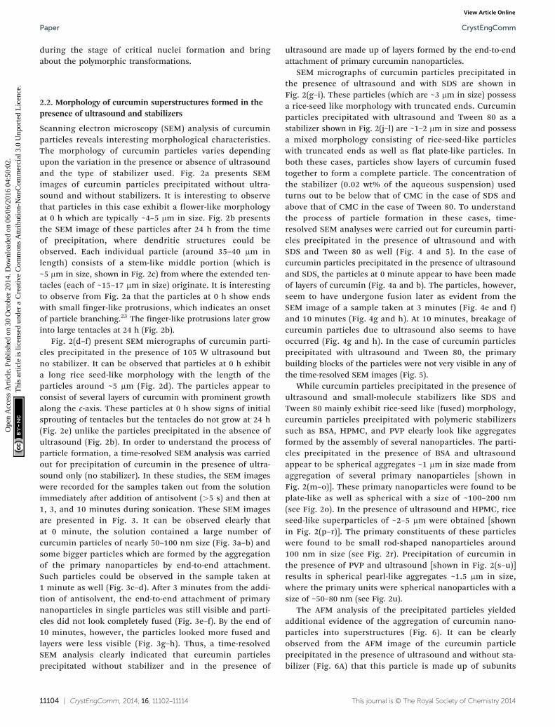
Scanning electron microscopy (SEM) analysis of curcuminparticles reveals interesting morphological characteristics.The morphology of curcumin particles varies dependingupon the variation in the presence or absence of ultrasoundand the type of stabilizer used. Fig. 2a presents SEMimages of curcumin particles precipitated without ultra-sound and without stabilizers. It is interesting to observethat particles in this case exhibit a flower-like morphologyat 0 h which are typically ~4–5 μm in size. Fig. 2b presentsthe SEM image of these particles after 24 h from the timeof precipitation, where dendritic structures could beobserved. Each individual particle (around 35–40 μm inlength) consists of a stem-like middle portion (which is~5 μm in size, shown in Fig. 2c) from where the extended ten-tacles (each of ~15–17 μm in size) originate. It is interestingto observe from Fig. 2a that the particles at 0 h show endswith small finger-like protrusions, which indicates an onsetof particle branching.23 The finger-like protrusions later growinto large tentacles at 24 h (Fig. 2b).
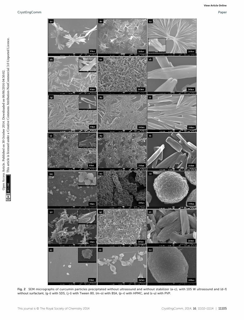
Fig. 2(d–f) present SEM micrographs of curcumin parti-cles precipitated in the presence of 105 W ultrasound butno stabilizer. It can be observed that particles at 0 h exhibita long rice seed-like morphology with the length of theparticles around ~5 μm (Fig. 2d). The particles appear toconsist of several layers of curcumin with prominent growthalong the c-axis. These particles at 0 h show signs of initialsprouting of tentacles but the tentacles do not grow at 24 h(Fig. 2e) unlike the particles precipitated in the absence ofultrasound (Fig. 2b). In order to understand the process ofparticle formation, a time-resolved SEM analysis was carriedout for precipitation of curcumin in the presence of ultra-sound only (no stabilizer). In these studies, the SEM imageswere recorded for the samples taken out from the solutionimmediately after addition of antisolvent (>5 s) and then at1, 3, and 10 minutes during sonication. These SEM imagesare presented in Fig. 3. It can be observed clearly thatat 0 minute, the solution contained a large number ofcurcumin particles of nearly 50–100 nm size (Fig. 3a–b) andsome bigger particles which are formed by the aggregationof the primary nanoparticles by end-to-end attachment.Such particles could be observed in the sample taken at1 minute as well (Fig. 3c–d). After 3 minutes from the addi-tion of antisolvent, the end-to-end attachment of primarynanoparticles in single particles was still visible and parti-cles did not look completely fused (Fig. 3e–f). By the end of10 minutes, however, the particles looked more fused andlayers were less visible (Fig. 3g–h). Thus, a time-resolvedSEM analysis clearly indicated that curcumin particlesprecipitated without stabilizer and in the presence of
11104 | CrystEngComm, 2014, 16, 11102–11114
ultrasound are made up of layers formed by the end-to-endattachment of primary curcumin nanoparticles.
SEM micrographs of curcumin particles precipitated inthe presence of ultrasound and with SDS are shown inFig. 2(g–i). These particles (which are ~3 μm in size) possessa rice-seed like morphology with truncated ends. Curcuminparticles precipitated with ultrasound and Tween 80 as astabilizer shown in Fig. 2(j–l) are ~1–2 μm in size and possessa mixed morphology consisting of rice-seed-like particleswith truncated ends as well as flat plate-like particles. Inboth these cases, particles show layers of curcumin fusedtogether to form a complete particle. The concentration ofthe stabilizer (0.02 wt% of the aqueous suspension) usedturns out to be below that of CMC in the case of SDS andabove that of CMC in the case of Tween 80. To understandthe process of particle formation in these cases, time-resolved SEM analyses were carried out for curcumin parti-cles precipitated in the presence of ultrasound and withSDS and Tween 80 as well (Fig. 4 and 5). In the case ofcurcumin particles precipitated in the presence of ultrasoundand SDS, the particles at 0 minute appear to have been madeof layers of curcumin (Fig. 4a and b). The particles, however,seem to have undergone fusion later as evident from theSEM image of a sample taken at 3 minutes (Fig. 4e and f)and 10 minutes (Fig. 4g and h). At 10 minutes, breakage ofcurcumin particles due to ultrasound also seems to haveoccurred (Fig. 4g and h). In the case of curcumin particlesprecipitated with ultrasound and Tween 80, the primarybuilding blocks of the particles were not very visible in any ofthe time-resolved SEM images (Fig. 5).
While curcumin particles precipitated in the presence ofultrasound and small-molecule stabilizers like SDS andTween 80 mainly exhibit rice-seed like (fused) morphology,curcumin particles precipitated with polymeric stabilizerssuch as BSA, HPMC, and PVP clearly look like aggregatesformed by the assembly of several nanoparticles. The parti-cles precipitated in the presence of BSA and ultrasoundappear to be spherical aggregates ~1 μm in size made fromaggregation of several primary nanoparticles [shown inFig. 2(m–o)]. These primary nanoparticles were found to beplate-like as well as spherical with a size of ~100–200 nm(see Fig. 2o). In the presence of ultrasound and HPMC, riceseed-like superparticles of ~2–5 μm were obtained [shownin Fig. 2(p–r)]. The primary constituents of these particleswere found to be small rod-shaped nanoparticles around100 nm in size (see Fig. 2r). Precipitation of curcumin inthe presence of PVP and ultrasound [shown in Fig. 2(s–u)]results in spherical pearl-like aggregates ~1.5 μm in size,where the primary units were spherical nanoparticles with asize of ~50–80 nm (see Fig. 2u).
The AFM analysis of the precipitated particles yieldedadditional evidence of the aggregation of curcumin nano-particles into superstructures (Fig. 6). It can be clearlyobserved from the AFM image of the curcumin particleprecipitated in the presence of ultrasound and without sta-bilizer (Fig. 6A) that this particle is made up of subunits
This journal is © The Royal Society of Chemistry 2014
CrystEngComm, 2014, 16, 11102–11114 | 11105This journal is © The Royal Society of Chemistry 2014
Fig. 2 SEM micrographs of curcumin particles precipitated without ultrasound and without stabilizer (a–c), with 105 W ultrasound and (d–f)without surfactant, (g–i) with SDS, ( j–l) with Tween 80, (m–o) with BSA, (p–r) with HPMC, and (s–u) with PVP.
CrystEngComm Paper
Ope
n A
cces
s A
rtic
le. P
ublis
hed
on 3
0 O
ctob
er 2
014.
Dow
nloa
ded
on 0
6/06
/201
6 04
:50:
02.
Thi
s ar
ticle
is li
cens
ed u
nder
a C
reat
ive
Com
mon
s A
ttrib
utio
n-N
onC
omm
erci
al 3
.0 U
npor
ted
Lic
ence
.View Article Online
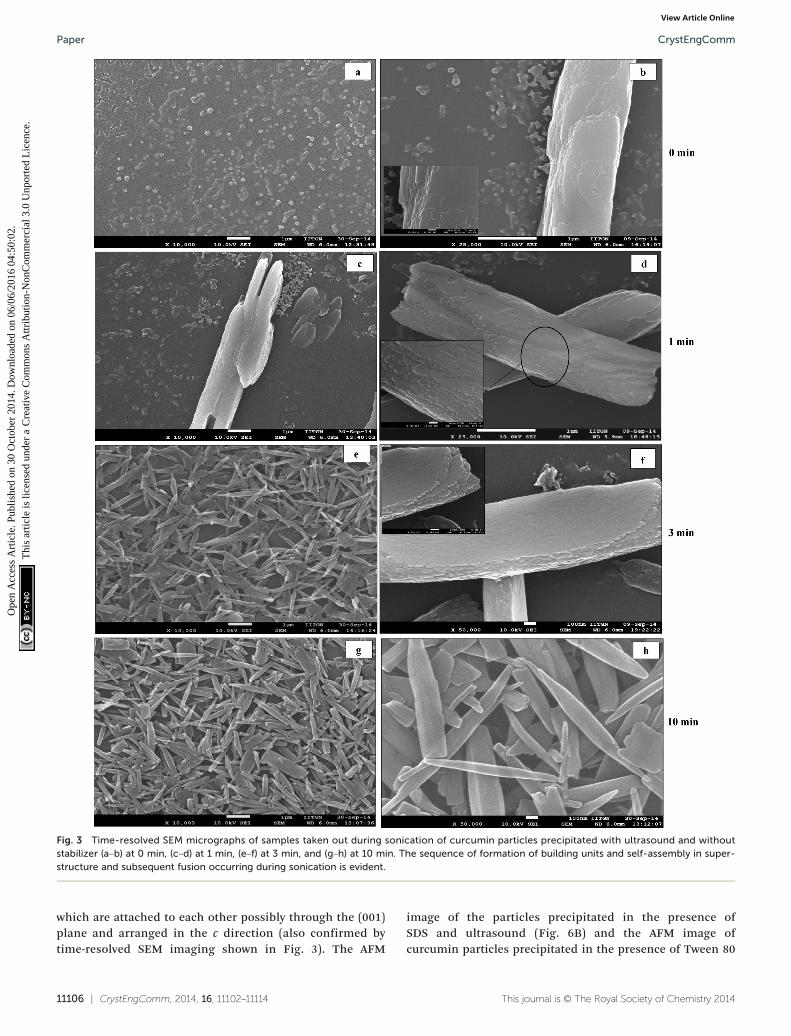
Fig. 3 Time-resolved SEM micrographs of samples taken out during sonication of curcumin particles precipitated with ultrasound and withoutstabilizer (a–b) at 0 min, (c–d) at 1 min, IJe–f) at 3 min, and (g–h) at 10 min. The sequence of formation of building units and self-assembly in super-structure and subsequent fusion occurring during sonication is evident.
CrystEngCommPaper
Ope
n A
cces
s A
rtic
le. P
ublis
hed
on 3
0 O
ctob
er 2
014.
Dow
nloa
ded
on 0
6/06
/201
6 04
:50:
02.
Thi
s ar
ticle
is li
cens
ed u
nder
a C
reat
ive
Com
mon
s A
ttrib
utio
n-N
onC
omm
erci
al 3
.0 U
npor
ted
Lic
ence
.View Article Online
which are attached to each other possibly through the (001)plane and arranged in the c direction (also confirmed bytime-resolved SEM imaging shown in Fig. 3). The AFM
11106 | CrystEngComm, 2014, 16, 11102–11114
image of the particles precipitated in the presence ofSDS and ultrasound (Fig. 6B) and the AFM image ofcurcumin particles precipitated in the presence of Tween 80
This journal is © The Royal Society of Chemistry 2014
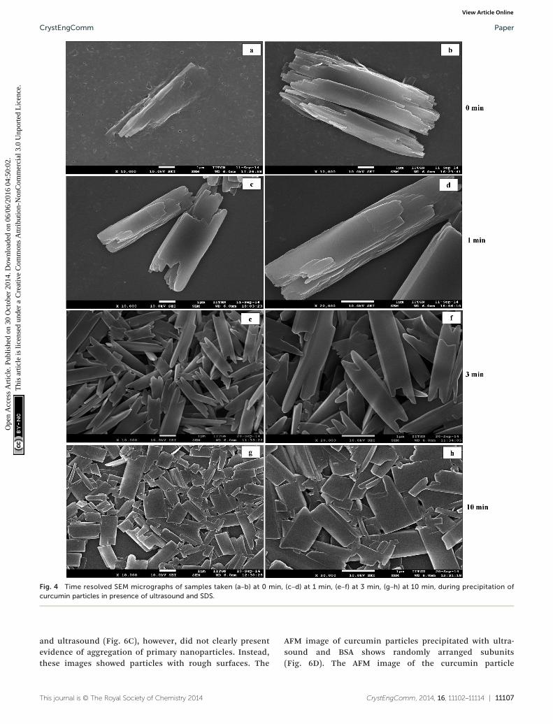
Fig. 4 Time resolved SEM micrographs of samples taken (a–b) at 0 min, (c–d) at 1 min, (e–f) at 3 min, (g–h) at 10 min, during precipitation ofcurcumin particles in presence of ultrasound and SDS.
CrystEngComm Paper
Ope
n A
cces
s A
rtic
le. P
ublis
hed
on 3
0 O
ctob
er 2
014.
Dow
nloa
ded
on 0
6/06
/201
6 04
:50:
02.
Thi
s ar
ticle
is li
cens
ed u
nder
a C
reat
ive
Com
mon
s A
ttrib
utio
n-N
onC
omm
erci
al 3
.0 U
npor
ted
Lic
ence
.View Article Online
and ultrasound (Fig. 6C), however, did not clearly presentevidence of aggregation of primary nanoparticles. Instead,these images showed particles with rough surfaces. The
This journal is © The Royal Society of Chemistry 2014
AFM image of curcumin particles precipitated with ultra-sound and BSA shows randomly arranged subunits(Fig. 6D). The AFM image of the curcumin particle
CrystEngComm, 2014, 16, 11102–11114 | 11107

Fig. 5 Time resolved SEM micrographs of samples taken (a–b) at 0 min, (c–d) at 1 min, (e–f) at 3 min, (g–h) at 10 min, during precipitation ofcurcumin particles in presence of ultrasound and Tween 80.
CrystEngCommPaper
Ope
n A
cces
s A
rtic
le. P
ublis
hed
on 3
0 O
ctob
er 2
014.
Dow
nloa
ded
on 0
6/06
/201
6 04
:50:
02.
Thi
s ar
ticle
is li
cens
ed u
nder
a C
reat
ive
Com
mon
s A
ttrib
utio
n-N
onC
omm
erci
al 3
.0 U
npor
ted
Lic
ence
.View Article Online
precipitated with ultrasound and HPMC shows primarynanoparticles arranged in the c-direction (Fig. 6E). Further,the curcumin particles precipitated with ultrasound and
11108 | CrystEngComm, 2014, 16, 11102–11114
PVP are randomly arranged (Fig. 6F). Although the exactarrangement of subunits in the particle is not completelyvisible with the help of AFM analysis, it still provides useful
This journal is © The Royal Society of Chemistry 2014
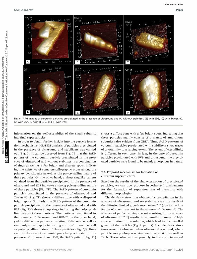
Fig. 6 AFM images of curcumin particles precipitated in the presence of ultrasound and (A) without stabilizer, (B) with SDS, (C) with Tween 80,(D) with BSA, (E) with HPMC, and (F) with PVP.
CrystEngComm Paper
Ope
n A
cces
s A
rtic
le. P
ublis
hed
on 3
0 O
ctob
er 2
014.
Dow
nloa
ded
on 0
6/06
/201
6 04
:50:
02.
Thi
s ar
ticle
is li
cens
ed u
nder
a C
reat
ive
Com
mon
s A
ttrib
utio
n-N
onC
omm
erci
al 3
.0 U
npor
ted
Lic
ence
.View Article Online
information on the self-assemblies of the small subunitsinto final superparticles.
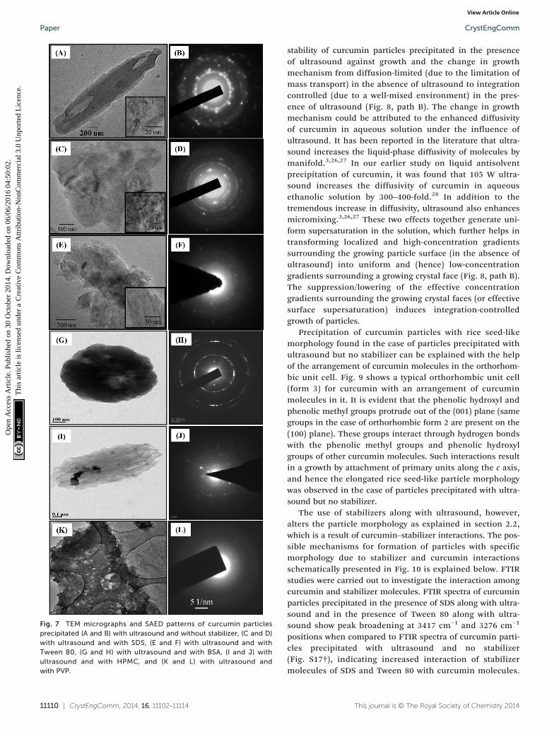
In order to obtain further insight into the particle forma-tion mechanisms, HR-TEM analysis of particles precipitatedin the presence of ultrasound and stabilizers was carriedout (Fig. 7). It can be observed from Fig. 7B that the SAEDpattern of the curcumin particle precipitated in the pres-ence of ultrasound and without stabilizer is a combinationof rings as well as a few bright and discrete spots, indicat-ing the existence of some crystallographic order among theprimary constituents as well as the polycrystalline nature ofthese particles. On the other hand, a sharp ring-like patternobtained from the particles precipitated in the presence ofultrasound and SDS indicates a strong polycrystalline natureof these particles (Fig. 7D). The SAED pattern of curcuminparticles precipitated in the presence of ultrasound andTween 80 (Fig. 7F) shows a diffuse zone with well-spreadbright spots. Similarly, the SAED pattern of the curcuminparticle precipitated in the presence of ultrasound and withBSA (Fig. 7H) shows sharp rings indicating the polycrystal-line nature of these particles. The particles precipitated inthe presence of ultrasound and HPMC, on the other hand,yield a diffraction pattern consisting of discrete as well asrandomly spread spots indicating a mix of ordered as wellas polycrystalline nature of these particles (Fig. 7J). How-ever, in the case of curcumin particles precipitated in thepresence of ultrasound and PVP, the SAED pattern (Fig. 7L)
This journal is © The Royal Society of Chemistry 2014
shows a diffuse zone with a few bright spots, indicating thatthese particles mainly consist of a matrix of amorphoussubunits (also evident from XRD). Thus, SAED patterns ofcurcumin particles precipitated with stabilizers show tracesof crystallinity to a varying extent. The extent of crystallinityis different in each case. In fact, in the case of curcuminparticles precipitated with PVP and ultrasound, the precipi-tated particles were found to be mainly amorphous in nature.
2.3. Proposed mechanism for formation ofcurcumin superstructures
Based on the results of the characterization of precipitatedparticles, we can now propose hypothesized mechanismsfor the formation of superstructures of curcumin withdifferent morphologies.
The dendritic structures obtained by precipitation in theabsence of ultrasound and no stabilizers are the result ofthe diffusion-limited growth mechanism24,25 (due to the lim-itation of mass transport in the absence of ultrasound). Theabsence of perfect mixing (no micromixing in the absenceof ultrasound3,26,27) results in non-uniform zones of highsupersaturation in the solution, which lead to uncontrolledgrowth of the particles (Fig. 8, path A). Such dendritic struc-tures were not observed when ultrasound was used, whereparticle morphology was rice seed-like at 0 h as well as24 h. These observations possibly indicate an increased
CrystEngComm, 2014, 16, 11102–11114 | 11109
11110 | CrystEngComm, 2014, 16, 11102–11114
Fig. 7 TEM micrographs and SAED patterns of curcumin particlesprecipitated (A and B) with ultrasound and without stabilizer, (C and D)with ultrasound and with SDS, (E and F) with ultrasound and withTween 80, (G and H) with ultrasound and with BSA, (I and J) withultrasound and with HPMC, and (K and L) with ultrasound andwith PVP.
CrystEngCommPaper
Ope
n A
cces
s A
rtic
le. P
ublis
hed
on 3
0 O
ctob
er 2
014.
Dow
nloa
ded
on 0
6/06
/201
6 04
:50:
02.
Thi
s ar
ticle
is li
cens
ed u
nder
a C
reat
ive
Com
mon
s A
ttrib
utio
n-N
onC
omm
erci
al 3
.0 U
npor
ted
Lic
ence
.View Article Online
stability of curcumin particles precipitated in the presenceof ultrasound against growth and the change in growthmechanism from diffusion-limited (due to the limitation ofmass transport) in the absence of ultrasound to integrationcontrolled (due to a well-mixed environment) in the pres-ence of ultrasound (Fig. 8, path B). The change in growthmechanism could be attributed to the enhanced diffusivityof curcumin in aqueous solution under the influence ofultrasound. It has been reported in the literature that ultra-sound increases the liquid-phase diffusivity of molecules bymanifold.3,26,27 In our earlier study on liquid antisolventprecipitation of curcumin, it was found that 105 W ultra-sound increases the diffusivity of curcumin in aqueousethanolic solution by 300–400-fold.28 In addition to thetremendous increase in diffusivity, ultrasound also enhancesmicromixing.3,26,27 These two effects together generate uni-form supersaturation in the solution, which further helps intransforming localized and high-concentration gradientssurrounding the growing particle surface (in the absence ofultrasound) into uniform and (hence) low-concentrationgradients surrounding a growing crystal face (Fig. 8, path B).The suppression/lowering of the effective concentrationgradients surrounding the growing crystal faces (or effectivesurface supersaturation) induces integration-controlledgrowth of particles.
Precipitation of curcumin particles with rice seed-likemorphology found in the case of particles precipitated withultrasound but no stabilizer can be explained with the helpof the arrangement of curcumin molecules in the orthorhom-bic unit cell. Fig. 9 shows a typical orthorhombic unit cell(form 3) for curcumin with an arrangement of curcuminmolecules in it. It is evident that the phenolic hydroxyl andphenolic methyl groups protrude out of the (001) plane (samegroups in the case of orthorhombic form 2 are present on the(100) plane). These groups interact through hydrogen bondswith the phenolic methyl groups and phenolic hydroxylgroups of other curcumin molecules. Such interactions resultin a growth by attachment of primary units along the c axis,and hence the elongated rice seed-like particle morphologywas observed in the case of particles precipitated with ultra-sound but no stabilizer.
The use of stabilizers along with ultrasound, however,alters the particle morphology as explained in section 2.2,which is a result of curcumin–stabilizer interactions. The pos-sible mechanisms for formation of particles with specificmorphology due to stabilizer and curcumin interactionsschematically presented in Fig. 10 is explained below. FTIRstudies were carried out to investigate the interaction amongcurcumin and stabilizer molecules. FTIR spectra of curcuminparticles precipitated in the presence of SDS along with ultra-sound and in the presence of Tween 80 along with ultra-sound show peak broadening at 3417 cm−1 and 3276 cm−1
positions when compared to FTIR spectra of curcumin parti-cles precipitated with ultrasound and no stabilizer(Fig. S17†), indicating increased interaction of stabilizermolecules of SDS and Tween 80 with curcumin molecules.
This journal is © The Royal Society of Chemistry 2014

Fig. 8 Hypothesized mechanism of formation of curcumin superparticles in the absence and presence of ultrasound without stabilizers.
CrystEngComm Paper
Ope
n A
cces
s A
rtic
le. P
ublis
hed
on 3
0 O
ctob
er 2
014.
Dow
nloa
ded
on 0
6/06
/201
6 04
:50:
02.
Thi
s ar
ticle
is li
cens
ed u
nder
a C
reat
ive
Com
mon
s A
ttrib
utio
n-N
onC
omm
erci
al 3
.0 U
npor
ted
Lic
ence
.View Article Online
However, based on the evidence gathered from SEM, TEM,AFM and FTIR studies, it is difficult to propose an exactmechanism of particle formation in the presence of Tween80 and SDS along with ultrasound. The observation of themorphology of these particles by SEM analysis (Fig. 2g–l) andalso by time-resolved SEM studies (Fig. 4 and 5) does notclearly show primary building blocks or aggregates of primarynanoparticles. The particles in these cases look more fused.This possibly indicates that either a faster fusion of primaryunits into single particles has occurred or particle growth bythe process of molecule-by-molecule addition has occurred.The exact mechanism is quite unclear. As mentioned earlier,the concentration of SDS and Tween 80 (0.02 wt% of theaqueous solution) used turns out to be lower than that ofCMC for SDS and higher than that of CMC for Tween 80. Ithas been reported in the literature that surfactants like SDS(at below CMC concentration) can act as growth inhibitors byadsorption on the particle surface.29 However, it is also possi-ble that these surfactants may act as secondary nucleation
This journal is © The Royal Society of Chemistry 2014
Fig. 9 Snapshot of molecular groups present on the (0 0 1) crystalplane of the orthorhombic curcumin form visualized using Mercurysoftware version 3.1.
agents and cause growth of particles at below CMC levels.30
At concentrations above CMC, growth of particles can beaccelerated by surfactant micelles by the mechanism ofsolubilisation of drug molecules into the micelles and trans-portation of these molecules to the particle surface.31 At pres-ent, the exact mechanism of particle formation in thepresence of Tween 80 and SDS (at different concentrationlevels) along with ultrasound is not clear (vis-à-vis the mecha-nisms reported in the literature29–31). Ascertaining an exactmechanism will require a detailed analysis which is currentlyan ongoing research effort in our group.
The hypothesized mechanism of particle formation inthe presence of large polymeric molecules along with ultra-sound, however, can be proposed based on the possibleinteractions between curcumin molecules and the stabilizermolecules. The curcumin particles precipitated with ultra-sound and BSA as a stabilizer are spherical and are madeby aggregation of several spherical and cubic curcuminnanoparticles. The zeta potential of these particles was foundto be −42 mV, indicating favourable interaction between BSAand curcumin molecules. BSA at pH 6.5 (all the experimentsin this work had a suspension pH in the range of 6–7)contains only deprotonated hydroxyl groups (–COOH) on thesurface, whereas –NH2 groups are embedded within theglobular structure of BSA.32 Therefore, BSA can interact atthe ends of the curcumin molecule at phenolic –OH andmethoxyl groups resulting in a quick arrest of curcuminparticle growth in all directions (schematic shown in Fig. 10).This may result in spherical primary nanoparticles precipi-tated in the presence of BSA. These primary units agglomer-ate further to form spherical superparticles.
HPMC, which is a neutral polymer, has several groups(such as –OH and –OCH3) which are capable of hydrogenbonding with curcumin molecules. However, a curcuminmolecule has only one deprotonized group in the pH range
CrystEngComm, 2014, 16, 11102–11114 | 11111
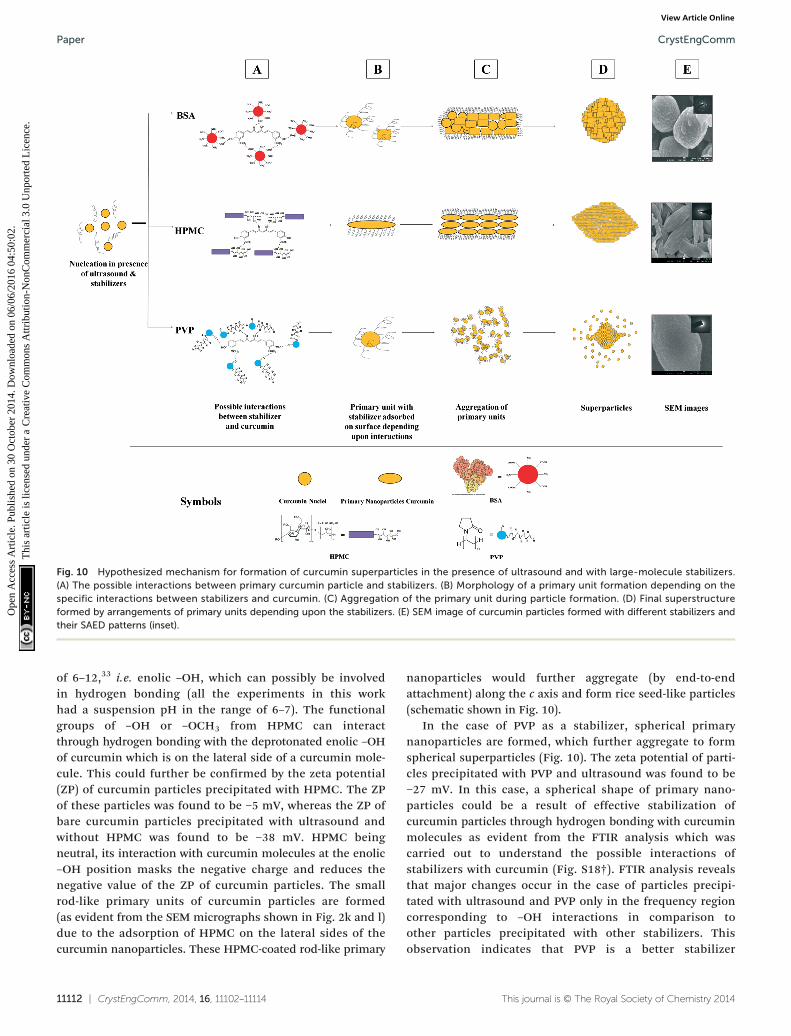
Fig. 10 Hypothesized mechanism for formation of curcumin superparticles in the presence of ultrasound and with large-molecule stabilizers.(A) The possible interactions between primary curcumin particle and stabilizers. (B) Morphology of a primary unit formation depending on thespecific interactions between stabilizers and curcumin. (C) Aggregation of the primary unit during particle formation. (D) Final superstructureformed by arrangements of primary units depending upon the stabilizers. (E) SEM image of curcumin particles formed with different stabilizers andtheir SAED patterns (inset).
CrystEngCommPaper
Ope
n A
cces
s A
rtic
le. P
ublis
hed
on 3
0 O
ctob
er 2
014.
Dow
nloa
ded
on 0
6/06
/201
6 04
:50:
02.
Thi
s ar
ticle
is li
cens
ed u
nder
a C
reat
ive
Com
mon
s A
ttrib
utio
n-N
onC
omm
erci
al 3
.0 U
npor
ted
Lic
ence
.View Article Online
of 6–12,33 i.e. enolic –OH, which can possibly be involvedin hydrogen bonding (all the experiments in this workhad a suspension pH in the range of 6–7). The functionalgroups of –OH or –OCH3 from HPMC can interactthrough hydrogen bonding with the deprotonated enolic –OHof curcumin which is on the lateral side of a curcumin mole-cule. This could further be confirmed by the zeta potential(ZP) of curcumin particles precipitated with HPMC. The ZPof these particles was found to be −5 mV, whereas the ZP ofbare curcumin particles precipitated with ultrasound andwithout HPMC was found to be −38 mV. HPMC beingneutral, its interaction with curcumin molecules at the enolic–OH position masks the negative charge and reduces thenegative value of the ZP of curcumin particles. The smallrod-like primary units of curcumin particles are formed(as evident from the SEM micrographs shown in Fig. 2k and l)due to the adsorption of HPMC on the lateral sides of thecurcumin nanoparticles. These HPMC-coated rod-like primary
11112 | CrystEngComm, 2014, 16, 11102–11114
nanoparticles would further aggregate (by end-to-endattachment) along the c axis and form rice seed-like particles(schematic shown in Fig. 10).
In the case of PVP as a stabilizer, spherical primarynanoparticles are formed, which further aggregate to formspherical superparticles (Fig. 10). The zeta potential of parti-cles precipitated with PVP and ultrasound was found to be−27 mV. In this case, a spherical shape of primary nano-particles could be a result of effective stabilization ofcurcumin particles through hydrogen bonding with curcuminmolecules as evident from the FTIR analysis which wascarried out to understand the possible interactions ofstabilizers with curcumin (Fig. S18†). FTIR analysis revealsthat major changes occur in the case of particles precipi-tated with ultrasound and PVP only in the frequency regioncorresponding to –OH interactions in comparison toother particles precipitated with other stabilizers. Thisobservation indicates that PVP is a better stabilizer
This journal is © The Royal Society of Chemistry 2014

CrystEngComm Paper
Ope
n A
cces
s A
rtic
le. P
ublis
hed
on 3
0 O
ctob
er 2
014.
Dow
nloa
ded
on 0
6/06
/201
6 04
:50:
02.
Thi
s ar
ticle
is li
cens
ed u
nder
a C
reat
ive
Com
mon
s A
ttrib
utio
n-N
onC
omm
erci
al 3
.0 U
npor
ted
Lic
ence
.View Article Online
compared to other stabilizers. Such favorable interactions ofPVP with curcumin molecules result in the formation ofspherical and amorphous primary nanoparticles (alsoevident from XRD and SAED patterns), which aggregate fur-ther to form superparticles.
Thus, the formation of curcumin particles in the presenceof ultrasound only (and no stabilizer) and in the presence ofultrasound along with polymeric stabilizers such as BSA,HPMC, and PVP seems to follow a non-classical crystalliza-tion pathway. Generally, a non-classical pathway of particleformation is followed when a large number of temporarilystabilized nanoparticles are formed and levels of supersatura-tion for particle growth decrease to a low value due to theconsumption of supersaturation for large nucleation rates.13
The use of ultrasound and additives leads to large nucleationrates (due to enhanced diffusion coefficients of solute mole-cules3,28 and reduction in surface tension at the solid–liquidinterface3). These large nucleation rates coupled withintegration-controlled growth of particles31 result in a largepopulation of nanoparticles and a reduced level of supersatu-ration. In addition to this, ultrasound and stabilizers alsofacilitate stabilization of nanoparticles by converting thermo-dynamically unstable matter into a more stable form5,28 andby preventing the growth of the particles due to adsorbed sta-bilizer molecules,26,34 respectively. These primary stabilizednanoparticles then aggregate randomly in the presence of sta-bilizers and ultrasound to form secondary superparticles. Theshape, morphology and crystallinity of these superstructuresdepend upon the interactions of stabilizers with curcuminmolecules. The particles precipitated in the presence of large-molecule stabilizers such as HPMC, BSA and PVP seem toundergo slower crystallographic fusion mainly due to occlu-sion of large polymeric molecules in the superstructure. Thepresence of these polymeric stabilizers prevents reorientationand fusion of primary nanoparticles resulting in the forma-tion of superstructures.35,36 As opposed to this, the particlesprecipitated in the presence of small-molecule stabilizerssuch as Tween 80 and SDS along with ultrasound lookedmore fused. Additional research is needed to ascertain anexact particle formation mechanism in these two cases.
3. Conclusions
Precipitation of a poorly water-soluble drug, curcumin, hasbeen carried out from its ethanolic solutions using water asan antisolvent in the presence of ultrasound and stabilizers.SEM analysis of precipitated particles reveals an interestingmorphological behavior of curcumin particles when precipi-tated in the presence of ultrasound and stabilizers. Theabsence of ultrasound during precipitation results in den-dritic particles due to a diffusion-limited growth in theabsence of uniform mixing, whereas the use of ultrasoundalone results in a rice seed-like particle morphology. Precipi-tation of curcumin particles in the presence of ultrasoundand (with or without) stabilizers seems to follow a non-classical crystallization pathway. The use of ultrasound and
This journal is © The Royal Society of Chemistry 2014
stabilizers leads to large nucleation rates and hence to a largepopulation of stabilized nanoparticles. These stabilized nano-particles assemble under the influence of ultrasound to formsuperstructures. Curcumin particles precipitated in the pres-ence of ultrasound and with SDS as well as Tween 80 have arice seed-like morphology and appear to be quite fused. Onthe other hand, curcumin particles precipitated with poly-meric stabilizers such as BSA, HPMC, and PVP clearly looklike the aggregates formed via assembly of several nano-particles. The particles precipitated in the presence of BSAand ultrasound appear to be spherical aggregates ~1 μm insize made by aggregation of several primary nanoparticles.The primary constituents of these superparticles werefound to be plate-like as well as spherical nanoparticles~100–200 nm in size. In the presence of ultrasound andHPMC the rice seed-like superparticles of ~2–5 μm wereobtained when precipitation of curcumin was carried out.The primary constituents of these particles were found to besmall rod-shaped nanoparticles around 100 nm in size. Pre-cipitation of curcumin in the presence of PVP and ultrasoundresults in spherical pearl-like aggregates ~1.5 μm in size,where the primary units were spherical nanoparticles with asize of ~50–80 nm. Thus, it can be proposed that the poly-meric stabilizers prevent crystallographic fusion and hencethe particles appear as loose aggregates, whereas small-molecule stabilizers like SDS and Tween 80 may not be ableto prevent fusion of individual particles and hence these par-ticles look more fused. It is also possible that the process ofparticle growth via molecule-by-molecule addition may havebeen followed. Further research is needed to ascertain anexact mechanism of particle formation in these cases.
Ultrasound induces formation of different polymorphs ofcurcumin as compared to the starting material. While rawcurcumin particles are in the monoclinic form, the particlesprecipitated in the presence of ultrasound and stabilizersmainly conform to the orthorhombic form of curcumin. Thepolymorphic transformations are induced mainly due toprevention of hydrogen bonding among curcumin molecules(which is responsible for macrocyclic ring formation in themonoclinic form) by ultrasound.
The results presented in this work demonstrate thatthe particle formation pathways of curcumin could be manip-ulated to obtain desired morphological and polymorphiccharacteristics through a judicious choice of stabilizers andmixing techniques (ultrasound vs. stirring). Moreover, thismethodology could be applied to other poorly water-solubledrugs as well as other organic and inorganic materials whichcan be precipitated by the liquid antisolvent technique.
Acknowledgements
We gratefully acknowledge financial support from the Depart-ment of Science and Technology (DST), Government of India,and the Indian Institute of Technology, Gandhinagar (IITGN).We would like to thank Dr. Abhijit Mishra (IITGN) andDr. Palash Sanphui (IISC Bangalore) for discussions regarding
CrystEngComm, 2014, 16, 11102–11114 | 11113
CrystEngCommPaper
Ope
n A
cces
s A
rtic
le. P
ublis
hed
on 3
0 O
ctob
er 2
014.
Dow
nloa
ded
on 0
6/06
/201
6 04
:50:
02.
Thi
s ar
ticle
is li
cens
ed u
nder
a C
reat
ive
Com
mon
s A
ttrib
utio
n-N
onC
omm
erci
al 3
.0 U
npor
ted
Lic
ence
.View Article Online
PXRD analysis. We are also thankful to Dr. Emila Panda (IITGN)for discussions on HR-TEM analysis. Further, we would also liketo thank the ICT-Mumbai, the NIPER-Mohali and the CSMCRI-Bhavnagar for providing access to their HR-TEM facility.
Notes and references
1 A. A. Thorat and S. V. Dalvi, Chem. Eng. J., 2012, 181–182,
1–34.2 E. Merisko-Liversidge, G. G. Liversidge and E. R. Cooper,
Eur. J. Pharm. Sci., 2003, 18, 113–120.3 S. V. Dalvi and R. N. Dave, Int. J. Pharm., 2010, 387, 172–179.
4 Y. He, Y. Huang and Y. Cheng, Cryst. Growth Des., 2010, 10,1021–1024.5 D. Xia, M. Ouyang, J. X. Wu, Y. Jiang, H. Piao, S. Sun,
L. Zheng, J. Rantanen, F. Cui and M. Yang, Pharm. Res.,2012, 29, 158–169.
6 Y. Ma, H. Cölfen and M. Antonietti, J. Phys. Chem. B,
2006, 110, 10822–10828.7 Y. Ma, H. G. Börner, J. Hartmann and H. Cölfen, Chem. –
Eur. J., 2006, 12(30), 7882–7888.8 D. D. Medina and Y. Mastai, Cryst. Growth Des., 2008, 8,
3646–3651.9 F. Benaouda, Z. Bachoo, M. B. Brown, G. P. Martin and
S. A. Jones, Soft Matter, 2013, 9, 10165–10173.10 T. Lee and C. Zhang, Pharm. Res., 2008, 25, 1563–1571.
11 Y. Jiang, L. Gower, D. Volkmer and H. Cölfen, Cryst. GrowthDes., 2011, 11, 3243–3249.12 M. Ejgenberg and Y. Mastai, Cryst. Growth Des., 2012, 12,
4995–5001.13 H. Cölfen and M. Antonietti, Mesocrystal Systems, in
Mesocrystals and Nonclassical Crystallization, John Wiley &Sons, Ltd, 2008, pp. 113–177.
14 Y. Su, X. Yan, A. Wang, J. Fei, Y. Cui, Q. Heab and J. Li,
J. Mater. Chem., 2010, 20, 6734–6740.15 B. B. Aggarwal and K. B. Harikumar, Int. J. Biochem. Cell
Biol., 2009, 41, 40–59.16 P. Sanphui, N. R. Goud, U. B. R. Khandavilli, S. Bhanoth and
A. Nangia, Chem. Commun., 2011, 47, 5013–5015.11114 | CrystEngComm, 2014, 16, 11102–11114
17 M. K. Mishra, P. Sanphui, U. Ramamurty and G. R. Desiraju,
Cryst. Growth Des., 2014, 14, 3054–3061.18 D. Xia, P. Quan, H. Piao, S. Sun, Y. Yin and F. Cui, Eur. J.
Pharm. Sci., 2010, 40, 325–334.19 M. Louhi-Kultanen, M. Karjalainen, J. Rantanen,
M. Huhtanen and J. Kallas, Int. J. Pharm., 2006, 320, 23–29.20 M. Kurotani and I. Hirasawa, J. Cryst. Growth, 2008, 310,
4576–4580.21 H. Hatakka, H. Alatalo, M. Louhi-Kultanen, I. Lassila and
E. Haeggstrom, Chem. Eng. Technol., 2010, 33, 751–756.22 Y. Gong, B. M. Collman, S. M. Mehrens, E. Lu, J. M. Miller,
A. Blackburn and D. J. W. Grant, J. Pharm. Sci., 2008, 97,2130–2144.23 Y. Zhu, Y. Liu, Q. Ruan, Y. Zeng, J. Xiao, Z. Liu, L. Cheng,
F. Xu and L. Zhang, J. Phys. Chem. C, 2009, 113, 6584–6588.24 H. Uchiyama and H. Imai, Langmuir, 2008, 24, 9038–9042.
25 O. D. Linnikov, Desalination, 1999, 122, 1–13. 26 S. V. Dalvi and R. N. Dave, Ind. Eng. Chem. Res., 2009, 48,7581–7593.27 C. Beck, S. V. Dalvi and R. N. Dave, Chem. Eng. Sci., 2010, 65,
5669–5675.28 A. A. Thorat, M. D. Yadav and S. V. Dalvi, Langmuir,
2014, 30, 4576–4592.29 M. Bujan, M. Sikirić, N. Filipović-Vinceković, N. Vdović,
N. Garti and H. Füredi-Milhofer, Langmuir, 2001, 17,6461–6470.
30 N. Rodríguez-Hornedo and D. Murphy, J. Pharm. Sci.,
2004, 93, 449–460.31 G. A. Ilevbare, H. Liu, K. J. Edgar and L. S. Taylor, Cryst.
Growth Des., 2012, 12, 6050–6060.32 P. Nithiyasi, K. Balaji, P. Brindha and M. Parthasarathy,
Nanotechnology, 2012, 23, 8.33 Z. Wang, M. H. M. Leung, T. W. Kee and D. S. English,
Langmuir, 2010, 26, 5520–5526.34 S. L. Raghavan, A. Trividic, A. F. Davis and J. Hadgraft, Int. J.
Pharm., 2001, 212, 213–221.35 A. N. Kulak, P. Iddon, Y. Li, S. P. Armes, H. Cölfen, O. Paris,
R. M. Wilson and F. C. Meldrum, J. Am. Chem. Soc.,2007, 129, 3729–3736.36 L. Zhou and P. O'Brien, Small, 2008, 4, 1566–1574.
This journal is © The Royal Society of Chemistry 2014