Paramecium paper dama TLS-2-10
11
www.sciencejournal.in Vol. 1 No. 2 (2012) ISSN: 2319–4731 (Print); 2319–5037 (Online) © 2012 DAMA International. All rights reserved. 41 MORPHOLOGICAL STUDY OF PARAMECIUM CAUDATUM FROM FRESH WATERS OF NASHIK DISTRICT OF MAHARASHTRA, INDIA Bhamare S. N.*, Nikam S. V.**, Jadhav B. N.*** and Dama L.B.**** *Department of Zoology, K.R.A. College, Deola, Nashik, Maharashtra, (M.S.), India. **Department of Zoology, Dr. Babasaheb Ambedkar Maharathwad University Aurangabad, (M.S.), India. ***Department of Zoology, Swami Muctanand College, Gangapur, Aurangabad (M.S.), India. ****Department of Zoology, D. B. F. Dayanand College, of Arts, Science and Commerce, Solapur, (M.S.), India. ABSTRACT Present study of free living ciliates the water samples were collected from different parts of Nashik District (Deola, Nandgaon; Surgana, Satana) of Maharashtra State, India. KEY WORDS: INTRODUCTION The protozoa of the freshwater environment range in size from about 1mm in the case of Stentor and some of the multinucleate amoebae, down to 5μm or so in the case of the smaller flagellates, and they are extremely varied in both appearance and lifestyle. Apart from the very smallest, which are apparently able to absorb nutrients directly through their cell wall, they feed upon other microorganisms. The amoebae feed mainly upon bacteria and algal unicells, as do most of the ciliates. Whereas the amoebae ingest food particles by absorbing them with their pseudopodia, the ciliates feed by either actively capturing or engulfing prey organisms, or by using their cilia to create currents in the surrounding water which bring the food organisms to their mouths. The captured organism is enclosed in a food vacuole, a membrane-bound vesicle which moves through the cytoplasm as digestion occurs. Undigested remains are discharged into the surrounding water, usually at a definite location in the organism's outer pellicle. The freshwater protozoa regulate the water content of their bodies by the expansion and periodic collapse of their contractile vacuole, a vesicle which increases in size as it extracts water from the interior of the cell and collapses to nothing as it expels the extracted water. The contractile vacuole is not observed in the marine protozoa. Both in organic pollution of the natural environment and in the biological processing of human and domestic animal sewage, the ceaseless activity of the protozoa - particularly the colonial ciliates - in the extraction and digestion of bacteria and other suspended particles is the main element of the natural process by which the water supply is rendered once again fit for consumption by humans and other creatures. Any change in our environment which threatens the life of a balanced community of protozoans threatens to the continuity of a clean water supply for humans as well. This is particularly relevant in the light of our current over-use of kitchen and lavatory disinfectants and their effect upon the ciliates at sewage processing plants and in the waterways beyond. The ciliates are one of the most important groups of Protozoa, common almost everywhere there is water (lake, pond, and oceans) and soils with many ecto and endosymbiotic members, as well as some obligate opportunistic parasites. Free living ciliates are found in fresh, marine, estuarine waters and in the soil. An excellent source is a shallow semi- permanent pond in a farm yard. The marshes and sloughs on the shore of the lakes are often rich sources of food. Any stream large or small, or any freshwater hole, temporary or permanent, water at the base of aquatic plants is likely to be a valuable source of material. Such places as moist crotches or bark (crevices) in trees or any other moist or water containing silt, floating algal mats provide food. Cilia covering the body of the organism or a part of it are a major defining characteristic of this group and hence the phylum name “Ciliophora”. Extensive studies have been made during the last 80 years, with numerous outstanding and noteworthy contributions in the field of Protozoology, covering cytology, cytochemistry, structure, life cycle transmission, evolution, and ultrastructure; and inter relationship a verity of protozoa. Ciliates were among the first living microscopic organisms to be discovered and described. Leeuwenhoek (Dobell, 1932) has attracted much scientific interest in Protozooloy. He made the first reliable observation of live ciliates, found ciliates (Nyctotherus) from the frog‟s gut (1683) and described chilodonella, coleps, clopidium, cyclidium, Dileptus, karana, Vorticella, paramecium and other ciliates (Corliss,1975). He observed morphological details of the “animalcules” and measured them, and described their reproduction, retroactivity and some conjugation stages. Present work is concern with the diversity of different types of Paramecia in Nashik district of Maharashtra
Transcript of Paramecium paper dama TLS-2-10

www.sciencejournal.in
Vol. 1 No. 2 (2012) ISSN: 2319–4731 (Print); 2319–5037 (Online) © 2012 DAMA International. All rights reserved. 41
MORPHOLOGICAL STUDY OF PARAMECIUM CAUDATUM FROM FRESH WATERS OF NASHIK
DISTRICT OF MAHARASHTRA, INDIA
Bhamare S. N.*, Nikam S. V.**, Jadhav B. N.*** and Dama L.B.****
*Department of Zoology, K.R.A. College, Deola, Nashik, Maharashtra, (M.S.), India.
**Department of Zoology, Dr. Babasaheb Ambedkar Maharathwad University Aurangabad, (M.S.), India.
***Department of Zoology, Swami Muctanand College, Gangapur, Aurangabad (M.S.), India.
****Department of Zoology, D. B. F. Dayanand College, of Arts, Science and Commerce, Solapur, (M.S.), India.
ABSTRACT
Present study of free living ciliates the water samples were collected from different parts of Nashik District (Deola,
Nandgaon; Surgana, Satana) of Maharashtra State, India.
KEY WORDS:
INTRODUCTION
The protozoa of the freshwater environment range in size from about 1mm in the case of Stentor and some of the
multinucleate amoebae, down to 5µm or so in the case of the smaller flagellates, and they are extremely varied in both
appearance and lifestyle. Apart from the very smallest, which are apparently able to absorb nutrients directly through
their cell wall, they feed upon other microorganisms. The amoebae feed mainly upon bacteria and algal unicells, as do
most of the ciliates. Whereas the amoebae ingest food particles by absorbing them with their pseudopodia, the ciliates
feed by either actively capturing or engulfing prey organisms, or by using their cilia to create currents in the
surrounding water which bring the food organisms to their mouths. The captured organism is enclosed in a food
vacuole, a membrane-bound vesicle which moves through the cytoplasm as digestion occurs. Undigested remains are
discharged into the surrounding water, usually at a definite location in the organism's outer pellicle.
The freshwater protozoa regulate the water content of their bodies by the expansion and periodic collapse of their
contractile vacuole, a vesicle which increases in size as it extracts water from the interior of the cell and collapses to
nothing as it expels the extracted water. The contractile vacuole is not observed in the marine protozoa.
Both in organic pollution of the natural environment and in the biological processing of human and domestic animal
sewage, the ceaseless activity of the protozoa - particularly the colonial ciliates - in the extraction and digestion of
bacteria and other suspended particles is the main element of the natural process by which the water supply is rendered
once again fit for consumption by humans and other creatures. Any change in our environment which threatens the life
of a balanced community of protozoans threatens to the continuity of a clean water supply for humans as well. This is
particularly relevant in the light of our current over-use of kitchen and lavatory disinfectants and their effect upon the
ciliates at sewage processing plants and in the waterways beyond.
The ciliates are one of the most important groups of Protozoa, common almost everywhere there is water (lake, pond,
and oceans) and soils with many ecto and endosymbiotic members, as well as some obligate opportunistic parasites.
Free living ciliates are found in fresh, marine, estuarine waters and in the soil. An excellent source is a shallow semi-
permanent pond in a farm yard. The marshes and sloughs on the shore of the lakes are often rich sources of food. Any
stream large or small, or any freshwater hole, temporary or permanent, water at the base of aquatic plants is likely to be
a valuable source of material. Such places as moist crotches or bark (crevices) in trees or any other moist or water
containing silt, floating algal mats provide food. Cilia covering the body of the organism or a part of it are a major
defining characteristic of this group and hence the phylum name “Ciliophora”.
Extensive studies have been made during the last 80 years, with numerous outstanding and noteworthy contributions in
the field of Protozoology, covering cytology, cytochemistry, structure, life cycle transmission, evolution, and
ultrastructure; and inter relationship a verity of protozoa. Ciliates were among the first living microscopic organisms to
be discovered and described. Leeuwenhoek (Dobell, 1932) has attracted much scientific interest in Protozooloy. He
made the first reliable observation of live ciliates, found ciliates (Nyctotherus) from the frog‟s gut (1683) and described
chilodonella, coleps, clopidium, cyclidium, Dileptus, karana, Vorticella, paramecium and other ciliates (Corliss,1975).
He observed morphological details of the “animalcules” and measured them, and described their reproduction,
retroactivity and some conjugation stages. Present work is concern with the diversity of different types of Paramecia in
Nashik district of Maharashtra

www.sciencejournal.in
Vol. 1 No. 2 (2012) ISSN: 2319–4731 (Print); 2319–5037 (Online) © 2012 DAMA International. All rights reserved. 42
MATERIALS AND METHODS
Present study of free living ciliates the water samples were collected from different parts of Nashik District (Deola,
Nandgaon; Surgana, Satana) of Maharashtra State, India. Water samples were collected in wide mouth, sterilized glass
bottles. Due care was taken and the samples were collected from where the submerged plants and decaying leaves were
present. Mostly the samples were collected during morning and evening. The temperature of the sample bottles were
maintained with the help of ice bags.
Rapid movements of ciliates make it difficult to identify ciliate species. To immobilize their movements methyl
cellulose solution was used.
Culture methods –
For cultivation various media are used such as
Hay infusion
Wheat infusion
Rice infusion
RESULTS AND DISCUSSIONS
Key for the identification of Paramecia
Genus- Paramecium
It seems highly probable that Antony von Leeuwenhoek, the discoverer of protozoa and bacteria also discovered
members of the genus Paramecium as early as 1674 and 1677. In an account of the early history of Paramecium,
Woodruff (1945) reported that, according to newly published manuscripts Leeuwenhoek corresponded to Constantijn
Huygens (senior) and informed him of some of his observation. Huygens passed on the information to his son
Christiaan, who repeated certain of the observation. In 1678 Chistiaan wrote a letter accompanied by sketches to his
brother Constantijn (Huygens, 1888) and several Protozoa which he found in infusions, including one that can be
identified as a species of Paramecium.
Figure 1. Map of Nashik District

www.sciencejournal.in
Vol. 1 No. 2 (2012) ISSN: 2319–4731 (Print); 2319–5037 (Online) © 2012 DAMA International. All rights reserved. 43
In a careful writing of Leeuwenhoek, Dobell (1932) mentioned that this discoverer of Protozoa also discovered
Paramecium. In 1718 Joblot published an account with figure generally accepted as representing Paramecium called by
him “Chausson” (slipper), Joblet was second, who represent a drawing of Paramecium and he is the first to refer to
Paramecium as the “Slipper-shaped animalcule” a term in use even today. Ledemuller (1763) utilize the name Infusoria
was at first to include all members of entire kingdum, Protozoa. As the name implies, Infusoria was at first used to
embrace all those forms commonly found in infusion of decaying animal and vegetable matter.
Hill (1752) first attempted to apply scientific names to microscopic animals and was the first use the genus name,
Paramecium. Hill probably used the Greek adjective „paramec‟ which means oblong or longish (to distinguish it from
the rounded forms) he then added ium to the stem Paramec to make a noun. He has done characterization of the
members in the group. Paramecium and description of four species is vague. For Paramecium he wrote “Animal which
have no visible limbs or tail and are of an irregularly oblong figure”. He described each of his four species with short
single sentence and used word „oblong‟ in each definition as follows-
“The Paramecium with an oblong voluble body, obtuse at each end”
“The paramecium with an oblong body, smallest towards the head”
“The paramecium with an oblong body, narrowest toward the middle”
“The paramecium with a slender, oblong body”
The next great scholar student of microscopic organisms was O. F. Muller, the first Microscopist to apply the Linnaean
system of nomenclature to these forms. There are parties to his important monographic studies. The first and second
part were published in 1773 and 1774 respectively where in a section is devoted to the „Infusoria’. Muller presented a
detailed analysis of this group and regarded members of the Infusoria as the simplest animal but included bacteria,
diatoms, worms and rotifers in addition to Protozoa. In Muller‟s first treatise (1773) his earliest reference to
Paramecium is characterized and spelled as follows-
“Paramecium vermis inconspicuous, simplex, pellucidus, complanatus, oblongus”
He listed two species, P. aurelia which is described as a paramecium oblongum antice in longitudinem plicatum and
Paramecium histrio which he later removed from the genus in his final volume of 1786 not believing it to be a species
of Paramecium.
The first description of Paramecium by Muller (1773) is not accompanied by figures but it identifies his Paramecium
aurelia with Hill‟s Paramecium „sp.3‟ which Hill apparently copied from the anonymous author of 1703. Here occurs
the first change in spelling of the name ‘Paramecium’ as given by Hill (1752) to Paramaecium this has led to consider
able confusion in the literature even up to the present the insertion of the letter „a‟ is it‟s etymologically incorrect since
it is an error of transcription (Ludwig, 1930; Kalmus, 1931; Woodfuff, 1945). Since Muller made direct reference to
Hill‟s Paramecium but miss-spelled it Paramaecium the latter is unacceptable according to article 19 of the
“International Rules of Zoological Nomenclature”.
This error of spelling was recognized by Herman (1784), Ehrenberg (1833, 1839) and Dujardin (1841). The species
name of Paramecium aurelia appears to have been taken from the resemblance of the ciliate to a golden coloured or
chrysalis of a butterfly; „aureus‟ (Latin) gold or golden colour; „chrysos‟ (Greek)= golden. It is the first species of the
paramecium named under the Linnaean system of nomenclature that has come down to the present time as Paramecium
aurelia O. F. Muller (1786).
Altogether, 56 species of Paramecium are listed by Ehrenberg (1833, 1838) as occurring in the literature. Of this
number he believed 48 to be synonyms. Ehrenberg described the remaining eight species which he believed to be valid
but out of this P. aurelia and P. caudatum are the only species are accepted today.
At the beginning of the 20th
century, Jennings (1908) in his studies on heredity in Paramecium showed the existence of
two main groups based statistically on mean length measurement of a considerable number of pure live cultures
1. An aurelia group
2. bursaria group
Woodruff (1921) called attention to the fact that the species of Paramecium fall naturally into two clearly defined
groups when based on body shape. He listed them as the “aurelia” group and the “bursaria” group respectively on the
basis of same specific characteristics.
www.sciencejournal.in
Vol. 1 No. 2 (2012) ISSN: 2319–4731 (Print); 2319–5037 (Online) © 2012 DAMA International. All rights reserved. 44
1. “Aurelia Group”:- individuals of this group characterized by a relatively long spindle or cigar-shaped body, a
round or circular in cross section with somewhat pointed posterior end. The species which are included in this group
are-
P. caudatum
P. aurelia
P. multimicronucleatum
2. “Bursaria group”:- Body is foot shape and somewhat compressed. The posterior end is broadly rounded while the
anterior end is somewhat obliquely truncate. Peristomal groove is long and broad. Compact macronucleus is present at
the anterior side which is round to ovoid or elongated in shape Species of this group are-
P. trichium
P. calkinsi
P. polycaryum
P. woodruffi
P. bursaria
Within each group one finds two characteristics types of micronuclei of important taxonomic values as follows-
a. Caudatum type- individuals in which the micronucleus is relatively large and composed rather compact mass
of chromatin bounded by nuclear membrane. This is a type of micronucleus found in P. caudatum, P. bursaria and P.
trichium.
b. Aurelia type- Individuals in which the micronucleus is relatively small and vesicular. It consists of an
extremely small concentrated mass of chromatin called the endosome centrally located with a distinct space between it
and the nuclear membrane. This distinction is based on fixed and Steined specimens because in living ones it is difficult
to identify micronuclei of the aurelia type, although micronucleus of the “caudatum” type may be seen easily in the
living condition. This is a type of micronucleus found in P. aurelia, P. multimicronucleatum, P. woodruffi, P. calkinsi
and P. polycaryum.
Key to the species of Paramecium:-
1. Slender, Cylindrical, or cigar-shaped animals (aurilia group); bluntly rounded, anteriorly somewhat pointed or
conic, posteriorly widest region about two- thirds of body length behind the anterior end.
2. Shorter and wider animals (“bursaria” group) somewhat dorsa-ventrally flattened with obliquely truncated anterior
end and broadly rounded posterior end.
3. One large compact micronucleus (“caudatum” type); body pointed or conic postereiorly; two canol fed contractile
vacuoles, length 170 to 290 µ ………P. caudatum.
4. More than one micronucleus and vesicular type…aurelia group
5. Usually less than 175µ in length two small vesicular micronuclei, two canal fed contractile vacuoles, smallest of
the “aurelia” group less than 120 to 170µ in length ……………P. aurelia
6. Usually large than 175µ three or four (occasionally up to seven) small vesicular micronuclei, commonly more than
two (frequently three) canal fed contractile vacuoles, posterior end of body pointed or conic as P. caudatum,
largest of aurelia group. 180 to 310µ in length …………………………P. multimicronucleatum.
7. Animals nearly coloured green because of zoochlorellae within cyclosis characteristically rapid, single large
compact micronucleus, two canal-fed contractile vacuoles, length 85 to 150µ…………………………P. bursaria
8. Two conspicuous vesicle-fed contractile vacuoles deeply set in endoplasm, each leading to exterior by means of
convoluted outlet canal, radial canals absent, single compact micronucleus, smallest of bursaria group, length 70
to 90µ ……………………… P. trichium
9. Without conspicuous vesicle-fed contractile vacuoles and convoluted outlet canal but with radial
canals…………………P. calkinsi
10. Rotations of body on long axis mainly indirection right spiral, usually two (occasionally one to five) vesicular
micronuclei, two canal-fed contractile vacuoles, fresh brackish water, length 110 to 140µ. ………………………
P. calkinsi
11. Length generally 150µ, usually four (occasionally three to eight) small vesicular micronuclei, mouth near centre of
body, two canal-fed contractile vacuoles, length 70 to 110µ …………………………P. polycaryum
12. Length generally greater than 115µ, mouth posterior to centre of body, three or four (occasionally up to eight)
scattered vesicular micronuclei, two canal-fed contractile vacuoles, Brackish water, largest of „bursaria‟ group,
length 120 to 210µ………………………………….P. woodruffi
Wenrich (1928a) made the important observation that the position of the anus or cytopyge is different in two groups.

www.sciencejournal.in
Vol. 1 No. 2 (2012) ISSN: 2319–4731 (Print); 2319–5037 (Online) © 2012 DAMA International. All rights reserved. 45
In member of “bursaria” group the anus is terminal and slightly to one side of the posterior end where as in the
“aurelia” group it is subterminal, situated on the ventral side between the posterior to the oral groove and the posterior
end of the body in P. caudatum and P. multimicronucleatum, the cytopyge lies between these two points or slightly
nearer the end of the oral groove, in P. aurelia the cytopyge is nearer to the posterior end of the body.
In general Paramecium is large enough to be visible to the unaided eye. However, the internal detail is resolved only by
the use of a microscope. A student is best able to observe the complex internal organization of the organism by using
what is termed the hanging drop technique. Here a drop of water is suspended upside-down and on a coverslip that is
position over a cavity on a microscope slide. The coverslip is sealed to prevent leakage.
Paramecium contains organized structures called vacuoles that are essentially a primitive mouth, stomach, and
excretion system. As food enters the organism it is stored in specialized vacuoles. These can circulate through the
cytoplasm of the organism, in order to provide food where needed. Characteristic of eukaryote, nuclear material is
segregated by nuclear membrane.
Another characteristic feature of Paramecium is the so called contractile vacuole. The vacuole is able to store water and
then by virtue expel the water out of the organism. In this way the amount of water inside the paramecia can be
controlled. The observation of contractile vacuole is another feature that is visible by the light microscope observation
of living paramecia.
On the exterior lies a membrane that is called the pellicle. The pellicle is both stiff, to provide support and to maintain
the shape of the organism, and is flexible, to allow some flexing of the surface. Also on the surface are hundreds of tiny
hairs called cilia. The cilia wave back and forth and act to sweep food particles (bacteria and smaller protozoa) towards
the primitive mouth of the organism (the gullet). The cilia are also important in locomotion, acting analogous to the
oars of a rowboat. The beating of the cilia is easily visible under light microscopic examination, especially if the
movement of the organism has been retarded by the addition of a viscous compound such as glycerol/ fumes of acetic
acid to the sample.
There are many Ciliatologists, Protozoologists, Ecologists and Protistologists have worked on Paramecium and still
doing. Following are the species of Paramecia, only few are described here;
Paramecium caudatum (Ehrenberg 1833)
P. aurelia (O. F. Muller, 1773)
P. multimicronucleatum (Powers and Mitchell, 1910)
P. trichium (Stokes, 1883)
P. calkinnsi (Woodruff 1921)
P. polucaryum (Woodfuff and Spencer, 1923)
P. bursaria (Focke, 1836)
P. woodruffi (Wenrich, 1928)
After comparing from above described species of Paramecium, present author has found P. caudatum (Ehrenberg
1833) and P. bursaria (Focke 1836).
Systematics of Paramecium
Domain: Eukaryota
Kingdom: Protozoa, Goldfuss, 1818, Rown, 1858
Subkingdom: Biciliata
Infrakingdom: Alveolata, Cavalier and Smith, 1991
Phylum: Ciliophora Doflein, 1901, Copeland, 1956
Subphylum: Intramacronucleata Lynn, 1996
Class: Oligohymenophorea de Puytorac et al., 1974
Subclass: Peniculia Fauré-Fremiet, in Corliss, 1956
Order: Peniculida Fauré-Fremiet, in Corliss, 1956
Suborder Parameciina Small and Lynn, 1985
Family: Parameciidae Dujardin, 1840
Genus: Paramecium O.F. Müller, 1773
Species: P. caudatum O.F. Müller, 1773
P. bursaria Woodruff
www.sciencejournal.in
Vol. 1 No. 2 (2012) ISSN: 2319–4731 (Print); 2319–5037 (Online) © 2012 DAMA International. All rights reserved. 46
Genus Paramecium Hill, 1752
The name Paramecium is first time attempted by Hill (1752) to apply scientific names to microscopic animals and the
first use the genus name, Paramecium. Hill probably used the Greek adjective, „paramec‟ which means oblong and
elongated to differentiate it from the rounded forms. He then added „ium‟ to the term, „paramec‟ to make it noun. He
has done the characterization of the members in the group. The next great scholar student of microscopic organism was
O.F. Muller, the first microbiologist who applied the Linnaean system of Nomenclature to these forms. They are three
parts to his monographic studies. The first and second part were published in 1773 and 1774 respectively where in a
section is devoted to the „Infusoria‟. Muller presented a detailed analysis of this group and regarded members of the
infusoria as the simplest animals but included bacteria, diatoms, worms and rotifers in addition to protozoa. He listed
two species, Paramecium aurelia and P. histrio but later he removed the P. histrio from the genus in his final volume
of 1786 not believing it to be a species of Paramecium.
The organism belongs to the genus Paramecium are having long spindle, cigar or foot shape body. It is circular or
ellipsoidal in cross section. The posterior end is bluntly pointed to round. In „Bursaria‟ group, body is somewhat
dorsoventrally flattened with obliquely truncated anterior end and broadly rounded posterior end. Single macronucleus
present and one to several vesicular or compact micronuclei are found. Peristome is long, broad and slightly oblique. 1
to 2 contractile vacuoles are presented at anterior or the posterior ends. Rotation of body is along the axis in direction of
left or right spiral. Some of the organisms are green in colour because of zoochlorella within, while some of the species
are golden coloured or crystals of butterfly „aureus‟ (Latin) gold or golden colour; „chrysos‟ (Greek) means golden.
Altogether 56 species of Paramecia are listed by Ehrenberg (1833, 1838) as occurring in the literature. Out of these he
believed 48 species to be homonyms. Ehrenberg described the remaining 8 species which he believed to be valid but
out of these P. aurelia and P. caudatum are the only species accepted today.
At the beginning of the 20th
century, Jennings (1908) in his studies on heredity in Paramecium showed the existence of
two main groups based statistically on mean length measurements of a considerably number of pure live cultures.
Woodruff (1921) called attention to the fact that the species of Paramecium fall naturally into two clearly defined
groups when based on body shape. The one group is „Aurelia‟ group and the individuals of this group characterized by
a relatively long spindle or cigar shaped body, round or circular in cross section with a somewhat pointed posterior end.
This group includes three species viz; P. caudatum, P. aurelia and P. multimicronucleatum.
Another group is „Bursaria‟ group in which the organisms are shorter and broader, dorsoventrally flattened and more
rounded posterior end with somewhat obliquely truncated anterior end. This group includes four species viz; P.
trichium, P. calkinsi, P. polycaryum, P. woodruffi and P. bursaria. Wenrich (1928) made the important observation that
the position of the anus or cytopyge is different into two groups.
In general Paramecium contains vacuoles through which digestion and excretion takes place. As food enters the
organism, it is stored in specialized vacuoles known as food vacuoles. These can circulate through the cytoplasm of the
organism in order to provide food where needed. Another characteristic feature of Paramecium is the contractile
vacuole. This vacuole stores water and then by compression of the side arms that radiate from the central vacuole to
expel the water out of the organism. They also play an important role in osmoregulation.
A membrane lies on the exterior called the pellicle. The stiff pellicle provides support and maintains the shape of
organism and the flexible pellicle gives flexibility to the surface, there are hundreds of tiny hairs called cilia. The cilia
are important in locomotion, acting analogous to the oars of rowboat. The cilia also help in sweeping food particles
(bacteria and smaller protozoa) towards the mouth by moving back and forth. Following are the species of Paramecium
and out of these two are redescribed here.
P. aurelia Muller, 1773
P. caudatum Ehrenberg, 1833
P. trichium Stokes, 1885
P. multimicronucleatum Power and Mitchell, 1910
P. calkinsi Woodruffi, 1921
P. polycaryum Woodruff and Spencer, 1923
P. bursaria Wenrich, 1928

www.sciencejournal.in
Vol. 1 No. 2 (2012) ISSN: 2319–4731 (Print); 2319–5037 (Online) © 2012 DAMA International. All rights reserved. 47
Percentage of prevalence of free living fresh water ciliates:
During the period of two years (Jan.2007 to Dec. 2008) a total number of water samples 1624 were examined, 879 of
these were positive with free living ciliates.
In the first year (Jan. 2007 to Dec. 2007) 677 fresh water samples were examined, 361 of these were positive with free
living ciliates. The percentage of prevalence of free living ciliates was about 53.32%.
In the second year (Jan. 2008 to Dec. 2008) 947 fresh water samples were examined, 518 of these were positive with
free living ciliates. . The percentage of prevalence of free living ciliates was about 54.70%.
A month wise analysis in first year (Jan.2007 to Dec.2007) shows the maximum percentage of prevalence of free living
ciliates during August and September (73.68%, 70.69%), lowest in May (31.15%) and minimum to moderate in
remaining months.
In second year (Jan.2008 to Dec.2008) maximum percentage of prevalence of fresh water free living ciliates was
showed again during August and September (74.47%, 71.25%), lowest in February, March and April
(38.10%,36.71%,39.13 %) and minimum to moderate in remaining months.
Comparatively percentage frequency of Paramecium caudatum is seen more than the various ciliates. Only it has been
found that the population of present species is slightly decrease in the month of May in both the years.
The pattern in both the years suggests that the peak is soon after the monsoon rain. The percentage then gradually
reduces at the end of the winter months and reaches a low with the onset of summer. The details of the number of
animals examined and the month wise prevalence is shown in Table 1 and 2.
Table 1. Showing the month wise prevalence of free living fresh water Ciliates, during the period from Jan.
2007 to Dec. 2007. Sr
No
Month No. of water
samples examined
No. of +ve water
samples
% of Total
Prevalence
% of Paramecium species in
the total ciliate positive samples
1 Jan-07 45 22 48.89 40.76
2 Feb-07 55 23 41.82 40.00
3 Mar-07 47 20 42.55 40.00
4 Apr-07 60 21 35.00 34.00
5 May-07 61 19 31.15 11.02
6 Jun-07 67 32 47.76 46.55
7 Jul-07 54 36 66.67 59.50
8 Aug-07 57 42 73.68 64.00
9 Sep-07 58 41 70.69 64.00
10 Oct-07 60 39 65.00 50.00
11 Nov-07 64 41 64.06 49.00
12 Dec-07 49 25 51.02 42.00
Total 677 361 53.32 44.15
Table 2. Showing the month wise prevalence of free living fresh water ciliates during the period from Jan.2008
to Dec. 2008.
Sr No
Month No. of water samples examined
No. of +ve water samples
% of Total Prevalence
% of Paramecium species in the total ciliate positive samples
1 Jan-08 68 37 54.41 35.00
2 Feb-08 84 32 38.10 35.00
3 Mar-08 79 29 36.71 34.12
4 Apr-08 69 27 39.13 32.19
5 May-08 62 27 43.55 22.21
6 Jun-08 89 49 55.06 21.00
7 Jul-08 94 70 74.47 59.55
8 Aug-08 80 57 71.25 69.15
9 Sep-08 89 58 65.17 65.14
10 Oct-08 87 60 68.97 64.19
11 Nov-08 69 32 46.38 45.00
12 Dec-08 77 40 51.95 39.10
Total 947 518 54.70 46.68
www.sciencejournal.in
Vol. 1 No. 2 (2012) ISSN: 2319–4731 (Print); 2319–5037 (Online) © 2012 DAMA International. All rights reserved. 48
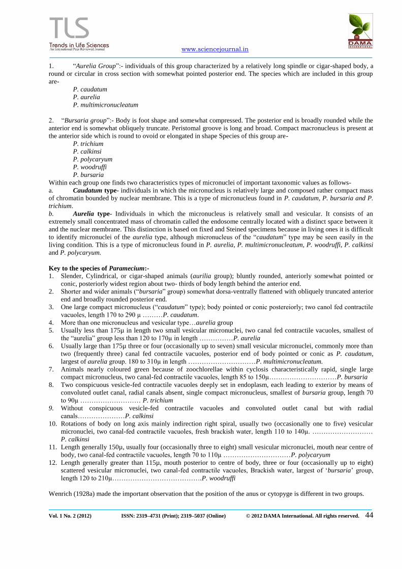
Figure 2. Showing the month wise prevalence of free living freshwater Ciliates, during the period from January
2007 to December 2007.
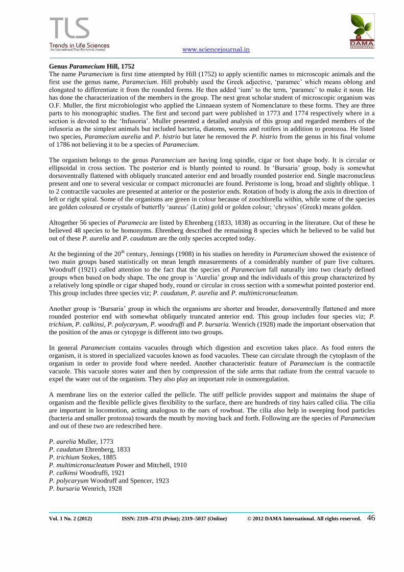
Figure 3. Showing the month wise prevalence of free living freshwater Ciliates, during the period from January
2008 to December 2008.
Paramecium caudatum Ehrenberg (1833)
Description of the species This species is first reported by Ehrenberg (1833). Body of this ciliate is cylindrical, cigar or foot shaped, or slipper
shaped. It is an active animalcule that is widely distributed and extensively studied organism. The anterior end of the
body is bluntly rounded whereas the posterior end is pointed. Paramecium swims the anterior end forward in a helix
because its ciliary beat is oblique to the long axis of the body. Paramecium can reverse the direction of its ciliary beat
and move backwards.
It measures 125 to 227.5µ in length and 65 to 120µ in width. Body ciliation is uniform, but caudal cilia are elongated.
The anterior region is relatively broader than the posterior end. The body size and shape studied and recorded in living
condition. The ventrally located ventral groove or vestibulum extends slightly more than half of the body length in the
direction of the right spiral. The groove oriented obliquely across the cell and opens anteriorly. Buccal cavity is with
one endoral membrane and two piniculi (peniculus- a group of specialized cilia characteristic of peniculina). The
entrance of the groove faces anteriorly and the cytostome is reaches to the posterior end. The cytostome opens into a
fluid region of the cytoplasm known as cytopharynx (P. caudatum, Shaikh, 2006). The cilia of the peristomal groove
move food particles (bacteria, small protozoan or organic particles) in to the cytopharynx to make food vacuoles in the
cytopharynx. After formation, the vacuoles move in a circuitous route through the cell while the various events of
digestion occur subsequently. Macronucleus is large, compact, elongated, and centrally placed. It measures about 30 to
75µ in length and 17.5 to 37.5 µ in width. Micronucleus is single spherical to ovoid in shape, situated close to the
macronucleus. There are two contractile vacuoles observed with radial canals in living condition. These vacuoles are
located in about first and last quarters of the body and empty to the outside by means of excretory pores. The cytopyge
is present on ventral side and sub terminal and rather close to the posterior tip of the body, through which the
indigestible residue of the food is extruded from the body. See in microphotographs.

www.sciencejournal.in
Vol. 1 No. 2 (2012) ISSN: 2319–4731 (Print); 2319–5037 (Online) © 2012 DAMA International. All rights reserved. 49
Paramecium caudatum W.M.
P. caudatum, Silver staining
P. caudatum Binary fission P. caudatum, Conjugation
Figure 4. Paramecium caudatum a) W.M. b) Silver staining c) Binary fission d) Conjugation
Comments
The cigar or foot shaped or slipper shaped body of this organism matches to genus Paramecium and hence it is a
member of this genus. Hill (1752) first time reported this genus. After that many other workers such as Muller (1713),
Ehrenberg, (1833), Stokes (1885), Power and Mitchell (1910), Woodruff (1921), Woodruff and Spencer (1923),
Wenrich (1928), Kudo (1966), Bick (1972), reported various species of this genus.
The body shape shows rounded anterior end and narrow posterior end matches to all the species of Aurelia group of
Paramecium and differs from Bursaria group in which the individuals have flattened anterior end hence present author
compares this species with aurelia group only which includes P. aurelia, P. caudatum, and P. multimicronucleatum.
Posterior end of the present species is bluntly pointed which is differ from P. aurelia and P. multimicronucleatum
because the posterior end in both of these species is usually more rounded. This character matches with P. caudatum.
The contractile vacuoles in the present species are two in number and are situated, one is at anterior end and another is
at posterior end on aboral surface of the body. This character matches to P. caudatum (Shaikh, 2006), and P. aurelia
(Deshmukh, 2010) but Muller (1773) described two contractile vacuoles in P. aurelia and both of them are located at

www.sciencejournal.in
Vol. 1 No. 2 (2012) ISSN: 2319–4731 (Print); 2319–5037 (Online) © 2012 DAMA International. All rights reserved. 50
the anterior end of aboral side. Present species also separates from P. multimicronucleatum, which possesses three to
seven contractile vacuoles scattered in anterior and posterior region.
Macronucleus in present case is centrally placed, which is elongated and occasionally triangular while the
macronucleus in P. aurelia and P. multimicronucleatum is usually compact, round to ovoid and located just above the
middle of the body, hence found to be different from P. aurelia and P. multimicronucleatum, but closer to P. caudatum
(Ehrenberg, 1833), which has centrally placed macronucleus. Shaikh, (2006) reported macronucleus in P. caudatum
which is situated anterior to middle, compact, smoothly ellipsoidal or kidney shaped.
Present species shows single compact micronucleus close to the macronucleus which matches to P. caudatum described
by Ehrenberg, 1833, Shaikh, 2006 while Muller (1773) reported two small, vesicular micronuclei in P. aurelia.
Recently in P. aurelia, Deshmukh (2010) reported single vesicular micronucleus, 4 or more micronuclei described by
Power and Mitchell, (1910) in P. multimicronucleatum. Hence this species separates from both the above species. But it
resembles to P. caudatum (Ehrenberg, 1833; Shaikh 2006) in which they also reported single vesicular micronucleus.
Body dimensions of the present species are closer to the P. caudatum (Ehrenberg and Shaikh) but smaller than P.
multimicronucleatum while the body is larger than P. aurelia. After the discussion and comparison of the present
species with the species of Aurelia group it is found similar to P. caudatum in body dimensions, shape, position of
macronucleus and single vesicular micronucleus matches to the P. caudatum described by Ehrenberg, (1833) while the
position of two contractile vacuoles matches to the P. caudatum described by Shaikh, (2006) and hence redescribed
here as a Paramecium caudatum (Table 3).
Table 3. Comparison of the present species with the species of Aurelia group of Paramecium
REFERENCES
Corliss J. O. (1977). Annotated assignment of families and to the order and classes currently comprising the Corlissan
scheme of higher classification for the Phylum Ciliophora. Trans. Am. Micros. Soc. 96: 104-140.
Corliss J. O. (1978). A salute to fifty- four great microscopist of the past: a pictorial to the history of Protozoology.
Part 1. Trans. Amer. Micros. Soc. 97: 419-458.
Corliss J. O. (1978). Protozoan ecology: a note on its current status. Amer Zool. 13: 145-148.
Corliss J. O. (1979). The Ciliated Protozoa: characterization classification and guide to the literature 2nd
ed. Pergtamon
Press Oxford.
Sr.
No.
Particulars P. aurelia
Muller, 1773
P. caudatum
Ehrenberg, 1833
P. multimicronucleatum
Power and Mitchell,
1910
P. caudatum
Shaikh, 2006
P. aurelia
Deshmukh,
2010
P. caudatum
Present author
1 Body shape Cigar or foot shaped,
posterior end is
more rounded than anterior
end.
Cigar or foot shaped, posterior
end is bluntly
pointed
Cigar or foot shaped, posterior end is rounded
Cigar or foot shaped,
posterior end
is bluntly pointed
Cigar or foot shaped.
Posterior end
is more rounded than
anterior end.
Cigar or foot shaped, posterior
end is bluntly
pointed
2 Body dimensions
120µ-180µ long
180µ-300µ long 200µ-330µ long 171 to 245µ long
74.64µ-111.23µ by
24.97µ-
38.59µ
125 to 227.5um By 65 to 120um
3 Contractile vacuole
2, anterior, on aboral
surface
2, posterior, on aboral surface
3-7, anterior and posterior 2, one posterior and
one anterior
on aboral surface,
2, one posterior and
one anterior,
on aboral surface.
2, one anterior and one
posterior,
on aboral surface
4 Macronucleus Ovoid to
elongated, just above the
middle of body
Compact,
elongated, centrally placed
Ovoid to elongated,
obliquely placed just above the middle of body
Anterior to
middle, compact,
smoothly
ellipsoidal or kidney
shaped.
Compact,
round to ovoid or triangular,
just above the
middle of the body
Compact,
elongated, centrally placed
30 to 75um by
17.5 to 37.5um
5 Micronucleus 2, small vesicular
1, compact 4 or more, vesicular 1, compact 1, vesicular 1, compact
6 Habitat Fresh water Fresh water Fresh water Fresh water Fresh water Fresh water
www.sciencejournal.in
Vol. 1 No. 2 (2012) ISSN: 2319–4731 (Print); 2319–5037 (Online) © 2012 DAMA International. All rights reserved. 51
Corliss J. O. (1991). Historically important events, discoveries and works in Protozoology from the mid-17th
to mid-
20th
century. Rev. Soc. Mex. Hist. Nat. 42: 45-81.
Corliss J. O. (1997). Some important anniversaries in the history of Protozoology. Rev. Soc. Mex. Hist. Nat. 47: 5-17
Ehrenberg C. G. 1833. Dritter beitrag zur erkenntniss grosser organisation in der richtung des kleinsten raumes. Abh.
Konig. Akad. Wissensch. Berl. 145-336.
Deshmukh (2010). Studies on some aquatic and soil protozoa from Aurangabad vicinity Ph. D. thesis of Dr. B.A.M.
University Aurangabad.
Dobell C. (1932). Antony van Leeuwenhoek and his “little animals.” Staples Press, London
Hall R. P. (1937): Growth of free-living protozoa in pure cultures. In Needham et al. Culture methods for invertebrate
animals.
Hall R. P. and Nigrelli, R. F. (1937). A note on the vauome of Paramecium bursaria and the contractile vacuole of
certain ciliates. Tr. Am. Micr. Soc. 56:185.
King R.L. (1935). The contractile vacuole of Paramecium multinucleatum. J. Morphol. 58:555.
King R.L. (1954). Origin and morphogenetic movements of the pores of the contractile vacuoles in Paramecium
aurelia. J. Protozool. 1:121.
Kudo R. R. (1975). Protozoology 5th
ed. Charles C. Thomas Springfield, Illinios; 1174
Kudo R. R. (1977). Protozoology 5th
ed. Springfield, III: Chane, C.C. Thomas (1774).
Ludwing W. (1930). Zur Nomenclatur und Systematick der Gattung Paramecium. Zool. Anz., 92: 33–41
Lynn D. H. and Small, E. B. (2000). Phylum Ciliophora In An illustrated guide to the protozoa.2-nd ed. (Eds. Lee J. J.
Hunter S. H. and Bovee E. C.) Allen press, Lawrence. 371-655.
Műller O. F. (1773). Vermium terrestrium et fluviatilium, Historia
Powers J. H. and Mitchell C. (1910). A new species of Paramecium (P. multimicronucleata) experimentally
determined. Biol. Bull. 19: 324-332.
Puytorac (1974). Proposition d‟une. Classification du phylum Ciliophora Doflein, 1901. C. R. Acad. Sci. Paris. 278:
2799-2802.
Wenrich D.H. (1928). Paramecium woodruffi n. sp. Tr. Am. Micr. Soc. 47:256.
Wenrich D.H. (1928a). Eight well defined species of Paramecium. Ibid. 47:275.
Wiley N.Y. Wenrich D.H. (1928a). Eight well defined species of Paramecium. Ibid. 47:275.
Woodruff L. L. (1921). The structure, life history, and intrageneric relationships of Paramecium
calkinsi, sp. nov. Biol. Bull. 41:171-180.
Woodruff L. L. and Spencer H. (1923). Parameciumpolycaryum sp. nov. Proc. Soc. Exp. Biol. Med.
20:338-339.
Woodruff L.L. (1931). Micronuclear variation in Paramecium bursaria. Quart. Micr. Sci. 74:537.