A Comparative Study of the Histopathologic Features of Bovine Tuberculosis in Cattle, Fallow Deer...
7
http://vet.sagepub.com/ Veterinary Pathology Online http://vet.sagepub.com/content/32/3/215 The online version of this article can be found at: DOI: 10.1177/030098589503200301 1995 32: 215 Vet Pathol J. C. Rhyan and D. A. Saari ) Cervus elaphus ), and Red Deer and Elk ( Cervus nippon ), Sika Deer ( dama Dama A Comparative Study of the Histopathologic Features of Bovine Tuberculosis in Cattle, Fallow Deer ( Published by: http://www.sagepublications.com On behalf of: Pathologists. American College of Veterinary Pathologists, European College of Veterinary Pathologists, & the Japanese College of Veterinary can be found at: Veterinary Pathology Online Additional services and information for http://vet.sagepub.com/cgi/alerts Email Alerts: http://vet.sagepub.com/subscriptions Subscriptions: http://www.sagepub.com/journalsReprints.nav Reprints: http://www.sagepub.com/journalsPermissions.nav Permissions: What is This? - May 1, 1995 Version of Record >> at MALAYSIAN RUBBER BOARD on February 3, 2014 vet.sagepub.com Downloaded from at MALAYSIAN RUBBER BOARD on February 3, 2014 vet.sagepub.com Downloaded from
Transcript of A Comparative Study of the Histopathologic Features of Bovine Tuberculosis in Cattle, Fallow Deer...

http://vet.sagepub.com/Veterinary Pathology Online
http://vet.sagepub.com/content/32/3/215The online version of this article can be found at:
DOI: 10.1177/030098589503200301
1995 32: 215Vet PatholJ. C. Rhyan and D. A. Saari
)Cervus elaphus), and Red Deer and Elk (Cervus nippon), Sika Deer (damaDamaA Comparative Study of the Histopathologic Features of Bovine Tuberculosis in Cattle, Fallow Deer (
Published by:
http://www.sagepublications.com
On behalf of:
Pathologists.American College of Veterinary Pathologists, European College of Veterinary Pathologists, & the Japanese College of Veterinary
can be found at:Veterinary Pathology OnlineAdditional services and information for
http://vet.sagepub.com/cgi/alertsEmail Alerts:
http://vet.sagepub.com/subscriptionsSubscriptions:
http://www.sagepub.com/journalsReprints.navReprints:
http://www.sagepub.com/journalsPermissions.navPermissions:
What is This?
- May 1, 1995Version of Record >>
at MALAYSIAN RUBBER BOARD on February 3, 2014vet.sagepub.comDownloaded from at MALAYSIAN RUBBER BOARD on February 3, 2014vet.sagepub.comDownloaded from

Vet Pathol 32:2 15-220 (1 995)
NATURAL DISEASE
A Comparative Study of the Histopathologic Features of Bovine Tuberculosis in Cattle, Fallow Deer (Dama dama),
Sika Deer (Cervus nippon), and Red Deer and Elk (Cervus elaphus)
J. C. RHYAN AND D. A. SAARI
Pathobiology Laboratory, National Veterinary Services Laboratories, Veterinary Services, Animal and Plant Health Inspection Service, US Department of Agriculture, Ames, IA
Abstract. Sections of tuberculous lesions from 23 elk (Cervus elaphus nelsoni) and red deer (Cervus elaphus elaphus), 12 fallow deer (Dama dama), 10 sika deer (Cervus nippon), and 30 cattle were examined and compared. Lesions were scored for caseous necrosis, mineralization, neutrophils, macrophages, giant cells, and acid-fast bacilli. Some differences in lesion morphology between the species were noted. ElMred deer lesions had marked variation and often differed from bovine lesions in several characteristics; elMred deer lesions usually had scattered peripheral mineralization rather than central mineralization and contained more neutrophils and fewer giant cells than did bovine lesions. Fallow deer lesions contained more giant cells but were otherwise indistin- guishable from elk lesions. Sika deer lesions had more giant cells and fewer neutrophils than did lesions from cattle or other cervid species. Sika deer giant cells were larger and contained more nuclei than did giant cells in the other species.
Key words: Cattle; deer; elk; Mycobacterium bovis; tuberculosis.
In recent years, tuberculosis caused by Mycobucte- rium bovis has been diagnosed in captive and free- ranging cervids in several c~unt r ies .~ Because of its economic17 and public health7.8 significance, tubercu- losis is emerging as the single most important infec- tious disease problem in captive ce~-vids.~,~JO As the farmed-deer industry and federal and state regulatory agencies attempt to control the disease, early identi- fication of M. bovis-infected animals and recognition of tuberculous lesions become of paramount impor- tance.
Gross and microscopic features of bovine tuber- culosis in several deer species have been de-
Many histopathologic descriptions, however, are brief or do not differentiate among deer species.
Lesions described in naturally infected red deer (Cer- vus eluphus efuphus) include granulomas with miner- alized caseous necrotic centers resembling tuberculous lesions in cattle1.2J8 and abscesses with no distinguish- ing features18 or with giant and epithelioid cells in the capsule. I Similar lesions and laminated lesions resem- bling ovine caseous lymphadenitis abscesses have been reported in captive North American elk (wapiti, Cervus efuphus nef~oni).'~ Lesions of tuberculosis in fallow deer (Dama duma) have been described as resembling typical bovine lesions16,20.21 or consisting of liquefactive necrosis bordered by a narrow zone of macrophages
~~ribed1,2,5,6,9.12,13,16,18-21 and recently reviewed.3.10
and giant cells.2o Reports of tuberculosis in sika deer (Cervus nippon) have described necrosis, mineraliza- tion, and granulomatous inflammation in lung and lymph node l e ~ i o n s . ~ J ~ J ~
The purpose of this study was to describe and com- pare the histopathologic features of lesions caused by naturally occurring M. bovis infection in cattle, sika deer, fallow deer, and elWred deer.
Materials and Methods
Laboratory records from October 1976 through July 199 1 were reviewed. During that period, tissues from 335 cervids were submitted to the National Veterinary Services Labo- ratories (NVSL) for histologic examination for tuberculosis or paratuberculosis. Cases for microscopic review were se- lected on the basis of two criteria: 1) inflammatory lesions were present in lymph node and/or lung specimens and 2) M. bovis had been isolated from the corresponding fresh tissue specimens. The following were selected for compara- tive review: 18 lymph node and 16 lung lesions from 20 elk, two red deer, and one elk-red deer cross; 13 lymph node and five lung lesions from 12 fallow deer; and 1 1 lymph node and five lung lesions from 10 sika deer. Available sections of other tissues were also reviewed; however, the compara- tive study was limited to lymph node and lung lesions. Except for a lymph node lesion from one red deer (No. 3009), which was originally diagnosed as an abscess, and pulmonary le- sions from one elk (No. 450), which were first diagnosed as necropurulent pneumonia of undetermined etiology, all cases
215
at MALAYSIAN RUBBER BOARD on February 3, 2014vet.sagepub.comDownloaded from
216 Rhyan and Saari Vet Pathol 32:3. 1995
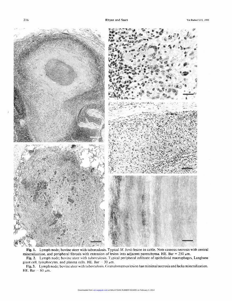
Fig. 1.
Fig. 2.
Fig. 3.
Lymph node; bovine steer with tuberculosis. Typical M. bovis lesion in cattle. Note caseous necrosis with central
Lymph node; bovine steer with tuberculosis. Typical peripheral infiltrate of epithelioid macrophages, Langhans
Lymph node; bovine steer with tuberculosis. Granulomatous lesion has minimal necrosis and lacks mineralization.
mineralization, and peripheral fibrosis with extension of lesion into adjacent parenchyma. HE. Bar = 250 pm.
giant cell, lymphocytes, and plasma cells. HE. Bar = 30 Fm.
HE. Bar = 80 Wm.
at MALAYSIAN RUBBER BOARD on February 3, 2014vet.sagepub.comDownloaded from

Vet Pathol 323 , 1995 Tuberculosis in Cattle, Deer, and Elk 217
were originally considered histologically compatible with or suggestive of mycobacteriosis. In all but seven cases, acid- fast or auramine-0-acridine orange (AOAO) fluorescent ba- cilli had been demonstrated in the lesions. All cervids were from captive herds, and many were tuberculin skin test pos- itive or were from known M. bovis-infected herds.
Twenty-nine lymph node and nine lung lesions from 30 randomly selected M. bovis-positive bovine cases were se- lected for comparative review. Bovine cases were propor- tionately chosen from each of the years in which cervid cases had been selected.
All available histologic material from bovine and cervid cases was independently reviewed by the, authors using a blind protocol. The histologic material consisted of formalin- fixed, 5-8-pm, paraffin-embedded sections stained with he- matoxylin and eosin (HE), new fuchsin (NF') acid-fast stain," and/or AOAO fluorescent stain.14
Lesions were examined and scored from 0 to 3+ for the following characteristics: caseous necrosis, mineralization, neutrophils, epithelioid macrophages, multinucleated giant cells, and the number of acid-fast- or AOAO-fluorescent or- ganisms present. Giant cells and mycobacteria were counted. An average of < 1 giant ce11/250 x field in the granulomatous cellular portions of the lesion was considered few, 1-5 was considered moderate, and > 5 was considered numerous; 1- 3 mycobacteria/25 250 x fields was considered few, 4-25 was moderate, and >25 was considered numerous. After all sec- tions were examined and scored, slides were identified ac- cording to species and reviewed.
Results
Cattle
The majority of bovine lymph node lesions were considered typical of tuberculous lesions in cattle. Most lesions consisted of central round, oval, or irregular, often coalescing areas of caseous necrosis. The necrotic foci usually had moderate central mineralization and were often surrounded by a moderate mantle of epi- thelioid macrophages, lymphocytes, plasma cells, and moderate numbers of Langhans giant cells (Figs. 1, 2). Most lesions were partially or completely surrounded by collagenous connective tissue with focal capsular penetration by the granulomatous process and exten- sion of the lesion into adjacent organ parenchyma. Small, focal accumulations of neutrophils and degen- erate leukocytes were often scattered around the pe- riphery of the caseous necrotic material at its interface with the mantle of inflammatory cells. The necrotic material was usually intensely eosinophilic and ho- mogeneous, containing scant chromatin debris and few
scattered neutrophils. Langhans giant cells were noted in every lesion. These cells were round to oval and routinely contained < 30 visible nuclei. Mineralized material usually did not extend into the inflammatory cell mantle, and only three lesions had microliths in macrophages or giant cells. In some lesions, the nec- rotizing and granulomatous processes were highly in- vasive with minimal fibrosis and mineralization (Fig. 3) . Bovine lung lesions were similar to lymph node lesions. Acid-fast bacilli were rare to numerous and were randomly scattered in the caseous necrotic tissue and inflammatory cell mantle.
Elk/red deer
There was marked variation in lesion morphology in the elWred deer group. Lymph node lesions varied from granulomas with partially mineralized, caseous, necrotic centers to suppurative abscesses or pyogran- ulomas. Most lesions consisted of large central areas of caseous necrosis with central and/or peripheral min- eralization surrounded by a narrow to moderate man- tle of mixed inflammatory cells and collagenous con- nective tissue (Fig. 4). Inflammatory cells consisted of focal or diffuse accumulations of neutrophils, moderate numbers of epithelioid macrophages, lymphocytes, and plasma cells, and rare to moderate numbers of giant cells (usually Langhans type). Giant cells were not ob- served in lesions from two animals. Mineralization was often present at the periphery of the necrotic foci and usually extended into the mantle of inflammatory cells. Microliths were frequently observed within macro- phages and giant cells. In some lesions, lymph node parenchyma was completely replaced by partially min- eralized, caseous, necrotic material. Inflammatory cells were rare or absent at the periphery of these lesions. A few lesions consisted of only small accumulations of macrophages and giant cells. Many lesions had large numbers of neutrophils and abundant chromatin de- bris scattered in the necrotic material and concentrated at the periphery of the necrosis. Cholesterol clefts were located near the periphery of two lesions. Lesions from three animals had distinct concentric laminations of collagenous and caseous necrotic material (Fig. 5).
Most elWred deer lung lesions were similar to those in lymph nodes. Neutrophils were more numerous in lung lesions, and there was occasional direct extension of necrotic lesions into adjacent airways.
Examination of NF acid-fast- and AOAO-stained
t
Fig. 4.
Fig. 5.
Lung; elk with tuberculosis. Lesion has irregular, scattered central and peripheral mineralization and paler staining
Lymph node; elk with M. bovis lesion that resembles ovine caseous lymphadenitis. Note concentric bands of areas of caseous necrosis with chromatin debris and neutrophils. HE. Bar = 320 pm.
fibrous and caseous necrotic material and absence of inflammatory cell mantle. HE. Bar = 500 pm.
at MALAYSIAN RUBBER BOARD on February 3, 2014vet.sagepub.comDownloaded from
218 Rhyan and Saari Vet Pathol 32:3. 1995
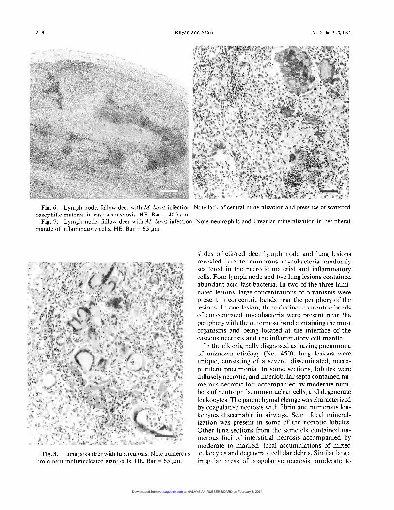
Fig. 6.
Fig. 7.
Lymph node; fallow deer with M. bovis infection. Note lack of central mineralization and presence of scattered
Lymph node; fallow deer with M. bovis infection. Note neutrophils and irregular mineralization in peripheral basophilic material in caseous necrosis. HE. Bar = 400 pm.
mantle of inflammatory cells. HE. Bar = 65 pm.
slides of elWred deer lymph node and lung lesions revealed rare to numerous mycobacteria randomly scattered in the necrotic material and inflammatory cells. Four lymph node and two lung lesions contained abundant acid-fast bacteria. In two of the three lami- nated lesions, large concentrations of organisms were present in concentric bands near the periphery of the lesions. In one lesion, three distinct concentric bands of concentrated mycobacteria were present near the periphery with the outermost band containing the most organisms and being located at the interface of the caseous necrosis and the inflammatory cell mantle.
In the elk originally diagnosed as having pneumonia of unknown etiology (No. 450), lung lesions were unique, consisting of a severe, disseminated, necro- purulent pneumonia. In some sections, lobules were diffusely necrotic, and interlobular septa contained nu- merous necrotic foci accompanied by moderate num- bers of neutrophils, mononuclear cells, and degenerate leukocytes. The parenchymal change was characterized by coagulative necrosis with fibrin and numerous leu- kocytes discernable in airways. Scant focal mineral- ization was present in some of the necrotic lobules. Other lung sections from the same elk contained nu- merous foci of interstitial necrosis accompanied by moderate to marked, focal accumulations of mixed leukocytes and degenerate cellular debris. Similar large, irregular areas of coagulative necrosis, moderate to
Fig. 8. Lung; sika deer with tuberculosis. Note numerous prominent multinucleated giant cells. HE. Bar = 65 pm.
at MALAYSIAN RUBBER BOARD on February 3, 2014vet.sagepub.comDownloaded from

Vet Pathol 323 , 1995 Tuberculosis in Cattle, Deer, and Elk 219
Table 1. Mean (SEM) scores of microscopic features of M. bovis lesions in the different species.
Lymph Node Lesions Lung Lesions Microscopic Elk/ Fallow Sika Elk/ Fallow Sika
Feature Red Deer Deer Deer Red Deer Deer Deer ( n = 16) ( n = 5 ) ( n = 5 ) ( n = 9) ( n = 18) ( n = 13) ( n = 11) ( n = 29)
~ ~ ~
Caseous necrosis 2.7 (0.1) 2.5 (0.2) 2.7 (0.2) 1.4 (0.2) 2.2 (0.3) 2.4 (0.2) 1.6 (0.4) 1.4 (0.4) Mineralization 2.1 (0.1) 1.4 (0.2) 1.6 (0.2) 1.3 (0.2) 1.3 (0.3) 1.5 (0.2) 0.6 (0.4) 1.3 (0.4) Neutrophils 1.9 (0.1) 2.3 (0.2) 2.2 (0.2) 1.0 (0.2) 2.0 (0.2) 2.6 (0.2) 1.9 (0.3) 1.6 (0.3) Macrophages 2.2 (0.1) 1.7 (0.1) 2.3 (0.1) 1.5 (0.1) 1.9 (0.2) 1.7 (0.1) 2.0 (0.3) 1.3 (0.3) Giant cells 2.0 (0.1) 1.3 (0.2) 2.0 (0.2) 2.6 (0.2) 1.6 (0.3) 1.4 (0.2) 1.7 (0.4) 2.3 (0.4) Acid-fast bacilli 0.9 (0.2) 1.4 (0.2) 1.0 (0.2) 1.0 (0.3) 0.5 (0.3) 1.3 (0.2) 1.0 (0.4) 1.4 (0.4)
marked accumulations of neutrophils, and degenerate leukocytes were observed in kidney sections. Liver sec- tions contained few small foci of hepatocellular degen- eration accompanied by a mild multifocal, pleocellular infiltrate. The lymph node lesion from this animal con- sisted of a caseous abscess with concentric layers of caseous necrotic and collagenous material extending to the thickened connective tissue capsule. Only a thin layer of neutrophils and degenerate leukocytes was present at the interface of the necrosis and connective tissue. Acid-fast bacilli were extremely abundant in lung, lymph node, and kidney lesions and were scat- tered in the small hepatic lesions.
The lymph node lesion from the red deer (No. 3009) that was originally diagnosed as having an abscess con- sisted of numerous small to large accumulations of neutrophils and abundant basophilic cellular debris scattered in a central zone of caseous necrosis. A thin mantle of neutrophils and large and small mononuclear inflammatory cells surrounded the necrotic center. Gi- ant cells were rare, and mineralization was limited to a few intracellular microliths located in the cellular mantle. Moderate numbers of AOAO-fluorescent ba- cilli were scattered in the necropurulent center.
Fallow deer
Fallow deer lesions were similar to elWred deer le- sions, possessing the same patterns of mineralization and moderate to numerous neutrophils (Figs. 6 , 7). Giant cells were present in all fallow deer lesions.
Sika deer
Lymph node and lung lesions from sika deer were distinctly different from the lesions of other species examined. Lesions were usually poorly encapsulated and consisted of marked accumulations of epithelioid macrophages and abundant large giant cells (Fig. 8). The giant cells were usually of the Langhans type but varied in shape from round to irregular and often had 30-60 visible nuclei. These bizarre giant cells were easily discernable at low magnification and were the most distinctive feature of the sika deer lesions. Several
lesions had no necrosis or mineralization; the majority of lesions had small to large irregular areas of caseous necrosis that lacked or had scant to moderate central and/or peripheral mineralization. Neutrophils were absent or present in low numbers, and few to numerous acid-fast- and/or AOAO-fluorescent bacilli were ran- domly scattered in the lesions.
Numerical scores
Mean numerical scores of the microscopic features (Table 1) reflect several interspecies differences. Elk lesions averaged fewer giant cells and more neutrophils than did lesions in other species. Sika deer lesions av- eraged more giant cells and fewer neutrophils than did lesions in the other species.
Discussion
The microscopic features of lesions of M. bovis in- fection in the different species usually included the hallmark characteristics of caseous necrosis, mineral- ization, epithelioid macrophages, giant cells, and fi- brosis. Although there was overlap in lesion charac- teristics between species, interspecies differences were noted.
Most elWred deer lesions were consistent with pre- vious descriptions. 1 , 2 7 1 5 , 1 8 The lesions had marked vari- ation ranging from suppurative abscesses to granulo- mas with partially mineralized, caseous, necrotic centers. Mineral deposits in elWred deer lesions fre- quently extended into the peripheral inflammatory cell mantle, whereas in bovine lesions they usually did not. ElWred deer lesions usually had more numerous neu- trophils and fewer giant cells than did bovine lesions. Concentrically laminated, caseous abscesses, morpho- logically similar to ovine caseous lymphadenitis le- sions, were present in three elk. The disseminated nec- ropurulent lesions in elk No. 450 were similar to experimentally induced lesions in red deer following intravenous inoculation of M. bovis. The atypical le- sions observed in elk No. 450 probably resulted from lesion invasion through a vascular wall with hematog- enous dissemination of the infection. Fallow deer le-
at MALAYSIAN RUBBER BOARD on February 3, 2014vet.sagepub.comDownloaded from

220 Rhyan and Saari Vet Pathol 32:3, 1995
sions had more giant cells but were otherwise consid- ered indistinguishable from elk lesions.
Most sika deer lesions were distinguishable from le- sions in the other species. The abundant bizarre giant cells, highly invasive granulomatous lesions, minimal fibrosis, fewer neutrophils, and smaller areas of necro- sis and mineralization were characteristic of the sika deer lesions in this study. Subsequent experience with tuberculosis lesions in sika deer at the NVSL has shown that some lesions have marked necrosis and moderate numbers of neutrophils, but the abundant bizarre giant cells are a consistent feature. Numerous Langhans-type giant cells and macrophages in M. bovis lesions in sika deer have been reported.6
The numbers of acid-fast bacilli were highly variable among lesions in each species; however, the largest numbers of organisms were noted in lesions from elk and sika deer. These findings are consistent with those in previous report^.^.'^,^^ An inverse relationship be- tween the cellularity of the mononuclear cell response mounted by the host and numbers of mycobacteria was noted in a study of farmed deer.'O Data from the current study do not confirm this; however, the nec- ropurulent lesions in elk No. 450, which lacked giant cells and epithelioid macrophages, contained abundant acid-fast bacilli.
The findings of this study indicate that there are differences between the microscopic features of lesions in cattle and the different cervid species. Additionally, there is wide variation in cervid lesion morphology, i.e., granulomas with mineralized, caseous, and ne- crotic centers, suppurative abscesses, ovine caseous lymphadenitis-like lesions, and widely disseminated necropurulent lesions. Because of these differences and variation in lesion morphology, tuberculosis should be included in the differential diagnosis whenever gran- ulomatous, necrotizing, or suppurative lesions are en- countered in c e r v i d ~ . ~ * l ~ . l ~
Acknowledgements
We thank H. Ridpath, K. Wilson, J. Campbell, B. Burle- son, W. Romp, G. Hedberg, and T. Glasson for their excellent technical assistance.
1
2
References
Beatson NS: Tuberculosis in red deer in New Zealand. R SOC NZ Bull 22: 147-1 50, 1985 Buchan GS, Griffin JFT: Tuberculosis in domesticated deer (Cervus eluphus): a large animal model for human tuberculosis. J Comp Pathol 103: 1 1-22, 1990
3
4
5
6
7
8
9
10
1 1
12
13
1 4
15
16
1 7
18
19
20
21
Clifton-Hadley RS, Wilesmith JW: Tuberculoses in deer: a review. Vet Rec 129:5-12, 1991 de Lisle GW, Havill TF: Mycobacteria isolated from deer in New Zealand from 1970-1 983. NZ Vet J 33: 138- 140, 1985 de Lisle GW, Welch PJ, Havill PF, Jilian AF, Poole WSW, Corrin KC, Gladden NR: Experimental tuber- culosis in red deer (Cervus eluphus). NZ Vet J 31:213- 216, 1983 Dodd K: Tuberculosis in free-living deer. Vet Rec 115:
Essey MA, Fanning A, Saari DA, Payeur J: Bovine tu- berculosis in Cervidae: human health concerns. Proc Annu Meet US Anim Health Assoc 9 5 4 2 7 4 3 6 , 199 1 Fanning A, Edwards S: Mycobucterium bovis infection in human beings in contact with elk (Cervus eluphus) in Alberta, Canada. Lancet 338:1253-1255, 1991 Fleetwood AJ, Stuart FA, Bode Maff R, Sutton FP: Tu- berculosis in deer. Vet Rec 123:279-280, 1988 Griffin JFT: The aetiology of tuberculosis and myco- bacterial diseases in farmed deer. Ir Vet J 42:23-26, 1988 Henry EJ, Cassidy DR: Rapid decalcification followed by a new fuchsin-methylene blue stain for acid-fast ba- cilli in frozen sections. Stain Technol 41:345-346, 1966 Itoh R, Kagabu Y , Itoh F: Mycobacteriurn bovisinfection in a herd of Japanese shika deer (Cervus nippon). J Vet Med Sci 542303-804, 1992 Mirsky ML, Morton D, Piehl JW, Gelberg H: Myco- bacterium bovis infection in a captive herd of sika deer. J Am Vet Med Assoc 200:1540-1542, 1992 Mote RF, Muhm RL, Gigstad DC: A staining method using acridine orange and auramine 0 for fungi and my- cobacteria in bovine tissue. Stain Technol 50:5-9, 1975 Rhyan JC, Saari DA, Williams ES, Miller MW, Davis AJ, Wilson AJ: Gross and microscopic lesions of nat- urally occurring tuberculosis in a captive herd of wapiti (Cerws eluphus nelsoni) in Colorado. J Vet Diagn Invest 4:428-433, 1992 Robinson RC, Phillips PH, Stevens G, Storm PA: An outbreak of Mycobacterium bovis infection in fallow deer (Darnu duma). Aust Vet J 66:195-197, 1989 Selwyn P, Hathaway S: A study of the prevalence and economic significance of diseases and defects of slaugh- tered farmed deer. NZ Vet J 38:94-97, 1990 Stuart FA, Manser PA, McIntosh FG: Tuberculosis in imported red deer (Cervus eluphus). Vet Rec 122:508- 511, 1988 Stumpff CD: Epidemiological study of an outbreak of bovine TB in confined elk herds. Proc Annu Meet US Anim Health Assoc 86:524-527, 1982 Towar DR, Scott PM, Goyings LS: Tuberculosis in a captive deer herd. Am J Vet Res 26: 339-346, 1965 Wilson P, Harrington R: A case of bovine tuberculosis in fallow deer. Vet Rec 98:74, 1976
592-593, 1984
Request reprints from Dr. J. C. Rhyan, National Veterinary Services Laboratories, PO Box 844, Ames, IA 50010 (USA).
at MALAYSIAN RUBBER BOARD on February 3, 2014vet.sagepub.comDownloaded from