
Papier SEP2015
31
A DFT/TDDFT study of complexation ability of the calcium cation complexes with Lateral chain ligand of Taxol Abstract: Theoritical investigation of the [Taxol-Ca] and [LC1- Ca] complexes in gas phase employing DFT and TD-DFT theory methods have been study. The electronic structure of the complexe have been taken into the count. Charge distribution one donor an acceptor atoms are evaluated by Mulliken analysis . The charge distribution indicate that the ligands transfer their negative charge to Calcium ion Ca 2+ during formation of the complexes (LMCT). Electronic excitation energies of Ca 2+ complexe are calculated. It is found that the Lateral chain ligand has the same total density as Taxol structure. In this work we investigate the performance of the DFT method, augmented with an empirical dispersion function (DFT-D), paired with the PCM implicit solvation model, for the computation of noncovalent interaction energies of biologically-relevant, solvated model complexes. It is found that this method describes intermolecular interactions within and (-like) environments with roughly the same accuracy as in the gas phase. Another important finding is that, when environmental effects are taken into account, the empirical dispersion term associated with the DFT-D method need be modified very little (or not at all), in order to obtain the optimum, most well balanced, performance. 1
-
Upload
independent -
Category
Documents
-
view
1 -
download
0
Transcript of Papier SEP2015
A DFT/TDDFT study of complexation ability of the calcium cation
complexes with Lateral chain ligand of Taxol
Abstract: Theoritical investigation of the [Taxol-Ca] and [LC1-
Ca] complexes in gas phase employing DFT and TD-DFT theory
methods have been study. The electronic structure of the complexe
have been taken into the count. Charge distribution one donor an
acceptor atoms are evaluated by Mulliken analysis . The charge
distribution indicate that the ligands transfer their negative
charge to Calcium ion Ca2+ during formation of the complexes
(LMCT). Electronic excitation energies of Ca2+ complexe are
calculated. It is found that the Lateral chain ligand has the
same total density as Taxol structure.
In this work we investigate the performance of the DFT method,
augmented with an empirical dispersion function (DFT-D), paired
with the PCM implicit solvation model, for the computation of
noncovalent interaction energies of biologically-relevant,
solvated model complexes. It is found that this method describes
intermolecular interactions within and (-like) environments with
roughly the same accuracy as in the gas phase. Another important
finding is that, when environmental effects are taken into
account, the empirical dispersion term associated with the DFT-D
method need be modified very little (or not at all), in order to
obtain the optimum, most well balanced, performance.
1

Keywords: Taxol, Ca2+ ion,conformation, binding energy, DFT/TDDFT,
UV-Visible.
INTRODUCTION
Cancer’s deseases (CD) is caused by an uncontrolled division of
abnormal cells in a part of the body. It is the consequences of a
combination of complex factors related to heredity, lifestyle and
environment1-3. It is caused by an accelerated cellular growth
accumulating genetic alteration as they progress to a more
malignant phenotype 4 and are the second major cause of the human
death in industrialized countries5.
These death rates were significantly reduced in the past decades
due in part to the successful use of chemotherapeutic agent3. non-
proteinogenic β –amino α-hydroxy acids are found in many natural
products and drugs, for example N-benzoyl-3-phenylisoserine as a
side-chain of taxol (generic name Paclitaxel) (figure1) which is
one of the most popular chemotherapeutic agents used nowadays for
treatment of breast, ovarian, and lung cancers6,7.
Soon after its isolation, taxol was shown to have a unique mode
of action it blocks cancer cell division by promoting the
polymerization and stabilization of the protein tubulin to
microtubules and inhibits the de-polymerization of microtubules
back to tubulin. Although an atomistic structure of tubulin has
been proposed8.
Four main groups (Na, K, Mg, and Ca) and ten transition (V, Cr,
Mn, Fe, Co, Ni, Cu, Zn, Mo,
and Cd) metals are currently known or thought to be required for
normal biological functions in humans9 , the medical consequences
of insufficient quantities where known.
2

Besides, the most common ions essential for biological processes
are K+,Na+,Mg2+ and Ca2+. Ca2+, in contrast to Mg2+, is found
principally in extracellular locations. Its main function is to
stabilize structural components in biological organisms- e.g.
extracellular proteins, cell membranes, cell walls and
extracellular de posits such as bone. In addition, it assumes
regulatory functions within cells10-12
The Ca2+ cation also has anticariogenic effects demonstrated in a
set of clinical trials13,14. Previous data suggest that carcinogenic
stimuli cause local increase in the Ca2+concentration leading to
activation of proto oncogenes and to inactivation of tumor-
suppressor genes, which lead to the manifestation of a malignant
phenotype. Tumor cell proliferation maybe stimulated by
persistent increase of Ca2+, in contrary the transitory fulminic
increase of Ca2+ induce the activation of mitochondrial apoptotic
pathway15
In the other hand, intracellular calcium concentration may play a
role in the development of chemoresistance. Altered
Ca2+ homeostasis of cell is correlated with cisplatin or Taxol
resistance in Non-small cell lung cancer (NSCLC) cell lines
(A549 and EPLC) or small cell lung cancer (SCLC) cell line
(H1339)16,17
W. Boehmerle et al. suggestion is understand the etiology of the
potent dose and therapy limiting side effects is still poorly
understood and difficult to explain with the know Taxol
interaction partners. Their conclusion of analysis is the
affinity of taxol for (NCS-1) protein and stabilized both probe
and Ca2+ depending characteristics18. However, their finding is
3

that taxol also affects intracellular Ca2+ signaling care should
be given when interpreting the results obtained with taxol as a
microtubule modifying drug. Their observations (that taxol has
new binding partner, NCS-1, and that binding to NCS-1 lead to
initiation of cytosolic Ca2+ oscillation) suggest that taxol’s
effects when used as a research to will be more complex than
originally expected.
Taxol is a compound that selectively stabilizes microtubules
and has been used extensively in studies of microtubule
function19. M. P .Mattson et al were employed Taxol to test the
hypothesis that the state of microtubule polymerization
influences neuronal Ca2+ homeostasis and vulnerability to
excitotoxicity, there data obtained with taxol suggested
that stabilization of microtubules suppresses Ca2+ influx
and protects neurons against excitotoxicity. However,
the possibility remained that taxol was protecting neurons
against excitotoxicity by a mechanism unrelated to
its microtubule stabilizing activity20. The mechanisms by which
microtubules and other cytoskeletal elements affect ion channel
function are not known21. However, Taxol administration is limited
by serious side effects including cardiac arrhythmia, which
cannot be explained by its microtubule-stabilizing effect.
Recently, neuronal calcium sensor 1, a calcium binding protein
that modulates the inositol-1,4,5-trisphosphate receptor
(InsP(3)R), was described as a binding partner of Taxol and as a
substrate of calpain. They have examined calcium signaling
processes in cardiomyocytes after treatment with Taxol to
investigate the basis of Taxol-induced cardiac arrhythmia. Taxol
treated cells had increased expression of NCS-1, an effect also
detectable after Taxol administration in vivo22. Several4

chemotherapeutic drugs are believed to cause an enhanced calcium
signal leading to hyperactivation of neurons23.
Figure 1: Chemical Structure of paclitaxel (Taxol) and biological
activity region of Lateral chain and DBAC.
Considering the topological structure (figure 1), Paclitaxel
(PTX) is a complex natural product with a rigid baccatin core
composed by the DBAC region, which is a natural substance
isolated from the leaves of Taxus species24 and four flexible side
chains emanating from C2, C4, C10 and C13. PTX is highly
substituted poly-oxygenated cyclic diterpenoid characterized by
the taxane ring system and it differs from other known taxanes
either in substitution pattern, the nature of the ester side-
chain or in the presence of the oxetane ring (D-ring) system.
Potent antimitotic activity has seen to be restricted to taxol
which possess an N-benzoyl-3 phenylisoserine side-chain at C13
atom. The lateral chain is essential for the antitumor activity of
taxoid family which is characterized by terpene core25,26.
Hydrophobic interaction plays a role, owing to the presence of
lipophilic ester groups on the chain and on the terpenoid core27.5
Likewise the LC molecule has no symmetry and exhibits a
significant internal rotational degree of freedom due to
rotations around the single bonds in the central part of the
carbon skeleton (figure 3). Therefore, study on conformational
landscape of lateral chain may provide insights and may support
practical efforts for medicine.
D.S. Galvão and co-workers have performed a theoretical study28 on
the electronic structure of 10-deacetylbaccatin-III by semi-
empirical methods PM3 and ZINDO. They have identified the
relative importance (in terms of electronic features) of specific
DBAC’s molecular regions and concluded that regions that are
considered important for the biological activity of taxol can be
traced by the electronic features already present in DBAC
regions.
Compared the topological structure to Paclitaxel (PTX), Lateral
Chain (LC) has a simple structure in term of molecular size, it
has 46 heavy atoms while Paclitaxel has 113 atoms. The LC makes a
good candidate for preliminary investigation.
Our main was to find a model system in which the Taxol molecule
interacts with some metallic cations while Ca2+, Fe2+ and Cu2+
present in biological fluids. The major focus of this work is on
the calcium anticancer drug taxol chelation, which is presented
as a case of study theoretically the scope of the problem. For
the calculations on isolated molecules (gas phase), we will
extend the approach of these previous studies to LC residue
attached to carbon C11 of the moiety and benzoate connected to
nitrogen N (-C3’) substituent of the C13 side chain (figure 2), in
order to come as to the structure of Taxol.
6

As a ligand, considering a lateral chain of taxol, it contains
three strong chelating sites in competition (figure 2).
Interaction of the LC with metal ion, both via their phenyls
groups, NH…OCar and NH…OEt, have been subject of details studies,
with OCar and OEt are the carboxylic and Ethoxy oxygen atoms,
respectively (see figure 2).
We determine whether or not LC can bind to Ca2+ ion to form
chelated complex, both via their phenyl groups or amide groups.
Figure 2: Structure of lateral chain, arrows indicates the
possible complexes sites via degree of freedom of the dihedral
angle φ, studied in this work.
Mulliken charge transfer complexes are being regarded as an
important materials for application in mechanism of drug action29 .
Thus, we have computed a variety of properties, like charge
transfer (CT) interaction between electron donor / acceptor and
the vibrational spectra of LC ligand. we attempted to
accommodate on a different way a wider forms complexes of the
[[LC-Ca]+ and [Taxol-Ca]+ with different stabilities. The effect
of the OH- counterion for complexes cation spectrum has been also
analyzed. To the best of our knowledge, there is no report of the
stability of Taxol complexes with Ca2+.
7

Computational details
Calculations reported here were performed with Gaussian 03
software package with a spin-restricted formalism29. Full geometry
optimization for different LC conformations and [LC-Ca]+ , [LC-
CaOH] and [Taxol-Ca]+, [Taxol-CaOH] were carried out by using
Density Functional Theory (DFT) with the Becke thee-parameter
hybrid functional (B3LYP) method30-32. 6-31G(d,p) is the standard
basis sets and a popular polarized basis set which adds p
functions to hydrogen atoms in addition to the d functions on
heavy atoms.
In this study one of the points we would like to evaluate, is
whether the geometry of LC with metal cation can form an active
complex. A preliminary geometry optimization of the deprotonated
LC as ligand has revealed that the most stable structure is also
same structure of protonated LC. In the optimized structure of
Ca(LC)2, four oxygen atoms from carboxylate groups and two
nitrogen atom make an “octahedral-like” environment around
calcium ion (figure 5) the most stable (LC)2Ca complex correspond
to hexacoordinated structure in which Ca2+ ion interact with two
nitrogen and four oxygen atoms of LC dimer .
Molecules are studied in gas phase adopting the zwiterionic form
to the carbon 3΄ set to NH for lateral chain. in the first step,
the geometries were fully optimized at the B3LYP/6-31G(d,p)
level for all atoms.
Although there is qualitative agreement between these methods,
the B3LYP hybrid density functional method appears to give a more
accurate representation of the energetic of LC.
8

The analyses of vibrational frequencies indicated that optimized
structures of complexes were at stationary points corresponding
to local minima without imaginary frequencies. The electronic
charge distribution of different complexes were evaluated by
Mullikan theory34 enable us to characterize the donor and acceptor
molecule.
We initially used the rotational step of 10 degrees to the C3’-C2’-
C1’-O1’ dihedral angle using B3LYP/6-31G(d,p). The complete
rotation of 180° shown existence of two best conformation (in
terms of energy) and were selected in both structures of 3’-N
benzoyl form hydrogen bound with …Ocar and with …OEt atoms
respectively .
Although, W. Boehmerle et al show experimental evidence of the
binding of taxol for (NCS-1) protein and stabilized both probe
and Calcium ion18.The LC dimer chelats with calcium ion (complex
(LC)2...Ca2+) has been carried out, in order to explore binding to
the chemotherapeutic drug as possible interaction of LC activity
of taxol. The electronic spectrum calculations of individual
molecules and ion complexation and their effects on spectral
properties were made by using time dependent density functional
(TDDFT) theory method with B3LYP/6-31G(d,p) basis set in gas
phase34 . In absorption or fluorescence spectra are observed when
the LC1 and Taxol are complexed by a Ca2+ .
Density functional DFT studies have been performed for three
possible structures of calcium complexes with LC assuming hexa-
coordination with two LC ligands. Calculation were carried out
using the B3LYP method with the 6-31G(d,p) basis sets.
It should be mentioned that Shen Li et al.35 performed
conformational studies of benzene-water complex at vdW-DF, they
suggest that either the H atoms or the O atom of water can9

interact with benzene, depending on the location of the water.
The water molecule will interact with the rim of the benzene ring
via its O atom.
Ground state electronic structure S0 calculation of complexes has
been performed in gas phase, the optimum structures were verified
by the absence of imaginary frequencies. The 20 lowest singlets
excited states of complexes were calculated within TDDFT
formalism. We used in place of the δ functions Gaussian having a
FWHM with 0,3 eV .For the better interpretation of Photoluminescence properties,
triplet excited states T1 of complexes have been optimized in gas
phase. Long-wavelength emission bands have been calculated with
ΔSCF approch [[56] Scalmani, G.; Frisch, M. J. J. Chem. Phys. 2010, 132, 114110. [57]
Andreiadis, E. S.; Imbert, D.; Pécaut, J.; Calborean, A.; Ciofini, I.; Adamo, C.;
Demadrille, R.; Mazzanti, M. Inorg. Chem. 2011, in press.
Results and Discussion
Conformational study of neutral and zwitterionic LCoptimization in gas phase
The first part of the computation is devoted to optimize the
geometry of LC at varying dihedral angle θ in order to find
stable structures LC1 and LC2 by B3LYP/6-31G(d,p) (figure 2),
and we determine ground state geometry for the LC ligand. The
result showed that rotation around C13-C10 bond is
restricted by the tendency to form intramolecular hydrogen bond
interaction between N-H…OEt and N-H…Ocar for LC1 and LC2
respectively.
10

Figure 3: Structures optimized for LC1 and LC2 calculated at
the B3LYP/6-31G (d,p), Bond lengths are in Å.
We observe weak intramolecular hydrogen bond interaction
between N-H… OEt of LC1 and between N-H…OCar of LC2 compound. The
energy level for both forms is the same (ΔE=0,0015kcal/mol).
In order to form complex with calcium, we assumed that the
zwiterionic form of LC is the deprotonation of the –NH in the
amine, their different ways are:
In zwitterionic form as β-amino acids which are known to
exist as zwitterions in the solid state and in solution and in
the no-ionized, neutral form in the vacuum phase34 the
zwitterionic form exists frequently in physiological conditions
but depends on the temperature and pH in cells. Existence of
11

intramolecular proton transfer (N-H O=C) . Intermolecular
proton transfer between (C=O) of the first LC and the (H-N) of
the second one.
Therefore, molecules are studied in gas phase adopting the
zwiterionic form of LC with NH deprotonated according to the
following reaction, LC-(N-H) → LC-(N:)- + H+.
LC(N:)- is believed to complex with Ca2+ ion . It appears that the
lowest energy lying and zwitterionic conformers were found to be
close in energy about 4.6 eV (106,07kcal/mol) in gas phase and
4.72 eV (108,84 kcal/mol) in PCM at DFT level. These energies
are more less to that obtained with cyclopentadienyl
deprotonation [ reference sihem).
Table 1, summarize the result for some relevant bond lengths,
valence and dihedrals angle in order to compare the predicted
B3LYP geometries of protonated , deprotonated LC and the highest
stable [LC1-Ca]+ complex. It has been observed that there is such
difference regarding bond length, valence and dihedrals angle
between various atoms of LC1,
Zwiterionic LC1 form and ([LC1/Ca]+) complex . The bond lengths
value between various atoms of, LC and LC deprotonated varied
about 0.01 Å at 0.07 Å and between LC deprotonated and [LC1/Ca]+
varied around 0.01 Å at 0.04 Å. In the other hand, valence angle
between LC and LC deprotonated varied around 0° to 10° and
between LC deprotonated and ([LC1/Ca]+ varied about 0° to 4°.
Table 1: Optimization parameters of LC1, zwiterionic LC1 form and
the highest stable [LC1/Ca]+ complex at B3LYP calculation in
ground state.
Parameter LC1 Zwiterio [LC1/Ca]+
12

nic
LC1
complex
Bond lengths
O7-N25
O7-C8
C8-C10
C10- C13
C13- N25
N25-C26
C26- O27
Valence
angle
O7- C8-C10
C8-C10-C13
C10-C13- N25
C13- N25-C26
N25-C26- O27
Torsional
angle
O7-C8-C10-C13
C8-C10-C13-N25
C10-C13-N25 -C26
C13-C10-C8-O7
Dipole
moment (D)
2.88
1.34
1.52
1.56
1.45
1.30
1.22
112.
73
112.
44
109.
78
122.
26
122.
30
-
61.7
7
73.3
5
126.
84
1.68
5.72
2.93
1.33
1.52
1.62
1.42
1.38
1.23
113.06
112.10
109.93
116.94
121.63
-77.96
58.46
81.10
- 25.70
3.35
3.05
1.42
1.53
1.57
1.46
1.36
1.22
113.44
113.80
110.08
114.76
129.0
-78.67
65.76
134.58
-3.58
15.34
13

aBond lengths R in Å, angles θ and dihedral
angles φ in degrees.
The most important point to be mentionned here is that the bond
length O7-N25 of oxygen O7 of LC1 increase in zwiterionic form and
in the complex about 0.05 and 0.17Å respectivelly.
Geometries and binding energies of LC-Ca complexes.
* [LC-Ca] + complex
Molecules are studied in vaccum level adopting form of LC, which
the total charge is equal one. It is believed to complex with ion
metals as Ca2+. Depending of the initial geometry, the lateralchain ligand (Fig.5) contains several coordination sites, i.e.
ethoxy atom and carboxy atom as well as nitrogen atom
deprotonated of amide groups of zwiternionic LC. Therefore, it
also can coordinate the metal calcium by phenyl groups on/down.
The interact ion between the metal ion and the ligand is
predominantly electrostatic. Several local minima of the
potential energy were found for LC and Ca2+ ion complexes are
displayed with their relative energies in figure 5. For complexes
5 and 6, zwiternionic form of LC deprotonated of NH amine is
complexed with Ca2+.
14
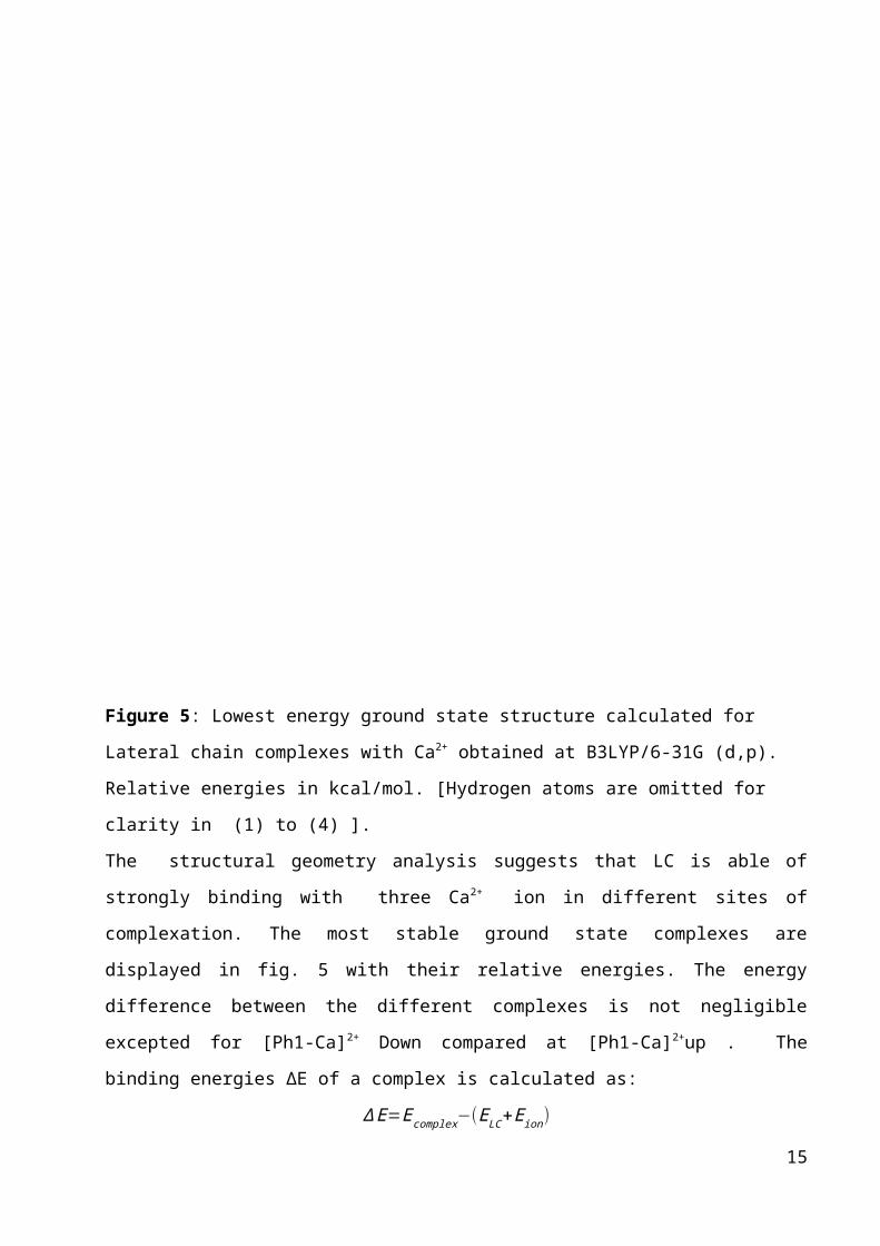
Figure 5: Lowest energy ground state structure calculated for
Lateral chain complexes with Ca2+ obtained at B3LYP/6-31G (d,p).
Relative energies in kcal/mol. [Hydrogen atoms are omitted for
clarity in (1) to (4) ].
The structural geometry analysis suggests that LC is able of
strongly binding with three Ca2+ ion in different sites of
complexation. The most stable ground state complexes are
displayed in fig. 5 with their relative energies. The energy
difference between the different complexes is not negligible
excepted for [Ph1-Ca]2+ Down compared at [Ph1-Ca]2+up . The
binding energies ∆E of a complex is calculated as:
∆E=Ecomplex−(ELC+Eion)
15

Where Ecomplex and ELC are the complex and the free LC energies,
respectively.
*[(LC)2 -Ca] complex.
In –NCCCOO- group, both O and N atoms are recommended as
coordination atoms in all kinds of complexes, and the
characteristic structure determines the strong chelating ability
of the LC with metal ions. In order to assign the positions of
Ca2+ ion in the complexes, three possible structures of calcium
complexes with LC assuming hexacoordination with two LC ligands.
Therefore, the Ca2+ ion chelates with, two nitrogen and four
oxygen of (LC)2-Ca complexes (figure 6).
Figure 6: Optimized structures of [(LC)2-Ca] complexes obtained at
B3LYP/6-31G(d,p) level [Hydrogen atoms are omited for clarity].
In Table 2, we present the Ca2+ binding energies obtained in
vacuum level using DFT level. The most stable
structure of each type of monomer complex follow an order:
5> 6 > 1 > 4 > 2 ≈ 3, as calculated using B3LYP/ 6-31+G (d)
method (Table 2). The BE of the studied complexes between
different structures is the largest for complex 5 with -502, 20
kcal/mol owing less flexible ligand. For complexes 1,2,3,4 have
16

high flexibility leading smaller binding energies, and type of
coordination is different. In the other hand the calculations of
the dimer complex have shown that the binding sequences follow
the order: 7 > 9 > 8. The stability of Ca-(LC)2 complex is
evidently far away from the other studied.
Table 2: Binding energies for calcium ion of monomer Lateral
Chain molecule calculated at B3LYP/6-31G(d,p) level for structure
optimized.
In the Complex 7,
all oxygen atom i.e.
two oxygen atoms of,
carbonyl and ethoxy
groups,
17
complexe
s
Ground stateComplexation energy
(kcal/mol)
Monomer
[Ph1-Ca]2+up
(1) -175,08
[Ph1-
Ca]2+down (2)
-161,12
[Ph2- Ca]2+-
up (3)-161,18
[Ph2-
Ca]2+down (4)
-128,25
[LC1-Ca]+
(5)-502,20
[LC2-Ca]+
(6) Dimer
[(LC1)2-Ca]
(7)
[(LC2)2-Ca]
(8)
[(LC1-LC2)-
Ca] (9)
-394,11
-689,27
-673,10
-681,13
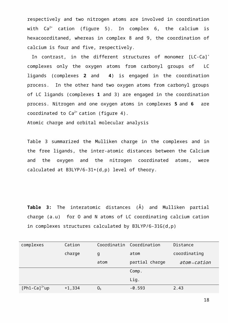
respectively and two nitrogen atoms are involved in coordination
with Ca2+ cation (figure 5). In complex 6, the calcium is
hexacoorditaned, whereas in complex 8 and 9, the coordination of
calcium is four and five, respectively.
In contrast, in the different structures of monomer [LC-Ca]+
complexes only the oxygen atoms from carbonyl groups of LC
ligands (complexes 2 and 4) is engaged in the coordination
process. In the other hand two oxygen atoms from carbonyl groups
of LC ligands (complexes 1 and 3) are engaged in the coordination
process. Nitrogen and one oxygen atoms in complexes 5 and 6 are
coordinated to Ca2+ cation (figure 4).
Atomic charge and orbital molecular analysis
Table 3 summarized the Mulliken charge in the complexes and in
the free ligands, the inter-atomic distances between the Calcium
and the oxygen and the nitrogen coordinated atoms, were
calculated at B3LYP/6-31+(d,p) level of theory.
Table 3: The interatomic distances (Å) and Mulliken partial
charge (a.u) for O and N atoms of LC coordinating calcium cation
in complexes structures calculated by B3LYP/6-31G(d,p)
complexes Cation
charge
Coordinatin
g
atom
Coordination
atom
partial charge
Distance
coordinating
atom→cationComp.
Lig.[Ph1-Ca]2+up +1,334 O6 -0.593 2.43
18

(1) O8 -0.503
-0.598
-0.538
2.32
[Ph1- Ca]2+down
(2)
+1,372 O27 -0.692
-0.525
2.19
[Ph2- Ca]2+-up
(3)
+1.243 O7
O12
-0.599
-0.478
-0.599
-0.5375
2.40
2.41
[Ph2- Ca]2+down
(4)
+1,645 O27 -0.472
-0.526
2.69
[LC1-Ca]+
(5)
+1.268 N25
O9
-0.702
-0.572
-0.569
-0.503
2.41
2.39
[LC2-Ca]+
(6)
+1.159 N25
O7
-0.711
-0.572
-0.595
-0.478
2.30
2.40
[(LC1)2-Ca]
(7)
+1.119 O10
N26
O28
O10'
N26'
O28'
-0.535
-0.526
-0.644
-0.572
-0.640
-0.503
-0.538
-0.478
-0.645
-0.572
-0.639
-0.526
2.47
2.47
2.39
2.47
2.47
2.39
[(LC2)2-Ca] +1.121 O28 -0.649 -
0.526
2.37
19

(8) N26
O28'
N26'
O8
O8'
-0.644
-0.572
-0.649
-0.526
-0.644
-0.572
-0.530
-0.478
-0.530
-0.478
2.46
2.37
2.46
2.72
2.72
[(LC1-LC2)-Ca]
(9)
+1.081 O28
N26
O10
O28'
N26'
O10
-0.654
-0.526
-0.639
-0.572
-0.549
-0.503
-0.653
-0.503
-0.651
-0.572
-0.538
-0.473
2.39
2.47
2.42
2.39
2.42
2.61
Table 3 shows that the charge of LC in the complexes decreased
compared to the free ligand. While the charge of Calcium
increased. For instance, before complexation the charge on
calcium is +2, while the charge of the calcium in monomer complex
4 is +1,645 a.u. and in dimer complex7 is +1.119 a.u.
This indicates that ligand transfer their negative charges
occurs to calcium ions during complexe formation with increase
the electron density of calcium in the order : (6) > (3) > (5) >
20

(1) > (2) for monomer LC complex. In the other hand for dimer
complex the electron density of calcium increases in the order :
1=2 > 3. LC of taxol has favourable donor-centers, namely oxygen
atoms from carbonyl (C=O), ethoxy (C-O) and nitrogen (N-C)
groups. They create good binding sites for hexadentate cation
for complex 7 and pentadentate cation for complex 9 (see table
2).
This results shows that ligand in dimer complex has better donor
atoms than in monomer complex. The maximum Mulliken charge (<2)
for all the complexes has been found on the
Ca – center atom and atoms coordinated.
Furthermore, the Mulliken’s electronic charge on the O atom of
the complex in its ground state is found to be more negative than
that obtained in the isolated LC molecule. For example, the
Mulliken’s electronic charges on the N atom of free ligand and in
complex 5 is (-0.572 a.u.) and (-0.702 a.u.) respectively by
DFT (B3LYP) calculations. In the other hand , the Mulliken’s
electronic charge on the calcium ion with +2 before
complexation , becomes less positive +1.268 a.u) for complex 5
about 0.732 e has been transferred. This indicates that
appreciable amount of electronic charge has been transferred from
LC ligand to Ca2+ in complex.
Photophysical properties
* Absorption spectra
Molecular orbital calculations using G09 and Gausview package on
selectedcomplexes were carried out to gain more insight into the
photophysical properties of the complexes. the features of the
highest occupied (HOMO-1 and HOMO-7) and the lowest unoccupied
(LUMO and LUMO+1) frontier orbitals mainly involved in the
21

transition are dipected in figure 7, and the description on
energy gaps of each transition are listed in the same figure .
Electronic excitation energies of LC, Taxol , [LC-Ca]+, and
[Taxol-Ca]+, [LC-CaOH], and [Taxol-CaOH] species have been
studied at the TD-DFT/B3LYP with 6-31+G(d,p) calculation in gas
phase. For all complexes, we have considered the most stable one
(with high binding energy) as like as complex 5 and complex
[Taxol-Ca]+ (see table 2). Orbital surfaces and energies for LC,
Taxol , [LC-Ca]+, and [Taxol-Ca]+ ,[LC-CaOH] and [Taxol-CaOH] are
given in figure 7.
In LC1 and Taxol complexes, the highest occupied MOs are
localized on the Elecronic excitation energies and the absorption
coefficient of the most intense bands are given in table 4. A
band with large oscillator coefficient for [LC-Ca]+, and [Taxol-
Ca]+ complexes appears at 414 and 1453 nm respectivelly, while
these bands decrease with used OH- counter-ion in [LC-CaOH] and
[Taxol-CaOH] neutral complexes about 302 and 438 nm respectively.
Table 4: Vertical excitation energies (eV), oscillator strengths
(f) and configurations of excitations for Taxol and LC1 with and
without counter-ion atoms (OH-) for complexes.
Complexes Wavelength
(nm)
f Configurati
on
Transiti
on
properti
esLC1
Taxol
[LC1-Ca]+
270
427*
414
0.58423
0.08304
0.0344
H-1 L
H
L
H-7 L
--
--
22

(LM
CT)[Taxol-Ca]+
[LC1-CaOH]
[Taxol-
CaOH]
1453
302
438
0.0142
0.0224
0.0234
H L
H
L+1
H-1
L+1
(LM
CT)
(LL
CT)
(L
LCT) *Experimental Wavelength36 (273nm).
Density is focus in same ligant of LC1 or Taxol appeared in
following figure.
Figure x : Total density of LC1 (in the left) and Taxol
(in the right) structures calculated by 6-31G(d,p)
23

-a-
-b-
24

-c-
-d-
Figure 7: Electronic density of LC1 and Taxol complexes
calculated by TD-DFT
25

Spectre d'absorption/émission de LC1-Ca : l'absorption présente
une bande qui est du à une transition intense HOMO-7/LUMO à
laquelle correspond un λmax de l'ordre de 414nm. Le spectre
d'émissions quant à lui présente aussi une bande qui résulte de
la transition HOMO-1/LUMO de longueur d'onde λmax = 937nm. Cette
émission se situe dans le proche IR.
Density is focus in same ligant of LC1 or Taxol appeared in
following figure.
26

Figure x : Total density of LC1 (in the left) and Taxol
(in the right) structures calculated by 6-31G(d,p)
Conclusion
in this work , we report DFT and TD-DFT. LC1 of taxol acts as
hexadentate ligand on forming complexes with calcium ion. TDDFT
method can be utilized successfully to evaluate the CT between
Taxol and Ca2+ in [Taxol-Ca]+ and [Taxol-Ca-OH] complex in vacuum
level. Frontier molecular orbital calculations reveal that there
is receivable opportunity to electron transfer from LC1/Taxol to
Ca2+ ion in [LC1-Ca]+ and [Taxol-Ca]+ CT that correspond to LMCT
process ( donnor-acceptor complex).
The result from the present investigations indicate that [Taxol-
Ca]+ CT complex could be a potential candidate to blocking cancer
cell division by promoting the polymerization of the protein
tubulin to microtubules.
References
1-The Biological Basis of Cancer; McKinnel, R. G., Ed.; Cambridge
University Press: Cambridge, 1998.
2- Lodish, H.; Baltimore, D.; Berk, A.; Zipursky, S. L.;
Sudaira, P. M.;Darnell, J.; Molecular Cell Biology; Scientific
American Books: New York, 1995.27

3-Cancer Biology; Ruddon, R. W., Ed.; Oxford University Press:
New York, 1987.
4- Sugimura, T. Multistep carcinogenesis: A 1992
perspective. Science 1992, 258, 603-607.
5- EIU Marketing in Europe, Trade Reviews, 337, December
1990.
6- Dubois, J. Expert Opin. Ther. Pat. 2006, 16, 1481–1496.
7- Marupudi, N. I.; Han, J. E.; Li, K. W.; Renard, V. M.;
Tyler, B. M.; Brem, H. Expert Opin. Drug Saf. 2007, 6, 609–621.
8- Nogales, E.; Wolf, S. G.; Downing, K. H. Nature 1998, 391
199-202. Lo¨we, J.; Amos, L. A. Nature 1998, 391, 203-206.
9- STEPHEN J. LIPPARD, ″METALS IN MEDICINE″, Department of
Chemistry Massachusetts Institute of Technology
10-WILLIAMS, R. J. P. (1970). The biochemistry of
sodium, potassium, magnesium a nd calcium. Q. Rev.
Chem. Soc., 24, 331-65.
11- WILLIAMS, R. J. P. (1976). Calcium chemistry and
its relation to biological function. In Calcium in
biological systems, Syrup. Soc. exp. Biol., 30th, 1-
17. London, Cambr idge University Press.
12- WILLIAMS, R. J. P. (1977). Calcium chemistry and its
relation to protein binding. In Calcium binding proteins
and calcium Junction, edited by R. H. Wasserman et al.,
3-12. Amsterdam, Elsevier North-Holland.)
13- D. Kandelman, G. Gagnon, A 24-month clinical-study of
the incidence and progression of dental-caries in relation
to consumption of chewing gum containing xylitol in school
preventive programs, J. Dent. Res. 69 (1990) 1771–1775
14- K.K. Makinen, C.A. Bennett, P.P. Hujoel, P.J.
Isokangas, K.P. Isotupa, H.R. Pape,Xylitol chewing gums and28
caries rates: a 40-month cohort study, J. Dent. Res.74
(1995) 1904–1913.
15- Jaffe LF. A calcium-based theory of carcinogenesis. Adv
Cancer Res.2005 ; 94:231–63
16- Padar S, van Breemen C, Thomas DW, Uchizono JA, Livesey
JC, Rahimian R. Differential regulation of calcium
homeostasis in adenocarcinoma cell line A549 and its Taxol-
resistant subclone. Br J Pharmacol. 2004; 142:305–16.
17- Schrodl K, Oelmez H, Edelmann M, Huber RM, Bergner A.
Altered Ca2+-homeostasis of cisplatin – treated and low
level resistant non-small-cell and small-cell lung cancer
cells. Cell Oncol. 2009; 31:301–15.
18- Review PNSA. 2006 N°48.
19- Horwitz, S.B., Mechanism of action of taxol, Trends
Pharmacol.Sci.,13(1992) 134-136.
20- K. Furukawa, M.P.Mattson, Taxol stabilizes [Ca2+] and
protects hippocampal neurons against
excitotoxicity ,Brain Research 689 (1995) 141-146.
21- Katshutoshi Furukawa, Mark P. Mattson, Brain Research
689 (1995) 141-146.
22- J.H. JH Benbow,T.T.Mann, Barbara E. Ehrlich, J.Biol Chem
287(45):37907-16(2012).
23- Ramos-Franco, J., Caenepeel, S., Fill, M., and Mignery,
G. (1998) Biophys J 75, 2783-2793.
24- A. Stierle, D. Stierle, G. Strobel, G. Bignami, P.
Grothaus, Bioactive metabolites of endophytic fungi of
pacific yew, Taxus Brevifolia, paclitaxel, taxanes, and other
bioactive compounds, in:G.I. Georg, T.T. Chen, I. Ojima,
D.M. Vyas (Eds.), Taxane Anticancer Agents, A.C.S. Symposium
29

Series 583, American Chemical Society,Washington, DC, 1995,
pp. 81–97.
25- C.S. Swindell, N.E. Krauss, Biologically active taxol
analogues with deleted A-ring side chain substituents and
variable C-2 configurations, J. Med. Chem. 34 (1991) 1176–
1184.
26- L.R. Jayasinghe, Structure-activity studies of
antitumor taxanes synthesis of novel C13 side chain
homologated taxol and taxotere analogs, J. Med. Chem. 37
(1994) 2981–2984.
27- M. Suffness, Overview of paclitaxel research: progress
on many fronts, in: G.I. Georg, T.T. Chen, I. Ojima, D.M.
Vyas (Eds.),Taxane Anticancer Agents, A.C.S. Symposium
Series 583, American Chemical Society, Washington, DC, 1995,
pp. 1–17.]
28- S.F. Braga, D.S. Galvão A semiempirical study on the
electronic structure of 10-deacetylbaccatin-III. J. Mol.
Graph. and Model. 21 (2002) 57–70).
29- A. Korolkovas, Essentials of Medicinal Chemistry, 2nd
ed., Wiley, New York, 1988 (chapter 3).[10] R. Mondal
(Karan), S.C. Lahiri, J. Ind. Chem. Soc. 76 (1999) 347.
30- Frisch, M. J. Trucks, G. W.; Schlegel, H. B.; Scuseria,
G. E.; Robb,M. A.; Cheeseman, J. R.;Montgomery, J. A., Jr.;
Vreven, T.; Kudin, K. N.; Burant, J. C.; Millam, J. M.;
Iyengar, S. S.; Tomasi, J.; Barone, V.; Mennucci, B.; Cossi,
M.; Scalmani, G.; Rega, N.; Petersson, G. A.; Nakatsuji, H.;
Hada,M.; Ehara, M.; Toyota, K.; Fukuda, R.; Hasegawa,
J.;Ishida, M.; Nakajima, T.; Honda, Y.; Kitao, O.; Nakai,
H.; Klene, M.; Li,X.; Knox, J. E.; Hratchian, H. P.; Cross,
J. B.; Adamo, C.; Jaramillo, J.;Gomperts, R.; Stratmann, R.30
E.; Yazyev, O.; Austin, A. J.; Cammi, R.;Pomelli, C.;
Ochterski, J. W.; Ayala, P. Y.; Morokuma, K.; Voth, G.
A.;Salvador, P.; Dannenberg, J. J.; Zakrzewski, V. G.;
Dapprich, S.; Daniels,A. D.; Strain, M. C.; Farkas, O.;
Malick, D. K.; Rabuck, A. D.;Raghavachari, K.; Foresman, J.
B.; Ortiz, J. V.; Cui, Q.; Baboul, A. G.;Clifford, S.;
Cioslowski, J.; Stefanov, B. B.; Liu, G.; Liashenko, A.;
Piskorz,P.; Komaromi, I.; Martin, R. L.; Fox, D. J.; Keith,
T.; Al-Laham, M. A.;Peng, C. Y.; Nanayakkara, A.;
Challacombe, M.; Gill, P. M. W.; Johnson,B.; Chen, W.; Wong,
M. W.; Gonzalez, C.; Pople, J. A.
31- Becke, A. D. J. Chem. Phys. 1993, 98, 5648–5652. (19)
32- Lee, C.; Yang, W.; Parr, R. G. Phys. Rev. B 1988, 37,
785–789. (20)
33- Stephens, P. J.; Devlin, F. J.; Chabalowski, C. F.;
Frisch, M. J. J. Phys. Chem. 1994, 98, 11623–11627.
34- R.S.Mulliken, J.A,Chem.Soc.74 (1952) 811.
35- E. D. Glendening, A. E. Reed, J. E.Carpenter, F.
Weinhold, 3.1. ed.
36- Shen Li,Valentino R. Cooper,T. Thonhauser, Aaron Puzder,
and David C. Langreth. A Density Functional Theory Study of the Benzene-
Water Complex. J. Phys. Chem. A 2008, 112, 9031–9036.
37- M.C.Wani, H.L.Taylor, M.E.Wall, P.Coggon, A.T.McPhail,
J.Am.Chem. Soc.93, Z1971.2325-2327.
31