Arginine vasopressin compromises gut mucosal microcirculation in septic rats

Pain sensitivity and vasopressin analgesia are mediated by agene-sex-environment interaction
Jeffrey S. Mogil1, Robert E. Sorge1, Michael L. LaCroix-Fralish1, Shad B. Smith1, AnnyFortin2, Susana G. Sotocinal1, Jennifer Ritchie1, Jean-Sebastien Austin1, Ara Schorscher-Petcu1, Kara Melmed1, Jan Czerminski1, Rosalie A. Bittong3, J. Brad Mokris3, John K.Neubert3, Claudia M. Campbell4, Robert R. Edwards4, James N. Campbell5, Jacqueline N.Crawley6, William R. Lariviere7, Margaret R. Wallace8, Wendy F. Sternberg9, Carey D.Balaban10, Inna Belfer7,†, and Roger B. Fillingim3,†
1Dept. of Psychology and Centre for Research on Pain, McGill University, Montreal, QC H3A 1B1Canada2Emerillon Therapeutics, Montreal, QC H3A 1L2 Canada3College of Dentistry, University of Florida, Gainesville, FL 32610 U.S.A.4Dept. of Psychiatry & Behavioral Sciences, The Johns Hopkins School of Medicine, Baltimore,MD 21287 U.S.A.5Dept. of Neurosurgery, The Johns Hopkins School of Medicine, Baltimore, MD 21287 U.S.A.6National Institute of Mental Health, Bethesda, MD 20852 U.S.A.7Dept. of Anesthesiology, University of Pittsburgh, Pittsburgh, PA 15260 U.S.A.8Dept. of Molecular Genetics and Microbiology, University of Florida, Gainesville, FL 23610U.S.A.9Dept. of Psychology, Haverford College, Haverford, PA 19041 U.S.A.10Depts. Of Otolaryngology, Neurobiology, Communication Sciences & Disorders, andBioengineering, University of Pittsburgh, Pittsburgh, PA 15260 U.S.A.
AbstractQuantitative trait locus mapping of chemical/inflammatory pain in the mouse identified theAvpr1a gene, encoding the vasopressin-1A receptor (V1AR), as responsible for strain-dependentpain sensitivity to formalin and capsaicin. A genetic association study in humans revealed theinfluence of a single nucleotide polymorphism (rs10877969) within AVPR1A on capsaicin painlevels, but only in male subjects reporting stress at the time of testing. The analgesic efficacy ofthe vasopressin analog, desmopressin, revealed a similar interaction between the drug and acutestress, as desmopressin inhibition of capsaicin pain was seen only in non-stressed subjects.Additional experiments in mice confirmed the male-specific interaction of V1AR and stress,leading to the conclusion that vasopressin activates endogenous analgesia mechanisms unless theyhave already been activated by stress. These findings represent the first explicit demonstration of
Correspondence to: Jeffrey S. Mogil.†These authors contributed equally to the manuscript.
Author Contributions: R.E.S., M.L.L.-F., S.G.S., J.R., J.-S.A., A.S.-P., K.M., J.C., R.A.B., J.B.M., and W.F.S. conductedexperiments. J.S.M., S.B.S., A.F., W.R.L., C.D.B., I.B., and R.B.F. performed data analyses. J.K.N., C.M.C., R.R.E., J.N.C., M.R.W.,I.B. and R.B.F. collected human phenotypic and genotypic data. J.N.C. provided transgenic mice and advice on their use. J.S.M., I.B.,and R.B.F. supervised the project. J.S.M. wrote the manuscript.
NIH Public AccessAuthor ManuscriptNat Neurosci. Author manuscript; available in PMC 2012 June 01.
Published in final edited form as:Nat Neurosci. ; 14(12): 1569–1573. doi:10.1038/nn.2941.
NIH
-PA Author Manuscript
NIH
-PA Author Manuscript
NIH
-PA Author Manuscript

analgesic efficacy depending on the emotional state of the recipient, and illustrate the heuristicpower of a bench-to-bedside-to-bench translational strategy.
IntroductionArginine vasopressin (AVP) has well-known roles in the regulation of varied biologicalsystems, with effects mediated by one of three receptors: V1AR, V1BR and V2R 1-2. V1ARis believed to predominate in the central nervous system (CNS) 3 and its involvement inaffiliation and social communication in microtine rodents 4 and humans 5-6 is known to bemale-specific and genetically determined. In humans, two studies using intranasal AVPanalogs observed no 7 or modest effects on acute pain 8. In rodents, AVP produces clear butmodest inhibition of acute, thermal pain after systemic, supraspinal or spinal injection inrodents, but most of the evidence suggests that this analgesia is mediated by the V1BR 9 orthe V2R 10-11.
Pain and analgesic responses in humans and rodents feature robust interindividualvariability, and the responsible genes are just starting to be identified 12. An explicit rodent-to-human translational strategy was successfully employed on a few occasions in thisdomain 13-16. We describe herein the identification of the mouse Avpr1a gene (encodingV1AR) as responsible for variable pain sensitivity to formalin and capsaicin. Translation ofthis finding to humans revealed that V1AR's role in pain interacts not only with genotypebut with sex and acute stress, and this interaction was then confirmed and explained in newexperiments in the mouse.
Positional Refinement of a Formalin Test GeneStandard inbred mouse strains display robust variability on virtually all nociceptiveassays 17-18, including on the formalin test, a widely used assay of spontaneous, tonicchemical/inflammatory nociception 19. Using an F2 intercross between the phenotypicallydivergent A/J (resistant) and C57BL/6J (B6; sensitive) strains, we previously identified aquantitative trait locus (QTL), which we named Nociq2, on distal mouse chromosome 10(>58 cM), associated with variability in the late/tonic phase (10-60 min post-injection) of theformalin test 20. A recent QTL mapping study of the related acetic acid abdominalconstriction test also identified significant linkage in this region 21.
We used a variety of strategies to confirm and refine the position of the QTL, including thetesting of recombinant congenic strains 22 and the breeding and testing of marker-assistedcongenic and subcongenic strains. A comparison of the formalin test phenotype andchromosome 10 genotype of these strains confirmed that the responsible polymorphism(s)could only lie between DNA markers D10Mit25 (at 121.781 Mb) and D10Mit103 (at125.176 Mb) (Figs. S1–S3).
This 3.4-Mb region of chromosome 10 contains only 11 unique transcripts, including fiveannotated genes (Table S1). Of these, the only obvious candidate was Avpr1a, although noattempt was made to rule out the others functionally. Instead, we conducted a haplotypemapping analysis comparing the late-phase formalin test phenotypes of 16 strains (Fig. 1a)with inheritance of single nucleotide polymorphisms (SNPs) throughout the region. Weinvestigated SNP haplotype patterns in the Mouse Phenome Database (MPD) high-density(≈8 million; CGD1 imputed 74-strain data set) SNP map (http://phenome.jax.org/SNP) inthe 3.4-Mb linked region. We noted a large number of SNPs in the proximal part of thisregion featuring two haplotypes with perfect alignment with formalin sensitivity (Fig. 1b).We then examined individual SNPs in this region, in 100-kb intervals. Of SNPs observed tobe polymorphic among the 16 strains tested, a very high percentage showed perfect
Mogil et al. Page 2
Nat Neurosci. Author manuscript; available in PMC 2012 June 01.
NIH
-PA Author Manuscript
NIH
-PA Author Manuscript
NIH
-PA Author Manuscript

correspondence in the 121.7–121.8 Mb region (Table S2), with smaller numbers of perfectlycorresponding SNPs between 122.1–122.3 Mb (Fig. 1c). Only two genes lie near theseregions: Avpr1a (121.886 Mb) and Ppm1h (122.115 Mb). Strains with the B6-like (highphenotypic) haplotype at SNPs within the 121.7–121.8 Mb region (e.g., rs29348576 at121.75 Mb) exhibited more than double the pain behavior of strains with the A/J-like (lowphenotypic) haplotype (24.7±1.0 vs. 11.8±0.7% samples featuring licking; t246 = 9.4,p<0.001).
Avpr1a Affects Inflammatory Pain Sensitivity in MiceGiven that multiple polymorphic SNPs exist upstream of Avpr1a, and that no SNPs arepolymorphic between A/J and B6 in the coding region of the gene itself, it is likely that oneor more of these SNPs are affecting the expression of Avpr1a. Quantitative real time RT-PCR experiments confirmed differential basal expression of Avpr1a between the strains inmultiple regions of the CNS and the liver (Fig. 2a). Since A/J, the pain-resistant strain,displays higher Avpr1a expression than B6, we tentatively concluded that higher basalexpression is protective against pain, or analgesic.
This would predict that a null mutant of Avpr1a should be more sensitive to late-phaseformalin nociception than wildtype (B6), and that is what we observed (F2,38 = 3.4, p<0.05)(Fig. 2b), providing causal evidence of the gene's involvement in the trait. There were nogenotype differences in early-phase behavior or edema (not shown). We tested theAvpr1a−/− mice on a battery of nociceptive assays (data reported in 23), and no othersignificant genotype differences were observed, except on the capsaicin (2.5 μg) test,another chemical assay featuring spontaneous licking behavior, in which null mutants againdisplayed higher pain-related behavior (F2,36 = 4.1, p<0.05) (Fig. 2c). We have not formallyexcluded a role for Ppm1h in inflammatory pain, but this gene is not known to be expressedin any pain-relevant loci (http://biogps.gnf.org/).
AVPR1A Associations Interact with Sex and StressTo investigate whether the human AVPR1A gene plays a role in variable responses to painin our species, we genotyped two SNPs within the small (<6.4 kb) gene (rs1042615 andrs10877969; contained in the single haplotype block covering the AVPR1A region) inavailable cohorts of human volunteers (n=104) with recorded capsaicin pain ratings andbanked genomic DNA samples. No genetic association with either SNP was seen in overallmean pain ratings (Fig. 3a and data not shown), nor in ratings of stress (n=61) at the time oftesting (Fig. 3b and data not shown). However, for the promoter SNP, rs10877969, weobserved a significant three-way interaction between genotype, stress level and sex (F1,53 =5.2, p<0.05), such that in male subjects reporting stress, pain ratings were lower in thosewith the GG/GA genotype compared to the AA genotype (Fig. 3c). We saw no effects ofstress or genotype in women (Fig. 3d). Allele frequencies of rs10877969 differed inCaucasian and Asian versus African-American subjects, and the lower pain ratings of maleGG/GA subjects was not observed among the latter racial group (Fig. S4). The overallresults remained significant regardless of whether data from African-American subjectswere included. Factor analysis of components explaining the temporally dynamic capsaicinpain response 24 revealed a factor representing the influence of stress on capsaicin pain; weobserved a significant sex × genotype interaction (genotype effect in males only) within this“stress-induced analgesia” factor (Fig. S5).
The demonstrated involvement of AVPR1A in capsaicin pain suggested that vasopressinmay have analgesic properties against this pain modality. We administered the syntheticvasopressin analog, desmopressin (1-desamino-8-D-arginine vasopressin; 50 μg), or saline(0.5 ml volume) intranasally to human subjects 25 min prior to pain testing with topical
Mogil et al. Page 3
Nat Neurosci. Author manuscript; available in PMC 2012 June 01.
NIH
-PA Author Manuscript
NIH
-PA Author Manuscript
NIH
-PA Author Manuscript
capsaicin (8%, applied to a 3-cm2 area overlying the ventral forearm). Subjects gave ratingson a 0-100 scale every 10 min post-capsaicin administration for 50 min, and these ratingswere averaged. We also measured pressure and heat pain thresholds, and heat pain tolerance,before and after drug administration. Critically, the experiment occurred on two testing daysper subject; the design was randomly counterbalanced such that half the subjects receiveddesmopressin on the first day and half on the second day. Overall, there was absolutely noeffect of desmopressin on capsaicin pain ratings compared to saline (Fig. 4a). However, wenoticed a striking and robust interaction between the apparent efficacy of desmopressin andthe order in which subjects completed the experiment (F1,36 = 15.4, p=0.0004): in thosereceiving saline on the first day and desmopressin on the second, desmopressin producedsignificant analgesia, whereas desmopressin was completely inefficacious in those receivingdesmopressin on the first day and saline on the second (Fig. 4b). The interaction betweenpain and stress noted previously suggested a hypothesis. On the first testing day, subjectstress levels might be higher—since subjects had no prior experience with capsaicin painand might fear its intensity—leading to stress-induced analgesia. On the second testing day,having already experienced capsaicin and realizing that pain levels are modest (<30/100 atpeak), stress levels would be low and thus no stress-induced analgesia would be present.Indeed, regardless of drug, stress ratings (immediately before capsaicin) decreasedsignificantly (F1,37 = 5.7, p<0.05) from the first to second testing day (Day 1: 3.4±0.4/10;Day 2: 2.3±0.3/10). We thus reanalyzed the data using a median split of stress ratingsirrespective of testing order, and again we observed that desmopressin produced significantanalgesia when stress levels were low, but not when stress levels were high (F1,37 = 7.1,p=0.01) (Fig. 4c). These effects were similar in both sexes, with the exception of a stronglytrending (p=0.06) interaction between drug and sex such that males given desmopressin onthe first testing day displayed the highest stress levels (4.8/10) of all groups, in accordancewith findings that intranasal vasopressin elevated anxiety to neutral stimuli in men but notwomen 5. We found the relationship between stress and desmopressin analgesia to alsointeract with sex and genotype, with a significant correlation between stress and analgesia (r= −0.62, p<0.01) only observed in men with the AA genotype of rs10877969 (Fig. 4d–g).All these effects were specific to capsaicin pain; we observed no analgesic effects ofdesmopressin nor interactions with sex, stress or genotype, for heat or pressure pain (Fig.S6).
AVP- and Stress-Induced Analgesia in MiceWe then designed experiments to investigate whether vasopressin analgesia interacted withstress and genotype in mice as well as in humans. It is well known in the rat that the noveltyof the testing situation can produce stress-induced analgesia that can be ameliorated by priorhabituation to the environment 25-27, and this struck us as highly analogous to the apparentsituation in the human desmopressin study. We thus explicitly compared the ability of AVP(0.1 mg/kg, s.c.) to inhibit capsaicin-induced licking behavior in male mice that were eitherhabituated to the testing room and observation cylinders six times (each for 60 min) or werenaïve to the testing room (i.e., not habituated). In a striking parallel to the human study,AVP was highly efficacious in habituated mice but produced no significant analgesia in non-habituated mice (drug × habituation: F1,23 = 4.7, p<0.05) (Fig. 5a). The AVP analgesia wasmediated via the V1AR, as no analgesia could be evinced in Avpr1a−/− mice (Fig. S7).Decreased licking behavior of saline-treated mice in the non-habituated condition comparedto the habituated condition revealed the existence of stress-induced analgesia in the former(Fig. 5a). The influence of Avpr1a gene deletion on capsaicin pain behavior (and formalinpain behavior; data not shown) depended on habituation status; we replicated the increasedsensitivity of knockout mice in a new cohort of non-habituated male mice, but no genotypedifferences were seen in habituated mice (genotype × habituation: F1,13 = 5.3, p<0.05) (Fig.
Mogil et al. Page 4
Nat Neurosci. Author manuscript; available in PMC 2012 June 01.
NIH
-PA Author Manuscript
NIH
-PA Author Manuscript
NIH
-PA Author Manuscript
5b). That habituation reduces stress was demonstrated by changes in fecal boli, plasmacorticosterone levels and plasma vasopressin levels (Fig. S8).
The finding that only non-habituated Avpr1a mutants would differ from wildtypes would bepredicted if mutant mice were unable to produce stress-induced analgesia that wouldotherwise decrease pain behavior. Accordingly, we demonstrated that male Avpr1a−/− micewere deficient in the opioid-mediated (i.e., naloxone-reversible) stress-induced analgesia onthe 56 °C hot-plate test 28 produced by forced swimming in 32 °C water (genotype × drug:F1,21 = 8.2, p<0.01) (Fig. 5c). Finally, we retested male A/J and B6 mice on the formalin testwith and without habituation; the expected strain difference was observed in the non-habituated condition, but no strain difference was seen if mice were habituated (genotype ×habituation: F1,17 = 5.3, p<0.05) (Fig. 5d). As predicted by results in the null mutants, thehigh Avpr1a-expressing A/J strain displayed evidence of stress-induced analgesia in thenon-habituated condition, whereas the lower-expressing B6 strain did not. We havepreviously demonstrated a relative deficit in opioid-mediated stress-induced analgesia inmale B6 mice 29. Thus, ironically, the elucidation of Avpr1a as the gene underlying Nociq2was wholly reliant on testing-related stress; if habituation had been used originally no QTLwould have been apparent. Nociq2 is not actually a pain QTL, but rather a stress-inducedanalgesia QTL. Notably, the interaction between AVP/V1AR and stress appeared in everycase described above to be specific to male mice: in female mice, AVP analgesia was seenregardless of habituation status; capsaicin pain and stress-induced analgesia were unalteredin Avpr1a−/− mice; and the A/J versus B6 strain difference was observed in both thehabituated and non-habituated conditions (Fig. S9).
DiscussionTaken together, these results suggest that—in both mice and humans—vasopressin, actingon the V1AR, produces inhibition of capsaicin-induced pain by activating endogenous(stress-induced) analgesic systems unless those same systems have already been activated bystress itself. The likelihood of stress-induced analgesia being present, affecting both painand desmopressin inhibition of that pain, is in turn strongly influenced by sex and AVPR1Agenotype. An explanatory model is provided as Fig. S10. It appears, therefore, thatdesmopressin analgesia in males results from a three-way interaction between genotype,drug, and acute stress. Similarly, capsaicin (and in mice, formalin) pain sensitivity is alsoinfluenced, in males, by an interaction between genotype and stress.
We believe this is the first explicit evidence of the dependence of analgesic efficacy with theacute emotional state of the subject at the time of administration. If the pharmacologicalefficacy of drugs interacts with both genotype and stress (especially chronic stress), thiscould have widespread implications for both pharmacotherapy and the design ofpharmacological studies. Moreover, these findings strikingly demonstrate the utility of thebidirectional (bench to bedside and back to bench) translational approach, since all threeresearch components (murine genetics, human genetics, human pharmacology) provided acrucial piece of information clarifying the analysis of the others, and successfully predictedthe results of new, more complex experiments. Although we observe the role of V1ARs inpain to be specific to the chemical/inflammatory modality, most chronic pain states featureinflammation, and the capsaicin test is thought to be an excellent model of human clinicalpain 30. Finally, these data represent yet another dramatic example of qualitative differencesin the operation of pain mechanisms between the sexes 31.
Supplementary MaterialRefer to Web version on PubMed Central for supplementary material.
Mogil et al. Page 5
Nat Neurosci. Author manuscript; available in PMC 2012 June 01.
NIH
-PA Author Manuscript
NIH
-PA Author Manuscript
NIH
-PA Author Manuscript

AcknowledgmentsThis study was supported by NIH grant NS41670 (R.B.F., J.S.M.) and the Louise and Alan Edwards Foundation(J.S.M.). We thank Pfeiffer of America Inc. for providing metered spray pumps.
References1. Donaldson ZR, Young LJ. Oxytocin, vasopressin, and the neurogenetics of sociality. Science. 2009;
322:900–904. [PubMed: 18988842]
2. Caldwell HK, Lee HJ, Macbeth AH, Young WS III. Vasopressin: behavioral roles of an “original”neuropeptide. Prog Neurobiol. 2008; 84:1–24. [PubMed: 18053631]
3. Tribollet E, Arsenijevic Y, Barberis C. Vasopressin binding sites in the central nervous system:distribution and regulation. Prog Brain Res. 1998; 119:45–55. [PubMed: 10074780]
4. Young LJ, Wang Z. The neurobiology of pair bonding. Nat Neurosci. 2004; 7:1048–1054.[PubMed: 15452576]
5. Thompson RR, George K, Walton JC, Orr SP, Benson J. Sex-specific influences of vasopressin onhuman social communication. Proc Natl Acad Sci U S A. 2006; 103:7889–7894. [PubMed:16682649]
6. Walum H, et al. Genetic variation in the vasopressin receptor 1a gene (AVPR1A) associates withpair-bonding behavior in humans. Proc Natl Acad Sci U S A. 2008; 105:14153–14156. [PubMed:18765804]
7. Kemppainen P, Pertovaara A, Huopaniemi T, Hamalainen O, Gronblad M. Human pain thresholdsafter the application of lypressin, a vasopressin analogue. Pharmacol Toxicol. 1987; 61:16–19.[PubMed: 3628176]
8. Pohl J, et al. Modulation of pain perception in man by a vasopressin analogue. Peptides. 1996;17:641–647. [PubMed: 8804075]
9. Honda K, Takano Y. New topics in vasopressin receptors and approach to novel drugs: involvementof vasopressin 1a and V1b receptors in nociceptive responses and morphine-induced effects. JPharmacol Sci. 2009; 109:38–43. [PubMed: 19151540]
10. Yang J, Chen JM, Liu WY, Snog CY, Lin BC. Through V2, not V1 receptor relating toendogenous opiate peptides, arginine vasopressin in periaqueductal gray regulates antinociceptionin the rat. Regul Pept. 2006; 137:156–161. [PubMed: 17011056]
11. Yang J, et al. Through the central V2, not V1 receptors influencing the endogenous opiate peptidesystem, arginine vasopressin, not oxytocin in the hypothalamic paraventricular nucleus involves inthe antinociception in the rat. Brain Res. 2006; 1069:127–138. [PubMed: 16409991]
12. LaCroix-Fralish ML, Mogil JS. Progress in genetic studies of pain and analgesia. Annu RevPharmacol Toxicol. 2009; 49:97–121. [PubMed: 18834308]
13. Mogil JS, et al. The melanocortin-1 receptor gene mediates female-specific mechanisms ofanalgesia in mice and humans. Proc Natl Acad Sci USA. 2003; 100:4867–4872. [PubMed:12663858]
14. Costigan M, et al. Multiple chronic pain states are associated with a common amino acid–changingallele in KCNS1. Brain. 2010; 133:2519–2527. [PubMed: 20724292]
15. Tegeder I, et al. GTP cyclohydrolase and tetrahydrobiopterin regulate pain sensitivity andpersistence. Nat Med. 2006; 12:1269–1277. [PubMed: 17057711]
16. Nissenbaum J, et al. Susceptibility to chronic pain following nerve injury is genetically affected byCACNG2. Genome Res. 2010; 20:1180–1190. [PubMed: 20688780]
17. Mogil JS, et al. Heritability of nociception. I. Responses of eleven inbred mouse strains on twelvemeasures of nociception. Pain. 1999; 80:67–82. [PubMed: 10204719]
18. Lariviere WR, et al. Heritability of nociception. III. Genetic relationships among commonly usedassays of nociception and hypersensitivity. Pain. 2002; 97:75–86. [PubMed: 12031781]
19. Mogil JS, Lichtensteiger CA, Wilson SG. The effect of genotype on sensitivity to inflammatorynociception: characterization of resistant (A/J) and sensitive (C57BL/6) inbred mouse strains.Pain. 1998; 76:115–125. [PubMed: 9696464]
Mogil et al. Page 6
Nat Neurosci. Author manuscript; available in PMC 2012 June 01.
NIH
-PA Author Manuscript
NIH
-PA Author Manuscript
NIH
-PA Author Manuscript
20. Wilson SG, et al. Identification of quantitative trait loci for chemical/inflammatory nociception inmice. Pain. 2002; 96:385–391. [PubMed: 11973013]
21. Nair HK, et al. Genomic loci and candidate genes underlying inflammatory nociception. Pain.2011; 152:599–606. [PubMed: 21195549]
22. Fortin A, et al. Recombinant congenic strains derived from A/J and C57BL/6J: a tool for geneticdissection of complex traits. Genomics. 2001; 74:21–35. [PubMed: 11374899]
23. Schorscher-Petcu A, et al. Oxytocin-induced analgesia and scratching are mediated by thevasopressin-1A receptor in the mouse. J Neurosci. 2010; 30:8274–8284. [PubMed: 20554879]
24. Balaban CD, McBurney DH, Affeltranger MA. Three distinct categories of time course of painproduced by oral capsaicin. J Pain. 2005; 6:315–322. [PubMed: 15890633]
25. Abbott FV, Franklin KBJ, Connell B. The stress of a novel environment reduces formalin pain:possible role of serotonin. Eur J Pharmacol. 1986; 126:141–144. [PubMed: 3758158]
26. Fanselow MS, Sigmundi RA. Species-specific danger signals, endogenous opioid analgesia, anddefensive behavior. J Exp Psychol: ABP. 1986; 12:301–309.
27. Gamble GD, Milne RJ. Repeated exposure to sham testing procedures reduces reflex withdrawaland hot-plate latencies: attenuation of tonic descending inhibition? Neurosci Lett. 1989; 96:312–317. [PubMed: 2717057]
28. Mogil JS, Sternberg WF, Balian H, Liebeskind JC, Sadowski B. Opioid and non-opioid swimstress-induced analgesia: a parametric analysis in mice. Physiol Behav. 1996; 59:123–132.[PubMed: 8848471]
29. Mogil JS, Belknap JK. Sex and genotype determine the selective activation of neurochemically-distinct mechanisms of swim stress-induced analgesia. Pharmacol Biochem Behav. 1997; 56:61–66. [PubMed: 8981610]
30. Schmelz M. Translating nociceptive processing into human pain models. Exp Brain Res. 2009;196:173–178. [PubMed: 19404625]
31. Mogil JS, Bailey AL. Sex and gender differences in pain and analgesia. Prog Brain Res. 2010;186:141–157. [PubMed: 21094890]
32. Frazer KA, et al. A sequence-based variation map of 8.27 million SNPs in inbred mouse strains.Nat. 2007; 448:1050–1055.
33. Wong GT. Speed congenics: applications for transgenic and knock-out mouse strains.Neuropeptides. 2002; 36:230–236. [PubMed: 12359513]
34. Hu SB, et al. Vasopressin receptor 1a-mediated negative regulation of B cell receptor signaling. JNeuroimmunol. 2003; 135:72–81. [PubMed: 12576226]
35. Campbell CM, et al. Polymorphisms in the GTP cyclohydrolase gene (GCH1) are associated withratings of capsaicin pain. Pain. 2009; 141:114–118. [PubMed: 19081190]
36. Shi MM, Myrand SP, Bleavins MR, de la Iglesia FA. High throughput genotyping for the detectionof a single nucleotide polymorphism in NAD(P)H quinine oxidoreductase (DT diaphoresis) usingTaqMan probes. Mol Pathol. 1999; 52:295–299. [PubMed: 10748880]
37. Purcell S, et al. PLINK: a toolset for whole-genome association and population-based linkageanalysis. Am J Hum Genet. 2007; 81:559–575. [PubMed: 17701901]
38. Lariviere WR, McBurney DH, Frot M, Balaban CD. Tonic, phasic, and integrator components ofpsychophysical responses to topical capsaicin account for differences of location and sex. J Pain.2005; 6:777–781. [PubMed: 16326365]
39. Stern, RA. Visual Analog Mood Scale. Psychological Assessment Resources; 1997.
40. Fillingim RB, et al. Morphine responses and experimental pain: sex differences in side effects andcardiovascular responses but not analgesia. J Pain. 2005; 6:116–124. [PubMed: 15694878]
Mogil et al. Page 7
Nat Neurosci. Author manuscript; available in PMC 2012 June 01.
NIH
-PA Author Manuscript
NIH
-PA Author Manuscript
NIH
-PA Author Manuscript

Fig. 1.Haplotype mapping localizes Nociq2 to a region of distal chromosome 10 upstream ofAvpr1a. Bars in graph a show mean (± SEM) % positive samples in the late-phase formalintest of 16 inbred mouse strains (n=10–26/strain). Two contrasting haplotypes (b; light vs.dark squares) were noted in many SNPs in the 3.4-Mb linked region in the CGD1 SNP dataset 32. c) An exhaustive analysis (by 100-kb segments) of SNPs within this region revealedhigh percentages of non-polymorphic SNPs among the 16 strains tested. Of SNPs showingpolymorphisms, those with perfect correspondence (“perfect”) as defined in b were foundonly from 121.7–121.8 Mb, just upstream from Avpr1a (121.886 Mb), and from 122.1–122.3 Mb, upstream or within Ppm1h (122.115 Mb).
Mogil et al. Page 8
Nat Neurosci. Author manuscript; available in PMC 2012 June 01.
NIH
-PA Author Manuscript
NIH
-PA Author Manuscript
NIH
-PA Author Manuscript

Fig. 2.Functional evidence for Avpr1a's involvement in pain. a) Formalin pain-resistant A/J micedisplay significantly higher basal expression of Avpr1a than pain-sensitive B6 mice invarious loci. A strong trend (p=0.13) towards significance was seen in the periaqueductalgray (PAG). Bars represent mean (± SEM) mRNA expression in arbitrary units compared tothe housekeeping gene, Gapdh; n=3–4 mice/strain. *p<0.05; ***p<0.001. b) As predicted,Avpr1a−/− mice (−/−) display more late-phase formalin pain behavior than wildtype mice (+/+); heterozygous mice (+/−) were intermediate. Bars represent mean (± SEM) % positivesamples (n=9–17 mice/genotype); *p<0.05 compared to +/+. c) Unique among a variety ofother pain modalities examined 23, Avpr1a−/− mice (−/−) display increased capsaicin painbehavior. Bars represent mean (± SEM) time spent licking the injected paw (s) (n=12–14mice/genotype); *p<0.05 compared to +/+.
Mogil et al. Page 9
Nat Neurosci. Author manuscript; available in PMC 2012 June 01.
NIH
-PA Author Manuscript
NIH
-PA Author Manuscript
NIH
-PA Author Manuscript

Fig. 3.Genetic association of a SNP within AVPR1A (rs10877969) to capsaicin pain ratings.Because of the paucity of subjects with GG genotypes (n=12), they were grouped with AGheterozygotes (n=33) for statistical comparison to AA homozygotes (n=59). a) Noassociation of rs10877969 to overall capsaicin VAS scores. Bars represent mean (± SEM)VAS score averaged over the 50-min testing period. b) Subjective stress ratings (0–10 scale)in the subset of subjects (n=61) in which these data were collected, stratified by genotype. c)Genotype at rs10877969 interacts with stress to affect capsaicin pain in male subjects. Barsrepresent mean (± SEM) VAS score. The stress groups were defined as those above andbelow the means shown in b. ***p<0.001 compared to analogous AA group. d) Nosignificant effect of stress (p=0.21) or genotype (p=0.14) on capsaicin pain in femalesubjects. Bars as in c.
Mogil et al. Page 10
Nat Neurosci. Author manuscript; available in PMC 2012 June 01.
NIH
-PA Author Manuscript
NIH
-PA Author Manuscript
NIH
-PA Author Manuscript

Fig. 4.Evidence for the interaction of stress with desmopressin analgesia in 38 human subjects. a)Numerical rating scale (NRS) ratings of capsaicin pain (0–100) at 10-min intervals fordesmopressin versus saline reveal that the overall effect of desmopressin (DES) was non-significant. Symbols represent mean (± SEM) NRS score. b) The order of drugadministration interacted significantly with drug condition, such that desmopressin washighly efficacious in those receiving it in the second session (Veh.→DES), but not amongthose receiving it in the first session (DES→Veh.). Bars represent mean (± SEM) NRSscores averaged over the 50-min testing period; ***p<0.001 compared to Veh. c) Using amedian split, subjects were divided into high- versus low-stress groups, which revealed asignificant stress × drug interaction, such that desmopressin produced significant analgesia
Mogil et al. Page 11
Nat Neurosci. Author manuscript; available in PMC 2012 June 01.
NIH
-PA Author Manuscript
NIH
-PA Author Manuscript
NIH
-PA Author Manuscript

for low-stress subjects, but no effect of desmopressin emerged among high-stress subjects;*p<0.05 compared to vehicle. Bars as in b. d–g) The correlation of desmopressin analgesia(expressed as saline–desmopressin difference scores in mean NRS scores; positive valuesrepresent desmopressin analgesia) with stress levels (0–10) is only seen in male subjectswith the TT genotype at rs10877969 within AVPR1A.
Mogil et al. Page 12
Nat Neurosci. Author manuscript; available in PMC 2012 June 01.
NIH
-PA Author Manuscript
NIH
-PA Author Manuscript
NIH
-PA Author Manuscript
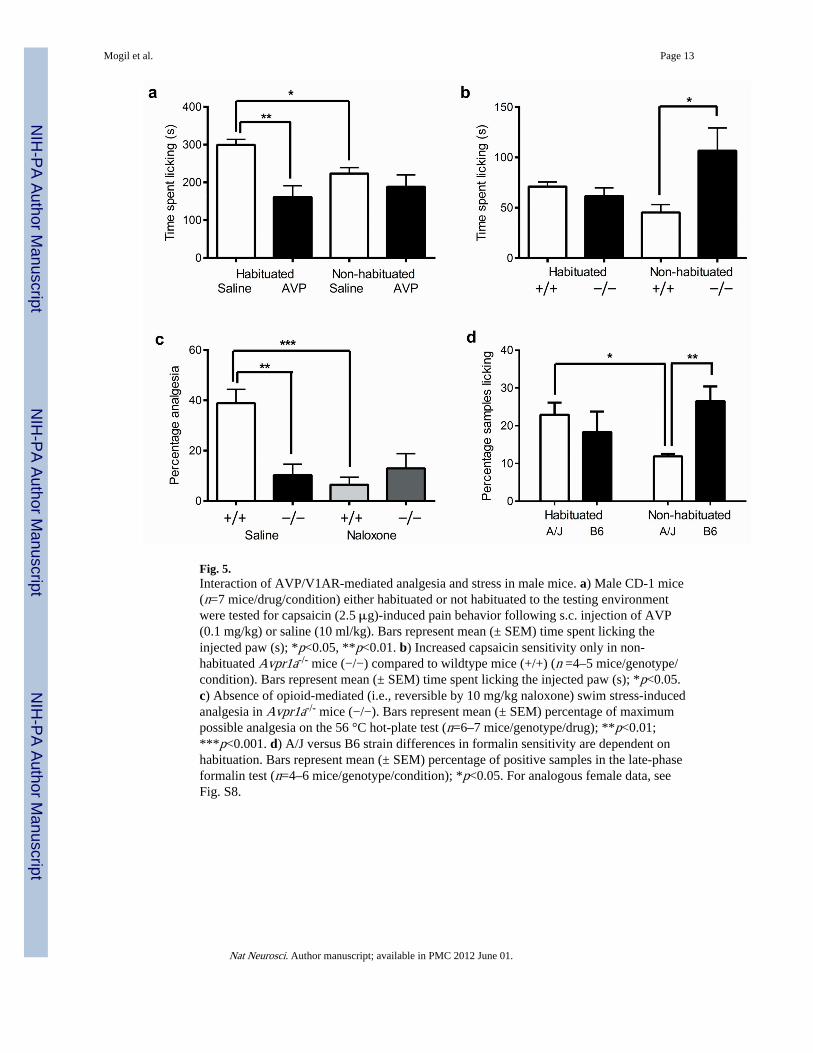
Fig. 5.Interaction of AVP/V1AR-mediated analgesia and stress in male mice. a) Male CD-1 mice(n=7 mice/drug/condition) either habituated or not habituated to the testing environmentwere tested for capsaicin (2.5 μg)-induced pain behavior following s.c. injection of AVP(0.1 mg/kg) or saline (10 ml/kg). Bars represent mean (± SEM) time spent licking theinjected paw (s); *p<0.05, **p<0.01. b) Increased capsaicin sensitivity only in non-habituated Avpr1a-/- mice (−/−) compared to wildtype mice (+/+) (n =4–5 mice/genotype/condition). Bars represent mean (± SEM) time spent licking the injected paw (s); *p<0.05.c) Absence of opioid-mediated (i.e., reversible by 10 mg/kg naloxone) swim stress-inducedanalgesia in Avpr1a-/- mice (−/−). Bars represent mean (± SEM) percentage of maximumpossible analgesia on the 56 °C hot-plate test (n=6–7 mice/genotype/drug); **p<0.01;***p<0.001. d) A/J versus B6 strain differences in formalin sensitivity are dependent onhabituation. Bars represent mean (± SEM) percentage of positive samples in the late-phaseformalin test (n=4–6 mice/genotype/condition); *p<0.05. For analogous female data, seeFig. S8.
Mogil et al. Page 13
Nat Neurosci. Author manuscript; available in PMC 2012 June 01.
NIH
-PA Author Manuscript
NIH
-PA Author Manuscript
NIH
-PA Author Manuscript
Copyright © 2022 FDOKUMEN