P2Y2 Nucleotide Receptors Expressed Heterologously in Sympathetic Neurons Inhibit Both N-Type Ca21...
10
P2Y 2 Nucleotide Receptors Expressed Heterologously in Sympathetic Neurons Inhibit Both N-Type Ca 21 and M-Type K 1 Currents Alexander K. Filippov, 1 Tania E. Webb, 2 Eric A. Barnard, 2 and David A. Brown 1 1 Department of Pharmacology, University College London, London WC1E 6BT, United Kingdom, and 2 Molecular Neurobiology Unit, Royal Free Hospital School of Medicine, London NW3 2PF, United Kingdom The P2Y 2 receptor is a uridine/adenosine triphosphate (UTP/ ATP)-sensitive G-protein-linked nucleotide receptor that previ- ously has been reported to stimulate the phosphoinositide signaling pathway. Messenger RNA for this receptor has been detected in brain tissue. We have investigated the coupling of the molecularly defined rat P2Y 2 receptor to neuronal N-type Ca 21 channels and to M-type K 1 channels by heterologous expression in rat superior cervical sympathetic (SCG) neurons. After the injection of P2Y 2 cRNA, UTP inhibited the currents carried by both types of ion channel. As previously reported [Filippov AK, Webb TE, Barnard EA, Brown DA (1997) Inhibition by heterologously expressed P2Y 2 nucleotide receptors of N-type calcium currents in rat sympathetic neurones. Br J Pharmacol 121:849–851], UTP inhibited the Ca 21 current (I Ca(N) ) by up to 64%, with an IC 50 of ;0.5 mM. We now find that UTP also inhibited the K 1 M current (I K(M) ) by up to 61%, with an IC 50 of ;1.5 mM. UTP had no effect on either current in neurons not injected with P2Y 2 cRNA. Structure–activity relations for the inhibition of I Ca(N) and I K(M) in P2Y 2 cRNA-injected neurons were similar, with UTP $ ATP . ITP .. GTP,UDP. However, coupling to these two channels involved different G-proteins: pretreat- ment with Pertussis toxin (PTX) did not affect UTP-induced inhibition of I K(M) but reduced inhibition of I Ca(N) by ;60% and abolished the voltage-dependent component of this inhibition. In unclamped neurons, UTP greatly facilitated depolarization- induced action potential discharges. Thus, the single P2Y 2 receptor can couple to at least two G-proteins to inhibit both Ca 21 N and K 1 M channels with near-equal facility. This implies that the P2Y 2 receptor may induce a broad range of effector responses in the nervous system. Key words: nucleotide receptors; uridine triphosphate; aden- osine triphosphate; sympathetic neurons; calcium currents; po- tassium currents; M currents Nucleotides such as ATP play a significant neurotransmitter role in the mammalian nervous system (Burnstock, 1972, 1990; Ed- wards and Gibb, 1993; Zimmermann, 1994). There are two fam- ilies of target nucleotide receptors known at the molecular level —ligand-gated P2X receptors and G-protein-coupled P2Y receptors (North and Barnard, 1997). The P2Y 2 receptor is a member of the family of P2Y G-protein-coupled receptors and is sensitive to both ATP and UTP; it was cloned originally from mouse NG108-15 neuroblas- toma X glioma hybrid cells (Lustig et al., 1993). In common with other P2Y receptors (Boarder et al., 1995), P2Y 2 receptors from different species all couple to the enzyme phospholipase C (PLC), thereby increasing inositol phosphate production and elevating intracellular [Ca 21 ] (Erb et al., 1993; Lustig et al., 1993; Parr et al., 1994; Rice et al., 1995; Chen et al., 1996; Nicholas et al., 1996). Messenger RNA for the P2Y 2 receptor, which is found in a range of tissues, is also present in the brain (Lustig et al., 1993), so the question arises as to what effect the activation of this receptor might have on neural f unction. In native NG108-15 cells, UTP inhibits two membrane ionic currents—an M-like K 1 cur- rent (“M-current”) and the voltage-gated C a 21 current (Filippov et al., 1994; Filippov and Brown, 1996). The former was to be expected because other receptors that activate PLC inhibit M-currents (Brown, 1988), but Ca 21 current inhibition was un- expected, especially because it was mediated (in part, at least) by a different G-protein from that responsible for M-current inhibi- tion (Filippov and Brown, 1996). This raised the question whether both effects actually were produced by the same receptor as that previously cloned from these cells or whether two different receptors were responsible. If the former, was this a peculiarity of this particular cell line, or would it also hold for primary neurons? To address these questions, we have expressed the recombinant rat P2Y 2 receptor in primary cultured rat superior cervical sym- pathetic (SCG) neurons by microinjecting cRNA, in the manner used by Ikeda et al. (1995) to express heterologous “metabo- tropic” glutamate receptors, and then recording the effects of activating these exogenous receptors with UTP on the N-type Ca 21 currents (I Ca(N) ; Hirning et al., 1988; Plummer et al., 1989; Regan et al., 1991) and M-type K 1 currents (I K(M) ; Constanti and Brown, 1981) that are present in these neurons. In prelimi- nary experiments (Filippov et al., 1997) we found that the acti- vation of these expressed receptors did indeed inhibit I Ca(N) . In the present experiments we have analyzed this action in more detail and have gone on to test whether the same single receptor also can inhibit the M-type K 1 current. We find that it can; the two currents are inhibited by UTP with near-equal potencies and efficacy, although mediated mainly by different G-proteins, and in joint response increase the excitability of these neurons. Thus, the P2Y 2 receptor appears to be unusually promiscuous in terms of its Received March 6, 1998; revised April 23, 1998; accepted April 28, 1998. This work was supported by The Wellcome Trust. We thank Brenda Browning and Misbah Malik for tissue culture. Correspondence should be addressed to Dr. Alexander K. Filippov, Department of Pharmacology, University College London, Gower Street, London WC1E 6BT, United Kingdom. Copyright © 1998 Society for Neuroscience 0270-6474/98/185170-10$05.00/0 The Journal of Neuroscience, July 15, 1998, 18(14):5170–5179
-
Upload
independent -
Category
Documents
-
view
2 -
download
0
Transcript of P2Y2 Nucleotide Receptors Expressed Heterologously in Sympathetic Neurons Inhibit Both N-Type Ca21...
P2Y2 Nucleotide Receptors Expressed Heterologously inSympathetic Neurons Inhibit Both N-Type Ca21 andM-Type K1 Currents
Alexander K. Filippov,1 Tania E. Webb,2 Eric A. Barnard,2 and David A. Brown1
1Department of Pharmacology, University College London, London WC1E 6BT, United Kingdom, and2Molecular Neurobiology Unit, Royal Free Hospital School of Medicine, London NW3 2PF, United Kingdom
The P2Y2 receptor is a uridine/adenosine triphosphate (UTP/ATP)-sensitive G-protein-linked nucleotide receptor that previ-ously has been reported to stimulate the phosphoinositidesignaling pathway. Messenger RNA for this receptor has beendetected in brain tissue. We have investigated the coupling ofthe molecularly defined rat P2Y2 receptor to neuronal N-typeCa21 channels and to M-type K1 channels by heterologousexpression in rat superior cervical sympathetic (SCG) neurons.After the injection of P2Y2 cRNA, UTP inhibited the currentscarried by both types of ion channel. As previously reported[Filippov AK, Webb TE, Barnard EA, Brown DA (1997) Inhibitionby heterologously expressed P2Y2 nucleotide receptors ofN-type calcium currents in rat sympathetic neurones. Br JPharmacol 121:849–851], UTP inhibited the Ca21 current(ICa(N) ) by up to 64%, with an IC50 of ;0.5 mM. We now find thatUTP also inhibited the K1
M current (IK(M) ) by up to 61%, with anIC50 of ;1.5 mM. UTP had no effect on either current in neurons
not injected with P2Y2 cRNA. Structure–activity relations for theinhibition of ICa(N) and IK(M) in P2Y2 cRNA-injected neurons weresimilar, with UTP $ ATP . ITP .. GTP,UDP. However, couplingto these two channels involved different G-proteins: pretreat-ment with Pertussis toxin (PTX) did not affect UTP-inducedinhibition of IK(M) but reduced inhibition of ICa(N) by ;60% andabolished the voltage-dependent component of this inhibition.In unclamped neurons, UTP greatly facilitated depolarization-induced action potential discharges. Thus, the single P2Y2
receptor can couple to at least two G-proteins to inhibit bothCa21
N and K1M channels with near-equal facility. This implies
that the P2Y2 receptor may induce a broad range of effectorresponses in the nervous system.
Key words: nucleotide receptors; uridine triphosphate; aden-osine triphosphate; sympathetic neurons; calcium currents; po-tassium currents; M currents
Nucleotides such as ATP play a significant neurotransmitter rolein the mammalian nervous system (Burnstock, 1972, 1990; Ed-wards and Gibb, 1993; Zimmermann, 1994). There are two fam-ilies of target nucleotide receptors known at the molecularlevel—ligand-gated P2X receptors and G-protein-coupled P2Yreceptors (North and Barnard, 1997).
The P2Y2 receptor is a member of the family of P2YG-protein-coupled receptors and is sensitive to both ATP andUTP; it was cloned originally from mouse NG108-15 neuroblas-toma X glioma hybrid cells (Lustig et al., 1993). In common withother P2Y receptors (Boarder et al., 1995), P2Y2 receptors fromdifferent species all couple to the enzyme phospholipase C (PLC),thereby increasing inositol phosphate production and elevatingintracellular [Ca21] (Erb et al., 1993; Lustig et al., 1993; Parr etal., 1994; Rice et al., 1995; Chen et al., 1996; Nicholas et al., 1996).
Messenger RNA for the P2Y2 receptor, which is found in arange of tissues, is also present in the brain (Lustig et al., 1993),so the question arises as to what effect the activation of thisreceptor might have on neural function. In native NG108-15 cells,UTP inhibits two membrane ionic currents—an M-like K1 cur-rent (“M-current”) and the voltage-gated Ca21 current (Filippov
et al., 1994; Filippov and Brown, 1996). The former was to beexpected because other receptors that activate PLC inhibitM-currents (Brown, 1988), but Ca21 current inhibition was un-expected, especially because it was mediated (in part, at least) bya different G-protein from that responsible for M-current inhibi-tion (Filippov and Brown, 1996). This raised the questionwhether both effects actually were produced by the same receptoras that previously cloned from these cells or whether two differentreceptors were responsible. If the former, was this a peculiarity ofthis particular cell line, or would it also hold for primary neurons?
To address these questions, we have expressed the recombinantrat P2Y2 receptor in primary cultured rat superior cervical sym-pathetic (SCG) neurons by microinjecting cRNA, in the mannerused by Ikeda et al. (1995) to express heterologous “metabo-tropic” glutamate receptors, and then recording the effects ofactivating these exogenous receptors with UTP on the N-typeCa21 currents (ICa(N) ; Hirning et al., 1988; Plummer et al., 1989;Regan et al., 1991) and M-type K1 currents (IK(M) ; Constantiand Brown, 1981) that are present in these neurons. In prelimi-nary experiments (Filippov et al., 1997) we found that the acti-vation of these expressed receptors did indeed inhibit ICa(N). Inthe present experiments we have analyzed this action in moredetail and have gone on to test whether the same single receptoralso can inhibit the M-type K1 current. We find that it can; thetwo currents are inhibited by UTP with near-equal potencies andefficacy, although mediated mainly by different G-proteins, and injoint response increase the excitability of these neurons. Thus, theP2Y2 receptor appears to be unusually promiscuous in terms of its
Received March 6, 1998; revised April 23, 1998; accepted April 28, 1998.This work was supported by The Wellcome Trust. We thank Brenda Browning
and Misbah Malik for tissue culture.Correspondence should be addressed to Dr. Alexander K. Filippov, Department
of Pharmacology, University College London, Gower Street, London WC1E 6BT,United Kingdom.Copyright © 1998 Society for Neuroscience 0270-6474/98/185170-10$05.00/0
The Journal of Neuroscience, July 15, 1998, 18(14):5170–5179

coupling to mammalian neuronal G-proteins and ion channels.This has interesting implications for its potential function innerve cells.
MATERIALS AND METHODScRNA preparation. The rat P2Y2 receptor cDNA was obtained from Dr.Zeng-Ping Chen (Department of Neuroscience and Cell Biology, Uni-versity of Medicine and Dentistry of New Jersey, Robert Wood JohnsonMedical School, Piscataway, NJ). A 2 kb EcoRI–XhoI fragment wascloned into the reciprocal sites of pBK/CMV. The wild-type greenfluorescent protein (GFP) cDNA, in pBluescript, also was used. Super-coiled plasmid DNA was prepared from both constructs and linearizedwith XhoI for P2Y2 and with EcoRI for GFP before use as a template forcRNA synthesis. Capped cRNA was transcribed with T3 polymerase(Message Machine, Ambion, Austin, TX), and an aliquot of each wasanalyzed on a denaturing agarose gel to check its integrity beforepolyadenylation, using poly(A 1) polymerase (Sigma, St. Louis, MO)according to the manufacturer’s recommendations. After extractions inturn with phenol, phenol /chloroform, and chloroform and isopropanolprecipitation, the cRNA was stored as an ethanol precipitate at 270°Cbefore use.
Neuron preparation and cRNA injection. Single SCG neurons weredissociated from rats aged 15–19 d, and plated on poly-D-lysine-coatedglass coverslips bordered by 2 cm plastic rings as previously described(Marrion et al., 1987). At 4 hr after plating, the neurons were microin-jected with an equal mixture of cRNA for the P2Y2 receptor (finalpipette concentration of 0.5 or 1.25 mg/ml dissolved in water) and cRNAfor GFP (used as a marker for foreign cRNA expression; Marshall et al.,1995) or, for controls, with GFP cRNA alone. cRNA solution (1.2 ml) wasloaded into prepulled high-resistance (;30 MV) Pyrex glass pipettes andinjected manually into single neurons by the application of gentle pres-sure to the back of the pipette with a syringe as described previously forinjection of antisera (Caulfield et al., 1994). Successful injection usuallyresulted in a ;10% increase in cell volume (Ikeda et al., 1995). Afterinjections, cells were incubated for 14–24 hr in a humidified incubator(5% CO2 /95% O2 ) at 37°C. Injected neurons that successfully expressedcRNA were identified as bright fluorescent cells, using an invertedmicroscope (Diaphot 200, Nikon, Tokyo, Japan) equipped with an epi-fluorescent N B2E block (Nikon). When required, Pertussis toxin (PTX)at a final concentration of 500 ng/ml was added to the culture media 1–2hr after neuron injection. Electrophysiological recordings were made14–24 hr after injection at room temperature (20°C). Some experimentson neuron excitability were made at 34°C.
Ca 21 channel current recording. Currents through voltage-gated Ca 21
channels were recorded by the conventional whole-cell patch-clampmethod as described previously (Caulfield et al., 1994). Cells weresuperfused (20–25 ml/min) with a solution consisting of (in mM) 120tetraethylammonium chloride, 3 KCl, 1.5 MgCl2 , 5 BaCl2 (or 5 CaCl2 ),10 HEPES, and 11.1 glucose plus 0.5 mM tetrodotoxin. The pH wasadjusted to 7.35 with NaOH. Patch electrodes (2–3 MV) were filled witha solution containing (in mM) 110 CsCl, 3 Mg Cl2 , 40 HEPES, 3 EGTA,2 Na2ATP, and 0.5 Na2GTP (pH-adjusted to 7.4 with CsOH). Neuronswere voltage-clamped with a discontinuous (“switching”) amplifier (Ax-oclamp 2B) with a sampling voltage at 6–8 kHz (50% duty cycle).Commands were generated via a Digidata 1200 interface, using pClamp6 computer software (Axon Instruments, Foster City, CA). Ca 21 channelcurrents were evoked routinely every 20 sec with a 100 msec depolarizingrectangular test pulse to 0 mV from a holding potential of 290 mV. Toobtain current–voltage ( I–V) relations, we evoked currents by test pulsesin 10 mV increments to 140 mV, starting from the holding potential of290 mV. Where required, I–V relations were obtained by using 750 msecramp depolarizations from 290 to 1 40 mV (see Docherty et al., 1991).Currents were digitized and stored on a computer for later analysis bypClamp 6 software (Axon Instruments). Ca 21 channel current ampli-tudes were measured isochronally 10 msec from the onset of the rectan-gular test pulse (Ikeda et al., 1995), i.e., near to the peak of the controlcurrent. To eliminate leak currents, we substituted Co 21 for Ca 21 andBa 21 in the external solution at the end of each experiment to block allCa 21 channel currents, and we digitally subtracted the residual currentfrom the corresponding currents in Ca 21 or Ba 21 solution.
M-type K 1 current recordings. Whole-cell M-currents (IK(M) ) wererecorded by the perforated patch-clamp method (Horn and Marty, 1988),as described for the application to SCG neurons by Caulfield et al.(1994). Briefly, patch pipettes (2–4 MV) were filled by dipping the tip
into a filtered solution containing (in mM) 90 potassium acetate, 20 KCl,3 MgCl2 , 40 HEPES, and 0.1 BAPTA (pH-adjusted to 7.4 by KOH) for20–60 sec. Then the pipette was back-filled with the same solutioncontaining 0.125 mg/ml amphotericin B as the permeabilizing agent (Raeet al., 1991). Access resistance after permeabilization was 8–15 MV.Neurons were superfused (20–25 ml/min) with external modified Krebs’solution containing (in mM) 120 NaCl, 3 KCl, 1.5 MgCl2 , 2.5 CaCl2 , 10HEPES, and 11.1 glucose (pH-adjusted to 7.3 with NaOH). Neuronswere voltage-clamped at 220 or 230 mV with a switching amplifier, andM-currents were deactivated with 1 sec hyperpolarizing steps at 5 secintervals. I–V relationships were obtained by using incremental voltagesteps of 10 mV between 210 and 2100 mV; currents were measured atthe end of each hyperpolarizing step. For dose–response curves, currentswere measured at 230 mV from steady-state I–V relations obtained byusing a ramp voltage command of 20 sec from 220 to 290 mV. The leakcomponent of current was estimated in both cases by extrapolating alinear fit to the I–V relationship from the negative potential region, whereonly ohmic currents were observed. All commands, current recordings,and analyses were made with Digidata 1200 interface and pClamp 6software (Axon Instruments).
Statistical analysis. Data are presented as mean 6 SEM as appropriate.Student’s t test (unpaired) was applied to determine statistical signifi-cance. The difference was considered significant if p # 0.05. Dose–response curves were determined by using concentrations that wereadded cumulatively, with 1 min exposure times. Curves were fit (usingOrigin 4.1 software) to pooled data points to the Hill equation: y 5ymax z x n H/(x n H 1 K n H), where y 5 the observed percentage of inhibition,ymax 5 extrapolated maximal percentage of inhibition, x 5 nucleotideconcentration (mM), K 5 IC50 (mM), and nH 5 the Hill coefficient.
Chemicals. UTP rather than ATP was used as the main agonistthroughout to preclude the activation of ATP-sensitive endogenous P2Xligand-gated channels (Cloues et al., 1993). Drugs were applied to theexternal solution by bath perfusion (bath exchange rate #5 sec). Tetro-dotoxin was obtained from Calbiochem (La Jolla, CA); uridine 59-triphosphate (UTP) was from Pharmacia Biotech (Uppsala, Sweden) andfrom Sigma; ATP, inosine 59-triphosphate (ITP), guanosine 59-triphosphate (GTP), acetylcholine chloride, (2)-norepinephrine bitar-trate, nifedipine, BAPTA, and amphotericin B were all from Sigma;adenosine 59-diphosphate (ADP) and uridine 59-diphosphate (UDP)were from Sigma and Boehringer Mannheim GmbH (Mannheim, Ger-many); hexokinase was from Boehringer Mannheim GmbH;oxotremorine-M (OxoM) was from Research Biochemicals (Natick,MA); Pertussis toxin (PTX) was from Porton Products (Dorset, UK);CdCl2 (AnalaR grade) was from BDH Chemicals (Poole, UK); BaCl2and CsCl were from Aldrich (Milwaukee, WI). Nifedipine was preparedas a stock solution (10 mM) in ethanol and protected from light duringstorage and use.
RESULTSCa21 channel current inhibitionWe have reported previously that, in SCG neurons preinjectedwith 1.25 mg/ml P2Y2 cRNA (together with GFP cRNA), theapplication of UTP produced a reversible inhibition of the Ca 21
channel current by up to 64.0 6 0.8%, with an IC50 of 0.50 6 0.03mM and that, at 0.5 mg/ml P2Y2 cRNA, UTP inhibited the currentby up to 50.2 6 0.6%, with an IC50 0.90 6 0.05 mM (Filippov et al.,1997). Because no significant inhibition was detected on applying100 mM UTP to neurons injected with GFP cRNA alone, thiseffect could be attributed entirely to the activation of newlyexpressed P2Y2 receptors and not to the activation of any endog-enous UTP-sensitive receptors that might have been present (seebelow).
Voltage dependenceCa21 current inhibition in SCG neurons produced by activationof some endogenous receptors (see Hille, 1994) or by heterolo-gously expressed mGluR2 receptors (Ikeda et al., 1995) isvoltage-dependent—that is, it is reduced at depolarized com-mands (or by predepolarization; Grassi and Lux, 1989) and isaccompanied by “kinetic slowing” resulting from time-dependent
Filippov et al. • Ion Channel Modulation by Cloned P2Y2 Receptors J. Neurosci., July 15, 1998, 18(14):5170–5179 5171

relief of block during the depolarizing command (Bean, 1989).Figure 1 shows that the block produced by activating heterologouslyexpressed P2Y2 receptors shares this property. Thus, current–voltage curves constructed by using either stepped (Fig. 1A,B) orramped (Fig. 1C) commands showed a greater inhibition by UTP atnegative potentials than at positive potentials (resulting in a positiveshift of the current peak). For example, in Figure 1A peak currentinhibition was reduced from ;63% at 0 mV to ;26% at 140 mV.Also, as shown in Figure 1, B and D, current activation was slowedin the presence of UTP, such that inhibition was less at the end of the40 msec command than at the beginning. Finally, inhibition wasreduced (from 65.0 6 3.1 to 24.9 6 5.4%) and the slowing of currentactivation was abolished when the test command was preceded by a20 msec depolarizing prepulse to 1120 mV (Fig. 1D). Such effectshave been interpreted to indicate that the activated G-protein (prob-ably the bg-subunit; Herlitze et al., 1996; Ikeda, 1996; Delmas et al.,
1998a,b) interacts directly with the Ca21 channel protein to inducea gating shift (Dolphin, 1995; Jones and Elmslie, 1997).
Pertussis toxin distinguishes two pathways for P2Y2-mediatedCa21 channel current inhibitionVoltage-dependent Ca21 current inhibition of the type illustratedin Figure 1 is usually (although not invariably; Ehrlich andElmslie, 1995) associated with activation by the receptor of aG-protein of the Pertussis toxin-sensitive Gi /Go family (Hille,1994). We tested whether this applied to expressed P2Y2 recep-tors by overnight incubation of injected neurons with 0.5 mg/mlPertussis toxin (PTX). In cells preinjected with 1.25 mg/ml P2Y2
cRNA, PTX substantially (;61%) but incompletely reduced theinhibition produced by 10 mM UTP (Fig. 2A). In contrast, in thesame neurons the inhibition produced by 10 mM norepinephrine(which is mediated primarily by Go; Caulfield et al., 1994; Del-
Figure 1. Voltage-dependent inhibition of Ca 21 channel currents by UTP in rat SCG neurons expressing heterologous P2Y2 receptors. Neurons werepreinjected with 1.25 mg/ml P2Y2 cRNA. Records show leak-subtracted Ca 21 channel currents recorded at room temperature (20°C), using thewhole-cell (ruptured patch) variant of the patch-clamp technique with 5 mM Ba 21 as the charge carrier (IBa ; see Materials and Methods). A, IBa amplitudeplotted against membrane voltage (I–V relationship) in the absence ( filled circles) and presence ( filled squares) of 100 mM UTP. Currents were evokedby 10 mV incrementing test pulses, starting from a holding potential of 290 to 140 mV; amplitudes were measured 10 msec from the onset of the testpulse. B, Superimposed currents from A at 0 and 140 mV test potentials in the absence and presence of UTP. Note that the inhibition is less at 140than at 0 mV. C, Superimposed currents recorded with 750 msec depolarizing voltage ramps from 290 to 1 40 mV (see Materials and Methods) in theabsence and presence of 100 mM UTP. Note that the inhibition is less at more positive voltages. D, Superimposed currents (lower traces) recorded witha double-pulse voltage protocol (upper traces) in the absence and presence of 100 mM UTP. The current was recorded first with a 40 msec test pulse to0 mV; then, after a 2 sec interval, a 25 msec conditioning prepulse to 1120 mV was applied, followed 4 msec later by a second 40 msec test pulse to 0mV. The bar chart at the bottom shows the mean percentage of current inhibition (measured after 10 msec at 0 mV command potential) by 100 mM UTPbefore (Prep.0) and after (Prep.120) the 1120 mV prepulse. Error bars show SEM; n 5 number of cells. Note that the inhibition is much less after theprepulse. Note also that the prepulse abolished the slowing of the current onset at 0 mV produced by UTP.
5172 J. Neurosci., July 15, 1998, 18(14):5170–5179 Filippov et al. • Ion Channel Modulation by Cloned P2Y2 Receptors

mas et al., 1998a) was reduced by .90%, as previously reported(Beech et al., 1992; Chen and Schofield, 1993; Caulfield et al.,1994; Delmas et al., 1998a), thus indicating the effectiveness ofthe PTX treatment. Nevertheless, because the initial inhibitionproduced by UTP exceeded that produced by norepinephrine, wewere concerned that the substantial component of PTX-resistantinhibition might have resulted from overexpression (and aberrantcoupling) of the P2Y2 receptors. This appeared not to be the case,because PTX produced a comparable degree of attenuation(254%) of the response to UTP in cells preinjected with 0.5mg/ml P2Y2 cRNA, although the initial inhibition produced byUTP (41.6 6 5.5%) was now less than that produced by norepi-nephrine (46.1 6 4.2%; Fig. 2D).
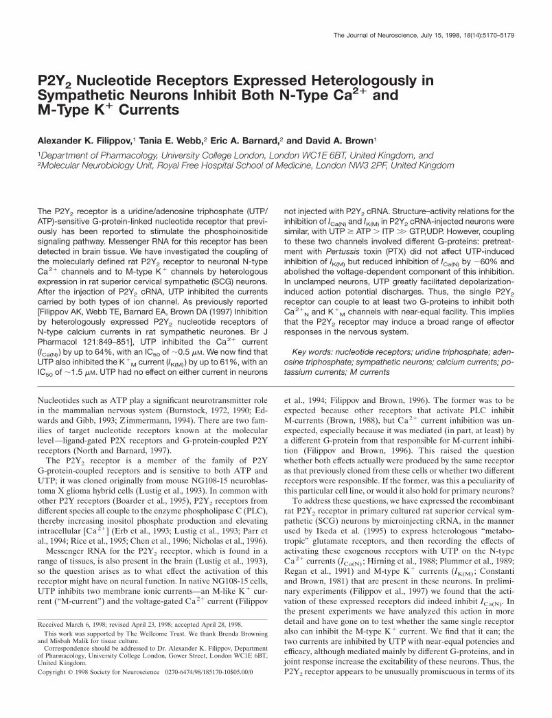
In contrast to the partial antagonism of overall inhibition, PTXpretreatment virtually eliminated the voltage dependence ofP2Y2-mediated inhibition; the characteristic slowing of currentactivation by UTP (compare with Fig. 1) was no longer apparentafter PTX treatment (Fig. 3A), and the 1120 mV depolarizingprepulse did not reverse the inhibition significantly (Fig. 3B,C).Thus, the PTX-insensitive component of block also appeared tobe voltage-insensitive.
One possible explanation for the PTX-resistant voltage-insensitive component of inhibition is that it reflects the inhibitionof an L-type current, rather than the N-type current (Hille, 1994).This possibility is enhanced by previous observations that UTPinhibits both L-type and N-type currents in NG108-15 cells(Filippov and Brown, 1996). To check this, we tested the effect of2–10 mM nifedipine on the response to UTP of PTX-pretreatedSCG neurons preinjected with 1.25 mg/ml P2Y2 cRNA. In agree-ment with previous observations on non-PTX-treated cells (Fil-ippov et al., 1997), nifedipine itself did not produce any significantinhibition of the current (22.5 6 1.7%; n 5 13), nor did it affectthe inhibitory action of UTP (2nifedipine, 229.4 6 3.5%; 1ni-fedipine, 227.3 6 4.25%; n 5 6). Thus, the residual PTX-insensitive block is not attributable to the inhibition of L-typechannels.
Coupling of P2Y2 receptors to M-type K1 currentsM-currents are sustained voltage-gated K1 currents that areactivated when SCG neurons are depolarized above 270 mV(Constanti and Brown, 1981) (for review, see Brown, 1988). Theycan be inhibited by activating endogenous M1-muscarinic acetyl-choline receptors (Marrion et al., 1989; Bernheim et al., 1992),angiotensin receptors (Shapiro et al., 1994), or bradykinin recep-tors (Jones et al., 1995)—in all cases via PTX-insensitiveG-proteins (probably Gq ; see Caulfield et al., 1994; Jones et al.,1995).
There is some evidence for the presence of endogenous P2Yreceptors in intact SCGs [Connolly et al. (1993); Boehm et al.(1995) and personal communication; Connolly and Harrison(1995); Von Kugelgen et al. (1997)]. However, we found that UTPdid not affect the M-current significantly in the uninjected disso-ciated neurons that we have used, although some other nucleo-tides did (see below and Fig. 7A). We therefore have testedwhether the activation of heterologously expressed P2Y2 recep-tors inhibits the M-current in these neurons, and, if so, how thiscompares with inhibition of Ca21 current.
Figure 4 illustrates the effect of UTP on M-currents in cellspreinjected with 1.25 mg/ml P2Y2 cRNA. The cell was predepo-larized to 220 mV to preactivate the M-current and then hyper-polarized in steps of 210 mV for 1 sec each to deactivate thecurrent; deactivation is signaled by the inward tail currents, whichreverse at EK (approximately 290 mV). UTP (10 mM) clearlyinhibited the M-current; this is indicated by (1) the inward shift inholding current at 220 mV (reflecting the reduction in outwardK1 current), (2) reduced current responses to depolarizing steps(reduced conductance) and the loss of M-current deactivationtails, and (3) reduced outward rectification in the current–voltagecurve positive to 270 mV, with no change in slope negative to270 mV (indicating no change in “leak” current).
M-current inhibition was quantitated by using voltage ramps toobtain current–voltage curves (see Fig. 6) and then subtractingfrom the total outward current at 230 mV the extrapolated linearleak currents from potentials negative to 270 mV. UTP (10 mM)inhibited the M-current by 60.8 6 9.6% (n 5 8) in neuronspreinjected with 1.25 mg/ml P2Y2 cRNA. As noted above, UTPhad no significant effect at 100 mM (0.6 6 0.4% inhibition) in fivenoninjected cells (Fig. 5A). In four of these same cells the acti-vation of the endogenous muscarinic acetylcholine receptors with10 mM oxotremorine-M (OxoM) inhibited the current by 55.4 611.4%, in accordance with previous observations (see Caulfield etal., 1994), indicating that the endogenous transduction machinerywas intact.
With the use of increasing concentrations of UTP, mean IC50
values and extrapolated maximum inhibitions were 1.49 6 0.18mM and 48.0 6 1.2%, respectively, after 1.25 mg/ml P2Y2 cRNAwas injected, and 2.41 6 0.43 mM and 43.2 6 1.9% after 0.5 mg/mlP2Y2 cRNA was injected (Fig. 6). These IC50 values are approx-imately three times higher than those determined for N-typeCa21 current inhibition (Filippov et al., 1997) (superimposedcurves in Fig. 6). The lower apparent maximum inhibition withincreasing UTP concentrations than those in Figure 5 may reflectsome degree of desensitization (apparent as a slow partial recov-ery of M-current during prolonged application of UTP); theextent to which this affected IC50 estimates is unclear.
In contrast to its effect on Ca 21 current inhibition, PTXproduced no significant attenuation of UTP-induced M-currentinhibition (Fig. 5B). Also, inhibition of M-current showed no
Figure 2. Pertussis toxin (PTX ) distinguishes two pathways for P2Y2-mediated Ca 21 channel current inhibition. The bar charts show the meaninhibition of IBa amplitude by 10 mM UTP and by 10 mM norepinephrinein neurons pretreated with PTX (0.5 mg/ml, overnight; 1PTX ) and inPTX-untreated neurons (Control ). Error bars show SEM; n 5 number ofcells. Neurons were injected with 1.25 mg/ml P2Y2 cRNA (A) or 0.5 mg/mlP2Y2 cRNA (B). Currents were recorded by stepping for 100 msec from290 to 0 mV and measured 10 msec from the onset of the test pulse. Notethat PTX pretreatment completely prevented the effect of norepineph-rine, but not that of UTP.
Filippov et al. • Ion Channel Modulation by Cloned P2Y2 Receptors J. Neurosci., July 15, 1998, 18(14):5170–5179 5173
clear voltage dependence in that neither the current–voltagecurve nor the kinetics of the deactivation relaxations was alteredappreciably in the presence of UTP.
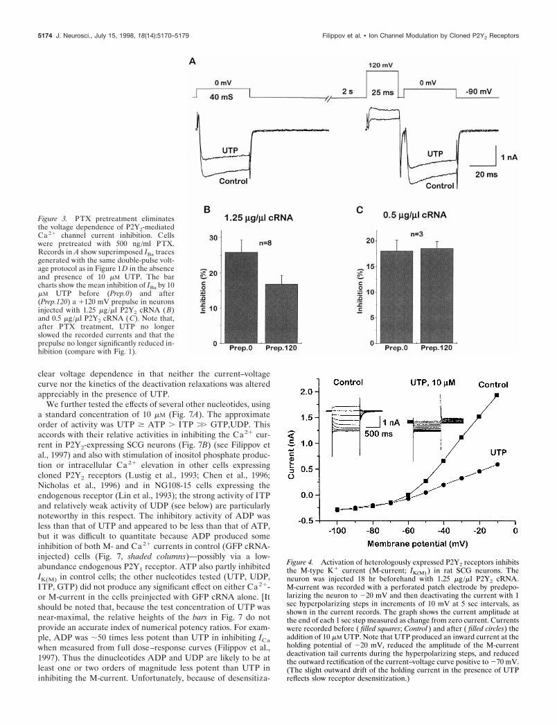
We further tested the effects of several other nucleotides, usinga standard concentration of 10 mM (Fig. 7A). The approximateorder of activity was UTP $ ATP . ITP .. GTP,UDP. Thisaccords with their relative activities in inhibiting the Ca21 cur-rent in P2Y2-expressing SCG neurons (Fig. 7B) (see Filippov etal., 1997) and also with stimulation of inositol phosphate produc-tion or intracellular Ca 21 elevation in other cells expressingcloned P2Y2 receptors (Lustig et al., 1993; Chen et al., 1996;Nicholas et al., 1996) and in NG108-15 cells expressing theendogenous receptor (Lin et al., 1993); the strong activity of ITPand relatively weak activity of UDP (see below) are particularlynoteworthy in this respect. The inhibitory activity of ADP wasless than that of UTP and appeared to be less than that of ATP,but it was difficult to quantitate because ADP produced someinhibition of both M- and Ca 21 currents in control (GFP cRNA-injected) cells (Fig. 7, shaded columns)—possibly via a low-abundance endogenous P2Y1 receptor. ATP also partly inhibitedIK(M) in control cells; the other nucleotides tested (UTP, UDP,ITP, GTP) did not produce any significant effect on either Ca21-or M-current in the cells preinjected with GFP cRNA alone. [Itshould be noted that, because the test concentration of UTP wasnear-maximal, the relative heights of the bars in Fig. 7 do notprovide an accurate index of numerical potency ratios. For exam-ple, ADP was ;50 times less potent than UTP in inhibiting ICa
when measured from full dose–response curves (Filippov et al.,1997). Thus the dinucleotides ADP and UDP are likely to be atleast one or two orders of magnitude less potent than UTP ininhibiting the M-current. Unfortunately, because of desensitiza-
Figure 4. Activation of heterologously expressed P2Y2 receptors inhibitsthe M-type K 1 current (M-current; IK(M) ) in rat SCG neurons. Theneuron was injected 18 hr beforehand with 1.25 mg/ml P2Y2 cRNA.M-current was recorded with a perforated patch electrode by predepo-larizing the neuron to 220 mV and then deactivating the current with 1sec hyperpolarizing steps in increments of 10 mV at 5 sec intervals, asshown in the current records. The graph shows the current amplitude atthe end of each 1 sec step measured as change from zero current. Currentswere recorded before ( filled squares; Control ) and after ( filled circles) theaddition of 10 mM UTP. Note that UTP produced an inward current at theholding potential of 220 mV, reduced the amplitude of the M-currentdeactivation tail currents during the hyperpolarizing steps, and reducedthe outward rectification of the current–voltage curve positive to 270 mV.(The slight outward drift of the holding current in the presence of UTPreflects slow receptor desensitization.)
Figure 3. PTX pretreatment eliminatesthe voltage dependence of P2Y2-mediatedCa 21 channel current inhibition. Cellswere pretreated with 500 ng/ml PTX.Records in A show superimposed IBa tracesgenerated with the same double-pulse volt-age protocol as in Figure 1D in the absenceand presence of 10 mM UTP. The barcharts show the mean inhibition of IBa by 10mM UTP before (Prep.0) and after(Prep.120) a 1120 mV prepulse in neuronsinjected with 1.25 mg/ml P2Y2 cRNA (B)and 0.5 mg/ml P2Y2 cRNA ( C). Note that,after PTX treatment, UTP no longerslowed the recorded currents and that theprepulse no longer significantly reduced in-hibition (compare with Fig. 1).
5174 J. Neurosci., July 15, 1998, 18(14):5170–5179 Filippov et al. • Ion Channel Modulation by Cloned P2Y2 Receptors

tion and slow recovery, it was not possible to construct fulldose–response curves for the inhibitory action of the weakeranalogs on the M-current.]
To check whether the effects of UDP and ADP in cRNA P2Y2
preinjected cells arise from possible contamination of these nu-cleotides by UTP and ATP (see Nicholas et al., 1996), we per-formed such tests by using purified UDP and ADP from Boehr-inger Mannheim (99% pure) immediately after their dissolutionin the medium. In addition, we used samples of UDP and ADPpretreated for 1 hr with hexokinase (1 mM UDP or ADP incu-bated with 10 U/ml hexokinase plus 22 mM glucose at 37°C); thisshould convert any UTP and ATP that was present to theirdiphosphates (Nicholas et al., 1996). The hexokinase pretreat-ment did not significantly reduce the inhibitory effects of 10 mM
UDP or ADP, and the pure UDP and ADP produced inhibitionswithin the range of those observed with the same nucleotidesfrom Sigma.
Effects on excitabilityThe inhibition of M-type K1 currents and N-type Ca21 currentsthrough endogenous G-protein-coupled receptors increases theexcitability of SCG neurons. This occurs because the M-currentitself acts as a “braking” current on action potential discharges(see Brown, 1988), and the entry of Ca 21 through N-type Ca21
channels opens Ca21-activated K1 channels and thereby inducesa long-lasting afterhyperpolarization (AHP), which further limitssubsequent spike activity [see Davies et al. (1996) and referencestherein]. Thus, the activation of expressed P2Y2 receptors alsomight be expected to enhance spike activity.
Figure 8 shows that this was the case. In Figure 8A, the cellswere challenged with long depolarizing or hyperpolarizing cur-rent injections from a preset potential of 260 mV at roomtemperature, at 20°C (Fig. 8Aa), and at 34°C (Fig. 8Ab). The longdepolarizing current induced a brief burst of two to three spikes,followed by silence. The application of UTP had no effect on thisspike discharge in control cells preinjected with GFP cRNA butgreatly prolonged the discharge in cells preinjected with P2Y2
cRNA. This effect of UTP was increased dramatically at 34°C(Fig. 8Ab); this occurs probably because M-current kinetics arefaster at 34°C (Brown, 1988), thereby exerting a faster and moreeffective “brake” on firing (see Cuevas et al., 1997). UTP alsoincreased the voltage response to hyperpolarizing current pulses(Fig. 8Ab), as expected after M-current inhibition (see Adams etal., 1982). These effects closely resemble the effect of activatingendogenous muscarinic receptors in SCG neurons (see Brownand Constanti, 1980).
Figure 8B shows the effect of stimulating expressed P2Y2
receptors on the Ca 21-activated afterhyperpolarization that fol-lows an action potential: UTP abbreviated the afterhyperpolar-ization in a manner resembling the effect of stimulating endoge-nous a2-adrenergic receptors with norepinephrine (Horn andMcAfee, 1980), which results from the inhibition of the N-typeCa21 current (Galvan and Adams, 1982; Schofield, 1990).
DISCUSSIONThe principal points emerging from these and the precedingexperiments (Filippov et al., 1997) are that the recombinant rat
Figure 5. M-current inhibition by UTP requires heterologous P2Y2receptor expression (A) and is not prevented by PTX (B). The bar chartin A shows the mean percentage of inhibition of the M-current at 230 mV(see Materials and Methods) by 10 mM UTP in neurons injected with 1.25mg/ml P2Y2 cRNA (injected) and in uninjected neurons. Current inhibi-tion by 10 mM oxotremorine-M (OxoM ) in uninjected neurons is shownfor comparison. Error bars show SEM; n 5 number of cells tested.The bar chart in B shows the mean percentage of inhibition of theM-current by 10 mM UTP at 230 mV in neurons preinjected with P2Y2cRNA without (Control ) or with (1PTX ) overnight pretreatment with 0.5mg/ml PTX.
Figure 6. Concentration dependence for UTP inhibition of M-current(solid lines) and N-type Ca 21 channel current (dashed lines) in cellspreinjected with 1.25 mg/ml P2Y2 cRNA (circles) or 0.5 mg/ml P2Y2 cRNA(triangles). M-current was recorded by using a voltage ramp protocol(inset) and was measured at 230 mV (see Materials and Methods). Thepoints show the mean 6 SEM of measurements in three to four cells;concentrations were added cumulatively, with 1 min exposure times.Curves were fit to pooled data points, using Origin 4.1 software to the Hillequation: y 5 ymax z x nH/(x nH 1 K nH), where y 5 the observed percentageof inhibition, ymax 5 extrapolated maximal percentage of inhibition, x 5nucleotide concentration (mM), K 5 IC50 (mM), and nH 5 the Hillcoefficient. Values of constants (mean 6 SEM) for M-current inhibitionwere ymax 5 48.0 6 1.20%, K 5 1.49 6 0.14 mM, nH 5 1.29 6 0.14 forneurons injected with 1.25 mg/ml P2Y2 cRNA; and ymax 5 43.2 6 1.95%,K 5 2.41 6 0.43 mM, nH 5 0.88 6 0.11 for neurons injected with 0.5 mg/mlP2Y2 cRNA. Data for Ca 21 channel current inhibition (dashed lines) aretaken from Filippov et al. (1997) and are superimposed for comparison.(Values for constants were ymax 5 64.0 6 0.75%, K 5 0.50 6 0.03 mM, nH5 1.29 6 0.0714 for neurons injected with 1.25 mg/ml P2Y2 cRNA; andymax 5 50.2 6 0.61%, K 5 0.90 6 0.05 mM, nH 5 1.21 6 0.06 for neuronsinjected with 0.5 mg/ml P2Y2 cRNA).
Filippov et al. • Ion Channel Modulation by Cloned P2Y2 Receptors J. Neurosci., July 15, 1998, 18(14):5170–5179 5175
P2Y2 receptor can couple with near-equal facility to two quitedifferent neural ion channels—the N-type voltage-gated Ca21
channel and the M-type K1 channel—when expressed in rat SCGneurons and that this involves the intermediation of at least twodifferent G-proteins. Dual coupling to these particular channelsby one species of G-protein-linked receptor is unusual (see be-low) and suggests that the P2Y2 receptor can have a potentiallybroader range of effects on neurons than many other neurotrans-mitter receptors.
This apparent cross-talk is unlikely to be an artifact of receptoroverexpression, for two reasons. First, it was preserved afterinjections of two different amounts of the receptor cRNA, such
that, at the lower level (0.5 mg/ml), the maximum response toUTP was less than that obtained on stimulating endogenousadrenergic and muscarinic receptors. Thus, although (in the ab-sence of appropriate antibodies) we have not been able to mea-sure the number of receptors expressed, they are unlikely to be“excessive” in comparison to other endogenous serpentine recep-tors in these cells. Second, the IC50 values for UTP observed inthe present experiments (0.5–0.9 mM for Ca21 current inhibitionand 1.5–2.4 mM for M-current inhibition) are not dissimilar tothose observed for UTP to elevate intracellular [Ca21] [1.1 mM,Lustig et al. (1993); ;1.1 mM, Parr et al. (1994); 0.2 mM, Chen etal. (1996)] or to stimulate inositol phosphate production (;0.1mM; Nicholas et al., 1996) when applied to recombinant P2Y2
receptors expressed in other cell lines. (IC50 values likely will varyfrom one expression system to another, depending on the level ofreceptor expression, as indicated by the dose–response curves inFig. 6.) More pertinently, perhaps, the present IC50 values also
Figure 7. Mean effects of different nucleotides on M-current ( A) andCa 21 channel current (B). Nucleotides were applied at 10 mM. Open barsshow the mean percentage of inhibition in neurons preinjected with 1.25mg/ml P2Y2 cRNA; the shaded bars show the effects in control cells(preinjected with GFP cRNA, but without P2Y2 cRNA). Error bars showSEM; n 5 number of cells. Data for P2Y2 cRNA-injected cells in B wererecalculated from Filippov et al. (1997). Inhibition was measured asdescribed in Figures 2 and 6.
Figure 8. Activation of heterologously expressed P2Y2 receptors en-hances repetitive firing (A) and reduces the Ca 21-activated spike after-hyperpolarization (B) in SCG neurons. Aa, Voltage responses to depo-larizing current pulses (1 sec) recorded from a control (uninjected) SCGneuron (top panel, 0.4 nA pulse) and from a P2Y2 cRNA (1.25 mg/ml)preinjected neuron (middle panel, 0.1 nA pulse) before and during appli-cation of UTP at 20°C. Ab, Voltage responses to depolarizing (0.3 nA)and hyperpolarizing (20.2 nA) current pulses from P2Y2 cRNA (1.25mg/ml) preinjected neuron before and during the application of UTP at34°C. B, Afterhyperpolarization (AHP) that followed an action potentialevoked by a brief depolarizing current pulse (1 nA, 1 msec) recorded froma P2Y2 cRNA (1.25 mg/ml) preinjected neuron before and during theapplication of UTP at 34°C.
5176 J. Neurosci., July 15, 1998, 18(14):5170–5179 Filippov et al. • Ion Channel Modulation by Cloned P2Y2 Receptors

accord with those that follow the stimulation of the endogenousP2Y2 receptor in NG108-15 cells [;3 mM for inositol phosphateproduction (Lin, 1994); 0.8 mM for M-like current inhibition(Filippov et al., 1994); 2.8 mM for N-type Ca21 current inhibition(Filippov and Brown, 1996)]. The agreement between the resultsobtained on stimulating the endogenous receptors in this neuralcell line and the exogenously expressed receptors in SCG neuronsstrongly suggests that the effects we see are likely to be a generalphenomenon, applicable in other cells in which the same receptormight be expressed.
These divergent responses start at the level of the G-protein.M-current inhibition was insensitive to PTX; although we havenot positively identified the species of PTX-resistant G-protein(s)responsible, previous experiments that used site-directed anti-bodies on the analogous effects of muscarinic agonists (Caulfieldet al., 1994) and bradykinin (Jones et al., 1995) suggest that it ismost likely Gq and/or G11 (principally Gq; Haley et al., 1997). Incontrast, Ca21 current inhibition is mediated by at least twoG-proteins: a PTX-sensitive G-protein, responsible for the gatingshift and for ;60% of the peak inhibition measured at 0 mV, anda PTX-insensitive G-protein responsible for the residual voltage-insensitive component of inhibition. By analogy with the voltage-dependent effects of norepinephrine, somatostatin, and M4 mus-carinic stimulation, the former (PTX-sensitive) G-protein isprobably GoA (Caulfield et al., 1994; Delmas et al., 1998a,b), andthe gating shift probably results from the interaction of thedissociated free bg-subunits with the Ca21 channel protein (seeHerlitze et al., 1996; Ikeda, 1996; Delmas et al., 1998a,b). ThePTX-insensitive G-protein responsible for the voltage-insensitivecomponent of Ca21 current inhibition could well be the same asthat (Gq?) postulated to cause M-current inhibition, because M1
muscarinic acetylcholine receptor stimulation produces the samedual-effector response via Gq (Delmas et al., 1998b).
In effect, therefore, stimulating the P2Y2 receptor imitates thecombined effects of stimulating two separate endogenous musca-rinic acetylcholine receptors, the M1 and the M4 receptors (seeHille, 1994). This is summarized in Figure 9; at a minimum, itrequires parallel coupling of the P2Y2 receptor to two G-proteins:Gq (leading to inhibition of the M-current and voltage-independent inhibition of the Ca21 current) and Go (producing agating shift of the Ca21 channels).
There are other instances of “promiscuity” in receptor/G-protein coupling (see Milligan, 1997). This can result in effects onmore than one ion channel—for example, the inhibition of Ca21
currents and the activation of inward rectifier (Kir ) K1 currents(Surprenant et al., 1992). However, dual coupling to Ca 21
N andK1
M channels through two G-proteins by a single, defined recep-tor is more unusual. Thus, although many transmitters can, col-lectively, modulate Ca21 currents or M-currents in SCG neuronsin a manner similar to that produced by P2Y2 receptors (see Hille,1994), this usually involves the activation of separate receptors,each coupled to a different G-protein. As indicated already,muscarinic acetylcholine agonists can produce the same dualresponse as UTP, but to do so they have to activate two differentmolecular species of muscarinic receptor; activation of Go (andthe Ca21 channel gating shift) requires M4 receptors, whereasactivation of Gq (and consequential voltage-independent inhibi-tion of M-current and Ca21 current) requires M1 receptors (seeBeech et al., 1992; Delmas et al., 1998b). This also applies toother heterologous receptors expressed by this method. Thus,there is negligible cross-talk between heterologous mGluR1a andmGluR2 receptors expressed from cRNA injections; the former
inhibit M-currents via a PTX-insensitive G-protein with insignif-icant effect on the Ca21 current, whereas mGluR2 receptorsinhibit Ca21 currents entirely through a PTX-sensitive G-proteinwithout any effect on M-currents (Ikeda et al., 1995). Likewise,heterologously expressed cannabinoid receptors selectively in-hibit Ca21 currents through a PTX-sensitive G-protein (Pan etal., 1996). The closest analogy is provided by the action ofangiotensin, which also couples through two G-proteins (PTX-sensitive and insensitive) to inhibit Ca21 and M-currents (Sha-piro et al., 1994); however, it has not yet been established whetherboth coupling routes are activated by the same or different mo-lecular species of angiotensin receptor.
Physiological significanceFrom a functional viewpoint, the net effect of stimulating ex-pressed P2Y2 receptors in SCG neurons is to increase theirexcitability (see Fig. 8A). This is the expected result of inhibitingthe M-current and the N-type Ca21 current, because reducingthe latter will decrease the Ca21-dependent K1 current andabbreviate the spike afterhyperpolarization (as shown in Fig.8B), and KM and KCa currents act as synergistic braking currentson spike discharge (see Jones and Adams, 1987).
This is essentially a postsynaptic response; it mimics the naturalpostsynaptic effect of activating the endogenous muscarinic re-ceptors by synaptically released acetylcholine in SCG neurons(see Brown, 1988), so it would provide a mechanism for slowpostsynaptic excitation by a nucleotide transmitter. Although no
Figure 9. Schematic diagram representing dual coupling of P2Y2 recep-tor to Ca 21
N and K 1M channels via two different G-proteins. Solid lines,
Direct connections; dashed lines, indirect connections.
Filippov et al. • Ion Channel Modulation by Cloned P2Y2 Receptors J. Neurosci., July 15, 1998, 18(14):5170–5179 5177

such nucleotide-mediated synaptic responses have been reportedyet, comparable excitatory effects of exogenous UTP and ATPhave been described in, for example, frog sympathetic (Siggins etal., 1977; Adams et al., 1982; Akasu et al., 1983; Lopez andAdams, 1989), frog sensory (Tokimasa and Akasu, 1990), and ratintracardiac (Cuevas et al., 1997) ganglion cells. However, thespecies of P2Y receptor responsible for these effects has not beendetermined, nor is there yet any direct evidence for equivalenteffects on central neurons.
On the other hand, if endogenous P2Y2 receptors were locatedpresynaptically, then the most likely effect of their stimulationwould be to reduce transmitter release via the inhibition of theN-type Ca21 current, in the same manner as stimulating endog-enous presynaptic adrenergic or muscarinic receptors in SCGneurons (Boehm and Huck, 1996; Koh and Hille, 1997). Thiswould provide a mechanism for autoinhibition in nucleotide-releasing nerve terminals. There is some evidence for P2Y-mediated autoinhibition in peripheral sympathetic nerves (Fuderand Muth, 1993) and chromaffin cells (Currie and Fox, 1996);also, P2Y-mediated inhibition of norepinephrine release fromisolated brain tissue by exogenous nucleotides has been reported(Von Kugelgen et al., 1994), but the identity of these receptorshas not yet been established.
As pointed out in the introductory remarks, there is now directevidence for the synaptic release of ATP in the brain and theconsequent activation of postsynaptic P2X receptors (Edwardsand Gibb, 1993). Although there is, as yet, no evidence for thesynaptic release of UTP, the latter nucleotide can be releasedfrom cells by other mechanisms (Lazarowski et al., 1997). Be-cause mRNA for the P2Y2 receptor is expressed in the brain(Lustig et al., 1993), were this receptor to be activated by eitherof these endogenously released nucleotides, its dual couplingwould provide scope for some unusually divergent effects onneural signaling.
REFERENCESAdams PR, Brown DA, Constanti A (1982) Pharmacological inhibition
of the M-current. J Physiol (Lond) 332:223–262.Akasu T, Hirai K, Koketsu K (1983) Modulatory actions of ATP on
membrane potentials of bullfrog sympathetic ganglion cells. Brain Res258:313–317.
Bean BP (1989) Neurotransmitter inhibition of neuronal calcium cur-rents by changes in channel voltage dependence. Nature 340:153–156.
Beech DJ, Bernheim L, Hille B (1992) Pertussis toxin and voltage de-pendence distinguish multiple pathways modulating calcium channelsof rat sympathetic neurons. Neuron 8:97–106.
Bernheim L, Mathie A, Hille B (1992) Characterization of muscarinicreceptor subtypes inhibiting Ca 21 current and M current in rat sym-pathetic neurons. Proc Natl Acad Sci USA 89:9544–9548.
Boarder MR, Weisman GA, Turner JT, Wilkinson GF (1995)G-protein-coupled P2 purinoceptors: from molecular biology to func-tional response. Trends Pharmacol Sci 16:133–139.
Boehm S, Huck S (1996) Inhibition of N-type calcium channels: the onlymechanism by which presynaptic alpha 2-autoreceptors control sympa-thetic transmitter release. Eur J Neurosci 8:1924–1931.
Boehm S, Huck S, I lles P (1995) UTP- and ATP-triggered transmitterrelease from rat sympathetic neurons via separate receptors. Br JPharmacol 116:2241–2243.
Brown DA (1988) M-currents. In: Ion channels, Vol 1 (Narahashi T, ed),pp 55–94. New York: Plenum.
Brown DA, Constanti A (1980) Intracellular observations of the effectsof muscarinic agonists on rat sympathetic neurones. Br J Pharmacol70:593–608.
Burnstock G (1972) Purinergic nerves. Pharmacol Rev 24:509–581.Burnstock G (1990) Purinergic mechanisms. Ann NY Acad Sci
603:1–19.Caulfield MP, Jones S, Vallis Y, Buckley NJ, Kim G-D, Milligan G,
Brown DA (1994) Muscarinic M-current inhibition via Gaq/11 anda-adrenoceptor inhibition of Ca 21 current via Gao in rat sympatheticneurones. J Physiol (Lond) 477:415–422.
Chen C, Schofield GG (1993) Differential neuromodulation of calciumcurrents by norepinephrine in rat sympathetic neurons. J Neurophysiol70:1440–1449.
Chen ZP, Krull N, Xu S, Levy A, Lightman SL (1996) Molecularcloning and functional characterization of a rat pituitary G-protein-coupled adenosine triphosphate (ATP) receptor. Endocrinology137:1833–1840.
Cloues R, Jones S, Brown DA (1993) Zn 21 potentiates ATP-activatedcurrents in rat sympathetic neurons. Pflugers Arch 424:152–158.
Connolly GP, Harrison PJ (1995) Structure–activity relationships of apyrimidine receptor in the rat isolated superior cervical ganglion. Br JPharmacol 116:2764–2770.
Connolly GP, Harrison PJ, Stone TW (1993) Action of purine andpyrimidine nucleotides on the rat superior cervical ganglion. Br JPharmacol 110:1297–1304.
Constanti A, Brown DA (1981) M-currents in voltage-clamped mamma-lian sympathetic neurones. Neurosci Lett 24:289–294.
Cuevas J, Harper AA, Trequattrini C, Adams DJ (1997) Passive andactive membrane properties of isolate rat intracardiac neurons: regu-lation by H- and M-currents. J Neurophysiol 78:1890–1902.
Currie KP, Fox AP (1996) ATP serves as a negative feedback inhibitorof voltage-gated Ca 21 channel currents in cultured bovine adrenalchromaffin cells. Neuron 16:1027–1036.
Davies PJ, Ireland DR, McLachlan EM (1996) Sources of Ca 21 fordifferent Ca 21-activated K 1 conductances in neurones of the rat su-perior cervical ganglion. J Physiol (Lond) 495:353–366.
Delmas P, Brown DA, Dayrell M, Abogadie FC, Caulfield MP, BuckleyNJ (1998a) On the role of endogenous G-protein bg subunits inN-type Ca 21 current inhibition by neurotransmitters in rat sympatheticneurones. J Physiol (Lond) 506:319–329.
Delmas P, Abogadie FC, Dayrell M, Haley JE, Milligan G, Caulfield MP,Brown DA, Buckley NJ (1998b) G-proteins and G-protein subunitsmediating cholinergic inhibition of N-type calcium currents in sympa-thetic neurones. Eur J Neurosci 10:1654–1666.
Docherty RJ, Robbins J, Brown DA (1991) NG 108-15 cells neuroblas-toma X glioma cell line as a model neuronal system. In: CellularNeurobiology: a practical approach (Wheal H, Chad J, eds), pp 75–79.Oxford: IRL.
Dolphin AC (1995) Voltage-dependent calcium channels and their mod-ulation by neurotransmitters and G-proteins. Exp Physiol 80:1–36.
Edwards FA, Gibb AJ (1993) ATP—a fast neurotransmitter. FEBS Lett325:86–89.
Ehrlich I, Elmslie KS (1995) Neurotransmitters acting via differentG-proteins inhibit N-type calcium current by an identical mechanism inrat sympathetic neurons. J Neurophysiol 74:2251–2257.
Erb L, Lustig KD, Sullivan DM, Turner JT, Weisman GA (1993) Func-tional expression and photoaffinity labeling of a cloned P2U purinergicreceptor. Proc Natl Acad Sci USA 90:10449–10453.
Filippov AK, Brown DA (1996) Activation of nucleotide receptors in-hibits high-threshold calcium currents in NG108-15 neuronal hybridcells. Eur J Neurosci 8:1149–1155.
Filippov AK, Selyanko AA, Robbins J, Brown DA (1994) Activation ofnucleotide receptors inhibits M-type K current [IK(M)] in neuroblas-toma X glioma hybrid cells. Pflugers Arch 429:223–230.
Filippov AK, Webb TE, Barnard EA, Brown DA (1997) Inhibition byheterologously expressed P2Y2 nucleotide receptors of N-type calciumcurrents in rat sympathetic neurones. Br J Pharmacol 121:849–851.
Fuder H, Muth U (1993) ATP and endogenous agonists inhibit evoked[ 3H]-noradrenaline release in rat iris via A1 and P2Y-like purinocep-tors. Naunyn Schmiedebergs Arch Pharmacol 348:352–357.
Galvan, M, Adams PR (1982) Control of calcium current in rat sympa-thetic neurones by norepinephrine. Brain Res 244:135–144.
Grassi F, Lux HD (1989) Voltage-dependent GABA-induced modula-tion of Ca 21 channels in chick sensory neurons. Neurosci Lett105:113–119.
Haley JE, Delmas P, Abogadie FC, Dayrell M, Caulfield MP, Buckley NJ,Brown DA (1997) Muscarinic inhibition of the M-current is mediatedby the a subunit of Gq. J Physiol (Lond) 504P:176P.
Herlitze S, Garcia DE, Mackie K, Hille B, Scheuer T, Catterall WA(1996) Modulation of Ca 21 channels by G-protein bg subunits. Nature105:113–119.
5178 J. Neurosci., July 15, 1998, 18(14):5170–5179 Filippov et al. • Ion Channel Modulation by Cloned P2Y2 Receptors

Hille B (1994) Modulation of ion-channel function by G-protein-coupledreceptors. Trends Neurosci 17:531–536.
Hirning LD, Fox AP, McLeskey EW, Olivera BM, Thayer SA, Miller RJ,Tsien RW (1988) Dominant role of N-type Ca 21 channels in evokedrelease of norepinephrine from sympathetic neurons. Science239:57–61.
Horn JP, McAfee DA (1980) Alpha-adrenergic inhibition of calcium-dependent potentials in rat sympathetic ganglion cells. J Physiol (Lond)301:191–204.
Horn R, Marty A (1988) Muscarinic activation of ionic currents mea-sured by a new whole-cell recording method. J Gen Physiol 92:145–159.
Ikeda SR (1996) Voltage-dependent modulation of N-type calciumchannels by G-protein bg subunits. Nature 380:255–258.
Ikeda SR, Lovinger DM, McCool BA, Lewis DL (1995) Heterologousexpression of metabotropic glutamate receptors in adult rat sympa-thetic neurons: subtype-specific coupling to ion channels. Neuron14:1029–1038.
Jones SW, Adams PR (1987) The M-current and other potassium cur-rents of vertebrate neurons. In: Neuromodulation (Kaczmarek LK,Levitan IB, eds), pp 159–186. New York: Oxford UP.
Jones SW, Elmslie KS (1997) Transmitter modulation of neuronal cal-cium channels. J Membr Biol 155:1–10.
Jones SW, Brown DA, Milligan G, Willer E, Buckley NJ, Caulfield MP(1995) Bradykinin excites rat sympathetic neurons by inhibition of Mcurrent through a mechanism involving B2 receptors and Gaq/11. Neu-ron 14:399–405.
Koh D-S, Hille B (1997) Modulation by neurotransmitters of catechol-amine secretion from sympathetic ganglion neurons detected by am-perometry. Proc Natl Acad Sci USA 94:1506–1511.
Lazarowski ER, Homolya L, Boucher RC, Harden TK (1997) Directdemonstration of mechanically induced release of cellular UTP and itsimplication for uridine nucleotide receptor activation. J Biol Chem272:24348–24354.
Lin TA, Lustig KD, Sportiello MG, Weisman GA, Sun GY (1993)Signal transduction pathways coupled to a P2U receptor in neuroblas-toma X glioma (NG108-15) cells. J Neurochem 60:1115–1125.
Lin W-W (1994) Heterogeneity of nucleotide receptors in NG108-15neuroblastoma and C6 glioma cells for mediating phosphoinositideturnover. J Neurochem 62:536–542.
Lopez HS, Adams PR (1989) A G-protein mediates the inhibition of thevoltage-gated potassium M-current by muscarine, LHRH, substance P,and UTP in bullfrog sympathetic neurones. Eur J Neurosci 1:529–542.
Lustig KD, Shiau AK, Brake AJ, Julius D (1993) Expression cloning ofan ATP receptor from mouse neuroblastoma cells. Proc Natl Acad SciUSA 90:5113–5117.
Marrion NV, Smart TG, Brown DA (1987) Membrane currents in adultrat superior cervical ganglia in dissociated tissue culture. Neurosci Lett77:55–60.
Marrion NV, Smart TG, Marsh SJ, Brown DA (1989) Muscarinic sup-pression of the M-current in the rat sympathetic ganglion is mediatedby receptors of the M1-subtype. Br J Pharmacol 98:557–573.
Marshall J, Molloy R, Moss GW, Howe JR, Hughes TE (1995) The
jellyfish green fluorescent protein: a new tool for studying ion channelexpression and function. Neuron 14:211–215.
Milligan G (1997) Is promiscuity of G-protein interaction an issue in theclassification of receptors? Ann NY Acad Sci 812:126–132.
Nicholas RA, Watt WC, Lazarowski ER, Li Q, Harden TK (1996)Uridine nucleotide selectivity of three phospholipase C-activating P2receptors: identification of a UDP-selective, a UTP-selective, and anATP- and UTP-specific receptor. Mol Pharmacol 50:224–229.
North RA, Barnard EA (1997) Nucleotide receptors. Curr Opin Neuro-biol 7:346–357.
Pan X, Ikeda SR, Lewis DL (1996) Rat brain cannabinoid receptormodulates N-type Ca 21 channels in a neuronal expression system. MolPharmacol 49:707–714.
Parr CE, Sullivan DM, Paradiso AM, Lazarowski ER, Burch LH, OlsenJC, Erb L, Weisman GA, Boucher RC, Turner JT (1994) Cloning andexpression of a human P2U nucleotide receptor, a target for cysticfibrosis. Proc Natl Acad Sci USA 91:3275–3279.
Plummer MR, Logothetis DE, Hess P (1989) Elementary properties andpharmacological sensitivities of calcium channels in mammalian periph-eral neurons. Neuron 2:1453–1463.
Rae J, Cooper K, Gates P, Watsky M (1991) Low access resistanceperforated patch recordings using amphotericin B. J Neurosci Methods37:15–26.
Regan LJ, Sah DW, Bean BP (1991) Ca 21 channels in rat central andperipheral neurons: high-threshold current resistant to dihydropyridineblockers and omega-conotoxin. Neuron 6:269–280.
Rice WR, Burton FM, Fiedeldey DT (1995) Cloning and expression ofthe alveolar type II cell P2U purinergic receptor. Am J Respir Cell MolBiol 12:27–32.
Schofield GG (1990) Norepinephrine blocks a calcium current of adultrat sympathetic neurons via an a2 adrenoceptor. Eur J Pharmacol180:37–47.
Shapiro MS, Wollmuth LP, Hille B (1994) Angiotensin II inhibits cal-cium and M-current channels in rat sympathetic neurons viaG-proteins. Neuron 12:1319–1329.
Siggins GR, Gruol D, Padjen A, Formand D (1977) Purine and pyrim-idine mononucleotides depolarize neurones of explanted amphibiansympathetic ganglia. Nature 270:263–265.
Surprenant A, Horstman DA, Akbarali H, Limbird LE (1992) A pointmutation of the a2-adrenoceptor that blocks coupling to potassium butnot calcium currents. Science 257:977–980.
Tokimasa T, Akasu T (1990) ATP regulates muscarine-sensitive potas-sium current in dissociated bullfrog primary afferent neurones.J Physiol (Lond) 426:241–264.
Von Kugelgen I, Spath L, Starke K (1994) Evidence for P2 purinoceptor-mediated inhibition of noradrenaline release in rat brain. Br J Phar-macol 113:815–822.
Von Kugelgen I, Norenberg W, Illes P, Schobert A, Starke K (1997)Differences in the mode of stimulation of cultured rat sympatheticneurons between ATP and UDP. Neuroscience 78:935–941.
Zimmerman H (1994) Signaling via ATP in the nervous system. TrendsNeurosci 17:420–426.
Filippov et al. • Ion Channel Modulation by Cloned P2Y2 Receptors J. Neurosci., July 15, 1998, 18(14):5170–5179 5179