Transfer of Fragilaria atomus Hust. to the genus Stauroforma (Bacillariophyta)
14
Accepted by David M. Williams: 14 Dec. 2013; published: 30 Jan. 2014 43 PHYTOTAXA ISSN 1179-3155 (print edition) ISSN 1179-3163 (online edition) Copyright © 2014 Magnolia Press Phytotaxa 158 (1): 043–056 www.mapress.com/ phytotaxa/ Article http://dx.doi.org/10.11646/phytotaxa.158.1.3 Transfer of Fragilaria atomus Hust. to the genus Stauroforma (Bacillariophyta) based on observation of type and newly collected material DÁVIA TALGATTI 1* , CARLOS E. WETZEL 2 , EDUARDO A. MORALES 3 , LUC ECTOR 2 & LEZILDA CARVALHO TORGAN 1,4 1 Programa de Pós-Graduação em Botânica, Universidade Federal do Rio Grande do Sul, Avenida Bento Gonçalves nº 9500, Campus do Vale, CEP 91501-970, Porto Alegre, RS, Brazil (*Corresponding author: [email protected]) 2 Public Research Centre–Gabriel Lippmann, Department Environment and Agro-biotechnologies (EVA), 41 Rue du Brill, L-4422 Belvaux, Luxembourg 3 Herbario Criptogámico, Universidad Católica Boliviana San Pablo, Calle M. Márquez esq. Plaza Jorge Trigo s/n, P.O. Box 5381, Cochabamba, Bolivia 4 Fundação Zoobotânica do Rio Grande do Sul, Museu de Ciências Naturais, Rua Salvador França, 1427, Jardim Botânico, CEP 90690-000, Porto Alegre, RS, Brazil Abstract Fragilaria atomus was described from a brackish water lagoon in southern Finland and has subsequently been reported from several localities worldwide. However, due to its small size, it can be easily mistaken with other small, morphologically similar araphid diatoms. To clarify the morphological, metric and structural features of the species, lectotype material from BRM (Hustedt’s diatom collection) and specimens from salt marshes in Brazil were studied using light and scanning electron microscopy. Fragilaria atomus is compared to seven morphologically similar taxa belonging to Fragilaria, Stauroforma and Psammoneis. The results revealed that some important features of Fragilaria (discoid closing plates, rimoportulae and spines) are not present in F. atomus. The absence of these structures, together with the opposite striation pattern, round areolae, features of the apical pore field, and reduced or absent sternum, suggest that a transfer of F. atomus to Stauroforma is appropriate. The distribution and ecology of F. atomus is discussed. Key words: araphids, Bacillariophyceae, Brazil, diatoms, Fragilaria atomus, Hustedt Collection, salt marsh, South America, Stauroforma, taxonomy, type material Introduction Araphid diatoms are well represented in planktonic and periphytic communities of freshwater lakes (Morales 2001). Studies from brackish habitats have shown that araphid diatoms are also important components of the diatom community in these environments (Witkowski & Lange-Bertalot 1993, Sabbe & Vyverman 1995, Vilbaste et al. 2000, Witkowski et al. 2000, Morales 2001, Garcia 2006, Horton et al. 2006, Ulanova et al. 2009). Since the 1980s, ultrastructural studies have revealed high morphological diversity in Fragilaria Lyngb. (1819: 182). Rosen & Lowe (1981) were among the first to observe specimens from Fragilaria sensu lato using the scanning electron microscope. Thereafter, Round (1984), Poulin et al. (1986) and Williams & Round (1987) followed with observations and revisions of Fragilaria sensu lato. Williams & Round (1987, 1988) described four new genera of fragilarioids: Fragilariforma D.M.Williams & Round (1988: 265), Pseudostaurosira D.M.Williams & Round (1987: 276), Punctastriata D.M.Williams & Round (1988: 278), Staurosirella D.M.Williams & Round (1987: 274) and revised the circumscription of both Fragilaria and Staurosira Ehrenb. (1843: 45) (Williams & Round 1987: 276). Subsequently, Flower et al. (1996) described Stauroforma Flower, V.J.Jones & Round (1996: 53) and Morales (2001, 2002) described Pseudostaurosiropsis E.Morales (2001: 116) and Sarcophagodes E.Morales (2002: 111). Thus, eight genera have been resurrected or newly derived from Fragilaria sensu lato, and revision on type material continues (Morales 2001, Morales & Edlund 2003, Morales & Manoylov 2006, Tuji & Williams 2006, 2008, Ács et al. 2009, Morales et al. 2012).
-
Upload
independent -
Category
Documents
-
view
1 -
download
0
Transcript of Transfer of Fragilaria atomus Hust. to the genus Stauroforma (Bacillariophyta)
PHYTOTAXA
ISSN 1179-3155 (print edition)
ISSN 1179-3163 (online edition)Copyright © 2014 Magnolia Press
Phytotaxa 158 (1): 043–056
www.mapress.com/phytotaxa/Article
http://dx.doi.org/10.11646/phytotaxa.158.1.3
Transfer of Fragilaria atomus Hust. to the genus Stauroforma (Bacillariophyta)
based on observation of type and newly collected material
DÁVIA TALGATTI1*, CARLOS E. WETZEL2, EDUARDO A. MORALES3, LUC ECTOR2 & LEZILDA
CARVALHO TORGAN1,4
1Programa de Pós-Graduação em Botânica, Universidade Federal do Rio Grande do Sul, Avenida Bento Gonçalves nº 9500, Campus
do Vale, CEP 91501-970, Porto Alegre, RS, Brazil (*Corresponding author: [email protected]) 2Public Research Centre–Gabriel Lippmann, Department Environment and Agro-biotechnologies (EVA), 41 Rue du Brill, L-4422
Belvaux, Luxembourg3Herbario Criptogámico, Universidad Católica Boliviana San Pablo, Calle M. Márquez esq. Plaza Jorge Trigo s/n, P.O. Box 5381,
Cochabamba, Bolivia4Fundação Zoobotânica do Rio Grande do Sul, Museu de Ciências Naturais, Rua Salvador França, 1427, Jardim Botânico, CEP
90690-000, Porto Alegre, RS, Brazil
Abstract
Fragilaria atomus was described from a brackish water lagoon in southern Finland and has subsequently been reported
from several localities worldwide. However, due to its small size, it can be easily mistaken with other small,
morphologically similar araphid diatoms. To clarify the morphological, metric and structural features of the species,
lectotype material from BRM (Hustedt’s diatom collection) and specimens from salt marshes in Brazil were studied
using light and scanning electron microscopy. Fragilaria atomus is compared to seven morphologically similar taxa
belonging to Fragilaria, Stauroforma and Psammoneis. The results revealed that some important features of Fragilaria
(discoid closing plates, rimoportulae and spines) are not present in F. atomus. The absence of these structures, together
with the opposite striation pattern, round areolae, features of the apical pore field, and reduced or absent sternum, suggest
that a transfer of F. atomus to Stauroforma is appropriate. The distribution and ecology of F. atomus is discussed.
Key words: araphids, Bacillariophyceae, Brazil, diatoms, Fragilaria atomus, Hustedt Collection, salt marsh, South
America, Stauroforma, taxonomy, type material
Introduction
Araphid diatoms are well represented in planktonic and periphytic communities of freshwater lakes (Morales2001). Studies from brackish habitats have shown that araphid diatoms are also important components of thediatom community in these environments (Witkowski & Lange-Bertalot 1993, Sabbe & Vyverman 1995, Vilbasteet al. 2000, Witkowski et al. 2000, Morales 2001, Garcia 2006, Horton et al. 2006, Ulanova et al. 2009).
Since the 1980s, ultrastructural studies have revealed high morphological diversity in Fragilaria Lyngb. (1819:182). Rosen & Lowe (1981) were among the first to observe specimens from Fragilaria sensu lato using thescanning electron microscope. Thereafter, Round (1984), Poulin et al. (1986) and Williams & Round (1987)followed with observations and revisions of Fragilaria sensu lato. Williams & Round (1987, 1988) described fournew genera of fragilarioids: Fragilariforma D.M.Williams & Round (1988: 265), Pseudostaurosira D.M.Williams& Round (1987: 276), Punctastriata D.M.Williams & Round (1988: 278), Staurosirella D.M.Williams & Round(1987: 274) and revised the circumscription of both Fragilaria and Staurosira Ehrenb. (1843: 45) (Williams &Round 1987: 276). Subsequently, Flower et al. (1996) described Stauroforma Flower, V.J.Jones & Round (1996:53) and Morales (2001, 2002) described Pseudostaurosiropsis E.Morales (2001: 116) and Sarcophagodes
E.Morales (2002: 111). Thus, eight genera have been resurrected or newly derived from Fragilaria sensu lato, andrevision on type material continues (Morales 2001, Morales & Edlund 2003, Morales & Manoylov 2006, Tuji &Williams 2006, 2008, Ács et al. 2009, Morales et al. 2012).
Accepted by David M. Williams: 14 Dec. 2013; published: 30 Jan. 2014 43
Fragilaria atomus Hust. (1931: 164) is a small fragilarioid diatom described from samples collected from brackish water in southern Finland by Hustedt (1931). It is apparently distributed worldwide and has since been found in several floristic and paleo-environmental investigations (e.g., Gibson et al. 2006, Andrén et al. 2007, Zgrundo et al. 2009, López Fuerte et al. 2010). These studies, however, contain only taxon lists and lack illustrations or they are poor and insufficiently detailed to accurately interpret the occurrence and distribution of this species. Due to its small size, Fragilaria atomus can easily be mistaken for other small araphid diatoms especially when observed only in the light microscope (Rosen & Lowe 1981, Flower et al. 1996, Sar 1996, Morales 2002, Sato et al. 2008).
Sixty years after the description of F. atomus, Snoeijs et al. (1991) transferred it to Martyana Round in Round et al. (1990: 362), based on several features such as: 1) presence of a “step” at the head-pole; 2) presence of a large pore field at the base-pole; 3) conspicuously curved copulae; 4) absence of marginal linking spines; and 5) absence of rimoportulae. Snoeijs et al. (1991) noted that although F. atomus does not present alternate, slit-like striae, it has more similarities than differences with Martyana martyi (Hérib.) Round, the type of Martyana. Shortly after Martyana was published (Round et al. 1990), their circumscription was questioned by Krammer & Lange-Bertalot (1991) and Witkowski et al. (1996), but it was only when Morales & Manoylov (2006) observed that the architecture of the valves, the characteristics of the sternum, the outer and internal view of the costae and shape of the areolae (slit-like) better related M. martyi to species of Staurosirella that the new combination S. martyi
(Hérib.) Morales & Manoylov (2006: 354) was proposed. This paper sets out to discuss the morphological features of Fragilaria atomus using lectotype material and an
epiphytic population from a salt marsh in southern Brazil and compare the observations to other morphologically similar araphid taxa. It is concluded that Fragilaria atomus is better placed in Stauroforma. The geographic distribution and ecology of the species are evaluated in the light of available literature.
Materials and Methods
Type material: BRM E2963 (lectotype of Fragilaria atomus, ‘Tvärminne 70 Lagune bei Synddalen, Finnland, 25 July 1923, 59º75’N–23º25’W’; see Simonsen 1987: 129), raw material related to slide BRM 80/25.
A small portion of the raw material was digested using concentrated H2O
2 (1:1 with the sample) and heated (90
ºC) for 24 hours in a sand bath. After, 1 ml of HCl (37%) was added and the mixture was allowed to rest for 2 hours; this was followed by three repetitions of rinsing and decanting using deionized water. Permanent slides were
mounted in Naphrax®. Optical observations, identifications, measurements and light micrographs (LM) were made
using a Leica® DMRB light microscope equipped with a DC500 high-resolution digital camera. For scanning electron microscopy (SEM), a small amount of the suspension was filtered and rinsed with additional deionized
water through a 3 µm Isopore™ polycarbonate membrane filter (Merck Millipore®) using a vacuum pump. Filters were mounted on aluminum stubs using double-sided carbon tape and coated with platinum (30 nm) using a BAL-TEC MED 020 Modular high vacuum coating system for 30 s at 100 mA. An ultra-high-resolution, analytical, field emission (FE) scanning electron microscope Hitachi SU–70 (Hitachi High-Technologies, Europe, GmbH), operated at 5 kV and 10 mm working distance, was used for the analysis.
Modern samples: Epiphyton material was collected in November 2009 from the salt marsh of the Torotoma Island (31º59’15.28”S–52º14’38.39”W) from an area adjacent to the estuary of the Patos Lagoon, Southern Brazil. The material was scraped from stems of Juncus kraussii Hochst. in Krauss (1845: 342) (Juncaceae) using a metal blade. The scraped sections corresponded to the 5 cm portions of the stem adjacent to the ground and that were submerged at the moment of sampling. The material was fixed using 4% formaldehyde after collection.
Samples were oxidized with HNO3 (1:1 with the sample), heated for 15 min over fire using a Bunsen burner,
and rinsed to neutral pH with distilled water. Permanent slides were mounted using Naphrax® and observed under
LM using a Zeiss® Axioplan and a Leica® DMR microscopes, equipped with Axiocam ERc 5s and a Leica DFC 500 digital cameras, respectively. Oxidized samples were dried on coverslips, which were attached to aluminium stubs using double side carbon tape and coated with platinum (16 nm) using a BAL-TEC SCD 005 coating
equipment. SEM observations were made with a JEOL® JSM-6060 microscope operating at 20 kV and 10 mm working distance. Permanent slides are held at the HAS (6495, 6496), Museu de Ciências Naturais-Fundação Zoobotânica do Rio Grande do Sul, Porto Alegre, Brazil
TALGATTI ET AL.44 • Phytotaxa 158 (1) © 2014 Magnolia Press

Morphological terminology follows Anonymous (1975), Barber & Haworth (1981), Williams & Round (1987)
and Round et al. (1990). All LM and SEM images were assembled using Corel Designer X6®.
Taxonomic Treatment
Stauroforma atomus (Hust.) Talgatti, C.E.Wetzel, E.Morales & Torgan comb. nov. (Figs 1–64)Basionym: Fragilaria atomus Hust. (1931: 164, fig. 672B).
Synonym: Martyana atomus (Hust.) Snoeijs (in Snoeijs et al. 1991: 166, figs 1–18, 23–25).
Type:—FINNLAND: Tvärminne 70 Lagune bei Synddalen, Finnland, 25 July 1923, 59º75’N–23º25’W (Lagoon at Synddalen,
Tvärminne) (BRM 80/25, lectotype designated by Simonsen 1987: 129, plate 211, figs 19–24, based on sample E2963;
BRM 80/26 and BRM 431/76, Lagune bei Synddalen 70, 25 July 1923, isolectotypes, designated by Simonsen 1987: 129).
Frustules solitary, rectangular in girdle view (Figs 28–31). Valves heteropolar, clavate to ovate (nearly elliptic in smaller individuals) with rounded ends. Length 4.5–7 μm; width 2.5–3.5 μm. Apical pore field present at foot-pole, composed of open, round poroids irregular in distribution (Figs 52, 57, 61). Head-pole with curved striae of different sizes (Figs 52, 53, 59, 62–64). Striae uniseriate, opposite, parallel at centre, slightly radiate toward apices, 25–30 in 10 μm. Areolae rounded to ovoid, decrease in size from edge to valve centre, 60–70 in 10 μm (Figs 52, 56, 59, 62). Areolae present, externally as a slightly branched structure, localized just below surface of valve (Figs 57, 61). Axial area absent or very narrow and linear (Figs 52–54, 59, 63–64). Internally, two poroids at foot-pole indicating presence of apical pore fields (Fig. 58). Spines and rimoportulae absent. Plastids not observed.
Observations:—Comparison with the type material: The Brazilian specimens of S. atomus are comparable in valve measurements to those in Hustedt´s (1931) description and to specimens in sample E2963 BRM (Table 1). However, differences in valve shape, as well as in striae and areolae patterns, were observed. The Brazilian specimens are mainly ovate to elliptical in shape (Figs 33–50, 59–60, 63–64), whereas the cells in the lectotype material have clavate to elliptical valves (Figs 1–27, 51–56). The striae of Brazilian specimens are more radiate at the ends (Figs 60, 64), probably because of the valve shape, than those in the Finnish type material (Figs 51–56). In the Brazilian specimens the areolae are rounded (Figs 59–64), while specimens from lectotype material tend to be more elliptical (Figs 51–57). The axial area of cells in the Brazilian and lectotype material is usually very narrow, even absent, differing from Hustedt’s drawing (1931, fig. 672B and Fig. 1). The images here (Figs 2–50), however, are similar to those of lectotype material as recorded by Simonsen (1987, pl. 211, figs 19–24).
Comparison with similar taxa: There are some taxa that can be easily confused with S. atomus (Table 1). Fragilaria schulzii C.Brockmann (1950: 13), described from the Wadden Sea, has a very similar valve outline, but it differs in having a broader length range (5–50 μm), a lower stria (13–16 in 10 μm) and areola density (ca. 50 in 10 μm). Fragilaria cassubica Witkowski & Lange-Bert. (1993: 65) is closer to F. schulzii than to S. atomus mainly in its valve length (9–20 μm) and stria density (15–18 in 10 μm), although it is similar to S. atomus in areola density (60 in 10 μm). However, F. cassubica can be separated from F. schulzii and S. atomus by the presence of spines with which it forms chains (Witkowski & Lange-Bertalot 1993).
Fragilaria gedanensis Witkowski (1993a: 498), described from Puck Bay, Baltic Sea (Poland), is very similar to F. schulzii, F. cassubica and S. atomus, but has biseriate striae (Witkowski 1993a). A further two species that resemble S. atomus are S. exiguiformis (Lange-Bert.) Flower, V.J.Jones & Round (1996: 53) and S. inermis Flower, V.J.Jones & Round (1996: 54). Stauroforma exiguiformis is the type of the genus Stauroforma and can be separated from S. atomus by its lower stria (19–22 in 10 μm) and areola density (38 in 10 μm). In addition, S. exiguiformis
has spines and forms chains (Flower et al. 1996). Stauroforma inermis has valves with measurements similar to S.
atomus, but the two can be distinguished by the large apical pore fields composed of complex pore fields in S.
inermis (see fig. 5a–i in Morales 2001), which are unlike the simple poroids found in S. atomus.Psammoneis pseudojaponica Shin.Sato, Kooistra & Medlin in Sato et al. (2008: 517) and P. senegalensis
Shin.Sato, Kooistra & Medlin in Sato et al. (2008: 518) are marine benthic species that may be confused with S.
atomus. These species are similar to S. atomus in the size of the valves, stria density and appearance of apical pore fields. Psammoneis pseudojaponica and P. senegalensis can be distinguished from S. atomus as they form chains, their noticeable axial area and the alternate striae. Furthermore, both species of Psammoneis have higher areola density, which are elongated, not round.
Phytotaxa 158 (1) © 2014 Magnolia Press • 45TRANSFER OF FRAGILARIA ATOMUS TO STAUROFORMA
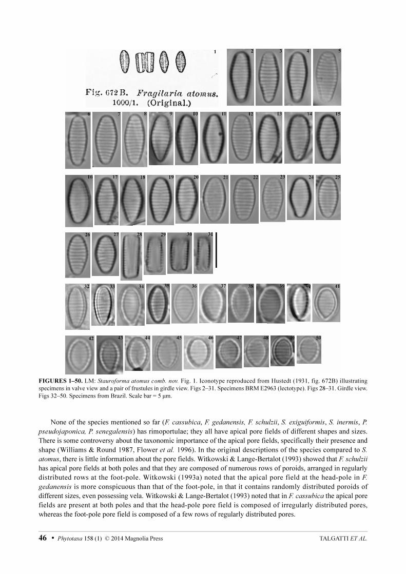
FIGURES 1–50. LM: Stauroforma atomus comb. nov. Fig. 1. Iconotype reproduced from Hustedt (1931, fig. 672B) illustrating specimens in valve view and a pair of frustules in girdle view. Figs 2–31. Specimens BRM E2963 (lectotype). Figs 28–31. Girdle view. Figs 32–50. Specimens from Brazil. Scale bar = 5 μm.
None of the species mentioned so far (F. cassubica, F. gedanensis, F. schulzii, S. exiguiformis, S. inermis, P.
pseudojaponica, P. senegalensis) has rimoportulae; they all have apical pore fields of different shapes and sizes. There is some controversy about the taxonomic importance of the apical pore fields, specifically their presence and shape (Williams & Round 1987, Flower et al. 1996). In the original descriptions of the species compared to S.
atomus, there is little information about the pore fields. Witkowski & Lange-Bertalot (1993) showed that F. schulzii
has apical pore fields at both poles and that they are composed of numerous rows of poroids, arranged in regularly distributed rows at the foot-pole. Witkowski (1993a) noted that the apical pore field at the head-pole in F.
gedanensis is more conspicuous than that of the foot-pole, in that it contains randomly distributed poroids of different sizes, even possessing vela. Witkowski & Lange-Bertalot (1993) noted that in F. cassubica the apical pore fields are present at both poles and that the head-pole pore field is composed of irregularly distributed pores, whereas the foot-pole pore field is composed of a few rows of regularly distributed pores.
TALGATTI ET AL.46 • Phytotaxa 158 (1) © 2014 Magnolia Press
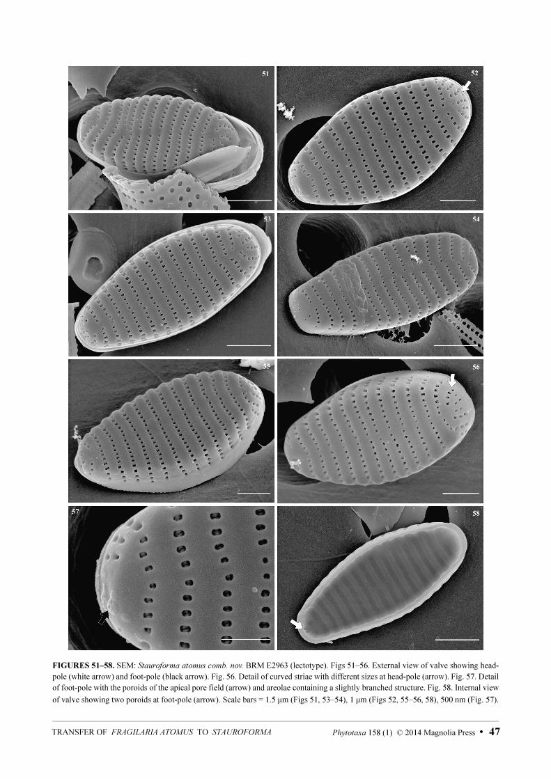
FIGURES 51–58. SEM: Stauroforma atomus comb. nov. BRM E2963 (lectotype). Figs 51–56. External view of valve showing head-pole (white arrow) and foot-pole (black arrow). Fig. 56. Detail of curved striae with different sizes at head-pole (arrow). Fig. 57. Detail of foot-pole with the poroids of the apical pore field (arrow) and areolae containing a slightly branched structure. Fig. 58. Internal view
of valve showing two poroids at foot-pole (arrow). Scale bars = 1.5 μm (Figs 51, 53–54), 1 μm (Figs 52, 55–56, 58), 500 nm (Fig. 57).
Phytotaxa 158 (1) © 2014 Magnolia Press • 47TRANSFER OF FRAGILARIA ATOMUS TO STAUROFORMA
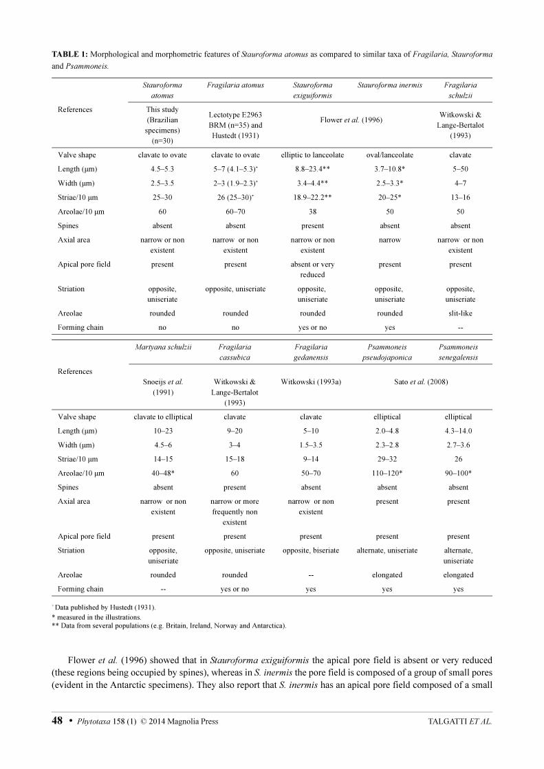
TABLE 1: Morphological and morphometric features of Stauroforma atomus as compared to similar taxa of Fragilaria, Stauroforma
and Psammoneis.
+ Data published by Hustedt (1931).
* measured in the illustrations.
** Data from several populations (e.g. Britain, Ireland, Norway and Antarctica).
Flower et al. (1996) showed that in Stauroforma exiguiformis the apical pore field is absent or very reduced (these regions being occupied by spines), whereas in S. inermis the pore field is composed of a group of small pores (evident in the Antarctic specimens). They also report that S. inermis has an apical pore field composed of a small
Stauroforma
atomus
Fragilaria atomus Stauroforma
exiguiformis
Stauroforma inermis Fragilaria
schulzii
References This study
(Brazilian
specimens)
(n=30)
Lectotype E2963
BRM (n=35) and
Hustedt (1931)
Flower et al. (1996)Witkowski &
Lange-Bertalot
(1993)
Valve shape clavate to ovate clavate to ovate elliptic to lanceolate oval/lanceolate clavate
Length (μm) 4.5–5.3 5–7 (4.1–5.3)+ 8.8–23.4** 3.7–10.8* 5–50
Width (μm) 2.5–3.5 2–3 (1.9–2.3)+ 3.4–4.4** 2.5–3.3* 4–7
Striae/10 μm 25–30 26 (25–30)+ 18.9–22.2** 20–25* 13–16
Areolae/10 μm 60 60–70 38 50 50
Spines absent absent present absent absent
Axial area narrow or non
existent
narrow or non
existent
narrow or non
existent
narrow narrow or non
existent
Apical pore field present present absent or very
reduced
present present
Striation opposite,
uniseriate
opposite, uniseriate opposite,
uniseriate
opposite,
uniseriate
opposite,
uniseriate
Areolae rounded rounded rounded rounded slit-like
Forming chain no no yes or no yes --
Martyana schulzii Fragilaria
cassubica
Fragilaria
gedanensis
Psammoneis
pseudojaponica
Psammoneis
senegalensis
References
Snoeijs et al.
(1991)
Witkowski &
Lange-Bertalot
(1993)
Witkowski (1993a) Sato et al. (2008)
Valve shape clavate to elliptical clavate clavate elliptical elliptical
Length (μm) 10–23 9–20 5–10 2.0–4.8 4.3–14.0
Width (μm) 4.5–6 3–4 1.5–3.5 2.3–2.8 2.7–3.6
Striae/10 μm 14–15 15–18 9–14 29–32 26
Areolae/10 μm 40–48* 60 50–70 110–120* 90–100*
Spines absent present absent absent absent
Axial area narrow or non
existent
narrow or more
frequently non
existent
narrow or non
existent
present present
Apical pore field present present present present present
Striation opposite,
uniseriate
opposite, uniseriate opposite, biseriate alternate, uniseriate alternate,
uniseriate
Areolae rounded rounded -- elongated elongated
Forming chain -- yes or no yes yes yes
TALGATTI ET AL.48 • Phytotaxa 158 (1) © 2014 Magnolia Press
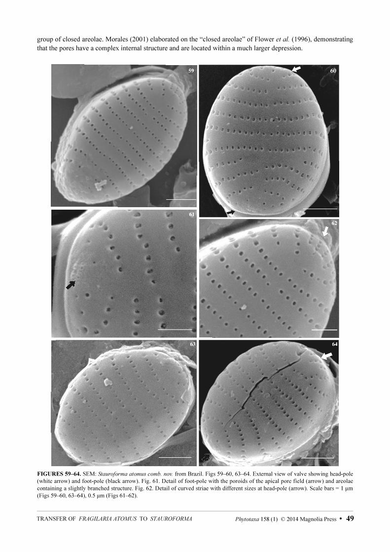
group of closed areolae. Morales (2001) elaborated on the “closed areolae” of Flower et al. (1996), demonstrating that the pores have a complex internal structure and are located within a much larger depression.
FIGURES 59–64. SEM: Stauroforma atomus comb. nov. from Brazil. Figs 59–60, 63–64. External view of valve showing head-pole (white arrow) and foot-pole (black arrow). Fig. 61. Detail of foot-pole with the poroids of the apical pore field (arrow) and areolae containing a slightly branched structure. Fig. 62. Detail of curved striae with different sizes at head-pole (arrow). Scale bars = 1 μm (Figs 59–60, 63–64), 0.5 μm (Figs 61–62).
Phytotaxa 158 (1) © 2014 Magnolia Press • 49TRANSFER OF FRAGILARIA ATOMUS TO STAUROFORMA

Psammoneis pseudojaponica and P. senegalensis have prominent apical pore fields, formed by rows of rounded areolae (Sato et al. 2008). Thus, the apical pore field of Stauroforma atomus is most similar to F. schulzii
than to any of the taxa in Table 1. Witkowski & Lange-Bertalot (1993) noted that for F. schulzii and S. atomus, the“Pore fields occur at both apices”. However, when we observed the poles of valves of S. atomus in SEM, the apical pore field occurred only at the foot-pole; the head-pole has striae similar to those that occur on the valve face. In contrast, the specimens of F. schulzii illustrated by Witkowski & Lange-Bertalot (1993) and Snoeijs et al. (1991) show an apical pore field present at the foot-pole, with the head-pole composed of only striae, such as observed in S. atomus.
Other features, such as the heteropolar valves, opposite striae, rounded (ovoid) areolae and narrow or non-existent axial area, are common in most of the taxa discussed above, with the exception of P. pseudojaponica and P.
senegalensis.
Distribution and ecology:—Stauroforma atomus is cited widely in the literature, but is poorly known with respect to data derived from the electron microscope, which may have resulted in errors of identification. The Baltic Sea is the region where S. atomus has the highest number of records (Fig. 65, Table 2): mainly from Poland, Sweden and Finland (e.g. Bogaczewicz-Adamczak & Dziengo 2003, Busse & Snoeijs 2003, Ulanova & Snoeijs 2006, Andrén et al. 2007, Ulanova et al. 2009, Leśniewska & Witak 2011).
FIGURE 65. Global distribution of Stauroforma atomus as recored in Table 2 (grey star = type locality; black star = population from Brazil; black dots = all other records).
For North America, Stoermer & Kreis (1978) mentioned S. atomus (recorded as Fragilaria atomus) in their checklist of diatoms from the Laurentian Great Lakes; Sullivan (1982) found S. atomus (as Fragilaria atomus) as a dominant element in the sediments and epiphytic material from the salt marshes of St. Louis Bay, Mississippi (USA); Poulin et al. (1984) found S. atomus (recorded as Fragilaria virescens var. oblongella f. clavata Grunow inVan Heurck 1881: pl. 44, fig. 4) in marine and estuarine benthic substrates in Québec (Canada); Burton et al.
(1994) discovered S. atomus (recorded as Fragilaria atomus) on artificial substrates in river systems in northern Michigan (USA); Bahls (2004) mentioned S. atomus (recorded as Fragilaria atomus) as abundant in periphytic samples from Careless Creek, Musselshell River, central area of Montana (USA); Krayesky et al. (2009) and López Fuerte et al. (2010) include S. atomus (recorded as Martyana atomus) for the Mexican flora (Krayesky et al. 2009, noted it from the Gulf of Mexico; López Fuerte et al. 2010, noted it on sediments from the coastal region of southern Baja California).
The only record for South America, albeit doubtful since no illustration was presented, is for the Uyuni-Coipasa salt flats, Southern Bolivian Altiplano (Bradbury et al. 2001). It should be noted here that the record included in Sar (1996: 429, pl 5, figs 40–43) from Bahia San Antonio, Argentina is not S. atomus (see Table 2).
TALGATTI ET AL.50 • Phytotaxa 158 (1) © 2014 Magnolia Press
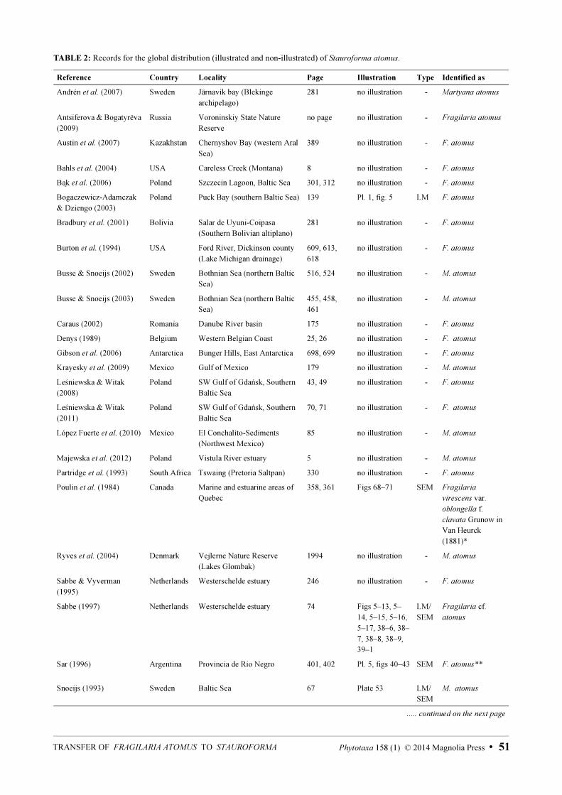
TABLE 2: Records for the global distribution (illustrated and non-illustrated) of Stauroforma atomus.
Reference Country Locality Page Illustration Type Identified as
Andrén et al. (2007) Sweden Järnavik bay (Blekinge
archipelago)
281 no illustration - Martyana atomus
Antsiferova & Bogatyrëva
(2009)
Russia Voroninskiy State Nature
Reserve
no page no illustration - Fragilaria atomus
Austin et al. (2007) Kazakhstan Chernyshov Bay (western Aral
Sea)
389 no illustration - F. atomus
Bahls et al. (2004) USA Careless Creek (Montana) 8 no illustration - F. atomus
Bąk et al. (2006) Poland Szczecin Lagoon, Baltic Sea 301, 312 no illustration - F. atomus
Bogaczewicz-Adamczak
& Dziengo (2003)
Poland Puck Bay (southern Baltic Sea) 139 Pl. 1, fig. 5 LM F. atomus
Bradbury et al. (2001) Bolivia Salar de Uyuni-Coipasa
(Southern Bolivian altiplano)
281 no illustration - F. atomus
Burton et al. (1994) USA Ford River, Dickinson county
(Lake Michigan drainage)
609, 613,
618
no illustration - F. atomus
Busse & Snoeijs (2002) Sweden Bothnian Sea (northern Baltic
Sea)
516, 524 no illustration - M. atomus
Busse & Snoeijs (2003) Sweden Bothnian Sea (northern Baltic
Sea)
455, 458,
461
no illustration - M. atomus
Caraus (2002) Romania Danube River basin 175 no illustration - F. atomus
Denys (1989) Belgium Western Belgian Coast 25, 26 no illustration - F. atomus
Gibson et al. (2006) Antarctica Bunger Hills, East Antarctica 698, 699 no illustration - F. atomus
Krayesky et al. (2009) Mexico Gulf of Mexico 179 no illustration - M. atomus
Leśniewska & Witak
(2008)
Poland SW Gulf of Gdańsk, Southern
Baltic Sea
43, 49 no illustration - F. atomus
Leśniewska & Witak
(2011)
Poland SW Gulf of Gdańsk, Southern
Baltic Sea
70, 71 no illustration - F. atomus
López Fuerte et al. (2010) Mexico El Conchalito-Sediments
(Northwest Mexico)
85 no illustration - M. atomus
Majewska et al. (2012) Poland Vistula River estuary 5 no illustration - M. atomus
Partridge et al. (1993) South Africa Tswaing (Pretoria Saltpan) 330 no illustration - F. atomus
Poulin et al. (1984) Canada Marine and estuarine areas of
Quebec
358, 361 Figs 68–71 SEM Fragilaria
virescens var.
oblongella f.
clavata Grunow in
Van Heurck
(1881)*
Ryves et al. (2004) Denmark Vejlerne Nature Reserve
(Lakes Glombak)
1994 no illustration - M. atomus
Sabbe & Vyverman
(1995)
Netherlands Westerschelde estuary 246 no illustration - F. atomus
Sabbe (1997) Netherlands Westerschelde estuary 74 Figs 5–13, 5–
14, 5–15, 5–16,
5–17, 38–6, 38–
7, 38–8, 38–9,
39–1
LM/
SEM
Fragilaria cf.
atomus
Sar (1996) Argentina Provincia de Rio Negro 401, 402 Pl. 5, figs 40–43 SEM F. atomus**
Snoeijs (1993) Sweden Baltic Sea 67 Plate 53 LM/
SEM
M. atomus
..... continued on the next page
Phytotaxa 158 (1) © 2014 Magnolia Press • 51TRANSFER OF FRAGILARIA ATOMUS TO STAUROFORMA
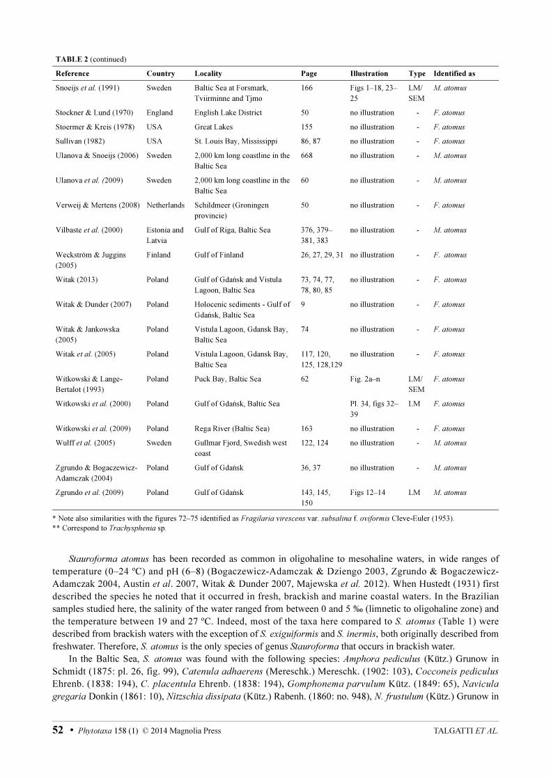
* Note also similarities with the figures 72–75 identified as Fragilaria virescens var. subsalina f. oviformis Cleve-Euler (1953).
** Correspond to Trachysphenia sp.
Stauroforma atomus has been recorded as common in oligohaline to mesohaline waters, in wide ranges of temperature (0–24 ºC) and pH (6–8) (Bogaczewicz-Adamczak & Dziengo 2003, Zgrundo & Bogaczewicz-Adamczak 2004, Austin et al. 2007, Witak & Dunder 2007, Majewska et al. 2012). When Hustedt (1931) first described the species he noted that it occurred in fresh, brackish and marine coastal waters. In the Brazilian samples studied here, the salinity of the water ranged from between 0 and 5 ‰ (limnetic to oligohaline zone) and the temperature between 19 and 27 ºC. Indeed, most of the taxa here compared to S. atomus (Table 1) were described from brackish waters with the exception of S. exiguiformis and S. inermis, both originally described from freshwater. Therefore, S. atomus is the only species of genus Stauroforma that occurs in brackish water.
In the Baltic Sea, S. atomus was found with the following species: Amphora pediculus (Kütz.) Grunow in Schmidt (1875: pl. 26, fig. 99), Catenula adhaerens (Mereschk.) Mereschk. (1902: 103), Cocconeis pediculus
Ehrenb. (1838: 194), C. placentula Ehrenb. (1838: 194), Gomphonema parvulum Kütz. (1849: 65), Navicula
gregaria Donkin (1861: 10), Nitzschia dissipata (Kütz.) Rabenh. (1860: no. 948), N. frustulum (Kütz.) Grunow in
TABLE 2 (continued)
Reference Country Locality Page Illustration Type Identified as
Snoeijs et al. (1991) Sweden Baltic Sea at Forsmark,
Tviirminne and Tjmo
166 Figs 1–18, 23–
25
LM/
SEM
M. atomus
Stockner & Lund (1970) England English Lake District 50 no illustration - F. atomus
Stoermer & Kreis (1978) USA Great Lakes 155 no illustration - F. atomus
Sullivan (1982) USA St. Louis Bay, Mississippi 86, 87 no illustration - F. atomus
Ulanova & Snoeijs (2006) Sweden 2,000 km long coastline in the
Baltic Sea
668 no illustration - M. atomus
Ulanova et al. (2009) Sweden 2,000 km long coastline in the
Baltic Sea
60 no illustration - M. atomus
Verweij & Mertens (2008) Netherlands Schildmeer (Groningen
provincie)
50 no illustration - F. atomus
Vilbaste et al. (2000) Estonia and
Latvia
Gulf of Riga, Baltic Sea 376, 379–
381, 383
no illustration - M. atomus
Weckström & Juggins
(2005)
Finland Gulf of Finland 26, 27, 29, 31 no illustration - F. atomus
Witak (2013) Poland Gulf of Gdańsk and Vistula
Lagoon, Baltic Sea
73, 74, 77,
78, 80, 85
no illustration - F. atomus
Witak & Dunder (2007) Poland Holocenic sediments - Gulf of
Gdańsk, Baltic Sea
9 no illustration - F. atomus
Witak & Jankowska
(2005)
Poland Vistula Lagoon, Gdansk Bay,
Baltic Sea
74 no illustration - F. atomus
Witak et al. (2005) Poland Vistula Lagoon, Gdansk Bay,
Baltic Sea
117, 120,
125, 128,129
no illustration - F. atomus
Witkowski & Lange-
Bertalot (1993)
Poland Puck Bay, Baltic Sea 62 Fig. 2a–n LM/
SEM
F. atomus
Witkowski et al. (2000) Poland Gulf of Gdańsk, Baltic Sea Pl. 34, figs 32–
39
LM F. atomus
Witkowski et al. (2009) Poland Rega River (Baltic Sea) 163 no illustration - F. atomus
Wulff et al. (2005) Sweden Gullmar Fjord, Swedish west
coast
122, 124 no illustration - M. atomus
Zgrundo & Bogaczewicz-
Adamczak (2004)
Poland Gulf of Gdańsk 36, 37 no illustration - M. atomus
Zgrundo et al. (2009) Poland Gulf of Gdańsk 143, 145,
150
Figs 12–14 LM M. atomus
TALGATTI ET AL.52 • Phytotaxa 158 (1) © 2014 Magnolia Press
Cleve & Grunow (1880: 98), Planothidium delicatulum (Kütz.) Round & Bukht. (1996: 353) and Pseudostaurosira
brevistriata (Grunow) D.M.Williams & Round (1987: 276). In this investigation and in the other studies from the Americas, the species was present together with the following species: Anaulus balticus Simonsen (1959: 74), Catenula adhaerens, Cyclotella choctawhatcheeana A.K.S.Prasad in Prasad et al. (1990: 419), Desikaneis gessneri
(Hust.) A.K.S.Prasad in Prasad & Livingston (1993: 435), Fallacia florinae (M.Møller) Witkowski (1993b: 215), N. frustulum, N. minutula Grunow in Van Heurck (1881: pl. 69, fig. 5), Placoneis sovereignae (Hust.) Torgan & Donadel in Torgan et al. (2010: 113), P. delicatulum, Tryblionella apiculata W.Greg. (1857: 79) and diverse araphid diatoms.
This is the first time S. atomus has been recorded from South America including illustrations. It is possible that the species is widely distributed in salt marshes and coastal regions of the world. More studies carried out in these regions should corroborate this hypothesis.
Acknowledgements
We thank to CAPES (Coordenação de Aperfeiçoamento de Pessoal de Nível Superior) for a doctoral grant to the first author. To Dr. César Serra Bonifácio Costa, Instituto de Oceanografia, Universidade Federal do Rio Grande, for support and help in fieldwork. To the Centro de Microscopia Eletrônica of the Universidade Federal do Rio Grande do Sul, Karina Marckmann, and Leandro Baum for technical assistance during SEM sessions. We are also thankful to Dr. Jorge Ernesto de Araujo Mariath and Juliana Troleis, Laboratório de Anatomia Vegetal (LAVeg), Instituto de Biociências, Universidade Federal do Rio Grande do Sul, for LM support and, to Friedel Hinz, Alfred-Wegener-Institut für Polar-und Meeresforschung, Bremerhaven, for providing the Hustedt’s lectotype material of Fragilaria atomus.
References
Ács, É., Morales, E.A., Kiss, K.T, Bolla, B., Plenkovic-Moraj, A., Reskóné, M.N. & Ector, L. (2009) Staurosira grigorszkyi nom. nov. (Bacillariophyceae) as araphid diatom from Lake Balaton, Hungary, with notes on Fragilaria hungarica Pantocsek. Nova Hedwigia 89: 469–483.http://dx.doi.org/10.1127/0029-5035/2009/0089-0469
Andrén, T., Andrén, E., Berglund, B.E. & Yu, S.-Y. (2007) New insights on the Yoldia Sea low stand in the Blekinge archipelago, southern Baltic Sea. GFF 129: 277–285.http://dx.doi.org/10.1080/11035890701294277
Anonymous (1975) Proposals for standardization of diatom terminology and diagnoses. Beihefte zur Nova Hedwigia 53: 323–354.Antsiferova, A. & Bogatyrëva S.N. (2009) Nizshie sinezelën' īe i diatomov'īe vodorosli gosudarstvennofo prirodnogo zapovednika
"Voroninski ĭ". Emel’ianov (predsedatel’), V.V., Gudina, A.N., Egorov, A.A., Campdurova, L.E.Trud’ īgosudarstvnnogo prirodnog zapovednika «Voroninski ĭ» [Proceedings of the State Nature Reserve «Voroninskiy»] 1: 52–107.
Austin, P., Mackay, A., Palagushkina, O. & Leng, M. (2007) A high-resolution diatom-inferred palaeoconductivity and lake level record of the Aral Sea for the last 1600 yr. Quaternary Research 67: 383–393. http://dx.doi.org/10.1016/j.yqres.2007.01.009
Bahls, L.L. (2004) Biological integrity of Careless Creek based on the structure and composition of the benthic algae community. Helena, Mont.: Montana Dept. of Environmental Quality: 18 pp. http://dx.doi.org/10.5962/bhl.title.24628
Bąk, M., Witkowski, A. & Lange-Bertalot, H. (2006) Diatom flora diversity in the strongly eutrophicated and β-mesosaprobic waters of the Szczecon Lagoon, NW Poland, southern Baltic Sea. In: Ognajanova-Rumenova, N. & Manoylov, K. (eds.) Advances in Phycological studies. Pensoft Publishers & University Publishing House Sofia, Moscow, pp. 293–317.
Barber, H.G. & Haworth, E.V. (1981) A guide to the morphology of the diatom frustule with a key to the British freshwater genera. Freshwater Biological Association Scientific Publication 44: 1–112.
Bogaczewicz-Adamczak, B. & Dziengo, M. (2003) Using benthic diatom communities and diatom indices to assess water pollution in the Puck Bay (southern Baltic Sea) littoral zone. Oceanological and Hydrobiological Studies 32: 131–157.
Bradbury, J.P., Grosjean, M., Stine, S. & Sylvestre, F. (2001) Full and late glacial lake records along the PEP1 Transect: Their role in developing interhemispheric palaeoclimate interactions. In: Markgraf, V. (ed.) Interhemispheric Climate Linkages. Academic Press, New York, pp. 265–291.
Brockmann, C. (1950) Die Watt-Diatomeen der schleswig-holsteinischen Westküste. Abhandlugen der Senckenbergischen Naturforschenden Gesellschaft, Frankfurt am Main 478: 1–26.
Burton, T.M., Oemke, M.P. & Molloy, J.M. (1994) Effects of stream order and alkalinity on the composition of diatom communities in
Phytotaxa 158 (1) © 2014 Magnolia Press • 53TRANSFER OF FRAGILARIA ATOMUS TO STAUROFORMA

two northern Michigan river systems. In Kociolek, J.P. (Ed.) Proceedings of the Eleventh International Diatom Symposium. California Academy of Sciences, San Francisco, pp. 609–620.
Busse, S. & Snoeijs, P. (2002) Gradient responses of diatom communities in the Bothnian Bay, northern Baltic Sea. Nova Hedwigia74: 501–525.http://dx.doi.org/10.1127/0029-5035/2002/0074-0501
Busse, S. & Snoeijs, P. (2003) Gradient responses of diatom communities in the Bothnian Sea (northern Baltic Sea), with emphasis on responses to water movement. Phycologia 42: 451–464. http://dx.doi.org/10.2216/i0031-8884-42-5-451.1
Caraus, I. (2002) The algae of Romania. A distributional checklist of actual algae. Studii şi Cercetări, Biologie. Universitatea Bacău 7: 1–694.
Cleve, P.T. & Grunow, A. (1880) Beiträge zur Kenntniss der Arctischen Diatomeen. Kongliga Svenska-Vetenskaps Akademiens Handlingar 17: 1–121.
Cleve-Euler, A. (1953) Die Diatomeen von Schweden und Finnland. Part II, Arraphideae, Brachyraphideae. Kongliga Svenska Vetenskaps-Akademiens Handligar, ser. 4, 4: 1–158.
Denys, L. (1989) Observations on the transition from Calais deposits to surface peat in the western Belgian Coastal plain. Results of a paleoenvironmental diatom study. Professional Paper Belgische Geologische Dienst 241: 20–43.
Donkin, A.S. (1861) On the marine Diatomaceae of Northumberland with a description of several new species. Quarterly Journal of Microscopical Science, new series, London 1: 1–15.
Ehrenberg, C.G. (1838) Die Infusionsthierchen als vollkommene Organismen. Ein Blick in das tiefere organische Leben der Natur. Verlag von Leopold Voss, Leipzig. pp. 1–xvii, 1–548, pls 1–64. [two volumes: Text, Atlas]
Ehrenberg, C.G. (1843) Mittheilungen über 2 neue asiatische Lager fossiler Infusorien-Erden aus dem russischen Trans-Kaukasien (Grusien) und Sibirien. Bericht über die zur Bekanntmachung geeigneten Verhandlungen der Königlich-Preussischen Akademie der Wissenschaften zu Berlin 1843: 43–49.
Flower, R.J., Jones, V.J. & Round, F.E. (1996) The distribution and classification of the problematic Fragilaria (virescens v.) exigua Grun./ Fragilaria exiguiformis (Grun.) Lange-Bertalot: a new species or a new genus? Diatom Research 11: 41–57.http://dx.doi.org/10.1080/0269249X.1996.9705363
Garcia, M. (2006) The transfer of Fragilaria obtusa Hustedt to the genus Staurosira Ehrenberg (Bacillariophyceae). Phycological Research 54: 87–93.http://dx.doi.org/10.1111/j.1440-1835.2006.00412.x
Gibson, J.A.E., Roberts, D. & Van de Vijver, B. (2006) Salinity control of the distribution of diatoms in lakes of the Bunger Hills, East Antarctica. Polar Biology 29: 694–704. http://dx.doi.org/10.1007/s00300-006-0107-8
Gregory, W. (1857) On the Post-Tertiary diatomaceous and of Glenshira. Part II. Transactions of the Microscopical Society of London5: 67–88.
Horton, B.P., Corbett, R., Culver, S.J., Edwards, R.J. & Hillier, C. (2006) Modern saltmarsh diatom distributions of the Outer Banks, North Carolina, and the development of a transfer function for high resolution reconstructions of sea level. Estuarine, Coastal and Shelf Science 69: 381–394.
Hustedt, F. (1931) Die Kieselalgen Deutschlands, Österreichs und der Schweiz. In: Dr. L. Rabenhorst’s Kryptogamenflora von Deutschland, Österreich und der Schweiz. Leipzig: Akademische Verlagsgesellschaft. Band 7, Teil 2, Lief. 1: 1–176.
Krammer, K. & Lange-Bertalot, H. (1991) Bacillariophyceae 3, Centrales, Fragilariaceae, Eunotiaceae. In: Ettl, H., Gerloff, J., Heynig, H. & Mollenhauer, D. (eds), Süsswasserflora von Mitteleuropa. Fisher, Stuttgart 2 (3): 576 pp.
Krauss, F. (1845) Pflanzen des Cap- und Natal–Landes, gesammelt und zusammengestellt. Flora oder Botanische Zeitung, Regensburg 28: 337–344.
Krayesky, D.M., Meave del Castillo, E., Zamudio, E., Norris, J.N. & Fredericq, S. (2009) Diatoms (Bacillariophyta) of the Gulf of Mexico. In: Felder, D.L. & Camp, D. K. (eds) Gulf of Mexico: Its Origin, Waters, and Biota. I. Biodiversity. Texas A & M University Press, United States of America, pp. 155–185.
Kützing, F.T. (1849) Species Algarum. Lipsiae. F.A. Brockhaus, 922 pp.Leśniewska, M. & Witak, M. (2008) Holocene diatom biostratigraphy of the SW Gulf of Gdańsk, Southern Baltic Sea (part III).
Oceanological and Hydrobiological Studies 37: 32–52.http://dx.doi.org/10.2478/v10009-008-0017-x
Leśniewska, M. & Witak, M. (2011) Diatoms as indicators of eutrophication in the SW part of the Gulf of Gdańsk, the Baltic Sea. Oceanological and Hydrobiological Studies 40: 68–81.http://dx.doi.org/10.2478/s13545-011-00 08-5
López Fuerte, F.O., Siqueiros Beltrones, D.A. & Navarro, J.N. (2010) Benthic diatoms associated with mangrove environments in the northwest region of México. CONABIO-UABCS-CICIMAR-IPN, Comité Editorial de CICIMAR-Oceánides. México, 206 pp.
Lyngbye, H.C. (1819) Tentamen hydrophytologiae danicae continens omnia hydrophyta cryptogama Daniae, Holsatiae, Faeroae, Islandiae, Groenlandiae hucusque cognita, systematice disposita, descripta et iconibus illustrata, adjectis simul speciebus norvegicis. pp. i–xxxii, 1–248, 70 pls. Hafniae [Copenhagen]: typis Schultzianis, in commissis Librariae Gyldendaliae.
Majewska, R., Zgrundo, A., Lemke, P. & De Stefano, M. (2012) Benthic diatoms of the Vistula River estuary (Northern Poland): Seasonality, substrata preferences, and the influence of water chemistry. Phycological Research 60: 1–19.http://dx.doi.org/10.1111/j.1440-1835.2011.00637.x
TALGATTI ET AL.54 • Phytotaxa 158 (1) © 2014 Magnolia Press
Mereschkowsky, C. (1902) Sur Catenula, un nouveau genre de diatomées. Scripta Botanica (Botanisheskia Zapiski, St. Petersburg)19: 93–116.
Morales, E.A. (2001) Morphological studies in selected fragilarioid diatoms (Bacillariophyceae) from Connecticut waters (U.S.A.). Proceedings of the Academy of Natural Sciences of Philadelphia 151: 105–120.
Morales, E.A. (2002) Studies in selected fragilarioid diatoms of potential indicator value from Florida (USA) with notes on the genusOpephora Petit (Bacillariophyceae). Limnologica 32: 102–113.
Morales, E.A. & Edlund, M.B. (2003) Studies in selected fragilarioid diatoms (Bacillariophyceae) from Lake Hovsgol, Mongolia. Phycological Research 51: 225–239.
Morales, E.A. & Manoylov, K.M. (2006) Morphological studies on selected taxa in the genus Staurosirella Williams et Round (Bacillariophyceae) from rivers in North America. Diatom Research 21: 343–364.
Morales, E.A., Novais, M.H., Chávez, G., Hoffmann L. & Ector, L. (2012) Diatoms (Bacillariophyceae) from the Bolivian Altiplano: three new araphid species from the Desaguadero River draining lake Titicaca. Fottea 12: 41–58.
Partridge, T.C., Kerr, S.J., Metcalfe, S.E., Scott, L., Talma, A.S. & Vogel, J.C. (1993) The Pretoria Saltpan: a 200000 year Southern African lacustrine sequence. Palaeogeography, Palaeoclimatology, Palaeocology 101: 317–337.
Poulin, M., Bérard-Therriault, L. & Cardinal, A. (1984) Les diatomées benthiques de substrats durs des eaux marines et saumâtres du Québec 3. Fragilarioideae (Fragilariales, Fragilariaceae). Naturaliste Canadien (Revue d'Ecologie et de Systématique) 111: 349–367.
Poulin, M., Bérard-Therriault & Cardinal, A. (1986) Fragilaria and Synedra (Bacillariophyceae): a morphological and ultrastrutural approach. Diatom Research 1 (1): 99–112. http://dx.doi.org/10.1080/0269249X.1986.9704961
Prasad, A.K.S.K. & Livingston, R.J. (1993) Frustule morphology of the planktonic pennate diatom Fragilaria gessneri Hustedt (Bacillariophyta) from the Florida coast of the Gulf of Mexico, with a description of Desikaneis gen. nov. Phycologia 32: 434–443.http://dx.doi.org/10.2216/i0031-8884-32-6-434.1
Prasad, A.K.S.K., Nienow, J.A. & Linvingston, R.J. (1990) The genus Cyclotella (Bacillariophyta) in Chocktawhatchee Bay, Florida, with special reference to C. striata and C. choctawhatcheeana sp. nov. Phycologia 29: 418–436.
Rabenhorst, L. (1860) Die Algen Sachsens resp. Mittel-Europa’s. Decas 95-96. nos 943-953.Rosen, B.H. & Lowe, R.L. (1981) Valve ultrastructure of some confusing Fragilariaceae. Micron 22: 293–294. Round, F.E. (1984) The circumscription of Synedra and Fragilaria and their subgroupings. In: Mann, D.G. (ed.). Proceedings of the
Seventh International Diatom Symposium, Koeltz, Koenigstein, pp. 241–253.Round, F.E. & Bukhtiyarova, L. (1996) Four new genera based on Achnanthes (Achnanthidium) together with a re-definition of
Achnanthidium. Diatom Research 11: 345–361. http://dx.doi.org/10.1080/0269249X.1996.9705389
Round, F.E., Crawford, R.M. & Mann, D.G. (1990) The Diatoms. Biology & morphology of the genera. Cambridge University Press, Cambridge. 741 pp.
Ryves, D.B., Clarke, A.L., Appleby, P.G., Amsinck, S.L., Jeppesen, E., Landkildehus, F. & Anderson, N.J. (2004) Reconstructing the salinity and environment of the Limfjord and Vejlerne Nature Reserve, Denmark, using a diatom model for brackish lakes and fjords. Canadian Journal of Fisheries and Aquatic Sciences 61: 1988–2006.http://dx.doi.org/10.1139/f04-127
Sabbe, K. (1997) Systematics and ecology of intertidal benthic diatoms of the Westerschelde estuary (The Netherlands). Thesis, University of Ghent, Ghent.
Sabbe, K. & Vyverman, W. (1995) Taxonomy, morphology and ecology of some widespread representatives of the diatom genus Opephora. European Journal of Phycology 30: 235–249. http://dx.doi.org/10.1080/09670269500651011
Sar, E. (1996) Flora diatomologica de Bahia San Antonio (Prov. de Rio Negro, Argentina). O. Pennales I. Revista del Museo de La Plata 14, Botánica 107: 309–432.
Sato, S., Kooistra, W.H.C.F., Watanabe, T., Matsumoto, S. & Medlin, L.K. (2008) A new araphid diatom genus Psammoneis gen. nov. (Plagiogrammaceae, Bacillariophyta) with three new species based on SSU and LSU rDNA sequence data and morphology. Phycologia 47: 510–528.http://dx.doi.org/10.2216/08-04.1
Schmidt, A. (1875) Atlas der Diatomaceen-kunde. Aschersleben, Commissions-Verlag Von Ludwig Siever’s Buchandlung Series I (Heft 7): pls 25–28.
Simonsen, R. (1959) Neue Diatomeen aus der Ostsee. I. Kieler Meeresforschungen 15: 74–83. Simonsen, R. (1987) Atlas and Catalogue of the Diatom Types of Friedrich Hustedt. J. Cramer, Berlin, Stuttgart. Vol: 1–3, 525 pp.,
772 pls.Snoeijs, P.J.M. (Ed.) (1993) Intercalibration and distribution of diatom species in the Baltic Sea, Vol. 1. Baltic Marine Biologists
Publication No. 16a. Opulus Press, Uppsala, 129 pp.Snoeijs, P.J.M., Hällfors, G. & Leskinen, E. (1991) The transfer of two epipsammic diatom species to the genus Martyana. Diatom
Research 6: 165–173. http://dx.doi.org/10.1080/0269249X.1991.9705155
Stockner, J.G. & Lund, J.W.G. (1970) Live algae in postglacial lake deposits. Limnology and Oceanography 15: 41–58.
Phytotaxa 158 (1) © 2014 Magnolia Press • 55TRANSFER OF FRAGILARIA ATOMUS TO STAUROFORMA
Stoermer, E.F. & Kreis, R.G.Jr. (1978) Preliminary checklist of diatoms (Bacillariophyta) from the Laurentian Great Lakes. International Association for Great Lakes Research 4: 149–169.
Sullivan, M.J. (1982) Similarity of an epiphytic and edaphic diatom community associated with Spartina alterniflora. Transactions of the American Microscopical Society 101: 84–90.
Torgan, L.C., Donadel, L. & Gonçalves da Silva, J. (2010) A transferência de Navicula sovereignae Hustedt para o gênero PlaconeisMereschkowsky (Bacillariophyta). Iheringia, Série Botânica, Porto Alegre 65: 107–114.
Tuji, A. & Williams, D.M. (2006) Examination of the type material of Synedra rumpens=Fragilaria rumpens, Bacillariophyceae. Phycological Research 54: 99–103.http://dx.doi.org/10.1111/j.1440-1835.2006.00414.x
Tuji, A. & Williams, D.M. (2008) Examination of types in the Fragilaria pectinalis-capitellata species complex. In: Likhoshway Y. (ed.). Proceedings of the 19th International Diatom Symposium, Listvyanka, Russia, 2006. Biopress Limited, Bristol, U.K. pp. 125–139.
Ulanova, A. & Snoeijs, P. (2006) Gradient responses of epilithic diatom communities in the Baltic Sea proper. Estuarine, Coastal and Shelf Science 68: 661–674. http://dx.doi.org/10.1016/j.ecss.2006.03.014
Ulanova, A., Busse, S. & Snoeijs, P. (2009) Coastal diatom-environment relationships in the brackish Baltic Sea. Journal of Phycology45: 54–68.http://dx.doi.org/10.1111/j.1529-8817.2008.00628.x
Van Heurck, H. (1881) Synopsis des Diatomées de Belgique. Atlas. Ducaju & Cie., Anvers. pls 31–77.Verweij, G. & Mertens, A. (2008) Prepareermethoden onder de loep. Diatomededelingen 32: 45–51.Vilbaste, S., Sundbäck, K., Nilsson, C. & Truu, J. (2000) Distribution of benthic diatoms in the littoral zone of the Gulf of Riga, the
Baltic Sea. European Journal of Phycology 35: 373–385.http://dx.doi.org/10.1080/09670260010001735981
Weckström, K. & Juggins, S. (2005) Coastal diatom-environment relationships from the Gulf of Finland, Baltic Sea. Journal of Phycology 42: 21–35.http://dx.doi.org/10.1111/j.1529-8817.2006.00166.x
Williams, D.M. & Round, F.E. (1987) Revision of the genus Fragilaria. Diatom Research 2: 267–288.http://dx.doi.org/10.1080/0269249X.1987.9705004
Williams, D.M. & Round, F.E. (1988) Fragilarifoma, nom. nov., a new generic name for Neofragilaria Williams & Round. Diatom Research 3: 265–267. http://dx.doi.org/10.1080/0269249X.1988.9705039
Witak, M. (2013) Diatom biofacies in the SW Gulf of Gdańsk and the Vistula Lagoon (the southern Baltic Sea) as indicators of the basin evolution in the Middle and Late Holocene. Oceanological and Hydrobiological Studies 42: 70–88.http://dx.doi.org/10.2478/s13545-013-0052-4
Witak, M. & Jankowska, D. (2005) The Vistula Lagoon evolution based on diatom records. Baltica 18: 68–76.Witak, M., Boryn, K. & Mayer, A. (2005) Holocene environmental changes recorded by diatom stratigraphy in the Vistula Lagoon.
Oceanological and Hydrobiological Studies 34: 111–113.Witak, M. & Dunder, J. (2007) Holocene diatom biostratigraphy of the SW Gulf of Gdańsk, Southern Baltic Sea (part II).
Oceanological and Hydrobiological Studies 36: 3–20. http://dx.doi.org/10.2478/v10009-007-0021-6
Witkowski, A. (1993a) Fragilaria gedanensis sp. nov. (Bacillariophyceae), a new epipsammic diatom species from the Baltic Sea. Nova Hedwigia 56: 497–503.
Witkowski, A. (1993b) Fallacia florinae (Moeller) comb. nov., a marine, epipsammic diatom. Diatom Research 8: 215–219. http://dx.doi.org/10.1080/0269249X.1993.9705254
Witkowski, A. & Lange-Bertalot, H. (1993) Established and new diatom taxa related to Fragilaria schulzii Brockmann. Limnologica23: 59–70.
Witkowski, A., Lange-Bertalot, H. & Metzeltin, D. (1996) The diatom species Fragilaria martyi (Heribaud) Lange-Bertalot, identity and ecology. Archiv für Protistenkunde 146: 281–292.
Witkowski, A., Lange-Bertalot, H. & Metzeltin, D. (2000) Diatom flora of marine coasts I. Iconographia Diatomologica 7: 1–925. Witkowski, A., Cedro, B., Kierzek, A. & Baranowski, D. (2009) Diatoms as a proxy in reconstructing the Holocene environmental
changes in the south-western Baltic Sea: the lower Rega River Valley sedimentary record. Hydrobiologia 631: 155–172. http://dx.doi.org/10.1007/s10750-009-9808-7
Wulff, A., Vilbaste, S. & Truu, J. (2005) Depth distribution of photosynthetic pigments and diatoms in the sediments of microtidal fjord. Hydrobiologia 534: 117–130.
Zgrundo, A. & Bogaczewicz-Adamczak, B. (2004) Applicability of diatom indices for monitoring water quality in coastal streams in the Gulf of Gdansk Region, northern Poland. Oceanological and Hydrobiological Studies 33: 31–46.
Zgrundo, A., Dziengo-Czaja, M., Bubak, I. & Bogaczewicz-Adamczak, B. (2009) Studies on the biodiversity of contemporary diatom assemblages in the Gulf of Gdańsk. Oceanological and Hydrobiological Studies 37, Supplement 2: 139–153.
TALGATTI ET AL.56 • Phytotaxa 158 (1) © 2014 Magnolia Press