Ozonation of Whole Blood Results in an Increased Release of ...
13
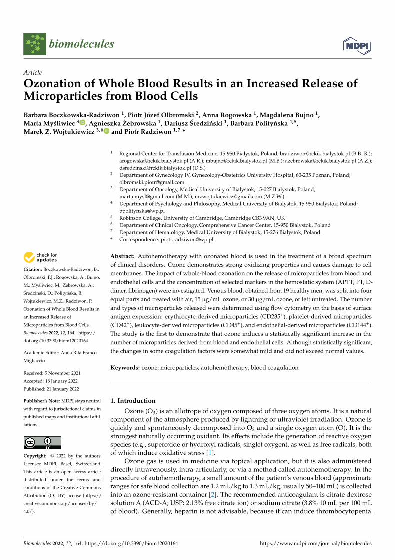
Citation: Boczkowska-Radziwon, B.; Olbromski, P.J.; Rogowska, A.; Bujno, M.; My´ sliwiec, M.; ˙ Zebrowska, A.; ´ Sredzi ´ nski, D.; Polity ´ nska, B.; Wojtukiewicz, M.Z.; Radziwon, P. Ozonation of Whole Blood Results in an Increased Release of Microparticles from Blood Cells. Biomolecules 2022, 12, 164. https:// doi.org/10.3390/biom12020164 Academic Editor: Anna Rita Franco Migliaccio Received: 5 November 2021 Accepted: 18 January 2022 Published: 21 January 2022 Publisher’s Note: MDPI stays neutral with regard to jurisdictional claims in published maps and institutional affil- iations. Copyright: © 2022 by the authors. Licensee MDPI, Basel, Switzerland. This article is an open access article distributed under the terms and conditions of the Creative Commons Attribution (CC BY) license (https:// creativecommons.org/licenses/by/ 4.0/). biomolecules Article Ozonation of Whole Blood Results in an Increased Release of Microparticles from Blood Cells Barbara Boczkowska-Radziwon 1 , Piotr Józef Olbromski 2 , Anna Rogowska 1 , Magdalena Bujno 1 , Marta My´ sliwiec 3 , Agnieszka ˙ Zebrowska 1 , Dariusz ´ Sredzi ´ nski 1 , Barbara Polity ´ nska 4,5 , Marek Z. Wojtukiewicz 3,6 and Piotr Radziwon 1,7, * 1 Regional Center for Transfusion Medicine, 15-950 Bialystok, Poland; [email protected] (B.B.-R.); [email protected] (A.R.); [email protected] (M.B.); [email protected] (A. ˙ Z.); [email protected] (D. ´ S.) 2 Department of Gynecology IV, Gynecology-Obstetrics University Hospital, 60-235 Poznan, Poland; [email protected] 3 Department of Oncology, Medical University of Bialystok, 15-027 Bialystok, Poland; [email protected] (M.M.); [email protected] (M.Z.W.) 4 Department of Psychology and Philosophy, Medical University of Bialystok, 15-950 Bialystok, Poland; [email protected] 5 Robinson College, University of Cambridge, Cambridge CB3 9AN, UK 6 Department of Clinical Oncology, Comprehensive Cancer Center, 15-950 Bialystok, Poland 7 Department of Hematology, Medical University of Bialystok, 15-276 Bialystok, Poland * Correspondence: [email protected] Abstract: Autohemotherapy with ozonated blood is used in the treatment of a broad spectrum of clinical disorders. Ozone demonstrates strong oxidizing properties and causes damage to cell membranes. The impact of whole-blood ozonation on the release of microparticles from blood and endothelial cells and the concentration of selected markers in the hemostatic system (APTT, PT, D- dimer, fibrinogen) were investigated. Venous blood, obtained from 19 healthy men, was split into four equal parts and treated with air, 15 μg/mL ozone, or 30 μg/mL ozone, or left untreated. The number and types of microparticles released were determined using flow cytometry on the basis of surface antigen expression: erythrocyte-derived microparticles (CD235 + ), platelet-derived microparticles (CD42 + ), leukocyte-derived microparticles (CD45 + ), and endothelial-derived microparticles (CD144 + ). The study is the first to demonstrate that ozone induces a statistically significant increase in the number of microparticles derived from blood and endothelial cells. Although statistically significant, the changes in some coagulation factors were somewhat mild and did not exceed normal values. Keywords: ozone; microparticles; autohemotherapy; blood coagulation 1. Introduction Ozone (O 3 ) is an allotrope of oxygen composed of three oxygen atoms. It is a natural component of the atmosphere produced by lightning or ultraviolet irradiation. Ozone is quickly and spontaneously decomposed into O 2 and a single oxygen atom (O). It is the strongest naturally occurring oxidant. Its effects include the generation of reactive oxygen species (e.g., superoxide or hydroxyl radicals, singlet oxygen), as well as free radicals, both of which induce oxidative stress [1]. Ozone gas is used in medicine via topical application, but it is also administered directly intravenously, intra-articularly, or via a method called autohemotherapy. In the procedure of autohemotherapy, a small amount of the patient’s venous blood (approximate ranges for safe blood collection are 1.2 mL/kg to 1.3 mL/kg, usually 50–100 mL) is collected into an ozone-resistant container [2]. The recommended anticoagulant is citrate dextrose solution A (ACD-A; USP: 2.13% free citrate ion) or sodium citrate (3.8% 10 mL per 100 mL of blood). Generally, heparin is not advisable, because it can induce thrombocytopenia. Biomolecules 2022, 12, 164. https://doi.org/10.3390/biom12020164 https://www.mdpi.com/journal/biomolecules
-
Upload
khangminh22 -
Category
Documents
-
view
0 -
download
0
Transcript of Ozonation of Whole Blood Results in an Increased Release of ...
�����������������
Citation: Boczkowska-Radziwon, B.;
Olbromski, P.J.; Rogowska, A.; Bujno,
M.; Mysliwiec, M.; Zebrowska, A.;
Sredzinski, D.; Politynska, B.;
Wojtukiewicz, M.Z.; Radziwon, P.
Ozonation of Whole Blood Results in
an Increased Release of
Microparticles from Blood Cells.
Biomolecules 2022, 12, 164. https://
doi.org/10.3390/biom12020164
Academic Editor: Anna Rita Franco
Migliaccio
Received: 5 November 2021
Accepted: 18 January 2022
Published: 21 January 2022
Publisher’s Note: MDPI stays neutral
with regard to jurisdictional claims in
published maps and institutional affil-
iations.
Copyright: © 2022 by the authors.
Licensee MDPI, Basel, Switzerland.
This article is an open access article
distributed under the terms and
conditions of the Creative Commons
Attribution (CC BY) license (https://
creativecommons.org/licenses/by/
4.0/).
biomolecules
Article
Ozonation of Whole Blood Results in an Increased Release ofMicroparticles from Blood CellsBarbara Boczkowska-Radziwon 1, Piotr Józef Olbromski 2, Anna Rogowska 1, Magdalena Bujno 1,Marta Mysliwiec 3 , Agnieszka Zebrowska 1, Dariusz Sredzinski 1, Barbara Politynska 4,5,Marek Z. Wojtukiewicz 3,6 and Piotr Radziwon 1,7,*
1 Regional Center for Transfusion Medicine, 15-950 Bialystok, Poland; [email protected] (B.B.-R.);[email protected] (A.R.); [email protected] (M.B.); [email protected] (A.Z.);[email protected] (D.S.)
2 Department of Gynecology IV, Gynecology-Obstetrics University Hospital, 60-235 Poznan, Poland;[email protected]
3 Department of Oncology, Medical University of Bialystok, 15-027 Bialystok, Poland;[email protected] (M.M.); [email protected] (M.Z.W.)
4 Department of Psychology and Philosophy, Medical University of Bialystok, 15-950 Bialystok, Poland;[email protected]
5 Robinson College, University of Cambridge, Cambridge CB3 9AN, UK6 Department of Clinical Oncology, Comprehensive Cancer Center, 15-950 Bialystok, Poland7 Department of Hematology, Medical University of Bialystok, 15-276 Bialystok, Poland* Correspondence: [email protected]
Abstract: Autohemotherapy with ozonated blood is used in the treatment of a broad spectrumof clinical disorders. Ozone demonstrates strong oxidizing properties and causes damage to cellmembranes. The impact of whole-blood ozonation on the release of microparticles from blood andendothelial cells and the concentration of selected markers in the hemostatic system (APTT, PT, D-dimer, fibrinogen) were investigated. Venous blood, obtained from 19 healthy men, was split into fourequal parts and treated with air, 15 µg/mL ozone, or 30 µg/mL ozone, or left untreated. The numberand types of microparticles released were determined using flow cytometry on the basis of surfaceantigen expression: erythrocyte-derived microparticles (CD235+), platelet-derived microparticles(CD42+), leukocyte-derived microparticles (CD45+), and endothelial-derived microparticles (CD144+).The study is the first to demonstrate that ozone induces a statistically significant increase in thenumber of microparticles derived from blood and endothelial cells. Although statistically significant,the changes in some coagulation factors were somewhat mild and did not exceed normal values.
Keywords: ozone; microparticles; autohemotherapy; blood coagulation
1. Introduction
Ozone (O3) is an allotrope of oxygen composed of three oxygen atoms. It is a naturalcomponent of the atmosphere produced by lightning or ultraviolet irradiation. Ozone isquickly and spontaneously decomposed into O2 and a single oxygen atom (O). It is thestrongest naturally occurring oxidant. Its effects include the generation of reactive oxygenspecies (e.g., superoxide or hydroxyl radicals, singlet oxygen), as well as free radicals, bothof which induce oxidative stress [1].
Ozone gas is used in medicine via topical application, but it is also administereddirectly intravenously, intra-articularly, or via a method called autohemotherapy. In theprocedure of autohemotherapy, a small amount of the patient’s venous blood (approximateranges for safe blood collection are 1.2 mL/kg to 1.3 mL/kg, usually 50–100 mL) is collectedinto an ozone-resistant container [2]. The recommended anticoagulant is citrate dextrosesolution A (ACD-A; USP: 2.13% free citrate ion) or sodium citrate (3.8% 10 mL per 100 mLof blood). Generally, heparin is not advisable, because it can induce thrombocytopenia.
Biomolecules 2022, 12, 164. https://doi.org/10.3390/biom12020164 https://www.mdpi.com/journal/biomolecules
Biomolecules 2022, 12, 164 2 of 13
The oxygen/ozone mixture is then bubbled into the blood, gently mixed, and reinfusedinto the vein of the patient. Thus, ozone does not enter the blood circulation. Its effects aremediated by the formation of secondary messengers and the induction of a further adaptiveresponse from the body in a hermetic dose–response relationship. Through this indirectmechanism of action, ozone stimulates adaptive mechanisms that can induce modulationsin the organism by affecting the immune system, blood flow, and oxygenation, as well asoxidative stress [3–5].
The number of treatment sessions and the ozone dosage administered depend on thegeneral condition of the patient, their age, and any underlying disease. From the clinicalpoint of view, improvement in the patient’s condition occurs between the fifth and tenthsessions. The treatment is given daily if necessary. It may also be administered two to threetimes a week.
Autohemotherapy with ozonated blood has been used to treat a wide variety ofdiseases, including heart disease, ischemic diseases (peripheral arterial, ocular circulatoryand cerebral circulatory disturbances), pain management, diabetic angiopathy, trophic skinlesions, arthritis, herpes zoster infection, cancer (complementary treatment), hepatitis, andbacterial infection (complementary therapy) [6–9].
Ozone therapy is considered to be relatively safe. Nevertheless, there are severalreports presenting adverse reactions associated with the use of ozone, such as sinus arrest,myocardial infarction, or vasospasm [10,11]. With enhanced exposure to ozone, erythro-cytes are increasingly damaged. Thus, the application of appropriate doses of ozone inautohemotherapy for patients with different diseases would appear to be crucial.
It is well known that ozonation affects the functions of blood cells. Increased ATP(adenosine triphosphate) and 2,3DPG (2,3-diphosphoglycerate) concentrations in erythro-cytes have been observed, as well as decreases in the pH of the cytoplasm. It has beenhypothesized that lipid oxidation products stimulate bone marrow and increase the releaseof erythrocytes with enhanced concentrations of glucose-6-dehydrogenase [12].
Ozone therapy is also known to increase neutrophil phagocytic activity [13]. Moreover,H2O2 activates tyrosine kinase and consequently causes increased synthesis and secre-tion of proinflammatory cytokines: IFN-γ, TNF-a, and IL-8 [14–16]. However, ozonatedautohemotherapy does not seem to affect NK-cell function, IFN-γ, TGF-β, IL-1α, IL-1β,IL-2, IL-4, IL-6, IL-10, IL-13, and CRP levels, platelet function, blood coagulation, vonWillebrand factor, erythrocyte function, and surprisingly protein peroxidation [17]. Reac-tive oxygen species or lipid ozonation products activate a wide spectrum of genes andconsequently upregulate synthesis of acute-phase proteins and cytokines responsible forimmunomodulatory effects [14,18]. The application of ozone autohemotherapy inducesthe release of PDGF-B, TGF-β1, IL-8, and EGF from platelets [19]. Ozonated blood is ableto activate endothelial cells through lipid peroxidation products and stimulates them torelease NO [20]. NO is a known vasorelaxant which dilates vessel walls, having beneficialeffects particularly in hypoxic lower-limb tissues or in the retina [21,22]. These effects seemto be mediated via activation of the NRF2 pathway [23–25].
Microparticles (MPs) are spherical fragments of cell membranes with diameters rang-ing from 0.1 to 1.0 µm, which are released from eukaryotic cells to the extracellular space.The formation of MPs occurs during maturation, activation, and apoptosis of a cell [26].MPs consist of cell membrane lipids and proteins, as well as elements of cytoskeletonoriginating from the mother cell. The properties of MPs mainly depend on the factorsinitiating their release. Different stimulants induce the release of MPs, which presentdistinct properties even if they are released from the same type of cells. There is no cellularcore in microparticles and they do not synthesize their own proteins; however, they maycontain biologically active molecules from the cell of origin, i.e., enzymes, membraneproteins, mRNAs and microRNAs, adhesive proteins, coagulation factors, and membranelipids [27–30]. All of these take part in the regulation and coordination of many processesoccurring within the organism, i.e., inflammatory processes, neoangiogenesis, coagulation,and fibrinolysis [27,31–35]. By means of their role in transmitting signals between cells,
Biomolecules 2022, 12, 164 3 of 13
microparticles constitute a connection between coagulation processes and local inflamma-tion [28,29]. They also play a role in the progression of cancer [36]. MPs are able to modifythe activity of certain medicines due to their sequestration [29]. To date, there are no dataon the effects of ozone on MPs.
The aim of the study was to assess the possible impact of whole-blood ozonation onthe release of erythrocyte-derived microparticles (ErMP), leukocyte-derived microparticles(LMP), endothelial-derived microparticles (EnMP), and platelet-derived microparticles(PMP), as well as on selected parameters of the hemostatic system.
2. Materials and Methods2.1. Subjects
Blood for experiments was obtained from 19 healthy men (25–35 years old) not takingany drugs, with no history of thrombosis, hypertension, or disorders of the endocrinesystem, and with similar basic blood morphology (RBC, WBC, PLC, and hemoglobin levels,as well as MHC). The selected group of donors was highly homogeneous in terms of age,gender, and hormonal factors. The presented study conforms with the principles outlinedin the Declaration of Helsinki and was approved by the Ethical Committee for HumanStudies of the Medical University of Bialystok (KB: R-J-002/78/2015); prior to the study, allparticipants gave their informed consent.
2.2. Blood Collection
Eighty milliliters of venous blood was drawn from an antecubital vein into a collectionbag and mixed with citrate phosphate dextrose solution (0.12% final concentration). Aftercollection, the blood was split into four equal parts and transferred into ozone-resistant(Teflon) blood bags (Fresenius Hemofreeze Bag DF 200, Fresenius, Germany). Each bloodsample was assigned to one of four groups (C, A, O1, and O2) and was treated accordingto the conditions defining each of the groups. There were 19 portions of blood from eachof the different donors in all of the groups. The control group (C) was not subjected toozonation or aeration, the second group (A) was subjected to aeration, the third group (O1)was ozonated with a lower dose of ozone, and the fourth group (O2) was ozonated with ahigher dose of ozone.
2.3. Blood Ozonation/Aeration
Blood samples from groups A, O1, and O2 were subjected to either aeration or ozona-tion. The oxygen–ozone gas mixture was freshly prepared from a medical-grade oxygenusing an ATO-3 MINI ozone generator (CryoFlex, Poland). The mixture was kept at anormal atmospheric pressure.
The doses of ozone used in our study are highly comparable to doses used in ozona-tion protocols designed for therapeutic use [8,37]. Ozonation/aeration was performedimmediately after blood collection and splitting. The blood samples were treated with asingle dose of 15 µg/mL (group O1) or 30 µg/mL (group O2) of oxygen–ozone gas mixture(final concentrations of gaseous ozone) or the same volume of air (group A). The gasmixtures were transferred into the blood using syringes made of ozone-resistant materialcontaining an antibacterial filter. Infusion pump-operated syringes were used in order tomaintain a constant flow of 1 mL/min. Although an infusion pump may not be accordingto the ozonation protocol designed for therapeutic use, it helped to maintain standardconditions of experimental ozonation in our study. During ozonation/aeration, blood bagswere placed on a laboratory shaker to ensure mixing of the gas with the blood. Threeminutes after the treatment, blood samples were taken from the blood bag: 5 mL for MPanalysis and 5 mL for coagulation tests. The samples for coagulation tests were centrifugedat 2000× g for 10 min to obtain plasma. All samples of plasma were stored at −80 ◦C untilthe day of analysis.
Biomolecules 2022, 12, 164 4 of 13
2.4. Microparticle Isolation
Two steps in the process of centrifugation were used in order to isolate microparticlesfrom whole blood. First, blood samples were centrifuged at 5000× g at 24 ◦C for 5 minwithout centrifuge brake (removal of blood cells) to prepare platelet-poor plasma (PPP).To collect MP, 600 µL of PPP was centrifuged at 17,000× g at 20 ◦C for 3 min, and thesupernatant was removed. The MP pellet was then resuspended in 500 µL of dilutedannexin V FITC binding buffer (BD bioscience, Franklin Lakes, NJ, USA).
2.5. Quantification and Characterization of MPs
Quantification of the number of MPs and their phenotypic characterization in the pelletwere conducted by flow cytometry (Facscalibur, Becton Dickinson, USA). The MP size gatewas set between 200 and 1000 nm using fluorescent latex beds 0.2, 0.5, and 1 µm (PrecisionSize Standards, Polysciences). The total number of MPs was defined as all the events fallingwithin the MP gate. To count the number of MPs, an aliquot of resuspended MP pellet(100 µL) was added to TruCount (Becton Dickinson, USA) (100 µL), followed by countingup to 2000 0.5 µL bead components of TruCount (total absolute count of MPs = (events inregion except beads/events in region of beads) × (absolute number of beads/µL/samplevolume (µL)). The origin of MPs was determined by flow cytometry using the cell specificmonoclonal antibodies (BD bioscience) and annexin V FITC as follows: erythrocyte-derivedmicroparticles—GlyA PE CD235 (clone GAR-2 (HIR2)), platelet-derived microparticles—CD42bPE-CyTM5 (clone HIP1), leukocyte-derived microparticles—CD45PerCP-CyTM5,5(clone TU116), endothelial-derived microparticles—CD144 PE (clone 55-7H1).
2.6. Coagulation Tests
APTT was measured using the Hemostat APTT-EL reagent. Measurement of thePT time was carried out with the use of Hemostat Tromboplastin SI reagent. Fibrinogenconcentration was determined with the use of the Hemostat Fibrinogen kit based on theClauss method. Concentration of D-dimers was determined using the qualitative test ofthe VIDAS system for immuno-enzymatic marking of fibrin degradation products with theuse of the immuno-fluorescence technique (BioMerieux).
2.7. Statistical Analysis
Data analysis was carried out using the GraphPad Prism 8.4.0 (Software LLC). Quan-titative variables were described in terms of means, standard deviations, and correlationcoefficients. Qualitative variables were described as a number and percentage. To com-pare parametric quantitative variables between two groups, the Friedman test was used.Multiple comparisons analysis was carried out with Dunn’s test. A p-value of <0.05 wasconsidered significant in all analyses.
3. Results3.1. Microparticles
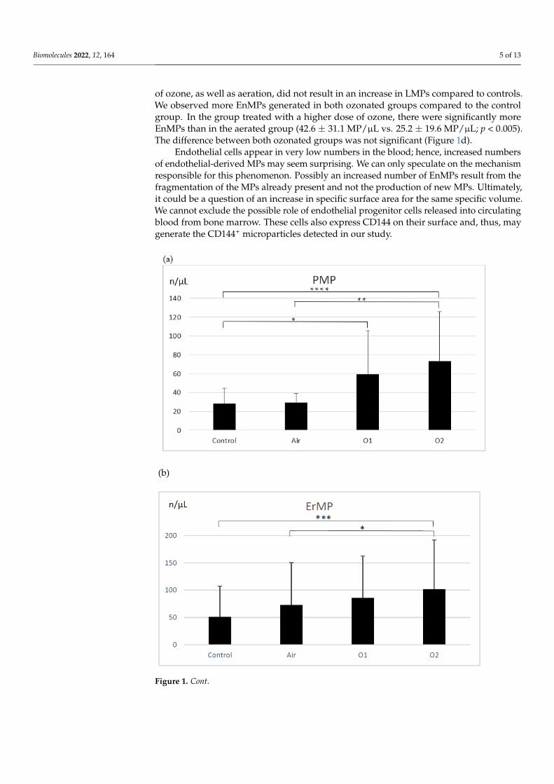
There were more PMPs generated in the ozonated samples than in the control group(28.1 ± 16.2 MP/µL; p < 0.0001 for the group ozonated with higher dose and p < 0.01for the group ozonated with a lower dose of ozone) (Figure 1a). The group treatedwith a higher dose of ozone generated significantly more PMPs than the aerated group(73.4 ± 52.5 MP/µL vs. 29.4 ± 9.5 MP/µL; p < 0.005). Aeration did not result in an increasein PMPs compared to controls. There were significantly more ErMPs generated in thegroup treated with a higher dose of ozone (102.0 ± 90 MP/µL) compared to the controlgroup (51.3 ± 56.2 MP/µL; p < 0.0005) and the aerated group (73.0 ± 77.7 MP/µL; p < 0.01)(Figure 1b). The lower dose of ozone, as well as aeration, did not result in an increase inErMPs compared to controls.
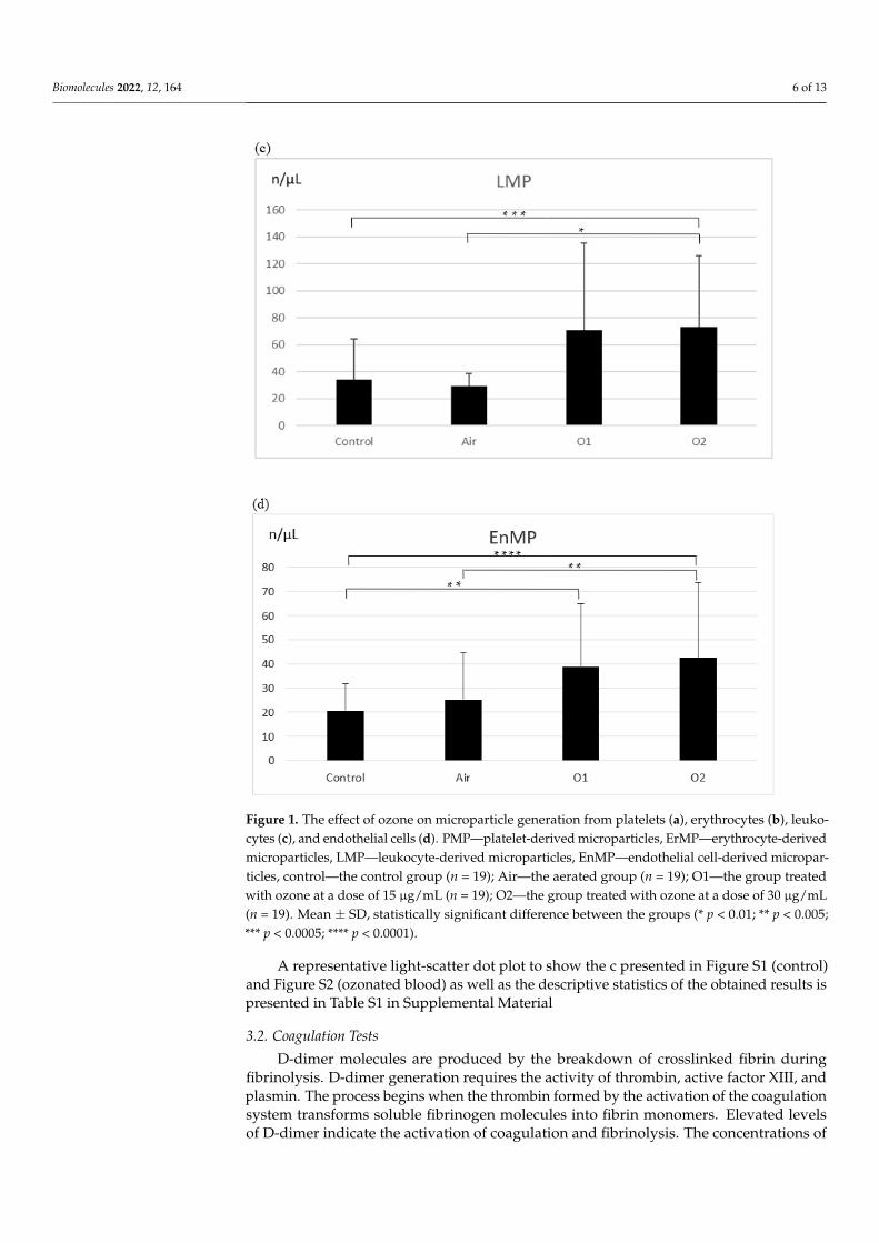
There were significantly more LMPs generated in the group ozonated with higherdose of ozone compared to the control group (34.3 ± 29.9 MP/µL; p < 0.0005) and theaerated group (24.4 ± 9.5 MP/µL; p < 0.01) (Figure 1c). The ozonation with the lower dose
Biomolecules 2022, 12, 164 5 of 13
of ozone, as well as aeration, did not result in an increase in LMPs compared to controls.We observed more EnMPs generated in both ozonated groups compared to the controlgroup. In the group treated with a higher dose of ozone, there were significantly moreEnMPs than in the aerated group (42.6 ± 31.1 MP/µL vs. 25.2 ± 19.6 MP/µL; p < 0.005).The difference between both ozonated groups was not significant (Figure 1d).
Endothelial cells appear in very low numbers in the blood; hence, increased numbersof endothelial-derived MPs may seem surprising. We can only speculate on the mechanismresponsible for this phenomenon. Possibly an increased number of EnMPs result from thefragmentation of the MPs already present and not the production of new MPs. Ultimately,it could be a question of an increase in specific surface area for the same specific volume.We cannot exclude the possible role of endothelial progenitor cells released into circulatingblood from bone marrow. These cells also express CD144 on their surface and, thus, maygenerate the CD144+ microparticles detected in our study.
Biomolecules 2022, 12, x FOR PEER REVIEW 5 of 13
1b). The lower dose of ozone, as well as aeration, did not result in an increase in ErMPs compared to controls.
There were significantly more LMPs generated in the group ozonated with higher dose of ozone compared to the control group (34.3 ± 29.9 MP/µL; p < 0.0005) and the aer-ated group (24.4 ± 9.5 MP/µL; p < 0.01) (Figure 1c). The ozonation with the lower dose of ozone, as well as aeration, did not result in an increase in LMPs compared to controls. We observed more EnMPs generated in both ozonated groups compared to the control group. In the group treated with a higher dose of ozone, there were significantly more EnMPs than in the aerated group (42.6 ± 31.1 MP/µL vs. 25.2 ± 19.6 MP/µL; p < 0.005). The differ-ence between both ozonated groups was not significant (Figure 1d).
(b)
Figure 1. Cont.
Biomolecules 2022, 12, 164 6 of 13Biomolecules 2022, 12, x FOR PEER REVIEW 6 of 13
Figure 1. The effect of ozone on microparticle generation from platelets (a), erythrocytes (b), leu-kocytes (c), and endothelial cells (d). PMP—platelet-derived microparticles, ErMP—erythrocyte-derived microparticles, LMP—leukocyte-derived microparticles, EnMP—endothelial cell-derived microparticles, control—the control group (n = 19); Air—the aerated group (n = 19); O1—the group treated with ozone at a dose of 15 µg/mL (n = 19); O2—the group treated with ozone at a dose of 30 µg/mL (n = 19). Mean ± SD, statistically significant difference between the groups (* p < 0.01; ** p < 0.005; *** p < 0.0005; **** p < 0.0001).
Endothelial cells appear in very low numbers in the blood; hence, increased numbers of endothelial-derived MPs may seem surprising. We can only speculate on the mecha-nism responsible for this phenomenon. Possibly an increased number of EnMPs result from the fragmentation of the MPs already present and not the production of new MPs. Ultimately, it could be a question of an increase in specific surface area for the same spe-cific volume. We cannot exclude the possible role of endothelial progenitor cells released into circulating blood from bone marrow. These cells also express CD144 on their surface and, thus, may generate the CD144+ microparticles detected in our study.
A representative light-scatter dot plot to show the MP region and representative flu-orescence plots of labeled samples are presented in Figure S1 (control) and Figure S2
Figure 1. The effect of ozone on microparticle generation from platelets (a), erythrocytes (b), leuko-cytes (c), and endothelial cells (d). PMP—platelet-derived microparticles, ErMP—erythrocyte-derivedmicroparticles, LMP—leukocyte-derived microparticles, EnMP—endothelial cell-derived micropar-ticles, control—the control group (n = 19); Air—the aerated group (n = 19); O1—the group treatedwith ozone at a dose of 15 µg/mL (n = 19); O2—the group treated with ozone at a dose of 30 µg/mL(n = 19). Mean ± SD, statistically significant difference between the groups (* p < 0.01; ** p < 0.005;*** p < 0.0005; **** p < 0.0001).
A representative light-scatter dot plot to show the c presented in Figure S1 (control)and Figure S2 (ozonated blood) as well as the descriptive statistics of the obtained results ispresented in Table S1 in Supplemental Material
3.2. Coagulation Tests
D-dimer molecules are produced by the breakdown of crosslinked fibrin duringfibrinolysis. D-dimer generation requires the activity of thrombin, active factor XIII, andplasmin. The process begins when the thrombin formed by the activation of the coagulationsystem transforms soluble fibrinogen molecules into fibrin monomers. Elevated levelsof D-dimer indicate the activation of coagulation and fibrinolysis. The concentrations of
Biomolecules 2022, 12, 164 7 of 13
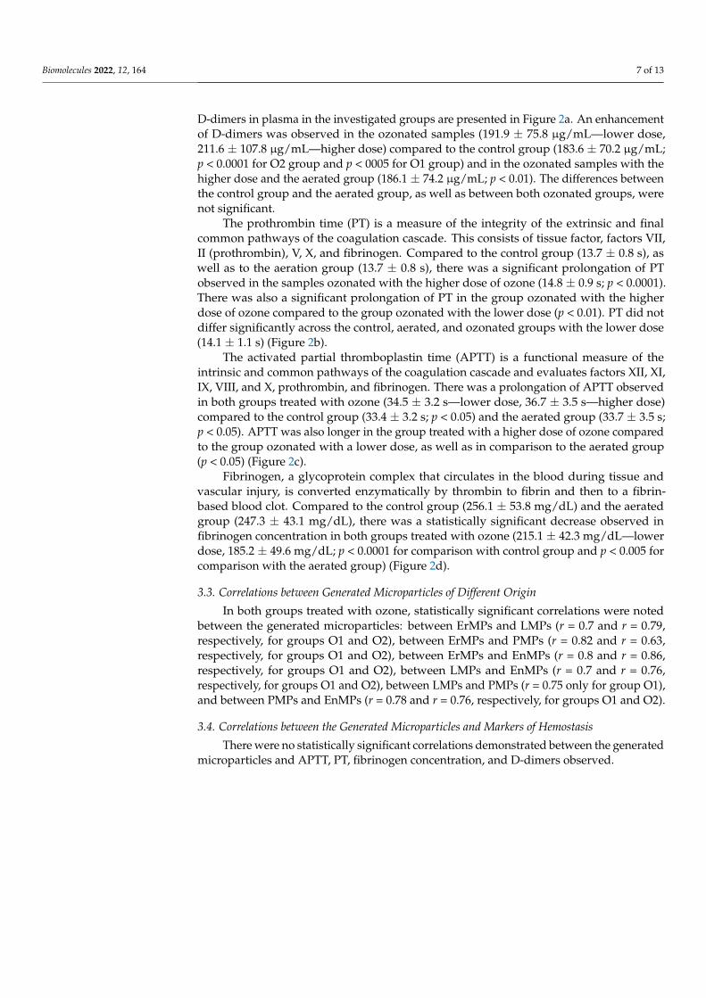
D-dimers in plasma in the investigated groups are presented in Figure 2a. An enhancementof D-dimers was observed in the ozonated samples (191.9 ± 75.8 µg/mL—lower dose,211.6 ± 107.8 µg/mL—higher dose) compared to the control group (183.6 ± 70.2 µg/mL;p < 0.0001 for O2 group and p < 0005 for O1 group) and in the ozonated samples with thehigher dose and the aerated group (186.1 ± 74.2 µg/mL; p < 0.01). The differences betweenthe control group and the aerated group, as well as between both ozonated groups, werenot significant.
The prothrombin time (PT) is a measure of the integrity of the extrinsic and finalcommon pathways of the coagulation cascade. This consists of tissue factor, factors VII,II (prothrombin), V, X, and fibrinogen. Compared to the control group (13.7 ± 0.8 s), aswell as to the aeration group (13.7 ± 0.8 s), there was a significant prolongation of PTobserved in the samples ozonated with the higher dose of ozone (14.8 ± 0.9 s; p < 0.0001).There was also a significant prolongation of PT in the group ozonated with the higherdose of ozone compared to the group ozonated with the lower dose (p < 0.01). PT did notdiffer significantly across the control, aerated, and ozonated groups with the lower dose(14.1 ± 1.1 s) (Figure 2b).
The activated partial thromboplastin time (APTT) is a functional measure of theintrinsic and common pathways of the coagulation cascade and evaluates factors XII, XI,IX, VIII, and X, prothrombin, and fibrinogen. There was a prolongation of APTT observedin both groups treated with ozone (34.5 ± 3.2 s—lower dose, 36.7 ± 3.5 s—higher dose)compared to the control group (33.4 ± 3.2 s; p < 0.05) and the aerated group (33.7 ± 3.5 s;p < 0.05). APTT was also longer in the group treated with a higher dose of ozone comparedto the group ozonated with a lower dose, as well as in comparison to the aerated group(p < 0.05) (Figure 2c).
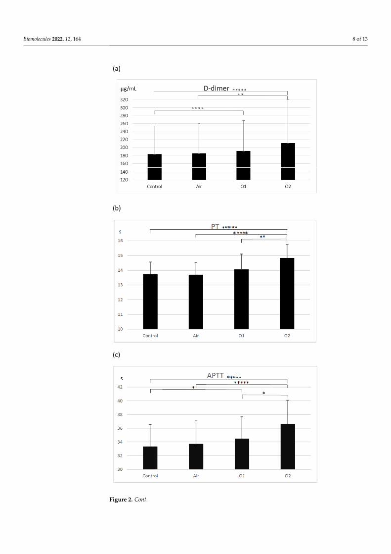
Fibrinogen, a glycoprotein complex that circulates in the blood during tissue andvascular injury, is converted enzymatically by thrombin to fibrin and then to a fibrin-based blood clot. Compared to the control group (256.1 ± 53.8 mg/dL) and the aeratedgroup (247.3 ± 43.1 mg/dL), there was a statistically significant decrease observed infibrinogen concentration in both groups treated with ozone (215.1 ± 42.3 mg/dL—lowerdose, 185.2 ± 49.6 mg/dL; p < 0.0001 for comparison with control group and p < 0.005 forcomparison with the aerated group) (Figure 2d).
3.3. Correlations between Generated Microparticles of Different Origin
In both groups treated with ozone, statistically significant correlations were notedbetween the generated microparticles: between ErMPs and LMPs (r = 0.7 and r = 0.79,respectively, for groups O1 and O2), between ErMPs and PMPs (r = 0.82 and r = 0.63,respectively, for groups O1 and O2), between ErMPs and EnMPs (r = 0.8 and r = 0.86,respectively, for groups O1 and O2), between LMPs and EnMPs (r = 0.7 and r = 0.76,respectively, for groups O1 and O2), between LMPs and PMPs (r = 0.75 only for group O1),and between PMPs and EnMPs (r = 0.78 and r = 0.76, respectively, for groups O1 and O2).
3.4. Correlations between the Generated Microparticles and Markers of Hemostasis
There were no statistically significant correlations demonstrated between the generatedmicroparticles and APTT, PT, fibrinogen concentration, and D-dimers observed.
Biomolecules 2022, 12, 164 8 of 13
(a)
(b)
(c)
Figure 2. Cont.
Biomolecules 2022, 12, 164 9 of 13
(d)
Figure 2. The effect of ozone on D-dimer concentration (a), PT—prothrombin time (b), APTT—activated thromboplastin time (c), and fibrinogen concentration (d). Control—the control group(n = 19); Air—the aerated group (n = 19); O1—the group treated with ozone at a dose of 15 µg/mL(n = 19); O2—the group treated with ozone at a dose of 30 µg/mL (n = 19). Mean ± SD, statisticallysignificant difference between the groups (* p < 0.05; ** p < 0.01; *** p < 0.005; **** p < 0.0005;***** p < 0.0001).
The descriptive statistics of the obtained results is presented in Table S1 in Supplemen-tal Material.
4. Discussion
In a previous study, we evaluated the effects of ozone on human blood, usingmetabolomics [38]. After ozonation of the whole blood, a slight hemolysis (up to 0.6%)was observed. We found several metabolites, whose presence was clearly related to theozone dose or which were present only in ozonated samples: bilirubin, biliverdin, dihy-droxyvitamin D3, pyroglutamic acid, hydroperoxyoctadecadienoic acid (HPODE), trihy-droxyoctadecenoic acid (Tri-HOME), and hydroxyoctadecatrienoic acid (HOTE). Few ofthese compounds are plasma antioxidants, with the exception of bilirubin and biliverdin,which have been shown to be potent antioxidants. Bilirubin acting as an antioxidant isoxidized to biliverdin, which may explain the increase in biliverdin and decrease in biliru-bin concentrations observed after the treatment of blood with ozone. The concentration ofpyroglutamic acid, which is a component of GSH metabolism and is involved in the plasmaantioxidative system, was also decreased. The other metabolite differentiating ozonatedsamples from the others was dihydroxyvitamin D3. Vitamin D3 has already been shown toprevent lipid peroxidation.
There are no published data on the effect of ozone on microparticle release. Ourstudy revealed increasing and dose-dependent MP release following ozonation, which maysuggest that membrane fluidity is elevated within the studied dose range.
The present study provides the first evidence that ozonation of blood induces mi-croparticle release from blood cells. Moreover, the number of released microparticles wasproportional to the applied dose of ozone. Statistically, microparticles released from differ-ent blood cells and endothelial cells correlated significantly with each other, which allowsus to assume that the effect of ozone is not cell-specific.
The observed release of microparticles raises an important question: what risk does itpose for patients transfused with ozonated blood? No pertinent data have been published sofar. During preparation and storage of blood components, blood cells release microparticleswhich remain in the extracellular fluid and are finally transfused to the recipients [39].
Biomolecules 2022, 12, 164 10 of 13
The number and origin of the microparticles generated differ significantly according tothe method and the time of preparation of blood components, depending also on thetype of storage prior to separation into components and after that, before release fortransfusion [40–42].
Due to the variation in methodologies used for isolation of microparticles and in theassessments of MPs by flow cytometry, it is very difficult to compare our data and theresults of studies published so far, on the release of MPs from blood components [43]. Weobserved a twofold increase in the MPs derived from red blood cells and leukocytes afterozonation, while other investigators reported increased MP concentrations ranging from6.8% [42] to 17% [44].
Microparticles in blood components are at least partially responsible for the im-munomodulatory effect of transfusions [45]. This adverse reaction contributes to immuno-suppression, cytokine inhibition, and stimulation of inflammatory reactions. Transfusion-related immunomodulation has been shown to have a favorable effect on exacerbations inthe course of Crohn’s disease, as well as on successful kidney transplantation [46].
Microparticles in the blood components may also contribute to thromboembolic com-plications observed in the recipients of blood transfusions [45]. These microparticles takepart in the processes of coagulation and fibrinolysis, bind coagulation factors [47], presenttissue factor [27], and activate coagulation on their surface. MPs express receptors foractivated protein C, an inhibitor inactivating coagulation factors Va and VIIIa [31].
In parallel, but not in statistical relation to the release of MPs, we demonstrated astatistically significant effect on the basic markers of hemostasis. We observed a prolonga-tion of APTT and PT, a decrease in fibrinogen concentration, and an increase in D-dimerconcentration. Activation of coagulation may be responsible for fibrinogen consumption;however, a direct effect of ozone on fibrinogen cannot be excluded. Rosenfeld et al. [48]revealed that ozone increases the number of hydroxyl and carbonyl groups on the fibrino-gen molecule. D-fragments and, to a lesser extent, fibrinogen fragments are particularlysensitive to ozone-induced oxidation. Oxidation-induced fibrinogen modifications may af-fect its secondary structure and result in higher crosslinking potential. The present in vitrostudy used purified plasma; thus, we cannot extrapolate the results to autohemotherapy.Decreased fibrinogen concentration might be one of the possible mechanisms for the ob-served APTT and PT prolongation. Although statistically significant, the changes in APTT,PT, and fibrinogen were somewhat mild (APTT—9.8%, PT—8%, fibrinogen—27.7%) anddid not exceed normal values. More detailed analyses of the effects of microparticleson blood coagulation have been published by other investigators [33–35,49,50]. While acoincidence of ozone-induced release of microparticles and changes in hemostasis markerswas observed in our study, we did not find a statistically significant correlation among theparameters investigated.
Elevated D-dimer concentration reflects the activation of both coagulation and fibri-nolysis. Some MPs are able to activate the release of plasmin, which suggests that the roleof MPs in hemostasis is more complex than previously assumed [50]. Meijden et al. [33]showed that MPs released from erythrocytes and platelets are able to directly activate factorXII, which most probably explains the activation of fibrinolysis rather than coagulation.Direct activation of factors IX and XI by MPs points to the probable mechanism of activationof the tissue factor-dependent coagulation pathway [34]. Elevated concentration of MPs inozonated blood and blood components may affect coagulation and fibrinolysis and increasethromboembolic risk [35]. It is important to emphasize that we studied the effect of ozoneon blood in vitro, and that our results may explain some of the adverse reactions observedin patients treated with autohemotherapy using ozonated blood. Üreyen et al. [11] reportedan occurrence of myocardial infarction after autohemotherapy in a patient with no riskfactors for thromboembolic complications. Vasospasm of the left main coronary artery andtotal occlusion of the right coronary artery due to thrombus were observed in the patient.However, these findings should be treated with caution, as there is no direct proof thatthe autohemotherapy was responsible for the observed adverse effects [51]. MPs were
Biomolecules 2022, 12, 164 11 of 13
not evaluated in the study. Nevertheless, during autohemotherapy with ozonated blood,the reinfusion of a quantity of ozone-treated blood (a maximum of 200 mL, comparedwith the approximately 5 L in circulation) in the same patient takes place drop by drop,thus allowing a buffer situation for any slight variations in the parameters linked to bloodcoagulation [52]. Moreover, some authors have published positive effects of systemic ozoneadministration in blood hemostasis parameters [52–54].
5. Conclusions
The present study provides evidence that blood ozonation induces the release ofmicroparticles from erythrocytes, leukocytes, platelets, and endothelial cells in a dose-dependent manner. The observed weak, but statistically significant changes in the hemosta-sis system need further research, as preclinical and clinical papers with systemic ozonepoint in the opposite direction.
Supplementary Materials: The following supporting information can be downloaded at: https://www.mdpi.com/article/10.3390/biom12020164/s1, Figure S1: The representative light-scatter dotplot demostrating the MP region and representative fluorescence plots of labeled samples (control);Figure S2: The representative light-scatter dot plot demostrating the MP region and representativefluorescence plots of labeled samples (ozonated blood); Table S1: titleDescriptive statistics of theobtained results.
Author Contributions: Conceptualization, P.R. and B.B.-R.; methodology, P.J.O.; software, M.B.;validation, A,Z., M.M. and M.B.; formal analysis, P.J.O.; investigation, P.J.O. and A.R. and A.Z.;resources, D.S.; data curation, P.J.O.; writing—original draft preparation, B.B.-R.; writing—review andediting, M.Z.W. and B.P.; visualization, M.M.; supervision, P.R. and M.Z.W.; project administration,P.J.O. All authors have read and agreed to the published version of the manuscript.
Funding: This research received no external funding.
Institutional Review Board Statement: The study was conducted in accordance with the Declarationof Helsinki, and approved by the Ethics Committee) of Medical University of Białystok (protocol codeR-I-002/78/2015; date of approval: 25 February 2016). Ethical review and approval were waived forthis study due to use of a part of donated blood by the blood donors.
Informed Consent Statement: Informed consent was obtained from all subjects involved in the study.
Data Availability Statement: Data is contained within the article and supplementary material.
Conflicts of Interest: The authors declare no conflict of interest.
References1. Brinkman, R.; Lamberts, H.B. Ozone as a possible radiomimetic gas. Nature 1958, 181, 1202–1203. [CrossRef]2. Smith, N.L.; Wilson, A.L.; Gandhi, J.; Vatsia, S.; Khan, A.S. Ozone therapy: An overview of pharmacodynamics, current research,
and clinical utility. Med. Gas Res. 2017, 7, 212–219.3. Bocci, V.; Larini, A.; Micheli, V. Restoration of normoxia by ozone therapy may control neoplastic growth: A review and a working
hypothesis. J. Altern. Complement. Med. 2005, 11, 257–265. [CrossRef]4. Inal, M.; Dokumacioglu, A.; Ozcelik, E.; Ucar, O. The effects of ozone therapy and coenzyme Q(1)(0) combination on oxidative
stress markers in healthy subjects. Ir. J. Med. Sci. 2011, 180, 703–707. [CrossRef]5. Bocci, V.A.; Zanardi, I.; Travagli, V. Ozone acting on human blood yields a hormetic dose-response relationship. J. Transl. Med.
2011, 9, 66. [CrossRef] [PubMed]6. Hu, B.; Zheng, J.; Liu, Q.; Yang, Y.; Zhang, Y. The effect and safety of ozone autohemotherapy combined with pharmacological
therapy in postherpetic neuralgia. J. Pain Res. 2018, 11, 1637–1643. [CrossRef] [PubMed]7. Braidy, N.; Izadi, M.; Sureda, A.; Jonaidi-Jafari, N.; Banki, A.; Nabavi, S.F.; Nabavi, S.M. Therapeutic relevance of ozone therapy
in degenerative diseases: Focus on diabetes and spinal pain. J. Cell Physiol. 2018, 233, 2705–2714. [CrossRef] [PubMed]8. Bocci, V.; Travagli, V.; Zanardi, I. May oxygen-ozone therapy improves cardiovascular disorders? Cardiovasc. Hematol. Disord.
Drug Targets 2009, 9, 78–85. [CrossRef] [PubMed]9. Viebahn-Hänsler, R.; Fernández, O.S.L.; Fahmy, Z. Ozone in medicine: Clinical evaluation and evidence classification of the
systemic ozone applications, major autohemotherapy and rectal insufflation, according to the requirements for evidence-basedmedicine. Ozone Sci. Eng. 2016, 38, 322–345. [CrossRef]
Biomolecules 2022, 12, 164 12 of 13
10. Ang, W.-J.; Jiang, L.; Wang, Y.; Kuang, Z.-M. Ozone therapy induced sinus arrest in a hypertensive patient with chronic kidneydisease. A case report. Medicine 2017, 96, e9265.
11. Üreyen, Ç.M.; Bas, C.Y.; Arslan, S. Myocardial infarction after ozone therapy: Is ozone therapy dr. Jekyll or Mr. Hyde? Cardiology2015, 132, 101–104. [CrossRef]
12. Giunta, R.; Coppola, A.; Luongo, C.; Sammartino, A.; Guastafierro, S.; Grassia, A.; Giunta, L.; Mascolo, L.; Tirelli, A.; Coppola, L.Ozonized autohemotransfusion improves hemorheological parameters and oxygen delivery to tissues in patients with peripheralocclusive arterial disease. Ann. Hematol. 2001, 80, 745–748. [CrossRef]
13. Volkhovskaya, N.B.; Tkachenko, S.B.; Belopolsky, A.A. Modulation of phagocytic activity of blood polynuclear leukocytes withozonized physiological saline. Bull. Exp. Biol. Med. 2008, 146, 559–561. [CrossRef]
14. Bocci, V.; Paulesu, L. Studies on the biological effects of ozone 1. Induction of interferon gamma on human leucocytes. Haemato-logica 1990, 75, 510–515.
15. Sen, R.; Baltimore, D. Inducibility of kappa immunoglobulin enhance binding protein Nf-kappa B by a posttranslationalmechanism. Cell 1986, 47, 921–928. [CrossRef]
16. Baeuerle, P.A.; Henkel, T. Function and activation of NF-kappa B in the immune system. Annu. Rev. Immunol. 1994, 12, 141–179.[CrossRef]
17. Yogev, Y. A humoral solution: Autologous blood products and tissue repair. Cell. Immunol. 2020, 356, 104178. [CrossRef]18. Burgassi, S.; Zanardi, I.; Travagli, V.; Montomoli, E.; Bocci, V. How much ozone bactericidal activity is compromised by plasma
components? J. Appl. Microbiol. 2009, 106, 1715–1721. [CrossRef]19. Valacchi, G.; Bocci, V. Studies on the biological effects of ozone: 10. Release of factors from ozonated human platelets. Mediat.
Inflamm. 1999, 8, 205–209. [CrossRef]20. Orakdogen, M.; Uslu, S.; Emon, S.T.; Somay, H.; Meric, Z.C.; Hakan, T. The effect of ozone therapy on experimental vasospasm in
the rat femoral artery. Turk. Neurosurg. 2016, 26, 860–865. [CrossRef]21. Di Paolo, N.; Bocci, V.; Garosi, G.; Borrelli, E.; Bravi, A.; Bruci, A.; Aldinucci, C.; Capotondo, L. Extracorporeal blood oxygenation
and ozonation (EBOO) in man. preliminary report. Int. J. Artif. Organs. 2000, 23, 131–141. [CrossRef]22. Borrelli, E.; Bocci, V. Visual improvement following ozone therapy in dry age related macular degeneration; a review. Med.
Hypothesis Discov. Innov. Ophthalmol. 2013, 2, 47–51.23. Re, L.; Martínez-Sánchez, G.; Bordicchia, M.; Malcangi, G.; Pocognoli, A.; Morales-Segura, M.A.; Rothchild, J.; Rojas, A. Is ozone
pre-conditioning effect linked to Nrf2/EpRE activation pathway in vivo? A preliminary result. Eur. J. Pharmacol. 2014, 742,158–162. [CrossRef]
24. Cisterna, B.; Costanzo, M.; Nodari, A.; Galiè, M.; Zanzoni, S.; Bernardi, P.; Covi, V.; Tabaracci, G.; Malatesta, M. Ozone activatesthe Nrf2 pathway and improves preservation of explanted adipose tissue in vitro. Antioxidants 2020, 9, 989. [CrossRef]
25. Galiè, M.; Covi, V.; Tabaracci, G.; Malatesta, M. The role of Nrf2 in the antioxidant cellular response to medical ozone exposure.Int. J. Mol. Sci. 2019, 20, 4009. [CrossRef]
26. Helbing, T.; Olivier, C.; Bode, C.; Moser, M.; Diehl, P. Role of microparticles in endothelial dysfunction and arterial hypertension.World J. Cadiol. 2014, 6, 1135–1139. [CrossRef]
27. Date, K.; Ettelaie, C.; Maraveyas, A. Tissue factor-bearing microparticles and inflammation: A potential mechanism for thedevelopment of venous thromboembolism in cancer. J. Thromb. Haemost. 2017, 15, 2289–2299. [CrossRef]
28. Mause, S.F.; Weber, C. Microparticles: Protagonists of a novel communication network for intercellular information exchange.Circ. Res. 2010, 107, 1047–1057. [CrossRef]
29. Kogure, T.; Yan, I.K.; Lin, W.-L.; Patel, T. Extracellular vesicle-mediated transfer of a novel long noncoding RNA TUC339: Amechanism of intercellular signaling in human hepatocellular cancer. Genes Cancer 2013, 4, 261–272. [CrossRef]
30. Boon, R.A.; Vickers, K.C. Intercellular transport of microRNAs. Arterioscler. Thromb. Vasc. Biol. 2013, 33, 186–192. [CrossRef]31. Schouten, M.; Wiersinga, W.J.; Levi, M.; van der Poll, T. Inflammation, endothelium, and coagulation in sepsis. J. Leukoc. Biol.
2008, 83, 536–545. [CrossRef]32. Rautou, P.-E.; Vion, A.-C.; Amabile, N.; Chironi, G.; Simon, A.; Tedgui, A.; Boulanger, C.M. Microparticles, vascular function, and
atherothrombosis. Circ. Res. 2011, 109, 593–606. [CrossRef]33. Van Der Meijden, P.E.; Van, S.M.; Van, O.R.; Van, T.; Renne, A.J. Platelet- and erythrocyte-derived microparticles trigger thrombin
generation via factor XIIa. J. Thromb. Haemost. 2012, 10, 1355–1362. [CrossRef] [PubMed]34. Lipets, E.; Vlasova, O.; Urnova, E.; Margolin, O.; Soloveva, A.; Ostapushchenko, O.; Andersen, J.; Ataullakhanov, F.; Panteleev, M.
Circulating contact-pathway-activating microparticles together with factors IXa and XIa induce spontaneous clotting in plasma ofhematology and cardiologic Patients. PLoS ONE 2014, 9, e87692. [CrossRef] [PubMed]
35. Vallier, L.; Cointe, S.; Lacroix, R.; Bonifay, A.; Judicone, C.; Dignant-George, F.; Kwaan, H.C. Microparticles and fibrinolysis.Semin. Thromb. Hemost. 2017, 43, 129–134. [CrossRef] [PubMed]
36. Wojtukiewicz, M.Z.; Mysliwiec, M.; Sierko, E.; Sobierska, M.; Kruszewska, J.; Lipska, A.; Radziwon, P.; Tucker, S.C.; Honn, K.V.Elevated microparticles, thrombin-antithrombin and VEGF levels in colorectal cancer patients undergoing chemotherapy. Pathol.Oncol. Res. 2020, 26, 2499–2507. [CrossRef] [PubMed]
37. Ciborowski, M.; Lipska, A.; Godzien, J.; Ferrarini, A.; Korsak, J.; Radziwon, P.; Tomasiak, M.; Barbas, C. Combination of LC-MS-and GC-MS-based metabolomics to study the effect of ozonated autohemotherapy on human blood. J. Proteome Res. 2012, 11,6231–6241. [CrossRef] [PubMed]
Biomolecules 2022, 12, 164 13 of 13
38. Travagli, V.; Zanardi, I.; Silvietti, A.; Nepi, S.; Tenori, L.; Bocci, V. Effects of ozone blood treatment on the metabolite profile ofhuman blood. Int. J. Toxicol. 2010, 29, 165–174. [CrossRef] [PubMed]
39. Burnouf, T.; Chou, M.L.; Goubran, H.; Cognasse, F.; Garraud, O.; Seghatchian, J. An overview of the role of microparti-cles/microvesicles in blood components: Are they clinically beneficial or harmful? Transfus. Apher. Sci. 2015, 53, 137–145.[CrossRef] [PubMed]
40. Almizraq, R.J.; Seghatchian, J.; Acker, J.P. Extracellular vesicles in transfusion-related immunomodulation and the role of bloodcomponent manufacturing. Transfus. Apher. Sci. 2016, 255, 281–291. [CrossRef] [PubMed]
41. Ayersa, L.; Kohler, M.; Harrison, P.; Sargent, I.; Dragovic, R.; Schaap, M.; Nieuwland, R.; Brooks, S.A.; Ferry, B. Measurement ofcirculating cell-derived microparticles by flow cytometry: Sources of variability within the assay. Thromb. Res. 2011, 127, 370–377.[CrossRef]
42. Noulsri, E.; Palasuwan, A. Effects of donor age, donor sex, blood-component processing, and storage on cell-derived microparticleconcentrations in routine blood-component preparation. Transfus. Apher. Sci. 2018, 57, 587–592. [CrossRef]
43. Almizraq, R.J.; Seghatchian, J.; Holovati, J.L.; Acker, J.P. Extracellular vesicle characteristics in stored red blood cell concentratesare influenced by the method of detection. Transfus. Apher. Sci. 2017, 56, 254–260. [CrossRef]
44. Gao, Y.; Lv, L.; Liu, S.; Ma, G.; Su, Y. Elevated levels of thrombin-generating microparticles in stored red blood cells. Vox Sang.2013, 105, 11–17. [CrossRef]
45. Rubin, O.; Canellini, G.; Delobel, J.; Lion, N.; Tissot, J.D. Red blood cell microparticles: Clinical relevance. Transfus. Med. Hemother.2012, 39, 342–347. [CrossRef]
46. Sadallah, S.; Eken, C.; Schifferli, J.A. Ectosomes as modulators of inflammation and immunity. Clin. Exp. Immunol. 2011, 163,26–32. [CrossRef] [PubMed]
47. Piccin, A.; Murphy, W.G.; Smith, O.P. Circulating microparticles: Pathophysiology and clinical implications. Blood Rev. 2007, 21,157–171. [CrossRef] [PubMed]
48. Rosenfeld, M.A.; Shchegolikhin, A.N.; Bychkova, A.V.; Leonova, V.B.; Biryukova, M.I.; Kostanova, E.A. Ozone-induced oxidativemodification of fibrinogen: Role of the D regions. Free Radic. Biol. Med. 2014, 77, 106–120. [CrossRef] [PubMed]
49. Olivier, R.; Delobel, J.; Prudent, M.; Lion, N.; Kohl, K.; Tucker, E.I.; Tissot, J.-D.; Angelillo-Scherrer, A. Red blood celliderivedmicroparticles isolated from blood units initiate and propagate thrombin generation. Transfusion 2013, 53, 1744–1754.
50. Lacroix, R.; Plawinski, L.; Robert, S.; Doeuvre, L.; Sabatier, F.; Martinez de Lizarrondo, S.; Mezzapesa, A.; Anfosso, F.; Leroyer,A.S.; Poullin, P.; et al. Leukocyte- and endothelial-derived microparticles: A circulating source for fibrinolysis. Haematologica 2012,97, 1864–1872. [CrossRef]
51. Re, L.; Rowen, R.; Travagli, V. Ozone therapy and its use in medicine. Cardiology 2016, 134, 99–100. [CrossRef]52. Biedunkiewicz, B.; Lizakowski, S.; Tylicki, L.; Skiboeska, A.; Nieweglowski, T.; Chamienia, A.; Debska-Slizien, A.; Rutkowski, B.
Blood coagulation unaffected by ozonated autohemotherapy in patients on maintenance hemodialysis. Arch. Med. Res. 2006, 37,1034–1037. [CrossRef] [PubMed]
53. Díaz, A.; García, M.; Pina, C.; Menéndez, S. Efecto del ozono sobre la actividad plaquetaria en paciente diabéticos tratados conozonoterapia: Informe preliminar. Rev. Cuba. Investig. Biomed. 2001, 20, 45–47.
54. Martínez, Y.; Zamora, Z.; González, R.; Guanche, D. Effect of ozone oxidative preconditioning on bleeding time and venousthrombosis in a septic shock model in rats. Cienc. Biol. 2010, 41, 41–51.