
Whole Thing for pdf - Open Research
372
Mechanisms of the Intriguing Rearrangements of Activated Organic Species A Thesis Submitted for the Degree of Doctor of Philosophy of The Australian National University by David Grant Harman Department of Chemistry Faculty of Science Canberra April 2003
-
Upload
khangminh22 -
Category
Documents
-
view
0 -
download
0
Transcript of Whole Thing for pdf - Open Research
Mechanisms of the Intriguing Rearrangementsof Activated Organic Species
A Thesis Submitted for the Degree
of
Doctor of Philosophy
of
The Australian National University
by
David Grant Harman
Department of Chemistry
Faculty of Science
Canberra
April 2003

ii

iii
Declaration
This thesis is an original work. None of the work has been previously submitted
by me for the purpose of obtaining a degree or diploma in any university or other tertiary
education institution. To the best of my knowledge, this thesis does not contain material
previously published by another person, except where due reference is made in the text.

iv
To those who tell me the truth in love,
particularly when I do not want to hear it.
v
Acknowledgements
Many people, too numerous to mention individually, assisted in the production of
this thesis. Corporately I wish to thank them and to express my deep and sincere
gratitude. I would briefly like to mention those to whom I am particularly indebted and I
apologise to anyone whom I have inadvertently omitted.
Professor Athel Beckwith was my supervisor while I was working in the
laboratories of the Research School of Chemistry. Athel and his wife Kaye made me feel
welcome in Canberra. I'm grateful to have worked on such challenging projects and to
have had such expert guidance. Athel's love for chemistry, his vast knowledge, his view
of the bigger picture and the humility to admit when he didn't know something have been
the source of much inspiration.
My RSC advisers were Professor Lew Mander, Professor Rod Rickards and Dr
John MacLeod. I would like to thank them and other academic, technical and
administrative staff for valuable advice and assistance. Mr Robert Longmore deserves a
particular mention for his laboratory prowess, his willingness to share his expertise and
his friendship. So does Mrs Joan Smith—RSC Librarian Extraordinaire—for her
helpfulness, patience, encouragement and continual grace when loans were overdue.
Dr Steven Brumby helped me record the esr spectra. My brother, Mr Ian
Harman, helped me with the computer program used to determine the rotational barriers
in β-substituted ethyl radicals. Ms Peta Simmonds, Mrs Tin Culnane and Mr Chris Blake
of the ANU NMR Centre helped me with two-dimensional and 17O nmr. Drs Graham
Heath, Richard Webster and Brett Yeomans assisted me with the recording and
interpretation of cyclic voltammagrams.
I thank the Australian Government for a Commonwealth Postgraduate Research
Award, known later as the Australian Postgraduate Research Award. I'm grateful to have
received an Australian National University PhD Scholarship for six months upon the
expiration of my APRA.
I'm one of the lucky few who have worked in both the Research School of
Chemistry and the Department of Chemistry. Professor Jack Elix has been my

vi
supervisor during my time in the Department. I'd like to thank him for taking on a
student from a field quite different to his own, for his efforts in correcting thesis drafts
and for his gentle advice. Drs Christina Chai and Geoff Salem, my current advisers, I
thank for their assistance. My passage into the Department was facilitated by past Heads
Professor Jack Elix and Dr Gad Fischer. Thanks also to the current Head, Dr Geoff
Salem, for his support. Mr Warren Griffiths provided tireless assistance with computer
problems.
And last, but by no means the least, I'd like to acknowledge the unending support
my parents Kay and Grant have given me in realms moral, spiritual and financial and for
some proof reading.
vii
Abstract
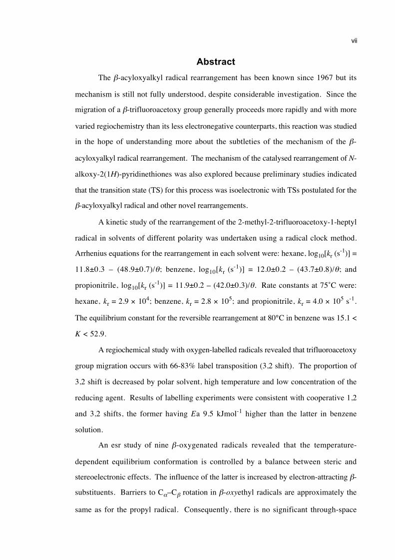
The β-acyloxyalkyl radical rearrangement has been known since 1967 but its
mechanism is still not fully understood, despite considerable investigation. Since the
migration of a β-trifluoroacetoxy group generally proceeds more rapidly and with more
varied regiochemistry than its less electronegative counterparts, this reaction was studied
in the hope of understanding more about the subtleties of the mechanism of the β-
acyloxyalkyl radical rearrangement. The mechanism of the catalysed rearrangement of N-
alkoxy-2(1H)-pyridinethiones was also explored because preliminary studies indicated
that the transition state (TS) for this process was isoelectronic with TSs postulated for the
β-acyloxyalkyl radical and other novel rearrangements.
A kinetic study of the rearrangement of the 2-methyl-2-trifluoroacetoxy-1-heptyl
radical in solvents of different polarity was undertaken using a radical clock method.
Arrhenius equations for the rearrangement in each solvent were: hexane, log10[kr (s-1)] =
11.8±0.3 – (48.9±0.7)/θ; benzene, log10[kr (s-1)] = 12.0±0.2 – (43.7±0.8)/θ; and
propionitrile, log10[kr (s-1)] = 11.9±0.2 – (42.0±0.3)/θ. Rate constants at 75˚C were:
hexane, kr = 2.9 × 10⁴; benzene, kr = 2.8 × 10⁵; and propionitrile, kr = 4.0 × 10⁵ s-1.
The equilibrium constant for the reversible rearrangement at 80°C in benzene was 15.1 <
K < 52.9.
A regiochemical study with oxygen-labelled radicals revealed that trifluoroacetoxy
group migration occurs with 66-83% label transposition (3,2 shift). The proportion of
3,2 shift is decreased by polar solvent, high temperature and low concentration of the
reducing agent. Results of labelling experiments were consistent with cooperative 1,2
and 3,2 shifts, the former having Ea 9.5 kJmol-1 higher than the latter in benzene
solution.
An esr study of nine β-oxygenated radicals revealed that the temperature-
dependent equilibrium conformation is controlled by a balance between steric and
stereoelectronic effects. The influence of the latter is increased by electron-attracting β-
substituents. Barriers to Cα–Cβ rotation in β-oxyethyl radicals are approximately the
same as for the propyl radical. Consequently, there is no significant through-space
viii
interaction between the β-substituent and the unpaired electron.
Experimental results were consistent with a mechanism involving a combination
of polarized 1,2 and 3,2 concerted shifts. The results may also be rationalised by the
intermediacy of a contact ion pair, as well as combinations of the three options.
The rearrangement of N-alkoxy-2(1H)-pyridinethiones is catalysed by oxidants,
Lewis acids and protic acids. Pseudo first order kinetics are observed and there are
moderate solvent effects. The migration of a 1,1-dideuteroallyl group occurs almost
exclusively in a 1,4 sense. Migration of an enantiomerically enriched 1-phenylethyl
group proceeds with predominant retention of configuration in chloroform, but with
virtual racemisation in acetonitrile. Migrating groups do not become diffusively free
during the rearrangement. Substituents which stablise positive charge at C1 migrate more
rapidly. The bulk of evidence indicates that a catalyst activates the pyridinethione for
rearrangement by promoting aromatisation. Mass-spectrometric analysis of an isolated
intermediate and kinetic results are consistent with an intermolecular mechanism.
ix
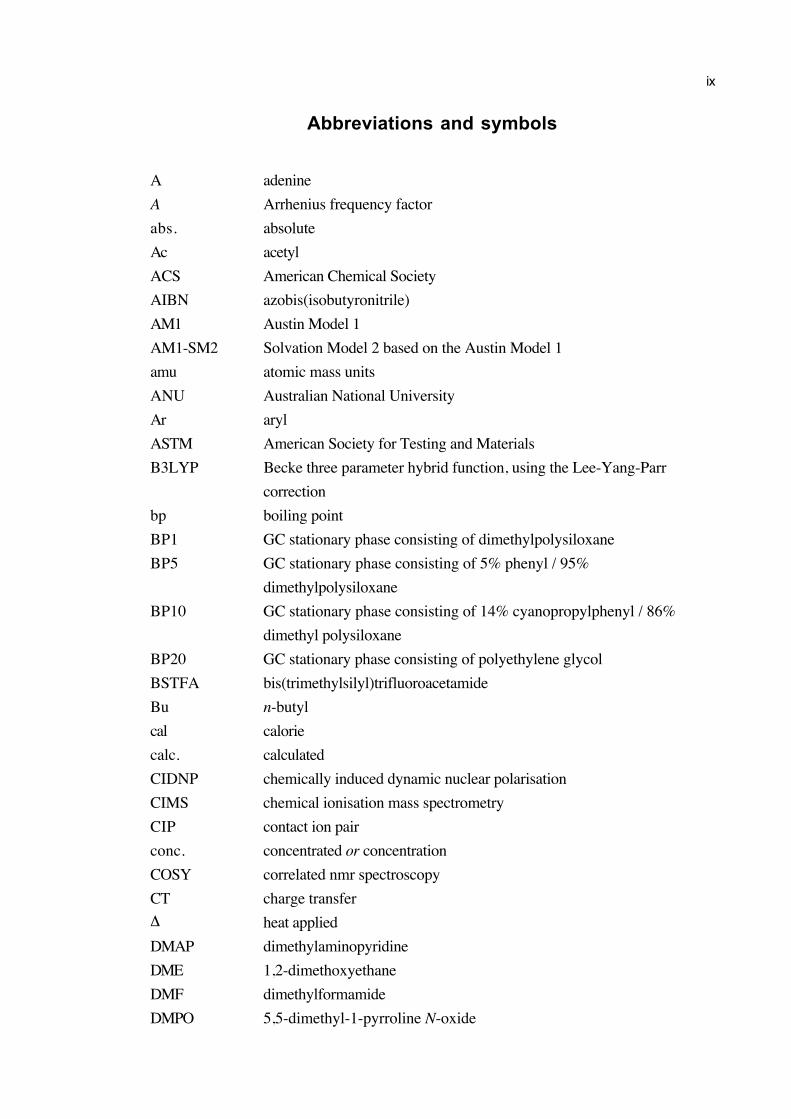
Abbreviations and symbols
A adenine
A Arrhenius frequency factor
abs. absolute
Ac acetyl
ACS American Chemical Society
AIBN azobis(isobutyronitrile)
AM1 Austin Model 1
AM1-SM2 Solvation Model 2 based on the Austin Model 1
amu atomic mass units
ANU Australian National University
Ar aryl
ASTM American Society for Testing and Materials
B3LYP Becke three parameter hybrid function, using the Lee-Yang-Parr
correction
bp boiling point
BP1 GC stationary phase consisting of dimethylpolysiloxane
BP5 GC stationary phase consisting of 5% phenyl / 95%
dimethylpolysiloxane
BP10 GC stationary phase consisting of 14% cyanopropylphenyl / 86%
dimethyl polysiloxane
BP20 GC stationary phase consisting of polyethylene glycol
BSTFA bis(trimethylsilyl)trifluoroacetamide
Bu n-butyl
cal calorie
calc. calculated
CIDNP chemically induced dynamic nuclear polarisation
CIMS chemical ionisation mass spectrometry
CIP contact ion pair
conc. concentrated or concentration
COSY correlated nmr spectroscopy
CT charge transfer
∆ heat applied
DMAP dimethylaminopyridine
DME 1,2-dimethoxyethane
DMF dimethylformamide
DMPO 5,5-dimethyl-1-pyrroline N-oxide
x
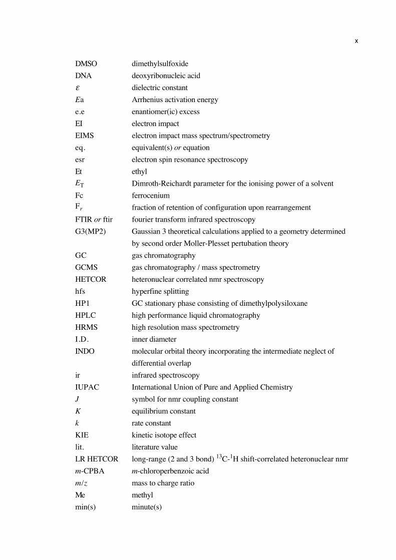
DMSO dimethylsulfoxide
DNA deoxyribonucleic acid
ε dielectric constant
Ea Arrhenius activation energy
e.e enantiomer(ic) excess
EI electron impact
EIMS electron impact mass spectrum/spectrometry
eq. equivalent(s) or equation
esr electron spin resonance spectroscopy
Et ethyl
ET Dimroth-Reichardt parameter for the ionising power of a solvent
Fc ferroceniumFr fraction of retention of configuration upon rearrangement
FTIR or ftir fourier transform infrared spectroscopy
G3(MP2) Gaussian 3 theoretical calculations applied to a geometry determined
by second order Moller-Plesset pertubation theory
GC gas chromatography
GCMS gas chromatography / mass spectrometry
HETCOR heteronuclear correlated nmr spectroscopy
hfs hyperfine splitting
HP1 GC stationary phase consisting of dimethylpolysiloxane
HPLC high performance liquid chromatography
HRMS high resolution mass spectrometry
I.D. inner diameter
INDO molecular orbital theory incorporating the intermediate neglect of
differential overlap
ir infrared spectroscopy
IUPAC International Union of Pure and Applied Chemistry
J symbol for nmr coupling constant
K equilibrium constant
k rate constant
KIE kinetic isotope effect
lit. literature value
LR HETCOR long-range (2 and 3 bond) 13C-1H shift-correlated heteronuclear nmr
m-CPBA m-chloroperbenzoic acid
m/z mass to charge ratio
Me methyl
min(s) minute(s)
xi
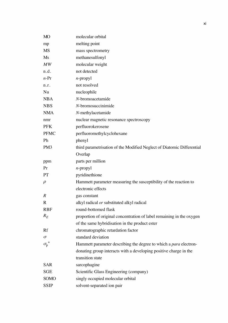
MO molecular orbital
mp melting point
MS mass spectrometry
Ms methanesulfonyl
MW molecular weight
n.d. not detected
n-Pr n-propyl
n.r. not resolved
Nu nucleophile
NBA N-bromoacetamide
NBS N-bromosuccinimide
NMA N-methylacetamide
nmr nuclear magnetic resonance spectroscopy
PFK perfluorokerosene
PFMC perfluoromethylcyclohexane
Ph phenyl
PM3 third parametrisation of the Modified Neglect of Diatomic Differential
Overlap
ppm parts per million
Pr n-propyl
PT pyridinethione
ρ Hammett parameter measuring the susceptibility of the reaction to
electronic effects
R gas constant
R alkyl radical or substituted alkyl radical
RBF round-bottomed flaskRE proportion of original concentration of label remaining in the oxygen
of the same hybridisation in the product ester
Rf chromatographic retardation factor
σ standard deviation
σp+ Hammett parameter describing the degree to which a para electron-
donating group interacts with a developing positive charge in the
transition state
SAR sarcophagine
SGE Scientific Glass Engineering (company)
SOMO singly occupied molecular orbital
SSIP solvent-separated ion pair
xii
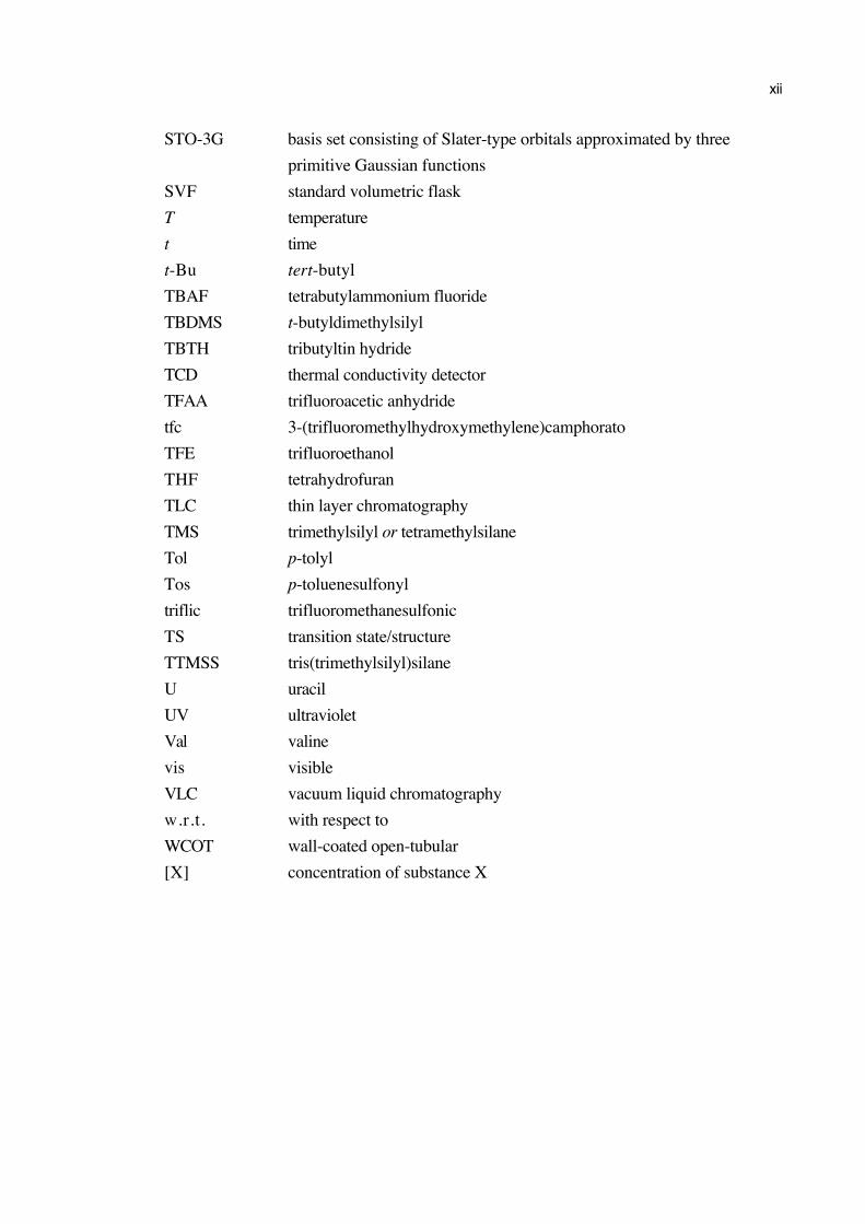
STO-3G basis set consisting of Slater-type orbitals approximated by three
primitive Gaussian functions
SVF standard volumetric flask
T temperature
t time
t-Bu tert-butyl
TBAF tetrabutylammonium fluoride
TBDMS t-butyldimethylsilyl
TBTH tributyltin hydride
TCD thermal conductivity detector
TFAA trifluoroacetic anhydride
tfc 3-(trifluoromethylhydroxymethylene)camphorato
TFE trifluoroethanol
THF tetrahydrofuran
TLC thin layer chromatography
TMS trimethylsilyl or tetramethylsilane
Tol p-tolyl
Tos p-toluenesulfonyl
triflic trifluoromethanesulfonic
TS transition state/structure
TTMSS tris(trimethylsilyl)silane
U uracil
UV ultraviolet
Val valine
vis visible
VLC vacuum liquid chromatography
w.r.t. with respect to
WCOT wall-coated open-tubular
[X] concentration of substance X
xiii
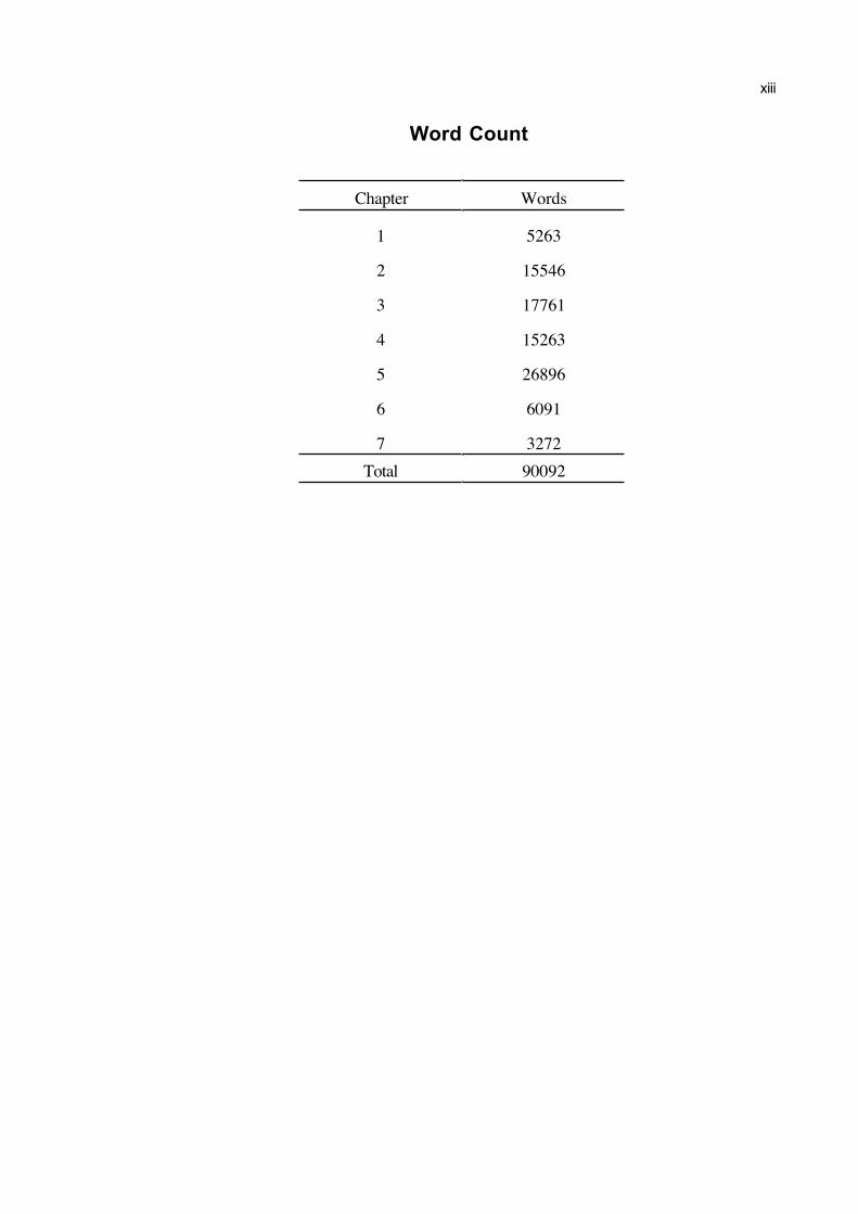
Word Count
Chapter Words
1 5263
2 15546
3 17761
4 15263
5 26896
6 6091
7 3272
Total 90092
xiv
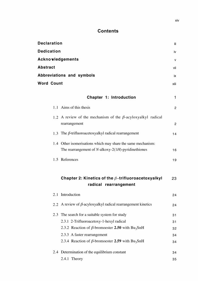
Contents
Declaration iii
Dedication iv
Acknowledgements v
Abstract vii
Abbreviations and symbols ix
Word Count xiii
Chapter 1: Introduction 1
1.1 Aims of this thesis 2
1.2 A review of the mechanism of the β-acyloxyalkyl radical
rearrangement 2
1.3 The β-trifluoroacetoxyalkyl radical rearrangement 14
1.4 Other isomerisations which may share the same mechanism:
The rearrangement of N-alkoxy-2(1H)-pyridinethiones 16
1.5 References 19
Chapter 2: Kinetics of the β−trifluoroacetoxyalkyl
radical rearrangement
23
2.1 Introduction 24
2.2 A review of β-acyloxyalkyl radical rearrangement kinetics 24
2.3 The search for a suitable system for study 31
2.3.1 2-Trifluoroacetoxy-1-hexyl radical 31
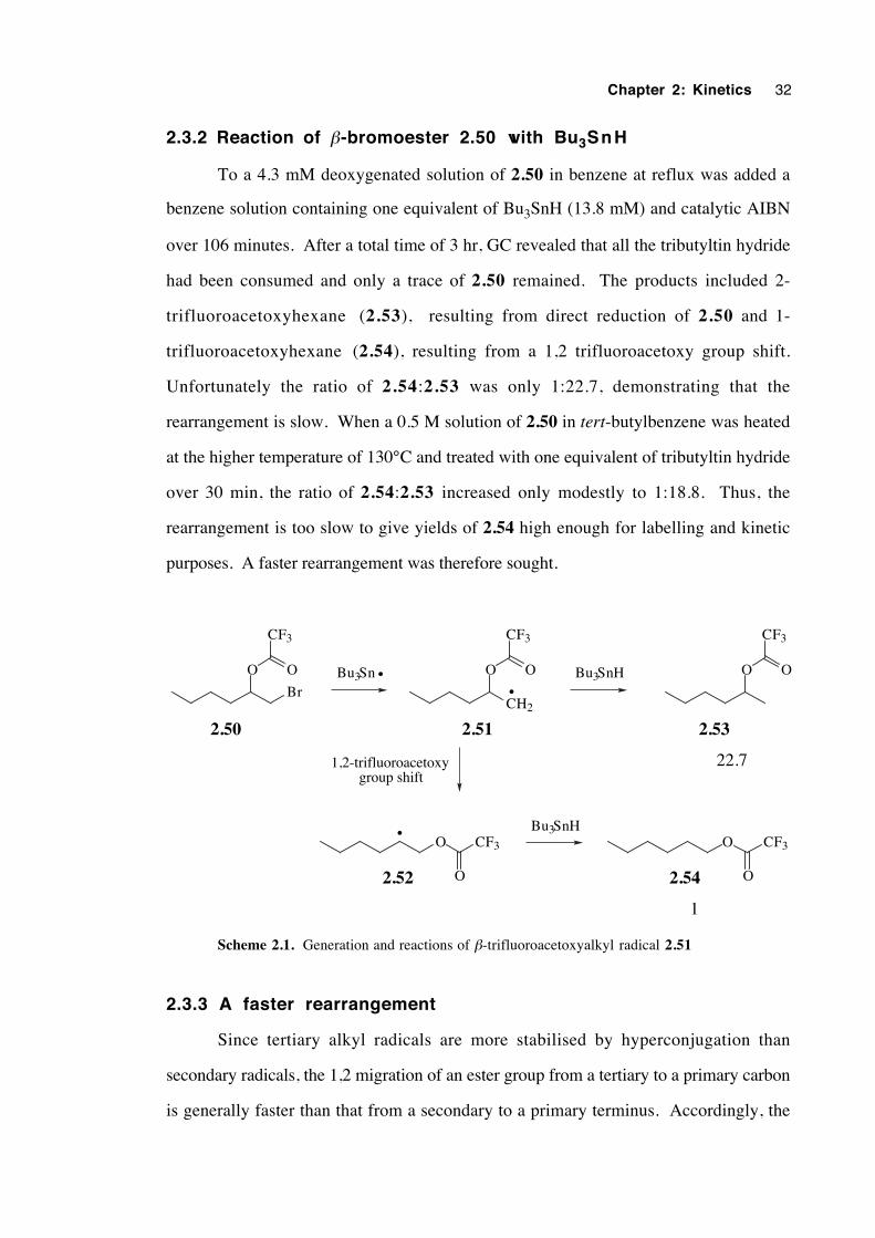
2.3.2 Reaction of β-bromoester 2.50 with Bu3SnH 32
2.3.3 A faster rearrangement 34
2.3.4 Reaction of β-bromoester 2.59 with Bu3SnH 34
2.4 Determination of the equilibrium constant 34
2.4.1 Theory 35
xv
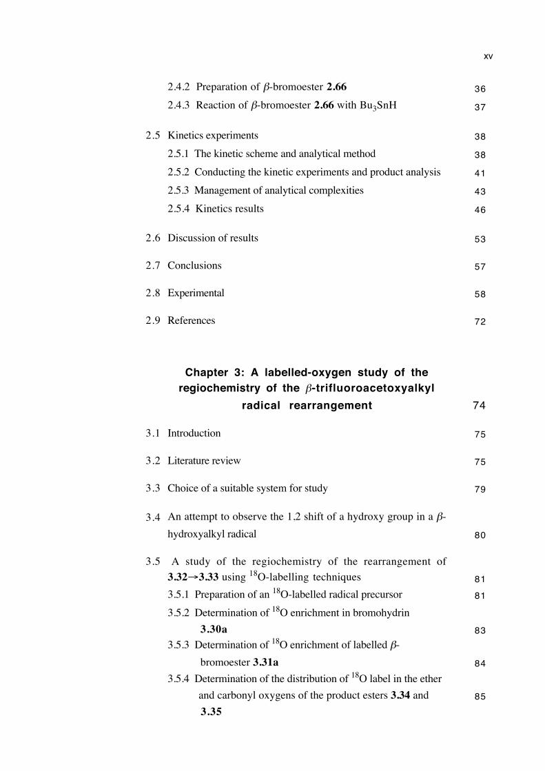
2.4.2 Preparation of β-bromoester 2.66 36
2.4.3 Reaction of β-bromoester 2.66 with Bu3SnH 37
2.5 Kinetics experiments 38
2.5.1 The kinetic scheme and analytical method 38
2.5.2 Conducting the kinetic experiments and product analysis 41
2.5.3 Management of analytical complexities 43
2.5.4 Kinetics results 46
2.6 Discussion of results 53
2.7 Conclusions 57
2.8 Experimental 58
2.9 References 72
Chapter 3: A labelled-oxygen study of theregiochemistry of the β-trifluoroacetoxyalkyl
radical rearrangement 74
3.1 Introduction 75
3.2 Literature review 75
3.3 Choice of a suitable system for study 79
3.4 An attempt to observe the 1,2 shift of a hydroxy group in a β-
hydroxyalkyl radical 80
3.5 A study of the regiochemistry of the rearrangement of
3.32→3.33 using 18O-labelling techniques 81
3.5.1 Preparation of an 18O-labelled radical precursor 81
3.5.2 Determination of 18O enrichment in bromohydrin
3.30a 83
3.5.3 Determination of 18O enrichment of labelled β-
bromoester 3.31a 84
3.5.4 Determination of the distribution of 18O label in the ether
and carbonyl oxygens of the product esters 3.34 and
3.3585
xvi
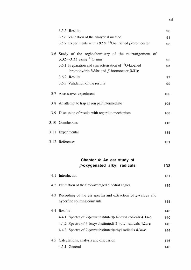
3.5.5 Results 90
3.5.6 Validation of the analytical method 91
3.5.7 Experiments with a 92 % 18O-enriched β-bromoester 93
3.6 Study of the regiochemistry of the rearrangement of
3.32→3.33 using 17O nmr 95
3.6.1 Preparation and characterisation of 17O-labelled
bromohydrin 3.30c and β-bromoester 3.31c95
3.6.2 Results 97
3.6.3 Validation of the results 99
3.7 A crossover experiment 100
3.8 An attempt to trap an ion pair intermediate 105
3.9 Discussion of results with regard to mechanism 108
3.10 Conclusions 116
3.11 Experimental 118
3.12 References 131
Chapter 4: An esr study ofβ-oxygenated alkyl radicals 133
4.1 Introduction 134
4.2 Estimation of the time-averaged dihedral angles 135
4.3 Recording of the esr spectra and extraction of g-values and
hyperfine splitting constants 138
4.4 Results 140
4.4.1 Spectra of 2-(oxysubstituted)-1-hexyl radicals 4.1a-c 140
4.4.2 Spectra of 3-(oxysubstituted)-2-butyl radicals 4.2a-c 142
4.4.3 Spectra of 2-(oxysubstituted)ethyl radicals 4.3a-c 144
4.5 Calculations, analysis and discussion 146
4.5.1 General 146
xvii
4.5.2 2-(Oxysubstituted)hexyl radicals 4.1a-c 152
4.5.3 3-(Oxysubstituted)-2-butyl radicals 4.2a-c 157
4.5.4 2-(Oxysubstituted)ethyl radicals 4.3a-c 160
4.6 Estimation of the energy barrier to internal rotation about theCα– Cβ bond in the β-oxygenated ethyl radicals 4.3a-c 170
4.7 Final discussion 176
4.8 Conclusions 180
4.9 Experimental 181
4.10 References 187
Chapter 5: The mechanism of the
catalysed rearrangement of
N-alkoxy-2(1H)-pyridinethiones 192
5.1 Introduction 193
5.2 Review of the chemistry of N-alkoxy-2(1H)-pyridinethiones
and related compounds 193
5.2.1 Prevalence of N-alkoxy-2(1H)-pyridinethione research 193
5.2.2 Barton esters and related classes of compounds 194
5.2.3 Radical chemistry of N-alkoxy-2(1H)-pyridinethiones 195
5.2.4 Related rearrangements 199
5.2.5 Catalysed N-alkoxy-2(1H)-pyridinethione rearrangement 202
5.2.6 The chemistry of 4 and 6 electron 1,4 sigmatropic shifts
and 5-electron, 6-centre electrocyclic processes 204
5.3 The mechanism of the rearrangement of N-alkoxy-2(1H)-
pyridinethiones 206
5.3.1 The mode of catalysis 206
5.3.1.1 Can the rearrangement of 2-alkoxypyridine N-
oxides be catalysed in the same manner? 211
5.3.2 Kinetics 212
5.3.3 A study of rearrangement regiochemistry 219
5.3.4 A study of rearrangement stereochemistry 224
xviii
5.3.4.1 Preparation of optically active reactants and
products 225
5.3.4.2 Results 230
5.3.4.3 Determination of the extent of solution-phase
racemisation of both the pyridinethione 5.1d
and the N-oxide 5.2d 232
5.3.4.4 Discussion of results 233
5.3.5 Electronic structure of the migrating group at C1 during
rearrangement 234
5.3.6 Substituent effects 237
5.3.7 Attempted detection and isolation of intermediates 239
5.3.7.1 Addition of a radical scavenger 239
5.3.7.2 Esr spectroscopy 240
5.3.7.3 Cyclic voltammetry 241
5.3.7.4 Isolation and attempted identification of
intermediates 244
5.4 Conclusions 247
5.5 Future work 249
5.6 Experimental 250
5.7 References 277
Chapter 6: General discussion and conclusions 284
6.1 Introduction 285
6.2 The β-trifluoroacetoxyalkyl radical rearrangement 285
6.2.1 What is known about the rearrangement of the 2-methyl-
2-trifluoroacetoxy-1-heptyl radical? 285
6.2.2 Migrating group electronic effects 287
6.2.3 Relationship between rearrangement regiochemistry and
kinetics 288
6.2.4 Is the regiochemistry controlled by the conformation of
the ester group? 289
6.2.5 Predicted dynamics for a radical ion pair intermediate 291
xix
6.2.6 The mechanism of the rearrangement of β-
trifluoroacetoxyalkyl radicals 295
6.3 Related radical-mediated rearrangements and β-eliminations 297
6.4 The mechanism of the rearrangement of N-alkoxy-2(1H)-
pyridinethiones 301
6.5 Final remarks 301
6.6 References 303
Chapter 7: General experimental 306
7.1 Melting points 307
7.2 Elemental analyses 307
7.3 Infrared spectroscopy 307
7.4 Optical rotations 307
7.5 Molecular ultraviolet and visible spectra 307
7.6 Bulb to bulb distillations 307
7.7 Liquid chromatography 307
7.7.1 Flash chromatography 307
7.7.2 Vacuum-liquid chromatography 307
7.7.3 Analytical thin layer chromatography 308
7.7.4 Preparative scale thin layer chromatography 308
7.7.5 Radial chromatography 308
7.8 Gas chromatography 308
7.8.1 Analytical gas chromatography 308
7.8.2 Chiral analytical gas chromatography 309
7.8.3 Preparative scale gas chromatography 309
7.9 Mass spectrometry 309
7.9.1 EIMS 309
7.9.2 HRMS 309
xx
7.9.3 CIMS 310
7.9.4 GCMS 310
7.9.5 FAB 310
7.9.6 Electrospray 310
7.10 Electron spin resonance spectroscopy 310
7.11 Nuclear magnetic resonance spectroscopy 310
7.11.1 1H nmr 310
7.11.2 2H nmr 311
7.11.3 13C nmr 311
7.11.4 17O nmr 311
7.11.5 19F nmr 312
7.11.6 2-Dimensional nmr 312
7.12 Cyclic voltammetry 312
7.13 Purification of solvents for radical reactions 312
7.13.1 Hexane 313
7.13.2 Benzene 313
7.13.3 Toluene 313
7.13.4 tert-Butylbenzene 313
7.13.5 Acetonitrile 313
7.13.6 Propionitrile 313
7.13.7 N-methylacetamide 314
7.13.8 Perfluoromethylcyclohexane 314
7.14 Purification of solvents for other purposes 314
7.14.1 Chloroform for pyridinethione rearrangements 314
7.14.2 Acetonitrile for electrochemistry 314
7.15 Reagents for synthesis 314
7.16 Evaporation of solvents 314
7.17 Drying of extract solutions 315
7.18 Nomenclature 315
7.19 References 316
xxi
Appendix A:
A description of the analytical method used to
obtain rearrangement rate constants and a
derivation of the integrated rate expression 317
A.1 Analytical method 317
A.2 Derivation of the integrated rate expression 318
A.3 References 320
Appendix B:17O nmr spectroscopy: Optimisation of
acquisition parameters for accurate
quantification of the ratio of 17O label in
carbonyl and alkoxy oxygens of esters 322
B.1 Introduction 322
B.2 Overcoming transmitter breakthrough 324
B.3 Spectrometer parameters and method of acquisition 325
B.3.1 Spectrometer parameters 325
B.3.2 Method for obtaining spectra 326
B.4 17O nmr spectra 328
B.5 References 331
Appendix C:
The assignment of the 13C and 1H nmr chemical
shifts of the heterocyclic ring systems of
N-alkoxy-2(1H)-pyridinethiones and
2-(alkylsulfanyl)pyridine N-oxides 333
C.1 Introduction 333
C.2 Assignment of the chemical shifts of N-cyclohexylmethoxy-
2(1H)-pyridinethione 334
xxii
C.3 Assignment of the chemical shifts of 2-
(cyclohexylmethylsulfanyl)pyridine N-oxide 340
C.4 References 343
Appendix D:
The preparation, purification, purity
determination and storage of
tributyltin hydride 344
D.1 Introduction 344
D.2 Preparation 345
D.3 Purification 346
D.4 Purity determination 347
D.5 Storage 348
D.6 References 349

xxiii
I had learned that all the greatest and most important problems of life are
fundamentally insoluble. They must be so, for they express the necessary
polarity in every self-regulating system. They can never be solved, but only
outgrown....What a fool I was! How I tried to force everything to go the
way I thought it ought to!
C. G. Jung
Commentary on 'The Secret of the Golden Flower', Collected Works 13 (1938).
Chapter 1
Introduction
1.1 Aims of this thesis 2
1.2 A review of the mechanism of the β-acyloxyalkyl radical
rearrangement 2
1.3 The β-trifluoroacetoxyalkyl radical rearrangement 14
1.4 Other isomerizations which may share the same mechanism:
The rearrangement of N-alkoxy-2(1H)-pyridinethiones 16
1.5 References 19
Chapter 1: Introduction 2
1.1 Aims of this research
The primary objective of this research was to elucidate the mechanism of the β-
acyloxyalkyl radical rearrangement. Generally, a β-trifluoroacetoxyalkyl radical is
known to rearrange more quickly and display more interesting regiochemistry than a β-
acetoxyalkyl radical. Chapter 2 contains a study of the solvent effects upon the kinetics
of the rearrangement of a β-trifluoroacetoxyalkyl radical. An investigation into the
solvent effects upon the regiochemistry of the same rearrangement is the topic of chapter
3. In an attempt to probe stereoelectronic effects, chapter 4 describes an electron spin
resonance study of the temperature dependence of radical conformations and the barriers
to internal rotation of alkyl radicals bearing three different, oxygenated β-substituents.
Chapter 5 comprises an enquiry into the mechanism of the catalysed
rearrangement of N-alkoxy-2(1H)-pyridinethiones. Preliminary investigations indicated
that this type of rearrangement may proceed via a pericyclic transition structure,
isoelectronic with that postulated for the β-acyloxyalkyl radical rearrangement.
A discussion of the experimental results, an analysis of the conformation of the β-
ester group, an investigation into the plausibility of a short-lived intermediate and a
summary of the chemistry of related rearrangements and β-eliminations are presented in
chapter 6. Implications for the mechanism of the β-trifluoroacetoxyalkyl radical
rearrangement, and more broadly for the β-acyloxyalkyl radical and related
rearrangements in general, are discussed.
1.2 A review of the mechanism of the β-acyloxyalkyl radicalrearrangement
Like many other intriguing chemical reactions, the β-acyloxyalkyl radical
rearrangement (scheme 1.1) was discovered by accident.1 Its mechanism has been of
particular interest to investigators because it appeared to have no intermolecular analogue,
unlike ordinary radical rearrangements. Forty-seven research papers dealing directly with
the chemistry the β-acyloxyalkyl radical rearrangement have been published.1-47 These
articles include a 1997 review of the β-acyloxyalkyl radical isomerization and related
rearrangements and fragmentations,35 ab initio computational studies,14,32,34,45,47 a
Chapter 1: Introduction 3
mechanistic commentary25 and an Organic Syntheses procedure for the stereospecific
preparation of a 2-deoxy sugar.20
OO
R
R2R1
OO
R
R2R1
R4R3
R4R3• •
Scheme 1.1. The general form of the β-acyloxyalkyl radical rearrangement
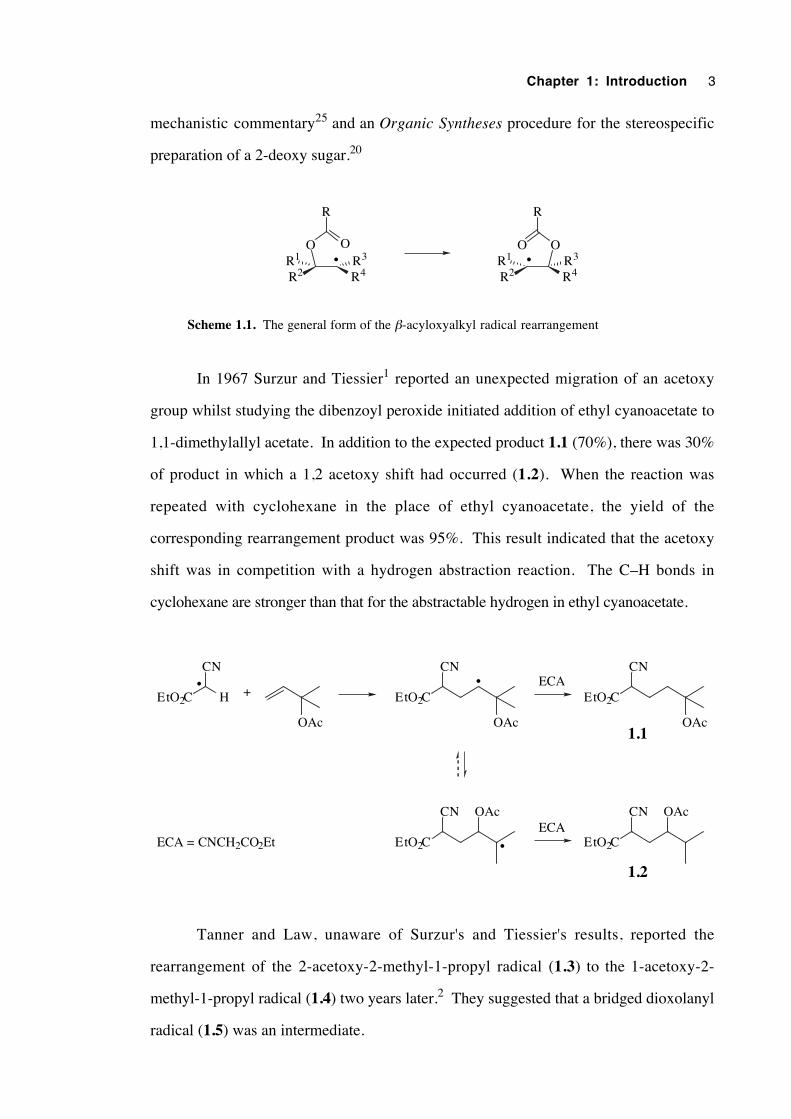
In 1967 Surzur and Tiessier1 reported an unexpected migration of an acetoxy
group whilst studying the dibenzoyl peroxide initiated addition of ethyl cyanoacetate to
1,1-dimethylallyl acetate. In addition to the expected product 1.1 (70%), there was 30%
of product in which a 1,2 acetoxy shift had occurred (1.2). When the reaction was
repeated with cyclohexane in the place of ethyl cyanoacetate, the yield of the
corresponding rearrangement product was 95%. This result indicated that the acetoxy
shift was in competition with a hydrogen abstraction reaction. The C–H bonds in
cyclohexane are stronger than that for the abstractable hydrogen in ethyl cyanoacetate.
OAc OAc
EtO2C
CN•
EtO2C H
CN•
+
EtO2C
CN
•
OAc
OAc
EtO2C
CN
EtO2C
CN OAc
1.1
1.2
ECA
ECAECA = CNCH2CO2Et
Tanner and Law, unaware of Surzur's and Tiessier's results, reported the
rearrangement of the 2-acetoxy-2-methyl-1-propyl radical (1.3) to the 1-acetoxy-2-
methyl-1-propyl radical (1.4) two years later.2 They suggested that a bridged dioxolanyl
radical (1.5) was an intermediate.
Chapter 1: Introduction 4
CH2
OAc•
•
OAc OO
CH3
•
1.3 1.4 1.5
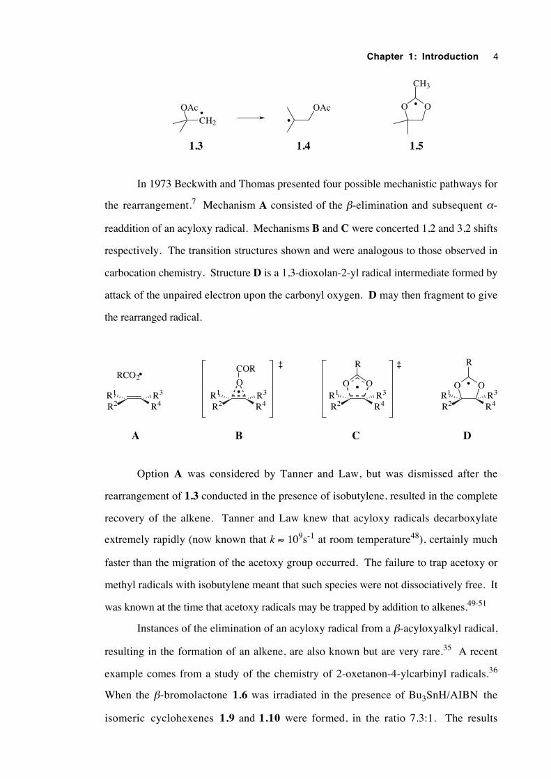
In 1973 Beckwith and Thomas presented four possible mechanistic pathways for
the rearrangement.7 Mechanism A consisted of the β-elimination and subsequent α-
readdition of an acyloxy radical. Mechanisms B and C were concerted 1,2 and 3,2 shifts
respectively. The transition structures shown and were analogous to those observed in
carbocation chemistry. Structure D is a 1,3-dioxolan-2-yl radical intermediate formed by
attack of the unpaired electron upon the carbonyl oxygen. D may then fragment to give
the rearranged radical.
R2R1
R4R3
•
R2R1
R4R3•
OO
R
R2R1
R4R3
•
R2R1
R4R3
•RCO2
O
COR ‡ ‡
A B C D
OO
R
Option A was considered by Tanner and Law, but was dismissed after the
rearrangement of 1.3 conducted in the presence of isobutylene, resulted in the complete
recovery of the alkene. Tanner and Law knew that acyloxy radicals decarboxylate
extremely rapidly (now known that k ≈ 109s-1 at room temperature48), certainly much
faster than the migration of the acetoxy group occurred. The failure to trap acetoxy or
methyl radicals with isobutylene meant that such species were not dissociatively free. It
was known at the time that acetoxy radicals may be trapped by addition to alkenes.49-51
Instances of the elimination of an acyloxy radical from a β-acyloxyalkyl radical,
resulting in the formation of an alkene, are also known but are very rare.35 A recent
example comes from a study of the chemistry of 2-oxetanon-4-ylcarbinyl radicals.36
When the β-bromolactone 1.6 was irradiated in the presence of Bu3SnH/AIBN the
isomeric cyclohexenes 1.9 and 1.10 were formed, in the ratio 7.3:1. The results
Chapter 1: Introduction 5
indicated that ring expansion of the highly-strained radical 1.7 by the β-acyloxyalkyl
radical rearrangement was an unfavourable process in this case, presumably because of
stereoelectronic and ring strain effects.
O
O
•
Bu3Sn •
Ph
Br
O
O Ph
• O
O Ph
Ph
•
PhPh
+
Bu3SnH
– CO2
β-fragmentation
1.6 1.7
1.101.9 1.8
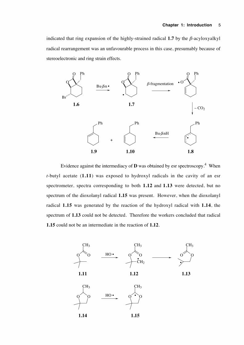
Evidence against the intermediacy of D was obtained by esr spectroscopy.4 When
t-butyl acetate (1.11) was exposed to hydroxyl radicals in the cavity of an esr
spectrometer, spectra corresponding to both 1.12 and 1.13 were detected, but no
spectrum of the dioxolanyl radical 1.15 was present. However, when the dioxolanyl
radical 1.15 was generated by the reaction of the hydroxyl radical with 1.14, the
spectrum of 1.13 could not be detected. Therefore the workers concluded that radical
1.15 could not be an intermediate in the reaction of 1.12.
OO
CH3
CH2
OO
CH3
OO
CH3
OO
CH3
OO
CH3
HO
HO •
••
•
•
1.11 1.12 1.13
1.14 1.15
Chapter 1: Introduction 6
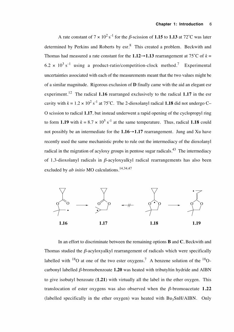
A rate constant of 7 × 10² s-1 for the β-scission of 1.15 to 1.13 at 72˚C was later
determined by Perkins and Roberts by esr.8 This created a problem. Beckwith and
Thomas had measured a rate constant for the 1.12→1.13 rearrangement at 75˚C of k =
6.2 × 10³ s-1 using a product-ratio/competition-clock method.7 Experimental
uncertainties associated with each of the measurements meant that the two values might be
of a similar magnitude. Rigorous exclusion of D finally came with the aid an elegant esr
experiment.12 The radical 1.16 rearranged exclusively to the radical 1.17 in the esr
cavity with k = 1.2 × 10² s-1 at 75˚C. The 2-dioxolanyl radical 1.18 did not undergo C–
O scission to radical 1.17, but instead underwent a rapid opening of the cyclopropyl ring
to form 1.19 with k = 8.7 × 10⁵ s-1 at the same temperature. Thus, radical 1.18 could
not possibly be an intermediate for the 1.16→1.17 rearrangement. Jung and Xu have
recently used the same mechanistic probe to rule out the intermediacy of the dioxolanyl
radical in the migration of acyloxy groups in pentose sugar radicals.43 The intermediacy
of 1,3-dioxolanyl radicals in β-acyloxyalkyl radical rearrangements has also been
excluded by ab initio MO calculations.14,34,47
OO OO OO OO//
1.16 1.17 1.18 1.19
• •
•
•
In an effort to discriminate between the remaining options B and C, Beckwith and
Thomas studied the β-acyloxyalkyl rearrangement of radicals which were specifically
labelled with 18O at one of the two ester oxygens.7 A benzene solution of the 18O-
carbonyl labelled β-bromobenzoate 1.20 was heated with tributyltin hydride and AIBN
to give isobutyl benzoate (1.21) with virtually all the label in the ether oxygen. This
translocation of ester oxygens was also observed when the β-bromoacetate 1.22
(labelled specifically in the ether oxygen) was heated with Bu3SnH/AIBN. Only
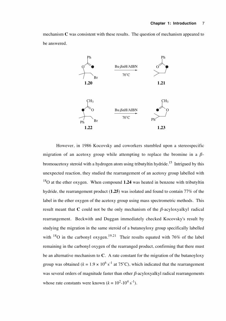
Chapter 1: Introduction 7
mechanism C was consistent with these results. The question of mechanism appeared to
be answered.
OO
Ph
OO
Ph
Bu3SnH/AIBN
OO
CH3
OO
CH3
Ph
Bu3SnH/AIBN
Ph
1.20 1.21
1.22 1.23
Br
Br
70˚C
70˚C
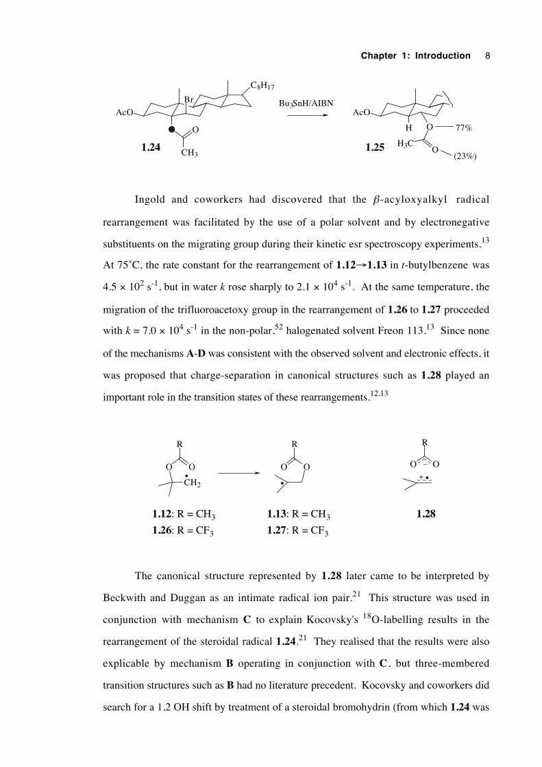
However, in 1986 Kocovsky and coworkers stumbled upon a stereospecific
migration of an acetoxy group while attempting to replace the bromine in a β-
bromoacetoxy steroid with a hydrogen atom using tributyltin hydride.15 Intrigued by this
unexpected reaction, they studied the rearrangement of an acetoxy group labelled with
18O at the ether oxygen. When compound 1.24 was heated in benzene with tributyltin
hydride, the rearrangement product (1.25) was isolated and found to contain 77% of the
label in the ether oxygen of the acetoxy group using mass spectrometric methods. This
result meant that C could not be the only mechanism of the β-acyloxyalkyl radical
rearrangement. Beckwith and Duggan immediately checked Kocovsky's result by
studying the migration in the same steroid of a butanoyloxy group specifically labelled
with 18O in the carbonyl oxygen.19,21 Their results equated with 76% of the label
remaining in the carbonyl oxygen of the rearranged product, confirming that there must
be an alternative mechanism to C. A rate constant for the migration of the butanoyloxy
group was obtained (k = 1.9 × 10⁶ s-1 at 75˚C), which indicated that the rearrangement
was several orders of magnitude faster than other β-acyloxyalkyl radical rearrangements
whose rate constants were known (k = 102-104 s-1).
Chapter 1: Introduction 8
AcO
C8H17
O O
CH3
AcO
O
OH3C
77%
(23%)1.24 1.25
Br
H
Bu3SnH/AIBN
Ingold and coworkers had discovered that the β-acyloxyalkyl radical
rearrangement was facilitated by the use of a polar solvent and by electronegative
substituents on the migrating group during their kinetic esr spectroscopy experiments.13
At 75˚C, the rate constant for the rearrangement of 1.12→1.13 in t-butylbenzene was
4.5 × 10² s-1, but in water k rose sharply to 2.1 × 10⁴ s-1. At the same temperature, the
migration of the trifluoroacetoxy group in the rearrangement of 1.26 to 1.27 proceeded
with k = 7.0 × 10⁴ s-1 in the non-polar,52 halogenated solvent Freon 113.13 Since none
of the mechanisms A-D was consistent with the observed solvent and electronic effects, it
was proposed that charge-separation in canonical structures such as 1.28 played an
important role in the transition states of these rearrangements.12,13
CH2
OO
R
OO
R
••
1.12: R = CH3 1.13: R = CH3
1.26: R = CF3 1.27: R = CF3
OO
R
•+
–
1.28
The canonical structure represented by 1.28 later came to be interpreted by
Beckwith and Duggan as an intimate radical ion pair.21 This structure was used in
conjunction with mechanism C to explain Kocovsky's 18O-labelling results in the
rearrangement of the steroidal radical 1.24.21 They realised that the results were also
explicable by mechanism B operating in conjunction with C, but three-membered
transition structures such as B had no literature precedent. Kocovsky and coworkers did
search for a 1,2 OH shift by treatment of a steroidal bromohydrin (from which 1.24 was
Chapter 1: Introduction 9
prepared by acetylation) with tributyltin hydride/AIBN, but only observed the
replacement of the bromine atom with hydrogen.15 However 1,2 OH shifts were
reported to be observed in biological systems subject to the action of enzymes.53 In
addition, early ab initio MO calculations predicted that the 1,2 shift of a hydroxyl group
in a β-hydroxyalkyl radical is energetically feasible provided that the oxygen is
protonated.54 Gilbert and coworkers have observed the acid-catalysed interconversion of
β-hydroxyalkyl radicals in aqueous solution, but concluded that the mechanism involved
formation and subsequent solvent-hydration of an alkene radical cation.55
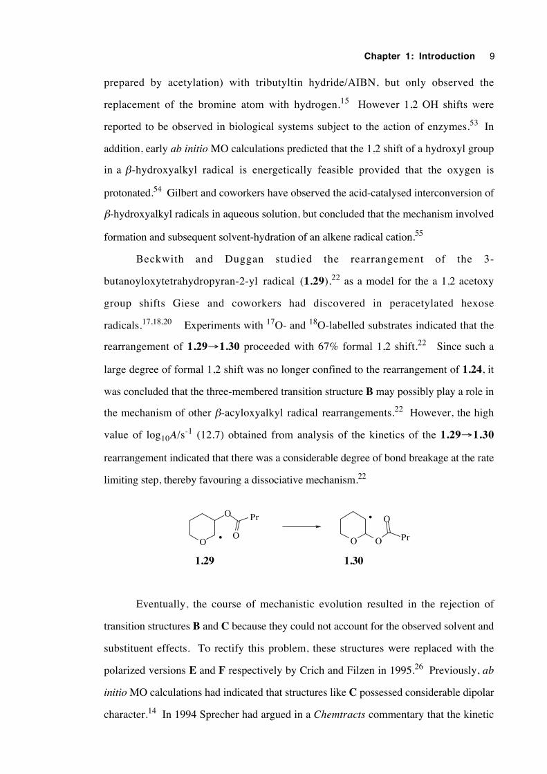
Beckwith and Duggan studied the rearrangement of the 3-
butanoyloxytetrahydropyran-2-yl radical (1.29),22 as a model for the a 1,2 acetoxy
group shifts Giese and coworkers had discovered in peracetylated hexose
radicals.17,18,20 Experiments with 17O- and 18O-labelled substrates indicated that the
rearrangement of 1.29→1.30 proceeded with 67% formal 1,2 shift.22 Since such a
large degree of formal 1,2 shift was no longer confined to the rearrangement of 1.24, it
was concluded that the three-membered transition structure B may possibly play a role in
the mechanism of other β-acyloxyalkyl radical rearrangements.22 However, the high
value of log10A/s-1 (12.7) obtained from analysis of the kinetics of the 1.29→1.30
rearrangement indicated that there was a considerable degree of bond breakage at the rate
limiting step, thereby favouring a dissociative mechanism.22
O •
O
O
Pr
O
O
PrO
•
1.29 1.30
Eventually, the course of mechanistic evolution resulted in the rejection of
transition structures B and C because they could not account for the observed solvent and
substituent effects. To rectify this problem, these structures were replaced with the
polarized versions E and F respectively by Crich and Filzen in 1995.26 Previously, ab
initio MO calculations had indicated that structures like C possessed considerable dipolar
character.14 In 1994 Sprecher had argued in a Chemtracts commentary that the kinetic
Chapter 1: Introduction 10
and oxygen-labelling results for all the β-acyloxyalkyl radical rearrangements known at
that time could be rationalised by a mechanism involving an intimate ion pair composed of
an alkene radical cation and a carboxylate anion (G).25 He claimed that the diversity in
rate constant and degree of formal 1,2 shift could be explained by a varying degree of
intimacy of the ion pair and the mutual orientation of the fragments, which in turn would
depend critically on the respective charge distributions.25
R2R1
R4R3•
OO
R
R2R1
R4R3
•
R2R1
R4R3•
O
COR ‡ ‡
E F G
OO
R
δ+
δ-
δ+
δ-
+
–
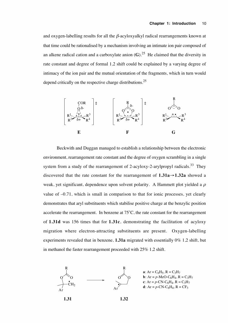
Beckwith and Duggan managed to establish a relationship between the electronic
environment, rearrangement rate constant and the degree of oxygen scrambling in a single
system from a study of the rearrangement of 2-acyloxy-2-arylpropyl radicals.33 They
discovered that the rate constant for the rearrangement of 1.31a→1.32a showed a
weak, yet significant, dependence upon solvent polarity. A Hammett plot yielded a ρ
value of –0.71, which is small in comparison to that for ionic processes, yet clearly
demonstrates that aryl substituents which stabilise positive charge at the benzylic position
accelerate the rearrangement. In benzene at 75˚C, the rate constant for the rearrangement
of 1.31d was 156 times that for 1.31c, demonstrating the facilitation of acyloxy
migration where electron-attracting substituents are present. Oxygen-labelling
experiments revealed that in benzene, 1.31a migrated with essentially 0% 1,2 shift, but
in methanol the faster rearrangement proceeded with 25% 1,2 shift.
CH2
OO
R
OO
R
Ar Ar
••
1.31 1.32
a: Ar = C6H5, R = C3H7b: Ar = p-MeO-C6H4, R = C3H7c: Ar = p-CN-C6H4, R = C3H7d: Ar = p-CN-C6H4, R = CF3
Chapter 1: Introduction 11
In a thoughtful conclusion to their 1997 review, the authors stated that the β-
acyloxyalkyl radical rearrangement has a spectrum of mechanisms.35 The type of
mechanism(s) manifested depends primarily upon radical structure and the solvent. Slow
rearrangements generally proceed via the relatively non-polarized, 5-membered transition
structure, F. Fast rearrangements generally proceed via the polarized, 3-membered
transition structure, E . In rare cases, it is possible that β-acyloxyalkyl radical
rearrangements proceed at least in part by caged, contact radical ions, G . Most
fragmentations of β-acyloxyalkyl radicals occur via the permanent separation of the
carboxylate and alkene-radical-cation moieties of G . Of course, rearrangements
proceeding at an intermediate rate were believed to proceed by a mixture of transition
structures E and F, possibly with a minor involvement of G. It is a difficult undertaking
to devise ways of enabling experimental differentiation between these mechanistic
alternatives, yet since the mid-1990s this has been the major goal of workers in the field.
There have been a number of important recent developments. Crich, Newcomb
and coworkers have measured rate constants for β-acyloxyalkyl radical rearrangements
directly by a laser flash photolysis technique, confirming that migrations are accelerated
by electron-donating groups on the alkyl framework, electron-withdrawing substituents
on the migrating group and more polar solvents.40 Rate constants were obtained directly
from time-resolved UV-vis spectra of the product radicals,40 thus addressing
Sprecher's25 reservations that kinetic parameters determined from competition methods
depend on some arguable assumptions.
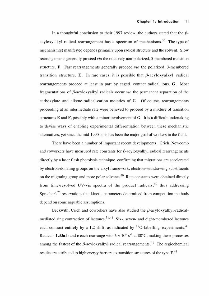
Beckwith, Crich and coworkers have also studied the β-acyloxyalkyl-radical-
mediated ring contraction of lactones.31,41 Six-, seven- and eight-membered lactones
each contract entirely by a 1,2 shift, as indicated by 17O-labelling experiments.41
Radicals 1.33a,b and c each rearrange with k ≈ 106 s-1 at 80˚C, making these processes
among the fastest of the β-acyloxyalkyl radical rearrangements.41 The regiochemical
results are attributed to high energy barriers to transition structures of the type F.41
Chapter 1: Introduction 12
O (CH2)n
O
•Ph
O (CH2)n
O
•Ph
a: n = 1b: n = 2c: n = 3
1.33 1.34
Renaud and coworkers have reported that β-acyloxyalkyl radicals which undergo
rearrangement in the presence of a Lewis acid can experience rate acceleration of up to
three orders of magnitude.38,44 The effect was attributed to the stabilisation of negative
charge in the migrating substituent upon complexation of the Lewis acid with the ester
group.
The stereospecific shifts of a pivaloxy group from C1' to C2' in radicals generated
from modified uridines and adenosines have been studied.28,29,37 Chatgilialoglu and
coworkers have measured rate constants of 5-10 × 104 s-1 for these processes at 80˚C.37
These rearrangements produce C1' anomeric nucleoside radicals, illustrating their
importance in biochemistry.
A 1998 review of the free radical reactions of anomeric monosaccharide and C-
nucleoside radicals includes a section on the use of the β-acyloxyalkyl radical
rearrangement as a synthetic tool for the preparation of 2-deoxy sugars from readily
available precursors, illustrating the growing importance of this intriguing reaction.39
Ab initio molecular orbital calculations14,32,34,45,47 on the β-acyloxyalkyl radical
rearrangement have provided meaningful mechanistic information where laboratory-based
experiments reach the limit of their capacity to discriminate mechanisms E-G. The way
forward will undoubtedly be a close collaboration of theory and experiment. In a
comparison of the 3,3 acyloxy shift in allyl formate with the 3,2 acyloxy shift in the 2-
formyloxyethyl radical, Zipse found that the open-shell reaction had a barrier of 92 to 113
kJmol-1 lower than the closed-shell process, showing that the transition state F is
energetically viable.32 Both reactions could be viewed as intramolecular, nucleophilic
substitutions.32 The idea of these rearrangements being seen as intramolecular
displacements was expanded into a general "methylenology principal", which states that
Chapter 1: Introduction 13
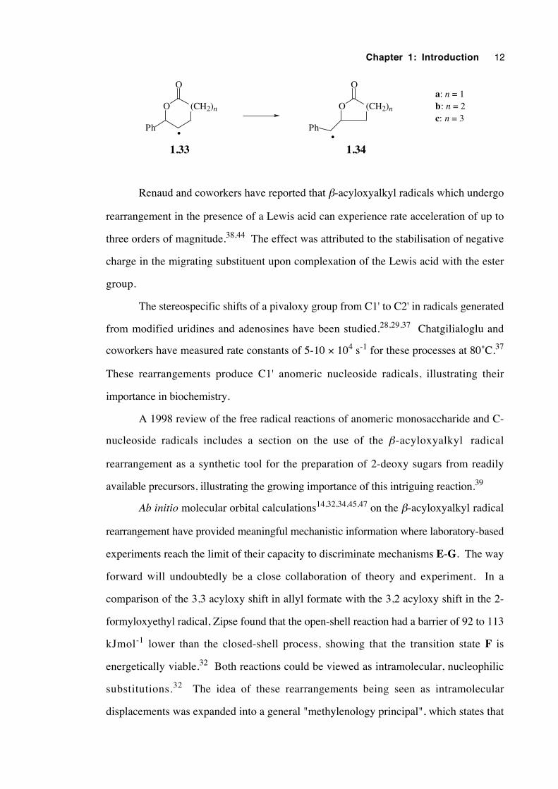
β-elimination initiated by nucleophilic attack upon a radical centre is an open-shell
analogy to the SN2' reaction.45 The source of the low barriers to these polar reactions in
open-shell systems is the mixing in behaviour of homolytic and heterolytic bond cleavage
processes.45 The similarity in the SRN2c ('c' represents cine regiochemistry) and SN2'
reactions is shown below, together with a diagram of valence electron delocalisation in
the β-acyloxyalkyl radical rearrangement, viewed as an SRN2i (intramolecular) reaction
(1.35) .
X
CH2 X
X
CH2 X
Nu ••Nu ••+
•Nu •• Nu ••+
SN2'
SRN2c•
OO
R
•
1.35
In a computational exploration of the rearrangement of 2-acyloxyethyl radicals,
Zipse found that a 3,2 shift was always favoured over a 1,2 shift, that trifluoroacetoxy
migration possessed a lower barrier than acetoxy migration and that a 3,2 acyloxy shift
will proceed more quickly in water than in the gas phase, in conformity with most
experimental evidence.34 In addition, protonation of a formyloxy group was found to
lower the rearrangement barrier by more than 40 kJmol-1 compared with the unprotonated
species.34 However, the solvent effects predicted were much smaller than those
observed experimentally.34
The results most pertinent to the work of this thesis come from Zipse and Bootz,
who investigated the rearrangements of 2-acyloxy-2-methyl-1-propyl radicals
(1.12→1.13 and 1.26→1.27) with a variety of theoretical methods.47 They found no
evidence of an ion pair intermediate (G) for either acetoxy or trifluoroacetoxy migration.
The three-membered transition structure (E) was always slightly less favourable (by ca. 4
kJmol-1) than the five-membered transition structure (F), but became more competitive as
absolute reaction barriers were lowered by electron-withdrawing migrating groups and
polar solvents. In effect, the amount of formal 1,2 shift is predicted to increase in polar
Chapter 1: Introduction 14
solvents and the effect will be greater for a trifluoroacetoxy group than for acetoxy.65
Solvent effects were predicted to be minor for both E and F and the barrier difference
between acetoxy and trifluoroacetoxy migration was less than half the size of that
measured experimentally. The large rate constant increase for acetoxy migration observed
experimentally in water was rationalised by protonation of the ester group. Carboxylate
group charge in both acetoxy and trifluoroacetoxy groups varied little between transition
states E and F, contrasting with the assumption that E must be significantly more
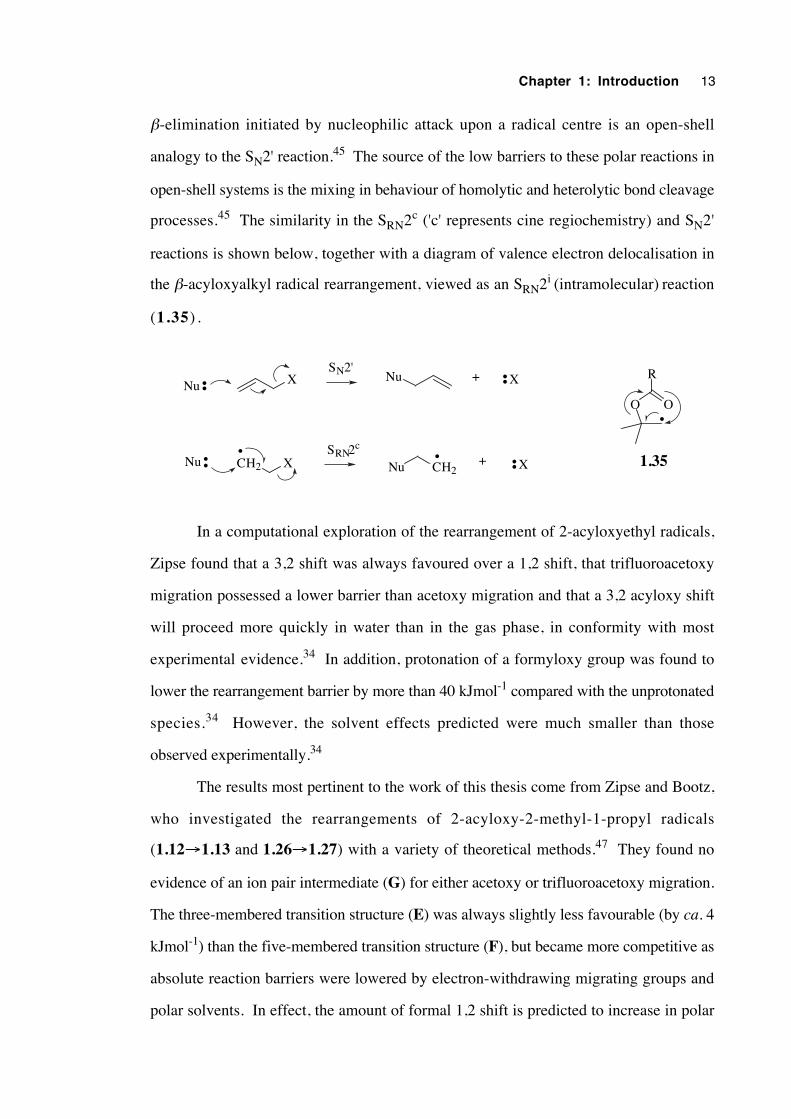
polarized than F.26,30,33,35 Kinetic isotope effects calculated for structures E and F were
generally small except for the strong inverse KIEs (0.66-0.72) found upon dideuteration
of the radical centre (dideuteration at C1 accelerates the rearrangement). It is claimed that
the ratio of the 18O KIEs of the ester oxygen atoms may be used to distinguish E from F,
but in practice the magnitude of such effects may be small compared with experimental
uncertainties. Pertinent KIEs are provided in scheme 1.2. Unfortunately, KIEs could
not be calculated for intermediate G since no transition structure for it could be found.
CH3
CH3HH•
OO
CH3
CH3
CH3HH
•O
H3C O 1.0064
1.0555
1.1295 0.7247
1.0422 1.0171
0.65881.1129
CH3
CH3HH•
OO
CF3
CH3
CH3HH
•O
F3C O 1.0040
1.0499
1.1574 0.7224
1.0400 1.0214
0.68661.1648
Scheme 1.2. Kinetic effects calculated for 16O/18O and 2H/1H isotopic substitutions
upon the transition states E and F for rearrangements of the 2-acetoxy-2-methyl-1-propyl
and 2-trifluoroacetoxy-2-methyl-1-propyl radicals.
1.3 The β-trifluoroacetoxyalkyl radical rearrangement
It is known that groups with electron-withdrawing substituents facilitate the β-
trifluoroacetoxyalky radical rearrangement and that faster rearrangements usually proceed
Chapter 1: Introduction 15
with a higher proportion of formal 1,2 shift. Given that an acetoxy group migrates over
an aliphatic framework with approximately 0% 1,2 shift, the question of the degree of 1,2
shift which accompanies the faster trifluoroacetoxy group migration is an interesting one.
Despite the considerable theoretical interest in the β-trifluoroacetoxyalkyl radical
rearrangement,34,47 there is comparatively little experimental data available.
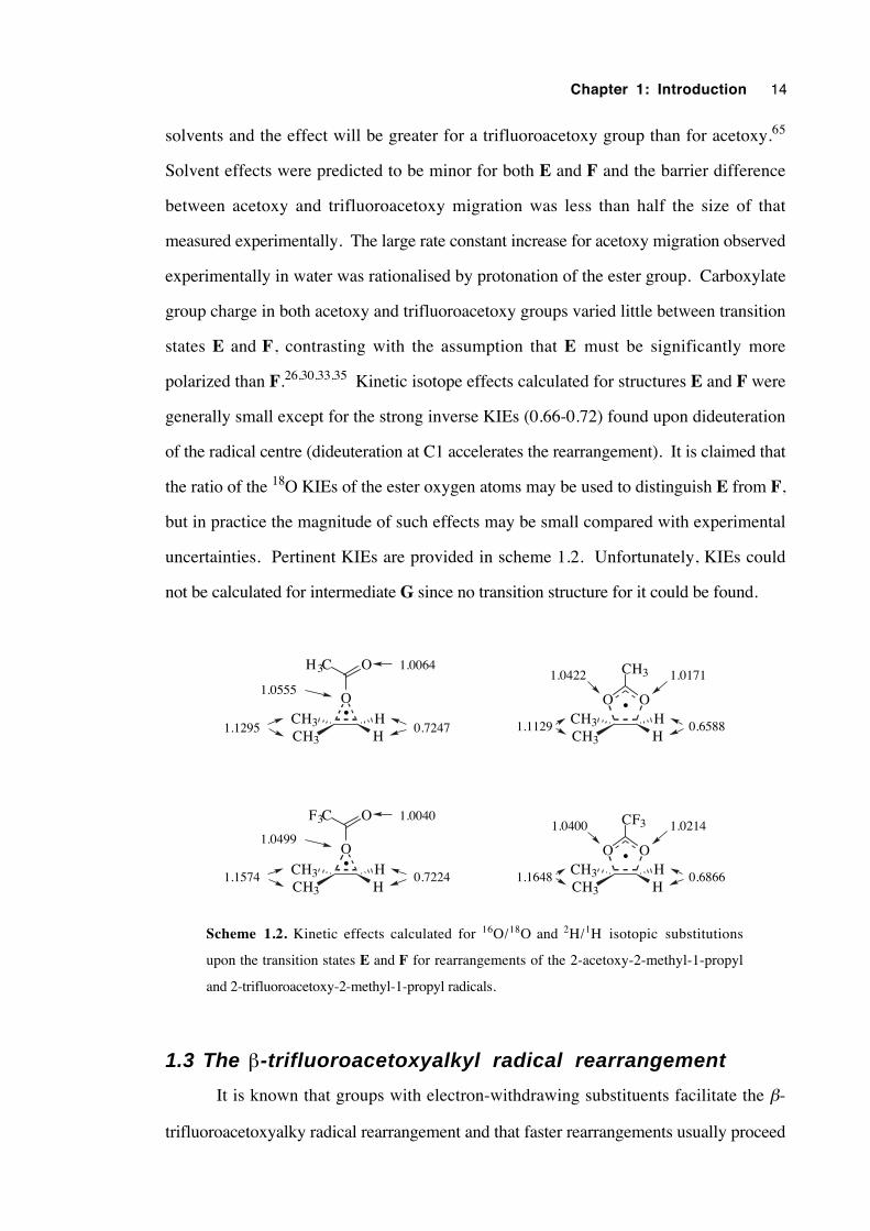
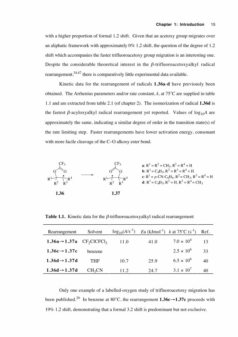
Kinetic data for the rearrangement of radicals 1.36a-d have previously been
obtained. The Arrhenius parameters and/or rate constant, k, at 75˚C are supplied in table
1.1 and are extracted from table 2.1 (of chapter 2). The isomerization of radical 1.36d is
the fastest β-acyloxyalkyl radical rearrangement yet reported. Values of log10A are
approximately the same, indicating a similar degree of order in the transition state(s) of
the rate limiting step. Faster rearrangements have lower activation energy, consonant
with more facile cleavage of the C–O alkoxy ester bond.
OO
R2R1 R4
R3
CF3
•OO
R2R1 R4
R3
CF3
•
a: R1 = R2 = CH3, R3 = R4 = H
b: R1 = C6H5, R2 = R3 = R4 = H
c: R1 = p-CN-C6H4, R2 = CH3, R3 = R4 = H
d: R1 = C6H5, R2 = H, R3 = R4 = CH3
1.36 1.37
Table 1.1. Kinetic data for the β-trifluoroacetoxyalkyl radical rearrangement
Rearrangement Solvent log10(A/s-1) Ea (kJmol-1) k at 75˚C (s-1) Ref.
1.36a→ 1.37a CF2ClCFCl2 11.0 41.0 7.0 × 10⁴ 13
1.36c→ 1.37c benzene 2.5 × 10⁶ 33
1.36d→ 1.37d THF 10.7 25.9 6.5 × 10⁶ 40
1.36d→ 1.37d CH3CN 11.2 24.7 3.1 × 10⁷ 40
Only one example of a labelled-oxygen study of trifluoroacetoxy migration has
been published.26 In benzene at 80˚C, the rearrangement 1.36c→1.37c proceeds with
19% 1,2 shift, demonstrating that a formal 3,2 shift is predominant but not exclusive.
Chapter 1: Introduction 16
We saw the need to study the relationship between the degree of oxygen
scrambling and rearrangement rate as a function of solvent polarity in a single, simple
system. Chapter 2 is concerned with the determination of the kinetics of a simply-
constituted, aliphatic β-trifluoroacetoxyalkyl radical in the solvents hexane, benzene and
propionitrile. Chapter 3 is concerned with an investigation of solvent-dependent oxygen
scrambling behaviour in the same rearrangement studied in chapter 2.
Zipse's MO calculations have indicated that the β-acyloxyalkyl radical
rearrangement can be viewed as an intramolecular nucleophilic substitution reaction.32
Chapter 4 comprises an electron spin resonance study of the temperature-dependent
average conformation of a series of β-oxygenated alkyl radicals. This work was
undertaken to evaluate whether there exists a significant electronic interaction between the
unpaired spin and the ester carbonyl oxygen, as suggested by theoretical calculations.
1.4 Other isomerizations which may share the samemechanism: The rearrangement of N -a lkoxy-2(1H ) -pyridinethiones
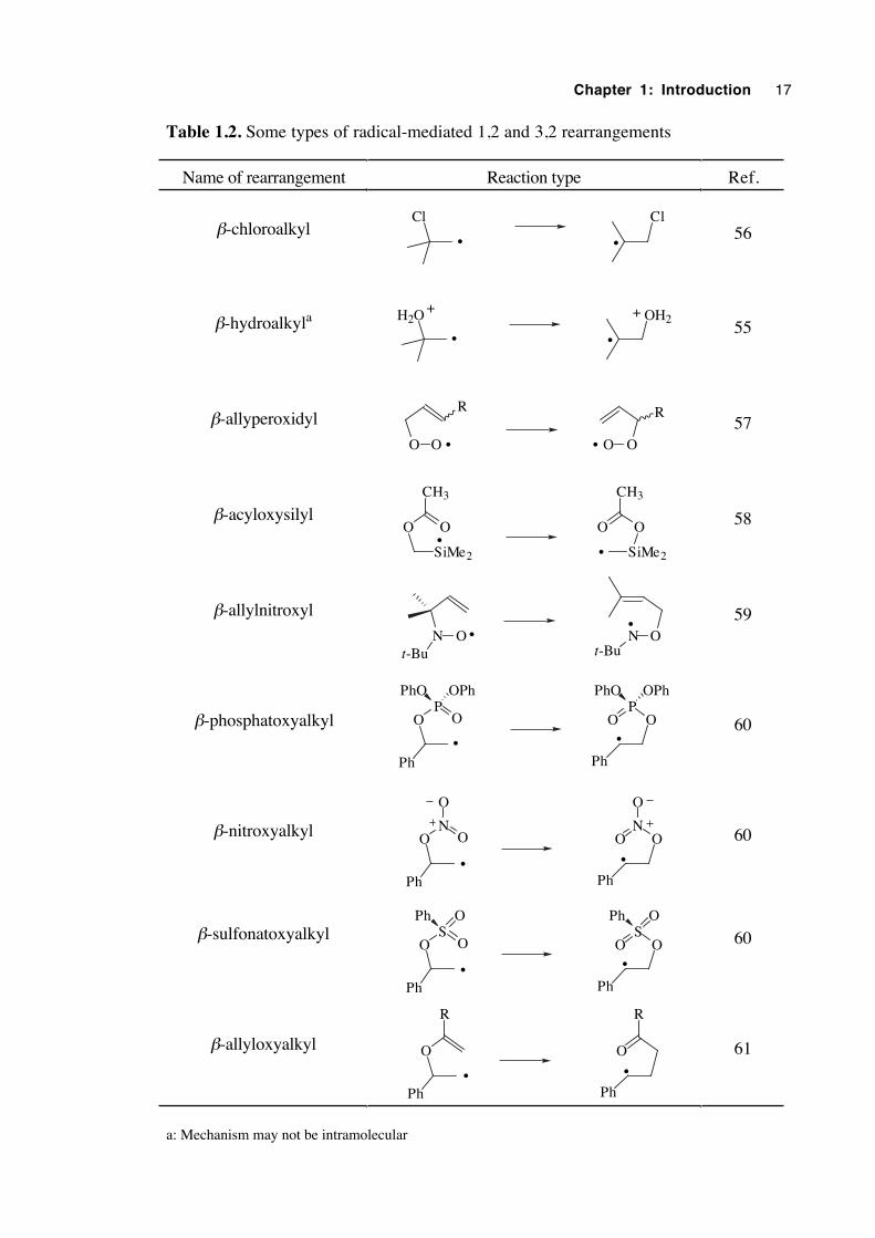
Since the discovery of the β-acyloxyalkyl radical rearrangement, there has been a
steadily growing number of rearrangements discovered which appear to have the same
type of mechanism. Rearrangements which display a radical mediated 1,2 or 3,2 shift are
illustrated in table 1.2.
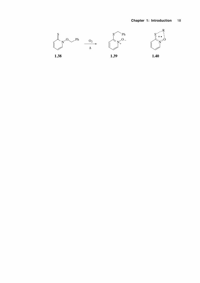
In 1989 the unexpected, oxygen-catalysed rearrangement of N-benzyloxy-2(1H)-
pyridinethione (1.38) was reported, in which a formal 1,4 benzyloxy group migration
had occurred.62 Since then reports of the facile rearrangement of other N-alkoxy-2(1H)-
pyridinethiones have appeared in the literature.63,64 With a growing awareness that
various rearrangements could plausibly proceed through a pericyclic transition state in
which five electrons were delocalised over five atoms, it was envisaged that the catalysed
rearrangement of N-alkoxy-2(1H)-pyridinethiones may proceed via a 5-electron, 5-centre
transition structure (1.40) resulting in 1,4 migration. Chapter 5 describes attempts to
determine the mechanism of this interesting isomerization.
Chapter 1: Introduction 17
Table 1.2. Some types of radical-mediated 1,2 and 3,2 rearrangements
Name of rearrangement Reaction type Ref.
β-chloroalkylCl Cl
• •56
β-hydroalkyla H2O OH2
• •
+ +55
β-allyperoxidyl
O O O O
R R
• •
57
β-acyloxysilyl
SiMe2
OO
SiMe2
OO
CH3 CH3
••
58
β-allylnitroxyl
N O N Ot-Bu
••
t-Bu
59
β-phosphatoxyalkyl OP
O OP
O
OPh OPhPhO PhO
Ph• •
Ph
60
β-nitroxyalkyl ON
O ON
O
Ph
O O
• •
Ph
60
β-sulfonatoxyalkylO
SO O
SO
Ph
Ph O Ph O
• •
Ph
60
β-allyloxyalkyl O O
Ph Ph
R R
• •
61
a: Mechanism may not be intramolecular
Chapter 1: Introduction 18
N
S
ON
S
O
1.38 1.39
Ph
Ph
O2
∆
N
S
O
1.40
R
+ •
Chapter 1: Introduction 19
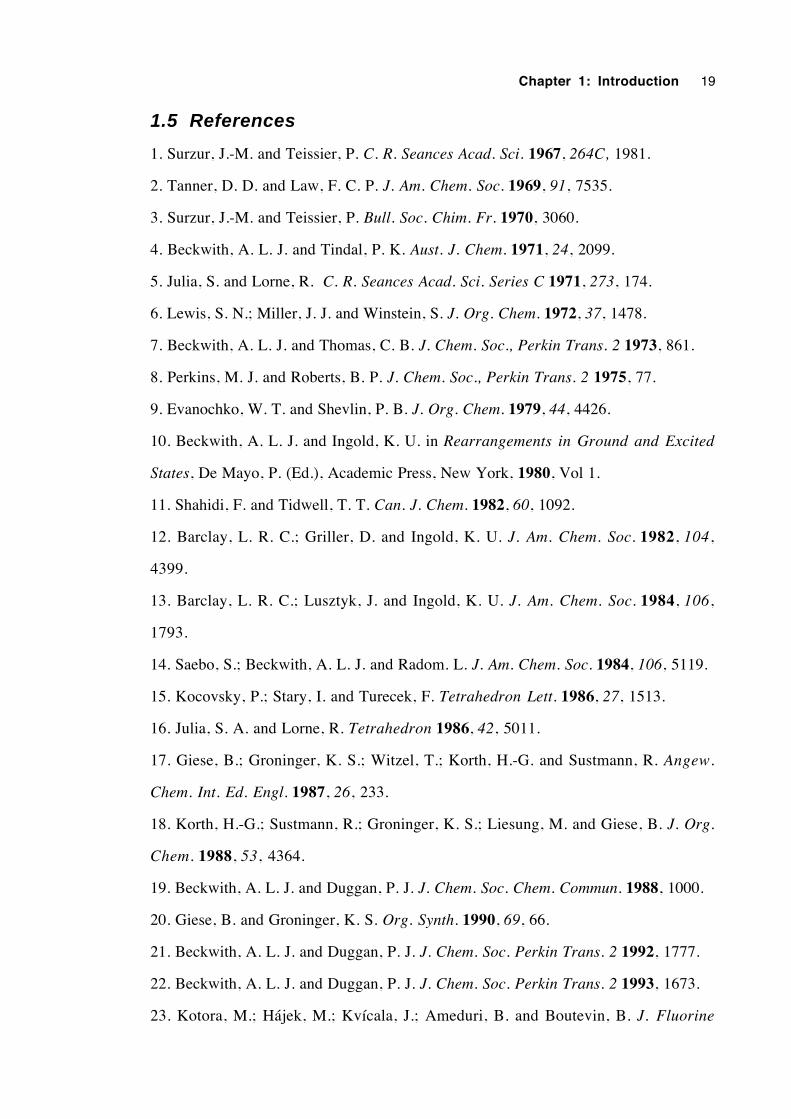
1.5 References
1. Surzur, J.-M. and Teissier, P. C. R. Seances Acad. Sci. 1967, 264C, 1981.
2. Tanner, D. D. and Law, F. C. P. J. Am. Chem. Soc. 1969, 91, 7535.
3. Surzur, J.-M. and Teissier, P. Bull. Soc. Chim. Fr. 1970, 3060.
4. Beckwith, A. L. J. and Tindal, P. K. Aust. J. Chem. 1971, 24, 2099.
5. Julia, S. and Lorne, R. C. R. Seances Acad. Sci. Series C 1971, 273, 174.
6. Lewis, S. N.; Miller, J. J. and Winstein, S. J. Org. Chem. 1972, 37, 1478.
7. Beckwith, A. L. J. and Thomas, C. B. J. Chem. Soc., Perkin Trans. 2 1973, 861.
8. Perkins, M. J. and Roberts, B. P. J. Chem. Soc., Perkin Trans. 2 1975, 77.
9. Evanochko, W. T. and Shevlin, P. B. J. Org. Chem. 1979, 44, 4426.
10. Beckwith, A. L. J. and Ingold, K. U. in Rearrangements in Ground and Excited
States, De Mayo, P. (Ed.), Academic Press, New York, 1980, Vol 1.
11. Shahidi, F. and Tidwell, T. T. Can. J. Chem. 1982, 60, 1092.
12. Barclay, L. R. C.; Griller, D. and Ingold, K. U. J. Am. Chem. Soc. 1982, 104,
4399.
13. Barclay, L. R. C.; Lusztyk, J. and Ingold, K. U. J. Am. Chem. Soc. 1984, 106,
1793.
14. Saebo, S.; Beckwith, A. L. J. and Radom. L. J. Am. Chem. Soc. 1984, 106, 5119.
15. Kocovsky, P.; Stary, I. and Turecek, F. Tetrahedron Lett. 1986, 27, 1513.
16. Julia, S. A. and Lorne, R. Tetrahedron 1986, 42, 5011.
17. Giese, B.; Groninger, K. S.; Witzel, T.; Korth, H.-G. and Sustmann, R. Angew.
Chem. Int. Ed. Engl. 1987, 26, 233.
18. Korth, H.-G.; Sustmann, R.; Groninger, K. S.; Liesung, M. and Giese, B. J. Org.
Chem. 1988, 53, 4364.
19. Beckwith, A. L. J. and Duggan, P. J. J. Chem. Soc. Chem. Commun. 1988, 1000.
20. Giese, B. and Groninger, K. S. Org. Synth. 1990, 69, 66.
21. Beckwith, A. L. J. and Duggan, P. J. J. Chem. Soc. Perkin Trans. 2 1992, 1777.
22. Beckwith, A. L. J. and Duggan, P. J. J. Chem. Soc. Perkin Trans. 2 1993, 1673.
23. Kotora, M.; Hájek, M.; Kvícala, J.; Ameduri, B. and Boutevin, B. J. Fluorine
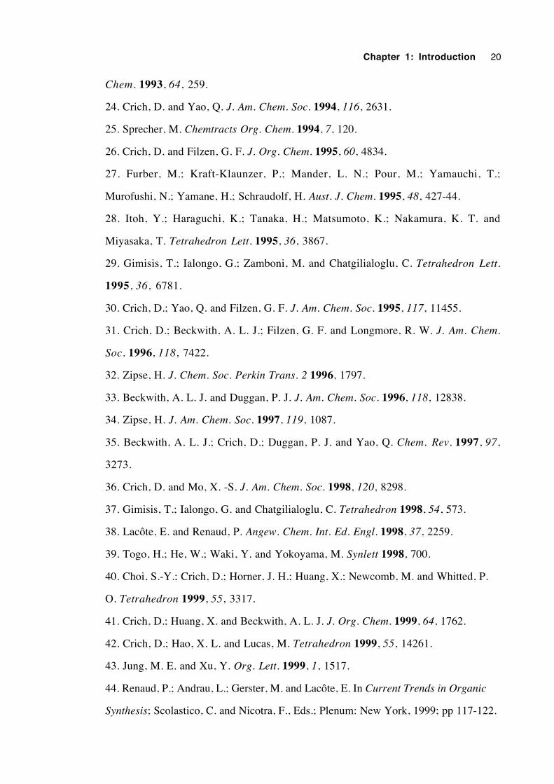
Chapter 1: Introduction 20
Chem. 1993, 64, 259.
24. Crich, D. and Yao, Q. J. Am. Chem. Soc. 1994, 116, 2631.
25. Sprecher, M. Chemtracts Org. Chem. 1994, 7, 120.
26. Crich, D. and Filzen, G. F. J. Org. Chem. 1995, 60, 4834.
27. Furber, M.; Kraft-Klaunzer, P.; Mander, L. N.; Pour, M.; Yamauchi, T.;
Murofushi, N.; Yamane, H.; Schraudolf, H. Aust. J. Chem. 1995, 48, 427-44.
28. Itoh, Y.; Haraguchi, K.; Tanaka, H.; Matsumoto, K.; Nakamura, K. T. and
Miyasaka, T. Tetrahedron Lett. 1995, 36, 3867.
29. Gimisis, T.; Ialongo, G.; Zamboni, M. and Chatgilialoglu, C. Tetrahedron Lett.
1995, 36, 6781.
30. Crich, D.; Yao, Q. and Filzen, G. F. J. Am. Chem. Soc. 1995, 117, 11455.
31. Crich, D.; Beckwith, A. L. J.; Filzen, G. F. and Longmore, R. W. J. Am. Chem.
Soc. 1996, 118, 7422.
32. Zipse, H. J. Chem. Soc. Perkin Trans. 2 1996, 1797.
33. Beckwith, A. L. J. and Duggan, P. J. J. Am. Chem. Soc. 1996, 118, 12838.
34. Zipse, H. J. Am. Chem. Soc. 1997, 119, 1087.
35. Beckwith, A. L. J.; Crich, D.; Duggan, P. J. and Yao, Q. Chem. Rev. 1997, 97,
3273.
36. Crich, D. and Mo, X. -S. J. Am. Chem. Soc. 1998, 120, 8298.
37. Gimisis, T.; Ialongo, G. and Chatgilialoglu, C. Tetrahedron 1998, 54, 573.
38. Lacôte, E. and Renaud, P. Angew. Chem. Int. Ed. Engl. 1998, 37, 2259.
39. Togo, H.; He, W.; Waki, Y. and Yokoyama, M. Synlett 1998, 700.
40. Choi, S.-Y.; Crich, D.; Horner, J. H.; Huang, X.; Newcomb, M. and Whitted, P.
O. Tetrahedron 1999, 55, 3317.
41. Crich, D.; Huang, X. and Beckwith, A. L. J. J. Org. Chem. 1999, 64, 1762.
42. Crich, D.; Hao, X. L. and Lucas, M. Tetrahedron 1999, 55, 14261.
43. Jung, M. E. and Xu, Y. Org. Lett. 1999, 1, 1517.
44. Renaud, P.; Andrau, L.; Gerster, M. and Lacôte, E. In Current Trends in Organic
Synthesis; Scolastico, C. and Nicotra, F., Eds.; Plenum: New York, 1999; pp 117-122.
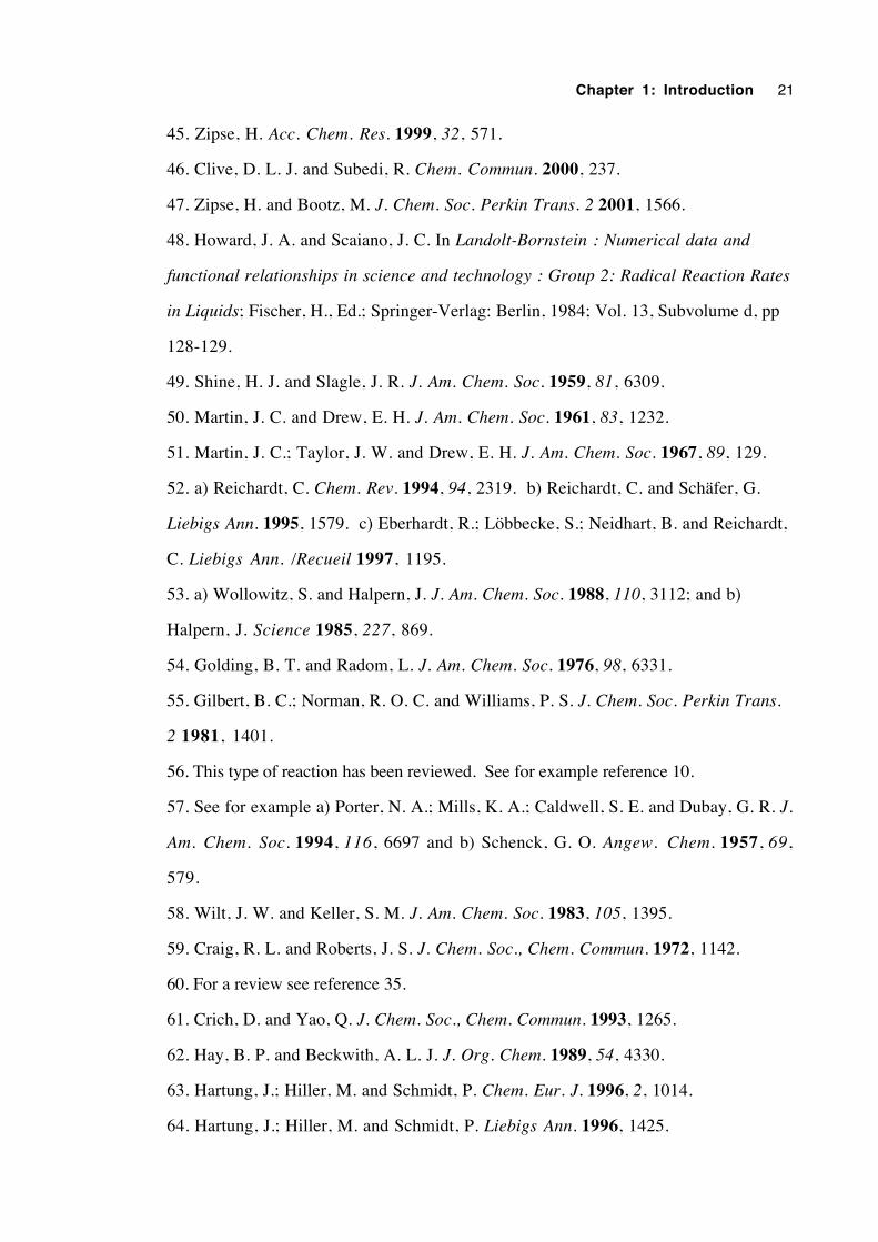
Chapter 1: Introduction 21
45. Zipse, H. Acc. Chem. Res. 1999, 32, 571.
46. Clive, D. L. J. and Subedi, R. Chem. Commun. 2000, 237.
47. Zipse, H. and Bootz, M. J. Chem. Soc. Perkin Trans. 2 2001, 1566.
48. Howard, J. A. and Scaiano, J. C. In Landolt-Bornstein : Numerical data and
functional relationships in science and technology : Group 2: Radical Reaction Rates
in Liquids; Fischer, H., Ed.; Springer-Verlag: Berlin, 1984; Vol. 13, Subvolume d, pp
128-129.
49. Shine, H. J. and Slagle, J. R. J. Am. Chem. Soc. 1959, 81, 6309.
50. Martin, J. C. and Drew, E. H. J. Am. Chem. Soc. 1961, 83, 1232.
51. Martin, J. C.; Taylor, J. W. and Drew, E. H. J. Am. Chem. Soc. 1967, 89, 129.
52. a) Reichardt, C. Chem. Rev. 1994, 94, 2319. b) Reichardt, C. and Schäfer, G.
Liebigs Ann. 1995, 1579. c) Eberhardt, R.; Löbbecke, S.; Neidhart, B. and Reichardt,
C. Liebigs Ann. /Recueil 1997, 1195.
53. a) Wollowitz, S. and Halpern, J. J. Am. Chem. Soc. 1988, 110, 3112; and b)
Halpern, J. Science 1985, 227, 869.
54. Golding, B. T. and Radom, L. J. Am. Chem. Soc. 1976, 98, 6331.
55. Gilbert, B. C.; Norman, R. O. C. and Williams, P. S. J. Chem. Soc. Perkin Trans.
2 1981, 1401.
56. This type of reaction has been reviewed. See for example reference 10.
57. See for example a) Porter, N. A.; Mills, K. A.; Caldwell, S. E. and Dubay, G. R. J.
Am. Chem. Soc. 1994, 116, 6697 and b) Schenck, G. O. Angew. Chem. 1957, 69,
579.
58. Wilt, J. W. and Keller, S. M. J. Am. Chem. Soc. 1983, 105, 1395.
59. Craig, R. L. and Roberts, J. S. J. Chem. Soc., Chem. Commun. 1972, 1142.
60. For a review see reference 35.
61. Crich, D. and Yao, Q. J. Chem. Soc., Chem. Commun. 1993, 1265.
62. Hay, B. P. and Beckwith, A. L. J. J. Org. Chem. 1989, 54, 4330.
63. Hartung, J.; Hiller, M. and Schmidt, P. Chem. Eur. J. 1996, 2, 1014.
64. Hartung, J.; Hiller, M. and Schmidt, P. Liebigs Ann. 1996, 1425.

Chapter 1: Introduction 22
65. Zipse, H. personal communication, April 2003.
Chapter 2
Kinetics of the β-trifluoroacetoxyalkyl radical
rearrangement
2.1 Introduction 24
2.2 A review of β-acyloxyalkyl radical rearrangement kinetics 24
2.3 The search for a suitable system for study 31
2.4 Determination of the equilibrium constant 34
2.5 Kinetics experiments 38
2.6 Discussion of results 53
2.7 Conclusions 57
2.8 Experimental 58
2.9 References 72
Chapter 2: Kinetics 24
2.1 Introduction
This chapter is concerned with the kinetics of the β-trifluoroacetoxyalkyl radical
rearrangement. To put the subject into perspective, this chapter begins with a review of
the kinetics of β-acyloxyalkyl radical rearrangements in general. A description of the
search for an appropriately-structured radical for the study follows. An equilibrium
constant is determined for the reversible rearrangement of the chosen radical in benzene
solution. The kinetic scheme, experimental method and solutions to attendant analytical
complexities are described. Arrhenius parameters, log10A and Ea, are obtained for the
rearrangement of the 2-methyl-2-trifluoroacetoxy-1-heptyl radical in each of three
solvents of varying polarity. The kinetics results and their implications for the
rearrangement mechanism are discussed.
2.2 A review of β -acyloxyalkyl radical rearrangement
kinetics
A substantial contribution to the elucidation of the mechanism of the β-
acyloxyalkyl radical rearrangement has come from the determination of the kinetics of the
ester migration step.1-10,40 In particular, the relationship of radical structure and solvent
polarity to the rearrangement rate constant have yielded vital information about the nature
of transition states or intermediates. For several rearrangements, the Arrhenius frequency
factor (A) and the activation energy (Ea) have been determined. A high log10A value
(>13) can indicate that a radical frequently achieves a geometry similar to that of the
rearrangement transition state, whereas a characteristic such as a strong alkoxy C–O bond
between the carbon framework and the ester group will contribute to a high Ea.
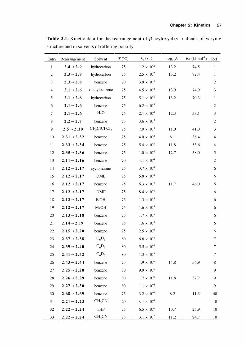
A comprehensive review,11 incorporating the kinetics of the β-acyloxyalkyl
radical rearrangement, covers the literature up to mid 1997. The amount of work in the
field is extensive but not exhaustive. Accordingly, these kinetic results are reproduced
here (table 2.1), including work published to date.
The earliest kinetic results (entries 3,6,8 and 13) were obtained by Beckwith and
Thomas in 19732 who used a product ratios technique. Ingold and coworkers1,3 who
Chapter 2: Kinetics 25
used kinetic esr spectroscopy (entries 1,2,4,5,7 and 9), discovered that comparable
rearrangement rate constants were an order of magnitude smaller than those of the initial
work. However, when a fully integrated rate expression which allows for a changing
Bu3SnH concentration is applied to the earlier data, the rate constants decrease and thus
correspond better to those obtained by the esr technique.1
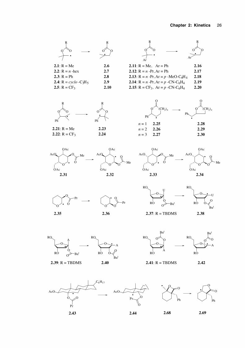
Considerable evidence on the rearrangement mechanism is provided by the results
in table 2.1. It is clear that the rearrangement is thermodynamically favoured when the
product radical is more stabilised than the reactant radical. Furthermore, a greater degree
of product stabilisation generally results in faster rearrangement. This is exemplified by
the difference in rate constants for the rearrangement of the 2-acetoxy-2-methyl-1-propyl
radical (2.1→2.6, entry 4) and the 2-acetoxy-2-phenyl-1-propyl radical (2.11→2.16,
entry 13). The latter rearrangement results in a highly stabilised benzylic radical.
A comparison of entries 5 (2.1→ 2.6) and 9 (2.5→ 2.10) reveals that a
trifluoroacetoxy group migrates more than two orders of magnitude as fast as an acetoxy
group in an otherwise identically-structured radical. This observation is corroborated by
a comparison of entries 21 and 22, as well as 31 and 32. For an appropriate comparison
of the latter, at 20˚C in CH3CN, radical 2.22 rearranges with kr = 6.2 × 10⁶ s-1. This
effect can be attributed to the stabilisation of negative charge that -CF3 brings to the ester
group. Renaud and coworkers report that Lewis acids can accelerate certain β-
acyloxyalkyl radical rearrangements by three orders of magnitude.12,13 For instance,
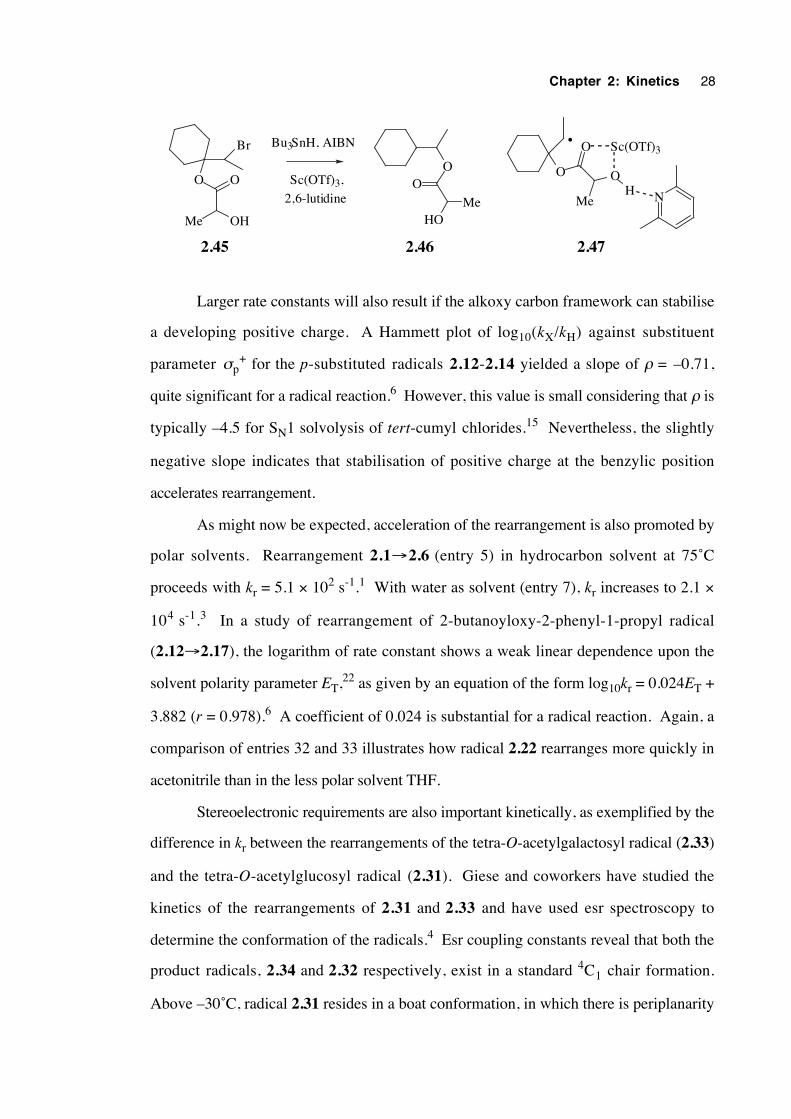
treatment of the lactate bromide 2.45 with tributyltin-hydride/AIBN in the absence of
Lewis acid affords only the directly-reduced product. However, the same reaction in the
presence of one equivalent of scandium (III) triflate/2,6-lutidine affords the rearrangement
product 2.46 in 60% yield at the extraordinarily low temperature of –20˚C. This rate
acceleration effect is consistent with the intermediacy of a complex of the type shown
(2.47), expected to stabilise developing negative charge in the migrating group. Zipse's
theoretical calculations14 predict a large decrease in energy of the three-membered
transition state (i) for a β-acyloxyalkyl radical 1,2 shift upon protonation of the carbonyl
oxygen.
Chapter 2: Kinetics 26
OO
Ph
O
O
Ph
••
2.68 2.69
OO
R
OO
R
OO
Ar
R
OO
Ar
R
OO
R
OO
R
Ph Ph
O
O
Ph
O
O
Ph
O
OAc
AcO
OAc
O
O Me
O
OAc
AcO
OAc
O
O
MeO
OAc
AcO
OAc
O
O Me
O
OAc
AcO
OAc
O
O
Me
OO
OPr
O O
OPr
OU
ORO
RO
O But
O
RO
RO
U
OO
But
OA
ORO
RO
O But
O
RO
RO
A
OO
But
OO
ARO
RO
O
RO
RO
A
O
But
O O
But
AcO
C8H17
O O
Pr
AcO
O
O
Pr
• •
2.1: R = Me2.2: R = n -hex2.3: R = Ph2.4: R = cyclo -C3H52.5: R = CF3
2.62.72.82.92.10
• •
2.11: R = Me,2.12: R = n -Pr,2.13: R = n -Pr,2.14: R = n -Pr,2.15: R = CF3,
• •
2.21: R = Me2.22: R = CF3
2.232.24
(CH2)n
•
(CH2)n
•
2.252.262.27
2.282.292.30
n = 1n = 2n = 3
•
•
2.31 2.32
•
•
2.33 2.34
•
•
2.35 2.36
••
2.37: R = TBDMS 2.38
••
2.39: R = TBDMS 2.40
••
2.41: R = TBDMS 2.42
• •
2.43 2.44
2.162.172.182.192.20
Ar = PhAr = PhAr = p -MeO-C6H4Ar = p -CN-C6H4Ar = p -CN-C6H4
Chapter 2: Kinetics 27
Table 2.1. Kinetic data for the rearrangement of β-acyloxyalkyl radicals of varying
structure and in solvents of differing polarity
Entry Rearrangement Solvent T (˚C) kr (s-1) log10A Ea (kJmol-1) Ref.
1 2.4→ 2 . 9 hydrocarbon 75 1.2 × 10² 13.2 74.5 1
2 2.3→ 2 . 8 hydrocarbon 75 2.5 × 10² 13.2 72.4 1
3 2.3→ 2 . 8 benzene 70 3.9 × 10³ 2
4 2.1→ 2 . 6 t-butylbenzene 75 4.5 × 10² 13.9 74.9 3
5 2.1→ 2 . 6 hydrocarbon 75 5.1 × 10² 13.2 70.3 1
6 2.1→ 2 . 6 benzene 75 6.2 × 10³ 2
7 2.1→ 2 . 6 H2O 75 2.1 × 10⁴ 12.3 53.1 3
8 2.2→ 2 . 7 benzene 75 3.6 × 10³ 2
9 2.5→ 2 .10 CF2ClCFCl2 75 7.0 × 10⁴ 11.0 41.0 3
10 2.31→ 2 .32 benzene 75 4.0 × 10² 8.1 36.4 4
11 2.33→ 2 .34 benzene 75 5.4 × 10³ 11.8 53.6 4
12 2.35→ 2 .36 benzene 75 1.0 × 10⁴ 12.7 58.0 5
13 2.11→ 2 .16 benzene 70 4.1 × 10⁴ 2
14 2.12→ 2 .17 cyclohexane 75 3.7 × 10⁴ 6
15 2.12→ 2 .17 DME 75 5.8 × 10⁴ 6
16 2.12→ 2 .17 benzene 75 6.3 × 10⁴ 11.7 46.0 6
17 2.12→ 2 .17 DMF 75 8.4 × 10⁴ 6
18 2.12→ 2 .17 EtOH 75 1.3 × 10⁵ 6
19 2.12→ 2 .17 MeOH 75 1.6 × 10⁵ 6
20 2.13→ 2 .18 benzene 75 1.7 × 10⁵ 6
21 2.14→ 2.19 benzene 75 1.6 × 10⁴ 6
22 2.15→ 2 .20 benzene 75 2.5 × 10⁶ 6
23 2.37→ 2 .38 C6D6 80 6.6 × 10⁴ 7
24 2.39→ 2 .40 C6D6 80 5.5 × 10⁴ 7
25 2.41→ 2 .42 C6D6 80 1.3 × 10⁵ 7
26 2.43→ 2 .44 benzene 75 1.9 × 10⁶ 14.8 56.9 8
27 2.25→ 2 .28 benzene 80 9.9 × 10⁵ 9
28 2.26→ 2 .29 benzene 80 1.7 × 10⁶ 11.8 37.7 9
29 2.27→ 2 .30 benzene 80 1.1 × 10⁶ 9
30 2.68→ 2 .69 benzene 75 3.2 × 10⁶ 8.2 11.3 40
31 2.21→ 2 .23 CH3CN 20 < 1 × 10⁴ 10
32 2.22→ 2 .24 THF 75 6.5 × 10⁶ 10.7 25.9 10
33 2.22→ 2 .24 CH3CN 75 3.1 × 10⁷ 11.2 24.7 10
Chapter 2: Kinetics 28
O O
Br
Me OH
OO
HOMe
O
O
Me
OH
Sc(OTf)3
N
•
2,6-lutidine
Sc(OTf)3,
Bu3SnH, AIBN
2.45 2.46 2.47
Larger rate constants will also result if the alkoxy carbon framework can stabilise
a developing positive charge. A Hammett plot of log10(kX/kH) against substituent
parameter σp+ for the p-substituted radicals 2.12-2.14 yielded a slope of ρ = –0.71,
quite significant for a radical reaction.6 However, this value is small considering that ρ is
typically –4.5 for SN1 solvolysis of tert-cumyl chlorides.15 Nevertheless, the slightly
negative slope indicates that stabilisation of positive charge at the benzylic position
accelerates rearrangement.
As might now be expected, acceleration of the rearrangement is also promoted by
polar solvents. Rearrangement 2.1→2.6 (entry 5) in hydrocarbon solvent at 75˚C
proceeds with kr = 5.1 × 10² s-1.1 With water as solvent (entry 7), kr increases to 2.1 ×
104 s-1.3 In a study of rearrangement of 2-butanoyloxy-2-phenyl-1-propyl radical
(2.12→2.17), the logarithm of rate constant shows a weak linear dependence upon the
solvent polarity parameter ET,22 as given by an equation of the form log10kr = 0.024ET +
3.882 (r = 0.978).6 A coefficient of 0.024 is substantial for a radical reaction. Again, a
comparison of entries 32 and 33 illustrates how radical 2.22 rearranges more quickly in
acetonitrile than in the less polar solvent THF.
Stereoelectronic requirements are also important kinetically, as exemplified by the
difference in kr between the rearrangements of the tetra-O-acetylgalactosyl radical (2.33)
and the tetra-O-acetylglucosyl radical (2.31). Giese and coworkers have studied the
kinetics of the rearrangements of 2.31 and 2.33 and have used esr spectroscopy to
determine the conformation of the radicals.4 Esr coupling constants reveal that both the
product radicals, 2.34 and 2.32 respectively, exist in a standard 4C1 chair formation.
Above –30˚C, radical 2.31 resides in a boat conformation, in which there is periplanarity
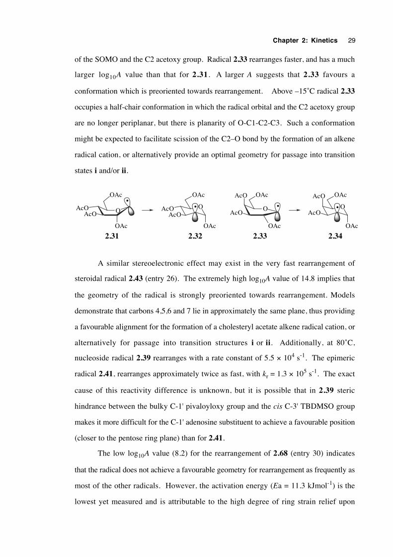
Chapter 2: Kinetics 29
of the SOMO and the C2 acetoxy group. Radical 2.33 rearranges faster, and has a much
larger log10A value than that for 2.31. A larger A suggests that 2.33 favours a
conformation which is preoriented towards rearrangement. Above –15˚C radical 2.33
occupies a half-chair conformation in which the radical orbital and the C2 acetoxy group
are no longer periplanar, but there is planarity of O-C1-C2-C3. Such a conformation
might be expected to facilitate scission of the C2–O bond by the formation of an alkene
radical cation, or alternatively provide an optimal geometry for passage into transition
states i and/or ii.
O
OAc
AcO
AcO OAc
•OAcO
AcO OAc
•
OAc
2.33 2.34
O
OAc
AcO
OAc
•AcOOAcO
OAc•
AcO
OAc
2.31 2.32
A similar stereoelectronic effect may exist in the very fast rearrangement of
steroidal radical 2.43 (entry 26). The extremely high log10A value of 14.8 implies that
the geometry of the radical is strongly preoriented towards rearrangement. Models
demonstrate that carbons 4,5,6 and 7 lie in approximately the same plane, thus providing
a favourable alignment for the formation of a cholesteryl acetate alkene radical cation, or
alternatively for passage into transition structures i or ii. Additionally, at 80˚C,
nucleoside radical 2.39 rearranges with a rate constant of 5.5 × 10⁴ s-1. The epimeric
radical 2.41, rearranges approximately twice as fast, with kr = 1.3 × 10⁵ s-1. The exact
cause of this reactivity difference is unknown, but it is possible that in 2.39 steric
hindrance between the bulky C-1' pivaloyloxy group and the cis C-3' TBDMSO group
makes it more difficult for the C-1' adenosine substituent to achieve a favourable position
(closer to the pentose ring plane) than for 2.41.
The low log10A value (8.2) for the rearrangement of 2.68 (entry 30) indicates
that the radical does not achieve a favourable geometry for rearrangement as frequently as
most of the other radicals. However, the activation energy (Ea = 11.3 kJmol-1) is the
lowest yet measured and is attributable to the high degree of ring strain relief upon
Chapter 2: Kinetics 30
rearrangement. The low activation energy term (weak C–O ether bond) more than
compensates for the poor stereoelectronic situation, making this rearrangement one of the
fastest yet measured (kr (75˚C) = 3.2 × 10⁶).
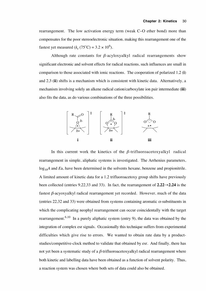
Although rate constants for β-acyloxyalkyl radical rearrangements show
significant electronic and solvent effects for radical reactions, such influences are small in
comparison to those associated with ionic reactions. The cooperation of polarized 1,2 (i)
and 2,3 (ii) shifts is a mechanism which is consistent with kinetic data. Alternatively, a
mechanism involving solely an alkene radical cation/carboxylate ion pair intermediate (iii)
also fits the data, as do various combinations of the three possibilities.
O O
R
•O
R O
•O O
R
+_
iiiiii
•
δ+
δ-
δ+
δ-
‡ ‡
In this current work the kinetics of the β-trifluoroacetoxyalkyl radical
rearrangement in simple, aliphatic systems is investigated. The Arrhenius parameters,
log10A and Ea, have been determined in the solvents hexane, benzene and propionitrile.
A limited amount of kinetic data for a 1,2 trifluoroacetoxy group shifts have previously
been collected (entries 9,22,33 and 33). In fact, the rearrangement of 2.22→2.24 is the
fastest β-acyoxyalkyl radical rearrangement yet recorded. However, much of the data
(entries 22,32 and 33) were obtained from systems containing aromatic α-substituents in
which the complicating neophyl rearrangement can occur coincidentally with the target
rearrangement.6,10 In a purely aliphatic system (entry 9), the data was obtained by the
integration of complex esr signals. Occasionally this technique suffers from experimental
difficulties which give rise to errors. We wanted to obtain rate data by a product-
studies/competitive-clock method to validate that obtained by esr. And finally, there has
not yet been a systematic study of a β-trifluoroacetoxyalkyl radical rearrangement where
both kinetic and labelling data have been obtained as a function of solvent polarity. Thus,
a reaction system was chosen where both sets of data could also be obtained.
Chapter 2: Kinetics 31
2.3 The search for a suitable system for study
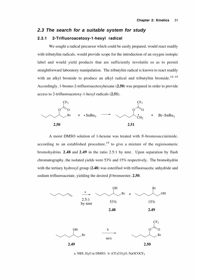
2.3.1 2-Trifluoroacetoxy-1-hexyl radical
We sought a radical precursor which could be easily prepared, would react readily
with tributyltin radicals, would provide scope for the introduction of an oxygen isotopic
label and would yield products that are sufficiently involatile so as to permit
straightforward laboratory manipulation. The tributyltin radical is known to react readily
with an alkyl bromide to produce an alkyl radical and tributyltin bromide.16-18
Accordingly, 1-bromo-2-trifluoroacetoxyhexane (2.50) was prepared in order to provide
access to 2-trifluoroacetoxy-1-hexyl radicals (2.51).
Br
O
CF3
O
2.50
CH2
O
CF3
O
2.51
•SnBu3• Br–SnBu3+ +
A moist DMSO solution of 1-hexene was treated with N-bromosuccinimide,
according to an established procedure,19 to give a mixture of the regioisomeric
bromohydrins 2.48 and 2.49 in the ratio 2.5:1 by nmr. Upon separation by flash
chromatography, the isolated yields were 53% and 15% respectively. The bromohydrin
with the tertiary hydroxyl group (2.48) was esterified with trifluoroacetic anhydride and
sodium trifluoroacetate, yielding the desired β-bromoester, 2.50.
Br
Br OH
OHa
a: NBS, H2O in DMSO; b: (CF3CO)2O, NaOCOCF3
2.48 2.49
+
53% 15% 2.5:1 by nmr
Br
OH
Br
O
CF3
Ob
66%
2.49 2.50
Chapter 2: Kinetics 32
2.3.2 Reaction of β-bromoester 2.50 with Bu3SnH
To a 4.3 mM deoxygenated solution of 2.50 in benzene at reflux was added a
benzene solution containing one equivalent of Bu3SnH (13.8 mM) and catalytic AIBN
over 106 minutes. After a total time of 3 hr, GC revealed that all the tributyltin hydride
had been consumed and only a trace of 2.50 remained. The products included 2-
trifluoroacetoxyhexane (2.53), resulting from direct reduction of 2.50 and 1-
trifluoroacetoxyhexane (2.54), resulting from a 1,2 trifluoroacetoxy group shift.
Unfortunately the ratio of 2.54:2.53 was only 1:22.7, demonstrating that the
rearrangement is slow. When a 0.5 M solution of 2.50 in tert-butylbenzene was heated
at the higher temperature of 130°C and treated with one equivalent of tributyltin hydride
over 30 min, the ratio of 2.54:2.53 increased only modestly to 1:18.8. Thus, the
rearrangement is too slow to give yields of 2.54 high enough for labelling and kinetic
purposes. A faster rearrangement was therefore sought.
O
Br
O
CF3
O O
CF3
O CF3
O
22.7
1
2.50 2.53
2.54
Bu3Sn •
CH2
O O
CF3
•
O CF3
O
•
Bu3SnH
Bu3SnH
1,2-trifluoroacetoxy group shift
2.51
2.52
Scheme 2.1. Generation and reactions of β-trifluoroacetoxyalkyl radical 2.51
2.3.3 A faster rearrangement
Since tertiary alkyl radicals are more stabilised by hyperconjugation than
secondary radicals, the 1,2 migration of an ester group from a tertiary to a primary carbon
is generally faster than that from a secondary to a primary terminus. Accordingly, the
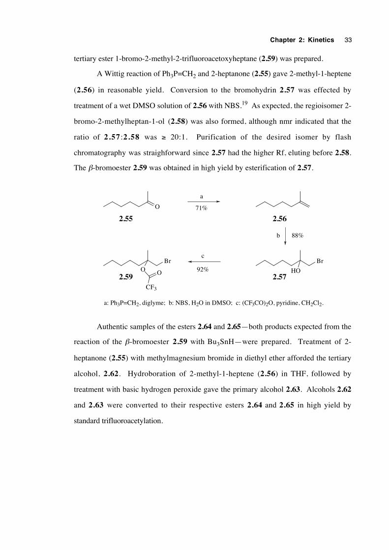
Chapter 2: Kinetics 33
tertiary ester 1-bromo-2-methyl-2-trifluoroacetoxyheptane (2.59) was prepared.
A Wittig reaction of Ph3P=CH2 and 2-heptanone (2.55) gave 2-methyl-1-heptene
(2.56) in reasonable yield. Conversion to the bromohydrin 2.57 was effected by
treatment of a wet DMSO solution of 2.56 with NBS.19 As expected, the regioisomer 2-
bromo-2-methylheptan-1-ol (2.58) was also formed, although nmr indicated that the
ratio of 2.57 :2.58 was ≥ 20:1. Purification of the desired isomer by flash
chromatography was straighforward since 2.57 had the higher Rf, eluting before 2.58.
The β-bromoester 2.59 was obtained in high yield by esterification of 2.57.
O
BrBr
HOO O
CF3
a
71%
b 88%
c
92%
a: Ph3P=CH2, diglyme; b: NBS, H2O in DMSO; c: (CF3CO)2O, pyridine, CH2Cl2.
2.55
2.59 2.57
2.56
Authentic samples of the esters 2.64 and 2.65—both products expected from the
reaction of the β-bromoester 2.59 with Bu3SnH—were prepared. Treatment of 2-
heptanone (2.55) with methylmagnesium bromide in diethyl ether afforded the tertiary
alcohol, 2.62. Hydroboration of 2-methyl-1-heptene (2.56) in THF, followed by
treatment with basic hydrogen peroxide gave the primary alcohol 2.63. Alcohols 2.62
and 2.63 were converted to their respective esters 2.64 and 2.65 in high yield by
standard trifluoroacetylation.
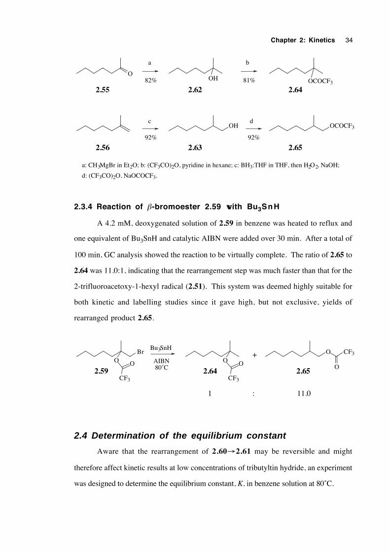
Chapter 2: Kinetics 34
O
2.55 2.62 2.64
OH OCOCF3
2.56 2.63 2.65
OH OCOCF3
a b
dc
92%
a: CH3MgBr in Et2O; b: (CF3CO)2O, pyridine in hexane; c: BH3:THF in THF, then H2O2, NaOH;
d: (CF3CO)2O, NaOCOCF3.
92%
82% 81%
2.3.4 Reaction of β-bromoester 2.59 with Bu3SnH
A 4.2 mM, deoxygenated solution of 2.59 in benzene was heated to reflux and
one equivalent of Bu3SnH and catalytic AIBN were added over 30 min. After a total of
100 min, GC analysis showed the reaction to be virtually complete. The ratio of 2.65 to
2.64 was 11.0:1, indicating that the rearrangement step was much faster than that for the
2-trifluoroacetoxy-1-hexyl radical (2.51). This system was deemed highly suitable for
both kinetic and labelling studies since it gave high, but not exclusive, yields of
rearranged product 2.65.
BrO O
CF3
O O
CF3
O CF3
O
Bu3SnH
AIBN 80˚C
+
: 11.01
2.59 2.64 2.65
2.4 Determination of the equilibrium constant
Aware that the rearrangement of 2.60→2.61 may be reversible and might
therefore affect kinetic results at low concentrations of tributyltin hydride, an experiment
was designed to determine the equilibrium constant, K, in benzene solution at 80˚C.
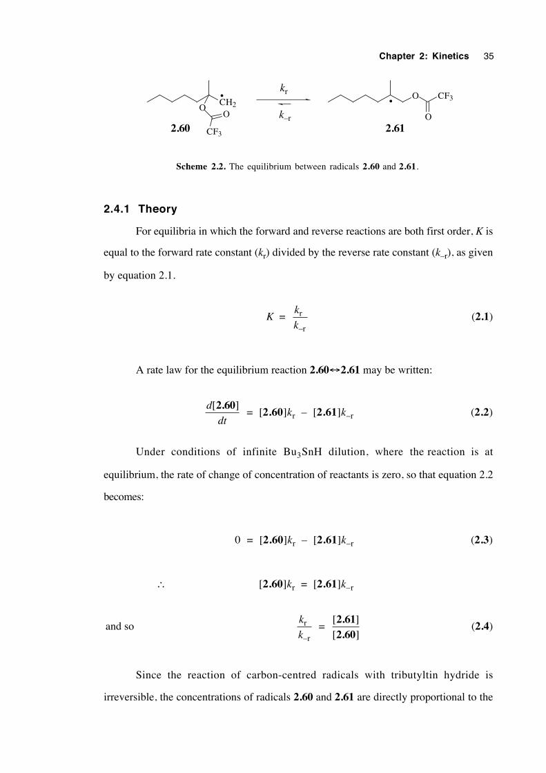
Chapter 2: Kinetics 35
CH2O
CF3
O
••
O CF3
O
kr
k–r2.60 2.61
Scheme 2.2. The equilibrium between radicals 2.60 and 2.61.
2.4.1 Theory
For equilibria in which the forward and reverse reactions are both first order, K is
equal to the forward rate constant (kr) divided by the reverse rate constant (k–r), as given
by equation 2.1.
K = k
kr
–r
(2.1)
A rate law for the equilibrium reaction 2.60↔2.61 may be written:
d
dt
[ ]2.60 = [2.60]kr – [2.61]k–r (2.2)
Under conditions of infinite Bu3SnH dilution, where the reaction is at
equilibrium, the rate of change of concentration of reactants is zero, so that equation 2.2
becomes:
0 = [2.60]kr – [2.61]k–r (2.3)
∴ [2.60]kr = [2.61]k–r
and so k
kr
–r
=
[ ][ ]2.612.60
(2.4)
Since the reaction of carbon-centred radicals with tributyltin hydride is
irreversible, the concentrations of radicals 2.60 and 2.61 are directly proportional to the
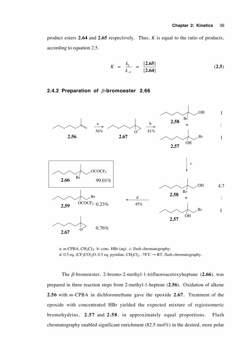
Chapter 2: Kinetics 36
product esters 2.64 and 2.65 respectively. Thus, K is equal to the ratio of products,
according to equation 2.5.
K = k
kr
–r
=
[ ][ ]2.652.64
(2.5)
2.4.2 Preparation of β-bromoester 2.66
O
OH
Br
Br
OH
2.56 2.67
2.58
2.57
1
:
1
+
OH
Br
Br
OH
2.58
2.57
4.7
:
1
+
c
OCOCF3
Br
Br
OCOCF3
2.66
2.59
2.67O
a b
d
99.01%
0.23%
0.76%
a: m-CPBA, CH2Cl2; b: conc. HBr (aq); c: flash chromatography;
d: 0.5 eq. (CF3CO)2O, 0.5 eq. pyridine, CH2Cl2, -78˚C → RT, flash chromatography.
56% 81%
45%
The β-bromoester, 2-bromo-2-methyl-1-trifluoroacetoxyheptane (2.66), was
prepared in three reaction steps from 2-methyl-1-heptene (2.56). Oxidation of alkene
2.56 with m-CPBA in dichloromethane gave the epoxide 2.67. Treatment of the
epoxide with concentrated HBr yielded the expected mixture of regioisomeric
bromohydrins, 2.57 and 2.58 , in approximately equal proportions. Flash
chromatography enabled significant enrichment (82.5 mol%) in the desired, more polar
Chapter 2: Kinetics 37
isomer, 2-bromo-2-methylheptan-1-ol (2.58). Selective esterification of the major
isomer was achieved by treatment of a cold, dichloromethane solution of the
bromohydrins with trifluoroacetic anhydride (0.5 eq.) and pyridine. Flash
chromatography enabled the desired β-bromoester 2.66 to be separated from the
unreacted bromohydrins.
Analysis of the purified product by GC showed it to consist of 2-bromo-2-methyl-
1-trifluoroacetoxyheptane (2.66, 99.01%), 1-bromo-2-methyl-2-trifluoroacetoxyheptane
(2.59, 0.23%) and 1,2-epoxy-2-methylheptane (2.67, 0.76%).
2.4.3 Reaction of β-bromoester 2.66 with Bu3SnH
A stirred 2.8 mM solution of 2-bromo-2-methyl-1-trifluoroacetoxyheptane (2.66)
in dry benzene was deoxygenated with a stream of nitrogen, heated to 80°C and injected
quickly with one equivalent of tributytin hydride followed by catalytic AIBN. After two
hours the reaction was concentrated and analysed by GC. All of the β-bromoester 2.66
had been consumed.
Unfortunately, retention times for the epoxide impurity 2.67 and the product
tertiary trifluoroacetate 2.64 were identical on a dimethylpolysiloxane (BP1) capillary
column, making the indicated proportion of 2.64 higher than its true value. Under these
conditions, the lower limit for the equilibrium constant was calculated to be 15.1.
However, these compounds did separate well on a polyethyleneglycol (BP20) stationary
phase, yielding a ratio of the product esters of 52.9:1, in favour of the primary
trifluoroacetate 2.65. There were small amounts of 2-methyl-1-heptene (2.56) and 2-
methyl-2-heptene (2.56a) present in the mixture, from the eliminative decomposition of
2.64. Therefore, K < 52.9.
It was assumed that the low concentration of Bu3SnH used in this experiment
allowed radicals 2.60 and 2.61 to reach equilibrium prior to their hydrogen atom
transfer reaction with tributyltin hydride. The small amount of 1-bromo-2-methyl-2-
trifluoroacetoxyheptane (2.59) present initially in the sample of 2.66 is therefore
deemed insignificant.
Chapter 2: Kinetics 38
In conclusion, the equilibrium constant at 80°C in benzene is 15.1 < K < 52.9, in
favour of the tertiary 2-methyl-1-trifluoroacetoxyheptan-2-yl radical (2.61). It was
deemed desirable to check this result using a different analytical method (e.g. HPLC) and
to determine K at a number of temperatures in solvents of widely differing polarity.
2.5 Kinetics experiments
2.5.1 The kinetic scheme and analytical method
Scheme 2.3 illustrates the kinetically important reaction steps from which a
numerical method of analysis may be derived. Removal of the bromine atom from
bromoester 2.59 by a tributyltin radical forms the incipient radical 2.60 in a kinetically
unimportant step. Disregarding the very small proportion of radical-radical termination
events which occur, radical 2.60 may react in two possible ways. The first of these
involves the abstraction of a hydrogen atom from tributyltin hydride, with second order
rate constant kH1, irreversibly forming the unrearranged product ester, 2.64. The second
path involves rearrangement to tertiary radical 2.61 with first order rate constant kr.
Radical 2.61 reacts irreversibly with tributyltin hydride with second order rate constant
kH2 to form the rearranged ester 2.65.
If the rearrangement step is irreversible, the size of kH2 is not important since
reduction of 2.61 occurs as a matter of course. However, the rearrangement is
reversible, with the radical 2.61 re-forming radical 2.60 with rate constant k–r. If K =
53 at 80˚C in benzene, then k–r is only about 2% the magnitude of kr under these
conditions. Owing to the relative slowness of the reverse reaction, the rearrangement was
treated as being irreversible for the purposes of the analytic method. Noticeable
deviations in the observed results for those expected from an irreversible rearrangement
should only be encountered at very low concentrations of Bu3SnH and at the highest
temperatures.
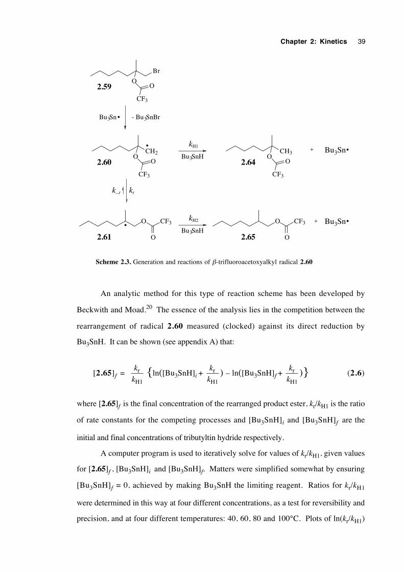
Chapter 2: Kinetics 39
Br
O
CF3
O
CH2O
CF3
O
O
CH3O
CF3
O
OCF3
O
CF3
O
Bu3Sn
Bu3Sn - Bu3SnBr
kr
kH2
Bu3SnH
kH1
Bu3SnH
2.59
2.60 2.64
2.61 2.65
k–r
•
•
•
•
Bu3Sn•
Scheme 2.3. Generation and reactions of β-trifluoroacetoxyalkyl radical 2.60
An analytic method for this type of reaction scheme has been developed by
Beckwith and Moad.20 The essence of the analysis lies in the competition between the
rearrangement of radical 2.60 measured (clocked) against its direct reduction by
Bu3SnH. It can be shown (see appendix A) that:
[2.65]f = k
kr
H1 {ln([Bu3SnH]i +
k
kr
H1) – ln([Bu3SnH]f +
k
kr
H1)} (2.6)
where [2.65]f is the final concentration of the rearranged product ester, kr/kH1 is the ratio
of rate constants for the competing processes and [Bu3SnH]i and [Bu3SnH]f are the
initial and final concentrations of tributyltin hydride respectively.
A computer program is used to iteratively solve for values of kr/kH1, given values
for [2.65]f , [Bu3SnH]i and [Bu3SnH]f. Matters were simplified somewhat by ensuring
[Bu3SnH]f = 0, achieved by making Bu3SnH the limiting reagent. Ratios for kr/kH1
were determined in this way at four different concentrations, as a test for reversibility and
precision, and at four different temperatures: 40, 60, 80 and 100°C. Plots of ln(kr/kH1)
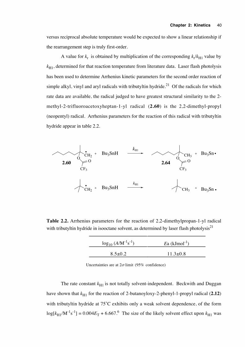
Chapter 2: Kinetics 40
versus reciprocal absolute temperature would be expected to show a linear relationship if
the rearrangement step is truly first-order.
A value for kr is obtained by multiplication of the corresponding kr/kH1 value by
kH1, determined for that reaction temperature from literature data. Laser flash photolysis
has been used to determine Arrhenius kinetic parameters for the second order reaction of
simple alkyl, vinyl and aryl radicals with tributyltin hydride.21 Of the radicals for which
rate data are available, the radical judged to have greatest structural similarity to the 2-
methyl-2-trifluoroacetoxyheptan-1-yl radical (2.60) is the 2,2-dimethyl-propyl
(neopentyl) radical. Arrhenius parameters for the reaction of this radical with tributyltin
hydride appear in table 2.2.
CH2O
CF3
O
CH3O
CF3
O
Bu3SnBu3SnHkH1
CH2 CH3Bu3SnH
kH1
Bu3Sn
•
••
•
2.60 2.64
Table 2.2. Arrhenius parameters for the reaction of 2,2-dimethylpropan-1-yl radical
with tributyltin hydride in isooctane solvent, as determined by laser flash photolysis21
log10 (A/M-1s-1) Ea (kJmol-1)
8.5±0.2 11.3±0.8
Uncertainties are at 2σ limit (95% confidence)
The rate constant kH1 is not totally solvent-independent. Beckwith and Duggan
have shown that kH1 for the reaction of 2-butanoyloxy-2-phenyl-1-propyl radical (2.12)
with tributyltin hydride at 75˚C exhibits only a weak solvent dependence, of the form
log[kH1/M-1s-1] = 0.004ET + 6.667.6 The size of the likely solvent effect upon kH1 was
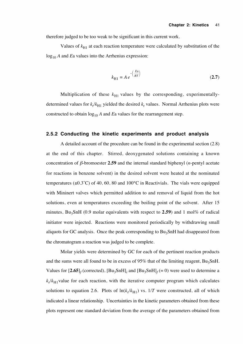
Chapter 2: Kinetics 41
therefore judged to be too weak to be significant in this current work.
Values of kH1 at each reaction temperature were calculated by substitution of the
log10 A and Ea values into the Arrhenius expression:
kH1 = A eE
RT−
a
(2.7)
Multiplication of these kH1 values by the corresponding, experimentally-
determined values for kr/kH1 yielded the desired kr values. Normal Arrhenius plots were
constructed to obtain log10 A and Ea values for the rearrangement step.
2.5.2 Conducting the kinetic experiments and product analysis
A detailed account of the procedure can be found in the experimental section (2.8)
at the end of this chapter. Stirred, deoxygenated solutions containing a known
concentration of β-bromoester 2.59 and the internal standard biphenyl (n-pentyl acetate
for reactions in benzene solvent) in the desired solvent were heated at the nominated
temperatures (±0.3˚C) of 40, 60, 80 and 100°C in Reactivials. The vials were equipped
with Mininert valves which permitted addition to and removal of liquid from the hot
solutions, even at temperatures exceeding the boiling point of the solvent. After 15
minutes, Bu3SnH (0.9 molar equivalents with respect to 2.59) and 1 mol% of radical
initiator were injected. Reactions were monitored periodically by withdrawing small
aliquots for GC analysis. Once the peak corresponding to Bu3SnH had disappeared from
the chromatogram a reaction was judged to be complete.
Molar yields were determined by GC for each of the pertinent reaction products
and the sums were all found to be in excess of 95% that of the limiting reagent, Bu3SnH.
Values for [2.65]f (corrected), [Bu3SnH]i and [Bu3SnH]f (= 0) were used to determine a
kr/kH1value for each reaction, with the iterative computer program which calculates
solutions to equation 2.6. Plots of ln(kr/kH1) vs. 1/T were constructed, all of which
indicated a linear relationship. Uncertainties in the kinetic parameters obtained from these
plots represent one standard deviation from the average of the parameters obtained from
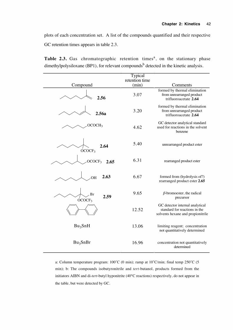
Chapter 2: Kinetics 42
plots of each concentration set. A list of the compounds quantified and their respective
GC retention times appears in table 2.3.
Table 2.3. Gas chromatographic retention timesa, on the stationary phase
dimethylpolysiloxane (BP1), for relevant compoundsb detected in the kinetic analysis.
Compound
Typicalretention time
(min) Comments
2.563.07
formed by thermal eliminationfrom unrearranged product
trifluoroacetate 2.64
2.56a3.20
formed by thermal eliminationfrom unrearranged product
trifluoroacetate 2.64
OCOCH3 4.62GC detector analytical standard
used for reactions in the solventbenzene
OCOCF3
2.64 5.40 unrearranged product ester
OCOCF3 2.65 6.31 rearranged product ester
OH 2.63 6.67 formed from (hydrolysis of?)rearranged product ester 2.65
OCOCF3
Br2.59
9.65 β-bromoester, the radicalprecursor
12.52GC detector internal analytical
standard for reactions in thesolvents hexane and propionitrile
Bu3SnH 13.06 limiting reagent; concentrationnot quantitatively determined
Bu3SnBr 16.96 concentration not quantitativelydetermined
a: Column temperature program: 100˚C (0 min); ramp at 10˚C/min; final temp 250˚C (5
min); b: The compounds isobutyronitrile and tert-butanol, products formed from the
initiators AIBN and di-tert-butyl hyponitrite (40°C reactions) respectively, do not appear in
the table, but were detected by GC.
Chapter 2: Kinetics 43
Values of kH1 at each temperature were determined from the literature data, then
kr values were calculated. Uncertainties in both the kH1 and kr values represent two
standard deviations (95% confidence) and are derived entirely from the uncertainties in
the kH1 Arrhenius parameters.
Plots of lnkr vs. 1/T were constructed and show a linear relationship, as expected.
Arrhenius parameters for the radical rearrangement step were obtained. Uncertainties in
these values are a sum of those inherent in the calculation of kr values, and from the
average of the Arrhenius parameters from plots of each concentration set. These
uncertainties are at one standard deviation limits.
Both experimental and recommended values of the kinetic parameters are given.
Experimental values are averages of the values obtained from the series of reactions at
each concentration, using all the experimental data. Recommended values are those
obtained by omitting lower quality data, usually from reactions for which the molar yields
were lowest and/or where there was a large proportion of alkenes formed by elimination
of trifluoroacetic acid from the product trifluoroacetate esters. This increased the
uncertainty in the yield of the product ester.
2.5.3 Management of analytical complexities
Owing to the relative lability of the trifluoroacetate products 2.64 and 2.65, the
analysis of the product concentrations was more complex than initially envisaged. As
table 2.3 illustrates, there are three by-products formed, namely 2-methyl-1-heptene
(2.56), 2-methyl-2-heptene (2.56a), and 2-methylheptan-1-ol (2.63). Some
adjustments were therefore made to the procedure used to determine the final
concentration of ester 2.65.
Injection of a benzene solution of 2-methyl-2-trifluoroacetoxyheptane (2.64) into
the GC (injector temperature of 250°C) caused the formation of a small amount of the two
alkenes, 2.56 and 2.56a. They were produced in approximately equal proportions as
the only detectable products in a combined yield of about 5%, presumably by thermal
elimination of trifluoroacetic acid from 2.64. The identity and relative retention times of
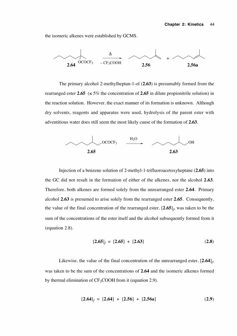
Chapter 2: Kinetics 44
the isomeric alkenes were established by GCMS.
2.56 2.56a2.64OCOCF3
+∆
– CF3COOH
The primary alcohol 2-methylheptan-1-ol (2.63) is presumably formed from the
rearranged ester 2.65 (≤ 5% the concentration of 2.65 in dilute propionitrile solution) in
the reaction solution. However, the exact manner of its formation is unknown. Although
dry solvents, reagents and apparatus were used, hydrolysis of the parent ester with
adventitious water does still seem the most likely cause of the formation of 2.63.
2.65 2.63
OCOCF3 OHH2O
Injection of a benzene solution of 2-methyl-1-trifluoroacetoxyheptane (2.65) into
the GC did not result in the formation of either of the alkenes, nor the alcohol 2.63.
Therefore, both alkenes are formed solely from the unrearranged ester 2.64. Primary
alcohol 2.63 is presumed to arise solely from the rearranged ester 2.65. Consequently,
the value of the final concentration of the rearranged ester, [2.65]f, was taken to be the
sum of the concentrations of the ester itself and the alcohol subsequently formed from it
(equation 2.8).
[2.65]f = [2.65] + [2.63] (2.8)
Likewise, the value of the final concentration of the unrearranged ester, [2.64]f,
was taken to be the sum of the concentrations of 2.64 and the isomeric alkenes formed
by thermal elimination of CF3COOH from it (equation 2.9).
[2.64]f = [2.64] + [2.56] + [2.56a] (2.9)
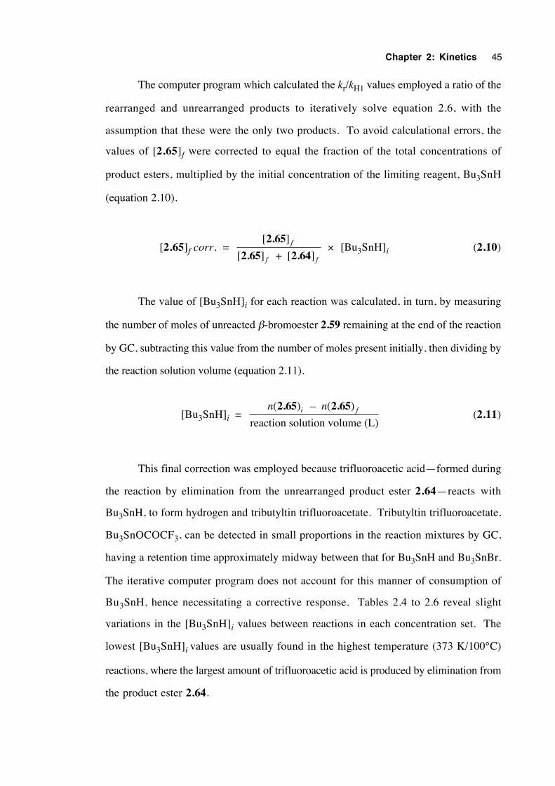
Chapter 2: Kinetics 45
The computer program which calculated the kr/kH1 values employed a ratio of the
rearranged and unrearranged products to iteratively solve equation 2.6, with the
assumption that these were the only two products. To avoid calculational errors, the
values of [2.65]f were corrected to equal the fraction of the total concentrations of
product esters, multiplied by the initial concentration of the limiting reagent, Bu3SnH
(equation 2.10).
[2.65]f corr. = [ ]
[ ] + [ ]
2.65
2.65 2.64f
f f
× [Bu3SnH]i (2.10)
The value of [Bu3SnH]i for each reaction was calculated, in turn, by measuring
the number of moles of unreacted β-bromoester 2.59 remaining at the end of the reaction
by GC, subtracting this value from the number of moles present initially, then dividing by
the reaction solution volume (equation 2.11).
[Bu3SnH]i = n ni f( ) – ( )
reaction solution volume (L)
2.65 2.65 (2.11)
This final correction was employed because trifluoroacetic acid—formed during
the reaction by elimination from the unrearranged product ester 2.64—reacts with
Bu3SnH, to form hydrogen and tributyltin trifluoroacetate. Tributyltin trifluoroacetate,
Bu3SnOCOCF3, can be detected in small proportions in the reaction mixtures by GC,
having a retention time approximately midway between that for Bu3SnH and Bu3SnBr.
The iterative computer program does not account for this manner of consumption of
Bu3SnH, hence necessitating a corrective response. Tables 2.4 to 2.6 reveal slight
variations in the [Bu3SnH]i values between reactions in each concentration set. The
lowest [Bu3SnH]i values are usually found in the highest temperature (373 K/100°C)
reactions, where the largest amount of trifluoroacetic acid is produced by elimination from
the product ester 2.64.

Chapter 2: Kinetics 46
2.5.4 Kinetics results
Table 2.4. Kinetic data for reactions with hexane as the solvent
Nominal[Bu3SnH]i T (K)
[2.65]f
corr. (M)[Bu3SnH]i
(M)[Bu3SnH]f
(M)k
k
r
H1
(M)
10-6 × kH1calc.
(M-1s-1)
10-3 × kr
(s-1)
0.015 M 313.9 0.00287 0.0144 0 0.00107 4.17±1.7 4.48±1.8
332.5 0.00479 0.0149 0 0.00244 5.31±2.1 13.0±3.4
353.6 0.00704 0.0149 0 0.00524 6.78±2.7 35.5±15
373.2 0.00911 0.0146 0 0.0103 8.29±3.2 85.4±35
0.050 M 313.9 0.00500 0.0503 0 0.00138 4.17±1.7 5.75±2.4
332.5 0.00845 0.0499 0 0.00292 5.31±2.1 15.5±6.4
353.6 0.00140 0.0497 0 0.00651 6.78±2.7 44.1±18
373.2 0.0211 0.0491 0 0.0140 8.30±3.2 116±48
0.10 M 313.9 0.00682 0.0996 0 0.00166 4.17±1.7 6.91±2.8
332.5 0.0112 0.0990 0 0.00326 5.31±2.1 17.3±7.1
353.6 0.0190 0.0975 0 0.00705 6.78±2.7 47.8±20
373.2 0.0318 0.0977 0 0.0164 8.30±3.2 136±56
0.15 M 313.9 0.00837 0.147 0 0.00193 4.17±1.7 8.03±3.3
332.5 0.0136 0.146 0 0.00367 5.31±2.1 19.5±8.0
353.6 0.0239 0.145 0 0.00813 6.78±2.7 55.1±23
373.2 0.0414 0.144 0 0.0194 8.30±3.2 161±66
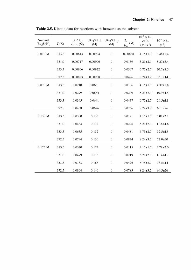
Chapter 2: Kinetics 47
Table 2.5. Kinetic data for reactions with benzene as the solvent
Nominal[Bu3SnH]i T (K)
[2.65]f
corr. (M)[Bu3SnH]i
(M)[Bu3SnH]f
(M)k
k
r
H1
(M)
10-6 × kH1calc.
(M-1s-1)
10-4 × kr
(s-1)
0.010 M 313.6 0.00613 0.00904 0 0.00838 4.15±1.7 3.48±1.4
331.0 0.00717 0.00906 0 0.0159 5.21±2.1 8.27±3.4
353.3 0.00806 0.00922 0 0.0307 6.75±2.7 20.7±8.5
372.5 0.00823 0.00908 0 0.0426 8.24±3.2 35.1±14
0.070 M 313.6 0.0210 0.0661 0 0.0106 4.15±1.7 4.39±1.8
331.0 0.0299 0.0664 0 0.0209 5.21±2.1 10.9±4.5
353.3 0.0395 0.0641 0 0.0437 6.75±2.7 29.5±12
372.5 0.0458 0.0626 0 0.0766 8.24±3.2 63.1±26
0.130 M 313.6 0.0300 0.133 0 0.0121 4.15±1.7 5.01±2.1
331.0 0.0434 0.132 0 0.0226 5.21±2.1 11.8±4.8
353.3 0.0635 0.132 0 0.0481 6.75±2.7 32.5±13
372.5 0.0794 0.130 0 0.0874 8.24±3.2 72.0±30
0.175 M 313.6 0.0320 0.174 0 0.0115 4.15±1.7 4.78±2.0
331.0 0.0479 0.173 0 0.0219 5.21±2.1 11.4±4.7
353.3 0.0733 0.168 0 0.0496 6.75±2.7 33.5±14
372.5 0.0804 0.140 0 0.0783 8.24±3.2 64.5±26
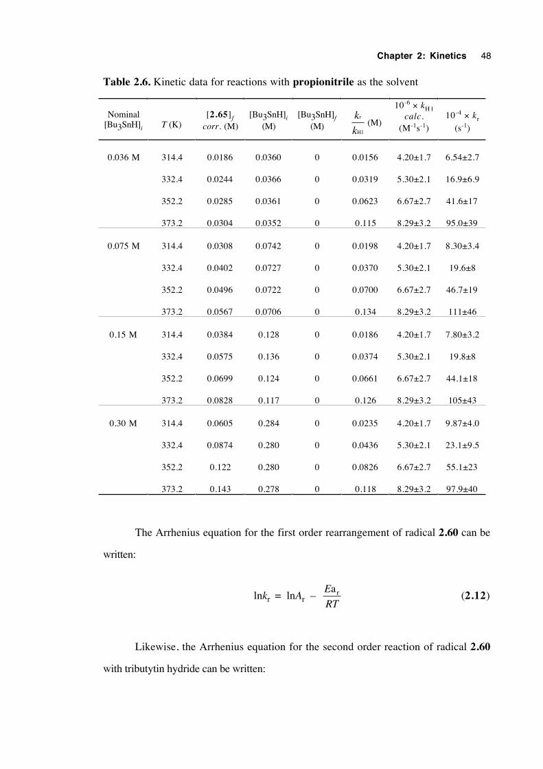
Chapter 2: Kinetics 48
Table 2.6. Kinetic data for reactions with propionitrile as the solvent
Nominal[Bu3SnH]i T (K)
[2.65]f
corr. (M)[Bu3SnH]i
(M)[Bu3SnH]f
(M)k
k
r
H1
(M)
10-6 × kH1calc.
(M-1s-1)
10-4 × kr
(s-1)
0.036 M 314.4 0.0186 0.0360 0 0.0156 4.20±1.7 6.54±2.7
332.4 0.0244 0.0366 0 0.0319 5.30±2.1 16.9±6.9
352.2 0.0285 0.0361 0 0.0623 6.67±2.7 41.6±17
373.2 0.0304 0.0352 0 0.115 8.29±3.2 95.0±39
0.075 M 314.4 0.0308 0.0742 0 0.0198 4.20±1.7 8.30±3.4
332.4 0.0402 0.0727 0 0.0370 5.30±2.1 19.6±8
352.2 0.0496 0.0722 0 0.0700 6.67±2.7 46.7±19
373.2 0.0567 0.0706 0 0.134 8.29±3.2 111±46
0.15 M 314.4 0.0384 0.128 0 0.0186 4.20±1.7 7.80±3.2
332.4 0.0575 0.136 0 0.0374 5.30±2.1 19.8±8
352.2 0.0699 0.124 0 0.0661 6.67±2.7 44.1±18
373.2 0.0828 0.117 0 0.126 8.29±3.2 105±43
0.30 M 314.4 0.0605 0.284 0 0.0235 4.20±1.7 9.87±4.0
332.4 0.0874 0.280 0 0.0436 5.30±2.1 23.1±9.5
352.2 0.122 0.280 0 0.0826 6.67±2.7 55.1±23
373.2 0.143 0.278 0 0.118 8.29±3.2 97.9±40
The Arrhenius equation for the first order rearrangement of radical 2.60 can be
written:
lnkr = lnAr – E
RT
ar (2.12)
Likewise, the Arrhenius equation for the second order reaction of radical 2.60
with tributytin hydride can be written:
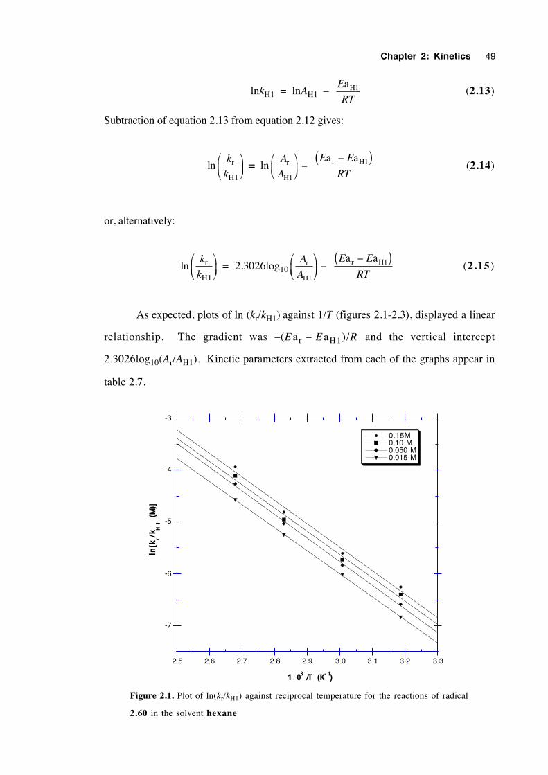
Chapter 2: Kinetics 49
lnkH1 = lnAH1 – E
RT
aH1 (2.13)
Subtraction of equation 2.13 from equation 2.12 gives:
lnk
kr
H1
= ln
A
A
E E
RTr
H1
r H1a a
−
−( ) (2.14)
or, alternatively:
lnk
kr
H1
= 2.3026log10
A
A
E E
RTr
H1
r H1a a
−
−( ) (2.15)
As expected, plots of ln (kr/kH1) against 1/T (figures 2.1-2.3), displayed a linear
relationship. The gradient was –(E ar – E aH 1)/R and the vertical intercept
2.3026log10(Ar/AH1). Kinetic parameters extracted from each of the graphs appear in
table 2.7.
-7
-6
-5
-4
-3
2.5 2.6 2.7 2.8 2.9 3.0 3.1 3.2 3.3
0.15M0.10 M0.050 M0.015 M
ln[k
r/kH
1 (M
)]
1 03 /T (K- 1)
Figure 2.1. Plot of ln(kr/kH1) against reciprocal temperature for the reactions of radical
2.60 in the solvent hexane
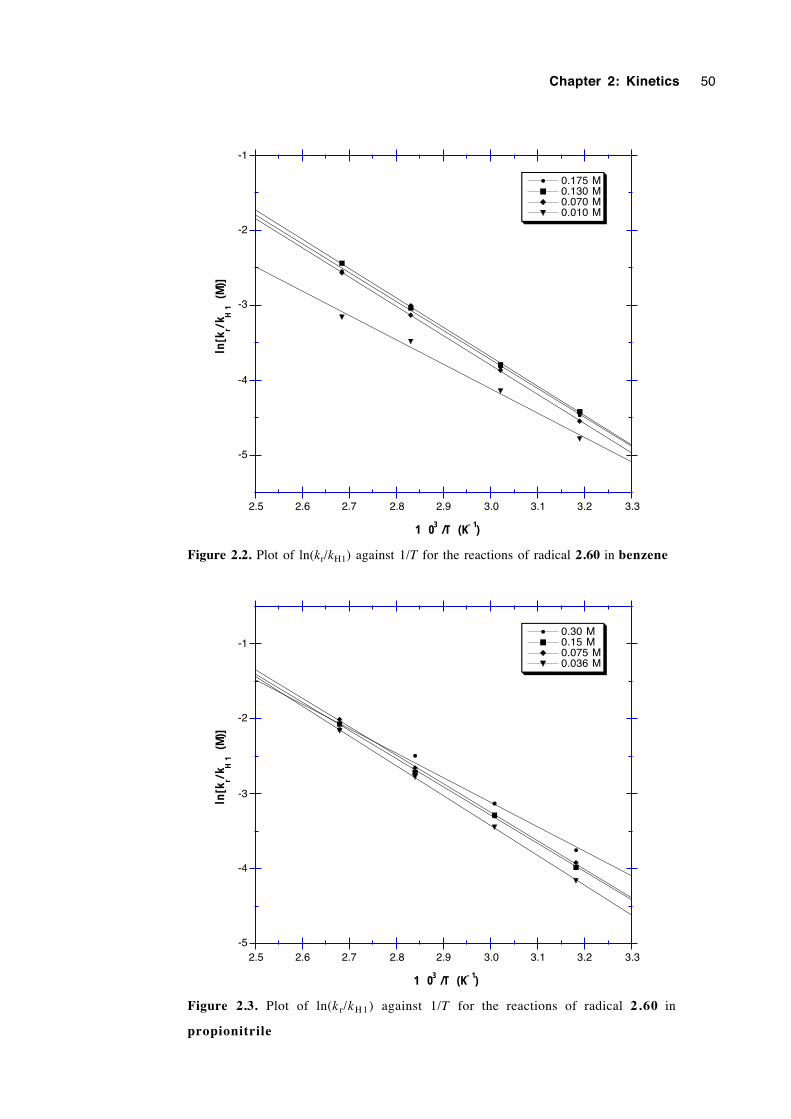
Chapter 2: Kinetics 50
-5
-4
-3
-2
-1
2.5 2.6 2.7 2.8 2.9 3.0 3.1 3.2 3.3
0.175 M0.130 M0.070 M0.010 M
ln[k
r/kH
1 (M
)]
1 03 /T (K- 1)
Figure 2.2. Plot of ln(kr/kH1) against 1/T for the reactions of radical 2.60 in benzene
-5
-4
-3
-2
-1
2.5 2.6 2.7 2.8 2.9 3.0 3.1 3.2 3.3
0.30 M0.15 M0.075 M0.036 M
ln[k
r/kH
1 (M
)]
1 03 /T (K- 1)
Figure 2.3. Plot of ln(k r/kH 1) against 1/T for the reactions of radical 2.60 in
propionitrile
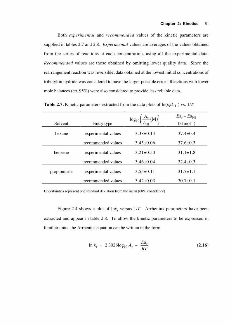
Chapter 2: Kinetics 51
Both experimental and recommended values of the kinetic parameters are
supplied in tables 2.7 and 2.8. Experimental values are averages of the values obtained
from the series of reactions at each concentration, using all the experimental data.
Recommended values are those obtained by omitting lower quality data. Since the
rearrangement reaction was reversible, data obtained at the lowest initial concentrations of
tributyltin hydride was considered to have the larger possible error. Reactions with lower
mole balances (ca. 95%) were also considered to provide less reliable data.
Table 2.7. Kinetic parameters extracted from the data plots of ln(kr/kH1) vs. 1/T
Solvent Entry typelog10
A
Ar
H1
M( )
Ear – EaH1
(kJmol-1)
hexane experimental values 3.38±0.14 37.4±0.4
recommended values 3.45±0.06 37.6±0.3
benzene experimental values 3.21±0.50 31.1±1.8
recommended values 3.46±0.04 32.4±0.3
propionitrile experimental values 3.55±0.11 31.7±1.1
recommended values 3.42±0.03 30.7±0.1
Uncertainties represent one standard deviation from the mean (68% confidence)
Figure 2.4 shows a plot of lnkr versus 1/T. Arrhenius parameters have been
extracted and appear in table 2.8. To allow the kinetic parameters to be expressed in
familiar units, the Arrhenius equation can be written in the form:
ln kr = 2.3026log10 Ar – E
RT
ar (2.16)
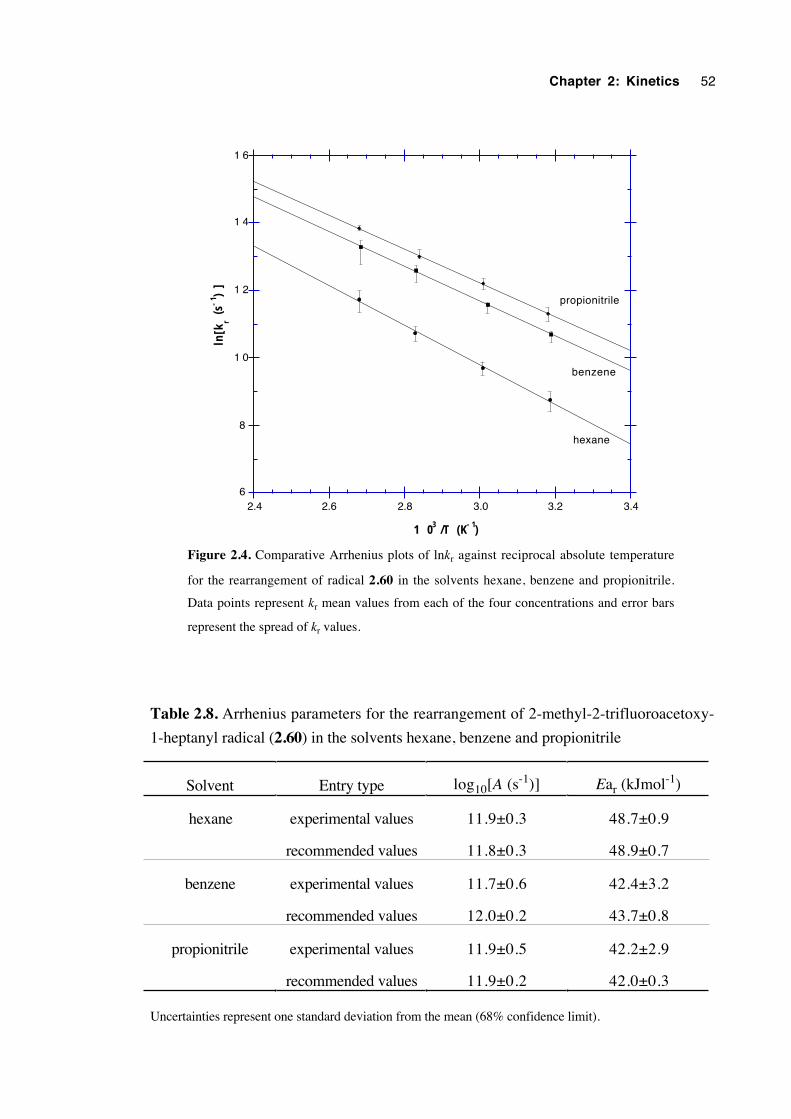
Chapter 2: Kinetics 52
propionitrile
benzene
6
8
1 0
1 2
1 4
1 6
2.4 2.6 2.8 3.0 3.2 3.4
ln[k
r (s-
1 )]
1 03 /T (K- 1)
hexane
Figure 2.4. Comparative Arrhenius plots of lnkr against reciprocal absolute temperature
for the rearrangement of radical 2.60 in the solvents hexane, benzene and propionitrile.
Data points represent kr mean values from each of the four concentrations and error bars
represent the spread of kr values.
Table 2.8. Arrhenius parameters for the rearrangement of 2-methyl-2-trifluoroacetoxy-
1-heptanyl radical (2.60) in the solvents hexane, benzene and propionitrile
Solvent Entry type log10[A (s-1)] Ear (kJmol-1)
hexane experimental values 11.9±0.3 48.7±0.9
recommended values 11.8±0.3 48.9±0.7
benzene experimental values 11.7±0.6 42.4±3.2
recommended values 12.0±0.2 43.7±0.8
propionitrile experimental values 11.9±0.5 42.2±2.9
recommended values 11.9±0.2 42.0±0.3
Uncertainties represent one standard deviation from the mean (68% confidence limit).
Chapter 2: Kinetics 53
2.6 Discussion of results
It was found that the rearrangement of radical 2.60 to 2.61 was reversible,
having an equilibrium constant, K, of between 15 and 53, depending upon the method of
determination. This reversibility was reflected in the plots of ln(kr/kH1) against reciprocal
temperature (figures 2.1-2.3), where it was observed that the lowest data points at each
temperature were those belonging to the reactions performed at the lowest concentrations
of tributyltin hydride.
The accuracy of the kr values are dependent on the respective kH1 values and
therefore on how well the model system mirrored the actual hydrogen atom transfer
reaction between 2.60 and Bu3SnH. It is believed that the model system selected to
mimic actual kH1 values was a good choice. If anything, kH1 for the reaction of 2-methyl-
2-trifluoroacetoxyheptan-1-yl radical (2.60) with Bu3SnH would be a little smaller than
for its model, neopentyl radical, owing to the increased steric bulk of a trifluoroacetate
group over a methyl substituent. Therefore, the true corresponding kr values are
probably slightly smaller than indicated, although certainly not as much as an order of
magnitude, since kH1 for the reaction of methyl radical with Bu3SnH at 75°C is only 7.5
times that for the reaction with the much more highly hindered tert-butyl radical.21 It has
been established previously that kH1 does show a solvent dependence, but the effect is
very weak.6
Table 2.9 contains rate constants, kr, for the radical rearrangement of radical 2.60
at 75°C, calculated using the recommended Arrhenius parameters. Also included are the
values of the empirical solvent polarity parameter ET (30).22 It is clear that kr increases
as the solvent changes from hexane to benzene to propionitrile, supporting others'
findings that polar solvents accelerate the β-acyloxyalkyl radical rearrangement.3,6,10
The A frequency factors are identical within experimental uncertainty, therefore the rate
constant differences lie with the activation energy term, Ear. The decrease observed in
Ear is consistent with a weakening of the C2–O alkoxy ester bond with increasing solvent
polarity.
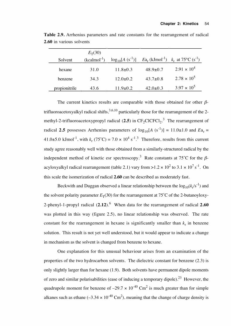
Chapter 2: Kinetics 54
Table 2.9. Arrhenius parameters and rate constants for the rearrangement of radical
2.60 in various solvents
Solvent
ET(30)
(kcalmol-1) log10[A (s-1)] Ear (kJmol-1) kr at 75°C (s-1)
hexane 31.0 11.8±0.3 48.9±0.7 2.91 × 10⁴
benzene 34.3 12.0±0.2 43.7±0.8 2.78 × 10⁵
propionitrile 43.6 11.9±0.2 42.0±0.3 3.97 × 10⁵
The current kinetics results are comparable with those obtained for other β-
trifluoroacetoxyalkyl radical shifts,3,6,10 particularly those for the rearrangement of the 2-
methyl-2-trifluoroacetoxypropyl radical (2.5) in CF2ClCFCl2.3 The rearrangement of
radical 2.5 possesses Arrhenius parameters of log10[A (s-1)] = 11.0±1.0 and Ear =
41.0±5.0 kJmol-1, with kr (75˚C) = 7.0 × 10⁴ s-1.3 Therefore, results from this current
study agree reasonably well with those obtained from a similarly-structured radical by the
independent method of kinetic esr spectroscopy.3 Rate constants at 75˚C for the β-
acyloxyalkyl radical rearrangement (table 2.1) vary from >1.2 × 10² to 3.1 × 10⁷ s-1. On
this scale the isomerization of radical 2.60 can be described as moderately fast.
Beckwith and Duggan observed a linear relationship between the log10(kr/s-1) and
the solvent polarity parameter ET(30) for the rearrangement at 75˚C of the 2-butanoyloxy-
2-phenyl-1-propyl radical (2.12).6 When data for the rearrangement of radical 2.60
was plotted in this way (figure 2.5), no linear relationship was observed. The rate
constant for the rearrangement in hexane is significantly smaller than kr in benzene
solution. This result is not yet well understood, but it would appear to indicate a change
in mechanism as the solvent is changed from benzene to hexane.
One explanation for this unusual behaviour arises from an examination of the
properties of the two hydrocarbon solvents. The dielectric constant for benzene (2.3) is
only slightly larger than for hexane (1.9). Both solvents have permanent dipole moments
of zero and similar polarisabilities (ease of inducing a temporary dipole).23 However, the
quadrupole moment for benzene of –29.7 × 10-40 Cm2 is much greater than for simple
alkanes such as ethane (–3.34 × 10-40 Cm2), meaning that the change of charge density is
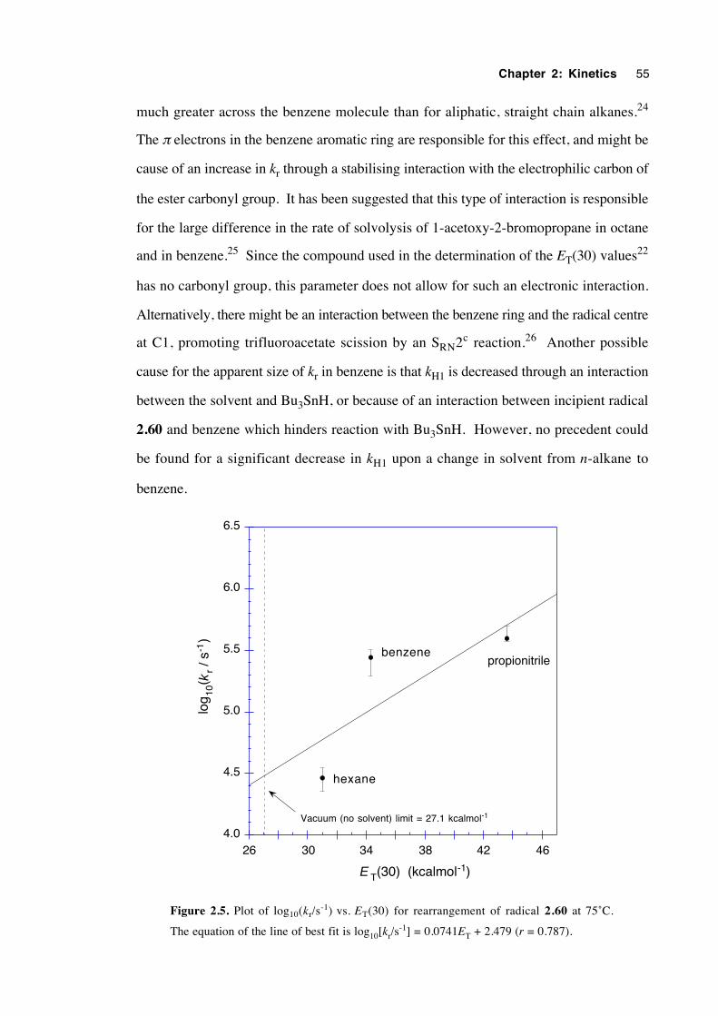
Chapter 2: Kinetics 55
much greater across the benzene molecule than for aliphatic, straight chain alkanes.24
The π electrons in the benzene aromatic ring are responsible for this effect, and might be
cause of an increase in kr through a stabilising interaction with the electrophilic carbon of
the ester carbonyl group. It has been suggested that this type of interaction is responsible
for the large difference in the rate of solvolysis of 1-acetoxy-2-bromopropane in octane
and in benzene.25 Since the compound used in the determination of the ET(30) values22
has no carbonyl group, this parameter does not allow for such an electronic interaction.
Alternatively, there might be an interaction between the benzene ring and the radical centre
at C1, promoting trifluoroacetate scission by an SRN2c reaction.26 Another possible
cause for the apparent size of kr in benzene is that kH1 is decreased through an interaction
between the solvent and Bu3SnH, or because of an interaction between incipient radical
2.60 and benzene which hinders reaction with Bu3SnH. However, no precedent could
be found for a significant decrease in kH1 upon a change in solvent from n-alkane to
benzene.
4.0
4.5
5.0
5.5
6.0
6.5
26 30 34 38 42 46
hexane
benzenepropionitrile
Vacuum (no solvent) limit = 27.1 kcalmol-1
log
10(
r / s
-1)
k
T(30) (kcalmol-1)E
Figure 2.5. Plot of log10(kr/s-1) vs. ET(30) for rearrangement of radical 2.60 at 75˚C.
The equation of the line of best fit is log10[kr/s-1] = 0.0741ET + 2.479 (r = 0.787).
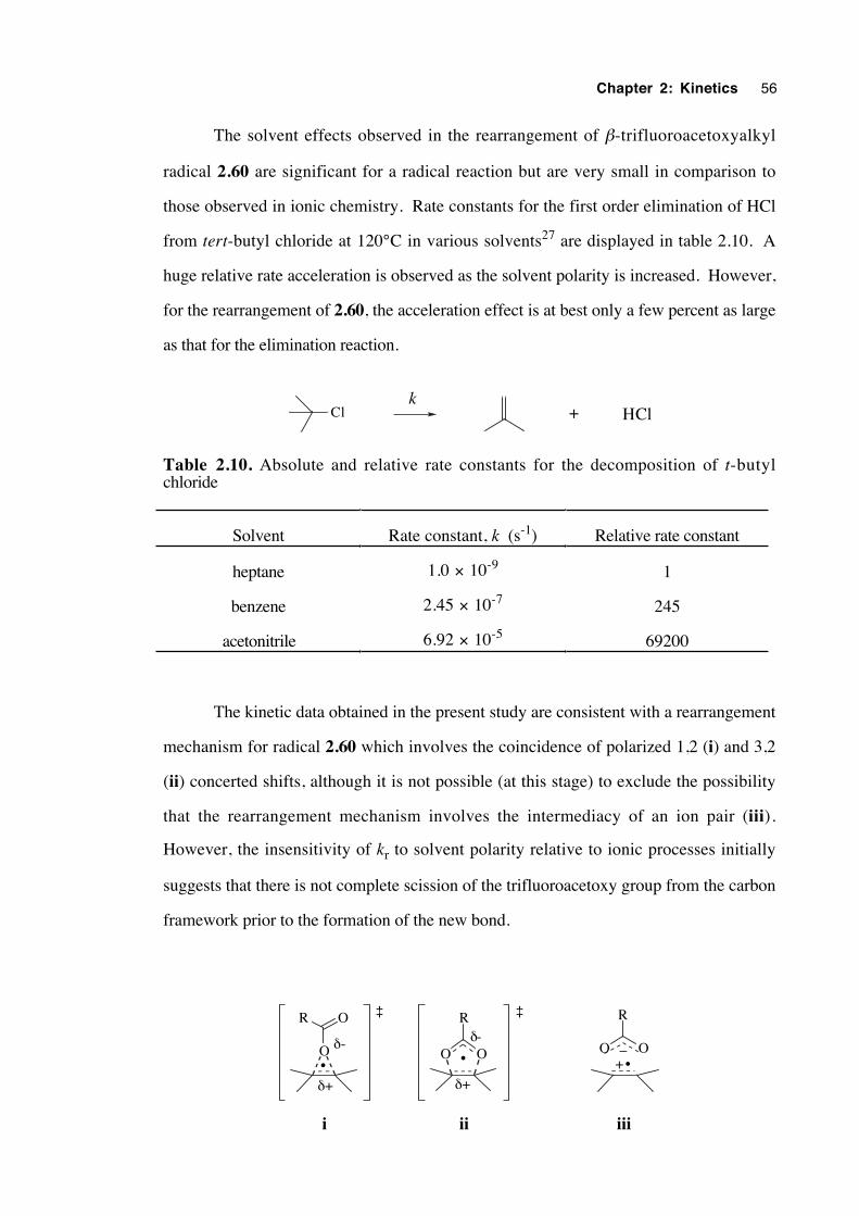
Chapter 2: Kinetics 56
The solvent effects observed in the rearrangement of β-trifluoroacetoxyalkyl
radical 2.60 are significant for a radical reaction but are very small in comparison to
those observed in ionic chemistry. Rate constants for the first order elimination of HCl
from tert-butyl chloride at 120°C in various solvents27 are displayed in table 2.10. A
huge relative rate acceleration is observed as the solvent polarity is increased. However,
for the rearrangement of 2.60, the acceleration effect is at best only a few percent as large
as that for the elimination reaction.
Cl + HClk
Table 2.10. Absolute and relative rate constants for the decomposition of t-butylchloride
Solvent Rate constant, k (s-1) Relative rate constant
heptane 1.0 × 10⁻⁹ 1
benzene 2.45 × 10⁻⁷ 245
acetonitrile 6.92 × 10⁻⁵ 69200
The kinetic data obtained in the present study are consistent with a rearrangement
mechanism for radical 2.60 which involves the coincidence of polarized 1,2 (i) and 3,2
(ii) concerted shifts, although it is not possible (at this stage) to exclude the possibility
that the rearrangement mechanism involves the intermediacy of an ion pair (iii).
However, the insensitivity of kr to solvent polarity relative to ionic processes initially
suggests that there is not complete scission of the trifluoroacetoxy group from the carbon
framework prior to the formation of the new bond.
O O
R
•O
R O
•O O
R
+_
iiiiii
•
δ+
δ-
δ+
δ-
‡ ‡
Chapter 2: Kinetics 57
2.7 Conclusions
The rearrangement of 2-methyl-2-trifluoroacetoxy-1-heptyl (2.60) to the 2-
methyl-1-trifluoroacetoxy-2-heptyl radical (2.61) is a reversible process. At 80˚C in
benzene, an equilibrium mixture consists of between 93.8% and 98.1% of 2.61.
The kinetics of the rearrangement of 2.60→2.61 have been determined by a
product-studies/competition-clock method in three different solvents. There was good
agreement with results obtained by kinetic esr spectroscopy for the rearrangement of the
structurally-similar 2-methyl-2-trifluoroacetoxy-1-propyl radical (2.5) in CF2ClCFCl2.
Arrhenius equations for the rearrangement of 2.60 in each solvent were: hexane, log10[kr
(s-1)] = 11.8±0.3 – (48.9±0.7)/θ; benzene, log10[kr (s-1)] = 12.0±0.2 – (43.7±0.8)/θ;
and propionitrile, log10[kr (s-1)] = 11.9±0.2 – (42.0±0.3)/θ. Rate constants at 75˚C in
each solvent were: hexane, kr = 2.9 × 10⁴; benzene, kr = 2.8 × 10⁵; and propionitrile, kr
= 4.0 × 10⁵ s-1. It is the activation energy term which is primarily responsible for the
dependence of the rate constant upon solvent polarity, indicating that the C–O ether bond
of 2.60 is weakened by polar solvent. It was unusual that kr in benzene was 9.5 times
as large as that in hexane, yet kr in propionitrile was only 13.6 times as large as that in
hexane. This effect is not well understood.
These solvent effects, although large for a radical reaction, were only a few
percent as large as those observed for a comparable solvolysis reaction. Overall, the
results most closely fit a mechanism which involves a combination of concerted and
polarized 1,2 and 3,2 pericyclic shifts, although the intermediacy of an alkene-radical-
cation/trifluoroacetate-anion pair cannot yet be excluded.
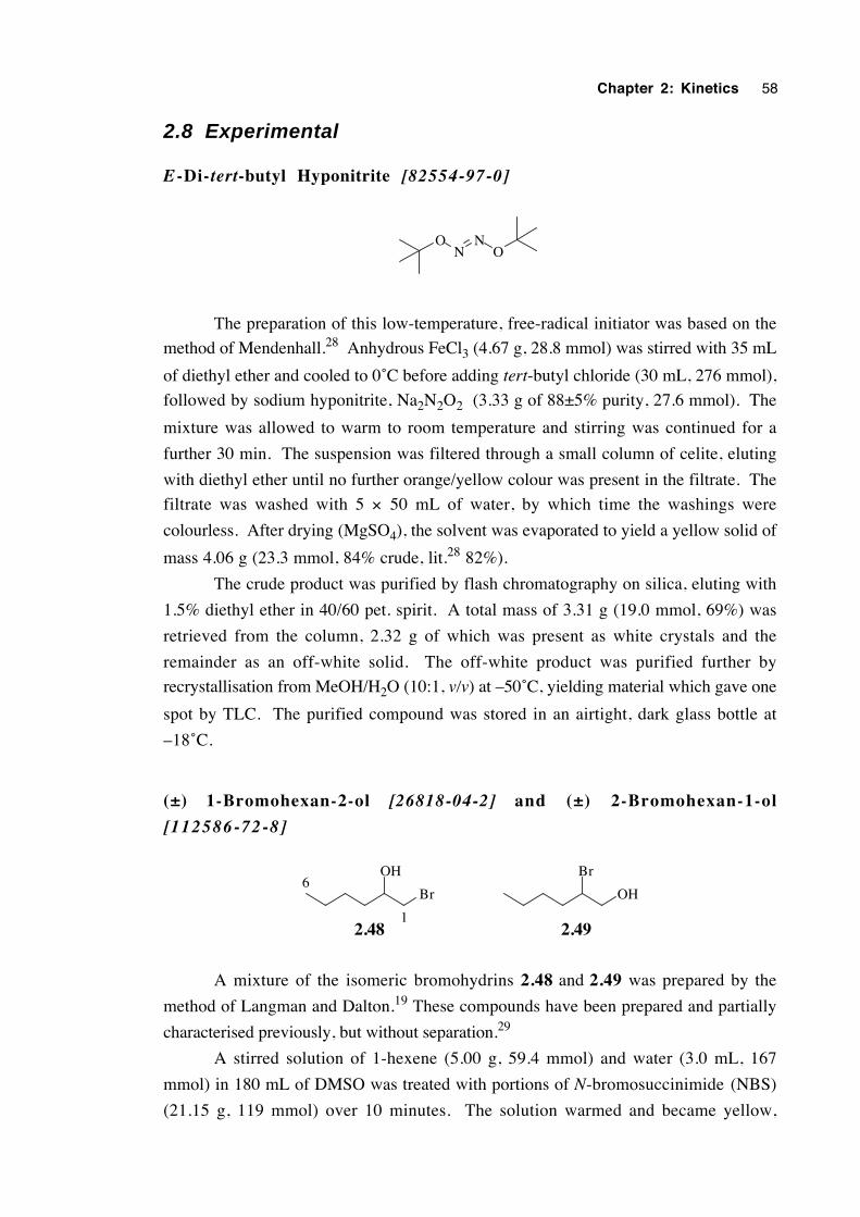
Chapter 2: Kinetics 58
2.8 Experimental
E -Di-tert-butyl Hyponitrite [82554-97-0]
NNO
O
The preparation of this low-temperature, free-radical initiator was based on the
method of Mendenhall.28 Anhydrous FeCl3 (4.67 g, 28.8 mmol) was stirred with 35 mL
of diethyl ether and cooled to 0˚C before adding tert-butyl chloride (30 mL, 276 mmol),
followed by sodium hyponitrite, Na2N2O2 (3.33 g of 88±5% purity, 27.6 mmol). The
mixture was allowed to warm to room temperature and stirring was continued for a
further 30 min. The suspension was filtered through a small column of celite, eluting
with diethyl ether until no further orange/yellow colour was present in the filtrate. The
filtrate was washed with 5 × 50 mL of water, by which time the washings were
colourless. After drying (MgSO4), the solvent was evaporated to yield a yellow solid of
mass 4.06 g (23.3 mmol, 84% crude, lit.28 82%).
The crude product was purified by flash chromatography on silica, eluting with
1.5% diethyl ether in 40/60 pet. spirit. A total mass of 3.31 g (19.0 mmol, 69%) was
retrieved from the column, 2.32 g of which was present as white crystals and the
remainder as an off-white solid. The off-white product was purified further by
recrystallisation from MeOH/H2O (10:1, v/v) at –50˚C, yielding material which gave one
spot by TLC. The purified compound was stored in an airtight, dark glass bottle at
–18˚C.
(±) 1-Bromohexan-2-ol [26818-04-2] and (±) 2-Bromohexan-1-ol
[112586-72-8]
OH
Br
Br
OH
2.48 2.491
6
A mixture of the isomeric bromohydrins 2.48 and 2.49 was prepared by the
method of Langman and Dalton.19 These compounds have been prepared and partially
characterised previously, but without separation.29
A stirred solution of 1-hexene (5.00 g, 59.4 mmol) and water (3.0 mL, 167
mmol) in 180 mL of DMSO was treated with portions of N-bromosuccinimide (NBS)
(21.15 g, 119 mmol) over 10 minutes. The solution warmed and became yellow,
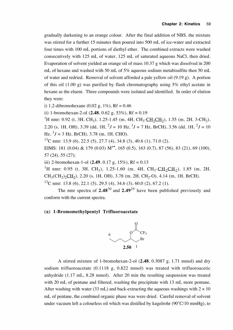
Chapter 2: Kinetics 59
gradually darkening to an orange colour. After the final addition of NBS, the mixture
was stirred for a further 15 minutes then poured into 500 mL of ice-water and extracted
four times with 100 mL portions of diethyl ether. The combined extracts were washed
consecutively with 125 mL of water, 125 mL of saturated aqueous NaCl, then dried.
Evaporation of solvent yielded an orange oil of mass 10.37 g which was dissolved in 200
mL of hexane and washed with 50 mL of 5% aqueous sodium metabisulfite then 50 mL
of water and redried. Removal of solvent afforded a pale yellow oil (9.19 g). A portion
of this oil (1.00 g) was purified by flash chromatography using 5% ethyl acetate in
hexane as the eluent. Three compounds were isolated and identified. In order of elution
they were:
i) 1,2-dibromohexane (0.02 g, 1%), Rf = 0.46
ii) 1-bromohexan-2-ol (2.48, 0.62 g, 53%), Rf = 0.191H nmr: 0.92 (t, 3H, CH3), 1.25-1.45 (m, 4H, CH3-CH2CH2), 1.55 (m, 2H, 3-CH2),
2.20 (s, 1H, OH), 3.39 (dd, 1H, 2J = 10 Hz, 3J = 7 Hz, BrCH), 3.56 (dd, 1H, 2J = 10
Hz, 3J = 3 Hz, BrCH), 3.78 (m, 1H, CHO).13C nmr: 13.9 (6), 22.5 (5), 27.7 (4), 34.8 (3), 40.6 (1), 71.0 (2).
EIMS: 181 (0.04) & 179 (0.03) M+•, 165 (0.5), 163 (0.7), 87 (56), 83 (21), 69 (100),
57 (24), 55 (27);
iii) 2-bromohexan-1-ol (2.49, 0.17 g, 15%), Rf = 0.131H nmr: 0.95 (t, 3H, CH3), 1.25-1.60 (m, 4H, CH3-C H 2C H 2), 1.85 (m, 2H,
CH3(CH2)2CH2), 2.20 (s, 1H, OH), 3.78 (m, 2H, CH2-O), 4.14 (m, 1H, BrCH).13C nmr: 13.8 (6), 22.1 (5), 29.5 (4), 34.6 (3), 60.0 (2), 67.2 (1).
The nmr spectra of 2.4830 and 2.4929 have been published previously and
conform with the current spectra.
(±) 1-Bromomethylpentyl Trifluoroacetate
O
Br
2.50
CF3
O
1
6
A stirred mixture of 1-bromohexan-2-ol (2.48, 0.3087 g, 1.71 mmol) and dry
sodium trifluoroacetate (0.1118 g, 0.822 mmol) was treated with trifluoroacetic
anhydride (1.17 mL, 8.28 mmol). After 20 min the resulting suspension was treated
with 20 mL of pentane and filtered, washing the precipitate with 13 mL more pentane.
After washing with water (33 mL) and back-extracting the aqueous washings with 2 × 10
mL of pentane, the combined organic phase was were dried. Careful removal of solvent
under vacuum left a colourless oil which was distilled by kugelrohr (90˚C/10 mmHg), to
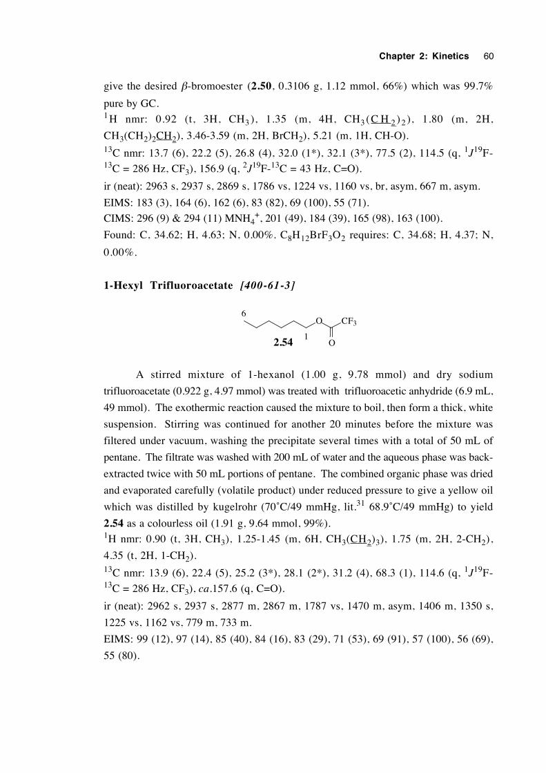
Chapter 2: Kinetics 60
give the desired β-bromoester (2.50, 0.3106 g, 1.12 mmol, 66%) which was 99.7%
pure by GC.1 H nmr: 0.92 (t, 3H, CH3 ), 1.35 (m, 4H, CH3 (C H 2 )2 ), 1.80 (m, 2H,
CH3(CH2)2CH2), 3.46-3.59 (m, 2H, BrCH2), 5.21 (m, 1H, CH-O).13C nmr: 13.7 (6), 22.2 (5), 26.8 (4), 32.0 (1*), 32.1 (3*), 77.5 (2), 114.5 (q, 1J19F-13C = 286 Hz, CF3), 156.9 (q, 2J19F-13C = 43 Hz, C=O).
ir (neat): 2963 s, 2937 s, 2869 s, 1786 vs, 1224 vs, 1160 vs, br, asym, 667 m, asym.
EIMS: 183 (3), 164 (6), 162 (6), 83 (82), 69 (100), 55 (71).
CIMS: 296 (9) & 294 (11) MNH4+, 201 (49), 184 (39), 165 (98), 163 (100).
Found: C, 34.62; H, 4.63; N, 0.00%. C8H12BrF3O2 requires: C, 34.68; H, 4.37; N,
0.00%.
1-Hexyl Trifluoroacetate [400-61-3]
O CF3
O2.541
6
A stirred mixture of 1-hexanol (1.00 g, 9.78 mmol) and dry sodium
trifluoroacetate (0.922 g, 4.97 mmol) was treated with trifluoroacetic anhydride (6.9 mL,
49 mmol). The exothermic reaction caused the mixture to boil, then form a thick, white
suspension. Stirring was continued for another 20 minutes before the mixture was
filtered under vacuum, washing the precipitate several times with a total of 50 mL of
pentane. The filtrate was washed with 200 mL of water and the aqueous phase was back-
extracted twice with 50 mL portions of pentane. The combined organic phase was dried
and evaporated carefully (volatile product) under reduced pressure to give a yellow oil
which was distilled by kugelrohr (70˚C/49 mmHg, lit.31 68.9˚C/49 mmHg) to yield
2.54 as a colourless oil (1.91 g, 9.64 mmol, 99%).1H nmr: 0.90 (t, 3H, CH3), 1.25-1.45 (m, 6H, CH3(CH2)3), 1.75 (m, 2H, 2-CH2),
4.35 (t, 2H, 1-CH2).13C nmr: 13.9 (6), 22.4 (5), 25.2 (3*), 28.1 (2*), 31.2 (4), 68.3 (1), 114.6 (q, 1J19F-13C = 286 Hz, CF3), ca.157.6 (q, C=O).
ir (neat): 2962 s, 2937 s, 2877 m, 2867 m, 1787 vs, 1470 m, asym, 1406 m, 1350 s,
1225 vs, 1162 vs, 779 m, 733 m.
EIMS: 99 (12), 97 (14), 85 (40), 84 (16), 83 (29), 71 (53), 69 (91), 57 (100), 56 (69),
55 (80).
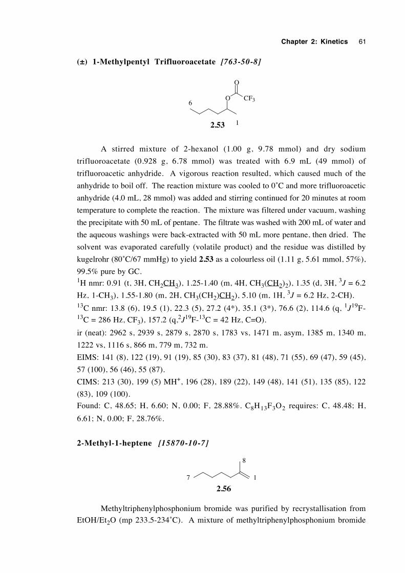
Chapter 2: Kinetics 61
(±) 1-Methylpentyl Trifluoroacetate [763-50-8]
O CF3
O
2.53 1
6
A stirred mixture of 2-hexanol (1.00 g, 9.78 mmol) and dry sodium
trifluoroacetate (0.928 g, 6.78 mmol) was treated with 6.9 mL (49 mmol) of
trifluoroacetic anhydride. A vigorous reaction resulted, which caused much of the
anhydride to boil off. The reaction mixture was cooled to 0˚C and more trifluoroacetic
anhydride (4.0 mL, 28 mmol) was added and stirring continued for 20 minutes at room
temperature to complete the reaction. The mixture was filtered under vacuum, washing
the precipitate with 50 mL of pentane. The filtrate was washed with 200 mL of water and
the aqueous washings were back-extracted with 50 mL more pentane, then dried. The
solvent was evaporated carefully (volatile product) and the residue was distilled by
kugelrohr (80˚C/67 mmHg) to yield 2.53 as a colourless oil (1.11 g, 5.61 mmol, 57%),
99.5% pure by GC.1H nmr: 0.91 (t, 3H, CH2CH3), 1.25-1.40 (m, 4H, CH3(CH2)2), 1.35 (d, 3H, 3J = 6.2
Hz, 1-CH3), 1.55-1.80 (m, 2H, CH3(CH2)CH2), 5.10 (m, 1H, 3J = 6.2 Hz, 2-CH).13C nmr: 13.8 (6), 19.5 (1), 22.3 (5), 27.2 (4*), 35.1 (3*), 76.6 (2), 114.6 (q, 1J19F-13C = 286 Hz, CF3), 157.2 (q,2J19F-13C = 42 Hz, C=O).
ir (neat): 2962 s, 2939 s, 2879 s, 2870 s, 1783 vs, 1471 m, asym, 1385 m, 1340 m,
1222 vs, 1116 s, 866 m, 779 m, 732 m.
EIMS: 141 (8), 122 (19), 91 (19), 85 (30), 83 (37), 81 (48), 71 (55), 69 (47), 59 (45),
57 (100), 56 (46), 55 (87).
CIMS: 213 (30), 199 (5) MH+, 196 (28), 189 (22), 149 (48), 141 (51), 135 (85), 122
(83), 109 (100).
Found: C, 48.65; H, 6.60; N, 0.00; F, 28.88%. C8H13F3O2 requires: C, 48.48; H,
6.61; N, 0.00; F, 28.76%.
2-Methyl-1-heptene [15870-10-7]
2.5617
8
Methyltriphenylphosphonium bromide was purified by recrystallisation from
EtOH/Et2O (mp 233.5-234˚C). A mixture of methyltriphenylphosphonium bromide
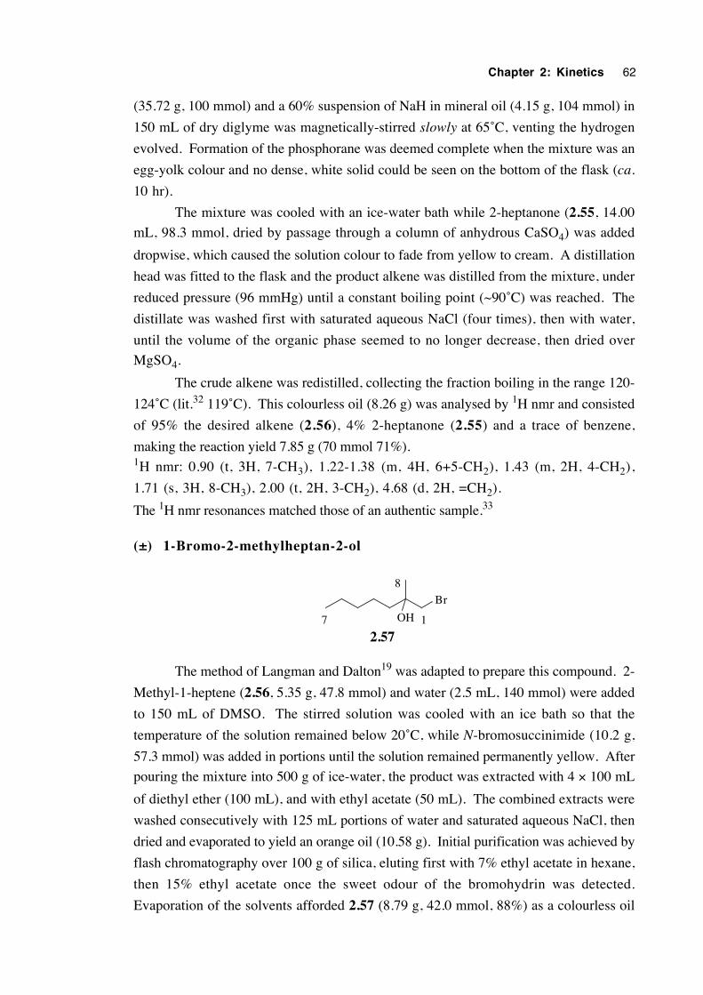
Chapter 2: Kinetics 62
(35.72 g, 100 mmol) and a 60% suspension of NaH in mineral oil (4.15 g, 104 mmol) in
150 mL of dry diglyme was magnetically-stirred slowly at 65˚C, venting the hydrogen
evolved. Formation of the phosphorane was deemed complete when the mixture was an
egg-yolk colour and no dense, white solid could be seen on the bottom of the flask (ca.
10 hr).
The mixture was cooled with an ice-water bath while 2-heptanone (2.55, 14.00
mL, 98.3 mmol, dried by passage through a column of anhydrous CaSO4) was added
dropwise, which caused the solution colour to fade from yellow to cream. A distillation
head was fitted to the flask and the product alkene was distilled from the mixture, under
reduced pressure (96 mmHg) until a constant boiling point (~90˚C) was reached. The
distillate was washed first with saturated aqueous NaCl (four times), then with water,
until the volume of the organic phase seemed to no longer decrease, then dried over
MgSO4.
The crude alkene was redistilled, collecting the fraction boiling in the range 120-
124˚C (lit.32 119˚C). This colourless oil (8.26 g) was analysed by 1H nmr and consisted
of 95% the desired alkene (2.56), 4% 2-heptanone (2.55) and a trace of benzene,
making the reaction yield 7.85 g (70 mmol 71%).1H nmr: 0.90 (t, 3H, 7-CH3), 1.22-1.38 (m, 4H, 6+5-CH2), 1.43 (m, 2H, 4-CH2),
1.71 (s, 3H, 8-CH3), 2.00 (t, 2H, 3-CH2), 4.68 (d, 2H, =CH2).
The 1H nmr resonances matched those of an authentic sample.33
(±) 1-Bromo-2-methylheptan-2-ol
Br
OH
2.5717
8
The method of Langman and Dalton19 was adapted to prepare this compound. 2-
Methyl-1-heptene (2.56, 5.35 g, 47.8 mmol) and water (2.5 mL, 140 mmol) were added
to 150 mL of DMSO. The stirred solution was cooled with an ice bath so that the
temperature of the solution remained below 20˚C, while N-bromosuccinimide (10.2 g,
57.3 mmol) was added in portions until the solution remained permanently yellow. After
pouring the mixture into 500 g of ice-water, the product was extracted with 4 × 100 mL
of diethyl ether (100 mL), and with ethyl acetate (50 mL). The combined extracts were
washed consecutively with 125 mL portions of water and saturated aqueous NaCl, then
dried and evaporated to yield an orange oil (10.58 g). Initial purification was achieved by
flash chromatography over 100 g of silica, eluting first with 7% ethyl acetate in hexane,
then 15% ethyl acetate once the sweet odour of the bromohydrin was detected.
Evaporation of the solvents afforded 2.57 (8.79 g, 42.0 mmol, 88%) as a colourless oil
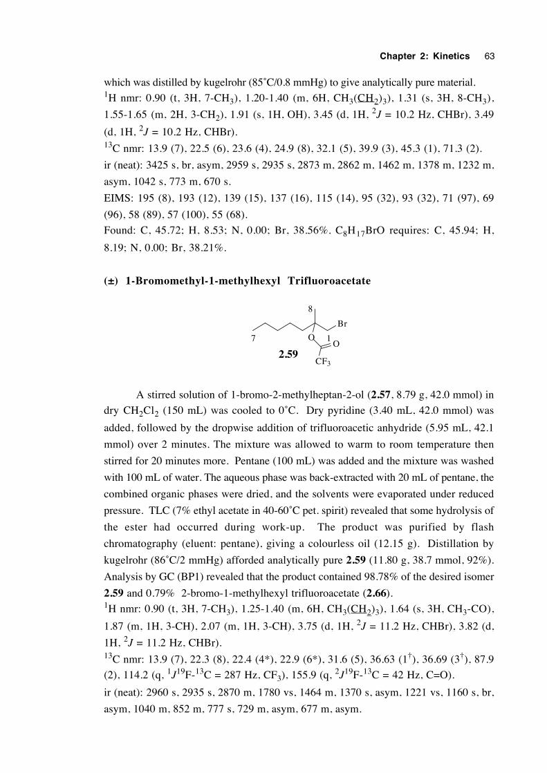
Chapter 2: Kinetics 63
which was distilled by kugelrohr (85˚C/0.8 mmHg) to give analytically pure material.1H nmr: 0.90 (t, 3H, 7-CH3), 1.20-1.40 (m, 6H, CH3(CH2)3), 1.31 (s, 3H, 8-CH3),
1.55-1.65 (m, 2H, 3-CH2), 1.91 (s, 1H, OH), 3.45 (d, 1H, 2J = 10.2 Hz, CHBr), 3.49
(d, 1H, 2J = 10.2 Hz, CHBr).13C nmr: 13.9 (7), 22.5 (6), 23.6 (4), 24.9 (8), 32.1 (5), 39.9 (3), 45.3 (1), 71.3 (2).
ir (neat): 3425 s, br, asym, 2959 s, 2935 s, 2873 m, 2862 m, 1462 m, 1378 m, 1232 m,
asym, 1042 s, 773 m, 670 s.
EIMS: 195 (8), 193 (12), 139 (15), 137 (16), 115 (14), 95 (32), 93 (32), 71 (97), 69
(96), 58 (89), 57 (100), 55 (68).
Found: C, 45.72; H, 8.53; N, 0.00; Br, 38.56%. C8H17BrO requires: C, 45.94; H,
8.19; N, 0.00; Br, 38.21%.
(±) 1-Bromomethyl-1-methylhexyl Trifluoroacetate
Br
O
2.59CF3
O17
8
A stirred solution of 1-bromo-2-methylheptan-2-ol (2.57, 8.79 g, 42.0 mmol) in
dry CH2Cl2 (150 mL) was cooled to 0˚C. Dry pyridine (3.40 mL, 42.0 mmol) was
added, followed by the dropwise addition of trifluoroacetic anhydride (5.95 mL, 42.1
mmol) over 2 minutes. The mixture was allowed to warm to room temperature then
stirred for 20 minutes more. Pentane (100 mL) was added and the mixture was washed
with 100 mL of water. The aqueous phase was back-extracted with 20 mL of pentane, the
combined organic phases were dried, and the solvents were evaporated under reduced
pressure. TLC (7% ethyl acetate in 40-60˚C pet. spirit) revealed that some hydrolysis of
the ester had occurred during work-up. The product was purified by flash
chromatography (eluent: pentane), giving a colourless oil (12.15 g). Distillation by
kugelrohr (86˚C/2 mmHg) afforded analytically pure 2.59 (11.80 g, 38.7 mmol, 92%).
Analysis by GC (BP1) revealed that the product contained 98.78% of the desired isomer
2.59 and 0.79% 2-bromo-1-methylhexyl trifluoroacetate (2.66).1H nmr: 0.90 (t, 3H, 7-CH3), 1.25-1.40 (m, 6H, CH3(CH2)3), 1.64 (s, 3H, CH3-CO),
1.87 (m, 1H, 3-CH), 2.07 (m, 1H, 3-CH), 3.75 (d, 1H, 2J = 11.2 Hz, CHBr), 3.82 (d,
1H, 2J = 11.2 Hz, CHBr).13C nmr: 13.9 (7), 22.3 (8), 22.4 (4*), 22.9 (6*), 31.6 (5), 36.63 (1†), 36.69 (3†), 87.9
(2), 114.2 (q, 1J19F-13C = 287 Hz, CF3), 155.9 (q, 2J19F-13C = 42 Hz, C=O).
ir (neat): 2960 s, 2935 s, 2870 m, 1780 vs, 1464 m, 1370 s, asym, 1221 vs, 1160 s, br,
asym, 1040 m, 852 m, 777 s, 729 m, asym, 677 m, asym.
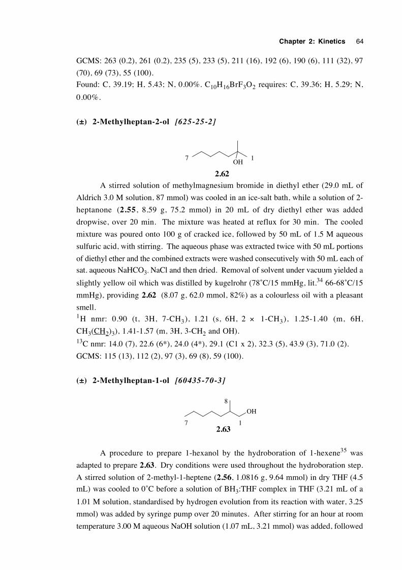
Chapter 2: Kinetics 64
GCMS: 263 (0.2), 261 (0.2), 235 (5), 233 (5), 211 (16), 192 (6), 190 (6), 111 (32), 97
(70), 69 (73), 55 (100).
Found: C, 39.19; H, 5.43; N, 0.00%. C10H16BrF3O2 requires: C, 39.36; H, 5.29; N,
0.00%.
(±) 2-Methylheptan-2-ol [625-25-2]
OH
2.62
17
A stirred solution of methylmagnesium bromide in diethyl ether (29.0 mL of
Aldrich 3.0 M solution, 87 mmol) was cooled in an ice-salt bath, while a solution of 2-
heptanone (2.55, 8.59 g, 75.2 mmol) in 20 mL of dry diethyl ether was added
dropwise, over 20 min. The mixture was heated at reflux for 30 min. The cooled
mixture was poured onto 100 g of cracked ice, followed by 50 mL of 1.5 M aqueous
sulfuric acid, with stirring. The aqueous phase was extracted twice with 50 mL portions
of diethyl ether and the combined extracts were washed consecutively with 50 mL each of
sat. aqueous NaHCO3, NaCl and then dried. Removal of solvent under vacuum yielded a
slightly yellow oil which was distilled by kugelrohr (78˚C/15 mmHg, lit.34 66-68˚C/15
mmHg), providing 2.62 (8.07 g, 62.0 mmol, 82%) as a colourless oil with a pleasant
smell.1H nmr: 0.90 (t, 3H, 7-CH3), 1.21 (s, 6H, 2 × 1-CH3), 1.25-1.40 (m, 6H,
CH3(CH2)3), 1.41-1.57 (m, 3H, 3-CH2 and OH).13C nmr: 14.0 (7), 22.6 (6*), 24.0 (4*), 29.1 (C1 x 2), 32.3 (5), 43.9 (3), 71.0 (2).
GCMS: 115 (13), 112 (2), 97 (3), 69 (8), 59 (100).
(±) 2-Methylheptan-1-ol [60435-70-3]
OH
2.6317
8
A procedure to prepare 1-hexanol by the hydroboration of 1-hexene35 was
adapted to prepare 2.63. Dry conditions were used throughout the hydroboration step.
A stirred solution of 2-methyl-1-heptene (2.56, 1.0816 g, 9.64 mmol) in dry THF (4.5
mL) was cooled to 0˚C before a solution of BH3:THF complex in THF (3.21 mL of a
1.01 M solution, standardised by hydrogen evolution from its reaction with water, 3.25
mmol) was added by syringe pump over 20 minutes. After stirring for an hour at room
temperature 3.00 M aqueous NaOH solution (1.07 mL, 3.21 mmol) was added, followed
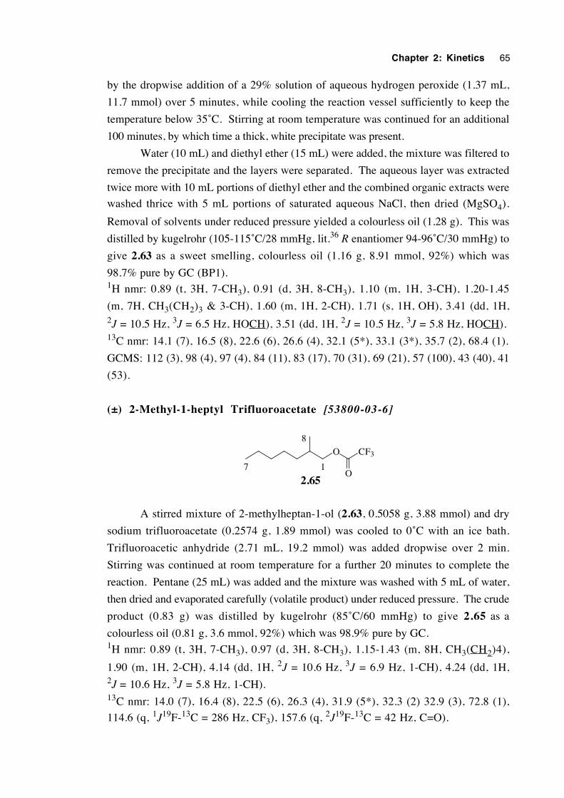
Chapter 2: Kinetics 65
by the dropwise addition of a 29% solution of aqueous hydrogen peroxide (1.37 mL,
11.7 mmol) over 5 minutes, while cooling the reaction vessel sufficiently to keep the
temperature below 35˚C. Stirring at room temperature was continued for an additional
100 minutes, by which time a thick, white precipitate was present.
Water (10 mL) and diethyl ether (15 mL) were added, the mixture was filtered to
remove the precipitate and the layers were separated. The aqueous layer was extracted
twice more with 10 mL portions of diethyl ether and the combined organic extracts were
washed thrice with 5 mL portions of saturated aqueous NaCl, then dried (MgSO4).
Removal of solvents under reduced pressure yielded a colourless oil (1.28 g). This was
distilled by kugelrohr (105-115˚C/28 mmHg, lit.36 R enantiomer 94-96˚C/30 mmHg) to
give 2.63 as a sweet smelling, colourless oil (1.16 g, 8.91 mmol, 92%) which was
98.7% pure by GC (BP1).1H nmr: 0.89 (t, 3H, 7-CH3), 0.91 (d, 3H, 8-CH3), 1.10 (m, 1H, 3-CH), 1.20-1.45
(m, 7H, CH3(CH2)3 & 3-CH), 1.60 (m, 1H, 2-CH), 1.71 (s, 1H, OH), 3.41 (dd, 1H,2J = 10.5 Hz, 3J = 6.5 Hz, HOCH), 3.51 (dd, 1H, 2J = 10.5 Hz, 3J = 5.8 Hz, HOCH).13C nmr: 14.1 (7), 16.5 (8), 22.6 (6), 26.6 (4), 32.1 (5*), 33.1 (3*), 35.7 (2), 68.4 (1).
GCMS: 112 (3), 98 (4), 97 (4), 84 (11), 83 (17), 70 (31), 69 (21), 57 (100), 43 (40), 41
(53).
(±) 2-Methyl-1-heptyl Trifluoroacetate [53800-03-6]
O CF3
O2.65
17
8
A stirred mixture of 2-methylheptan-1-ol (2.63, 0.5058 g, 3.88 mmol) and dry
sodium trifluoroacetate (0.2574 g, 1.89 mmol) was cooled to 0˚C with an ice bath.
Trifluoroacetic anhydride (2.71 mL, 19.2 mmol) was added dropwise over 2 min.
Stirring was continued at room temperature for a further 20 minutes to complete the
reaction. Pentane (25 mL) was added and the mixture was washed with 5 mL of water,
then dried and evaporated carefully (volatile product) under reduced pressure. The crude
product (0.83 g) was distilled by kugelrohr (85˚C/60 mmHg) to give 2.65 as a
colourless oil (0.81 g, 3.6 mmol, 92%) which was 98.9% pure by GC.1H nmr: 0.89 (t, 3H, 7-CH3), 0.97 (d, 3H, 8-CH3), 1.15-1.43 (m, 8H, CH3(CH2)4),
1.90 (m, 1H, 2-CH), 4.14 (dd, 1H, 2J = 10.6 Hz, 3J = 6.9 Hz, 1-CH), 4.24 (dd, 1H,2J = 10.6 Hz, 3J = 5.8 Hz, 1-CH).13C nmr: 14.0 (7), 16.4 (8), 22.5 (6), 26.3 (4), 31.9 (5*), 32.3 (2) 32.9 (3), 72.8 (1),
114.6 (q, 1J19F-13C = 286 Hz, CF3), 157.6 (q, 2J19F-13C = 42 Hz, C=O).
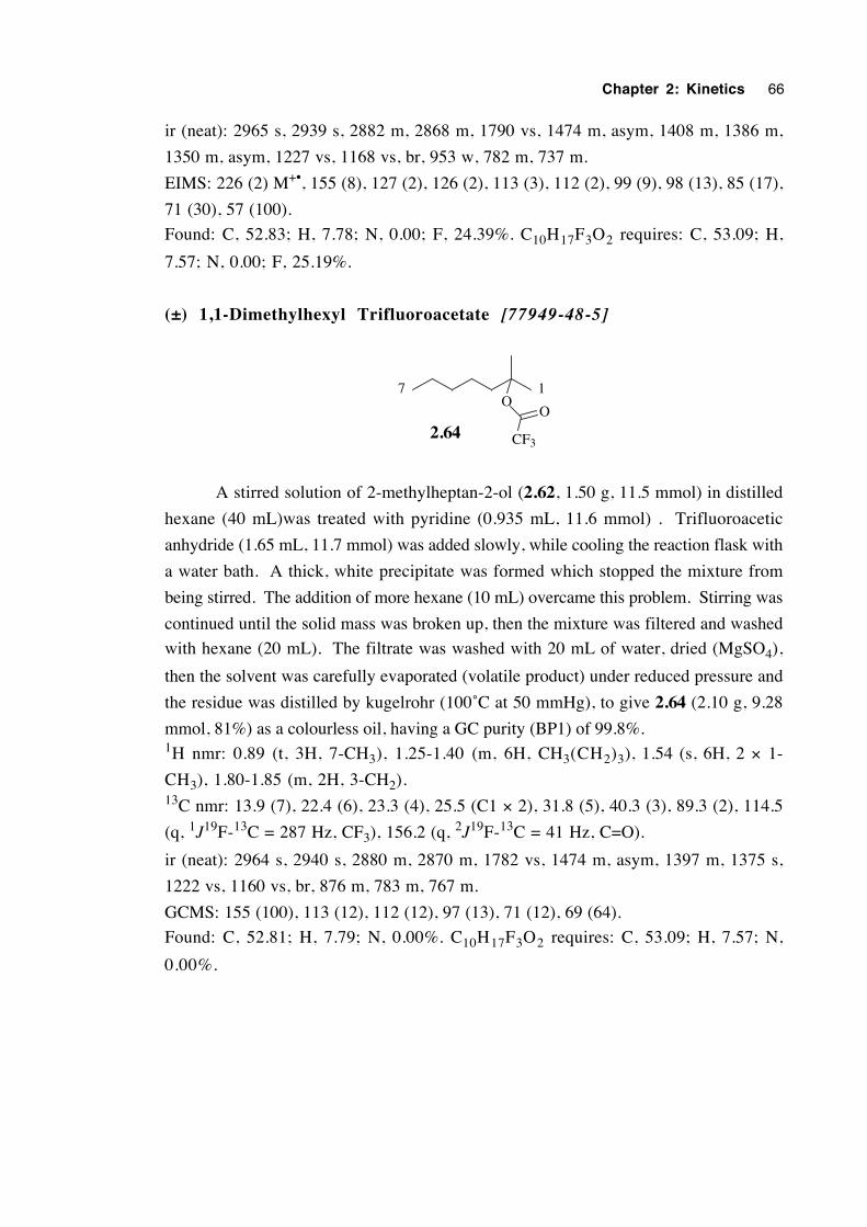
Chapter 2: Kinetics 66
ir (neat): 2965 s, 2939 s, 2882 m, 2868 m, 1790 vs, 1474 m, asym, 1408 m, 1386 m,
1350 m, asym, 1227 vs, 1168 vs, br, 953 w, 782 m, 737 m.
EIMS: 226 (2) M+•, 155 (8), 127 (2), 126 (2), 113 (3), 112 (2), 99 (9), 98 (13), 85 (17),
71 (30), 57 (100).
Found: C, 52.83; H, 7.78; N, 0.00; F, 24.39%. C10H17F3O2 requires: C, 53.09; H,
7.57; N, 0.00; F, 25.19%.
(±) 1,1-Dimethylhexyl Trifluoroacetate [77949-48-5]
O
CF3
O
2.64
17
A stirred solution of 2-methylheptan-2-ol (2.62, 1.50 g, 11.5 mmol) in distilled
hexane (40 mL)was treated with pyridine (0.935 mL, 11.6 mmol) . Trifluoroacetic
anhydride (1.65 mL, 11.7 mmol) was added slowly, while cooling the reaction flask with
a water bath. A thick, white precipitate was formed which stopped the mixture from
being stirred. The addition of more hexane (10 mL) overcame this problem. Stirring was
continued until the solid mass was broken up, then the mixture was filtered and washed
with hexane (20 mL). The filtrate was washed with 20 mL of water, dried (MgSO4),
then the solvent was carefully evaporated (volatile product) under reduced pressure and
the residue was distilled by kugelrohr (100˚C at 50 mmHg), to give 2.64 (2.10 g, 9.28
mmol, 81%) as a colourless oil, having a GC purity (BP1) of 99.8%.1H nmr: 0.89 (t, 3H, 7-CH3), 1.25-1.40 (m, 6H, CH3(CH2)3), 1.54 (s, 6H, 2 × 1-
CH3), 1.80-1.85 (m, 2H, 3-CH2).13C nmr: 13.9 (7), 22.4 (6), 23.3 (4), 25.5 (C1 × 2), 31.8 (5), 40.3 (3), 89.3 (2), 114.5
(q, 1J19F-13C = 287 Hz, CF3), 156.2 (q, 2J19F-13C = 41 Hz, C=O).
ir (neat): 2964 s, 2940 s, 2880 m, 2870 m, 1782 vs, 1474 m, asym, 1397 m, 1375 s,
1222 vs, 1160 vs, br, 876 m, 783 m, 767 m.
GCMS: 155 (100), 113 (12), 112 (12), 97 (13), 71 (12), 69 (64).
Found: C, 52.81; H, 7.79; N, 0.00%. C10H17F3O2 requires: C, 53.09; H, 7.57; N,
0.00%.
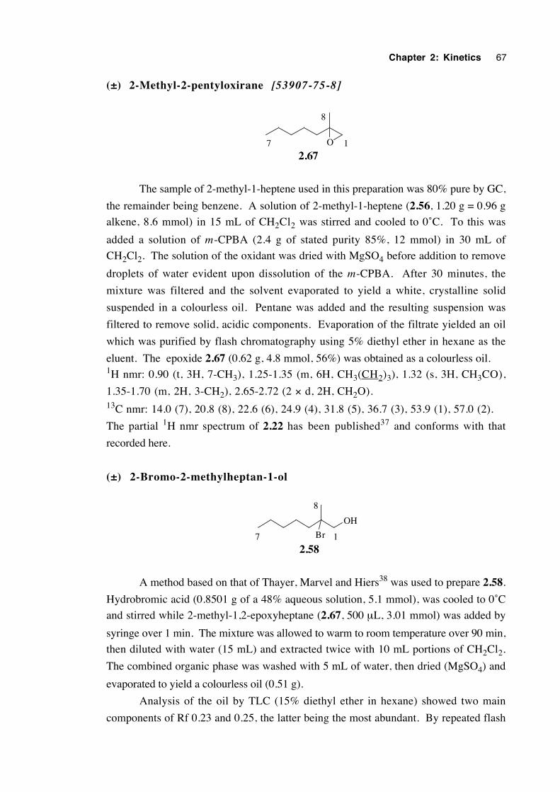
Chapter 2: Kinetics 67
(±) 2-Methyl-2-pentyloxirane [53907-75-8]
2.67O 17
8
The sample of 2-methyl-1-heptene used in this preparation was 80% pure by GC,
the remainder being benzene. A solution of 2-methyl-1-heptene (2.56, 1.20 g = 0.96 g
alkene, 8.6 mmol) in 15 mL of CH2Cl2 was stirred and cooled to 0˚C. To this was
added a solution of m-CPBA (2.4 g of stated purity 85%, 12 mmol) in 30 mL of
CH2Cl2. The solution of the oxidant was dried with MgSO4 before addition to remove
droplets of water evident upon dissolution of the m-CPBA. After 30 minutes, the
mixture was filtered and the solvent evaporated to yield a white, crystalline solid
suspended in a colourless oil. Pentane was added and the resulting suspension was
filtered to remove solid, acidic components. Evaporation of the filtrate yielded an oil
which was purified by flash chromatography using 5% diethyl ether in hexane as the
eluent. The epoxide 2.67 (0.62 g, 4.8 mmol, 56%) was obtained as a colourless oil.1H nmr: 0.90 (t, 3H, 7-CH3), 1.25-1.35 (m, 6H, CH3(CH2)3), 1.32 (s, 3H, CH3CO),
1.35-1.70 (m, 2H, 3-CH2), 2.65-2.72 (2 × d, 2H, CH2O).13C nmr: 14.0 (7), 20.8 (8), 22.6 (6), 24.9 (4), 31.8 (5), 36.7 (3), 53.9 (1), 57.0 (2).
The partial 1H nmr spectrum of 2.22 has been published37 and conforms with that
recorded here.
(±) 2-Bromo-2-methylheptan-1-ol
OH
Br
2.5817
8
A method based on that of Thayer, Marvel and Hiers38 was used to prepare 2.58.
Hydrobromic acid (0.8501 g of a 48% aqueous solution, 5.1 mmol), was cooled to 0˚C
and stirred while 2-methyl-1,2-epoxyheptane (2.67, 500 µL, 3.01 mmol) was added by
syringe over 1 min. The mixture was allowed to warm to room temperature over 90 min,
then diluted with water (15 mL) and extracted twice with 10 mL portions of CH2Cl2.
The combined organic phase was washed with 5 mL of water, then dried (MgSO4) and
evaporated to yield a colourless oil (0.51 g).
Analysis of the oil by TLC (15% diethyl ether in hexane) showed two main
components of Rf 0.23 and 0.25, the latter being the most abundant. By repeated flash
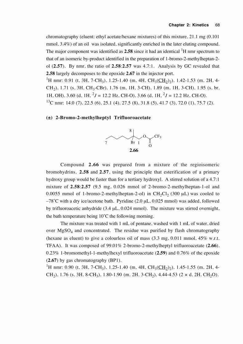
Chapter 2: Kinetics 68
chromatography (eluent: ethyl acetate/hexane mixtures) of this mixture, 21.1 mg (0.101
mmol, 3.4%) of an oil was isolated, significantly enriched in the later eluting compound.
The major component was identified as 2.58 since it had an identical 1H nmr spectrum to
that of an isomeric by-product identified in the preparation of 1-bromo-2-methylheptan-2-
ol (2.57). By nmr, the ratio of 2.58:2.57 was 4.7:1. Analysis by GC revealed that
2.58 largely decomposes to the epoxide 2.67 in the injector port.1H nmr: 0.91 (t, 3H, 7-CH3), 1.25-1.40 (m, 4H, CH3(CH2)2), 1.42-1.53 (m, 2H, 4-
CH2), 1.71 (s, 3H, CH3-CBr), 1.76 (m, 1H, 3-CH), 1.89 (m, 1H, 3-CH), 1.95 (s, br,
1H, OH), 3.60 (d, 1H, 2J = 12.2 Hz, CH-O), 3.66 (d, 1H, 2J = 12.2 Hz, CH-O).13C nmr: 14.0 (7), 22.5 (6), 25.1 (4), 27.5 (8), 31.8 (5), 41.7 (3), 72.0 (1), 75.7 (2).
(±) 2-Bromo-2-methylheptyl Trifluoroacetate
O
Br
2.66
CF3
O17
8
Compound 2.66 was prepared from a mixture of the regioisomeric
bromohydrins, 2.58 and 2.57, using the principle that esterification of a primary
hydroxy group would be faster than for a tertiary hydroxyl. A stirred solution of a 4.7:1
mixture of 2.58:2.57 (9.5 mg, 0.026 mmol of 2-bromo-2-methylheptan-1-ol and
0.0055 mmol of 1-bromo-2-methylheptan-2-ol) in CH2Cl2 (300 µL) was cooled to
–78˚C with a dry ice/acetone bath. Pyridine (2.0 µL, 0.025 mmol) was added, followed
by trifluoroacetic anhydride (3.4 µL, 0.024 mmol). The mixture was stirred overnight,
the bath temperature being 10˚C the following morning.
The mixture was treated with 1 mL of pentane, washed with 1 mL of water, dried
over MgSO4 and concentrated. The residue was purified by flash chromatography
(hexane as eluent) to give a colourless oil of mass (3.3 mg, 0.011 mmol, 45% w.r.t.
TFAA). It was composed of 99.01% 2-bromo-2-methylheptyl trifluoroacetate (2.66),
0.23% 1-bromomethyl-1-methylhexyl trifluoroacetate (2.59) and 0.76% of the epoxide
(2.67) by gas chromatography (BP1).1H nmr: 0.90 (t, 3H, 7-CH3), 1.25-1.40 (m, 4H, CH3(CH2)2), 1.45-1.55 (m, 2H, 4-
CH2), 1.76 (s, 3H, 8-CH3), 1.80-1.90 (m, 2H, 3-CH2), 4.44-4.53 (2 × d, 2H, CH2O).
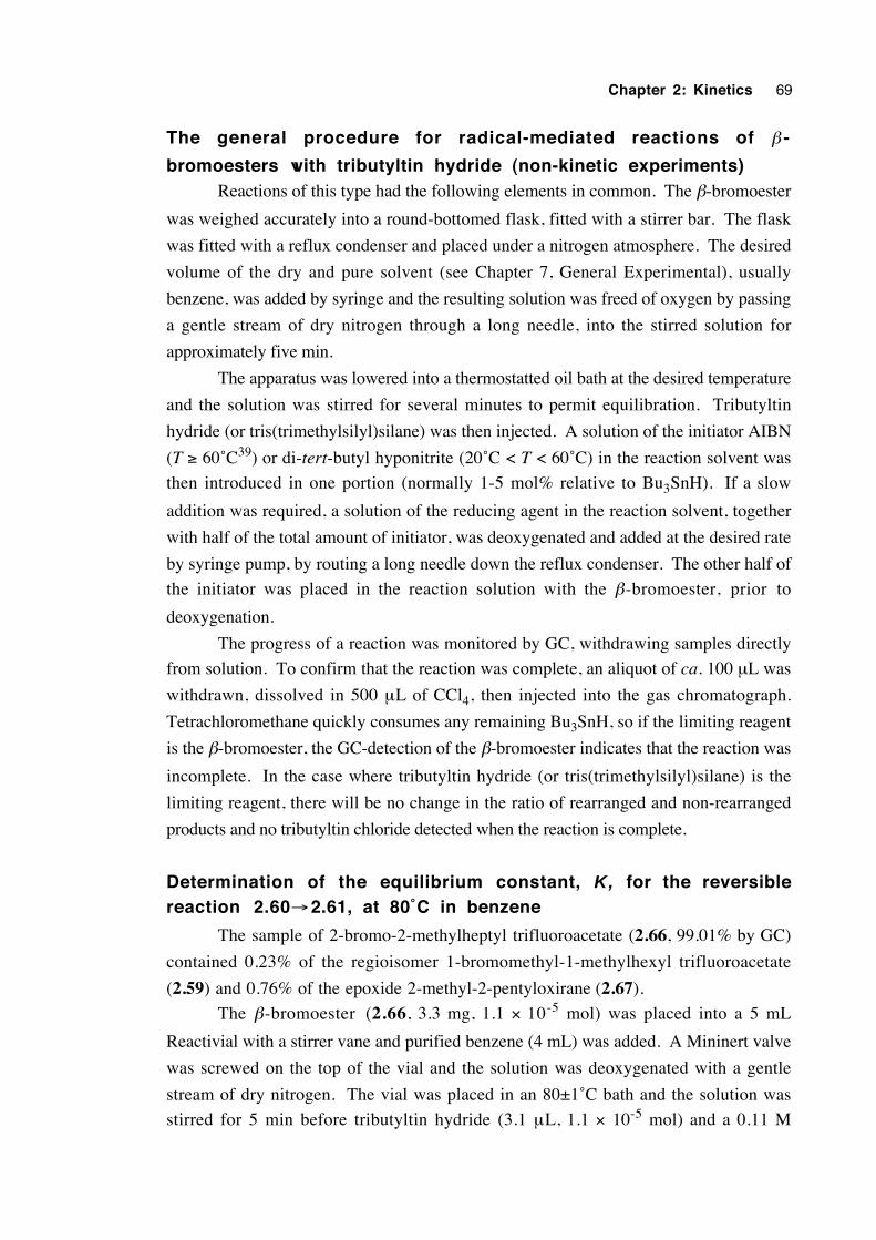
Chapter 2: Kinetics 69
The general procedure for radical-mediated reactions of β -
bromoesters with tributyltin hydride (non-kinetic experiments)Reactions of this type had the following elements in common. The β-bromoester
was weighed accurately into a round-bottomed flask, fitted with a stirrer bar. The flask
was fitted with a reflux condenser and placed under a nitrogen atmosphere. The desired
volume of the dry and pure solvent (see Chapter 7, General Experimental), usually
benzene, was added by syringe and the resulting solution was freed of oxygen by passing
a gentle stream of dry nitrogen through a long needle, into the stirred solution for
approximately five min.
The apparatus was lowered into a thermostatted oil bath at the desired temperature
and the solution was stirred for several minutes to permit equilibration. Tributyltin
hydride (or tris(trimethylsilyl)silane) was then injected. A solution of the initiator AIBN
(T ≥ 60˚C39) or di-tert-butyl hyponitrite (20˚C < T < 60˚C) in the reaction solvent was
then introduced in one portion (normally 1-5 mol% relative to Bu3SnH). If a slow
addition was required, a solution of the reducing agent in the reaction solvent, together
with half of the total amount of initiator, was deoxygenated and added at the desired rate
by syringe pump, by routing a long needle down the reflux condenser. The other half of
the initiator was placed in the reaction solution with the β-bromoester, prior to
deoxygenation.
The progress of a reaction was monitored by GC, withdrawing samples directly
from solution. To confirm that the reaction was complete, an aliquot of ca. 100 µL was
withdrawn, dissolved in 500 µL of CCl4, then injected into the gas chromatograph.
Tetrachloromethane quickly consumes any remaining Bu3SnH, so if the limiting reagent
is the β-bromoester, the GC-detection of the β-bromoester indicates that the reaction was
incomplete. In the case where tributyltin hydride (or tris(trimethylsilyl)silane) is the
limiting reagent, there will be no change in the ratio of rearranged and non-rearranged
products and no tributyltin chloride detected when the reaction is complete.
Determination of the equilibrium constant, K, for the reversiblereaction 2.60→2.61, at 80˚C in benzene
The sample of 2-bromo-2-methylheptyl trifluoroacetate (2.66, 99.01% by GC)
contained 0.23% of the regioisomer 1-bromomethyl-1-methylhexyl trifluoroacetate
(2.59) and 0.76% of the epoxide 2-methyl-2-pentyloxirane (2.67).
The β-bromoester (2.66, 3.3 mg, 1.1 × 10⁻⁵ mol) was placed into a 5 mL
Reactivial with a stirrer vane and purified benzene (4 mL) was added. A Mininert valve
was screwed on the top of the vial and the solution was deoxygenated with a gentle
stream of dry nitrogen. The vial was placed in an 80±1˚C bath and the solution was
stirred for 5 min before tributyltin hydride (3.1 µL, 1.1 × 10⁻⁵ mol) and a 0.11 M
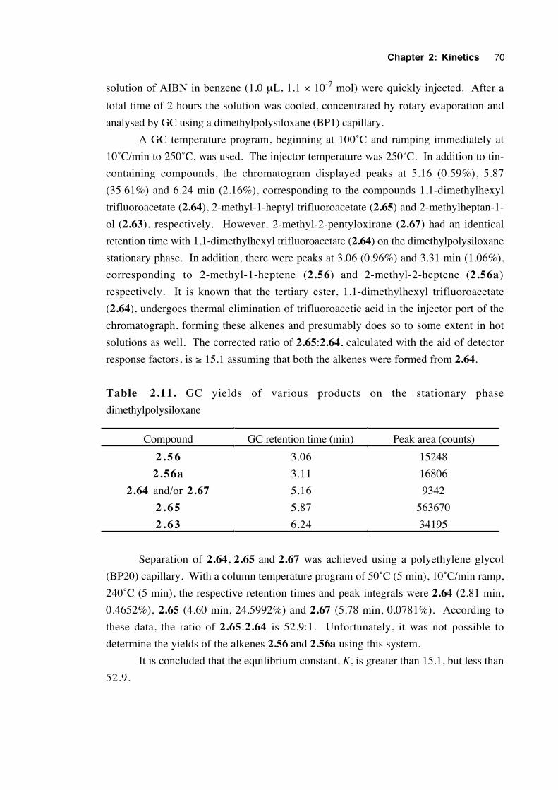
Chapter 2: Kinetics 70
solution of AIBN in benzene (1.0 µL, 1.1 × 10⁻⁷ mol) were quickly injected. After a
total time of 2 hours the solution was cooled, concentrated by rotary evaporation and
analysed by GC using a dimethylpolysiloxane (BP1) capillary.
A GC temperature program, beginning at 100˚C and ramping immediately at
10˚C/min to 250˚C, was used. The injector temperature was 250˚C. In addition to tin-
containing compounds, the chromatogram displayed peaks at 5.16 (0.59%), 5.87
(35.61%) and 6.24 min (2.16%), corresponding to the compounds 1,1-dimethylhexyl
trifluoroacetate (2.64), 2-methyl-1-heptyl trifluoroacetate (2.65) and 2-methylheptan-1-
ol (2.63), respectively. However, 2-methyl-2-pentyloxirane (2.67) had an identical
retention time with 1,1-dimethylhexyl trifluoroacetate (2.64) on the dimethylpolysiloxane
stationary phase. In addition, there were peaks at 3.06 (0.96%) and 3.31 min (1.06%),
corresponding to 2-methyl-1-heptene (2.56) and 2-methyl-2-heptene (2.56a)
respectively. It is known that the tertiary ester, 1,1-dimethylhexyl trifluoroacetate
(2.64), undergoes thermal elimination of trifluoroacetic acid in the injector port of the
chromatograph, forming these alkenes and presumably does so to some extent in hot
solutions as well. The corrected ratio of 2.65:2.64, calculated with the aid of detector
response factors, is ≥ 15.1 assuming that both the alkenes were formed from 2.64.
Table 2.11. GC yields of various products on the stationary phase
dimethylpolysiloxane
Compound GC retention time (min) Peak area (counts)
2 . 5 6 3.06 15248
2.56a 3.11 16806
2.64 and/or 2.67 5.16 9342
2 . 6 5 5.87 563670
2 . 6 3 6.24 34195
Separation of 2.64, 2.65 and 2.67 was achieved using a polyethylene glycol
(BP20) capillary. With a column temperature program of 50˚C (5 min), 10˚C/min ramp,
240˚C (5 min), the respective retention times and peak integrals were 2.64 (2.81 min,
0.4652%), 2.65 (4.60 min, 24.5992%) and 2.67 (5.78 min, 0.0781%). According to
these data, the ratio of 2.65:2.64 is 52.9:1. Unfortunately, it was not possible to
determine the yields of the alkenes 2.56 and 2.56a using this system.
It is concluded that the equilibrium constant, K, is greater than 15.1, but less than
52.9.
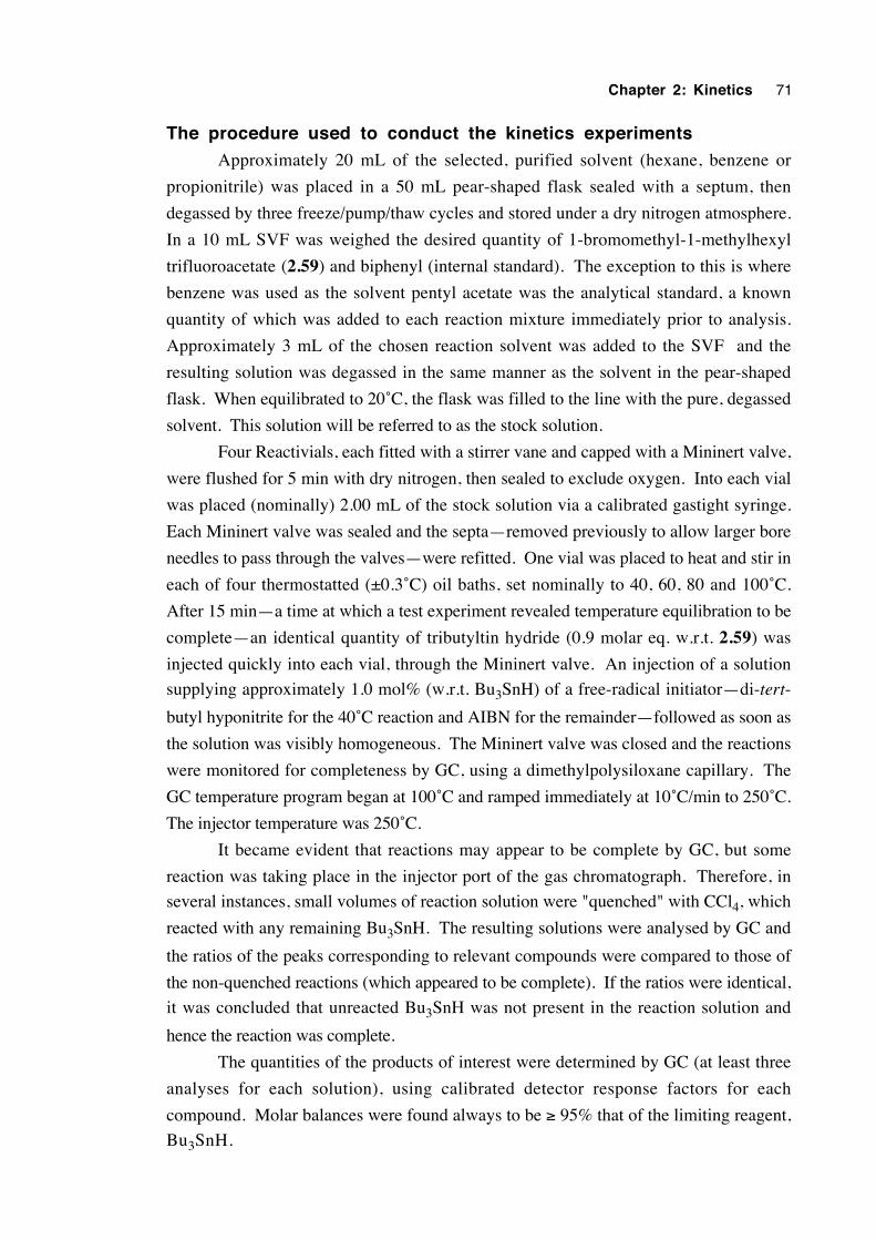
Chapter 2: Kinetics 71
The procedure used to conduct the kinetics experiments
Approximately 20 mL of the selected, purified solvent (hexane, benzene or
propionitrile) was placed in a 50 mL pear-shaped flask sealed with a septum, then
degassed by three freeze/pump/thaw cycles and stored under a dry nitrogen atmosphere.
In a 10 mL SVF was weighed the desired quantity of 1-bromomethyl-1-methylhexyl
trifluoroacetate (2.59) and biphenyl (internal standard). The exception to this is where
benzene was used as the solvent pentyl acetate was the analytical standard, a known
quantity of which was added to each reaction mixture immediately prior to analysis.
Approximately 3 mL of the chosen reaction solvent was added to the SVF and the
resulting solution was degassed in the same manner as the solvent in the pear-shaped
flask. When equilibrated to 20˚C, the flask was filled to the line with the pure, degassed
solvent. This solution will be referred to as the stock solution.
Four Reactivials, each fitted with a stirrer vane and capped with a Mininert valve,
were flushed for 5 min with dry nitrogen, then sealed to exclude oxygen. Into each vial
was placed (nominally) 2.00 mL of the stock solution via a calibrated gastight syringe.
Each Mininert valve was sealed and the septa—removed previously to allow larger bore
needles to pass through the valves—were refitted. One vial was placed to heat and stir in
each of four thermostatted (±0.3˚C) oil baths, set nominally to 40, 60, 80 and 100˚C.
After 15 min—a time at which a test experiment revealed temperature equilibration to be
complete—an identical quantity of tributyltin hydride (0.9 molar eq. w.r.t. 2.59) was
injected quickly into each vial, through the Mininert valve. An injection of a solution
supplying approximately 1.0 mol% (w.r.t. Bu3SnH) of a free-radical initiator—di-tert-
butyl hyponitrite for the 40˚C reaction and AIBN for the remainder—followed as soon as
the solution was visibly homogeneous. The Mininert valve was closed and the reactions
were monitored for completeness by GC, using a dimethylpolysiloxane capillary. The
GC temperature program began at 100˚C and ramped immediately at 10˚C/min to 250˚C.
The injector temperature was 250˚C.
It became evident that reactions may appear to be complete by GC, but some
reaction was taking place in the injector port of the gas chromatograph. Therefore, in
several instances, small volumes of reaction solution were "quenched" with CCl4, which
reacted with any remaining Bu3SnH. The resulting solutions were analysed by GC and
the ratios of the peaks corresponding to relevant compounds were compared to those of
the non-quenched reactions (which appeared to be complete). If the ratios were identical,
it was concluded that unreacted Bu3SnH was not present in the reaction solution and
hence the reaction was complete.
The quantities of the products of interest were determined by GC (at least three
analyses for each solution), using calibrated detector response factors for each
compound. Molar balances were found always to be ≥ 95% that of the limiting reagent,
Bu3SnH.
Chapter 2: Kinetics 72
2.9 References
1. Barclay, L. R. C.; Griller, D. and Ingold, K. U. J. Am. Chem. Soc. 1982, 104,
4399.
2. Beckwith, A. L. J. and Thomas, C. B. J. Chem. Soc., Perkin Trans. 2 1973, 861.
3. Barclay, L. R. C.; Lusztyk, J. and Ingold, K. U. J. Am. Chem. Soc. 1984, 106,
1793.
4. Korth, H.-G.; Sustmann, R.; Groninger, K. S.; Liesung, M. and Giese, B. J. Org.
Chem. 1988, 53, 4364.
5. Beckwith, A. L. J. and Duggan, P. J. J. Chem. Soc., Perkin Trans. 2 1993, 1673.
6. Beckwith, A. L. J. and Duggan, P. J. J. Am. Chem. Soc. 1996, 118, 12838.
7. Gimisis, T.; Ialongo, G. and Chatgilialoglu, C. Tetrahedron 1998, 54, 573.
8. Beckwith, A. L. J. and Duggan, P. J. J. Chem. Soc. Perkin Trans. 2 1992, 1777.
9. Crich, D.; Huang, X. and Beckwith, A. L. J. J. Org. Chem. 1999, 64, 1762.
10. Choi, S.-Y.; Crich, D.; Horner, J. H.; Huang, X.; Newcomb, M. and Whitted, P.
O. Tetrahedron 1999, 55, 3317.
11. Beckwith, A. L. J.; Crich, D.; Duggan, P. J. and Yao, Q. Chem. Rev. 1997, 97,
3273.
12. Lacôte, E. and Renaud, P. Angew. Chem. Int. Ed. Engl. 1998, 37, 2259.
13. Renaud, P.; Andrau, L.; Gerster, M. and Lacôte, E. In Current Trends in Organic
Synthesis; Scolastico, C. and Nicotra, F., Eds.; Plenum: New York, 1999; pp 117-122.
14. Zipse, H. J. Am. Chem. Soc. 1997, 119, 1087.
15. Okamoto, Y.; Inukai, T. and Brown, H. C. J. Am. Chem. Soc. 1958, 80, 4972.
16. Menapace, L. W. and Kuivila, H. G. J. Am. Chem. Soc. 1964, 86, 3047.
17. Kuivila, H. G. Synthesis 1970, 499.
18. Neumann, W. P. Synthesis 1987, 665.
19. Langman, A. W. and Dalton, D. R. Org. Synth. 1980, 59, 16.
20. Beckwith, A. L. J. and Moad, G. J. Chem. Soc. Chem. Commun., 1974, 472.
21. Johnston, L. J.; Lusztyk, J.; Wayner, D. D. M.; Abeywickreyma, A. N.; Beckwith,
A. L. J.; Scaiano, J. C. and Ingold, K. U. J. Am. Chem. Soc. 1985, 107, 4594.
Chapter 2: Kinetics 73
22. a) Reichardt, C. Chem. Rev. 1994, 94, 2319; b) Reichardt, C.,Schäfer G. , Liebigs
Ann. 1995, 1579; c) Eberhardt, R., Löbbecke S., Neidhardt, B. and Reichardt C.
Liebigs Ann. /Recueil 1 9 9 7 , 1195; d) Website: www.chemie.uni-
marburg.de/~ak39/et_home.html
23. Bottcher, C. J. F. Theory of Electric Polarization; Elsevier Scientific Pub. Co.:
New York, 1973, 2nd Ed., vol. 1, p. 88.
24. Rigby, M.; Smith, E. B.; Wakeham, W. A. and Maitland, G. C. The Forces
Between Molecules; Clarendon Press: Oxford, 1986, p. 188.
25. Smolina, T. A.; Brusova, G. P.; Shchekut'eva, L. F.; Gopius, E. D.; Permin, A. B.
and Reutov, O. A. Izv. Akad. Nauk SSSR, Ser. Khim.1980, 9, 2079.
26. Zipse, H. Acc. Chem. Res. 1999, 32, 571.
27. Abraham, M. H. and Abraham, R. J. J. Chem. Soc. Perkin Trans. 2 1974, 47.
28. Mendenhall, G. D. Tetrahedron Lett. 1983, 24, 451.
29. Cerichelli, G.; Grande, C.; Luchetti, L. and Mancini, G. J. Org. Chem. 1991, 56,
3025.
30. Kraus, G. and Gottschalk, P. J. Org. Chem. 1983, 48, 2111.
31. Macey, W. A. T. J. Phys. Chem. 1960, 64, 254.
32. Dictionary of Organic Compounds; 5th ed.; Buckinham, J., Ed.; Chapman and Hall:
New York, 1982, M-01935.
33. Aldrich FT-NMR 1(1), 33A.
34. Dictionary of Organic Compounds; 5th ed.; Buckinham, J., Ed.; Chapman and Hall:
New York, 1982, M-01918.
35. Kono, H. and Hooz, J. Org. Synth. 1973, 53, 77.
36. Sonnet, P. E. and Gazzillo, J. Org. Prep. Proceed. Int. 1990, 22, 203.
37. Quenard, M.; Bonmarin, V. and Gelbard, G. New. J. Chem. 1989, 13, 183.
38. Thayer, F. K.; Marvel, C. S. and Hiers, G. S. Org. Synth. 1941, Coll. Vol. 1, 117.
39. Walling, C. Free Radicals in Solution; John Wiley, New York, 1957; pp. 467-537.
40. Crich, D. and Mo, X.-S. J. Am Chem. Soc. 1998, 120, 8298.
Chapter 3
A labelled-oxygen study of the regiochemistry of the β-
trifluoroacetoxyalkyl radical rearrangement
3.1 Introduction 75
3.2 Literature review 75
3.3 Choice of a suitable system for study 79
3.4 An attempt to observe the 1,2 shift of a hydroxy group in a β-
hydroxyalkyl radical 80
3.5 A study of the regiochemistry of the rearrangement
3.32→3.33 using 18O-labelling techniques 81
3.6 Study of the regiochemistry of the rearrangement of
3.32→3.33 using 17O nmr 95
3.7 A crossover experiment 100
3.8 An attempt to trap an ion pair intermediate 105
3.9 Discussion of results with regard to mechanism 108
3.10 Conclusions 116
3.11 Experimental 118
3.12 References 131
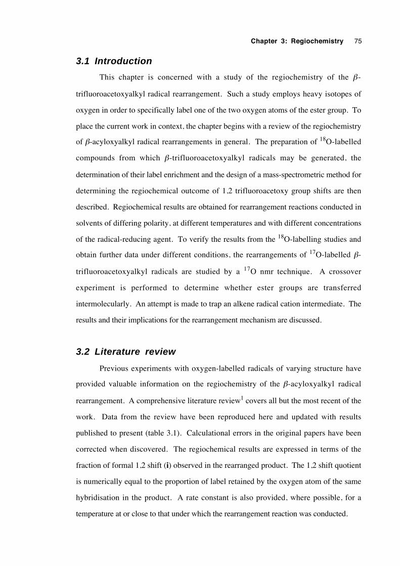
Chapter 3: Regiochemistry 75
3.1 Introduction
This chapter is concerned with a study of the regiochemistry of the β-
trifluoroacetoxyalkyl radical rearrangement. Such a study employs heavy isotopes of
oxygen in order to specifically label one of the two oxygen atoms of the ester group. To
place the current work in context, the chapter begins with a review of the regiochemistry
of β-acyloxyalkyl radical rearrangements in general. The preparation of 18O-labelled
compounds from which β-trifluoroacetoxyalkyl radicals may be generated, the
determination of their label enrichment and the design of a mass-spectrometric method for
determining the regiochemical outcome of 1,2 trifluoroacetoxy group shifts are then
described. Regiochemical results are obtained for rearrangement reactions conducted in
solvents of differing polarity, at different temperatures and with different concentrations
of the radical-reducing agent. To verify the results from the 18O-labelling studies and
obtain further data under different conditions, the rearrangements of 17O-labelled β-
trifluoroacetoxyalkyl radicals are studied by a 17O nmr technique. A crossover
experiment is performed to determine whether ester groups are transferred
intermolecularly. An attempt is made to trap an alkene radical cation intermediate. The
results and their implications for the rearrangement mechanism are discussed.
3.2 Literature review
Previous experiments with oxygen-labelled radicals of varying structure have
provided valuable information on the regiochemistry of the β-acyloxyalkyl radical
rearrangement. A comprehensive literature review1 covers all but the most recent of the
work. Data from the review have been reproduced here and updated with results
published to present (table 3.1). Calculational errors in the original papers have been
corrected when discovered. The regiochemical results are expressed in terms of the
fraction of formal 1,2 shift (i) observed in the rearranged product. The 1,2 shift quotient
is numerically equal to the proportion of label retained by the oxygen atom of the same
hybridisation in the product. A rate constant is also provided, where possible, for a
temperature at or close to that under which the rearrangement reaction was conducted.
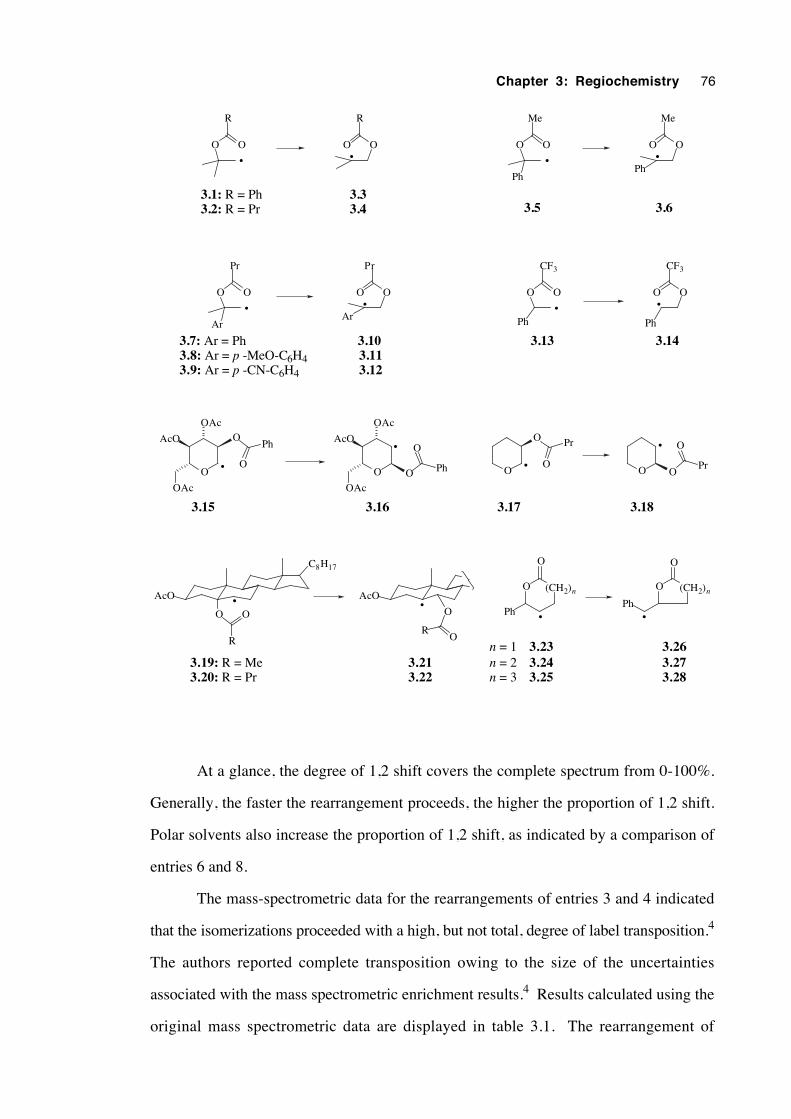
Chapter 3: Regiochemistry 76
OO
R
OO
R
OO
Ar
Pr
OO
Ar
Pr
OO
CF3
OO
CF3
Ph Ph
O
OAc
AcO
OAc
O
OPh
O
OAc
AcO
OAc
O
O
Ph OO
O Pr
O O
O
Pr
AcO
C8H17
O O
R
AcO
O
OR
O
O
Ph
O
O
Ph
OO
Ph
Me
OO
Ph
Me
• •
3.1: R = Ph3.2: R = Pr
3.33.4
• •
3.7: Ar = Ph3.8: Ar = p -MeO-C6H43.9: Ar = p -CN-C6H4
3.103.113.12
• •
•
•
3.15 3.16
•
•
3.17 3.18
• •
3.19: R = Me3.20: R = Pr
3.213.22
(CH2)n
•
(CH2)n
•
3.13 3.14
• •
3.5 3.6
3.233.243.25
3.263.273.28
n = 1n = 2n = 3
At a glance, the degree of 1,2 shift covers the complete spectrum from 0-100%.
Generally, the faster the rearrangement proceeds, the higher the proportion of 1,2 shift.
Polar solvents also increase the proportion of 1,2 shift, as indicated by a comparison of
entries 6 and 8.
The mass-spectrometric data for the rearrangements of entries 3 and 4 indicated
that the isomerizations proceeded with a high, but not total, degree of label transposition.4
The authors reported complete transposition owing to the size of the uncertainties
associated with the mass spectrometric enrichment results.4 Results calculated using the
original mass spectrometric data are displayed in table 3.1. The rearrangement of
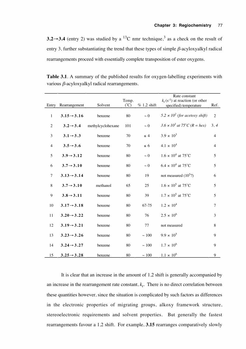
Chapter 3: Regiochemistry 77
3.2→3.4 (entry 2) was studied by a 13C nmr technique,3 as a check on the result of
entry 3, further substantiating the trend that these types of simple β-acyloxyalkyl radical
rearrangements proceed with essentially complete transposition of ester oxygens.
Table 3.1. A summary of the published results for oxygen-labelling experiments with
various β-acyloxyalkyl radical rearrangements.
Entry Rearrangement SolventTemp.(˚C) % 1,2 shift
Rate constant kr (s-1) at reaction (or other
specified) temperature Ref.
1 3.15→ 3 .16 benzene 80 ~ 0 5.2 × 10² (for acetoxy shift) 2
2 3.2→ 3 . 4 methylcyclohexane 101 ~ 0 3.6 × 10³ at 75˚C (R = hex) 3, 4
3 3.1→ 3 . 3 benzene 70 ≤ 4 3.9 × 10³ 4
4 3.5→ 3 . 6 benzene 70 ≤ 6 4.1 × 10⁴ 4
5 3.9→ 3 .12 benzene 80 ~ 0 1.6 × 10⁴ at 75˚C 5
6 3.7→ 3 .10 benzene 80 ~ 0 6.4 × 10⁴ at 75˚C 5
7 3.13→ 3 .14 benzene 80 19 not measured (105?) 6
8 3.7→ 3 .10 methanol 65 25 1.6 × 10⁵ at 75˚C 5
9 3.8→ 3 .11 benzene 80 39 1.7 × 10⁵ at 75˚C 5
10 3.17→ 3 .18 benzene 80 67-75 1.2 × 10⁴ 7
11 3.20→ 3 .22 benzene 80 76 2.5 × 10⁶ 3
12 3.19→ 3 .21 benzene 80 77 not measured 8
13 3.23→ 3 .26 benzene 80 ~ 100 9.9 × 10⁵ 9
14 3.24→ 3 .27 benzene 80 ~ 100 1.7 × 10⁶ 9
15 3.25→ 3 .28 benzene 80 ~ 100 1.1 × 10⁶ 9
It is clear that an increase in the amount of 1,2 shift is generally accompanied by
an increase in the rearrangement rate constant, kr. There is no direct correlation between
these quantities however, since the situation is complicated by such factors as differences
in the electronic properties of migrating groups, alkoxy framework structure,
stereoelectronic requirements and solvent properties. But generally the fastest
rearrangements favour a 1,2 shift. For example, 3.15 rearranges comparatively slowly
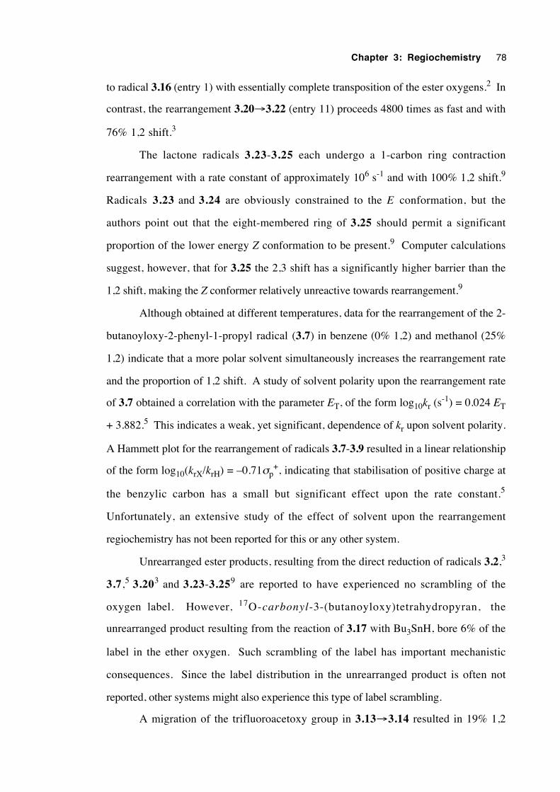
Chapter 3: Regiochemistry 78
to radical 3.16 (entry 1) with essentially complete transposition of the ester oxygens.2 In
contrast, the rearrangement 3.20→3.22 (entry 11) proceeds 4800 times as fast and with
76% 1,2 shift.3
The lactone radicals 3.23-3.25 each undergo a 1-carbon ring contraction
rearrangement with a rate constant of approximately 106 s-1 and with 100% 1,2 shift.9
Radicals 3.23 and 3.24 are obviously constrained to the E conformation, but the
authors point out that the eight-membered ring of 3.25 should permit a significant
proportion of the lower energy Z conformation to be present.9 Computer calculations
suggest, however, that for 3.25 the 2,3 shift has a significantly higher barrier than the
1,2 shift, making the Z conformer relatively unreactive towards rearrangement.9
Although obtained at different temperatures, data for the rearrangement of the 2-
butanoyloxy-2-phenyl-1-propyl radical (3.7) in benzene (0% 1,2) and methanol (25%
1,2) indicate that a more polar solvent simultaneously increases the rearrangement rate
and the proportion of 1,2 shift. A study of solvent polarity upon the rearrangement rate
of 3.7 obtained a correlation with the parameter ET, of the form log10kr (s-1) = 0.024 ET
+ 3.882.5 This indicates a weak, yet significant, dependence of kr upon solvent polarity.
A Hammett plot for the rearrangement of radicals 3.7-3.9 resulted in a linear relationship
of the form log10(krX/krH) = –0.71σp+, indicating that stabilisation of positive charge at
the benzylic carbon has a small but significant effect upon the rate constant.5
Unfortunately, an extensive study of the effect of solvent upon the rearrangement
regiochemistry has not been reported for this or any other system.
Unrearranged ester products, resulting from the direct reduction of radicals 3.2,3
3.7,5 3.203 and 3.23-3.259 are reported to have experienced no scrambling of the
oxygen label. However, 17O-carbonyl-3-(butanoyloxy)tetrahydropyran, the
unrearranged product resulting from the reaction of 3.17 with Bu3SnH, bore 6% of the
label in the ether oxygen. Such scrambling of the label has important mechanistic
consequences. Since the label distribution in the unrearranged product is often not
reported, other systems might also experience this type of label scrambling.
A migration of the trifluoroacetoxy group in 3.13→3.14 resulted in 19% 1,2
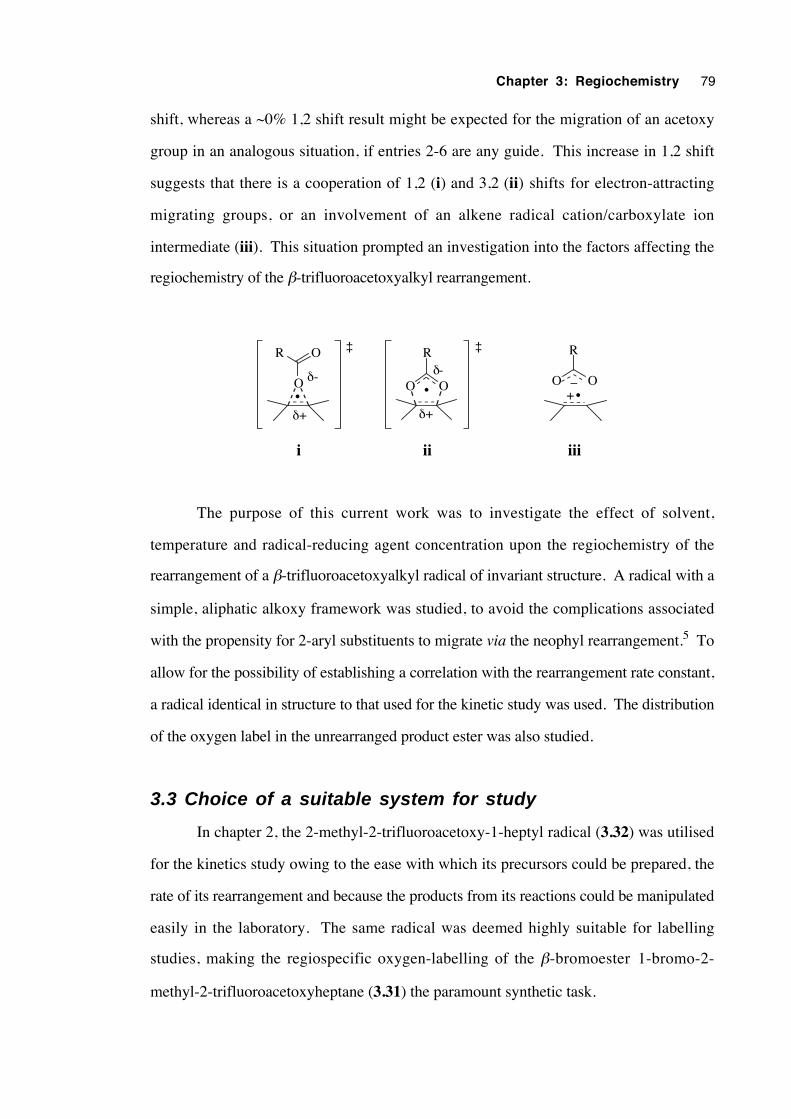
Chapter 3: Regiochemistry 79
shift, whereas a ~0% 1,2 shift result might be expected for the migration of an acetoxy
group in an analogous situation, if entries 2-6 are any guide. This increase in 1,2 shift
suggests that there is a cooperation of 1,2 (i) and 3,2 (ii) shifts for electron-attracting
migrating groups, or an involvement of an alkene radical cation/carboxylate ion
intermediate (iii). This situation prompted an investigation into the factors affecting the
regiochemistry of the β-trifluoroacetoxyalkyl rearrangement.
O O
R
•O
R O
•O O
R
+_
iiiiii
•
δ+
δ-
δ+
δ-
‡ ‡
The purpose of this current work was to investigate the effect of solvent,
temperature and radical-reducing agent concentration upon the regiochemistry of the
rearrangement of a β-trifluoroacetoxyalkyl radical of invariant structure. A radical with a
simple, aliphatic alkoxy framework was studied, to avoid the complications associated
with the propensity for 2-aryl substituents to migrate via the neophyl rearrangement.5 To
allow for the possibility of establishing a correlation with the rearrangement rate constant,
a radical identical in structure to that used for the kinetic study was used. The distribution
of the oxygen label in the unrearranged product ester was also studied.
3.3 Choice of a suitable system for study
In chapter 2, the 2-methyl-2-trifluoroacetoxy-1-heptyl radical (3.32) was utilised
for the kinetics study owing to the ease with which its precursors could be prepared, the
rate of its rearrangement and because the products from its reactions could be manipulated
easily in the laboratory. The same radical was deemed highly suitable for labelling
studies, making the regiospecific oxygen-labelling of the β-bromoester 1-bromo-2-
methyl-2-trifluoroacetoxyheptane (3.31) the paramount synthetic task.
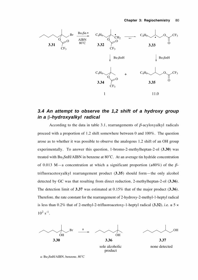
Chapter 3: Regiochemistry 80
Br
O O
CF3
C4H9O
O
CF3
C4H9 O CF3
O
Bu3Sn •
AIBN 80˚C
+
11.01
3.31
3.34 3.35
C4H9CH2O O
CF3
3.32
• C4H9 O CF3
O3.33
•
Bu3SnH Bu3SnH
3.4 An attempt to observe the 1,2 shift of a hydroxy groupin a β-hydroxyalkyl radical
According to the data in table 3.1, rearrangements of β-acyloxyalkyl radicals
proceed with a proportion of 1,2 shift somewhere between 0 and 100%. The question
arose as to whether it was possible to observe the analogous 1,2 shift of an OH group
experimentally. To answer this question, 1-bromo-2-methylheptan-2-ol (3.30) was
treated with Bu3SnH/AIBN in benzene at 80˚C. At an average tin hydride concentration
of 0.013 M—a concentration at which a significant proportion (≥80%) of the β-
trifluoracetoxyalkyl rearrangement product (3.35) should form—the only alcohol
detected by GC was that resulting from direct reduction, 2-methylheptan-2-ol (3.36).
The detection limit of 3.37 was estimated at 0.15% that of the major product (3.36).
Therefore, the rate constant for the rearrangement of 2-hydroxy-2-methyl-1-heptyl radical
is less than 0.2% that of 2-methyl-2-trifluoroacetoxy-1-heptyl radical (3.32), i.e. ≤ 5 ×
102 s-1.
Br
OH OH
OH
3.30 3.36 3.37
sole alcoholicproduct
none detected
a: Bu3SnH/AIBN, benzene, 80˚C
a
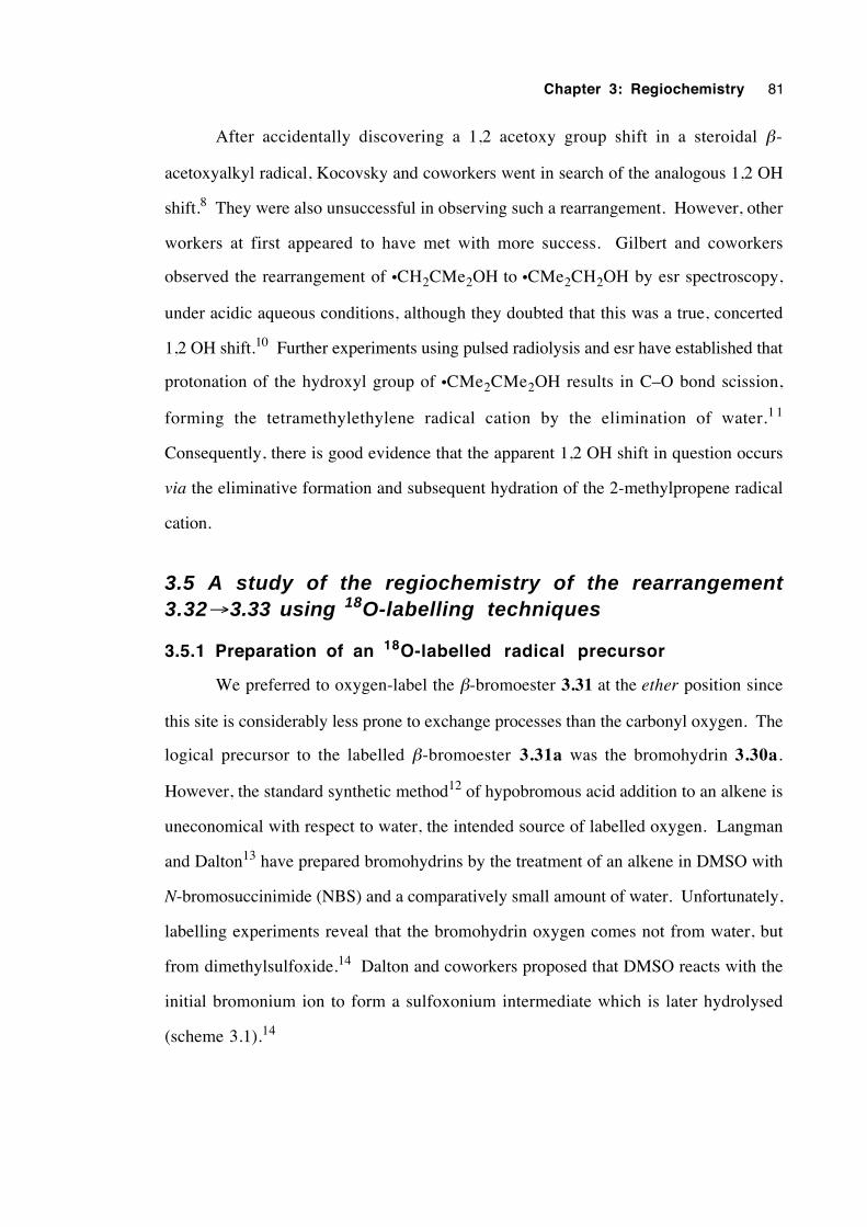
Chapter 3: Regiochemistry 81
After accidentally discovering a 1,2 acetoxy group shift in a steroidal β-
acetoxyalkyl radical, Kocovsky and coworkers went in search of the analogous 1,2 OH
shift.8 They were also unsuccessful in observing such a rearrangement. However, other
workers at first appeared to have met with more success. Gilbert and coworkers
observed the rearrangement of •CH2CMe2OH to •CMe2CH2OH by esr spectroscopy,
under acidic aqueous conditions, although they doubted that this was a true, concerted
1,2 OH shift.10 Further experiments using pulsed radiolysis and esr have established that
protonation of the hydroxyl group of •CMe2CMe2OH results in C–O bond scission,
forming the tetramethylethylene radical cation by the elimination of water.1 1
Consequently, there is good evidence that the apparent 1,2 OH shift in question occurs
via the eliminative formation and subsequent hydration of the 2-methylpropene radical
cation.
3.5 A study of the regiochemistry of the rearrangement3.32→3.33 using 18O-labelling techniques
3.5.1 Preparation of an 18O-labelled radical precursor
We preferred to oxygen-label the β-bromoester 3.31 at the ether position since
this site is considerably less prone to exchange processes than the carbonyl oxygen. The
logical precursor to the labelled β-bromoester 3.31a was the bromohydrin 3.30a.
However, the standard synthetic method12 of hypobromous acid addition to an alkene is
uneconomical with respect to water, the intended source of labelled oxygen. Langman
and Dalton13 have prepared bromohydrins by the treatment of an alkene in DMSO with
N-bromosuccinimide (NBS) and a comparatively small amount of water. Unfortunately,
labelling experiments reveal that the bromohydrin oxygen comes not from water, but
from dimethylsulfoxide.14 Dalton and coworkers proposed that DMSO reacts with the
initial bromonium ion to form a sulfoxonium intermediate which is later hydrolysed
(scheme 3.1).14
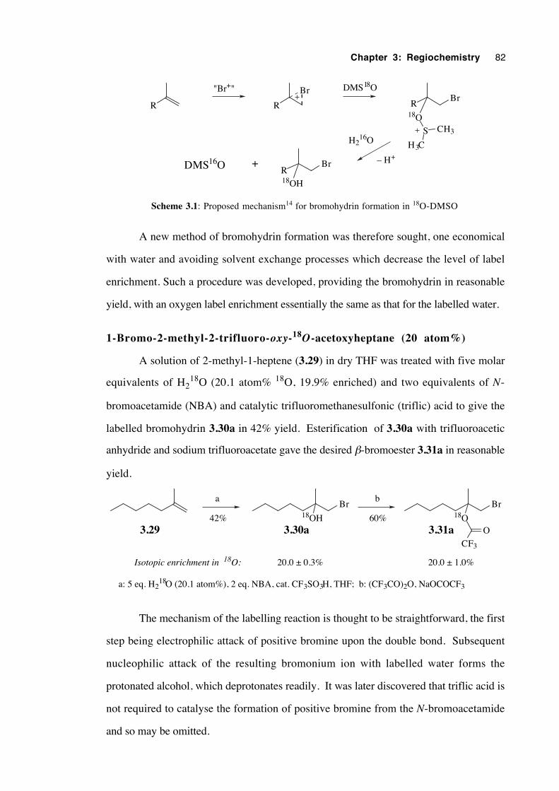
Chapter 3: Regiochemistry 82
R R
Br
RBr
18O
S
H3C
CH3
RBr
18OH
+
"Br+"
– H+
DMS18O
H216O
DMS16O
Scheme 3.1: Proposed mechanism14 for bromohydrin formation in 18O-DMSO
A new method of bromohydrin formation was therefore sought, one economical
with water and avoiding solvent exchange processes which decrease the level of label
enrichment. Such a procedure was developed, providing the bromohydrin in reasonable
yield, with an oxygen label enrichment essentially the same as that for the labelled water.
1-Bromo-2-methyl-2-trifluoro-oxy-18O-acetoxyheptane (20 atom%)
A solution of 2-methyl-1-heptene (3.29) in dry THF was treated with five molar
equivalents of H218O (20.1 atom% 18O, 19.9% enriched) and two equivalents of N-
bromoacetamide (NBA) and catalytic trifluoromethanesulfonic (triflic) acid to give the
labelled bromohydrin 3.30a in 42% yield. Esterification of 3.30a with trifluoroacetic
anhydride and sodium trifluoroacetate gave the desired β-bromoester 3.31a in reasonable
yield.
Br Br18OH
a b
60%42%
20.0 ± 0.3% 20.0 ± 1.0%Isotopic enrichment in 18O:
a: 5 eq. H218O (20.1 atom%), 2 eq. NBA, cat. CF3SO3H, THF; b: (CF3CO)2O, NaOCOCF3
18OO
CF3
3.29 3.30a 3.31a
The mechanism of the labelling reaction is thought to be straightforward, the first
step being electrophilic attack of positive bromine upon the double bond. Subsequent
nucleophilic attack of the resulting bromonium ion with labelled water forms the
protonated alcohol, which deprotonates readily. It was later discovered that triflic acid is
not required to catalyse the formation of positive bromine from the N-bromoacetamide
and so may be omitted.
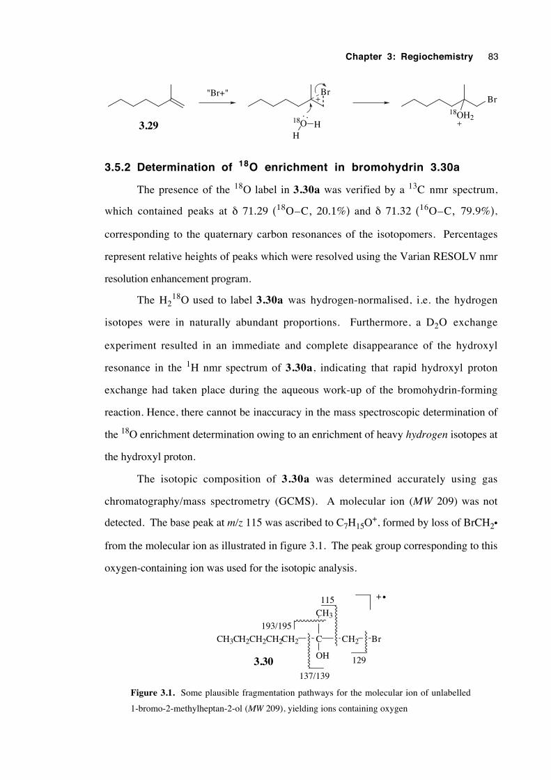
Chapter 3: Regiochemistry 83
"Br+"
3.29
Br
H
18O H
Br18OH2
3.5.2 Determination of 18O enrichment in bromohydrin 3.30a
The presence of the 18O label in 3.30a was verified by a 13C nmr spectrum,
which contained peaks at δ 71.29 (18O–C, 20.1%) and δ 71.32 (16O–C, 79.9%),
corresponding to the quaternary carbon resonances of the isotopomers. Percentages
represent relative heights of peaks which were resolved using the Varian RESOLV nmr
resolution enhancement program.
The H218O used to label 3.30a was hydrogen-normalised, i.e. the hydrogen
isotopes were in naturally abundant proportions. Furthermore, a D2O exchange
experiment resulted in an immediate and complete disappearance of the hydroxyl
resonance in the 1H nmr spectrum of 3.30a, indicating that rapid hydroxyl proton
exchange had taken place during the aqueous work-up of the bromohydrin-forming
reaction. Hence, there cannot be inaccuracy in the mass spectroscopic determination of
the 18O enrichment determination owing to an enrichment of heavy hydrogen isotopes at
the hydroxyl proton.
The isotopic composition of 3.30a was determined accurately using gas
chromatography/mass spectrometry (GCMS). A molecular ion (MW 209) was not
detected. The base peak at m/z 115 was ascribed to C7H15O+, formed by loss of BrCH2•
from the molecular ion as illustrated in figure 3.1. The peak group corresponding to this
oxygen-containing ion was used for the isotopic analysis.
C CH2
CH3
OH
BrCH3CH2CH2CH2CH2
137/139
129
193/195
115
3.30
+ •
Figure 3.1. Some plausible fragmentation pathways for the molecular ion of unlabelled
1-bromo-2-methylheptan-2-ol (MW 209), yielding ions containing oxygen
Chapter 3: Regiochemistry 84
An averaged mass spectrum (5 trials) of the peak group at m/z 115-119 was
obtained for the unlabelled bromohydrin 3.30. Natural abundance levels of other stable
oxygen isotopes are 0.200% for 18O and 0.038% for 17O. Using the respective natural
abundances, predicted mass spectra were calculated for isotopomers of 3.30 having the
oxygen composed entirely of 18O and entirely 17O. The averaged mass spectrum for the
labelled bromohydrin 3.30a was treated as being a linear combination of the mass
spectra of the natural abundance (3.30), 17O and 18O isotopomers (equation 3.1), where
A, B and C represent the respective proportions. This type of procedure was used for all
analyses of oxygen isotope enrichments. Using this method, the bromohydrin 3.30a
was determined to have an 18O enrichment of 20.0±0.3% (20.2±0.3 atom% 18O) and a
17O enrichment of 0.66±0.08%, where uncertainties represent one standard deviation. It
is clear that the label introduction method does not significantly decrease the 18O
enrichment in 3.30a relative to that of the H218O (20.1 atom%).
Mass spectrum (3.30a) = A (3.30) + B (3.30 17O) + C (3.30 18O) (3.1)
Concentrations of 18O label are expressed in terms of enrichment rather than
content. This convention requires no further compensation for natural abundance 18O
and thus simplifies later calculations of label distribution in the esters 3.34 and 3.35,
resulting from the radical-mediated reduction of β-bromoester 3.31a. The simple
relationship between 18O content and enrichment is given by equation 3.2.
atom% 18O = %18O enrichment + 0.200%(100% – %18O enrichment)
= 99.796(%18O enrichment) + 0.200% (3.2)
3.5.3 Determination of 18O enrichment of labelled β -bromoester
3 .31a
A 13C nmr spectrum of 3.31a contained resonances at δ 87.834 (18O–C, 19.6%)
and δ 87.883 (16O–C, 80.4%), corresponding to the quaternary carbons at the 2-heptyl
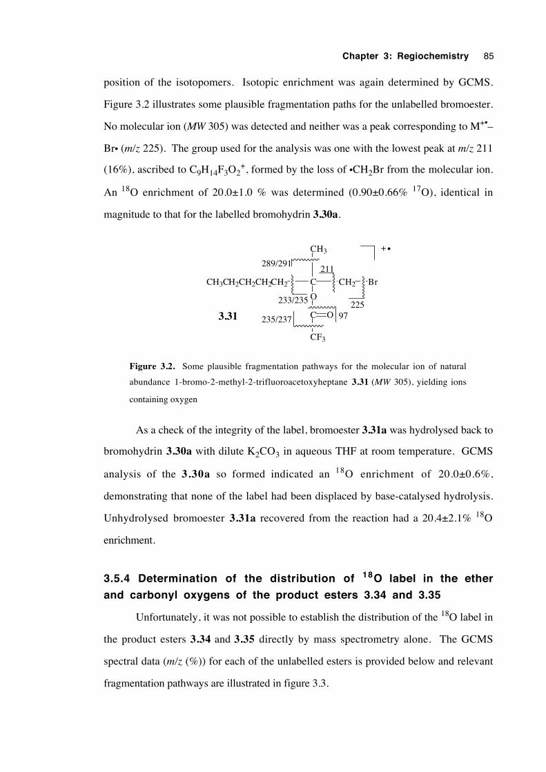
Chapter 3: Regiochemistry 85
position of the isotopomers. Isotopic enrichment was again determined by GCMS.
Figure 3.2 illustrates some plausible fragmentation paths for the unlabelled bromoester.
No molecular ion (MW 305) was detected and neither was a peak corresponding to M+•–
Br• (m/z 225). The group used for the analysis was one with the lowest peak at m/z 211
(16%), ascribed to C9H14F3O2+, formed by the loss of •CH2Br from the molecular ion.
An 18O enrichment of 20.0±1.0 % was determined (0.90±0.66% 17O), identical in
magnitude to that for the labelled bromohydrin 3.30a.
CH3CH2CH2CH2CH2 C
CH3
O
CH2 Br
C O
CF3
225
211
233/235
97235/237
289/291
3.31
+ •
Figure 3.2. Some plausible fragmentation pathways for the molecular ion of natural
abundance 1-bromo-2-methyl-2-trifluoroacetoxyheptane 3.31 (MW 305), yielding ions
containing oxygen
As a check of the integrity of the label, bromoester 3.31a was hydrolysed back to
bromohydrin 3.30a with dilute K2CO3 in aqueous THF at room temperature. GCMS
analysis of the 3.30a so formed indicated an 18O enrichment of 20.0±0.6%,
demonstrating that none of the label had been displaced by base-catalysed hydrolysis.
Unhydrolysed bromoester 3.31a recovered from the reaction had a 20.4±2.1% 18O
enrichment.
3.5.4 Determination of the distribution of 18O label in the ether
and carbonyl oxygens of the product esters 3.34 and 3.35
Unfortunately, it was not possible to establish the distribution of the 18O label in
the product esters 3.34 and 3.35 directly by mass spectrometry alone. The GCMS
spectral data (m/z (%)) for each of the unlabelled esters is provided below and relevant
fragmentation pathways are illustrated in figure 3.3.
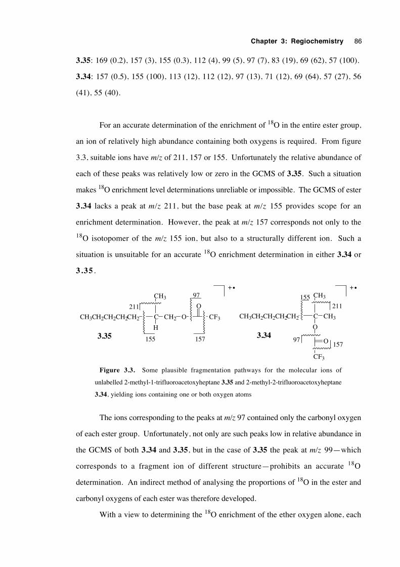
Chapter 3: Regiochemistry 86
3.35: 169 (0.2), 157 (3), 155 (0.3), 112 (4), 99 (5), 97 (7), 83 (19), 69 (62), 57 (100).
3.34: 157 (0.5), 155 (100), 113 (12), 112 (12), 97 (13), 71 (12), 69 (64), 57 (27), 56
(41), 55 (40).
For an accurate determination of the enrichment of 18O in the entire ester group,
an ion of relatively high abundance containing both oxygens is required. From figure
3.3, suitable ions have m/z of 211, 157 or 155. Unfortunately the relative abundance of
each of these peaks was relatively low or zero in the GCMS of 3.35. Such a situation
makes 18O enrichment level determinations unreliable or impossible. The GCMS of ester
3.34 lacks a peak at m/z 211, but the base peak at m/z 155 provides scope for an
enrichment determination. However, the peak at m/z 157 corresponds not only to the
18O isotopomer of the m/z 155 ion, but also to a structurally different ion. Such a
situation is unsuitable for an accurate 18O enrichment determination in either 3.34 or
3.35 .
CH3CH2CH2CH2CH2 C
CH3
CH3
O
CF3
O
211
15797
155
3.34
CH3CH2CH2CH2CH2 C
CH3
O
H3.35
CF3
O
97
157155
211
CH2
+ • + •
Figure 3.3. Some plausible fragmentation pathways for the molecular ions of
unlabelled 2-methyl-1-trifluoroacetoxyheptane 3.35 and 2-methyl-2-trifluoroacetoxyheptane
3.34, yielding ions containing one or both oxygen atoms
The ions corresponding to the peaks at m/z 97 contained only the carbonyl oxygen
of each ester group. Unfortunately, not only are such peaks low in relative abundance in
the GCMS of both 3.34 and 3.35, but in the case of 3.35 the peak at m/z 99—which
corresponds to a fragment ion of different structure—prohibits an accurate 18O
determination. An indirect method of analysing the proportions of 18O in the ester and
carbonyl oxygens of each ester was therefore developed.
With a view to determining the 18O enrichment of the ether oxygen alone, each
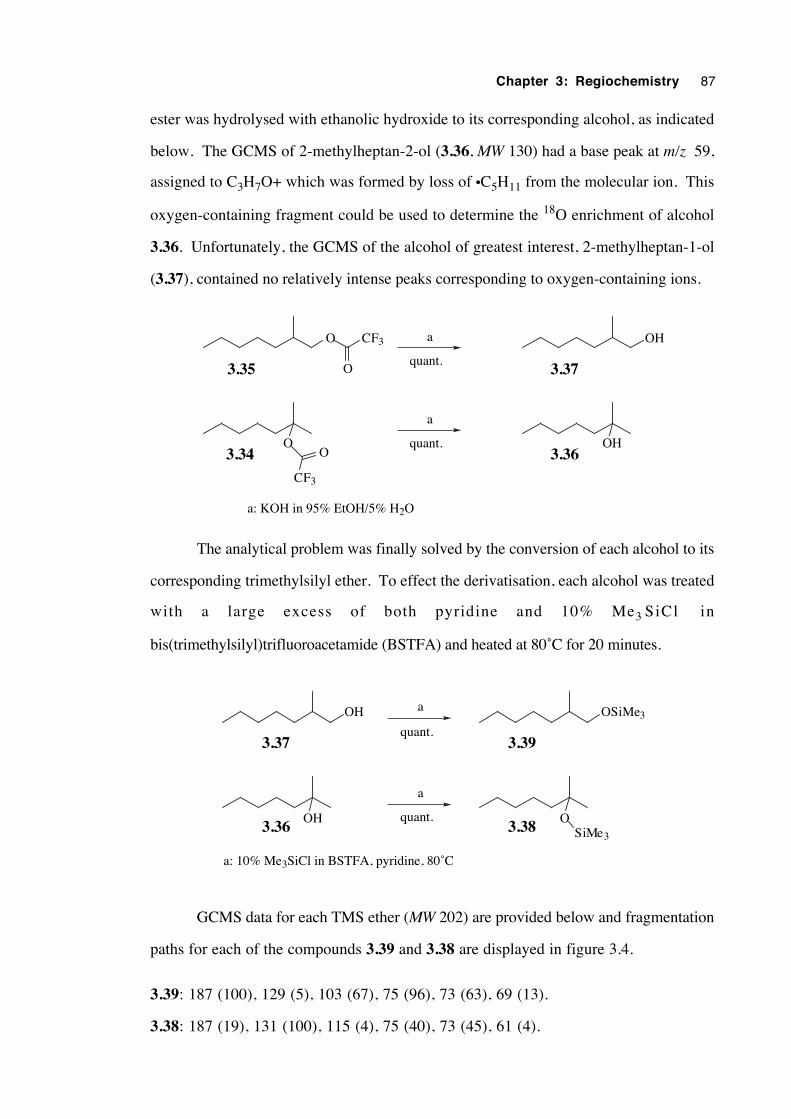
Chapter 3: Regiochemistry 87
ester was hydrolysed with ethanolic hydroxide to its corresponding alcohol, as indicated
below. The GCMS of 2-methylheptan-2-ol (3.36, MW 130) had a base peak at m/z 59,
assigned to C3H7O+ which was formed by loss of •C5H11 from the molecular ion. This
oxygen-containing fragment could be used to determine the 18O enrichment of alcohol
3.36. Unfortunately, the GCMS of the alcohol of greatest interest, 2-methylheptan-1-ol
(3.37), contained no relatively intense peaks corresponding to oxygen-containing ions.
O
CF3
OOH
a
a
3.35
3.34 3.36
3.37
a: KOH in 95% EtOH/5% H2O
OHO CF3
Oquant.
quant.
The analytical problem was finally solved by the conversion of each alcohol to its
corresponding trimethylsilyl ether. To effect the derivatisation, each alcohol was treated
with a large excess of both pyridine and 10% Me3 SiCl in
bis(trimethylsilyl)trifluoroacetamide (BSTFA) and heated at 80˚C for 20 minutes.
OH3.36
3.37
O3.38
3.39
SiMe3
a
a
a: 10% Me3SiCl in BSTFA, pyridine, 80˚C
OH OSiMe3
quant.
quant.
GCMS data for each TMS ether (MW 202) are provided below and fragmentation
paths for each of the compounds 3.39 and 3.38 are displayed in figure 3.4.
3.39: 187 (100), 129 (5), 103 (67), 75 (96), 73 (63), 69 (13).
3.38: 187 (19), 131 (100), 115 (4), 75 (40), 73 (45), 61 (4).
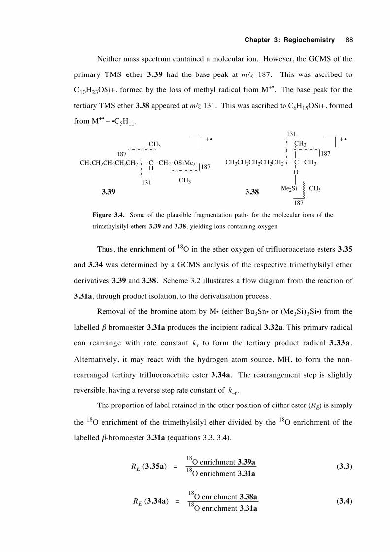
Chapter 3: Regiochemistry 88
Neither mass spectrum contained a molecular ion. However, the GCMS of the
primary TMS ether 3.39 had the base peak at m/z 187. This was ascribed to
C10H23OSi+, formed by the loss of methyl radical from M+•. The base peak for the
tertiary TMS ether 3.38 appeared at m/z 131. This was ascribed to C6H15OSi+, formed
from M+• – •C5H11.
CH3CH2CH2CH2CH2 CH
CH3
CH2 OSiMe2
131
187
CH3
187CH3CH2CH2CH2CH2 C
CH3
CH3
O
Me2Si CH3
187
131
187
3.383.39
+ • + •
Figure 3.4. Some of the plausible fragmentation paths for the molecular ions of the
trimethylsilyl ethers 3.39 and 3.38, yielding ions containing oxygen
Thus, the enrichment of 18O in the ether oxygen of trifluoroacetate esters 3.35
and 3.34 was determined by a GCMS analysis of the respective trimethylsilyl ether
derivatives 3.39 and 3.38. Scheme 3.2 illustrates a flow diagram from the reaction of
3.31a, through product isolation, to the derivatisation process.
Removal of the bromine atom by M• (either Bu3Sn• or (Me3Si)3Si•) from the
labelled β-bromoester 3.31a produces the incipient radical 3.32a. This primary radical
can rearrange with rate constant kr to form the tertiary product radical 3.33a.
Alternatively, it may react with the hydrogen atom source, MH, to form the non-
rearranged tertiary trifluoroacetate ester 3.34a. The rearrangement step is slightly
reversible, having a reverse step rate constant of k–r.
The proportion of label retained in the ether position of either ester (RE) is simply
the 18O enrichment of the trimethylsilyl ether divided by the 18O enrichment of the
labelled β-bromoester 3.31a (equations 3.3, 3.4).
RE (3.35a) = 18
18O enrichment
O enrichment
3.39a3.31a
(3.3)
RE (3.34a) = 18
18O enrichment
O enrichment
3.38a3.31a
(3.4)
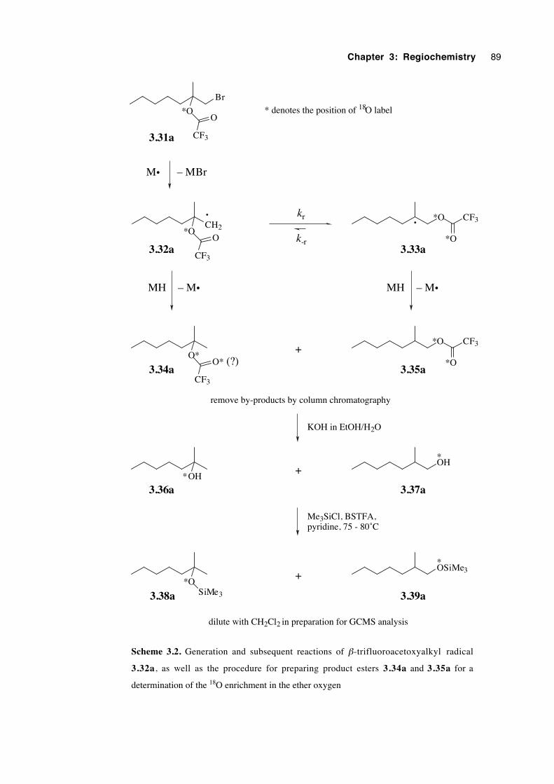
Chapter 3: Regiochemistry 89
Br
*O
CF3
O
CH2*O
CF3
O
*O CF3
*O
MH
3.31a
3.32a 3.33a
kr
k-r
*O
O*
CF3
O*
OH
OH
OSiMe3
*O
+
+
+
CF3
*O
SiMe3
3.35a3.34a
3.37a3.36a
3.39a3.38a
remove by-products by column chromatography
dilute with CH2Cl2 in preparation for GCMS analysis
KOH in EtOH/H2O
Me3SiCl, BSTFA,pyridine, 75 - 80˚C
* denotes the position of 18O label
*
*
*
– M• MH – M•
M• – MBr
(?)
Scheme 3.2. Generation and subsequent reactions of β-trifluoroacetoxyalkyl radical
3.32a, as well as the procedure for preparing product esters 3.34a and 3.35a for a
determination of the 18O enrichment in the ether oxygen
Chapter 3: Regiochemistry 90
3.5.5 Results
In these experiments, tris(trimethylsilyl)silane (TTMSS, (Me3Si)3SiH) was used
instead of tributyltin hydride (TBTH, Bu3SnH) as the hydrogen atom source, MH.
Traces of organotin compounds remaining on the GCMS injector port septum and
capillary column have reportedly contaminated very dilute samples of natural products,
injected frequently by other users of the same instrument. Neither TTMSS nor the
products from its reaction with alkyl bromides cause this type of contamination.
TTMSS is an efficient chain transfer agent and silicon-centred radicals readily
abstract bromine atoms from alkyl bromides.15 It has advantages over tributyltin hydride
in other regards.15-18 The Si–H bond strength (330 kJmol-1) is greater than that for Sn–
H (309 kJmol-1),16 making TTMSS a less reactive H atom donor. Since (Me3Si)3SiH
reacts more slowly than Bu3SnH with incipient radical 3.32a, this direct reduction step
competes less strongly with the rearrangement step, leading to a higher proportion of the
rearranged product 3.35a for identical initial MH concentrations.
All labelling experiments were performed in the absence of air in Reactivials fitted
with Mininert19 syringe valve caps (see experimental section 3.11). This apparatus
permitted addition or removal of fluids during the reaction at temperatures greater than the
boiling point of the solvent. A deoxygenated solution of the labelled β-bromoester
3.31a in the desired solvent was heated at 80±1˚C (or another nominated temperature)
for at least two minutes, then injected with an excess of TTMSS and catalytic AIBN.
When the reaction was complete by GC, the reaction was cooled and the solvent was
evaporated. In the case of the water-miscible solvents acetonitrile and N -
methylacetamide, the reaction mixture was taken up in diethyl ether, washed with water,
dried and evaporated. The trifluoroacetate ester products 3.34a and 3.35a were
separated from other components by flash chromatography, hydrolysed by aqueous base
to the corresponding alcohols 3.36a and 3.37a , converted to the respective
trimethylsilyl ethers 3.38a and 3.39a, then analysed by GCMS. Equations 3.3 and 3.4
were used to calculate the proportion of 18O label residing in the ether position of esters
3.35a and 3.34a respectively. The 18O enrichment of 3.31a was taken to be identical
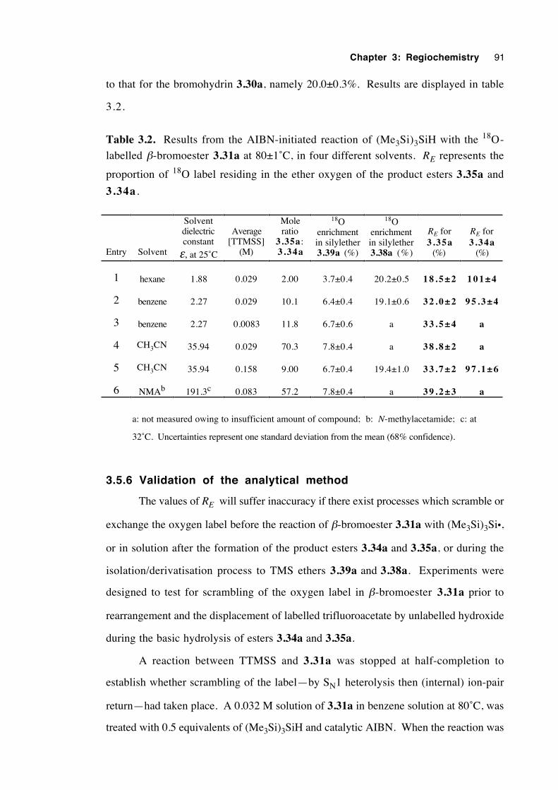
Chapter 3: Regiochemistry 91
to that for the bromohydrin 3.30a, namely 20.0±0.3%. Results are displayed in table
3.2.
Table 3.2. Results from the AIBN-initiated reaction of (Me3Si)3SiH with the 18O-
labelled β-bromoester 3.31a at 80±1˚C, in four different solvents. RE represents the
proportion of 18O label residing in the ether oxygen of the product esters 3.35a and
3.34a .
Entry Solvent
Solventdielectricconstant
ε, at 25˚C
Average[TTMSS]
(M)
Moleratio
3.35a :3.34a
18Oenrichmentin silylether3.39a (%)
18Oenrichmentin silylether3.38a (%)
RE for3.35a
(%)
RE for3.34a
(%)
1 hexane 1.88 0.029 2.00 3.7±0.4 20.2±0.5 1 8 . 5 ± 2 1 0 1 ± 4
2 benzene 2.27 0.029 10.1 6.4±0.4 19.1±0.6 3 2 . 0 ± 2 9 5 . 3 ± 4
3 benzene 2.27 0.0083 11.8 6.7±0.6 a 3 3 . 5 ± 4 a
4 CH3CN 35.94 0.029 70.3 7.8±0.4 a 3 8 . 8 ± 2 a
5 CH3CN 35.94 0.158 9.00 6.7±0.4 19.4±1.0 3 3 . 7 ± 2 9 7 . 1 ± 6
6 NMAb 191.3c 0.083 57.2 7.8±0.4 a 3 9 . 2 ± 3 a
a: not measured owing to insufficient amount of compound; b: N-methylacetamide; c: at
32˚C. Uncertainties represent one standard deviation from the mean (68% confidence).
3.5.6 Validation of the analytical method
The values of RE will suffer inaccuracy if there exist processes which scramble or
exchange the oxygen label before the reaction of β-bromoester 3.31a with (Me3Si)3Si•,
or in solution after the formation of the product esters 3.34a and 3.35a, or during the
isolation/derivatisation process to TMS ethers 3.39a and 3.38a. Experiments were
designed to test for scrambling of the oxygen label in β-bromoester 3.31a prior to
rearrangement and the displacement of labelled trifluoroacetate by unlabelled hydroxide
during the basic hydrolysis of esters 3.34a and 3.35a.
A reaction between TTMSS and 3.31a was stopped at half-completion to
establish whether scrambling of the label—by SN1 heterolysis then (internal) ion-pair
return—had taken place. A 0.032 M solution of 3.31a in benzene solution at 80˚C, was
treated with 0.5 equivalents of (Me3Si)3SiH and catalytic AIBN. When the reaction was
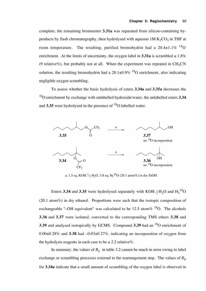
Chapter 3: Regiochemistry 92
complete, the remaining bromoester 3.31a was separated from silicon-containing by-
products by flash chromatography, then hydrolysed with aqueous 1M K2CO3 in THF at
room temperature. The resulting, purified bromohydrin had a 20.4±1.1% 18O
enrichment. At the limits of uncertainty, the oxygen label in 3.31a is scrambled ≤ 1.8%
(9 relative%), but probably not at all. When the experiment was repeated in CH3CN
solution, the resulting bromohydrin had a 20.1±0.9% 18O enrichment, also indicating
negligible oxygen scrambling.
To assess whether the basic hydrolysis of esters 3.34a and 3.35a decreases the
18O enrichment by exchange with unlabelled hydroxide/water, the unlabelled esters 3.34
and 3.35 were hydrolysed in the presence of 18O-labelled water.
O
CF3
OOH
a
a
3.35
3.34 3.36
3.37
a: 1.5 eq. KOH.1/2 H2O, 3.8 eq. H218O (20.1 atom%) in dry EtOH
OHO CF3
O
no 18O incorporation
no 18O incorporation
Esters 3.34 and 3.35 were hydrolysed separately with KOH. 12 H2O and H2
18O
(20.1 atom%) in dry ethanol. Proportions were such that the isotopic composition of
exchangeable "–OH equivalent" was calculated to be 12.5 atom% 18O. The alcohols
3.36 and 3.37 were isolated, converted to the corresponding TMS ethers 3.38 and
3.39 and analysed isotopically by GCMS. Compound 3.39 had an 18O enrichment of
0.00±0.28% and 3.38 had –0.03±0.27%, indicating an incorporation of oxygen from
the hydrolysis reagents in each case to be ≤ 2.2 relative%.
In summary, the values of RE in table 3.2 cannot be much in error owing to label
exchange or scrambling processes external to the rearrangement step. The values of RE
for 3.34a indicate that a small amount of scrambling of the oxygen label is observed in
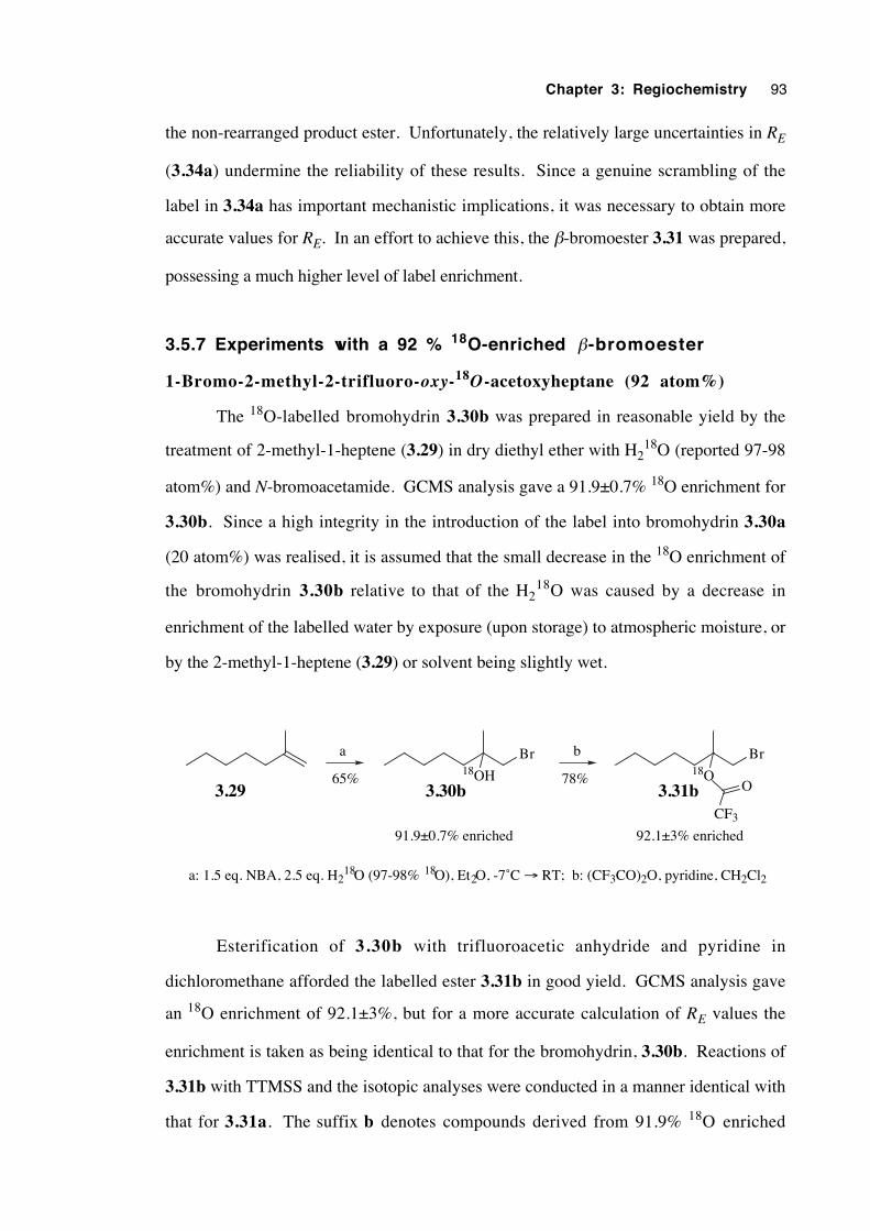
Chapter 3: Regiochemistry 93
the non-rearranged product ester. Unfortunately, the relatively large uncertainties in RE
(3.34a) undermine the reliability of these results. Since a genuine scrambling of the
label in 3.34a has important mechanistic implications, it was necessary to obtain more
accurate values for RE. In an effort to achieve this, the β-bromoester 3.31 was prepared,
possessing a much higher level of label enrichment.
3.5.7 Experiments with a 92 % 18O-enriched β-bromoester
1-Bromo-2-methyl-2-trifluoro-oxy-18O-acetoxyheptane (92 atom%)
The 18O-labelled bromohydrin 3.30b was prepared in reasonable yield by the
treatment of 2-methyl-1-heptene (3.29) in dry diethyl ether with H218O (reported 97-98
atom%) and N-bromoacetamide. GCMS analysis gave a 91.9±0.7% 18O enrichment for
3.30b. Since a high integrity in the introduction of the label into bromohydrin 3.30a
(20 atom%) was realised, it is assumed that the small decrease in the 18O enrichment of
the bromohydrin 3.30b relative to that of the H218O was caused by a decrease in
enrichment of the labelled water by exposure (upon storage) to atmospheric moisture, or
by the 2-methyl-1-heptene (3.29) or solvent being slightly wet.
Br Br18OH 18O
CF3
O
a b
65% 78%3.29 3.30b 3.31b
a: 1.5 eq. NBA, 2.5 eq. H218O (97-98% 18O), Et2O, -7˚C → RT; b: (CF3CO)2O, pyridine, CH2Cl2
91.9±0.7% enriched 92.1±3% enriched
Esterification of 3.30b with trifluoroacetic anhydride and pyridine in
dichloromethane afforded the labelled ester 3.31b in good yield. GCMS analysis gave
an 18O enrichment of 92.1±3%, but for a more accurate calculation of RE values the
enrichment is taken as being identical to that for the bromohydrin, 3.30b. Reactions of
3.31b with TTMSS and the isotopic analyses were conducted in a manner identical with
that for 3.31a. The suffix b denotes compounds derived from 91.9% 18O enriched
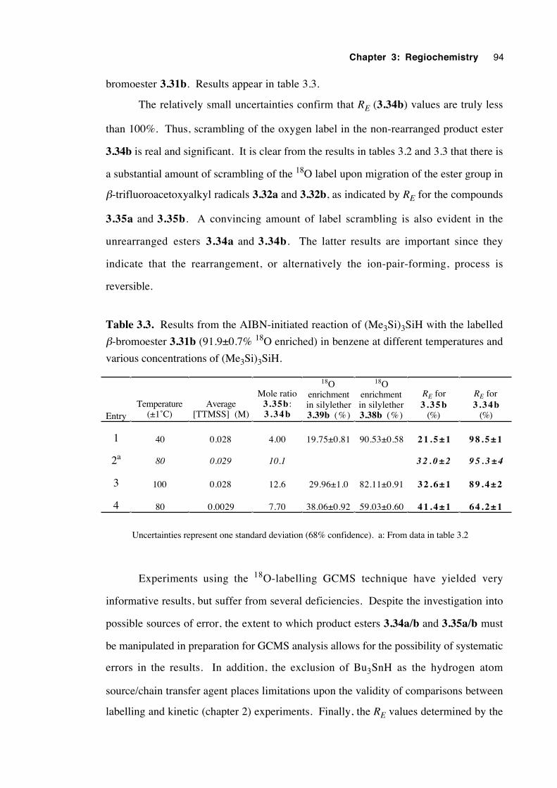
Chapter 3: Regiochemistry 94
bromoester 3.31b. Results appear in table 3.3.
The relatively small uncertainties confirm that RE (3.34b) values are truly less
than 100%. Thus, scrambling of the oxygen label in the non-rearranged product ester
3.34b is real and significant. It is clear from the results in tables 3.2 and 3.3 that there is
a substantial amount of scrambling of the 18O label upon migration of the ester group in
β-trifluoroacetoxyalkyl radicals 3.32a and 3.32b, as indicated by RE for the compounds
3.35a and 3.35b. A convincing amount of label scrambling is also evident in the
unrearranged esters 3.34a and 3.34b. The latter results are important since they
indicate that the rearrangement, or alternatively the ion-pair-forming, process is
reversible.
Table 3.3. Results from the AIBN-initiated reaction of (Me3Si)3SiH with the labelled
β-bromoester 3.31b (91.9±0.7% 18O enriched) in benzene at different temperatures and
various concentrations of (Me3Si)3SiH.
EntryTemperature
(±1˚C)Average
[TTMSS] (M)
Mole ratio3.35b :3 .34b
18Oenrichmentin silylether3.39b (%)
18Oenrichmentin silylether3.38b (%)
RE for3 .35b
(%)
RE for3 .34b
(%)
1 40 0.028 4.00 19.75±0.81 90.53±0.58 2 1 . 5 ± 1 9 8 . 5 ± 1
2a 80 0.029 10.1 3 2 . 0 ± 2 9 5 . 3 ± 4
3 100 0.028 12.6 29.96±1.0 82.11±0.91 3 2 . 6 ± 1 8 9 . 4 ± 2
4 80 0.0029 7.70 38.06±0.92 59.03±0.60 4 1 . 4 ± 1 6 4 . 2 ± 1
Uncertainties represent one standard deviation (68% confidence). a: From data in table 3.2
Experiments using the 18O-labelling GCMS technique have yielded very
informative results, but suffer from several deficiencies. Despite the investigation into
possible sources of error, the extent to which product esters 3.34a/b and 3.35a/b must
be manipulated in preparation for GCMS analysis allows for the possibility of systematic
errors in the results. In addition, the exclusion of Bu3SnH as the hydrogen atom
source/chain transfer agent places limitations upon the validity of comparisons between
labelling and kinetic (chapter 2) experiments. Finally, the RE values determined by the
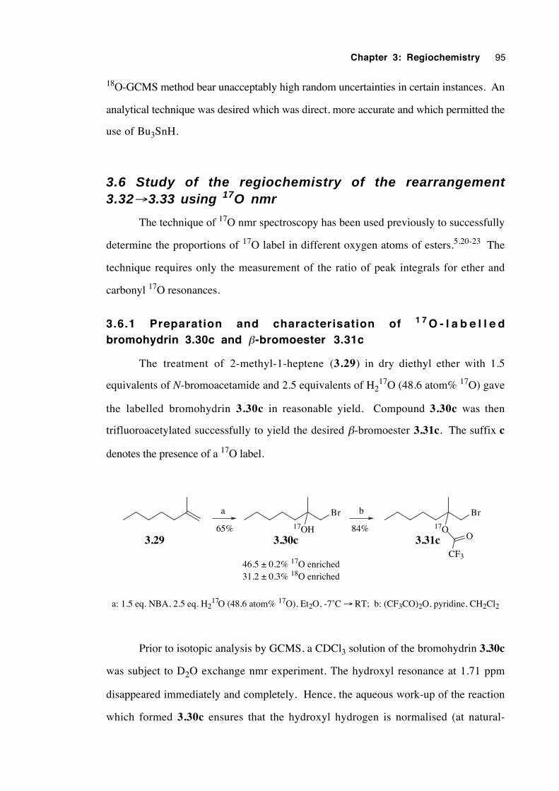
Chapter 3: Regiochemistry 95
18O-GCMS method bear unacceptably high random uncertainties in certain instances. An
analytical technique was desired which was direct, more accurate and which permitted the
use of Bu3SnH.
3.6 Study of the regiochemistry of the rearrangement3.32→3.33 using 17O nmr
The technique of 17O nmr spectroscopy has been used previously to successfully
determine the proportions of 17O label in different oxygen atoms of esters.5,20-23 The
technique requires only the measurement of the ratio of peak integrals for ether and
carbonyl 17O resonances.
3.6.1 Preparation and characterisation of 1 7 O - l a b e l l e dbromohydrin 3.30c and β-bromoester 3.31c
The treatment of 2-methyl-1-heptene (3.29) in dry diethyl ether with 1.5
equivalents of N-bromoacetamide and 2.5 equivalents of H217O (48.6 atom% 17O) gave
the labelled bromohydrin 3.30c in reasonable yield. Compound 3.30c was then
trifluoroacetylated successfully to yield the desired β-bromoester 3.31c. The suffix c
denotes the presence of a 17O label.
Br Br
17OH 17O
CF3
O
a b
65% 84%3.29 3.30c 3.31c
46.5 ± 0.2% 17O enriched31.2 ± 0.3% 18O enriched
a: 1.5 eq. NBA, 2.5 eq. H217O (48.6 atom% 17O), Et2O, -7˚C → RT; b: (CF3CO)2O, pyridine, CH2Cl2
Prior to isotopic analysis by GCMS, a CDCl3 solution of the bromohydrin 3.30c
was subject to D2O exchange nmr experiment. The hydroxyl resonance at 1.71 ppm
disappeared immediately and completely. Hence, the aqueous work-up of the reaction
which formed 3.30c ensures that the hydroxyl hydrogen is normalised (at natural-
Chapter 3: Regiochemistry 96
abundance isotopic composition). Using GCMS, in conjunction with an adapted version
of equation 3.1, the bromohydrin 3.30c analysed for 46.5±0.2% 17O enrichment and
31.2±0.3% 18O enrichment.
The presence of an 18O label in 3.30c was corroborated by a 13C nmr spectrum
containing quaternary resonances at 71.279 (58%) and 71.309 (42%) ppm, assigned to
the tertiary carbons of the 18O and 16O isotopomers respectively. No resonance was
detected for the 17O isotopomer, owing presumably to the combination of the effects of
the signal multiplicity resulting from 13C –17O coupling (sextet) and the broadening of the
13C signal owing to rapid relaxation brought about by the quadrupole moment (I = 52 ) of
the 17O nucleus.24
An 17O nmr spectrum of a pentane solution of labelled bromohydrin 3.30c
contained a single resonance, 50.6 ppm downfield of external H217O (0 ppm), within the
expected shift range of –40 to +70 ppm for aliphatic alcohols.25 A pentane solution of
the labelled β-bromoester 3.31c gave a spectrum with a single peak at 176.7 ppm,
assigned to the alkoxy oxygen. A normal range for alkoxy resonances is 125-210 ppm,
whereas carbonyl oxygens resonate in the range 352-392 ppm.26 For instance, methyl
trifluoroacetate in acetonitrile at 75°C gives a carbonyl peak at 352.8 and an alkoxy peak
at 133.0 ppm.26 The chemical shifts of the various 17O-labelled compounds isolated
during this work are displayed in table 3.4. To view some of the 17O nmr spectra, see
appendix B.
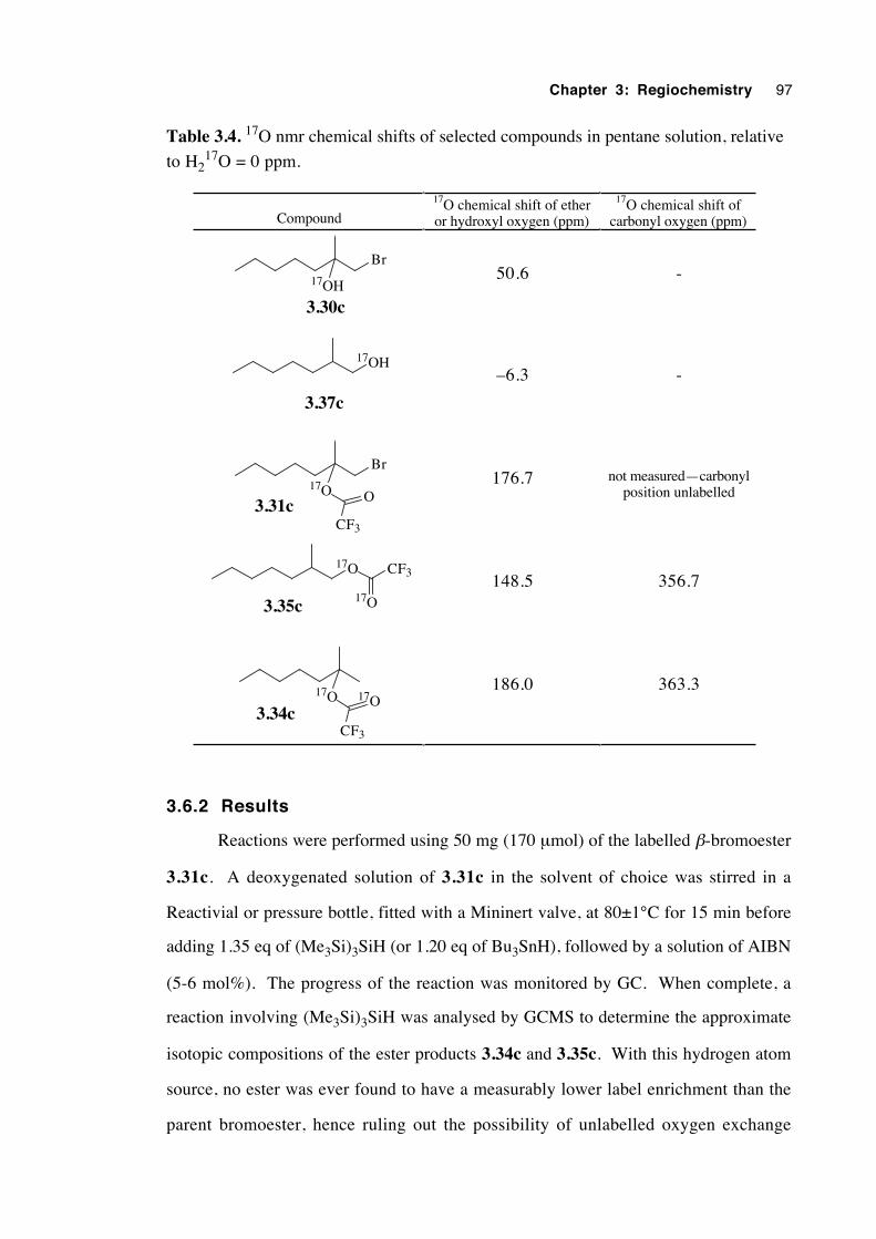
Chapter 3: Regiochemistry 97
Table 3.4. 17O nmr chemical shifts of selected compounds in pentane solution, relative
to H217O = 0 ppm.
Compound
17O chemical shift of etheror hydroxyl oxygen (ppm)
17O chemical shift ofcarbonyl oxygen (ppm)
Br
17OH
3.30c
50.6 -
17OH
3.37c
–6.3 -
Br
17O
CF3
O3.31c
176.7 not measured—carbonylposition unlabelled
17O CF3
17O3.35c148.5 356.7
17O
CF3
17O3.34c
186.0 363.3
3.6.2 Results
Reactions were performed using 50 mg (170 µmol) of the labelled β-bromoester
3.31c. A deoxygenated solution of 3.31c in the solvent of choice was stirred in a
Reactivial or pressure bottle, fitted with a Mininert valve, at 80±1°C for 15 min before
adding 1.35 eq of (Me3Si)3SiH (or 1.20 eq of Bu3SnH), followed by a solution of AIBN
(5-6 mol%). The progress of the reaction was monitored by GC. When complete, a
reaction involving (Me3Si)3SiH was analysed by GCMS to determine the approximate
isotopic compositions of the ester products 3.34c and 3.35c. With this hydrogen atom
source, no ester was ever found to have a measurably lower label enrichment than the
parent bromoester, hence ruling out the possibility of unlabelled oxygen exchange
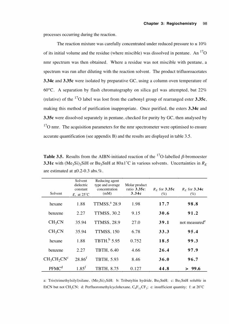
Chapter 3: Regiochemistry 98
processes occurring during the reaction.
The reaction mixture was carefully concentrated under reduced pressure to ≤ 10%
of its initial volume and the residue (where miscible) was dissolved in pentane. An 17O
nmr spectrum was then obtained. Where a residue was not miscible with pentane, a
spectrum was run after diluting with the reaction solvent. The product trifluoroacetates
3.34c and 3.35c were isolated by preparative GC, using a column oven temperature of
60°C. A separation by flash chromatography on silica gel was attempted, but 22%
(relative) of the 17O label was lost from the carbonyl group of rearranged ester 3.35c,
making this method of purification inappropriate. Once purified, the esters 3.34c and
3.35c were dissolved separately in pentane, checked for purity by GC, then analysed by
17O nmr. The acquisition parameters for the nmr spectrometer were optimised to ensure
accurate quantification (see appendix B) and the results are displayed in table 3.5.
Table 3.5. Results from the AIBN-initiated reaction of the 17O-labelled β-bromoester
3.31c with (Me3Si)3SiH or Bu3SnH at 80±1˚C in various solvents. Uncertainties in RE
are estimated at ±0.2-0.3 abs.%.
Solvent
Solventdielectricconstant
ε, at 25˚C
Reducing agenttype and average
concentration(mM)
Molar productratio 3.35c:
3 . 3 4 cRE for 3.35c
(%)RE for 3.34c
(%)
hexane 1.88 TTMSS,a 28.9 1.98 1 7 . 7 9 8 . 8
benzene 2.27 TTMSS, 30.2 9.15 3 0 . 6 9 1 . 2
CH3CN 35.94 TTMSS, 28.9 27.0 3 9 . 1 not measurede
CH3CN 35.94 TTMSS, 150 6.78 3 3 . 3 9 5 . 4
hexane 1.88 TBTH,b 5.95 0.752 1 8 . 5 9 9 . 3
benzene 2.27 TBTH, 6.40 4.66 2 6 . 4 9 7 . 9
CH3CH2CNc 28.86f TBTH, 5.93 8.46 3 6 . 0 9 6 . 7
PFMCd 1.85f TBTH, 8.75 0.127 4 4 . 8 > 99.6
a: Tris(trimethylsilyl)silane, (Me3Si)3SiH; b: Tributyltin hydride, Bu3SnH; c: Bu3SnH soluble in
EtCN but not CH3CN; d: Perfluoromethylcyclohexane, C6F11CF3; e: insufficient quantity; f: at 20˚C
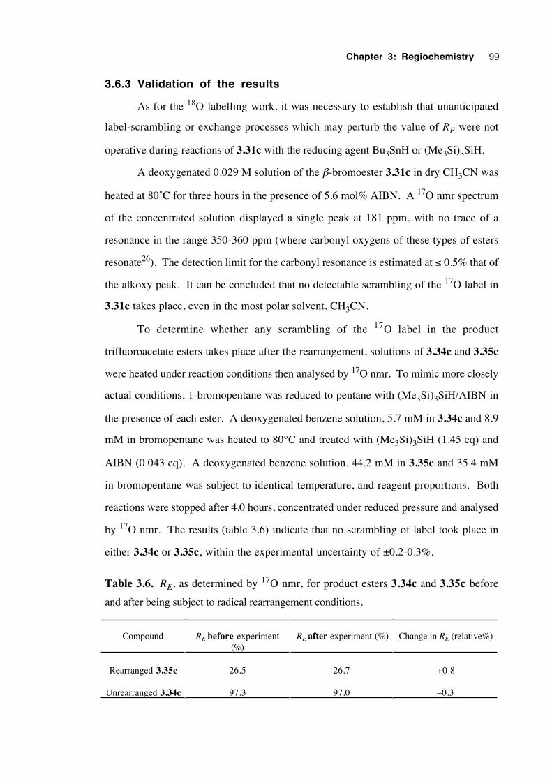
Chapter 3: Regiochemistry 99
3.6.3 Validation of the results
As for the 18O labelling work, it was necessary to establish that unanticipated
label-scrambling or exchange processes which may perturb the value of RE were not
operative during reactions of 3.31c with the reducing agent Bu3SnH or (Me3Si)3SiH.
A deoxygenated 0.029 M solution of the β-bromoester 3.31c in dry CH3CN was
heated at 80˚C for three hours in the presence of 5.6 mol% AIBN. A 17O nmr spectrum
of the concentrated solution displayed a single peak at 181 ppm, with no trace of a
resonance in the range 350-360 ppm (where carbonyl oxygens of these types of esters
resonate26). The detection limit for the carbonyl resonance is estimated at ≤ 0.5% that of
the alkoxy peak. It can be concluded that no detectable scrambling of the 17O label in
3.31c takes place, even in the most polar solvent, CH3CN.
To determine whether any scrambling of the 17O label in the product
trifluoroacetate esters takes place after the rearrangement, solutions of 3.34c and 3.35c
were heated under reaction conditions then analysed by 17O nmr. To mimic more closely
actual conditions, 1-bromopentane was reduced to pentane with (Me3Si)3SiH/AIBN in
the presence of each ester. A deoxygenated benzene solution, 5.7 mM in 3.34c and 8.9
mM in bromopentane was heated to 80°C and treated with (Me3Si)3SiH (1.45 eq) and
AIBN (0.043 eq). A deoxygenated benzene solution, 44.2 mM in 3.35c and 35.4 mM
in bromopentane was subject to identical temperature, and reagent proportions. Both
reactions were stopped after 4.0 hours, concentrated under reduced pressure and analysed
by 17O nmr. The results (table 3.6) indicate that no scrambling of label took place in
either 3.34c or 3.35c, within the experimental uncertainty of ±0.2-0.3%.
Table 3.6. RE, as determined by 17O nmr, for product esters 3.34c and 3.35c before
and after being subject to radical rearrangement conditions.
Compound RE before experiment(%)
RE after experiment (%) Change in RE (relative%)
Rearranged 3.35c 26.5 26.7 +0.8
Unrearranged 3.34c 97.3 97.0 –0.3
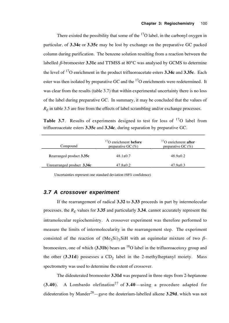
Chapter 3: Regiochemistry 100
There existed the possibility that some of the 17O label, in the carbonyl oxygen in
particular, of 3.34c or 3.35c may be lost by exchange on the preparative GC packed
column during purification. The benzene solution resulting from a reaction between the
labelled β-bromoester 3.31c and TTMSS at 80°C was analysed by GCMS to determine
the level of 17O enrichment in the product trifluoroacetate esters 3.34c and 3.35c. Each
ester was then isolated by preparative GC and the 17O enrichments were redetermined. It
was clear from the results (table 3.7) that within experimental uncertainty there is no loss
of the label during preparative GC. In summary, it may be concluded that the values of
RE in table 3.5 are free from the effects of label scrambling and/or exchange processes.
Table 3.7. Results of experiments designed to test for loss of 17O label from
trifluoroacetate esters 3.35c and 3.34c, during separation by preparative GC.
Compound
17O enrichment beforepreparative GC (%)
17O enrichment afterpreparative GC (%)
Rearranged product 3.35c 48.1±0.7 48.9±0.2
Unrearranged product 3.34c 47.8±0.2 47.9±0.3
Uncertainties represent one standard deviation (68% confidence)
3.7 A crossover experiment
If the rearrangement of radical 3.32 to 3.33 proceeds in part by intermolecular
processes, the RE values for 3.35 and particularly 3.34, cannot accurately represent the
intramolecular regiochemistry. A crossover experiment was therefore performed to
measure the limits of intermolecularity in the rearrangement step. The experiment
consisted of the reaction of (Me3Si)3SiH with an equimolar mixture of two β-
bromoesters, one of which (3.31b) bears an 18O label in the trifluoroacetoxy group and
the other (3.31d) possesses a CD2 label in the 2-methylheptanyl moiety. Mass
spectrometry was used to determine the extent of crossover.
The dideuterated bromoester 3.31d was prepared in three steps from 2-heptanone
(3.40). A Lombardo olefination27 of 3.40—using a procedure adapted for
dideuteration by Mander28—gave the deuterium-labelled alkene 3.29d, which was not
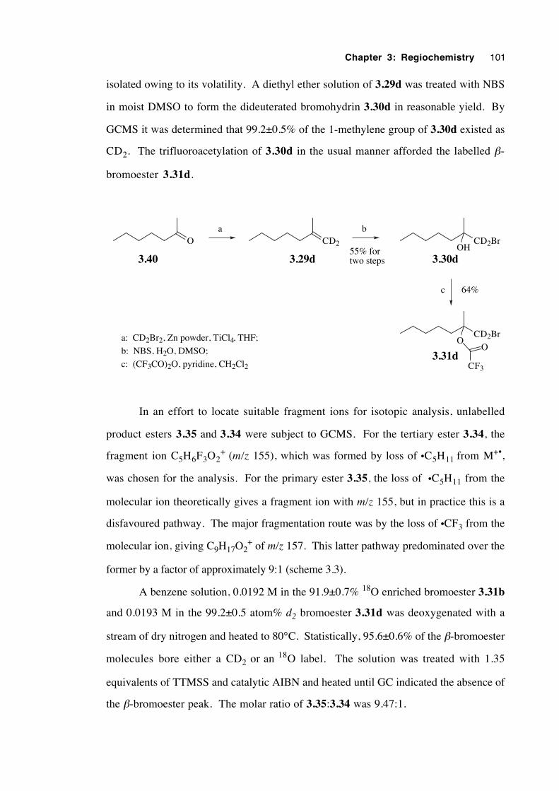
Chapter 3: Regiochemistry 101
isolated owing to its volatility. A diethyl ether solution of 3.29d was treated with NBS
in moist DMSO to form the dideuterated bromohydrin 3.30d in reasonable yield. By
GCMS it was determined that 99.2±0.5% of the 1-methylene group of 3.30d existed as
CD2. The trifluoroacetylation of 3.30d in the usual manner afforded the labelled β-
bromoester 3.31d.
O CD2 CD2BrOH
3.30d3.40 3.29d
CD2BrO
O
CF3
3.31d
a b
c
a: CD2Br2, Zn powder, TiCl4, THF;
b: NBS, H2O, DMSO;
c: (CF3CO)2O, pyridine, CH2Cl2
55% fortwo steps
64%
In an effort to locate suitable fragment ions for isotopic analysis, unlabelled
product esters 3.35 and 3.34 were subject to GCMS. For the tertiary ester 3.34, the
fragment ion C5H6F3O2+ (m/z 155), which was formed by loss of •C5H11 from M+•,
was chosen for the analysis. For the primary ester 3.35, the loss of •C5H11 from the
molecular ion theoretically gives a fragment ion with m/z 155, but in practice this is a
disfavoured pathway. The major fragmentation route was by the loss of •CF3 from the
molecular ion, giving C9H17O2+ of m/z 157. This latter pathway predominated over the
former by a factor of approximately 9:1 (scheme 3.3).
A benzene solution, 0.0192 M in the 91.9±0.7% 18O enriched bromoester 3.31b
and 0.0193 M in the 99.2±0.5 atom% d2 bromoester 3.31d was deoxygenated with a
stream of dry nitrogen and heated to 80°C. Statistically, 95.6±0.6% of the β-bromoester
molecules bore either a CD2 or an 18O label. The solution was treated with 1.35
equivalents of TTMSS and catalytic AIBN and heated until GC indicated the absence of
the β-bromoester peak. The molar ratio of 3.35:3.34 was 9.47:1.
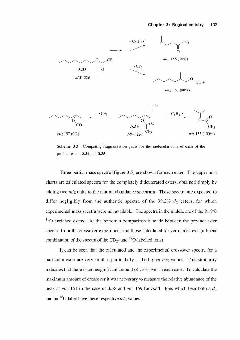
Chapter 3: Regiochemistry 102
O
O
CF3
MW 226
O
CF3
O
MW 226
+•
O
CF3
O+
– C5H11•
m/z 155 (100%)
– • CF3
OCO +
m/z 157 (0%)
O
O
CF3
m/z 155 (10%)
+
OCO +
m/z 157 (90%)
+•
– C5H11•
– • CF33.35
3.34
Scheme 3.3. Competing fragmentation paths for the molecular ions of each of the
product esters 3.34 and 3.35
Three partial mass spectra (figure 3.5) are shown for each ester. The uppermost
charts are calculated spectra for the completely dideuterated esters, obtained simply by
adding two m/z units to the natural abundance spectrum. These spectra are expected to
differ negligibly from the authentic spectra of the 99.2% d2 esters, for which
experimental mass spectra were not available. The spectra in the middle are of the 91.9%
18O enriched esters. At the bottom a comparison is made between the product ester
spectra from the crossover experiment and those calculated for zero crossover (a linear
combination of the spectra of the CD2- and 18O-labelled ions).
It can be seen that the calculated and the experimental crossover spectra for a
particular ester are very similar, particularly at the higher m/z values. This similarity
indicates that there is an insignificant amount of crossover in each case. To calculate the
maximum amount of crossover it was necessary to measure the relative abundance of the
peak at m/z 161 in the case of 3.35 and m/z 159 for 3.34. Ions which bear both a d2
and an 18O label have these respective m/z values.
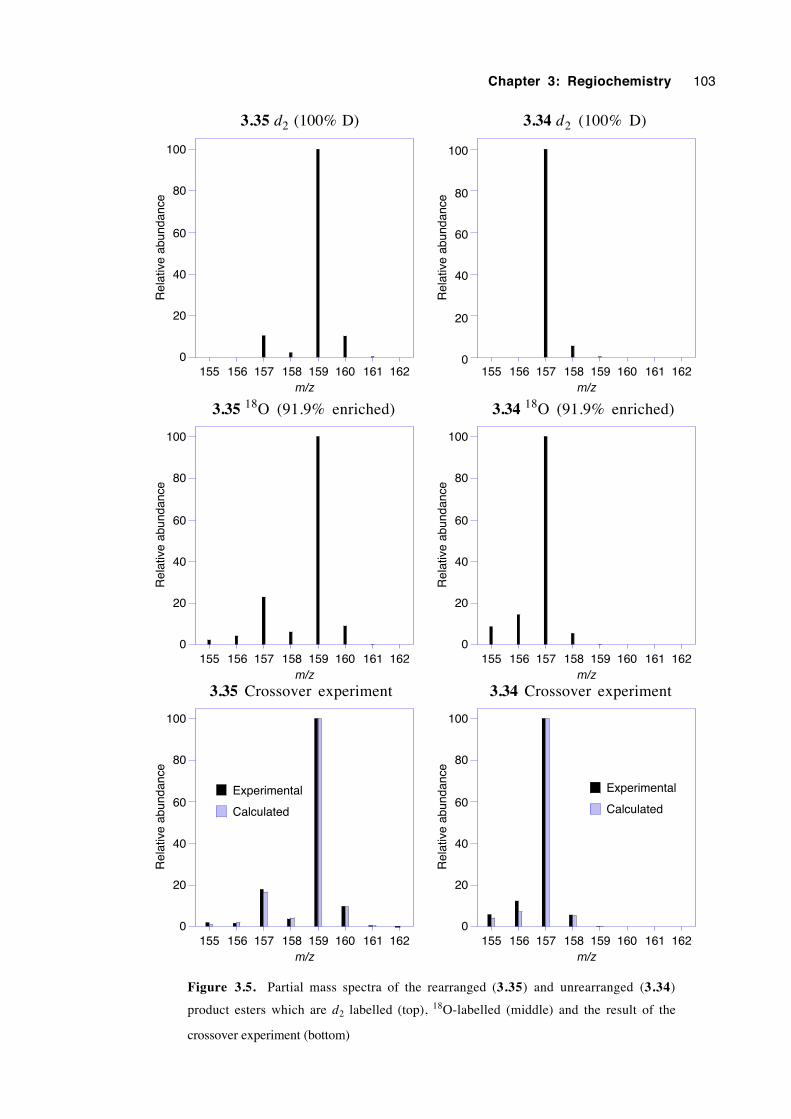
Chapter 3: Regiochemistry 103
3.35 d2 (100% D)
0
20
40
60
80
100
155 156 157 158 159 160 161 162
Rel
ativ
e ab
unda
nce
m/z
3.34 d2 (100% D)
0
20
40
60
80
100
155 156 157 158 159 160 161 162
Rel
ativ
e ab
unda
nce
m/z
3.35 18O (91.9% enriched)
0
20
40
60
80
100
155 156 157 158 159 160 161 162
Rel
ativ
e ab
unda
nce
m/z
3.34 18O (91.9% enriched)
0
20
40
60
80
100
155 156 157 158 159 160 161 162
Rel
ativ
e ab
unda
nce
m/z 3.35 Crossover experiment
0
20
40
60
80
100
155 156 157 158 159 160 161 162
Rel
ativ
e ab
unda
nce
m/z
Experimental
Calculated
3.34 Crossover experiment
0
20
40
60
80
100
155 156 157 158 159 160 161 162
Rel
ativ
e ab
unda
nce
m/z
Experimental
Calculated
Figure 3.5. Partial mass spectra of the rearranged (3.35) and unrearranged (3.34)
product esters which are d2 labelled (top), 18O-labelled (middle) and the result of the
crossover experiment (bottom)
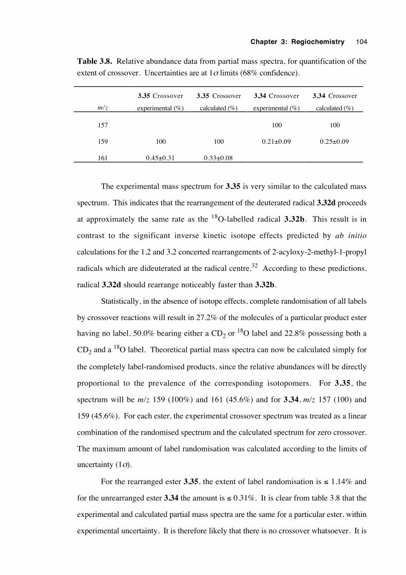
Chapter 3: Regiochemistry 104
Table 3.8. Relative abundance data from partial mass spectra, for quantification of the
extent of crossover. Uncertainties are at 1σ limits (68% confidence).
m/z
3.35 Crossover
experimental (%)
3.35 Crossover
calculated (%)
3.34 Crossover
experimental (%)
3.34 Crossover
calculated (%)
157 100 100
159 100 100 0.21±0.09 0.25±0.09
161 0.45±0.31 0.33±0.08
The experimental mass spectrum for 3.35 is very similar to the calculated mass
spectrum. This indicates that the rearrangement of the deuterated radical 3.32d proceeds
at approximately the same rate as the 18O-labelled radical 3.32b. This result is in
contrast to the significant inverse kinetic isotope effects predicted by ab initio
calculations for the 1,2 and 3,2 concerted rearrangements of 2-acyloxy-2-methyl-1-propyl
radicals which are dideuterated at the radical centre.32 According to these predictions,
radical 3.32d should rearrange noticeably faster than 3.32b.
Statistically, in the absence of isotope effects, complete randomisation of all labels
by crossover reactions will result in 27.2% of the molecules of a particular product ester
having no label, 50.0% bearing either a CD2 or 18O label and 22.8% possessing both a
CD2 and a 18O label. Theoretical partial mass spectra can now be calculated simply for
the completely label-randomised products, since the relative abundances will be directly
proportional to the prevalence of the corresponding isotopomers. For 3.35, the
spectrum will be m/z 159 (100%) and 161 (45.6%) and for 3.34, m/z 157 (100) and
159 (45.6%). For each ester, the experimental crossover spectrum was treated as a linear
combination of the randomised spectrum and the calculated spectrum for zero crossover.
The maximum amount of label randomisation was calculated according to the limits of
uncertainty (1σ).
For the rearranged ester 3.35, the extent of label randomisation is ≤ 1.14% and
for the unrearranged ester 3.34 the amount is ≤ 0.31%. It is clear from table 3.8 that the
experimental and calculated partial mass spectra are the same for a particular ester, within
experimental uncertainty. It is therefore likely that there is no crossover whatsoever. It is
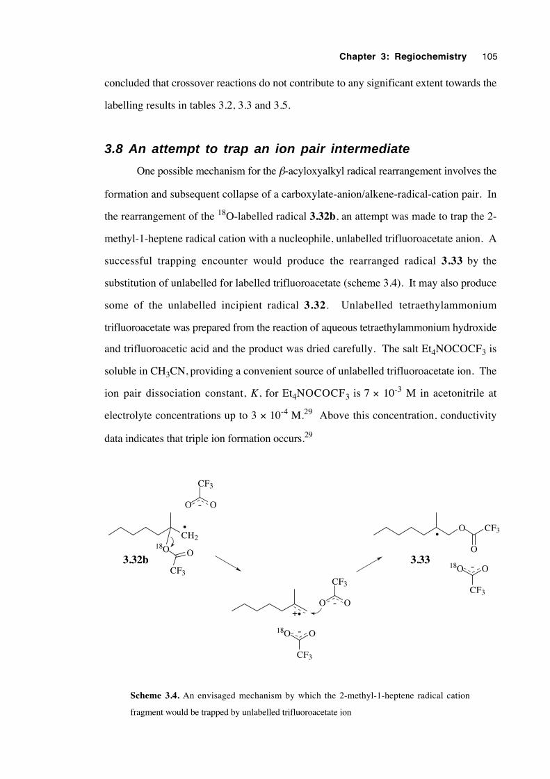
Chapter 3: Regiochemistry 105
concluded that crossover reactions do not contribute to any significant extent towards the
labelling results in tables 3.2, 3.3 and 3.5.
3.8 An attempt to trap an ion pair intermediate
One possible mechanism for the β-acyloxyalkyl radical rearrangement involves the
formation and subsequent collapse of a carboxylate-anion/alkene-radical-cation pair. In
the rearrangement of the 18O-labelled radical 3.32b, an attempt was made to trap the 2-
methyl-1-heptene radical cation with a nucleophile, unlabelled trifluoroacetate anion. A
successful trapping encounter would produce the rearranged radical 3.33 by the
substitution of unlabelled for labelled trifluoroacetate (scheme 3.4). It may also produce
some of the unlabelled incipient radical 3.32. Unlabelled tetraethylammonium
trifluoroacetate was prepared from the reaction of aqueous tetraethylammonium hydroxide
and trifluoroacetic acid and the product was dried carefully. The salt Et4NOCOCF3 is
soluble in CH3CN, providing a convenient source of unlabelled trifluoroacetate ion. The
ion pair dissociation constant, K, for Et4NOCOCF3 is 7 × 10⁻³ M in acetonitrile at
electrolyte concentrations up to 3 × 10⁻⁴ M.29 Above this concentration, conductivity
data indicates that triple ion formation occurs.29
CH218O
CF3
O
O CF3
O3.32b 3.33
••
CF3
18O O-
CF3
OO -
CF3
OO -
•+
CF3
18O O-
Scheme 3.4. An envisaged mechanism by which the 2-methyl-1-heptene radical cation
fragment would be trapped by unlabelled trifluoroacetate ion
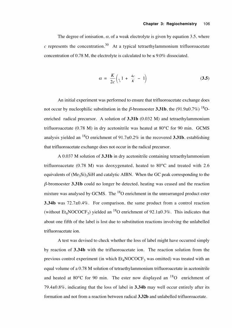
Chapter 3: Regiochemistry 106
The degree of ionisation, α, of a weak electrolyte is given by equation 3.5, where
c represents the concentration.30 At a typical tetraethylammonium trifluoroacetate
concentration of 0.78 M, the electrolyte is calculated to be ≤ 9.0% dissociated.
α = K
c21 14 + −
c
K (3.5)
An initial experiment was performed to ensure that trifluoroacetate exchange does
not occur by nucleophilic substitution in the β-bromoester 3.31b, the (91.9±0.7%) 18O-
enriched radical precursor. A solution of 3.31b (0.032 M) and tetraethylammonium
trifluoroacetate (0.78 M) in dry acetonitrile was heated at 80°C for 90 min. GCMS
analysis yielded an 18O enrichment of 91.7±0.2% in the recovered 3.31b, establishing
that trifluoroacetate exchange does not occur in the radical precursor.
A 0.037 M solution of 3.31b in dry acetonitrile containing tetraethylammonium
trifluoroacetate (0.78 M) was deoxygenated, heated to 80°C and treated with 2.6
equivalents of (Me3Si)3SiH and catalytic AIBN. When the GC peak corresponding to the
β-bromoester 3.31b could no longer be detected, heating was ceased and the reaction
mixture was analysed by GCMS. The 18O enrichment in the unrearranged product ester
3.34b was 72.7±0.4%. For comparison, the same product from a control reaction
(without Et4NOCOCF3) yielded an 18O enrichment of 92.1±0.3%. This indicates that
about one fifth of the label is lost due to substitution reactions involving the unlabelled
trifluoroacetate ion.
A test was devised to check whether the loss of label might have occurred simply
by reaction of 3.34b with the trifluoroacetate ion. The reaction solution from the
previous control experiment (in which Et4NOCOCF3 was omitted) was treated with an
equal volume of a 0.78 M solution of tetraethylammonium trifluoroacetate in acetonitrile
and heated at 80°C for 90 min. The ester now displayed an 18O enrichment of
79.4±0.8%, indicating that the loss of label in 3.34b may well occur entirely after its
formation and not from a reaction between radical 3.32b and unlabelled trifluoroacetate.
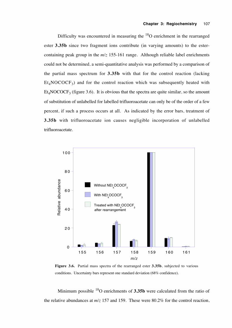
Chapter 3: Regiochemistry 107
Difficulty was encountered in measuring the 18O enrichment in the rearranged
ester 3.35b since two fragment ions contribute (in varying amounts) to the ester-
containing peak group in the m/z 155-161 range. Although reliable label enrichments
could not be determined, a semi-quantitative analysis was performed by a comparison of
the partial mass spectrum for 3.35b with that for the control reaction (lacking
Et4NOCOCF3) and for the control reaction which was subsequently heated with
Et4NOCOCF3 (figure 3.6). It is obvious that the spectra are quite similar, so the amount
of substitution of unlabelled for labelled trifluoroacetate can only be of the order of a few
percent, if such a process occurs at all. As indicated by the error bars, treatment of
3.35b with trifluoroacetate ion causes negligible incorporation of unlabelled
trifluoroacetate.
0
2 0
4 0
6 0
8 0
1 0 0
1 5 5 1 5 6 1 5 7 1 5 8 1 5 9 1 6 0 1 6 1
Without NEt4OCOCF
3
With NEt4OCOCF
3
Treated with NEt4OCOCF
3
Rel
ativ
e ab
unda
nce
m/z
after rearrangement
Figure 3.6. Partial mass spectra of the rearranged ester 3.35b, subjected to various
conditions. Uncertainty bars represent one standard deviation (68% confidence).
Minimum possible 18O enrichments of 3.35b were calculated from the ratio of
the relative abundances at m/z 157 and 159. These were 80.2% for the control reaction,
Chapter 3: Regiochemistry 108
80.5% for the control reaction subsequently heated with Et4NOCOCF3 and 78.1% for the
rearrangement reaction performed in the presence of Et4NOCOCF3. According to this
data, 97.4% of the amount of label present in 3.35b from the control experiment
remained in 3.35b when it was formed in the presence of unlabelled trifluoroacetate ion.
Thus, there appeared to be very little exchange of trifluoroacetate, if any at all, during the
rearrangement step. It can be concluded that if a radical ion pair is an actual intermediate
it must exist as a contact ion pair (CIP) and not as a solvent-separated (SSIP), a solvent-
shared ion pair, or a dissociated ion pair.
It is envisaged that more accurate results might be obtained if the experiment was
repeated by conducting the TTMSS/AIBN reduction of the unlabelled β-bromoester 3.31
in the presence of Et4NOCO14CF3. The product esters could be separated by preparative
GC, then analysed for radioactivity by beta particle detection. Radioactivity in the
product esters would indicate exchange.
3.9 Discussion of results with regard to mechanism
It is clear that the rearrangement of the 2-methyl-2-trifluoroacetoxy-1-heptyl
radical (3.32) to the 2-methyl-1-trifluoroacetoxy-2-heptyl radical (3.33) proceeds with
neither complete positional retention nor complete transposition of the ester oxygens, but
rather with considerable scrambling of the oxygen label. A formal 3,2 shift predominated
over a formal 1,2 shift under all the experimental conditions of this study. An increase in
the proportion of 1,2 shift in polar solvents is in agreement with theoretical predictions
for a mechanism consisting of concerted 3- and 5-membered transition structures32 and
with labelling results from the rearrangement of the 2-butanoyloxy-2-phenyl-1-propyl
radical.5 In addition, the unrearranged product ester (3.34) displays a usually small but
significant amount of scrambling of the label. Therefore, the rearrangement of 3.32
cannot occur by a single, concerted mechanism. The results dictate that the mechanism
consists of the cooperation of reversible, polarized 1,2 (i) and 2,3 (ii) shifts; or involves
the intermediacy of an alkene radical cation/trifluoroacetate anion pair (iii); or is a
combination of these options.
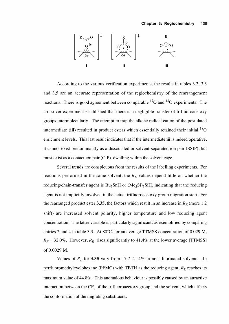
Chapter 3: Regiochemistry 109
O O
R
•O
R O
•O O
R
+_
iiiiii
•
δ+
δ-
δ+
δ-
‡ ‡
According to the various verification experiments, the results in tables 3.2, 3.3
and 3.5 are an accurate representation of the regiochemistry of the rearrangement
reactions. There is good agreement between comparable 17O and 18O experiments. The
crossover experiment established that there is a negligible transfer of trifluoroacetoxy
groups intermolecularly. The attempt to trap the alkene radical cation of the postulated
intermediate (iii) resulted in product esters which essentially retained their initial 18O
enrichment levels. This last result indicates that if the intermediate iii is indeed operative,
it cannot exist predominantly as a dissociated or solvent-separated ion pair (SSIP), but
must exist as a contact ion pair (CIP), dwelling within the solvent cage.
Several trends are conspicuous from the results of the labelling experiments. For
reactions performed in the same solvent, the RE values depend little on whether the
reducing/chain-transfer agent is Bu3SnH or (Me3Si)3SiH, indicating that the reducing
agent is not implicitly involved in the actual trifluoroacetoxy group migration step. For
the rearranged product ester 3.35, the factors which result in an increase in RE (more 1,2
shift) are increased solvent polarity, higher temperature and low reducing agent
concentration. The latter variable is particularly significant, as exemplified by comparing
entries 2 and 4 in table 3.3. At 80˚C, for an average TTMSS concentration of 0.029 M,
RE = 32.0%. However, RE rises significantly to 41.4% at the lower average [TTMSS]
of 0.0029 M.
Values of RE for 3.35 vary from 17.7–41.4% in non-fluorinated solvents. In
perfluoromethylcyclohexane (PFMC) with TBTH as the reducing agent, RE reaches its
maximum value of 44.8%. This anomalous behaviour is possibly caused by an attractive
interaction between the CF3 of the trifluoroacetoxy group and the solvent, which affects
the conformation of the migrating substituent.
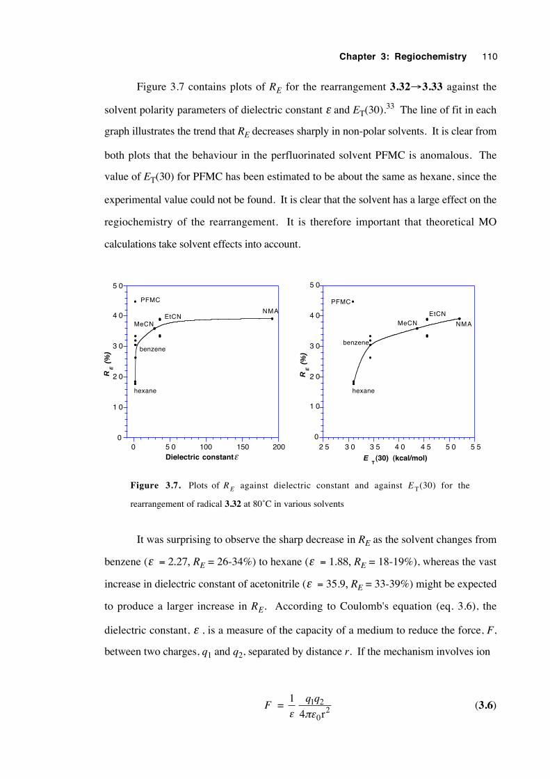
Chapter 3: Regiochemistry 110
Figure 3.7 contains plots of RE for the rearrangement 3.32→3.33 against the
solvent polarity parameters of dielectric constant ε and ET(30).33 The line of fit in each
graph illustrates the trend that RE decreases sharply in non-polar solvents. It is clear from
both plots that the behaviour in the perfluorinated solvent PFMC is anomalous. The
value of ET(30) for PFMC has been estimated to be about the same as hexane, since the
experimental value could not be found. It is clear that the solvent has a large effect on the
regiochemistry of the rearrangement. It is therefore important that theoretical MO
calculations take solvent effects into account.
0
1 0
2 0
3 0
4 0
5 0
0 5 0 100 150 200
RE (
%)
Dielectric constantε
PFMC
NMA
hexane
benzene
EtCNMeCN
0
1 0
2 0
3 0
4 0
5 0
2 5 3 0 3 5 4 0 4 5 5 0 5 5
RE (
%)
T(30) (kcal/mol)E
PFMC
hexane
benzene
NMA
EtCNMeCN
Figure 3.7. Plots of R E against dielectric constant and against E T(30) for the
rearrangement of radical 3.32 at 80˚C in various solvents
It was surprising to observe the sharp decrease in RE as the solvent changes from
benzene (ε = 2.27, RE = 26-34%) to hexane (ε = 1.88, RE = 18-19%), whereas the vast
increase in dielectric constant of acetonitrile (ε = 35.9, RE = 33-39%) might be expected
to produce a larger increase in RE. According to Coulomb's equation (eq. 3.6), the
dielectric constant, ε , is a measure of the capacity of a medium to reduce the force, F,
between two charges, q1 and q2, separated by distance r. If the mechanism involves ion
F = 1ε
q q1 2
024 rπε
(3.6)
Chapter 3: Regiochemistry 111
pairs, benzene probably promotes an increase in distance r between the oppositely-
charged fragments relative to hexane since the difference in dielectric constants is
considered too small by itself to cause such a large change in RE. Alternatively, the π
-electron cloud of the benzene ring may have enough nucleophilic character to interact
with the electron-deficient carbonyl carbon of the trifluoroacetoxy group, affecting not
only the regiochemistry but the rate constant of the rearrangement. Such interactions have
been used previously to explain the large difference in the rate of solvolysis of 1-acetoxy-
2-bromopropane in octane and in benzene.31
If the mechanism for the rearrangement of radical 3.32 involves the cooperation
of concerted 1,2 (i) and 3,2 (ii) shifts, then the increase in RE in polar solvent implies
that the 3-membered transition structure i is more polarized than the 5-membered structure
ii. Theoretical calculations of carboxylate group charge for the ground state (3.32-like)
and for structures i and ii (R = CF3) indicate that the charge separation in structures i and
ii is essentially the same,32 conflicting with the mechanistic hypotheses of some
experimental chemists.1,5
The regiochemistry of the rearrangement of the 2-methyl-2-trifluoroacetoxy-1-
heptyl radical (3.32) resembles that of the 2-phenyl-2-trifluoroacetoxyethyl radical
(3.13, RE = 19%), and also that of the 2-aryl-2-butanoyloxypropyl radicals 3.7 (25% in
methanol) and 3.8 (39%). Like 3.7, radical 3.32 experiences greater label scrambling
as solvent polarity increases.
From the data in table 3.3 it can be seen that RE increases at higher temperatures.
This indicates that a greater randomisation of the label is taking place in the radical
cation/carboxylate anion pair intermediate (iii) at higher temperatures, or alternatively that
the activation energy for a concerted 1,2 shift (i) is greater than that for a 3,2 shift (ii). In
latter alternative, it is possible to estimate the difference in activation energy, ∆Ea, and
Arrhenius frequency factor, A, between the two transition states. The ratio of rate
constants from the 3,2 and 1,2 rearrangements can be calculated simply from the
regiochemical results (table 3.9). The Arrhenuis expressions (equations 3.7 and 3.8) for
the two competing rate constants, k3,2 and k1,2 are:
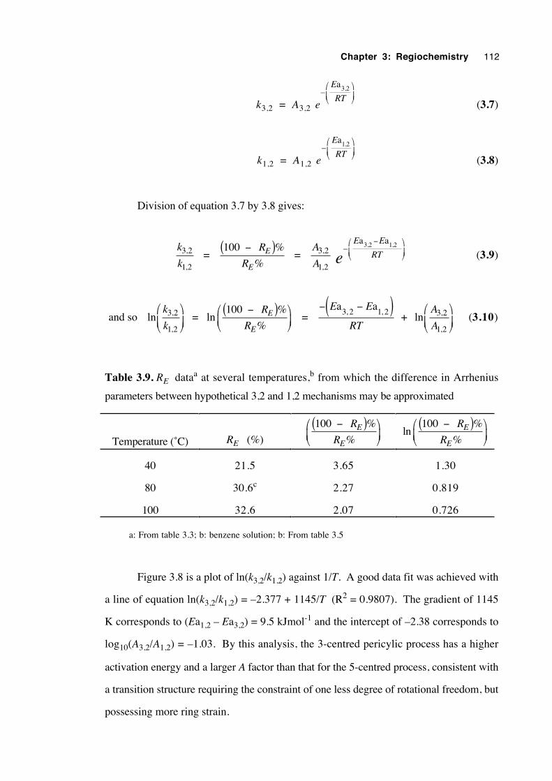
Chapter 3: Regiochemistry 112
k3,2 = A3,2 e
E
RT– ,a3 2
(3.7)
k1,2 = A1,2 e
E
RT– ,a1 2
(3.8)
Division of equation 3.7 by 3.8 gives:
k
k3 2
1 2
,
, =
100 %−( )R
RE
E% =
A
A3 2
1 2
,
,
– , ,E E
RTea a3 2 1 2−
(3.9)
and so lnk
k3 2
1 2
,
,
= ln
100 %−( )
R
RE
E% =
− −( )E E
RT
a a3 2 1 2, , + ln
A
A3 2
1 2
,
,
(3.10)
Table 3.9. RE dataa at several temperatures,b from which the difference in Arrhenius
parameters between hypothetical 3,2 and 1,2 mechanisms may be approximated
Temperature (˚C) RE (%)
100 %−( )
R
RE
E%ln
100 %−( )
R
RE
E%
40 21.5 3.65 1.30
80 30.6c 2.27 0.819
100 32.6 2.07 0.726
a: From table 3.3; b: benzene solution; b: From table 3.5
Figure 3.8 is a plot of ln(k3,2/k1,2) against 1/T. A good data fit was achieved with
a line of equation ln(k3,2/k1,2) = –2.377 + 1145/T (R2 = 0.9807). The gradient of 1145
K corresponds to (Ea1,2 – Ea3,2) = 9.5 kJmol-1 and the intercept of –2.38 corresponds to
log10(A3,2/A1,2) = –1.03. By this analysis, the 3-centred pericylic process has a higher
activation energy and a larger A factor than that for the 5-centred process, consistent with
a transition structure requiring the constraint of one less degree of rotational freedom, but
possessing more ring strain.
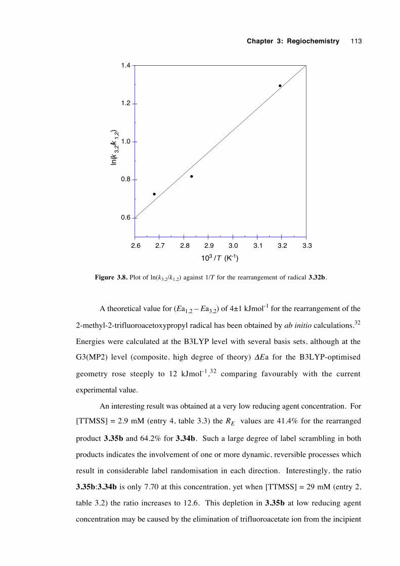
Chapter 3: Regiochemistry 113
0.6
0.8
1.0
1.2
1.4
2.6 2.7 2.8 2.9 3.0 3.1 3.2 3.3
ln(
3,2
/ 1,
2)
103 / (K-1)
kk
T
Figure 3.8. Plot of ln(k3,2/k1,2) against 1/T for the rearrangement of radical 3.32b.
A theoretical value for (Ea1,2 – Ea3,2) of 4±1 kJmol-1 for the rearrangement of the
2-methyl-2-trifluoroacetoxypropyl radical has been obtained by ab initio calculations.32
Energies were calculated at the B3LYP level with several basis sets, although at the
G3(MP2) level (composite, high degree of theory) ∆Ea for the B3LYP-optimised
geometry rose steeply to 12 kJmol-1,32 comparing favourably with the current
experimental value.
An interesting result was obtained at a very low reducing agent concentration. For
[TTMSS] = 2.9 mM (entry 4, table 3.3) the RE values are 41.4% for the rearranged
product 3.35b and 64.2% for 3.34b. Such a large degree of label scrambling in both
products indicates the involvement of one or more dynamic, reversible processes which
result in considerable label randomisation in each direction. Interestingly, the ratio
3.35b:3.34b is only 7.70 at this concentration, yet when [TTMSS] = 29 mM (entry 2,
table 3.2) the ratio increases to 12.6. This depletion in 3.35b at low reducing agent
concentration may be caused by the elimination of trifluoroacetate ion from the incipient
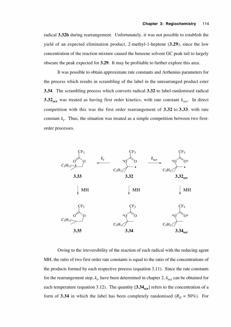
Chapter 3: Regiochemistry 114
radical 3.32b during rearrangement. Unfortunately, it was not possible to establish the
yield of an expected elimination product, 2-methyl-1-heptene (3.29), since the low
concentration of the reaction mixture caused the benzene solvent GC peak tail to largely
obscure the peak expected for 3.29. It may be profitable to further explore this area.
It was possible to obtain approximate rate constants and Arrhenius parameters for
the process which results in scrambling of the label in the unrearranged product ester
3.34. The scrambling process which converts radical 3.32 to label-randomised radical
3.32scr was treated as having first order kinetics, with rate constant kscr. In direct
competition with this was the first order rearrangement of 3.32 to 3.33, with rate
constant kr. Thus, the situation was treated as a simple competition between two first-
order processes.
C5H11
O*O
CF3
•C5H11
O**O
CF3
•C5H11
OO
CF3
•
kscrkr
3.33 3.32 3.32scr
MH MH MH
3.35 3.34 3.34scr
C5H11
O*O
CF3
C5H11
O**O
CF3
C5H11
OO
CF3
Owing to the irreversibility of the reaction of each radical with the reducing agent
MH, the ratio of two first order rate constants is equal to the ratio of the concentrations of
the products formed by each respective process (equation 3.11). Since the rate constants
for the rearrangement step, kr, have been determined in chapter 2, kscr can be obtained for
each temperature (equation 3.12). The quantity [3.34scr] refers to the concentration of a
form of 3.34 in which the label has been completely randomised (RE = 50%). For
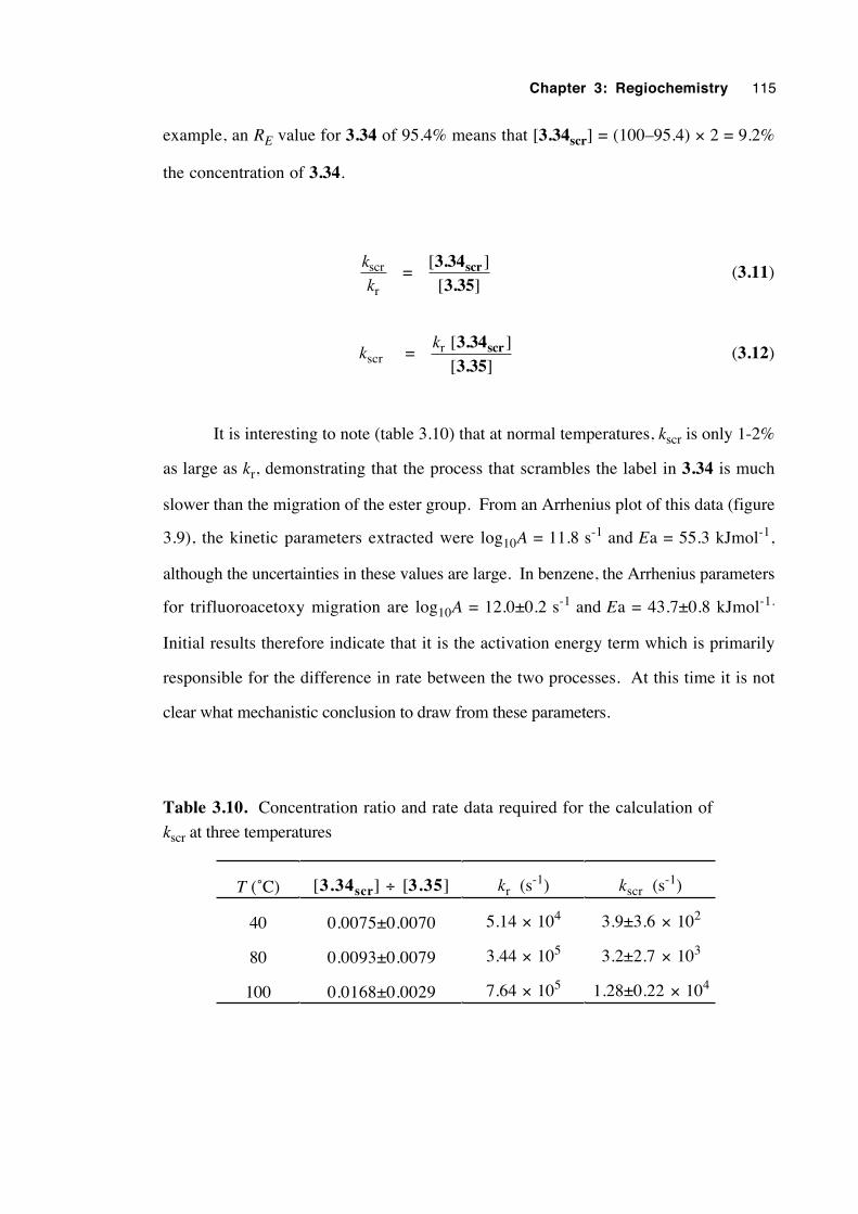
Chapter 3: Regiochemistry 115
example, an RE value for 3.34 of 95.4% means that [3.34scr] = (100–95.4) × 2 = 9.2%
the concentration of 3.34.
k
kscr
r =
[ ][ ]3.34
3.35scr (3.11)
kscr = kr [ ]
[ ]3.343.35
scr (3.12)
It is interesting to note (table 3.10) that at normal temperatures, kscr is only 1-2%
as large as kr, demonstrating that the process that scrambles the label in 3.34 is much
slower than the migration of the ester group. From an Arrhenius plot of this data (figure
3.9), the kinetic parameters extracted were log10A = 11.8 s-1 and Ea = 55.3 kJmol-1,
although the uncertainties in these values are large. In benzene, the Arrhenius parameters
for trifluoroacetoxy migration are log10A = 12.0±0.2 s-1 and Ea = 43.7±0.8 kJmol-1.
Initial results therefore indicate that it is the activation energy term which is primarily
responsible for the difference in rate between the two processes. At this time it is not
clear what mechanistic conclusion to draw from these parameters.
Table 3.10. Concentration ratio and rate data required for the calculation of
kscr at three temperatures
T (˚C) [3.34scr] ÷ [3.35] kr (s-1) kscr (s
-1)
40 0.0075±0.0070 5.14 × 10⁴ 3.9±3.6 × 10²
80 0.0093±0.0079 3.44 × 10⁵ 3.2±2.7 × 10³
100 0.0168±0.0029 7.64 × 10⁵ 1.28±0.22 × 10⁴
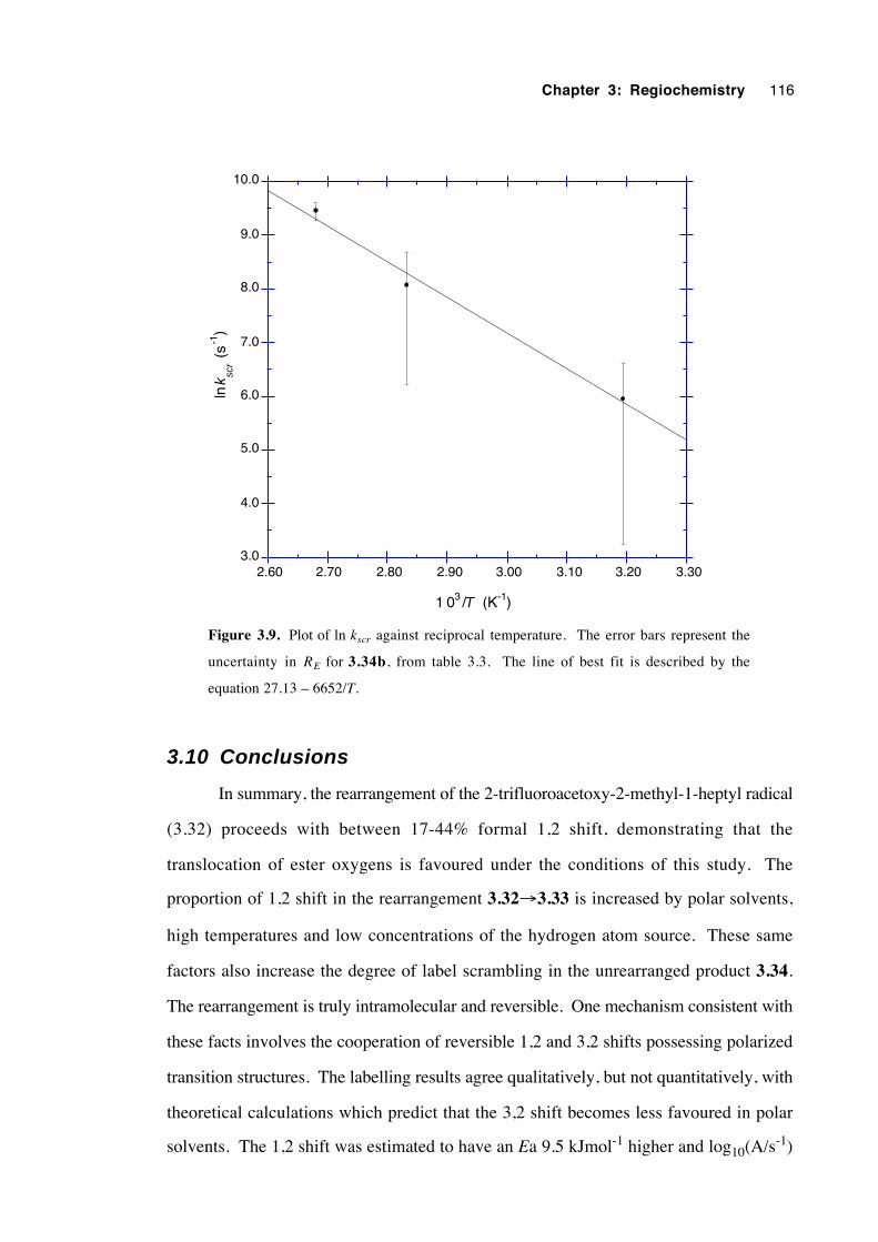
Chapter 3: Regiochemistry 116
3.0
4.0
5.0
6.0
7.0
8.0
9.0
10.0
2.60 2.70 2.80 2.90 3.00 3.10 3.20 3.30
ln
(
s-1
)
1 03 / (K-1)T
k scr
Figure 3.9. Plot of ln kscr against reciprocal temperature. The error bars represent the
uncertainty in RE for 3.34b, from table 3.3. The line of best fit is described by the
equation 27.13 – 6652/T.
3.10 Conclusions
In summary, the rearrangement of the 2-trifluoroacetoxy-2-methyl-1-heptyl radical
(3.32) proceeds with between 17-44% formal 1,2 shift, demonstrating that the
translocation of ester oxygens is favoured under the conditions of this study. The
proportion of 1,2 shift in the rearrangement 3.32→3.33 is increased by polar solvents,
high temperatures and low concentrations of the hydrogen atom source. These same
factors also increase the degree of label scrambling in the unrearranged product 3.34.
The rearrangement is truly intramolecular and reversible. One mechanism consistent with
these facts involves the cooperation of reversible 1,2 and 3,2 shifts possessing polarized
transition structures. The labelling results agree qualitatively, but not quantitatively, with
theoretical calculations which predict that the 3,2 shift becomes less favoured in polar
solvents. The 1,2 shift was estimated to have an Ea 9.5 kJmol-1 higher and log10(A/s-1)
Chapter 3: Regiochemistry 117
1.01 units larger for the 3,2 shift. This is consistent with the 3-membered TS possessing
greater ring strain but more degrees of freedom than its 5-membered counterpart. The
difference in energy between the two transition structures is consonant with one high-
level theoretical calculation.
A second mechanism which is compatible with the results is a reversible process
involving an alkene-radical-cation/trifluoroacetate-anion contact pair. This ion pair
collapses to form either the rearranged radical 3.33—in which the label is preferentially
distributed into the carbonyl oxygen—or radical 3.32, in which the label has been partly
scrambled. Such an ion pair must exist primarily inside the solvent cage and not as a
solvent-separated, solvent-shared or dissociated pair. At this stage it is not possible to
confidently exclude either of these possible mechanisms using the available evidence.
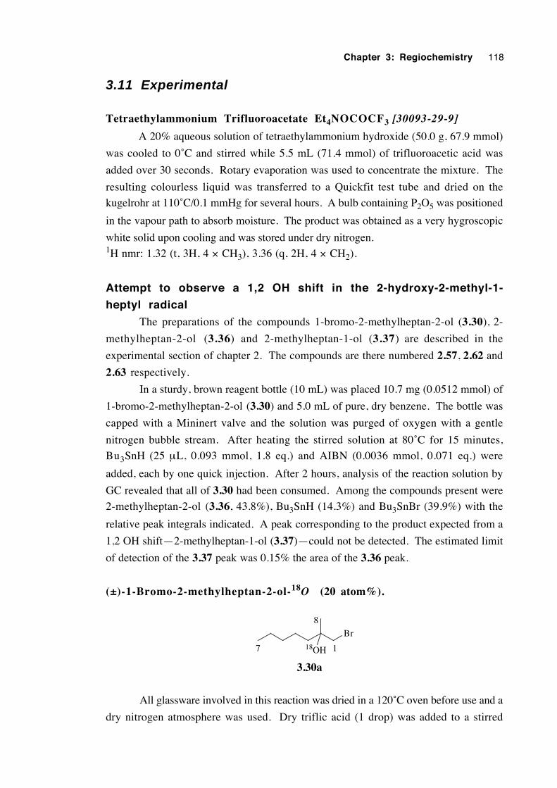
Chapter 3: Regiochemistry 118
3.11 Experimental
Tetraethylammonium Trifluoroacetate Et4NOCOCF3 [30093-29-9]
A 20% aqueous solution of tetraethylammonium hydroxide (50.0 g, 67.9 mmol)
was cooled to 0˚C and stirred while 5.5 mL (71.4 mmol) of trifluoroacetic acid was
added over 30 seconds. Rotary evaporation was used to concentrate the mixture. The
resulting colourless liquid was transferred to a Quickfit test tube and dried on the
kugelrohr at 110˚C/0.1 mmHg for several hours. A bulb containing P2O5 was positioned
in the vapour path to absorb moisture. The product was obtained as a very hygroscopic
white solid upon cooling and was stored under dry nitrogen.1H nmr: 1.32 (t, 3H, 4 × CH3), 3.36 (q, 2H, 4 × CH2).
Attempt to observe a 1,2 OH shift in the 2-hydroxy-2-methyl-1-
heptyl radical
The preparations of the compounds 1-bromo-2-methylheptan-2-ol (3.30), 2-
methylheptan-2-ol (3.36) and 2-methylheptan-1-ol (3.37) are described in the
experimental section of chapter 2. The compounds are there numbered 2.57, 2.62 and
2.63 respectively.
In a sturdy, brown reagent bottle (10 mL) was placed 10.7 mg (0.0512 mmol) of
1-bromo-2-methylheptan-2-ol (3.30) and 5.0 mL of pure, dry benzene. The bottle was
capped with a Mininert valve and the solution was purged of oxygen with a gentle
nitrogen bubble stream. After heating the stirred solution at 80˚C for 15 minutes,
Bu3SnH (25 µL, 0.093 mmol, 1.8 eq.) and AIBN (0.0036 mmol, 0.071 eq.) were
added, each by one quick injection. After 2 hours, analysis of the reaction solution by
GC revealed that all of 3.30 had been consumed. Among the compounds present were
2-methylheptan-2-ol (3.36, 43.8%), Bu3SnH (14.3%) and Bu3SnBr (39.9%) with the
relative peak integrals indicated. A peak corresponding to the product expected from a
1,2 OH shift—2-methylheptan-1-ol (3.37)—could not be detected. The estimated limit
of detection of the 3.37 peak was 0.15% the area of the 3.36 peak.
(±)-1-Bromo-2-methylheptan-2-ol-18O (20 atom%).
Br
18OH
3.30a
17
8
All glassware involved in this reaction was dried in a 120˚C oven before use and a
dry nitrogen atmosphere was used. Dry triflic acid (1 drop) was added to a stirred
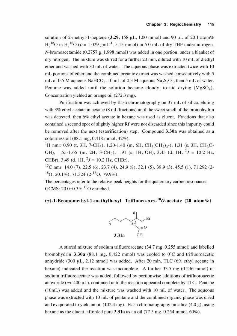
Chapter 3: Regiochemistry 119
solution of 2-methyl-1-heptene (3.29, 158 µL, 1.00 mmol) and 90 µL of 20.1 atom%
H218O in H2
16O (ρ = 1.029 gmL-1, 5.15 mmol) in 5.0 mL of dry THF under nitrogen.
N-bromoacetamide (0.2757 g, 1.998 mmol) was added in one portion, under a blanket of
dry nitrogen. The mixture was stirred for a further 20 min, diluted with 10 mL of diethyl
ether and washed with 30 mL of water. The aqueous phase was extracted twice with 10
mL portions of ether and the combined organic extract was washed consecutively with 5
mL of 0.5 M aqueous NaHCO3, 10 mL of 0.3 M aqueous Na2S2O3, then 5 mL of water.
Pentane was added until the solution became cloudy, to aid drying (MgSO4).
Concentration yielded an orange oil (272.3 mg).
Purification was achieved by flash chromatography on 37 mL of silica, eluting
with 3% ethyl acetate in hexane (8 mL fractions) until the sweet smell of the bromohydrin
was detected, then 6% ethyl acetate in hexane was used as eluent. Fractions that also
contained a second spot of slightly higher Rf were not discarded since this impurity could
be removed after the next (esterification) step. Compound 3.30a was obtained as a
colourless oil (88.1 mg, 0.418 mmol, 42%).1H nmr: 0.90 (t, 3H, 7-CH3), 1.20-1.40 (m, 6H, CH3(CH2)3-), 1.31 (s, 3H, CH3C-
OH), 1.55-1.65 (m, 2H, 3-CH2), 1.91 (s, 1H, OH), 3.45 (d, 1H, 2J = 10.2 Hz,
CHBr), 3.49 (d, 1H, 2J = 10.2 Hz, CHBr).13C nmr: 14.0 (7), 22.5 (6), 23.7 (4), 24.9 (8), 32.1 (5), 39.9 (3), 45.5 (1), 71.292 (2-18O, 20.1%), 71.324 (2-16O, 79.9%).
The percentages refer to the relative peak heights for the quaternary carbon resonances.
GCMS: 20.0±0.3% 18O enriched.
(±)-1-Bromomethyl-1-methylhexyl Trifluoro-oxy-18O-acetate (20 atom%)
Br18O
CF3
O
3.31a
17
8
A stirred mixture of sodium trifluoroacetate (34.7 mg, 0.255 mmol) and labelled
bromohydrin 3.30a (88.1 mg, 0.422 mmol) was cooled to 0˚C and trifluoroacetic
anhydride (300 µL, 2.12 mmol) was added. After 20 min, TLC (6% ethyl acetate in
hexane) indicated the reaction was incomplete. A further 33.5 mg (0.246 mmol) of
sodium trifluoroacetate was added, followed by portionwise additions of trifluoroacetic
anhydride (ca. 400 µL), continued until the reaction appeared complete by TLC. Pentane
(10mL) was added and the mixture was washed with 10 mL of water. The aqueous
phase was extracted with 10 mL of pentane and the combined organic phase was dried
and evaporated to yield an oil (102.4 mg). Flash chromatography on silica (4.0 g), using
hexane as the eluent, afforded pure 3.31a as an oil (77.5 mg, 0.254 mmol, 60%).
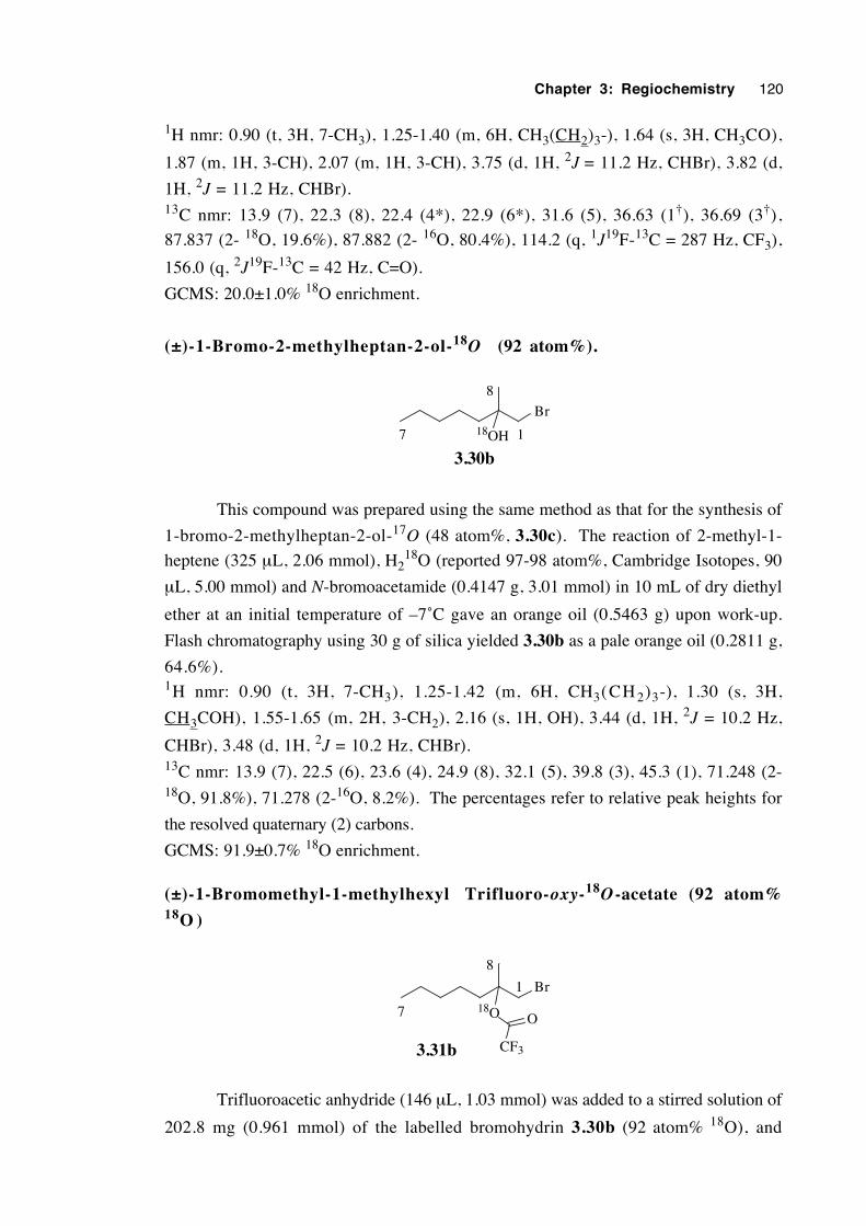
Chapter 3: Regiochemistry 120
1H nmr: 0.90 (t, 3H, 7-CH3), 1.25-1.40 (m, 6H, CH3(CH2)3-), 1.64 (s, 3H, CH3CO),
1.87 (m, 1H, 3-CH), 2.07 (m, 1H, 3-CH), 3.75 (d, 1H, 2J = 11.2 Hz, CHBr), 3.82 (d,
1H, 2J = 11.2 Hz, CHBr).13C nmr: 13.9 (7), 22.3 (8), 22.4 (4*), 22.9 (6*), 31.6 (5), 36.63 (1†), 36.69 (3†),
87.837 (2- 18O, 19.6%), 87.882 (2- 16O, 80.4%), 114.2 (q, 1J19F-13C = 287 Hz, CF3),
156.0 (q, 2J19F-13C = 42 Hz, C=O).
GCMS: 20.0±1.0% 18O enrichment.
(±)-1-Bromo-2-methylheptan-2-ol-18O (92 atom%).
Br18OH
3.30b17
8
This compound was prepared using the same method as that for the synthesis of
1-bromo-2-methylheptan-2-ol-17O (48 atom%, 3.30c). The reaction of 2-methyl-1-
heptene (325 µL, 2.06 mmol), H218O (reported 97-98 atom%, Cambridge Isotopes, 90
µL, 5.00 mmol) and N-bromoacetamide (0.4147 g, 3.01 mmol) in 10 mL of dry diethyl
ether at an initial temperature of –7˚C gave an orange oil (0.5463 g) upon work-up.
Flash chromatography using 30 g of silica yielded 3.30b as a pale orange oil (0.2811 g,
64.6%).1H nmr: 0.90 (t, 3H, 7-CH3), 1.25-1.42 (m, 6H, CH3(CH 2)3-), 1.30 (s, 3H,
CH3COH), 1.55-1.65 (m, 2H, 3-CH2), 2.16 (s, 1H, OH), 3.44 (d, 1H, 2J = 10.2 Hz,
CHBr), 3.48 (d, 1H, 2J = 10.2 Hz, CHBr).13C nmr: 13.9 (7), 22.5 (6), 23.6 (4), 24.9 (8), 32.1 (5), 39.8 (3), 45.3 (1), 71.248 (2-18O, 91.8%), 71.278 (2-16O, 8.2%). The percentages refer to relative peak heights for
the resolved quaternary (2) carbons.
GCMS: 91.9±0.7% 18O enrichment.
(±)-1-Bromomethyl-1-methylhexyl Trifluoro-oxy-18O -acetate (92 atom%18O )
Br
18O
CF3
O
3.31b
1
7
8
Trifluoroacetic anhydride (146 µL, 1.03 mmol) was added to a stirred solution of
202.8 mg (0.961 mmol) of the labelled bromohydrin 3.30b (92 atom% 18O), and
Chapter 3: Regiochemistry 121
pyridine (84 µL, 1.04 mmol) in 5.0 mL of CH2Cl2 at 0˚C. TLC revealed that the
reaction was complete within 20 min. Pentane (5 mL) was added and the mixture was
washed with 5 mL of water. The aqueous phase was extracted with 2 × 10 mL of
pentane and the combined organic phase was dried and evaporated to give an orange oil
(0.23 g). Purification was achieved by flash chromatography on 3 g of silica, eluting
with pentane. The labelled β-bromoester 3.31b was obtained as a colourless oil (0.2294
g, 77.7%).1H nmr: 0.90 (t, 3H, 7-CH3), 1.25-1.40 (m, 6H, CH3(CH2)3-), 1.64 (s, 3H, CH3CO),
1.87 (m, 1H, 3-CH), 2.07 (m, 1H, 3-CH), 3.75 (d, 1H, 2J = 11.1 Hz, CHBr), 3.82 (d,
1H, 2J = 11.1 Hz, CHBr).13C nmr: 13.8 (7), 22.2 (CH3CO or 8), 22.3 (4*), 22.9 (6*), 31.6 (5), 36.63 (1†),
36.66 (3†), 87.830 (2-18O, 92.5%), 87.884 (2-16O, 7.5%), 114.3 (q, 1J19F-13C = 287
Hz, CF3), 155.9 (q, 2J19F-13C = 42 Hz, C=O). The percentages refer to relative
resolved peak heights.
GCMS: 92.1±3% 18O enrichment.
Procedure for regiochemical studies with 1 8 O-labelled β -
bromoesters 3.31a and 3.31b
In a dried 1 mL Reactivial, fitted with a stirrer vane and capped with a Mininert
valve,19 was placed the labelled β-bromoester (5-6 mg, 0.015-0.020 mmol). The
appropriate type and quantity of dry solvent (usually 250-1000 µL, depending upon
concentration required) was added by syringe and the resulting solution was freed of
oxygen by bubbling a slow stream of dry nitrogen through it for approximately 1 min.
The Mininert valve was closed, sealing the vessel and the flask was lowered into an
80±1˚C (or other nominated temperature) thermostatted oil bath and stirred for at least 2
minutes to allow temperature equilibration.
The desired quantity of tris(trimethylsilyl)silane was injected (nominally 1.35
eq.), in one shot, followed by a volume of AIBN solution (usually 5-10 µL, about 5
mol% relative to the silane) in the reaction solvent. For reactions performed in hexane
solution, benzene was the solvent for the initiator solution owing to the very low
solubility of AIBN in hexane. The progress of the reaction was monitored by GC
(dimethylpolysiloxane capillary) and more initiator was injected if the reaction appeared to
have stalled. When the reaction was judged to be complete, the reaction vessel was
cooled to room temperature and the bulk of the solvent was removed carefully on the
rotary evaporator, under reduced pressure. The water-miscible solvents acetonitrile and
N-methylacetamide were not removed in this way. Acetonitrile and NMA solutions were
taken up in 5 mL of diethyl ether, washed with 1 mL of water, treated with 5 mL of
pentane, dried over MgSO4 and concentrated under reduced pressure.
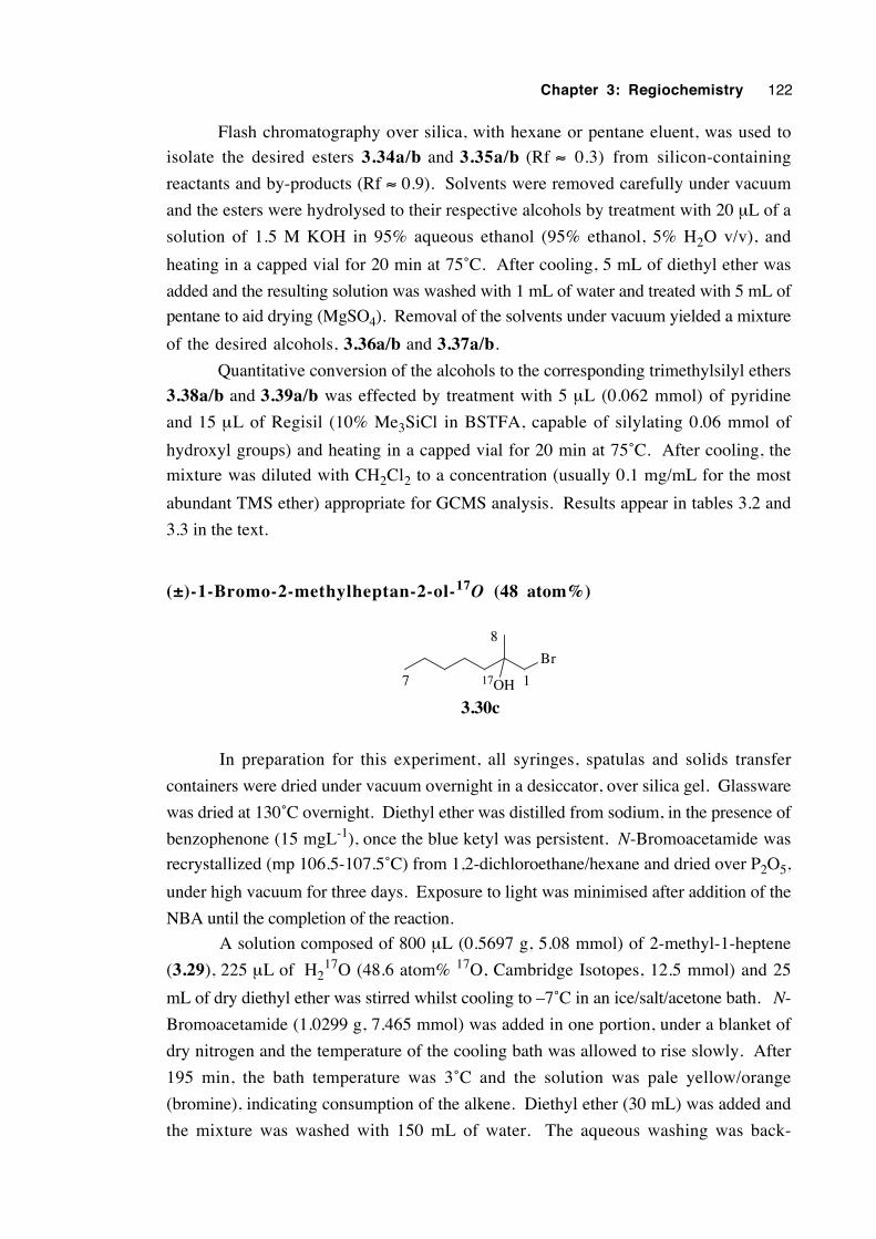
Chapter 3: Regiochemistry 122
Flash chromatography over silica, with hexane or pentane eluent, was used to
isolate the desired esters 3.34a/b and 3.35a/b (Rf ≈ 0.3) from silicon-containing
reactants and by-products (Rf ≈ 0.9). Solvents were removed carefully under vacuum
and the esters were hydrolysed to their respective alcohols by treatment with 20 µL of a
solution of 1.5 M KOH in 95% aqueous ethanol (95% ethanol, 5% H2O v/v), and
heating in a capped vial for 20 min at 75˚C. After cooling, 5 mL of diethyl ether was
added and the resulting solution was washed with 1 mL of water and treated with 5 mL of
pentane to aid drying (MgSO4). Removal of the solvents under vacuum yielded a mixture
of the desired alcohols, 3.36a/b and 3.37a/b.
Quantitative conversion of the alcohols to the corresponding trimethylsilyl ethers
3.38a/b and 3.39a/b was effected by treatment with 5 µL (0.062 mmol) of pyridine
and 15 µL of Regisil (10% Me3SiCl in BSTFA, capable of silylating 0.06 mmol of
hydroxyl groups) and heating in a capped vial for 20 min at 75˚C. After cooling, the
mixture was diluted with CH2Cl2 to a concentration (usually 0.1 mg/mL for the most
abundant TMS ether) appropriate for GCMS analysis. Results appear in tables 3.2 and
3.3 in the text.
(±)-1-Bromo-2-methylheptan-2-ol-17O (48 atom%)
Br
17OH
3.30c
17
8
In preparation for this experiment, all syringes, spatulas and solids transfer
containers were dried under vacuum overnight in a desiccator, over silica gel. Glassware
was dried at 130˚C overnight. Diethyl ether was distilled from sodium, in the presence of
benzophenone (15 mgL-1), once the blue ketyl was persistent. N-Bromoacetamide was
recrystallized (mp 106.5-107.5˚C) from 1,2-dichloroethane/hexane and dried over P2O5,
under high vacuum for three days. Exposure to light was minimised after addition of the
NBA until the completion of the reaction.
A solution composed of 800 µL (0.5697 g, 5.08 mmol) of 2-methyl-1-heptene
(3.29), 225 µL of H217O (48.6 atom% 17O, Cambridge Isotopes, 12.5 mmol) and 25
mL of dry diethyl ether was stirred whilst cooling to –7˚C in an ice/salt/acetone bath. N-
Bromoacetamide (1.0299 g, 7.465 mmol) was added in one portion, under a blanket of
dry nitrogen and the temperature of the cooling bath was allowed to rise slowly. After
195 min, the bath temperature was 3˚C and the solution was pale yellow/orange
(bromine), indicating consumption of the alkene. Diethyl ether (30 mL) was added and
the mixture was washed with 150 mL of water. The aqueous washing was back-
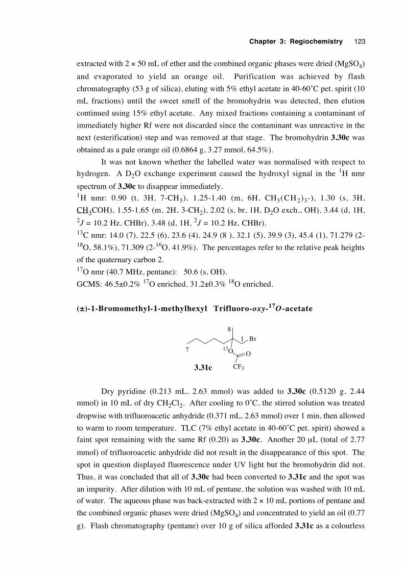
Chapter 3: Regiochemistry 123
extracted with 2 × 50 mL of ether and the combined organic phases were dried (MgSO4)
and evaporated to yield an orange oil. Purification was achieved by flash
chromatography (53 g of silica), eluting with 5% ethyl acetate in 40-60˚C pet. spirit (10
mL fractions) until the sweet smell of the bromohydrin was detected, then elution
continued using 15% ethyl acetate. Any mixed fractions containing a contaminant of
immediately higher Rf were not discarded since the contaminant was unreactive in the
next (esterification) step and was removed at that stage. The bromohydrin 3.30c was
obtained as a pale orange oil (0.6864 g, 3.27 mmol, 64.5%).
It was not known whether the labelled water was normalised with respect to
hydrogen. A D2O exchange experiment caused the hydroxyl signal in the 1H nmr
spectrum of 3.30c to disappear immediately.1H nmr: 0.90 (t, 3H, 7-CH3), 1.25-1.40 (m, 6H, CH3(CH 2)3-), 1.30 (s, 3H,
CH3COH), 1.55-1.65 (m, 2H, 3-CH2), 2.02 (s, br, 1H, D2O exch., OH), 3.44 (d, 1H,2J = 10.2 Hz, CHBr), 3.48 (d, 1H, 2J = 10.2 Hz, CHBr).13C nmr: 14.0 (7), 22.5 (6), 23.6 (4), 24.9 (8 ), 32.1 (5), 39.9 (3), 45.4 (1), 71.279 (2-18O, 58.1%), 71.309 (2-16O, 41.9%). The percentages refer to the relative peak heights
of the quaternary carbon 2.17O nmr (40.7 MHz, pentane): 50.6 (s, OH).
GCMS: 46.5±0.2% 17O enriched, 31.2±0.3% 18O enriched.
(±)-1-Bromomethyl-1-methylhexyl Trifluoro-oxy-17O-acetate
Br
17O
CF3
O
3.31c
7
1
8
Dry pyridine (0.213 mL, 2.63 mmol) was added to 3.30c (0.5120 g, 2.44
mmol) in 10 mL of dry CH2Cl2. After cooling to 0˚C, the stirred solution was treated
dropwise with trifluoroacetic anhydride (0.371 mL, 2.63 mmol) over 1 min, then allowed
to warm to room temperature. TLC (7% ethyl acetate in 40-60˚C pet. spirit) showed a
faint spot remaining with the same Rf (0.20) as 3.30c. Another 20 µL (total of 2.77
mmol) of trifluoroacetic anhydride did not result in the disappearance of this spot. The
spot in question displayed fluorescence under UV light but the bromohydrin did not.
Thus, it was concluded that all of 3.30c had been converted to 3.31c and the spot was
an impurity. After dilution with 10 mL of pentane, the solution was washed with 10 mL
of water. The aqueous phase was back-extracted with 2 × 10 mL portions of pentane and
the combined organic phases were dried (MgSO4) and concentrated to yield an oil (0.77
g). Flash chromatography (pentane) over 10 g of silica afforded 3.31c as a colourless
Chapter 3: Regiochemistry 124
oil (0.6289 g, 2.06 mmol, 84%) which was 99.0% pure by GC (BP1).17O nmr (40.7 MHz, pentane): 176.7 (s, alkoxy oxygen).
Procedure for regiochemical studies with 1 7 O-labelled β -
bromoester 3.31c
Two separate series of experiments were conducted: the first using
tris(trimethylsilyl)silane as the hydrogen atom source; and the second using tributyltin
hydride. For each series of reactions, approximately 52 mg (0.17 mmol) of β-
bromoester 3.31c was used, to provide sufficient quantities of products for nmr
analysis. For the tris(trimethylsilyl)silane reactions the quantity of the reducing agent was
usually about 71 µL (0.23 mmol), corresponding to an intended 1.35 molar equivalents
and the amount of AIBN was approximately 3.5 mol% relative to the silane. The volume
of each solvent (hexane, benzene, acetonitrile) was 5.0 mL. For reactions performed
with tributyltin hydride the quantity of reducing agent used was approximately 55 µL
(0.20 mmol), corresponding to an intended 1.2 molar equivalents and approximately 4.5
mol% AIBN relative to the hydride. The volume of each solvent (hexane, benzene,
propionitrile, perfluoromethylcyclohexane) was 20 mL (16 mL for PFMC).
An appropriately-sized (10 or 50 mL), sturdy, brown glass bottle was equipped
with a stirrer bar and charged with the labelled β-bromoester 3.31c. The vessel was
capped with a Mininert valve19 and the dry, purified solvent of choice was introduced by
syringe. A slow stream of dry nitrogen bubbles was passed through the stirred
solution—via a needle inserted through the Mininert valve—for three minutes, to remove
oxygen. The valve was then closed and the bottle was placed in an 80±1˚C thermostatted
oil bath, and surrounded with a metal gauze protective shield. The solution was stirred at
this temperature for 15 min prior to a rapid injection of the reducing agent, followed by a
solution of the initiator. The progress of reactions was monitored by GC
(polydimethylsiloxane stationary phase) and the reaction vessel was withdrawn from the
bath and cooled after consumption of β-bromoester 3.31c. Reactions involving
tris(trimethylsilyl)silane were analysed by GCMS to determine the isotopic composition
of each of the ester products (table 3.11). Reactions involving tributyltin hydride were
not analysed directly because of GCMS contamination issues and were only analysed
after tin-containing residues had been removed by preparative GC.
Solvents were removed by distillation under reduced-pressure through a column
comprising a B10 still head packed with glass rings. The heating bath temperature was
kept below 55˚C. The residue was transferred to an nmr tube, sometimes with the aid of
a little pentane to lower the solution viscosity, and a 17O nmr spectrum of the mixture was
obtained (see 17O nmr appendix).
Preparative GC (see general experimental section, column temperature 60˚C) was
then used to separate the non-rearranged (3.34c) and rearranged (3.35c) esters from
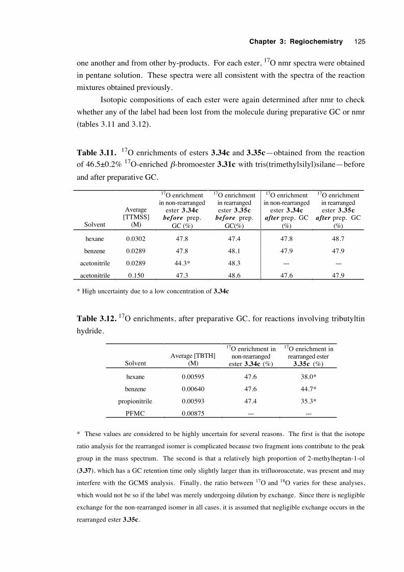
Chapter 3: Regiochemistry 125
one another and from other by-products. For each ester, 17O nmr spectra were obtained
in pentane solution. These spectra were all consistent with the spectra of the reaction
mixtures obtained previously.
Isotopic compositions of each ester were again determined after nmr to check
whether any of the label had been lost from the molecule during preparative GC or nmr
(tables 3.11 and 3.12).
Table 3.11. 17O enrichments of esters 3.34c and 3.35c—obtained from the reaction
of 46.5±0.2% 17O-enriched β-bromoester 3.31c with tris(trimethylsilyl)silane—before
and after preparative GC.
Solvent
Average[TTMSS]
(M)
17O enrichmentin non-rearranged
ester 3.34cbefore prep.
GC (%)
17O enrichmentin rearrangedester 3.35c
before prep.GC(%)
17O enrichmentin non-rearranged
ester 3.34cafter prep. GC
(%)
17O enrichmentin rearrangedester 3.35c
after prep. GC(%)
hexane 0.0302 47.8 47.4 47.8 48.7
benzene 0.0289 47.8 48.1 47.9 47.9
acetonitrile 0.0289 44.3* 48.3 --- ---
acetonitrile 0.150 47.3 48.6 47.6 47.9
* High uncertainty due to a low concentration of 3.34c
Table 3.12. 17O enrichments, after preparative GC, for reactions involving tributyltin
hydride.
SolventAverage [TBTH]
(M)
17O enrichment innon-rearranged
ester 3.34c (%)
17O enrichment inrearranged ester
3.35c (%)
hexane 0.00595 47.6 38.0*
benzene 0.00640 47.6 44.7*
propionitrile 0.00593 47.4 35.3*
PFMC 0.00875 --- ---
* These values are considered to be highly uncertain for several reasons. The first is that the isotope
ratio analysis for the rearranged isomer is complicated because two fragment ions contribute to the peak
group in the mass spectrum. The second is that a relatively high proportion of 2-methylheptan-1-ol
(3.37), which has a GC retention time only slightly larger than its trifluoroacetate, was present and may
interfere with the GCMS analysis. Finally, the ratio between 17O and 18O varies for these analyses,
which would not be so if the label was merely undergoing dilution by exchange. Since there is negligible
exchange for the non-rearranged isomer in all cases, it is assumed that negligible exchange occurs in the
rearranged ester 3.35c.
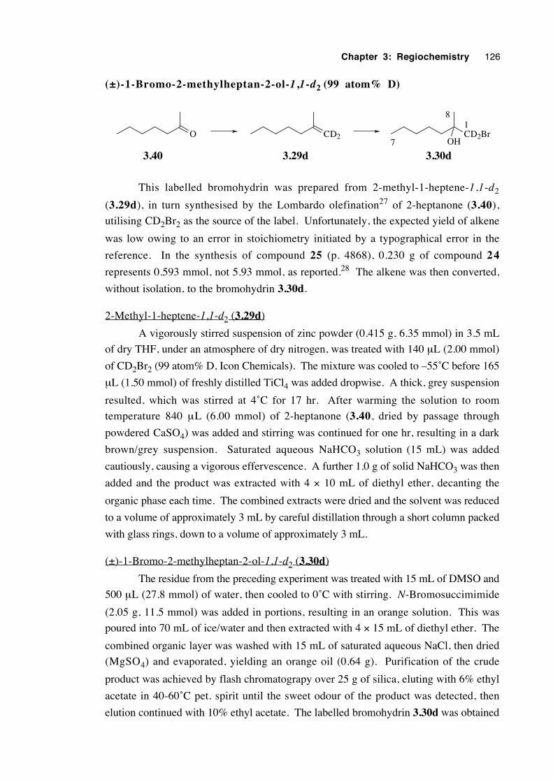
Chapter 3: Regiochemistry 126
(±)-1-Bromo-2-methylheptan-2-ol-1,1-d2 (99 atom% D)
O CD2 CD2BrOH
3.30d
18
7
3.29d3.40
This labelled bromohydrin was prepared from 2-methyl-1-heptene-1,1-d2
(3.29d), in turn synthesised by the Lombardo olefination27 of 2-heptanone (3.40),
utilising CD2Br2 as the source of the label. Unfortunately, the expected yield of alkene
was low owing to an error in stoichiometry initiated by a typographical error in the
reference. In the synthesis of compound 25 (p. 4868), 0.230 g of compound 24
represents 0.593 mmol, not 5.93 mmol, as reported.28 The alkene was then converted,
without isolation, to the bromohydrin 3.30d.
2-Methyl-1-heptene-1,1-d2 (3.29d)
A vigorously stirred suspension of zinc powder (0.415 g, 6.35 mmol) in 3.5 mL
of dry THF, under an atmosphere of dry nitrogen, was treated with 140 µL (2.00 mmol)
of CD2Br2 (99 atom% D, Icon Chemicals). The mixture was cooled to –55˚C before 165
µL (1.50 mmol) of freshly distilled TiCl4 was added dropwise. A thick, grey suspension
resulted, which was stirred at 4˚C for 17 hr. After warming the solution to room
temperature 840 µL (6.00 mmol) of 2-heptanone (3.40, dried by passage through
powdered CaSO4) was added and stirring was continued for one hr, resulting in a dark
brown/grey suspension. Saturated aqueous NaHCO3 solution (15 mL) was added
cautiously, causing a vigorous effervescence. A further 1.0 g of solid NaHCO3 was then
added and the product was extracted with 4 × 10 mL of diethyl ether, decanting the
organic phase each time. The combined extracts were dried and the solvent was reduced
to a volume of approximately 3 mL by careful distillation through a short column packed
with glass rings, down to a volume of approximately 3 mL.
(±)-1-Bromo-2-methylheptan-2-ol-1,1-d2 (3.30d)
The residue from the preceding experiment was treated with 15 mL of DMSO and
500 µL (27.8 mmol) of water, then cooled to 0˚C with stirring. N-Bromosuccimimide
(2.05 g, 11.5 mmol) was added in portions, resulting in an orange solution. This was
poured into 70 mL of ice/water and then extracted with 4 × 15 mL of diethyl ether. The
combined organic layer was washed with 15 mL of saturated aqueous NaCl, then dried
(MgSO4) and evaporated, yielding an orange oil (0.64 g). Purification of the crude
product was achieved by flash chromatograpy over 25 g of silica, eluting with 6% ethyl
acetate in 40-60˚C pet. spirit until the sweet odour of the product was detected, then
elution continued with 10% ethyl acetate. The labelled bromohydrin 3.30d was obtained
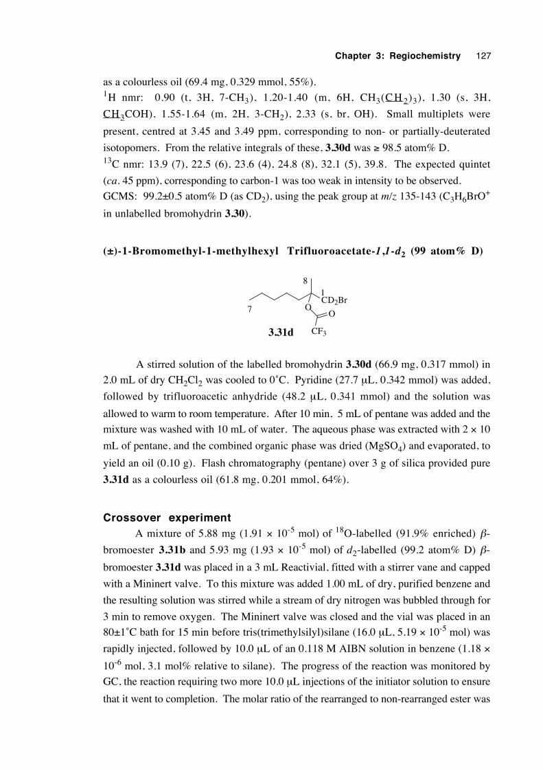
Chapter 3: Regiochemistry 127
as a colourless oil (69.4 mg, 0.329 mmol, 55%).1H nmr: 0.90 (t, 3H, 7-CH3), 1.20-1.40 (m, 6H, CH3(C H 2)3), 1.30 (s, 3H,
CH3COH), 1.55-1.64 (m, 2H, 3-CH2), 2.33 (s, br, OH). Small multiplets were
present, centred at 3.45 and 3.49 ppm, corresponding to non- or partially-deuterated
isotopomers. From the relative integrals of these, 3.30d was ≥ 98.5 atom% D.13C nmr: 13.9 (7), 22.5 (6), 23.6 (4), 24.8 (8), 32.1 (5), 39.8. The expected quintet
(ca. 45 ppm), corresponding to carbon-1 was too weak in intensity to be observed.
GCMS: 99.2±0.5 atom% D (as CD2), using the peak group at m/z 135-143 (C3H6BrO+
in unlabelled bromohydrin 3.30).
(±)-1-Bromomethyl-1-methylhexyl Trifluoroacetate-1,1-d2 (99 atom% D)
CD2BrO
CF3
O
3.31d
1
7
8
A stirred solution of the labelled bromohydrin 3.30d (66.9 mg, 0.317 mmol) in
2.0 mL of dry CH2Cl2 was cooled to 0˚C. Pyridine (27.7 µL, 0.342 mmol) was added,
followed by trifluoroacetic anhydride (48.2 µL, 0.341 mmol) and the solution was
allowed to warm to room temperature. After 10 min, 5 mL of pentane was added and the
mixture was washed with 10 mL of water. The aqueous phase was extracted with 2 × 10
mL of pentane, and the combined organic phase was dried (MgSO4) and evaporated, to
yield an oil (0.10 g). Flash chromatography (pentane) over 3 g of silica provided pure
3.31d as a colourless oil (61.8 mg, 0.201 mmol, 64%).
Crossover experimentA mixture of 5.88 mg (1.91 × 10⁻⁵ mol) of 18O-labelled (91.9% enriched) β-
bromoester 3.31b and 5.93 mg (1.93 × 10⁻⁵ mol) of d2-labelled (99.2 atom% D) β-
bromoester 3.31d was placed in a 3 mL Reactivial, fitted with a stirrer vane and capped
with a Mininert valve. To this mixture was added 1.00 mL of dry, purified benzene and
the resulting solution was stirred while a stream of dry nitrogen was bubbled through for
3 min to remove oxygen. The Mininert valve was closed and the vial was placed in an
80±1˚C bath for 15 min before tris(trimethylsilyl)silane (16.0 µL, 5.19 × 10⁻⁵ mol) was
rapidly injected, followed by 10.0 µL of an 0.118 M AIBN solution in benzene (1.18 ×
10-6 mol, 3.1 mol% relative to silane). The progress of the reaction was monitored by
GC, the reaction requiring two more 10.0 µL injections of the initiator solution to ensure
that it went to completion. The molar ratio of the rearranged to non-rearranged ester was
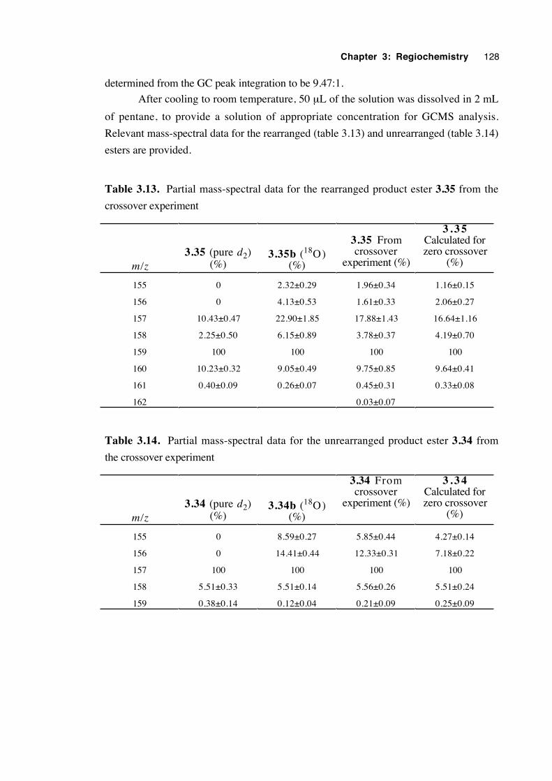
Chapter 3: Regiochemistry 128
determined from the GC peak integration to be 9.47:1.
After cooling to room temperature, 50 µL of the solution was dissolved in 2 mL
of pentane, to provide a solution of appropriate concentration for GCMS analysis.
Relevant mass-spectral data for the rearranged (table 3.13) and unrearranged (table 3.14)
esters are provided.
Table 3.13. Partial mass-spectral data for the rearranged product ester 3.35 from the
crossover experiment
m/z3.35 (pure d2)
(%)3.35b (18O)
(%)
3.35 Fromcrossover
experiment (%)
3 . 3 5Calculated forzero crossover
(%)
155 0 2.32±0.29 1.96±0.34 1.16±0.15
156 0 4.13±0.53 1.61±0.33 2.06±0.27
157 10.43±0.47 22.90±1.85 17.88±1.43 16.64±1.16
158 2.25±0.50 6.15±0.89 3.78±0.37 4.19±0.70
159 100 100 100 100
160 10.23±0.32 9.05±0.49 9.75±0.85 9.64±0.41
161 0.40±0.09 0.26±0.07 0.45±0.31 0.33±0.08
162 0.03±0.07
Table 3.14. Partial mass-spectral data for the unrearranged product ester 3.34 from
the crossover experiment
m/z3.34 (pure d2)
(%)3.34b (18O)
(%)
3.34 Fromcrossover
experiment (%)
3 . 3 4Calculated forzero crossover
(%)
155 0 8.59±0.27 5.85±0.44 4.27±0.14
156 0 14.41±0.44 12.33±0.31 7.18±0.22
157 100 100 100 100
158 5.51±0.33 5.51±0.14 5.56±0.26 5.51±0.24
159 0.38±0.14 0.12±0.04 0.21±0.09 0.25±0.09
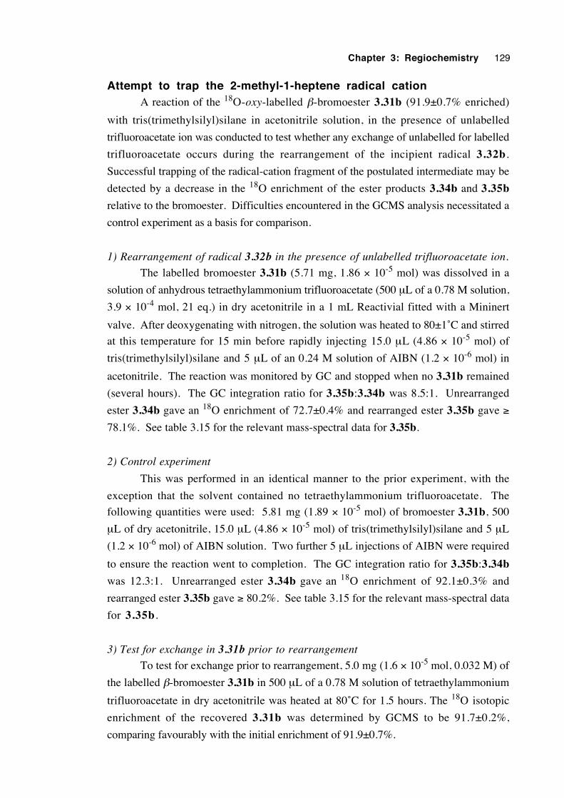
Chapter 3: Regiochemistry 129
Attempt to trap the 2-methyl-1-heptene radical cationA reaction of the 18O-oxy-labelled β-bromoester 3.31b (91.9±0.7% enriched)
with tris(trimethylsilyl)silane in acetonitrile solution, in the presence of unlabelled
trifluoroacetate ion was conducted to test whether any exchange of unlabelled for labelled
trifluoroacetate occurs during the rearrangement of the incipient radical 3.32b.
Successful trapping of the radical-cation fragment of the postulated intermediate may be
detected by a decrease in the 18O enrichment of the ester products 3.34b and 3.35b
relative to the bromoester. Difficulties encountered in the GCMS analysis necessitated a
control experiment as a basis for comparison.
1) Rearrangement of radical 3.32b in the presence of unlabelled trifluoroacetate ion.
The labelled bromoester 3.31b (5.71 mg, 1.86 × 10⁻⁵ mol) was dissolved in a
solution of anhydrous tetraethylammonium trifluoroacetate (500 µL of a 0.78 M solution,
3.9 × 10⁻⁴ mol, 21 eq.) in dry acetonitrile in a 1 mL Reactivial fitted with a Mininert
valve. After deoxygenating with nitrogen, the solution was heated to 80±1˚C and stirred
at this temperature for 15 min before rapidly injecting 15.0 µL (4.86 × 10⁻⁵ mol) of
tris(trimethylsilyl)silane and 5 µL of an 0.24 M solution of AIBN (1.2 × 10⁻⁶ mol) in
acetonitrile. The reaction was monitored by GC and stopped when no 3.31b remained
(several hours). The GC integration ratio for 3.35b:3.34b was 8.5:1. Unrearranged
ester 3.34b gave an 18O enrichment of 72.7±0.4% and rearranged ester 3.35b gave ≥
78.1%. See table 3.15 for the relevant mass-spectral data for 3.35b.
2) Control experiment
This was performed in an identical manner to the prior experiment, with the
exception that the solvent contained no tetraethylammonium trifluoroacetate. The
following quantities were used: 5.81 mg (1.89 × 10⁻⁵ mol) of bromoester 3.31b, 500
µL of dry acetonitrile, 15.0 µL (4.86 × 10⁻⁵ mol) of tris(trimethylsilyl)silane and 5 µL
(1.2 × 10⁻⁶ mol) of AIBN solution. Two further 5 µL injections of AIBN were required
to ensure the reaction went to completion. The GC integration ratio for 3.35b:3.34b
was 12.3:1. Unrearranged ester 3.34b gave an 18O enrichment of 92.1±0.3% and
rearranged ester 3.35b gave ≥ 80.2%. See table 3.15 for the relevant mass-spectral data
for 3.35b.
3) Test for exchange in 3.31b prior to rearrangement
To test for exchange prior to rearrangement, 5.0 mg (1.6 × 10⁻⁵ mol, 0.032 M) of
the labelled β-bromoester 3.31b in 500 µL of a 0.78 M solution of tetraethylammonium
trifluoroacetate in dry acetonitrile was heated at 80˚C for 1.5 hours. The 18O isotopic
enrichment of the recovered 3.31b was determined by GCMS to be 91.7±0.2%,
comparing favourably with the initial enrichment of 91.9±0.7%.
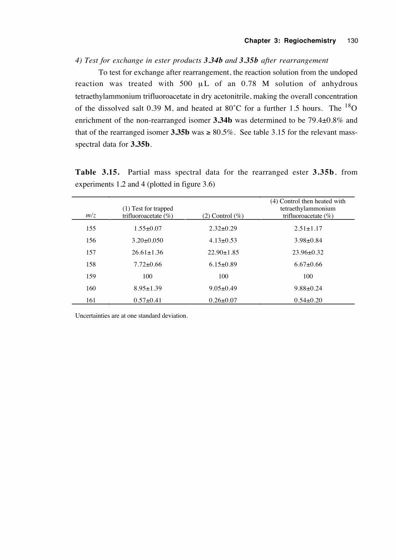
Chapter 3: Regiochemistry 130
4) Test for exchange in ester products 3.34b and 3.35b after rearrangement
To test for exchange after rearrangement, the reaction solution from the undoped
reaction was treated with 500 µ L of an 0.78 M solution of anhydrous
tetraethylammonium trifluoroacetate in dry acetonitrile, making the overall concentration
of the dissolved salt 0.39 M, and heated at 80˚C for a further 1.5 hours. The 18O
enrichment of the non-rearranged isomer 3.34b was determined to be 79.4±0.8% and
that of the rearranged isomer 3.35b was ≥ 80.5%. See table 3.15 for the relevant mass-
spectral data for 3.35b.
Table 3.15. Partial mass spectral data for the rearranged ester 3.35b , from
experiments 1,2 and 4 (plotted in figure 3.6)
m/z(1) Test for trappedtrifluoroacetate (%) (2) Control (%)
(4) Control then heated withtetraethylammoniumtrifluoroacetate (%)
155 1.55±0.07 2.32±0.29 2.51±1.17
156 3.20±0.050 4.13±0.53 3.98±0.84
157 26.61±1.36 22.90±1.85 23.96±0.32
158 7.72±0.66 6.15±0.89 6.67±0.66
159 100 100 100
160 8.95±1.39 9.05±0.49 9.88±0.24
161 0.57±0.41 0.26±0.07 0.54±0.20
Uncertainties are at one standard deviation.
Chapter 3: Regiochemistry 131
3.12 References
1. Beckwith, A. L. J.; Crich, D.; Duggan, P. J. and Yao, Q. Chem. Rev. 1997, 97,
3273.
2. Korth, H.-G.; Sustmann, R.; Groninger, K. S.; Liesung, M. and Giese, B. J. Org.
Chem. 1988, 53, 4364.
3. Beckwith, A. L. J. and Duggan, P. J. J. Chem. Soc., Perkin Trans. 2 1992, 1777.
4. Beckwith, A. L. J. and Thomas, C. B. J. Chem. Soc., Perkin Trans. 2 1973, 861.
5. Beckwith, A. L. J. and Duggan, P. J. J. Am. Chem. Soc. 1996, 118, 12838.
6. Crich, D. and Filzen, G. F. J. Org. Chem. 1995, 60, 4834.
7. Beckwith, A. L. J. and Duggan, P. J. J. Chem. Soc., Perkin Trans. 2 1993, 1673.
8. Kocovsky, P.; Stary, I. and Turecek, F. Tetrahedron Lett. 1986, 27, 1513.
9. Crich, D.; Huang, X. and Beckwith, A. L. J. J. Org. Chem. 1999, 64, 1762.
10. Gilbert, B. C.; Norman, R. O. C. and Williams, P. S. J. Chem. Soc. Perkin Trans.
2 1981, 1401.
11. Asmus, K.-D.; Williams, P. S.; Gilbert, B. C. and Winter, J. N. J. Chem. Soc.
Chem. Commun. 1987, 208.
12. Guss, C. O. and Rosenthal, R. J. Am. Chem. Soc. 1955, 77, 2549.
13. Langman, A. W. and Dalton, D. R. Org. Synth. 1980, 59, 16.
14. Dalton, D. R.; Dutta, V. P. and Jones, D. C. J. Am. Chem. Soc. 1968, 90, 5498.
15. Chatgilialoglu, C.; Griller, D. and Lesage, M. J. Org. Chem. 1988, 53, 3641.
16. Giese, B.; Kopping, B. and Chatgilialoglu, C. Tetrahedron Lett. 1989, 30, 681.
17. Dickhaut, J. and Giese, B. Org Synth. 1992, 70, 164.
18. Chatgilialoglu, C.; Ferreri, C. and Gimisis, T. In Chemistry of Organic Silicon
Compounds; Rappoport, Z. and Apeloig, Y., Eds.; Wiley: Chichester, 1998; Vol. 2(pt.
2), pp 1539-1579.
19. Aldrich Fine Chemicals and Laboratory Equipment Catalogue, Australia and New
Zealand Edition, 2000-2001, T778.
20. Nakanishi, W.; Jo, T.; Miura, K.; Ikeda, Y.; Sugawara, T.; Kawada, Y. and
Iwamura, H. Chem. Lett. 1981, 387.
Chapter 3: Regiochemistry 132
21. Creary, X. and Inocenio, P. A. J. Am. Chem. Soc. 1986, 108, 5979.
22. See reference 7.
23. Crich, D.; Huang, X. and Beckwith, A. L. J. J. Org. Chem. 1999, 64, 1762.
24. See appendix B
25. McFarlane, W. and McFarlane, H. C. E. in Multinuclear NMR; Mason, J., Ed.;
Plenum Press: New York, 1987.
26. Boykin, D. W. and Baumstark, A. L. in 17O NMR Spectroscopy in Organic
Chemistry; Boykin, D. W., Ed.; CRC Press: Boston, 1991, p. 205.
27. Lombardo, L. Org. Synth. 1993, Coll. Vol. 8, 386.
28. Furber, M.; Mander, L. N. and Patrick, G. L. J. Org. Chem. 1990, 55, 4860.
29. Forcier, G. A. and Olver, J. W. Electrochim. Acta 1970, 15, 1609.
30. Atkins, P. W. Physical Chemistry; 3rd ed.; Oxford University Press Oxford, 1986;
p. 667.
31. Smolina, T. A.; Brusova, G. P.; Shchekut'eva, L. F.; Gopius, E. D.; Permin, A. B.
and Reutov, O. A. Izv. Akad. Nauk SSSR, Ser. Khim.1980, 9, 2079.
32. Zipse, H. and Bootz, M. J. Chem. Soc. Perkin Trans. 2 2001, 1566.
33. a) Reichardt, C. Chem. Rev. 1994, 94, 2319; b) Reichardt, C.,Schäfer G. , Liebigs
Ann. 1995, 1579; c) Eberhardt, R., Löbbecke S., Neidhardt, B. and Reichardt C.
Liebigs Ann. /Recueil 1 9 9 7 , 1195; d) Website: www.chemie.uni-
marburg.de/~ak39/et_home.html
Chapter 4
An esr study of β-oxygenated alkyl radicals
4.1 Introduction 134
4.2 Estimation of time-averaged dihedral angles 135
4.3 Recording of the esr spectra and extraction of g-values and
hyperfine splitting constants 138
4.4 Results 140
4.5 Calculations, analysis and discussion 146
4.6 Estimation of the energy barrier to internal rotation about theCα– Cβ bond in the β-oxygenated ethyl radicals 4.3a-c 170
4.7 Final discussion 176
4.8 Conclusions 180
4.9 Experimental 181
4.10 References 187
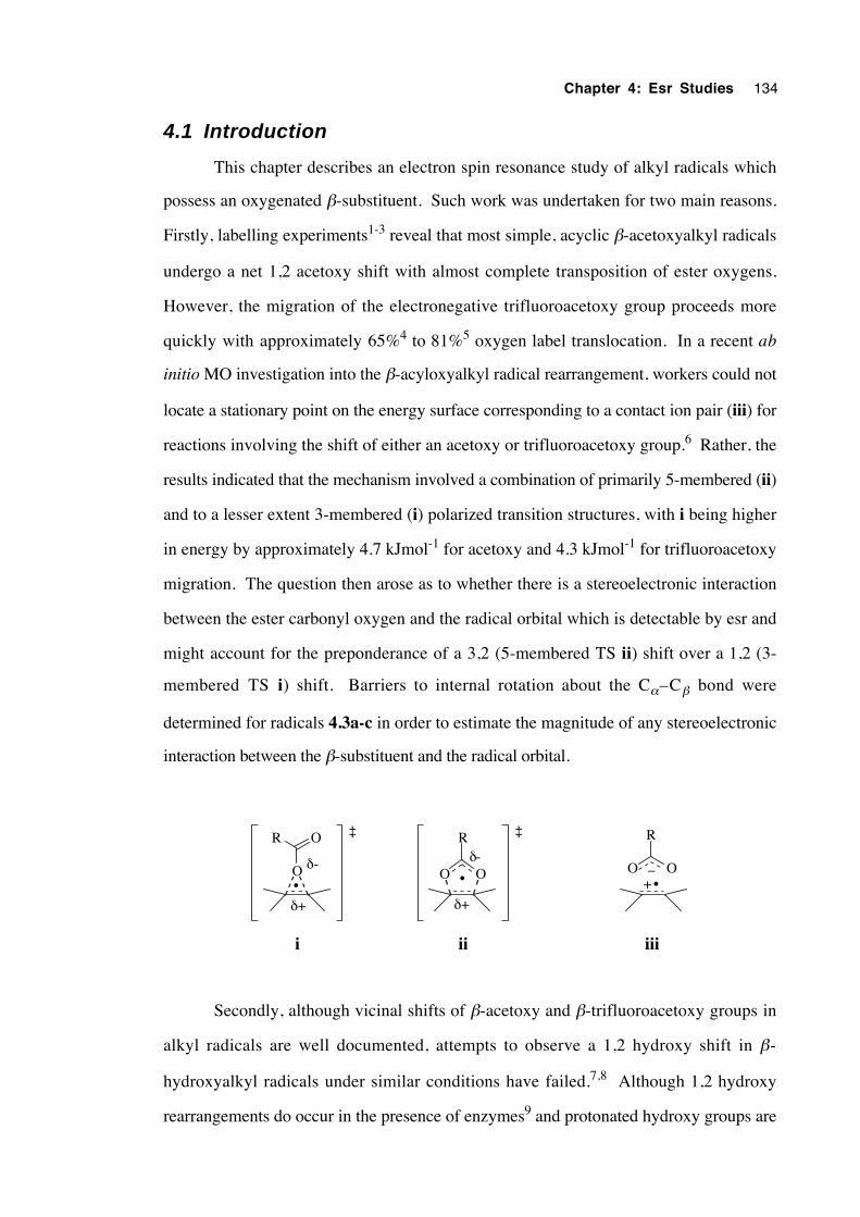
Chapter 4: Esr Studies 134
4.1 Introduction
This chapter describes an electron spin resonance study of alkyl radicals which
possess an oxygenated β-substituent. Such work was undertaken for two main reasons.
Firstly, labelling experiments1-3 reveal that most simple, acyclic β-acetoxyalkyl radicals
undergo a net 1,2 acetoxy shift with almost complete transposition of ester oxygens.
However, the migration of the electronegative trifluoroacetoxy group proceeds more
quickly with approximately 65%4 to 81%5 oxygen label translocation. In a recent ab
initio MO investigation into the β-acyloxyalkyl radical rearrangement, workers could not
locate a stationary point on the energy surface corresponding to a contact ion pair (iii) for
reactions involving the shift of either an acetoxy or trifluoroacetoxy group.6 Rather, the
results indicated that the mechanism involved a combination of primarily 5-membered (ii)
and to a lesser extent 3-membered (i) polarized transition structures, with i being higher
in energy by approximately 4.7 kJmol-1 for acetoxy and 4.3 kJmol-1 for trifluoroacetoxy
migration. The question then arose as to whether there is a stereoelectronic interaction
between the ester carbonyl oxygen and the radical orbital which is detectable by esr and
might account for the preponderance of a 3,2 (5-membered TS ii) shift over a 1,2 (3-
membered TS i) shift. Barriers to internal rotation about the Cα–Cβ bond were
determined for radicals 4.3a-c in order to estimate the magnitude of any stereoelectronic
interaction between the β-substituent and the radical orbital.
O O
R
•O
R O
•O O
R
+_
iiiiii
•
δ+
δ-
δ+
δ-
‡ ‡
Secondly, although vicinal shifts of β-acetoxy and β-trifluoroacetoxy groups in
alkyl radicals are well documented, attempts to observe a 1,2 hydroxy shift in β-
hydroxyalkyl radicals under similar conditions have failed.7,8 Although 1,2 hydroxy
rearrangements do occur in the presence of enzymes9 and protonated hydroxy groups are
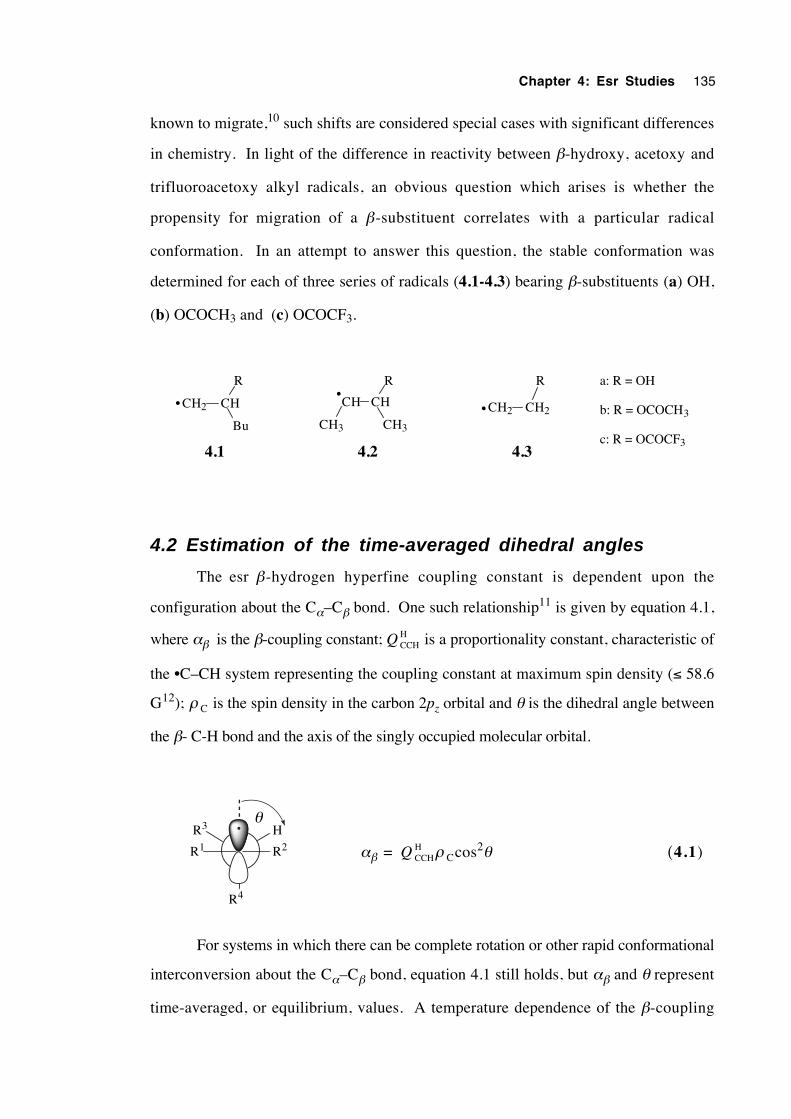
Chapter 4: Esr Studies 135
known to migrate,10 such shifts are considered special cases with significant differences
in chemistry. In light of the difference in reactivity between β-hydroxy, acetoxy and
trifluoroacetoxy alkyl radicals, an obvious question which arises is whether the
propensity for migration of a β-substituent correlates with a particular radical
conformation. In an attempt to answer this question, the stable conformation was
determined for each of three series of radicals (4.1-4.3) bearing β-substituents (a) OH,
(b) OCOCH3 and (c) OCOCF3.
CH2 CH
R
Bu
CH CH
R
CH3
CH2 CH2
R
CH3
4.1 4.2 4.3
a: R = OH
b: R = OCOCH3
c: R = OCOCF3
•••
4.2 Estimation of the time-averaged dihedral angles
The esr β-hydrogen hyperfine coupling constant is dependent upon the
configuration about the Cα–Cβ bond. One such relationship11 is given by equation 4.1,
where αβ is the β-coupling constant; Q CCHH is a proportionality constant, characteristic of
the •C–CH system representing the coupling constant at maximum spin density (≤ 58.6
G12); ρ C is the spin density in the carbon 2pz orbital and θ is the dihedral angle between
the β- C-H bond and the axis of the singly occupied molecular orbital.
R4
R3 H
R2R1
θ
αβ = Q CCHH ρ Ccos2θ (4.1)
For systems in which there can be complete rotation or other rapid conformational
interconversion about the Cα–Cβ bond, equation 4.1 still holds, but αβ and θ represent
time-averaged, or equilibrium, values. A temperature dependence of the β-coupling
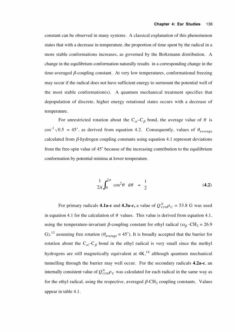
Chapter 4: Esr Studies 136
constant can be observed in many systems. A classical explanation of this phenomenon
states that with a decrease in temperature, the proportion of time spent by the radical in a
more stable conformations increases, as governed by the Boltzmann distribution. A
change in the equilibrium conformation naturally results in a corresponding change in the
time-averaged β-coupling constant. At very low temperatures, conformational freezing
may occur if the radical does not have sufficient energy to surmount the potential well of
the most stable conformation(s). A quantum mechanical treatment specifies that
depopulation of discrete, higher energy rotational states occurs with a decrease of
temperature.
For unrestricted rotation about the Cα–Cβ bond, the average value of θ is
cos-1 0 5. = 45˚, as derived from equation 4.2. Consequently, values of θaverage
calculated from β-hydrogen coupling constants using equation 4.1 represent deviations
from the free-spin value of 45˚ because of the increasing contribution to the equilibrium
conformation by potential minima at lower temperature.
12π
π
∫02
cos2θ dθ = 12
(4.2)
For primary radicals 4.1a-c and 4.3a-c, a value of Q CCHH ρ C = 53.8 G was used
in equation 4.1 for the calculation of θ values. This value is derived from equation 4.1,
using the temperature-invariant β-coupling constant for ethyl radical (αβ -CH3 = 26.9
G),13 assuming free rotation (θaverage = 45˚). It is broadly accepted that the barrier for
rotation about the Cα–Cβ bond in the ethyl radical is very small since the methyl
hydrogens are still magnetically equivalent at 4K,14 although quantum mechanical
tunnelling through the barrier may well occur. For the secondary radicals 4.2a-c, an
internally consistent value of Q CCHH ρ C was calculated for each radical in the same way as
for the ethyl radical, using the respective, averaged β-CH3 coupling constants. Values
appear in table 4.1.
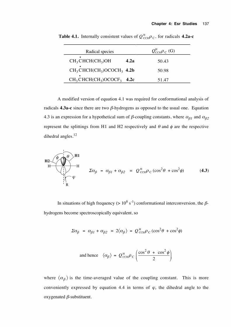
Chapter 4: Esr Studies 137
Table 4.1. Internally consistent values of Q CCHH ρ C, for radicals 4.2a-c
Radical species QCCHH ρ C (G)
CH3 C•
HCH(CH3)OH 4.2a 50.43
CH3 C•
HCH(CH3)OCOCH3 4.2b 50.98
CH3 C•
HCH(CH3)OCOCF3 4.2c 51.47
A modified version of equation 4.1 was required for conformational analysis of
radicals 4.3a-c since there are two β-hydrogens as opposed to the usual one. Equation
4.3 is an expression for a hypothetical sum of β-coupling constants, where αβ1 and αβ2
represent the splittings from Η1 and H2 respectively and θ and φ are the respective
dihedral angles.12
H2
R
H1
HH
θφ
ϕΣαβ = αβ1 + αβ2 = Q CCH
H ρ C (cos2θ + cos2φ) (4.3)
In situations of high frequency (> 108 s-1) conformational interconversion, the β-
hydrogens become spectroscopically equivalent, so
Σαβ = αβ1 + αβ2 = 2⟨αβ ⟩ = Q CCHH ρ C (cos2θ + cos2φ)
and hence ⟨αβ ⟩ = Q CCHH ρ C
cos cos2 2
2θ φ+
where ⟨αβ ⟩ is the time-averaged value of the coupling constant. This is more
conveniently expressed by equation 4.4 in terms of ϕ, the dihedral angle to the
oxygenated β-substituent.
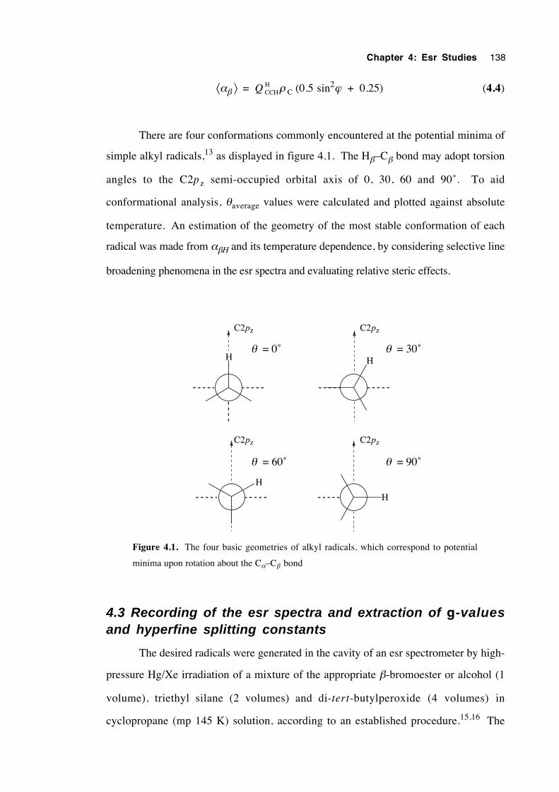
Chapter 4: Esr Studies 138
⟨αβ ⟩ = Q CCHH ρ C (0.5 sin2ϕ + 0.25) (4.4)
There are four conformations commonly encountered at the potential minima of
simple alkyl radicals,13 as displayed in figure 4.1. The Hβ–Cβ bond may adopt torsion
angles to the C2pz semi-occupied orbital axis of 0, 30, 60 and 90˚. To aid
conformational analysis, θaverage values were calculated and plotted against absolute
temperature. An estimation of the geometry of the most stable conformation of each
radical was made from αβH and its temperature dependence, by considering selective line
broadening phenomena in the esr spectra and evaluating relative steric effects.
H
H
H
C2pz C2pz
C2pzC2pz
θ = 0˚ θ = 30˚
θ = 90˚θ = 60˚
H
Figure 4.1. The four basic geometries of alkyl radicals, which correspond to potential
minima upon rotation about the Cα–Cβ bond
4.3 Recording of the esr spectra and extraction of g-valuesand hyperfine splitting constants
The desired radicals were generated in the cavity of an esr spectrometer by high-
pressure Hg/Xe irradiation of a mixture of the appropriate β-bromoester or alcohol (1
volume), triethyl silane (2 volumes) and di-tert-butylperoxide (4 volumes) in
cyclopropane (mp 145 K) solution, according to an established procedure.15,16 The
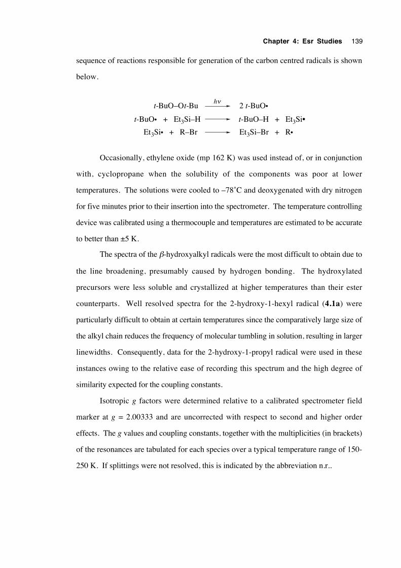
Chapter 4: Esr Studies 139
sequence of reactions responsible for generation of the carbon centred radicals is shown
below.
2 t-BuO•hν
t-BuO• + Et3Si–H t-BuO–H + Et3Si•
Et3Si• + R–Br Et3Si–Br + R•
t-BuO–Ot-Bu
Occasionally, ethylene oxide (mp 162 K) was used instead of, or in conjunction
with, cyclopropane when the solubility of the components was poor at lower
temperatures. The solutions were cooled to –78˚C and deoxygenated with dry nitrogen
for five minutes prior to their insertion into the spectrometer. The temperature controlling
device was calibrated using a thermocouple and temperatures are estimated to be accurate
to better than ±5 K.
The spectra of the β-hydroxyalkyl radicals were the most difficult to obtain due to
the line broadening, presumably caused by hydrogen bonding. The hydroxylated
precursors were less soluble and crystallized at higher temperatures than their ester
counterparts. Well resolved spectra for the 2-hydroxy-1-hexyl radical (4.1a) were
particularly difficult to obtain at certain temperatures since the comparatively large size of
the alkyl chain reduces the frequency of molecular tumbling in solution, resulting in larger
linewidths. Consequently, data for the 2-hydroxy-1-propyl radical were used in these
instances owing to the relative ease of recording this spectrum and the high degree of
similarity expected for the coupling constants.
Isotropic g factors were determined relative to a calibrated spectrometer field
marker at g = 2.00333 and are uncorrected with respect to second and higher order
effects. The g values and coupling constants, together with the multiplicities (in brackets)
of the resonances are tabulated for each species over a typical temperature range of 150-
250 K. If splittings were not resolved, this is indicated by the abbreviation n.r..
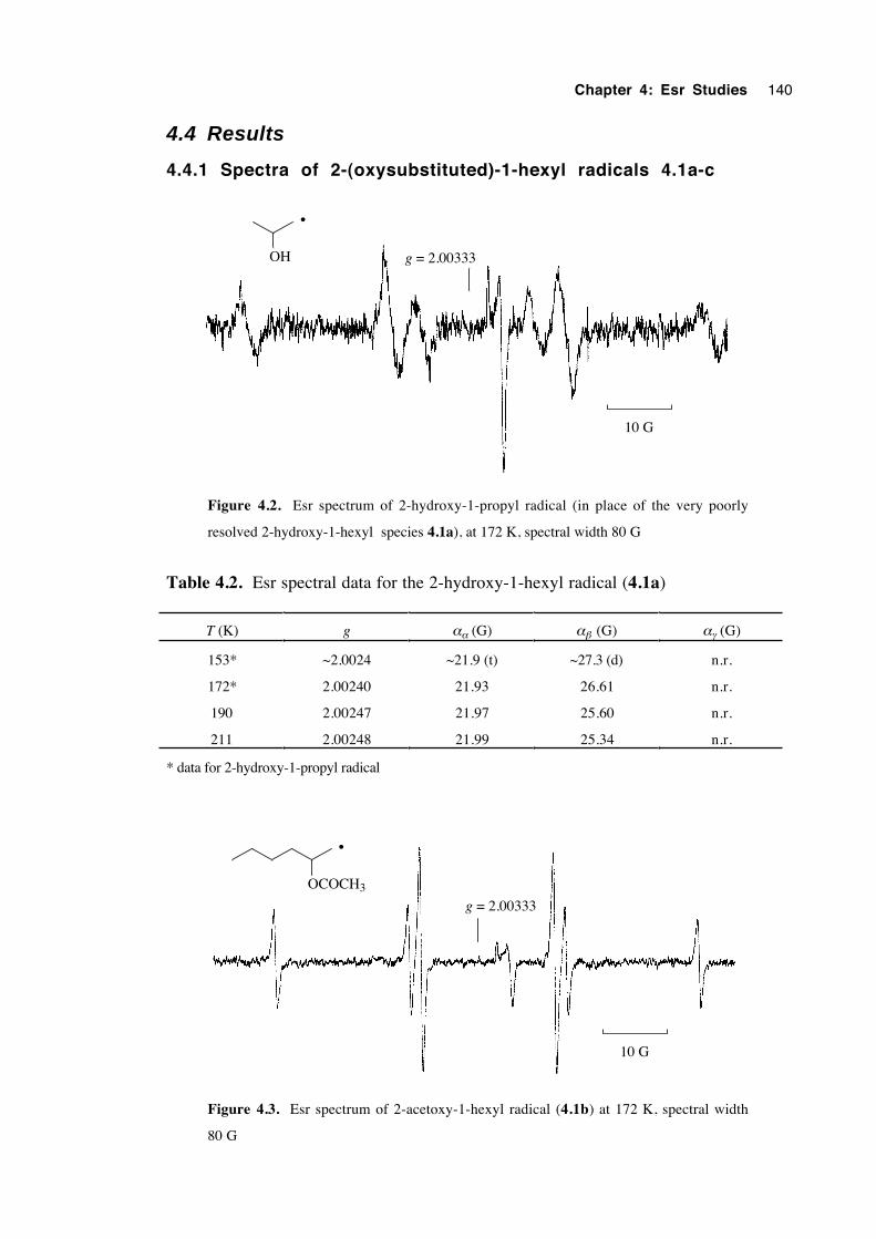
Chapter 4: Esr Studies 140
4.4 Results
4.4.1 Spectra of 2-(oxysubstituted)-1-hexyl radicals 4.1a-c
10 G
OH g = 2.00333
•
Figure 4.2. Esr spectrum of 2-hydroxy-1-propyl radical (in place of the very poorly
resolved 2-hydroxy-1-hexyl species 4.1a), at 172 K, spectral width 80 G
Table 4.2. Esr spectral data for the 2-hydroxy-1-hexyl radical (4.1a)
T (K) g αα (G) αβ (G) αγ (G)
153* ~2.0024 ~21.9 (t) ~27.3 (d) n.r.
172* 2.00240 21.93 26.61 n.r.
190 2.00247 21.97 25.60 n.r.
211 2.00248 21.99 25.34 n.r.
* data for 2-hydroxy-1-propyl radical
10 G
OCOCH3
g = 2.00333
•
Figure 4.3. Esr spectrum of 2-acetoxy-1-hexyl radical (4.1b) at 172 K, spectral width
80 G
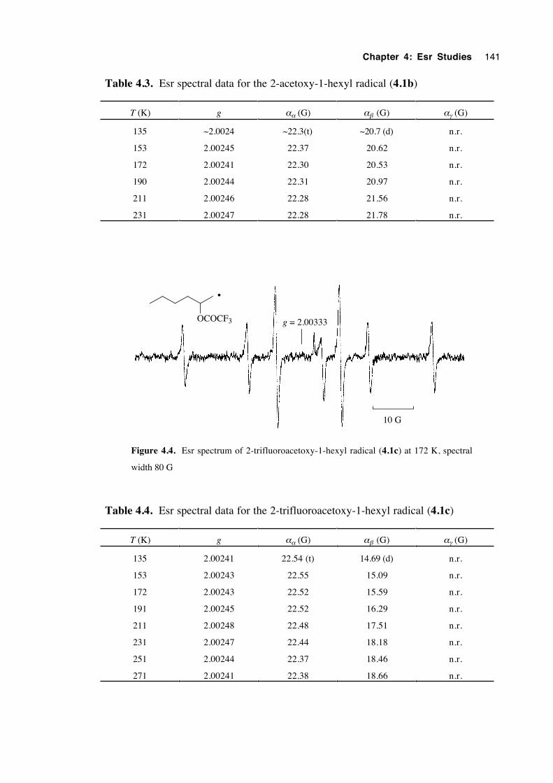
Chapter 4: Esr Studies 141
Table 4.3. Esr spectral data for the 2-acetoxy-1-hexyl radical (4.1b)
T (K) g αα (G) αβ (G) αγ (G)
135 ~2.0024 ~22.3(t) ~20.7 (d) n.r.
153 2.00245 22.37 20.62 n.r.
172 2.00241 22.30 20.53 n.r.
190 2.00244 22.31 20.97 n.r.
211 2.00246 22.28 21.56 n.r.
231 2.00247 22.28 21.78 n.r.
10 G
OCOCF3 g = 2.00333
•
Figure 4.4. Esr spectrum of 2-trifluoroacetoxy-1-hexyl radical (4.1c) at 172 K, spectral
width 80 G
Table 4.4. Esr spectral data for the 2-trifluoroacetoxy-1-hexyl radical (4.1c)
T (K) g αα (G) αβ (G) αγ (G)
135 2.00241 22.54 (t) 14.69 (d) n.r.
153 2.00243 22.55 15.09 n.r.
172 2.00243 22.52 15.59 n.r.
191 2.00245 22.52 16.29 n.r.
211 2.00248 22.48 17.51 n.r.
231 2.00247 22.44 18.18 n.r.
251 2.00244 22.37 18.46 n.r.
271 2.00241 22.38 18.66 n.r.
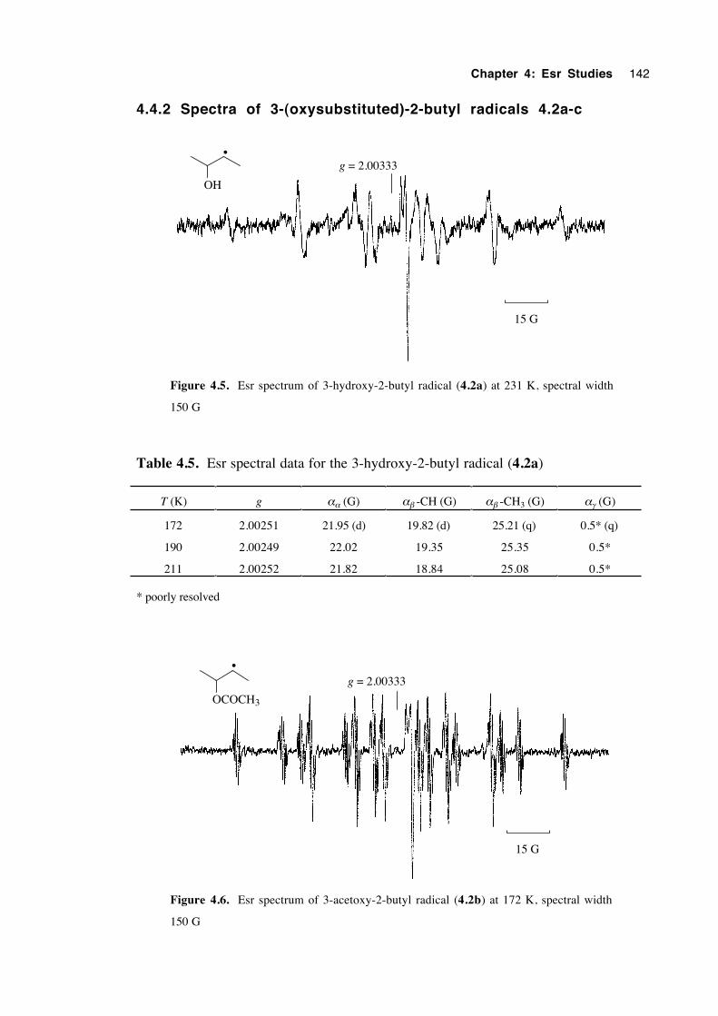
Chapter 4: Esr Studies 142
4.4.2 Spectra of 3-(oxysubstituted)-2-butyl radicals 4.2a-c
15 G
OH
g = 2.00333•
Figure 4.5. Esr spectrum of 3-hydroxy-2-butyl radical (4.2a) at 231 K, spectral width
150 G
Table 4.5. Esr spectral data for the 3-hydroxy-2-butyl radical (4.2a)
T (K) g αα (G) αβ -CH (G) αβ -CH3 (G) αγ (G)
172 2.00251 21.95 (d) 19.82 (d) 25.21 (q) 0.5* (q)
190 2.00249 22.02 19.35 25.35 0.5*
211 2.00252 21.82 18.84 25.08 0.5*
* poorly resolved
15 G
OCOCH3
g = 2.00333•
Figure 4.6. Esr spectrum of 3-acetoxy-2-butyl radical (4.2b) at 172 K, spectral width
150 G
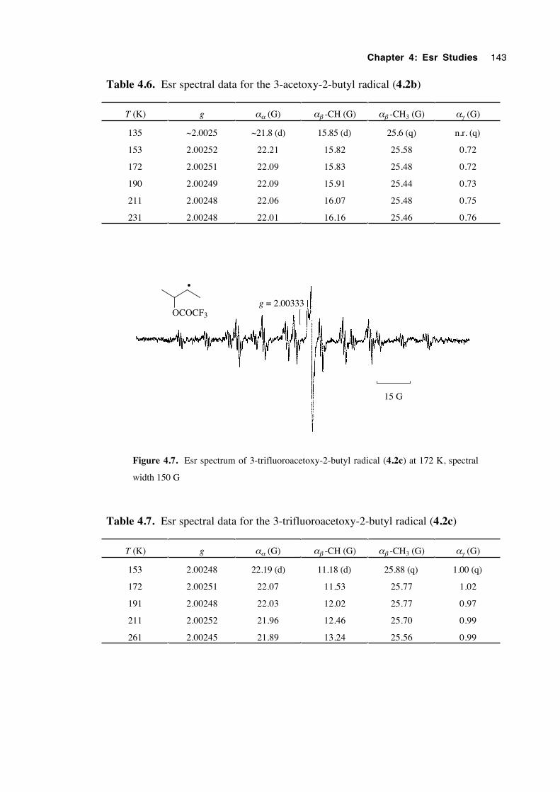
Chapter 4: Esr Studies 143
Table 4.6. Esr spectral data for the 3-acetoxy-2-butyl radical (4.2b)
T (K) g αα (G) αβ -CH (G) αβ -CH3 (G) αγ (G)
135 ~2.0025 ~21.8 (d) 15.85 (d) 25.6 (q) n.r. (q)
153 2.00252 22.21 15.82 25.58 0.72
172 2.00251 22.09 15.83 25.48 0.72
190 2.00249 22.09 15.91 25.44 0.73
211 2.00248 22.06 16.07 25.48 0.75
231 2.00248 22.01 16.16 25.46 0.76
15 G
g = 2.00333OCOCF3
•
Figure 4.7. Esr spectrum of 3-trifluoroacetoxy-2-butyl radical (4.2c) at 172 K, spectral
width 150 G
Table 4.7. Esr spectral data for the 3-trifluoroacetoxy-2-butyl radical (4.2c)
T (K) g αα (G) αβ -CH (G) αβ -CH3 (G) αγ (G)
153 2.00248 22.19 (d) 11.18 (d) 25.88 (q) 1.00 (q)
172 2.00251 22.07 11.53 25.77 1.02
191 2.00248 22.03 12.02 25.77 0.97
211 2.00252 21.96 12.46 25.70 0.99
261 2.00245 21.89 13.24 25.56 0.99
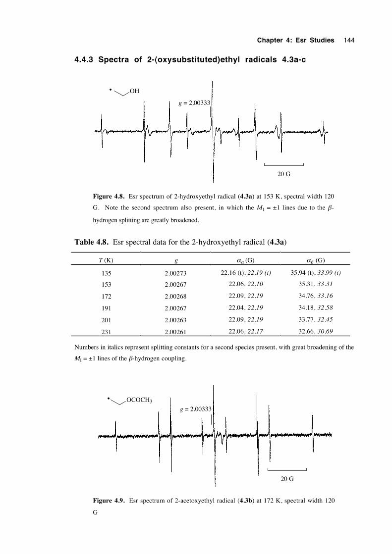
Chapter 4: Esr Studies 144
4.4.3 Spectra of 2-(oxysubstituted)ethyl radicals 4.3a-c
OH
20 G
g = 2.00333
•
Figure 4.8. Esr spectrum of 2-hydroxyethyl radical (4.3a) at 153 K, spectral width 120
G. Note the second spectrum also present, in which the M I = ±1 lines due to the β-
hydrogen splitting are greatly broadened.
Table 4.8. Esr spectral data for the 2-hydroxyethyl radical (4.3a)
T (K) g αα (G) αβ (G)
135 2.00273 22.16 (t), 22.19 (t) 35.94 (t), 33.99 (t)
153 2.00267 22.06, 22.10 35.31, 33.31
172 2.00268 22.09, 22.19 34.76, 33.16
191 2.00267 22.04, 22.19 34.18, 32.58
201 2.00263 22.09, 22.19 33.77, 32.45
231 2.00261 22.06, 22.17 32.66, 30.69
Numbers in italics represent splitting constants for a second species present, with great broadening of the
MI = ±1 lines of the β-hydrogen coupling.
OCOCH3
20 G
g = 2.00333
•
Figure 4.9. Esr spectrum of 2-acetoxyethyl radical (4.3b) at 172 K, spectral width 120
G
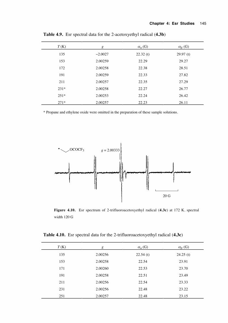
Chapter 4: Esr Studies 145
Table 4.9. Esr spectral data for the 2-acetoxyethyl radical (4.3b)
T (K) g αα (G) αβ (G)
135 ~2.0027 22.32 (t) 29.97 (t)
153 2.00259 22.29 29.27
172 2.00258 22.38 28.51
191 2.00259 22.33 27.82
211 2.00257 22.35 27.29
231* 2.00258 22.27 26.77
251* 2.00253 22.24 26.42
271* 2.00257 22.23 26.11
* Propane and ethylene oxide were omitted in the preparation of these sample solutions.
OCOCF3
20 G
g = 2.00333•
Figure 4.10. Esr spectrum of 2-trifluoroacetoxyethyl radical (4.3c) at 172 K, spectral
width 120 G
Table 4.10. Esr spectral data for the 2-trifluoroacetoxyethyl radical (4.3c)
T (K) g αα (G) αβ (G)
135 2.00256 22.54 (t) 24.25 (t)
153 2.00258 22.54 23.91
171 2.00260 22.53 23.70
191 2.00258 22.51 23.49
211 2.00256 22.54 23.33
231 2.00256 22.48 23.22
251 2.00257 22.48 23.15
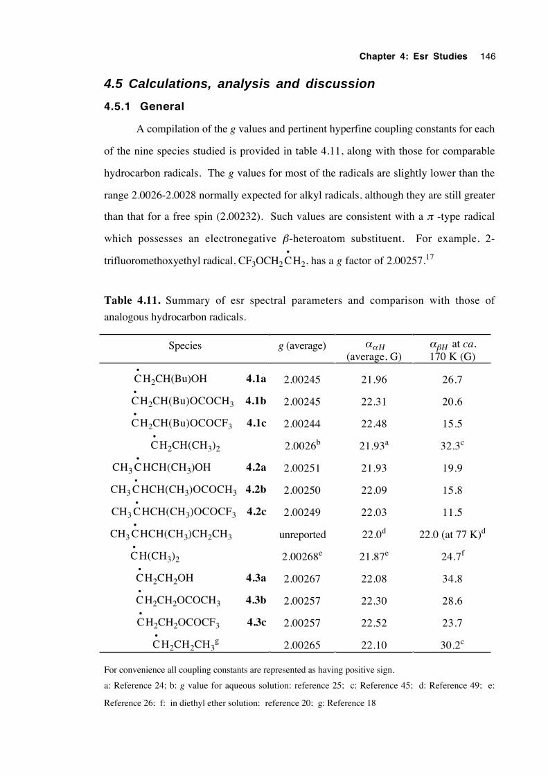
Chapter 4: Esr Studies 146
4.5 Calculations, analysis and discussion
4.5.1 General
A compilation of the g values and pertinent hyperfine coupling constants for each
of the nine species studied is provided in table 4.11, along with those for comparable
hydrocarbon radicals. The g values for most of the radicals are slightly lower than the
range 2.0026-2.0028 normally expected for alkyl radicals, although they are still greater
than that for a free spin (2.00232). Such values are consistent with a π -type radical
which possesses an electronegative β-heteroatom substituent. For example, 2-
trifluoromethoxyethyl radical, CF3OCH2 C•
H2, has a g factor of 2.00257.17
Table 4.11. Summary of esr spectral parameters and comparison with those of
analogous hydrocarbon radicals.
Species g (average) ααΗ(average, G)
αβΗ at ca.170 K (G)
C•
H2CH(Bu)OH 4.1a 2.00245 21.96 26.7
C•
H2CH(Bu)OCOCH3 4.1b 2.00245 22.31 20.6
C•
H2CH(Bu)OCOCF3 4.1c 2.00244 22.48 15.5
C•
H2CH(CH3)2 2.0026b 21.93a 32.3c
CH3 C•
HCH(CH3)OH 4.2a 2.00251 21.93 19.9
CH3 C•
HCH(CH3)OCOCH3 4.2b 2.00250 22.09 15.8
CH3 C•
HCH(CH3)OCOCF3 4.2c 2.00249 22.03 11.5
CH3 C•
HCH(CH3)CH2CH3 unreported 22.0d 22.0 (at 77 K)d
C•
H(CH3)2 2.00268e 21.87e 24.7f
C•
H2CH2OH 4.3a 2.00267 22.08 34.8
C•
H2CH2OCOCH3 4.3b 2.00257 22.30 28.6
C•
H2CH2OCOCF3 4.3c 2.00257 22.52 23.7
C•
H2CH2CH3g 2.00265 22.10 30.2c
For convenience all coupling constants are represented as having positive sign.
a: Reference 24; b: g value for aqueous solution: reference 25; c: Reference 45; d: Reference 49; e:
Reference 26; f: in diethyl ether solution: reference 20; g: Reference 18
Chapter 4: Esr Studies 147
These lower g values are consistent with delocalisation of the odd electron on to
the β-substituent, as postulated for β-chloroethyl radicals,18-21 although this may not be
the case with β-oxyalkyl radicals. The low g value of 2.00199 for β-chloroethyl radical
is attributed to a delocalisation of the odd electron into the p orbitals of the chlorine
atom.20 However, Kochi and coworkers doubt that such homoconjugation with β-
oxygen substituents is detectable in g since the spin orbit coupling constant of oxygen (ξp
= 151 cm-1) is only a fraction of that for Cl (ξp = 586 cm-1).17 More recent theoretical
calculations discount the significance of such homoconjugation altogether.22,23
Furthermore, since many workers do not indicate whether the g factors have been
corrected for second-order field effects, care should be taken before drawing conclusions
about radical structure from small deviations in this parameter.
Values for the α coupling constants are often slightly larger than, yet consonant
with, those of comparable alkyl radicals. Values of ααΗ are relatively invariant between
species, deviating not more than 1.5% from the mean. This indicates that spin density at
Cα varies little with changes in radical structure according to equation 4.5,27 where Q CHH
is a proportionality constant estimated to be between –2228 and –28 gauss27 and ρ C is
the spin density at Cα.
αα = Q CHH ρ C (4.5)
However, in the series 4.1a-c and 4.3a-c, it is clear that ααH increases steadily
in the order R = OH < OCOCH3 < OCOCF3. It is to be expected that ααH will increase
only slightly with a correspondingly large decrease in αβH since the hyperfine splitting at
the β-hydrogens does not exceed 10% that of the 507 G splitting of an isolated hydrogen
atom.29
Also affecting ααH is the degree of geometric distortion at the radical site. It is
known that α-coupling constants are usually negative in sign and become more positive
with increasing pyramidalisation at the radical centre.28 The ethyl radical is known to be
slightly pyramidal at Cα due to its low resistance to pyramidalisation and for steric and
hyperconjugative reasons.30 So the correlation between the β-substituent R and ααH is
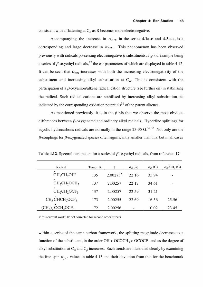
Chapter 4: Esr Studies 148
consistent with a flattening at Cα as R becomes more electronegative.
Accompanying the increase in ααH , in the series 4.1a-c and 4.3a-c, is a
corresponding and large decrease in αβH . This phenomenon has been observed
previously with radicals possessing electronegative β-substituents, a good example being
a series of β-oxyethyl radicals,17 the esr parameters of which are displayed in table 4.12.
It can be seen that ααH increases with both the increasing electronegativity of the
substituent and increasing alkyl substitution at Cα. This is consistent with the
participation of a β-oxyanion/alkene radical cation structure (see further on) in stabilising
the radical. Such radical cations are stabilised by increasing alkyl substitution, as
indicated by the corresponding oxidation potentials31 of the parent alkenes.
As mentioned previously, it is in the β-hfs that we observe the most obvious
differences between β-oxygenated and ordinary alkyl radicals. Hyperfine splittings for
acyclic hydrocarbons radicals are normally in the range 23-35 G.32,33 Not only are the
β-couplings for β-oxygenated species often significantly smaller than this, but in all cases
Table 4.12. Spectral parameters for a series of β-oxyethyl radicals, from reference 17
Radical Temp. K g αα (G) αβ (G) αβ -CH3 (G)
C•
H2CH2OHa 135 2.00273b 22.16 35.94 -
C•
H2CH2OCH3 137 2.00257 22.17 34.61 -
C•
H2CH2OCF3 137 2.00257 22.59 31.21 -
CH3 C•
HCH2OCF3 173 2.00255 22.69 16.56 25.56
(CH3)2 C•
CH2OCF3 172 2.00256 - 10.02 23.45
a: this current work; b: not corrected for second order effects
within a series of the same carbon framework, the splitting magnitude decreases as a
function of the substituent, in the order OH > OCOCH3 > OCOCF3 and as the degree of
alkyl substitution at Cα and Cβ increases. Such trends are illustrated clearly by examining
the free-spin αβΗ values in table 4.13 and their deviation from that for the benchmark
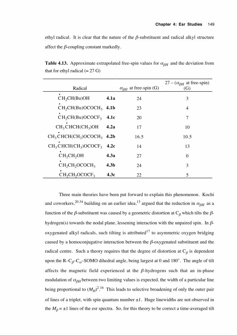
Chapter 4: Esr Studies 149
ethyl radical. It is clear that the nature of the β-substituent and radical alkyl structure
affect the β-coupling constant markedly.
Table 4.13. Approximate extrapolated free-spin values for αβΗ and the deviation from
that for ethyl radical (≈ 27 G)
Radical αβΗ at free-spin (G)27 – (αβΗ at free-spin)
(G)
C•
H2CH(Bu)OH 4.1a 24 3
C•
H2CH(Bu)OCOCH3 4.1b 23 4
C•
H2CH(Bu)OCOCF3 4.1c 20 7
CH3 C•
HCH(CH3)OH 4.2a 17 10
CH3 C•
HCH(CH3)OCOCH3 4.2b 16.5 10.5
CH3 C•
HCH(CH3)OCOCF3 4.2c 14 13
C•
H2CH2OH 4.3a 27 0
C•
H2CH2OCOCH3 4.3b 24 3
C•
H2CH2OCOCF3 4.3c 22 5
Three main theories have been put forward to explain this phenomenon. Kochi
and coworkers,20,34 building on an earlier idea,13 argued that the reduction in αβΗ as a
function of the β-substituent was caused by a geometric distortion at Cβ which tilts the β-
hydrogen(s) towards the nodal plane, lessening interaction with the unpaired spin. In β-
oxygenated alkyl radicals, such tilting is attributed17 to asymmetric oxygen bridging
caused by a homoconjugative interaction between the β-oxygenated substituent and the
radical centre. Such a theory requires that the degree of distortion at Cβ is dependent
upon the R–Cβ–Cα–SOMO dihedral angle, being largest at 0 and 180˚. The angle of tilt
affects the magnetic field experienced at the β-hydrogens such that an in-phase
modulation of αβΗ between two limiting values is expected, the width of a particular line
being proportional to (MH)2.16 This leads to selective broadening of only the outer pair
of lines of a triplet, with spin quantum number ±1. Huge linewidths are not observed in
the Mβ = ±1 lines of the esr spectra. So, for this theory to be correct a time-averaged tilt
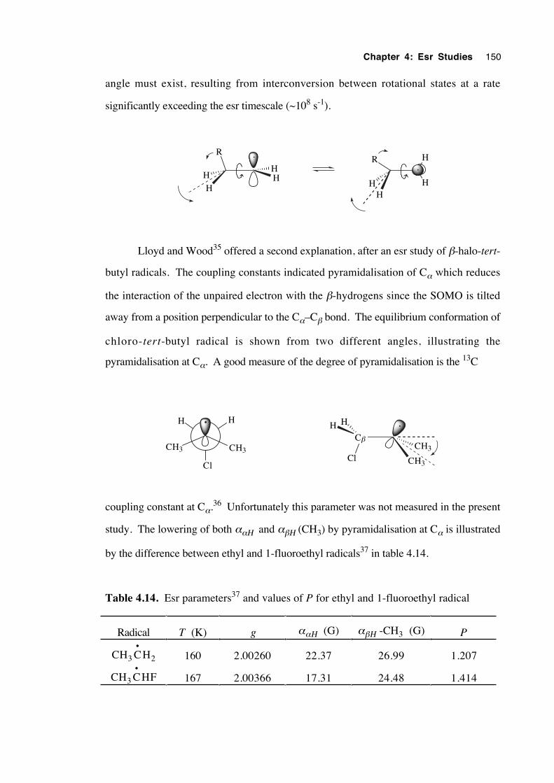
Chapter 4: Esr Studies 150
angle must exist, resulting from interconversion between rotational states at a rate
significantly exceeding the esr timescale (~108 s-1).
HH
H
H
R
HH
R H
H
Lloyd and Wood35 offered a second explanation, after an esr study of β-halo-tert-
butyl radicals. The coupling constants indicated pyramidalisation of Cα which reduces
the interaction of the unpaired electron with the β-hydrogens since the SOMO is tilted
away from a position perpendicular to the Cα–Cβ bond. The equilibrium conformation of
chloro-tert-butyl radical is shown from two different angles, illustrating the
pyramidalisation at Cα. A good measure of the degree of pyramidalisation is the 13C
Cl
CH3CH3
HH
Cβ
CH3
CH3Cl
H H
coupling constant at Cα.36 Unfortunately this parameter was not measured in the present
study. The lowering of both ααΗ and αβΗ (CH3) by pyramidalisation at Cα is illustrated
by the difference between ethyl and 1-fluoroethyl radicals37 in table 4.14.
Table 4.14. Esr parameters37 and values of P for ethyl and 1-fluoroethyl radical
Radical T (K) g ααΗ (G) αβΗ -CH3 (G) P
CH3 C•
H2 160 2.00260 22.37 26.99 1.207
CH3 C•
HF 167 2.00366 17.31 24.48 1.414
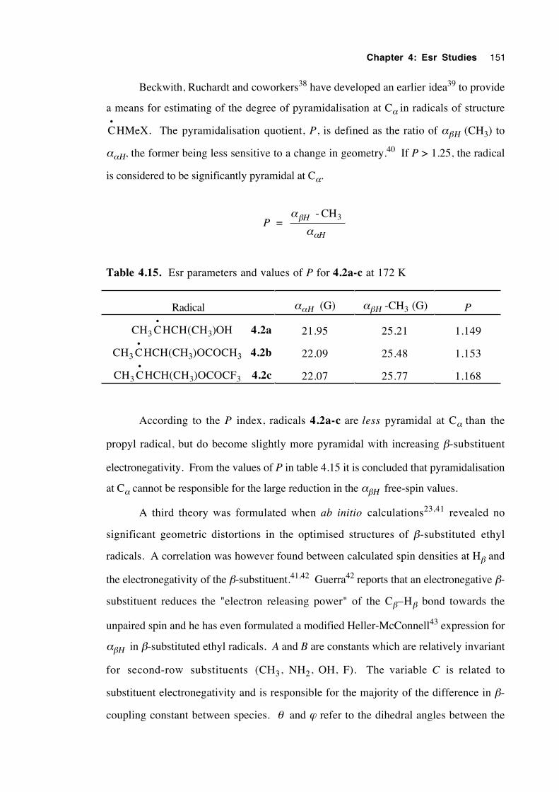
Chapter 4: Esr Studies 151
Beckwith, Ruchardt and coworkers38 have developed an earlier idea39 to provide
a means for estimating of the degree of pyramidalisation at Cα in radicals of structure
C•
HMeX. The pyramidalisation quotient, P, is defined as the ratio of αβΗ (CH3) to
ααΗ, the former being less sensitive to a change in geometry.40 If P > 1.25, the radical
is considered to be significantly pyramidal at Cα.
P = α
αβ
α
H
H
- CH
3
Table 4.15. Esr parameters and values of P for 4.2a-c at 172 K
Radical ααΗ (G) αβΗ -CH3 (G) P
CH3 C•
HCH(CH3)OH 4.2a 21.95 25.21 1.149
CH3 C•
HCH(CH3)OCOCH3 4.2b 22.09 25.48 1.153
CH3 C•
HCH(CH3)OCOCF3 4.2c 22.07 25.77 1.168
According to the P index, radicals 4.2a-c are less pyramidal at Cα than the
propyl radical, but do become slightly more pyramidal with increasing β-substituent
electronegativity. From the values of P in table 4.15 it is concluded that pyramidalisation
at Cα cannot be responsible for the large reduction in the αβΗ free-spin values.
A third theory was formulated when ab initio calculations23,41 revealed no
significant geometric distortions in the optimised structures of β-substituted ethyl
radicals. A correlation was however found between calculated spin densities at Hβ and
the electronegativity of the β-substituent.41,42 Guerra42 reports that an electronegative β-
substituent reduces the "electron releasing power" of the Cβ–Hβ bond towards the
unpaired spin and he has even formulated a modified Heller-McConnell43 expression for
αβΗ in β-substituted ethyl radicals. A and B are constants which are relatively invariant
for second-row substituents (CH3, NH2, OH, F). The variable C is related to
substituent electronegativity and is responsible for the majority of the difference in β-
coupling constant between species. θ and ϕ refer to the dihedral angles between the
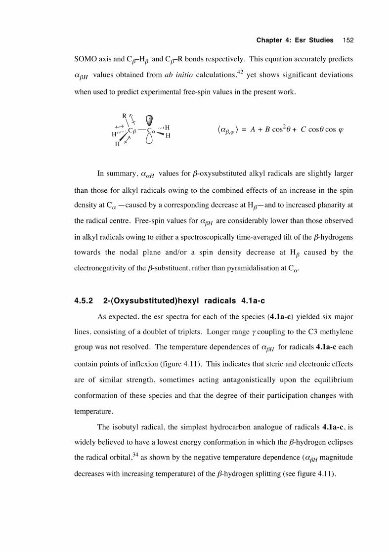
Chapter 4: Esr Studies 152
SOMO axis and Cβ–Hβ and Cβ–R bonds respectively. This equation accurately predicts
αβΗ values obtained from ab initio calculations,42 yet shows significant deviations
when used to predict experimental free-spin values in the present work.
Cβ CαHH
H
H
R
⟨αβ,ϕ ⟩ = A + B cos2θ + C cosθ cos ϕ
In summary, ααH values for β-oxysubstituted alkyl radicals are slightly larger
than those for alkyl radicals owing to the combined effects of an increase in the spin
density at Cα —caused by a corresponding decrease at Hβ—and to increased planarity at
the radical centre. Free-spin values for αβH are considerably lower than those observed
in alkyl radicals owing to either a spectroscopically time-averaged tilt of the β-hydrogens
towards the nodal plane and/or a spin density decrease at Hβ caused by the
electronegativity of the β-substituent, rather than pyramidalisation at Cα.
4.5.2 2-(Oxysubstituted)hexyl radicals 4.1a-c
As expected, the esr spectra for each of the species (4.1a-c) yielded six major
lines, consisting of a doublet of triplets. Longer range γ coupling to the C3 methylene
group was not resolved. The temperature dependences of αβH for radicals 4.1a-c each
contain points of inflexion (figure 4.11). This indicates that steric and electronic effects
are of similar strength, sometimes acting antagonistically upon the equilibrium
conformation of these species and that the degree of their participation changes with
temperature.
The isobutyl radical, the simplest hydrocarbon analogue of radicals 4.1a-c, is
widely believed to have a lowest energy conformation in which the β-hydrogen eclipses
the radical orbital,34 as shown by the negative temperature dependence (αβH magnitude
decreases with increasing temperature) of the β-hydrogen splitting (see figure 4.11).
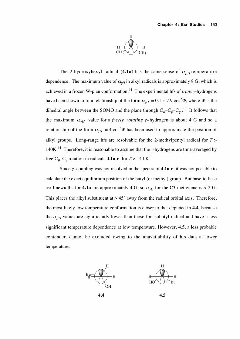
Chapter 4: Esr Studies 153
CH3
H
CH3
HH
The 2-hydroxyhexyl radical (4.1a) has the same sense of αβH temperature
dependence. The maximum value of αγH in alkyl radicals is approximately 8 G, which is
achieved in a frozen W-plan conformation.44 The experimental hfs of trans γ-hydrogens
have been shown to fit a relationship of the form αγH = 0.1 + 7.9 cos2Φ, where Φ is the
dihedral angle between the SOMO and the plane through Cα–Cβ–Cγ .44 It follows that
the maximum αγH value for a freely rotating γ−hydrogen is about 4 G and so a
relationship of the form αγH = 4 cos2Φ has been used to approximate the position of
alkyl groups. Long-range hfs are resolvable for the 2-methylpentyl radical for T >
140K.44 Therefore, it is reasonable to assume that the γ-hydrogens are time-averaged by
free Cβ–Cγ rotation in radicals 4.1a-c, for T > 140 K.
Since γ−coupling was not resolved in the spectra of 4.1a-c, it was not possible to
calculate the exact equilibrium position of the butyl (or methyl) group. But base-to-base
esr linewidths for 4.1a are approximately 4 G, so αγH for the C3-methylene is < 2 G.
This places the alkyl substituent at > 45˚ away from the radical orbital axis. Therefore,
the most likely low temperature conformation is closer to that depicted in 4.4, because
the αβH values are significantly lower than those for isobutyl radical and have a less
significant temperature dependence at low temperature. However, 4.5, a less probable
contender, cannot be excluded owing to the unavailability of hfs data at lower
temperatures.
H
HH
H
Bu
OH
HHHO Bu
4.54.4
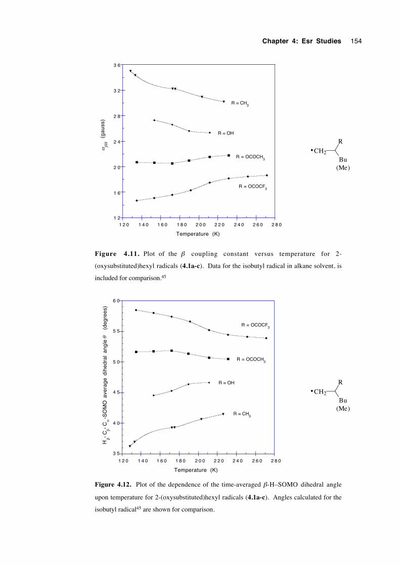
Chapter 4: Esr Studies 154
1 2
1 6
2 0
2 4
2 8
3 2
3 6
1 2 0 1 4 0 1 6 0 1 8 0 2 0 0 2 2 0 2 4 0 2 6 0 2 8 0
(g
au
ss)
αβΗ
Temperature (K)
R = CH3
R = OH
R = OCOCH3
R = OCOCF3
CH2
R
Bu
•
(Me)
Figure 4.11. Plot of the β coupling constant versus temperature for 2-
(oxysubstituted)hexyl radicals (4.1a-c). Data for the isobutyl radical in alkane solvent, is
included for comparison.45
3 5
4 0
4 5
5 0
5 5
6 0
1 2 0 1 4 0 1 6 0 1 8 0 2 0 0 2 2 0 2 4 0 2 6 0 2 8 0
Temperature (K)
Hβ-
Cβ-
Cα
-SO
MO
ave
rage
dih
edra
l an
gle
(
degr
ees)
θ
R = OH
R = OCOCH3
R = OCOCF3
R = CH3
CH2
R
Bu
•
(Me)
Figure 4.12. Plot of the dependence of the time-averaged β-H–SOMO dihedral angle
upon temperature for 2-(oxysubstituted)hexyl radicals (4.1a-c). Angles calculated for the
isobutyl radical45 are shown for comparison.
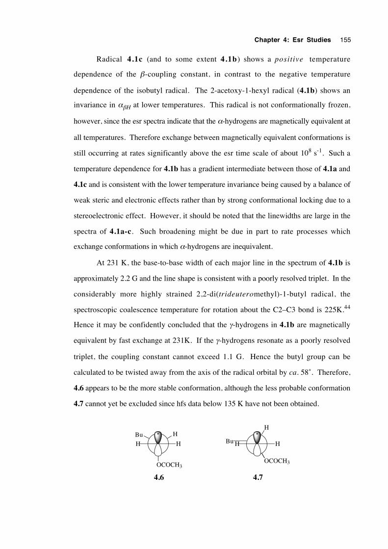
Chapter 4: Esr Studies 155
Radical 4.1c (and to some extent 4.1b) shows a positive temperature
dependence of the β-coupling constant, in contrast to the negative temperature
dependence of the isobutyl radical. The 2-acetoxy-1-hexyl radical (4.1b) shows an
invariance in αβH at lower temperatures. This radical is not conformationally frozen,
however, since the esr spectra indicate that the α-hydrogens are magnetically equivalent at
all temperatures. Therefore exchange between magnetically equivalent conformations is
still occurring at rates significantly above the esr time scale of about 108 s-1. Such a
temperature dependence for 4.1b has a gradient intermediate between those of 4.1a and
4.1c and is consistent with the lower temperature invariance being caused by a balance of
weak steric and electronic effects rather than by strong conformational locking due to a
stereoelectronic effect. However, it should be noted that the linewidths are large in the
spectra of 4.1a-c. Such broadening might be due in part to rate processes which
exchange conformations in which α-hydrogens are inequivalent.
At 231 K, the base-to-base width of each major line in the spectrum of 4.1b is
approximately 2.2 G and the line shape is consistent with a poorly resolved triplet. In the
considerably more highly strained 2,2-di(trideuteromethyl)-1-butyl radical, the
spectroscopic coalescence temperature for rotation about the C2–C3 bond is 225K.44
Hence it may be confidently concluded that the γ-hydrogens in 4.1b are magnetically
equivalent by fast exchange at 231K. If the γ-hydrogens resonate as a poorly resolved
triplet, the coupling constant cannot exceed 1.1 G. Hence the butyl group can be
calculated to be twisted away from the axis of the radical orbital by ca. 58˚. Therefore,
4.6 appears to be the more stable conformation, although the less probable conformation
4.7 cannot yet be excluded since hfs data below 135 K have not been obtained.
HBu
OCOCH3
HH
H
OCOCH3
HHBu
4.6 4.7
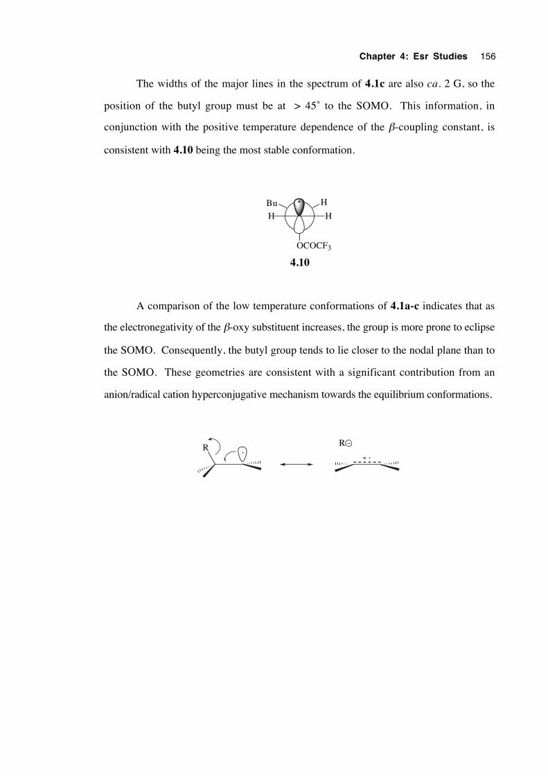
Chapter 4: Esr Studies 156
The widths of the major lines in the spectrum of 4.1c are also ca. 2 G, so the
position of the butyl group must be at > 45˚ to the SOMO. This information, in
conjunction with the positive temperature dependence of the β-coupling constant, is
consistent with 4.10 being the most stable conformation.
HBu
OCOCF3
HH
4.10
A comparison of the low temperature conformations of 4.1a-c indicates that as
the electronegativity of the β-oxy substituent increases, the group is more prone to eclipse
the SOMO. Consequently, the butyl group tends to lie closer to the nodal plane than to
the SOMO. These geometries are consistent with a significant contribution from an
anion/radical cation hyperconjugative mechanism towards the equilibrium conformations.
R R
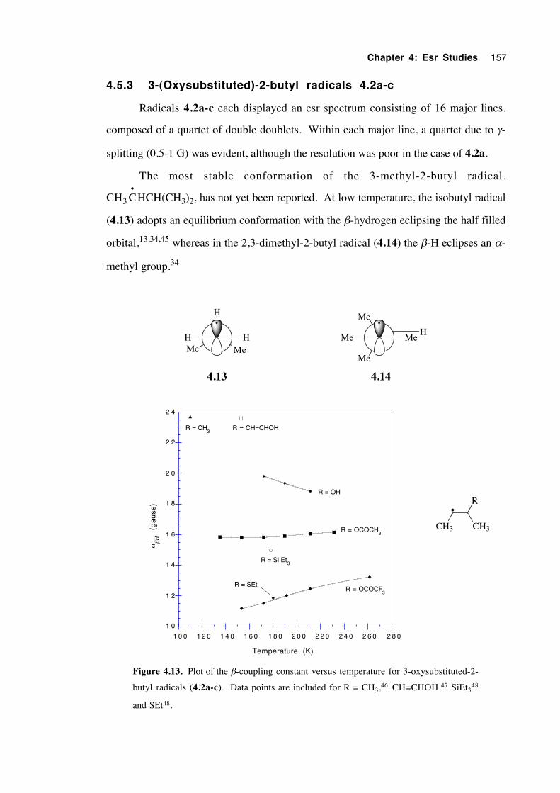
Chapter 4: Esr Studies 157
4.5.3 3-(Oxysubstituted)-2-butyl radicals 4.2a-c
Radicals 4.2a-c each displayed an esr spectrum consisting of 16 major lines,
composed of a quartet of double doublets. Within each major line, a quartet due to γ-
splitting (0.5-1 G) was evident, although the resolution was poor in the case of 4.2a.
The most stable conformation of the 3-methyl-2-butyl radical,
CH3 C•
HCH(CH3)2, has not yet been reported. At low temperature, the isobutyl radical
(4.13) adopts an equilibrium conformation with the β-hydrogen eclipsing the half filled
orbital,13,34,45 whereas in the 2,3-dimethyl-2-butyl radical (4.14) the β-H eclipses an α-
methyl group.34
Me
H
MeHH
Me
Me
MeMeH
4.13 4.14
1 0
1 2
1 4
1 6
1 8
2 0
2 2
2 4
1 0 0 1 2 0 1 4 0 1 6 0 1 8 0 2 0 0 2 2 0 2 4 0 2 6 0 2 8 0
(g
au
ss)
αβΗ
Temperature (K)
R = CH3
R = CH=CHOH
R = OH
R = OCOCH3
R = OCOCF3
R = Si Et3
R = SEt
R
CH3CH3
•
Figure 4.13. Plot of the β-coupling constant versus temperature for 3-oxysubstituted-2-
butyl radicals (4.2a-c). Data points are included for R = CH3,46 CH=CHOH,47 SiEt348
and SEt48.
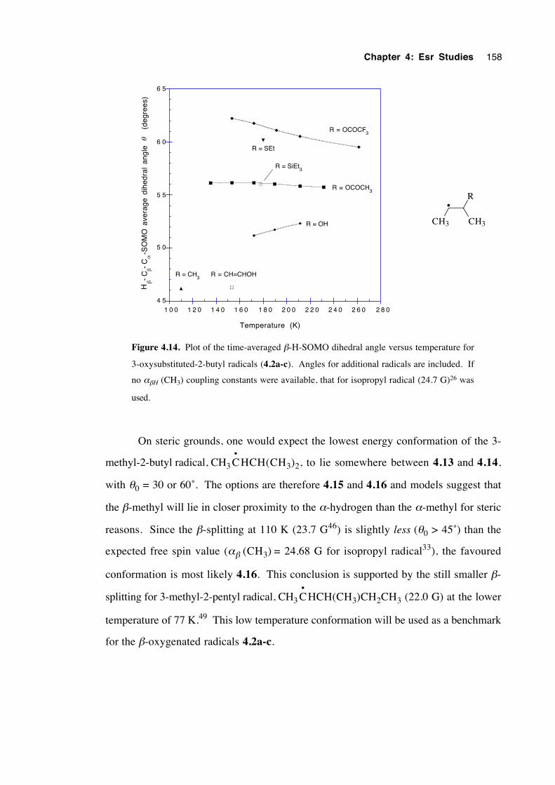
Chapter 4: Esr Studies 158
4 5
5 0
5 5
6 0
6 5
1 0 0 1 2 0 1 4 0 1 6 0 1 8 0 2 0 0 2 2 0 2 4 0 2 6 0 2 8 0
Temperature (K)
Hβ-
Cβ-
Cα
-SO
MO
ave
rage
dih
edra
l an
gle
(deg
rees
)θ
R = CH3
R = CH=CHOH
R = OH
R = OCOCH3
R = OCOCF3
R = SEt
R = SiEt3
R
CH3CH3
•
Figure 4.14. Plot of the time-averaged β-H-SOMO dihedral angle versus temperature for
3-oxysubstituted-2-butyl radicals (4.2a-c). Angles for additional radicals are included. If
no αβH (CH3) coupling constants were available, that for isopropyl radical (24.7 G)26 was
used.
On steric grounds, one would expect the lowest energy conformation of the 3-
methyl-2-butyl radical, CH3 C•
HCH(CH3)2, to lie somewhere between 4.13 and 4.14,
with θ0 = 30 or 60˚. The options are therefore 4.15 and 4.16 and models suggest that
the β-methyl will lie in closer proximity to the α-hydrogen than the α-methyl for steric
reasons. Since the β-splitting at 110 K (23.7 G46) is slightly less (θ0 > 45˚) than the
expected free spin value (αβ (CH3) = 24.68 G for isopropyl radical33), the favoured
conformation is most likely 4.16. This conclusion is supported by the still smaller β-
splitting for 3-methyl-2-pentyl radical, CH3 C•
HCH(CH3)CH2CH3 (22.0 G) at the lower
temperature of 77 K.49 This low temperature conformation will be used as a benchmark
for the β-oxygenated radicals 4.2a-c.
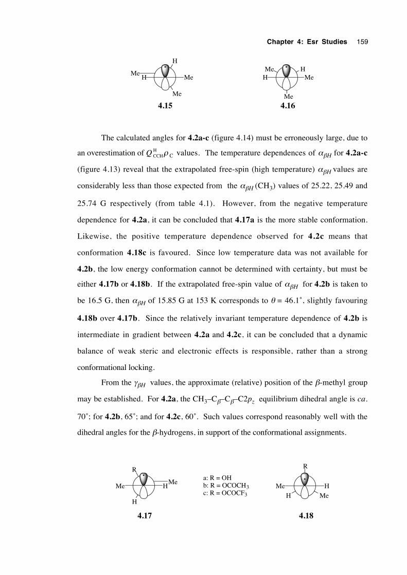
Chapter 4: Esr Studies 159
Me
Me HH
Me
MeH MeHMe
4.15 4.16
The calculated angles for 4.2a-c (figure 4.14) must be erroneously large, due to
an overestimation of Q CCHH ρ C values. The temperature dependences of αβH for 4.2a-c
(figure 4.13) reveal that the extrapolated free-spin (high temperature) αβH values are
considerably less than those expected from the αβH (CH3) values of 25.22, 25.49 and
25.74 G respectively (from table 4.1). However, from the negative temperature
dependence for 4.2a, it can be concluded that 4.17a is the more stable conformation.
Likewise, the positive temperature dependence observed for 4.2c means that
conformation 4.18c is favoured. Since low temperature data was not available for
4.2b, the low energy conformation cannot be determined with certainty, but must be
either 4.17b or 4.18b. If the extrapolated free-spin value of αβH for 4.2b is taken to
be 16.5 G, then αβH of 15.85 G at 153 K corresponds to θ = 46.1˚, slightly favouring
4.18b over 4.17b. Since the relatively invariant temperature dependence of 4.2b is
intermediate in gradient between 4.2a and 4.2c, it can be concluded that a dynamic
balance of weak steric and electronic effects is responsible, rather than a strong
conformational locking.
From the γβH values, the approximate (relative) position of the β-methyl group
may be established. For 4.2a, the CH3–Cβ–Cβ–C2pz equilibrium dihedral angle is ca.
70˚; for 4.2b, 65˚; and for 4.2c, 60˚. Such values correspond reasonably well with the
dihedral angles for the β-hydrogens, in support of the conformational assignments.
R
H
Me HMe
R
HMe
H Me
4.17 4.18
a: R = OHb: R = OCOCH3c: R = OCOCF3
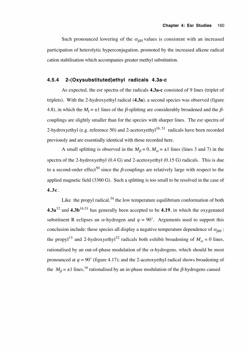
Chapter 4: Esr Studies 160
Such pronounced lowering of the αβH values is consistent with an increased
participation of heterolytic hyperconjugation, promoted by the increased alkene radical
cation stabilisation which accompanies greater methyl substitution.
4.5.4 2-(Oxysubstituted)ethyl radicals 4.3a-c
As expected, the esr spectra of the radicals 4.3a-c consisted of 9 lines (triplet of
triplets). With the 2-hydroxyethyl radical (4.3a), a second species was observed (figure
4.8), in which the MI = ±1 lines of the β-splitting are considerably broadened and the β-
couplings are slightly smaller than for the species with sharper lines. The esr spectra of
2-hydroxyethyl (e.g. reference 50) and 2-acetoxyethyl16, 51 radicals have been recorded
previously and are essentially identical with those recorded here.
A small splitting is observed in the Mβ = 0, Mα = ±1 lines (lines 3 and 7) in the
spectra of the 2-hydroxyethyl (0.4 G) and 2-acetoxyethyl (0.15 G) radicals. This is due
to a second-order effect50 since the β-couplings are relatively large with respect to the
applied magnetic field (3360 G). Such a splitting is too small to be resolved in the case of
4.3c .
Like the propyl radical,34 the low temperature equilibrium conformation of both
4.3a32 and 4.3b16,51 has generally been accepted to be 4.19, in which the oxygenated
substituent R eclipses an α-hydrogen and ϕ = 90˚. Arguments used to support this
conclusion include: these species all display a negative temperature dependence of αβH ;
the propyl13 and 2-hydroxyethyl32 radicals both exhibit broadening of Mα = 0 lines,
rationalised by an out-of-phase modulation of the α-hydrogens, which should be most
pronounced at ϕ = 90˚ (figure 4.17); and the 2-acetoxyethyl radical shows broadening of
the Mβ = ±1 lines,16 rationalised by an in-phase modulation of the β-hydrogens caused
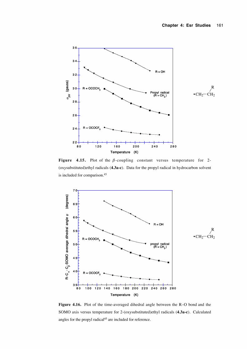
Chapter 4: Esr Studies 161
2 2
2 4
2 6
2 8
3 0
3 2
3 4
3 6
8 0 1 2 0 1 6 0 2 0 0 2 4 0 2 8 0
Temperature (K)
(g
au
ss)
αβΗ
R = OH
Propyl radical (R = CH
3)
R = OCOCH3
R = OCOCF3
2 2
2 4
2 6
2 8
3 0
3 2
3 4
3 6
8 0 1 2 0 1 6 0 2 0 0 2 4 0 2 8 0
Temperature (K)
(g
au
ss)
αβΗ
R = OH
Propyl radical (R = CH
3)
R = OCOCH3
R = OCOCF3
CH2 CH2
R
•
Figure 4.15. Plot of the β -coupling constant versus temperature for 2-
(oxysubstituted)ethyl radicals (4.3a-c). Data for the propyl radical in hydrocarbon solvent
is included for comparison.45
3 5
4 0
4 5
5 0
5 5
6 0
6 5
7 0
8 0 1 0 0 1 2 0 1 4 0 1 6 0 1 8 0 2 0 0 2 2 0 2 4 0 2 6 0 2 8 0
Temperature (K)
R = OH
propyl radical (R = CH
3)
R = OCOCH3
R = OCOCF3
R-C
α-
Cβ-S
OM
O a
vera
ge d
ihed
ral
angl
e
(
degr
ees)
ϕ
3 5
4 0
4 5
5 0
5 5
6 0
6 5
7 0
8 0 1 0 0 1 2 0 1 4 0 1 6 0 1 8 0 2 0 0 2 2 0 2 4 0 2 6 0 2 8 0
Temperature (K)
R = OH
propyl radical (R = CH
3)
R = OCOCH3
R = OCOCF3
R-C
α-
Cβ-S
OM
O a
vera
ge d
ihed
ral
angl
e
(
degr
ees)
ϕ
CH2 CH2
R
•
Figure 4.16. Plot of the time-averaged dihedral angle between the R–O bond and the
SOMO axis versus temperature for 2-(oxysubstituted)ethyl radicals (4.3a-c). Calculated
angles for the propyl radical45 are included for reference.
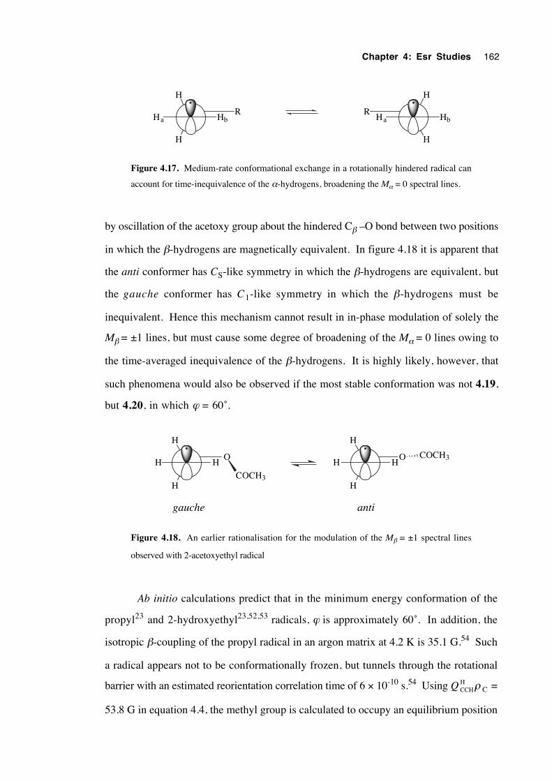
Chapter 4: Esr Studies 162
H
H
HbHaR
H
H
Ha HbR
Figure 4.17. Medium-rate conformational exchange in a rotationally hindered radical can
account for time-inequivalence of the α-hydrogens, broadening the Mα = 0 spectral lines.
by oscillation of the acetoxy group about the hindered Cβ –O bond between two positions
in which the β-hydrogens are magnetically equivalent. In figure 4.18 it is apparent that
the anti conformer has CS-like symmetry in which the β-hydrogens are equivalent, but
the gauche conformer has C1-like symmetry in which the β-hydrogens must be
inequivalent. Hence this mechanism cannot result in in-phase modulation of solely the
Mβ = ±1 lines, but must cause some degree of broadening of the Mα = 0 lines owing to
the time-averaged inequivalence of the β-hydrogens. It is highly likely, however, that
such phenomena would also be observed if the most stable conformation was not 4.19,
but 4.20, in which ϕ = 60˚.
H
H
HHO
COCH3
H
H
HHO COCH3
gauche anti
Figure 4.18. An earlier rationalisation for the modulation of the Mβ = ±1 spectral lines
observed with 2-acetoxyethyl radical
Ab initio calculations predict that in the minimum energy conformation of the
propyl23 and 2-hydroxyethyl23,52,53 radicals, ϕ is approximately 60˚. In addition, the
isotropic β-coupling of the propyl radical in an argon matrix at 4.2 K is 35.1 G.54 Such
a radical appears not to be conformationally frozen, but tunnels through the rotational
barrier with an estimated reorientation correlation time of 6 × 10⁻¹⁰ s.54 Using Q CCHH ρ C =
53.8 G in equation 4.4, the methyl group is calculated to occupy an equilibrium position
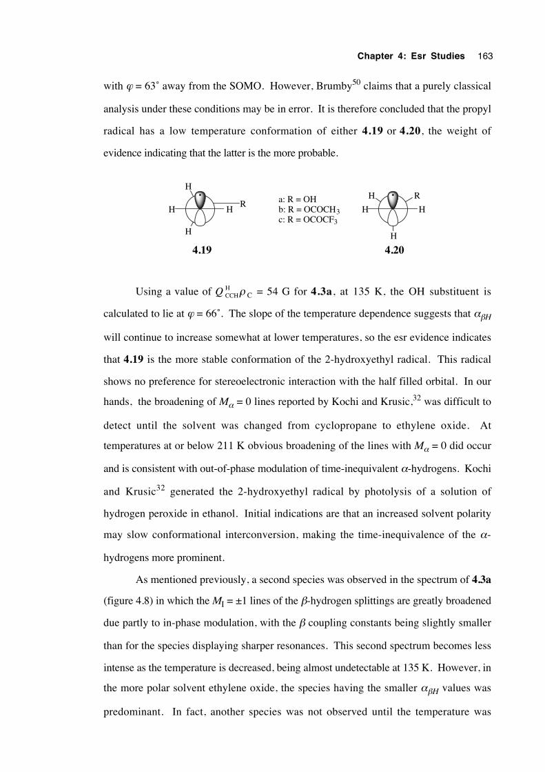
Chapter 4: Esr Studies 163
with ϕ = 63˚ away from the SOMO. However, Brumby50 claims that a purely classical
analysis under these conditions may be in error. It is therefore concluded that the propyl
radical has a low temperature conformation of either 4.19 or 4.20, the weight of
evidence indicating that the latter is the more probable.
H
H
HHR
4.19
a: R = OHb: R = OCOCH3c: R = OCOCF3
H
H
HH
R
4.20
Using a value of Q CCHH ρ C = 54 G for 4.3a, at 135 K, the OH substituent is
calculated to lie at ϕ = 66˚. The slope of the temperature dependence suggests that αβH
will continue to increase somewhat at lower temperatures, so the esr evidence indicates
that 4.19 is the more stable conformation of the 2-hydroxyethyl radical. This radical
shows no preference for stereoelectronic interaction with the half filled orbital. In our
hands, the broadening of Mα = 0 lines reported by Kochi and Krusic,32 was difficult to
detect until the solvent was changed from cyclopropane to ethylene oxide. At
temperatures at or below 211 K obvious broadening of the lines with Mα = 0 did occur
and is consistent with out-of-phase modulation of time-inequivalent α-hydrogens. Kochi
and Krusic32 generated the 2-hydroxyethyl radical by photolysis of a solution of
hydrogen peroxide in ethanol. Initial indications are that an increased solvent polarity
may slow conformational interconversion, making the time-inequivalence of the α-
hydrogens more prominent.
As mentioned previously, a second species was observed in the spectrum of 4.3a
(figure 4.8) in which the MI = ±1 lines of the β-hydrogen splittings are greatly broadened
due partly to in-phase modulation, with the β coupling constants being slightly smaller
than for the species displaying sharper resonances. This second spectrum becomes less
intense as the temperature is decreased, being almost undetectable at 135 K. However, in
the more polar solvent ethylene oxide, the species having the smaller αβH values was
predominant. In fact, another species was not observed until the temperature was
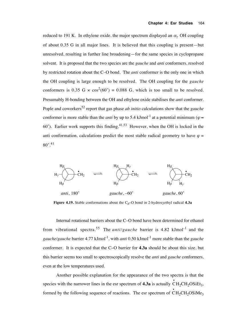
Chapter 4: Esr Studies 164
reduced to 191 K. In ethylene oxide, the major spectrum displayed an αγ OH coupling
of about 0.35 G in all major lines. It is believed that this coupling is present—but
unresolved, resulting in further line broadening—for the same species in cyclopropane
solvent. It is proposed that the two species are the gauche and anti conformers, resolved
by restricted rotation about the C–O bond. The anti conformer is the only one in which
the OH coupling is large enough to be resolved. The OH coupling for the gauche
conformers is 0.35 G × cos2(60˚) = 0.088 G, which is too small to be resolved.
Presumably H-bonding between the OH and ethylene oxide stabilises the anti conformer.
Pople and coworkers52 report that gas phase ab initio calculations show that the gauche
conformer is more stable than the anti by up to 5.4 kJmol-1 at a potential minimum (ϕ ≈
60˚). Earlier work supports this finding.41,53 However, when the OH is locked in the
anti conformation, calculations predict the most stable radical geometry to have ϕ =
80˚.41
Hγ CH2
Hβ
Hβ Hγ
CH2
Hβ
Hβ
Hγ
CH2
Hβ
Hβ
• • •
anti, 180˚ gauche, –60˚ gauche, 60˚
Figure 4.19. Stable conformations about the Cβ–O bond in 2-hydroxyethyl radical 4.3a
Internal rotational barriers about the C–O bond have been determined for ethanol
from vibrational spectra.55 The anti/gauche barrier is 4.82 kJmol-1 and the
gauche/gauche barrier 4.77 kJmol-1, with anti 0.50 kJmol-1 more stable than the gauche
conformer. It is expected that the C–O barrier for 4.3a should be about this size, but
this barrier seems too small to spectroscopically resolve the anti and gauche conformers,
even at the low temperatures used.
Another possible explanation for the appearance of the two spectra is that the
species with the narrower lines in the esr spectrum of 4.3a is actually C•
H2CH2OSiEt3,
formed by the following sequence of reactions. The esr spectrum of C•
H2CH2OSiMe3
Chapter 4: Esr Studies 165
has been recorded,56 yielding g = 2.00259, ααH = 22.10 G and αβH = 34.36 G at 160
K. Such parameters are very similar to those for 4.3a, but the authors report that the
spectrum undergoes no detectable linewidth alternation with changes in temperature,
contrary to the behaviour observed with 4.3a. In addition, a preponderance of
C•
H2CH2OSiEt3 in the spectrum would necessitate that the triethylsilyl radicals react
much faster with the tiny proportion of Et3SiOCH2CH2Br formed than with 2-
bromoethanol, a scenario which is highly unlikely.
t-BuO–OBut 2 t-BuO•hν
t-BuO• + Et3Si–H t-BuO–H + Et3Si•
Et3Si• + Br–CH2CH2OH Et3Si–Br + • CH2CH2OH
Et3Si–Br + H–OCH2CH2Br Et3Si–OCH2CH2Br + H–Br
Et3Si• + Br–CH2CH2OSiEt3 Et3Si–Br + • CH2CH2OSiEt3
Smaller β-splittings were observed for 4.3a in the more polar solvent. At 172 K,
the anti conformer gave αβH = 33.16 G in cyclopropane, and 32.60 G in ethylene oxide,
with values of 34.77 and 34.69 G respectively for the gauche conformers. The dielectric
constant for liquid cyclopropane remains unreported, but the refractive index at 230.65 K
is know to be 1.3799.57 Since Maxwell's equation states that the refractive index is
equal to approximately the square root of the dielectric constant, ε is approximately
equal to (1.3799)2 = 1.90. Ethylene oxide, having ε = 12.42 at 293 K58 is significantly
more polar than cyclopropane. Such a lowering in αβH values is consistent with an
increasing contribution of the ϕ = 0˚ rotamer to the time-averaged conformation by
stabilisation of the charged, hyperconjugative canonical structure. Thus, in ethylene
oxide the β-oxygen lone pairs appear to exhibit more steric repulsion from the α-H's or
alternatively, greater interaction with the SOMO, in support of some theoretical results.52
The most stable conformation of the 2-acetoxyethyl radical appears to be 4.19b
as judged from the slope of the temperature dependence, although steric and electronic
Chapter 4: Esr Studies 166
effects would be expected to destabilise this conformation relative to 4.19a. Since the
ester conformations of β-formyloxy radicals agree with those of the parent formates,59 it
is reasonable to assume that the Z conformation of β-acyloxyalkyl radicals is favoured, as
observed in the corresponding acetates and trifluoroacetates.60 In-phase modulation of
the M I = ±1 lines of the β-hydrogen splittings is also detected in spectra of 4.3b,
although it is less pronounced than for 4.3a. Kochi16 points out that such in-phase
modulation occurs because the magnetic environment of the β-hydrogens fluctuates
periodically and in such a way that each hydrogen is spectroscopically equivalent at all
times, with field fluctuation driven by oscillations of the β-substituent. Such processes
must occur at a rate not so slow that two discrete resonances are resolved (slow exchange
limit) and not so fast that a spectroscopically averaged field results (fast exchange limit).
However such an effect must also broaden the Mα = 0 spectral lines (2,5 and 8) of
radicals 4.3a-c, since rotamers about the Cβ–O can occupy one anti or two gauche
positions. When in the gauche conformation, the β-hydrogens are inequivalent. In
figures 4.8 and 4.9 it can be seen that line 2 is less than twice the height of line 1, an
outcome which supports the in-phase modulation explanation.
The selective broadening of the Mβ = ±1 lines can also be rationalised by the β-
CH2 group rocking in phase with Cα–Cβ bond rotation processes. As the β-methylene
group plane tilts toward the nodal plane, the β-hyperfine splitting decreases in size. It is
noteworthy that the in-phase modulation is most noticeable when the rotational barrier is
largest (see section 4.6); that is, the energy difference between the mutually perpendicular
conformations illustrated is large and conformational interconversion is slower. At high
interconversion rates, a time-averaged tilt angle results. As the temperature is decreased
and lower interconversion frequencies result, the averaged β-hyperfine splitting moves
away from the fast exchange limit and a broader range of magnetic field strength is
detected at the β-hydrogens. A time-averaged rock of amplitude 1˚ either side of an
equilibrium position may correspond to line broadening of up to 0.75 G, for a 30 G β-
coupling, assuming a cos2 relationship with the tilt angle.
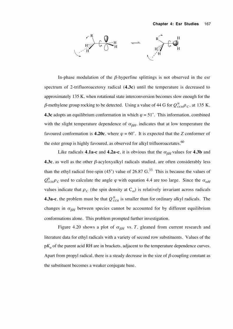
Chapter 4: Esr Studies 167
HH
H
H
R
HH
R H
H
In-phase modulation of the β-hyperfine splittings is not observed in the esr
spectrum of 2-trifluoroacetoxy radical (4.3c) until the temperature is decreased to
approximately 135 K, when rotational state interconversion becomes slow enough for the
β-methylene group rocking to be detected. Using a value of 44 G for Q CCHH ρ C, at 135 K,
4.3c adopts an equilibrium conformation in which ϕ = 51˚. This information, combined
with the slight temperature dependence of αβH, indicates that at low temperature the
favoured conformation is 4.20c, where ϕ = 60˚. It is expected that the Z conformer of
the ester group is highly favoured, as observed for alkyl trifluoroacetates.60
Like radicals 4.1a-c and 4.2a-c, it is obvious that the αβH values for 4.3b and
4.3c, as well as the other β-acyloxyalkyl radicals studied, are often considerably less
than the ethyl radical free-spin (45˚) value of 26.87 G.33 This is because the values of
QCCHH ρ C used to calculate the angle ϕ with equation 4.4 are too large. Since the ααH
values indicate that ρ C (the spin density at Cα) is relatively invariant across radicals
4.3a-c, the problem must be that Q CCHH is smaller than for ordinary alkyl radicals. The
changes in αβH between species cannot be accounted for by different equilibrium
conformations alone. This problem prompted further investigation.
Figure 4.20 shows a plot of αβH vs. T, gleaned from current research and
literature data for ethyl radicals with a variety of second row substituents. Values of the
pKa of the parent acid RH are in brackets, adjacent to the temperature dependence curves.
Apart from propyl radical, there is a steady decrease in the size of β-coupling constant as
the substituent becomes a weaker conjugate base.
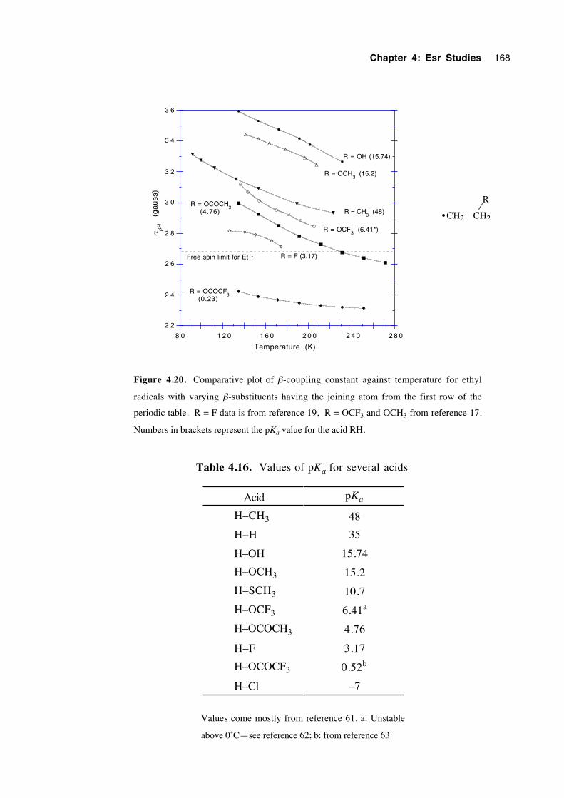
Chapter 4: Esr Studies 168
2 2
2 4
2 6
2 8
3 0
3 2
3 4
3 6
8 0 1 2 0 1 6 0 2 0 0 2 4 0 2 8 0
(g
au
ss)
Temperature (K)
R = OH (15.74)
R = CH3
(48)R = OCOCH
3
R = OCOCF3
R = F (3.17)
R = OCH3
(15.2)
R = OCF3
(6.41*)
(4.76)
(0.23)
Free spin limit for Et •
αβ
H
CH2 CH2
R
•
Figure 4.20. Comparative plot of β-coupling constant against temperature for ethyl
radicals with varying β-substituents having the joining atom from the first row of the
periodic table. R = F data is from reference 19, R = OCF3 and OCH3 from reference 17.
Numbers in brackets represent the pKa value for the acid RH.
Table 4.16. Values of pKa for several acids
Acid pKa
H–CH3 48
H–H 35
H–OH 15.74
H–OCH3 15.2
H–SCH3 10.7
H–OCF3 6.41a
H–OCOCH3 4.76
H–F 3.17
H–OCOCF3 0.52b
H–Cl –7
Values come mostly from reference 61. a: Unstable
above 0˚C—see reference 62; b: from reference 63
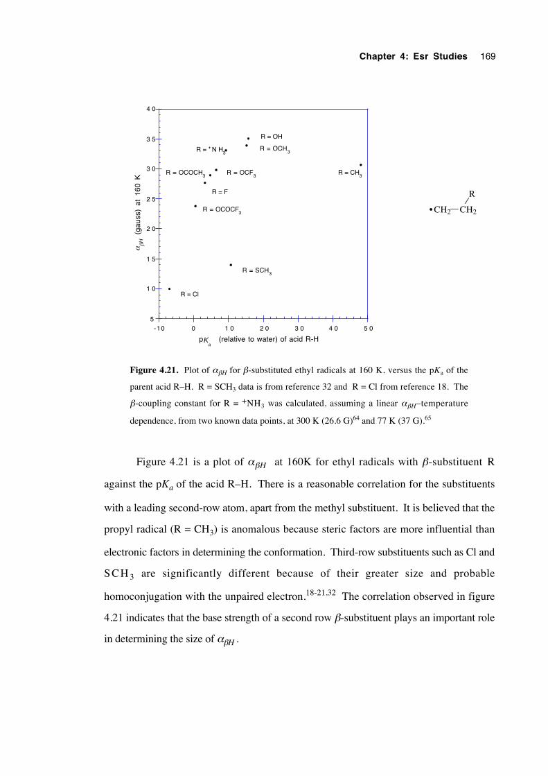
Chapter 4: Esr Studies 169
5
1 0
1 5
2 0
2 5
3 0
3 5
4 0
-10 0 1 0 2 0 3 0 4 0 5 0
R = OCOCF3
R = OCOCH3
R = Cl
R = OCF3
R = OCH3
R = CH3
R = OH
R = F
R = SCH3
R = + N H3
(gau
ss)
at 1
60 K
αβ
H
p (relative to water) of acid R-HKa
CH2 CH2
R
•
Figure 4.21. Plot of αβH for β-substituted ethyl radicals at 160 K, versus the pKa of the
parent acid R–H. R = SCH3 data is from reference 32 and R = Cl from reference 18. The
β-coupling constant for R = +NH3 was calculated, assuming a linear αβH–temperature
dependence, from two known data points, at 300 K (26.6 G)64 and 77 K (37 G).65
Figure 4.21 is a plot of αβH at 160K for ethyl radicals with β-substituent R
against the pKa of the acid R–H. There is a reasonable correlation for the substituents
with a leading second-row atom, apart from the methyl substituent. It is believed that the
propyl radical (R = CH3) is anomalous because steric factors are more influential than
electronic factors in determining the conformation. Third-row substituents such as Cl and
S C H 3 are significantly different because of their greater size and probable
homoconjugation with the unpaired electron.18-21,32 The correlation observed in figure
4.21 indicates that the base strength of a second row β-substituent plays an important role
in determining the size of αβH .
Chapter 4: Esr Studies 170
4.6 Estimation of the energy barrier to internal rotationabout the Cα–Cβ bond in the β-oxygenated ethyl radicals4.3a-c
The temperature dependence of αβH results from hindered rotation about the Cα–
Cβ bond. Recent 13C nmr studies66 indicate that internal rotational barriers in n-alkanes
vary from 14 to 20 kJmol-1. The rotational barriers for alkyl radicals34, 67 (which usually
range between 1.25 and 4.9 kJmol-1) are considerably smaller, due largely to the decrease
in steric hindrance associated with the change in hybridisation at Cα from sp3 to sp2.
Internal rotational barriers have been determined for the series of β-substituted
ethyl radicals (4.3a-c) in an attempt to discover whether a significant stereoelectronic
interaction exists between the unpaired electron and the vicinal substituent. The propyl
radical, which lacks any significant stereoelectronic effect, was used as a benchmark. A
repulsive electrostatic interaction would be expected to result in an increased barrier due to
an increase in the energy of the least stable conformation (ϕ = 0, 180˚). Similarly, a
weakly-attracting interaction should lower the barrier. A significantly stabilising
interaction (> 15 kJmol-1) would be expected to lock a radical into a particular
conformation, creating a large rotational barrier, as in the β-haloethyl radicals.18-21
However, this latter effect was not observed. Radicals 4.3a-c were chosen specifically
for reasons of structural simplicity and degree of symmetry, so enabling a greater
confidence in the suitability of the theoretical model and a simplification of the
calculations.
Rotational energy barriers have previously been determined experimentally from
temperature dependent esr β-coupling constants by quantum mechanical,32,45,68 classical
limit approximation32,67,68 and lineshape analysis13,37,44,69-71 methods. Numerous
attempts have also been made to estimate rotational barriers by theoretical
methods.22,23,41,52,67,72-76 An adapted classical limit approximation method was chosen
in this study.
An expression for the averaged, temperature-dependent value of the β-coupling
constant at the classical limit of a quantum mechanical analysis is given by equation
4.6,13
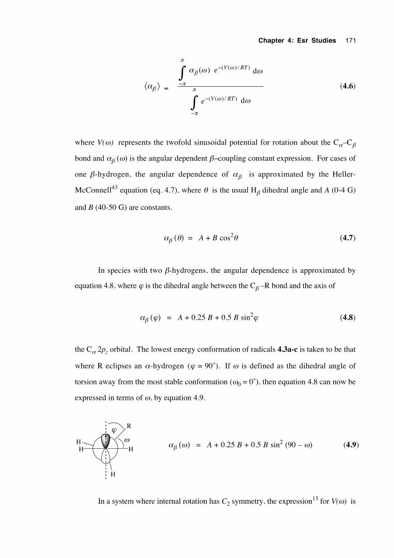
Chapter 4: Esr Studies 171
⟨αβ ⟩ =
−
−
−
−
∫
∫
( ( ) / )
( ( ) / )
( )
V RT
V RT
e
e
ωβ
π
π
ω
π
π
α ω ω
ω
d
d
(4.6)
where V(ω) represents the twofold sinusoidal potential for rotation about the Cα–Cβ
bond and αβ (ω) is the angular dependent β−coupling constant expression. For cases of
one β-hydrogen, the angular dependence of αβ is approximated by the Heller-
McConnell43 equation (eq. 4.7), where θ is the usual Hβ dihedral angle and A (0-4 G)
and B (40-50 G) are constants.
αβ (θ) = A + B cos2θ (4.7)
In species with two β-hydrogens, the angular dependence is approximated by
equation 4.8, where ϕ is the dihedral angle between the Cβ –R bond and the axis of
αβ (ϕ) = A + 0.25 B + 0.5 B sin2ϕ (4.8)
the Cα 2pz orbital. The lowest energy conformation of radicals 4.3a-c is taken to be that
where R eclipses an α-hydrogen (ϕ = 90˚). If ω is defined as the dihedral angle of
torsion away from the most stable conformation (ω0 = 0˚), then equation 4.8 can now be
expressed in terms of ω, by equation 4.9.
H
R
HHH
ωϕ
αβ (ω) = A + 0.25 B + 0.5 B sin2 (90 – ω) (4.9)
In a system where internal rotation has C2 symmetry, the expression13 for V(ω) is
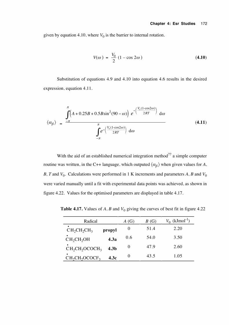
Chapter 4: Esr Studies 172
given by equation 4.10, where V0 is the barrier to internal rotation.
V(ω ) = V0
2 (1 – cos 2ω ) (4.10)
Substitution of equations 4.9 and 4.10 into equation 4.6 results in the desired
expression, equation 4.11.
⟨αβ ⟩ =
−
−
−
−
+ + −( )∫
∫
V
RT
V
RT
A B B e
e
0
0
22
2
0 25 0 5 90(1-cos2
(1-cos2
d
d
ω
π
π
ω
π
π
ω ω
ω
)
)
. . sin ( )
(4.11)
With the aid of an established numerical integration method77 a simple computer
routine was written, in the C++ language, which outputed ⟨αβ ⟩ when given values for A,
B, T and V0. Calculations were performed in 1 K increments and parameters A, B and V0
were varied manually until a fit with experimental data points was achieved, as shown in
figure 4.22. Values for the optimised parameters are displayed in table 4.17.
Table 4.17. Values of A, B and V0 giving the curves of best fit in figure 4.22
Radical A (G) B (G) V0 (kJmol-1)
C•
H2CH2CH3 propyl 0 51.4 2.20
C•
H2CH2OH 4.3a 0.6 54.0 3.50
C•
H2CH2OCOCH3 4.3b 0 47.9 2.60
C•
H2CH2OCOCF3 4.3c 0 43.5 1.05
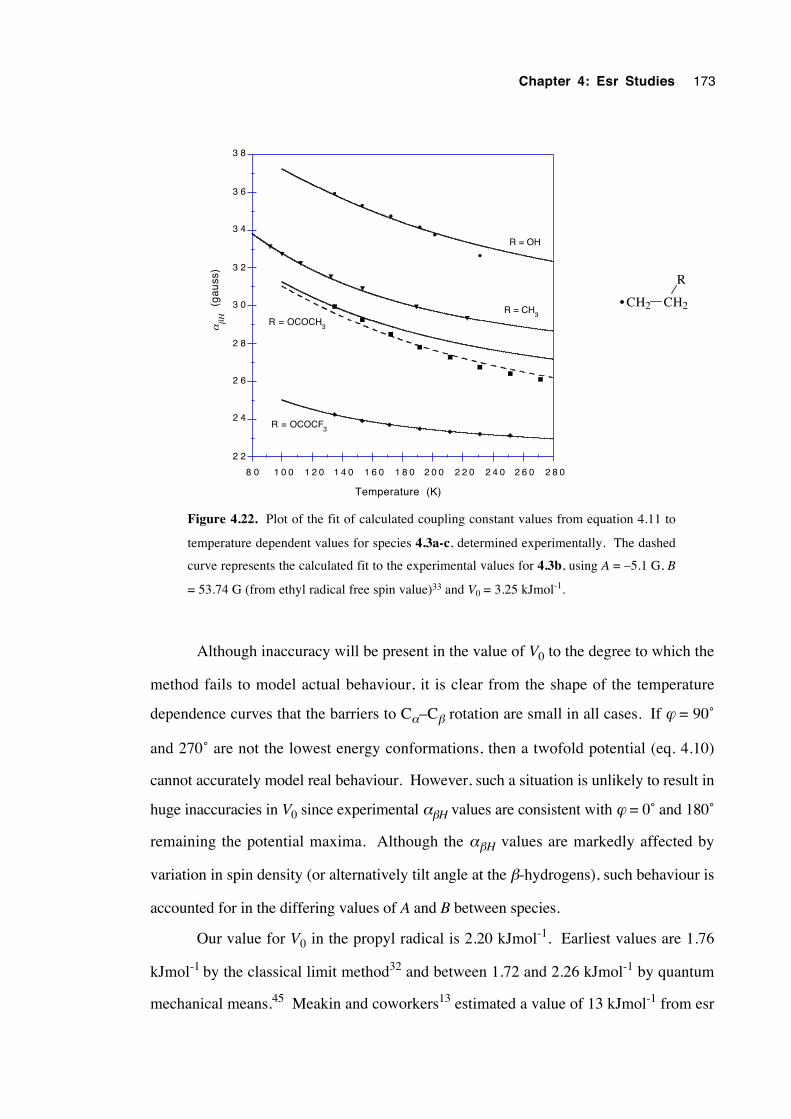
Chapter 4: Esr Studies 173
2 2
2 4
2 6
2 8
3 0
3 2
3 4
3 6
3 8
8 0 1 0 0 1 2 0 1 4 0 1 6 0 1 8 0 2 0 0 2 2 0 2 4 0 2 6 0 2 8 0
(g
au
ss)
αβΗ
Temperature (K)
R = OH
R = CH3
R = OCOCH3
R = OCOCF3
CH2 CH2
R
•
Figure 4.22. Plot of the fit of calculated coupling constant values from equation 4.11 to
temperature dependent values for species 4.3a-c, determined experimentally. The dashed
curve represents the calculated fit to the experimental values for 4.3b, using A = –5.1 G, B
= 53.74 G (from ethyl radical free spin value)33 and V0 = 3.25 kJmol-1.
Although inaccuracy will be present in the value of V0 to the degree to which the
method fails to model actual behaviour, it is clear from the shape of the temperature
dependence curves that the barriers to Cα–Cβ rotation are small in all cases. If ϕ = 90˚
and 270˚ are not the lowest energy conformations, then a twofold potential (eq. 4.10)
cannot accurately model real behaviour. However, such a situation is unlikely to result in
huge inaccuracies in V0 since experimental αβH values are consistent with ϕ = 0˚ and 180˚
remaining the potential maxima. Although the αβH values are markedly affected by
variation in spin density (or alternatively tilt angle at the β-hydrogens), such behaviour is
accounted for in the differing values of A and B between species.
Our value for V0 in the propyl radical is 2.20 kJmol-1. Earliest values are 1.76
kJmol-1 by the classical limit method32 and between 1.72 and 2.26 kJmol-1 by quantum
mechanical means.45 Meakin and coworkers13 estimated a value of 13 kJmol-1 from esr
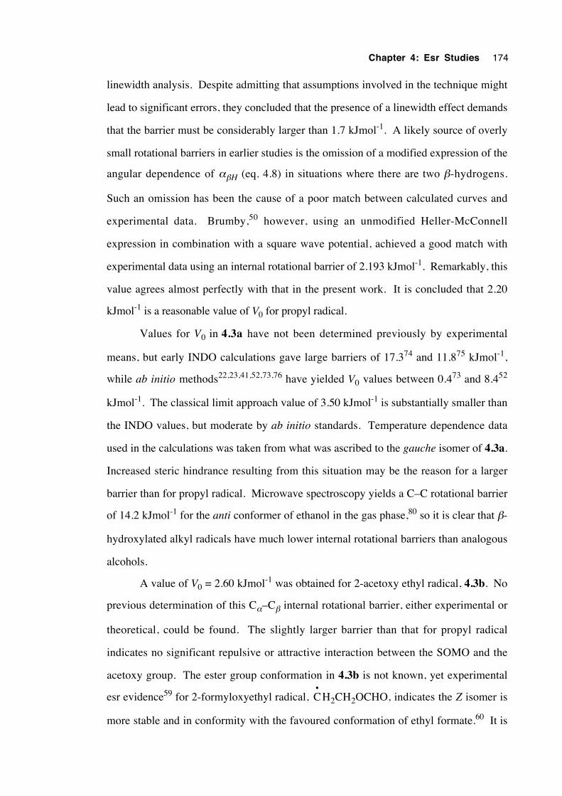
Chapter 4: Esr Studies 174
linewidth analysis. Despite admitting that assumptions involved in the technique might
lead to significant errors, they concluded that the presence of a linewidth effect demands
that the barrier must be considerably larger than 1.7 kJmol-1. A likely source of overly
small rotational barriers in earlier studies is the omission of a modified expression of the
angular dependence of αβH (eq. 4.8) in situations where there are two β-hydrogens.
Such an omission has been the cause of a poor match between calculated curves and
experimental data. Brumby,50 however, using an unmodified Heller-McConnell
expression in combination with a square wave potential, achieved a good match with
experimental data using an internal rotational barrier of 2.193 kJmol-1. Remarkably, this
value agrees almost perfectly with that in the present work. It is concluded that 2.20
kJmol-1 is a reasonable value of V0 for propyl radical.
Values for V0 in 4.3a have not been determined previously by experimental
means, but early INDO calculations gave large barriers of 17.374 and 11.875 kJmol-1,
while ab initio methods22,23,41,52,73,76 have yielded V0 values between 0.473 and 8.452
kJmol-1. The classical limit approach value of 3.50 kJmol-1 is substantially smaller than
the INDO values, but moderate by ab initio standards. Temperature dependence data
used in the calculations was taken from what was ascribed to the gauche isomer of 4.3a.
Increased steric hindrance resulting from this situation may be the reason for a larger
barrier than for propyl radical. Microwave spectroscopy yields a C–C rotational barrier
of 14.2 kJmol-1 for the anti conformer of ethanol in the gas phase,80 so it is clear that β-
hydroxylated alkyl radicals have much lower internal rotational barriers than analogous
alcohols.
A value of V0 = 2.60 kJmol-1 was obtained for 2-acetoxy ethyl radical, 4.3b. No
previous determination of this Cα–Cβ internal rotational barrier, either experimental or
theoretical, could be found. The slightly larger barrier than that for propyl radical
indicates no significant repulsive or attractive interaction between the SOMO and the
acetoxy group. The ester group conformation in 4.3b is not known, yet experimental
esr evidence59 for 2-formyloxyethyl radical, C•
H2CH2OCHO, indicates the Z isomer is
more stable and in conformity with the favoured conformation of ethyl formate.60 It is
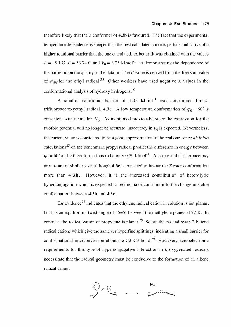
Chapter 4: Esr Studies 175
therefore likely that the Z conformer of 4.3b is favoured. The fact that the experimental
temperature dependence is steeper than the best calculated curve is perhaps indicative of a
higher rotational barrier than the one calculated. A better fit was obtained with the values
A = –5.1 G, B = 53.74 G and V0 = 3.25 kJmol-1, so demonstrating the dependence of
the barrier upon the quality of the data fit. The B value is derived from the free spin value
of αβH for the ethyl radical.33 Other workers have used negative A values in the
conformational analysis of hydroxy hydrogens.40
A smaller rotational barrier of 1.05 kJmol-1 was determined for 2-
trifluoroacetoxyethyl radical, 4.3c. A low temperature conformation of ϕ0 = 60˚ is
consistent with a smaller V0. As mentioned previously, since the expression for the
twofold potential will no longer be accurate, inaccuracy in V0 is expected. Nevertheless,
the current value is considered to be a good approximation to the real one, since ab initio
calculations23 on the benchmark propyl radical predict the difference in energy between
ϕ0 = 60˚ and 90˚ conformations to be only 0.59 kJmol-1. Acetoxy and trifluoroacetoxy
groups are of similar size, although 4.3c is expected to favour the Z ester conformation
more than 4.3b . However, it is the increased contribution of heterolytic
hyperconjugation which is expected to be the major contributor to the change in stable
conformation between 4.3b and 4.3c.
Esr evidence78 indicates that the ethylene radical cation in solution is not planar,
but has an equilibrium twist angle of 45±5˚ between the methylene planes at 77 K. In
contrast, the radical cation of propylene is planar.79 So are the cis and trans 2-butene
radical cations which give the same esr hyperfine splittings, indicating a small barrier for
conformational interconversion about the C2–C3 bond.79 However, stereoelectronic
requirements for this type of hyperconjugative interaction in β-oxygenated radicals
necessitate that the radical geometry must be conducive to the formation of an alkene
radical cation.
R R
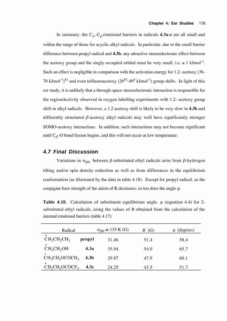
Chapter 4: Esr Studies 176
In summary, the Cα–Cβ rotational barriers in radicals 4.3a-c are all small and
within the range of those for acyclic alkyl radicals. In particular, due to the small barrier
difference between propyl radical and 4.3b, any attractive stereoelectronic effect between
the acetoxy group and the singly occupied orbital must be very small, i.e. ≤ 1 kJmol-1.
Such an effect is negligible in comparison with the activation energy for 1,2- acetoxy (36-
70 kJmol-1)81 and even trifluoroacetoxy (2682-494 kJmol-1) group shifts. In light of this
esr study, it is unlikely that a through-space stereoelectronic interaction is responsible for
the regioselectivity observed in oxygen labelling experiments with 1,2- acetoxy group
shift in alkyl radicals. However, a 1,2 acetoxy shift is likely to be very slow in 4.3b and
differently structured β-acetoxy alkyl radicals may well have significantly stronger
SOMO-acetoxy interactions. In addition, such interactions may not become significant
until Cβ–O bond fission begins, and this will not occur at low temperature.
4.7 Final Discussion
Variations in αβH between β-substituted ethyl radicals arise from β-hydrogen
tilting and/or spin density reduction as well as from differences in the equilibrium
conformation (as illustrated by the data in table 4.18). Except for propyl radical, as the
conjugate base strength of the anion of R decreases, so too does the angle ϕ.
Table 4.18. Calculation of substituent equilibrium angle, ϕ (equation 4.4) for 2-
substituted ethyl radicals, using the values of B obtained from the calculation of the
internal rotational barriers (table 4.17)
Radical αβH at 135 K (G) B (G) ϕ (degrees)
C•
H2CH2CH3 propyl 31.46 51.4 58.4
C•
H2CH2OH 4.3a 35.94 54.0 65.7
C•
H2CH2OCOCH3 4.3b 29.97 47.9 60.1
C•
H2CH2OCOCF3 4.3c 24.25 43.5 51.7
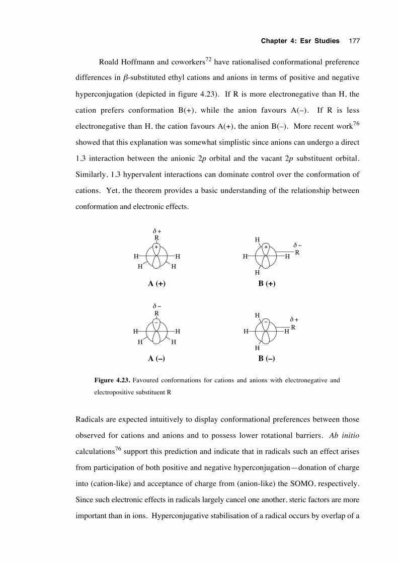
Chapter 4: Esr Studies 177
Roald Hoffmann and coworkers72 have rationalised conformational preference
differences in β-substituted ethyl cations and anions in terms of positive and negative
hyperconjugation (depicted in figure 4.23). If R is more electronegative than H, the
cation prefers conformation B(+), while the anion favours A(–). If R is less
electronegative than H, the cation favours A(+), the anion B(–). More recent work76
showed that this explanation was somewhat simplistic since anions can undergo a direct
1,3 interaction between the anionic 2p orbital and the vacant 2p substituent orbital.
Similarly, 1,3 hypervalent interactions can dominate control over the conformation of
cations. Yet, the theorem provides a basic understanding of the relationship between
conformation and electronic effects.
H
H
HHR
H
R
H
HH
+ +
H
H
HHR
H
R
H
HH
− −
δ +
δ −
δ +
δ −
A (+) B (+)
A (–) B (–)
Figure 4.23. Favoured conformations for cations and anions with electronegative and
electropositive substituent R
Radicals are expected intuitively to display conformational preferences between those
observed for cations and anions and to possess lower rotational barriers. Ab initio
calculations76 support this prediction and indicate that in radicals such an effect arises
from participation of both positive and negative hyperconjugation—donation of charge
into (cation-like) and acceptance of charge from (anion-like) the SOMO, respectively.
Since such electronic effects in radicals largely cancel one another, steric factors are more
important than in ions. Hyperconjugative stabilisation of a radical occurs by overlap of a
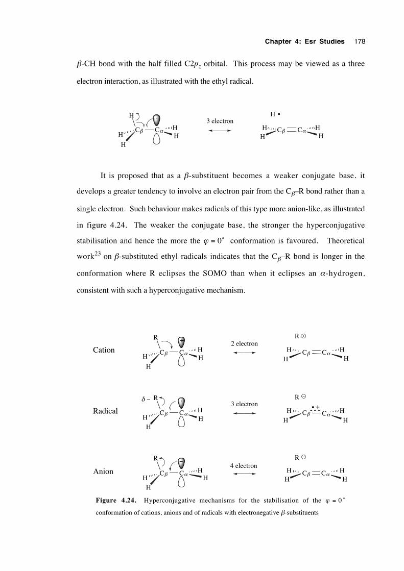
Chapter 4: Esr Studies 178
β-CH bond with the half filled C2pz orbital. This process may be viewed as a three
electron interaction, as illustrated with the ethyl radical.
Cβ CαHH
H
H
H
Cβ CαH
HHH
H3 electron
•
It is proposed that as a β-substituent becomes a weaker conjugate base, it
develops a greater tendency to involve an electron pair from the Cβ–R bond rather than a
single electron. Such behaviour makes radicals of this type more anion-like, as illustrated
in figure 4.24. The weaker the conjugate base, the stronger the hyperconjugative
stabilisation and hence the more the ϕ = 0˚ conformation is favoured. Theoretical
work23 on β-substituted ethyl radicals indicates that the Cβ–R bond is longer in the
conformation where R eclipses the SOMO than when it eclipses an α-hydrogen,
consistent with such a hyperconjugative mechanism.
Cβ CαH
H
H
H
R
Cβ CαH
HHH
R
Cβ CαHH
H
H
R
Cβ CαH
H
H
H
R
+
Cβ CαHH
H
H
R
Cβ CαH
H
H
H
R+
Cation
Radical
Anion
2 electron
3 electron
4 electron−
δ −•
Figure 4.24. Hyperconjugative mechanisms for the stabilisation of the ϕ = 0 ˚
conformation of cations, anions and of radicals with electronegative β-substituents
Chapter 4: Esr Studies 179
As well as β-substituent base strength, the degree of alkyl substitution of the
carbon skeleton influences the radical conformation. Alkyl substitution stabilises alkene
radical cations. At 191 K, the esr spectrum of 2-methyl-2-trifluoroacetoxy-1-heptyl
radical, C5H11(CH3)C(OCOCF3) C•
H2, has resolved γ splitting, where αγH averages to
about 1.0 G83. This is consistent with a conformation in which the trifluoroacetoxy
group eclipses the half-filled orbital. This radical is known to rearrange in a facile
manner.4
Kochi argues that it is not necessary to have a direct correlation between a
radical's stable conformation and propensity for rearrangement,16 yet many radicals
known to rearrange possess a favourable stereoelectronic preorientation. For instance, β-
chloroalkyl radicals, many of which are known to rearrange extremely rapidly, are known
to exist in an asymmetrically-bridged conformation in which the chlorine is eclipsed by
the SOMO.84 Similar conformational preferences are observed in carbohydrate radicals
with a β-C–O bond85 where such radicals undergo facile 1,2 acyloxy rearrangement.86
The neophyl radical, PhC(CH3)2 C•
H2, which rearranges readily to (CH3)2 C•
CH2Ph,
displays a γ splitting to the methyl groups of 1.05 G at 177 K.16 Such a splitting places
each methyl group at 60˚ to the SOMO so that the phenyl group eclipses the radical
orbital, the perfect preorientation for rearrangement. However 2-phenylethyl radical,
PhCH2 C•
H2, which does not rearrange readily, gives αβH = 30.94 G at 173 K.16 This
result is consistent with the favoured conformation where the phenyl group lies instead in
the equatorial plane (ϕ = 90˚).
In summary, the equilibrium conformation of alkyl radicals bearing second row β-
substituents is controlled by a combination of steric and electronic effects. The
conformation where the β-substituent eclipses the radical orbital becomes increasingly
favoured as the conjugate base strength of the β-substituent decreases and as the degree
of alkyl substitution at Cα and Cβ increases. This effect is attributed to an increasing
contribution from three electron, heterolytic hyperconjugation. In several cases a
correlation exists between this eclipsed conformation and the propensity for 1,2
rearrangement.
Chapter 4: Esr Studies 180
4.8 Conclusions
The esr spectra of β-oxysubstituted alkyl radicals are similar to those of
unsubstituted hydrocarbon radicals. There is no evidence for symmetric bridging or tight
conformational locking. The g factors are slightly lower than for alkyl radicals. Values of
ααH are slightly larger owing to an increase in spin density and decreased
pyramidalisation at Cα. Most conspicuous, however, is the large decrease in αβH for
radicals with β-substituents R in the order R = OH > OCOCH3 > OCOCF3. This is
caused not by pyramidalisation at Cα, but by either geometric distortion at Cβ , or by spin
density decrease at Hβ due to the electronegativity of R, or both.
Low temperature studies indicate that the conformation where R eclipses the
SOMO is favoured in radicals where R •• has low conjugate basicity and where there is
alkyl substitution at Cα and Cβ. An eclipsed conformation is optimal for β-acyloxyalkyl
radical rearrangement and is also stereoelectronically favourable for the formation of a
radical cation by the elimination of R •• . For β-substituents where the linking atom is
from the second row of the periodic table, there is a correlation between ααH and pKa of
the acid RH at 160 K.
Barriers to internal rotation are very low, within the range of normal alkyl radicals
and decrease in the order R = OH > OCOCH3 > OCOCF3. This situation is consistent
with increasing stabilisation by R of the conformation where the oxygenated substituent
and the half filled orbital are eclipsed. Any stereoelectronic interaction between the
acetoxy group and the SOMO is negligible, estimated to be < 1 kJmol-1.
The conformation of the radicals is governed by a balance of steric and
stereoelectronic effects. The principle stereoelectronic effect in this study was 3 electron
heterolytic hyperconjugation.
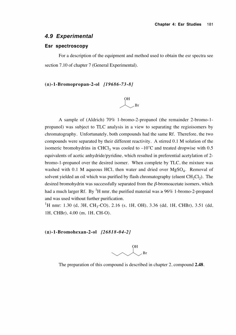
Chapter 4: Esr Studies 181
4.9 Experimental
Esr spectroscopy
For a description of the equipment and method used to obtain the esr spectra see
section 7.10 of chapter 7 (General Experimental).
(±)-1-Bromopropan-2-ol [19686-73-8]
OH
Br
A sample of (Aldrich) 70% 1-bromo-2-propanol (the remainder 2-bromo-1-
propanol) was subject to TLC analysis in a view to separating the regioisomers by
chromatography. Unfortunately, both compounds had the same Rf. Therefore, the two
compounds were separated by their different reactivity. A stirred 0.1 M solution of the
isomeric bromohydrins in CHCl3 was cooled to –10˚C and treated dropwise with 0.5
equivalents of acetic anhydride/pyridine, which resulted in preferential acetylation of 2-
bromo-1-propanol over the desired isomer. When complete by TLC, the mixture was
washed with 0.1 M aqueous HCl, then water and dried over MgSO4. Removal of
solvent yielded an oil which was purified by flash chromatography (eluent CH2Cl2). The
desired bromohydrin was successfully separated from the β-bromoacetate isomers, which
had a much larger Rf. By 1H nmr, the purified material was ≥ 96% 1-bromo-2-propanol
and was used without further purification.1H nmr: 1.30 (d, 3H, CH3-CO), 2.16 (s, 1H, OH), 3.36 (dd, 1H, CHBr), 3.51 (dd,
1H, CHBr), 4.00 (m, 1H, CH-O).
(±)-1-Bromohexan-2-ol [26818-04-2]
OH
Br
The preparation of this compound is described in chapter 2, compound 2.48.
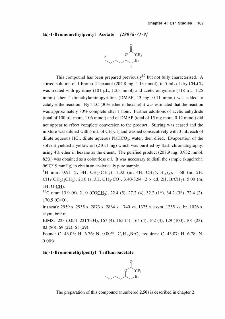
Chapter 4: Esr Studies 182
(±)-1-Bromomethylpentyl Acetate [28078-71-9]
O
Br
CH3
O
1
6
This compound has been prepared previously87 but not fully characterised. A
stirred solution of 1-bromo-2-hexanol (204.8 mg, 1.13 mmol), in 5 mL of dry CH2Cl2was treated with pyridine (101 µL, 1.25 mmol) and acetic anhydride (118 µL, 1.25
mmol), then 4-dimethylaminopyridine (DMAP, 13 mg, 0.11 mmol) was added to
catalyse the reaction. By TLC (30% ether in hexane) it was estimated that the reaction
was approximately 80% complete after 1 hour. Further additions of acetic anhydride
(total of 100 µL more, 1.06 mmol) and of DMAP (total of 15 mg more, 0.12 mmol) did
not appear to effect complete conversion to the product. Stirring was ceased and the
mixture was diluted with 5 mL of CH2Cl2 and washed consecutively with 3 mL each of
dilute aqueous HCl, dilute aqueous NaHCO3, water, then dried. Evaporation of the
solvent yielded a yellow oil (210.4 mg) which was purified by flash chromatography,
using 4% ether in hexane as the eluent. The purified product (207.9 mg, 0.932 mmol,
82%) was obtained as a colourless oil. It was necessary to distil the sample (kugelrohr,
96˚C/19 mmHg) to obtain an analytically pure sample.1H nmr: 0.91 (t, 3H, CH2-C H 3), 1.33 (m, 4H, CH3(C H 2)2), 1.68 (m, 2H,
CH3(CH2)2CH2), 2.10 (s, 3H, CH3-CO), 3.40-3.54 (2 × dd, 2H, BrCH2), 5.00 (m,
1H, O-CH).13C nmr: 13.9 (6), 21.0 (COCH3), 22.4 (5), 27.2 (4), 32.2 (1*), 34.2 (3*), 72.4 (2),
170.5 (C=O).
ir (neat): 2959 s, 2935 s, 2873 s, 2864 s, 1740 vs, 1375 s, asym, 1235 vs, br, 1026 s,
asym, 669 m.
EIMS: 223 (0.05), 221(0.04), 167 (4), 165 (5), 164 (4), 162 (4), 129 (100), 101 (23),
83 (80), 69 (22), 61 (29).
Found: C, 43.03; H, 6.76; N, 0.00%. C8H15BrO2 requires: C, 43.07; H, 6.78; N,
0.00%.
(±)-1-Bromomethylpentyl Trifluoroacetate
O
Br
CF3
O
The preparation of this compound (numbered 2.50) is described in chapter 2.
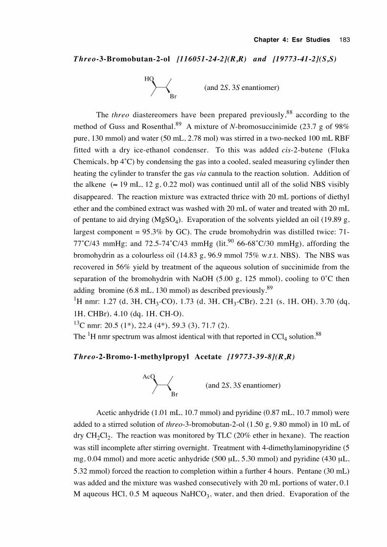
Chapter 4: Esr Studies 183
Threo-3-Bromobutan-2-ol [116051-24-2](R,R) and [19773-41-2](S,S)
HO
Br(and 2S, 3S enantiomer)
The threo diastereomers have been prepared previously,88 according to the
method of Guss and Rosenthal.89 A mixture of N-bromosuccinimide (23.7 g of 98%
pure, 130 mmol) and water (50 mL, 2.78 mol) was stirred in a two-necked 100 mL RBF
fitted with a dry ice-ethanol condenser. To this was added cis-2-butene (Fluka
Chemicals, bp 4˚C) by condensing the gas into a cooled, sealed measuring cylinder then
heating the cylinder to transfer the gas via cannula to the reaction solution. Addition of
the alkene (≈ 19 mL, 12 g, 0.22 mol) was continued until all of the solid NBS visibly
disappeared. The reaction mixture was extracted thrice with 20 mL portions of diethyl
ether and the combined extract was washed with 20 mL of water and treated with 20 mL
of pentane to aid drying (MgSO4). Evaporation of the solvents yielded an oil (19.89 g,
largest component = 95.3% by GC). The crude bromohydrin was distilled twice: 71-
77˚C/43 mmHg; and 72.5-74˚C/43 mmHg (lit.90 66-68˚C/30 mmHg), affording the
bromohydrin as a colourless oil (14.83 g, 96.9 mmol 75% w.r.t. NBS). The NBS was
recovered in 56% yield by treatment of the aqueous solution of succinimide from the
separation of the bromohydrin with NaOH (5.00 g, 125 mmol), cooling to 0˚C then
adding bromine (6.8 mL, 130 mmol) as described previously.89
1H nmr: 1.27 (d, 3H, CH3-CO), 1.73 (d, 3H, CH3-CBr), 2.21 (s, 1H, OH), 3.70 (dq,
1H, CHBr), 4.10 (dq, 1H, CH-O).13C nmr: 20.5 (1*), 22.4 (4*), 59.3 (3), 71.7 (2).
The 1H nmr spectrum was almost identical with that reported in CCl4 solution.88
Threo-2-Bromo-1-methylpropyl Acetate [19773-39-8](R,R)
AcO
Br(and 2S, 3S enantiomer)
Acetic anhydride (1.01 mL, 10.7 mmol) and pyridine (0.87 mL, 10.7 mmol) were
added to a stirred solution of threo-3-bromobutan-2-ol (1.50 g, 9.80 mmol) in 10 mL of
dry CH2Cl2. The reaction was monitored by TLC (20% ether in hexane). The reaction
was still incomplete after stirring overnight. Treatment with 4-dimethylaminopyridine (5
mg, 0.04 mmol) and more acetic anhydride (500 µL, 5.30 mmol) and pyridine (430 µL,
5.32 mmol) forced the reaction to completion within a further 4 hours. Pentane (30 mL)
was added and the mixture was washed consecutively with 20 mL portions of water, 0.1
M aqueous HCl, 0.5 M aqueous NaHCO3, water, and then dried. Evaporation of the
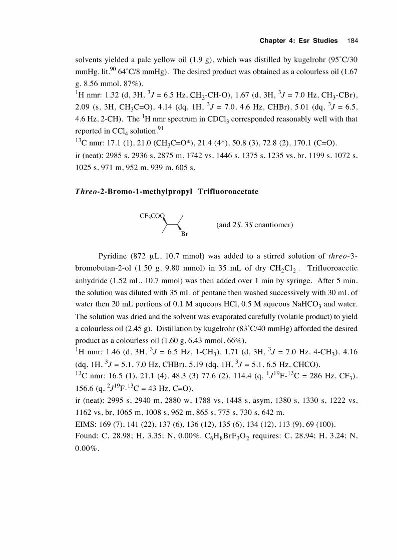
Chapter 4: Esr Studies 184
solvents yielded a pale yellow oil (1.9 g), which was distilled by kugelrohr (95˚C/30
mmHg, lit.90 64˚C/8 mmHg). The desired product was obtained as a colourless oil (1.67
g, 8.56 mmol, 87%).1H nmr: 1.32 (d, 3H, 3J = 6.5 Hz, CH3-CH-O), 1.67 (d, 3H, 3J = 7.0 Hz, CH3-CBr),
2.09 (s, 3H, CH3C=O), 4.14 (dq, 1H, 3J = 7.0, 4.6 Hz, CHBr), 5.01 (dq, 3J = 6.5,
4.6 Hz, 2-CH). The 1H nmr spectrum in CDCl3 corresponded reasonably well with that
reported in CCl4 solution.91
13C nmr: 17.1 (1), 21.0 (CH3C=O*), 21.4 (4*), 50.8 (3), 72.8 (2), 170.1 (C=O).
ir (neat): 2985 s, 2936 s, 2875 m, 1742 vs, 1446 s, 1375 s, 1235 vs, br, 1199 s, 1072 s,
1025 s, 971 m, 952 m, 939 m, 605 s.
Threo-2-Bromo-1-methylpropyl Trifluoroacetate
CF3COO
Br(and 2S, 3S enantiomer)
Pyridine (872 µL, 10.7 mmol) was added to a stirred solution of threo-3-
bromobutan-2-ol (1.50 g, 9.80 mmol) in 35 mL of dry CH2Cl2.. Trifluoroacetic
anhydride (1.52 mL, 10.7 mmol) was then added over 1 min by syringe. After 5 min,
the solution was diluted with 35 mL of pentane then washed successively with 30 mL of
water then 20 mL portions of 0.1 M aqueous HCl, 0.5 M aqueous NaHCO3 and water.
The solution was dried and the solvent was evaporated carefully (volatile product) to yield
a colourless oil (2.45 g). Distillation by kugelrohr (83˚C/40 mmHg) afforded the desired
product as a colourless oil (1.60 g, 6.43 mmol, 66%).1H nmr: 1.46 (d, 3H, 3J = 6.5 Hz, 1-CH3), 1.71 (d, 3H, 3J = 7.0 Hz, 4-CH3), 4.16
(dq, 1H, 3J = 5.1, 7.0 Hz, CHBr), 5.19 (dq, 1H, 3J = 5.1, 6.5 Hz, CHCO).13C nmr: 16.5 (1), 21.1 (4), 48.3 (3) 77.6 (2), 114.4 (q, 1J19F-13C = 286 Hz, CF3),
156.6 (q, 2J19F-13C = 43 Hz, C=O).
ir (neat): 2995 s, 2940 m, 2880 w, 1788 vs, 1448 s, asym, 1380 s, 1330 s, 1222 vs,
1162 vs, br, 1065 m, 1008 s, 962 m, 865 s, 775 s, 730 s, 642 m.
EIMS: 169 (7), 141 (22), 137 (6), 136 (12), 135 (6), 134 (12), 113 (9), 69 (100).
Found: C, 28.98; H, 3.35; N, 0.00%. C6H8BrF3O2 requires: C, 28.94; H, 3.24; N,
0.00%.
Chapter 4: Esr Studies 185
2-Bromoethyl Acetate [927-68-4]
Br OAc
A stirred solution of 2-bromoethanol (2.76 g, 22.1 mmol) in 15 mL of dry
CH2Cl2 was treated with acetic anhydride (3.45 mL, 36.6 mmol), pyridine (2.96 mL,
36.6 mmol) and 4-dimethylaminopyridine (10 mg, 0.08 mmol). After 6 hours the
mixture was poured into 50 mL of water, shaken and the layers were separated. The
aqueous phase was extracted twice with 10 mL portions of CH2Cl2, the organic phases
were combined and washed with 20 mL portions of 0.1 M aqueous HCl until the
washings were acidic. The organic extract was then washed with 20 mL of 0.5 M
aqueous NaHCO3 and 20 mL of water then dried. Careful removal of solvent under
reduced pressure (volatile product) and distillation by kugelrohr (96˚C/60 mmHg, lit.93
159˚C), afforded 2-bromoethyl acetate as a colourless oil (2.17 g, 13.0 mmol, 59%).
Accidental spillage was primarily responsible for the lowered yield.1H nmr: 2.11 (s, 3H, CH3), 3.52 (t, 2H, CH2Br), 4.39 (t, 2H, CH2O). The nmr
spectrum was identical with that of authentic material (Aldrich).
2-Bromoethyl Trifluoroacetate [76045-93-7]
Br OCOCF3
Pyridine (1.60 mL, 19.8 mmol) was added to a stirred solution of 2-bromoethanol
(2.24 g, 17.9 mmol) in 50 mL of CH2Cl2 at 0˚C. Trifluoroacetic anhydride (2.79 mL,
19.8 mmol) was added slowly over 1 min. The mixture was stirred at room temperature
overnight (10 min is enough) then washed with 100 mL of water and the aqueous
washings were back-extracted twice with 50 mL portions of pentane. The organic
extracts were combined and washed successively with 30 mL each of 0.1 M aqueous
HCl, 0.5 M aqueous NaHCO3, water and then dried. Careful evaporation (volatile
product) of the solvent yielded a mobile oil which was distilled by kegelrohr (80˚C/60
mmHg) to give 2-bromoethyl trifluoroacetate as a colourless oil (2.76 g, 12.5 mmol,
70%). The 1H nmr spectrum was similar to that previously published in trifluoroacetic
acid solution.92
1H nmr: 3.59 (t, 2H, 3J = 6.2 Hz, CH2Br), 4.66 (t, 2H, 3J = 6.2 Hz, CH2O).13C nmr: 26.5 (2), 66.7 (1), 114.4 (q, 1J19F-13C = 286 Hz, CF3), 157.0 (q, 2J19F-13C
= 43 Hz, C=O).
Chapter 4: Esr Studies 186
ir (neat): 2975 w, asym, 1791 vs, 1398 m, 1350 s, 1283 m, asym, 1222 vs, 1160 vs, br,
989 w, 949 w, 775 m, sharp, 734 m, sharp, 672 w.
EIMS: 141 (13), 109 (18), 108 (8), 107 (18), 106 (8), 79 (92), 69 (53), 57 (29), 52
(100).
Found: C, 21.87; H, 2.02; N, 0.00%. C4H4BrF3O2 requires: C, 21.74; H, 1.82; N,
0.00%.
Chapter 4: Esr Studies 187
4.10 References
1. Beckwith, A. L. J. and Thomas, C. B. J. Chem. Soc., Perkin Trans. 2 1973, 861.
2. Beckwith, A. L. J. and Duggan, P. J. J. Chem. Soc., Chem. Commun. 1988, 1000.
3. Beckwith, A. L. J. and Duggan, P. J. J. Chem. Soc., Perkin Trans. 2 1992, 1777.
4. Harman, D. G. This thesis, Chapter 3.
5. Crich, D. and Filzen, G. F. J. Org. Chem. 1995, 60, 4834. Note that there is a
calculational error, giving the amount of translocation as 93%.
6. Zipse, H. and Bootz, M. J. Chem. Soc., Perkin Trans. 2 2001, 1566.
7. Kocovsky, P.; Stary, I. and Turecek, F. Tetrahedron Lett. 1986, 27, 1513.
8. Harman, D. G. This thesis, Chapter 3; Section 3.4
9. Halpern, J. Science 1985, 227, 869.
10. Gilbert, B. C., Norman, R. O. C. and Williams, P. S. J. Chem. Soc., Perkin Trans.
2 1981, 1401.
11. Ayscough, P. B. Electron Spin Resonance in Chemistry; Methuen and Co. Ltd:
London, 1967; p 76.
12. Radtsig, V. A. J. Struct. Chem. 1970, 11, 221.
13. Krusic, P. J.; Meakin, P. and Jesson, J. P. J. Phys. Chem. 1971, 75, 3438.
14. Cochran, E. L.; Adrian, F. J. and Bowers, V. A. J. Chem. Phys. 1961, 34, 1161.
15. Hudson, A. and Jackson, R. A. Chem. Commun. 1969, 1323.
16. Edge, D. J. and Kochi, J. K. J. Am. Chem. Soc. 1972, 94, 7695.
17. Chen, K. S. and Kochi, J. K. J. Am. Chem. Soc. 1974, 96, 1383.
18. Kawamura, T.; Edge, D. J. and Kochi, J. K. J. Am. Chem. Soc. 1972, 94, 1752.
19. Edge, D. J. and Kochi, J. K. J. Am. Chem. Soc. 1972, 94, 6485.
20. Chen, K. S.; Elson, I. H. and Kochi, J. K. J. Am. Chem. Soc. 1973, 95, 5341.
21. Elson, I. H.; Chen, K. S. and Kochi, J. K. Chem. Phys. Lett. 1973, 21, 72.
22. Bernardi, F.; Bottoni, A.; Fossey, J. and Sorba, J. Tetrahedron 1986, 42, 5567.
23. Guerra, M. J. Am. Chem. Soc. 1992, 114, 2077.
24. Kochi, J. K. and Krusic, P. J. J. Am. Chem. Soc. 1969, 91, 3940.
25. Davies, M. J.; Gilbert, B. C.; Thomas, C. B. and Young, J. J. Chem. Soc., Perkin
Chapter 4: Esr Studies 188
Trans. 2 1985, 1199.
26. Elson, I. H. and Kochi, J. K. J. Org. Chem. 1974, 39, 2091.
27. Ayscough, P. B. Electron Spin Resonance in Chemistry; Methuen and Co. Ltd:
London, 1967; p 73.
28. Fischer, H. In Free Radicals; Kochi, J. K., Ed.; John Wiley and Sons: New York,
1973; Vol. 2, p 445.
29. Ibid. p 442.
30. Fossey, J.; Lefort, D. and Sorba, J. Free Radicals in Organic Chemistry; John
Wiley and Sons/Masson Paris, 1995, p 23.
31. Eberson, L.; Utley, J. H. P. and Hammerich, O. In Organic Electrochemistry; 3rd
ed.; Lund, H. and Baizer, M. M., Eds.; Marcel Dekker: New York, 1991; p 516.
32. Krusic, P. J. and Kochi, J. K. J. Am. Chem. Soc. 1971, 93, 846.
33. Fessenden, R. W. and Schuler, R. H. J. Chem. Phys. 1963, 39, 2147.
34. Kochi, J. K. Adv. Free-Radical Chem. 1975, 5, 189.
35. Lloyd, R. V. and Wood, D. E. J. Am. Chem. Soc. 1975, 97, 5986.
36. Fischer, H. In Free Radicals; Kochi, J. K., Ed.; John Wiley and Sons: New York,
1973; Vol. 2, p 448.
37. Chen, K. S.; Krusic, P. J.; Meakin, P. and Kochi, J. K. J. Phys. Chem. 1974, 78,
2014.
38. Brocks, J. J.; Beckhaus, H.-D.; Beckwith, A. L. J. and Ruchardt, C. J. Org. Chem.
1998, 63, 1935.
39. Dobbs, A. J.; Gilbert, B. C. and Norman, R. O. C. J. Chem. Soc. (A) 1971, 124.
40. See reference 36
41. Bernardi, F. and Fossey, J. J. Mol. Struct. 1988, 180, 79.
42. Guerra, M. Chem. Phys. Lett. 1987, 139, 463.
43. Heller, C. and McConnell, H. M. J. Chem. Phys. 1960, 32, 1535.
44. Ingold, K. U.; Nonhebel, D. C. and Walton, J. C. J. Phys. Chem. 1986, 90, 2859.
45. Fessenden, R. W. J. Chim. Phys. Phys.-Chim. Biol. 1964, 61, 1570.
46. Nunome, K.; Toriyama, K. and Iwasaki, M. Chem. Phys. Lett. 1984, 105, 414.
Chapter 4: Esr Studies 189
47. Mariano, P. S. and Bay, E. J. Org. Chem. 1980, 45, 1763.
48. Leardini, R.; Tundo, A.; Zanardi, G. and Pedulli, G. F. Tetrahedron 1983, 39,
2715.
49. Lin, D. P. J. Chin. Chem. Soc. 1974, 21, 201.
50. Brumby, S. J. Phys. Chem. 1983, 87, 1917.
51. Smith, P. and Karukstis, K. K. J. Mag. Reson. 1982, 46, 200.
52. Curtiss, L. A.; Lucas, D. J. and Pople, J. A. J. Chem. Phys. 1995, 102, 3292.
53. Sosa, C. and Schlegel, H. B. J. Am. Chem. Soc. 1987, 109, 4193.
54. McDowell, C. A. and Shimokoshi, K. J. Chem. Phys. 1974, 60, 1619.
55. Durig, J. R. and Larsen, R. A. J. Mol. Struct. 1989, 238, 195.
56. Edge, D. J. and Kochi, J. K. J. Chem. Soc. Perkin Trans. 2 1973, 182.
57. Grosse, A. V. J. Am. Chem. Soc. 1937, 59, 2739.
58. CRC Handbook of Physics and Chemistry, 82nd ed. (2001-2002); Lide, D. R.,
Ed.; CRC Press: Boca Raton, 2001; p 6-154.
59. Smith, P. and Karukstis, K. K. J. Mag. Reson. 1981, 43, 122.
60. Deslongchamps, P. Stereoelectronic Effects in Organic Chemistry; Pergamon:
Oxford, 1983; p 54.
61. March, J. Advanced Organic Chemistry; 3rd ed.; John Wiley: New York, 1985; pp
220-222, 230.
62. Kloter, G. and Seppelt, K. J. Am. Chem. Soc. 1979, 101, 347. Value of pKa
predicted by SciFinder Scholar database (CAS, 2002).
63. CRC Handbook of Physics and Chemistry, 82nd ed. (2001-2002); Lide, D. R.,
Ed.; CRC Press: Boca Raton, 2001; p 8-46.
64. Taniguchi, H.; Fukui, K.; Ohnishi, S.-i.; Hatano, H.; Hasegawa, H. and Maruyama,
T. J. Phys. Chem. 1968, 72, 1926.
65. Kotov, A. G.; Pukhal'skaya, G. V. and Pshezhetskii, S. Y. Khim. Vys. Energ.
1970, 4, 93.
66. Min, B. and Lee, J. W. J. Mol. Liq. 1999, 80, 33.
67. Kemball, M. L.; Walton, J. C. and Ingold, K. U. J. Chem. Soc., Perkin Trans. 2
Chapter 4: Esr Studies 190
1982, 1017.
68. Stone, E. W. and Maki, A. H. J. Chem. Phys. 1962, 37, 1326.
69. Hudson, A. and Luckhurst, G. R. Chem. Rev. 1969, 69, 191.
70. Kawamura, T.; Meakin, P. and Kochi, J. K. J. Am. Chem. Soc. 1972, 94, 8065.
71. Ingold, K. U.; Nonhebel, D. C. and Walton, J. C. J. Phys. Chem. 1985, 89, 4424.
72. Hoffmann, R.; Radom, L.; Pople, J. A.; Schleyer, P. v. R.; Hehre, W. J. and
Salem, L. J. Am. Chem. Soc. 1972, 94, 6221.
73. Radom, L.; Paviot, J. and Pople, J. A. Chem. Commun. 1974, 58.
74. Gilbert, B. C.; Trenwith, M. and Dobbs, A. J. J. Chem. Soc. Perkin Trans. 2 1974,
1772.
75. Wunsche, P. and Grossohme, T. Z. Phys. Chem. (Leipzig) 1977, 258, 426.
76. Pross, A. and Radom, L. Tetrahedron 1980, 36, 1999.
77. Press, W. H.; Flannery, B. P.; Teukolsky, A. S. and Vetterling, W. T. Numerical
Recipes; Cambridge University Press: Cambridge, 1986; pp 110-114.
78. Shiotani, M.; Nagata, Y. and Sohma, J. J. Am. Chem. Soc. 1984, 106, 4640.
79. Shiotani, M.; Nagata, Y. and Sohma, J. J. Phys. Chem. 1984, 88, 4078.
80. Sasada, Y. J. Mol. Struct. 1988, 190, 93.
81. Beckwith, A. L. J. and Duggan, P. J. J. Chem. Soc., Perkin Trans. 2 1993, 1673.
82. Choi, S.-Y.; Crich, D.; Horner, J. H.; Huang, X.; Newcomb, M. and Whitted, P.
O. Tetrahedron 1999, 55, 3317.
83. Beckwith, A. L. J. and Harman D. G. Unpublished results.
84. Beckwith, A. L. J and Ingold, K. U. In Rearrangements in Ground and Excited
States; Mayo, P. d., Ed.; Academic Press: New York, 1980; pp 162-310.
85. Korth, H. G.; Sustmann, R.; Dupuis, J.; Groninger, K. S.; Witzel, T. and Giese, B.
In Substituent Effects in Radical Chemistry; Viehe, H. G., Janousek, Z. and Merenyi,
R., Eds.; D. Reidel: Dordrecht, 1986; Vol. NATO ASI Series. Series C: Vol 89, p 297.
86. Beckwith, A. L. J.; Crich, D.; Duggan, P. J. and Yao, Q. Chem. Rev. 1997, 97,
3273.
87. Heasley, V. L.; Frye, C. L.; Heasley, G. E.; Martin, K. A.; Redfield, D. A. and
Chapter 4: Esr Studies 191
Wilday, P. S. Tetrahedron Lett. 1970, 1573.
88. Maurette, M.-T.; Gaset, A.; Mathis, R. and Lattes, A. Bull. Soc. Chim. Fr. 1975,
398.
89. Guss, G. O. and Rosenthal, R. J. Am. Chem. Soc. 1955, 77, 2549.
90. Dictionary of Organic Compounds; 5th ed.; Buckinham, J., Ed.: Chapman and
Hall: New York, 1982; Vol. 1, p 765, listing B-02025.
91. Gelas, J. and Michaud, S. Bull. Soc. Chim. Fr. 1972, 2445.
92. Smolina, T. A.; Brusova, G. P.; Shchekut'eva, L. F.; Gopius, E. D.; Permin, A. B.
and Reutov, O. A. Izv. Akad. Nauk SSSR, Ser. Khim. 1980, 2079.
93. Aldrich Fine Chemicals and Laboratory Equipment Catalogue, Australia and New
Zealand Edition, 2000-2001.
Chapter 5
The mechanism of the catalysed rearrangement of
N-alkoxy-2(1H)-pyridinethiones
5.1 Introduction 193
5.2 Review of the chemistry of N-alkoxy-2(1H)-pyridinethiones
and related compounds 193
5.3 The mechanism of the rearrangement of N-alkoxy-2(1H)-
pyridinethiones 206
5.3.1 The mode of catalysis 206
5.3.2 Kinetics 212
5.3.3 A study of rearrangement regiochemistry 219
5.3.4 A study of rearrangement stereochemistry 224
5.3.5 Electronic structure of the migrating group at C1 during
rearrangement 234
5.3.6 Substituent effects 237
5.3.7 Attempted detection and isolation of intermediates 239
5.4 Conclusions 247
5.5 Future work 249
5.6 Experimental 250
5.7 References 277
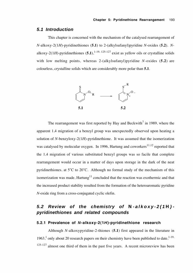
Chapter 5: Pyridinethione Rearrangement 193
5.1 Introduction
This chapter is concerned with the mechanism of the catalysed rearrangement of
N-alkoxy-2(1H)-pyridinethiones (5.1) to 2-(alkylsufanyl)pyridine N-oxides (5.2). N-
alkoxy-2(1H)-pyridinethiones (5.1),1-19, 125-127 exist as yellow oils or crystalline solids
with low melting points, whereas 2-(alkylsufanyl)pyridine N-oxides (5.2) are
colourless, crystalline solids which are considerably more polar than 5.1.
N
S
OR N
S
O
R
5.1 5.2
The rearrangement was first reported by Hay and Beckwith7 in 1989, where the
apparent 1,4 migration of a benzyl group was unexpectedly observed upon heating a
solution of N-benzyloxy-2(1H)-pyridinethione. It was assumed that the isomerization
was catalysed by molecular oxygen. In 1996, Hartung and coworkers11,12 reported that
the 1,4 migration of various substituted benzyl groups was so facile that complete
rearrangement would occur in a matter of days upon storage in the dark of the neat
pyridinethiones, at 5˚C to 20˚C. Although no formal study of the mechanism of this
isomerization was made, Hartung11 concluded that the reaction was exothermic and that
the increased product stability resulted from the formation of the heteroaromatic pyridine
N-oxide ring from a cross-conjugated cyclic olefin.
5.2 Review of the chemistry of N - a l k o x y - 2 ( 1 H ) -pyridinethiones and related compounds
5.2.1 Prevalence of N -alkoxy-2(1H )-pyridinethione research
Although N-alkoxypyridine-2-thiones (5.1) first appeared in the literature in
1963,1 only about 20 research papers on their chemistry have been published to date,1-19,
125-127 almost one third of them in the past five years. A recent microreview has been
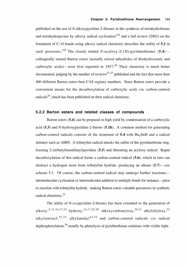
Chapter 5: Pyridinethione Rearrangement 194
published on the use of N-alkoxypyridine-2-thiones in the synthesis of tetrahydrofurans
and tetrahydropyrans by alkoxy radical cyclization125 and a full review (2002) on the
formation of C–O bonds using alkoxy radical chemistry describes the utility of 5.1 in
such processes.129 The closely related N-acyloxy-2(1H)-pyridinethiones (5.4) —
colloquially named Barton esters (actually mixed anhydrides of thiohydroxamic and
carboxylic acids)—were first reported in 1957.20 Their chemistry is much better
documented, judging by the number of reviews21-23 published and the fact that more than
400 different Barton esters bear CAS registry numbers. Since Barton esters provide a
convenient means for the decarboxylation of carboxylic acids via carbon-centred
radicals24, much has been published on their radical chemistry.
5.2.2 Barton esters and related classes of compounds
Barton esters (5.4) can be prepared in high yield by condensation of a carboxylic
acid (5.3) and N-hydroxypyridine-2-thione (5.1h). A common method for generating
carbon-centred radicals consists of the treatment of 5.4 with Bu3SnH and a radical
initiator such as AIBN. A tributyltin radical attacks the sulfur of the pyridinethione ring,
forming 2-(tributyltinsulfanyl)pyridine (5.5) and liberating an acyloxy radical. Rapid
decarboxylation of this radical forms a carbon-centred radical (5.6), which in turn can
abstract a hydrogen atom from tributyltin hydride, producing an alkane (5.7)—see
scheme 5.1. Of course, the carbon-centred radical may undergo further reactions—
intramolecular cyclization or intermolecular addition to multiple bonds for instance—prior
to reaction with tributyltin hydride, making Barton esters valuable precursors in synthetic
radical chemistry.21
The utility of N-oxypyridine-2-thiones has been extended to the generation of
alkoxy,5-12,16,17,19 hydroxy,16,17,25-29 alkoxycarbonyloxy,30,31 alkylsilyloxy,2 5
alkylaminyl,32,33 alkylamidyl34,35 and carbon-centred radicals via radical
dephosphorylation,36 usually by photolysis of pyridinethione solutions with visible light.
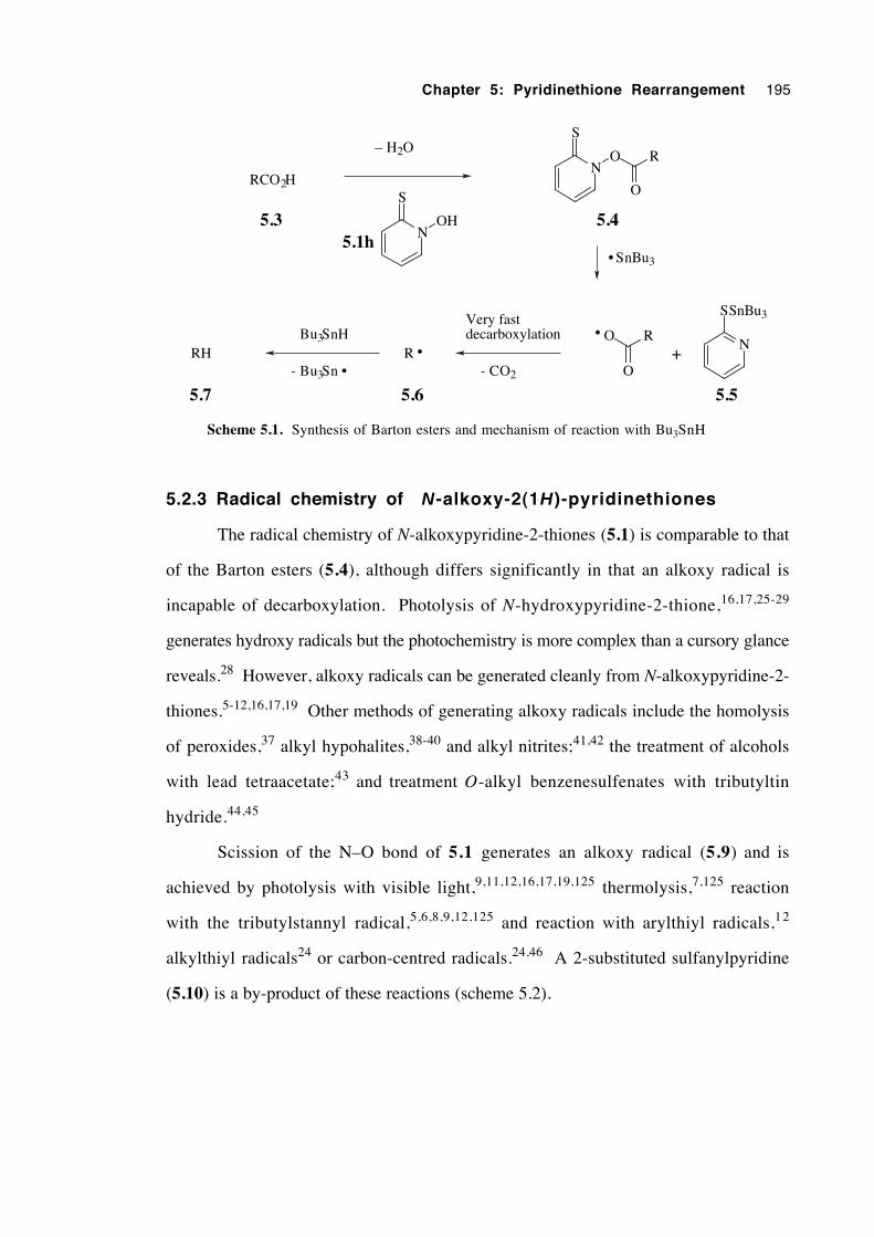
Chapter 5: Pyridinethione Rearrangement 195
RCO2H
N
S
OH
N
S
O R
O
SnBu3•
5.4
N
SSnBu3
– H2O
5.5
Bu3SnH
- Bu3Sn •RH
5.7
5.3
O R
O+
•
R •
5.6
Very fastdecarboxylation
- CO2
5.1h
Scheme 5.1. Synthesis of Barton esters and mechanism of reaction with Bu3SnH
5.2.3 Radical chemistry of N-alkoxy-2(1H)-pyridinethiones
The radical chemistry of N-alkoxypyridine-2-thiones (5.1) is comparable to that
of the Barton esters (5.4), although differs significantly in that an alkoxy radical is
incapable of decarboxylation. Photolysis of N-hydroxypyridine-2-thione,16,17,25-29
generates hydroxy radicals but the photochemistry is more complex than a cursory glance
reveals.28 However, alkoxy radicals can be generated cleanly from N-alkoxypyridine-2-
thiones.5-12,16,17,19 Other methods of generating alkoxy radicals include the homolysis
of peroxides,37 alkyl hypohalites,38-40 and alkyl nitrites;41,42 the treatment of alcohols
with lead tetraacetate;43 and treatment O-alkyl benzenesulfenates with tributyltin
hydride.44,45
Scission of the N–O bond of 5.1 generates an alkoxy radical (5.9) and is
achieved by photolysis with visible light,9,11,12,16,17,19,125 thermolysis,7,125 reaction
with the tributylstannyl radical,5,6,8,9,12,125 and reaction with arylthiyl radicals,12
alkylthiyl radicals24 or carbon-centred radicals.24,46 A 2-substituted sulfanylpyridine
(5.10) is a by-product of these reactions (scheme 5.2).
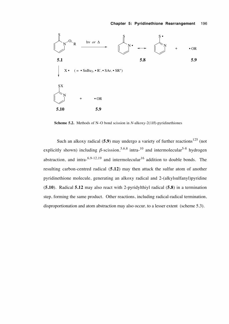
Chapter 5: Pyridinethione Rearrangement 196
N
S
OR
5.1
hν or ∆N
S •
+ • OR
5.8 5.9
X • ( = • SnBu3, • R', • SAr, • SR")
N
SX
5.10
+ • OR
5.9
N •
S
Scheme 5.2. Methods of N–O bond scission in N-alkoxy-2(1H)-pyridinethiones
Such an alkoxy radical (5.9) may undergo a variety of further reactions125 (not
explicitly shown) including β-scission,5,6,8 intra-10 and intermolecular5-8 hydrogen
abstraction, and intra-6,9-12,19 and intermolecular16 addition to double bonds. The
resulting carbon-centred radical (5.12) may then attack the sulfur atom of another
pyridinethione molecule, generating an alkoxy radical and 2-(alkylsulfanyl)pyridine
(5.10). Radical 5.12 may also react with 2-pyridylthiyl radical (5.8) in a termination
step, forming the same product. Other reactions, including radical-radical termination,
disproportionation and atom abstraction may also occur, to a lesser extent (scheme 5.3).
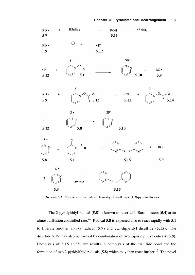
Chapter 5: Pyridinethione Rearrangement 197
RO • + ROH + • SnBu3HSnBu3
RO • • R'
RO •N
O Ar
H
S
+ ROH +N
O Ar
S
•
• R' + N
S •
N
SR'
N
S •
+ NO
S
RN S S N
+ RO •
N
S •
2hν or ∆ N S S N
5.9 5.11
5.9 5.12
5.9 5.13 5.11 5.14
5.12 5.8 5.10
5.8 5.1 5.15 5.9
5.8 5.15
• R'
5.12+ N
O
S
R
5.1
N
SR'
5.10+ RO •
5.9
Scheme 5.3. Overview of the radical chemistry of N-alkoxy-2(1H)-pyridinethiones
The 2-pyridylthiyl radical (5.8) is known to react with Barton esters (5.4) at an
almost diffusion controlled rate.46 Radical 5.8 is expected also to react rapidly with 5.1
to liberate another alkoxy radical (5.9) and 2,2'-dipyridyl disulfide (5.15). The
disulfide 5.15 may also be formed by combination of two 2-pyridylthiyl radicals (5.8).
Photolysis of 5.15 at 350 nm results in homolysis of the disulfide bond and the
formation of two 2-pyridylthiyl radicals (5.8) which may then react further.17 The novel
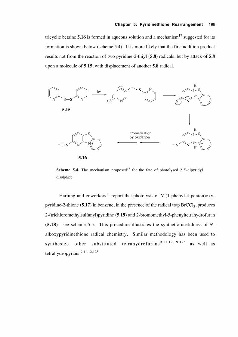
Chapter 5: Pyridinethione Rearrangement 198
tricyclic betaine 5.16 is formed in aqueous solution and a mechanism17 suggested for its
formation is shown below (scheme 5.4). It is more likely that the first addition product
results not from the reaction of two pyridine-2-thiyl (5.8) radicals, but by attack of 5.8
upon a molecule of 5.15, with displacement of another 5.8 radical.
N• SN S S N
hν N• S
N N
S
S
N N
S
S
H
H
HN N
S
O3S
aromatisationby oxidation
5.15
5.16
Scheme 5.4. The mechanism proposed17 for the fate of photolysed 2,2'-dipyridyl
disulphide
Hartung and coworkers11 report that photolysis of N-(1-phenyl-4-penten)oxy-
pyridine-2-thione (5.17) in benzene, in the presence of the radical trap BrCCl3, produces
2-(trichloromethylsulfanyl)pyridine (5.19) and 2-bromomethyl-5-phenyltetrahydrofuran
(5.18)—see scheme 5.5. This procedure illustrates the synthetic usefulness of N-
alkoxypyridinethione radical chemistry. Similar methodology has been used to
synthesize other substituted tetrahydrofurans9,11,12,19,125 as well as
tetrahydropyrans.9,11,12,125
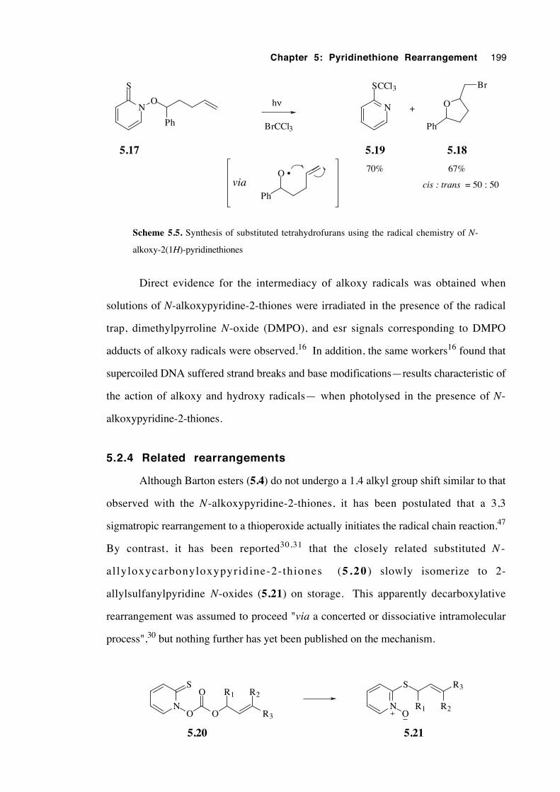
Chapter 5: Pyridinethione Rearrangement 199
NO
S
Ph
hν
O •
Ph
BrCCl3
N
SCCl3
+ O
Ph
Br
5.17 5.19 5.18
cis : trans = 50 : 50
70% 67%
via
Scheme 5.5. Synthesis of substituted tetrahydrofurans using the radical chemistry of N-
alkoxy-2(1H)-pyridinethiones
Direct evidence for the intermediacy of alkoxy radicals was obtained when
solutions of N-alkoxypyridine-2-thiones were irradiated in the presence of the radical
trap, dimethylpyrroline N-oxide (DMPO), and esr signals corresponding to DMPO
adducts of alkoxy radicals were observed.16 In addition, the same workers16 found that
supercoiled DNA suffered strand breaks and base modifications—results characteristic of
the action of alkoxy and hydroxy radicals— when photolysed in the presence of N-
alkoxypyridine-2-thiones.
5.2.4 Related rearrangements
Although Barton esters (5.4) do not undergo a 1,4 alkyl group shift similar to that
observed with the N-alkoxypyridine-2-thiones, it has been postulated that a 3,3
sigmatropic rearrangement to a thioperoxide actually initiates the radical chain reaction.47
By contrast, it has been reported30,31 that the closely related substituted N -
al lyloxycarbonyloxypyridine-2-thiones (5 .20 ) slowly isomerize to 2-
allylsulfanylpyridine N-oxides (5.21) on storage. This apparently decarboxylative
rearrangement was assumed to proceed "via a concerted or dissociative intramolecular
process",30 but nothing further has yet been published on the mechanism.
N
S
O O
O R1
R3
R2
N
S
O
R3
R2R1
5.20 5.21
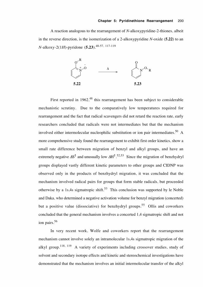
Chapter 5: Pyridinethione Rearrangement 200
A reaction analogous to the rearrangement of N-alkoxypyridine-2-thiones, albeit
in the reverse direction, is the isomerization of a 2-alkoxypyridine N-oxide (5.22) to an
N-alkoxy-2(1H)-pyridone (5.23).48-57, 117-119
N
OR
ON
O
O∆
5.22 5.23
R
First reported in 1962,48 this rearrangement has been subject to considerable
mechanistic scrutiny. Due to the comparatively low temperatures required for
rearrangement and the fact that radical scavengers did not retard the reaction rate, early
researchers concluded that radicals were not intermediates but that the mechanism
involved either intermolecular nucleophilic substitution or ion pair intermediates.50 A
more comprehensive study found the rearrangement to exhibit first order kinetics, show a
small rate difference between migration of benzyl and alkyl groups, and have an
extremely negative ∆S‡ and unusually low ∆H‡ .52,53 Since the migration of benzhydryl
groups displayed vastly different kinetic parameters to other groups and CIDNP was
observed only in the products of benzhydryl migration, it was concluded that the
mechanism involved radical pairs for groups that form stable radicals, but proceeded
otherwise by a 1s,4s sigmatropic shift.53 This conclusion was supported by le Noble
and Daka, who determined a negative activation volume for benzyl migration (concerted)
but a positive value (dissociative) for benzhydryl groups.55 Ollis and coworkers
concluded that the general mechanism involves a concerted 1,4 sigmatropic shift and not
ion pairs.56
In very recent work, Wolfe and coworkers report that the rearrangement
mechanism cannot involve solely an intramolecular 1s,4s sigmatropic migration of the
alkyl group.118, 119 A variety of experiments including crossover studies, study of
solvent and secondary isotope effects and kinetic and stereochemical investigations have
demonstrated that the mechanism involves an initial intermolecular transfer of the alkyl
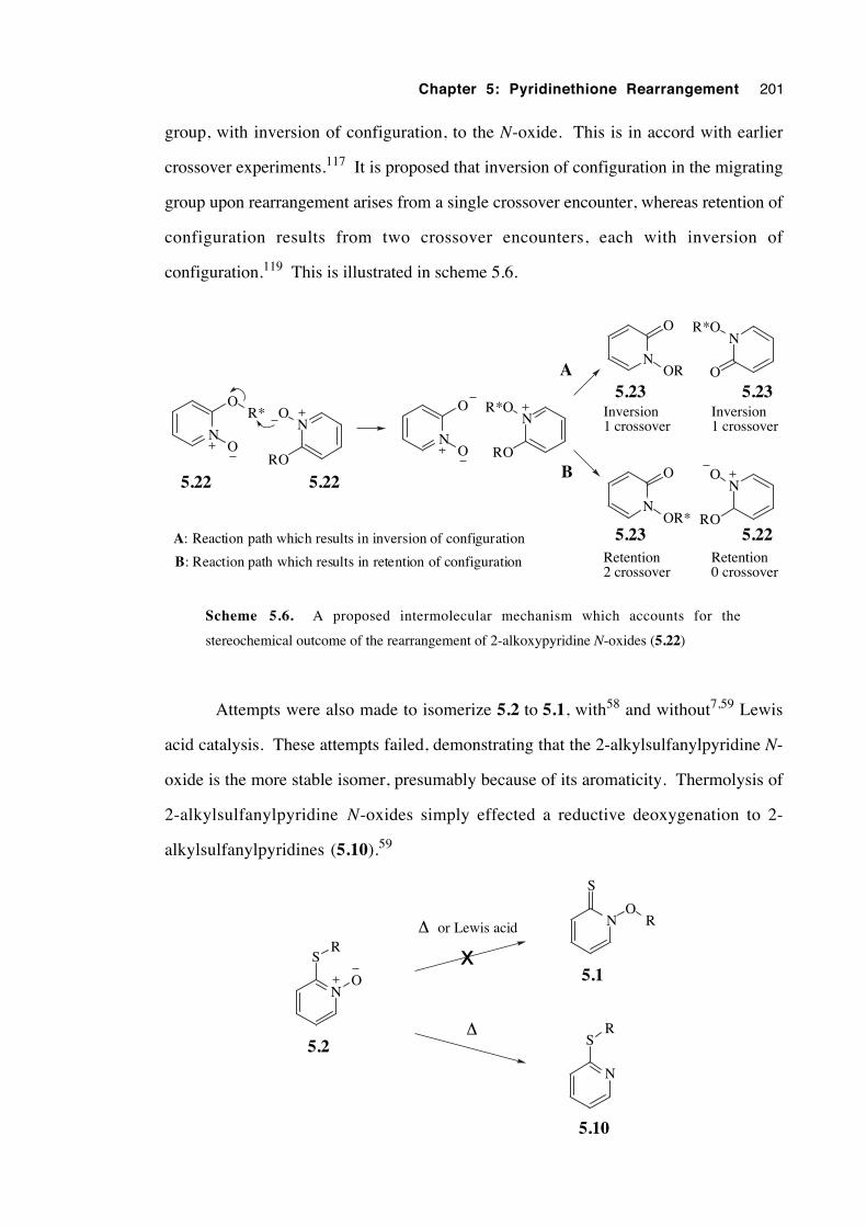
Chapter 5: Pyridinethione Rearrangement 201
group, with inversion of configuration, to the N-oxide. This is in accord with earlier
crossover experiments.117 It is proposed that inversion of configuration in the migrating
group upon rearrangement arises from a single crossover encounter, whereas retention of
configuration results from two crossover encounters, each with inversion of
configuration.119 This is illustrated in scheme 5.6.
N
O
O
N
RO
O
N
O
O
N
RO
R*O
N
O
OR
N
O
R*O
N
O
OR*
N
RO
O
R* Inversion1 crossover
Inversion1 crossover
Retention2 crossover
Retention0 crossover
A
B
A: Reaction path which results in inversion of configuration
B: Reaction path which results in retention of configuration
5.22 5.22
5.23 5.23
5.23 5.22
Scheme 5.6. A proposed intermolecular mechanism which accounts for the
stereochemical outcome of the rearrangement of 2-alkoxypyridine N-oxides (5.22)
Attempts were also made to isomerize 5.2 to 5.1, with58 and without7,59 Lewis
acid catalysis. These attempts failed, demonstrating that the 2-alkylsulfanylpyridine N-
oxide is the more stable isomer, presumably because of its aromaticity. Thermolysis of
2-alkylsulfanylpyridine N-oxides simply effected a reductive deoxygenation to 2-
alkylsulfanylpyridines (5.10).59
N
S
O
R
N
S
O
N
SR
5.2
5.1
5.10
R
x∆ or Lewis acid
∆
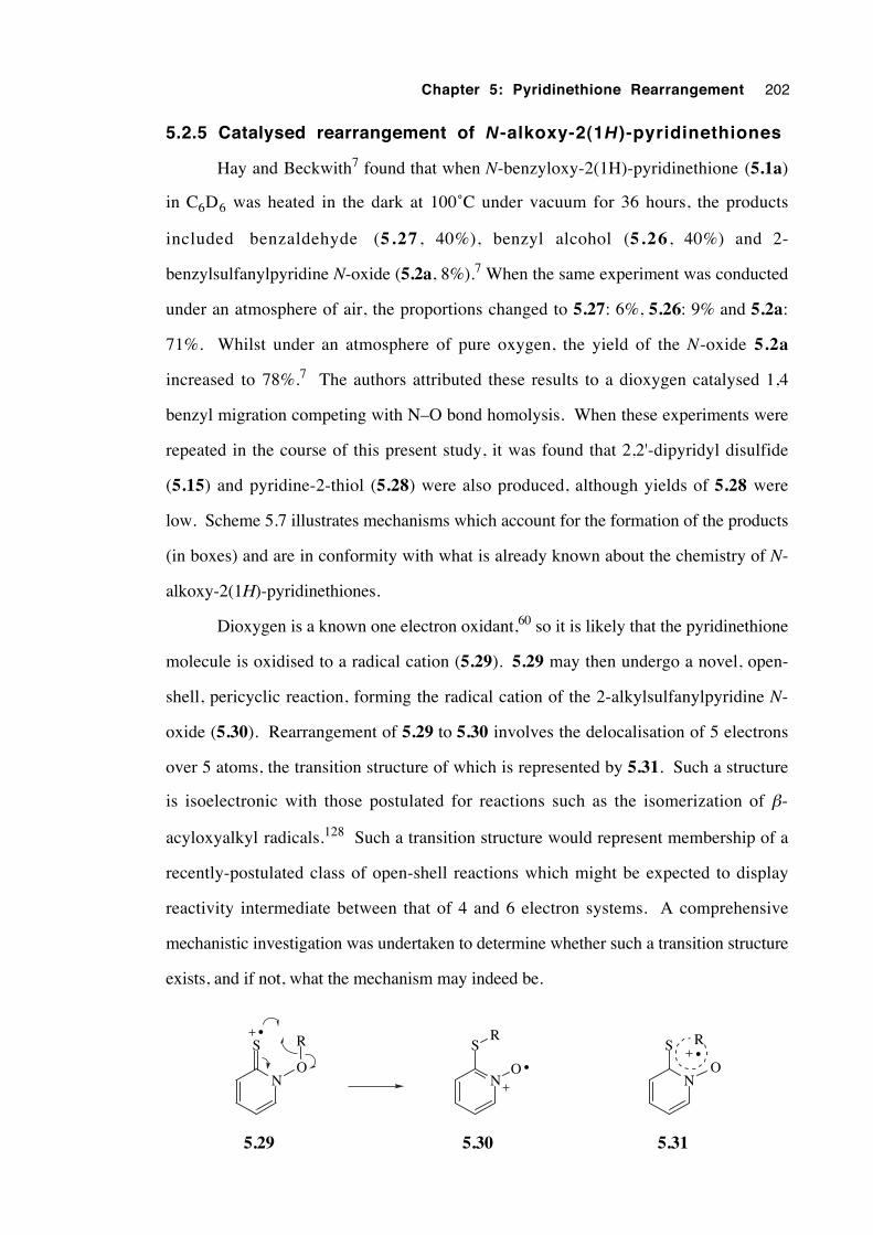
Chapter 5: Pyridinethione Rearrangement 202
5.2.5 Catalysed rearrangement of N-alkoxy-2(1H)-pyridinethiones
Hay and Beckwith7 found that when N-benzyloxy-2(1H)-pyridinethione (5.1a)
in C6D6 was heated in the dark at 100˚C under vacuum for 36 hours, the products
included benzaldehyde (5.27 , 40%), benzyl alcohol (5.26 , 40%) and 2-
benzylsulfanylpyridine N-oxide (5.2a, 8%).7 When the same experiment was conducted
under an atmosphere of air, the proportions changed to 5.27: 6%, 5.26: 9% and 5.2a:
71%. Whilst under an atmosphere of pure oxygen, the yield of the N-oxide 5.2a
increased to 78%.7 The authors attributed these results to a dioxygen catalysed 1,4
benzyl migration competing with N–O bond homolysis. When these experiments were
repeated in the course of this present study, it was found that 2,2'-dipyridyl disulfide
(5.15) and pyridine-2-thiol (5.28) were also produced, although yields of 5.28 were
low. Scheme 5.7 illustrates mechanisms which account for the formation of the products
(in boxes) and are in conformity with what is already known about the chemistry of N-
alkoxy-2(1H)-pyridinethiones.
Dioxygen is a known one electron oxidant,60 so it is likely that the pyridinethione
molecule is oxidised to a radical cation (5.29). 5.29 may then undergo a novel, open-
shell, pericyclic reaction, forming the radical cation of the 2-alkylsulfanylpyridine N-
oxide (5.30). Rearrangement of 5.29 to 5.30 involves the delocalisation of 5 electrons
over 5 atoms, the transition structure of which is represented by 5.31. Such a structure
is isoelectronic with those postulated for reactions such as the isomerization of β-
acyloxyalkyl radicals.128 Such a transition structure would represent membership of a
recently-postulated class of open-shell reactions which might be expected to display
reactivity intermediate between that of 4 and 6 electron systems. A comprehensive
mechanistic investigation was undertaken to determine whether such a transition structure
exists, and if not, what the mechanism may indeed be.
N
S
ON
S
O
R
5.29 5.30
R•
•N
S
O•R
5.31
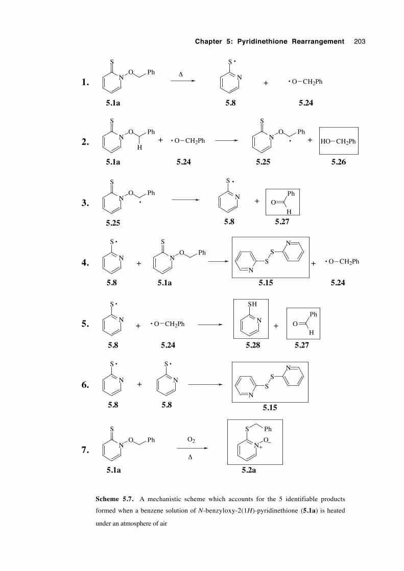
Chapter 5: Pyridinethione Rearrangement 203
N
S
O PhN
S
O CH2Ph+
5.1a 5.8 5.24
N
S
O Ph
5.1a
O CH2Ph
5.24
N
S
O Ph
5.25
HO CH2Ph
5.26
N
S
O Ph
5.25
N
S
5.8
OPh
H
5.27
+ +
+
N
S
5.8
N
S
O Ph
5.1a
+N
S
SN
O CH2Ph
5.15 5.24
N
S
5.8
O CH2Ph
5.24
+
+N
SH
5.28
O
Ph
H
5.27
+
N
S
5.8
N
S
5.8N
S
SN
5.15
+
2.
3.
4.
5.
6.
1.
H
N
S
5.1a
N
S
5.2a
7.O Ph
Ph
OO2
∆
∆
Scheme 5.7. A mechanistic scheme which accounts for the 5 identifiable products
formed when a benzene solution of N-benzyloxy-2(1H)-pyridinethione (5.1a) is heated
under an atmosphere of air
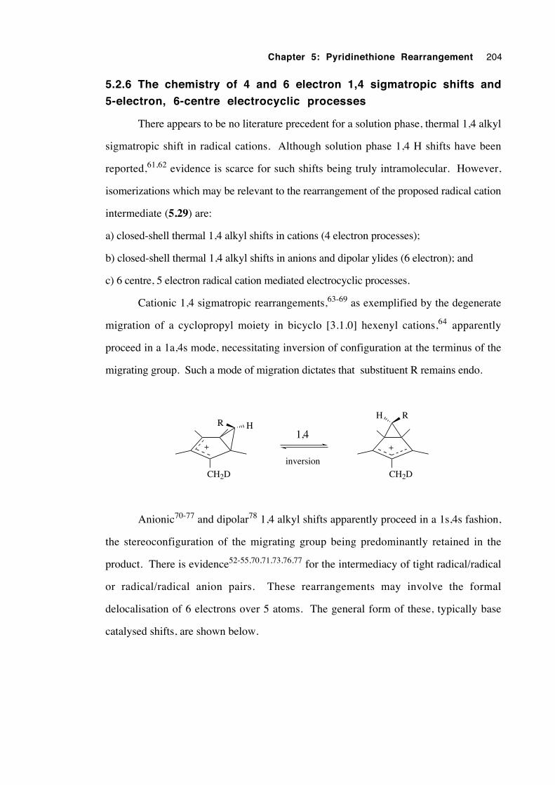
Chapter 5: Pyridinethione Rearrangement 204
5.2.6 The chemistry of 4 and 6 electron 1,4 sigmatropic shifts and
5-electron, 6-centre electrocyclic processes
There appears to be no literature precedent for a solution phase, thermal 1,4 alkyl
sigmatropic shift in radical cations. Although solution phase 1,4 H shifts have been
reported,61,62 evidence is scarce for such shifts being truly intramolecular. However,
isomerizations which may be relevant to the rearrangement of the proposed radical cation
intermediate (5.29) are:
a) closed-shell thermal 1,4 alkyl shifts in cations (4 electron processes);
b) closed-shell thermal 1,4 alkyl shifts in anions and dipolar ylides (6 electron); and
c) 6 centre, 5 electron radical cation mediated electrocyclic processes.
Cationic 1,4 sigmatropic rearrangements,63-69 as exemplified by the degenerate
migration of a cyclopropyl moiety in bicyclo [3.1.0] hexenyl cations,64 apparently
proceed in a 1a,4s mode, necessitating inversion of configuration at the terminus of the
migrating group. Such a mode of migration dictates that substituent R remains endo.
CH2D
HR
CH2D
RH
1,4
inversion
Anionic70-77 and dipolar78 1,4 alkyl shifts apparently proceed in a 1s,4s fashion,
the stereoconfiguration of the migrating group being predominantly retained in the
product. There is evidence52-55,70,71,73,76,77 for the intermediacy of tight radical/radical
or radical/radical anion pairs. These rearrangements may involve the formal
delocalisation of 6 electrons over 5 atoms. The general form of these, typically base
catalysed shifts, are shown below.
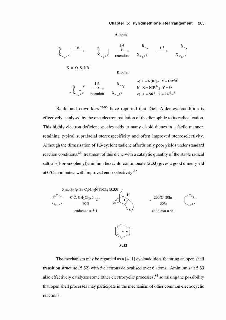
Chapter 5: Pyridinethione Rearrangement 205
R
X X X XoB- H+1,4
retention
X = O, S, NR1
RR R
Anionic
X
Y
X
Yo1,4
retention
RR
Dipolar
a) X = N(R1)2 , Y = CR2R3
b) X = N(R1)2 , Y = O
c) X = SR1, Y = CR2R3
Bauld and coworkers79-85 have reported that Diels-Alder cycloaddition is
effectively catalysed by the one electron oxidation of the dienophile to its radical cation.
This highly electron deficient species adds to many cisoid dienes in a facile manner,
retaining typical suprafacial stereospecificity and often improved stereoselectivity.
Although the dimerisation of 1,3-cyclohexadiene affords only poor yields under standard
reaction conditions,86 treatment of this diene with a catalytic quantity of the stable radical
salt tris(4-bromophenyl)aminium hexachloroantimonate (5.33) gives a good dimer yield
at 0˚C in minutes, with improved endo selectivity.82
H
H
5 mol% (p-Br-C6H4)3N SbCl6 (5.33)
0˚C, CH2Cl2, 5 min
70%
endo:exo = 5:1
30%
endo:exo = 4:1
200˚C, 20hr
5.32
•
The mechanism may be regarded as a [4+1] cycloaddition, featuring an open shell
transition structure (5.32) with 5 electrons delocalised over 6 atoms. Aminium salt 5.33
also effectively catalyses some other electrocyclic processes,82 so raising the possibility
that open shell processes may participate in the mechanism of other common electrocyclic
reactions.
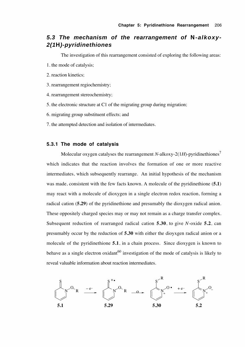
Chapter 5: Pyridinethione Rearrangement 206
5.3 The mechanism of the rearrangement of N -alkoxy-2(1H)-pyridinethiones
The investigation of this rearrangement consisted of exploring the following areas:
1. the mode of catalysis;
2. reaction kinetics;
3. rearrangement regiochemistry;
4. rearrangement stereochemistry;
5. the electronic structure at C1 of the migrating group during migration;
6. migrating group substituent effects; and
7. the attempted detection and isolation of intermediates.
5.3.1 The mode of catalysis
Molecular oxygen catalyses the rearrangement N-alkoxy-2(1H)-pyridinethiones7
which indicates that the reaction involves the formation of one or more reactive
intermediates, which subsequently rearrange. An initial hypothesis of the mechanism
was made, consistent with the few facts known. A molecule of the pyridinethione (5.1)
may react with a molecule of dioxygen in a single electron redox reaction, forming a
radical cation (5.29) of the pyridinethione and presumably the dioxygen radical anion.
These oppositely charged species may or may not remain as a charge transfer complex.
Subsequent reduction of rearranged radical cation 5.30, to give N-oxide 5.2, can
presumably occur by the reduction of 5.30 with either the dioyxgen radical anion or a
molecule of the pyridinethione 5.1, in a chain process. Since dioxygen is known to
behave as a single electron oxidant60 investigation of the mode of catalysis is likely to
reveal valuable information about reaction intermediates.
N
S
OR N
S
OR N
S
ON
S
O
R R
o– e_ + e_
5.1 5.25.29 5.30
•
•+
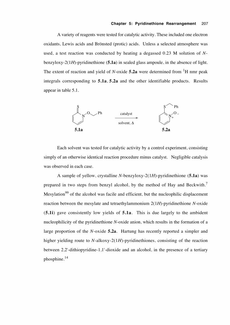
Chapter 5: Pyridinethione Rearrangement 207
A variety of reagents were tested for catalytic activity. These included one electron
oxidants, Lewis acids and Brönsted (protic) acids. Unless a selected atmosphere was
used, a test reaction was conducted by heating a degassed 0.23 M solution of N-
benzyloxy-2(1H)-pyridinethione (5.1a) in sealed glass ampoule, in the absence of light.
The extent of reaction and yield of N-oxide 5.2a were determined from 1H nmr peak
integrals corresponding to 5.1a, 5.2a and the other identifiable products. Results
appear in table 5.1.
N
S
O PhN
S
O
Ph
solvent, ∆
5.2a5.1a
catalyst
Each solvent was tested for catalytic activity by a control experiment, consisting
simply of an otherwise identical reaction procedure minus catalyst. Negligible catalysis
was observed in each case.
A sample of yellow, crystalline N-benzyloxy-2(1H)-pyridinethione (5.1a) was
prepared in two steps from benzyl alcohol, by the method of Hay and Beckwith.7
Mesylation99 of the alcohol was facile and efficient, but the nucleophilic displacement
reaction between the mesylate and tetraethylammonium 2(1H)-pyridinethione N-oxide
(5.1i) gave consistently low yields of 5.1a. This is due largely to the ambident
nucleophilicity of the pyridinethione N-oxide anion, which results in the formation of a
large proportion of the N-oxide 5.2a. Hartung has recently reported a simpler and
higher yielding route to N-alkoxy-2(1H)-pyridinethiones, consisting of the reaction
between 2,2'-dithiopyridine-1,1'-dioxide and an alcohol, in the presence of a tertiary
phosphine.14
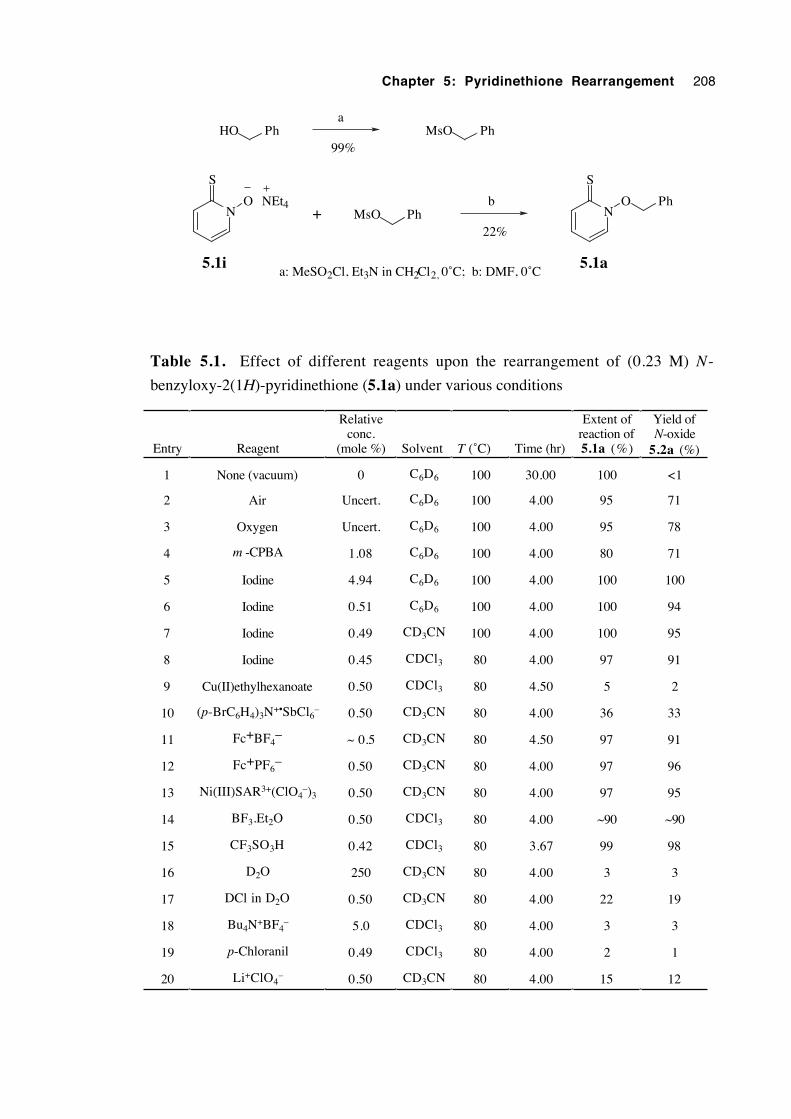
Chapter 5: Pyridinethione Rearrangement 208
N
S
O Ph
5.1a
PhHO PhMsOa
b
a: MeSO2Cl, Et3N in CH2Cl2, 0˚C; b: DMF, 0˚C
99%
22%PhMsON
S
O
5.1i
+NEt4
Table 5.1. Effect of different reagents upon the rearrangement of (0.23 M) N-
benzyloxy-2(1H)-pyridinethione (5.1a) under various conditions
Entry Reagent
Relativeconc.
(mole %) Solvent T (˚C) Time (hr)
Extent ofreaction of5.1a (%)
Yield ofN-oxide
5.2a (%)
1 None (vacuum) 0 C6D6 100 30.00 100 <1
2 Air Uncert. C6D6 100 4.00 95 71
3 Oxygen Uncert. C6D6 100 4.00 95 78
4 m -CPBA 1.08 C6D6 100 4.00 80 71
5 Iodine 4.94 C6D6 100 4.00 100 100
6 Iodine 0.51 C6D6 100 4.00 100 94
7 Iodine 0.49 CD3CN 100 4.00 100 95
8 Iodine 0.45 CDCl3 80 4.00 97 91
9 Cu(II)ethylhexanoate 0.50 CDCl3 80 4.50 5 2
10 (p-BrC6H4)3N+•SbCl6– 0.50 CD3CN 80 4.00 36 33
11 Fc+BF4– ~ 0.5 CD3CN 80 4.50 97 91
12 Fc+PF6– 0.50 CD3CN 80 4.00 97 96
13 Ni(III)SAR3+(ClO4–)3 0.50 CD3CN 80 4.00 97 95
14 BF3.Et2O 0.50 CDCl3 80 4.00 ~90 ~90
15 CF3SO3H 0.42 CDCl3 80 3.67 99 98
16 D2O 250 CD3CN 80 4.00 3 3
17 DCl in D2O 0.50 CD3CN 80 4.00 22 19
18 Bu4N+BF4– 5.0 CDCl3 80 4.00 3 3
19 p-Chloranil 0.49 CDCl3 80 4.00 2 1
20 Li+ClO4– 0.50 CD3CN 80 4.00 15 12
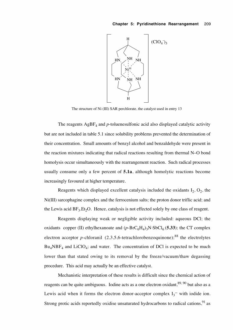
Chapter 5: Pyridinethione Rearrangement 209
HN NH NH
HN NHNH
NiIII
H
H
(ClO4-)3
The structure of Ni (III) SAR perchlorate, the catalyst used in entry 13
The reagents AgBF4 and p-toluenesulfonic acid also displayed catalytic activity
but are not included in table 5.1 since solubility problems prevented the determination of
their concentration. Small amounts of benzyl alcohol and benzaldehyde were present in
the reaction mixtures indicating that radical reactions resulting from thermal N–O bond
homolysis occur simultaneously with the rearrangement reaction. Such radical processes
usually consume only a few percent of 5.1a, although homolytic reactions become
increasingly favoured at higher temperature.
Reagents which displayed excellent catalysis included the oxidants I2, O2, the
Ni(III) sarcophagine complex and the ferrocenium salts; the proton donor triflic acid; and
the Lewis acid BF3.Et2O. Hence, catalysis is not effected solely by one class of reagent.
Reagents displaying weak or negligible activity included: aqueous DCl; the
oxidants copper (II) ethylhexanoate and (p-BrC6H4)3N.SbCl6 (5.33); the CT complex
electron acceptor p-chloranil (2,3,5,6-tetrachlorobenzoquinone);88 the electrolytes
Bu4NBF4 and LiClO4; and water. The concentration of DCl is expected to be much
lower than that stated owing to its removal by the freeze/vacuum/thaw degassing
procedure. This acid may actually be an effective catalyst.
Mechanistic interpretation of these results is difficult since the chemical action of
reagents can be quite ambiguous. Iodine acts as a one electron oxidant,89, 90 but also as a
Lewis acid when it forms the electron donor-acceptor complex I3– with iodide ion.
Strong protic acids reportedly oxidise unsaturated hydrocarbons to radical cations,91 as
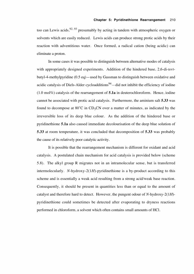
Chapter 5: Pyridinethione Rearrangement 210
too can Lewis acids,92, 93 presumably by acting in tandem with atmospheric oxygen or
solvents which are easily reduced. Lewis acids can produce strong protic acids by their
reaction with adventitious water. Once formed, a radical cation (being acidic) can
eliminate a proton.
In some cases it was possible to distinguish between alternative modes of catalysis
with appropriately designed experiments. Addition of the hindered base, 2,6-di-tert-
butyl-4-methylpyridine (0.5 eq)—used by Gassman to distinguish between oxidative and
acidic catalysis of Diels-Alder cycloadditions94—did not inhibit the efficiency of iodine
(1.0 mol%) catalysis of the rearrangement of 5.1a in deuterochloroform. Hence, iodine
cannot be associated with protic acid catalysis. Furthermore, the aminium salt 5.33 was
found to decompose at 80˚C in CD3CN over a matter of minutes, as indicated by the
irreversible loss of its deep blue colour. As the addition of the hindered base or
pyridinethione 5.1a also caused immediate decolourisation of the deep blue solution of
5.33 at room temperature, it was concluded that decomposition of 5.33 was probably
the cause of its relatively poor catalytic activity.
It is possible that the rearrangement mechanism is different for oxidant and acid
catalysis. A postulated chain mechanism for acid catalysis is provided below (scheme
5.8). The alkyl group R migrates not in an intramolecular sense, but is transferred
intermolecularly. N-hydroxy-2(1H)-pyridinethione is a by-product according to this
scheme and is essentially a weak acid resulting from a strong acid/weak base reaction.
Consequently, it should be present in quantities less than or equal to the amount of
catalyst and therefore hard to detect. However, the pungent odour of N-hydroxy-2(1H)-
pyridinethione could sometimes be detected after evaporating to dryness reactions
performed in chloroform, a solvent which often contains small amounts of HCl.
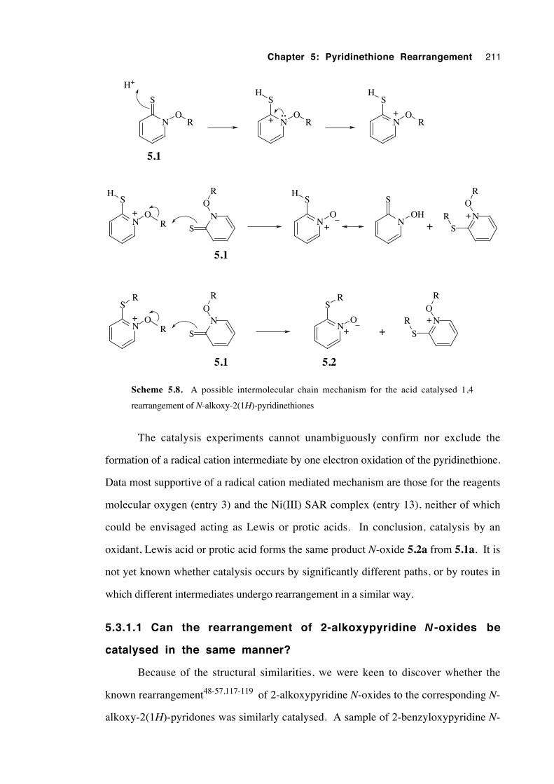
Chapter 5: Pyridinethione Rearrangement 211
N
S
OR
H+
N
S
OR
H
+ N
S
OR
H
+
N
S
OR
H
+ NS
OR
N
S
O
H
+N
S
OR
R ++
• •
N
S
OR
+ N
S
O
R
N
S
O+
N
S
O
R
R ++
R R
5.1
5.1
5.1 5.2
N
S
OH
Scheme 5.8. A possible intermolecular chain mechanism for the acid catalysed 1,4
rearrangement of N-alkoxy-2(1H)-pyridinethiones
The catalysis experiments cannot unambiguously confirm nor exclude the
formation of a radical cation intermediate by one electron oxidation of the pyridinethione.
Data most supportive of a radical cation mediated mechanism are those for the reagents
molecular oxygen (entry 3) and the Ni(III) SAR complex (entry 13), neither of which
could be envisaged acting as Lewis or protic acids. In conclusion, catalysis by an
oxidant, Lewis acid or protic acid forms the same product N-oxide 5.2a from 5.1a. It is
not yet known whether catalysis occurs by significantly different paths, or by routes in
which different intermediates undergo rearrangement in a similar way.
5.3.1.1 Can the rearrangement of 2-alkoxypyridine N -oxides be
catalysed in the same manner?
Because of the structural similarities, we were keen to discover whether the
known rearrangement48-57,117-119 of 2-alkoxypyridine N-oxides to the corresponding N-
alkoxy-2(1H)-pyridones was similarly catalysed. A sample of 2-benzyloxypyridine N-
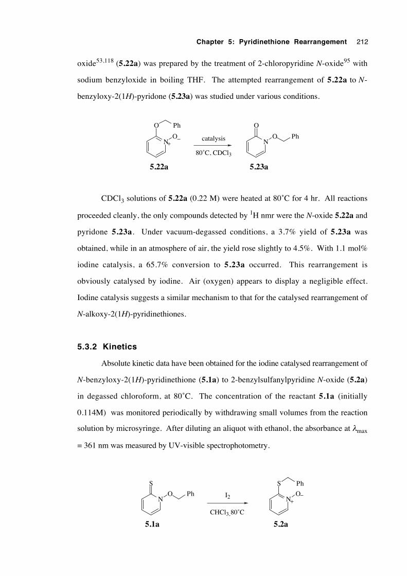
Chapter 5: Pyridinethione Rearrangement 212
oxide53,118 (5.22a) was prepared by the treatment of 2-chloropyridine N-oxide95 with
sodium benzyloxide in boiling THF. The attempted rearrangement of 5.22a to N-
benzyloxy-2(1H)-pyridone (5.23a) was studied under various conditions.
N
O Ph
ON
O
O Phcatalysis
80˚C, CDCl3
5.22a 5.23a
CDCl3 solutions of 5.22a (0.22 M) were heated at 80˚C for 4 hr. All reactions
proceeded cleanly, the only compounds detected by 1H nmr were the N-oxide 5.22a and
pyridone 5.23a. Under vacuum-degassed conditions, a 3.7% yield of 5.23a was
obtained, while in an atmosphere of air, the yield rose slightly to 4.5%. With 1.1 mol%
iodine catalysis, a 65.7% conversion to 5.23a occurred. This rearrangement is
obviously catalysed by iodine. Air (oxygen) appears to display a negligible effect.
Iodine catalysis suggests a similar mechanism to that for the catalysed rearrangement of
N-alkoxy-2(1H)-pyridinethiones.
5.3.2 Kinetics
Absolute kinetic data have been obtained for the iodine catalysed rearrangement of
N-benzyloxy-2(1H)-pyridinethione (5.1a) to 2-benzylsulfanylpyridine N-oxide (5.2a)
in degassed chloroform, at 80˚C. The concentration of the reactant 5.1a (initially
0.114M) was monitored periodically by withdrawing small volumes from the reaction
solution by microsyringe. After diluting an aliquot with ethanol, the absorbance at λmax
= 361 nm was measured by UV-visible spectrophotometry.
N
S
O PhN
S
O
Ph
CHCl3, 80˚C
5.2a5.1a
I2
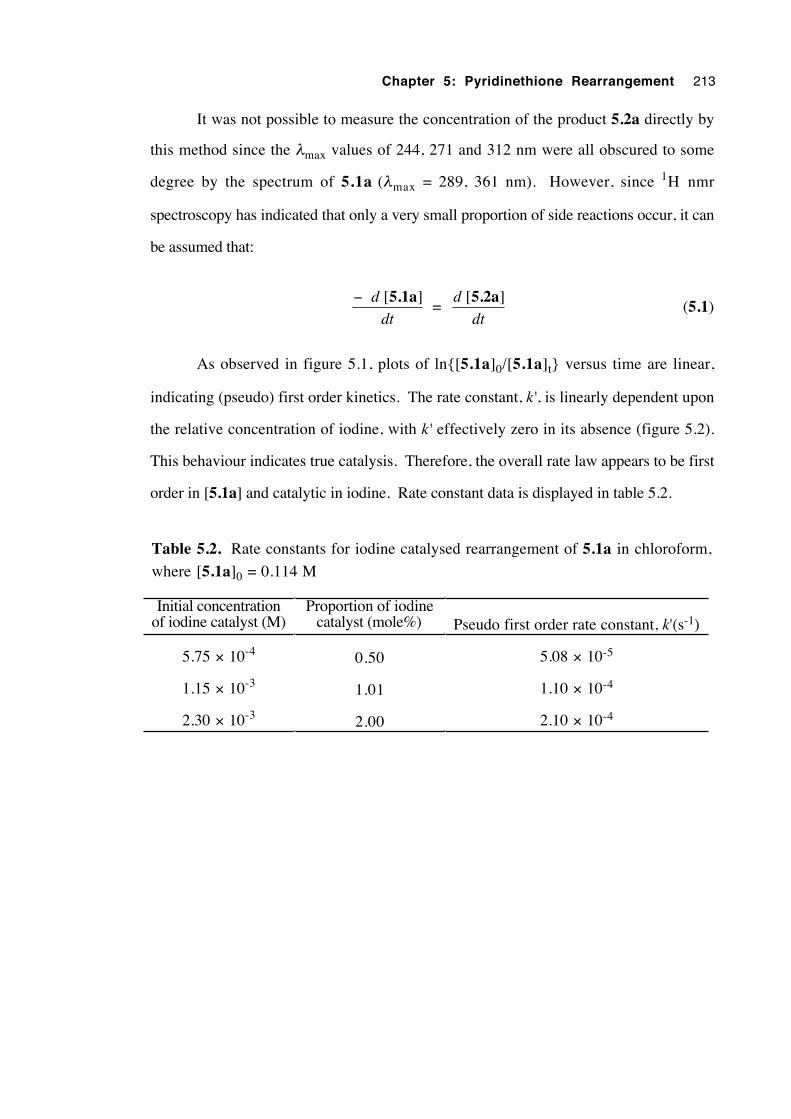
Chapter 5: Pyridinethione Rearrangement 213
It was not possible to measure the concentration of the product 5.2a directly by
this method since the λmax values of 244, 271 and 312 nm were all obscured to some
degree by the spectrum of 5.1a (λmax = 289, 361 nm). However, since 1H nmr
spectroscopy has indicated that only a very small proportion of side reactions occur, it can
be assumed that:
− [ ]d
dt
5.1a =
d
dt
[ ]5.2a (5.1)
As observed in figure 5.1, plots of ln{[5.1a]0/[5.1a]t} versus time are linear,
indicating (pseudo) first order kinetics. The rate constant, k', is linearly dependent upon
the relative concentration of iodine, with k' effectively zero in its absence (figure 5.2).
This behaviour indicates true catalysis. Therefore, the overall rate law appears to be first
order in [5.1a] and catalytic in iodine. Rate constant data is displayed in table 5.2.
Table 5.2. Rate constants for iodine catalysed rearrangement of 5.1a in chloroform,
where [5.1a]0 = 0.114 M
Initial concentrationof iodine catalyst (M)
Proportion of iodinecatalyst (mole%) Pseudo first order rate constant, k'(s-1)
5.75 × 10⁻⁴ 0.50 5.08 × 10⁻⁵
1.15 × 10⁻³ 1.01 1.10 × 10⁻⁴
2.30 × 10⁻³ 2.00 2.10 × 10⁻⁴
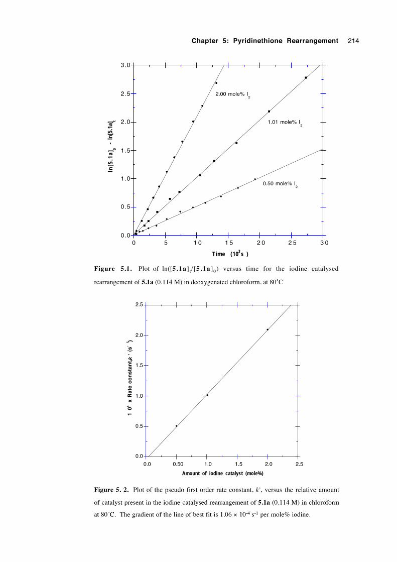
Chapter 5: Pyridinethione Rearrangement 214
0.50 mole% I2
0.0
0.5
1.0
1.5
2.0
2.5
3.0
0 5 1 0 1 5 2 0 2 5 3 0
ln[5
.1a]
0 -
ln[
5.1a
] t
Time (103 s )
2.00 mole% I2
1.01 mole% I2
Figure 5.1. Plot of ln([5.1a ] t/[5.1a ]0) versus time for the iodine catalysed
rearrangement of 5.1a (0.114 M) in deoxygenated chloroform, at 80˚C
0.0
0.5
1.0
1.5
2.0
2.5
0.0 0.50 1.0 1.5 2.0 2.5
104
x R
ate
con
stan
t,
(s
-1 )
Amount of iodine catalyst (mole%)
k'
Figure 5. 2. Plot of the pseudo first order rate constant, k', versus the relative amount
of catalyst present in the iodine-catalysed rearrangement of 5.1a (0.114 M) in chloroform
at 80˚C. The gradient of the line of best fit is 1.06 × 10⁻⁴ s-1 per mole% iodine.
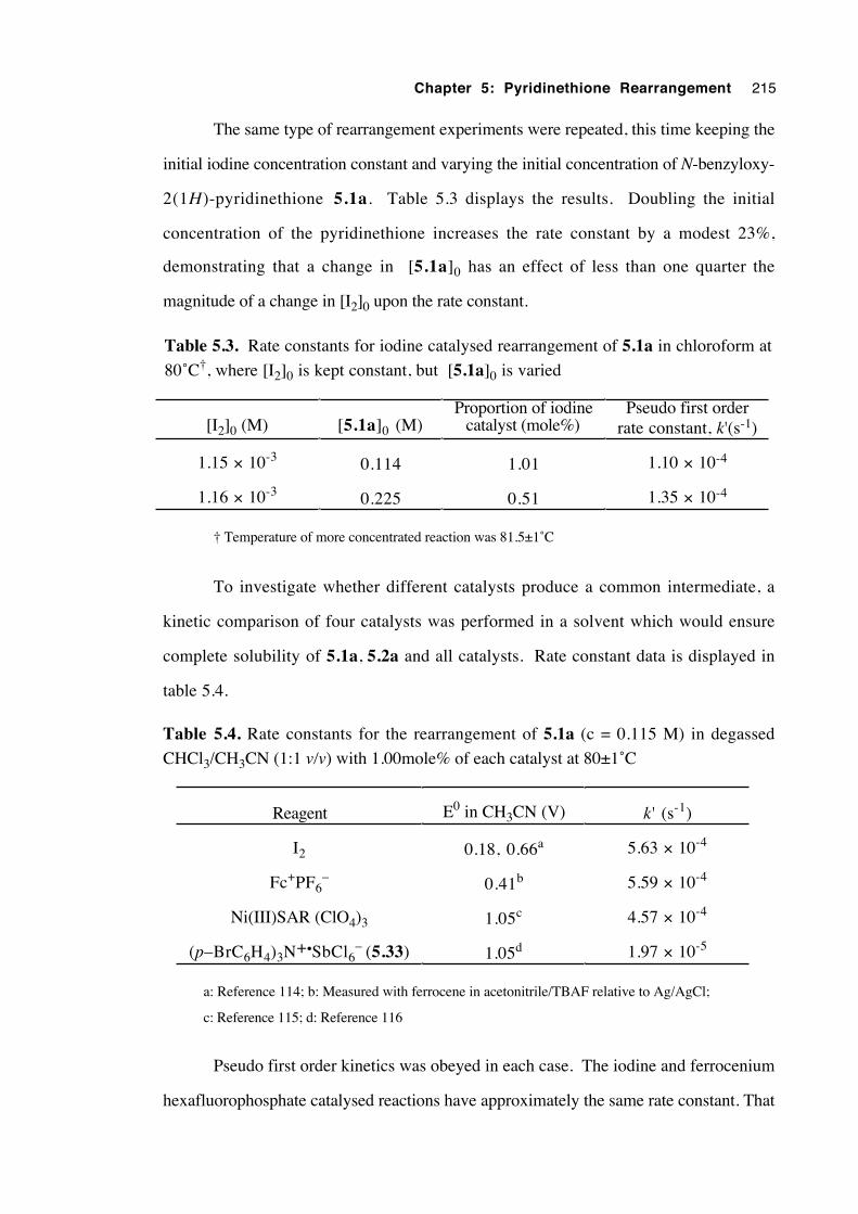
Chapter 5: Pyridinethione Rearrangement 215
The same type of rearrangement experiments were repeated, this time keeping the
initial iodine concentration constant and varying the initial concentration of N-benzyloxy-
2(1H)-pyridinethione 5.1a. Table 5.3 displays the results. Doubling the initial
concentration of the pyridinethione increases the rate constant by a modest 23%,
demonstrating that a change in [5.1a]0 has an effect of less than one quarter the
magnitude of a change in [I2]0 upon the rate constant.
Table 5.3. Rate constants for iodine catalysed rearrangement of 5.1a in chloroform at
80˚C†, where [I2]0 is kept constant, but [5.1a]0 is varied
[I2]0 (M) [5.1a]0 (M)Proportion of iodine
catalyst (mole%)Pseudo first order
rate constant, k'(s-1)
1.15 × 10⁻³ 0.114 1.01 1.10 × 10⁻⁴
1.16 × 10⁻³ 0.225 0.51 1.35 × 10⁻⁴
† Temperature of more concentrated reaction was 81.5±1˚C
To investigate whether different catalysts produce a common intermediate, a
kinetic comparison of four catalysts was performed in a solvent which would ensure
complete solubility of 5.1a, 5.2a and all catalysts. Rate constant data is displayed in
table 5.4.
Table 5.4. Rate constants for the rearrangement of 5.1a (c = 0.115 M) in degassed
CHCl3/CH3CN (1:1 v/v) with 1.00mole% of each catalyst at 80±1˚C
Reagent E0 in CH3CN (V) k' (s-1)
I2 0.18, 0.66a 5.63 × 10⁻⁴
Fc+PF6– 0.41b 5.59 × 10⁻⁴
Ni(III)SAR (ClO4)3 1.05c 4.57 × 10⁻⁴
(p–BrC6H4)3N+•SbCl6– (5.33) 1.05d 1.97 × 10⁻⁵
a: Reference 114; b: Measured with ferrocene in acetonitrile/TBAF relative to Ag/AgCl;
c: Reference 115; d: Reference 116
Pseudo first order kinetics was obeyed in each case. The iodine and ferrocenium
hexafluorophosphate catalysed reactions have approximately the same rate constant. That
Chapter 5: Pyridinethione Rearrangement 216
for Ni (III) SAR catalysis is about 81% of k' for iodine. The rate constant for the
aminium salt (5.33) catalysed reaction is smaller by more than an order of magnitude,
presumably owing to thermal decomposition of the catalyst, as mentioned previously.
Differences may also be partially attributable due to differing solubilities of activated
intermediates.
The similarity of the first three rate constants is consistent with a common
intermediate, most probably a pyridinethione radical cation formed by a one electron
oxidation. However, the reduction potentials for the catalysts are varied, some being
below 0.5 V. Pyridinethione 5.1a has a one electron oxidation potential of 0.9 V
(section 5.3.7.3), which leaves a presently unanswered question of how it is that each
catalyst is capable of oxidising 5.1a. Mehta and Pinto116 encountered the same question
when they observed one electron oxidation of phenyl selenoglycosides (oxidation
potential 1.35-1.50 V) by aminium salt 5.33 (reduction potential 1.05 V). They suggest
that a prior complexation of the selenoglycoside and the aminium salt produces a complex
of lowered oxidation potential.116
A comparison of data in tables 5.2 and 5.4, reveals a 5.5 fold increase in rate
constant k' as the solvent is changed from chloroform to chloroform/acetonitrile. Such a
rate acceleration with increased solvent polarity indicates a polarized rate limiting step.
The equilibrium constant for the reaction between iodine and 5.1a in chloroform
has been measured. The initial reaction between one catalyst (5.33) and 5.1a is very
rapid, as indicated by the immediate and complete loss of blue colour observed upon
mixing a deep blue CH2Cl2 solution of the aminium salt 5.33 with the yellow
pyridinethione solution. Owing to the colours of both iodine and the pyridinethione, it
was more difficult to observe such a decolourisation with this system. However,
decolourisation does occur when a colourless solution of the product N-oxide (5.2a) is
mixed with a CH2Cl2 solution of either iodine, aminium salt 5.33 or ferrocenium
hexafluorophosphate.96 This behaviour indicates a ready oxidation of, or complexation
with, the product also by the catalyst.
If the equilibrium is treated as having the stoichiometry:
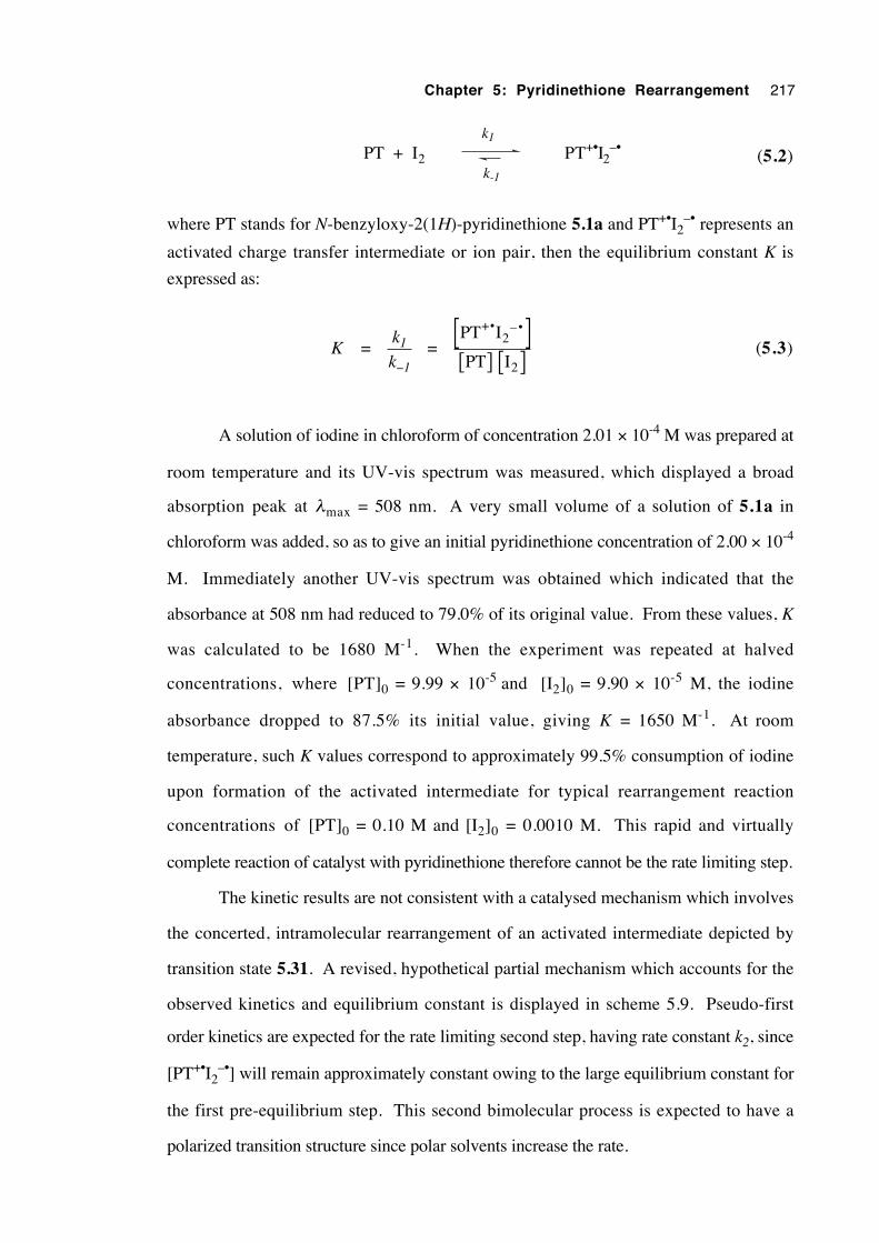
Chapter 5: Pyridinethione Rearrangement 217
PT + I2 PT+•I2–•
k1
k-1 (5.2)
where PT stands for N-benzyloxy-2(1H)-pyridinethione 5.1a and PT+•I2–• represents an
activated charge transfer intermediate or ion pair, then the equilibrium constant K is
expressed as:
K = k
k1
1− =
PT I
PT I
+2
–
2
• •[ ][ ] [ ]
(5.3)
A solution of iodine in chloroform of concentration 2.01 × 10⁻⁴ M was prepared at
room temperature and its UV-vis spectrum was measured, which displayed a broad
absorption peak at λmax = 508 nm. A very small volume of a solution of 5.1a in
chloroform was added, so as to give an initial pyridinethione concentration of 2.00 × 10⁻⁴
M. Immediately another UV-vis spectrum was obtained which indicated that the
absorbance at 508 nm had reduced to 79.0% of its original value. From these values, K
was calculated to be 1680 M-1. When the experiment was repeated at halved
concentrations, where [PT]0 = 9.99 × 10⁻⁵ and [I2]0 = 9.90 × 10⁻⁵ M, the iodine
absorbance dropped to 87.5% its initial value, giving K = 1650 M-1. At room
temperature, such K values correspond to approximately 99.5% consumption of iodine
upon formation of the activated intermediate for typical rearrangement reaction
concentrations of [PT]0 = 0.10 M and [I2]0 = 0.0010 M. This rapid and virtually
complete reaction of catalyst with pyridinethione therefore cannot be the rate limiting step.
The kinetic results are not consistent with a catalysed mechanism which involves
the concerted, intramolecular rearrangement of an activated intermediate depicted by
transition state 5.31. A revised, hypothetical partial mechanism which accounts for the
observed kinetics and equilibrium constant is displayed in scheme 5.9. Pseudo-first
order kinetics are expected for the rate limiting second step, having rate constant k2, since
[PT+•I2–•] will remain approximately constant owing to the large equilibrium constant for
the first pre-equilibrium step. This second bimolecular process is expected to have a
polarized transition structure since polar solvents increase the rate.
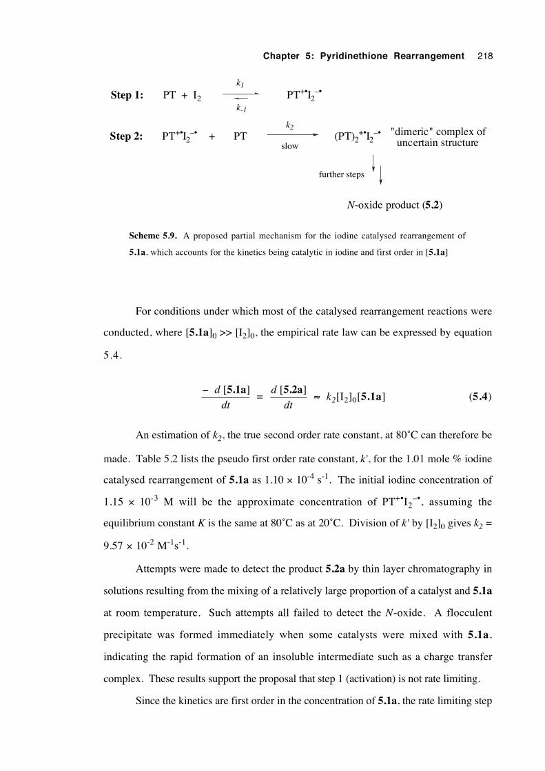
Chapter 5: Pyridinethione Rearrangement 218
PT + I2 PT+•I2–•
k1
k-1
PT+•I2–• PT+
k2 "dimeric" complex of uncertain structure
N-oxide product (5.2)
further steps
slow
Step 1:
Step 2: (PT)2+•I2
–•
Scheme 5.9. A proposed partial mechanism for the iodine catalysed rearrangement of
5.1a, which accounts for the kinetics being catalytic in iodine and first order in [5.1a]
For conditions under which most of the catalysed rearrangement reactions were
conducted, where [5.1a]0 >> [I2]0, the empirical rate law can be expressed by equation
5.4.
− [ ]d
dt
5.1a =
d
dt
[ ]5.2a ≈ k2[I2]0[5.1a] (5.4)
An estimation of k2, the true second order rate constant, at 80˚C can therefore be
made. Table 5.2 lists the pseudo first order rate constant, k', for the 1.01 mole % iodine
catalysed rearrangement of 5.1a as 1.10 × 10⁻⁴ s-1. The initial iodine concentration of
1.15 × 10⁻³ M will be the approximate concentration of PT+•I2–•, assuming the
equilibrium constant K is the same at 80˚C as at 20˚C. Division of k' by [I2]0 gives k2 =
9.57 × 10⁻² M-1s-1.
Attempts were made to detect the product 5.2a by thin layer chromatography in
solutions resulting from the mixing of a relatively large proportion of a catalyst and 5.1a
at room temperature. Such attempts all failed to detect the N-oxide. A flocculent
precipitate was formed immediately when some catalysts were mixed with 5.1a,
indicating the rapid formation of an insoluble intermediate such as a charge transfer
complex. These results support the proposal that step 1 (activation) is not rate limiting.
Since the kinetics are first order in the concentration of 5.1a, the rate limiting step
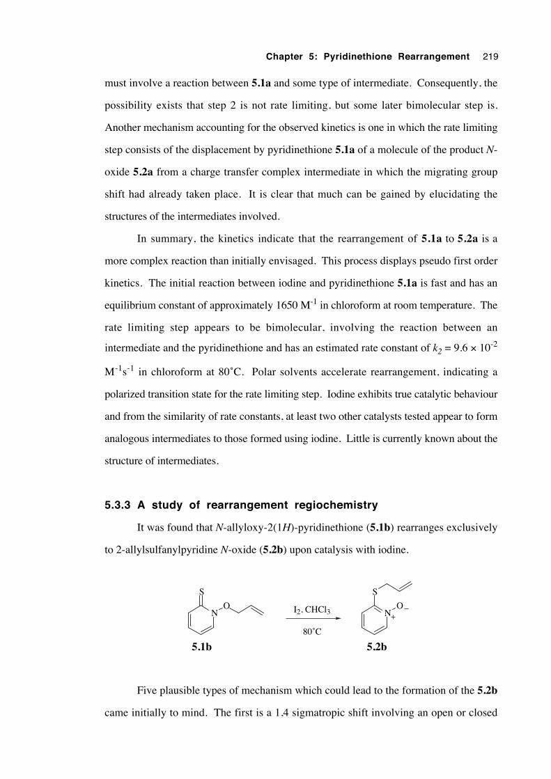
Chapter 5: Pyridinethione Rearrangement 219
must involve a reaction between 5.1a and some type of intermediate. Consequently, the
possibility exists that step 2 is not rate limiting, but some later bimolecular step is.
Another mechanism accounting for the observed kinetics is one in which the rate limiting
step consists of the displacement by pyridinethione 5.1a of a molecule of the product N-
oxide 5.2a from a charge transfer complex intermediate in which the migrating group
shift had already taken place. It is clear that much can be gained by elucidating the
structures of the intermediates involved.
In summary, the kinetics indicate that the rearrangement of 5.1a to 5.2a is a
more complex reaction than initially envisaged. This process displays pseudo first order
kinetics. The initial reaction between iodine and pyridinethione 5.1a is fast and has an
equilibrium constant of approximately 1650 M-1 in chloroform at room temperature. The
rate limiting step appears to be bimolecular, involving the reaction between an
intermediate and the pyridinethione and has an estimated rate constant of k2 = 9.6 × 10⁻²
M-1s-1 in chloroform at 80˚C. Polar solvents accelerate rearrangement, indicating a
polarized transition state for the rate limiting step. Iodine exhibits true catalytic behaviour
and from the similarity of rate constants, at least two other catalysts tested appear to form
analogous intermediates to those formed using iodine. Little is currently known about the
structure of intermediates.
5.3.3 A study of rearrangement regiochemistry
It was found that N-allyloxy-2(1H)-pyridinethione (5.1b) rearranges exclusively
to 2-allylsulfanylpyridine N-oxide (5.2b) upon catalysis with iodine.
N
S
ON
S
O
80˚C
5.2b
I2, CHCl3
5.1b
Five plausible types of mechanism which could lead to the formation of the 5.2b
came initially to mind. The first is a 1,4 sigmatropic shift involving an open or closed
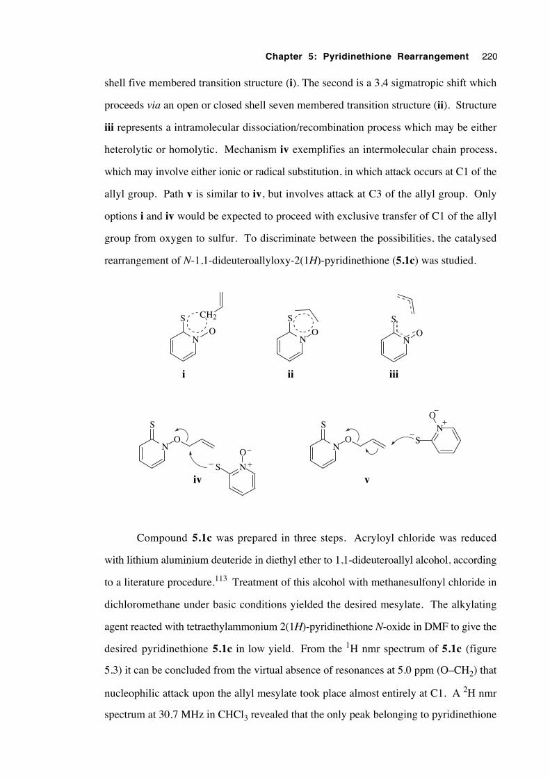
Chapter 5: Pyridinethione Rearrangement 220
shell five membered transition structure (i). The second is a 3,4 sigmatropic shift which
proceeds via an open or closed shell seven membered transition structure (ii). Structure
iii represents a intramolecular dissociation/recombination process which may be either
heterolytic or homolytic. Mechanism iv exemplifies an intermolecular chain process,
which may involve either ionic or radical substitution, in which attack occurs at C1 of the
allyl group. Path v is similar to iv, but involves attack at C3 of the allyl group. Only
options i and iv would be expected to proceed with exclusive transfer of C1 of the allyl
group from oxygen to sulfur. To discriminate between the possibilities, the catalysed
rearrangement of N-1,1-dideuteroallyloxy-2(1H)-pyridinethione (5.1c) was studied.
CH2
N
S
ON
S
O
N
S
ON
S
O
N
S
O
i ii
v
iii
N
S
O
NS
O
iv
Compound 5.1c was prepared in three steps. Acryloyl chloride was reduced
with lithium aluminium deuteride in diethyl ether to 1,1-dideuteroallyl alcohol, according
to a literature procedure.113 Treatment of this alcohol with methanesulfonyl chloride in
dichloromethane under basic conditions yielded the desired mesylate. The alkylating
agent reacted with tetraethylammonium 2(1H)-pyridinethione N-oxide in DMF to give the
desired pyridinethione 5.1c in low yield. From the 1H nmr spectrum of 5.1c (figure
5.3) it can be concluded from the virtual absence of resonances at 5.0 ppm (O–CH2) that
nucleophilic attack upon the allyl mesylate took place almost entirely at C1. A 2H nmr
spectrum at 30.7 MHz in CHCl3 revealed that the only peak belonging to pyridinethione
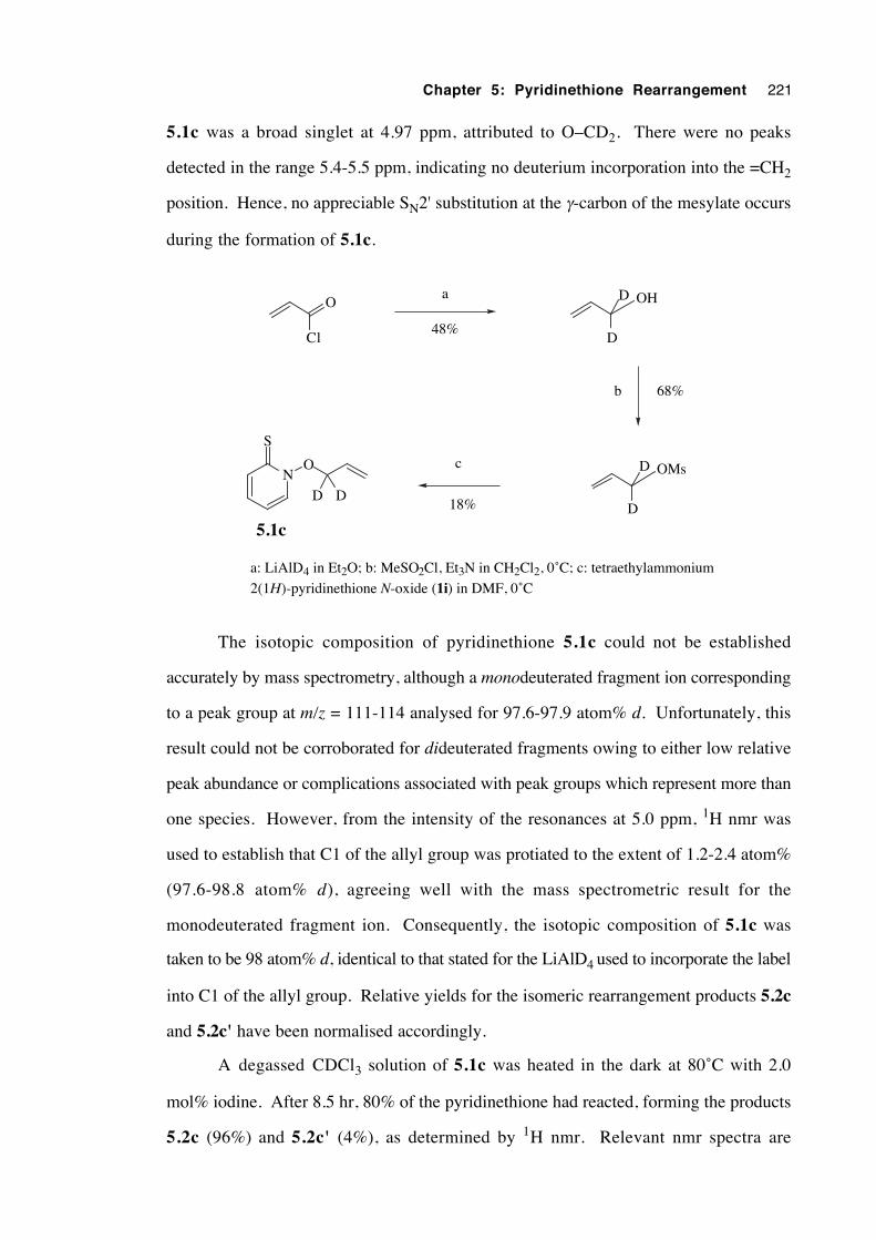
Chapter 5: Pyridinethione Rearrangement 221
5.1c was a broad singlet at 4.97 ppm, attributed to O–CD2. There were no peaks
detected in the range 5.4-5.5 ppm, indicating no deuterium incorporation into the =CH2
position. Hence, no appreciable SN2' substitution at the γ-carbon of the mesylate occurs
during the formation of 5.1c.
N
S
O
D D
5.1c
O OH
Cl D
D
48%
68%
OMs
D
D
18%
a: LiAlD4 in Et2O; b: MeSO2Cl, Et3N in CH2Cl2, 0˚C; c: tetraethylammonium
2(1H)-pyridinethione N-oxide (1i) in DMF, 0˚C
a
b
c
The isotopic composition of pyridinethione 5.1c could not be established
accurately by mass spectrometry, although a monodeuterated fragment ion corresponding
to a peak group at m/z = 111-114 analysed for 97.6-97.9 atom% d. Unfortunately, this
result could not be corroborated for dideuterated fragments owing to either low relative
peak abundance or complications associated with peak groups which represent more than
one species. However, from the intensity of the resonances at 5.0 ppm, 1H nmr was
used to establish that C1 of the allyl group was protiated to the extent of 1.2-2.4 atom%
(97.6-98.8 atom% d), agreeing well with the mass spectrometric result for the
monodeuterated fragment ion. Consequently, the isotopic composition of 5.1c was
taken to be 98 atom% d, identical to that stated for the LiAlD4 used to incorporate the label
into C1 of the allyl group. Relative yields for the isomeric rearrangement products 5.2c
and 5.2c' have been normalised accordingly.
A degassed CDCl3 solution of 5.1c was heated in the dark at 80˚C with 2.0
mol% iodine. After 8.5 hr, 80% of the pyridinethione had reacted, forming the products
5.2c (96%) and 5.2c' (4%), as determined by 1H nmr. Relevant nmr spectra are
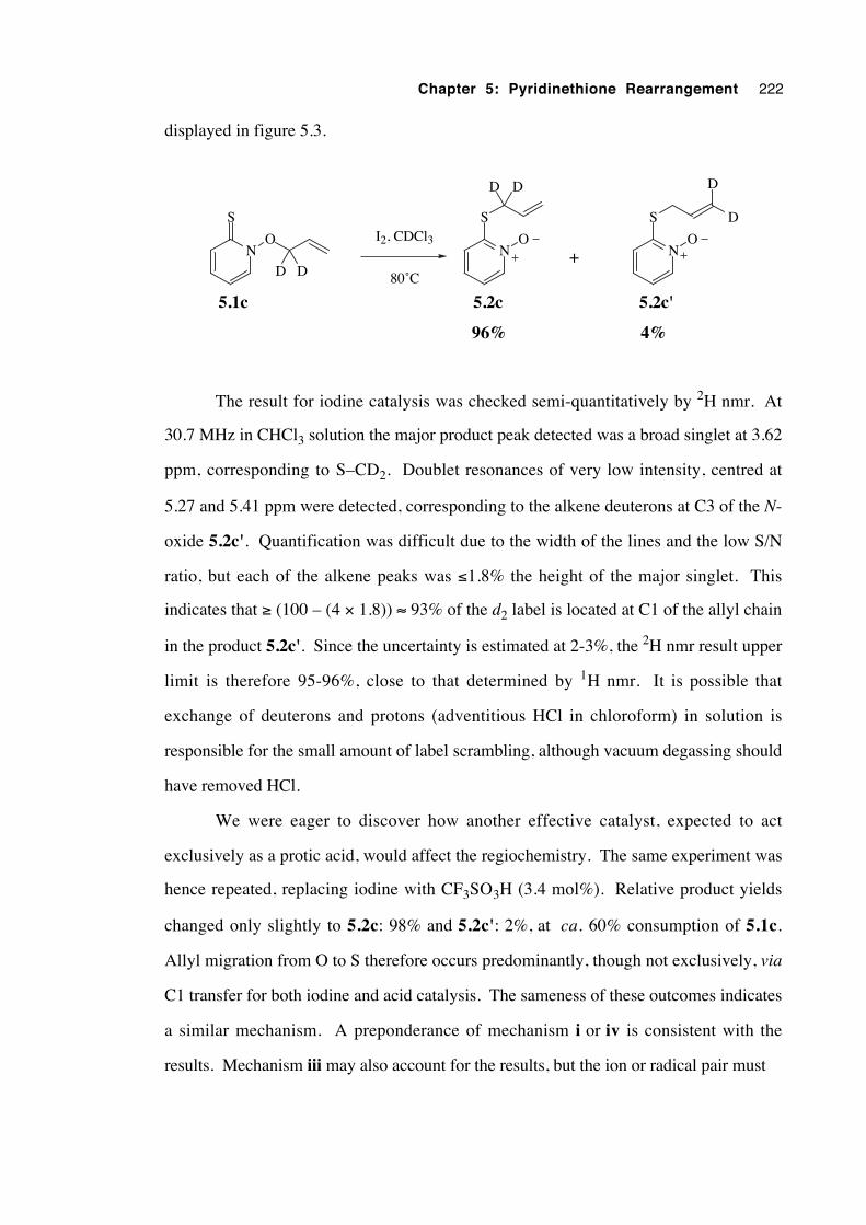
Chapter 5: Pyridinethione Rearrangement 222
displayed in figure 5.3.
N
S
O
D D
N
S
ON
S
O
D
DDD
+I2, CDCl3
80˚C
5.1c 5.2c 5.2c'
96% 4%
The result for iodine catalysis was checked semi-quantitatively by 2H nmr. At
30.7 MHz in CHCl3 solution the major product peak detected was a broad singlet at 3.62
ppm, corresponding to S–CD2. Doublet resonances of very low intensity, centred at
5.27 and 5.41 ppm were detected, corresponding to the alkene deuterons at C3 of the N-
oxide 5.2c'. Quantification was difficult due to the width of the lines and the low S/N
ratio, but each of the alkene peaks was ≤1.8% the height of the major singlet. This
indicates that ≥ (100 – (4 × 1.8)) ≈ 93% of the d2 label is located at C1 of the allyl chain
in the product 5.2c'. Since the uncertainty is estimated at 2-3%, the 2H nmr result upper
limit is therefore 95-96%, close to that determined by 1H nmr. It is possible that
exchange of deuterons and protons (adventitious HCl in chloroform) in solution is
responsible for the small amount of label scrambling, although vacuum degassing should
have removed HCl.
We were eager to discover how another effective catalyst, expected to act
exclusively as a protic acid, would affect the regiochemistry. The same experiment was
hence repeated, replacing iodine with CF3SO3H (3.4 mol%). Relative product yields
changed only slightly to 5.2c: 98% and 5.2c': 2%, at ca. 60% consumption of 5.1c.
Allyl migration from O to S therefore occurs predominantly, though not exclusively, via
C1 transfer for both iodine and acid catalysis. The sameness of these outcomes indicates
a similar mechanism. A preponderance of mechanism i or iv is consistent with the
results. Mechanism iii may also account for the results, but the ion or radical pair must
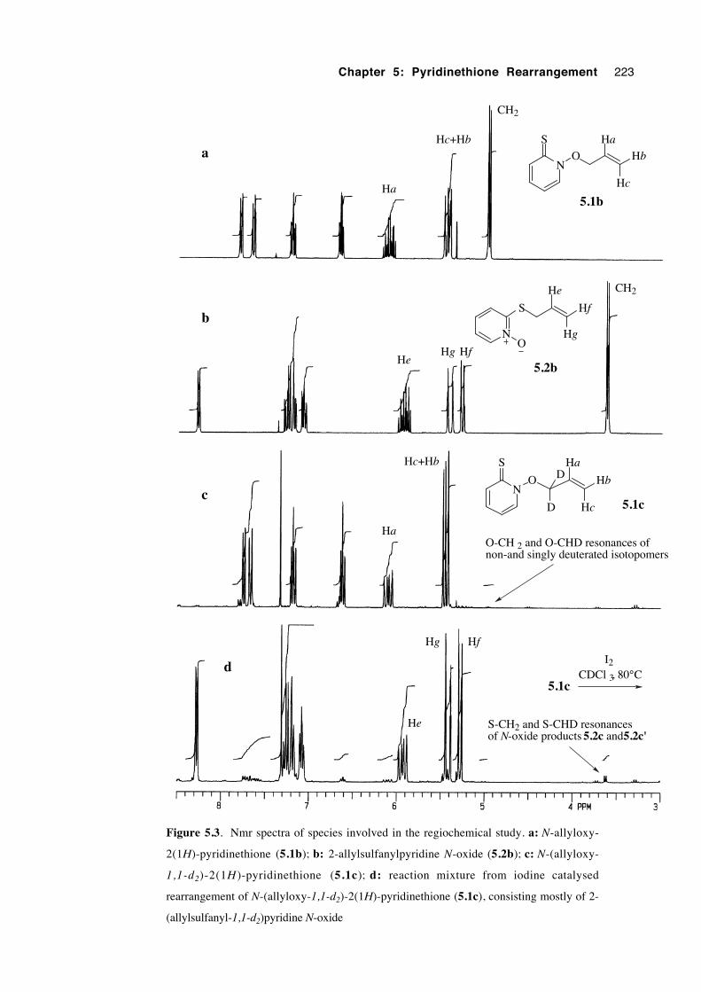
Chapter 5: Pyridinethione Rearrangement 223
NO
S Ha
Hb
Hc
NO
S Ha
Hb
HcD
D
N
S
He
Hf
HgO
CH2
HeHg Hf
CH2
He
Hg Hf
I2CDCl 3, 80°C
Ha
Hc+Hb
Ha
Hc+Hb
O-CH 2 and O-CHD resonances of non-and singly deuterated isotopomers
S-CH2 and S-CHD resonances of N-oxide products 5.2c and 5.2c'
a
b
c
d5.1c
5.1b
5.2b
5.1c
Figure 5.3. Nmr spectra of species involved in the regiochemical study. a: N-allyloxy-
2(1H)-pyridinethione (5.1b); b: 2-allylsulfanylpyridine N-oxide (5.2b); c: N-(allyloxy-
1,1-d2)-2(1H )-pyridinethione (5.1c); d: reaction mixture from iodine catalysed
rearrangement of N-(allyloxy-1,1-d2)-2(1H)-pyridinethione (5.1c), consisting mostly of 2-
(allylsulfanyl-1,1-d2)pyridine N-oxide
Chapter 5: Pyridinethione Rearrangement 224
have a very short lifetime and be highly selective to produce the high level of
regioselectivity observed.
In conclusion, the migration of the allyl group from O to S in the 5.1c→5.2c
rearrangement proceeds with high regioselectivity for C1 over C3 for both iodine and
trifluoromethanesulfonic acid catalysis. The predominance of an intramolecular 1,4 shift
(i) or an intermolecular allyl C1 substitution reaction (iv) is consistent with the results. A
tight, short lived and highly selective ion or radical pair (iii) is less likely, but possible.
The sameness of the results between catalysts suggests a similarity of mechanism.
5.3.4 A study of rearrangement stereochemistry
Results from the kinetic work of section 5.3.2 are consistent with an
intermolecular rate limiting step, yet do not yet exclude the possibility that the actual alkyl
group migration step is intramolecular. Mechanistic work on the rearrangement of
analogous 2-alkoxypyridine N-oxides117-119 has shown that an intermolecular transfer of
the alkyl group occurs, but such a process has not yet been tested with N-alkoxy-2(1H)-
pyridinethione rearrangement. For the purposes of this current stereochemical study the
initial hypothesis—that the isomerization proceeds by a catalysed 1,4 sigmatropic shift—
remains intact.
The stereochemical outcome of closed shell, thermal 1,4 sigmatropic
rearrangements should be unambiguous. As discussed previously, those involving the
redistribution of four electrons proceed in the 1a,4s mode, bringing about inversion of
configuration at C1 of the migrating group, while those involving six electrons proceed
with retention of configuration, via a 1s,4s path. Predicting the stereochemical outcome
of rearrangements within open shell systems—having one valence orbital singly
occupied—is less clear. The stereochemistry of the migration of an enantiomerically
enriched 1-phenylethyl group was studied to reveal whether the rearrangement more
closely resembles the characteristics of four (inversion) or of six (retention) electron
systems.
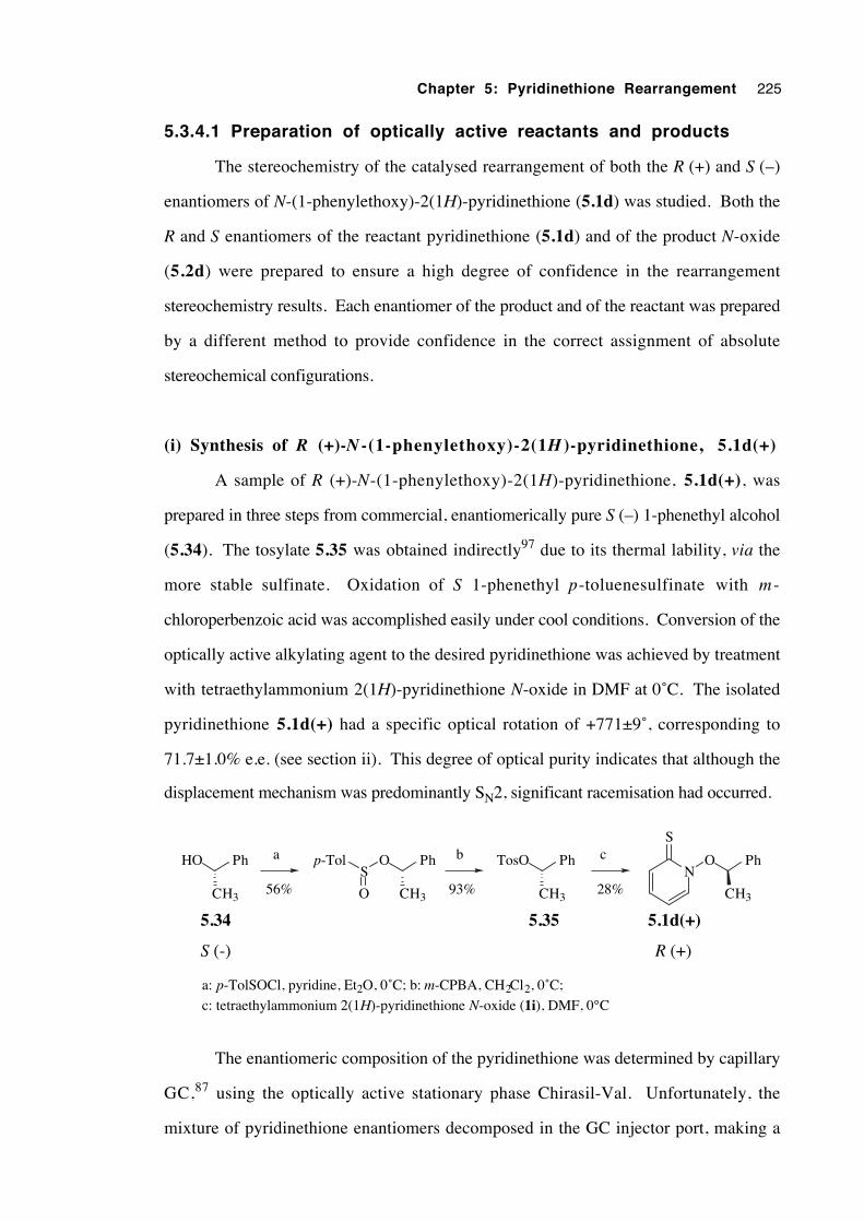
Chapter 5: Pyridinethione Rearrangement 225
5.3.4.1 Preparation of optically active reactants and products
The stereochemistry of the catalysed rearrangement of both the R (+) and S (–)
enantiomers of N-(1-phenylethoxy)-2(1H)-pyridinethione (5.1d) was studied. Both the
R and S enantiomers of the reactant pyridinethione (5.1d) and of the product N-oxide
(5.2d) were prepared to ensure a high degree of confidence in the rearrangement
stereochemistry results. Each enantiomer of the product and of the reactant was prepared
by a different method to provide confidence in the correct assignment of absolute
stereochemical configurations.
(i) Synthesis of R (+)-N -(1-phenylethoxy)-2(1H )-pyridinethione, 5.1d(+)
A sample of R (+)-N-(1-phenylethoxy)-2(1H)-pyridinethione, 5.1d(+), was
prepared in three steps from commercial, enantiomerically pure S (–) 1-phenethyl alcohol
(5.34). The tosylate 5.35 was obtained indirectly97 due to its thermal lability, via the
more stable sulfinate. Oxidation of S 1-phenethyl p-toluenesulfinate with m -
chloroperbenzoic acid was accomplished easily under cool conditions. Conversion of the
optically active alkylating agent to the desired pyridinethione was achieved by treatment
with tetraethylammonium 2(1H)-pyridinethione N-oxide in DMF at 0˚C. The isolated
pyridinethione 5.1d(+) had a specific optical rotation of +771±9˚, corresponding to
71.7±1.0% e.e. (see section ii). This degree of optical purity indicates that although the
displacement mechanism was predominantly SN2, significant racemisation had occurred.
CH3
HO Ph
CH3
O Ph
CH3
TosO PhS
O
p-Tol
CH3
O PhN
Sa b c
56% 93% 28%
S (-) R (+)
a: p-TolSOCl, pyridine, Et2O, 0˚C; b: m-CPBA, CH2Cl2, 0˚C;
c: tetraethylammonium 2(1H)-pyridinethione N-oxide (1i), DMF, 0°C
5.34 5.35 5.1d(+)
The enantiomeric composition of the pyridinethione was determined by capillary
GC,87 using the optically active stationary phase Chirasil-Val. Unfortunately, the
mixture of pyridinethione enantiomers decomposed in the GC injector port, making a
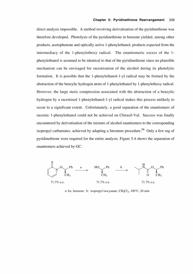
Chapter 5: Pyridinethione Rearrangement 226
direct analysis impossible. A method involving derivatisation of the pyridinethione was
therefore developed. Photolysis of the pyridinethione in benzene yielded, among other
products, acetophenone and optically active 1-phenylethanol, products expected from the
intermediacy of the 1-phenylethoxy radical. The enantiomeric excess of the 1-
phenylethanol is assumed to be identical to that of the pyridinethione since no plausible
mechanism can be envisaged for racemisation of the alcohol during its photolytic
formation. It is possible that the 1-phenylethanol-1-yl radical may be formed by the
abstraction of the benzylic hydrogen atom of 1-phenylethanol by 1-phenylethoxy radical.
However, the large steric compression associated with the abstraction of a benzylic
hydrogen by a racemised 1-phenylethanol-1-yl radical makes this process unlikely to
occur to a significant extent. Unfortunately, a good separation of the enantiomers of
racemic 1-phenylethanol could not be achieved on Chirasil-Val. Success was finally
encountered by derivatisation of the mixture of alcohol enantiomers to the corresponding
isopropyl carbamates, achieved by adapting a literature procedure.98 Only a few mg of
pyridinethione were required for the entire analysis. Figure 5.4 shows the separation of
enantiomers achieved by GC.
N
S
O Ph
CH3
HO Ph
CH3
O Ph
CH3
HN
O
b
71.7% e.e. 71.7% e.e. 71.7% e.e.
a: hν, benzene; b: isopropyl isocyanate, CH2Cl2, 100˚C, 20 min
a
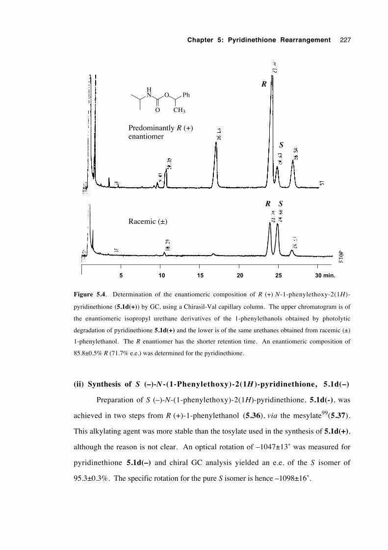
Chapter 5: Pyridinethione Rearrangement 227
5 10 15 20 25 30 min.
R S
R
S
Racemic (±)
Predominantly R (+)enantiomer
O Ph
CH3
HN
O
Figure 5.4. Determination of the enantiomeric composition of R (+) N-1-phenylethoxy-2(1H)-
pyridinethione (5.1d(+)) by GC, using a Chirasil-Val capillary column. The upper chromatogram is of
the enantiomeric isopropyl urethane derivatives of the 1-phenylethanols obtained by photolytic
degradation of pyridinethione 5.1d(+) and the lower is of the same urethanes obtained from racemic (±)
1-phenylethanol. The R enantiomer has the shorter retention time. An enantiomeric composition of
85.8±0.5% R (71.7% e.e.) was determined for the pyridinethione.
(ii) Synthesis of S (–)-N -(1-Phenylethoxy)-2(1H )-pyridinethione, 5.1d(–)
Preparation of S (–)-N-(1-phenylethoxy)-2(1H)-pyridinethione, 5.1d(-), was
achieved in two steps from R (+)-1-phenylethanol (5.36), via the mesylate99(5.37).
This alkylating agent was more stable than the tosylate used in the synthesis of 5.1d(+),
although the reason is not clear. An optical rotation of –1047±13˚ was measured for
pyridinethione 5.1d(–) and chiral GC analysis yielded an e.e. of the S isomer of
95.3±0.3%. The specific rotation for the pure S isomer is hence –1098±16˚.
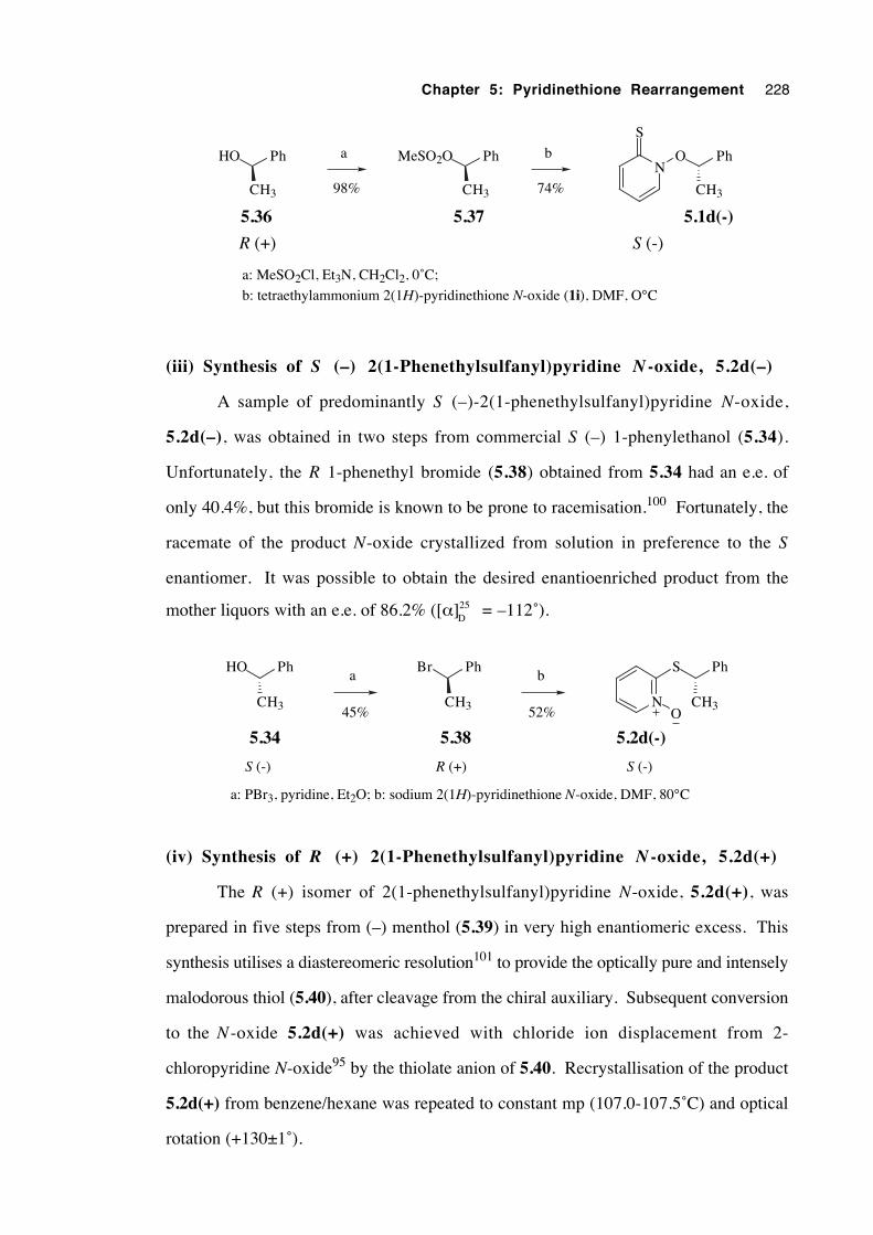
Chapter 5: Pyridinethione Rearrangement 228
CH3
HO Ph
5.36
CH3
MeSO2O Ph
5.37
O Ph
5.1d(-)
CH3
N
S
a
98%
b
74%
R (+) S (-)
a: MeSO2Cl, Et3N, CH2Cl2, 0˚C;
b: tetraethylammonium 2(1H)-pyridinethione N-oxide (1i), DMF, O°C
(iii) Synthesis of S (–) 2(1-Phenethylsulfanyl)pyridine N -oxide, 5.2d(–)
A sample of predominantly S (–)-2(1-phenethylsulfanyl)pyridine N-oxide,
5.2d(–), was obtained in two steps from commercial S (–) 1-phenylethanol (5.34).
Unfortunately, the R 1-phenethyl bromide (5.38) obtained from 5.34 had an e.e. of
only 40.4%, but this bromide is known to be prone to racemisation.100 Fortunately, the
racemate of the product N-oxide crystallized from solution in preference to the S
enantiomer. It was possible to obtain the desired enantioenriched product from the
mother liquors with an e.e. of 86.2% ([α]25D
= –112˚).
CH3
HO Ph
5.34
CH3
Br Ph
5.38
CH3
S Ph
5.2d(-)
NO
a b
45% 52%
S (-) R (+) S (-)
a: PBr3, pyridine, Et2O; b: sodium 2(1H)-pyridinethione N-oxide, DMF, 80°C
(iv) Synthesis of R (+) 2(1-Phenethylsulfanyl)pyridine N -oxide, 5.2d(+)
The R (+) isomer of 2(1-phenethylsulfanyl)pyridine N-oxide, 5.2d(+), was
prepared in five steps from (–) menthol (5.39) in very high enantiomeric excess. This
synthesis utilises a diastereomeric resolution101 to provide the optically pure and intensely
malodorous thiol (5.40), after cleavage from the chiral auxiliary. Subsequent conversion
to the N -oxide 5.2d(+) was achieved with chloride ion displacement from 2-
chloropyridine N-oxide95 by the thiolate anion of 5.40. Recrystallisation of the product
5.2d(+) from benzene/hexane was repeated to constant mp (107.0-107.5˚C) and optical
rotation (+130±1˚).
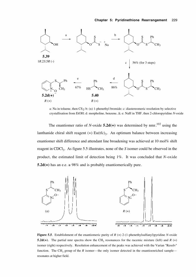
Chapter 5: Pyridinethione Rearrangement 229
OH
5.39
O S
S
Na O S
S Ph
CH3
O S
S Ph
CH3
Ph
HS CH3N
S Ph
CH3O
1R,2S,5R (-)
5.405.2d(+)R (+)R (+)
a b
c
de
56% (for 3 steps)
86%67%
a: Na in toluene, then CS2; b: (±) 1-phenethyl bromide; c: diastereomeric resolution by selective
crystallisation from EtOH; d: morpholine, benzene, ∆; e: NaH in THF, then 2-chloropyridine N-oxide
The enantiomer ratio of N-oxide 5.2d(+) was determined by nmr,102 using the
lanthanide chiral shift reagent (+) Eu(tfc)3. An optimum balance between increasing
enantiomer shift difference and attendant line broadening was achieved at 10 mol% shift
reagent in CDCl3. As figure 5.5 illustrates, none of the S isomer could be observed in the
product, the estimated limit of detection being 1%. It was concluded that N-oxide
5.2d(+) has an e.e. ≥ 98% and is probably enantiomerically pure.
NO
S
Ph
CH3
NO
S
Ph
CH3
(±) R (+)
Figure 5.5. Establishment of the enantiomeric purity of R (+) 2-(1-phenethylsulfanyl)pyridine N-oxide
5.2d(+). The partial nmr spectra show the CH3 resonances for the racemic mixture (left) and R (+)
isomer (right) respectively. Resolution enhancement of the peaks was achieved with the Varian "Resolv"
function. The CH3 group of the R isomer—the only isomer detected in the enantioenriched sample—
resonates at higher field.
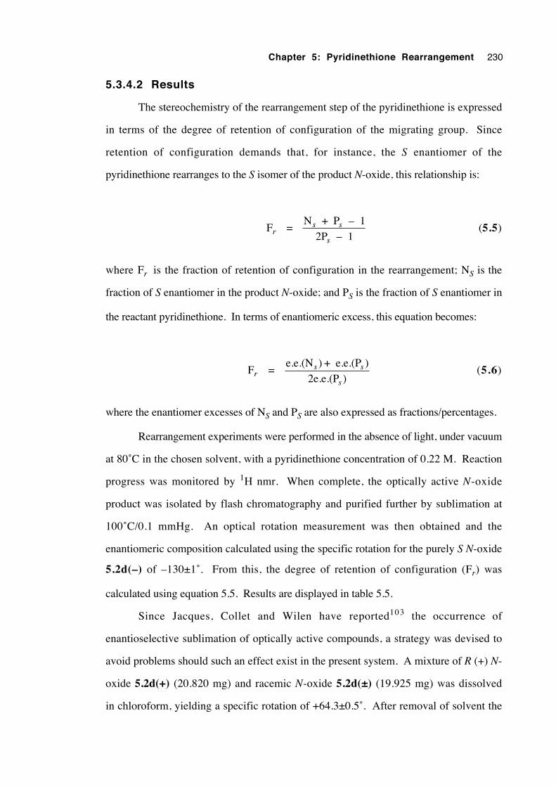
Chapter 5: Pyridinethione Rearrangement 230
5.3.4.2 Results
The stereochemistry of the rearrangement step of the pyridinethione is expressed
in terms of the degree of retention of configuration of the migrating group. Since
retention of configuration demands that, for instance, the S enantiomer of the
pyridinethione rearranges to the S isomer of the product N-oxide, this relationship is:
Fr = N + P – 1
2P – 1s s
s (5.5)
where Fr is the fraction of retention of configuration in the rearrangement; NS is the
fraction of S enantiomer in the product N-oxide; and PS is the fraction of S enantiomer in
the reactant pyridinethione. In terms of enantiomeric excess, this equation becomes:
Fr = e.e.(N ) + e.e.(P
2e.e.(Ps s
s
))
(5.6)
where the enantiomer excesses of NS and PS are also expressed as fractions/percentages.
Rearrangement experiments were performed in the absence of light, under vacuum
at 80˚C in the chosen solvent, with a pyridinethione concentration of 0.22 M. Reaction
progress was monitored by 1H nmr. When complete, the optically active N-oxide
product was isolated by flash chromatography and purified further by sublimation at
100˚C/0.1 mmHg. An optical rotation measurement was then obtained and the
enantiomeric composition calculated using the specific rotation for the purely S N-oxide
5.2d(–) of –130±1˚. From this, the degree of retention of configuration (Fr) was
calculated using equation 5.5. Results are displayed in table 5.5.
Since Jacques, Collet and Wilen have reported103 the occurrence of
enantioselective sublimation of optically active compounds, a strategy was devised to
avoid problems should such an effect exist in the present system. A mixture of R (+) N-
oxide 5.2d(+) (20.820 mg) and racemic N-oxide 5.2d(±) (19.925 mg) was dissolved
in chloroform, yielding a specific rotation of +64.3±0.5˚. After removal of solvent the
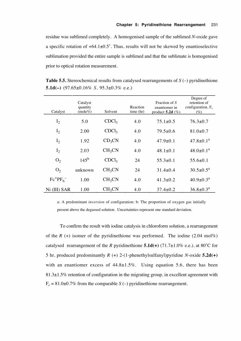
Chapter 5: Pyridinethione Rearrangement 231
residue was sublimed completely. A homogenised sample of the sublimed N-oxide gave
a specific rotation of +64.1±0.5˚. Thus, results will not be skewed by enantioselective
sublimation provided the entire sample is sublimed and that the sublimate is homogenised
prior to optical rotation measurement.
Table 5.5. Stereochemical results from catalysed rearrangements of S (–) pyridinethione
5.1d(–) (97.65±0.16% S, 95.3±0.3% e.e.)
Catalyst
Catalystquantity(mole%) Solvent
Reactiontime (hr)
Fraction of Senantiomer in
product 5.2d (%)
Degree ofretention of
configuration, Fr
(%)
I2 5.0 CDCl3 4.0 75.1±0.5 76.3±0.7
I2 2.00 CDCl3 4.0 79.5±0.6 81.0±0.7
I2 1.92 CD3CN 4.0 47.9±0.1 47.8±0.1a
I2 2.03 CH3CN 4.0 48.1±0.1 48.0±0.1a
O2 145b CDCl3 24 55.3±0.1 55.6±0.1
O2 unknown CH3CN 24 31.4±0.4 30.5±0.5a
Fc+PF6– 1.00 CH3CN 4.0 41.3±0.2 40.9±0.3a
Ni (III) SAR 1.00 CH3CN 4.0 37.4±0.2 36.8±0.3a
a: A predominant inversion of configuration; b: The proportion of oxygen gas initially
present above the degassed solution. Uncertainties represent one standard deviation.
To confirm the result with iodine catalysis in chloroform solution, a rearrangement
of the R (+) isomer of the pyridinethione was performed. The iodine (2.04 mol%)
catalysed rearrangement of the R pyridinethione 5.1d(+) (71.7±1.0% e.e.), at 80˚C for
5 hr, produced predominantly R (+) 2-(1-phenethylsulfanyl)pyridine N-oxide 5.2d(+)
with an enantiomer excess of 44.8±1.5%. Using equation 5.6, there has been
81.3±1.5% retention of configuration in the migrating group, in excellent agreement with
Fr = 81.0±0.7% from the comparable S (–) pyridinethione rearrangement.
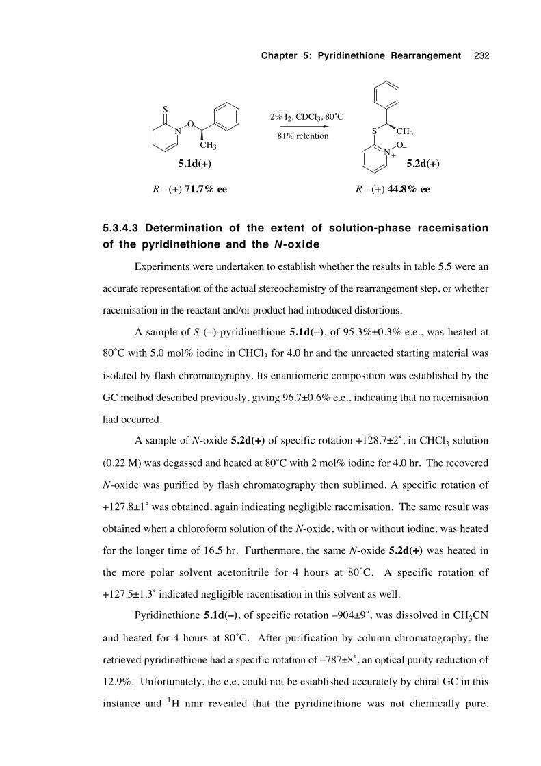
Chapter 5: Pyridinethione Rearrangement 232
N
S
O
CH3N
S
O
CH3
2% I2, CDCl3, 80˚C
81% retention
R - (+) 71.7% ee R - (+) 44.8% ee
5.2d(+)5.1d(+)
5.3.4.3 Determination of the extent of solution-phase racemisation
of the pyridinethione and the N-oxide
Experiments were undertaken to establish whether the results in table 5.5 were an
accurate representation of the actual stereochemistry of the rearrangement step, or whether
racemisation in the reactant and/or product had introduced distortions.
A sample of S (–)-pyridinethione 5.1d(–), of 95.3%±0.3% e.e., was heated at
80˚C with 5.0 mol% iodine in CHCl3 for 4.0 hr and the unreacted starting material was
isolated by flash chromatography. Its enantiomeric composition was established by the
GC method described previously, giving 96.7±0.6% e.e., indicating that no racemisation
had occurred.
A sample of N-oxide 5.2d(+) of specific rotation +128.7±2˚, in CHCl3 solution
(0.22 M) was degassed and heated at 80˚C with 2 mol% iodine for 4.0 hr. The recovered
N-oxide was purified by flash chromatography then sublimed. A specific rotation of
+127.8±1˚ was obtained, again indicating negligible racemisation. The same result was
obtained when a chloroform solution of the N-oxide, with or without iodine, was heated
for the longer time of 16.5 hr. Furthermore, the same N-oxide 5.2d(+) was heated in
the more polar solvent acetonitrile for 4 hours at 80˚C. A specific rotation of
+127.5±1.3˚ indicated negligible racemisation in this solvent as well.
Pyridinethione 5.1d(–), of specific rotation –904±9˚, was dissolved in CH3CN
and heated for 4 hours at 80˚C. After purification by column chromatography, the
retrieved pyridinethione had a specific rotation of –787±8˚, an optical purity reduction of
12.9%. Unfortunately, the e.e. could not be established accurately by chiral GC in this
instance and 1H nmr revealed that the pyridinethione was not chemically pure.
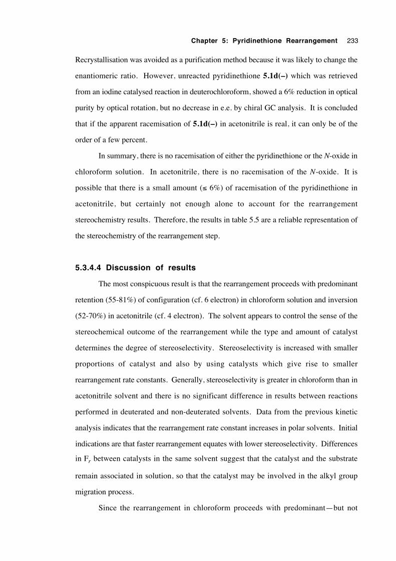
Chapter 5: Pyridinethione Rearrangement 233
Recrystallisation was avoided as a purification method because it was likely to change the
enantiomeric ratio. However, unreacted pyridinethione 5.1d(–) which was retrieved
from an iodine catalysed reaction in deuterochloroform, showed a 6% reduction in optical
purity by optical rotation, but no decrease in e.e. by chiral GC analysis. It is concluded
that if the apparent racemisation of 5.1d(–) in acetonitrile is real, it can only be of the
order of a few percent.
In summary, there is no racemisation of either the pyridinethione or the N-oxide in
chloroform solution. In acetonitrile, there is no racemisation of the N-oxide. It is
possible that there is a small amount (≤ 6%) of racemisation of the pyridinethione in
acetonitrile, but certainly not enough alone to account for the rearrangement
stereochemistry results. Therefore, the results in table 5.5 are a reliable representation of
the stereochemistry of the rearrangement step.
5.3.4.4 Discussion of results
The most conspicuous result is that the rearrangement proceeds with predominant
retention (55-81%) of configuration (cf. 6 electron) in chloroform solution and inversion
(52-70%) in acetonitrile (cf. 4 electron). The solvent appears to control the sense of the
stereochemical outcome of the rearrangement while the type and amount of catalyst
determines the degree of stereoselectivity. Stereoselectivity is increased with smaller
proportions of catalyst and also by using catalysts which give rise to smaller
rearrangement rate constants. Generally, stereoselectivity is greater in chloroform than in
acetonitrile solvent and there is no significant difference in results between reactions
performed in deuterated and non-deuterated solvents. Data from the previous kinetic
analysis indicates that the rearrangement rate constant increases in polar solvents. Initial
indications are that faster rearrangement equates with lower stereoselectivity. Differences
in Fr between catalysts in the same solvent suggest that the catalyst and the substrate
remain associated in solution, so that the catalyst may be involved in the alkyl group
migration process.
Since the rearrangement in chloroform proceeds with predominant—but not
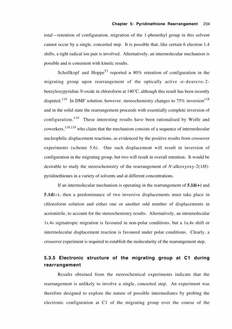
Chapter 5: Pyridinethione Rearrangement 234
total—retention of configuration, migration of the 1-phenethyl group in this solvent
cannot occur by a single, concerted step. It is possible that, like certain 6 electron 1,4
shifts, a tight radical ion pair is involved. Alternatively, an intermolecular mechanism is
possible and is consistent with kinetic results.
Schollkopf and Hoppe53 reported ≥ 80% retention of configuration in the
migrating group upon rearrangement of the optically active α -deutero-2-
benzyloxypyridine N-oxide in chloroform at 140˚C, although this result has been recently
disputed.119 In DMF solution, however, stereochemistry changes to 75% inversion118
and in the solid state the rearrangement proceeds with essentially complete inversion of
configuration.119 These interesting results have been rationalised by Wolfe and
coworkers,118,119 who claim that the mechanism consists of a sequence of intermolecular
nucleophilic displacement reactions, as evidenced by the positive results from crossover
experiments (scheme 5.6). One such displacement will result in inversion of
configuration in the migrating group, but two will result in overall retention. It would be
desirable to study the stereochemistry of the rearrangement of N-alkoxyoxy-2(1H)-
pyridinethiones in a variety of solvents and at different concentrations.
If an intermolecular mechanism is operating in the rearrangement of 5.1d(+) and
5.1d(–), then a predominance of two inversive displacements must take place in
chloroform solution and either one or another odd number of displacements in
acetonitrile, to account for the stereochemistry results. Alternatively, an intramolecular
1s,4s sigmatropic migration is favoured in non-polar conditions, but a 1a,4s shift or
intermolecular displacement reaction is favoured under polar conditions. Clearly, a
crossover experiment is required to establish the molecularity of the rearrangement step.
5.3.5 Electronic structure of the migrating group at C1 during
rearrangement
Results obtained from the stereochemical experiments indicate that the
rearrangement is unlikely to involve a single, concerted step. An experiment was
therefore designed to explore the nature of possible intermediates by probing the
electronic configuration at C1 of the migrating group over the course of the
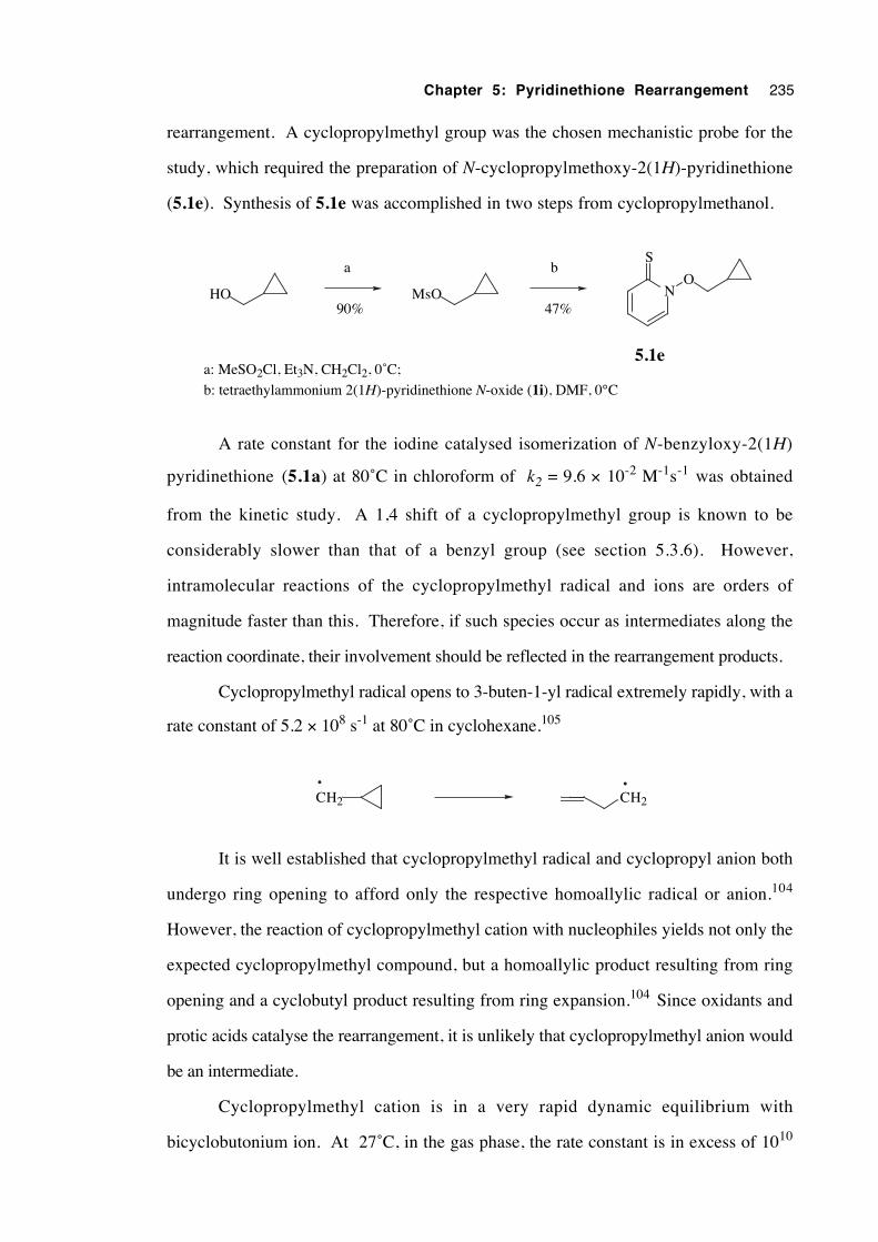
Chapter 5: Pyridinethione Rearrangement 235
rearrangement. A cyclopropylmethyl group was the chosen mechanistic probe for the
study, which required the preparation of N-cyclopropylmethoxy-2(1H)-pyridinethione
(5.1e). Synthesis of 5.1e was accomplished in two steps from cyclopropylmethanol.
N
S
O
5.1e
HO MsO
a b
90% 47%
a: MeSO2Cl, Et3N, CH2Cl2, 0˚C;
b: tetraethylammonium 2(1H)-pyridinethione N-oxide (1i), DMF, 0°C
A rate constant for the iodine catalysed isomerization of N-benzyloxy-2(1H)
pyridinethione (5.1a) at 80˚C in chloroform of k2 = 9.6 × 10⁻² M-1s-1 was obtained
from the kinetic study. A 1,4 shift of a cyclopropylmethyl group is known to be
considerably slower than that of a benzyl group (see section 5.3.6). However,
intramolecular reactions of the cyclopropylmethyl radical and ions are orders of
magnitude faster than this. Therefore, if such species occur as intermediates along the
reaction coordinate, their involvement should be reflected in the rearrangement products.
Cyclopropylmethyl radical opens to 3-buten-1-yl radical extremely rapidly, with a
rate constant of 5.2 × 10⁸ s-1 at 80˚C in cyclohexane.105
CH2 CH2
It is well established that cyclopropylmethyl radical and cyclopropyl anion both
undergo ring opening to afford only the respective homoallylic radical or anion.104
However, the reaction of cyclopropylmethyl cation with nucleophiles yields not only the
expected cyclopropylmethyl compound, but a homoallylic product resulting from ring
opening and a cyclobutyl product resulting from ring expansion.104 Since oxidants and
protic acids catalyse the rearrangement, it is unlikely that cyclopropylmethyl anion would
be an intermediate.
Cyclopropylmethyl cation is in a very rapid dynamic equilibrium with
bicyclobutonium ion. At 27˚C, in the gas phase, the rate constant is in excess of 1010
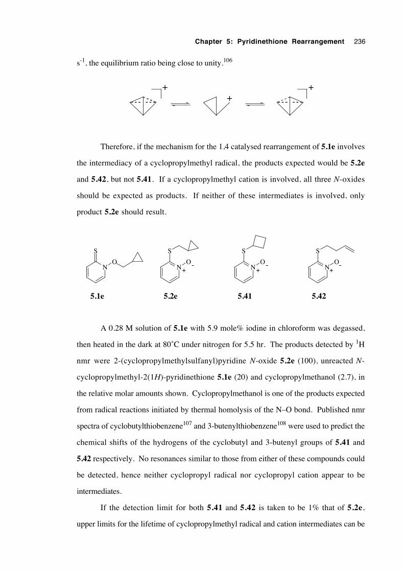
Chapter 5: Pyridinethione Rearrangement 236
s-1, the equilibrium ratio being close to unity.106
++ +
Therefore, if the mechanism for the 1,4 catalysed rearrangement of 5.1e involves
the intermediacy of a cyclopropylmethyl radical, the products expected would be 5.2e
and 5.42, but not 5.41. If a cyclopropylmethyl cation is involved, all three N-oxides
should be expected as products. If neither of these intermediates is involved, only
product 5.2e should result.
N
S
ON
S
ON
S
ON
S
O+
-+
-+
-
5.1e 5.2e 5.41 5.42
A 0.28 M solution of 5.1e with 5.9 mole% iodine in chloroform was degassed,
then heated in the dark at 80˚C under nitrogen for 5.5 hr. The products detected by 1H
nmr were 2-(cyclopropylmethylsulfanyl)pyridine N-oxide 5.2e (100), unreacted N-
cyclopropylmethyl-2(1H)-pyridinethione 5.1e (20) and cyclopropylmethanol (2.7), in
the relative molar amounts shown. Cyclopropylmethanol is one of the products expected
from radical reactions initiated by thermal homolysis of the N–O bond. Published nmr
spectra of cyclobutylthiobenzene107 and 3-butenylthiobenzene108 were used to predict the
chemical shifts of the hydrogens of the cyclobutyl and 3-butenyl groups of 5.41 and
5.42 respectively. No resonances similar to those from either of these compounds could
be detected, hence neither cyclopropyl radical nor cyclopropyl cation appear to be
intermediates.
If the detection limit for both 5.41 and 5.42 is taken to be 1% that of 5.2e,
upper limits for the lifetime of cyclopropylmethyl radical and cation intermediates can be
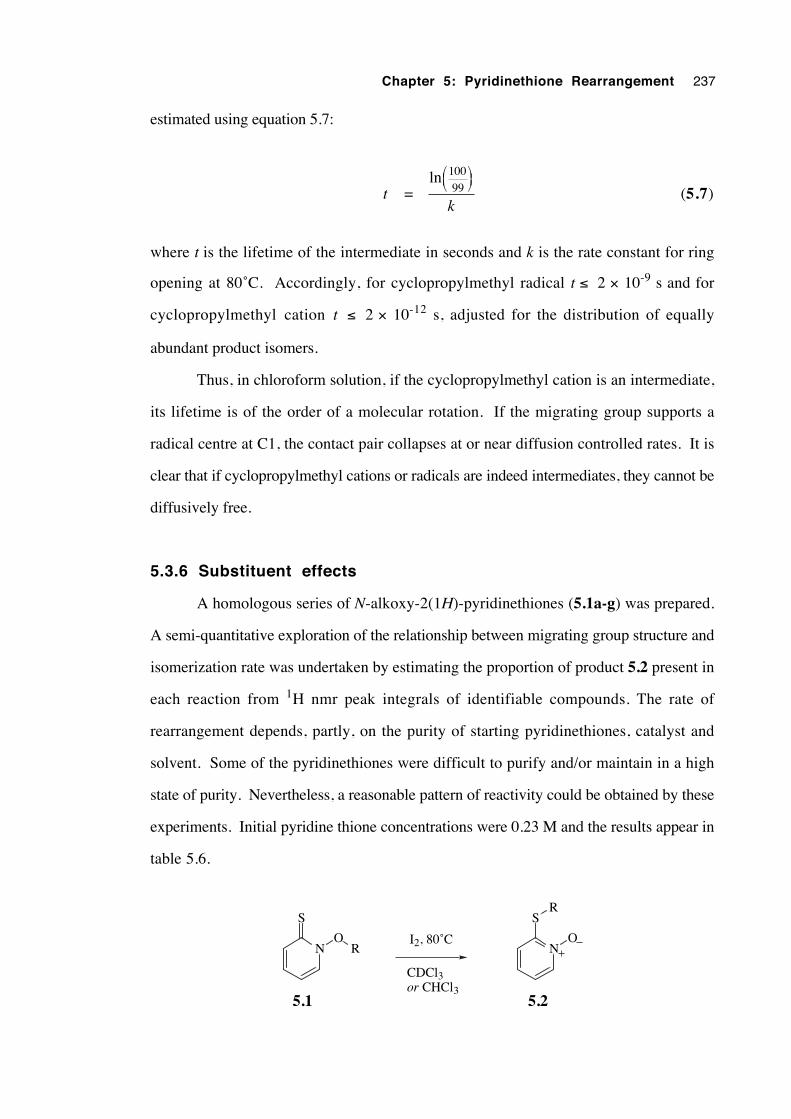
Chapter 5: Pyridinethione Rearrangement 237
estimated using equation 5.7:
t = ln 100
99
k (5.7)
where t is the lifetime of the intermediate in seconds and k is the rate constant for ring
opening at 80˚C. Accordingly, for cyclopropylmethyl radical t ≤ 2 × 10⁻⁹ s and for
cyclopropylmethyl cation t ≤ 2 × 10-12 s, adjusted for the distribution of equally
abundant product isomers.
Thus, in chloroform solution, if the cyclopropylmethyl cation is an intermediate,
its lifetime is of the order of a molecular rotation. If the migrating group supports a
radical centre at C1, the contact pair collapses at or near diffusion controlled rates. It is
clear that if cyclopropylmethyl cations or radicals are indeed intermediates, they cannot be
diffusively free.
5.3.6 Substituent effects
A homologous series of N-alkoxy-2(1H)-pyridinethiones (5.1a-g) was prepared.
A semi-quantitative exploration of the relationship between migrating group structure and
isomerization rate was undertaken by estimating the proportion of product 5.2 present in
each reaction from 1H nmr peak integrals of identifiable compounds. The rate of
rearrangement depends, partly, on the purity of starting pyridinethiones, catalyst and
solvent. Some of the pyridinethiones were difficult to purify and/or maintain in a high
state of purity. Nevertheless, a reasonable pattern of reactivity could be obtained by these
experiments. Initial pyridine thione concentrations were 0.23 M and the results appear in
table 5.6.
N
S
OR N
S
O
R
5.1 5.2
I2, 80˚C
CDCl3or CHCl3
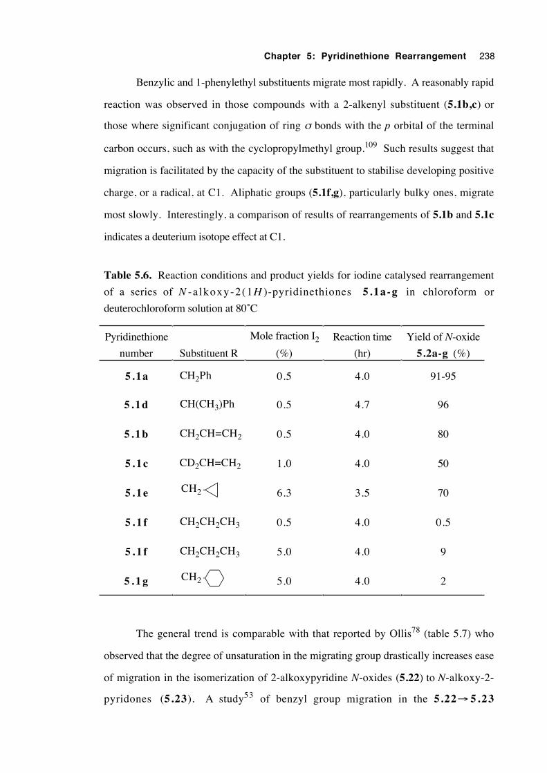
Chapter 5: Pyridinethione Rearrangement 238
Benzylic and 1-phenylethyl substituents migrate most rapidly. A reasonably rapid
reaction was observed in those compounds with a 2-alkenyl substituent (5.1b,c) or
those where significant conjugation of ring σ bonds with the p orbital of the terminal
carbon occurs, such as with the cyclopropylmethyl group.109 Such results suggest that
migration is facilitated by the capacity of the substituent to stabilise developing positive
charge, or a radical, at C1. Aliphatic groups (5.1f,g), particularly bulky ones, migrate
most slowly. Interestingly, a comparison of results of rearrangements of 5.1b and 5.1c
indicates a deuterium isotope effect at C1.
Table 5.6. Reaction conditions and product yields for iodine catalysed rearrangement
of a series of N -a lkoxy-2(1H )-pyridinethiones 5.1a-g in chloroform or
deuterochloroform solution at 80˚C
Pyridinethione
number Substituent R
Mole fraction I2
(%)
Reaction time
(hr)
Yield of N-oxide
5.2a-g (%)
5.1a CH2Ph 0.5 4.0 91-95
5 .1d CH(CH3)Ph 0.5 4.7 96
5 .1b CH2CH=CH2 0.5 4.0 80
5 . 1 c CD2CH=CH2 1.0 4.0 50
5 . 1 e CH2 6.3 3.5 70
5 . 1 f CH2CH2CH3 0.5 4.0 0.5
5 . 1 f CH2CH2CH3 5.0 4.0 9
5 . 1 g CH2 5.0 4.0 2
The general trend is comparable with that reported by Ollis78 (table 5.7) who
observed that the degree of unsaturation in the migrating group drastically increases ease
of migration in the isomerization of 2-alkoxypyridine N-oxides (5.22) to N-alkoxy-2-
pyridones (5.23). A study53 of benzyl group migration in the 5.22→ 5 .23
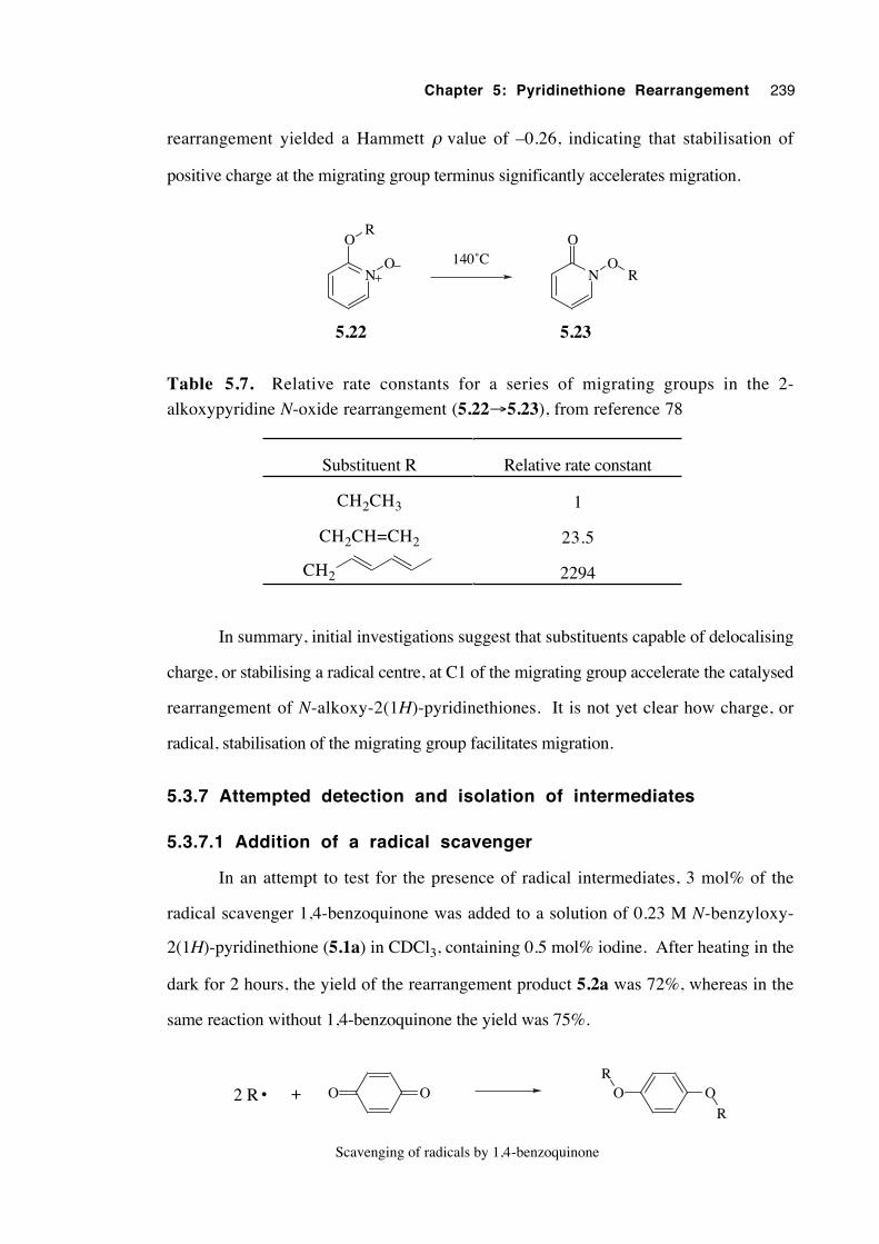
Chapter 5: Pyridinethione Rearrangement 239
rearrangement yielded a Hammett ρ value of –0.26, indicating that stabilisation of
positive charge at the migrating group terminus significantly accelerates migration.
N
O
ON
O
O
R
R140˚C
5.22 5.23
Table 5.7. Relative rate constants for a series of migrating groups in the 2-
alkoxypyridine N-oxide rearrangement (5.22→5.23), from reference 78
Substituent R Relative rate constant
CH2CH3 1
CH2CH=CH2 23.5
CH2 2294
In summary, initial investigations suggest that substituents capable of delocalising
charge, or stabilising a radical centre, at C1 of the migrating group accelerate the catalysed
rearrangement of N-alkoxy-2(1H)-pyridinethiones. It is not yet clear how charge, or
radical, stabilisation of the migrating group facilitates migration.
5.3.7 Attempted detection and isolation of intermediates
5.3.7.1 Addition of a radical scavenger
In an attempt to test for the presence of radical intermediates, 3 mol% of the
radical scavenger 1,4-benzoquinone was added to a solution of 0.23 M N-benzyloxy-
2(1H)-pyridinethione (5.1a) in CDCl3, containing 0.5 mol% iodine. After heating in the
dark for 2 hours, the yield of the rearrangement product 5.2a was 72%, whereas in the
same reaction without 1,4-benzoquinone the yield was 75%.
O O2 R • + O OR
R
Scavenging of radicals by 1,4-benzoquinone
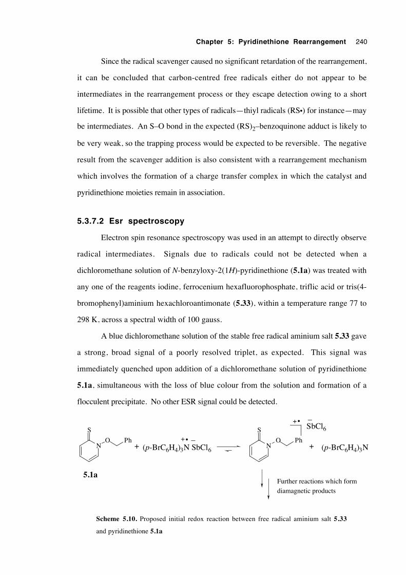
Chapter 5: Pyridinethione Rearrangement 240
Since the radical scavenger caused no significant retardation of the rearrangement,
it can be concluded that carbon-centred free radicals either do not appear to be
intermediates in the rearrangement process or they escape detection owing to a short
lifetime. It is possible that other types of radicals—thiyl radicals (RS•) for instance—may
be intermediates. An S–O bond in the expected (RS)2–benzoquinone adduct is likely to
be very weak, so the trapping process would be expected to be reversible. The negative
result from the scavenger addition is also consistent with a rearrangement mechanism
which involves the formation of a charge transfer complex in which the catalyst and
pyridinethione moieties remain in association.
5.3.7.2 Esr spectroscopy
Electron spin resonance spectroscopy was used in an attempt to directly observe
radical intermediates. Signals due to radicals could not be detected when a
dichloromethane solution of N-benzyloxy-2(1H)-pyridinethione (5.1a) was treated with
any one of the reagents iodine, ferrocenium hexafluorophosphate, triflic acid or tris(4-
bromophenyl)aminium hexachloroantimonate (5.33), within a temperature range 77 to
298 K, across a spectral width of 100 gauss.
A blue dichloromethane solution of the stable free radical aminium salt 5.33 gave
a strong, broad signal of a poorly resolved triplet, as expected. This signal was
immediately quenched upon addition of a dichloromethane solution of pyridinethione
5.1a, simultaneous with the loss of blue colour from the solution and formation of a
flocculent precipitate. No other ESR signal could be detected.
N
S
ON
S
O(p-BrC6H4)3N SbCl6
–+ •+
+ •
(p-BrC6H4)3N
SbCl6–
+
Further reactions which form
diamagnetic products
Ph Ph
5.1a
Scheme 5.10. Proposed initial redox reaction between free radical aminium salt 5.33
and pyridinethione 5.1a
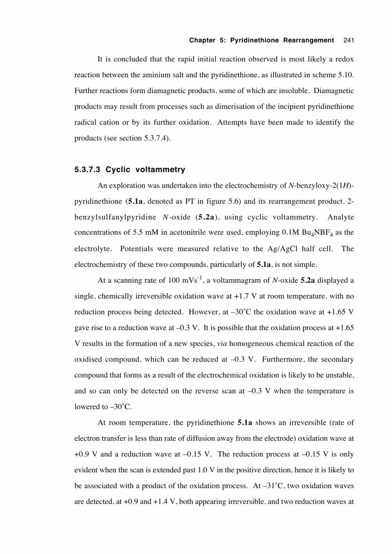
Chapter 5: Pyridinethione Rearrangement 241
It is concluded that the rapid initial reaction observed is most likely a redox
reaction between the aminium salt and the pyridinethione, as illustrated in scheme 5.10.
Further reactions form diamagnetic products, some of which are insoluble. Diamagnetic
products may result from processes such as dimerisation of the incipient pyridinethione
radical cation or by its further oxidation. Attempts have been made to identify the
products (see section 5.3.7.4).
5.3.7.3 Cyclic voltammetry
An exploration was undertaken into the electrochemistry of N-benzyloxy-2(1H)-
pyridinethione (5.1a, denoted as PT in figure 5.6) and its rearrangement product, 2-
benzylsulfanylpyridine N -oxide (5.2a), using cyclic voltammetry. Analyte
concentrations of 5.5 mM in acetonitrile were used, employing 0.1M Bu4NBF4 as the
electrolyte. Potentials were measured relative to the Ag/AgCl half cell. The
electrochemistry of these two compounds, particularly of 5.1a, is not simple.
At a scanning rate of 100 mVs-1, a voltammagram of N-oxide 5.2a displayed a
single, chemically irreversible oxidation wave at +1.7 V at room temperature, with no
reduction process being detected. However, at –30˚C the oxidation wave at +1.65 V
gave rise to a reduction wave at –0.3 V. It is possible that the oxidation process at +1.65
V results in the formation of a new species, via homogeneous chemical reaction of the
oxidised compound, which can be reduced at –0.3 V. Furthermore, the secondary
compound that forms as a result of the electrochemical oxidation is likely to be unstable,
and so can only be detected on the reverse scan at –0.3 V when the temperature is
lowered to –30˚C.
At room temperature, the pyridinethione 5.1a shows an irreversible (rate of
electron transfer is less than rate of diffusion away from the electrode) oxidation wave at
+0.9 V and a reduction wave at –0.15 V. The reduction process at –0.15 V is only
evident when the scan is extended past 1.0 V in the positive direction, hence it is likely to
be associated with a product of the oxidation process. At –31˚C, two oxidation waves
are detected, at +0.9 and +1.4 V, both appearing irreversible, and two reduction waves at
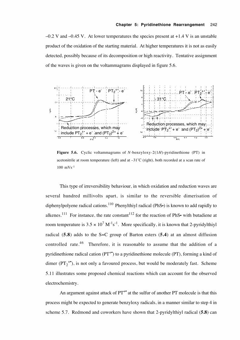
Chapter 5: Pyridinethione Rearrangement 242
–0.2 V and –0.45 V. At lower temperatures the species present at +1.4 V is an unstable
product of the oxidation of the starting material. At higher temperatures it is not as easily
detected, possibly because of its decomposition or high reactivity. Tentative assignment
of the waves is given on the voltammagrams displayed in figure 5.6.
21°C - 31°C
PT - e- PT2+• - e-
Reduction processes, which may include PT2
+• + e- and (PT2)2+ + e-
PT - e- PT2+• - e-
Reduction processes, which may include PT2
+• + e- and (PT2)2+ + e-
Figure 5.6. Cyclic voltammagrams of N-benzyloxy-2(1H)-pyridinethione (PT) in
acetonitrile at room temperature (left) and at –31˚C (right), both recorded at a scan rate of
100 mVs-1
This type of irreversibility behaviour, in which oxidation and reduction waves are
several hundred millivolts apart, is similar to the reversible dimerisation of
diphenylpolyene radical cations.110 Phenylthiyl radical (PhS•) is known to add rapidly to
alkenes.111 For instance, the rate constant112 for the reaction of PhS• with butadiene at
room temperature is 3.5 × 10⁷ M-1s-1. More specifically, it is known that 2-pyridylthiyl
radical (5.8) adds to the S=C group of Barton esters (5.4) at an almost diffusion
controlled rate.46 Therefore, it is reasonable to assume that the addition of a
pyridinethione radical cation (PT+•) to a pyridinethione molecule (PT), forming a kind of
dimer (PT2+•), is not only a favoured process, but would be moderately fast. Scheme
5.11 illustrates some proposed chemical reactions which can account for the observed
electrochemistry.
An argument against attack of PT+• at the sulfur of another PT molecule is that this
process might be expected to generate benzyloxy radicals, in a manner similar to step 4 in
scheme 5.7. Redmond and coworkers have shown that 2-pyridylthiyl radical (5.8) can

Chapter 5: Pyridinethione Rearrangement 243
displace RCOO• radicals by its reaction with Barton esters,46 although the rate of N–O
bond homolysis in a structure like PT2+• is difficult to estimate.
NO Ph
S
NO Ph
S
NO Ph
S+•
+
•
- e-
+ e-
PT+•
PT+• + PT
PT
fast
N
SS
N
O Ph
OPh
+
•
PT2+•
N
SS
N
O Ph
OPh
+
(PT2)2+
- e-
+ e-+
furtherreactions?
furtherreactions?
Scheme 5.11. Possible reactions which account for the results from cyclic voltammetry
Cyclic voltammetric results for the pyridinethione are consistent with a fast
reaction that takes place between an incipient pyridinethione radical cation, PT+•, and
another molecule of the pyridinethione, PT. The first oxidation wave corresponds to the
unstable species PT+•, which quickly reacts with a molecule of PT to form PT2+•. This
rationalises the low current on the return (reduction) wave. Removal of a second electron
from PT2+• results in a doubly aromatised dication, (PT2)2+, a process corresponding to
the second oxidation wave. Voltammetric data indicate that (PT2)2+, if this is the true
structure, is unstable at room temperature and has a half life significantly less than 1
second. It may be a species of this type in which benzyl group migration takes place.
There is support from mass spectrometry for dimerisation processes in solution (section
5.3.7.4). Structures PT2+• and (PT2)2+ have one and two aromatised heterocyclic rings
respectively, in which the nitrogen atom bears a positive charge. Such aromatisation
makes the 2-sulfanylpyridine N-oxide moiety a better leaving group. Processes which
Chapter 5: Pyridinethione Rearrangement 244
cause N to become positively charged may be responsible for promoting NO–R bond
scission.
Cyclic voltammetry can achieve potentials exceeding those encountered in solution
phase chemical reactions. Therefore, it is possible that some processes may be occur
during cyclic voltammetry which do not during the catalysed rearrangement of the
pyridinethione.
5.3.7.4 Isolation and attempted identification of intermediates
Addition of yellow N-benzyloxy-2(1H)-pyridinethione (5.1a) to a deep blue
CH2Cl2 solution of the aminium salt tris(4-bromophenyl)aminium hexachloroantimonate
(33) results in an instantaneous loss of the blue colour with a simultaneous formation of a
precipitate. An attempt was made to isolate and identify the products of this reaction.
A stirred solution of aminium salt 5.33 in dichloromethane was treated with
portions of solid 5.1a until the blue colour had just disappeared and a yellow/brown
solution with a similarly coloured, flocculent precipitate resulted. This required 1.29
molar equivalents of the pyridinethione. TLC analysis at this point indicated that neither
pyridinethione 5.1a nor product N-oxide 5.2a was present in solution. Centrifugation
was used to settle the solid. The liquid phase was drawn off, evaporated and its
constituents separated by flash chromatography. The major component from the solution
was tris(4-bromophenyl)amine, (p-BrC6H4)3N (87% yield), identified by comparison of
1H and 13C nmr spectra with that of the authentic compound. The total mass recovery
from the reaction was 96%.
Elemental analysis of the precipitate gave the results listed in table 5.8. At the
limits of a conservative tolerance of 0.3% for each element, these results are consistent
with the molecular formula C12H11NOSSbCl6. Such a formula indicates a molar ratio of
(pyridinethione 5.1a)+• to SbCl6– of 1:1. Thus, this compound is tentatively identified
as the hexachloroantimonate salt of the radical cation of N-benzyloxy-2(1H )-
pyridinethione 5.1a. Assuming that this molecular formula is correct, this compound
was isolated in 92% yield.
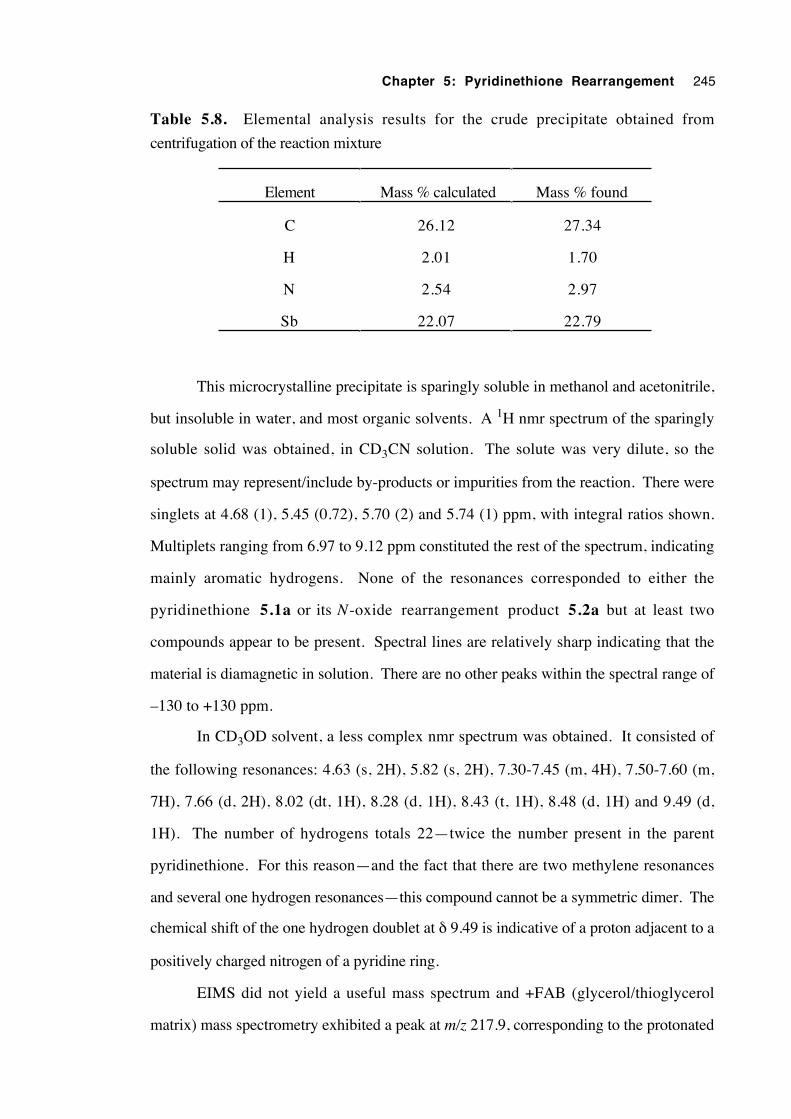
Chapter 5: Pyridinethione Rearrangement 245
Table 5.8. Elemental analysis results for the crude precipitate obtained from
centrifugation of the reaction mixture
Element Mass % calculated Mass % found
C 26.12 27.34
H 2.01 1.70
N 2.54 2.97
Sb 22.07 22.79
This microcrystalline precipitate is sparingly soluble in methanol and acetonitrile,
but insoluble in water, and most organic solvents. A 1H nmr spectrum of the sparingly
soluble solid was obtained, in CD3CN solution. The solute was very dilute, so the
spectrum may represent/include by-products or impurities from the reaction. There were
singlets at 4.68 (1), 5.45 (0.72), 5.70 (2) and 5.74 (1) ppm, with integral ratios shown.
Multiplets ranging from 6.97 to 9.12 ppm constituted the rest of the spectrum, indicating
mainly aromatic hydrogens. None of the resonances corresponded to either the
pyridinethione 5.1a or its N-oxide rearrangement product 5.2a but at least two
compounds appear to be present. Spectral lines are relatively sharp indicating that the
material is diamagnetic in solution. There are no other peaks within the spectral range of
–130 to +130 ppm.
In CD3OD solvent, a less complex nmr spectrum was obtained. It consisted of
the following resonances: 4.63 (s, 2H), 5.82 (s, 2H), 7.30-7.45 (m, 4H), 7.50-7.60 (m,
7H), 7.66 (d, 2H), 8.02 (dt, 1H), 8.28 (d, 1H), 8.43 (t, 1H), 8.48 (d, 1H) and 9.49 (d,
1H). The number of hydrogens totals 22—twice the number present in the parent
pyridinethione. For this reason—and the fact that there are two methylene resonances
and several one hydrogen resonances—this compound cannot be a symmetric dimer. The
chemical shift of the one hydrogen doublet at δ 9.49 is indicative of a proton adjacent to a
positively charged nitrogen of a pyridine ring.
EIMS did not yield a useful mass spectrum and +FAB (glycerol/thioglycerol
matrix) mass spectrometry exhibited a peak at m/z 217.9, corresponding to the protonated
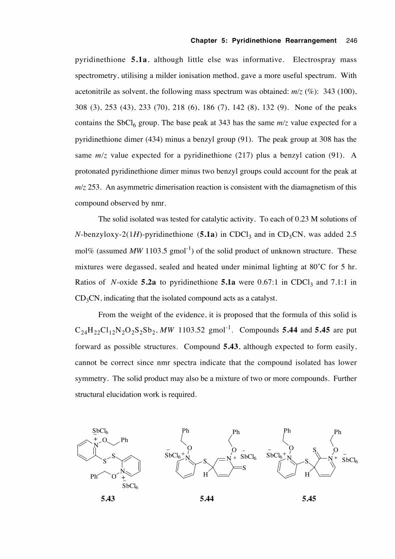
Chapter 5: Pyridinethione Rearrangement 246
pyridinethione 5.1a, although little else was informative. Electrospray mass
spectrometry, utilising a milder ionisation method, gave a more useful spectrum. With
acetonitrile as solvent, the following mass spectrum was obtained: m/z (%): 343 (100),
308 (3), 253 (43), 233 (70), 218 (6), 186 (7), 142 (8), 132 (9). None of the peaks
contains the SbCl6 group. The base peak at 343 has the same m/z value expected for a
pyridinethione dimer (434) minus a benzyl group (91). The peak group at 308 has the
same m/z value expected for a pyridinethione (217) plus a benzyl cation (91). A
protonated pyridinethione dimer minus two benzyl groups could account for the peak at
m/z 253. An asymmetric dimerisation reaction is consistent with the diamagnetism of this
compound observed by nmr.
The solid isolated was tested for catalytic activity. To each of 0.23 M solutions of
N-benzyloxy-2(1H)-pyridinethione (5.1a) in CDCl3 and in CD3CN, was added 2.5
mol% (assumed MW 1103.5 gmol-1) of the solid product of unknown structure. These
mixtures were degassed, sealed and heated under minimal lighting at 80˚C for 5 hr.
Ratios of N-oxide 5.2a to pyridinethione 5.1a were 0.67:1 in CDCl3 and 7.1:1 in
CD3CN, indicating that the isolated compound acts as a catalyst.
From the weight of the evidence, it is proposed that the formula of this solid is
C24H22Cl12N2O2S2Sb2, MW 1103.52 gmol-1. Compounds 5.44 and 5.45 are put
forward as possible structures. Compound 5.43, although expected to form easily,
cannot be correct since nmr spectra indicate that the compound isolated has lower
symmetry. The solid product may also be a mixture of two or more compounds. Further
structural elucidation work is required.
N
O
Ph
S NS O
Ph
H
SbCl6SbCl6
N
SS
N
O Ph
OPh
+
+
SbCl6
SbCl6
N
O
Ph
S N
H
SbCl6
S
O
Ph
SbCl6
5.43 5.44 5.45
Chapter 5: Pyridinethione Rearrangement 247
5.4 Conclusions
The mechanism of the catalysed rearrangement of N -alkoxy-2(1H ) -
pyridinethiones (5.1) to corresponding 2-alkylsulfanylpyridine N-oxides (5.2) is more
complex than initially envisaged. In particular, evidence for the participation of
intermolecular reactions lessens the probability that this migration may belong to a new
class of intramolecular rearrangement in which 5 electrons are delocalised over 5 atoms.
However, since a crossover experiment has not been undertaken to establish the
molecularity of the migration step, an intramolecular shift of the alkyl group cannot yet be
excluded.
Evidence from cyclic voltammetry and catalysis experiments is consistent with an
incipient pyridinethione radical cation being formed by the one electron oxidation of the
pyridinethione molecule. Owing to a similarity in reactivity, the mechanisms of
rearrangement with protic acid and with oxidative catalysis appear to be closely related—
perhaps having several identical or analogous steps. Results from studies of
stereochemistry, esr spectroscopy, radical scavenger addition and isolation of
intermediates suggest that if a one electron oxidation does occur, the oxidised
pyridinethione and the reduced catalyst remain associated in solution.
Kinetic studies show that the rearrangement is first order in the concentration of
pyridinethione and catalytic in iodine. The similarity in rate constants between oxidant
catalysts is consistent with an identical catalysis mechanism. Initial reaction between the
pyridinethione and catalyst is fast and for iodine has a large equilibrium constant. Polar
solvents accelerate the reaction, indicating significant charge development in the rate
limiting step. Kinetic results are consistent with an intermolecular rate limiting step.
High regioselectivity was observed in the apparent 1,4 migration of the allyl group
with both iodine and triflic acid catalysis, suggesting highly ordered mechanisms for the
transfer of the migrating group for both types of catalyst.
Results from the stereochemical study are difficult to interpret at this stage. The
migration of an optically active 1-phenethyl group proceeds with about 80% retention of
configuration in chloroform and up to 70% inversion of configuration in acetonitrile. A
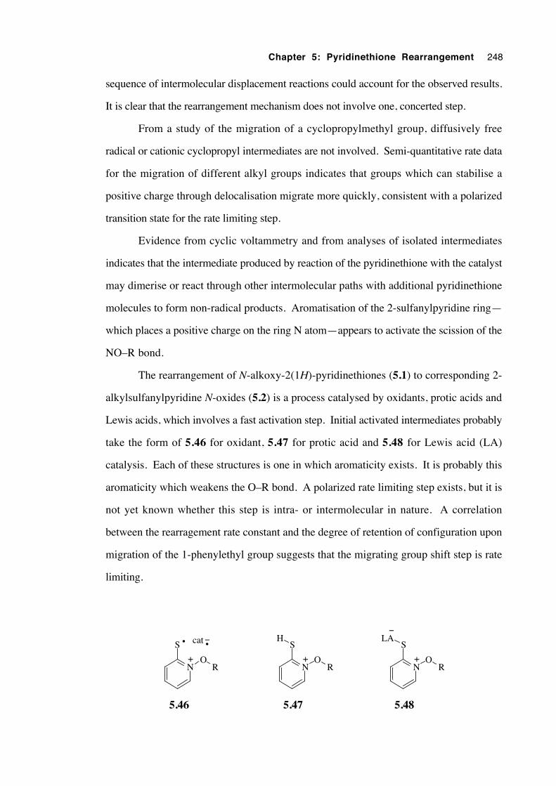
Chapter 5: Pyridinethione Rearrangement 248
sequence of intermolecular displacement reactions could account for the observed results.
It is clear that the rearrangement mechanism does not involve one, concerted step.
From a study of the migration of a cyclopropylmethyl group, diffusively free
radical or cationic cyclopropyl intermediates are not involved. Semi-quantitative rate data
for the migration of different alkyl groups indicates that groups which can stabilise a
positive charge through delocalisation migrate more quickly, consistent with a polarized
transition state for the rate limiting step.
Evidence from cyclic voltammetry and from analyses of isolated intermediates
indicates that the intermediate produced by reaction of the pyridinethione with the catalyst
may dimerise or react through other intermolecular paths with additional pyridinethione
molecules to form non-radical products. Aromatisation of the 2-sulfanylpyridine ring—
which places a positive charge on the ring N atom—appears to activate the scission of the
NO–R bond.
The rearrangement of N-alkoxy-2(1H)-pyridinethiones (5.1) to corresponding 2-
alkylsulfanylpyridine N-oxides (5.2) is a process catalysed by oxidants, protic acids and
Lewis acids, which involves a fast activation step. Initial activated intermediates probably
take the form of 5.46 for oxidant, 5.47 for protic acid and 5.48 for Lewis acid (LA)
catalysis. Each of these structures is one in which aromaticity exists. It is probably this
aromaticity which weakens the O–R bond. A polarized rate limiting step exists, but it is
not yet known whether this step is intra- or intermolecular in nature. A correlation
between the rearragement rate constant and the degree of retention of configuration upon
migration of the 1-phenylethyl group suggests that the migrating group shift step is rate
limiting.
NO
R
S
+
• cat−•
NO
R
S
+
H
5.46 5.47
NO
R
S
+
LA
5.48
−
Chapter 5: Pyridinethione Rearrangement 249
5.5 Future work
First and foremost, a crossover experiment must be designed and executed to
determine whether the net transfer of the migrating group takes place intramolecularly or
intermolecularly. Crossover experiments should be performed with different catalysts to
establish whether the molecularity of the rearrangement step is dependent on the mode of
catalysis and also in solvents of different polarity to explore whether there is a correlation
with results from the stereochemistry study. Secondly, more work should be put into the
elucidation of the structure of isolable intermediates. Also of interest would be a
Hammett study of benzyl group migration to examine the importance of charge
development at C1 of the migrating group. An identification of the products from a bulk
electrolysis (oxidation) of the pyridinethione would be illuminating since the results
would reveal not only whether the product N-oxide is actually produced by this process,
but how many moles of electrons must be removed per mole of pyridinethione in order
for reaction to occur. A kinetic study of the triflic acid catalysis of the rearrangement of
5.1a would provide valuable information about the similarities between acid and
oxidative catalysis. A more detailed study of the effect of both solvent, catalyst and
concentration upon rearrangement stereochemistry is also desirable.
In addition, testing the rearrangement of N-allyloxycarbonyloxypyridine-2-thiones
(5.20) to 2-allylsulfanylpyridine N-oxides (5.21) for crossover behaviour will establish
whether rearrangement in this system is intramolecular, as early researchers assumed it
was.
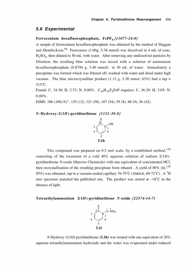
Chapter 5: Pyridinethione Rearrangement 250
5.6 Experimental
Ferrocenium hexafluorophosphate, FcPF6 [11077-24-0]
A sample of ferrocenium hexafluorophosphate was obtained by the method of Duggan
and Hendrickson.96 Ferrocence (1.00g, 5.38 mmol) was dissolved in 4 mL of conc.
H2SO4, then diluted to 50 mL with water. After removing any undissolved particles by
filtration, the resulting blue solution was mixed with a solution of ammonium
hexafluorophosphate (0.8794 g, 5.40 mmol) in 30 mL of water. Immediately a
precipitate was formed which was filtered off, washed with water and dried under high
vacuum. The blue microcrystalline product (1.12 g, 3.38 mmol, 63%) had a mp >
315˚C.
Found: C, 34.38; H, 2.73; N, 0.00%. C10H10F6FeP requires: C, 36.29; H, 3.05; N,
0.00%.
EIMS: 186 (100) Fc+, 129 (12), 121 (58), 107 (54), 95 (8), 88 (9), 56 (42).
N -Hydroxy-2(1H )-pyridinethione [1121-30-8]
N
S
OH
5.1h
164
3
5
2
This compound was prepared on 0.5 mol scale, by a established method,120
consisting of the treatment of a cold 40% aqueous solution of sodium 2(1H ) -
pyridinethione N-oxide (Harcros Chemicals) with one equivalent of concentrated HCl,
then recrystallisation of the resulting precipitate from ethanol. A yield of 80% (lit.120
95%) was obtained, mp in a vacuum-sealed capillary 76-79˚C (Aldrich, 69-72˚C). A 1H
nmr spectrum matched the published one. The product was stored at –18˚C in the
absence of light.
Tetraethylammonium 2(1H)-pyridinethione N-oxide [22574-14-7]
N
S
O
5.1i
164
32
5
NEt4
N-Hydroxy-2(1H)-pyridinethione (5.1h) was treated with one equivalent of 20%
aqueous tetraethylammonium hydroxide and the water was evaporated under reduced
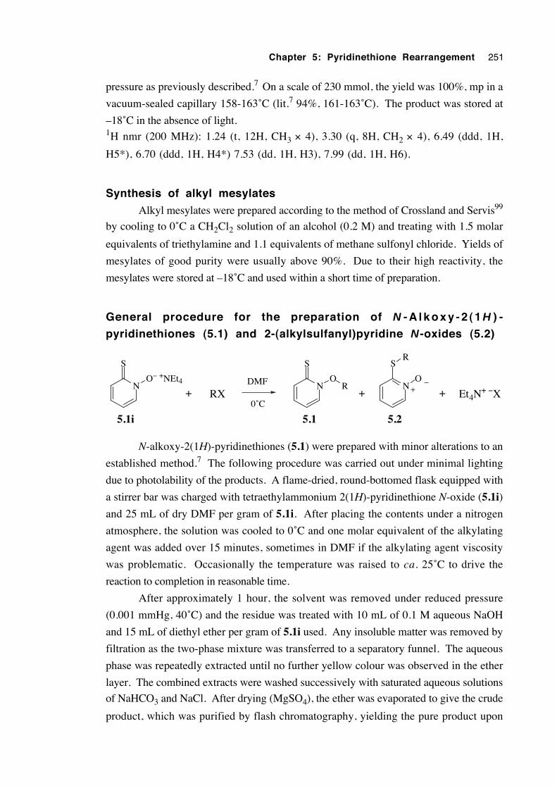
Chapter 5: Pyridinethione Rearrangement 251
pressure as previously described.7 On a scale of 230 mmol, the yield was 100%, mp in a
vacuum-sealed capillary 158-163˚C (lit.7 94%, 161-163˚C). The product was stored at
–18˚C in the absence of light.1H nmr (200 MHz): 1.24 (t, 12H, CH3 × 4), 3.30 (q, 8H, CH2 × 4), 6.49 (ddd, 1H,
H5*), 6.70 (ddd, 1H, H4*) 7.53 (dd, 1H, H3), 7.99 (dd, 1H, H6).
Synthesis of alkyl mesylates
Alkyl mesylates were prepared according to the method of Crossland and Servis99
by cooling to 0˚C a CH2Cl2 solution of an alcohol (0.2 M) and treating with 1.5 molar
equivalents of triethylamine and 1.1 equivalents of methane sulfonyl chloride. Yields of
mesylates of good purity were usually above 90%. Due to their high reactivity, the
mesylates were stored at –18˚C and used within a short time of preparation.
General procedure for the preparation of N - A l k o x y - 2 ( 1 H ) -
pyridinethiones (5.1) and 2-(alkylsulfanyl)pyridine N-oxides (5.2)
N
S
O− +NEt4
RXDMF
0˚C+
N
S
O
Et4N+ −XR
+
5.1i 5.1
N
S
O
5.2
R
+
N-alkoxy-2(1H)-pyridinethiones (5.1) were prepared with minor alterations to an
established method.7 The following procedure was carried out under minimal lighting
due to photolability of the products. A flame-dried, round-bottomed flask equipped with
a stirrer bar was charged with tetraethylammonium 2(1H)-pyridinethione N-oxide (5.1i)
and 25 mL of dry DMF per gram of 5.1i. After placing the contents under a nitrogen
atmosphere, the solution was cooled to 0˚C and one molar equivalent of the alkylating
agent was added over 15 minutes, sometimes in DMF if the alkylating agent viscosity
was problematic. Occasionally the temperature was raised to ca. 25˚C to drive the
reaction to completion in reasonable time.
After approximately 1 hour, the solvent was removed under reduced pressure
(0.001 mmHg, 40˚C) and the residue was treated with 10 mL of 0.1 M aqueous NaOH
and 15 mL of diethyl ether per gram of 5.1i used. Any insoluble matter was removed by
filtration as the two-phase mixture was transferred to a separatory funnel. The aqueous
phase was repeatedly extracted until no further yellow colour was observed in the ether
layer. The combined extracts were washed successively with saturated aqueous solutions
of NaHCO3 and NaCl. After drying (MgSO4), the ether was evaporated to give the crude
product, which was purified by flash chromatography, yielding the pure product upon
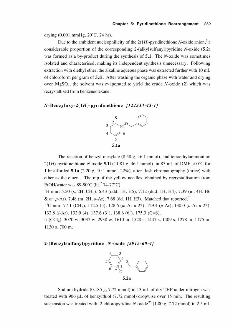
Chapter 5: Pyridinethione Rearrangement 252
drying (0.001 mmHg, 20˚C, 24 hr).
Due to the ambident nucleophilicity of the 2(1H)-pyridinethione N-oxide anion,7 a
considerable proportion of the corresponding 2-(alkylsulfanyl)pyridine N-oxide (5.2)
was formed as a by-product during the synthesis of 5.1. The N-oxide was sometimes
isolated and characterised, making its independent synthesis unnecessary. Following
extraction with diethyl ether, the alkaline aqueous phase was extracted further with 10 mL
of chloroform per gram of 5.1i. After washing the organic phase with water and drying
over MgSO4, the solvent was evaporated to yield the crude N-oxide (2) which was
recrystallized from benzene/hexane.
N -Benzyloxy-2(1H )-pyridinethione [122333-43-1]
N
S
O
5.1a
164
32
5
The reaction of benzyl mesylate (8.58 g, 46.1 mmol), and tetraethylammonium
2(1H)-pyridinethione N-oxide 5.1i (11.81 g, 46.1 mmol), in 85 mL of DMF at 0˚C for
1 hr afforded 5.1a (2.20 g, 10.1 mmol, 22%), after flash chromatography (thrice) with
ether as the eluent. The mp of the yellow needles, obtained by recrystallisation from
EtOH/water was 89-90˚C (lit.7 74-77˚C).1H nmr: 5.50 (s, 2H, CH2), 6.43 (ddd, 1H, H5), 7.12 (ddd, 1H, H4), 7.39 (m, 4H, H6
& m+p-Ar), 7.48 (m, 2H, o-Ar), 7.68 (dd, 1H, H3). Matched that reported.7
13C nmr: 77.1 (CH2), 112.5 (5), 128.6 (m-Ar × 2*), 129.4 (p-Ar), 130.0 (o-Ar × 2*),
132.8 (i-Ar), 132.9 (4), 137.6 (3†), 138.6 (6†), 175.3 (C=S).
ir (CCl4): 3070 w, 3037 w, 2938 w, 1610 m, 1528 s, 1447 s, 1409 s, 1278 m, 1175 m,
1130 s, 700 m.
2-(Benzylsulfanyl)pyridine N -oxide [3915-60-4]
NS
O
5.2a
1
2
34
5
6
Sodium hydride (0.185 g, 7.72 mmol) in 13 mL of dry THF under nitrogen was
treated with 906 µL of benzylthiol (7.72 mmol) dropwise over 15 min. The resulting
suspension was treated with 2-chloropyridine N-oxide95 (1.00 g, 7.72 mmol) in 2.5 mL
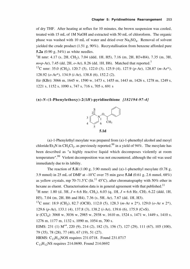
Chapter 5: Pyridinethione Rearrangement 253
of dry THF. After heating at reflux for 10 minutes, the brown suspension was cooled,
treated with 15 mL of 1M NaOH and extracted with 50 mL of chloroform. The organic
phase was washed with 10 mL of water and dried over Na2SO4. Removal of solvent
yielded the crude product (1.51 g, 90%). Recrystallisation from benzene afforded pure
5.2a (0.90 g, 54%) as white needles.1H nmr: 4.17 (s, 2H, CH2), 7.04 (ddd, 1H, H5), 7.16 (m, 2H, H3+H4), 7.35 (m, 3H,
m+p-Ar), 7.45 (dd, 2H, o-Ar), 8.26 (dd, 1H, H6). Matched that reported.7
13C nmr: 35.0 (CH2), 120.7 (5), 122.0 (3), 125.9 (4), 127.9 (p-Ar), 128.87 (m-Ar*),
128.92 (o-Ar*), 134.9 (i-Ar), 138.8 (6), 152.2 (2).
ftir (KBr): 3066 m, 1645 w, 1590 w, 1473 s, 1455 m, 1443 m, 1426 s, 1278 m, 1249 s,
1221 s, 1152 s, 1090 s, 747 s, 716 s, 705 s, 691 s
(±)-N -(1-Phenylethoxy)-2(1H )-pyridinethione [182194-97-4]
N
S
O
CH31
4 6
3
2
5
5.1d
(±)-1-Phenylethyl mesylate was prepared from (±)-1-phenethyl alcohol and mesyl
chloride/Et3N in CH2Cl2, as previously reported,99 in a yield of 94%. The mesylate has
been described as "a highly reactive liquid which decomposes violently at room
temperature".99 Violent decomposition was not encountered, although the oil was used
immediately due to its lability.
The reaction of 5.1i (1.00 g, 3.90 mmol) and (±)-1-phenethyl mesylate (0.78 g,
3.9 mmol) in 25 mL of DMF at –10˚C over 75 min gave 5.1d (0.61 g, 2.6 mmol, 68%)
as yellow crystals, mp 70-71.5˚C (lit.11 45˚C), after chromatography with 50% ether in
hexane as eluent. Characterisation data is in general agreement with that published.11
1H nmr: 1.80 (d, 3H, J = 6.6 Hz, CH3), 6.03 (q, 1H, J = 6.6 Hz, CH), 6.22 (ddd, 1H,
H5), 7.04 (m, 2H, H6 and H4), 7.36 (s, 5H, Ar), 7.67 (dd, 1H, H3).13C nmr: 18.9 (CH3), 82.7 (OCH), 112.0 (5), 128.3 (m-Ar × 2*), 129.0 (o-Ar × 2*),
129.6 (p-Ar), 133.1 (4), 137.8 (3), 138.2 (i-Ar), 139.6 (6), 175.9 (C=S).
ir (CCl4): 3068 w, 3036 w, 2985 w, 2938 w, 1610 m, 1524 s, 1471 w, 1449 s, 1410 s,
1276 m, 1177 m, 1132 s, 1090 m, 1054 m, 700 s.
EIMS: 231 (1) M+•, 220 (9), 214 (2), 182 (3), 156 (7), 127 (29), 111 (67), 105 (100),
79 (35), 78 (28), 77 (40), 67 (19), 51 (27).
HRMS: C13H13NOS requires 231.0718. Found 231.0717
C13H12NS requires 214.0690. Found 214.0692
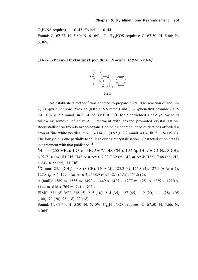
Chapter 5: Pyridinethione Rearrangement 254
C5H5NS requires 111.0143. Found 111.0144.
Found: C, 67.27; H, 5.89; N, 6.16%. C13H13NOS requires: C, 67.50; H, 5.66; N,
6.06%.
(±)-2-(1-Phenylethylsufanyl)pyridine N -oxide [60263-93-6]
N
S
O CH3
1
234
6
5
5.2d
An established method7 was adapted to prepare 5.2d. The reaction of sodium
2(1H)-pyridinethione N-oxide (0.82 g, 5.5 mmol) and (±)-1-phenethyl bromide (0.75
mL, 1.02 g, 5.5 mmol) in 8 mL of DMF at 80˚C for 2 hr yielded a pale yellow solid
following removal of solvent. Treatment with hexane promoted crystallisation.
Recrystallisation from benzene/hexane (including charcoal decolourisation) afforded a
crop of fine white needles, mp 113-114˚C, (0.52 g, 2.2 mmol, 41%, lit.11 118-119˚C).
The low yield is due partially to spillage during recrystallisation. Characterisation data is
in agreement with that published.11
1H nmr (200 MHz): 1.73 (d, 3H, J = 7.1 Hz, CH3), 4.52 (q, 1H, J = 7.1 Hz, S-CH),
6.92-7.10 (m, 3H, H5, H4* & p-Ar*), 7.22-7.39 (m, 3H, m-Ar & H3*), 7.48 (dd, 2H,
o-Ar), 8.22 (dd, 1H, H6).13C nmr: 23.1 (CH3), 43.8 (S-CH), 120.8 (5), 123.3 (3), 125.8 (4), 127.1 (o-Ar × 2),
127.8 (p-Ar), 129.0 (m-Ar × 2), 138.9 (6), 142.1 (i-Ar), 151.6 (2).
ir (mull): 1589 m, 1555 m, 1492 s, 1449 s, 1427 s, 1277 m, 1251 s, 1239 s, 1220 s,
1144 m, 838 s, 765 m, 741 s, 703 s.
EIMS: 231 (8) M+•, 216 (5), 215 (10), 214 (35), 127 (45), 112 (20), 111 (28), 105
(100), 79 (20), 78 (18), 77 (18).
Found: C, 67.60; H, 5.80; N, 6.10%. C13H13NOS requires: C, 67.50; H, 5.66; N,
6.06%.
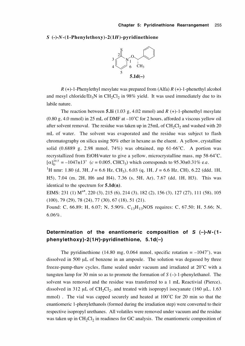
Chapter 5: Pyridinethione Rearrangement 255
S (-)-N-(1-Phenylethoxy)-2(1H )-pyridinethione
N
S
O
CH3
1
23
4 6
55.1d(–)
R (+)-1-Phenylethyl mesylate was prepared from (Alfa) R (+)-1-phenethyl alcohol
and mesyl chloride/Et3N in CH2Cl2 in 98% yield. It was used immediately due to its
labile nature.
The reaction between 5.1i (1.03 g, 4.02 mmol) and R (+)-1-phenethyl mesylate
(0.80 g, 4.0 mmol) in 25 mL of DMF at –10˚C for 2 hours, afforded a viscous yellow oil
after solvent removal. The residue was taken up in 25mL of CH2Cl2 and washed with 20
mL of water. The solvent was evaporated and the residue was subject to flash
chromatography on silica using 50% ether in hexane as the eluent. A yellow, crystalline
solid (0.6889 g, 2.98 mmol, 74%) was obtained, mp 61-66˚C. A portion was
recrystallized from EtOH/water to give a yellow, microcrystalline mass, mp 58-64˚C,[α]20.5
D= –1047±13˚ (c = 0.005, CHCl3) which corresponds to 95.30±0.31% e.e.
1H nmr: 1.80 (d, 3H, J = 6.6 Hz, CH3), 6.03 (q, 1H, J = 6.6 Hz, CH), 6.22 (ddd, 1H,
H5), 7.04 (m, 2H, H6 and H4), 7.36 (s, 5H, Ar), 7.67 (dd, 1H, H3). This was
identical to the spectrum for 5.1d(±).
EIMS: 231 (1) M+•, 220 (3), 215 (6), 214 (3), 182 (2), 156 (3), 127 (27), 111 (58), 105
(100), 79 (29), 78 (24), 77 (30), 67 (18), 51 (21).
Found: C, 66.89; H, 6.07; N, 5.90%. C13H13NOS requires: C, 67.50; H, 5.66; N,
6.06%.
Determination of the enantiomeric composition of S (–)-N - (1 -
phenylethoxy)-2(1H )-pyridinethione, 5.1d(–)
The pyridinethione (14.80 mg, 0.064 mmol, specific rotation = –1047˚), was
dissolved in 500 µL of benzene in an ampoule. The solution was degassed by three
freeze-pump-thaw cycles, flame sealed under vacuum and irradiated at 20˚C with a
tungsten lamp for 30 min so as to promote the formation of S (–)-1-phenylethanol. The
solvent was removed and the residue was transferred to a 1 mL Reactivial (Pierce),
dissolved in 312 µL of CH2Cl2, and treated with isopropyl isocyanate (160 µL, 1.63
mmol) . The vial was capped securely and heated at 100˚C for 20 min so that the
enantiomeric 1-phenylethanols (formed during the irradiation step) were converted to their
respective isopropyl urethanes. All volatiles were removed under vacuum and the residue
was taken up in CH2Cl2 in readiness for GC analysis. The enantiomeric composition of
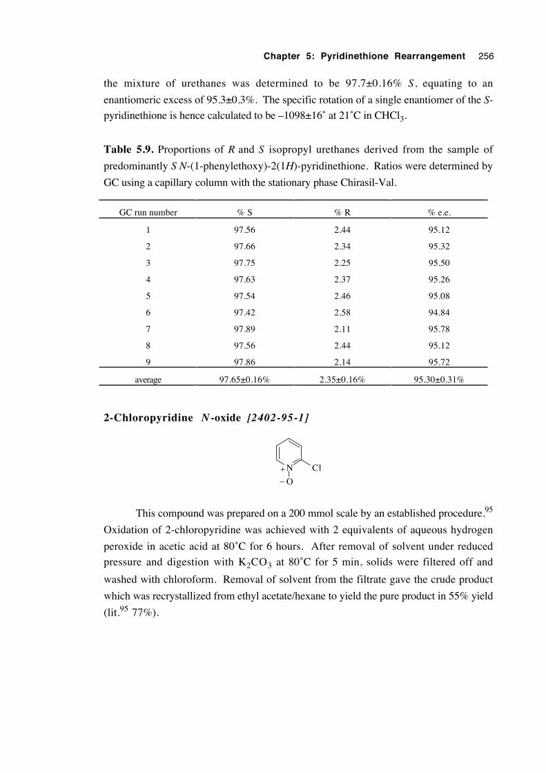
Chapter 5: Pyridinethione Rearrangement 256
the mixture of urethanes was determined to be 97.7±0.16% S , equating to an
enantiomeric excess of 95.3±0.3%. The specific rotation of a single enantiomer of the S-
pyridinethione is hence calculated to be –1098±16˚ at 21˚C in CHCl3.
Table 5.9. Proportions of R and S isopropyl urethanes derived from the sample of
predominantly S N-(1-phenylethoxy)-2(1H)-pyridinethione. Ratios were determined by
GC using a capillary column with the stationary phase Chirasil-Val.
GC run number % S % R % e.e.
1 97.56 2.44 95.12
2 97.66 2.34 95.32
3 97.75 2.25 95.50
4 97.63 2.37 95.26
5 97.54 2.46 95.08
6 97.42 2.58 94.84
7 97.89 2.11 95.78
8 97.56 2.44 95.12
9 97.86 2.14 95.72
average 97.65±0.16% 2.35±0.16% 95.30±0.31%
2-Chloropyridine N -oxide [2402-95-1]
N
O
Cl
This compound was prepared on a 200 mmol scale by an established procedure.95
Oxidation of 2-chloropyridine was achieved with 2 equivalents of aqueous hydrogen
peroxide in acetic acid at 80˚C for 6 hours. After removal of solvent under reduced
pressure and digestion with K2CO3 at 80˚C for 5 min, solids were filtered off and
washed with chloroform. Removal of solvent from the filtrate gave the crude product
which was recrystallized from ethyl acetate/hexane to yield the pure product in 55% yield
(lit.95 77%).
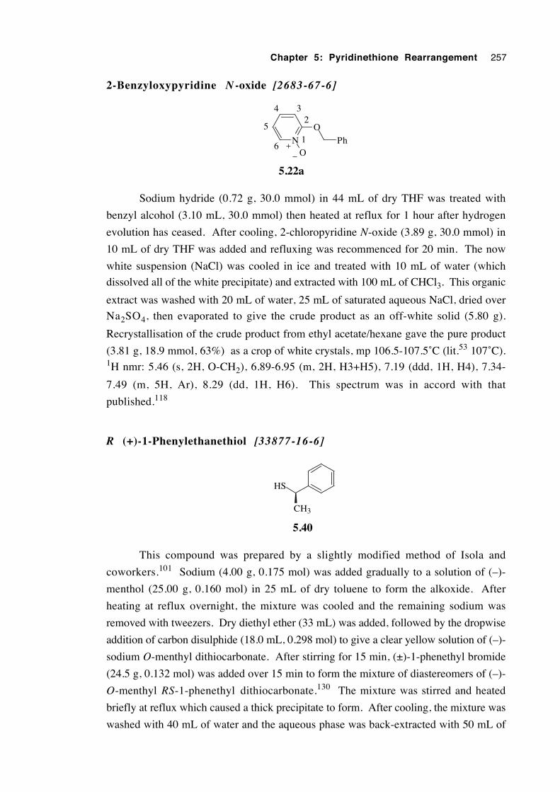
Chapter 5: Pyridinethione Rearrangement 257
2-Benzyloxypyridine N -oxide [2683-67-6]
N
O
OPh1
234
6
5
5.22a
Sodium hydride (0.72 g, 30.0 mmol) in 44 mL of dry THF was treated with
benzyl alcohol (3.10 mL, 30.0 mmol) then heated at reflux for 1 hour after hydrogen
evolution has ceased. After cooling, 2-chloropyridine N-oxide (3.89 g, 30.0 mmol) in
10 mL of dry THF was added and refluxing was recommenced for 20 min. The now
white suspension (NaCl) was cooled in ice and treated with 10 mL of water (which
dissolved all of the white precipitate) and extracted with 100 mL of CHCl3. This organic
extract was washed with 20 mL of water, 25 mL of saturated aqueous NaCl, dried over
Na2SO4, then evaporated to give the crude product as an off-white solid (5.80 g).
Recrystallisation of the crude product from ethyl acetate/hexane gave the pure product
(3.81 g, 18.9 mmol, 63%) as a crop of white crystals, mp 106.5-107.5˚C (lit.53 107˚C).1H nmr: 5.46 (s, 2H, O-CH2), 6.89-6.95 (m, 2H, H3+H5), 7.19 (ddd, 1H, H4), 7.34-
7.49 (m, 5H, Ar), 8.29 (dd, 1H, H6). This spectrum was in accord with that
published.118
R (+)-1-Phenylethanethiol [33877-16-6]
HS
CH3
5.40
This compound was prepared by a slightly modified method of Isola and
coworkers.101 Sodium (4.00 g, 0.175 mol) was added gradually to a solution of (–)-
menthol (25.00 g, 0.160 mol) in 25 mL of dry toluene to form the alkoxide. After
heating at reflux overnight, the mixture was cooled and the remaining sodium was
removed with tweezers. Dry diethyl ether (33 mL) was added, followed by the dropwise
addition of carbon disulphide (18.0 mL, 0.298 mol) to give a clear yellow solution of (–)-
sodium O-menthyl dithiocarbonate. After stirring for 15 min, (±)-1-phenethyl bromide
(24.5 g, 0.132 mol) was added over 15 min to form the mixture of diastereomers of (–)-
O-menthyl RS-1-phenethyl dithiocarbonate.130 The mixture was stirred and heated
briefly at reflux which caused a thick precipitate to form. After cooling, the mixture was
washed with 40 mL of water and the aqueous phase was back-extracted with 50 mL of
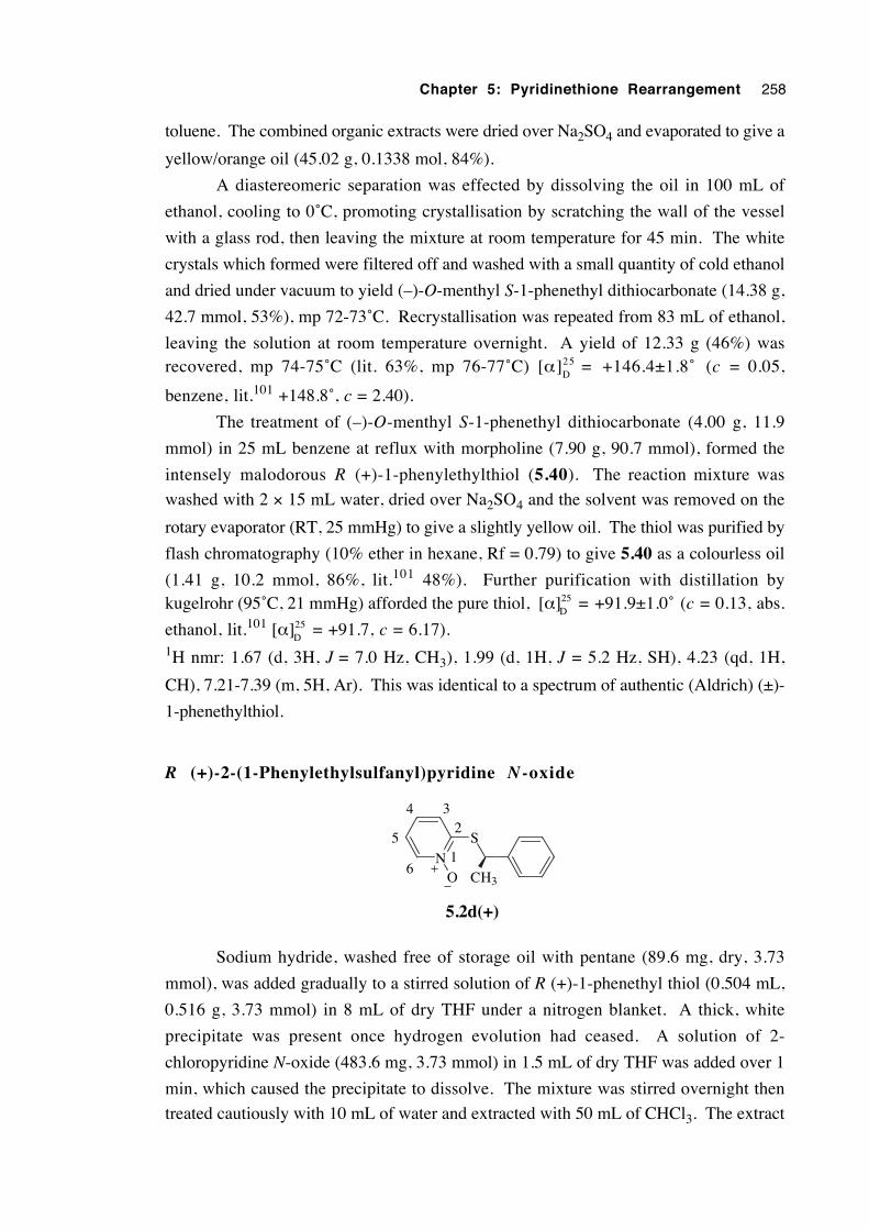
Chapter 5: Pyridinethione Rearrangement 258
toluene. The combined organic extracts were dried over Na2SO4 and evaporated to give a
yellow/orange oil (45.02 g, 0.1338 mol, 84%).
A diastereomeric separation was effected by dissolving the oil in 100 mL of
ethanol, cooling to 0˚C, promoting crystallisation by scratching the wall of the vessel
with a glass rod, then leaving the mixture at room temperature for 45 min. The white
crystals which formed were filtered off and washed with a small quantity of cold ethanol
and dried under vacuum to yield (–)-O-menthyl S-1-phenethyl dithiocarbonate (14.38 g,
42.7 mmol, 53%), mp 72-73˚C. Recrystallisation was repeated from 83 mL of ethanol,
leaving the solution at room temperature overnight. A yield of 12.33 g (46%) wasrecovered, mp 74-75˚C (lit. 63%, mp 76-77˚C) [α]25
D= +146.4±1.8˚ (c = 0.05,
benzene, lit.101 +148.8˚, c = 2.40).
The treatment of (–)-O-menthyl S-1-phenethyl dithiocarbonate (4.00 g, 11.9
mmol) in 25 mL benzene at reflux with morpholine (7.90 g, 90.7 mmol), formed the
intensely malodorous R (+)-1-phenylethylthiol (5.40). The reaction mixture was
washed with 2 × 15 mL water, dried over Na2SO4 and the solvent was removed on the
rotary evaporator (RT, 25 mmHg) to give a slightly yellow oil. The thiol was purified by
flash chromatography (10% ether in hexane, Rf = 0.79) to give 5.40 as a colourless oil
(1.41 g, 10.2 mmol, 86%, lit.101 48%). Further purification with distillation bykugelrohr (95˚C, 21 mmHg) afforded the pure thiol, [α]25
D= +91.9±1.0˚ (c = 0.13, abs.
ethanol, lit.101 [α]25D
= +91.7, c = 6.17).1H nmr: 1.67 (d, 3H, J = 7.0 Hz, CH3), 1.99 (d, 1H, J = 5.2 Hz, SH), 4.23 (qd, 1H,
CH), 7.21-7.39 (m, 5H, Ar). This was identical to a spectrum of authentic (Aldrich) (±)-
1-phenethylthiol.
R (+)-2-(1-Phenylethylsulfanyl)pyridine N -oxide
N
S
O CH3
1
234
6
5
5.2d(+)
Sodium hydride, washed free of storage oil with pentane (89.6 mg, dry, 3.73
mmol), was added gradually to a stirred solution of R (+)-1-phenethyl thiol (0.504 mL,
0.516 g, 3.73 mmol) in 8 mL of dry THF under a nitrogen blanket. A thick, white
precipitate was present once hydrogen evolution had ceased. A solution of 2-
chloropyridine N-oxide (483.6 mg, 3.73 mmol) in 1.5 mL of dry THF was added over 1
min, which caused the precipitate to dissolve. The mixture was stirred overnight then
treated cautiously with 10 mL of water and extracted with 50 mL of CHCl3. The extract
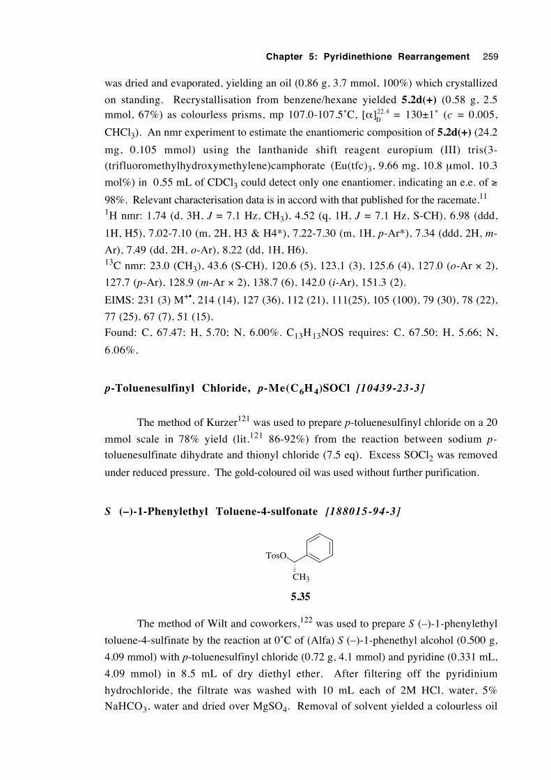
Chapter 5: Pyridinethione Rearrangement 259
was dried and evaporated, yielding an oil (0.86 g, 3.7 mmol, 100%) which crystallized
on standing. Recrystallisation from benzene/hexane yielded 5.2d(+) (0.58 g, 2.5mmol, 67%) as colourless prisms, mp 107.0-107.5˚C, [α]22.4
D= 130±1˚ (c = 0.005,
CHCl3). An nmr experiment to estimate the enantiomeric composition of 5.2d(+) (24.2
mg, 0.105 mmol) using the lanthanide shift reagent europium (III) tris(3-
(trifluoromethylhydroxymethylene)camphorate (Eu(tfc)3, 9.66 mg, 10.8 µmol, 10.3
mol%) in 0.55 mL of CDCl3 could detect only one enantiomer, indicating an e.e. of ≥
98%. Relevant characterisation data is in accord with that published for the racemate.11
1H nmr: 1.74 (d, 3H, J = 7.1 Hz, CH3), 4.52 (q, 1H, J = 7.1 Hz, S-CH), 6.98 (ddd,
1H, H5), 7.02-7.10 (m, 2H, H3 & H4*), 7.22-7.30 (m, 1H, p-Ar*), 7.34 (ddd, 2H, m-
Ar), 7.49 (dd, 2H, o-Ar), 8.22 (dd, 1H, H6).13C nmr: 23.0 (CH3), 43.6 (S-CH), 120.6 (5), 123.1 (3), 125.6 (4), 127.0 (o-Ar × 2),
127.7 (p-Ar), 128.9 (m-Ar × 2), 138.7 (6), 142.0 (i-Ar), 151.3 (2).
EIMS: 231 (3) M+•, 214 (14), 127 (36), 112 (21), 111(25), 105 (100), 79 (30), 78 (22),
77 (25), 67 (7), 51 (15).
Found: C, 67.47; H, 5.70; N, 6.00%. C13H13NOS requires: C, 67.50; H, 5.66; N,
6.06%.
p-Toluenesulfinyl Chloride, p-Me(C6H 4)SOCl [10439-23-3]
The method of Kurzer121 was used to prepare p-toluenesulfinyl chloride on a 20
mmol scale in 78% yield (lit.121 86-92%) from the reaction between sodium p-
toluenesulfinate dihydrate and thionyl chloride (7.5 eq). Excess SOCl2 was removed
under reduced pressure. The gold-coloured oil was used without further purification.
S (–)-1-Phenylethyl Toluene-4-sulfonate [188015-94-3]
TosO
CH3
5.35
The method of Wilt and coworkers,122 was used to prepare S (–)-1-phenylethyl
toluene-4-sulfinate by the reaction at 0˚C of (Alfa) S (–)-1-phenethyl alcohol (0.500 g,
4.09 mmol) with p-toluenesulfinyl chloride (0.72 g, 4.1 mmol) and pyridine (0.331 mL,
4.09 mmol) in 8.5 mL of dry diethyl ether. After filtering off the pyridinium
hydrochloride, the filtrate was washed with 10 mL each of 2M HCl, water, 5%
NaHCO3, water and dried over MgSO4. Removal of solvent yielded a colourless oil
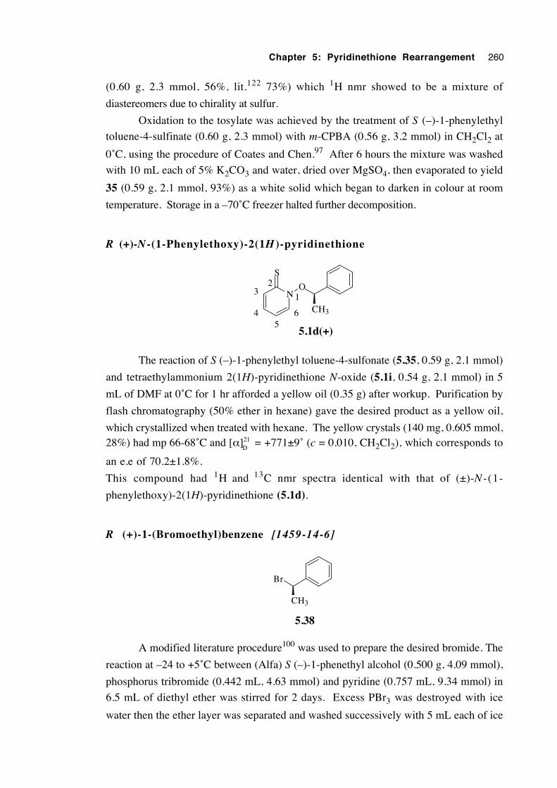
Chapter 5: Pyridinethione Rearrangement 260
(0.60 g, 2.3 mmol, 56%, lit.122 73%) which 1H nmr showed to be a mixture of
diastereomers due to chirality at sulfur.
Oxidation to the tosylate was achieved by the treatment of S (–)-1-phenylethyl
toluene-4-sulfinate (0.60 g, 2.3 mmol) with m-CPBA (0.56 g, 3.2 mmol) in CH2Cl2 at
0˚C, using the procedure of Coates and Chen.97 After 6 hours the mixture was washed
with 10 mL each of 5% K2CO3 and water, dried over MgSO4, then evaporated to yield
35 (0.59 g, 2.1 mmol, 93%) as a white solid which began to darken in colour at room
temperature. Storage in a –70˚C freezer halted further decomposition.
R (+)-N -(1-Phenylethoxy)-2(1H )-pyridinethione
N
S
O
5.1d(+)
CH3
1
64
32
5
The reaction of S (–)-1-phenylethyl toluene-4-sulfonate (5.35, 0.59 g, 2.1 mmol)
and tetraethylammonium 2(1H)-pyridinethione N-oxide (5.1i, 0.54 g, 2.1 mmol) in 5
mL of DMF at 0˚C for 1 hr afforded a yellow oil (0.35 g) after workup. Purification by
flash chromatography (50% ether in hexane) gave the desired product as a yellow oil,
which crystallized when treated with hexane. The yellow crystals (140 mg, 0.605 mmol,28%) had mp 66-68˚C and [α]21
D= +771±9˚ (c = 0.010, CH2Cl2), which corresponds to
an e.e of 70.2±1.8%.
This compound had 1H and 13C nmr spectra identical with that of (±)-N -(1-
phenylethoxy)-2(1H)-pyridinethione (5.1d).
R (+)-1-(Bromoethyl)benzene [1459-14-6]
Br
CH3
5.38
A modified literature procedure100 was used to prepare the desired bromide. The
reaction at –24 to +5˚C between (Alfa) S (–)-1-phenethyl alcohol (0.500 g, 4.09 mmol),
phosphorus tribromide (0.442 mL, 4.63 mmol) and pyridine (0.757 mL, 9.34 mmol) in
6.5 mL of diethyl ether was stirred for 2 days. Excess PBr3 was destroyed with ice
water then the ether layer was separated and washed successively with 5 mL each of ice
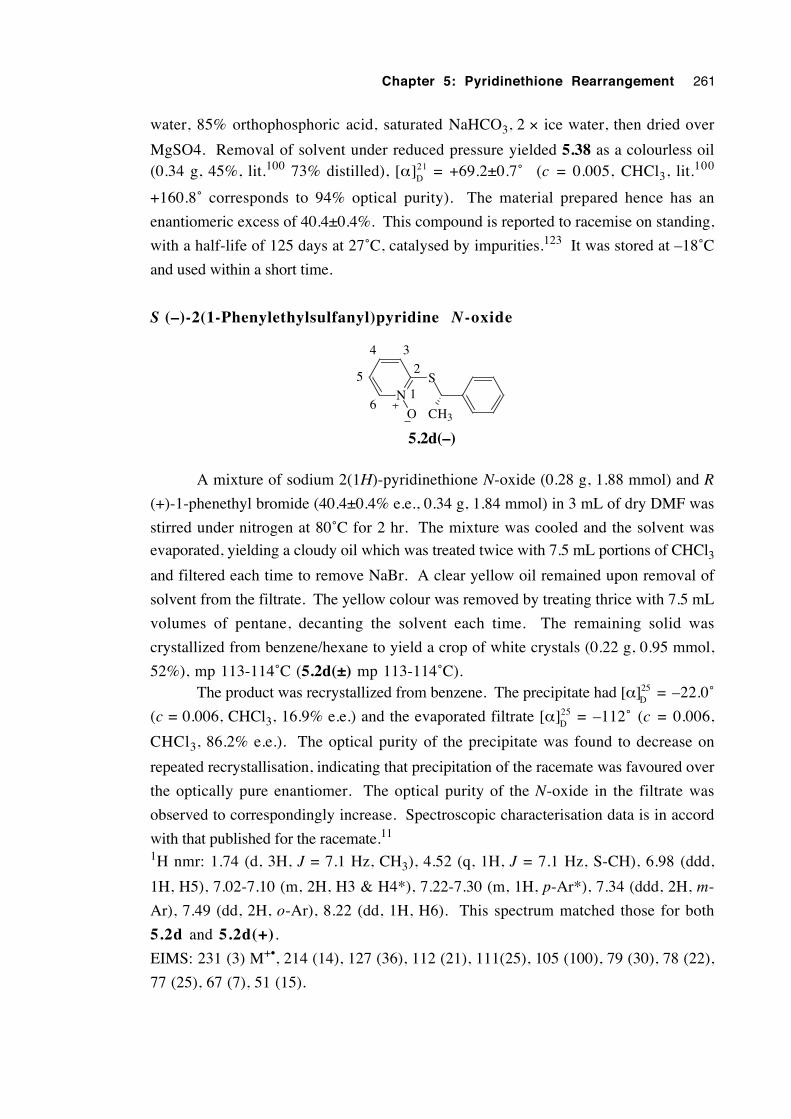
Chapter 5: Pyridinethione Rearrangement 261
water, 85% orthophosphoric acid, saturated NaHCO3, 2 × ice water, then dried over
MgSO4. Removal of solvent under reduced pressure yielded 5.38 as a colourless oil(0.34 g, 45%, lit.100 73% distilled), [α]21
D= +69.2±0.7˚ (c = 0.005, CHCl3, lit.100
+160.8˚ corresponds to 94% optical purity). The material prepared hence has an
enantiomeric excess of 40.4±0.4%. This compound is reported to racemise on standing,
with a half-life of 125 days at 27˚C, catalysed by impurities.123 It was stored at –18˚C
and used within a short time.
S (–)-2(1-Phenylethylsulfanyl)pyridine N-oxide
NS
O
5.2d(–)
CH3
1
2
34
6
5
A mixture of sodium 2(1H)-pyridinethione N-oxide (0.28 g, 1.88 mmol) and R
(+)-1-phenethyl bromide (40.4±0.4% e.e., 0.34 g, 1.84 mmol) in 3 mL of dry DMF was
stirred under nitrogen at 80˚C for 2 hr. The mixture was cooled and the solvent was
evaporated, yielding a cloudy oil which was treated twice with 7.5 mL portions of CHCl3and filtered each time to remove NaBr. A clear yellow oil remained upon removal of
solvent from the filtrate. The yellow colour was removed by treating thrice with 7.5 mL
volumes of pentane, decanting the solvent each time. The remaining solid was
crystallized from benzene/hexane to yield a crop of white crystals (0.22 g, 0.95 mmol,
52%), mp 113-114˚C (5.2d(±) mp 113-114˚C).The product was recrystallized from benzene. The precipitate had [α]25
D= –22.0˚
(c = 0.006, CHCl3, 16.9% e.e.) and the evaporated filtrate [α]25D
= –112˚ (c = 0.006,
CHCl3, 86.2% e.e.). The optical purity of the precipitate was found to decrease on
repeated recrystallisation, indicating that precipitation of the racemate was favoured over
the optically pure enantiomer. The optical purity of the N-oxide in the filtrate was
observed to correspondingly increase. Spectroscopic characterisation data is in accord
with that published for the racemate.11
1H nmr: 1.74 (d, 3H, J = 7.1 Hz, CH3), 4.52 (q, 1H, J = 7.1 Hz, S-CH), 6.98 (ddd,
1H, H5), 7.02-7.10 (m, 2H, H3 & H4*), 7.22-7.30 (m, 1H, p-Ar*), 7.34 (ddd, 2H, m-
Ar), 7.49 (dd, 2H, o-Ar), 8.22 (dd, 1H, H6). This spectrum matched those for both
5.2d and 5.2d(+).
EIMS: 231 (3) M+•, 214 (14), 127 (36), 112 (21), 111(25), 105 (100), 79 (30), 78 (22),
77 (25), 67 (7), 51 (15).
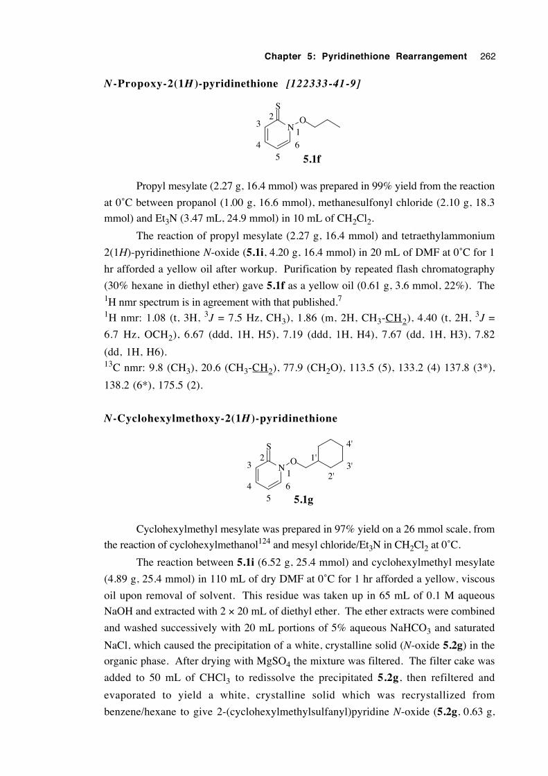
Chapter 5: Pyridinethione Rearrangement 262
N -Propoxy-2(1H )-pyridinethione [122333-41-9]
N
S
O
5.1f
1
64
32
5
Propyl mesylate (2.27 g, 16.4 mmol) was prepared in 99% yield from the reaction
at 0˚C between propanol (1.00 g, 16.6 mmol), methanesulfonyl chloride (2.10 g, 18.3
mmol) and Et3N (3.47 mL, 24.9 mmol) in 10 mL of CH2Cl2.
The reaction of propyl mesylate (2.27 g, 16.4 mmol) and tetraethylammonium
2(1H)-pyridinethione N-oxide (5.1i, 4.20 g, 16.4 mmol) in 20 mL of DMF at 0˚C for 1
hr afforded a yellow oil after workup. Purification by repeated flash chromatography
(30% hexane in diethyl ether) gave 5.1f as a yellow oil (0.61 g, 3.6 mmol, 22%). The1H nmr spectrum is in agreement with that published.7
1H nmr: 1.08 (t, 3H, 3J = 7.5 Hz, CH3), 1.86 (m, 2H, CH3-CH2), 4.40 (t, 2H, 3J =
6.7 Hz, OCH2), 6.67 (ddd, 1H, H5), 7.19 (ddd, 1H, H4), 7.67 (dd, 1H, H3), 7.82
(dd, 1H, H6).13C nmr: 9.8 (CH3), 20.6 (CH3-CH2), 77.9 (CH2O), 113.5 (5), 133.2 (4) 137.8 (3*),
138.2 (6*), 175.5 (2).
N-Cyclohexylmethoxy-2(1H)-pyridinethione
N
S
O
5.1g
1
64
32
5
1'
2'
4'
3'
Cyclohexylmethyl mesylate was prepared in 97% yield on a 26 mmol scale, from
the reaction of cyclohexylmethanol124 and mesyl chloride/Et3N in CH2Cl2 at 0˚C.
The reaction between 5.1i (6.52 g, 25.4 mmol) and cyclohexylmethyl mesylate
(4.89 g, 25.4 mmol) in 110 mL of dry DMF at 0˚C for 1 hr afforded a yellow, viscous
oil upon removal of solvent. This residue was taken up in 65 mL of 0.1 M aqueous
NaOH and extracted with 2 × 20 mL of diethyl ether. The ether extracts were combined
and washed successively with 20 mL portions of 5% aqueous NaHCO3 and saturated
NaCl, which caused the precipitation of a white, crystalline solid (N-oxide 5.2g) in the
organic phase. After drying with MgSO4 the mixture was filtered. The filter cake was
added to 50 mL of CHCl3 to redissolve the precipitated 5.2g, then refiltered and
evaporated to yield a white, crystalline solid which was recrystallized from
benzene/hexane to give 2-(cyclohexylmethylsulfanyl)pyridine N-oxide (5.2g, 0.63 g,
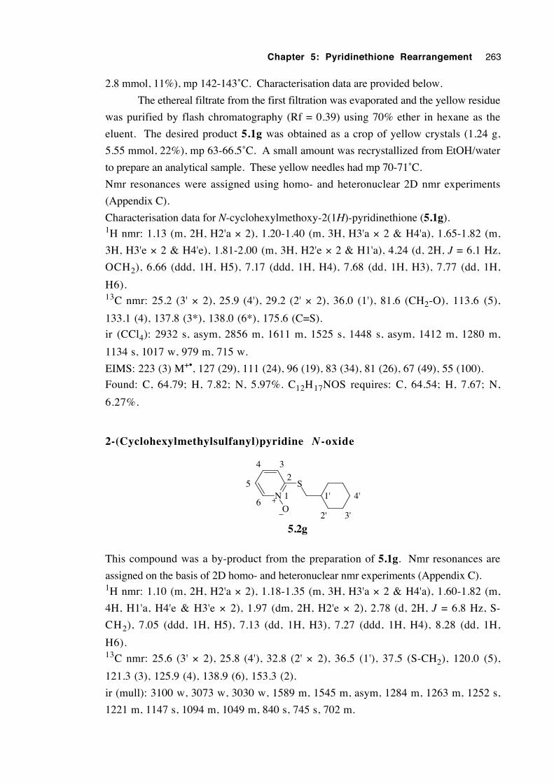
Chapter 5: Pyridinethione Rearrangement 263
2.8 mmol, 11%), mp 142-143˚C. Characterisation data are provided below.
The ethereal filtrate from the first filtration was evaporated and the yellow residue
was purified by flash chromatography (Rf = 0.39) using 70% ether in hexane as the
eluent. The desired product 5.1g was obtained as a crop of yellow crystals (1.24 g,
5.55 mmol, 22%), mp 63-66.5˚C. A small amount was recrystallized from EtOH/water
to prepare an analytical sample. These yellow needles had mp 70-71˚C.
Nmr resonances were assigned using homo- and heteronuclear 2D nmr experiments
(Appendix C).
Characterisation data for N-cyclohexylmethoxy-2(1H)-pyridinethione (5.1g).1H nmr: 1.13 (m, 2H, H2'a × 2), 1.20-1.40 (m, 3H, H3'a × 2 & H4'a), 1.65-1.82 (m,
3H, H3'e × 2 & H4'e), 1.81-2.00 (m, 3H, H2'e × 2 & H1'a), 4.24 (d, 2H, J = 6.1 Hz,
OCH2), 6.66 (ddd, 1H, H5), 7.17 (ddd, 1H, H4), 7.68 (dd, 1H, H3), 7.77 (dd, 1H,
H6).13C nmr: 25.2 (3' × 2), 25.9 (4'), 29.2 (2' × 2), 36.0 (1'), 81.6 (CH2-O), 113.6 (5),
133.1 (4), 137.8 (3*), 138.0 (6*), 175.6 (C=S).
ir (CCl4): 2932 s, asym, 2856 m, 1611 m, 1525 s, 1448 s, asym, 1412 m, 1280 m,
1134 s, 1017 w, 979 m, 715 w.
EIMS: 223 (3) M+•, 127 (29), 111 (24), 96 (19), 83 (34), 81 (26), 67 (49), 55 (100).
Found: C, 64.79; H, 7.82; N, 5.97%. C12H17NOS requires: C, 64.54; H, 7.67; N,
6.27%.
2-(Cyclohexylmethylsulfanyl)pyridine N-oxide
NS
O
1
2
34
6
51'
2' 3'
4'
5.2g
This compound was a by-product from the preparation of 5.1g. Nmr resonances are
assigned on the basis of 2D homo- and heteronuclear nmr experiments (Appendix C).1H nmr: 1.10 (m, 2H, H2'a × 2), 1.18-1.35 (m, 3H, H3'a × 2 & H4'a), 1.60-1.82 (m,
4H, H1'a, H4'e & H3'e × 2), 1.97 (dm, 2H, H2'e × 2), 2.78 (d, 2H, J = 6.8 Hz, S-
CH2), 7.05 (ddd, 1H, H5), 7.13 (dd, 1H, H3), 7.27 (ddd, 1H, H4), 8.28 (dd, 1H,
H6).13C nmr: 25.6 (3' × 2), 25.8 (4'), 32.8 (2' × 2), 36.5 (1'), 37.5 (S-CH2), 120.0 (5),
121.3 (3), 125.9 (4), 138.9 (6), 153.3 (2).
ir (mull): 3100 w, 3073 w, 3030 w, 1589 m, 1545 m, asym, 1284 m, 1263 m, 1252 s,
1221 m, 1147 s, 1094 m, 1049 m, 840 s, 745 s, 702 m.
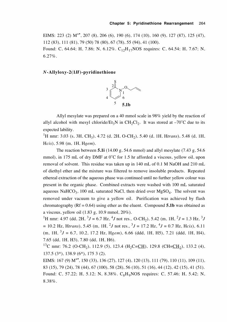
Chapter 5: Pyridinethione Rearrangement 264
EIMS: 223 (2) M+•, 207 (8), 206 (6), 190 (6), 174 (10), 160 (9), 127 (87), 125 (47),
112 (83), 111 (81), 79 (50) 78 (80), 67 (78), 55 (94), 41 (100).
Found: C, 64.64; H, 7.86; N, 6.12%. C12H17NOS requires: C, 64.54; H, 7.67; N,
6.27%.
N-Allyloxy-2(1H )-pyridinethione
N
S
O
5.1b
164
32
5
Allyl mesylate was prepared on a 40 mmol scale in 98% yield by the reaction of
allyl alcohol with mesyl chloride/Et3N in CH2Cl2. It was stored at –70˚C due to its
expected lability.1H nmr: 3.03 (s, 3H, CH3), 4.72 (d, 2H, O-CH2), 5.40 (d, 1H, Htrans), 5.48 (d, 1H,
Hcis), 5.98 (m, 1H, Hgem).
The reaction between 5.1i (14.00 g, 54.6 mmol) and allyl mesylate (7.43 g, 54.6
mmol), in 175 mL of dry DMF at 0˚C for 1.5 hr afforded a viscous, yellow oil, upon
removal of solvent. This residue was taken up in 140 mL of 0.1 M NaOH and 210 mL
of diethyl ether and the mixture was filtered to remove insoluble products. Repeated
ethereal extraction of the aqueous phase was continued until no further yellow colour was
present in the organic phase. Combined extracts were washed with 100 mL saturated
aqueous NaHCO3, 100 mL saturated NaCl, then dried over MgSO4. The solvent was
removed under vacuum to give a yellow oil. Purification was achieved by flash
chromatography (Rf = 0.64) using ether as the eluent. Compound 5.1b was obtained as
a viscous, yellow oil (1.83 g, 10.9 mmol, 20%).1H nmr: 4.97 (dd, 2H, 3J = 6.7 Hz, 4J not res., O-CH2), 5.42 (m, 1H, 2J = 1.3 Hz, 3J
= 10.2 Hz, Htrans), 5.45 (m, 1H, 2J not res., 3J = 17.2 Hz, 4J = 0.7 Hz, Hcis), 6.11
(m, 1H, 3J = 6.7, 10.2, 17.2 Hz, Hgem), 6.66 (ddd, 1H, H5), 7.21 (ddd, 1H, H4),
7.65 (dd, 1H, H3), 7.80 (dd, 1H, H6).13C nmr: 76.2 (O-CH2), 112.9 (5), 123.4 (H2C=CH), 129.8 (CH=CH2), 133.2 (4),
137.5 (3*), 138.9 (6*), 175 3 (2).
EIMS: 167 (9) M+•, 150 (33), 136 (27), 127 (4), 120 (13), 111 (79), 110 (11), 109 (11),
83 (15), 79 (24), 78 (44), 67 (100), 58 (28), 56 (10), 51 (16), 44 (12), 42 (15), 41 (51).
Found: C, 57.22; H, 5.12; N, 8.38%. C8H9NOS requires: C, 57.46; H, 5.42; N,
8.38%.
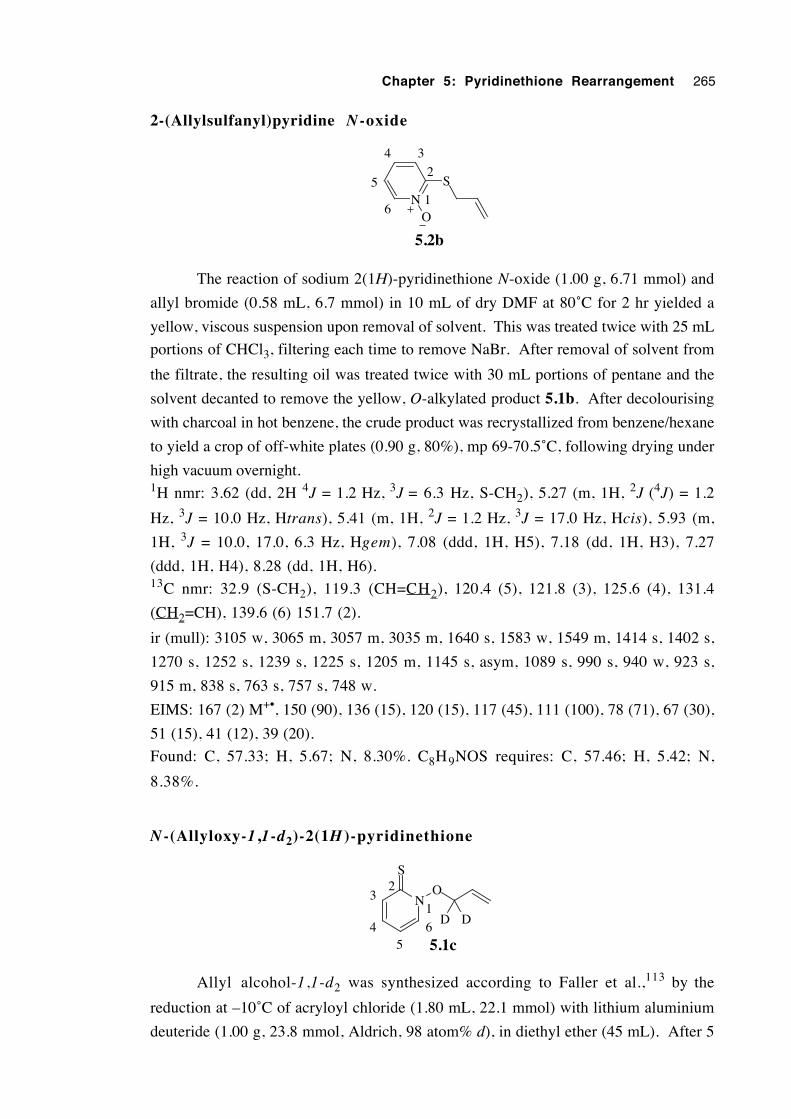
Chapter 5: Pyridinethione Rearrangement 265
2-(Allylsulfanyl)pyridine N-oxide
N
S
O
5.2b
1
2
34
6
5
The reaction of sodium 2(1H)-pyridinethione N-oxide (1.00 g, 6.71 mmol) and
allyl bromide (0.58 mL, 6.7 mmol) in 10 mL of dry DMF at 80˚C for 2 hr yielded a
yellow, viscous suspension upon removal of solvent. This was treated twice with 25 mL
portions of CHCl3, filtering each time to remove NaBr. After removal of solvent from
the filtrate, the resulting oil was treated twice with 30 mL portions of pentane and the
solvent decanted to remove the yellow, O-alkylated product 5.1b. After decolourising
with charcoal in hot benzene, the crude product was recrystallized from benzene/hexane
to yield a crop of off-white plates (0.90 g, 80%), mp 69-70.5˚C, following drying under
high vacuum overnight.1H nmr: 3.62 (dd, 2H 4J = 1.2 Hz, 3J = 6.3 Hz, S-CH2), 5.27 (m, 1H, 2J (4J) = 1.2
Hz, 3J = 10.0 Hz, Htrans), 5.41 (m, 1H, 2J = 1.2 Hz, 3J = 17.0 Hz, Hcis), 5.93 (m,
1H, 3J = 10.0, 17.0, 6.3 Hz, Hgem), 7.08 (ddd, 1H, H5), 7.18 (dd, 1H, H3), 7.27
(ddd, 1H, H4), 8.28 (dd, 1H, H6).13C nmr: 32.9 (S-CH2), 119.3 (CH=CH2), 120.4 (5), 121.8 (3), 125.6 (4), 131.4
(CH2=CH), 139.6 (6) 151.7 (2).
ir (mull): 3105 w, 3065 m, 3057 m, 3035 m, 1640 s, 1583 w, 1549 m, 1414 s, 1402 s,
1270 s, 1252 s, 1239 s, 1225 s, 1205 m, 1145 s, asym, 1089 s, 990 s, 940 w, 923 s,
915 m, 838 s, 763 s, 757 s, 748 w.
EIMS: 167 (2) M+•, 150 (90), 136 (15), 120 (15), 117 (45), 111 (100), 78 (71), 67 (30),
51 (15), 41 (12), 39 (20).
Found: C, 57.33; H, 5.67; N, 8.30%. C8H9NOS requires: C, 57.46; H, 5.42; N,
8.38%.
N -(Allyloxy-1 ,1-d2)-2(1H )-pyridinethione
N
S
O
5.1c
D D1
64
32
5
Allyl alcohol-1,1-d2 was synthesized according to Faller et al.,113 by the
reduction at –10˚C of acryloyl chloride (1.80 mL, 22.1 mmol) with lithium aluminium
deuteride (1.00 g, 23.8 mmol, Aldrich, 98 atom% d), in diethyl ether (45 mL). After 5
Chapter 5: Pyridinethione Rearrangement 266
hours, the mixture was quenched carefully with 5 mL of 3% NaOH and stirred overnight.
Extraction of the aqueous phase was achieved with 3 × 30 mL of diethyl ether and the
combined extracts were dried over Na2SO4 and evaporated under vacuum at 0˚C to yield
a colourless oil (1.27 g). Nmr analysis showed the crude product to be impure.
Purification by kugelrohr distillation (50˚C/115 mmHg) gave the desired product (0.64 g,
10.6 mmol, 48%, lit.113 80% undistilled). Isotopic composition determined from 1H
nmr peak integrals is 98 atom% d.1H nmr: 1.67 (br, 1H, OH), 4.15 (m, ≈ 0.02 H residual O–CH2 and O–CDH), 5.17 (dd,
1H, Htrans), 5.30 (dd, 1H, Hcis), 5.97-6.17 (m, 1H, Hgem).
Allyl-1,1-d2 mesylate (0.67 g, 4.8 mmol, 68%) was then prepared by the reaction
at 0˚C of the labelled alcohol (0.43 g, 7.2 mmol), mesyl chloride (0.610 mL, 7.88 mmol)
and Et3N (1.50 mL, 10.7 mmol) in 35 mL of CH2Cl2. An estimated isotopic
composition from 1H nmr peak integrals is 98 atom% d.1H nmr: 3.03 (s, 3H, CH3), 4.71 (m, ≈ 0.02 H, residual O–CH2 and O–CDH), 5.40
(d, 1H, 3J = 10.5 Hz, Htrans), 5.48 (d, 1H, 3J = 16.9 Hz, Hcis), 5.97 (dd, 1H, 3J =
10.5, 16.9 Hz, Hgem).
The reaction between 5.1i (2.86 g, 11.2 mmol), and the labelled allyl mesylate
(0.67 g, 4.85 mmol) in 17.5 mL of dry DMF at 0˚C for 2.5 hr afforded a yellow, cloudy
oil upon removal of the solvent. The residue was treated with 12 mL of aqueous 0.1 M
NaOH and extracted with 5 × 10 mL of diethyl ether until the extracts were no longer
yellow in colour. Combined extracts were washed with 10 mL of water, dried over
MgSO4 and evaporated to afford a yellow oil (0.21 g). Purification of this crude product
by vacuum-liquid chromatography (VLC, hexane→ether) gave 5.1c (0.15 g, 0.89
mmol, 8%) as a viscous, yellow oil.1H nmr: 4.97 (m, ≈0.017H, 3J = 6.7 Hz, 4J not res., O-CH2), 5.43 (dd, 1H, 3J = 10.2
Hz, 2J = 1.4 Hz, Htrans), 5.45 (dd, 1H, 3J = 17.2 Hz, 2J =1.4 Hz, Hcis), 6.10 (dd,
1H, 3J = 17.2, 10.2 Hz, Hgem), 6.62 (ddd, 1H, H5), 7.18 (ddd, 1H, H4), 7.66 (dd,
1H, H3), 7.75 (dd, 1H, H6).13C nmr: 77.2 (O-CD2), 113.5 (5), 124.4 (CH2=CH), 130.5 (CH2=CH), 133.8 (4),
138.5 (3*), 139.6 (6*), 176.3 (C=S).
EIMS: 169 (4) M+•, 152 (8), 136 (6), 127 (4), 121 (17), 112 (48), 83 (15), 78 (45), 68
(100), 58 (13), 51 (22), 43 (23).2H nmr (30.7 MHz, CHCl3): 4.97 (s, br, O-CD2).
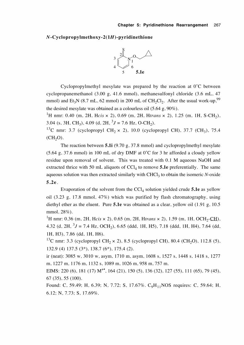
Chapter 5: Pyridinethione Rearrangement 267
N-Cyclopropylmethoxy-2(1H)-pyridinethione
N
S
O
5.1e
1
64
32
5
Cyclopropylmethyl mesylate was prepared by the reaction at 0˚C between
cyclopropanemethanol (3.00 g, 41.6 mmol), methanesulfonyl chloride (3.6 mL, 47
mmol) and Et3N (8.7 mL, 62 mmol) in 200 mL of CH2Cl2. After the usual work-up,99
the desired mesylate was obtained as a colourless oil (5.64 g, 90%).1H nmr: 0.40 (m, 2H, Hcis × 2), 0.69 (m, 2H, Htrans × 2), 1.25 (m, 1H, S-CH2),
3.04 (s, 3H, CH3), 4.09 (d, 2H, 3J = 7.6 Hz, O-CH2).13C nmr: 3.7 (cyclopropyl CH2 × 2), 10.0 (cyclopropyl CH), 37.7 (CH3), 75.4
(CH2O).
The reaction between 5.1i (9.70 g, 37.8 mmol) and cyclopropylmethyl mesylate
(5.64 g, 37.6 mmol) in 100 mL of dry DMF at 0˚C for 3 hr afforded a cloudy yellow
residue upon removal of solvent. This was treated with 0.1 M aqueous NaOH and
extracted thrice with 50 mL aliquots of CCl4 to remove 5.1e preferentially. The same
aqueous solution was then extracted similarly with CHCl3 to obtain the isomeric N-oxide
5.2e .
Evaporation of the solvent from the CCl4 solution yielded crude 5.1e as yellow
oil (3.23 g, 17.8 mmol, 47%) which was purified by flash chromatography, using
diethyl ether as the eluent. Pure 5.1e was obtained as a clear, yellow oil (1.91 g, 10.5
mmol, 28%).1H nmr: 0.36 (m, 2H, Hcis × 2), 0.65 (m, 2H, Htrans × 2), 1.59 (m, 1H, OCH2-CH),
4.32 (d, 2H, 3J = 7.4 Hz, OCH2), 6.65 (ddd, 1H, H5), 7.18 (ddd, 1H, H4), 7.64 (dd,
1H, H3), 7.86 (dd, 1H, H6).13C nmr: 3.3 (cyclopropyl CH2 × 2), 8.5 (cyclopropyl CH), 80.4 (CH2O), 112.8 (5),
132.9 (4) 137.5 (3*), 138.7 (6*), 175.4 (2).
ir (neat): 3085 w, 3010 w, asym, 1710 m, asym, 1608 s, 1527 s, 1448 s, 1418 s, 1277
m, 1227 m, 1176 m, 1132 s, 1089 m, 1026 m, 958 m, 757 m.
EIMS: 220 (6), 181 (17) M+•, 164 (21), 150 (5), 136 (32), 127 (55), 111 (65), 79 (45),
67 (35), 55 (100).
Found: C, 59.49; H, 6.39; N, 7.72; S, 17.67%. C9H11NOS requires: C, 59.64; H,
6.12; N, 7.73; S, 17.69%.
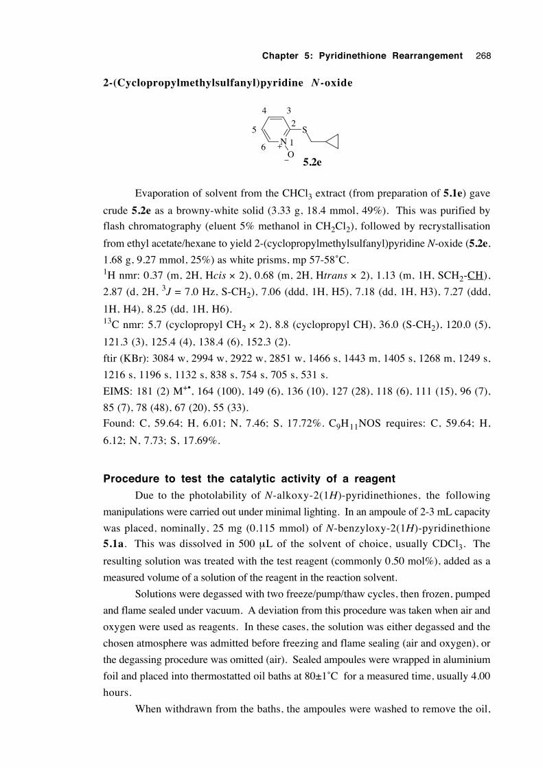
Chapter 5: Pyridinethione Rearrangement 268
2-(Cyclopropylmethylsulfanyl)pyridine N-oxide
NS
O1
2
34
6
5
5.2e
Evaporation of solvent from the CHCl3 extract (from preparation of 5.1e) gave
crude 5.2e as a browny-white solid (3.33 g, 18.4 mmol, 49%). This was purified by
flash chromatography (eluent 5% methanol in CH2Cl2), followed by recrystallisation
from ethyl acetate/hexane to yield 2-(cyclopropylmethylsulfanyl)pyridine N-oxide (5.2e,
1.68 g, 9.27 mmol, 25%) as white prisms, mp 57-58˚C.1H nmr: 0.37 (m, 2H, Hcis × 2), 0.68 (m, 2H, Htrans × 2), 1.13 (m, 1H, SCH2-CH),
2.87 (d, 2H, 3J = 7.0 Hz, S-CH2), 7.06 (ddd, 1H, H5), 7.18 (dd, 1H, H3), 7.27 (ddd,
1H, H4), 8.25 (dd, 1H, H6).13C nmr: 5.7 (cyclopropyl CH2 × 2), 8.8 (cyclopropyl CH), 36.0 (S-CH2), 120.0 (5),
121.3 (3), 125.4 (4), 138.4 (6), 152.3 (2).
ftir (KBr): 3084 w, 2994 w, 2922 w, 2851 w, 1466 s, 1443 m, 1405 s, 1268 m, 1249 s,
1216 s, 1196 s, 1132 s, 838 s, 754 s, 705 s, 531 s.
EIMS: 181 (2) M+•, 164 (100), 149 (6), 136 (10), 127 (28), 118 (6), 111 (15), 96 (7),
85 (7), 78 (48), 67 (20), 55 (33).
Found: C, 59.64; H, 6.01; N, 7.46; S, 17.72%. C9H11NOS requires: C, 59.64; H,
6.12; N, 7.73; S, 17.69%.
Procedure to test the catalytic activity of a reagent
Due to the photolability of N-alkoxy-2(1H)-pyridinethiones, the following
manipulations were carried out under minimal lighting. In an ampoule of 2-3 mL capacity
was placed, nominally, 25 mg (0.115 mmol) of N-benzyloxy-2(1H)-pyridinethione
5.1a. This was dissolved in 500 µL of the solvent of choice, usually CDCl3. The
resulting solution was treated with the test reagent (commonly 0.50 mol%), added as a
measured volume of a solution of the reagent in the reaction solvent.
Solutions were degassed with two freeze/pump/thaw cycles, then frozen, pumped
and flame sealed under vacuum. A deviation from this procedure was taken when air and
oxygen were used as reagents. In these cases, the solution was either degassed and the
chosen atmosphere was admitted before freezing and flame sealing (air and oxygen), or
the degassing procedure was omitted (air). Sealed ampoules were wrapped in aluminium
foil and placed into thermostatted oil baths at 80±1˚C for a measured time, usually 4.00
hours.
When withdrawn from the baths, the ampoules were washed to remove the oil,
Chapter 5: Pyridinethione Rearrangement 269
opened and the contents were transferred to an nmr tube. When the solvents C6D6 and
CD3CN were used, 500 µL of CDCl3 was added to ensure complete solubility of all
components. Analysis was performed by 1H nmr. The results are listed in table 5.1.
"Extent of reaction" was calculated by dividing the sum of the relative concentrations of
identifiable products by the total. "Yield of N-oxide" represents the concentration of
5.2a divided by the sum of the concentrations of all identifiable compounds.
Procedure for conducting kinetics experiments
These experiments were performed in three different groups. In the first, kinetic
data was measured as a function of the proportion of the catalyst iodine. The second set
of experiments was concerned with obtaining kinetic data for four different types of
catalyst. In the third, kinetic data was measured as a function of changing both
pyridinethione and iodine concentrations whilst keeping their ratio the same.
1. Kinetic data as a function of the proportion of iodine present
Into a 3 mL Reactivial, was placed of N-benzyloxy-2(1H)-pyridinethione (5.1a,
nominally 50.0 mg, 0.230 mmol), which was dissolved in 2.00 mL of purified, dry
CHCl3. The vial was capped with a Mininert valve and the solution was degassed using
three freeze/pump/thaw cycles via a needle through the valve and placed under a dry
nitrogen atmosphere. Iodine was then injected by microsyringe from a 5.755 × 10⁻⁴ M
solution of iodine in CHCl3. Proportions of 0.500 (20.0 µL), 1.01 (40.0 µL) and 2.00
mol% (80.0 µL) were added, for the three reactions.
The vials were placed in an 80±1˚C thermostatted oil bath and aliquots of 25.0 mL
were withdrawn over time and diluted to 10.00 mL with spectroscopic grade ethanol.
Analysis was performed by UV-vis spectrophotometry, by monitoring with time the
absorbance of the absorption peak with λmax = 361 nm, belonging to the compound N-
benzyloxy-2(1H)-pyridinethione (5.1a).
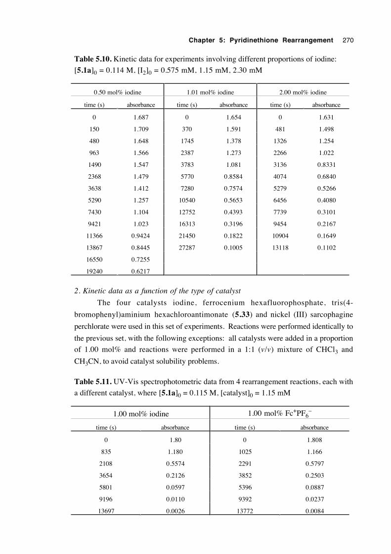
Chapter 5: Pyridinethione Rearrangement 270
Table 5.10. Kinetic data for experiments involving different proportions of iodine:
[5.1a]0 = 0.114 M, [I2]0 = 0.575 mM, 1.15 mM, 2.30 mM
0.50 mol% iodine 1.01 mol% iodine 2.00 mol% iodine
time (s) absorbance time (s) absorbance time (s) absorbance
0 1.687 0 1.654 0 1.631
150 1.709 370 1.591 481 1.498
480 1.648 1745 1.378 1326 1.254
963 1.566 2387 1.273 2266 1.022
1490 1.547 3783 1.081 3136 0.8331
2368 1.479 5770 0.8584 4074 0.6840
3638 1.412 7280 0.7574 5279 0.5266
5290 1.257 10540 0.5653 6456 0.4080
7430 1.104 12752 0.4393 7739 0.3101
9421 1.023 16313 0.3196 9454 0.2167
11366 0.9424 21450 0.1822 10904 0.1649
13867 0.8445 27287 0.1005 13118 0.1102
16550 0.7255
19240 0.6217
2. Kinetic data as a function of the type of catalyst
The four catalysts iodine, ferrocenium hexafluorophosphate, tris(4-
bromophenyl)aminium hexachloroantimonate (5.33) and nickel (III) sarcophagine
perchlorate were used in this set of experiments. Reactions were performed identically to
the previous set, with the following exceptions: all catalysts were added in a proportion
of 1.00 mol% and reactions were performed in a 1:1 (v/v) mixture of CHCl3 and
CH3CN, to avoid catalyst solubility problems.
Table 5.11. UV-Vis spectrophotometric data from 4 rearrangement reactions, each with
a different catalyst, where [5.1a]0 = 0.115 M, [catalyst]0 = 1.15 mM
1.00 mol% iodine 1.00 mol% Fc+PF6–
time (s) absorbance time (s) absorbance
0 1.80 0 1.808
835 1.180 1025 1.166
2108 0.5574 2291 0.5797
3654 0.2126 3852 0.2503
5801 0.0597 5396 0.0887
9196 0.0110 9392 0.0237
13697 0.0026 13772 0.0084
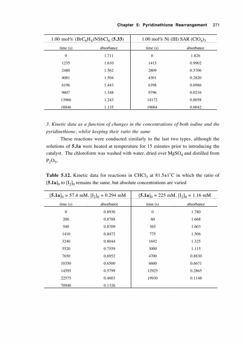
Chapter 5: Pyridinethione Rearrangement 271
1.00 mol% (BrC6H4)NSbCl6 (5.33) 1.00 mol% Ni (III) SAR (ClO4)3
time (s) absorbance time (s) absorbance
0 1.711 0 1.826
1235 1.610 1415 0.9902
2480 1.562 2809 0.5706
4081 1.504 4301 0.2820
6196 1.443 6398 0.0986
9607 1.348 9796 0.0216
13966 1.243 14172 0.0058
18846 1.135 19064 0.0042
3. Kinetic data as a function of changes in the concentrations of both iodine and the
pyridinethione, whilst keeping their ratio the same
These reactions were conducted similarly to the last two types, although the
solutions of 5.1a were heated at temperature for 15 minutes prior to introducing the
catalyst. The chloroform was washed with water, dried over MgSO4 and distilled from
P2O5.
Table 5.12. Kinetic data for reactions in CHCl3 at 81.5±1˚C in which the ratio of
[5.1a]0 to [I2]0 remains the same, but absolute concentrations are varied
[5.1a]0 = 57.4 mM, [I2]0 = 0.294 mM [5.1a]0 = 225 mM, [I2]0 = 1.16 mM
time (s) absorbance time (s) absorbance
0 0.8936 0 1.780
200 0.8788 60 1.668
540 0.8709 365 1.603
1410 0.8472 775 1.506
3240 0.8044 1692 1.325
5520 0.7559 3000 1.115
7650 0.6952 4700 0.8830
10350 0.6500 6600 0.6671
14595 0.5799 12925 0.2865
22575 0.4603 19930 0.1148
70940 0.1326
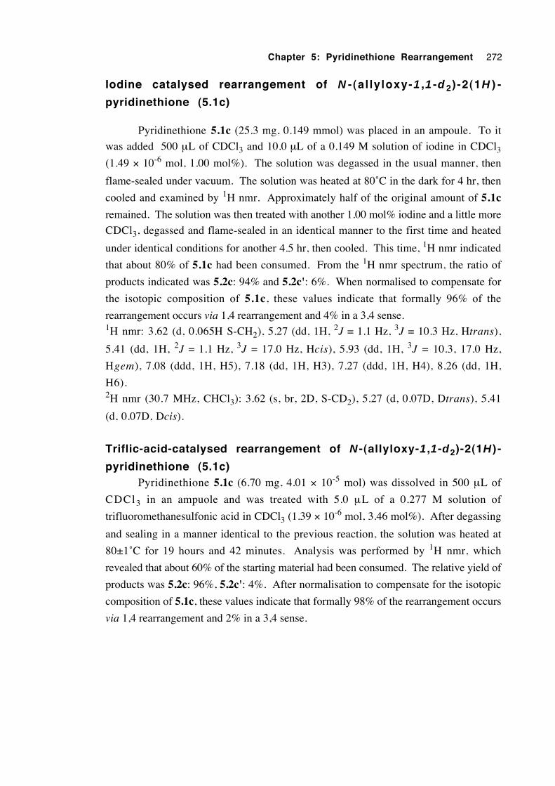
Chapter 5: Pyridinethione Rearrangement 272
Iodine catalysed rearrangement of N - (al ly loxy-1 ,1 -d 2) -2(1H ) -
pyridinethione (5.1c)
Pyridinethione 5.1c (25.3 mg, 0.149 mmol) was placed in an ampoule. To it
was added 500 µL of CDCl3 and 10.0 µL of a 0.149 M solution of iodine in CDCl3(1.49 × 10⁻⁶ mol, 1.00 mol%). The solution was degassed in the usual manner, then
flame-sealed under vacuum. The solution was heated at 80˚C in the dark for 4 hr, then
cooled and examined by 1H nmr. Approximately half of the original amount of 5.1c
remained. The solution was then treated with another 1.00 mol% iodine and a little more
CDCl3, degassed and flame-sealed in an identical manner to the first time and heated
under identical conditions for another 4.5 hr, then cooled. This time, 1H nmr indicated
that about 80% of 5.1c had been consumed. From the 1H nmr spectrum, the ratio of
products indicated was 5.2c: 94% and 5.2c': 6%. When normalised to compensate for
the isotopic composition of 5.1c, these values indicate that formally 96% of the
rearrangement occurs via 1,4 rearrangement and 4% in a 3,4 sense.1H nmr: 3.62 (d, 0.065H S-CH2), 5.27 (dd, 1H, 2J = 1.1 Hz, 3J = 10.3 Hz, Htrans),
5.41 (dd, 1H, 2J = 1.1 Hz, 3J = 17.0 Hz, Hcis), 5.93 (dd, 1H, 3J = 10.3, 17.0 Hz,
Hgem), 7.08 (ddd, 1H, H5), 7.18 (dd, 1H, H3), 7.27 (ddd, 1H, H4), 8.26 (dd, 1H,
H6).2H nmr (30.7 MHz, CHCl3): 3.62 (s, br, 2D, S-CD2), 5.27 (d, 0.07D, Dtrans), 5.41
(d, 0.07D, Dcis).
Triflic-acid-catalysed rearrangement of N -(allyloxy-1 ,1-d2)-2(1H ) -
pyridinethione (5.1c)Pyridinethione 5.1c (6.70 mg, 4.01 × 10⁻⁵ mol) was dissolved in 500 µL of
CDCl3 in an ampuole and was treated with 5.0 µL of a 0.277 M solution of
trifluoromethanesulfonic acid in CDCl3 (1.39 × 10⁻⁶ mol, 3.46 mol%). After degassing
and sealing in a manner identical to the previous reaction, the solution was heated at
80±1˚C for 19 hours and 42 minutes. Analysis was performed by 1H nmr, which
revealed that about 60% of the starting material had been consumed. The relative yield of
products was 5.2c: 96%, 5.2c': 4%. After normalisation to compensate for the isotopic
composition of 5.1c, these values indicate that formally 98% of the rearrangement occurs
via 1,4 rearrangement and 2% in a 3,4 sense.
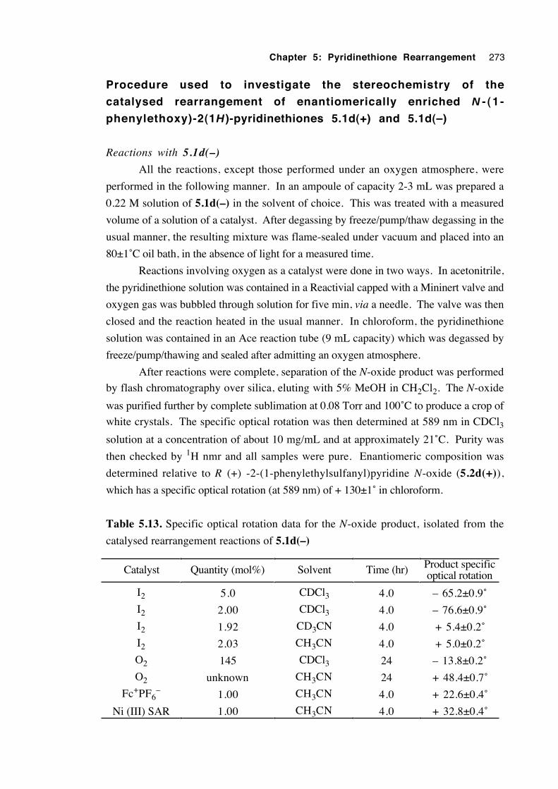
Chapter 5: Pyridinethione Rearrangement 273
Procedure used to investigate the stereochemistry of the
catalysed rearrangement of enantiomerically enriched N - (1 -
phenylethoxy)-2(1H )-pyridinethiones 5.1d(+) and 5.1d(–)
Reactions with 5.1d(–)
All the reactions, except those performed under an oxygen atmosphere, were
performed in the following manner. In an ampoule of capacity 2-3 mL was prepared a
0.22 M solution of 5.1d(–) in the solvent of choice. This was treated with a measured
volume of a solution of a catalyst. After degassing by freeze/pump/thaw degassing in the
usual manner, the resulting mixture was flame-sealed under vacuum and placed into an
80±1˚C oil bath, in the absence of light for a measured time.
Reactions involving oxygen as a catalyst were done in two ways. In acetonitrile,
the pyridinethione solution was contained in a Reactivial capped with a Mininert valve and
oxygen gas was bubbled through solution for five min, via a needle. The valve was then
closed and the reaction heated in the usual manner. In chloroform, the pyridinethione
solution was contained in an Ace reaction tube (9 mL capacity) which was degassed by
freeze/pump/thawing and sealed after admitting an oxygen atmosphere.
After reactions were complete, separation of the N-oxide product was performed
by flash chromatography over silica, eluting with 5% MeOH in CH2Cl2. The N-oxide
was purified further by complete sublimation at 0.08 Torr and 100˚C to produce a crop of
white crystals. The specific optical rotation was then determined at 589 nm in CDCl3solution at a concentration of about 10 mg/mL and at approximately 21˚C. Purity was
then checked by 1H nmr and all samples were pure. Enantiomeric composition was
determined relative to R (+) -2-(1-phenylethylsulfanyl)pyridine N-oxide (5.2d(+)),
which has a specific optical rotation (at 589 nm) of + 130±1˚ in chloroform.
Table 5.13. Specific optical rotation data for the N-oxide product, isolated from the
catalysed rearrangement reactions of 5.1d(–)
Catalyst Quantity (mol%) Solvent Time (hr) Product specificoptical rotation
I2 5.0 CDCl3 4.0 – 65.2±0.9˚
I2 2.00 CDCl3 4.0 – 76.6±0.9˚
I2 1.92 CD3CN 4.0 + 5.4±0.2˚
I2 2.03 CH3CN 4.0 + 5.0±0.2˚
O2 145 CDCl3 24 – 13.8±0.2˚
O2 unknown CH3CN 24 + 48.4±0.7˚
Fc+PF6– 1.00 CH3CN 4.0 + 22.6±0.4˚
Ni (III) SAR 1.00 CH3CN 4.0 + 32.8±0.4˚
Chapter 5: Pyridinethione Rearrangement 274
Rearrangement reactions of 5.1d(+)
1. Pyridinethione 5.1d(+) (48.33 mg, 0.2090 mmol) was dissolved in 1.00 mL
of CDCl3 and treated with 9.30 µL of a 0.116 M solution of iodine in CDCl3 (1.08 × 10-
6 mol, 0.518 mol%). This solution was degassed and flame sealed under vacuum in the
usual manner, then heated in the dark at 80±1˚ for 4.00 hours. Analysis by 1H nmr
revealed that only 30% of the starting material had been consumed. After treatment with a
further 20.0 µL of the iodine solution (total moles = 3.40 × 10⁻⁶ mol, 1.63 mol%) and
degassing and resealing, the mixture was heated for a further 11 hours. Treatment with a
little hexane produced a yellow precipitate to form, then the solvents were removed under
vacuum. The resulting solid was washed three times with hexane to remove iodine and
any remaining pyridinethione. Reprecipitation from benzene/hexane gave a white solid of
specific optical rotation (589 nm) of + 53.1±0.8˚.
2. A reaction was performed in the same way as for 5.1d(–), using 2.04 mol%
iodine, and heating at 80˚C for 5.0 hours. The N-oxide product was isolated by flash
chromatography, followed by sublimation, to give a white solid of specific optical
rotation of + 58.2±0.8˚.
The iodine catalysed rearrangement of N -cyclopropylmethoxy-
2(1H )-pyridinethione (5.1e)
In an Ace brand pressure reaction tube (9 mL capacity) was placed pyridinethione
5.1e (0.40 g, 2.2 mmol) and 8.0 mL of purified CHCl3. To the resulting solution was
added iodine (33.0 mg, 0.130 mmol, 5.9 mol%) and the mixture was swirled until the
iodine dissolved. Deoxygenation was accomplished by three freeze/pump/thaw cycles,
then the solution was placed under a dry nitrogen atmosphere. The vessel was heated at
80±1˚C in a thermostatted oil bath for 5.5 hours under minimal light. After cooling, the
solvent was removed by rotary evaporation then high vacuum to yield a yellow, viscous
oil (0.44 g). Analysis by 1H nmr indicated the following compounds were present in the
molar ratios shown: 2-(cyclopropylmethylsulfanyl)pyridine N-oxide (5.2e, 100); N-
cyclopropylmethoxy-2(1H)-pyridinethione (5.1e, 20); and cyclopropylmethanol (2.7).
There was no detectable resonance in the range 5.0-6.5 ppm, indicating an absence of
alkene functionality. In addition, no peak was detected in the ranges 1.5-2.5 ppm and
3.5-4.1 ppm, which is where hydrogens of sulfur-substituted cyclobutyl rings normally
resonate. It can be concluded that the cyclopropylmethyl group does not undergo ring
opening to an alkene to any measurable extent, nor ring expansion to a cyclobutane
compound.
Chapter 5: Pyridinethione Rearrangement 275
Investigation of the effect of alkoxy substituent structure upon
migration rate (product yield)
The following procedure was used for each pyridinethione undergoing testing. A
CDCl3 solution of the pyridinethione was prepared in an ampoule, such that the
concentration of the solute was approximately 50 mg/mL. This corresponded with
molarities in the range 0.2-0.3 M. After treating the solution with a measured amount of
a solution of iodine in CDCl3 (usually 0.50 mol%), the mixture was degassed with two
freeze/pump/thaw cycles, then frozen, pumped and flame-sealed under vacuum. The
ampoule was placed in an 80±1˚C thermostatted oil bath for a measured period
(commonly 4.0 hours) under minimal light. After cooling, the yield of the N-oxide
product was determined by 1H nmr spectroscopy. Results of these experiments are listed
in table 5.6.
Attempted detection/interception of intermediates in the
rearrangement of N-benzyloxy-2(1H)-pyridinethione (5.1a)
Effect of a radical scavenger
A 0.230 M solution of N-benzyloxy-2(1H)-pyridinethione in CDCl3, a 0.342 M
solution of the radical scavenger 1,4-benzoquinone in CDCl3 and a 0.140 M solution of
iodine in CDCl3 were prepared. Three different reactions were conducted, in ampoules,
to determine the effect the scavenger had upon the rate of the iodine-catalysed
rearrangement of the pyridinethione. The contents of each ampoule were:
a) 500 µL (0.115 mmol) of the pyridinethione solution, 10.0 µL (3.42 × 10⁻⁶ mol, 2.97
mol%) of the benzoquinone solution and 4.10 µL (5.74 × 10⁻⁷ mol, 0.50 mol%) of the
iodine solution;
b) 500 µL (0.115 mmol) of the pyridinethione solution and 4.10 µL (5.74 × 10⁻⁷ mol,
0.50 mol%) of the iodine solution; and
c) 500 µL (0.115 mmol) of the pyridinethione solution and 10.0 µL (3.42 × 10⁻⁶ mol,
2.97 mol%) of the benzoquinone solution.
Each solution was degassed and flame-sealed under vacuum in the usual manner,
then heated under minimal light in an 80±1˚C thermostatted oil bath for a total of 2.00
hours. Yields of the product 2-(benzylsulfanyl)pyridine N-oxide (5.2a) were established
using 1H nmr spectroscopy. The yields were: a) 72.2%; b) 74.7%; and c) 3.2%.
Chapter 5: Pyridinethione Rearrangement 276
Isolation and attempted identification of products resulting from the reaction of N-
benzyloxy-2(1H )-pyridinethione (5.1a) and tris(p -bromophenyl )aminium
hexachloroantimonate (5.33)
The aminium salt 5.33 (0.2991 g, 0.3663 mmol) was added under a nitrogen
blanket to 25 mL of dichloromethane, forming a deep blue solution. To this was added,
in small portions, N-benzyloxy-2(1H)-pyridinethione 5.1a, which caused a gradual
colour change. Additions were continued until there was a permanent loss of the blue
colour and a yellow/brown precipitate in a brown solution resulted. At this stage 0.1025
g (0.4717 mmol, 1.29 eq.) of 5.1a had been added. Centrifugation settled the solids and
the liquid phase was drawn off. The remaining precipitate (0.1864 g) was dried under
high vacuum. If the molecular formula of C12H11NOSSbCl6 is correct, 0.3378 mmol
(92%) was obtained.1H nmr (CD3OD): 4.63 (s, 2H), 5.82 (s, 2H), 7.30-7.45 (m, 4H), 7.50-7.60 (m, 7H),
7.66 (d, 2H), 8.02 (dt, 1H), 8.28 (d, 1H), 8.43 (t, 1H), 8.48 (d, 1H), 9.49 (d, 1H).
MS (+FAB, 1:1 glycerol:thioglycerol matrix 160-730 amu): 497 (80), 429 (7), 391 (19),
308 (33), 280 (54), 218 (100).
MS (+Electrospray, CH3CN): 343 (100), 308 (3), 253 (43), 233 (70), 218 (6), 186 (7),
142 (8), 132 (9).
Found: C, 27.34; H, 1.70; N, 2.97; Sb, 22.79%. C12H11NOSSbCl6 requires: C, 26.12;
H, 2.01; N, 2.54; Sb, 22.07%.
The liquid phase from centrifugation was evaporated and the components were
separated by flash chromatography. Eluent polarity was gradually increased in the order
light alkanes, diethyl ether, 50/50 methanol/dichloromethane, then methanol. By far the
largest component was tris(p-bromophenyl) amine (0.1528 g, 0.3170 mmol, 87%),
identified by comparison of nmr spectra with those of an authentic sample. A second
fraction (14.9 mg) was a yellow/brown solid which had a 1H nmr spectrum consisting of
aromatic resonances ranging from 6.6 to 7.9 ppm, but its structure remains unknown. A
third fraction (32.0 mg) was a yellow solid but no reasonable 1H nmr spectrum could be
obtained in CDCl3 solution. The mass recovery for the reaction was 0.3861 g (96%).

Chapter 5: Pyridinethione Rearrangement 277
5.7 References
1. Wagner, G. and Pischel, H. Arch. Pharm. 1963, 296, 576.
2. Pluijgers, C. W.; Berg, J. and Thorn, G. D. Recl. Trav. Chim. Pays-Bas 1969, 88,
241.
3. Jeffcoat, A. R.; Gibson, W. B.; Rodriguez, P. A.; Turan, T. S.; Hughes, P. F. and
Twine, M. E. Toxicol. Appl. Pharmacol. 1980, 56, 141.
4. Tachikawa, R.; Wachi, K.; Sato, S. and Terada, A. Chem. Pharm. Bull. 1981, 29,
3529.
5. Beckwith, A. L. J. and Hay, B. P. J. Am. Chem. Soc. 1988, 110, 4415.
6. Beckwith, A. L. J. and Hay, B. P. J. Am. Chem. Soc. 1989, 111, 230.
7. Hay, B. P. and Beckwith, A. L. J. J. Org. Chem. 1989, 54, 4330.
8. Lampard, C.; Murphy, J. A. and Lewis, N. Tetrahedron, 1993, 49, 3841.
9. Hartung, J. and Gallou, F. J. Org. Chem. 1995, 60, 6706.
10. Girard, P.; Guillot, N.; Motherwell, W. B. and Potier, P. J. Chem. Soc. Chem.
Commun. 1995, 2385.
11. Hartung, J.; Hiller, M. and Schmidt, P. Chem. Eur. J. 1996, 2, 1014.
12. Hartung, J.; Hiller, M. and Schmidt, P. Liebigs Ann. 1996, 1425.
13. Hartung, J.; Hiller, M.; Schwarz, M.; Svoboda, I. and Fuess, H. Liebigs Ann.
1996, 2091.
14. Hartung, J. Synlett 1996, 1206.
15. Hartung, J.; Svoboda, I.; Fuess, H. and Duarte, M. T. Acta Cryst. C. 1997, 53,
1629.
16. Adam, W.; Grimm, G. N. and Saha-Moller, C. R. Free Radic. Biol. Med. 1998,
24, 234.
17. Adam, W.; Grimm, G. N.; Marquardt, S. and Saha-Moller, C. R. J. Am. Chem. Soc.
1999, 121, 1179.
18. Hartung, J.; Kneuer, R.; Schwarz, M.; Svoboda, I. and Fuess, H. Eur. J. Org.
Chem. 1999, 97.
19. Hartung, J. and Kneuer, R. Eur. J. Org. Chem. 2000, 1677.
Chapter 5: Pyridinethione Rearrangement 278
20. Wiselogle, F. Y.; Losee, K. A. and Bernstein, J. Chemical Abstracts 1957, 51,
P2881d.
21. Crich, D. Aldichimica Acta 1987, 20, 35.
22. Zamojski, A. Chemtracts - Org. Chem. 1995, 8, 181.
23. Barton, D. H. R. and Ferreira, J. A. Phosphorus Sulfur Silicon Relat. Elem. 1997,
120, 1.
24. Barton, D. H. R.; Crich, D. and Motherwell, W. B. J. Chem. Soc. Chem. Commun.
1983, 939.
25. Barton, D. H. R.; Jaszberenyi, J. Cs. and Morrell, A. I. Tetrahedron Lett. 1991,
32, 311.
26. Adam, W.; Ballmaier, D.; Epe, B.; Grimm, G. N. and Saha-Moller, C. R. Angew.
Chem. Int. Ed. Engl. 1995, 34, 2156.
27. Epe, B.; Ballmaier, D.; Adam, W.; Grimm, G. N. and Saha-Moller, C. R. Nucleic
Acids Res. 1996, 24, 1625.
28. Aveline, B. M.; Kochevar, I. E. and Redmond, R. W. J. Am. Chem. Soc. 1996,
118, 10113.
29. Adam, W.; Marquardt, S. and Saha-Moller, C. R. Photochem. Photobiol. 1999,
70, 287.
30. Newcomb, M. and Kumar, M. U. Tetrahedron Lett. 1991, 32, 45.
31. Beckwith, A. L. J. and Davison, I. G. E. Tetrahedron Lett. 1991, 32, 49.
32. Newcomb, M.; Park, S. -U.; Kaplan, J. and Marquardt, D. J. Tetrahedron Lett.
1985, 26, 5651.
33. Newcomb, M. and Deeb, T. M. J. Am. Chem. Soc. 1987, 109, 3163.
34. Esker, J. L. and Newcomb, M. Tetrahedron Lett. 1992, 33, 5913.
35. Horner, J. H.; Musa, O.; Bouvier, A. and Newcomb, M. J. Am. Chem. Soc. 1998,
120, 7738.
36. Avila, L. Z. and Frost, J. W. J. Am Chem. Soc. 1988, 110, 7904.
37. Koenig, T. in Free Radicals; Kochi, J. K., Ed.; John Wiley and Sons: New York,
1973, vol. 1, p. 113.
Chapter 5: Pyridinethione Rearrangement 279
38. Walling, C. and Padwa, A. J. Am. Chem. Soc. 1963, 85, 1593.
39. Walling, C. and Clark, R. T. J. Am. Chem. Soc. 1974, 96, 4530.
40. Brun, P. and Waegell, B. Tetrahedron 1976, 32, 517.
41. Barton, D. H. R. Pure Appl. Chem. 1968, 16, 1.
42. Hesse, R. H. Adv. Free-Radical Chem. 1969, 3, 83.
43. Mihailovic, M. Lj. and Cekovic, Z. Synthesis 1970, 209.
44. Beckwith, A. L. J.; Hay, B. P. and Williams, G. W. J. Chem. Soc., Chem.
Commun. 1989, 1202.
45. Pasto, D. J. and Cottard, F. Tetrahedron Lett. 1994, 35, 4303.
46. Aveline, B. M.; Kochevar, I. E. and Redmond, R. W. J. Am. Chem. Soc. 1995,
117, 9699.
47. Yadav, V. K.; Yadav, A.; Pande, P. and Kapoor, K. K. Indian J. Chem. Sect. B.
1994, 33, 1129.
48. Wagner, G. and Pischel, H. Arch. Pharm. 1962, 295, 897.
49. Shaw, E. J. Am. Chem. Soc. 1949, 71, 67.
50. Dinan, F. J. and Tieckelmann, H. J. Org. Chem. 1964, 29, 1650.
51. Litster, J. E. and Tieckelmann, H. J. Am. Chem. Soc. 1968, 90, 4361.
52. Schollkopf, U. and Hoppe, I. Tetrahedron Lett. 1970, 4527.
53. Schollkopf, U. and Hoppe, I. Liebigs Ann. Chem. 1972, 765, 153.
54. Gerhart, F. and Wilde, L. Tetrahedron Lett. 1974, 475.
55. le Noble, W. J. and Daka, M. R. J. Am. Chem. Soc. 1978, 100, 5961.
56. Alker, D.; Mageswaran, S.; Ollis, W. D. and Shahriari-Zavareh, H. J. Chem. Soc.,
Perkin Trans. 1 1990, 1631.
57. Alker, D.; Ollis, W. D. and Shahriari-Zavareh, H. J. Chem. Soc., Perkin Trans. 1
1990, 1637.
58. Jones, R. A. and Katritzky, A. R. J. Chem. Soc. 1960, 2937.
59. Kohrman, R. E.; West, D. X. and Little, M. A. J. Heterocycl. Chem. 1974, 11,
101.
60. Wallace, T. J.; Schriesheim, A. and Bartok, W. J. Org. Chem. 1963, 28, 1311.
Chapter 5: Pyridinethione Rearrangement 280
61. Gilbert, B. C. and Whitwood, A. C. J. Chem. Soc. Perkin Trans. 2 1989, 1921.
62. Janes, R. and Symons, M. C. R. J. Chem. Soc. Faraday Trans. 1990, 86, 2173.
63. Hart, H.; Rodgers, T. R. and Griffiths, J. J. Am. Chem. Soc. 1969, 91, 752.
64. Childs, R. F. and Winstein, S. J. Am. Chem. Soc. 1974, 96, 6409.
65. Miller, B. and Saidi, M. R. Tetrahedron Lett. 1975, 1365.
66. Miller, B. Acc. Chem. Res. 1975, 8, 245.
67. Vittorelli, P.; Peter-Katalinic, J.; Mukherjee-Muller, G.; Hansen, H. J. and Schmid,
H. Helv. Chim. Acta 1975, 58, 1379.
68. Miller, B. and Saidi, M. R. J. Am. Chem. Soc. 1976, 98, 2227.
69. Miller, B. and Lin, W.-O. J. Org. Chem. 1979, 44, 887.
70. Felkin, H. and Tambute, A. Tetrahedron Lett. 1969, 821.
71. Biellman, J. F. and Ducep, J. B. Tetrahedron Lett. 1970, 2899.
72. Magid, R. M. and Wilson, S. E. Tetrahedron Lett. 1971, 19.
73. Felkin, H. and Frajerman, C. Tetrahedron Lett. 1977, 3485.
74. Rautenstrauch, V. Helv. Chim. Acta 1972, 55, 594.
75. Garst, J. F. and Smith, C. D. J. Am. Chem. Soc. 1976, 98, 1526.
76. Crombie, L.; Darnbrough, G. and Pattenden, G. J. Chem. Soc., Chem. Commun.
1976, 684.
77. Briere, R.; Cherest, M.; Felkin, H. and Frajerman, C. Tetrahedron Lett. 1977,
3489.
78. See references 56 and 57.
79. Bellville, D. J.; Wirth, D. D. and Bauld, N. L. J. Am. Chem. Soc. 1981, 103, 718.
80. Bauld, N. L.; Bellville, D. J.; Pabon, R.; Chelsky, R. and Green, G. J. Am. Chem.
Soc. 1983, 105, 2378.
81. Bellville, D. J. and Bauld, N. L. Tetrahedron 1986, 42, 6167.
82. Bauld, N. L.; Bellville, D. J.; Harirchian, B.; Lorenz, K. T.; Pabon, R. A.;
Reynolds, D. W.; Wirth, D. D.; Chiou, H. -S. and Marsh, B. K. Acc. Chem. Res.
1987, 20, 371.
83. Reynolds, D. W.; Lorenz, K. T.; Chiou, H. -S.; Bellville, D. J.; Pabon, R. A. and
Chapter 5: Pyridinethione Rearrangement 281
Bauld, N. L. J. Am. Chem. Soc. 1987, 109, 4960.
84. Bauld, N. L. Tetrahedron 1989, 45, 5307.
85. Yueh, W. and Bauld, N. L. J. Am. Chem. Soc. 1995, 117, 5671.
86. Valentine, D.; Turro, N. J., Jr and Hammond, G. S. J. Am. Chem. Soc. 1964, 86,
5202.
87. Konig, W. A.; Francke, W. and Benecke, I. J. Chromatogr. 1982, 239, 227.
88. Huck, H. and Schmidt, H.-L. Angew. Chem. Int. Ed. Engl. 1981, 20, 402.
89. Cavalieri, E. and Roth, R. J. Org. Chem. 1976, 41, 2679.
90. Smejtek, P.; Honzl, J. and Metalova, V. Coll. Czech. Chem. Commun. 1965, 30,
3875.
91. Wheeler, L. O.; Santhanam, K. S. V. and Bard, A. J. J. Phys. Chem. 1966, 70,
404.
92. Forbes, W. F. and Sullivan, P. D. J. Am Chem. Soc. 1966, 88, 2862.
93. Lucken, E. A. C. Theor. Chim. Acta. 1963, 1, 397.
94. Gassman, P. G. and Singleton, D. A. J. Am Chem. Soc. 1984, 106, 7993.
95. Katritzky, A. R. J. Chem. Soc. (London) 1957, 191.
96. Duggan, D. M. and Hendrickson, D. N. Inorg. Chem. 1975, 14, 955.
97. Coates, R. M. and Chen, J. P. Tetrahedron Lett. 1969, 2705.
98. Benecke, I. and Konig, W. A. Angew. Chem., Int. Ed. Engl. 1982, 21, 709.
99. Crossland, R. K. and Servis, K. L. J. Org. Chem. 1970, 35, 3195.
100. Hutchins, R. O.; Masilamani, D. and Maryanoff, C. A. J. Org. Chem. 1976, 41,
1071.
101. Isola, M.; Ciuffarin, E. and Sagramora, L. Synthesis 1976, 326.
102. Rinaldi, P. L. Prog. NMR Spectrosc. 1982, 15, 291.
103. Jacques, J.; Collet, A. and Wilen, S. H. Enantiomers, Racemates and
Resolutions; Wiley-Interscience, 1981, pp 162-165.
104. Suckling, C. J. Angew. Chem. Int. Ed. Engl. 1988, 27, 537.
105. Beckwith, A. L. J.; Bowry, V. W. and Moad, G. J. Org. Chem. 1988, 53, 1632.
106. Cacace, F.; Chiavarino, B. and Crestoni, M. E. Chem. Eur. J. 2000, 6, 2024.
Chapter 5: Pyridinethione Rearrangement 282
107. Szeimies, G.; Schlosser, A.; Philipp, F.; Dietz, P. and Mickler, W. Chem. Ber.
1978, 111, 1922.
108. Kimmelma, R. Acta Chem. Scand. Ser. B 1987, 41, 344.
109. March, J. Advanced Organic Chemistry, 3rd Ed.; Wiley-Interscience, New York,
1985, p 145.
110. Smie, A. and Heinze, J. Angew. Chem. Int. Ed. Engl. 1997, 36, 363.
111. Claridge, R. F. C. and Dohrmann, J. Landolt-Bornstein Group 2; Vol. 18,
Subvolume E1; Springer-Verlag, Berlin-Heidelberg; 1997; pp 40-133.
112. Ito, O.; Tamura, S.; Murakami, K. and Matsuda, M. J. Org. Chem. 1988, 53,
4758.
113. Faller, J. W.; Blankenship, C.; Whitmore, B. and Sena, S. Inorg. Chem. 1985,
24, 4483.
114. Nakata, R.; Okazaki, S. and Fujinaga, T. J. Electroanal. Chem. 1981, 125, 413.
115. Compound and E0 value supplied by Professor A. M. Sargeson, Research School
of Chemistry, Australian National University.
116. Mehta, S. and Pinto, B. M. Carbohydr. Res. 1998, 310, 43.
117. Ballesteros, P.; Senent, S. and Elguero, J. Bull. Soc. Chim. Belg. 1990, 99, 595.
118. Wolfe, S.; Yang, K.; Weinberg, N.; Shi, Z.; Hsieh, Y.-H.; Sharma, R. D.; Ro, S.
and Kim, C.-K. Chem. Eur. J. 1998, 4, 886.
119. Wolfe, S.; Hsieh, Y.-H.; Batchelor, R. J.; Einstein, F. W. B. and Gay, I. D. Can.
J. Chem. 2001, 79, 1272.
120. Barton, D. H. R.; Bridon, D.; Fernandez-Picot, I. and Zard, S. Z. Tetrahedron
1987, 43, 2733.
121. Kurzer, F. Org. Synth. 1963, Coll. Vol. 4, 937.
122. Wilt, J. W.; Stein, R. G. and Wagner, W. G. J. Org. Chem. 1967, 32, 2097.
123. Hoffmann, H. M. R. and Hughes, E. D. J. Chem. Soc. 1964, 1244.
124. Gilman, H. and Catlin, W. E. Org. Synth. 1941, Coll. Vol. 1, 188.
125. Hartung, J. Eur. J. Org. Chem. 2001, 619.
126. Hartung, J.; Kneuer, R.; Schwarz, M. and Heubes, M. Eur. J. Org. Chem. 2001,

Chapter 5: Pyridinethione Rearrangement 283
4733.
127. Hartung, J.; Kneuer, R.; Kopf, T. M. and Schmidt, P. C. R. Acad. Sci. Paris,
Chimie 2001, 4, 649.
128. See Harman, D. G. This thesis, Chapters 1, 2 and 3 and references within.
129. Hartung, J., Gottwald, T. and S̆pehar, K. Synthesis 2002, 1469.
130. Tschugaeff, L. Ber. Dtsch. Chem. Ges. 1899, 32, 3332.
Chapter 6
General discussion and conclusions
6.1 Introduction 285
6.2 The β-trifluoroacetoxyalkyl radical rearrangement 285
6.3 Related radical-mediated rearrangements and β-eliminations 297
6.4 The mechanism of the rearrangement of N-alkoxy-2(1H)-
pyridinethiones 301
6.5 Final remarks 301
6.6 References 303
Chapter 6: General Discussion & Conclusions 285
6.1 Introduction
This chapter begins with a summary of what the current research has revealed
about the mechanism of the rearrangement of the 2-methyl-2-trifluoroacetoxy-1-heptyl
radical. The relative magnitude of migrating group effects of the β-trifluoroacetoxyalkyl
radical rearrangement and an analogous ionic process are then discussed. The
relationship between rearrangement regiochemistry and kinetics is then examined. Low-
level theoretical calculations are performed in order to discover whether the E/Z
conformational distribution in the ester group is controlled by solvent polarity and can
therefore account for the observed rearrangement regiochemistry. The dynamics for a
radical ion pair are investigated to determine whether such an intermediate is plausible and
conclusions about the mechanism of the β-trifluoroacetoxyalkyl radical rearrangement are
provided. Conclusions are drawn about the mechanism of the β-trifluoroacetoxyalkyl
radical rearrangement.
Rearrangements and β-elimination reactions related to the β-acyloxyalkyl radical
rearrangement are briefly discussed. The mechanism of the rearrangement of N-alkoxy-
2(1H)-pyridinethiones and its relationship to that of the β-acyloxyalkyl radical
rearrangement are discussed. The chapter concludes with a summary of the mechanism
of the β-trifluoroacetoxyalkyl radical rearrangement and the differences between
pericyclic processes and radical ion pairs. The chapter concludes with reflections on the
differences between polarized pericyclic processes and radical ion pairs and with
hypotheses as to the chemical physics of reactions which occur over an extremely short
time period.
6.2 The β-trifluoroacetoxyalkyl radical rearrangement
6.2.1 What is known about the rearrangement of the 2-methyl-2-
trifluoroacetoxy-1-heptyl radical?
The rearrangement is definitely radical in nature. Trifluoroacetoxy group
migration occurs under conditions known to produce carbon-centred radicals and stalled
reactions are restarted by the addition of the radical initiators AIBN or di-tert-butyl
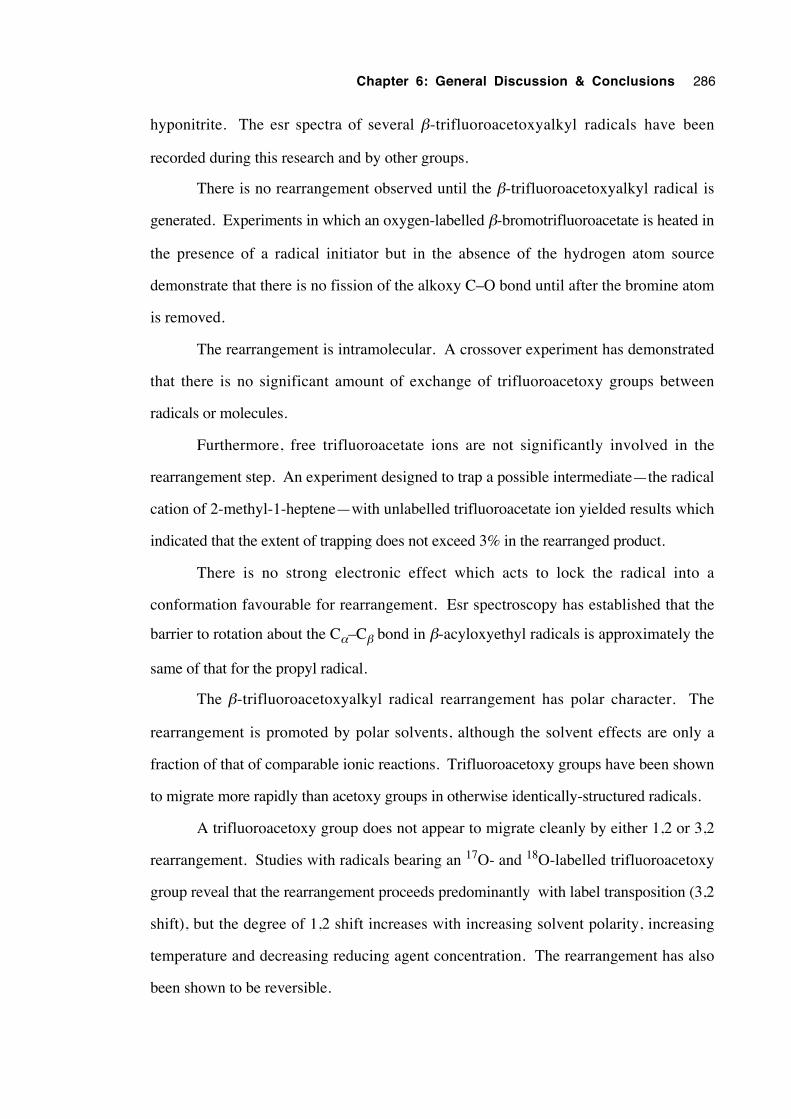
Chapter 6: General Discussion & Conclusions 286
hyponitrite. The esr spectra of several β-trifluoroacetoxyalkyl radicals have been
recorded during this research and by other groups.
There is no rearrangement observed until the β-trifluoroacetoxyalkyl radical is
generated. Experiments in which an oxygen-labelled β-bromotrifluoroacetate is heated in
the presence of a radical initiator but in the absence of the hydrogen atom source
demonstrate that there is no fission of the alkoxy C–O bond until after the bromine atom
is removed.
The rearrangement is intramolecular. A crossover experiment has demonstrated
that there is no significant amount of exchange of trifluoroacetoxy groups between
radicals or molecules.
Furthermore, free trifluoroacetate ions are not significantly involved in the
rearrangement step. An experiment designed to trap a possible intermediate—the radical
cation of 2-methyl-1-heptene—with unlabelled trifluoroacetate ion yielded results which
indicated that the extent of trapping does not exceed 3% in the rearranged product.
There is no strong electronic effect which acts to lock the radical into a
conformation favourable for rearrangement. Esr spectroscopy has established that the
barrier to rotation about the Cα–Cβ bond in β-acyloxyethyl radicals is approximately the
same of that for the propyl radical.
The β-trifluoroacetoxyalkyl radical rearrangement has polar character. The
rearrangement is promoted by polar solvents, although the solvent effects are only a
fraction of that of comparable ionic reactions. Trifluoroacetoxy groups have been shown
to migrate more rapidly than acetoxy groups in otherwise identically-structured radicals.
A trifluoroacetoxy group does not appear to migrate cleanly by either 1,2 or 3,2
rearrangement. Studies with radicals bearing an 17O- and 18O-labelled trifluoroacetoxy
group reveal that the rearrangement proceeds predominantly with label transposition (3,2
shift), but the degree of 1,2 shift increases with increasing solvent polarity, increasing
temperature and decreasing reducing agent concentration. The rearrangement has also
been shown to be reversible.
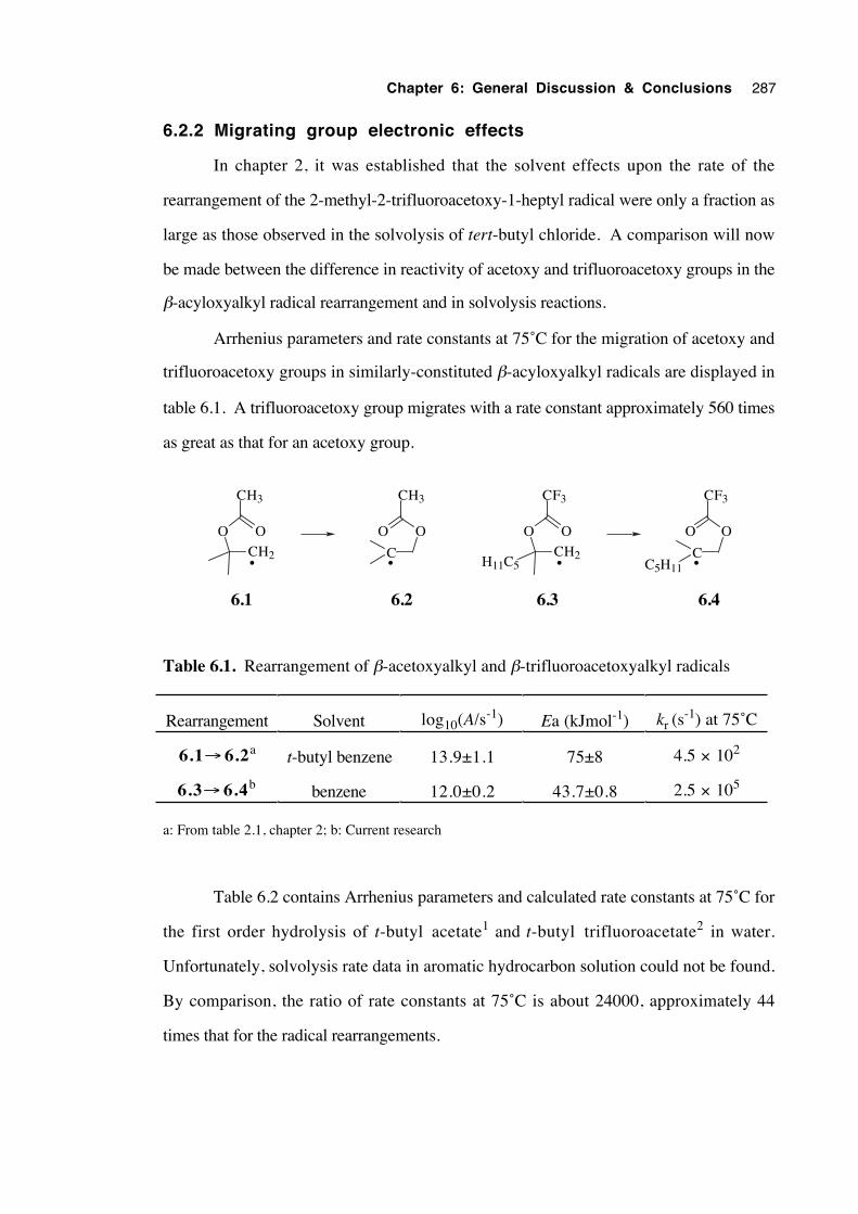
Chapter 6: General Discussion & Conclusions 287
6.2.2 Migrating group electronic effects
In chapter 2, it was established that the solvent effects upon the rate of the
rearrangement of the 2-methyl-2-trifluoroacetoxy-1-heptyl radical were only a fraction as
large as those observed in the solvolysis of tert-butyl chloride. A comparison will now
be made between the difference in reactivity of acetoxy and trifluoroacetoxy groups in the
β-acyloxyalkyl radical rearrangement and in solvolysis reactions.
Arrhenius parameters and rate constants at 75˚C for the migration of acetoxy and
trifluoroacetoxy groups in similarly-constituted β-acyloxyalkyl radicals are displayed in
table 6.1. A trifluoroacetoxy group migrates with a rate constant approximately 560 times
as great as that for an acetoxy group.
CH2
OO
CH3
C
OO
CH3
6.1 6.2
••CH2
OO
H11C5
CF3
C
OO
CF3
C5H11
6.3 6.4
••
Table 6.1. Rearrangement of β-acetoxyalkyl and β-trifluoroacetoxyalkyl radicals
Rearrangement Solvent log10(A/s-1) Ea (kJmol-1) kr (s-1) at 75˚C
6.1→ 6.2a t-butyl benzene 13.9±1.1 75±8 4.5 × 10²
6.3→ 6.4b benzene 12.0±0.2 43.7±0.8 2.5 × 10⁵
a: From table 2.1, chapter 2; b: Current research
Table 6.2 contains Arrhenius parameters and calculated rate constants at 75˚C for
the first order hydrolysis of t-butyl acetate1 and t-butyl trifluoroacetate2 in water.
Unfortunately, solvolysis rate data in aromatic hydrocarbon solution could not be found.
By comparison, the ratio of rate constants at 75˚C is about 24000, approximately 44
times that for the radical rearrangements.
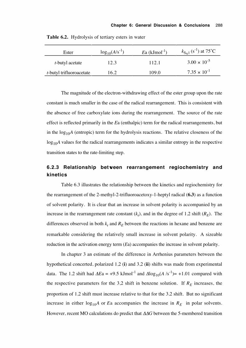
Chapter 6: General Discussion & Conclusions 288
Table 6.2. Hydrolysis of tertiary esters in water
Ester log10(A/s-1) Ea (kJmol-1) kSN1 (s-1) at 75˚C
t-butyl acetate 12.3 112.1 3.00 × 10⁻⁵
t-butyl trifluoroacetate 16.2 109.0 7.35 × 10⁻¹
The magnitude of the electron-withdrawing effect of the ester group upon the rate
constant is much smaller in the case of the radical rearrangement. This is consistent with
the absence of free carboxylate ions during the rearrangement. The source of the rate
effect is reflected primarily in the Ea (enthalpic) term for the radical rearrangements, but
in the log10A (entropic) term for the hydrolysis reactions. The relative closeness of the
log10A values for the radical rearrangements indicates a similar entropy in the respective
transition states to the rate-limiting step.
6.2.3 Relationship between rearrangement regiochemistry and
kinetics
Table 6.3 illustrates the relationship between the kinetics and regiochemistry for
the rearrangement of the 2-methyl-2-trifluoroacetoxy-1-heptyl radical (6.3) as a function
of solvent polarity. It is clear that an increase in solvent polarity is accompanied by an
increase in the rearrangement rate constant (kr), and in the degree of 1,2 shift (RE). The
differences observed in both kr and RE between the reactions in hexane and benzene are
remarkable considering the relatively small increase in solvent polarity. A sizeable
reduction in the activation energy term (Ea) accompanies the increase in solvent polarity.
In chapter 3 an estimate of the difference in Arrhenius parameters between the
hypothetical concerted, polarized 1,2 (i) and 3,2 (ii) shifts was made from experimental
data. The 1,2 shift had ∆Ea = +9.5 kJmol-1 and ∆log10(A /s-1)= +1.01 compared with
the respective parameters for the 3,2 shift in benzene solution. If RE increases, the
proportion of 1,2 shift must increase relative to that for the 3,2 shift. But no significant
increase in either log10A or Ea accompanies the increase in RE in polar solvents.
However, recent MO calculations do predict that ∆∆G between the 5-membered transition
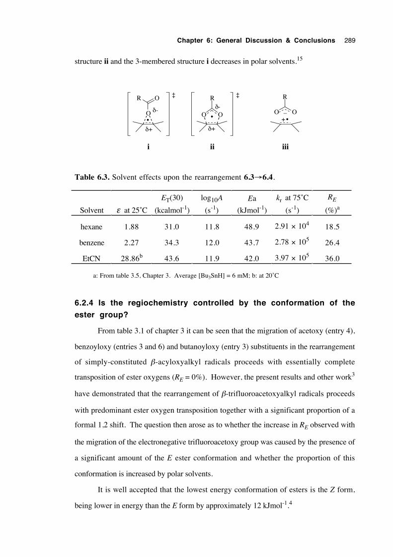
Chapter 6: General Discussion & Conclusions 289
structure ii and the 3-membered structure i decreases in polar solvents.15
O O
R
•O
R O
•O O
R
+_
iiiiii
•
δ+
δ-
δ+
δ-
‡ ‡
Table 6.3. Solvent effects upon the rearrangement 6.3→6.4.
Solvent ε at 25˚C
ET(30)
(kcalmol-1)
log10A
(s-1)
Ea
(kJmol-1)
kr at 75˚C
(s-1)
RE
(%)a
hexane 1.88 31.0 11.8 48.9 2.91 × 10⁴ 18.5
benzene 2.27 34.3 12.0 43.7 2.78 × 10⁵ 26.4
EtCN 28.86b 43.6 11.9 42.0 3.97 × 10⁵ 36.0
a: From table 3.5, Chapter 3. Average [Bu3SnH] = 6 mM; b: at 20˚C
6.2.4 Is the regiochemistry controlled by the conformation of the
ester group?
From table 3.1 of chapter 3 it can be seen that the migration of acetoxy (entry 4),
benzoyloxy (entries 3 and 6) and butanoyloxy (entry 3) substituents in the rearrangement
of simply-constituted β-acyloxyalkyl radicals proceeds with essentially complete
transposition of ester oxygens (RE = 0%). However, the present results and other work3
have demonstrated that the rearrangement of β-trifluoroacetoxyalkyl radicals proceeds
with predominant ester oxygen transposition together with a significant proportion of a
formal 1,2 shift. The question then arose as to whether the increase in RE observed with
the migration of the electronegative trifluoroacetoxy group was caused by the presence of
a significant amount of the E ester conformation and whether the proportion of this
conformation is increased by polar solvents.
It is well accepted that the lowest energy conformation of esters is the Z form,
being lower in energy than the E form by approximately 12 kJmol-1.4
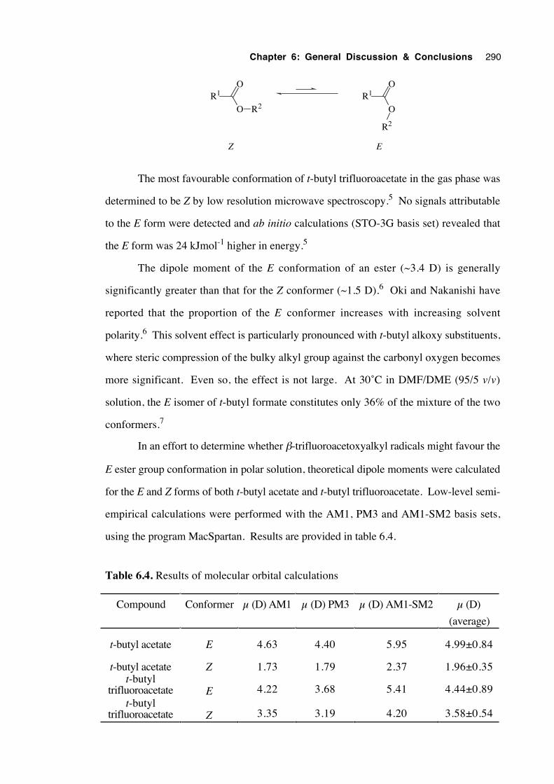
Chapter 6: General Discussion & Conclusions 290
R1O
O R2R1
O
O
R2
Z E
The most favourable conformation of t-butyl trifluoroacetate in the gas phase was
determined to be Z by low resolution microwave spectroscopy.5 No signals attributable
to the E form were detected and ab initio calculations (STO-3G basis set) revealed that
the E form was 24 kJmol-1 higher in energy.5
The dipole moment of the E conformation of an ester (~3.4 D) is generally
significantly greater than that for the Z conformer (~1.5 D).6 Oki and Nakanishi have
reported that the proportion of the E conformer increases with increasing solvent
polarity.6 This solvent effect is particularly pronounced with t-butyl alkoxy substituents,
where steric compression of the bulky alkyl group against the carbonyl oxygen becomes
more significant. Even so, the effect is not large. At 30˚C in DMF/DME (95/5 v/v)
solution, the E isomer of t-butyl formate constitutes only 36% of the mixture of the two
conformers.7
In an effort to determine whether β-trifluoroacetoxyalkyl radicals might favour the
E ester group conformation in polar solution, theoretical dipole moments were calculated
for the E and Z forms of both t-butyl acetate and t-butyl trifluoroacetate. Low-level semi-
empirical calculations were performed with the AM1, PM3 and AM1-SM2 basis sets,
using the program MacSpartan. Results are provided in table 6.4.
Table 6.4. Results of molecular orbital calculations
Compound Conformer µ (D) AM1 µ (D) PM3 µ (D) AM1-SM2 µ (D)
(average)
t-butyl acetate E 4.63 4.40 5.95 4.99±0.84
t-butyl acetate Z 1.73 1.79 2.37 1.96±0.35t-butyl
trifluoroacetate E 4.22 3.68 5.41 4.44±0.89t-butyl
trifluoroacetate Z 3.35 3.19 4.20 3.58±0.54
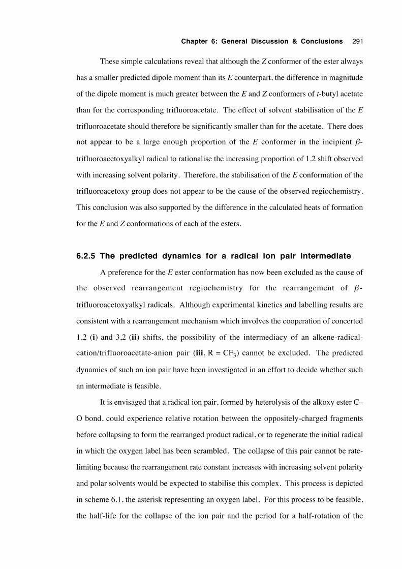
Chapter 6: General Discussion & Conclusions 291
These simple calculations reveal that although the Z conformer of the ester always
has a smaller predicted dipole moment than its E counterpart, the difference in magnitude
of the dipole moment is much greater between the E and Z conformers of t-butyl acetate
than for the corresponding trifluoroacetate. The effect of solvent stabilisation of the E
trifluoroacetate should therefore be significantly smaller than for the acetate. There does
not appear to be a large enough proportion of the E conformer in the incipient β-
trifluoroacetoxyalkyl radical to rationalise the increasing proportion of 1,2 shift observed
with increasing solvent polarity. Therefore, the stabilisation of the E conformation of the
trifluoroacetoxy group does not appear to be the cause of the observed regiochemistry.
This conclusion was also supported by the difference in the calculated heats of formation
for the E and Z conformations of each of the esters.
6.2.5 The predicted dynamics for a radical ion pair intermediate
A preference for the E ester conformation has now been excluded as the cause of
the observed rearrangement regiochemistry for the rearrangement of β -
trifluoroacetoxyalkyl radicals. Although experimental kinetics and labelling results are
consistent with a rearrangement mechanism which involves the cooperation of concerted
1,2 (i) and 3,2 (ii) shifts, the possibility of the intermediacy of an alkene-radical-
cation/trifluoroacetate-anion pair (iii, R = CF3) cannot be excluded. The predicted
dynamics of such an ion pair have been investigated in an effort to decide whether such
an intermediate is feasible.
It is envisaged that a radical ion pair, formed by heterolysis of the alkoxy ester C–
O bond, could experience relative rotation between the oppositely-charged fragments
before collapsing to form the rearranged product radical, or to regenerate the initial radical
in which the oxygen label has been scrambled. The collapse of this pair cannot be rate-
limiting because the rearrangement rate constant increases with increasing solvent polarity
and polar solvents would be expected to stabilise this complex. This process is depicted
in scheme 6.1, the asterisk representing an oxygen label. For this process to be feasible,
the half-life for the collapse of the ion pair and the period for a half-rotation of the
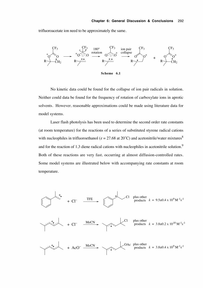
Chapter 6: General Discussion & Conclusions 292
trifluoroacetate ion need to be approximately the same.
CH2
OOR
CF3
R
O O
CF3
+_* *
R
O O
CF3
_ *OO
R
CF3
*
CH2
OOR
CF3
*+
180°rotation
ion paircollapse
• • + • • •
Scheme 6.1
No kinetic data could be found for the collapse of ion pair radicals in solution.
Neither could data be found for the frequency of rotation of carboxylate ions in aprotic
solvents. However, reasonable approximations could be made using literature data for
model systems.
Laser flash photolysis has been used to determine the second order rate constants
(at room temperature) for the reactions of a series of substituted styrene radical cations
with nucleophiles in trifluoroethanol (ε = 27.68 at 20˚C) and acetonitrile/water mixtures8
and for the reaction of 1,3 diene radical cations with nucleophiles in acetonitrile solution.9
Both of these reactions are very fast, occurring at almost diffusion-controlled rates.
Some model systems are illustrated below with accompanying rate constants at room
temperature.
•+
•+
•+
•
•
•
Cl
Cl
OAc
+ Cl–
+ Cl–
+ AcO–
TFE
MeCN
MeCN
k = 9.5±0.4 x 10⁹ M-1s-1
k = 3.8±0.2 x 10¹⁰ M-1s-1
k = 3.8±0.4 x 10⁹ M-1s-1
plus other products
plus other products
plus other products
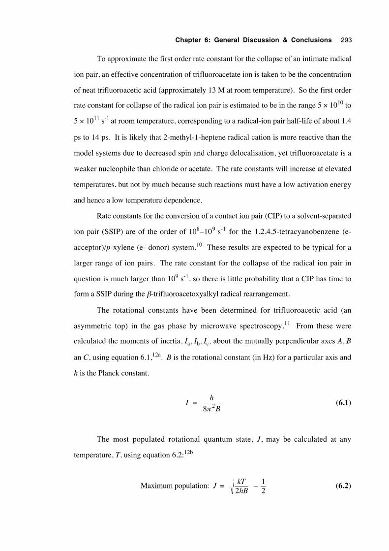
Chapter 6: General Discussion & Conclusions 293
To approximate the first order rate constant for the collapse of an intimate radical
ion pair, an effective concentration of trifluoroacetate ion is taken to be the concentration
of neat trifluoroacetic acid (approximately 13 M at room temperature). So the first order
rate constant for collapse of the radical ion pair is estimated to be in the range 5 × 10¹⁰ to
5 × 10¹¹ s-1 at room temperature, corresponding to a radical-ion pair half-life of about 1.4
ps to 14 ps. It is likely that 2-methyl-1-heptene radical cation is more reactive than the
model systems due to decreased spin and charge delocalisation, yet trifluoroacetate is a
weaker nucleophile than chloride or acetate. The rate constants will increase at elevated
temperatures, but not by much because such reactions must have a low activation energy
and hence a low temperature dependence.
Rate constants for the conversion of a contact ion pair (CIP) to a solvent-separated
ion pair (SSIP) are of the order of 108–109 s-1 for the 1,2,4,5-tetracyanobenzene (e-
acceptor)/p-xylene (e- donor) system.10 These results are expected to be typical for a
larger range of ion pairs. The rate constant for the collapse of the radical ion pair in
question is much larger than 109 s-1, so there is little probability that a CIP has time to
form a SSIP during the β-trifluoroacetoxyalkyl radical rearrangement.
The rotational constants have been determined for trifluoroacetic acid (an
asymmetric top) in the gas phase by microwave spectroscopy.11 From these were
calculated the moments of inertia, Ia, Ib, Ic, about the mutually perpendicular axes A, B
an C, using equation 6.1,12a. B is the rotational constant (in Hz) for a particular axis and
h is the Planck constant.
I = h
B8π 2 (6.1)
The most populated rotational quantum state, J, may be calculated at any
temperature, T, using equation 6.2:12b
Maximum population: J = kThB2
– 12
(6.2)
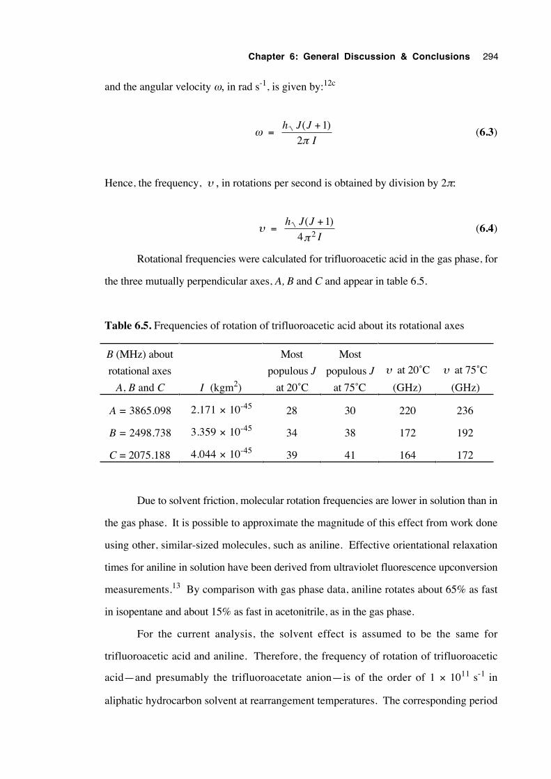
Chapter 6: General Discussion & Conclusions 294
and the angular velocity ω, in rad s-1, is given by:12c
ω = h J J
I
( )+ 12π
(6.3)
Hence, the frequency, υ , in rotations per second is obtained by division by 2π:
υ = h J J
I
( )+ 14 2π
(6.4)
Rotational frequencies were calculated for trifluoroacetic acid in the gas phase, for
the three mutually perpendicular axes, A, B and C and appear in table 6.5.
Table 6.5. Frequencies of rotation of trifluoroacetic acid about its rotational axes
B (MHz) about
rotational axes
A, B and C I (kgm2)
Most
populous J
at 20˚C
Most
populous J
at 75˚C
υ at 20˚C
(GHz)
υ at 75˚C
(GHz)
A = 3865.098 2.171 × 10-45 28 30 220 236
B = 2498.738 3.359 × 10-45 34 38 172 192
C = 2075.188 4.044 × 10-45 39 41 164 172
Due to solvent friction, molecular rotation frequencies are lower in solution than in
the gas phase. It is possible to approximate the magnitude of this effect from work done
using other, similar-sized molecules, such as aniline. Effective orientational relaxation
times for aniline in solution have been derived from ultraviolet fluorescence upconversion
measurements.13 By comparison with gas phase data, aniline rotates about 65% as fast
in isopentane and about 15% as fast in acetonitrile, as in the gas phase.
For the current analysis, the solvent effect is assumed to be the same for
trifluoroacetic acid and aniline. Therefore, the frequency of rotation of trifluoroacetic
acid—and presumably the trifluoroacetate anion—is of the order of 1 × 10¹¹ s-1 in
aliphatic hydrocarbon solvent at rearrangement temperatures. The corresponding period
Chapter 6: General Discussion & Conclusions 295
of rotation is about 10 ps, or 5 ps per half rotation.
From these approximations, the rotation period of the trifluoroacetate ion and the
half-life for the collapse of the radical ion pair are predicted to be of approximately the
same duration. Therefore, it is possible that these processes may effectively compete
with each other, enabling the rearrangement regiochemistry to be affected by subtle
changes in the dynamics of the tight ion pair, in turn governed by temperature and solvent
polarity. Furthermore, low concentrations of the reducing agent allow radicals more time
on average for intramolecular reactions and therefore a greater proportion of
rearrangement reversibility. From the similarity of the rotational frequencies about the A,
B and C axes of trifluoroacetic acid, it is assumed that the trifluoroacetate group can also
rotate about these three axes with approximately equal ease in solution in the absence of
directional electrodynamic influences. Some modes of rotation may favour a 1,2 shift of
oxygen by positioning the initial ether oxygen of the trifluoroacetate group closest to C1
of the alkene radical cation when the radical ion pair collapses.
6.2.6 The mechanism of the rearrangement of β -
trifluoroacetoxyalkyl radicals
The bulk of the evidence tends to support the theory that the mechanism of the β-
trifluoroacetoxyalkyl radical rearrangement involves a cooperation of reversible, polarized
and concerted 1,2 (i) and 3,2 (ii) shifts (R = CF3). Experiments with radicals bearing an
oxygen-labelled trifluoroacetoxy group have shown that the migration occurs with 55.5-
82.3% transposition of label, demonstrating that a formal 3,2 shift predominates under all
the conditions investigated in this study. These labelling experiments also revealed some
scrambling of the oxygen label in the unrearranged product, necessitating that any
concerted rearrangement processes be reversible.
Solvent exerted only a modest effect upon the rearrangement rate constant,
compared with the solvent effects observed in comparable ionic chemistry. Likewise,
migrating group electronic effects were small in comparison with those observed in
solvolysis reactions.
Attempts to trap the 2-methyl-1-heptene radical cation—a fragment of a
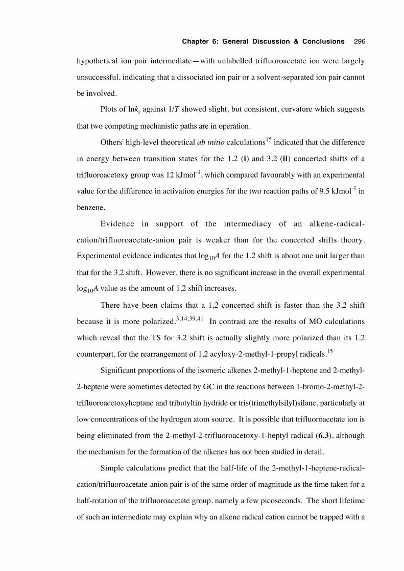
Chapter 6: General Discussion & Conclusions 296
hypothetical ion pair intermediate—with unlabelled trifluoroacetate ion were largely
unsuccessful, indicating that a dissociated ion pair or a solvent-separated ion pair cannot
be involved.
Plots of lnkr against 1/T showed slight, but consistent, curvature which suggests
that two competing mechanistic paths are in operation.
Others' high-level theoretical ab initio calculations15 indicated that the difference
in energy between transition states for the 1,2 (i) and 3,2 (ii) concerted shifts of a
trifluoroacetoxy group was 12 kJmol-1, which compared favourably with an experimental
value for the difference in activation energies for the two reaction paths of 9.5 kJmol-1 in
benzene.
Evidence in support of the intermediacy of an alkene-radical-
cation/trifluoroacetate-anion pair is weaker than for the concerted shifts theory.
Experimental evidence indicates that log10A for the 1,2 shift is about one unit larger than
that for the 3,2 shift. However, there is no significant increase in the overall experimental
log10A value as the amount of 1,2 shift increases.
There have been claims that a 1,2 concerted shift is faster than the 3,2 shift
because it is more polarized.3,14,39,41 In contrast are the results of MO calculations
which reveal that the TS for 3,2 shift is actually slightly more polarized than its 1,2
counterpart, for the rearrangement of 1,2 acyloxy-2-methyl-1-propyl radicals.15
Significant proportions of the isomeric alkenes 2-methyl-1-heptene and 2-methyl-
2-heptene were sometimes detected by GC in the reactions between 1-bromo-2-methyl-2-
trifluoroacetoxyheptane and tributyltin hydride or tris(trimethylsilyl)silane, particularly at
low concentrations of the hydrogen atom source. It is possible that trifluoroacetate ion is
being eliminated from the 2-methyl-2-trifluoroacetoxy-1-heptyl radical (6.3), although
the mechanism for the formation of the alkenes has not been studied in detail.
Simple calculations predict that the half-life of the 2-methyl-1-heptene-radical-
cation/trifluoroacetate-anion pair is of the same order of magnitude as the time taken for a
half-rotation of the trifluoroacetate group, namely a few picoseconds. The short lifetime
of such an intermediate may explain why an alkene radical cation cannot be trapped with a
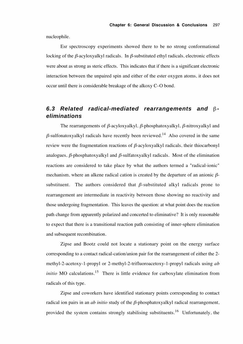
Chapter 6: General Discussion & Conclusions 297
nucleophile.
Esr spectroscopy experiments showed there to be no strong conformational
locking of the β-acyloxyalkyl radicals. In β-substituted ethyl radicals, electronic effects
were about as strong as steric effects. This indicates that if there is a significant electronic
interaction between the unpaired spin and either of the ester oxygen atoms, it does not
occur until there is considerable breakage of the alkoxy C–O bond.
6.3 Related radical-mediated rearrangements and β -eliminations
The rearrangements of β-acyloxyalkyl, β-phosphatoxyalkyl, β-nitroxyalkyl and
β-sulfonatoxyalkyl radicals have recently been reviewed.14 Also covered in the same
review were the fragmentation reactions of β-acyloxyalkyl radicals, their thiocarbonyl
analogues, β-phosphatoxyalkyl and β-sulfatoxyalkyl radicals. Most of the elimination
reactions are considered to take place by what the authors termed a "radical-ionic"
mechanism, where an alkene radical cation is created by the departure of an anionic β-
substituent. The authors considered that β-substituted alkyl radicals prone to
rearrangement are intermediate in reactivity between those showing no reactivity and
those undergoing fragmentation. This leaves the question: at what point does the reaction
path change from apparently polarized and concerted to eliminative? It is only reasonable
to expect that there is a transitional reaction path consisting of inner-sphere elimination
and subsequent recombination.
Zipse and Bootz could not locate a stationary point on the energy surface
corresponding to a contact radical-cation/anion pair for the rearrangement of either the 2-
methyl-2-acetoxy-1-propyl or 2-methyl-2-trifluoroacetoxy-1-propyl radicals using ab
initio MO calculations.15 There is little evidence for carboxylate elimination from
radicals of this type.
Zipse and coworkers have identified stationary points corresponding to contact
radical ion pairs in an ab initio study of the β-phosphatoxyalkyl radical rearrangement,
provided the system contains strongly stabilising substituents.16 Unfortunately, the
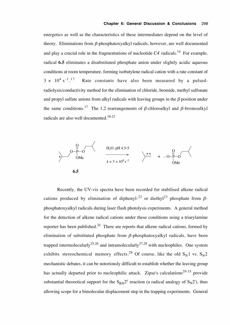
Chapter 6: General Discussion & Conclusions 298
energetics as well as the characteristics of these intermediates depend on the level of
theory. Eliminations from β-phosphatoxyalkyl radicals, however, are well documented
and play a crucial role in the fragmentations of nucleotide C4' radicals.14 For example,
radical 6.5 eliminates a disubstituted phosphate anion under slightly acidic aqueous
conditions at room temperature, forming isobutylene radical cation with a rate constant of
3 × 10⁴ s - 1 .1 7 Rate constants have also been measured by a pulsed-
radiolysis/conductivity method for the elimination of chloride, bromide, methyl sulfonate
and propyl sulfate anions from alkyl radicals with leaving groups in the β position under
the same conditions.17 The 1,2 rearrangements of β-chloroalkyl and β-bromoalkyl
radicals are also well documented.18-21
O P
OMe
O
• – O P
OMe
O
O•+
6.5
+
H2O, pH 4.5-5
k = 3 × 10⁴ s-1
O
Recently, the UV-vis spectra have been recorded for stabilised alkene radical
cations produced by elimination of diphenyl-22 or diethyl23 phosphate from β-
phosphatoxyalkyl radicals during laser flash photolysis experiments. A general method
for the detection of alkene radical cations under these conditions using a triarylamine
reporter has been published.24 There are reports that alkene radical cations, formed by
elimination of substituted phosphate from β-phosphatoxyalkyl radicals, have been
trapped intermolecularly25,26 and intramolecularly27,28 with nucleophiles. One system
exhibits stereochemical memory effects.28 Of course, like the old SN1 vs. SN2
mechanistic debates, it can be notoriously difficult to establish whether the leaving group
has actually departed prior to nucleophilic attack. Zipse's calculations29-33 provide
substantial theoretical support for the SRN2c reaction (a radical analogy of SN2'), thus
allowing scope for a bimolecular displacement step in the trapping experiments. General
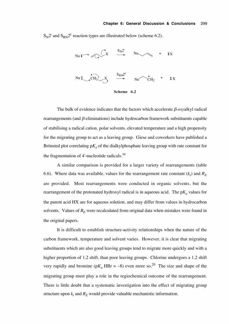
Chapter 6: General Discussion & Conclusions 299
SN2' and SRN2c reaction types are illustrated below (scheme 6.2).
X
CH2 X
X
CH2 X
Nu ••Nu ••+
•Nu •• Nu ••+
SN2'
SRN2c•
Scheme 6.2
The bulk of evidence indicates that the factors which accelerate β-oxyalkyl radical
rearrangements (and β-eliminations) include hydrocarbon framework substituents capable
of stabilising a radical cation, polar solvents, elevated temperature and a high propensity
for the migrating group to act as a leaving group. Giese and coworkers have published a
Brönsted plot correlating pKa of the dialkylphosphate leaving group with rate constant for
the fragmentation of 4'-nucleotide radicals.34
A similar comparison is provided for a larger variety of rearrangements (table
6.6). Where data was available, values for the rearrangement rate constant (kr) and RE
are provided. Most rearrangements were conducted in organic solvents, but the
rearrangement of the protonated hydroxyl radical is in aqueous acid. The pKa values for
the parent acid HX are for aqueous solution, and may differ from values in hydrocarbon
solvents. Values of RE were recalculated from original data when mistakes were found in
the original papers.
It is difficult to establish structure-activity relationships when the nature of the
carbon framework, temperature and solvent varies. However, it is clear that migrating
substituents which are also good leaving groups tend to migrate more quickly and with a
higher proportion of 1,2 shift, than poor leaving groups. Chlorine undergoes a 1,2 shift
very rapidly and bromine (pKa HBr = –8) even more so.20 The size and shape of the
migrating group must play a role in the regiochemical outcome of the rearrangement.
There is little doubt that a systematic investigation into the effect of migrating group
structure upon kr and RE would provide valuable mechanistic information.
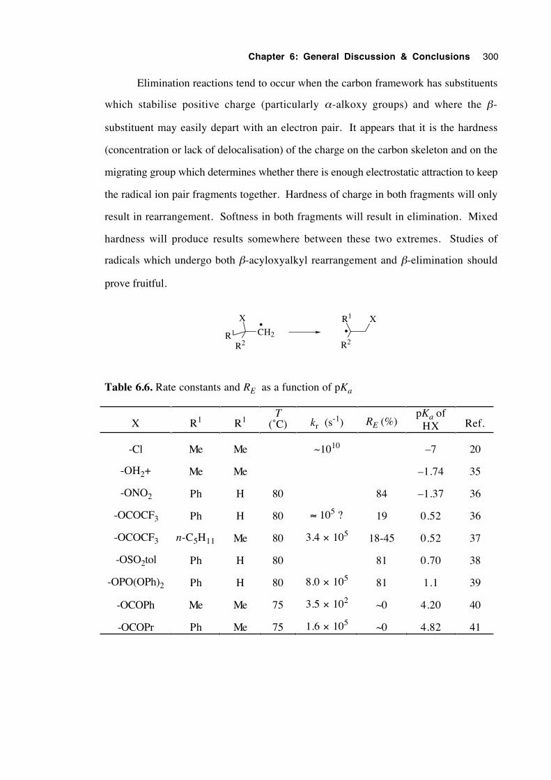
Chapter 6: General Discussion & Conclusions 300
Elimination reactions tend to occur when the carbon framework has substituents
which stabilise positive charge (particularly α-alkoxy groups) and where the β-
substituent may easily depart with an electron pair. It appears that it is the hardness
(concentration or lack of delocalisation) of the charge on the carbon skeleton and on the
migrating group which determines whether there is enough electrostatic attraction to keep
the radical ion pair fragments together. Hardness of charge in both fragments will only
result in rearrangement. Softness in both fragments will result in elimination. Mixed
hardness will produce results somewhere between these two extremes. Studies of
radicals which undergo both β-acyloxyalkyl rearrangement and β-elimination should
prove fruitful.
CH2
X
R2R1
•
R2
R1 X•
Table 6.6. Rate constants and RE as a function of pKa
X R1 R1T
(˚C) kr (s-1) RE (%)
pKa ofHX Ref.
-Cl Me Me ~1010 –7 20
-OH2+ Me Me –1.74 35
-ONO2 Ph H 80 84 –1.37 36
-OCOCF3 Ph H 80 ≈ 105 ? 19 0.52 36
-OCOCF3 n-C5H11 Me 80 3.4 × 10⁵ 18-45 0.52 37
-OSO2tol Ph H 80 81 0.70 38
-OPO(OPh)2 Ph H 80 8.0 × 10⁵ 81 1.1 39
-OCOPh Me Me 75 3.5 × 10² ~0 4.20 40
-OCOPr Ph Me 75 1.6 × 10⁵ ~0 4.82 41
Chapter 6: General Discussion & Conclusions 301
6.4 The mechanism of the rearrangement of N -alkoxy-2(1H)-pyridinethiones
The mechanism of the catalysed rearrangement of N -alkoxy-2(1H ) -
pyridinethiones is not fully understood. Kinetic evidence indicates that the rate limiting
step may be the intermolecular transfer of the migrating group. Results from a mass
spectrometric analysis of an isolated intermediate also support the participation of
intermolecular processes. An intermolecular process has previously been shown to be in
operation in the analogous rearrangement of 2-alkoxypyridine N-oxides. A crossover
experiment is necessary to establish whether the rearrangement of N-alkoxy-2(1H)-
pyridinethiones occurs with intermolecular alkyl transfer.
Stereochemical experiments have demonstrated that the rearrangement cannot
occur via one, concerted mechanism. It remains possible that this isomerization does
belong to a class of rearrangements which proceed via a transition structure in which 5
electrons are delocalised over 5 atoms, although other processes must also be in operation
alongside this 1,4 concerted shift.
6.5 Final remarks
It is difficult to devise experiments which would enable one to distinguish
between the cooperation of two concerted, pericyclic processes and the intermediacy of a
radical ion pair. This endeavour is sure to provide continuing challenges to workers in
the field. In the case of the rearrangement of the 2-methyl-2-trifluoroacetoxy-1-heptyl
radical (6.3) to the 2-methyl-1-trifluoroacetoxy-2-heptyl radical (6.4), it has not been
possible to unequivocally establish the mechanism.
It became evident that solvent plays an important role in the rate of rearrangement
and in the regiochemistry. Therefore, future theoretical calculations must take into
account solvent effects since the mechanism in the gaseous state may differ markedly
from that observed in not only a polar solvent, but non-polar solvents also.
One might expect the rate of processes which involve contact ion pairs to show
considerably smaller solvent effects than reactions with more completely solvated
Chapter 6: General Discussion & Conclusions 302
intermediates. For CIPs, the overall charge within the solvent sphere sums to zero. The
solvation enthalpy will be less for CIPs since each of the oppositely-charged fragments
are less stabilised due to an incomplete solvent sphere. The solvation entropy will also be
lower for CIPs because the solvent molecules are less ordered than in a completely-
solvated solvent-separated (SSIP) or solvent-shared ion pair.
For intermediates with very short lifetimes, it is possible that the distinction
between polarized, concerted transition states and tight ion pairs may become an artificial
one. The parameters which determine this segregation are bond length, bond strength
and charge distribution. The location of the valence electrons in two adjacent atoms
formally governs whether they are covalently bonded. Solvents exert a significant
influence on the positions of valence electrons in solutes at any point in time. For β-
acyloxyalkyl radical rearrangements in which the migrating group is a stronger conjugate
base (weaker leaving group) and in which the alkyl framework does not readily delocalise
a developing positive charge, any such radical ion intermediate must have a half life of
considerably less than 10 picoseconds (cf. section 6.2.5). For species with little
stabilisation, it is envisaged that the actual time interval may not be much larger than the
period of a molecular vibration (10-50 fs).
The rate constants for valence bond tautomerism have been measured for the
trimethylcyclopropenyl radical (> 108 s-1)42 and for a number of conjugated and highly
hindered aryloxy radicals (≈ 2 × 10⁷ s-1 at RT).43-45 One is led to wonder whether it is
possible that the resonance structures of a carboxylate ion become time-resolved on the
timescale of the lifetime of a radical ion intermediate in such a way that regiochemistry is
directly affected.
From a quantum-mechanical perspective, a tight radical ion pair should exist in a
number of discrete, quantised geometries. Are there two of these allowed states which
are isomorphic with the polarized transition structures? At what point does a strong
electrostatic interaction become a partial bond?
Chapter 6: General Discussion & Conclusions 303
6.6 References
1. Adam, K. R., Lauder, I. and Stimson, V. R. Aust. J. Chem. 1962, 15, 467.
2. Barnes, D. J., Cole, M., Lobo, S., Winter, J. G. and Scott, J. M. W. Can. J. Chem.
1972, 50, 2175.
3. Crich, D. and Filzen, G. F. J. Org. Chem. 1995, 60, 4834.
4. Deslongchamps, P. Stereoelectronic effects in organic chemistry 1983, Pergamon,
Oxford, p 54.
5. True, N. S. and Bohn, R. K. J. Phys. Chem. 1978, 82 (4), 478.
6. Oki, M. and Nakanishi, H. Bull. Chem. Soc. Jpn 1970, 43, 2558.
7. Drakenberg, T. and Forsen, S. J. Phys. Chem. 1972, 76 (24), 3582.
8. Johnston, L. J. and Schepp, N. P. J. Am. Chem. Soc. 1993, 115, 6564.
9. Lew, C. S. Q., Brisson, J. R. and Johnston, L. J. J. Org. Chem. 1997, 62, 4047.
10. Arnold, B. R.; Farid, S.; Goodman, J. L. and Gould, I. R. J. Am. Chem. Soc.
1996, 118, 5482.
11. Stolwijk, V. M. and van Eijck, B. P. J. Mol. Spectrosc. 1985, 113, 196.
12. Banwell, C. N. Fundamentals of molecular spectroscopy, 2nd Ed. 1972, McGraw-
Hill, London. a: p 33, b: p 41, c: p 38.
13. Pereira, M. A., Share, P. E., Sarisky, M. J. and Hochstrasser, R. M. J. Chem.
Phys. 1991, 94 (4), 2513.
14. Beckwith, A. L. J.; Crich, D.; Duggan, P. J. and Yao, Q. Chem. Rev. 1997, 97,
3273.
15. Zipse, H. and Bootz, M. J. Chem. Soc., Perkin Trans. 2 2001, 1566.
16. Wang, Y. and H. Zipse, H. unpublished results (as of April 2003)
17. Koltzenburg, G. Behrens, G. and Schulte-Frohlinde, D. J. Am. Chem. Soc. 1982,
104, 7311.
18. Wilt, J. W. in Free Radicals; Kochi, J. K., Ed.; John Wiley and Sons: New York,
1973, vol. 1, p. 333.
19. Skell, P. S. and Shea, K. J. in Free Radicals; Kochi, J. K., Ed.; John Wiley and
Sons: New York, 1973, vol. 2, p. 809.
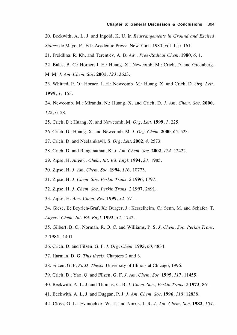
Chapter 6: General Discussion & Conclusions 304
20. Beckwith, A. L. J. and Ingold, K. U. in Rearrangements in Ground and Excited
States; de Mayo, P., Ed.; Academic Press: New York, 1980, vol. 1, p. 161.
21. Freidlina, R. Kh. and Terent'ev, A. B. Adv. Free-Radical Chem. 1980, 6, 1.
22. Bales, B. C.; Horner, J. H.; Huang, X.; Newcomb, M.; Crich, D. and Greenberg,
M. M. J. Am. Chem. Soc. 2001, 123, 3623.
23. Whitted, P. O.; Horner, J. H.; Newcomb, M.; Huang, X. and Crich, D. Org. Lett.
1999, 1, 153.
24. Newcomb, M.; Miranda, N.; Huang, X. and Crich, D. J. Am. Chem. Soc. 2000,
122, 6128.
25. Crich, D.; Huang, X. and Newcomb, M. Org. Lett. 1999, 1, 225.
26. Crich, D.; Huang, X. and Newcomb, M. J. Org. Chem. 2000, 65, 523.
27. Crich, D. and Neelamkavil, S. Org. Lett. 2002, 4, 2573.
28. Crich, D. and Ranganathan, K. J. Am. Chem. Soc. 2002, 124, 12422.
29. Zipse, H. Angew. Chem. Int. Ed. Engl. 1994, 33, 1985.
30. Zipse, H. J. Am. Chem. Soc. 1994, 116, 10773.
31. Zipse, H. J. Chem. Soc. Perkin Trans. 2 1996, 1797.
32. Zipse, H. J. Chem. Soc. Perkin Trans. 2 1997, 2691.
33. Zipse, H. Acc. Chem. Res. 1999, 32, 571.
34. Giese, B; Beyrich-Graf, X.; Burger, J.; Kesselheim, C.; Senn, M. and Schafer, T.
Angew. Chem. Int. Ed. Engl. 1993, 32, 1742.
35. Gilbert, B. C.; Norman, R. O. C. and Williams, P. S. J. Chem. Soc. Perkin Trans.
2 1981, 1401.
36. Crich, D. and Filzen, G. F. J. Org. Chem. 1995, 60, 4834.
37. Harman, D. G. This thesis, Chapters 2 and 3.
38. Filzen, G. F. Ph.D. Thesis, University of Illinois at Chicago, 1996.
39. Crich, D.; Yao, Q. and Filzen, G. F. J. Am. Chem. Soc. 1995, 117, 11455.
40. Beckwith, A. L. J. and Thomas, C. B. J. Chem. Soc., Perkin Trans. 2 1973, 861.
41. Beckwith, A. L. J. and Duggan, P. J. J. Am. Chem. Soc. 1996, 118, 12838.
42. Closs, G. L.; Evanochko, W. T. and Norris, J. R. J. Am. Chem. Soc. 1982, 104,

Chapter 6: General Discussion & Conclusions 305
350.
43. Kirste, B.; Harrer, W.; Kurreck, H.; Schubert, K.; Bauer, H. and Gierke, W. J. Am.
Chem. Soc. 1981, 103, 6280.
44.Kirste, B. J. Magn. Reson. 1987, 73, 213.
45. Gersdorff, J.; Kirste, B.; Niethammer, D.; Harrer, W. and Kurreck, H. Magn. Res.
Chem. 1988, 26, 416.
Chapter 7
General Experimental
7.1 Melting points 307
7.2 Elemental analyses 307
7.3 Infrared spectroscopy 307
7.4 Optical rotations 307
7.5 Molecular ultraviolet and visible spectra 307
7.6 Bulb to bulb distillations 307
7.7 Liquid chromatography 307
7.8 Gas chromatography 308
7.9 Mass spectrometry 309
7.10 Electron spin resonance spectroscopy 310
7.11 Nuclear magnetic resonance spectroscopy 310
7.12 Cyclic voltammetry 312
7.13 Purification of solvents for radical reactions 312
7.14 Purification of solvents for other purposes 314
7.15 Reagents for synthesis 314
7.16 Evaporation of solvents 314
7.17 Drying of extract solutions 315
7.18 Nomenclature 315
7.19 References 316

Chapter 7: General Experimental 307
7.1 Melting points are uncorrected and were determined using a Reichert microscope
Kofler hot-stage apparatus.
7.2 Elemental analyses were performed by the ANU Microanalytical Services Unit,
Canberra.
7.3 Infrared spectroscopy
Routine infrared spectra were recorded with a Perkin-Elmer 686 infrared
spectrophotometer. Liquid samples were generally prepared as a thin film, smeared
between NaCl plates. Solid samples were run as either a Nujol mull smeared between
NaCl plates, or as a solution contained in a NaCl cell of cavity thickness 0.5 mm run with
a reference cell to balance solvent absorptions. Polystyrene film was used as a
wavenumber reference.
FTIR spectra were generally recorded by the ANU Infrared Spectrometry Service
on a Perkin-Elmer 1800 fourier transform infrared spectrophotometer, coupled to a
Perkin-Elmer 7500 computer. Samples were prepared as KBr pellets. Some FTIR
spectra were recorded on a Perkin-Elmer 1600 series instrument, using 4 scans for both
background and sample spectra.
Infrared data are listed as the wavenumber (υmax) in units of cm-1, followed by a
description of the peak's size and/or shape. Abbreviations used to indicate peak
characteristics are: vs (very strong); s (strong); m (medium); w (weak); asym
(asymmetric); and br (broad).
7.4 Optical rotations were measured on a Perkin-Elmer 241 polarimeter, at a wavelength
of 589 nm and at ambient temperature, in spectroscopic grade solvents.
7.5 Molecular ultraviolet and visible spectra were recorded using a Hewlett-Packard
8450A spectrophotometer. Spectroscopic grade solvents were used in preparing
solutions.
7.6 Bulb-to-bulb distillations were performed with a Buchi GKR-50 Kugelrohr
apparatus. The boiling temperature quoted is that which just causes the liquid to distil.
7.7 Liquid Chromatography
7.7.1 Flash chromatography was performed according to an established method,1 using
Merck Kieselgel 60 (230-400 mesh ASTM).
7.7.2 Vacuum liquid chromatography (VLC) was performed according to an established
Chapter 7: General Experimental 308
method,2 with the same type of silica.
7.7.3 Analytical thin layer chromatography (TLC) was performed using either Whatman
type MK6F, precoated silica microscope slide plates (75 mm × 25 mm × 10-12 µm), or
Merck Kieselgel 60 plates (100 × 200 × 0.25 mm), both containing a 254 nm
fluorescence indicator. Once developed, chromatograms were first inspected under
ultraviolet light, then sprayed with either 3% vanillin in conc. H2SO4 or alkaline KMnO4
solutions, and heated at ca. 200˚C until spots became visible.
7.7.4 Preparative scale thin layer chromatography was undertaken using Merck 20 × 20
cm glass plates coated with Kieselgel 60 (with F254 indicator), generally of thickness 2
mm.
7.7.5 Radial chromatography was performed on a Chromatotron, model 7924, using
plates coated 2 mm thick with Merck Kieselgel 60 PF254 (with gypsum). A hand-held
short-wave ultraviolet lamp was used to detect UV-absorbing compounds on the plate
during chromatography.
7.8 Gas Chromatography
7.8.1 Analytical
Three capillary GC instruments were used for analytical gas chromatography.
High-purity helium was used as the carrier and make-up gas for each. Each instrument
employed flame ionisation detection. Capillary columns with prefix "BP" were
purchased from SGE. The details for each instrument follow.
i) A Varian 6000 Vista Series instrument, fitted with an SGE Unijector control module
(replacing the original injector), was used for the majority of the work. A
dimethylpolysiloxane (BP1) capillary of dimensions 25 m × 0.32 mm I.D. × 0.5 µm film
thickness was employed, with a carrier gas average linear velocity of 30 cm s-1. A
Hewlett-Packard 3390A integrator controlled signal plotting and integration.
ii) A Varian 3400 instrument with an on-board integrator was fitted with both a 25 m ×
0.32 mm I.D.× 0.5 µm film thickness Carbowax (BP20), and a 25 m × 0.32 mm I.D. ×
0.5 µm film thickness 95% dimethyl, 5% diphenyl polysiloxane (BP5) capillary. Both
capillaries employed a carrier gas average linear velocity of 40 cm s-1.
ii) A Varian 3400 instrument, coupled to an HP 3390A integrator, contained a 14%
cyanopropylphenyl, 86% dimethyl polysiloxane (BP10) capillary, of dimensions 25 m ×
Chapter 7: General Experimental 309
0.5 µm × 0.32 mm I.D. The average linear carrier gas velocity was 40 cm s-1.
7.8.2 Chiral analytical GC3
The ratios of enantiomers in selected optically active mixtures were determined by
using a Chirasil-Val WCOT capillary column (Applied Science), of length 25 m and I.D.
of 0.30 mm. The film thickness was not stated. This column was installed in either
instrument i) or ii).
7.8.3 Preparative scale gas chromatography
This work was performed using a Varian 6000 Vista Series GC equipped with a
thermal conductivity detector (TCD), containing a 2 m × 4 mm I.D. glass column, packed
with 10% SE30 (dimethyl polysiloxane) on 80/100 Gas-Chrom Q support (Applied
Science). The carrier gas flow was set to 20 mL min-1 (average linear velocity = 2.65 cm
s-1); the injector temperature to 200˚C; detector oven temperature to 200˚C; and detector
filament an indicated 230˚C, giving a detector current of 150 mA. The column oven
temperature was usually 60˚C. A Hewlett-Packard 3396A integrator controlled signal
plotting and integration.
7.9 Mass Spectrometry
Mass spectral data are listed as values of m/z, followed by the intensity of the ion
as a percentage of the base peak intensity (in parentheses). The symbol M+• accompanies
the m/z value assigned to the molecular ion. The symbols MH+ and MNH4+ correspond
respectively to the protonated molecule and the species corresponding to the molecular
addition of the ammonium ion, commonly seen using CIMS with ammonia as the reagent
gas.
7.9.1 EIMS
Direct-probe, low resolution, electron impact mass spectra (EIMS) were recorded
at 70 eV on either a VG 7070F or a VG ZAB-2SEQ mass spectrometer. Where
necessary, the compounds were frozen in the sample cup using liquid nitrogen, before
insertion into the vacuum lock.
7.9.2 HRMS
High resolution mass spectrometry (HRMS) provided accurate mass
measurements using EIMS at a resolution (10% valley) of ca. 6,000. Peaks were
nominally matched using perfluorokerosene (PFK) to provide reference ions.
Chapter 7: General Experimental 310
7.9.3 CIMS
Chemical ionisation mass spectra (CIMS) were measured on the VG 7070F
instrument using NH3 as the reagent gas. The ion source temperature was generally
maintained at ca. 200˚C, although lower temperatures were used for samples which were
temperature-sensitive.
7.9.4 GCMS
Gas-chromatography/mass-spectrometry was performed using a Hewlett-Packard
system, comprised of an HP 5890 gas chromatograph (splitless injection) connected to an
HP 5970 mass-selective detector (70 eV electron beam energy), fitted with an HP
59822A gauge controller. An HP 59970 MS Chemstation computer was used to control
the system. A dimethylpolysiloxane (HP1) capillary of 12.5 m length, 0.33 µm film
thickness and 0.2 mm I.D. was fitted to the chromatograph. High purity helium was
used as the carrier gas, employing an average linear velocity of 40 cm s-1.
7.9.5 FAB MS
Fast atom bombardment mass spectra were recorded on the ZAB-2SEQ
instrument, using a 1:1 glycerol:thioglycerol matrix and a beam of Cs+ ions.
7.9.6 Electrospray MS
Positive electrospray mass spectra were obtained on a ZAB-AutoSpec instrument,
using acetonitrile as the solvent.
7.10 Electron Spin Resonance Spectroscopy
A Bruker 200D-SRC EPR spectrometer was used to measure esr spectra. Values
of g were determined relative to a marker at g = 2.00333, in turn calibrated using a Bruker
ER 035 NMR gaussmeter. The spectrometer was equipped with a Bruker ER 4111
calibrated variable temperature unit and an irradiation system containing a Hanovia L5175
Hg (Xe) lamp. A more comprehensive description of the system used has been supplied
by Beckwith and Brumby,4 the only differences being that a frequency counter was
omitted and the IBM XT computer was replaced with an IBM 386 clone. A simulation
program, designed by Brumby5 was used to aid in the analysis of the spectra.
7.11 Nuclear Magnetic Resonance Spectroscopy
7.11.1 1H nmr
The 1H nmr spectra of all compounds, unless stated otherwise, were obtained in
CDCl3 solution, internally referenced with tetramethylsilane (TMS). Spectra were
recorded on these machines at their respective frequencies : a Varian Gemini (300.1
Chapter 7: General Experimental 311
MHz); a Varian VXR 300S, equipped with an Oxford magnet and broad-band probe
(299.9 MHz); and a Varian XL 200, with a broad-band probe (200.0 MHz). Spectral
information is listed for each resonance as : chemical shift in parts per million (ppm)
downfield of TMS (0.00 ppm); multiplicity; peak integration (number of nuclei); coupling
constant, J, in hertz, if appropriate; and peak assignment. Abbreviations used are s
(singlet), d (doublet), t (triplet), q (quartet), m (multiplet), br (broad), Ar, (aryl), Ph
(phenyl), o (ortho), m (meta), p (para), a (axial), e (equatorial), exch. (exchangeable
proton). Combinations of multiplicities are listed as their consecutive respective
abbreviations, the multiplicities of clearly larger coupling constants cited first.
7.11.2 2H nmr
The spectra of deuterium-labelled compounds were recorded at 30.7 MHz on a
Varian XL 200 spectrometer, equipped with a broad-band probe. Samples were run as
CHCl3 or CHCl3/CDCl3 solutions, using C6D6 (benzene-d6) as an internal reference
(7.27 ppm). Spectral information is quoted as the chemical shift followed by peak
integration and assignment.
7.11.3 13C nmr
Unless stated otherwise, the 13C nmr spectra of compounds were recorded at 75
MHz, in CDCl3 solution. The middle line of the 1:1:1 CDCl3 resonance (77.00 ppm)
was used to reference spectra indirectly to TMS (0 ppm). Spectral information is listed
for each resonance as a chemical shift, a coupling constant with a heteronucleus (if
appropriate), followed by an assignment. To establish the degree of hydrogen
substitution on carbons, the Attached Proton Test (APT)6 was used, in the first instance.
Where necessary, discrimination of APT carbon resonances of the same phase, but
differing in degree of hydrogen substitution was enabled by obtaining a fully-1H-coupled
carbon spectrum. A group of resonances that could not be assigned unambiguously were
given tentative assignments and marked with asteriscs (*), or a similar symbol if there
was more than one group.
Spectra were recorded on the following machines at their respective frequencies: a
Varian Gemini (75.5 MHz); a Varian VXR 300S, equipped with an Oxford magnet and
broad-band probe (75.4 MHz); and a Jeol PNM FX200 (50 MHz). Abbreviations used
for assignments include Ar (aryl), Ph (phenyl), o (ortho), m (meta), p (para), and i
(ipso).
7.11.4 17O nmr
Oxygen-17 nmr spectra were recorded at 298 K in a field of 7.04 T, using a
Varian VXR 300S (broad-band probe) spectrometer, operating at 40.7 MHz. Chemical
shifts are listed in ppm, downfield of H217O (0 ppm). For a more detailed description of
Chapter 7: General Experimental 312
how these experiments were performed, see Appendix B.
7.11.5 19F nmrThe 19F spectra of compounds were obtained with their CDCl3 solutions, using a
Varian Gemini spectrometer (with a broad-band probe) operating at 282.2 MHz, with a
transmitter offset of 9770 Hz. The compound CFCl3 was used as an internal reference
(0.00 ppm). Spectral information is quoted as: chemical shift, downfield of CFCl3;
multiplicity; peak integration (number of nuclei); coupling constant, J, in Hz; and
assignment.
7.11.6 2-D nmr spectra: COSY, HETCOR and LR HETCOR
Two-dimensional nmr experiments were performed on a Varian VXR 300S,
equipped with an Oxford magnet and broad-band probe, using CDCl3 solutions of
compounds.
7.12 Cyclic Voltammetry
Electrochemical measurements were taken using a PAR Model 170
Electrochemical System. The cell was of a standard electrode configuration, consisting of
a polished platinum disc working electrode (0.5-1.0 mm diameter), platinum bar counter
electrode and a double-fritted Ag/AgCl reference electrode (Metrohm). The internal
compartment of the reference electrode was filled with 0.45 M n-Bu4NBF4/0.05 M n-
Bu4NCl in CH3CN (or CH2Cl2). The external compartment was filled with standard
electrolyte solutions, i.e. 0.50 M n-Bu4NBF 4 in CH3CN (or CH2Cl2). The
ferrocenium/ferrocene couple was recorded at + 0.41 V in CH3CN at room temperature.
The electrolyte solutions were deoxygenated with nitrogen and the cell was
maintained under an inert atmosphere. Low temperature measurements were recorded in
a jacketed cell, connected to a Lauda RL6 circulating cooling bath. The temperature was
monitored by a Comark 2001 digital thermometer, with the probe located directly in the
electrochemical solution.
7.13 Purification of solvents for radical reactions
Purification procedures for solvents in which reactions with tributyltin hydride
would be performed were chosen so that contaminants which might interfere with
processes mediated by carbon and tin-centred radicals, would be removed. These
contaminants included alkenes, halogenated compounds, acids, bivalent sulfur-containing
compounds and carbonyl-containing compounds. Solvents for reactions involving
trifluoroacetate esters were also dried to limit the extent of hydrolysis of the esters.
Procedures for the purification of solvents for other physico-chemical studies are also
Chapter 7: General Experimental 313
described. Many of these procedures were performed as reported in the literature,7-12 or
are adaptations of these procedures. All distillations were performed under dry nitrogen.
The air pressure in Canberra averages 720 mmHg and boiling points are uncorrected. All
solvents were stored in well sealed, dark glass bottles, under an atmosphere of dry
nitrogen, unless specified otherwise.
7.13.1 Hexane
A sample of AR grade 95% hexanes was washed repeatedly with ca. 5% of its
volume of concentrated H2SO4, until the acid layer no longer became coloured. The
organic phase was then washed consecutively with 10% of its volume of distilled water,
25% aqueous Na2CO3, distilled water, and then filtered through silica. The hexanes
were distilled from sodium wire under nitrogen, collecting the fraction of bp 65-66˚C.
7.13.2 Benzene
AR grade benzene was treated with one quarter its volume of AR grade ethanol
and crystallized whilst stirring vigorously at –10˚C. The resulting slurry was filtered
under vacuum and the precipitate was melted and washed thrice with distilled water,
filtered through a column of silica, then distilled from sodium wire (no benzophenone),
under nitrogen.
7.13.3 Toluene
AR grade toluene was distilled twice from sodium wire.
7.13.4 tert -Butylbenzene
Aldrich (99%) tert-butylbenzene was washed repeatedly with ca. 10% of its
volume of concentrated H2SO4, until a fresh portion of acid was no longer discoloured,
then consecutively with 10% aqueous NaOH, twice with distilled water, and dried over
MgSO4. The drying agent was removed by filtration and the solvent was distilled,
collecting the fraction of bp 166-167˚C.
7.13.5 AcetonitrileHPLC grade acetonitrile was distilled under nitrogen, from CaH2 (2g/L) and
stored over dried 4Å molecular sieves.
7.13.6 Propionitrile
A slightly yellow sample of Aldrich propionitrile (reported purity 98% by GC)
was washed successively with 30% of its volume of 2 M aqueous HCl, distilled water, 1
M aqueous K2CO3, distilled water, then saturated aqueous NaCl solution. After filtering
Chapter 7: General Experimental 314
through silica, the nitrile was stirred with 15g/L CaH2, then filtered through celite and
distilled from 10g/L P2O5. The distillate was treated with 10g/L CaH2 and redistilled,
collecting the fraction of bp 93.8-94.2˚C (99.3% by GC).
7.13.7 N -Methylacetamide
Aldrich (99%) N-methylacetamide was preliminarily dried over powdered CaO,
filtered, then distilled by kugelrohr under reduced pressure. It was then dried more
rigorously in the presence of P2O5, in a dessicator at 0.5 mmHg. It was stored under
nitrogen, in a flask, sealed with two septa (one inverted on top of the other).
7.13.8 Perfluoromethylcyclohexane
Perfluoromethylcyclohexane was filtered through silica, then fractionally distilled,
collecting the fraction of bp 72.5-74.5˚C.
7.14 Purification of solvents for other purposes
7.14.1 Chloroform for pyridinethione rearrangements
UV-spectroscopic grade chloroform was washed twice with distilled water to
remove ethanol, filtered through a short plug of anhydrous K2CO3, then distilled from
P2O5.
7.14.2 Acetonitrile for electrochemistry
A literature procedure (Method B)13 was followed to obtain solvent of sufficient
purity for electrochemistry.
7.15 Reagents for synthesis
For synthetic purposes, most commercial fine chemicals and AR grade solvents
were used without further purification. Purification and/or drying (when necessary) was
usually performed by established methods.7 Mention is usually made in the text or
respective experimental sections where purification of reagents has been necessary.
7.16 Evaporation of solvents
Unless stated to the contrary, the terms "evaporation", "evaporated", "removal" and
"removed" when concerning solvents, mean that solvent(s) were vaporised from solutes,
under reduced pressure, using a Buchi Rotavapor. If the residues were sufficiently
involatile, high vacuum ( ≥0.001 mmHg) was used to remove traces of solvent. When
compounds have been purified by wet chromatographic methods, it is inferred that the
solvent(s) have been evaporated afterward. Relative proportions of solvents in mixtures
are expressed as v/v %.
Chapter 7: General Experimental 315
7.17 Organic extracts were dried with powdered, anhydrous MgSO4, unless otherwise
stated. It is implied that the drying agent is removed by filtration, before evaporation of
the solvent.
17.18 Nomenclature
Compounds have been named according to IUPAC rules14 in the experimental
sections of each chapter. Isotopically-labelled compounds have been indexed by the
Boughton System (see the ACS Chemical Abstracts indexing system) applied to the
IUPAC rules. Numbers in square brackets adjacent to the names of compounds in
experimental sections are CAS registry numbers.
Chapter 7: General Experimental 316
17.19 References
1. Still, W. C.; Kahn, M. and Mitra, A. J. Org. Chem. 1978, 43, 2923.
2. Coll, J. C. and Bowden, B. F. J. Nat. Prod. 1986, 49, 934.
3. Konig, W. A.; Francke, W. and Benecke, I. J. Chromatogr. 1982, 239, 227.
4. Beckwith, A. L. J. and Brumby, S. J. Magn. Reson. 1987, 73, 252.
5. Brumby, S. Appl. Spectrosc. 1992, 46, 176.
6. Williams, K. R. and King, R. W. J. Chem. Educ. 1990, 67, A93.
7. Perrin, D. D. and Armarego, W. L. F. Purification of Laboratory Chemicals;
Pergamon Press: Oxford, 1988, 3rd ed.
8. Burfield, D. R.; Lee, K.-H. and Smithers, R. H. J. Org, Chem. 1977, 42, 3060.
9. Burfield, D. R. and Smithers, R. H. J. Org. Chem. 1978, 43, 3966.
10. Burfield, D. R.; Smithers, R. H. and Tan, A. S. C. J. Org. Chem. 1981, 46, 629.
11. Burfield, D. R. and Smithers, R. H. J. Org. Chem. 1983, 48, 2420.
12. Burfield, D. R. J. Org. Chem. 1984, 49, 3852.
13. Walter, M. and Ramaley, L. Anal. Chem. 1973, 45, 165.
14. IUPAC Nomenclature of Organic Chemistry; Rigaudy, J. and Klesney, S. P., Eds.;
Pergamon Press: Oxford, 1979 (Blue Book) Website: www.chem.qmw.ac.uk/iupac/ or
www.iupac.org
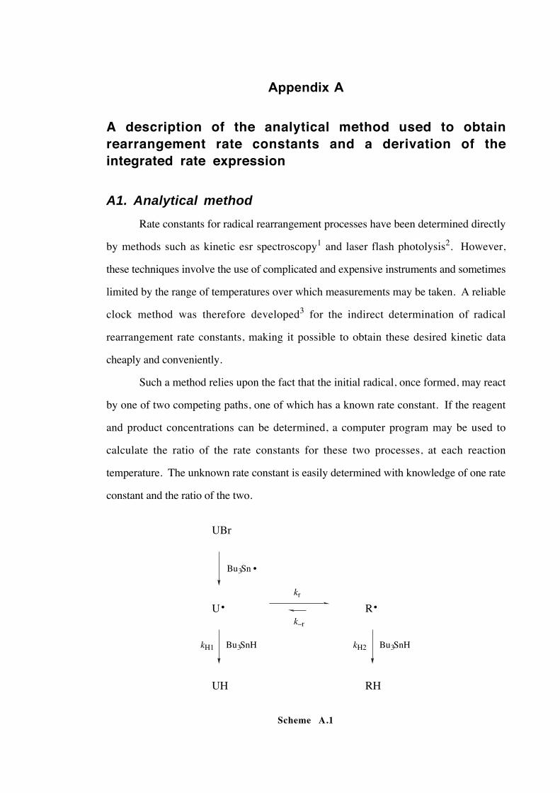
Appendix A
A description of the analytical method used to obtainrearrangement rate constants and a derivation of theintegrated rate expression
A1. Analytical method
Rate constants for radical rearrangement processes have been determined directly
by methods such as kinetic esr spectroscopy1 and laser flash photolysis2. However,
these techniques involve the use of complicated and expensive instruments and sometimes
limited by the range of temperatures over which measurements may be taken. A reliable
clock method was therefore developed3 for the indirect determination of radical
rearrangement rate constants, making it possible to obtain these desired kinetic data
cheaply and conveniently.
Such a method relies upon the fact that the initial radical, once formed, may react
by one of two competing paths, one of which has a known rate constant. If the reagent
and product concentrations can be determined, a computer program may be used to
calculate the ratio of the rate constants for these two processes, at each reaction
temperature. The unknown rate constant is easily determined with knowledge of one rate
constant and the ratio of the two.
UBr
U
UH
R
RH
Bu3Sn
Bu3SnH Bu3SnHkH1 kH2
kr
•
• •
k–r
Scheme A.1
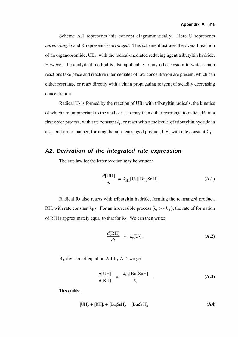
Appendix A 318
Scheme A.1 represents this concept diagrammatically. Here U represents
unrearranged and R represents rearranged. This scheme illustrates the overall reaction
of an organobromide, UBr, with the radical-mediated reducing agent tributyltin hydride.
However, the analytical method is also applicable to any other system in which chain
reactions take place and reactive intermediates of low concentration are present, which can
either rearrange or react directly with a chain propagating reagent of steadily decreasing
concentration.
Radical U• is formed by the reaction of UBr with tributyltin radicals, the kinetics
of which are unimportant to the analysis. U• may then either rearrange to radical R• in a
first order process, with rate constant kr, or react with a molecule of tributyltin hydride in
a second order manner, forming the non-rearranged product, UH, with rate constant kH1.
A2. Derivation of the integrated rate expression
The rate law for the latter reaction may be written:
d
dt
[UH] = kH1[U•][Bu3SnH] (A.1)
Radical R• also reacts with tributyltin hydride, forming the rearranged product,
RH, with rate constant kH2. For an irreversible process (kr >> k-r ), the rate of formation
of RH is approximately equal to that for R•. We can then write:
d
dt
[RH] ≈ kr[U•] . (A.2)
By division of equation A.1 by A.2, we get:
d
d
[UH][RH]
= k
kH1 3
r
[Bu SnH] . (A.3)
The equality:
[UH]t + [RH]t + [Bu3SnH]t = [Bu3SnH]i (A.4)
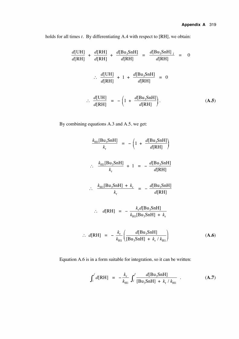
Appendix A 319
holds for all times t. By differentiating A.4 with respect to [RH], we obtain:
d[UH]
d[RH] +
d[RH]
d[RH] +
d
d
[Bu SnH][RH]
3 = d
di[Bu SnH]
[RH]3 = 0
∴ d[UH]
d[RH] + 1 +
d
d
[Bu SnH][RH]
3 = 0
∴ d[UH]
d[RH] = − 1
[Bu SnH][RH]
3+
d
d. (A.5)
By combining equations A.3 and A.5, we get:
k
kH1 3
r
[Bu SnH] = − 1
[Bu SnH][RH]
3+
d
d
∴ k
kH1 3
r
[Bu SnH] + 1 = −
d
d
[Bu SnH][RH]
3
∴ k k
kH1 3 r
r
[Bu SnH] + = −
d
d
[Bu SnH][RH]
3
∴ d[RH] = − k d
k kr 3
H1 3 r
[Bu SnH][Bu SnH] +
∴ d[RH] = − k
kr
H1
d
k k
[Bu SnH][Bu SnH] + /
3
3 r H1
(A.6)
Equation A.6 is in a form suitable for integration, so it can be written:
d[RH]i
f
∫ = −k
kr
H1
d
k ki
f [Bu SnH][Bu SnH] + /
3
3 r H1∫ . (A.7)
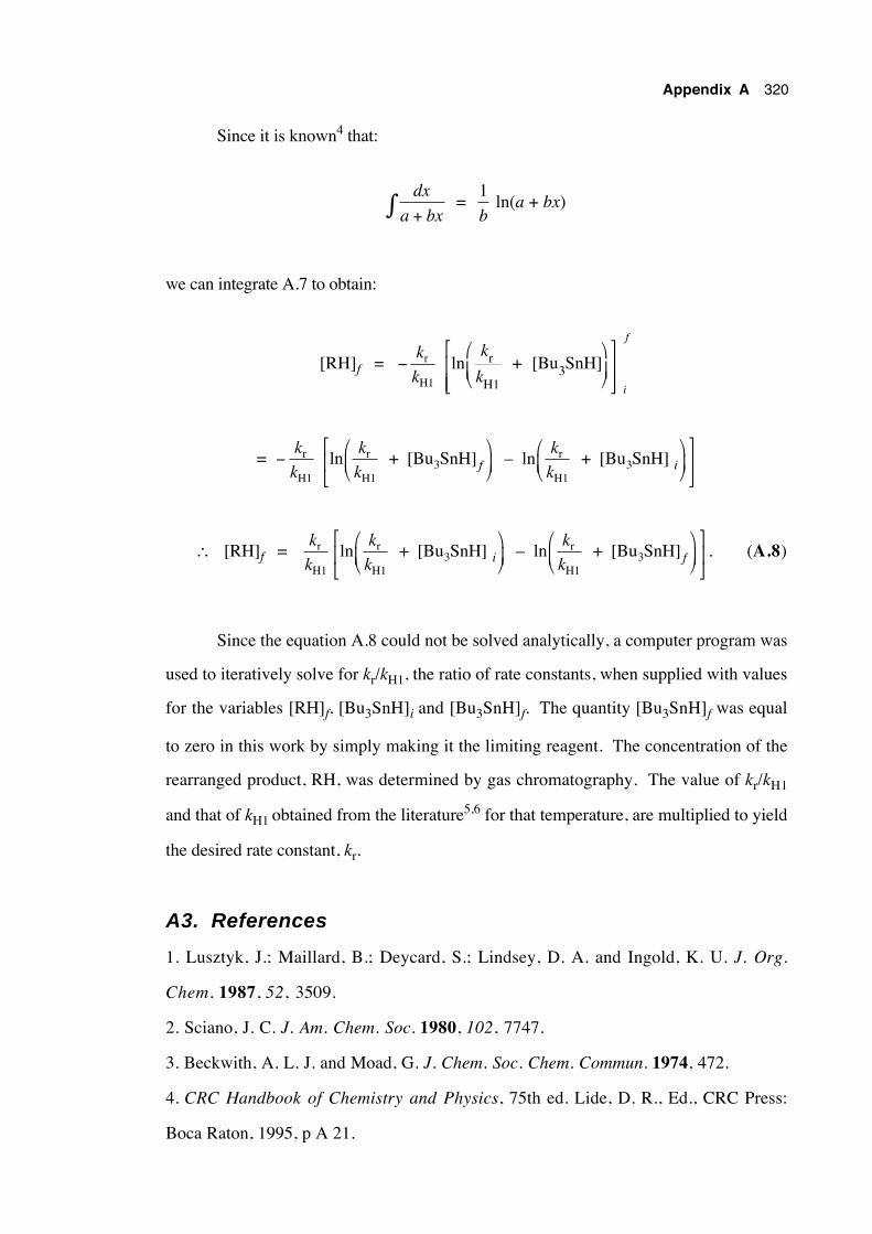
Appendix A 320
Since it is known4 that:
dx
a + bx∫ = 1
b ln(a + bx)
we can integrate A.7 to obtain:
[RH]f = −k
kr
H1
i
fk
kln r
H13 + [Bu SnH]
= −k
kr
H1
ln k
k
k
kf ir
H13
r
H13 + [Bu SnH] – ln + [Bu SnH]
∴ [RH]f = k
kr
H1
lnk
k
k
ki fr
H13
r
H13 + [Bu SnH] – ln + [Bu SnH]
. (A.8)
Since the equation A.8 could not be solved analytically, a computer program was
used to iteratively solve for kr/kH1, the ratio of rate constants, when supplied with values
for the variables [RH]f, [Bu3SnH]i and [Bu3SnH]f. The quantity [Bu3SnH]f was equal
to zero in this work by simply making it the limiting reagent. The concentration of the
rearranged product, RH, was determined by gas chromatography. The value of kr/kH1
and that of kH1 obtained from the literature5,6 for that temperature, are multiplied to yield
the desired rate constant, kr.
A3. References
1. Lusztyk, J.; Maillard, B.; Deycard, S.; Lindsey, D. A. and Ingold, K. U. J. Org.
Chem. 1987, 52, 3509.
2. Sciano, J. C. J. Am. Chem. Soc. 1980, 102, 7747.
3. Beckwith, A. L. J. and Moad, G. J. Chem. Soc. Chem. Commun. 1974, 472.
4. CRC Handbook of Chemistry and Physics, 75th ed. Lide, D. R., Ed., CRC Press:
Boca Raton, 1995, p A 21.

Appendix A 321
5. Chatgilialoglu, C.; Ingold, K. U. and Sciano, J. C. J. Am. Chem. Soc. 1981, 103,
7739.
6. Johnston, L. J.; Lusztyk, J.; Wayner, D. D. M.; Abeywickreyma, A. N.; Beckwith,
A. L. J.; Sciano, J. C. and Ingold, K. U. J. Am. Chem. Soc. 1985, 107, 4594.
Appendix B
1 7O nmr spectroscopy: Optimisation of acquisitionparameters for accurate quantification of the ratio of 17Olabel in carbonyl and alkoxy oxygens of esters
B.1 Introduction
Oxygen is an important element, forming compounds with all of the elements
except some of the noble gases. It is a constituent of many functional groups and is
fundamental in living systems. Oxygen has three stable, naturally occurring isotopes:
16O (99.762%), 17O (0.038%) and 18O (0.200%).1 Chemical and biological studies
based on the properties of oxygen have usually focussed on the mass spectrometry of 18O
enriched molecules. This technique is limited because the exact location of the label can
be difficult to assign with confidence and a high enrichment is usually required for
accurate quantitative work. In recent years advantage has been taken of the difference in
chemical shift of an nmr-active nucleus (eg. 13C, 31P) attached to an 16O and to an 18O
atom2. The position of an 18O label in a molecule can often be established this way, but
the technique suffers from a difficulty encountered in accurate quantitation and, being an
indirect method of observation, 18O nuclei attached solely to nmr-inactive nuclei escape
detection.
In 17O nmr, the site of substitution (type of functional group or position within a
functional group with inequivalent oxygens) is determined directly from the chemical
shift. If two or more oxygens are labelled, the relative amount present in each can be
obtained from the ratio of the respective peak integrals.
The field of 17O nmr spectroscopy has been the subject of chapters in
monographs,3-6 whole monographs,7 as well as reviews.8 Of particular interest are
topics such as 17O enrichment methods9 and the use of 17O nmr as a mechanistic probe in
chemistry.10 Directly relevant to the work in this thesis is the use of 17O nmr in
establishing the proportions of label in ether and carbonyl positions of esters,11-15 to
enable the determination of rearrangement regiochemistry.
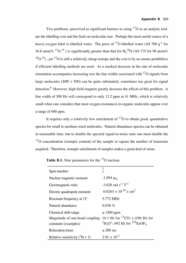
Appendix B 323
Two problems, perceived as significant barriers to using 17O as an analytic tool,
are the labelling cost and the limit on molecular size. Perhaps the most useful source of a
heavy-oxygen label is labelled water. The price of 17O labelled water (A$ 700 g-1 for
36.8 atom% 17O,15 ) is significantly greater than that for H218O (A$ 175 for 98 atom%
18O,14) , yet 17O is still a relatively cheap isotope and the cost is by no means prohibitive
if efficient labelling methods are used. As a marked decrease in the rate of molecular
orientation accompanies increasing size the line widths associated with 17O signals from
large molecules (MW > 500) can be quite substantial, sometimes too great for signal
detection.8 However, high-field magnets greatly decrease the effects of this problem. A
line width of 500 Hz will correspond to only 12.2 ppm at 41 MHz, which is relatively
small when one considers that most oxygen resonances in organic molecules appear over
a range of 600 ppm.
It requires only a relatively low enrichment of 17O to obtain good, quantitative
spectra for small to medium-sized molecules. Natural abundance spectra can be obtained
in reasonable time, but to double the spectral signal-to-noise ratio one must double the
17O concentration (isotopic content) of the sample or square the number of transients
acquired. Therefore, isotopic enrichment of samples makes a great deal of sense.
Table B.1. Nmr parameters for the 17O nucleus
Spin number52
Nuclear magnetic moment -1.894 µN
Gyromagnetic ratio -3.628 rad s-1 T-1
Electric quadrupole moment -0.0263 × 10⁻²⁴ e cm2
Resonant frequency at 1T 5.772 MHz
Natural abundance 0.038 %
Chemical shift range ≥ 1500 ppm
Magnitude of one-bond coupling
constants (examples)
16.1 Hz for 13CO; (-)106 Hz for1H3O+; 692 Hz for 129XeOF4
Relaxation times ≤ 200 ms
Relative sensitivity (1H = 1) 2.91 × 10⁻²
Appendix B 324
The 17O nucleus has spin number 52 and is the only stable nmr-active isotope of
oxygen. Although 17O is a reasonably sensitive nucleus, its combination of low natural
abundance and large spectral line widths have made 17O nmr a relatively unpopular
method of chemical and biological research. However, by increasing the abundance of
17O in a sample to 10 atom percent, the relative receptivity becomes 3.12 × 10⁻³ times
that for 1H, atom for atom. This now compares favourably with a value of 1.76 × 10⁻⁴
for 13C at natural abundance. Relaxation times are relatively short because the principal
mode of relaxation is through the electric quadrupole. The equality T1 = T2 generally
holds and T2 values may be obtained easily from spectral line widths by the relationship:
w12 =
1πT2
where w12 is the line width at half-height and T2 is the transverse relaxation time constant.
T1 represents the longitudinal relaxation time constant. For a typical line width of 200
Hz, T2 is 1.6 ms. Line widths may be reduced (T2 values increased) by increasing the
rate of molecular reorientation. This is achieved in practice by using non-viscous, non-
hydrogen-bonding solvents and acquiring data at elevated temperature. Reasonable line
widths may be achieved for molecules with molecular weight ≤ 500 g mol-1.
B.2 Overcoming transmitter breakthrough
An additional hindrance to obtaining good 17O nmr spectra has been the
combination of relatively low Larmor resonance frequencies and fast relaxation times. At
lower frequencies high resolution FT nmr probes are prone to acoustic ringing, where the
residual RF signal resonates for a short time within the probe after the pulse has been
switched off. This effect becomes significant for nuclei with short relaxation times
because the decaying RF pulse signal significantly distorts a large proportion of the FID,
causing "transmitter breakthrough", which manifests as an undulating baseline in spectra.
Elimination of this effect is possible by leaving a delay of up to several hundred µs
between the 90˚ excitation pulse and acquisition. This is done at the expense of losing a
Appendix B 325
sometimes significant proportion of the signal and introducing phase errors, which in turn
causes considerable errors when accurate peak integrals are required. Acoustic ringing
decreases markedly at higher pulse frequencies however, so that shorter pre-acquisition
delay times are required with high-field spectrometers. Baseline roll is also less of a
problem when 17O enriched samples are used owing to higher signal-to-noise ratios.
Figure B.1 illustrates how the transmitter spike at the beginning of the FID can be
eliminated by the correct choice of a pre-acquisition delay time. Here the FID (total time
= 10,000 µs) for the relaxation of the 17O nucleus in the oxy-labelled β-bromoester is
shown for delays of 0, 10 and 50 µs. No delay results in a marked spike at the beginning
of the FID, whereas a 10 µs delay eliminates this spike. A 50 µs delay eliminates this
spike, but an unnecessarily large amount of the start of the FID is also lost. A 10 µs
delay was therefore used in data acquisition.
B.3 Spectrometer parameters and method of acquisition
B.3.1 Spectrometer parameters
All of the 17O spectra were obtained in a field of 7.05 T on a Varian VXR 300
spectrometer operating at 40.681 MHz. A spectral width of 30,030 Hz and transmitter
offset of 10,000 Hz were employed, corresponding with the transmitter RF pulse being
centred at about 235 ppm. For each spectrum 16,384 complex data points were collected
and zero filled to 32,768 complex points prior to Fourier transform, without exponential
weighting. A 90˚ pulse of 21.0 µs duration was followed by a 10 µs delay before
acquisition, to arrest transmitter breakthrough. The total acquisition time for each cycle
was 273 ms and the number of transients collected varied from 8192 (30 min acquisition
time) to ca. 2 × 10⁵ (16 hours), depending upon sample concentration. The S/N ratio for
the smallest peak of interest in each of the spectra was always greater than 6.
Quantitatively reliable spectra have been obtained with samples containing as little as 7 ×
10-6 moles of 17O in 16 hours. The longitudinal relaxation time constant (T1) for the
oxy-labelled ester, 1-bromomethyl-1-methylhexyl trifluoroacetate, was determined to be
847 ± 90 µs and T2 values for all compounds were calculated from spectral line widths
and were in the range 500 - 2300 µs.
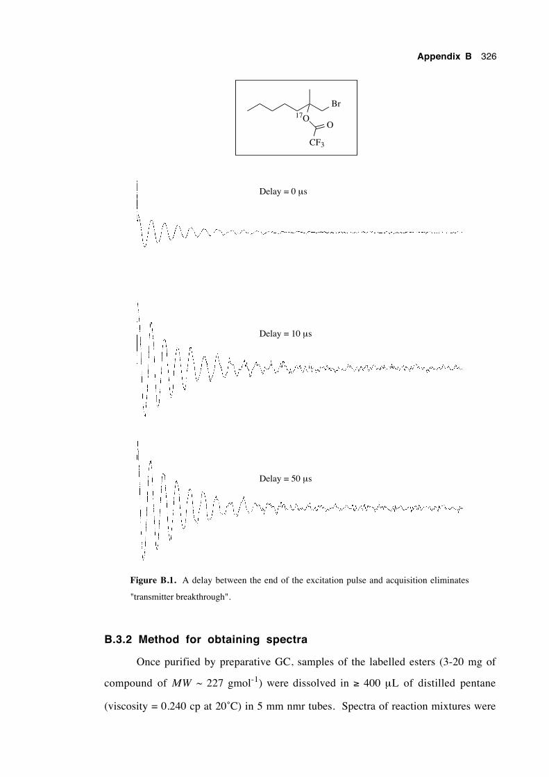
Appendix B 326
Br17O
CF3
O
Delay = 0 µs
Delay = 10 µs
Delay = 50 µs
Figure B.1. A delay between the end of the excitation pulse and acquisition eliminates
"transmitter breakthrough".
B.3.2 Method for obtaining spectra
Once purified by preparative GC, samples of the labelled esters (3-20 mg of
compound of MW ~ 227 gmol-1) were dissolved in ≥ 400 µL of distilled pentane
(viscosity = 0.240 cp at 20˚C) in 5 mm nmr tubes. Spectra of reaction mixtures were
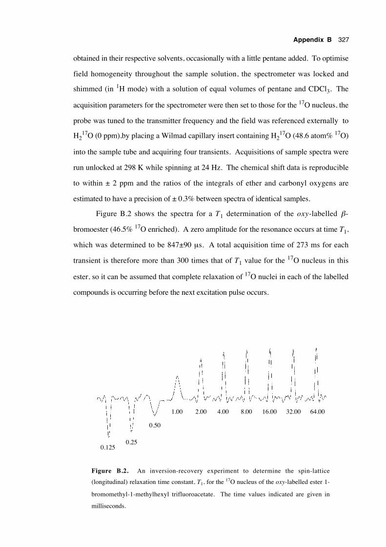
Appendix B 327
obtained in their respective solvents, occasionally with a little pentane added. To optimise
field homogeneity throughout the sample solution, the spectrometer was locked and
shimmed (in 1H mode) with a solution of equal volumes of pentane and CDCl3. The
acquisition parameters for the spectrometer were then set to those for the 17O nucleus, the
probe was tuned to the transmitter frequency and the field was referenced externally to
H217O (0 ppm),by placing a Wilmad capillary insert containing H2
17O (48.6 atom% 17O)
into the sample tube and acquiring four transients. Acquisitions of sample spectra were
run unlocked at 298 K while spinning at 24 Hz. The chemical shift data is reproducible
to within ± 2 ppm and the ratios of the integrals of ether and carbonyl oxygens are
estimated to have a precision of ± 0.3% between spectra of identical samples.
Figure B.2 shows the spectra for a T1 determination of the oxy-labelled β-
bromoester (46.5% 17O enriched). A zero amplitude for the resonance occurs at time T1,
which was determined to be 847±90 µs. A total acquisition time of 273 ms for each
transient is therefore more than 300 times that of T1 value for the 17O nucleus in this
ester, so it can be assumed that complete relaxation of 17O nuclei in each of the labelled
compounds is occurring before the next excitation pulse occurs.
0.1250.25
0.50
1.00 2.00 4.00 8.00 16.00 32.00 64.00
Figure B.2. An inversion-recovery experiment to determine the spin-lattice
(longitudinal) relaxation time constant, T1, for the 17O nucleus of the oxy-labelled ester 1-
bromomethyl-1-methylhexyl trifluoroacetate. The time values indicated are given in
milliseconds.
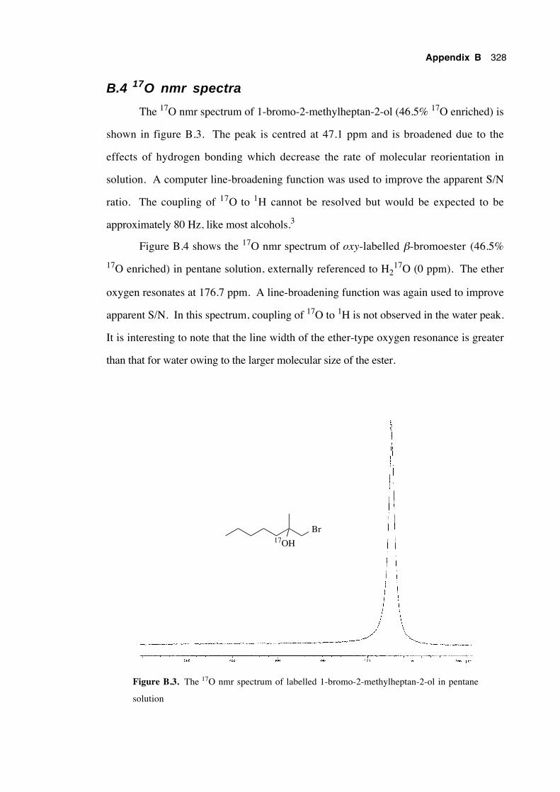
Appendix B 328
B.4 17O nmr spectra
The 17O nmr spectrum of 1-bromo-2-methylheptan-2-ol (46.5% 17O enriched) is
shown in figure B.3. The peak is centred at 47.1 ppm and is broadened due to the
effects of hydrogen bonding which decrease the rate of molecular reorientation in
solution. A computer line-broadening function was used to improve the apparent S/N
ratio. The coupling of 17O to 1H cannot be resolved but would be expected to be
approximately 80 Hz, like most alcohols.3
Figure B.4 shows the 17O nmr spectrum of oxy-labelled β-bromoester (46.5%
17O enriched) in pentane solution, externally referenced to H217O (0 ppm). The ether
oxygen resonates at 176.7 ppm. A line-broadening function was again used to improve
apparent S/N. In this spectrum, coupling of 17O to 1H is not observed in the water peak.
It is interesting to note that the line width of the ether-type oxygen resonance is greater
than that for water owing to the larger molecular size of the ester.
Br17OH
Figure B.3. The 17O nmr spectrum of labelled 1-bromo-2-methylheptan-2-ol in pentane
solution
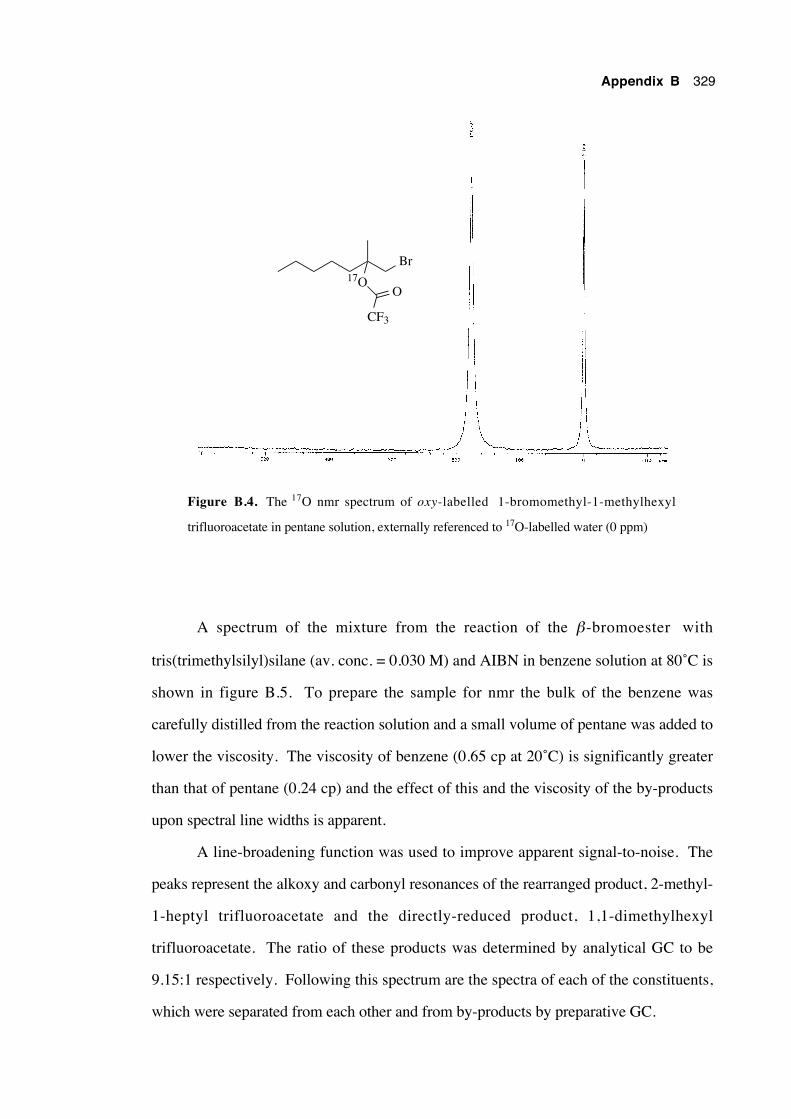
Appendix B 329
Br17O
CF3
O
Figure B.4. The 17O nmr spectrum of oxy-labelled 1-bromomethyl-1-methylhexyl
trifluoroacetate in pentane solution, externally referenced to 17O-labelled water (0 ppm)
A spectrum of the mixture from the reaction of the β-bromoester with
tris(trimethylsilyl)silane (av. conc. = 0.030 M) and AIBN in benzene solution at 80˚C is
shown in figure B.5. To prepare the sample for nmr the bulk of the benzene was
carefully distilled from the reaction solution and a small volume of pentane was added to
lower the viscosity. The viscosity of benzene (0.65 cp at 20˚C) is significantly greater
than that of pentane (0.24 cp) and the effect of this and the viscosity of the by-products
upon spectral line widths is apparent.
A line-broadening function was used to improve apparent signal-to-noise. The
peaks represent the alkoxy and carbonyl resonances of the rearranged product, 2-methyl-
1-heptyl trifluoroacetate and the directly-reduced product, 1,1-dimethylhexyl
trifluoroacetate. The ratio of these products was determined by analytical GC to be
9.15:1 respectively. Following this spectrum are the spectra of each of the constituents,
which were separated from each other and from by-products by preparative GC.
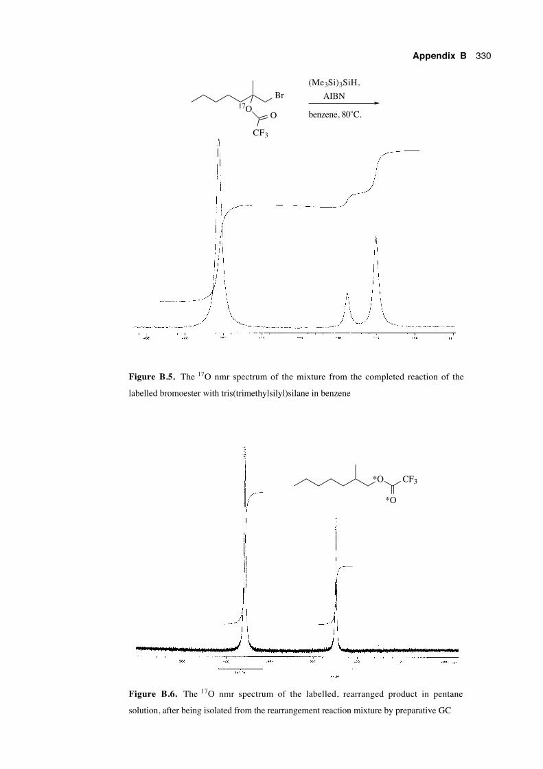
Appendix B 330
Br17O
CF3
O
(Me3Si)3SiH,
AIBN
benzene, 80˚C.
Figure B.5. The 17O nmr spectrum of the mixture from the completed reaction of the
labelled bromoester with tris(trimethylsilyl)silane in benzene
*O CF3
*O
Figure B.6. The 17O nmr spectrum of the labelled, rearranged product in pentane
solution, after being isolated from the rearrangement reaction mixture by preparative GC
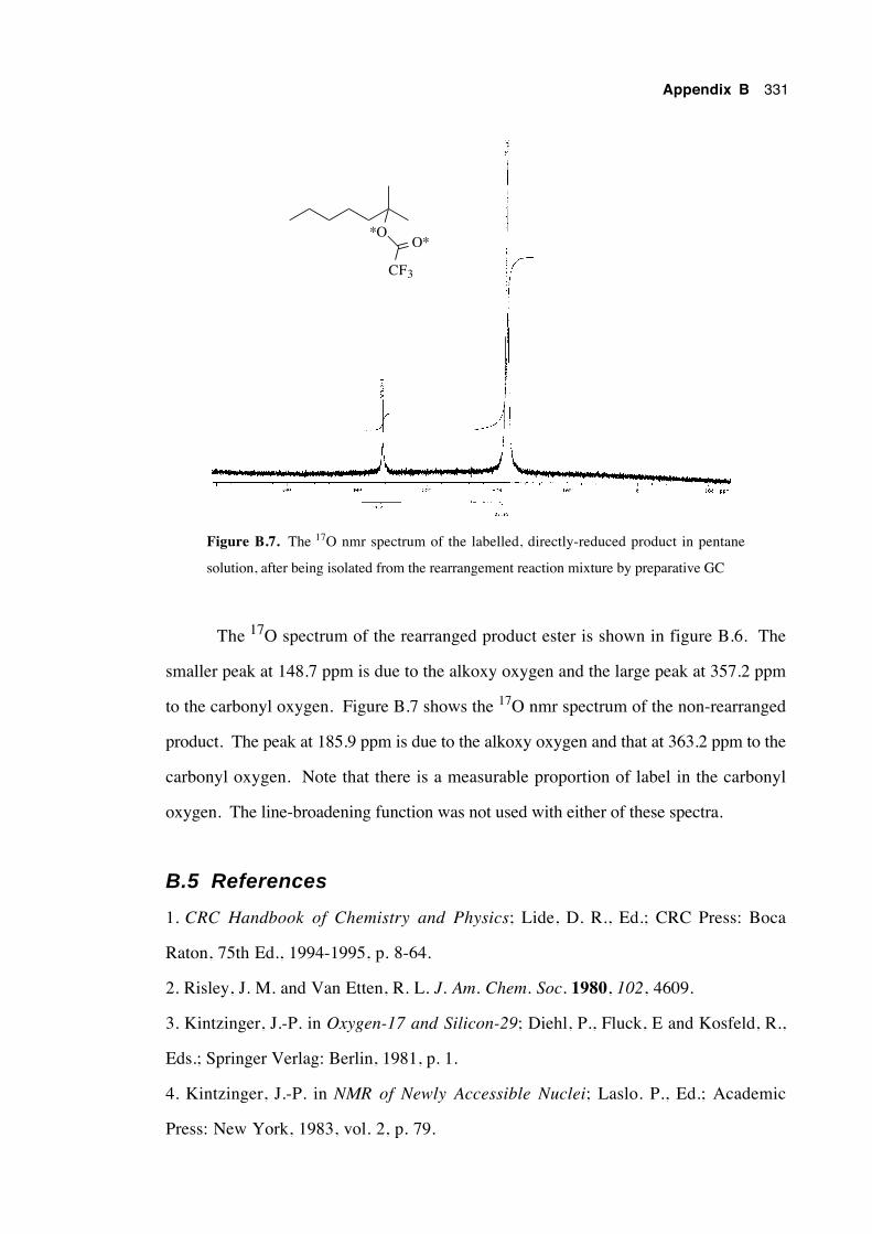
Appendix B 331
*O
CF3
O*
Figure B.7. The 17O nmr spectrum of the labelled, directly-reduced product in pentane
solution, after being isolated from the rearrangement reaction mixture by preparative GC
The 17O spectrum of the rearranged product ester is shown in figure B.6. The
smaller peak at 148.7 ppm is due to the alkoxy oxygen and the large peak at 357.2 ppm
to the carbonyl oxygen. Figure B.7 shows the 17O nmr spectrum of the non-rearranged
product. The peak at 185.9 ppm is due to the alkoxy oxygen and that at 363.2 ppm to the
carbonyl oxygen. Note that there is a measurable proportion of label in the carbonyl
oxygen. The line-broadening function was not used with either of these spectra.
B.5 References
1. CRC Handbook of Chemistry and Physics; Lide, D. R., Ed.; CRC Press: Boca
Raton, 75th Ed., 1994-1995, p. 8-64.
2. Risley, J. M. and Van Etten, R. L. J. Am. Chem. Soc. 1980, 102, 4609.
3. Kintzinger, J.-P. in Oxygen-17 and Silicon-29; Diehl, P., Fluck, E and Kosfeld, R.,
Eds.; Springer Verlag: Berlin, 1981, p. 1.
4. Kintzinger, J.-P. in NMR of Newly Accessible Nuclei; Laslo. P., Ed.; Academic
Press: New York, 1983, vol. 2, p. 79.
Appendix B 332
5. Klemperer, W. G. in The Multinuclear Approach to NMR Spectroscopy; Lambert,
J. B. and Riddell, F. G., Eds.; D. Reidel Publishing: Dordrecht, 1983, p. 245.
6. McFarlane, W. and McFarlane, H. C. E. in Multinuclear NMR; Mason, J., Ed.;
Plenum Press: New York, 1987, p. 403.
7. 17O NMR Spectroscopy in Organic Chemistry; Boykin, D. W., Ed.; CRC Press:
Boston, 1991.
8. Klemperer, W. G. Angew. Chem., Int. Ed. Engl. 1978, 17, 246.
9. Kabalka, G. W. and Goudgaon, N. M. in 17O NMR Spectroscopy in Organic
Chemistry; Boykin, D. W., Ed.; CRC Press: Boston, 1991, p. 21.
10. Woodard, R. W. in 17O NMR Spectroscopy in Organic Chemistry; Boykin, D. W.,
Ed.; CRC Press: Boston, 1991, p. 115.
11. Nakanishi, W.; Jo, T.; Miura, K.; Ikeda, Y.; Sugawara, T.; Kawada, Y. and
Iwamura, H. Chem. Lett. 1981, 387.
12. Creary, X. and Inocenio, P. A. J. Am. Chem. Soc. 1986, 108, 5979.
13. Beckwith, A. L. J. and Duggan, P. J. J. Chem. Soc., Perkin Trans. 2 1993, 1673.
14. Beckwith, A. L. J. and Duggan, P. J. J. Am. Chem. Soc. 1996, 118, 12838.
15. Crich, D.; Huang, X. and Beckwith, A. L. J. J. Org. Chem. 1999, 64, 1762.
16. Cambridge Isotope Laboratories, Inc., Andover, MA, USA.
17. Isotec Inc., Miamisburg, OH, USA.
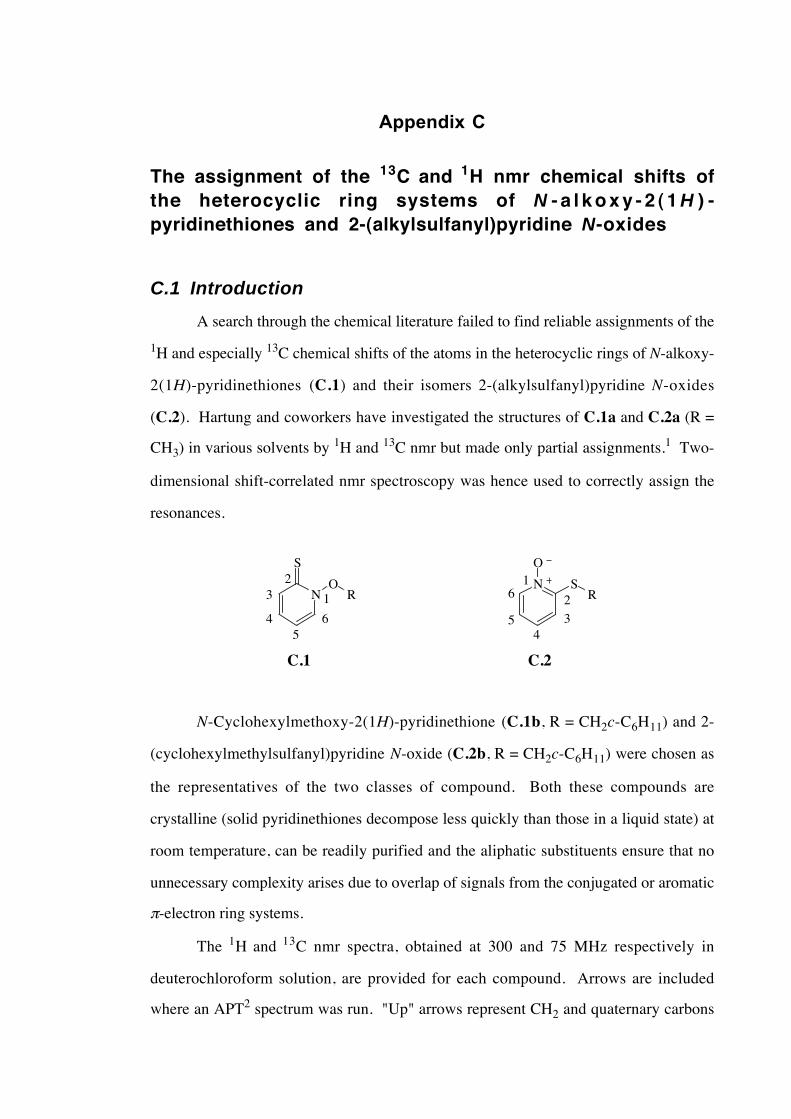
Appendix C
The assignment of the 13C and 1H nmr chemical shifts ofthe heterocyclic ring systems of N - a l k o x y - 2 ( 1 H ) -pyridinethiones and 2-(alkylsulfanyl)pyridine N-oxides
C.1 Introduction
A search through the chemical literature failed to find reliable assignments of the
1H and especially 13C chemical shifts of the atoms in the heterocyclic rings of N-alkoxy-
2(1H)-pyridinethiones (C.1) and their isomers 2-(alkylsulfanyl)pyridine N-oxides
(C.2). Hartung and coworkers have investigated the structures of C.1a and C.2a (R =
CH3) in various solvents by 1H and 13C nmr but made only partial assignments.1 Two-
dimensional shift-correlated nmr spectroscopy was hence used to correctly assign the
resonances.
NO
R
S
N SR
O
1
23
45
6
1
2
34
5
6
C.1 C.2
N-Cyclohexylmethoxy-2(1H)-pyridinethione (C.1b, R = CH2c-C6H11) and 2-
(cyclohexylmethylsulfanyl)pyridine N-oxide (C.2b, R = CH2c-C6H11) were chosen as
the representatives of the two classes of compound. Both these compounds are
crystalline (solid pyridinethiones decompose less quickly than those in a liquid state) at
room temperature, can be readily purified and the aliphatic substituents ensure that no
unnecessary complexity arises due to overlap of signals from the conjugated or aromatic
π-electron ring systems.
The 1H and 13C nmr spectra, obtained at 300 and 75 MHz respectively in
deuterochloroform solution, are provided for each compound. Arrows are included
where an APT2 spectrum was run. "Up" arrows represent CH2 and quaternary carbons
Appendix C 334
and "down" arrows CH and CH3 groups. It was possible to correctly assign some
closely separated resonances from their relative peak heights, as the magnitude of the
signal resonance is often approximately proportional to the numbers of degenerate atoms
contributing to the signal.
C.2 Assignment of the chemical shifts of N -cyclohexylmethoxy-2(1H)-pyridinethione
The one-dimensional nmr spectra for N-cyclohexylmethoxy-2(1H)-pyridinethione
are provided below:
1H: 1.13 (m, 2H), 1.20-1.40 (m, 3H), 1.65-1.82 (m, 4H), 1.81-2.00 (m, 3H), 4.24 (d,
2H, J = 6.1 Hz), 6.66 (ddd, 1H), 7.17 (ddd, 1H), 7.68 (dd, 1H), 7.77 (dd, 1H); and
13C: 25.2↑, 25.9↑, 29.2↑, 36.0↓, 81.6↑, 113.6↓, 133.1↓, 137.8,↓ 138.0↓, 175.6↑.
In the 1H spectrum, it is clear from the multiplicities of the double doublet
resonances at 7.68 and 7.77 ppm that the hydrogens corresponding to these signals lie at
the extremities (positions 3 and 6) of the four sequential ring protons. The two remaining
downfield signals at 6.66 and 7.17 hence represent the hydrogens at positions 4 and 5,
but absolute assignment of any one of these resonances cannot be made with certainty on
the basis of this spectrum alone.
A homonuclear shift-correlated spectrum (COSY) was obtained (figure C.1) and
the correlations for the pyridinethione ring function are listed in table C.1.
Table C.1. 1H–1H shift correlations for the heterocyclic
ring of N-cyclohexylmethoxy-2(1H)-pyridinethione
1H chemical shift (ppm) 1H chemical shift (ppm)
6.66 7.17 and 7.77
7.17 6.66 and 7.68
7.68 7.17
7.77 6.66
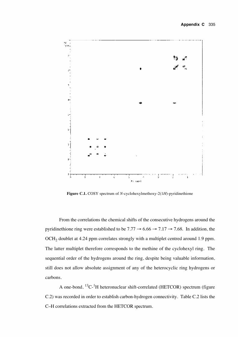
Appendix C 335
Figure C.1. COSY spectrum of N-cyclohexylmethoxy-2(1H)-pyridinethione
From the correlations the chemical shifts of the consecutive hydrogens around the
pyridinethione ring were established to be 7.77 → 6.66 → 7.17 → 7.68. In addition, the
OCH2 doublet at 4.24 ppm correlates strongly with a multiplet centred around 1.9 ppm.
The latter multiplet therefore corresponds to the methine of the cyclohexyl ring. The
sequential order of the hydrogens around the ring, despite being valuable information,
still does not allow absolute assignment of any of the heterocyclic ring hydrogens or
carbons.
A one-bond, 13C-1H heteronuclear shift-correlated (HETCOR) spectrum (figure
C.2) was recorded in order to establish carbon-hydrogen connectivity. Table C.2 lists the
C–H correlations extracted from the HETCOR spectrum.
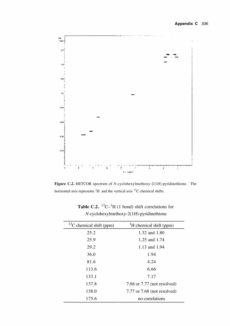
Appendix C 336
Figure C.2. HETCOR spectrum of N-cyclohexylmethoxy-2(1H)-pyridinethione. The
horizontal axis represents 1H and the vertical axis 13C chemical shifts.
Table C.2. 13C–1H (1 bond) shift correlations for
N-cyclohexylmethoxy-2(1H)-pyridinethione
13C chemical shift (ppm) 1H chemical shift (ppm)
25.2 1.32 and 1.80
25.9 1.25 and 1.74
29.2 1.13 and 1.94
36.0 1.94
81.6 4.24
113.6 6.66
133.1 7.17
137.8 7.68 or 7.77 (not resolved)
138.0 7.77 or 7.68 (not resolved)
175.6 no correlations
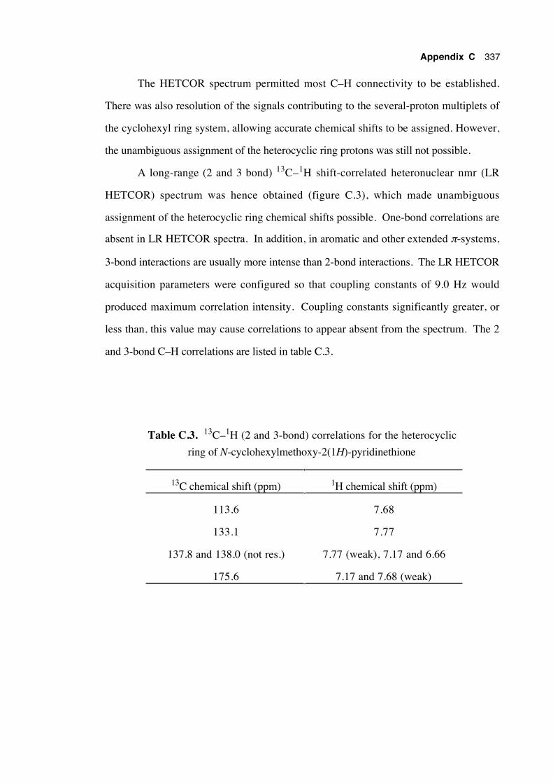
Appendix C 337
The HETCOR spectrum permitted most C–H connectivity to be established.
There was also resolution of the signals contributing to the several-proton multiplets of
the cyclohexyl ring system, allowing accurate chemical shifts to be assigned. However,
the unambiguous assignment of the heterocyclic ring protons was still not possible.
A long-range (2 and 3 bond) 13C–1H shift-correlated heteronuclear nmr (LR
HETCOR) spectrum was hence obtained (figure C.3), which made unambiguous
assignment of the heterocyclic ring chemical shifts possible. One-bond correlations are
absent in LR HETCOR spectra. In addition, in aromatic and other extended π-systems,
3-bond interactions are usually more intense than 2-bond interactions. The LR HETCOR
acquisition parameters were configured so that coupling constants of 9.0 Hz would
produced maximum correlation intensity. Coupling constants significantly greater, or
less than, this value may cause correlations to appear absent from the spectrum. The 2
and 3-bond C–H correlations are listed in table C.3.
Table C.3. 13C–1H (2 and 3-bond) correlations for the heterocyclic
ring of N-cyclohexylmethoxy-2(1H)-pyridinethione
13C chemical shift (ppm) 1H chemical shift (ppm)
113.6 7.68
133.1 7.77
137.8 and 138.0 (not res.) 7.77 (weak), 7.17 and 6.66
175.6 7.17 and 7.68 (weak)
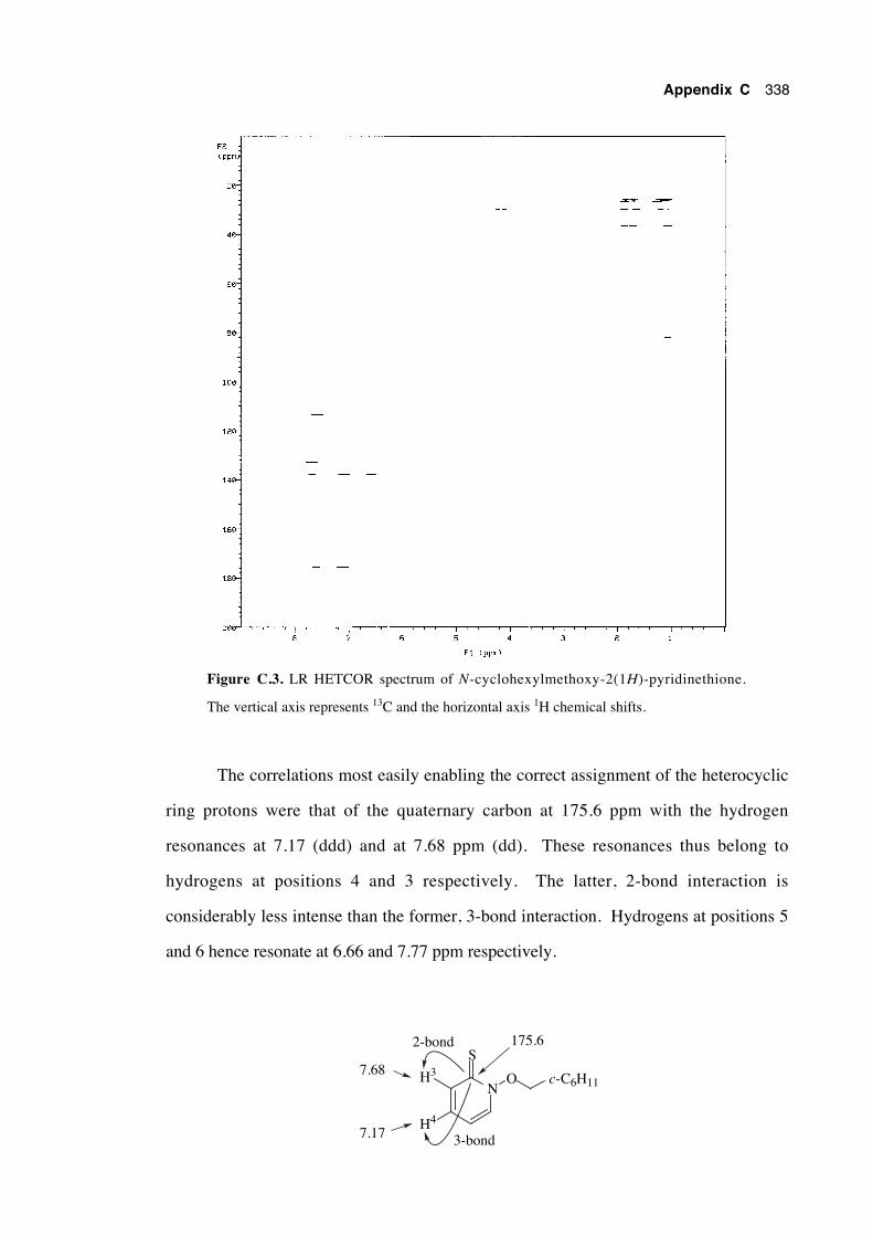
Appendix C 338
Figure C.3. LR HETCOR spectrum of N-cyclohexylmethoxy-2(1H)-pyridinethione.
The vertical axis represents 13C and the horizontal axis 1H chemical shifts.
The correlations most easily enabling the correct assignment of the heterocyclic
ring protons were that of the quaternary carbon at 175.6 ppm with the hydrogen
resonances at 7.17 (ddd) and at 7.68 ppm (dd). These resonances thus belong to
hydrogens at positions 4 and 3 respectively. The latter, 2-bond interaction is
considerably less intense than the former, 3-bond interaction. Hydrogens at positions 5
and 6 hence resonate at 6.66 and 7.77 ppm respectively.
NO
S
H3
H4
2-bond
3-bond
175.6
7.17
7.68c-C6H11
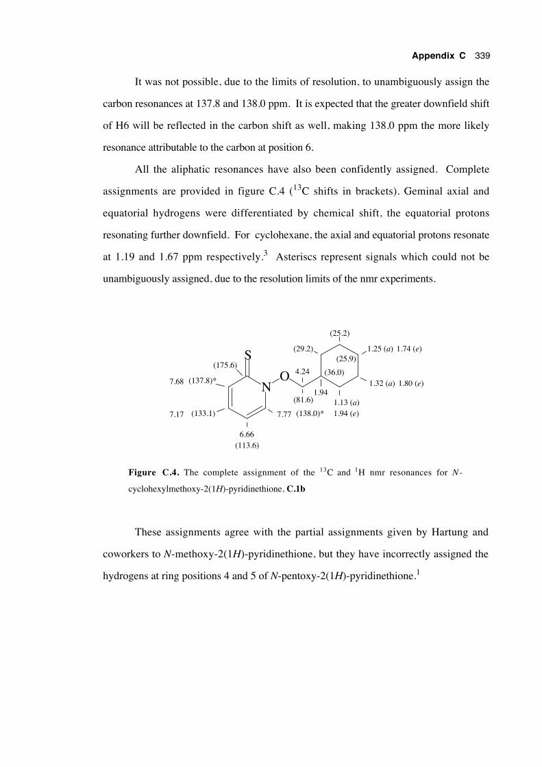
Appendix C 339
It was not possible, due to the limits of resolution, to unambiguously assign the
carbon resonances at 137.8 and 138.0 ppm. It is expected that the greater downfield shift
of H6 will be reflected in the carbon shift as well, making 138.0 ppm the more likely
resonance attributable to the carbon at position 6.
All the aliphatic resonances have also been confidently assigned. Complete
assignments are provided in figure C.4 (13C shifts in brackets). Geminal axial and
equatorial hydrogens were differentiated by chemical shift, the equatorial protons
resonating further downfield. For cyclohexane, the axial and equatorial protons resonate
at 1.19 and 1.67 ppm respectively.3 Asteriscs represent signals which could not be
unambiguously assigned, due to the resolution limits of the nmr experiments.
N
S
O7.68 (137.8)*
(175.6)
7.17 (133.1)
6.66(113.6)
7.77 (138.0)*
(81.6)
4.24
1.94
(36.0)
1.13 (a)1.94 (e)
(25.9)
1.32 (a) 1.80 (e)
1.25 (a) 1.74 (e)
(25.2)
(29.2)
Figure C.4. The complete assignment of the 13C and 1H nmr resonances for N -
cyclohexylmethoxy-2(1H)-pyridinethione, C.1b
These assignments agree with the partial assignments given by Hartung and
coworkers to N-methoxy-2(1H)-pyridinethione, but they have incorrectly assigned the
hydrogens at ring positions 4 and 5 of N-pentoxy-2(1H)-pyridinethione.1
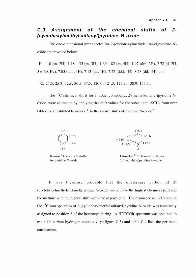
Appendix C 340
C.3 Assignment of the chemical shifts of 2-(cyclohexylmethylsulfanyl)pyridine N-oxide
The one-dimensional nmr spectra for 2-(cyclohexylmethylsulfanyl)pyridine N-
oxide are provided below:
1H: 1.10 (m, 2H), 1.18-1.35 (m, 3H), 1.60-1.82 (m, 4H), 1.97 (dm, 2H), 2.78 (d, 2H,
J = 6.8 Hz), 7.05 (ddd, 1H), 7.13 (dd, 1H), 7.27 (ddd, 1H), 8.28 (dd, 1H); and
13C: 25.6, 25.8, 32.8, 36.5, 37.5, 120.0, 121.3, 125.9, 138.9, 153.3.
The 13C chemical shifts for a model compound, 2-(methylsulfanyl)pyridine N-
oxide, were estimated by applying the shift values for the substituent -SCH3 from nmr
tables for substituted benzenes,4 to the known shifts of pyridine N-oxide.5
N
O
139.4
127.2
125.7
N
O
139.8
123.6
125.7
CH3S149.6
125.2
Known 13C chemical shiftsfor pyridine N-oxide
Estimated 13C chemical shifts for2-(methylthio)pyridine N-oxide
It was therefore probable that the quaternary carbon of 2-
(cyclohexylmethylsulfanyl)pyridine N-oxide would have the highest chemical shift and
the methine with the highest shift would be at position 6. The resonance at 139.8 ppm in
the 13C nmr spectrum of 2-(cyclohexylmethylsulfanyl)pyridine N-oxide was tentatively
assigned to position 6 of the heterocyclic ring. A HETCOR spectrum was obtained to
establish carbon-hydrogen connectivity (figure C.5) and table C.4 lists the pertinent
correlations.
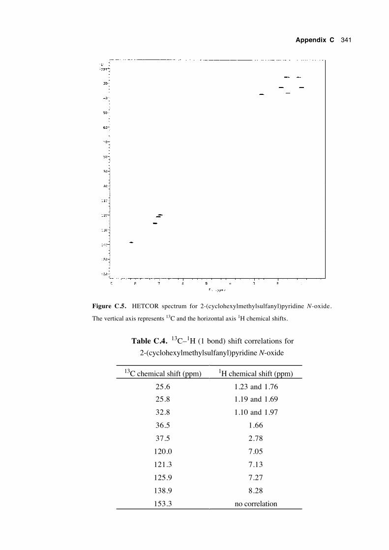
Appendix C 341
Figure C.5. HETCOR spectrum for 2-(cyclohexylmethylsulfanyl)pyridine N-oxide.
The vertical axis represents 13C and the horizontal axis 1H chemical shifts.
Table C.4. 13C–1H (1 bond) shift correlations for
2-(cyclohexylmethylsulfanyl)pyridine N-oxide
13C chemical shift (ppm) 1H chemical shift (ppm)
25.6 1.23 and 1.76
25.8 1.19 and 1.69
32.8 1.10 and 1.97
36.5 1.66
37.5 2.78
120.0 7.05
121.3 7.13
125.9 7.27
138.9 8.28
153.3 no correlation
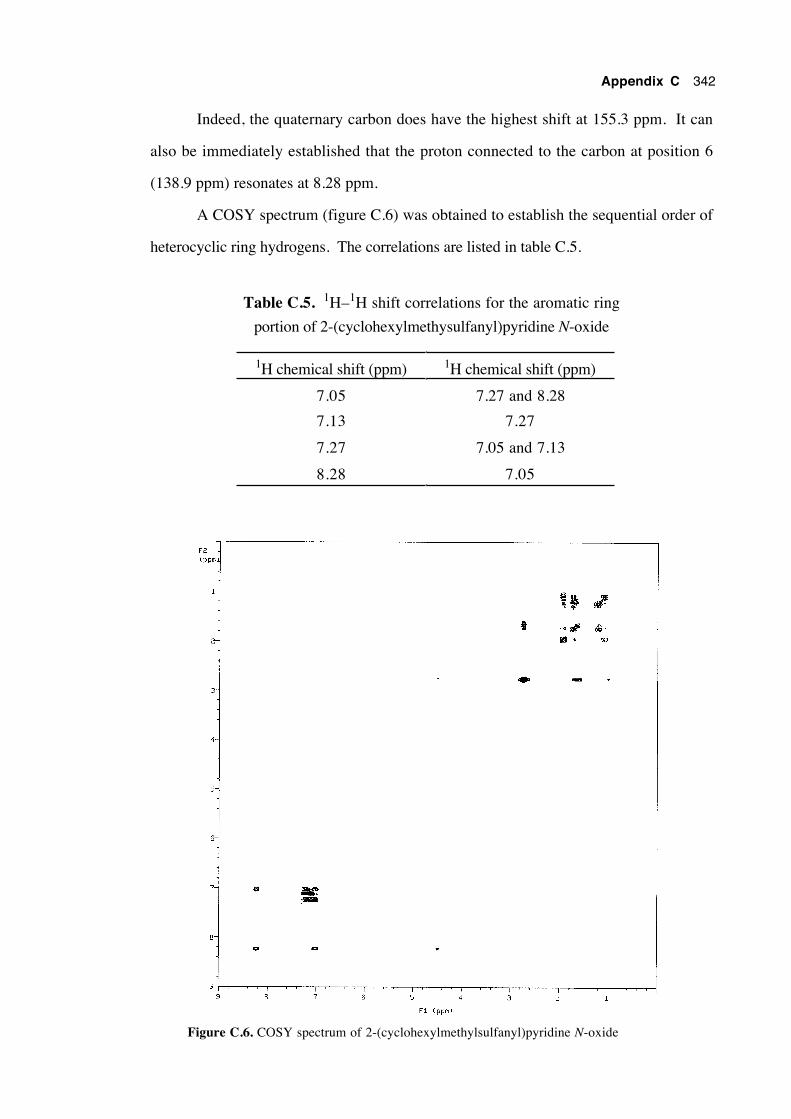
Appendix C 342
Indeed, the quaternary carbon does have the highest shift at 155.3 ppm. It can
also be immediately established that the proton connected to the carbon at position 6
(138.9 ppm) resonates at 8.28 ppm.
A COSY spectrum (figure C.6) was obtained to establish the sequential order of
heterocyclic ring hydrogens. The correlations are listed in table C.5.
Table C.5. 1H–1H shift correlations for the aromatic ring
portion of 2-(cyclohexylmethysulfanyl)pyridine N-oxide
1H chemical shift (ppm) 1H chemical shift (ppm)
7.05 7.27 and 8.28
7.13 7.27
7.27 7.05 and 7.13
8.28 7.05
Figure C.6. COSY spectrum of 2-(cyclohexylmethylsulfanyl)pyridine N-oxide
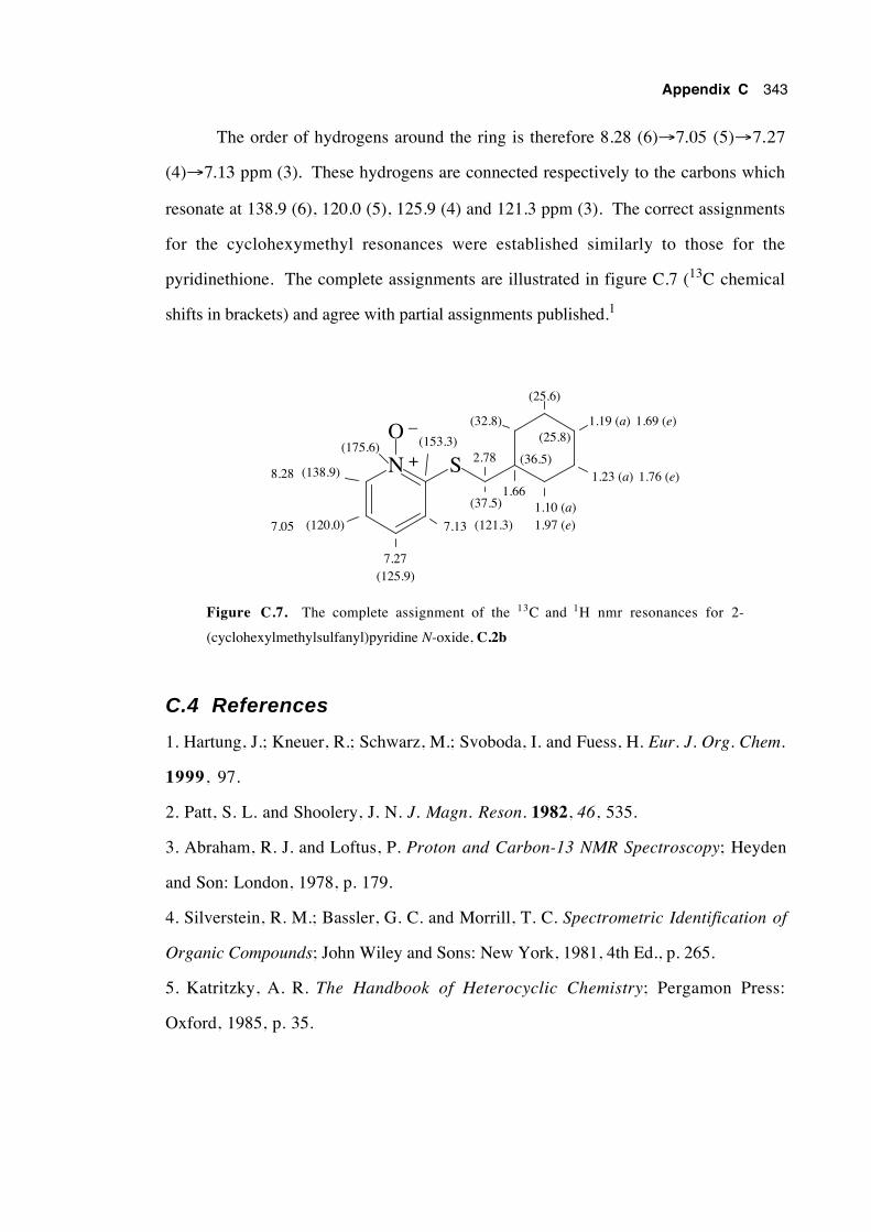
Appendix C 343
The order of hydrogens around the ring is therefore 8.28 (6)→7.05 (5)→7.27
(4)→7.13 ppm (3). These hydrogens are connected respectively to the carbons which
resonate at 138.9 (6), 120.0 (5), 125.9 (4) and 121.3 ppm (3). The correct assignments
for the cyclohexymethyl resonances were established similarly to those for the
pyridinethione. The complete assignments are illustrated in figure C.7 (13C chemical
shifts in brackets) and agree with partial assignments published.1
N
O
S8.28 (138.9)
(175.6)
7.05 (120.0)
7.27(125.9)
7.13 (121.3)
(37.5)
2.78
1.66
(36.5)
1.10 (a)1.97 (e)
(25.8)
1.23 (a) 1.76 (e)
1.19 (a) 1.69 (e)
(25.6)
(32.8)
+
–(153.3)
Figure C.7. The complete assignment of the 13C and 1H nmr resonances for 2-
(cyclohexylmethylsulfanyl)pyridine N-oxide, C.2b
C.4 References
1. Hartung, J.; Kneuer, R.; Schwarz, M.; Svoboda, I. and Fuess, H. Eur. J. Org. Chem.
1999, 97.
2. Patt, S. L. and Shoolery, J. N. J. Magn. Reson. 1982, 46, 535.
3. Abraham, R. J. and Loftus, P. Proton and Carbon-13 NMR Spectroscopy; Heyden
and Son: London, 1978, p. 179.
4. Silverstein, R. M.; Bassler, G. C. and Morrill, T. C. Spectrometric Identification of
Organic Compounds; John Wiley and Sons: New York, 1981, 4th Ed., p. 265.
5. Katritzky, A. R. The Handbook of Heterocyclic Chemistry; Pergamon Press:
Oxford, 1985, p. 35.
Appendix D
The preparation, purification, purity determination andstorage of tributyltin hydride
D.1 Introduction
Tributyltin hydride is a mild, selective reagent used frequently to replace a variety
of functional groups with a hydrogen atom, or to mediate in the generation of carbon-
centred radicals which then undergo a variety of useful reactions including cyclisation,
addition and substitution.1-3 Considering the importance and widespread use of this
compound, a short description of its inexpensive preparation, purification, purity
determination and recommended storage will be presented.
Several problems with the use of tributyltin hydride have been encountered in the
ANU laboratories, the first of which is the substantial cost of the reagent at A$ 1.45 per
gram.4
The second problem was the difficulty associated with the purification of
tributyltin hydride. Distillation can be a difficult procedure, especially if finely divided
solids are present in the sample.
The third problem is the determination of purity. Rigorous kinetic work, for
instance, requires that the concentration of tributyltin hydride in reaction solutions is
accurately known, in turn demanding that the purity of the neat hydride is known
accurately.
The fourth problem is preservation of sample purity upon storage. Pure tributyltin
hydride (a colourless liquid) often decomposes slowly to a white solid upon storage,
especially when leakage of air into the container is suspected. Neumann claims that the
hydride is oxidised first to the colourless liquid bis(tributyltin) oxide, then further to the
white solid dibutyltin oxide.3 Support for this claim is found in the observation that
formation of the white solid appears to cease once partially-oxidised tributyltin hydride
samples are stored in the absence of air. Interestingly, no experimental support could be
found for the claim that tributyltin hydride is moisture-sensitive,4 as a pure sample could
Appendix D 345
be left in contact with air-free water overnight with negligible decomposition.
D.2 Preparation
Tributyltin hydride is commonly prepared by the reduction of either tributyltin
chloride or bis(tributyltin) oxide with lithium aluminium hydride in diethyl ether.2 Other
methods include the reduction of bis(tributyltin) oxide with borane,5 reaction of tributyltin
chloride and sodium borohydride,6 or from the reaction of bis(tributyltin) oxide and
polymeric methyl siloxane.7 However, a simpler, cheaper, safer and higher-yielding
method has been described by Szammer and Otvos.8 This procedure consists of the
treatment of a 0.33 M ethanolic solution of bis(tributyltin) oxide with 0.7 molar
equivalents of sodium borohydride at room temperature. The sodium borohydride
dissolves and the reaction proceeds according to the following equation, producing
tributyltin hydride and precipitating sodium metaborate. Reaction progress can be
monitored by thin layer chromoatography, using iodine as an indicator.8
2 (Bu3Sn)2O + NaBH4 → 4 Bu3SnH + NaBO2
The original workers reported that the solvent was removed after 30 minutes by
rotary evaporation and the residue was extracted with hexane. No further purification
was attempted. Dry ethanol need not be used since the authors of the original procedure
claim that small proportions of water (< 10%) in the reaction solvent does not affect the
yield.8 It is important that there is no acetic acid present since acids react with Bu3SnH
and NaBH4. We suggest that several changes be made to the original procedure.
Stirring the reaction overnight (30 min recommended by authors), provided the
reaction is kept under an oxygen-free atmosphere, is convenient and does not affect the
yield adversely. The crude tributyltin hydride is purified by distillation after first
removing the ethanol by rotary evaporation and filtering off the precipitate. Light
petroleum is added to the residue and the resulting suspension is filtered through a short
column of silica, eluting with several portions of the same solvent. Fine particles should
Appendix D 346
be removed since they cause frothing and bumping upon distillation. The solvent is
removed under rotary evaporation, leaving crude tributyltin hydride as a colourless oil.
High yields (≥ 95%) of the tin hydride were usually obtained prior to distillation.
This procedure enabled the cost of the reagent to decrease to approximately A30c per
gram, 20% the price of that purchased commercially.4 This method has been used to
prepare tributyltin deuteride successfully and it is reported8 that tributyltin tritide has been
similarly prepared on a 0.1 mol scale.
D.3 Purification
Contaminants in the crude tributyltin hydride would be expected to be small
amounts of: unreacted bis(tributyltin) oxide, which is a colourless liquid with a boiling
point of 180˚C/2 mmHg4 (equivalent to 135˚C/0.15 mm); light petroleum from the work-
up procedure; non-polar impurities in the reagents and their possible reaction products;
and any of the decomposition products resulting from the atmospheric oxidation of
tributyltin hydride (very high boiling points expected).
It is possible to purify the crude tributyltin hydride by reduced-pressure kugelrohr
distillation, but since several distillations have bumped using this technique we
recommended conventional reduced-pressure distillation through a short-path apparatus.
Packed distillation columns flood easily during these distillations so their use is best
avoided. Other workers have reported significant decomposition upon distillation,9 so
care must be taken during this step to avoid both excessive temperature and distillation
time. We found the best method was to place the distillation flask—containing a rapidly
magnetically-stirred, homogeneous sample of the crude Bu3SnH—under a nitrogen
atmosphere and lower it into an oil bath maintained at 95-100˚C, then decrease the
pressure gradually using a bleed valve and high vacuum until distillation commences
(75˚C/0.15 mmHg). This technique has reliably produced samples in good recovery, of
purities in excess of 98% (w/w).
Appendix D 347
D.4 Purity Determination
Neumann3 lists several ways by which the purity of tributyltin hydride has
previously been measured: halide titration; treatment with 3 mols of ethylmagnesium
bromide which forms ethyl tributyltin, in turn analysed by GC against an internal
standard; infrared spectroscopy; and gas volumetrically by reaction with dichloroacetic
acid. Recently, Crich and coworkers used 119Sn nmr spectroscopy with trimethylphenyl
tin as an internal standard.10 Three other methods of purity assay will be discussed: gas
chromatography; volumetric measurement of hydrogen evolved by reaction with
methanesulfonic acid; and titration with iodine.
Analytical gas chromatography, using a dimethylpolysiloxane-coated capillary
column, provides a method of detecting and quantifying all stable, volatile compounds
present in the tributyltin hydride sample. Bis(tributyltin) oxide, hexabutylditin, dibutyltin
dihydride and possibly atmospheric degradation products may be detected in this way.
Purity determination precision of ±0.1% has been obtained.
The measurement by gas burette of the volume of hydrogen evolved by reaction of
a known mass of Bu3SnH with a large excess of stirred methanesulfonic acid is a reliable
method provided the equipment is well maintained and skilfully used and that the sample
does not contain a significant proportion of Bu2SnH2 (detectable by GC) or other
compounds which also liberate hydrogen upon reaction with acid. Methanesulfonic acid
(pKa –2.0) is stronger than dichloroacetic acid (pKa 1.1), providing a faster and more
complete reaction. The apparatus used for this measurement can be left permanently
assembled so purity determinations are quick and convenient. Precision to ±1% is
usually possible.
Bu3SnH + CH3SO3H → H2 (g) + CH3SO3-SnBu3
Titration of a known mass of Bu3SnH with a 0.1 M solution of iodine in benzene
to a permanent, pale pink endpoint (iodine excess)11 has been recommended by Szammer
and Otvos.8 We performed these titrations under nitrogen in a small two-necked flask.
Appendix D 348
2 Bu3SnH + I2 → 2 Bu3SnI + H2 (g)
The reaction proceeds rapidly and this method can achieve a precision of better
than ±0.3%. However, hexabutylditin also reacts with iodine (see below), interfering
with the analysis if it is present.11 We have observed that bis(tributyltin) oxide also
reacts with iodine in an as yet undetermined manner, producing a pale yellow solution.
Dibutyltin dihydride is reported to form hydrogen iodide when the same procedure is
used, although accurate quantitation is reportedly still possible by back-titration with
alkali to determine the amount of acid present.11
Bu3Sn-SnBu3 + I2 → 2 Bu3SnI.
In summary, all three methods are quick and convenient. Gas chromatography is
a good method of assay if a very accurate determination of purity is required—for kinetic
work for instance—although it does not easily provide a measure of "available H•
equivalent" as the other methods do. Both of the other methods are reliable, although
they don't inform the analyst of the identity and proportion of contaminants. Only the
iodometric analysis is practical for neat samples with a large proportion of solids present.
D.5 Storage
The choice and correct use of a storage container will determine the amount of
time the tributyltin hydride retains its purity. For synthetic work samples are best stored
below 4˚C under dry nitrogen, in a container which is completely gastight and robust
enough to tolerate high vacuum. A suitable container is a two-necked, round-bottomed
flask fitted with a greased stopper in one neck and a teflon "rotaflo" tap with a greased
Quickfit male joint in the other. Before opening, the container is evacuated and the
contents are blanketed with oxygen-free nitrogen or argon via the rotaflo tap. The
stopper is removed, ensuring that a stream of the inert gas is flowing continually over the
liquid and from the flask. Slightly more of the reagent than needed is withdrawn.
Appendix D 349
Contamination is avoided by ensuring not to pump the syringe in the reagent nor return
any excess. Before re-storage the container is evacuated again and sealed under an inert
atmosphere. Samples of tin hydride have been kept like this for months with no
measurable decrease in purity.
For kinetic work, where small volumes of tributyltin hydride of identical purity
are required, it is handy to store the reagent in 1-5 mL portions in sealed ampoules which
can be opened immediately before use. The reagent is placed in the ampoule,
freeze/pump/thaw degassed several times and the ampoule is flame-sealed under vacuum.
Samples stored in this way may be kept indefinitely at 4˚C.
D.6 References
1. Menapace, L. W. and Kuivila, H. G. J. Am. Chem. Soc. 1964, 86, 3047.
2. Kuivila, H. G. Synthesis 1970, 499.
3. Neumann, W. P. Synthesis 1987, 665.
4. Aldrich Catalogue of Fine Chemicals, 1994-1995.
5. Chopa, A. B.; Koll, L. C.; Podesta, J. C. and Thorpe, F. G. Synthesis 1983, 722.
6. Corey, E. J. and Suggs, J. W. J. Org. Chem. 1975, 40, 2554.
7. Keinan, E. and Peretz, M. J. Org. Chem. 1983, 48, 5302.
8. Szammer, J. and Otvos, L. Chem. Ind (London) 1988, 23, 764.
9. Kuivila, H. G. Adv. Organomet. Chem. 1964, 1, 47.
10. Crich, D.; Suk, D.-H. and Hao, X. Tetrahedron 2002, 58, 5789.
11. Hayashi, K.; Iyoda, J. and Shiihara, I. J. Organometal. Chem. 1967, 10, 81.