Oxaliplatin, tetraplatin, cisplatin, and carboplatin: Spectrum of activity in drug-resistant cell...
11
Biochemical Pharmacology, Vol. 52, pp. 1855-1865, 1996. Copyright © 1996 Elsevier Science Inc. ELSEVIER ISSN 0006-2952/96/$17.00 + 0.00 PII S0006-2952(96)00561-8 Oxaliplatin, Tetraplatin, Cisplatin, and Carboplatin: Spectrum of Activity in Drug-Resistant Cell Lines and in the Cell Lines of the National Cancer Institute's Anticancer Drug Screen Panel Olivier Rixe,* Waldo Ortuzar,* Manuel Alvarez, Ricardo Parker, Eddie Reed, Ken Paull and Tito Fojo~ MEDICINEBRANCH,CLINICAL ONCOLOGY PROORAM, DIVISION OF CLINICAL SCIENCES, NCI, NIH, BETHESDA, MD 20892, U.S.A.; SERVICE DE ONCOLOGIE MEDICALE, HOPITAL DE LA PITIE-SALPETRIERE, 75651, PARIS CEDEX, 13-FRANCE;AND INFORMATION TECHNOLOGY, DEVELOPMENTAL THERAPEUTIC PROGRAM,DIVISION OF CANCER TREATMENT,NCI, NIH, BETHESDA, MD 20892, U.S.A. ABSTRACT. The present study was designed to explore the activity of platinum compounds in cisplatin- resistant cell lines, the unselected cell lines of the National Cancer Institute's Anticancer Drug Screen, and the potential for use in combination. The activities of four platinum compounds in cisplatin-resistant KB and A2780 cells were investigated. The cells were highly resistant to cisplatin and cross-resistant to carboplatin, but less than one-tenth as resistant to oxaliplatin and tetraplatin. Cellular accumulation of all platinum compounds was decreased in both resistant cell lines. When the activities of cisplatin and oxaliplatin were evaluated in the National Cancer Institute's Anticancer Drug Screen, marked differences were observed. Evaluation of the activity profile using the COMPARE program revealed a different pattern for both agents: the cisplatin activity profile was similar to those of other diamine-platinum compounds, alkylating agents including melphalan, and camptothecin analogs, whereas the activity profile of oxaliplatin resembled those of other "dach" {diaminocy- clohexane) platinum compounds and of acridine derivatives. The sensitivity profiles are influenced by the target(s)/mechanism(s) of action and the mechanism(s) of resistance of a drug. The dissimilarity in profiles suggests that these two platinum compounds have a different target(s)/mechanism(s) of action, a different mechanism(s) of resistance, or most likely both. Studies evaluating combinations of cisplatin/oxaliplatin suggest that the activities of these two agents are at least additive and possibly synergistic. Oxaliplatin has a differ- ent spectrum of activity and low cross-resistance to cisplatin and should be valuable in cisplatin refractory patients or in combination with cisplatin. Copyright © 1996 Elsevier Science Inc. BIOCHEM PHARMA- COL52;12:1855--1865, 1996. KEY WORDS. platinum; oxaliplatin; cisplatin; platinum resistance; drug screen; drug discovery Accidentally discovered as an anticancer agent in 1968, cisplatin has been in clinical use since 1971 when studies first demonstrated its efficacy [1]. Cisplatin and more re- cently, cisplatin analogues have been used in the treatment of a wide range of malignancies, most importantly testicular and ovarian cancer [2, 3]. Recognition of the limitations of cisplatin therapy has served as a catalyst for both the de- velopment of more active and less toxic derivatives and for studies of the mechanisms of resistance. As with all other chemotherapeutic agents, cisplatin ef- ficacy has been limited by the existence or development of resistance [4, 5]. Although identification of the mecha- nism(s) of cisplatin resistance has often provided conflict- * The order of the first two authors is to be considered arbitrary. t Corresponding author: Dr. Tito Fojo, NC1, NIH, Bldg. 10, Rm. 12N226, 9000 Rockville Pike, Bethesda, MD 20892. Tel. (301) 496-4916; FAX (301) 402-0172. Received 1 December 1995; accepted 5 June 1996. ing or incomplete explanations, an examination of the pub- lished literature suggests that cisplatin resistance is likely to be multifactorial and that additional mechanisms other than those heretofore described may help to complete our understanding [4, 5]. Clarification of the mechanism(s) of cisplatin resistance should provide greater insight into its mechanism of action, and a stimulus for rational attempts to overcome resistance. Although no single agent has been examined extensively, reversal of resistance has been at- tempted using a variety of non-chemotherapeutic agents, including cyclosporin A, several methylxanthines, trifluo- perazine, forskolin, BSO,$ and amphotericin [6-11]. In ad- dition to these attempts, the use of novel platinum com- pounds offers an alternative approach, especially if lack of SAbbreviations: BSO, buthionine sulfoximine; "dach," 1,2- diaminocyclohexane; RPMI, Roswell Park Memorial Institute; FBS, fetal bovine serum; and TCA, trichloroacetic acid.
-
Upload
independent -
Category
Documents
-
view
0 -
download
0
Transcript of Oxaliplatin, tetraplatin, cisplatin, and carboplatin: Spectrum of activity in drug-resistant cell...

Biochemical Pharmacology, Vol. 52, pp. 1855-1865, 1996. Copyright © 1996 Elsevier Science Inc.
ELSEVIER
ISSN 0006-2952/96/$17.00 + 0.00 PII S0006-2952(96)00561-8
Oxaliplatin, Tetraplatin, Cisplatin, and Carboplatin: Spectrum of Activity in Drug-Resistant Cell Lines and in the Cell Lines of the National Cancer Institute's Anticancer Drug Screen Panel
Olivier Rixe,* Waldo Ortuzar,* Manuel Alvarez, Ricardo Parker, Eddie Reed, Ken Paull and Tito Fojo~
MEDICINE BRANCH, CLINICAL ONCOLOGY PROORAM, DIVISION OF CLINICAL SCIENCES, NCI, NIH, BETHESDA, MD 20892, U.S.A.; SERVICE DE ONCOLOGIE MEDICALE, HOPITAL DE LA PITIE-SALPETRIERE, 75651, PARIS CEDEX,
13-FRANCE; AND INFORMATION TECHNOLOGY, DEVELOPMENTAL THERAPEUTIC PROGRAM, DIVISION OF CANCER TREATMENT, NCI, NIH, BETHESDA, MD 20892, U.S.A.
ABSTRACT. The present study was designed to explore the activity of platinum compounds in cisplatin- resistant cell lines, the unselected cell lines of the National Cancer Institute's Anticancer Drug Screen, and the potential for use in combination. The activities of four platinum compounds in cisplatin-resistant KB and A2780 cells were investigated. The cells were highly resistant to cisplatin and cross-resistant to carboplatin, but less than one-tenth as resistant to oxaliplatin and tetraplatin. Cellular accumulation of all platinum compounds was decreased in both resistant cell lines. When the activities of cisplatin and oxaliplatin were evaluated in the National Cancer Institute's Anticancer Drug Screen, marked differences were observed. Evaluation of the activity profile using the COMPARE program revealed a different pattern for both agents: the cisplatin activity profile was similar to those of other diamine-platinum compounds, alkylating agents including melphalan, and camptothecin analogs, whereas the activity profile of oxaliplatin resembled those of other "dach" {diaminocy- clohexane) platinum compounds and of acridine derivatives. The sensitivity profiles are influenced by the target(s)/mechanism(s) of action and the mechanism(s) of resistance of a drug. The dissimilarity in profiles suggests that these two platinum compounds have a different target(s)/mechanism(s) of action, a different mechanism(s) of resistance, or most likely both. Studies evaluating combinations of cisplatin/oxaliplatin suggest that the activities of these two agents are at least additive and possibly synergistic. Oxaliplatin has a differ- ent spectrum of activity and low cross-resistance to cisplatin and should be valuable in cisplatin refractory patients or in combination with cisplatin. Copyright © 1996 Elsevier Science Inc. BIOCHEM PHARMA- COL 52;12:1855--1865, 1996.
KEY WORDS. platinum; oxaliplatin; cisplatin; platinum resistance; drug screen; drug discovery
Accidentally discovered as an anticancer agent in 1968, cisplatin has been in clinical use since 1971 when studies first demonstrated its efficacy [1]. Cisplatin and more re- cently, cisplatin analogues have been used in the treatment of a wide range of malignancies, most importantly testicular and ovarian cancer [2, 3]. Recognition of the limitations of cisplatin therapy has served as a catalyst for both the de- velopment of more active and less toxic derivatives and for studies of the mechanisms of resistance.
As with all other chemotherapeutic agents, cisplatin ef- ficacy has been limited by the existence or development of resistance [4, 5]. Al though identification of the mecha- nism(s) of cisplatin resistance has often provided conflict-
* The order of the first two authors is to be considered arbitrary. t Corresponding author: Dr. Tito Fojo, NC1, NIH, Bldg. 10, Rm.
12N226, 9000 Rockville Pike, Bethesda, MD 20892. Tel. (301) 496-4916; FAX (301) 402-0172.
Received 1 December 1995; accepted 5 June 1996.
ing or incomplete explanations, an examination of the pub- lished literature suggests that cisplatin resistance is likely to be multifactorial and that additional mechanisms other than those heretofore described may help to complete our understanding [4, 5]. Clarification of the mechanism(s) of cisplatin resistance should provide greater insight into its mechanism of action, and a stimulus for rational attempts to overcome resistance. Although no single agent has been examined extensively, reversal of resistance has been at- tempted using a variety of non-chemotherapeutic agents, including cyclosporin A, several methylxanthines, trifluo- perazine, forskolin, BSO,$ and amphotericin [6-11]. In ad- dition to these attempts, the use of novel platinum com- pounds offers an alternative approach, especially if lack of
SAbbreviations: BSO, buthionine sulfoximine; "dach," 1,2- diaminocyclohexane; RPMI, Roswell Park Memorial Institute; FBS, fetal bovine serum; and TCA, trichloroacetic acid.
1856 O. Rixe et al.
cross-resistance can be demonstrated. Emerging in vitro and clinical evidence suggests that this latter goal may be achieved using platinum compounds from different classes.
Of special interest in overcoming resistance is the "dach" family of platinum compounds. Synthesized originally in 1972 by Connors [12], the substitution of the amine radicals in cisplatin by a "dach" radical resulted in a stable complex with good antitumor activity; however, the prototype com- plex was virtually insoluble in water. To improve solubility, several "dach" platinum derivatives were prepared by re- placing the chloride leaving groups with a variety of an- ionic leaving groups. Interest in this class of compounds was increased further when Burchenal et al. [13] demonstrated activity against cisplatin-resistant L-1210 leukemia. Kidani et al. [14, 15] succeeded in separating the "dach" platinum derivatives into geometric isomers, cis and trans, and then separated the trans into two optical isomers: trans-d and trans-l. Oxaliplatin, the trans-l "dach" oxalato platinum compound, was shown to be soluble in aqueous solvents and active in the L1210 leukemia model [16]; it has undergone Phase I and Phase II evaluation in Europe [17-24]. These clinical studies together with in vitro results suggest that oxaliplatin and other "dach" platinum compounds possess a wide spectrum of activity and are active in cisplatin- resistant tumors [25-27].
In the present study, information obtained from the drug sensitivity profile of the cell lines comprising the NCI Drug Screen Panel and cisplatin-resistant cell lines is presented. The results demonstrate a different spectrum of activity for "dach" platinum compounds, including oxaliplatin, com- pared with cisplatin and carboplatin. The data suggest that oxaliplatin is active in cisplatin-resistant cells and lacks cross-resistance with melphalan and carboplatin. Potential clinical implications of these results are discussed.
M A T E R I A L S A N D M E T H O D S Materials
Cisplatin and carboplatin were products of the Bristol- Myers Co., Wallingford, CT. Oxaliplatin was obtained from Debiopharm, Lausanne, Switzerland. Tetraplatin was obtained from the Pharmaceutical Resources Branch of the Developmental Therapeutics Program of the National Can- cer Institute.
Cell Lines
The cell lines comprising the National Cancer Institute's Anticancer Drug Screen Panel were obtained and processed as previously described [28, 29]. Briefly, following an initial acquisition, in vitro expansion was followed by cryopreser- vation of a large number of master stock samples for serial rethawing at 20 passage intervals. All cell lines have been analyzed for the presence of potential pathogens and have undergone karyotyping, isoenzyme analysis, and morpho- logic analysis at both the light and electron microscopic levels.
Cisplatin-resistant cell lines were derived from KB 3-1 cells (a single clone of KB cells, a HeLa subclone) and A2780 (1A9) cells (a single clone of A2780 ovarian car- cinoma cells) by stepwise increases in the extracellular con- centration of cisplatin. All cells were maintained in RPMI medium with 10% FBS, 2 mM glutamine, 100 U/mL peni- cillin and 100 txg/mL streptomycin. Briefly, cells were ex- posed initially to 0.25 I~M cisplatin, and a single clone was isolated from the few surviving ceils. This clone was ex- panded and carried as a population during the subsequent selection process. The concentration of cisplatin was in- creased at intervals of several weeks to several months. The resistant cell lines used in these studies were propagated in medium containing 20 IxM cisplatin [KB CP(20)] or 80 I~M cisplatin [A2780-E(80)].
Cytotoxicity Assays
Cytotoxicity studies were performed in triplicate in the presence of increasing concentrations of drug in 96-well plates. Eight hundred to 1200 cells were seeded in each well and treated 24 hr after plating. The assay was terminated by fixing cells with 10% TCA 4 days after the addition of drug. Then cells were stained for determination of protein using 0.4% sulforhodamine B as previously described [29]. Un- bound dye was washed from the plates, and the bound dye was extracted prior to measuring optical density at 565 nm using an LKB Ultraspec Plus. A separate plate that had been fixed at the time of drug addition was used to deter- mine the zero value. Untreated control wells were assigned a value of 100%, and the lc50 was defined as the concen- tration of drug that reduced the optical density measured at 564 nm to 50% of the control value.
Platinum Accumulation
For these experiments, 5 x 10⁶ cells were plated in 15-cm dishes. The following day, fresh 37 ° medium containing various concentrations of platinum was added. All experi- ments were performed in triplicate. After incubation for 2 hr at 37 °, the medium was decanted, and the cells were washed three times with ice-cold PBS before scraping and collecting the cells by centrifugation. An aliquot was re- moved to determine cell counts, and the rest was processed for platinum accumulations. To measure intracellular plati- num, cell pellets were prepared using the method of Mc- Gahan and Tyczkowska [30]. Concentrated nitric acid (0.5 mL) was added to the cells, and this was incubated over- night at room temperature. Then the sample was boiled for 5 rnin and cooled to room temperature; 0.5 mL of 30% H202 was added, and the solution was boiled again and then cooled at room temperature. A Perkin-Elmer model 3030 Atomic Absorption Spectrophotometer and Zeeman background correction was used to analyze the sample [31]. Platinum standards were prepared by serial aqueous dilution using SPEX T M Aqueous Standard Dilution Cisplatin H2PtC13H20 (1000 g/mL). Aliquots (50 IzL) were mea-
Platinum Activity in Resistant Cells and NCI Cell Panel 1857
sured, and a standard curve was plotted using integrated absorbance-seconds.
COMPARE Analysis and Determination of Pearson Correlation Coefficients
The version of COMPARE used in this work differs from the original version of COMPARE that made comparisons based on calculated mean difference in "deltas" [32]. The current version of COMPARE is usually configured to cal- culate pairwise correlations with the -log10 (minus log 10) of one of the specific NCI cell line activity parameters: G[50, TGI or LC50 [33]. For instance, the GI50 is the NC1 designa- tion for a time zero corrected LC50, that is, the concentra- tion of agent causing a 50% growth inhibition. Thus -log]0 (0150 values) for a seed or probe compound is correlated with the corresponding data from each compound in a da- tabase containing data for tens of thousands of screened compounds. In this study, the GI50 data were utilized in the COMPARE studies with the seed data provided by the platinum compounds of interest (cisplatin and oxaliplatin). The correlation coefficients reported are Pearson correla- tion coefficients that are output by the SAS R procedure PROC CORR using the out = output option (SAS R is a registered trademark of the SAS Institute Inc., SAS Circle, P.O. Box 8000 Cary, NC 27512-8000).
Determination of Combination Indices: Antagonism/Synergism/Additivity
Evaluation of the activity of cisplatin and oxaliplatin alone or in combination was performed according to the method described by Chou and Talalay [34, 35]. Mathematical evaluation of the data was carried out by computerized analysis of the median dose effect and the combination index. Cytotoxicity curves were performed using either agent alone or in combination at one of three cisplatin:ox- aliplatin molar ratios: 1:1, 2:1, and 1:2. The dose ranges studied were chosen such that the IC50 values for the indi- vidual drugs fell in the middle of the concentration range examined (It50 is the concentration giving 50% inhibi- tion). A median effect plot was constructed and, using the least squares method of linear regression, the median effect parameters m and Dm were calculated. With these values, the fraction affected for each dose was then calcu- lated. Once these values were available, the combination index was calculated, and plotted on the vertical axis versus the fraction affected on the horizontal axis. This plot de- scribes the extent of synergism, additivity, or antagonism at each dose or fraction affected. The combination index was calculated using both the mutually exclusive assumption and the mutually non-exclusive assumption.
RESULTS
Single clone isolates from both KB 3-1 cells and the A2780 subclone, 1A9, were obtained by exposure to 0.25 IzM cis-
platin. These clones were expanded and exposed to gradu- ally increasing concentrations of cisplatin, over a period of 2-3 years, before reaching the concentrations of 20 and 80 txM cisplatin for the KB and A2780 sublines, respectively.
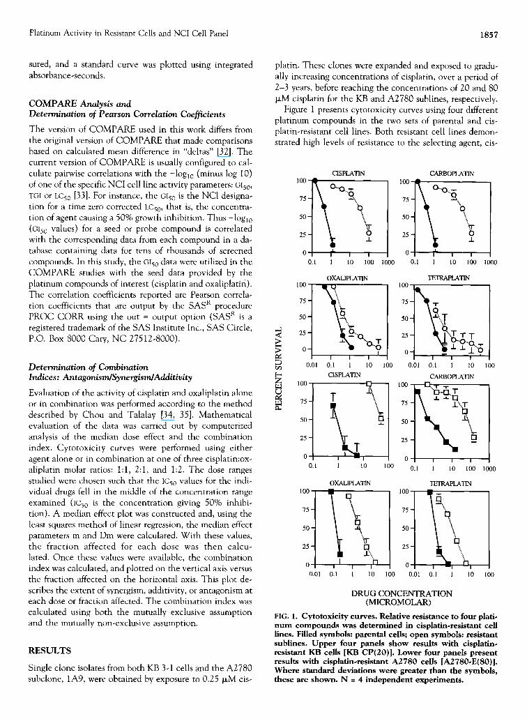
Figure 1 presents cytotoxicity curves using four different platinum compounds in the two sets of parental and cis- platin-resistant cell lines. Both resistant cell lines demon- strated high levels of resistance to the selecting agent, cis-
CISPLATIN 100 ~ 0 . . 0 , '
0 50
25 o
0 ~- ! I I 0.1 1 10 100 1000 1000
CARBOPLATIN 100 ~ O . ~ Q,
75
0 ..t_ I ll0 I 0.1 1 100
> m oq
100
75
50
25-
0-
0.01
100
75-
50-
25-
0 0.1
OXALIPLATIN
.o gO I
i i i 0.1 1 10 100 CISPLATIN
T
..i_
I - - ! 1 10 100
100
75-
50-
25-
0 0.01
OXALIPLATIN
Q .~...,
I , ,i-I I
0.1 1 10 100
"IEIRAPLATIN 100
75- ~ ~ . ,..!
50- ~"...~
25 . . . . T..T T
o_ I I I
0.01 0.1 1 10 CARBOPLATIN
1 0 0 - " ~ I . T -I" I~ "O..n. r
'oN% 25-
0 f I I
0.1 I 10 I 0 0
iPSIRAPLATIN
]oo Q, h 75
50
0 i i 0.01 0.1 1 10
100
1000
I 0 0
DRUG CONCENTRATION (MICROMOLAR)
FIG. 1. Cytotoxicity curves. Relative resistance to four plati- num compounds was determined in cisplatln-resistant cell lines. Filled symbols: parental cells; open symbols: resistant sublines. Upper four panels show results with cisplatin- resistant KB cells [KB CP(20)]. Lower four panels present results with cisplatin.resistant A2780 cells [A2780-E(80)l. Where standard deviations were greater than the symbols, these are shown. N = 4 independent experiments.
1858 O. Rixe et al.
platin, and also to carboplatin, with less cross-resistance to the two "dach" platinum compounds, oxaliplatin and tet- raplatin. These results as well as the resistance to two other agents are summarized in Table 1. As can be seen, com- pared with the resistance observed for cisplatin and carbo- platin, that to oxaliplatin and tetraplatin was less than one-tenth as great. In addition, one can see that both cell lines were also cross-resistant to the alkylating agent, mel- phalan, an observation that is addressed in greater detail below.
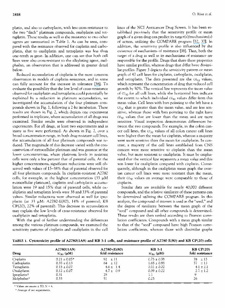
Reduced accumulation of cisplatin is the most common observation in models of cisplatin resistance, and in some can fully account for the increase in tolerance [36]. To evaluate the possibility that the low level of cross-resistance observed for oxaliplatin and tetraplatin could potentially be explained by a reduction in platinum accumulation, we investigated the accumulation of the four platinum com- pounds shown in Fig. 1, following a 2-hr incubation. These results are shown in Fig. 2, which depicts one experiment performed in triplicate, where accumulation of all drugs was examined. Similar results were observed in independent experiments. For all drugs, at least two experiments and as many as five were performed. As shown in Fig. 2, over a broad concentration range, in both drug-resistant cell lines, the accumulation of all four platinum compounds was re- duced. The magnitude of this decrease varied with the con- centration of extracellular platinum and was greatest at the lower concentrations, where platinum levels in resistant cells were only a few percent that of parental cells. At the higher concentrations, significant reductions were still ob- served with values of 15-38% that of parental observed for all four platinum compounds. In cisplatin-resistant A2780 cells, for example, at the highest concentration (33 >M extracellular platinum), cisplatin and carboplatin accumu- lation were 19 and 15% that of parental cells, while ox- aliplatin and tetraplatin levels were 38 and 33% of parental values. Similar reductions were observed as well for ipro- platin (at 33 IxM: A2780-E(80), 14% of parental; KB CP(20), 22% of parental). This decrease in accumulation may explain the low levels of cross-resistance observed for oxaliplatin and tetraplatin.
With the goal of further understanding the differences among the various platinum compounds, we examined the sensitivity patterns of cisplatin and oxaliplatin in the cell
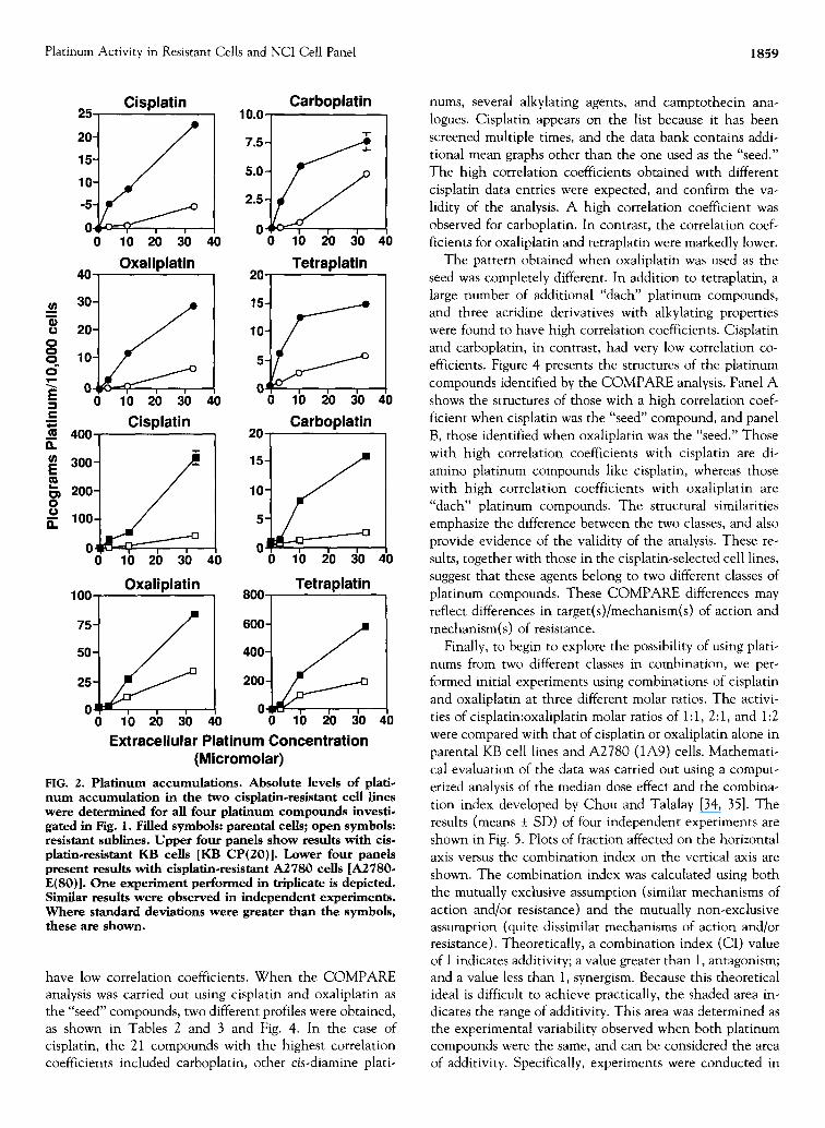
lines of the NCI Anticancer Drug Screen. It has been es- tablished previously that the sensitivity profile or mean graph of a given drug can predict its target(s)/mechanism(s) of action, utilizing the COMPARE program [32, 37]. In addition, the sensitivity profile is also influenced by the existence of mechanisms of resistance [38]. Thus, both the target of a drug as well as its mechanisms of resistance are responsible for the profile. Drugs that share these properties have similar profiles, whereas drugs that differ have dissimi- lar profiles. Figure 3 depicts the sensitivity pattern or mean graph of 47 cell lines for cisplatin, carboplatin, oxaliplatin, and tetraplatin. The data presented are the G150 values, which represent the concentration of drug that reduced cell growth by 50%. The vertical line represents the mean value of 0150 for all cell lines, while the horizontal bars indicate the extent to which individual GI50 values differ from this mean value. Cell lines with bars pointing to the left have a GI50 that is greater than the mean value, and are less sen- sitive, whereas those with bars pointing to the right have G150 values that are lower than the mean and are more sensitive. Visual inspection demonstrates differences be- tween the two compounds. For example, in the colon can- cer cell lines, the oi50 values of all colon cancer cell lines were higher than the mean for cisplatin, whereas a majority were more sensitive than the mean for oxaliplatin. In con- trast, a majority of the cell lines established from CNS cancers were more sensitive to cisplatin than the mean value, but more resistant to oxaliplatin. It must be empha- sized that the vertical line represents a mean value and this was lower for oxaliplatin compared with cisplatin. Conse- quently, although in the oxaliplatin mean graph the ovar- ian cancer cell lines were more resistant than the mean, their GI50 values on average were comparable to those of cisplatin.
Similar data are available for nearly 40,000 different compounds, and the relative similarity of these patterns can be determined utilizing the COMPARE program. In this analysis, the compound of interest is used as the "seed," and the degree of similarity between the mean graph of the "seed" compound and all other compounds is determined. These results are then ranked according to Pearson corre- lation coefficients. Compounds with a mean graph similar to that of the "seed" compound have high Pearson corre- lation coefficients, whereas those with dissimilar graphs
TABLE 1. Cytotoxicity profile of A2780(1A9) and KB 3-1 ceils, and resistance profile of A2780 E(80) and KB CP(20) cells
A2780(1A9) A2780.(E(80) KB 3.1 KB CP(20) Drug IC5o (IJM) fold resistance IC50 (laM) fold resistance
Cisplatin 0.21 _+ 0.05* 92 _+ 11 0.75 + 0.38 78 + 15 Carboplatin 0.35 + 0.13 64 + 12 1.65 + 0.88 57 + 13 Tetraplatin 0.15 + 0.07 4.6 + 1.4 0.11 + 0.02 4.1 + 2.1 Oxaliplatin 0.12 + 0.07 4.7 -+ 0.9 0.39 _+ 0.22 2.7 + 1.2 Iproplatint 0.31 28 1.1 9 Melphalant 0.33 51 0.25 26
* Values are means _+ SD, N = 4. t Average of two experiments.
Platinum Activity in Resistant Cells and NCI Cell Panel 1859
Cisplatin Carboplatin 25 10.0 .oy 15-
10-
-5-
O~ - , ' ~ " ~ ~ , , 0 10 20 30
Oxaliplatin 4O
I 40
7 . 5 - ~
5.0-
2.5-
O~ 0 10 20 30
Tetraplatin 20 ,
30-
20-
10-
0 10 20 30 40
~ 400 Cisplatin _
300
O ~ , , 0 10 20 30 40
Oxaliplatin 100
75-
50-
25-
0 0
10
5
0 0 10 20 30
Carboplatin 20
'iY 1
v I I I I
0 10 20 30
Tetraplatin 800
600 -
400 - S
10 2b 3b 40 0 10 20 30 Extracellular Platinum Concentration
(Micromolar)
40
40
40
40
FIG. 2. Platinum accumulations. Absolute levels of plati- num accumulation in the two cisplatin-resistant cell lines were determined for all four platinum compounds investi- gated in Fig. 1. Filled symbols: parental cells; open symbols: resistant sublines. Upper four panels show results with cis. platin.resistant KB cells [KB CP(20)]. Lower four panels present results with cisplatin-resistant A2780 cells [A2780- E(80)]. One experiment performed in triplicate is depicted. Similar results were observed in independent experiments. Where standard deviations were greater than the symbols, these are shown.
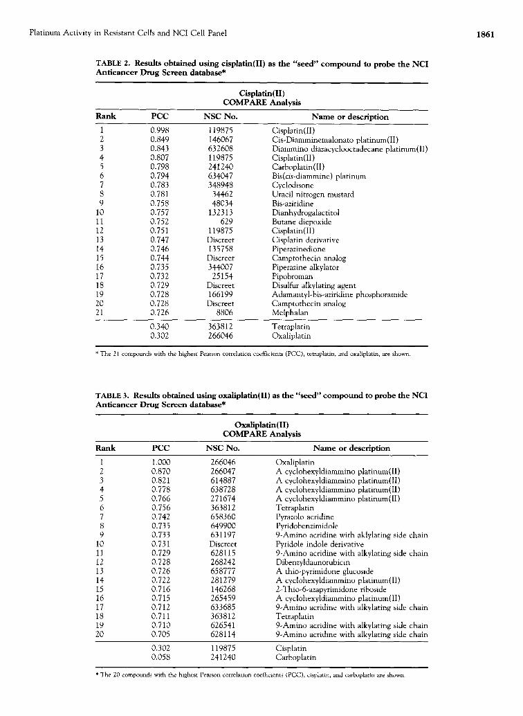
have low correlation coefficients. When the COMPARE analysis was carried out using cisplatin and oxaliplatin as the "seed" compounds, two different profiles were obtained, as shown in Tables 2 and 3 and Fig. 4. In the case of cisplatin, the 21 compounds with the highest correlation coefficients included carboplatin, other cis.diamine plati-
nums, several alkylating agents, and camptothecin ana- logues. Cisplatin appears on the list because it has been screened multiple times, and the data bank contains addi- tional mean graphs other than the one used as the "seed." The high correlation coefficients obtained with different cisplatin data entries were expected, and confirm the va- lidity of the analysis. A high correlation coefficient was observed for carboplatin. In contrast, the correlation coef- ficients for oxaliplatin and tetraplatin were markedly lower.
The pattern obtained when oxaliplatin was used as the seed was completely different. In addition to tetraplatin, a large number of additional "dach" platinum compounds, and three acridine derivatives with alkylating properties were found to have high correlation coefficients. Cisplatin and carboplatin, in contrast, had very low correlation co- efficients. Figure 4 presents the structures of the platinum compounds identified by the COMPARE analysis. Panel A shows the structures of those with a high correlation coef- ficient when cisplatin was the "seed" compound, and panel B, those identified when oxaliplatin was the "seed." Those with high correlation coefficients with cisplatin are di- amino platinum compounds like cisplatin, whereas those with high correlation coefficients with oxaliplatin are "dach" platinum compounds. The structural similarities emphasize the difference between the two classes, and also provide evidence of the validity of the analysis. These re- suits, together with those in the cisplatin-selected cell lines, suggest that these agents belong to two different classes of platinum compounds. These COMPARE differences may reflect differences in target(s)/mechanism(s) of action and mechanism(s) of resistance.
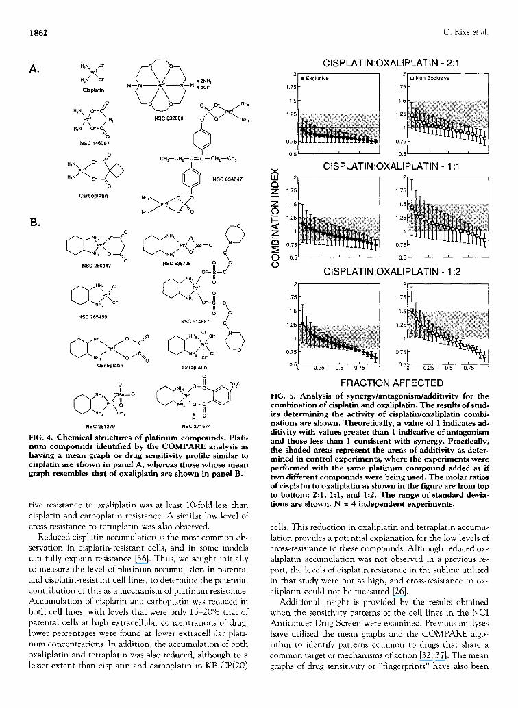
Finally, to begin to explore the possibility of using plati- nums from two different classes in combination, we per- formed initial experiments using combinations of cisplatin and oxaliplatin at three different molar ratios. The activi- ties of cisplatin:oxaliplatin molar ratios of 1:1, 2:1, and 1:2 were compared with that of cisplatin or oxaliplatin alone in parental KB cell lines and A2780 (1A9) cells. Mathemati- cal evaluation of the data was carried out using a comput- erized analysis of the median dose effect and the combina- tion index developed by Chou and Talalay [34, 35]. The results (means + SD) of four independent experiments are shown in Fig. 5. Plots of fraction affected on the horizontal axis versus the combination index on the vertical axis are shown. The combination index was calculated using both the mutually exclusive assumption (similar mechanisms of action and/or resistance) and the mutually non-exclusive assumption (quite dissimilar mechanisms of action and/or resistance). Theoretically, a combination index (CI) value of 1 indicates additivity; a value greater than 1, antagonism; and a value less than 1, synergism. Because this theoretical ideal is difficult to achieve practically, the shaded area in- dicates the range of additivity. This area was determined as the experimental variability observed when both platinum compounds were the same, and can be considered the area of additivity. Specifically, experiments were conducted in

1860 O. Rixe et al.
C I S P L A T I N C A R B O P L A T I N O X A L I P L A T I N T E T R A P L A T I N
G | 5 0 GI50 GI50 GI50
Leukemia CCRF CEM HL-60(TB) K-562 MOLT-4 RPMI-8226
Non-Small Dell Lung Cancer A549/ATCC HOP-62 HOP 92 NCI-H226 NCI-H23 NCI-H460 NCI-H522
Small Cell Lung Cancer DMS 144 DMS 273
Colon Cancer COLO 2O5 DLD-1 HCC-2998 HCT-116 HCT-15 HT29 KM2OL2 SW-620
CNS Canc¢¢ SF-268 SF 29S SF-539 SNB-19 SNB-75 SNB 78 U251
MelanOma LOX IMVI MALME 3M M19-MEL SK-MEL-2 SK-MEL-28 SK-MEL-5 UACC-257 UACC-62
Ovarian Cancer ]GROV1 OVCAR-3 OVCAR-4 OVCAR-5 OVCAR-8 SK OV-3
Renal Cancer A498 CAKI-1 SN12C UO 31
MG-MID Delta
Range I I I I I I I I I . . . . . I . . . . . . . . . . . . . . . . . . . . . . . . 22 -3 /3 ~ . . . . . . . . . .
FIG. 3. Mean graph for cisplatin, carboplatin, oxaliplatin, and tetraplatin. The sensitivities of the cell lines in the National Cancer Instimte's Anticancer Drug Screen Program for cisplatin, carboplatln, oxaliplatln, and tetraplatin are depicted. The vertical line represents the mean Glso (concentration required to inhibit 50% growth) for all the cell lines. The horizontal bars represent the difference in the Gl~o of a particular cell line relative to the mean. A log scale is utilized. Cell lines with a horizontal bar pointing to the left have a Glso that is greater than the mean and are more resistant than the mean, whereas cell lines with a horizontal bar pointing to the right have a Glso that is less than the mean and are more sensitive.
which the activities of different molar ratios of cisplatin: cisplatin and oxaliplatin:oxaliplatin were determined. Be- cause the same platinum compound was used as both plati- num compounds, a combination index of 1 would have been expected, theoretically, since combining the same platinum compound should be simply additive, not syner- gistic or antagonistic. However, experimental variation around the theoretical value of 1 was obtained, and it is this range of variability that is depicted as the shaded area. Additivity (and possibly some synergism) was observed throughout the range of concentrations tested; some ex- periments demonstrated marked synergism. Taken to- gether, the results suggest that in combination the activities of these two platinum compounds are at least additive and possibly synergistic.
D I S C U S S I O N
In the present study, we report our findings on the activities of four different platinum compounds in both drug-selected and unselected cell lines. Beginning with cisplatin-selected A2780 ovarian and KB 3-1 epidermoid cells, we found low levels of cross-resistance to both oxaliplatin and tetraplatin,
although high levels of resistance to both cisplatin and carboplatin were demonstrable. Data in the NCI Antican- cer Drug Screen demonstrated different patterns in the COMPARE analysis for cisplatin and oxaliplatin. The lat- ter provides evidence that the compounds may have dis- similar targets, mechanisms of resistance, or both. Studies to determine the effectiveness of cisplatin and oxaliplatin in combination suggest that these drugs are at least additive and possibly synergistic. These results confirm and extend previous observations documenting differences in the resis- tance profile for different platinum compounds [27, 39-44].
Selection of A2780 and KB 3-1 cells with cisplatin was successful in establishing sublines with high levels of cis- platin resistance after prolonged incubation in medium containing gradually increasing concentrations of cisplatin. High levels of cross-resistance to carboplatin were observed. In contrast, lower levels of cross-resistance were found to both oxaliplatin and tetraplatin. These results confirm and extend previous observations in cisplatin-resistant cells [25-27, 41-43]. Cross-resistance to oxaliplatin was some- what higher in the cisplatin-resistant A2780 E(80) cells, probably a result of the higher level of cisplatin resistance in this subline; but in both cisplatin-resistant sublines, rela-
Platinum Activity in Resistant Cells and NCI Cell Panel 1861
TABLE 2. Results obtained using cisplatin(II) as the "seed" compound to probe the NCI Anticancer Drug Screen database*
Cisplatin(II) COMPARE Analysis
Rank PCC NSC No. Name or description
1 0.998 1 1 9 8 7 5 Cisplatin(II) 2 0.849 1 4 6 0 6 7 Cis-Diamminemalonato platinum(II) 3 0.843 632608 Diammino diazacyclooctadecane platinum(II) 4 0.807 1 1 9 8 7 5 Cisplatin(II) 5 0.798 241240 Carboplatin(II) 6 0.794 634047 Bis(cis-diammine) platinum 7 0.783 348948 Cyclodisone 8 0.781 34462 Uracil nitrogen mustard 9 0.758 48034 Bis-aziridine
10 0.757 1 3 2 3 1 3 Dianhydrogalactitol 11 0.752 629 Butane diepoxide 12 0.751 119875 Cisplatin(II ) 13 0.747 Discreet Cisplatin derivative 14 0.746 1 3 5 7 5 8 Piperazinedione 15 0.744 Discreet Camptothecin analog 16 0.735 344007 Piperazine alkylator 17 0.732 2 5 1 5 4 Pipobroman 18 0.729 Discreet Disulfur alkylating agent 19 0.728 1 6 6 1 9 9 Adamantyl-bis-aziridine phosphoramide 20 0.728 Discreet Camptothecin analog 21 0.726 8806 Melphalan
0.340 363812 Tetraplatin 0.302 266046 Oxaliplatin
* The 21 compounds with the highest Pearson correlation coefficients (PCC), tetraplatin, and oxaliplatin, are shown.
TABLE 3. Results obtained using oxaliplatin(II) as the "seed" compound to probe the NCI Anticancer Drug Screen database*
Oxaliplatin(lI) COMPARE Analysis
Rank PCC NSC No. Name or description
1 1.000 266046 2 0.870 266047 3 0.821 614887 4 0.778 638728 5 0.766 271674 6 0.756 363812 7 0.742 658360 8 0.735 649900 9 0.733 631197
10 0.731 Discreet 11 0.729 628115 12 0.728 268242 13 0.726 658777 14 0.722 281279 15 0.716 146268 16 0.715 265459 17 0.712 633685 18 0.711 363812 19 0.710 626541 20 0.705 628114
Oxaliplatin A cyclohexyldiammino platinum(II) A cyclohexyldiammino platinum(lI) A cyclohexyldiammino platinum(II) A cyclohexyldiammino platinum(II) Tetraplatin Pyrazolo acridine Pyridobenzimidole 9-Amino acridine with aklylating side chain Pyridole indole derivative 9-Amino acridine with alkylating side chain Dibenzyldaunorubicin A thio-pyrimidone glucoside A cyclohexyldiammino platinum(II) 2-Thio-6-azapyrimidone riboside A cyclohexyldiammino platinum(II) 9-Amino acridine with alkylating side chain Tetraplatin 9-Amino acridine with alkylating side chain 9-Amino acridine with alkylating side chain
0.302 119875 Cisplatin 0.058 241240 Carboplatin
* The 20 compounds with the highest Pearson correlation coefficients (PCC), cisplatin, and carboplatin are shown.
1862 O. Rixe et al.
A.
B.
H~N x (Cl" P \
H=N Cl-
CIsplatln
H~N\ / O - - < O
Pt 4 /CH 2 / \ H~N O ' - C \
O NSC 146067
H'Nxpl.//O" " ~
H~N / NO_ ~ . ~ V %
Carboplatin
\ / \ 2 NSC 632608
• 2NH~ • 2cr
CH:i--CHz--C~ C- - CH2-- CH~
NSC 634047
f3 f " r - ' " , " ' / ° -~ ° r " "~ " " . ;~, pt- 2 [ I Pt'N / S e = O
\ O C NSC 266047 NSC 638728 O = ISI --C /
C NSC 265459 O /
NSC 614887 Ck '
c r
%,,.-<,,,:-" ~ , / ° , , °
Oxaliplatln Tetraplatin
0 o- II -o c l ( ~ - (" ° ' - c ~ ' ~ '
A N H = -OSe = O ~H~Ip~ "/ r" "r" \ .,/, v . . .A . . / . , o_c ~..>.,.~j L .L / ~ \ o tl
V "NH 2 OH t ~+o NSC 281279 NSC 271674
FIG. 4. Chemical structures of platinum compounds. Plati. num compounds identified by the COMPARE analysis as having a mean graph or drug sensitivity profile similar to cisplatin are shown in panel A, whereas those whose mean graph resembles that of oxaliplatin are shown in panel B.
tive resistance to oxaliplatin was at least 10-fold less than cisplatin and carboplatin resistance. A similar low level of cross-resistance to tetraplatin was also observed.
Reduced cisplatin accumulation is the most common ob- servation in cisplatin-resistant ceils, and in some models can fully explain resistance [36]. Thus, we sought initially to measure the level of platinum accumulation in parental and cisplatin-resistant cell lines, to determine the potential contribution of this as a mechanism of platinum resistance. Accumulat ion of cisplatin and carboplatin was reduced in both cell lines, with levels that were only 15-20% that of parental cells at high extracellular concentrations of drug; lower percentages were found at lower extracellular plati- num concentrations. In addition, the accumulation of both oxaliplatin and tetraplatin was also reduced, although to a lesser extent than cisplatin and carboplatin in KB CP(20)
X IJJ Q Z
z _o I-- <l: z 03
O O
2
1.75
1,5
1.25
1
0.75
0.5
C I S P L A T I N : O X A L I P L A T I N - 2:1 2
1.75
1.5
1.25
1
0.75
0.5
C I S P L A T I N : O X A L I P L A T I N - 1:1 2
1.75 1.75
1.5 1.5 ~: ,
1.25 125 ~ i: i~:; ;. : / i
1 1 ' ~ > {:
0 . 7 5 0 . 7 5
0.5 ~ 05 ~
C I S P L A T I N : O X A L I P L A T I N - 1:2
1.7
1.
1.2
0.7
0. O U.ZD U.D U.IO l
FRACTION AFFECTED
FIG. 5. Analysis of synergy/antagonism/additivity for the combination of cisplatin and oxaliplatin. The results of stud- ies determining the activity of cisplatirdoxaliplatin combi. nations are shown. Theoretically, a value of 1 indicates ad- ditivity with values greater than 1 indicative of antagonism and those less than 1 consistent with synergy. Practically, the shaded areas represent the areas of additivity as deter. mined in control experiments, where the experiments were performed with the same platinum compound added as if two different compounds were being used. The molar ratios of cisplatin to oxaliplatin as shown in the figure are from top to bottom: 2:1, 1:1, and 1:2. The range of standard devia. tions are shown. N = 4 independent experiments.
cells. This reduction in oxaliplatin and tetraplatin accumu- lation provides a potential explanation for the low levels of cross-resistance to these compounds. Although reduced ox- aliplatin accumulation was not observed in a previous re- port, the levels of cisplatin resistance in the subline utilized in that study were not as high, and cross-resistance to ox- aliplatin could not be measured [26].
Additional insight is provided by the results obtained when the sensitivity patterns of the cell lines in the NCI Anticancer Drug Screen were examined. Previous analyses have utilized the mean graphs and the COMPARE algo- rithm to identify patterns common to drugs that share a common target or mechanisms of action [32, 37]. The mean graphs of drug sensitivity or "fingerprints" have also been

Platinum Activity in Resistant Cells and NCI Cell Panel 1863
used to identify putative targets of new or unknown com- pounds. The high correlations observed in these analyses have suggested that the target(s) or the mechanism(s) of action is an important determinant of drug sensitivity as determined in the Drug Screen assays. In addition, recent evidence using a drug-resistance measurement (mdr-1 ex- pression) as the "seed" suggests that the mechanism(s) of resistance also influences the drug sensitivity profile [38]. Thus, the sensitivity pattern of a drug appears to depend on both its target or mechanisms(s) of action as well as its mechanism(s) of resistance. The dissimilar patterns ob- tained for cisplatin and oxaliplatin, as evidenced by the low Pearson correlation coefficients, suggest but do not prove that both are different. Differences in the mechanism(s) of resistance were suggested by the cross-resistance data in the cisplatin-tolerant cells, whereas differences in the target(s)/ mechanism(s) or action are inferred from the COMPARE analysis. Clarification of the latter would be important in furthering our understanding of the mechanism(s) of action of platinum compounds. Biophysical analysis of DNA modified by "dach" platinum complexes indicates that al- though adducts similar to those of cisplatin are formed by the "dach" platinums, different effects on DNA appear to result from steric crowding of the axially oriented cyclo- hexane ring [45, 46].
Visual inspection of the mean graphs shows differences in the sensitivity patterns for both the colon cancer and CNS cancer subpanels. Oxaliplatin was more toxic to the colon cancer cell lines than to the panel as a whole, whereas it appeared less active against the cell lines derived from CNS tumors. These results extend previous observa- tions in a smaller number of cell lines [25]. Further studies will be required to pursue these observations, although de- termination of clinical activity will be most important. Pre- liminary results suggest that oxaliplatin may have some ac- tivity against colon carcinomas [18, 47].
Examination of the COMPARE analysis provides further information of the differences between different platinum compounds. For oxaliplatin, high Pearson correlation coef- ficients were observed principally with other "dach" plati- num compounds and several acridine derivatives. In con- trast, for cisplatin, high correlation coefficients were found with platinum agents containing diaminoplatinum, and also with camptothecin analogs and alkylating agents in- cluding uracil nitrogen mustard and melphalan. The latter result supports the observation of cross-resistance to mel- phalan in the cisplatin-selected cell lines. Although cyclo- phosphamide cannot be tested in vitro, one prediction of these results is that cross-tolerance to cyclophosphamide may be observed when this drug is used in combination with cisplatin. Combinations of either cisplatin and ox- aliplatin or cyclophosphamide and oxaliplatin may be less cross-resistant.
To address this latter issue, and to obtain preliminary in vitro data on the use of platinum compounds from different classes in combination, we have performed preliminary

1864 O. Rixe et al.
carcinoma cell lines by buthionine sulfoximine mediated glu- tathione depletion. Biochem Pharmacol 34" 2583-2586, 1985.
10. Mann SC, Andrews PA and Howell SB, Modulation of cis- diamminedichloroplatinum(II) accumulation and sensitivity by forskolin and 3-isobutyl-l-methylxanthine in sensitive and resistant human ovarian carcinoma cells. Int J Cancer 48: 866-872, 1991.
11. Perez RP, Handel LM and Hamilton TC, Potentiation of cisplatin cytotoxicity in human ovarian carcinoma cell lines by trifluoperazine, a calmodulin inhibitor. Gynecol Oncol 46" 82-87, 1992.
12. Connors TA, Jones M, Ross WC, Braddock PD, Khokhar AR and Lobe ML, New platinum complexes with anti-tumor ac- tivity. Chem Biol Inter~t 5: 415-424, 1972.
13. Burchenal JH, Kalaher K, O'Toole T and Chisholm J, Lack of cross-resistance between certain platinum coordination com- pounds in mouse leukemia. Cancer Res 37: 3455-3457, 1977.
14. Kidani Y, Inagaki K, Saito R and Tsukagoshi S, Synthesis and anti-tumor activities of platinum(II) complexes of 1,2-diami- nocyclohexane isomers and their related derivatives. J Clin Hematol Oncol 7" 197-202, 1977.
15. Kidani Y, Noji M and Tashiro T, Antitumor activity of plat- inum(II) complexes of 1,2-diaminocyclohexane isomers. Gann 71: 637-643, 1980.
16. Math6 G, Kidani Y, Noji M, Maral R, Bourut C and Chenu E, Antitumor activity of I-OHP in mice. Cancer Lett 27" 135-143, 1985.
17. Levi F, Adam R, Caussanel JP, Lecouturier S and Jasmin C, A chronopharmacologic phase II clinical trial with 5-fluoroura- cil, folinic acid, and oxaliplatin using an ambulatory multi- channel programmable pump. Cancer 69: 893-900, 1992.
18. Levi F, Brienza S, Focan C, Perpoint B, Garufi C, Gatt G, La Torre Y, Gastiaburu J, Depres P and Misset JL. A phase II chronopharmacological trial of oxaliplatin (L-OHP) against metastatic colorectal cancer. Proc Am Assoc Cancer Res 32" 185, 1991.
19. Extra JM, Espie M, Calvo F, Ferme C, Mignot L and Marty M, Phase I study of oxaliplatin in patients with advanced cancer. Cancer Chemother Pharmacol 25: 299-303, 1990.
20. Caussanel JP, Levi F, Brienza S, Misset JL, Itzhaki M, Adam R, Milano G, Hecquet B and Mathe G, Phase I trial of 5-day continuous venous infusion of oxaliplatin at circadian rhythm-modulated rate compared with constant rate. J Natl Cancer Inst 82: 1046-1050, 1990.
21. Misset JL, Soulle P, Fereres M, Llory JF, Brain E, Musset M, Chollet P, Jasmin C, Garrino C and Cvitkovic E, High ac- tivity of combined oxaliplatin (L-OHP) cisplatin (CP) as sal- vage treatment in pretreated ovarian cancer (OC). Proc Am Soc Clin Oncol 94 13: 273, 1994.
22. Soulle P, Llory JF, Fereres M, Garrino C, Gastlaburu S, Brienza S, Bensmaine MA, Misset JL and Cvitkovic E, Pre- liminary results of an active oxaliplatin (LOHP)-CDDP as- sociation based salvage program in pretreated germ cell tu- mors. Proc ASCO 94 13: 250, 1994.
23. Chollet P, Bensmaine MA, Deloche C, Gastiaburu J, Cvit- kovic E and Brienza S, Phase II study of oxaliplatin (L-OHP, transplatin) a new active agent in cis and/or carboplatin pre- treated ovarian cancer (OC). Proc ASCO 94 13: 163, 1994.
24. Garrino C, Cvitkovic E, Soulle P, Llory JF, Musset M, Fereres M, Bensmaine MA, Brain E, Gastiaburu J, Brienza S, Itzhaki M, Jasmin C and Misset JL, Preliminary report on the toler- ance of transplatin (LOHP) cisplatin (CP) association alone (Bi) or in combination with ifosfamide and epirubicin (Bic) in platinum (PT) pretreated patients. Proc ASCO 94 13" 143, 1994.
25. Pendyala L and Creaven PJ, In vitro cytotoxicity, protein binding, red blood cell partitioning, and biotransformation of oxaliplatin, Cancer Res 53" 5970-5976, 1993.
26. Kraker AJ and Moore CW, Accumulation of cis-diam- minedichloroplatinum(II) and platinum analogues by plati- num-resistant murine leukemia cells in vitro. Cancer Res 48: 9-13, 1988.
27. Tashiro T, Kawada Y, Sakurai Y and Kidani Y, Antitumor activity of a new platinum complex, oxalato (trans-l-l,2,- diaminocyclohexane)platinum(II): New experimental data. Biomed Pharmacother 43: 251-260, 1989.
28. Paull KD, Hamel E and Malspeis L, Prediction of biochemical mechanisms of action from the in vitro antitumor screen of the National Cancer Institute. In: Cancer Chemotherapeutic Agents (Ed. Foye WE), pp. 9-45. American Chemical Society, Washington, DC, 1995.
29. Monks A, Scudiero D, Skehan P, Shoemaker R, Paull K, Vistica D, Hose C, Langley J, Cronise P, Vaigro-Wolff A, Gray-Goodrich M, Campbell H, Mayo J and Boyd M, Feasi- bility of a high-flux anticancer drug screen using a diverse panel of cultured human tumor cell lines. J Natl Cancer Inst 83: 757-766, 1991.
30. McGahan MC and Tyczkowska K, The determination of platinum in biological materials by electrothermal atomic ab- sorption spectroscopy. Spectrochim Acta 42B- 665-668, 1987.
31. Parker RJ, Eastman A, Bostick-Bruton F and Reed E, Ac- quired cisplatin resistance in human ovarian cancer cells is associated with enhanced repair of cisplatin-DNA lesions and reduced drug accumulation. J Clin Invest 87: 772-777, 1991.
32. Pautl KD, Shoemaker RH, Hodes L, Monks A, Scudiero DA, Rubinstein L, Plowman J and Boyd MR, Display and analysis of patterns of differential activity of drugs against human tu- mor cell lines: Development of mean graph and COMPARE algorithm. J Natl Cancer Inst 81: 1088-1092, 1989.
33. Boyd MR, Paull KD and Rubinstein LR, Data display and analysis strategies for the NCI disease oriented in-vitro anti- tumor drug screen. In: Cytotoxic Anticancer Drugs: Models and Concept for Drug Discovery and Development (Eds. Valeriote FA, Corbett TH and Baker LH), pp. 11-34. Kluwer Aca- demic Publishers, Boston, 1992.
34. Chou T-C and Talalay P, Quantitative analysis of dose-effect relationships: The combined effects of multiple drugs or en- zyme inhibitors. Adv Enzyme Regul 22: 27-55, 1984.
35. Chou T-C, Quantitation of synergism and antagonism of two or more drugs of computerized analysis. In: Synergism and An- tagonism in Chemotherapy (Eds. Chou TC and Rideout DC), pp. 223-241. Academic Press, San Diego, 1991.
36. Andrews PA and Howell SB, Cellular pharmacology of cis- platin: Perspectives on mechanisms of acquired resistance. Cancer Cells 2: 35-43, 1990.
37. Paull KD, Lin CM, Malspeis L and Hamel E, Identification of novel antimitotic agents acting at the tubulin level by com- puter-assisted evaluation of differential cytotoxicity data. Cancer Res 52: 3892-3900, 1992.
38. Lee J-S, Paull K, Alvarez M, Hose C, Monks A, Grever M, Fojo AT and Bates SE, Rhodamine efflux patterns predict P-glycoprotein substrates in the National Cancer Institute drug screen. Mol Pharmacol 46: 627-638, 1994.
39. Harrap KR, Jones M, Siracky J, Pollard LA and Kelland LR, The establishment, characterization and calibration of human ovarian carcinoma xenografts for the evaluation of novel platinum anticancer drugs. Ann Oncol 1: 65-76, 1990.
40. Jones M, Siracky J, Kelland LR and Harrap KR, Acquisition of platinum drug resistance and platinum cross resistance pat- terns in a panel of human ovarian carcinoma xenografts. Br J Cancer 67: 24-29, 1993.
41. Los G, Mutsaers PHA, Ruevekamp M and McVie JG, The use of oxaliplatin versus cisplatin in intraperitoneal chemo- therapy in cancers restricted to the peritoneal cavity in the rat. Cancer Lett 51" 109-117, 1990.
42. Behrens BC, Hamilton TC, Masuda H, Grotzinger KR,

Platinum Activity in Resistant Cells and NCI Cell Panel 1865
Whang-Peng J, Louie KG, Knutsen T, McKoy WM, Young RC and Ozols RF, Characterization of a cis-diamminedichlo- roplatinum(II)-resistant human ovarian cancer cell line and its use in evaluation of platinum analogues. Cancer Res 47: 414418, 1987.
43. Perez RP, O'Dwyer PJ, Handel LM, Ozols RF and Hamilton TC, Comparative cytotoxicity of C1-973, cisplatin, carbopla- tin and tetraplatin in human ovarian carcinoma cell lines. Int J Cancer 48: 265-269, 1991.
44. Parker RJ, Vionnet JA, Bostick-Bruton F and Reed E, Orma- platin sensitivity/resistance in human ovarian cancer cells made resistant to cisplatin. Cancer Res 53" 242-247, 1993.
45. Boudny V, Vrana O, Gaucheron F, Kleinwatcher V, Leng M and Brabec V, Biophysical analysis of DNA modified by 1,2- diaminocyclohexane platinum(II) complexes. Nucleic Acids Res 20: 267-272, 1991.
46. Jennerwein MM, Eastman A and Khokhar A, Characteriza-
47.
48.
tion of adducts produced in DNA by isomeric 1,2- diaminocyclohexaneplatinum(II) complexes. Chem Biol Inter- act 70: 39-49, 1989. Misset JL, Kidani Y, Gastiaburu J, Jasmin C, Levi F, Boughat- tas N, Lemaigre G, Causannel JP, Brienza S, Kim Triana B, Goldschmidt E, Musset M, Mauvernay RY and Mathe G, Oxalatoplatinum (L-OHP): Experimental and clinical studies. In: Platinum and Other Metal Coordination Compounds in Can- cer Chemotherapy (Ed. Howell SB), pp. 369-375. Plenum Press, New York, 1991. Math~ G, Kidani Y, Segiguchi M, Eriguchi M, Fredj G, Pey- tavin G, Misset JL, Brienza S, de Vassals F, Chenu E and Bourut C, Oxalato-platinum or 1-OHP, a third-generation platinum complex: An experimental and clinical appraisal and preliminary comparison with cis-platinum and carbo- platinum. Biomed Pharmacother 43: 237-250, 1989.