Overexpression of Glucose6Phosphate Dehydrogenase Is Associated with Lipid Dysregulation and Insulin...
12
MOLECULAR AND CELLULAR BIOLOGY, June 2005, p. 5146–5157 Vol. 25, No. 12 0270-7306/05/$08.000 doi:10.1128/MCB.25.12.5146–5157.2005 Copyright © 2005, American Society for Microbiology. All Rights Reserved. Overexpression of Glucose-6-Phosphate Dehydrogenase Is Associated with Lipid Dysregulation and Insulin Resistance in Obesity† Jiyoung Park, Ho Kyung Rho, Kang Ho Kim, Sung Sik Choe, Yun Sok Lee, and Jae Bum Kim* School of Biological Sciences, Seoul National University, Seoul 151-742, South Korea Received 16 September 2004/Returned for modification 20 October 2004/Accepted 11 March 2005 Glucose-6-phosphate dehydrogenase (G6PD) produces cellular NADPH, which is required for the biosyn- thesis of fatty acids and cholesterol. Although G6PD is required for lipogenesis, it is poorly understood whether G6PD in adipocytes is involved in energy homeostasis, such as lipid and glucose metabolism. We report here that G6PD plays a role in adipogenesis and that its increase is tightly associated with the dysregulation of lipid metabolism and insulin resistance in obesity. We observed that the enzymatic activity and expression levels of G6PD were significantly elevated in white adipose tissues of obese models, including db/db, ob/ob, and diet- induced obesity mice. In 3T3-L1 cells, G6PD overexpression stimulated the expression of most adipocyte marker genes and elevated the levels of cellular free fatty acids, triglyceride, and FFA release. Consistently, G6PD knockdown via small interfering RNA attenuated adipocyte differentiation with less lipid droplet accumulation. Surprisingly, the expression of certain adipocytokines such as tumor necrosis factor alpha and resistin was increased, whereas that of adiponectin was decreased in G6PD overexpressed adipocytes. In accordance with these results, overexpression of G6PD impaired insulin signaling and suppressed insulin- dependent glucose uptake in adipocytes. Taken together, these data strongly suggest that aberrant increase of G6PD in obese and/or diabetic subjects would alter lipid metabolism and adipocytokine expression, thereby resulting in failure of lipid homeostasis and insulin resistance in adipocytes. Adipocytes are highly specialized cell type that maintains whole-body energy homeostasis by regulating glucose and lipid metabolism (60, 61). For a long time, adipocytes have been considered as an energy depot that stores and mobilizes tri- glyceride, a major energy fuel. Recently, adipocytes are also being recognized as an endocrine tissue with the discoveries of several adipocyte-secreted molecules, including lipid metabo- lites and adipocytokines (55). Using these molecules, adipo- cytes are able to communicate with other tissues and organs to regulate lipid and glucose metabolism, energy balance, insulin action, and reproduction. To maintain lipid homeostasis, adipocytes carry out two re- ciprocal biochemical processes: lipogenesis and lipolysis. These two processes are tightly controlled by several hormones, lipid metabolites, and nutritional conditions such as feeding and fasting. With these signals, lipogenic transcription factors, in- cluding ADD1/SREBP1c, LXRs, and PPAR actively partici- pate in lipid metabolism in adipocytes. These transcription factors are highly expressed in adipose tissue and play a key role in adipocyte differentiation by coordinating lipogenic and adipocyte-specific gene expression (50). Because lipogenesis is tightly linked with adipogenesis, it is difficult to distinguish the two processes—lipogenesis and adipogenesis—during adipo- cyte differentiation. Malonyl coenzyme A (malonyl-CoA) and NADPH are con- sidered to be essential elements in lipogenesis (29, 54). Malo- nyl-CoA, which is produced by acetyl coenzyme A (acetyl- CoA) carboxylase (ACC), is a major substrate for long-chain fatty acid synthesis. It also suppresses fatty acid oxidation by inhibiting carnitine palmitoyltransferase (CPT-1) and acts as a signal molecule in appetite regulation (24, 52, 53, 66). There- fore, ACC2-null mice, with defects in malonyl-CoA synthesis, show a higher fatty acid oxidation rate and a significant loss of body fat mass (1, 2). NADPH also contributes to fatty acid synthesis by supplying reducing power. NADPH is produced by several enzymes, including malic enzyme (ME) and the first two enzymes of the pentose phosphate pathway (PPP), glu- cose-6-phosphate dehydrogenase (G6PD) and 6-phosphoglu- conate dehydrogenase (6PGD) (54). Although isocitrate dehy- drogenase (IDH) also generates NADPH, its major function is irrelevant to lipogenesis (21, 54). Since NADPH is indispens- able for the synthesis of fatty acids and cholesterol, it is plau- sible to speculate that NADPH producing enzymes might be associated with lipid metabolism disorders such as hyperlipid- emia and lipid toxicity in metabolic diseases, including obesity and diabetes. Among the four NADPH-producing enzymes, G6PD is the rate-limiting enzyme of PPP, and it is highly conserved in most mammalian species (33). G6PD plays a key role in the main- tenance of redox potential and cell survival via production of NADPH and pentose phosphates (33). Previous researches on G6PD have been mostly focused on erythrocytes since G6PD deficiency in erythrocytes is a common human enzymopathy which results from a mutation in the G6PD gene (8). Other G6PD studies have been directed to cell growth, survival, and redox regulation with nonerythrocyte cells, including fibro- blasts, hepatocytes, and neuronal cells (9, 33, 38, 54). G6PD also participates in reductive biosynthesis of fatty acids and cholesterol. Regarding the lipogenic activity of G6PD, it has been demonstrated that hepatic G6PD is regulated by nutri- * Corresponding author. Mailing address: School of Biological Sci- ences, Seoul National University, San 56-1, Sillim-Dong, Kwanak-Gu, Seoul 151-742, South Korea. Phone: 82-2-880-5852. Fax: 82-2-878- 5852. E-mail: [email protected]. † Supplemental material for this article may be found at http://mcb .asm.org/. 5146 on January 6, 2015 by guest http://mcb.asm.org/ Downloaded from
Transcript of Overexpression of Glucose6Phosphate Dehydrogenase Is Associated with Lipid Dysregulation and Insulin...

MOLECULAR AND CELLULAR BIOLOGY, June 2005, p. 5146–5157 Vol. 25, No. 120270-7306/05/$08.00�0 doi:10.1128/MCB.25.12.5146–5157.2005Copyright © 2005, American Society for Microbiology. All Rights Reserved.
Overexpression of Glucose-6-Phosphate Dehydrogenase Is Associatedwith Lipid Dysregulation and Insulin Resistance in Obesity†
Jiyoung Park, Ho Kyung Rho, Kang Ho Kim, Sung Sik Choe, Yun Sok Lee, and Jae Bum Kim*School of Biological Sciences, Seoul National University, Seoul 151-742, South Korea
Received 16 September 2004/Returned for modification 20 October 2004/Accepted 11 March 2005
Glucose-6-phosphate dehydrogenase (G6PD) produces cellular NADPH, which is required for the biosyn-thesis of fatty acids and cholesterol. Although G6PD is required for lipogenesis, it is poorly understood whetherG6PD in adipocytes is involved in energy homeostasis, such as lipid and glucose metabolism. We report herethat G6PD plays a role in adipogenesis and that its increase is tightly associated with the dysregulation of lipidmetabolism and insulin resistance in obesity. We observed that the enzymatic activity and expression levels ofG6PD were significantly elevated in white adipose tissues of obese models, including db/db, ob/ob, and diet-induced obesity mice. In 3T3-L1 cells, G6PD overexpression stimulated the expression of most adipocytemarker genes and elevated the levels of cellular free fatty acids, triglyceride, and FFA release. Consistently,G6PD knockdown via small interfering RNA attenuated adipocyte differentiation with less lipid dropletaccumulation. Surprisingly, the expression of certain adipocytokines such as tumor necrosis factor alpha andresistin was increased, whereas that of adiponectin was decreased in G6PD overexpressed adipocytes. Inaccordance with these results, overexpression of G6PD impaired insulin signaling and suppressed insulin-dependent glucose uptake in adipocytes. Taken together, these data strongly suggest that aberrant increase ofG6PD in obese and/or diabetic subjects would alter lipid metabolism and adipocytokine expression, therebyresulting in failure of lipid homeostasis and insulin resistance in adipocytes.
Adipocytes are highly specialized cell type that maintainswhole-body energy homeostasis by regulating glucose and lipidmetabolism (60, 61). For a long time, adipocytes have beenconsidered as an energy depot that stores and mobilizes tri-glyceride, a major energy fuel. Recently, adipocytes are alsobeing recognized as an endocrine tissue with the discoveries ofseveral adipocyte-secreted molecules, including lipid metabo-lites and adipocytokines (55). Using these molecules, adipo-cytes are able to communicate with other tissues and organs toregulate lipid and glucose metabolism, energy balance, insulinaction, and reproduction.
To maintain lipid homeostasis, adipocytes carry out two re-ciprocal biochemical processes: lipogenesis and lipolysis. Thesetwo processes are tightly controlled by several hormones, lipidmetabolites, and nutritional conditions such as feeding andfasting. With these signals, lipogenic transcription factors, in-cluding ADD1/SREBP1c, LXRs, and PPAR� actively partici-pate in lipid metabolism in adipocytes. These transcriptionfactors are highly expressed in adipose tissue and play a keyrole in adipocyte differentiation by coordinating lipogenic andadipocyte-specific gene expression (50). Because lipogenesis istightly linked with adipogenesis, it is difficult to distinguish thetwo processes—lipogenesis and adipogenesis—during adipo-cyte differentiation.
Malonyl coenzyme A (malonyl-CoA) and NADPH are con-sidered to be essential elements in lipogenesis (29, 54). Malo-nyl-CoA, which is produced by acetyl coenzyme A (acetyl-
CoA) carboxylase (ACC), is a major substrate for long-chainfatty acid synthesis. It also suppresses fatty acid oxidation byinhibiting carnitine palmitoyltransferase (CPT-1) and acts as asignal molecule in appetite regulation (24, 52, 53, 66). There-fore, ACC2-null mice, with defects in malonyl-CoA synthesis,show a higher fatty acid oxidation rate and a significant loss ofbody fat mass (1, 2). NADPH also contributes to fatty acidsynthesis by supplying reducing power. NADPH is produced byseveral enzymes, including malic enzyme (ME) and the firsttwo enzymes of the pentose phosphate pathway (PPP), glu-cose-6-phosphate dehydrogenase (G6PD) and 6-phosphoglu-conate dehydrogenase (6PGD) (54). Although isocitrate dehy-drogenase (IDH) also generates NADPH, its major function isirrelevant to lipogenesis (21, 54). Since NADPH is indispens-able for the synthesis of fatty acids and cholesterol, it is plau-sible to speculate that NADPH producing enzymes might beassociated with lipid metabolism disorders such as hyperlipid-emia and lipid toxicity in metabolic diseases, including obesityand diabetes.
Among the four NADPH-producing enzymes, G6PD is therate-limiting enzyme of PPP, and it is highly conserved in mostmammalian species (33). G6PD plays a key role in the main-tenance of redox potential and cell survival via production ofNADPH and pentose phosphates (33). Previous researches onG6PD have been mostly focused on erythrocytes since G6PDdeficiency in erythrocytes is a common human enzymopathywhich results from a mutation in the G6PD gene (8). OtherG6PD studies have been directed to cell growth, survival, andredox regulation with nonerythrocyte cells, including fibro-blasts, hepatocytes, and neuronal cells (9, 33, 38, 54). G6PDalso participates in reductive biosynthesis of fatty acids andcholesterol. Regarding the lipogenic activity of G6PD, it hasbeen demonstrated that hepatic G6PD is regulated by nutri-
* Corresponding author. Mailing address: School of Biological Sci-ences, Seoul National University, San 56-1, Sillim-Dong, Kwanak-Gu,Seoul 151-742, South Korea. Phone: 82-2-880-5852. Fax: 82-2-878-5852. E-mail: [email protected].
† Supplemental material for this article may be found at http://mcb.asm.org/.
5146
on January 6, 2015 by guesthttp://m
cb.asm.org/
Dow
nloaded from
tional signals, including high-carbohydrate diet, polyunsatu-rated fatty acids, and hormonal signals such as insulin, gluca-gon, thyroid, and glucocorticoids (33, 54). Furthermore, it hasbeen reported that G6PD-deficient patients show a decrease inlipogenic rate and serum lipoprotein concentrations, implyingthe importance of G6PD in fatty acid synthesis (15, 16). None-theless, the functional roles of G6PD in adipocytes are poorlyunderstood.
In the present study, we demonstrated that both G6PDmRNA and protein were highly expressed in adipocytes, andtheir levels were significantly elevated in fat tissues of severalobese mouse models. In adipocytes, G6PD overexpressionstimulated the expression of adipocyte marker genes, as well asthe elevation of cellular free fatty acid (FFA), and triglyceride(TG) and FFA release into the medium. In addition, expres-sion levels of several adipocytokines, including tumor necrosisfactor alpha (TNF-�), resistin, and adiponectin were sensi-tively modulated by G6PD, which would then influence insulinsensitivity in adipocytes. To determine more precisely thephysiological roles of G6PD, we suppressed G6PD expressionvia small interfering RNA (siRNA). The resulting G6PDknockdown impaired normal adipocyte differentiation by de-creasing both intracellular lipid accumulation and adipocytemarker gene expression. Therefore, these data suggest thatG6PD functions to enhance both lipogenesis and adipogenesisand that high levels of G6PD in fat cells would lead to abnor-mal lipid metabolism and insulin resistance, which are fre-quently found in obese animals.
MATERIALS AND METHODS
Cell culture. 3T3-L1 cells were grown to confluence in Dulbecco modifiedEagle medium (DMEM) supplemented with 10% bovine calf serum (BCS;Gibco-BRL). Differentiation of 3T3-L1 cells was induced as described previously(30). Briefly, after 2 days of postconfluence, 3T3-L1 cells were incubated withDMEM containing 10% fetal bovine serum (FBS; Gibco-BRL), 3-isobutyl-1-methylxanthine (500 �M), dexamethasone (1 �M), and insulin (5 �g/ml) for48 h. Culture medium was changed every other day with DMEM containing 10%FBS and insulin (5 �g/ml). 3T3-F442A cells were maintained in DMEM con-taining 10% BCS and were differentiated into adipocytes by addition of themedium with 10% FBS and insulin (5 �g/ml) when the cells were confluent (30).
Northern blot analysis and real-time RT-PCR. Total RNA was isolated withTRIzol reagent (Invitrogen/Life Technologies) according to the manufacturer’sprotocol. Each RNA was then denatured in formamide and formaldehyde, andseparated by electrophoresis on formaldehyde-containing agarose gels. Afterelectrophoresis, RNA was transferred to nylon membrane (Schleicher &Schuell), which was cross-linked with UV and hybridized with DNA probes.DNA probes were labeled by random priming method by using the Klenowfragment of DNA polymerase I (Takara) and [�-32P]dCTP (Amersham-Phar-macia). cDNAs used as probes were those for G6PD, 6PGD, ME, IDH, ADD1/SREBP1c, FAS, peroxisome proliferator-activated receptor � (PPAR�),C/EBP�, aP2, HSL, and 36B4. To normalize RNA loading, blots were hybridizedwith a cDNA probe for human acidic ribosomal protein, 36B4. For real-timereverse transcription-PCR (RT-PCR) analysis, cDNAs were synthesized withSuperScript First-Strand Synthesis System for RT-PCR kit (Invitrogen). It wasanalyzed in a model iCycler Real-Time PCR Detection System (Bio-Rad) withfollowing primer sets: G6PD, sense (5�-CGATGGCAGAGCAGGT-3�) and an-tisense (5�-GATCTGGTCCTCACG-3�); FAS, sense (5�-TGCTCCCAGCTGCAGGC-3�) and antisense (5�-GCCCGGTAGCTCTGGGTGTA-3�); PPAR�,sense (5�-TTGCTGAACGTGAAGCCCATCGAGG-3�) and antisense (5�-GTCCTTGTAGATCTCCTGGAGCAG-3�); aP2, sense (5�-CAAAATGTGTGATGCCTTTGTG-3�) and antisense (5�-CTCTTCCTTTGGCTCATGCC-3�);TNF-�, sense (5�-GCCACCACGCTCTTCTGCCT-3�) and antisense (5�-CTGATGGTGTGGGTGAGGAG-3�); resistin, sense (5�-CAGAAGGCACAGCAGTCTTG-3�) and antisense (5�-GACCGGAGGACATCAGACAT-3�); adiponec-tin, sense (5�-GGCAGGAAAGGAGAACCTGG-3�) and antisense (5�-GCCTTGTCCTTCTTGAAGAG-3�); GAPDH, sense (5�-TGCACCACCAACTGCTT
AG-3�) and antisense (5�-GGATGCAGGGATGATGTTC-3�); and cyclophilin,sense (5�-CAGACGCCACTGTCGCTTT-3�) and antisense (5�-TGTCTTTGGAACTTTGTCTGCAA-3�).
Western blot analysis. White adipose tissues and adipocytes were lysed withTGN buffer (50 mM Tris [pH 7.5], 150 mM NaCl, 1% Tween 20, 0.2% NP-40, 1mM phenylmethylsulfonyl fluoride, 100 mM NaF, 1 mM Na3VO4, 10 �g ofaprotinin/ml, 2 �g of pepstatin A/ml, and 10 �g of leupeptin/ml). Total celllysates were centrifuged at 12,000 rpm at 4°C for 15 min for the removal of fatdebris. The protein concentration was determined by BCA assay kit (Pierce).Western blot analyses were conducted according to Amersham Life Science’sprotocol. The proteins were separated by electrophoresis on a sodium dodecylsulfate (SDS)-polyacrylamide gel and transferred to polyvinylidene difluoridemembranes (Millipore). After the transfer, the membranes were blocked withnonfat milk containing PBST and were probed with primary antibodies againstG6PD, PPAR�, glycogen synthase kinase 3� (GSK3�), phospho-GSK3�, Myc,Flag, adiponectin, IR, IRS-1, Akt, phospho-Akt, and phosphotyrosine (4G10).G6PD antibodies were purchased from Sigma Aldrich. PPAR�, Myc, and Flagantibodies were acquired from Santa Cruz Biotechnology. GSK3� and phospho-GSK3� were purchased from Transduction Laboratory. IR, IRS-1, Akt, phos-pho-Akt and 4G10 antibodies were purchased from Cell Signaling Technology.Lastly, mouse adiponectin antibodies were provided by KOMED (Seoul, Korea).The results were visualized with horseradish peroxidase-conjugated secondaryantibodies (Sigma Aldrich) and enhanced chemiluminescence.
G6PD enzyme assay. G6PD enzyme activities were determined by measuringthe rate NADPH production. Since 6PGD, the second enzyme of PPP, alsoproduces NADPH, both 6PGD and total dehydrogenase activity (G6PD plus6PGD) were measured separately as previously described (64). G6PD activitywas calculated by subtracting the activity of 6PGD from the total enzyme activity.Glucose-6-phosphate, 6-phosphogluconate, and NADP� were obtained fromSigma Aldrich. Protein levels were determined for each sample by using a BCAassay kit (Pierce), and each enzyme activity was normalized by determining theprotein concentration.
Measurement of cellular lipid metabolites. Cell contents of triglycerides andcholesterol in the 3T3-L1 cells were measured by using a TG and cholesterolassay kit (Sigma Aldrich). The amount of free fatty acids was determined with aNEFA assay kit (Roche). Each analysis was performed as recommended by themanufacturer.
Retrovirus infection. Retroviruses were constructed in pBabe vectors by usingpuromycin selectable markers. Viral constructs were transfected into BOSC cellsby using calcium-phosphate transfection method (48). Cells were incubated inDMEM containing 10% FBS for 48 h. The cell culture medium was filteredthrough a 0.45-�m-pore-size filter, and the viral supernatant was used for infec-tion of 3T3-L1 preadipocytes with Polybrene (4 �g/ml). The cells were infectedfor at least 12 h and allowed to recover for 24 h with fresh medium. The infectedcells were selected with puromycin (1 to 5 �g/ml).
Adenovirus infection. G6PD adenovirus was produced by Neurogenex (Seoul,Korea). G6PD cDNA was in frame fused with Flag epitope tag in its N terminus.For adenovirus infection, 3T3-L1 adipocytes (at day 6 after differentiation) wereincubated with serum-free DMEM, and various titers of adenovirus for 16 h at37°C. Then, the culture medium was replaced with fresh medium. Each experi-ment was performed at 72 h after viral infection.
siRNA for G6PD. The sequences of the oligonucleotides used to create pSU-PER-retro-siRNA-G6PD were designed from Oligoengine tools. Three sets ofmouse G6PD siRNA oligonucleotides are positioned at ca. 279 to 297, 546 to564, and 1,149 to 1,167 nucleotides downstream from the transcription start siteof mouse G6PD cDNA. Three constructs were referred as pSUPER-retro-siRNA-G6PD-2i (ca. 279 to 297 nucleotides), �5i (ca. 546 to 564 nucleotides),and �11i (ca. 1,149 to 1,167 nucleotides), respectively. The siRNA sequenceswere as follows: G6PD-2i-sense (5�-GATCCCCGAAAGACCTAAGCTGGAGGTTCAAGAGACCTCCAGCTTAGGTCTTTCTTTTTGGAAA-3�), G6PD-2i-antisense (5�-AGCTTTTCCAAAAAGAAAGACCTAAGCTGGAGGTCTCTTGAACCTCCAGCTTAGGTCTTTCGGG-3�), G6PD-5i-sense (5�-GATCCCCCTGTCGAACCACATCTCCTTTCAAGAGAAGGAGATGTGGTTCGACAGTTTTTGGAAA-3�), G6PD-5i-antisense (5�-AGCTTTTCCAAAAACTGTCGAACCACATCTCCTTCTCTTGAAAGGAGATGTGGTTCGACAGGGG-3�), G6PD-11i-sense (5�-GATCCCCCAGTGCAAGCGTAATGAGCTTCAAGAGAGCTCATTACGCTTGCACTGTTTTTGGAAA-3�), and G6PD-11i-anti-sense (5�-AGCTTTTCCAAAAACAGTGCAAGCGTAATGAGCTCTCTTGAAGCTCATTACGCTTGCACTGGGG-3�). These oligonucleotides were an-nealed and then cloned into pSUPER-Retro vector (OligoEngine). The DNAconstructs were used to produce G6PD siRNA retrovirus. siRNA experimentswere performed as described by the manufacturer’s protocols (OligoEngine).
VOL. 25, 2005 FUNCTIONAL ROLES OF G6PD IN ADIPOCYTES 5147
on January 6, 2015 by guesthttp://m
cb.asm.org/
Dow
nloaded from
Generation of G6PD enzyme dead mutants. Site-directed mutagenesis ofmouse G6PD cDNA was performed by using the QuikChange kit (Stratagene)with the following mutagenic primers (mutated sites were underlined): G6PD-S188F(m2) (5�-CTGTCGAACCACATCTTCTCTCTTTTCCGTGACG-3�) andG6PD-K386E(m4) (5�-CTTCCACCAACAGTGCGAGCGTAATGAGCTGGTC-3�).
Glucose uptake assay. Insulin stimulated glucose uptake in 3T3-L1 adipocyteswas determined by measuring [14C]2-deoxy-glucose uptake as previously de-scribed (7). In short, adenovirus-infected 3T3-L1 adipocytes were incubated inlow-glucose DMEM containing 0.1% bovine serum albumin for 16 h at 37°C.Cells were stimulated with or without 100 nM insulin for 1 h at 37°C. Glucoseuptake was initiated by the addition of [14C]2-deoxy-D-glucose at a final concen-tration of 3 �mol/liter for 10 min in HEPES buffer saline (140 mM NaCl, 5 mMKCl, 2.5 mM MgCl2, 1 mM CaCl2, 20 mM HEPES [pH 7.4]). The reaction wasterminated by separating cells from HEPES buffer saline and [14C]2-deoxy-D-glucose. After three washes in ice-cold PBS, the cells were extracted by 0.1%SDS and subjected to scintillation counting for 14C radioactivity. The proteinconcentration was determined with a BCA assay kit (Pierce), and the radioac-tivities were normalized by determining each protein concentration.
RESULTS
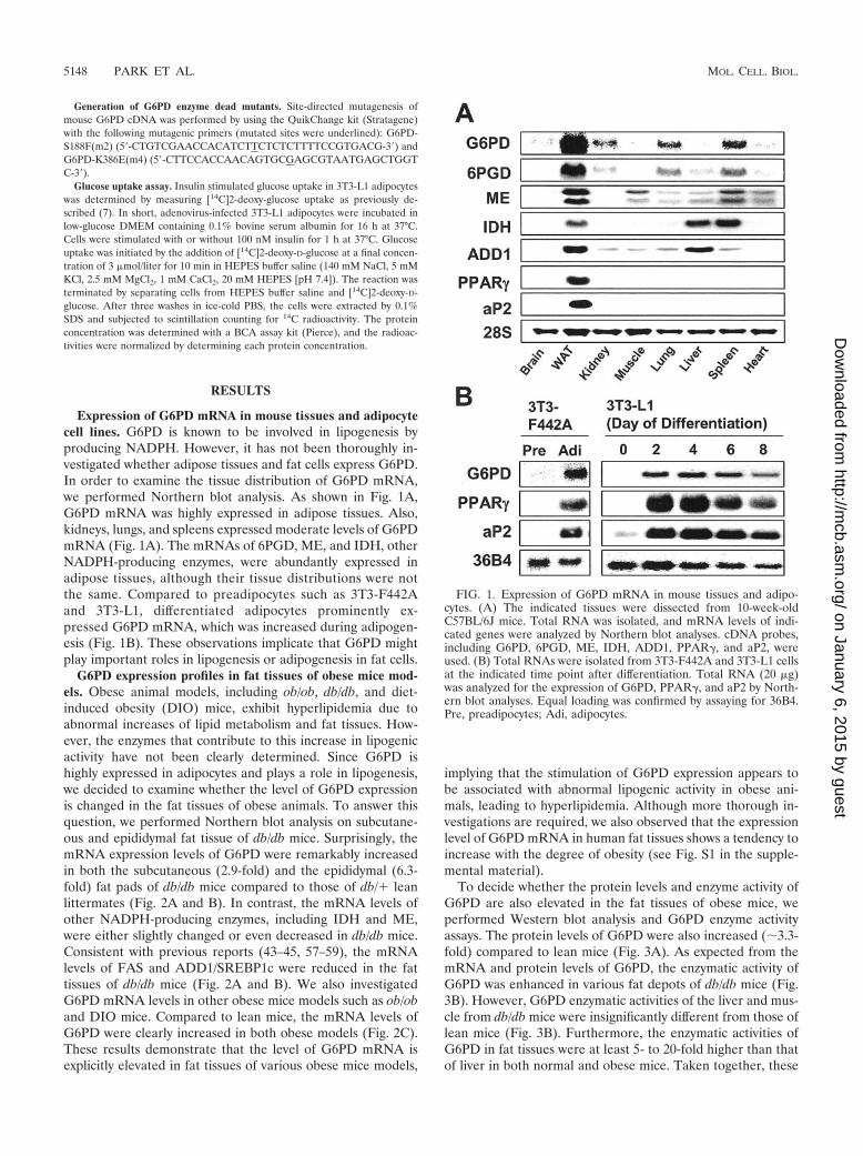
Expression of G6PD mRNA in mouse tissues and adipocytecell lines. G6PD is known to be involved in lipogenesis byproducing NADPH. However, it has not been thoroughly in-vestigated whether adipose tissues and fat cells express G6PD.In order to examine the tissue distribution of G6PD mRNA,we performed Northern blot analysis. As shown in Fig. 1A,G6PD mRNA was highly expressed in adipose tissues. Also,kidneys, lungs, and spleens expressed moderate levels of G6PDmRNA (Fig. 1A). The mRNAs of 6PGD, ME, and IDH, otherNADPH-producing enzymes, were abundantly expressed inadipose tissues, although their tissue distributions were notthe same. Compared to preadipocytes such as 3T3-F442Aand 3T3-L1, differentiated adipocytes prominently ex-pressed G6PD mRNA, which was increased during adipogen-esis (Fig. 1B). These observations implicate that G6PD mightplay important roles in lipogenesis or adipogenesis in fat cells.
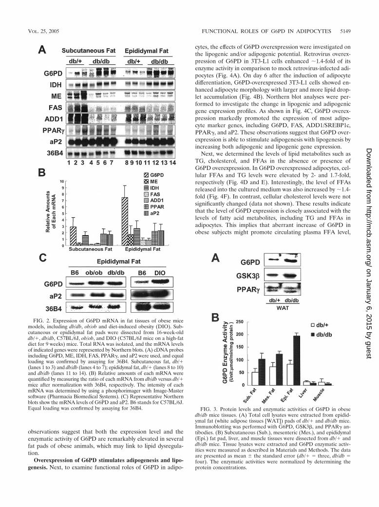
G6PD expression profiles in fat tissues of obese mice mod-els. Obese animal models, including ob/ob, db/db, and diet-induced obesity (DIO) mice, exhibit hyperlipidemia due toabnormal increases of lipid metabolism and fat tissues. How-ever, the enzymes that contribute to this increase in lipogenicactivity have not been clearly determined. Since G6PD ishighly expressed in adipocytes and plays a role in lipogenesis,we decided to examine whether the level of G6PD expressionis changed in the fat tissues of obese animals. To answer thisquestion, we performed Northern blot analysis on subcutane-ous and epididymal fat tissue of db/db mice. Surprisingly, themRNA expression levels of G6PD were remarkably increasedin both the subcutaneous (2.9-fold) and the epididymal (6.3-fold) fat pads of db/db mice compared to those of db/� leanlittermates (Fig. 2A and B). In contrast, the mRNA levels ofother NADPH-producing enzymes, including IDH and ME,were either slightly changed or even decreased in db/db mice.Consistent with previous reports (43–45, 57–59), the mRNAlevels of FAS and ADD1/SREBP1c were reduced in the fattissues of db/db mice (Fig. 2A and B). We also investigatedG6PD mRNA levels in other obese mice models such as ob/oband DIO mice. Compared to lean mice, the mRNA levels ofG6PD were clearly increased in both obese models (Fig. 2C).These results demonstrate that the level of G6PD mRNA isexplicitly elevated in fat tissues of various obese mice models,
implying that the stimulation of G6PD expression appears tobe associated with abnormal lipogenic activity in obese ani-mals, leading to hyperlipidemia. Although more thorough in-vestigations are required, we also observed that the expressionlevel of G6PD mRNA in human fat tissues shows a tendency toincrease with the degree of obesity (see Fig. S1 in the supple-mental material).
To decide whether the protein levels and enzyme activity ofG6PD are also elevated in the fat tissues of obese mice, weperformed Western blot analysis and G6PD enzyme activityassays. The protein levels of G6PD were also increased (�3.3-fold) compared to lean mice (Fig. 3A). As expected from themRNA and protein levels of G6PD, the enzymatic activity ofG6PD was enhanced in various fat depots of db/db mice (Fig.3B). However, G6PD enzymatic activities of the liver and mus-cle from db/db mice were insignificantly different from those oflean mice (Fig. 3B). Furthermore, the enzymatic activities ofG6PD in fat tissues were at least 5- to 20-fold higher than thatof liver in both normal and obese mice. Taken together, these
FIG. 1. Expression of G6PD mRNA in mouse tissues and adipo-cytes. (A) The indicated tissues were dissected from 10-week-oldC57BL/6J mice. Total RNA was isolated, and mRNA levels of indi-cated genes were analyzed by Northern blot analyses. cDNA probes,including G6PD, 6PGD, ME, IDH, ADD1, PPAR�, and aP2, wereused. (B) Total RNAs were isolated from 3T3-F442A and 3T3-L1 cellsat the indicated time point after differentiation. Total RNA (20 �g)was analyzed for the expression of G6PD, PPAR�, and aP2 by North-ern blot analyses. Equal loading was confirmed by assaying for 36B4.Pre, preadipocytes; Adi, adipocytes.
5148 PARK ET AL. MOL. CELL. BIOL.
on January 6, 2015 by guesthttp://m
cb.asm.org/
Dow
nloaded from
observations suggest that both the expression level and theenzymatic activity of G6PD are remarkably elevated in severalfat pads of obese animals, which may link to lipid dysregula-tion.
Overexpression of G6PD stimulates adipogenesis and lipo-genesis. Next, to examine functional roles of G6PD in adipo-
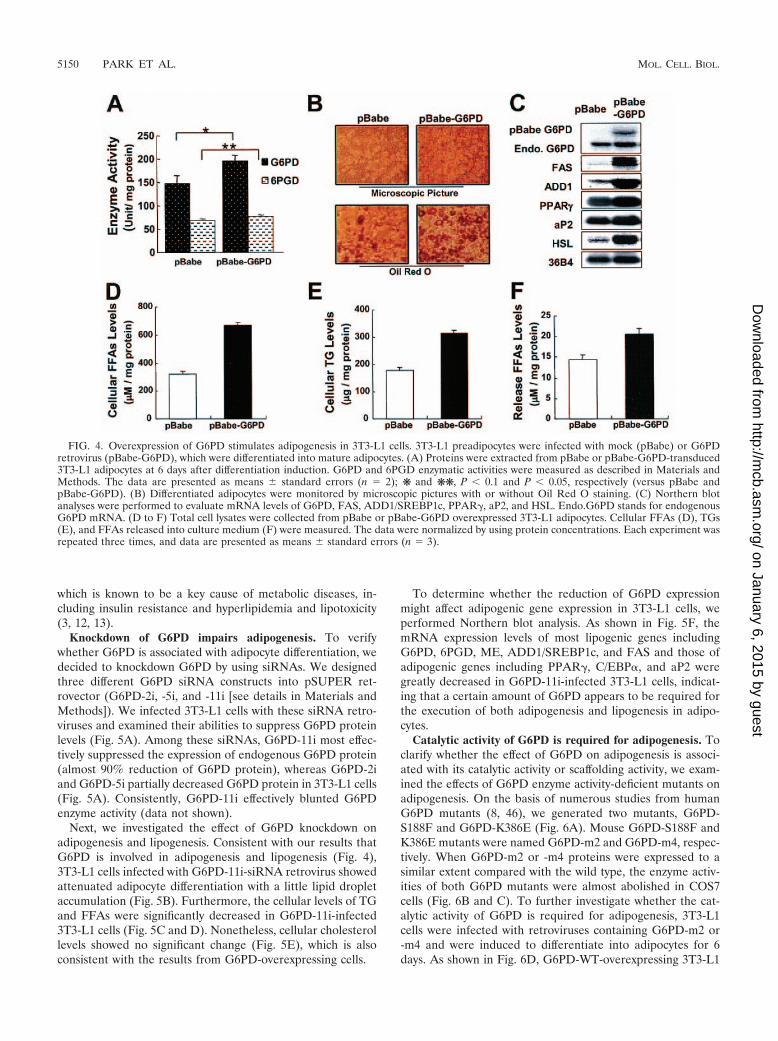
cytes, the effects of G6PD overexpression were investigated onthe lipogenic and/or adipogenic potential. Retrovirus overex-pression of G6PD in 3T3-L1 cells enhanced �1.4-fold of itsenzyme activity in comparison to mock retrovirus-infected adi-pocytes (Fig. 4A). On day 6 after the induction of adipocytedifferentiation, G6PD-overexpressed 3T3-L1 cells showed en-hanced adipocyte morphology with larger and more lipid drop-let accumulation (Fig. 4B). Northern blot analyses were per-formed to investigate the change in lipogenic and adipogenicgene expression profiles. As shown in Fig. 4C, G6PD overex-pression markedly promoted the expression of most adipo-cyte marker genes, including G6PD, FAS, ADD1/SREBP1c,PPAR�, and aP2. These observations suggest that G6PD over-expression is able to stimulate adipogenesis with lipogenesis byincreasing both adipogenic and lipogenic gene expression.
Next, we determined the levels of lipid metabolites such asTG, cholesterol, and FFAs in the absence or presence ofG6PD overexpression. In G6PD overexpressed adipocytes, cel-lular FFAs and TG levels were elevated by 2- and 1.7-fold,respectively (Fig. 4D and E). Interestingly, the level of FFAsreleased into the cultured medium was also increased by �1.4-fold (Fig. 4F). In contrast, cellular cholesterol levels were notsignificantly changed (data not shown). These results indicatethat the level of G6PD expression is closely associated with thelevels of fatty acid metabolites, including TG and FFAs inadipocytes. This implies that aberrant increase of G6PD inobese subjects might promote circulating plasma FFA level,
FIG. 2. Expression of G6PD mRNA in fat tissues of obese micemodels, including db/db, ob/ob and diet-induced obesity (DIO). Sub-cutaneous or epididymal fat pads were dissected from 16-week-olddb/�, db/db, C57BL/6J, ob/ob, and DIO (C57BL/6J mice on a high-fatdiet for 9 weeks) mice. Total RNA was isolated, and the mRNA levelsof indicated genes were represented by Northern blots. (A) cDNA probesincluding G6PD, ME, IDH, FAS, PPAR�, and aP2 were used, and equalloading was confirmed by assaying for 36B4. Subcutaneous fat, db/�(lanes 1 to 3) and db/db (lanes 4 to 7); epididymal fat, db/� (lanes 8 to 10)and db/db (lanes 11 to 14). (B) Relative amounts of each mRNA werequantified by measuring the ratio of each mRNA from db/db versus db/�mice after normalization with 36B4, respectively. The intensity of eachmRNA was determined by using a phosphorimager with Image-Mastersoftware (Pharmacia Biomedical Systems). (C) Representative Northernblots show the mRNA levels of G6PD and aP2. B6 stands for C57BL/6J.Equal loading was confirmed by assaying for 36B4. FIG. 3. Protein levels and enzymatic activities of G6PD in obese
db/db mice tissues. (A) Total cell lysates were extracted from epidid-ymal fat (white adipose tissues [WAT]) pads of db/� and db/db mice.Immunoblotting was performed with G6PD, GSK3�, and PPAR� an-tibodies. (B) Subcutaneous (Sub.), mesenteric (Mes.), and epididymal(Epi.) fat pad, liver, and muscle tissues were dissected from db/� anddb/db mice. Tissue lysates were extracted and G6PD enzymatic activ-ities were measured as described in Materials and Methods. The dataare presented as mean the standard error (db/� three, db/db four). The enzymatic activities were normalized by determining theprotein concentrations.
VOL. 25, 2005 FUNCTIONAL ROLES OF G6PD IN ADIPOCYTES 5149
on January 6, 2015 by guesthttp://m
cb.asm.org/
Dow
nloaded from
which is known to be a key cause of metabolic diseases, in-cluding insulin resistance and hyperlipidemia and lipotoxicity(3, 12, 13).
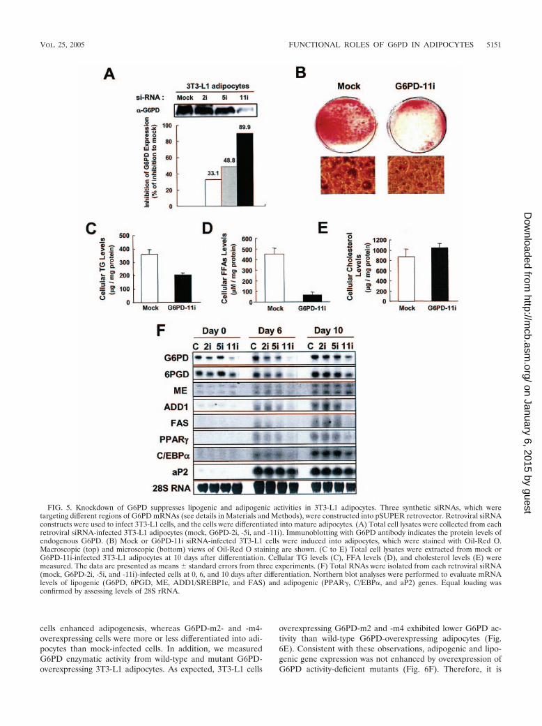
Knockdown of G6PD impairs adipogenesis. To verifywhether G6PD is associated with adipocyte differentiation, wedecided to knockdown G6PD by using siRNAs. We designedthree different G6PD siRNA constructs into pSUPER ret-rovector (G6PD-2i, -5i, and -11i [see details in Materials andMethods]). We infected 3T3-L1 cells with these siRNA retro-viruses and examined their abilities to suppress G6PD proteinlevels (Fig. 5A). Among these siRNAs, G6PD-11i most effec-tively suppressed the expression of endogenous G6PD protein(almost 90% reduction of G6PD protein), whereas G6PD-2iand G6PD-5i partially decreased G6PD protein in 3T3-L1 cells(Fig. 5A). Consistently, G6PD-11i effectively blunted G6PDenzyme activity (data not shown).
Next, we investigated the effect of G6PD knockdown onadipogenesis and lipogenesis. Consistent with our results thatG6PD is involved in adipogenesis and lipogenesis (Fig. 4),3T3-L1 cells infected with G6PD-11i-siRNA retrovirus showedattenuated adipocyte differentiation with a little lipid dropletaccumulation (Fig. 5B). Furthermore, the cellular levels of TGand FFAs were significantly decreased in G6PD-11i-infected3T3-L1 cells (Fig. 5C and D). Nonetheless, cellular cholesterollevels showed no significant change (Fig. 5E), which is alsoconsistent with the results from G6PD-overexpressing cells.
To determine whether the reduction of G6PD expressionmight affect adipogenic gene expression in 3T3-L1 cells, weperformed Northern blot analysis. As shown in Fig. 5F, themRNA expression levels of most lipogenic genes includingG6PD, 6PGD, ME, ADD1/SREBP1c, and FAS and those ofadipogenic genes including PPAR�, C/EBP�, and aP2 weregreatly decreased in G6PD-11i-infected 3T3-L1 cells, indicat-ing that a certain amount of G6PD appears to be required forthe execution of both adipogenesis and lipogenesis in adipo-cytes.
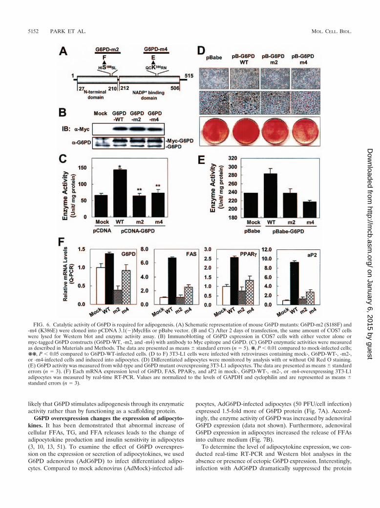
Catalytic activity of G6PD is required for adipogenesis. Toclarify whether the effect of G6PD on adipogenesis is associ-ated with its catalytic activity or scaffolding activity, we exam-ined the effects of G6PD enzyme activity-deficient mutants onadipogenesis. On the basis of numerous studies from humanG6PD mutants (8, 46), we generated two mutants, G6PD-S188F and G6PD-K386E (Fig. 6A). Mouse G6PD-S188F andK386E mutants were named G6PD-m2 and G6PD-m4, respec-tively. When G6PD-m2 or -m4 proteins were expressed to asimilar extent compared with the wild type, the enzyme activ-ities of both G6PD mutants were almost abolished in COS7cells (Fig. 6B and C). To further investigate whether the cat-alytic activity of G6PD is required for adipogenesis, 3T3-L1cells were infected with retroviruses containing G6PD-m2 or-m4 and were induced to differentiate into adipocytes for 6days. As shown in Fig. 6D, G6PD-WT-overexpressing 3T3-L1
FIG. 4. Overexpression of G6PD stimulates adipogenesis in 3T3-L1 cells. 3T3-L1 preadipocytes were infected with mock (pBabe) or G6PDretrovirus (pBabe-G6PD), which were differentiated into mature adipocytes. (A) Proteins were extracted from pBabe or pBabe-G6PD-transduced3T3-L1 adipocytes at 6 days after differentiation induction. G6PD and 6PGD enzymatic activities were measured as described in Materials andMethods. The data are presented as means standard errors (n 2); ❋ and ❋❋, P � 0.1 and P � 0.05, respectively (versus pBabe andpBabe-G6PD). (B) Differentiated adipocytes were monitored by microscopic pictures with or without Oil Red O staining. (C) Northern blotanalyses were performed to evaluate mRNA levels of G6PD, FAS, ADD1/SREBP1c, PPAR�, aP2, and HSL. Endo.G6PD stands for endogenousG6PD mRNA. (D to F) Total cell lysates were collected from pBabe or pBabe-G6PD overexpressed 3T3-L1 adipocytes. Cellular FFAs (D), TGs(E), and FFAs released into culture medium (F) were measured. The data were normalized by using protein concentrations. Each experiment wasrepeated three times, and data are presented as means standard errors (n 3).
5150 PARK ET AL. MOL. CELL. BIOL.
on January 6, 2015 by guesthttp://m
cb.asm.org/
Dow
nloaded from
cells enhanced adipogenesis, whereas G6PD-m2- and -m4-overexpressing cells were more or less differentiated into adi-pocytes than mock-infected cells. In addition, we measuredG6PD enzymatic activity from wild-type and mutant G6PD-overexpressing 3T3-L1 adipocytes. As expected, 3T3-L1 cells
overexpressing G6PD-m2 and -m4 exhibited lower G6PD ac-tivity than wild-type G6PD-overexpressing adipocytes (Fig.6E). Consistent with these observations, adipogenic and lipo-genic gene expression was not enhanced by overexpression ofG6PD activity-deficient mutants (Fig. 6F). Therefore, it is
FIG. 5. Knockdown of G6PD suppresses lipogenic and adipogenic activities in 3T3-L1 adipocytes. Three synthetic siRNAs, which weretargeting different regions of G6PD mRNAs (see details in Materials and Methods), were constructed into pSUPER retrovector. Retroviral siRNAconstructs were used to infect 3T3-L1 cells, and the cells were differentiated into mature adipocytes. (A) Total cell lysates were collected from eachretroviral siRNA-infected 3T3-L1 adipocytes (mock, G6PD-2i, -5i, and -11i). Immunoblotting with G6PD antibody indicates the protein levels ofendogenous G6PD. (B) Mock or G6PD-11i siRNA-infected 3T3-L1 cells were induced into adipocytes, which were stained with Oil-Red O.Macroscopic (top) and microscopic (bottom) views of Oil-Red O staining are shown. (C to E) Total cell lysates were extracted from mock orG6PD-11i-infected 3T3-L1 adipocytes at 10 days after differentiation. Cellular TG levels (C), FFA levels (D), and cholesterol levels (E) weremeasured. The data are presented as means standard errors from three experiments. (F) Total RNAs were isolated from each retroviral siRNA(mock, G6PD-2i, -5i, and -11i)-infected cells at 0, 6, and 10 days after differentiation. Northern blot analyses were performed to evaluate mRNAlevels of lipogenic (G6PD, 6PGD, ME, ADD1/SREBP1c, and FAS) and adipogenic (PPAR�, C/EBP�, and aP2) genes. Equal loading wasconfirmed by assessing levels of 28S rRNA.
VOL. 25, 2005 FUNCTIONAL ROLES OF G6PD IN ADIPOCYTES 5151
on January 6, 2015 by guesthttp://m
cb.asm.org/
Dow
nloaded from
likely that G6PD stimulates adipogenesis through its enzymaticactivity rather than by functioning as a scaffolding protein.
G6PD overexpression changes the expression of adipocyto-kines. It has been demonstrated that abnormal increase ofcellular FFAs, TG, and FFA releases leads to the change ofadipocytokine production and insulin sensitivity in adipocytes(3, 10, 13, 51). To examine the effect of G6PD overexpres-sion on the expression or secretion of adipocytokines, we usedG6PD adenovirus (AdG6PD) to infect differentiated adipo-cytes. Compared to mock adenovirus (AdMock)-infected adi-
pocytes, AdG6PD-infected adipocytes (50 PFU/cell infection)expressed 1.5-fold more of G6PD protein (Fig. 7A). Accord-ingly, the enzyme activity of G6PD was increased by adenoviralG6PD expression (data not shown). Furthermore, adenoviralG6PD expression in adipocytes increased the release of FFAsinto culture medium (Fig. 7B).
To determine the level of adipocytokine expression, we con-ducted real-time RT-PCR and Western blot analyses in theabsence or presence of ectopic G6PD expression. Interestingly,infection with AdG6PD dramatically suppressed the protein
FIG. 6. Catalytic activity of G6PD is required for adipogenesis. (A) Schematic representation of mouse G6PD mutants: G6PD-m2 (S188F) and-m4 (K386E) were cloned into pCDNA 3.1(�)MycHis or pBabe vector. (B and C) After 2 days of transfection, the same amount of COS7 cellswere lysed for Western blot and enzyme activity assay. (B) Immunoblotting of G6PD expression in COS7 cells with either vector alone ormyc-tagged G6PD constructs (G6PD-WT, -m2, and -m4) with antibody to Myc epitope and G6PD. (C) G6PD enzymatic activities were measuredas described in Materials and Methods. The data are presented as means standard errors (n 5). ❋, P � 0.01 compared to mock-infected cells;❋❋, P � 0.05 compared to G6PD-WT-infected cells. (D to F) 3T3-L1 cells were infected with retroviruses containing mock-, G6PD-WT-, -m2-,or -m4-infected cells and induced into adipocytes. (D) Differentiated adipocytes were monitored by analysis with or without Oil Red O staining.(E) G6PD activity was measured from wild-type and G6PD mutant overexpressing 3T3-L1 adipocytes. The data are presented as means standarderrors (n 3). (F) Each mRNA expression level of G6PD, FAS, PPAR�, and aP2 in mock-, G6PD-WT-, -m2-, or -m4-overexpressing 3T3-L1adipocytes was measured by real-time RT-PCR. Values are normalized to the levels of GAPDH and cyclophilin and are represented as means standard errors (n 3).
5152 PARK ET AL. MOL. CELL. BIOL.
on January 6, 2015 by guesthttp://m
cb.asm.org/
Dow
nloaded from
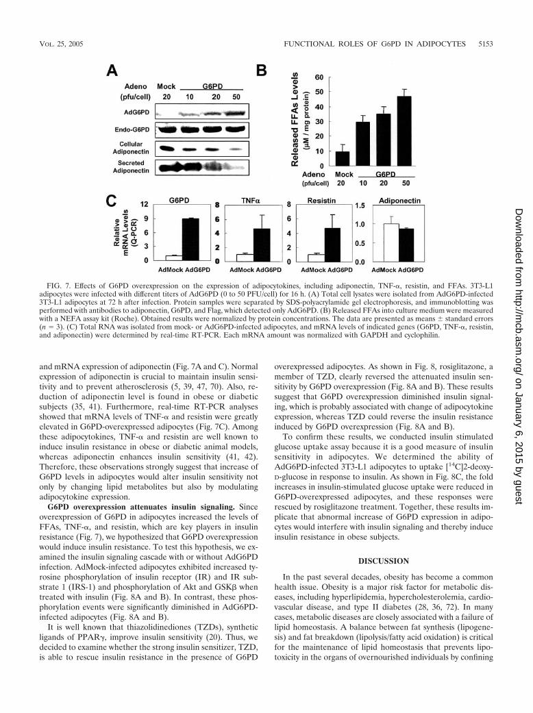
and mRNA expression of adiponectin (Fig. 7A and C). Normalexpression of adiponectin is crucial to maintain insulin sensi-tivity and to prevent atherosclerosis (5, 39, 47, 70). Also, re-duction of adiponectin level is found in obese or diabeticsubjects (35, 41). Furthermore, real-time RT-PCR analysesshowed that mRNA levels of TNF-� and resistin were greatlyelevated in G6PD-overexpressed adipocytes (Fig. 7C). Amongthese adipocytokines, TNF-� and resistin are well known toinduce insulin resistance in obese or diabetic animal models,whereas adiponectin enhances insulin sensitivity (41, 42).Therefore, these observations strongly suggest that increase ofG6PD levels in adipocytes would alter insulin sensitivity notonly by changing lipid metabolites but also by modulatingadipocytokine expression.
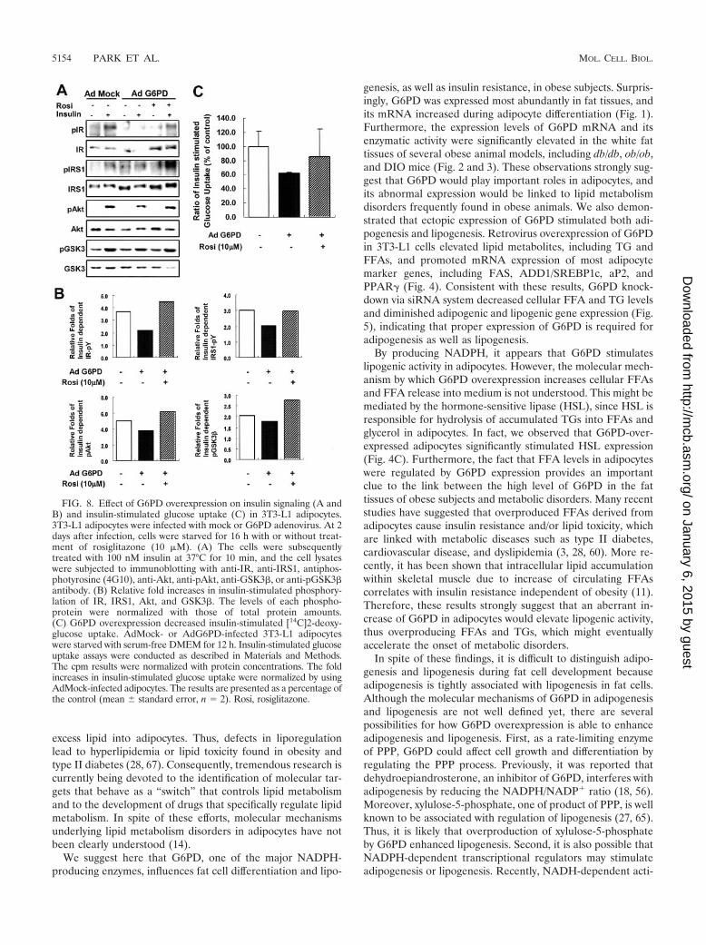
G6PD overexpression attenuates insulin signaling. Sinceoverexpression of G6PD in adipocytes increased the levels ofFFAs, TNF-�, and resistin, which are key players in insulinresistance (Fig. 7), we hypothesized that G6PD overexpressionwould induce insulin resistance. To test this hypothesis, we ex-amined the insulin signaling cascade with or without AdG6PDinfection. AdMock-infected adipocytes exhibited increased ty-rosine phosphorylation of insulin receptor (IR) and IR sub-strate 1 (IRS-1) and phosphorylation of Akt and GSK� whentreated with insulin (Fig. 8A and B). In contrast, these phos-phorylation events were significantly diminished in AdG6PD-infected adipocytes (Fig. 8A and B).
It is well known that thiazolidinediones (TZDs), syntheticligands of PPAR�, improve insulin sensitivity (20). Thus, wedecided to examine whether the strong insulin sensitizer, TZD,is able to rescue insulin resistance in the presence of G6PD
overexpressed adipocytes. As shown in Fig. 8, rosiglitazone, amember of TZD, clearly reversed the attenuated insulin sen-sitivity by G6PD overexpression (Fig. 8A and B). These resultssuggest that G6PD overexpression diminished insulin signal-ing, which is probably associated with change of adipocytokineexpression, whereas TZD could reverse the insulin resistanceinduced by G6PD overexpression (Fig. 8A and B).
To confirm these results, we conducted insulin stimulatedglucose uptake assay because it is a good measure of insulinsensitivity in adipocytes. We determined the ability ofAdG6PD-infected 3T3-L1 adipocytes to uptake [14C]2-deoxy-D-glucose in response to insulin. As shown in Fig. 8C, the foldincreases in insulin-stimulated glucose uptake were reduced inG6PD-overexpressed adipocytes, and these responses wererescued by rosiglitazone treatment. Together, these results im-plicate that abnormal increase of G6PD expression in adipo-cytes would interfere with insulin signaling and thereby induceinsulin resistance in obese subjects.
DISCUSSION
In the past several decades, obesity has become a commonhealth issue. Obesity is a major risk factor for metabolic dis-eases, including hyperlipidemia, hypercholesterolemia, cardio-vascular disease, and type II diabetes (28, 36, 72). In manycases, metabolic diseases are closely associated with a failure oflipid homeostasis. A balance between fat synthesis (lipogene-sis) and fat breakdown (lipolysis/fatty acid oxidation) is criticalfor the maintenance of lipid homeostasis that prevents lipo-toxicity in the organs of overnourished individuals by confining
FIG. 7. Effects of G6PD overexpression on the expression of adipocytokines, including adiponectin, TNF-�, resistin, and FFAs. 3T3-L1adipocytes were infected with different titers of AdG6PD (0 to 50 PFU/cell) for 16 h. (A) Total cell lysates were isolated from AdG6PD-infected3T3-L1 adipocytes at 72 h after infection. Protein samples were separated by SDS-polyacrylamide gel electrophoresis, and immunoblotting wasperformed with antibodies to adiponectin, G6PD, and Flag, which detected only AdG6PD. (B) Released FFAs into culture medium were measuredwith a NEFA assay kit (Roche). Obtained results were normalized by protein concentrations. The data are presented as means standard errors(n 3). (C) Total RNA was isolated from mock- or AdG6PD-infected adipocytes, and mRNA levels of indicated genes (G6PD, TNF-�, resistin,and adiponectin) were determined by real-time RT-PCR. Each mRNA amount was normalized with GAPDH and cyclophilin.
VOL. 25, 2005 FUNCTIONAL ROLES OF G6PD IN ADIPOCYTES 5153
on January 6, 2015 by guesthttp://m
cb.asm.org/
Dow
nloaded from
excess lipid into adipocytes. Thus, defects in liporegulationlead to hyperlipidemia or lipid toxicity found in obesity andtype II diabetes (28, 67). Consequently, tremendous research iscurrently being devoted to the identification of molecular tar-gets that behave as a “switch” that controls lipid metabolismand to the development of drugs that specifically regulate lipidmetabolism. In spite of these efforts, molecular mechanismsunderlying lipid metabolism disorders in adipocytes have notbeen clearly understood (14).
We suggest here that G6PD, one of the major NADPH-producing enzymes, influences fat cell differentiation and lipo-
genesis, as well as insulin resistance, in obese subjects. Surpris-ingly, G6PD was expressed most abundantly in fat tissues, andits mRNA increased during adipocyte differentiation (Fig. 1).Furthermore, the expression levels of G6PD mRNA and itsenzymatic activity were significantly elevated in the white fattissues of several obese animal models, including db/db, ob/ob,and DIO mice (Fig. 2 and 3). These observations strongly sug-gest that G6PD would play important roles in adipocytes, andits abnormal expression would be linked to lipid metabolismdisorders frequently found in obese animals. We also demon-strated that ectopic expression of G6PD stimulated both adi-pogenesis and lipogenesis. Retrovirus overexpression of G6PDin 3T3-L1 cells elevated lipid metabolites, including TG andFFAs, and promoted mRNA expression of most adipocytemarker genes, including FAS, ADD1/SREBP1c, aP2, andPPAR� (Fig. 4). Consistent with these results, G6PD knock-down via siRNA system decreased cellular FFA and TG levelsand diminished adipogenic and lipogenic gene expression (Fig.5), indicating that proper expression of G6PD is required foradipogenesis as well as lipogenesis.
By producing NADPH, it appears that G6PD stimulateslipogenic activity in adipocytes. However, the molecular mech-anism by which G6PD overexpression increases cellular FFAsand FFA release into medium is not understood. This might bemediated by the hormone-sensitive lipase (HSL), since HSL isresponsible for hydrolysis of accumulated TGs into FFAs andglycerol in adipocytes. In fact, we observed that G6PD-over-expressed adipocytes significantly stimulated HSL expression(Fig. 4C). Furthermore, the fact that FFA levels in adipocyteswere regulated by G6PD expression provides an importantclue to the link between the high level of G6PD in the fattissues of obese subjects and metabolic disorders. Many recentstudies have suggested that overproduced FFAs derived fromadipocytes cause insulin resistance and/or lipid toxicity, whichare linked with metabolic diseases such as type II diabetes,cardiovascular disease, and dyslipidemia (3, 28, 60). More re-cently, it has been shown that intracellular lipid accumulationwithin skeletal muscle due to increase of circulating FFAscorrelates with insulin resistance independent of obesity (11).Therefore, these results strongly suggest that an aberrant in-crease of G6PD in adipocytes would elevate lipogenic activity,thus overproducing FFAs and TGs, which might eventuallyaccelerate the onset of metabolic disorders.
In spite of these findings, it is difficult to distinguish adipo-genesis and lipogenesis during fat cell development becauseadipogenesis is tightly associated with lipogenesis in fat cells.Although the molecular mechanisms of G6PD in adipogenesisand lipogenesis are not well defined yet, there are severalpossibilities for how G6PD overexpression is able to enhanceadipogenesis and lipogenesis. First, as a rate-limiting enzymeof PPP, G6PD could affect cell growth and differentiation byregulating the PPP process. Previously, it was reported thatdehydroepiandrosterone, an inhibitor of G6PD, interferes withadipogenesis by reducing the NADPH/NADP� ratio (18, 56).Moreover, xylulose-5-phosphate, one of product of PPP, is wellknown to be associated with regulation of lipogenesis (27, 65).Thus, it is likely that overproduction of xylulose-5-phosphateby G6PD enhanced lipogenesis. Second, it is also possible thatNADPH-dependent transcriptional regulators may stimulateadipogenesis or lipogenesis. Recently, NADH-dependent acti-
FIG. 8. Effect of G6PD overexpression on insulin signaling (A andB) and insulin-stimulated glucose uptake (C) in 3T3-L1 adipocytes.3T3-L1 adipocytes were infected with mock or G6PD adenovirus. At 2days after infection, cells were starved for 16 h with or without treat-ment of rosiglitazone (10 �M). (A) The cells were subsequentlytreated with 100 nM insulin at 37°C for 10 min, and the cell lysateswere subjected to immunoblotting with anti-IR, anti-IRS1, antiphos-photyrosine (4G10), anti-Akt, anti-pAkt, anti-GSK3�, or anti-pGSK3�antibody. (B) Relative fold increases in insulin-stimulated phosphory-lation of IR, IRS1, Akt, and GSK3�. The levels of each phospho-protein were normalized with those of total protein amounts.(C) G6PD overexpression decreased insulin-stimulated [14C]2-deoxy-glucose uptake. AdMock- or AdG6PD-infected 3T3-L1 adipocyteswere starved with serum-free DMEM for 12 h. Insulin-stimulated glucoseuptake assays were conducted as described in Materials and Methods.The cpm results were normalized with protein concentrations. The foldincreases in insulin-stimulated glucose uptake were normalized by usingAdMock-infected adipocytes. The results are presented as a percentage ofthe control (mean standard error, n 2). Rosi, rosiglitazone.
5154 PARK ET AL. MOL. CELL. BIOL.
on January 6, 2015 by guesthttp://m
cb.asm.org/
Dow
nloaded from

vators or histone deacetylases have been identified, and severaltarget genes involved in the differentiation process are modu-lated by these NADH-dependent coregulators (17, 25, 37, 71).Lastly, it appears that G6PD could accelerate adipogenesis bypromoting free fatty acid metabolism, which might eventuallygenerate ligands of PPAR�, a master transcription factor foradipocyte differentiation. A similar idea has been demon-strated that overexpression of the active form of ADD1/SREBP1c, which coordinates most lipogenic genes, acceleratesadipogenesis in a nonadipogenic cell line by producing endog-enous PPAR� ligands (31).
As a major tissue for whole-body energy homeostasis, adi-pose tissue integrates both central and peripheral metabolicsignals and orchestrates energy balance. Therefore, abnormalincrease or decrease of adipose tissue often causes systemicdisorders, also known as metabolic diseases. Adipose tissuesare also actively involved in many physiological and patholog-ical processes by expressing various adipocytokines, includingleptin, TNF-�, resistin, adiponectin, interleukin-6 (IL-6), andFFAs (28, 40, 61). Therefore, adipose tissues locally influencethe equilibrium between lipogenesis and lipolysis and system-atically contribute to whole-body energy homeostasis by anendocrine mechanism. It is well known that certain adipocyto-kines are important in provoking the insulin resistance foundin many obese and diabetic subjects. For example, the eleva-tion in circulating inflammatory cytokines such as TNF-� andIL-6 mediates insulin resistance and is common in obesity (19,22, 68). An increased level of TNF-� in obese states leads toinsulin resistance via a number of mechanisms, including thedecrease of insulin signaling or GLUT4 expression (26, 62). Inaddition, the newly identified resistin has been proposed tolink obesity and insulin resistance (32, 63). Resistin expressionis increased in obese animals and decreased by the antidiabe-tes drug, rosiglitazone (63). Thus, it has been suggested thatTNF-�, IL-6, and resistin participate in the regulation of glu-cose metabolism and that high levels of their expression resultin insulin resistance and type II diabetes (4, 41, 49, 68). Unlikethese adipocytokines, adiponectin that regulates both glucoseand lipid metabolism increases insulin sensitivity (6, 40), andits expression levels are inversely correlated with pathologicalconditions such as obesity, diabetes, or atherosclerosis (23, 35).In addition, many reports have shown that adiponectin treat-ment improves insulin resistance, atherosclerosis and lipid me-tabolism disorders in several animal models (5, 39, 47, 69, 70).
It is of interest to note that the levels of adipocytokines areexplicitly changed in G6PD-overexpressed adipocytes. Asshown in Fig. 7C, overexpression of G6PD in adipocytes ele-vated the expression of TNF-� and resistin mRNAs. Incontrast, both protein and mRNA levels of adiponectin weredecreased (Fig. 7A and C). The molecular mechanism(s)underlying the G6PD-dependent changes in adipocytokine ex-pression remains to be elucidated. Nonetheless, the idea thatanomalous increase of G6PD in adipocytes would induce in-sulin resistance was confirmed by the observation that G6PD-overexpressed adipocytes exhibited downregulated insulin sig-naling and insulin-dependent glucose uptake (Fig. 8). Takentogether, these results strongly support the hypothesis thataberrantly high expression of G6PD in fat tissues of obeseanimals would result in metabolic disorders including hyper-
lipidemia and insulin resistance not only by increasing lipo-genic activity but also by modulating adipocytokine expression.
Very recently, Koh et al. reported that IDPc is important formodulating fat and cholesterol synthesis as well as adipogen-esis by showing that overexpression of IDPc increases 3T3-L1adipocyte differentiation, whereas reduced expression of IDPcblocks adipogenesis (34). These results are somewhat similarto our observations that overexpression of G6PD in adipocytespromote lipogenesis and adipogenesis like IDPc overexpres-sion, which could increase eventually cellular NADPH level.However, several evidences suggest that IDPc would have dif-ferent physiological roles from G6PD. First, compared toG6PD, the mRNA level of IDPc is not significantly changed inthe fat tissue of obese mice models (Fig. 2). Second, themRNA level of G6PD was increased during adipocytes differ-entiation (Fig. 1), whereas that of IDPc is not changed (34).Furthermore, IDPc transgenic mice show lower fasting bloodglucose levels and improvement of glucose sensitivity com-pared to wild-type mice, even though they exhibit obesity,hyperlipidemia, and fatty liver. In contrast, G6PD overexpres-sion caused insulin resistance in adipocytes (Fig. 8). In accor-dance with this observation, G6PD overexpression increasedthe expression of TNF-� and resistin but decreased expressionof adiponectin, which is closely related to a decrease in insulinsensitivity (Fig. 8). Thus, G6PD overexpression would lead toabnormal lipid metabolism and insulin signaling, which aredifferent from IDPc overexpression.
In summary, we have established a novel link between G6PDand lipid metabolism disorders in the adipocytes of obese an-imals (Fig. 9). Our findings that G6PD expression is increasedin obese animals and that G6PD elevation in adipocyteschanges the levels of lipid metabolites provide strong evidencethat obesity induced lipid metabolism disorders and insulinresistance is mediated, at least in part, by G6PD overexpres-sion in fat tissues. Therefore, our observations that an abnor-
FIG. 9. Functional roles of G6PD in adipocytes and its impact onobesity. Obese subjects stimulate G6PD expression in adipocytes andenhance adiposity with elevation of TG and FFAs, which would affectthe expression of adipocytokines and metabolic syndromes, includinginsulin resistance, hyperlipidemia, and lipotoxicity. See the text forfurther explanation.
VOL. 25, 2005 FUNCTIONAL ROLES OF G6PD IN ADIPOCYTES 5155
on January 6, 2015 by guesthttp://m
cb.asm.org/
Dow
nloaded from
mal increase of G6PD in adipocytes modulates lipid metabo-lism and adipocytokine expression suggest a potential target inthe successful development of therapeutic treatment for obeseand diabetic patients.
ACKNOWLEDGMENTS
We thank Thomas Lee and Jun-Jae Chung for critically reading themanuscript.
This study was supported in part by grants from Stem Cell ResearchCenter of the 21st Century Frontier Research Program, the Molecularand Cellular Biodiscovery Research Program, and the National Re-search Laboratory Program of Korea Institute of Science and Tech-nology Evaluation and Planning. J.P., H.K.R., K.H.K., S.S.C., Y.S.L.,and J.B.K. are supported by the BK21 Research Fellowship from theMinistry of Education and Human Resources Development.
REFERENCES
1. Abu-Elheiga, L., M. M. Matzuk, K. A. Abo-Hashema, and S. J. Wakil. 2001.Continuous fatty acid oxidation and reduced fat storage in mice lackingacetyl-CoA carboxylase 2. Science 291:2613–2616.
2. Abu-Elheiga, L., W. Oh, P. Kordari, and S. J. Wakil. 2003. Acetyl-CoAcarboxylase 2 mutant mice are protected against obesity and diabetes in-duced by high-fat/high-carbohydrate diets. Proc. Natl. Acad. Sci. USA 100:10207–10212.
3. Arner, P. 2002. Insulin resistance in type 2 diabetes: role of fatty acids.Diabetes Metab. Res. Rev. 18(Suppl. 2):S5–S9.
4. Banerjee, R. R., S. M. Rangwala, J. S. Shapiro, A. S. Rich, B. Rhoades, Y. Qi,J. Wang, M. W. Rajala, A. Pocai, P. E. Scherer, C. M. Steppan, R. S. Ahima,S. Obici, L. Rossetti, and M. A. Lazar. 2004. Regulation of fasted bloodglucose by resistin. Science 303:1195–1198.
5. Berg, A. H., T. P. Combs, X. Du, M. Brownlee, and P. E. Scherer. 2001. Theadipocyte-secreted protein Acrp30 enhances hepatic insulin action. Nat.Med. 7:947–953.
6. Berg, A. H., T. P. Combs, and P. E. Scherer. 2002. ACRP30/adiponectin: anadipokine regulating glucose and lipid metabolism. Trends Endocrinol.Metab. 13:84–89.
7. Berti, L., M. Kellerer, E. Capp, and H. U. Haring. 1997. Leptin stimulatesglucose transport and glycogen synthesis in C2C12 myotubes: evidence for aP13-kinase mediated effect. Diabetologia 40:606–609.
8. Beutler, E., and T. J. Vulliamy. 2002. Hematologically important mutations:glucose-6-phosphate dehydrogenase. Blood Cells Mol. Dis. 28:93–103.
9. Biagiotti, E., L. Guidi, P. Del Grande, and P. Ninfali. 2003. Glucose-6-phosphate dehydrogenase expression associated with NADPH-dependentreactions in cerebellar neurons. Cerebellum 2:178–183.
10. Boden, G. 2001. Free fatty acids: the link between obesity and insulin resis-tance. Endocrinol. Pract. 7:44–51.
11. Boden, G. 2002. Interaction between free fatty acids and glucose metabolism.Curr. Opin. Clin. Nutr. Metab. Care. 5:545–549.
12. Boden, G. 1997. Role of fatty acids in the pathogenesis of insulin resistanceand NIDDM. Diabetes 46:3–10.
13. Boden, G., and G. I. Shulman. 2002. Free fatty acids in obesity and type 2diabetes: defining their role in the development of insulin resistance andbeta-cell dysfunction. Eur. J. Clin. Investig. 32(Suppl. 3):14–23.
14. Bray, G. A., and L. A. Tartaglia. 2000. Medicinal strategies in the treatmentof obesity. Nature 404:672–677.
15. Dessi, S., B. Batetta, O. Spano, D. Pulisci, M. F. Mulas, S. Muntoni, M.Armeni, C. Sanna, R. Antonucci, and P. Pani. 1992. Serum lipoproteinpattern as modified in G6PD-deficient children during hemolytic anaemiainduced by fava bean ingestion. Int. J. Exp. Pathol. 73:157–160.
16. Dessi, S., C. Chiodino, B. Batetta, A. M. Fadda, C. Anchisi, and P. Pani.1986. Hepatic glucose-6-phosphate dehydrogenase, cholesterogenesis, andserum lipoproteins in liver regeneration after partial hepatectomy. Exp. Mol.Pathol. 44:169–176.
17. Fulco, M., R. L. Schiltz, S. Iezzi, M. T. King, P. Zhao, Y. Kashiwaya, E.Hoffman, R. L. Veech, and V. Sartorelli. 2003. Sir2 regulates skeletal muscledifferentiation as a potential sensor of the redox state. Mol. Cell 12:51–62.
18. Gordon, G. B., J. A. Newitt, L. M. Shantz, D. E. Weng, and P. Talalay. 1986.Inhibition of the conversion of 3T3 fibroblast clones to adipocytes by dehy-droepiandrosterone and related anticarcinogenic steroids. Cancer Res. 46:3389–3395.
19. Halle, M., A. Berg, H. Northoff, and J. Keul. 1998. Importance of TNF-alphaand leptin in obesity and insulin resistance: a hypothesis on the impact ofphysical exercise. Exerc. Immunol. Rev. 4:77–94.
20. Hauner, H. 2002. The mode of action of thiazolidinediones. Diabetes Metab.Res. Rev. 18(Suppl. 2):S10–S15.
21. Horton, J. D., J. L. Goldstein, and M. S. Brown. 2002. SREBPs: activators ofthe complete program of cholesterol and fatty acid synthesis in the liver.J. Clin. Investig. 109:1125–1131.
22. Hotamisligil, G. S., N. S. Shargill, and B. M. Spiegelman. 1993. Adiposeexpression of tumor necrosis factor-alpha: direct role in obesity-linked insu-lin resistance. Science 259:87–91.
23. Hu, E., P. Liang, and B. M. Spiegelman. 1996. AdipoQ is a novel adipose-specific gene dysregulated in obesity. J. Biol. Chem. 271:10697–10703.
24. Hu, Z., S. H. Cha, S. Chohnan, and M. D. Lane. 2003. Hypothalamicmalonyl-CoA as a mediator of feeding behavior. Proc. Natl. Acad. Sci. USA100:12624–12629.
25. Imai, S., C. M. Armstrong, M. Kaeberlein, and L. Guarente. 2000. Tran-scriptional silencing and longevity protein Sir2 is an NAD-dependent histonedeacetylase. Nature 403:795–800.
26. Jain, R. G., K. D. Phelps, and P. H. Pekala. 1999. Tumor necrosis factor-alpha initiated signal transduction in 3T3-L1 adipocytes. J. Cell Physiol. 179:58–66.
27. Kabashima, T., T. Kawaguchi, B. E. Wadzinski, and K. Uyeda. 2003. Xylu-lose 5-phosphate mediates glucose-induced lipogenesis by xylulose 5-phos-phate-activated protein phosphatase in rat liver. Proc. Natl. Acad. Sci. USA100:5107–5112.
28. Kahn, B. B., and J. S. Flier. 2000. Obesity and insulin resistance. J. Clin.Investig. 106:473–481.
29. Kersten, S. 2001. Mechanisms of nutritional and hormonal regulation oflipogenesis. EMBO Rep. 2:282–286.
30. Kim, J. B., and B. M. Spiegelman. 1996. ADD1/SREBP1 promotes adipo-cyte differentiation and gene expression linked to fatty acid metabolism.Genes Dev. 10:1096–1107.
31. Kim, J. B., H. M. Wright, M. Wright, and B. M. Spiegelman. 1998. ADD1/SREBP1 activates PPAR� through the production of endogenous ligand.Proc. Natl. Acad. Sci. USA 95:4333–4337.
32. Kim, K. H., K. Lee, Y. S. Moon, and H. S. Sul. 2001. A cysteine-rich adiposetissue-specific secretory factor inhibits adipocyte differentiation. J. Biol.Chem. 276:11252–11256.
33. Kletzien, R. F., P. K. Harris, and L. A. Foellmi. 1994. Glucose-6-phosphatedehydrogenase: a “housekeeping” enzyme subject to tissue-specific regula-tion by hormones, nutrients, and oxidant stress. FASEB J. 8:174–181.
34. Koh, H. J., S. M. Lee, B. G. Son, S. H. Lee, Z. Y. Ryoo, K. T. Chang, J. W.Park, D. C. Park, B. J. Song, R. L. Veech, H. Song, and T. L. Huh. 2004.Cytosolic NADP�-dependent isocitrate dehydrogenase plays a key role inlipid metabolism. J. Biol. Chem. 279:29968–29974.
35. Kondo, H., I. Shimomura, Y. Matsukawa, M. Kumada, M. Takahashi, M.Matsuda, N. Ouchi, S. Kihara, T. Kawamoto, S. Sumitsuji, T. Funahashi,and Y. Matsuzawa. 2002. Association of adiponectin mutation with type 2diabetes: a candidate gene for the insulin resistance syndrome. Diabetes 51:2325–2328.
36. Kopelman, P. G. 2000. Obesity as a medical problem. Nature 404:635–643.37. Kumar, V., J. E. Carlson, K. A. Ohgi, T. A. Edwards, D. W. Rose, C. R.
Escalante, M. G. Rosenfeld, and A. K. Aggarwal. 2002. Transcription core-pressor CtBP is an NAD(�)-regulated dehydrogenase. Mol. Cell 10:857–869.
38. Longo, L., O. C. Vanegas, M. Patel, V. Rosti, H. Li, J. Waka, T. Merghoub,P. P. Pandolfi, R. Notaro, K. Manova, and L. Luzzatto. 2002. Maternallytransmitted severe glucose 6-phosphate dehydrogenase deficiency is an em-bryonic lethal. EMBO J. 21:4229–4239.
39. Matsuda, M., I. Shimomura, M. Sata, Y. Arita, M. Nishida, N. Maeda, M.Kumada, Y. Okamoto, H. Nagaretani, H. Nishizawa, K. Kishida, R.Komuro, N. Ouchi, S. Kihara, R. Nagai, T. Funahashi, and Y. Matsuzawa.2002. Role of adiponectin in preventing vascular stenosis. The missing link ofadipo-vascular axis. J. Biol. Chem. 277:37487–37491.
40. Matsuzawa, Y., T. Funahashi, S. Kihara, and I. Shimomura. 2004. Adi-ponectin and metabolic syndrome. Arterioscler. Thromb. Vasc. Biol. 24:29–33.
41. Matsuzawa, Y., T. Funahashi, and T. Nakamura. 1999. Molecular mecha-nism of metabolic syndrome X: contribution of adipocytokines adipocyte-derived bioactive substances. Ann. N. Y. Acad. Sci. 892:146–154.
42. Mohamed-Ali, V., J. H. Pinkney, and S. W. Coppack. 1998. Adipose tissue asan endocrine and paracrine organ. Int. J. Obesity Relat. Metab. Disorders22:1145–1158.
43. Moitra, J., M. M. Mason, M. Olive, D. Krylov, O. Gavrilova, B. Marcus-Samuels, L. Feigenbaum, E. Lee, T. Aoyama, M. Eckhaus, M. L. Reitman,and C. Vinson. 1998. Life without white fat: a transgenic mouse. Genes Dev.12:3168–3181.
44. Nadler, S. T., and A. D. Attie. 2001. Please pass the chips: genomic insightsinto obesity and diabetes. J. Nutr. 131:2078–2081.
45. Nadler, S. T., J. P. Stoehr, K. L. Schueler, G. Tanimoto, B. S. Yandell, andA. D. Attie. 2000. The expression of adipogenic genes is decreased in obesityand diabetes mellitus. Proc. Natl. Acad. Sci. USA 97:11371–11376.
46. Naylor, C. E., P. Rowland, A. K. Basak, S. Gover, P. J. Mason, J. M.Bautista, T. J. Vulliamy, L. Luzzatto, and M. J. Adams. 1996. Glucose6-phosphate dehydrogenase mutations causing enzyme deficiency in a modelof the tertiary structure of the human enzyme. Blood 87:2974–2982.
47. Okamoto, Y., S. Kihara, N. Ouchi, M. Nishida, Y. Arita, M. Kumada, K.Ohashi, N. Sakai, I. Shimomura, H. Kobayashi, N. Terasaka, T. Inaba, T.
5156 PARK ET AL. MOL. CELL. BIOL.
on January 6, 2015 by guesthttp://m
cb.asm.org/
Dow
nloaded from
Funahashi, and Y. Matsuzawa. 2002. Adiponectin reduces atherosclerosis inapolipoprotein E-deficient mice. Circulation 106:2767–2770.
48. Pear, W. S., G. P. Nolan, M. L. Scott, and D. Baltimore. 1993. Production ofhigh-titer helper-free retroviruses by transient transfection. Proc. Natl. Acad.Sci. USA 90:8392–8396.
49. Rajala, M. W., and P. E. Scherer. 2003. Minireview: the adipocyte–at thecrossroads of energy homeostasis, inflammation, and atherosclerosis. Endo-crinology 144:3765–3773.
50. Rosen, E. D., C. J. Walkey, P. Puigserver, and B. M. Spiegelman. 2000.Transcriptional regulation of adipogenesis. Genes Dev. 14:1293–1307.
51. Ruan, H., and H. F. Lodish. 2003. Insulin resistance in adipose tissue: directand indirect effects of tumor necrosis factor-alpha. Cytokine Growth FactorRev. 14:447–455.
52. Ruderman, N., and M. Prentki. 2004. AMP kinase and malonyl-CoA: targetsfor therapy of the metabolic syndrome. Nat. Rev. Drug Discov. 3:340–351.
53. Ruderman, N. B., A. K. Saha, and E. W. Kraegen. 2003. Minireview: malonylCoA, AMP-activated protein kinase, and adiposity. Endocrinology 144:5166–5171.
54. Salati, L. M., and B. Amir-Ahmady. 2001. Dietary regulation of expression ofglucose-6-phosphate dehydrogenase. Annu. Rev. Nutr. 21:121–140.
55. Saltiel, A. R. 2001. You are what you secrete. Nat. Med. 7:887–888.56. Shantz, L. M., P. Talalay, and G. B. Gordon. 1989. Mechanism of inhibition
of growth of 3T3-L1 fibroblasts and their differentiation to adipocytes bydehydroepiandrosterone and related steroids: role of glucose-6-phosphatedehydrogenase. Proc. Natl. Acad. Sci. USA 86:3852–3856.
57. Shimomura, I., Y. Bashmakov, and J. D. Horton. 1999. Increased levels ofnuclear SREBP-1c associated with fatty livers in two mouse models of dia-betes mellitus. J. Biol. Chem. 274:30028–30032.
58. Shimomura, I., R. E. Hammer, J. A. Richardson, S. Ikemoto, Y. Bashmakov,J. L. Goldstein, and M. S. Brown. 1998. Insulin resistance and diabetesmellitus in transgenic mice expressing nuclear SREBP-1c in adipose tissue:model for congenital generalized lipodystrophy. Genes Dev. 12:3182–3194.
59. Soukas, A., P. Cohen, N. D. Socci, and J. M. Friedman. 2000. Leptin-specificpatterns of gene expression in white adipose tissue. Genes Dev. 14:963–980.
60. Spiegelman, B. M., and J. S. Flier. 1996. Adipogenesis and obesity: roundingout the big picture. Cell 87:377–389.
61. Spiegelman, B. M., and J. S. Flier. 2001. Obesity and the regulation ofenergy balance. Cell 104:531–543.
62. Stephens, J. M., J. Lee, and P. F. Pilch. 1997. Tumor necrosis factor-alpha-induced insulin resistance in 3T3-L1 adipocytes is accompanied by a loss ofinsulin receptor substrate-1 and GLUT4 expression without a loss of insulinreceptor-mediated signal transduction. J. Biol. Chem. 272:971–976.
63. Steppan, C. M., S. T. Bailey, S. Bhat, E. J. Brown, R. R. Banerjee, C. M.Wright, H. R. Patel, R. S. Ahima, and M. A. Lazar. 2001. The hormoneresistin links obesity to diabetes. Nature 409:307–312.
64. Tian, W. N., L. D. Braunstein, J. Pang, K. M. Stuhlmeier, Q. C. Xi, X. Tian,and R. C. Stanton. 1998. Importance of glucose-6-phosphate dehydrogenaseactivity for cell growth. J. Biol. Chem. 273:10609–10617.
65. Towle, H. C., E. N. Kaytor, and H. M. Shih. 1997. Regulation of the expres-sion of lipogenic enzyme genes by carbohydrate. Annu. Rev. Nutr. 17:405–433.
66. Unger, R. H. 2004. The hyperleptinemia of obesity-regulator of caloric sur-pluses. Cell 117:145–146.
67. Unger, R. H. 2003. Minireview: weapons of lean body mass destruction: therole of ectopic lipids in the metabolic syndrome. Endocrinology 144:5159–5165.
68. Wellen, K. E., and G. S. Hotamisligil. 2003. Obesity-induced inflammatorychanges in adipose tissue. J. Clin. Investig. 112:1785–1788.
69. Yamauchi, T., J. Kamon, Y. Minokoshi, Y. Ito, H. Waki, S. Uchida, S.Yamashita, M. Noda, S. Kita, K. Ueki, K. Eto, Y. Akanuma, P. Froguel, F.Foufelle, P. Ferre, D. Carling, S. Kimura, R. Nagai, B. B. Kahn, and T.Kadowaki. 2002. Adiponectin stimulates glucose utilization and fatty-acidoxidation by activating AMP-activated protein kinase. Nat. Med. 8:1288–1295.
70. Yamauchi, T., J. Kamon, H. Waki, Y. Imai, N. Shimozawa, K. Hioki, S.Uchida, Y. Ito, K. Takakuwa, J. Matsui, M. Takata, K. Eto, Y. Terauchi, K.Komeda, M. Tsunoda, K. Murakami, Y. Ohnishi, T. Naitoh, K. Yamamura,Y. Ueyama, P. Froguel, S. Kimura, R. Nagai, and T. Kadowaki. 2003. Glob-ular adiponectin protected ob/ob mice from diabetes and ApoE-deficientmice from atherosclerosis. J. Biol. Chem. 278:2461–2468.
71. Zhang, Q., D. W. Piston, and R. H. Goodman. 2002. Regulation of corepres-sor function by nuclear NADH. Science 295:1895–1897.
72. Zimmet, P., K. G. Alberti, and J. Shaw. 2001. Global and societal implica-tions of the diabetes epidemic. Nature 414:782–787.
VOL. 25, 2005 FUNCTIONAL ROLES OF G6PD IN ADIPOCYTES 5157
on January 6, 2015 by guesthttp://m
cb.asm.org/
Dow
nloaded from