Overactivation of Fear Systems to Neutral Faces in Schizophrenia
37
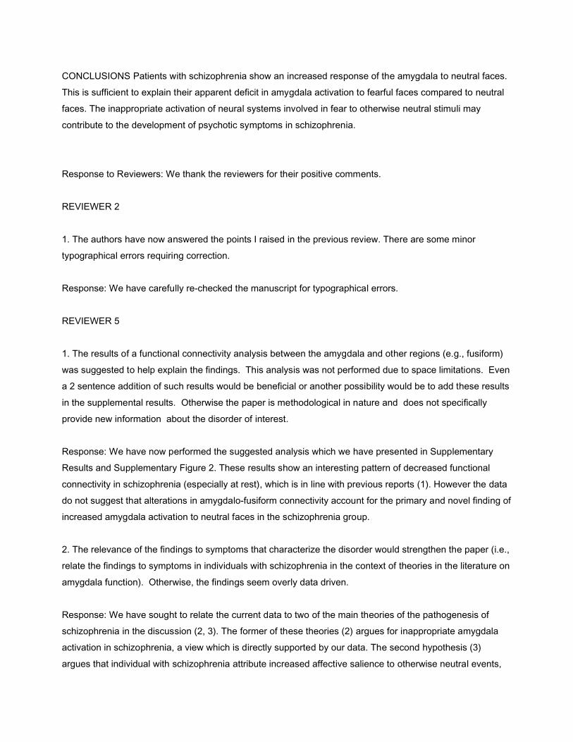
Elsevier Editorial System(tm) for Biological Psychiatry Manuscript Draft Manuscript Number: BPS-D-07-01136R2 Title: Overactivation of fear systems to neutral faces in schizophrenia Article Type: Brief Report Keywords: Fear; Amygdala; Schizophrenia; fMRI; face; emotion Corresponding Author: Dr Jeremy Hall, MRCPsych PhD Corresponding Author's Institution: Edinburgh University First Author: Jeremy Hall, PhD MRCPsych Order of Authors: Jeremy Hall, PhD MRCPsych; Heather C Whalley, PhD; James W McKirdy, PhD; Liana Romaniuk, MSc; David McGonigle, PhD; Andrew M McIntosh, MD MRCPsych; Ben J Baig, MRCPsych; Viktoria-Eleni Gountouna, MSc; Dominic E Job, PhD; David I Donaldson, PhD; Reiner Sprengelmeyer, PhD; Andrew W Young, PhD; Eve C Johnstone, MD FRCPsych; Stephen M Lawrie, MD MRCPsych Abstract: BACKGROUND The amygdala plays a central role in detecting and responding to fear related stimuli. A number of recent studies have reported decreased amygdala activation in schizophrenia to emotional stimuli (such as fearful faces) compared to matched neutral stimuli (such as neutral faces). Here we have investigated whether the apparent decrease in amygdala activation in schizophrenia could actually derive from increased amygdala activation to the neutral comparator stimuli. METHODS Nineteen patients with schizophrenia and 24 matched control participants viewed pictures of faces with either fearful or neutral facial expressions, and a baseline condition, during functional magnetic resonance imaging scanning. RESULTS Patients with schizophrenia showed a relative decrease in amygdala activation to fearful faces when compared to neutral faces. However this difference resulted from an increase in amygdala activation to the neutral faces in patients with schizophrenia, not from a decreased response to the fearful faces.
Transcript of Overactivation of Fear Systems to Neutral Faces in Schizophrenia
Elsevier Editorial System(tm) for Biological Psychiatry
Manuscript Draft
Manuscript Number: BPS-D-07-01136R2
Title: Overactivation of fear systems to neutral faces in schizophrenia
Article Type: Brief Report
Keywords: Fear; Amygdala; Schizophrenia; fMRI; face; emotion
Corresponding Author: Dr Jeremy Hall, MRCPsych PhD
Corresponding Author's Institution: Edinburgh University
First Author: Jeremy Hall, PhD MRCPsych
Order of Authors: Jeremy Hall, PhD MRCPsych; Heather C Whalley, PhD; James W McKirdy, PhD; Liana
Romaniuk, MSc; David McGonigle, PhD; Andrew M McIntosh, MD MRCPsych; Ben J Baig, MRCPsych;
Viktoria-Eleni Gountouna, MSc; Dominic E Job, PhD; David I Donaldson, PhD; Reiner Sprengelmeyer, PhD;
Andrew W Young, PhD; Eve C Johnstone, MD FRCPsych; Stephen M Lawrie, MD MRCPsych
Abstract: BACKGROUND The amygdala plays a central role in detecting and responding to fear related
stimuli. A number of recent studies have reported decreased amygdala activation in schizophrenia to
emotional stimuli (such as fearful faces) compared to matched neutral stimuli (such as neutral faces). Here
we have investigated whether the apparent decrease in amygdala activation in schizophrenia could actually
derive from increased amygdala activation to the neutral comparator stimuli.
METHODS Nineteen patients with schizophrenia and 24 matched control participants viewed pictures of
faces with either fearful or neutral facial expressions, and a baseline condition, during functional magnetic
resonance imaging scanning.
RESULTS Patients with schizophrenia showed a relative decrease in amygdala activation to fearful faces
when compared to neutral faces. However this difference resulted from an increase in amygdala activation
to the neutral faces in patients with schizophrenia, not from a decreased response to the fearful faces.
CONCLUSIONS Patients with schizophrenia show an increased response of the amygdala to neutral faces.
This is sufficient to explain their apparent deficit in amygdala activation to fearful faces compared to neutral
faces. The inappropriate activation of neural systems involved in fear to otherwise neutral stimuli may
contribute to the development of psychotic symptoms in schizophrenia.
Response to Reviewers: We thank the reviewers for their positive comments.
REVIEWER 2
1. The authors have now answered the points I raised in the previous review. There are some minor
typographical errors requiring correction.
Response: We have carefully re-checked the manuscript for typographical errors.
REVIEWER 5
1. The results of a functional connectivity analysis between the amygdala and other regions (e.g., fusiform)
was suggested to help explain the findings. This analysis was not performed due to space limitations. Even
a 2 sentence addition of such results would be beneficial or another possibility would be to add these results
in the supplemental results. Otherwise the paper is methodological in nature and does not specifically
provide new information about the disorder of interest.
Response: We have now performed the suggested analysis which we have presented in Supplementary
Results and Supplementary Figure 2. These results show an interesting pattern of decreased functional
connectivity in schizophrenia (especially at rest), which is in line with previous reports (1). However the data
do not suggest that alterations in amygdalo-fusiform connectivity account for the primary and novel finding of
increased amygdala activation to neutral faces in the schizophrenia group.
2. The relevance of the findings to symptoms that characterize the disorder would strengthen the paper (i.e.,
relate the findings to symptoms in individuals with schizophrenia in the context of theories in the literature on
amygdala function). Otherwise, the findings seem overly data driven.
Response: We have sought to relate the current data to two of the main theories of the pathogenesis of
schizophrenia in the discussion (2, 3). The former of these theories (2) argues for inappropriate amygdala
activation in schizophrenia, a view which is directly supported by our data. The second hypothesis (3)
argues that individual with schizophrenia attribute increased affective salience to otherwise neutral events,
providing the setting for the formation of symptoms such as delusional beliefs. We believe that the present
finding of increased amygdala activation to neutral faces in schizophrenia provides a potential biological
basis for such a liability to psychosis. We have attempted to re-word part of the discussion to make these
links more explicit, although fuller coverage is precluded by the word limit.
REFERENCES
1. Zhou Y, Liang M, Tian L, Wang K, Hao Y, Liu H, et al. (2007): Functional disintegration in paranoid
schizophrenia using resting-state fMRI. Schizophrenia Research. 97:194.
2. Grace AA (2000): Gating of information flow within the limbic system and the pathophysiology of
schizophrenia. Brain Research Reviews. 31:330-341.
3. Kapur S (2003): Psychosis as a State of Aberrant Salience: A Framework Linking Biology,
Phenomenology, and Pharmacology in Schizophrenia. Am J Psychiatry. 160:13-23.
Fear of faces in schizophrenia
Hall et al looked at the response of the amygdala, a brain region mediating fear, to faces in control subjects and participants with schizophrenia. They found that control subjects show amygdala activation to fearful faces, but not neutral faces. However patients with schizophrenia activated the amygdala fear system to both neutral and fearful faces. These results suggest that people with schizophrenia may perceive neutral faces as fearful, potentially contributing to the development of psychotic symptoms.
IN THIS ISSUE Statement
Hall J et al 1
Overactivation of fear systems to neutral faces in schizophrenia
Jeremy Hall1*, Heather C. Whalley1, James W. McKirdy1, Liana Romaniuk1, David McGonigle2, Andrew M. McIntosh1, Ben J. Baig1, Viktoria-Eleni Gountouna1, Dominic E. Job1, David I. Donaldson3, Reiner Sprengelmeyer4, Andrew W. Young5, Eve C. Johnstone1 and Stephen M. Lawrie1
1Division of Psychiatry, University of Edinburgh, UK2Schools of Psychology/Biosciences, University of Cardiff, UK3Psychology Department, University of Stirling, UK4School of Psychology, University of St Andrews, UK5Department of Psychology, University of York, UK
* Corresponding author:Dr Jeremy HallDivision of PsychiatryUniversity of EdinburghKennedy Tower Royal Edinburgh HospitalEdinburghUKEH10 5HF
T: +44 131 537 6313F: +44 131 537 6531E: [email protected]
Keywords: Fear, amygdala, schizophrenia, fMRI, face, emotion
Count:Abstract 200Main Text 1500Figures 3Tables 1Supplementary material – 2 figures, 2 text documents
* Manuscript
Hall J et al 2
Abstract
Background The amygdala plays a central role in detecting and responding to fear
related stimuli. A number of recent studies have reported decreased amygdala
activation in schizophrenia to emotional stimuli (such as fearful faces) compared to
matched neutral stimuli (such as neutral faces). Here we have investigated whether the
apparent decrease in amygdala activation in schizophrenia could actually derive from
increased amygdala activation to the neutral comparator stimuli.
Methods Nineteen patients with schizophrenia and 24 matched control participants
viewed pictures of faces with either fearful or neutral facial expressions, and a
baseline condition, during functional magnetic resonance imaging scanning.
Results Patients with schizophrenia showed a relative decrease in amygdala
activation to fearful faces when compared to neutral faces. However this difference
resulted from an increase in amygdala activation to the neutral faces in patients with
schizophrenia, not from a decreased response to the fearful faces.
Conclusions Patients with schizophrenia show an increased response of the amygdala
to neutral faces. This is sufficient to explain their apparent deficit in amygdala
activation to fearful faces compared to neutral faces. The inappropriate activation of
neural systems involved in fear to otherwise neutral stimuli may contribute to the
development of psychotic symptoms in schizophrenia.
Hall J et al 3
Introduction
The amygdala plays a central role in detecting and responding to fear-provoking
stimuli (1). Individuals with bilateral lesions of the amygdala show a marked
impairment in their ability to recognise fear from faces, and neuroimaging studies
have confirmed that the amygdala shows greater activation to the presentation of faces
expressing fear than to matched neutral faces (1, 2).
There is evidence of structural and functional abnormalities of the amygdala in
schizophrenia (3). A number of functional neuroimaging studies have reported
decreased amygdala activation in patients with schizophrenia compared to control
subjects (4-13). Decreased amygdala activation in schizophrenia has been particularly
shown in studies in which the response of the amygdala to an emotional stimulus
(such as a face expressing fear) has been compared to a matched neutral stimulus
(such as a neutral face) (7-9, 12, 13). These results have been interpreted as revealing
a deficit in amygdala activation in response to emotion in patients.
The finding of decreased amygdala activation in patients with schizophrenia,
especially those with paranoid symptoms, poses a paradox. Why should it be that
patients whose clinical presentation is characterised by increased fear and arousal
have decreased activation of the key brain region involved in fear? One possibility is
that the apparent decrease in amygdala activation in response to fearful stimuli in
these individuals actually derives from an increase in amygdala activation to the
neutral comparator stimuli (3). Amygdala activation is known to occur to the
presentation of neutral stimuli (14) and there is evidence that this response may be
enhanced in individuals with schizophrenia (15). In the current study we have directly
addressed the hypothesis that the apparent deficit in amygdala activation in
Hall J et al 4
schizophrenia to emotional stimuli actually derives from an increase in amygdala
response to the neutral comparator stimuli.
Hall J et al 5
Materials and Methods
Participants
Twenty-four patients meeting DSM-IV diagnostic criteria for schizophrenia and 24
matched control subjects participated in the study. One patient was excluded due to
the presence of a benign cerebral cyst and four were excluded due to failure to make
behavioural responses. Full details of subject recruitment and characteristics are given
in the Supplementary Methods.
Experimental design
A block design was used with three conditions: fear, neutral and baseline. During fear
blocks 6 faces expressing the emotion of fear were presented for 3.5s each; during
neutral blocks the same faces showing a neutral emotional expression were presented,
and during baseline blocks participants were instructed to look at a fixation cross.
Participants were required to select the gender of the face by pressing a button. The
full task consisted of two runs each comprising 3 fear blocks (25s each), 3 neutral
blocks (25s) and 7 interleaved blocks of the baseline condition (12.5s). Pilot data
demonstrated that maximal amygdala activation was seen in the first run, consistent
with previous studies (2), so analysis was primarily focussed on this period.
Image acquisition, processing and analysis
Details are given in the Supplementary Methods. Image analysis was conducted using
SPM2 (www.fil.ion.ucl.ac.uk/spm). Contrast images were generated for each
participant for the contrasts of interest (fear versus neutral, fear versus baseline and
neutral versus baseline). One contrast image per participant was then entered into a

Hall J et al 6
second-level random effects analysis for each contrast of interest to examine regions
of significant difference between groups using a t-test. All statistical maps were
thresholded at a level of P<0.001 uncorrected and regions were considered significant
at P<0.05 at the cluster level, corrected for multiple comparisons. A region of interest
(ROI) analysis was conducted for the bilateral amygdala.
Behavioural tests of emotion recognition
A standardised test of recognition of the six basic facial emotions facial emotion
recognition was conducted after the scanning session (see Supplementary Methods).
Hall J et al 7
Results
Demographics and behavioural responses in the scanner
There were no significant differences between the groups in terms of age (F1,41=0.9,
P>0.3), NART IQ (F1,41=1.3, P>0.2) or gender (Fisher’s exact test, P=1.0). Both
groups performed the incidental gender discrimination task to high degree of accuracy
(patients 91% correct (SD 13%), controls 98% correct (SD 3%)), although the
schizophrenia group did show a deficit in accuracy of gender judgments compared to
controls (F1,41=6.4, P<0.05).
Response to fearful faces versus neutral faces
Patients with schizophrenia showed a relative decrease in left amygdala activation
compared to control participants in the contrast of fearful faces versus neutral faces
(P<0.05 corrected within amygdala ROI; Table 1 and Figure 1). In addition patients
showed less activation relative to controls in the right fusiform gyrus and right lingual
gyrus (Table 1).
Response to neutral faces versus baseline
Patient showed greater activation of the amygdala to neutral stimuli than the control
group (P<0.01 corrected within an amygdala ROI; Table 1 and Figure 1). Patients also
showed relatively greater activation than controls in the right lingual gyrus, fusiform
gyrus and posterior cingulate (Table 1).
Hall J et al 8
Response to fearful faces versus baseline
There was no significant difference in activation of the amygdala or other brain
regions between patient and control groups in the contrast of fearful faces versus
baseline (Table 1).
Comparison of amygdala activation across the task conditions
We extracted data for activation in the left amygdala for both task conditions (fear and
neutral) relative to baseline and performed an ANOVA with group as a between
subjects factor and emotion as a within subjects factor (Figure 2). This analysis
revealed a significant group by emotion interaction (F1,41=4.9, P<0.05). Planned post-
hoc t-tests confirmed that this derived from greater activation of the amygdala in the
schizophrenia group to neutral faces (P<0.01), with no difference between groups in
amygdala activation to fearful faces (P=0.4).
Correlation analyses
Correlation analyses were performed to assess whether left amygdala activation (fear
versus baseline or neutral versus baseline) was related to medication dose, PANSS
total score, PANSS positive or PANSS negative symptoms, PANSS anxiety and
depressive symptoms or gender judgment performance. None of these correlations
was significant.
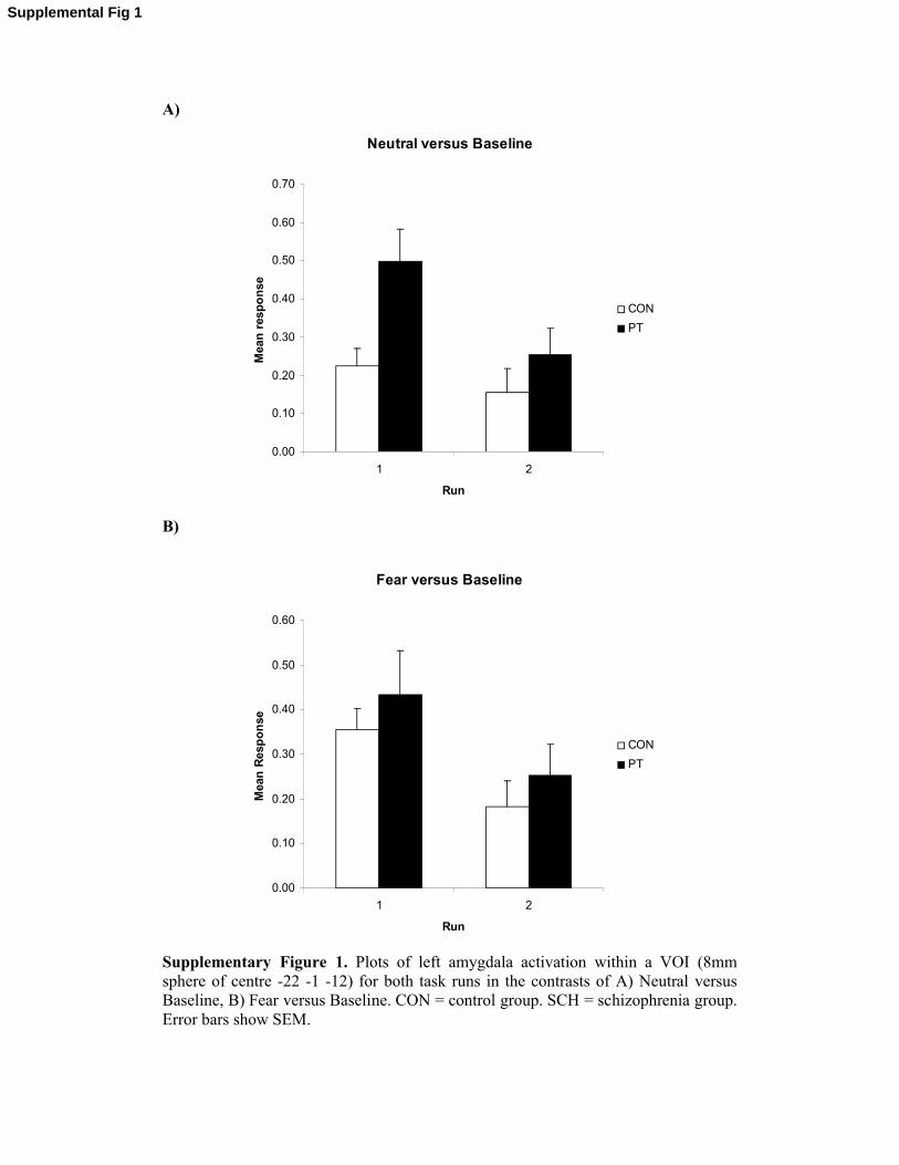
Amygdala activation over time
A comparison of amygdala activation between the first and second task runs showed
significant habituation of amygdala activation over time, with group differences only
apparent in the first run (Supplementary Figure 1).
Hall J et al 9
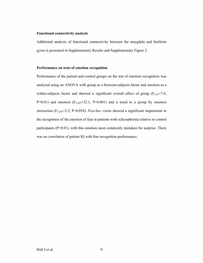
Functional connectivity analysis
Additional analysis of functional connectivity between the amygdala and fusiform
gyrus is presented in Supplementary Results and Supplementary Figure 2.
Performance on tests of emotion recognition
Performance of the patient and control groups on the test of emotion recognition was
analysed using an ANOVA with group as a between-subjects factor and emotion as a
within-subjects factor and showed a significant overall effect of group (F1,41=7.0,
P=0.01) and emotion (F5,205=32.1, P<0.001) and a trend to a group by emotion
interaction (F5,205=2.2, P=0.054). Post-hoc t-tests showed a significant impairment in
the recognition of the emotion of fear in patients with schizophrenia relative to control
participants (P<0.01), with this emotion most commonly mistaken for surprise. There
was no correlation of patient IQ with fear recognition performance.

Hall J et al 10
Discussion
A number of neuroimaging studies have reported decreased activation of the
amygdala to emotional stimuli compared to neutral stimuli in schizophrenia (3). We
have investigated this effect by examining amygdala response to fearful and neutral
faces in patients with schizophrenia and control subjects. We found that whilst
patients did not differ significantly from controls in their amygdala response to fearful
faces, they showed increased activation of the amygdala to neutral faces.
Patients with schizophrenia experiencing psychotic symptoms, especially
paranoid beliefs, characteristically have increased levels of fear and arousal. The
amygdala plays a central role in mediating fear and arousal and it is therefore
paradoxical that previous studies have shown a decrease in amygdala activation in
schizophrenia (7-9, 12, 13). The present findings provide a resolution to this paradox
by showing that the apparent deficit in amygdala activation in individuals with
schizophrenia can in fact be explained by increased amygdala response to the neutral
comparator stimuli. This finding is supported by other studies which have shown
increased medial temporal lobe activation in response to neutral stimuli in individuals
with schizophrenia (15-17). Notably enhanced amygdala activation to neutral faces is
also seen in children, suggesting the current findings may have a neurodevelopmental
origin (18).
The present results help explain the findings of behavioural studies, which
have typically shown deficits in the recognition of facial emotions in patients with
schizophrenia, especially for negative emotions such as fear. Our findings suggest that
the behavioural deficits may derive from an indiscriminate activation of the amygdala
Hall J et al 11
to both fearful and non-fearful facial stimuli rather than a failure to activate the
amygdala to fearful faces.
Patients with psychosis ascribe affective importance to stimuli and events that
would generally be considered neutral. An influential theoretical model of the origin
of psychosis is that it represents a state characterized by the aberrant attribution of
salience to internal and external events (19). A related hypothesis posits amygdala
hyper-activation, related to increased dopamine responsiveness, as a key pathogenic
process in schizophrenia causing exaggerated affective drive to govern behavioural
response selection (20). The current findings support these theories by demonstrating
increased activation of the amygdala to neutral stimuli in schizophrenia, which may
contribute to liability for psychosis.
Hall J et al 12
Acknowledgements
The work was funded by the Dr Mortimer and Theresa Sackler Foundation. JH is
supported by a MRC Clinical Research Training Fellowship and AMcI is supported
by The Health Foundation. HCW and SML were supported by the Sackler Foundation.
Imaging was performed at the SFC Brain Imaging Research Centre in Edinburgh. We
would like to express our thanks to Iona Hamilton, Elaine Sandeman and Andrew
Saul for their expert assistance in this work. We would also like to thank all the
participants.
Financial Disclosures
The authors have no competing financial interests to declare in relation to the current
work.
Hall J et al 13
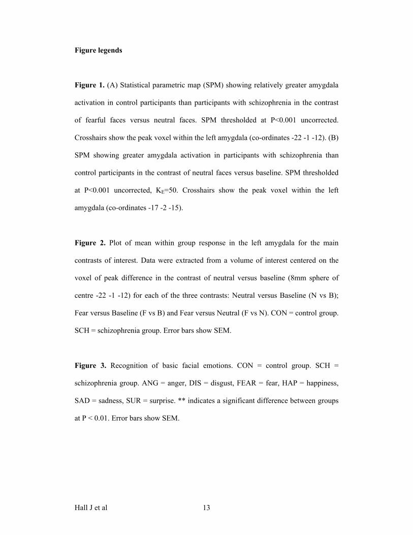
Figure legends
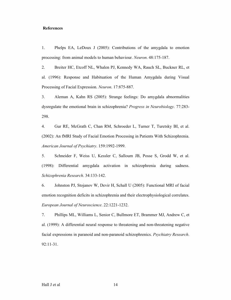
Figure 1. (A) Statistical parametric map (SPM) showing relatively greater amygdala
activation in control participants than participants with schizophrenia in the contrast
of fearful faces versus neutral faces. SPM thresholded at P<0.001 uncorrected.
Crosshairs show the peak voxel within the left amygdala (co-ordinates -22 -1 -12). (B)
SPM showing greater amygdala activation in participants with schizophrenia than
control participants in the contrast of neutral faces versus baseline. SPM thresholded
at P<0.001 uncorrected, KE=50. Crosshairs show the peak voxel within the left
amygdala (co-ordinates -17 -2 -15).
Figure 2. Plot of mean within group response in the left amygdala for the main
contrasts of interest. Data were extracted from a volume of interest centered on the
voxel of peak difference in the contrast of neutral versus baseline (8mm sphere of
centre -22 -1 -12) for each of the three contrasts: Neutral versus Baseline (N vs B);
Fear versus Baseline (F vs B) and Fear versus Neutral (F vs N). CON = control group.
SCH = schizophrenia group. Error bars show SEM.
Figure 3. Recognition of basic facial emotions. CON = control group. SCH =
schizophrenia group. ANG = anger, DIS = disgust, FEAR = fear, HAP = happiness,
SAD = sadness, SUR = surprise. ** indicates a significant difference between groups
at P < 0.01. Error bars show SEM.
Hall J et al 14
References
1. Phelps EA, LeDoux J (2005): Contributions of the amygdala to emotion
processing: from animal models to human behaviour. Neuron. 48:175-187.
2. Breiter HC, Etcoff NL, Whalen PJ, Kennedy WA, Rauch SL, Buckner RL, et
al. (1996): Response and Habituation of the Human Amygdala during Visual
Processing of Facial Expression. Neuron. 17:875-887.
3. Aleman A, Kahn RS (2005): Strange feelings: Do amygdala abnormalities
dysregulate the emotional brain in schizophrenia? Progress in Neurobiology. 77:283-
298.
4. Gur RE, McGrath C, Chan RM, Schroeder L, Turner T, Turetsky BI, et al.
(2002): An fMRI Study of Facial Emotion Processing in Patients With Schizophrenia.
American Journal of Psychiatry. 159:1992-1999.
5. Schneider F, Weiss U, Kessler C, Salloum JB, Posse S, Grodd W, et al.
(1998): Differential amygdala activation in schizophrenia during sadness.
Schizophrenia Research. 34:133-142.
6. Johnston PJ, Stojanov W, Devir H, Schall U (2005): Functional MRI of facial
emotion recognition deficits in schizophrenia and their electrophysiological correlates.
European Journal of Neuroscience. 22:1221-1232.
7. Phillips ML, Williams L, Senior C, Bullmore ET, Brammer MJ, Andrew C, et
al. (1999): A differential neural response to threatening and non-threatening negative
facial expressions in paranoid and non-paranoid schizophrenics. Psychiatry Research.
92:11-31.
Hall J et al 15
8. Williams LM, Das P, Harris AWF, Liddell BB, Brammer MJ, Olivieri G, et al.
(2004): Dysregulation of Arousal and Amygdala-Prefrontal Systems in Paranoid
Schizophrenia. American Journal of Psychiatry. 161:480-489.
9. Williams LM, Das P, Liddell BJ, Olivieri G, Peduto AS, David AS, et al.
(2007): Fronto-limbic and autonomic disjunctions to negative emotion distinguish
schizophrenia subtypes. Psychiatry Research: Neuroimaging. 155:29-44.
10. Taylor SF, Liberzon I, Decker LR, Koeppe RA (2002): A functional anatomic
study of emotion in schizophrenia. Schizophrenia Research. 58:159-172.
11. Paradiso S, Andreasen NC, Crespo-Facorro B, O'Leary DS, Watkins GL,
Boles Ponto LL, et al. (2003): Emotions in Unmedicated Patients With Schizophrenia
During Evaluation With Positron Emission Tomography. American Journal of
Psychiatry. 160:1775-1783.
12. Takahashi H, Koeda M, Oda K, Matsuda T, Matsushima E, Matsuura M, et al.
(2004): An fMRI study of differential neural response to affective pictures in
schizophrenia. NeuroImage. 22:1247-1254.
13. Hempel A, Hempel E, Schonknecht P, Stippich C, Schroder J (2003):
Impairment in basal limbic function in schizophrenia during affect recognition.
Psychiatry Research: Neuroimaging. 122:115-124.
14. Schwartz CE, Wright CI, Shin LM, Kagan J, Whalen PJ, McMullin KG, et al.
(2003): Differential amygdalar response to novel versus newly familiar neutral faces:
a functional MRI probe developed for studying inhibited temperament. Biological
Psychiatry. 53:854-862.
15. Holt DJ, Kunkel L, Weiss AP, Goff DC, Wright CI, Shin LM, et al. (2006):
Increased medial temporal lobe activation during the passive viewing of emotional
and neutral facial expressions in schizophrenia. Schizophrenia Research. 82:153-162.
Hall J et al 16
16. Taylor SF, Luan Phan K, Britton JC, Liberzon I (2005): Neural Response to
Emotional Salience in Schizophrenia. Neuropsychopharmacology. 30:984-995.
17. Surguladze S, Russell T, Kucharska-Pietura K, Travis MJ, Giampietro V,
David AS, et al. (2006): A Reversal of the Normal Pattern of Parahippocampal
Response to Neutral and Fearful Faces Is Associated with Reality Distortion in
Schizophrenia. Biological Psychiatry. 60:423-431.
18. Thomas KM, Drevets WC, Whalen PJ, Eccard CH, Dahl RE, Ryan ND, et al.
(2001): Amygdala response to facial expressions in children and adults. Biological
Psychiatry. 49:309-316.
19. Kapur S (2003): Psychosis as a State of Aberrant Salience: A Framework
Linking Biology, Phenomenology, and Pharmacology in Schizophrenia. Am J
Psychiatry. 160:13-23.
20. Grace AA (2000): Gating of information flow within the limbic system and
the pathophysiology of schizophrenia. Brain Research Reviews. 31:330-341.
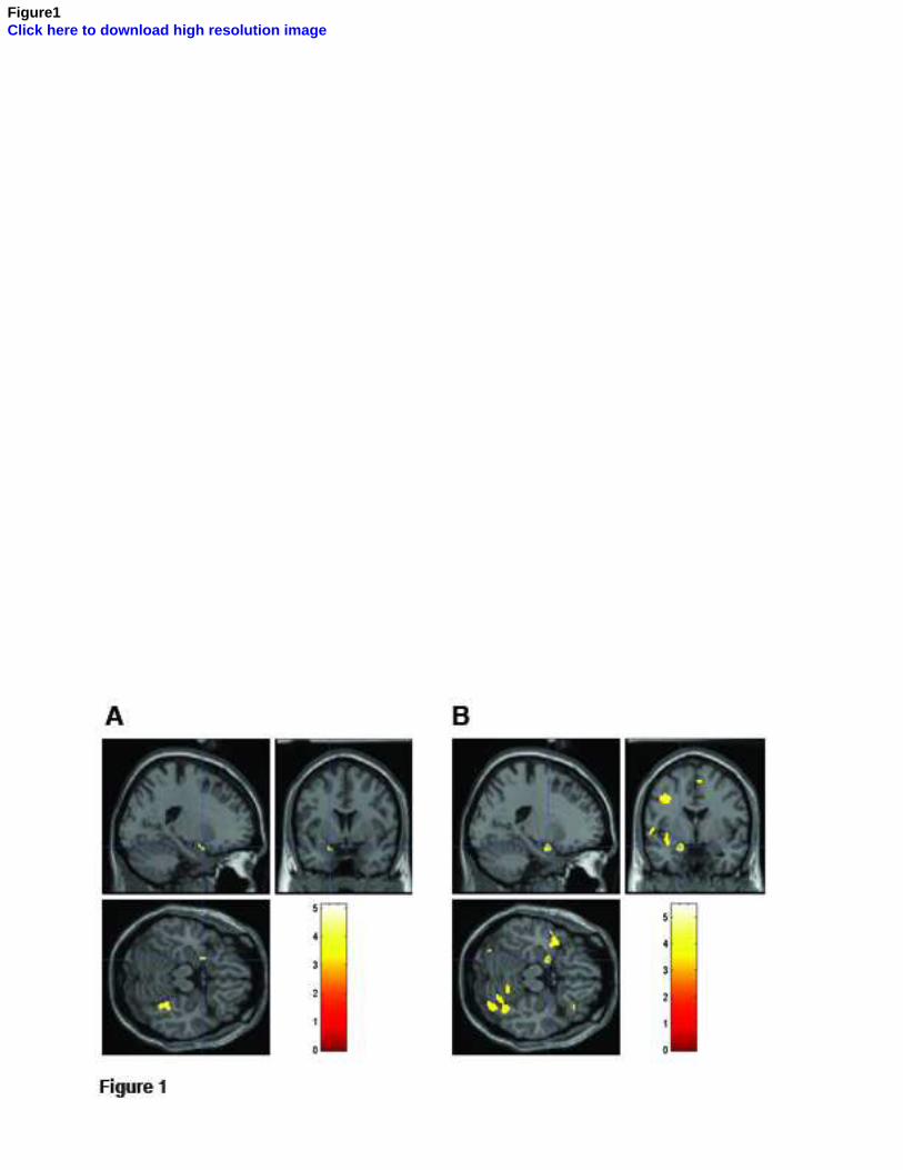
Figure1Click here to download high resolution image
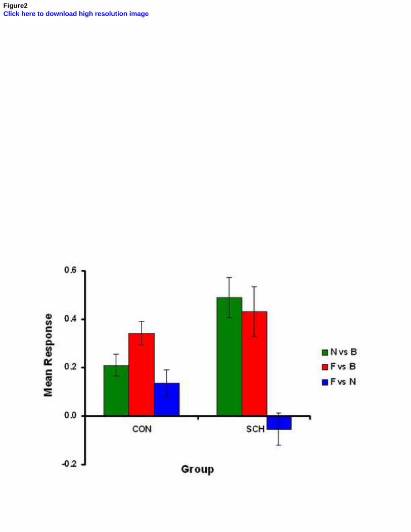
Figure2Click here to download high resolution image
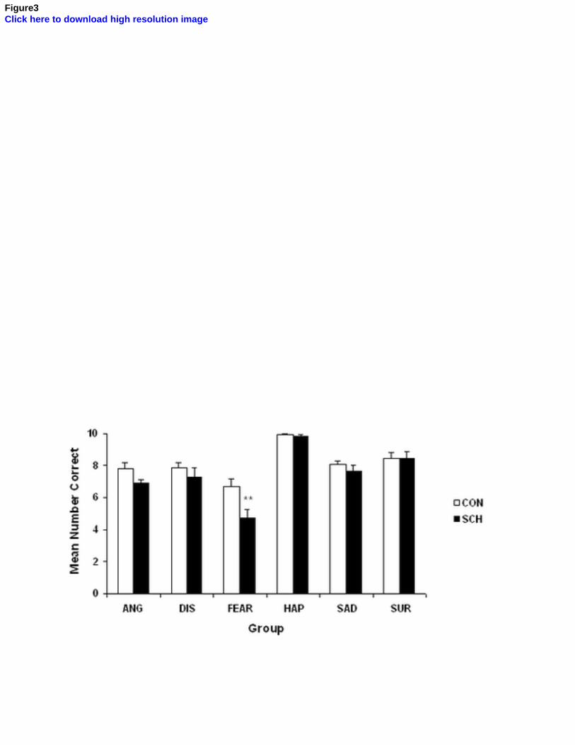
Figure3Click here to download high resolution image
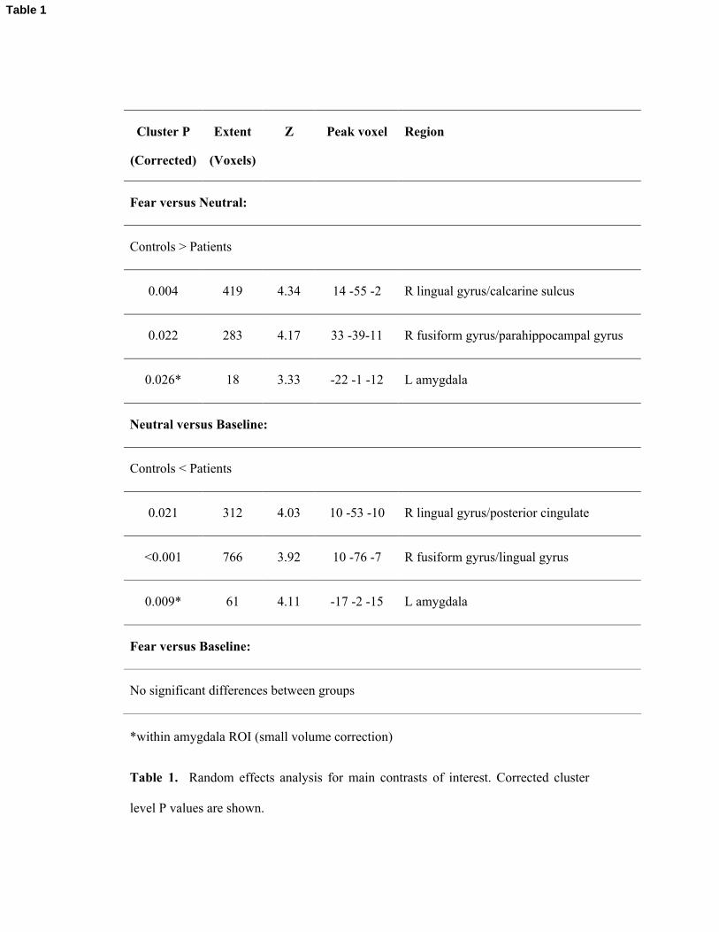
Cluster P
(Corrected)
Extent
(Voxels)
Z Peak voxel Region
Fear versus Neutral:
Controls > Patients
0.004 419 4.34 14 -55 -2 R lingual gyrus/calcarine sulcus
0.022 283 4.17 33 -39-11 R fusiform gyrus/parahippocampal gyrus
0.026* 18 3.33 -22 -1 -12 L amygdala
Neutral versus Baseline:
Controls < Patients
0.021 312 4.03 10 -53 -10 R lingual gyrus/posterior cingulate
<0.001 766 3.92 10 -76 -7 R fusiform gyrus/lingual gyrus
0.009* 61 4.11 -17 -2 -15 L amygdala
Fear versus Baseline:
No significant differences between groups
*within amygdala ROI (small volume correction)
Table 1. Random effects analysis for main contrasts of interest. Corrected cluster
level P values are shown.
Table 1
Supplementary Methods
Participants
Twenty-four patients meeting DSM-IV diagnostic criteria for schizophrenia
participated in the study, recruited from both inpatient and outpatient populations.
Diagnoses were made by clinical consensus amongst clinicians highly experienced in
working with patients with psychosis and was confirmed from case-notes using
OPCRIT criteria (1). Exclusion criteria were age under 18 or over 65, neurological
disease, dependence on alcohol or non-prescribed drugs, and other concomitant axis I
or axis II disorder. Sixteen were men and eight were women. One subject was
excluded due to the presence of a benign cerebral cyst and four individuals were
excluded prior to analysis due to a failure to make any behavioural responses in the
scanner. The remaining 19 individuals in the patient group (twelve men and seven
women) had a mean age of 37.7 years (SD 8.4) and a mean pre-morbid IQ as assessed
by the National Adult Reading Test (NART) (2) of 111.6 (SD 10.1). All patients were
Caucasian, 17 were right handed and 2 were left handed. All were treated with
antipsychotic medication (16 with atypical antipsychotics) with a mean
chlorpromazine equivalent dose of 496mg (SD 377mg). Symptoms were rated on the
day of the scanning session using the positive and negative syndrome scale (PANSS)
(3) and the mean PANSS score was 46.8 (SD 8.5). Mean positive syndrome score on
the PANSS was 12.3 (SD 4.5) with 15 out of the 19 individuals scoring 3 or greater
on one or more positive syndrome items. Mean negative syndrome score on the
PANSS was 11.8 (SD 3.4).
Twenty-four healthy control volunteers were recruited from the same regions
and communities as the patients themselves. None of these subjects reported any
Supplemental Methods
personal history of psychiatric illness or any family history of psychosis. Sixteen were
men and eight were women; mean age was 35.1 years (SD 9.7), and mean IQ was
114.5 (SD 6.5). All control participants were Caucasian and right handed. Control
participants were screened for any family or personal history of psychiatric illness and
were additionally subject to the same exclusion criteria as the patients.
The study was approved by the local ethics committee and after complete
description of the study written informed consent was obtained from all participants.
Task design
During fear and neutral blocks participants were initially presented with a 1s visual
prompt (“Gender?”) followed by six greyscale pictures of faces from the Ekman and
Friesen series (4) each presented for 3.5s in a random order with a 0.5s inter-stimulus
interval. Three of the faces were male and three were female in each block.
Participants were required to select the gender of the face by pressing a button. The
alternative response choices (“Male” and “Female”) were shown on the screen.
Pictures of the same individuals were shown in both the fear blocks and neutral blocks,
differing only in the emotion shown (fearful or neutral expressions respectively). The
order of presentation of fear and neutral blocks was counterbalanced across subjects.
A block design was chosen because of the statistical power afforded by this approach,
and the fact that it has proven effective in demonstrating amygdala activation in
previous studies (5, 6). An implicit task was selected because increasing cognitive
task demands has previously been shown to decrease limbic, and in particular
amygdala activation (7).
Image acquisition
Imaging was performed at the SFC Brain Imaging Research Centre in Edinburgh
using a GE 1.5TE Signa scanner (GE Medical, Milwaukee, WI, USA). After a
localiser scan participants underwent functional scanning (99 volumes; Field of View
22cm; Time to Echo (TE) 40ms; Volume acquisition time (TR) 2.5s). Interleaved
axial slices were acquired with a thickness of 5mm with no gap and matrix size of 64
x 64. The first four EPIs were discarded to avoid T1 equilibrium effects.
Image processing and analysis
The EPI images were reconstructed offline in ANALYZE format (Mayo Foundation,
Rochester, MN, USA). Image analysis was conducted using SPM2 (Statistical
Parametric Mapping; Wellcome Department of Cognitive Neurology and
collaborators, Institute of Neurology, London, UK). Pre-processing consisted of re-
orientation of the images and realignment to the mean EPI image, followed by
normalisation to the standard Montreal Neurological Institute EPI template and spatial
smoothing using a Gaussian kernel (8mm3 full-width at half-maximum). Images from
all subjects were inspected to exclude susceptibility artefacts in the medial temporal
lobe. Within-scanner movement data was examined for all subjects and no subject
was found to have moved more than 3.0mm in any axis across the duration of the scan.
Before fitting the model, the subject’s data were filtered in time using a high pass
filter (150 s cut-off) and temporal autocorrelations were accounted for by imposing an
AR(1) model.
Statistical analysis was performed using the general linear model approach as
implemented in SPM2. At the individual participant level the data for each task were
modelled with three conditions (fear, neutral and baseline) each modelled by a boxcar
convolved with a canonical haemodynamic response function. Parameters
representing the participants movement during the scan were also entered into the
model as covariates of no interest. Contrast images were generated for each
participant for the contrasts of interest (fear versus neutral, fear versus baseline and
neutral versus baseline) representing the pair-wise comparison of parameter estimates
for the conditions. One contrast image per participant was then entered into a second-
level random effects analysis for each contrast of interest to examine regions of
significant difference between groups using a two sample t-test.
All statistical maps were thresholded at a level of P<0.001 uncorrected and
regions were considered significant at P<0.05 at the cluster level, corrected for
multiple comparisons. Co-ordinates were converted from MNI to Talairach co-
ordinates using a non-linear transformation (http://www.mrc-cbu.cam.ac.uk/Imaging).
A region of interest (ROI) analysis was conducted for the bilateral amygdala using a
mask derived from the automated anatomical labelling atlas in WFU_PickAtlas v2.0
(8, 9).
Functional connectivity analysis
Time series were extracted from spheres (radius 5mm) centred on voxels of peak
difference in the amygdala and fusiform gyrus for the fear versus neutral contrast.
These data were regressed against motion parameters, and time series extracted from
white matter and CSF, to reduce the effects of noise unrelated to the task. Pearson
correlation coefficients were then calculated between the regions of interest and
transformed to normal space using Fisher's r-to-z. The conditions of interest were
considered separately by truncating the timecourses according to condition blocks.
Independent t-tests were then performed between the patient and control groups to
compare connectivity across the conditions.
Behavioural tests of emotion recognition
A well charactersised behavioural test of facial emotion recognition was conducted
after the scanning session (10-13). Photographs of the faces of 10 people used in this
test were taken from the Ekman and Friesen series (4). For each face, there were poses
corresponding to each of 6 basic emotions (happiness, surprise, fear, sadness, disgust,
and anger), giving a total of 60 photographs (10 for each emotion). These were shown
one at a time on a computer screen in pseudo-random order, for 3 seconds each. The
task involved deciding which of the emotion names (happiness, surprise, fear, sadness,
disgust, or anger) best described the facial expression shown. All participants were
asked if they understood the meanings of the emotional names. If not, brief
standardised descriptions were given. The names of the six emotions were displayed
at the bottom of the screen and they could be consulted throughout the test. The order
of the emotions on the screen was varied between tests. Responses were made by
clicking the computer mouse over the name of the selected emotion. There was no
time limit for responding. The next face was not shown until the subject had made a
response. No feedback was given as to the appropriateness of any responses. Results
were analysed using SPSS for Windows (version 14.0, SPSS Inc., US).
References
1. McGuffin P, Farmer A, Harvey I (1991): A polydiagnostic application of
operational criteria in studies of psychotic illness. Development and reliability of the
OPCRIT system. Arch Gen Psychiatry. 48:764-770.
2. Nelson HE, Willison JR (1991): The Revised National Adult Reading Test –
Test Manual.
3. Kay SR, Fiszbein A, Opler LA (1987): The positive and negative syndrome
scale (PANSS) for schizophrenia. Schizophrenia Bulletin. 13:261-276.
4. Ekman P, Friesen WV (1976): Pictures of facial affect. Palo Alto, CA:
Consulting Psychologists Press.
5. Breiter HC, Etcoff NL, Whalen PJ, Kennedy WA, Rauch SL, Buckner RL, et
al. (1996): Response and Habituation of the Human Amygdala during Visual
Processing of Facial Expression. Neuron. 17:875-887.
6. Phillips ML, Young AW, Senior C, Brammer M, Andrew C, Calder AJ, et al.
(1997): A specific neural substrate for perceiving facial expressions of disgust. Nature.
389:495.
7. Hariri A, Bookheimer SY, Mazziotta JC (2000): Modulating emotional
responses: effects of a neocortical network on the limbic system. Neuroreport. 11:43-
48.
8. Maldjian JA, Laurienti PJ, Kraft RA, Burdette JH (2003): An automated
method for neuroanatomic and cytoarchitectonic atlas-based interrogation of fMRI
data sets. NeuroImage. 19:1233-1239.
9. Tzourio-Mazoyer N, Landeau B, Papathanassiou D, Crivello F, Etard O,
Delcroix N, et al. (2002): Automated Anatomical Labeling of Activations in SPM
Using a Macroscopic Anatomical Parcellation of the MNI MRI Single-Subject Brain.
NeuroImage. 15:273-289.
10. Young AW, Perrett DI, Calder AJ, Sprengelmeyer R, Ekman P (2002): Facial
emotional expressions: stimuli and tests (FEEST). Bury St. Edmunds: Thames Valley
Test Company.
11. Hall J, Harris JM, Sprengelmeyer R, Sprengelmeyer A, Young AW, Santos
IM, et al. (2004): Social cognition and face processing in schizophrenia. Br J
Psychiatry. 185:169-170.
12. Sprengelmeyer R, Young AW, Calder AJ, Karnat A, Lange H, Homberg V, et
al. (1996): Loss of disgust: Perception of faces and emotions in Huntington's disease.
Brain. 119:1647-1665.
13. Sprengelmeyer R, Young AW, Schroeder U, Grossenbacher PG, Federlein J,
Buttner T, et al. (1999): Knowing no fear. Proc Biol Sci. 266:2451-2456.
Supplementary Results
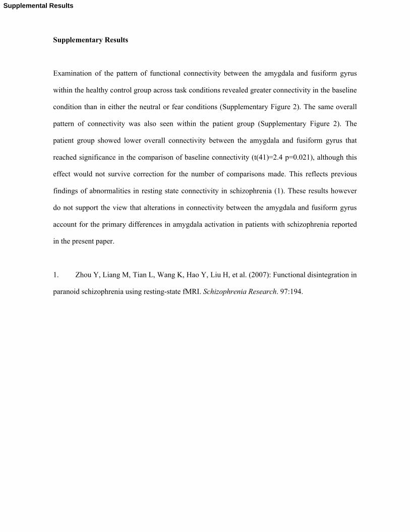
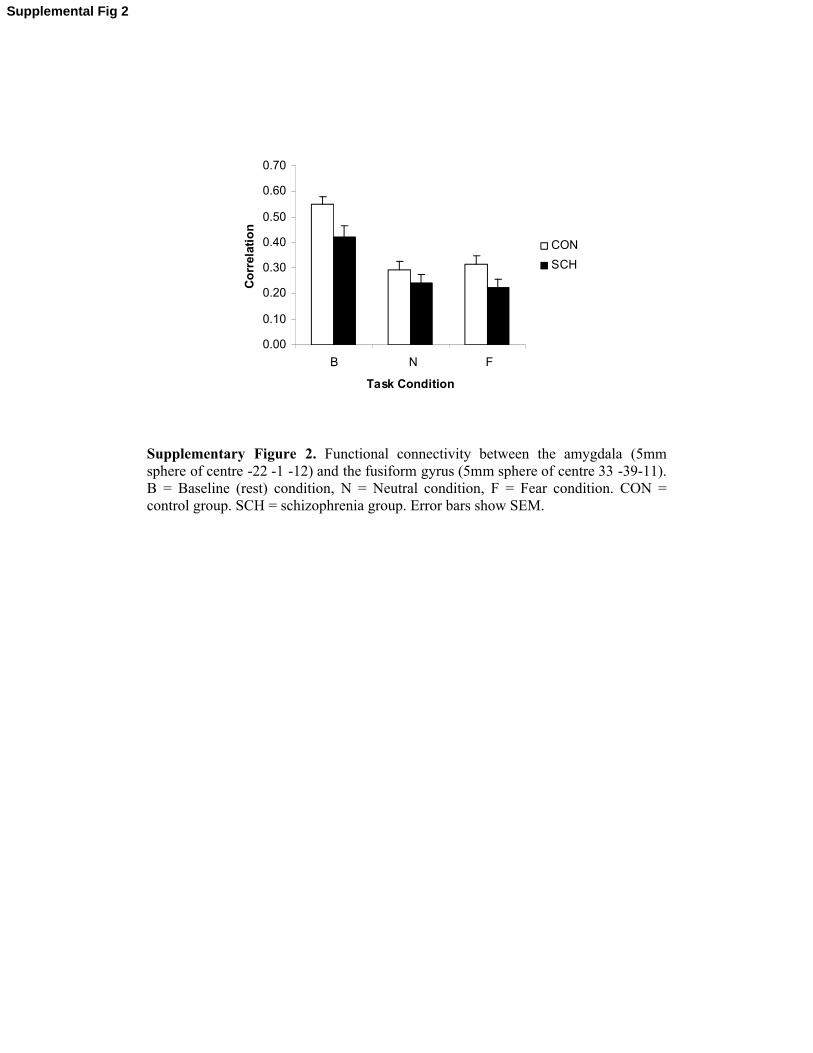
Examination of the pattern of functional connectivity between the amygdala and fusiform gyrus
within the healthy control group across task conditions revealed greater connectivity in the baseline
condition than in either the neutral or fear conditions (Supplementary Figure 2). The same overall
pattern of connectivity was also seen within the patient group (Supplementary Figure 2). The
patient group showed lower overall connectivity between the amygdala and fusiform gyrus that
reached significance in the comparison of baseline connectivity (t(41)=2.4 p=0.021), although this
effect would not survive correction for the number of comparisons made. This reflects previous
findings of abnormalities in resting state connectivity in schizophrenia (1). These results however
do not support the view that alterations in connectivity between the amygdala and fusiform gyrus
account for the primary differences in amygdala activation in patients with schizophrenia reported
in the present paper.
1. Zhou Y, Liang M, Tian L, Wang K, Hao Y, Liu H, et al. (2007): Functional disintegration in
paranoid schizophrenia using resting-state fMRI. Schizophrenia Research. 97:194.
Supplemental Results
A)
Neutral versus Baseline
0.00
0.10
0.20
0.30
0.40
0.50
0.60
0.70
1 2
Run
Mea
n r
esp
on
se
CON
PT
B)
Fear versus Baseline
0.00
0.10
0.20
0.30
0.40
0.50
0.60
1 2
Run
Mea
n R
esp
on
se
CON
PT
Supplementary Figure 1. Plots of left amygdala activation within a VOI (8mm sphere of centre -22 -1 -12) for both task runs in the contrasts of A) Neutral versus Baseline, B) Fear versus Baseline. CON = control group. SCH = schizophrenia group. Error bars show SEM.
Supplemental Fig 1
0.00
0.10
0.20
0.30
0.40
0.50
0.60
0.70
B N F
Task Condition
Co
rrel
atio
nCON
SCH
Supplementary Figure 2. Functional connectivity between the amygdala (5mm sphere of centre -22 -1 -12) and the fusiform gyrus (5mm sphere of centre 33 -39-11). B = Baseline (rest) condition, N = Neutral condition, F = Fear condition. CON = control group. SCH = schizophrenia group. Error bars show SEM.
Supplemental Fig 2
* Manuscript Submission Form
Acknowledgements
The work was funded by the Sir Mortimer and Theresa Sackler Foundation. JH is
supported by a MRC Clinical Research Training Fellowship and AMcI is supported
by The Health Foundation. We would like to express our thanks to Iona Hamilton,
Elaine Sandeman and Andrew Saul for their expert assistance in this work. We would
also like to thank all the participants.
Financial Disclosures
The authors have no competing financial interests to declare in relation to the current
work.
* Manuscript Submission Form 2