ORTHOPAEDIC RESEARCH CENTER
99
ORTHOPAEDIC RESEARCH CENTER 2016 REPORT
-
Upload
khangminh22 -
Category
Documents
-
view
1 -
download
0
Transcript of ORTHOPAEDIC RESEARCH CENTER

ORTHOPAEDIC RESEARCH CENTER2016 REPORT

MISSIONTO INVESTIGATE THE PATHOGENESIS, DIAGNOSIS,
TREATMENT, AND PREVENTION OF MUSCULOSKELETAL
DISEASE AND INJURY FOR THE BETTERMENT OF BOTH
ANIMALS AND HUMANS.

It is my pleasure again to present our 2016 report
from the Musculoskeletal Research Program
which consists of the Orthopaedic Research
Center (including the Orthopaedic Bioengineering
Research Laboratory), as well as, the Preclinical
Surgical Research Laboratory at Colorado State
University and Orthopaedic Oncology. Our principal
focus continues to be solving the significant
problems in equine musculoskeletal disease as
can be seen in this report but we also continue to
investigate comparative disease problems and
questions relevant to human joint disease including
techniques and devices for human osteoarthritis and
articular cartilage repair when the technique can
potentially benefit the horse. The increased number
of translational projects and funding support from
the National Institute of Health (NIH) and human
orientated industry partners support our mission of
helping both horses and humans.
As part of that evolution the big news for 2016 is
that we completed funding for what is now going to
be called the Translational Medicine Institute (TMI)
(previously described as the Institute of Biological
Translational Therapies but people were having
difficulty with this name and understanding what
we did). In 2015 we reported that we received
a matching challenge of one half the cost of the
building (then estimated at $65 million), followed
by a $10 million commitment from Colorado State
University (per President Dr. Tony Frank) and $20
million from another donor who at that time wished
to remain anonymous. Since then, her name has
been disclosed. It is Abigail K. Kawananakoa
who has been a long term client of mine and
previously donated a University Endowed Chair
in Musculoskeletal Integrative Therapies. After
Abigail’s gift of $20 million we were left with a $2.5
million difference which has now been obtained
from other sources.
As I write this preface for our 2016 report in
September 2017, we held the groundbreaking for
the TMI on June 2, 2017 and there will be more news
on that next year in our 2017 report. The vision of
the TMI is to investigate next generation remedies
based on living cells and their products including
patient derived stem cells to treat musculoskeletal
disease and other ailments and to literally be able to
carry basic science discoveries in the institute all the
way to bedside with entrepreneurial and regulatory
abilities within the TMI. At the ORC we have
developed expertise in analyzing and developing
medical treatments for animal patients, and then
providing knowledge gained to boost human
medical advancements. This progression is known
as translational medicine and is successful because
of similarities in animal and human physiology and
disease. Our being able to raise the funding for the
TMI building in a two and one half year period is truly
PREFACE
transformational and will take us to a higher level. It
is also an endorsement of what we have achieved
already. Those achievements have come from a
combination of ingenuity and work of our faculty,
research associates, graduate students, veterinary
students and undergraduate students as well as
terrific help from our donors.
In 2016 there was also major progress towards
building a new Equine Veterinary Teaching Hospital
and this will be located on South Campus east of the
Orthopaedic Research Center buildings and north of
the new TMI building. A lead gift of $10 million has
been obtained from the Johnson Family Foundation
and another $20.3 million identified from state funds.
Plans are underway again with Tetrad (the developer
“Our principal focus continues to be solving the signif icant problems in equine musculoskeletal disease”
continued...
Renderings of Translational Medicine Institute...
2 O R T H O P A E D I C R E S E A R C H C E N T E R 2 0 1 6 R E P O R T 3

for TMI) to build a brand new conventional hospital
and then also renovate and enlarge the present
Gail Holmes Equine Orthopaedic Research Center
into a full service sports medicine center focused
on rehabilitation of horses with musculoskeletal
problems. Dr. Chris Kawcak has been appointed
Director of the Equine Veterinary Teaching Hospital
and Dr. Melissa King is in charge of the new
rehabilitation endeavor. Renovation and occupancy
of the Gail Holmes building as a sports medicine and
rehabilitation center is planned after November 30,
2018 when we have been told we can occupy TMI.
Research endeavors applicable to sports medicine
and rehabilitation will be retained within the Gail
Holmes Equine Orthopaedic Research Center.
As part of the imaging development progress made
(albeit a little slowly) on developing a CT that will
image the standing limb (the earlier unit has a
circular ring which will only allow standing imaging of
the head and neck). The Louis L. Borick Foundation
is providing $500,000 of their total donation of
$850,000 to allow us to partner with an Italian
company to develop a prototype of the standing
CT and this development process has commenced.
Once we have the prototype we will then be ready
to manufacture such equipment to be sold to equine
veterinarians throughout the world.
The people of our program are our strength and
we added two faculty members to the program
this year, Dr. Kurt Selberg who is a Board Certified
Radiologist/Imager and we now have built the
program to include two full time equine imagers,
Drs. Myra Barrett and Kurt Selberg which is unique
and a particular strength. Funding within the
Kenneth Atkinson College Endowed Chair has been
able to provide some of the financial support for
these additions. Dr. Katie Seabaugh fills the newly
created fourth position in Equine Sports Medicine
and Rehabilitation. Katie was a product of our
surgical residency program and since becoming
Board Certified in Surgery has also become Board
Certified in Sports Medicine and Rehabilitation. Dr.
Ellison Aldrich completed her surgical residency in
2015 and is now a senior lecturer at the veterinary
school at Massey University in New Zealand. Dr.
Philippe Manchon finished his residency program in
Sports Medicine and Rehabilitation July 1, 2016 and
has moved back to Australia to take up a position in
Scone. Our new resident in Equine Sports Medicine
and Rehabilitation is Dr. Jodie Daglish and she has
become an intern in imaging within the program.
Because of the demand of expertise in ultrasound
examination we select an intern as first year of a
four year program with the latter three years being
the Sports Medicine and Rehabilitation Program.
The internship mainly involves training in diagnostic
imaging and, in particular, ultrasonography.
Other accomplishments at the ORC over the
past year are detailed in this report. This is the
second single year report because of the amount
of information we have. As stated previously,
our accomplishments in this report could not be
achieved without our team of faculty and staff as well
as the excellent support of equine funding agencies
(Grayson-Jockey Club Research Foundation,
American Quarter Horse Association and United
States Equestrian Federation), industry funding,
NIH funding and individual donors. With this help
we continue to achieve our goals and also the new
ones of TMI are particularly exciting.
Another minor event that happened in 2016 is that
in August I moved into transitional retirement. This
involved my appointing Dr. David Frisbie as Director
of Research for the ORC as well as Interim Director
of Operations for the TMI and Dr. Chris Kawcak
extended his duties as Director of the Equine
Veterinary Teaching Hospital to include the Equine
Sports Medicine and Rehabilitation Program. Chris
and Dave have been with me 25 and 24 years
respectively and were ready to take over these
important leadership roles and are doing well. I
committed to Dr. Tony Frank, the President of CSU,
Best wishes,
Wayne McIlwraith
for four more years in transitional retirement. So far
people have a smile on their face when they ask me,
“How’s transitional retirement going?” knowing that
I’m doing very well at markedly lowering my work
presence or consequently not increasing my leisure
time. Retirement is definitely a work in progress but
most of the reasons for continuing to be “flat-out” is
how much is going on as we add the new tier of the
TMI and expand our research as well as our Equine
Sports Medicine and Rehabilitation Program. It has
been a great 37 years for me at CSU and would
not have been so without the terrific support of the
faculty and staff at the ORC as well as the support of
our research funders and donors.
4 O R T H O P A E D I C R E S E A R C H C E N T E R 2 0 1 6 R E P O R T 5

Research Techniques Available at the Orthopaedic Research Center .................................80
Research Techniques Available at the Orthopaedic Bioengineering Research Laboratory.........................................................................................82
Scientific Publications and Presentations .................................................................................84
Textbooks .................................................................................................................................................................86
Textbook Chapters ................................................................................................................................................86
Refereed Publications ........................................................................................................................................... 87
Published Abstracts/Proceedings .....................................................................................................................90
Oral Presentations .................................................................................................................................................92
Funding, Revenue and Expenses ................................................................................................94
Funded Research Projects ..................................................................................................................................96
Revenue and Expenses ........................................................................................................................................98
Summary of Research Projects ................................................................................................. 100
FOCUS 1
MUSCULOSKELETAL TISSUE HEALING
Cartilage therapy & repair in equine athletes ............................................................................................. 102
Computational characterization of fracture healing under
reduced gravity loading conditions .................................................................................................................103
Direct versus indirect ACL femoral attachment fibers and
their implications on ACL graft placement ................................................................................................... 108
FOCUS 2
EARLY DIAGNOSIS OF BONE AND JOINT DISEASE
A study of acute and chronic tissue changes in surgical and
traumatically-induced experimental models of knee joint injury
using magnetic resonance imaging and micro-computed tomography ................................................111
Comparison between computed tomographic arthrography, radiography,
ultrasonography, and arthroscopy for the diagnosis of femorotibial joint
disease in Western performance horses ........................................................................................................115
Evaluation of a portable media device for use in determining
postural stability in standing horses ................................................................................................................118
Effects of sensor position on kinematic data obtained with an
inertial sensor system during gait analysis of trotting horses ................................................................ 120
Comparison of T2 mapping, histologic endpoints and arthroscopic
grading of damaged and healthy-appearing articular cartilage
specimens in patients with femoroacetabular impingement .................................................................. 122
continued...
Research Focuses of the Orthopaedic Research Center including the Orthopaedic Bioengineering Research Laboratory ............................................................10
Musculoskeletal Research Program ............................................................................................12
School of Biomedical Engineering ...............................................................................................14
ORC Advisory Board ......................................................................................................................16
Editorial and Scientific Advisory Boards of ORC Faculty ........................................................17
Our Donors ......................................................................................................................................18
Faculty ............................................................................................................................................24
College of Veterinary Medicine and Biomedical Science ........................................................................26
College of Engineering ........................................................................................................................................36
Post Doctoral Fellow .............................................................................................................................................40
Equine Surgery Residents ....................................................................................................................................41
Equine Sports Medicine Residents .................................................................................................................. 43
Ph.D. Graduate Students .....................................................................................................................................42
Visiting Researcher ................................................................................................................................................ 47
Research Scientist.................................................................................................................................................. 48
Sports Medicine Manager ...................................................................................................................................49
Research Associates .............................................................................................................................................50
Administrative Staff................................................................................................................................................52
ORC Student Hourlies and Volunteers ............................................................................................................53
Affiliate Faculty .......................................................................................................................................................54
Collaborators ..........................................................................................................................................................59
Graduate Students Placement ....................................................................................................65
Surgery Residents Supervised (and outcome) ..........................................................................68
Equine Sports Medicine and Rehabilitation Residents (and outcome) .................................69
Visiting Speakers ........................................................................................................................... 70
Program Synopsis ......................................................................................................................... 72
History ....................................................................................................................................................................... 74
Research Activities ................................................................................................................................................ 75
Impact ........................................................................................................................................................................ 78
Program Trends ...................................................................................................................................................... 78
CONTENTS
6 O R T H O P A E D I C R E S E A R C H C E N T E R 2 0 1 6 R E P O R T 7

TABLE OF CONTENTS TABLE OF CONTENTS
FOCUS 4
CONTINUED DEVELOPMENT OF NOVEL THERAPIES FOR TRAUMATIC
SYNOVITIS, CAPSULITIS, AND OSTEOARTHRITIS IN THE HORSE
Adeno-associated virus gene therapy vector scAAVIGF-1 for
transduction of equine articular chondrocytes and RNA-seq analysis ............................................... 166
Donor-derived equine mesenchymal stem cells suppress
proliferation of mismatched lymphocytes .................................................................................................... 168
Effects of Dexamethasone concentration and timing of exposure on
chondrogenesis of equine bone marrow-derived mesenchymal stem cells ......................................170
The effects of mepivacaine hydrochloride on antimicrobial
activity and mechanical nociceptive threshold during amikacin
sulfate regional limb perfusion in the horse .................................................................................................172
Small molecules alone or in combination to treat joint
disease and progress toward gene therapy ................................................................................................174
Autologous and allogeneic equine mesenchymal stem
cells exhibit equivalent immunomodulatory properties in vitro ..............................................................176
FOCUS 5
VALIDATION OF REHABILITATION AND PHYSICAL THERAPY
TECHNIQUES FOR MUSCULOSKELETAL DISEASE
Assessment of novel digital and smartphone goniometers for
measurement of canine stifle joint angles ................................................................................................... 180
Joint mobilization and manipulation for the equine athlete ....................................................................184
Comparison of pet-owner satisfaction with stifle orthoses (braces)
or tibial plateau leveling osteotomy for the management of
cranial cruciate ligament disease in medium to large breed dogs .......................................................187
Evaluation of inertial measurement units as a novel method
for kinematic gait evaluation in dogs ..............................................................................................................124
Morphological characteristics of subchondral bone cysts in medial femoral
condyles of adult horses as determined by computer tomography .....................................................127
Evaluation of factors influencing thigh circumference measurement in dogs ................................... 129
A technique of needle redirection at a single craniolateral site
for injection of three compartments of the equine stifle joint ..................................................................132
The relationship between radiological findings and
performance outcome in Quarter Horse cutting horses ............................................................................135
Radiographic localization of the entheses of the equine stifle ...............................................................138
FOCUS 3
IMPROVEMENT IN THE UNDERSTANDING OF THE PATHOGENESIS OF
EXERCISE-INDUCED TRAUMATIC DISEASE
Analysis of failure to finish a race in a cohort of
Thoroughbred racehorses in New Zealand ................................................................................................. 140
Qualitative assessment of bone density at the distal
articulating surface of the third metacarpal in Thoroughbred
racehorses with and without condylar fracture ...........................................................................................143
Dynamic testing of horseshoe designs at impact on
synthetic and dirt Thoroughbred racetrack materials ...............................................................................145
Qualitative and quantitative measurements of the anterior and
posterior meniscal root attachments of the New Zealand white rabbit ...............................................147
Overlap between anterior cruciate ligament (ACL) and anterolateral
meniscal root insertions; a scanning electron microscopy study.......................................................... 150
Quantitative comparison of bone mineral density characteristics
of the distal epiphysis of third metacarpal bones from
Thoroughbred racehorses with or without condylar fracture ..................................................................153
Skeletal muscle tensile strain dependence: hyperviscoelastic nonlinearity ..................................... 156
Comparison of two models of post-traumatic osteoarthritis;
temporal degradation of articular cartilage and menisci ..........................................................................161
8 O R T H O P A E D I C R E S E A R C H C E N T E R 2 0 1 6 R E P O R T 9

Including the Orthopaedic Bioengineering Research Laborator y
RESEARCH FOCUSES OF THE ORTHOPAEDIC
RESEARCH CENTER
MUSCULOSKELETAL TISSUE HEALING
EARLY DIAGNOSIS OF MUSCULOSKELETAL DISEASE
CONTINUED DEVELOPMENT OF NOVEL THERAPIES FOR TRAUMATIC SYNOVITIS, CAPSULITIS, AND OSTEOARTHRITIS
IMPROVEMENT IN THE UNDERSTANDING OF THE PATHOGENESIS OF EXERCISE-INDUCED AND DEVELOPMENTAL MUSCULOSKELETAL DISEASE (INCLUDING NEW MODELS)
VALIDATION OF REHABILITATION AND PHYSICAL THERAPY TECHNIQUES FOR MUSCULOSKELETAL DISEASE
This focus addresses articular cartilage, tendon, ligament, and menisci healing.
This includes the development of novel imaging techniques (present and future), body fluid markers,
and also molecular monitoring. The uses of these early diagnostic techniques include:
a. Evaluation of the pathogenesis of bone and joint disease
b. Early detection of disease processes
c. Monitoring of therapy, with the long-term goal of preventing severe arthritis or failure
This focus includes evaluation of biologic inhibitors of critical mediators in joint disease,
novel protein therapies, including platelet-rich plasma (PRP), gene therapy techniques, and
mesenchymal stem cell therapies.
These investigations use molecular tools such as reverse transcriptase PCR for evaluation of
tissues in various stages of the disease, biomechanical and modeling studies, and imaging
techniques, including magnetic resonance imaging (MRI) and computed tomography (CT), to
monitor early events in bone disease.
These include objective assessment of integrative therapies, including manipulation and
acupuncture for management of musculoskeletal disease and pain, as well as rehabilitative
techniques of swimming, underwater treadmilling, and hyperbaric therapy.
2 0 1 6 R E P O R T 1 1

The Musculoskeletal Research Program has been designated as a Program of Research and Scholarly Excellence at
Colorado State University (initially designated in 2004, renewed in 2008, 2012, and again in 2014).
THE MUSCULOSKELETAL RESEARCH PROGRAM COVERS ALL ORTHOPAEDIC RESEARCH AT COLORADO STATE UNIVERSITY AND INCLUDES:
1. Orthopaedic Research Center, including Orthopaedic Bioengineering Research Laboratory
2. Preclinical Surgical Research Laboratory
3. Orthopaedic Oncology
MUSCULOSKELETAL RESEARCH PROGRAM
2 0 1 6 R E P O R T 1 3

SCHOOL OF BIOMEDICAL ENGINEERING
Most of the faculty within the Musculoskeletal Research
Program are also faculty in the School of Biomedical
Engineering. Colorado State University’s School of
Biomedical Engineering (SBME) was formed in March
2007 to address society’s needs in bioengineering,
one of the fastest emerging areas of scientific discov-
ery. The SBME is an interdisciplinary program built on
strong faculty and research programs in the Colleges
of Applied Human Sciences, Engineering, Natural
Sciences, and Veterinary Medicine and Biomedical
Sciences. Drs. Christian Puttlitz, Tammy Donahue,
Wayne McIlwraith, David Frisbie, Chris Kawcak,
Seth Donahue, Laurie Goodrich, Kevin Haussler,
Kirk McGilvray and John Kisiday of the Orthopaedic
Research Center are core faculty members of the
program in biomedical engineering research, which is
rapidly expanding to all areas of human health. New
technologies being developed at CSU are enabling
people to continue active and healthy lifestyles. SBME
students have the opportunity to collaborate with fac-
ulty from these four colleges and eleven departments,
including the highly ranked Professional Veterinary
Medicine program.
SBME now offers bachelor of science (B.S.), master of
engineering (M.E.), master of science (M.S.), and doc-
tor of philosophy (Ph.D.) degrees. The M.S. and Ph.D.
programs focus on three main research areas: bio-
mechanics and biomaterials; molecular, cellular, and
tissue engineering; and medical diagnostics, devices,
and imaging. Within these three areas, students par-
ticipate in cutting-edge research from therapies and
imaging modalities for fighting cancer to improving
equipment used in open heart surgery. In order to
allow flexibility to explore the multiple research pos-
sibilities, fully funded (stipend and tuition) lab rotation
fellowships are available for first-year Ph.D. students.
2 0 1 6 R E P O R T 1 5

GAIL HOLMES (CHAIR)Quarter Horse Owner and Breeder
RICK ARTHUR, D.V.M.Equine Medical Director, California
Past President, American Association of Equine Practitioners
THOMAS BAILEYCutting Horse Owner and Breeder, Iron Rose Ranch
LARRY BRAMLAGE, D.V.M.Past President, American Association of Equine Practitioners, American College of Veterinary Surgeons
Specialist Equine Surgeon, Rood & Riddle Equine Hospital
LINDY BURCHHall of Fame/Cutting Horse Trainer and Breeder
MARK DEDOMENICO, M.D.Thoroughbred Owner and Breeder
Pegasus Thoroughbred Training and Rehabilitation Center
RON ELLISThoroughbred Racehorse Trainer
JOHN HALLEY, M.V.B. (D.V.M.)Veterinarian for Coolmore and Ballydoyle, Ireland
BOBBY LEWIS, D.V.M.Elgin Veterinary Hospital
Past President, American Association of Equine Practitioners
RICHARD MANDELLARacing Thoroughbred Trainer, Racing Hall of Fame
C. WAYNE MCILWRAITH, B.V.Sc. (D.V.M.), PH.D.Past President, American Association of Equine Practitioners, American College of Veterinary Surgeons, and Veterinary Orthopaedic Society
Director, Musculoskeletal Research Program
MARIA I. NIARCHOS-GOUAZÉThoroughbred Owner, Europe & USA
DAN ROSENBERGRosenberg Thoroughbred Consulting
MELANIE SMITH TAYLOROlympic Gold Medalist, Show Jumping
JON WINKELRIEDCutting Horse Breeder
Marvine Ranch
MARTIN WYGODThoroughbred Owner, California
President and CEO, WebMD
SETH W. DONAHUEAursos, Inc. Scientific Advisory Board
TAMMY HAUT DONAHUEJournal of Biomechanics
Journal of Biomechanical Engineering
DAVID D. FRISBIEJournal of Sports Medicine & Doping Studies
C. WAYNE MCILWRAITHCartilage Associate Editor
Veterinary Record Advisory Board
Equine Veterinary Journal Advisory Board
Equine Veterinary Education Assistant Editor
New Zealand Equine Trust Board of Trustees (Chair)
Steadman Philippon Research Institute Scientific Advisory Board
C.M. PUTTLITZComputational Methods in Ortho Biomechanics (Co-Chair)
Journal of Histotechnology
R.F. REISERJournal of Strength and Conditioning
ORTHOPAEDIC RESEARCH CENTERADVISORY BOARD
EDITORIAL AND SCIENTIFIC ADVISORY BOARDS OF ORC FACULTY
1 6 O R T H O P A E D I C R E S E A R C H C E N T E R 2 0 1 6 R E P O R T 1 7

Mr. Jon & Mrs. Abby Winkelried Family
Steadman Philippon Research Institute
Mr. Yaron Goldman
American Quarter Horse Association
$25,000 to $99,999
John Andreini, Andreini & Company
Bayer Corporation
Burnett Ranches, LTD
E E Ranches, Inc.
The Equus Foundation, Inc.
EquuSys, Inc.
Gooding Family Foundation
Mr. Stephen Grove
Ms. Elaine Hall
Raymond James Charitable Endowment Fund
Volodar and Zory Kuzyk/Oak Creek Ranch
Dellora A. & Lester J. Norris Foundation
Oaktree Charitable Foundation
Pavillard Scholarship
Progenteq Limited
Stephen and Paula Reynolds, TBR Ranch
Rosenthal Ranch Trust
Mace Siegel
Southern California Equine Foundation, Inc.
Thoroughbred Charities of America, LTD.
Thoroughbred Corporation/ Prince Ahmed Salman*
Mr. Jack E. Waggoner Family
Martin J. and Pamela S. Wygod
$10,000 to $24,999
Ms. Susan Allen
Arthro Dynamic Technologies, Inc.
Vincent A. Baker, D.V.M.
Ms. Sandy Bonelli
Ms. Lindy Burch
Britt Land & Cattle Company, Inc.
Buffalo Ranch
California Authority of Racing Fairs
Calmark Corporation
Circle C Ranch Company Del Mar Thoroughbred Charities
Vaughn and Jill Cook, D.V.M.
Heather S. Dedomenico Estate
Ms. Nancy Dickerson
Doolin Family Foundation
Mr. Glenn Drake Family
Dual Peppy Partners
Mr. George R. Hearst, Jr. Family
Mr. Winston Hansma Family
HMT High Medical Technologies USA, Inc.
Hollywood PK Racetrack Charities
Holmes Cutting Horses
Los Angeles Turf Club, Inc.
Mr. Benny Martinez Family
Nutramax Laboratories
Platinum Performance
Rocky Mountain LAE, Inc.
Rood and Riddle Foundation, Inc.
S&S Farms
Slate River Ranch, LLC
Esperanza Ranch
Schwab Charitable Fund
Barry W. Simon, D.V.M. and Kari H. Simon
Smart Little Lena Syndicate
Spectravet, Inc.
$35,000,000 and above
Dr. John C. Malone and Mrs. Leslie A. Malone, Malone Family Foundation
$20,000,000 to $34,999,999
Abigail K. Kawananakoa, Kawananakoa Foundation
$1,000,000 to $4,999,999
Herbert A. Allen, Allen & Co.
Barbara Cox Anthony, James M. Cox, Jr. Foundation
Ken and Virginia Atkinson,* Ken and Virginia Atkinson Estate
Thomas Bailey, Iron Rose Ranch
Steadman Hawkins Research Foundation
Alice Walton, Walton Family Foundation
$100,000 to $999,999
Trailsend Foundation
Stavros S. Niarchos Foundation
Mr. Jon & Abby Winkelried Family
Marilyn M. Simpson Trust
Mr. Kenneth E. Atkinson
Louis L. Borick Foundation
Dea Family Foundation/Mr. Peter A. & Mrs. Cathy L. Dea
Dr. Mark P. Dedomenico
Mr. Walter C. & Mrs. Jaynn M. Emery
Equine Sports Medicine, LLC
Mr. Keith Goett Family
Ms. Gail Holmes
IDEXX Laboratories, Inc.
Robert B.* and Beverly J. Lewis
Luitpold Pharmaceuticals, Inc.
Dan Lufkin, Lufkin Family Foundation
Mr. John & Mrs. Susan Magnier Family/Coolmore Stud Corporation
Mr. Tommy Manion
Dr. Wayne McIlwraith and Dr. Nancy Goodman McIlwraith
Mr. George R. Pidgeon, Sr.
Pfizer, Inc.
The Peter Jay Sharp Foundation
Marilyn M. Simpson Trust
Prince Sultan bin Muhammed
Dr. John M. Sparks* Family Steadman Philippon Research Institute
Trailsend Foundation
Mr. Frederick & Mrs. Melissa Westerman Family
DONORS
With grateful acknowledgment to those who are so cr it ical to the continued success of our program.
continued...
1 8 O R T H O P A E D I C R E S E A R C H C E N T E R 2 0 1 6 R E P O R T 1 9

Mr. Peter D. Stent Family
Strawn Valley Ranch
THORN BioScience, LLC
Thoroughbred Owners of California
Verdad Foundation
$1,000 to $9,999
Abrakadabracre Partnership
Advanced Regenerative Therapies (ART)
Mr. Duncan M. Alexander American Association of Equine Practitioners
American Livestock Insurance Company
Animal Health Options-ProMotion Studies
Mr. Jorg A. Auer
Bartlett and Ann Baker D.V.M.*
Banuelos Ranches
Dr. Chip Beckett
Mr. Charles A. Bess Family
Bet On Me 498 Syndicate
J. Mark Beverly, D.V.M.
BioVision Technologies, LLC
Biovision Veterinary Endoscopy
BiTerra Quarter Horse, LLC
Dr. Edward and Darci Blach
Blessed Twice Ltd. Partnership
Mr. Maynard M. Brittan
Brokaw Family Foundation
Mr. Buckeye Blake Family
California Thoroughbred Breeders Association
R.A. and Farall Canning Capps Radio Ranch CARE Research, Inc.
Celavie Biosciences, LLC
Mr. Andrew H. Chavers Family
Coalson Acres Ranch
Mr. Ron Crockett
David C. Davis, D.V.M. Family
C. George Dewell, D.V.M. Family
Ms. Nancy G. Dickenson
Kim Ellis
Mr. Billy Emerson
Mrs. Marylynn A. Fischer Family
Equine Medical Center
Equine Trust Foundation
Essar Charitable Foundation
David D. Frisbie, D.V.M., Ph.D.
Fairlea Ranch
Fossil Creek Veterinary Hospital
GCH Land and Cattle Company LLC
Glenwood Veterinary Clinic, Inc.
Henry and Lorie Gordon
Graystone Ranch
Ms. Candace Gregory
Mr. Jimmy W. Guest Family
Hacienda Colima Quarter Horses
Mr. Shannon Hall
Mr. Paul L. Hansma
Hidden Paint Ranch
Mr. Mike Hollibaugh Family
Jim Holmes Cutting Horses
Henry and Lorie Gordon
Dorothy Russell Havemeyer Foundation., Inc.
Ms. Sandy Haskins Family
Mr. Ken Hill
Brad R. Jackman, D.V.M. and A. Lindsay Croom, D.V.M.
Jenkins Veterinary Services, P.C.
Christopher E. Kawcak, D.V.M., Ph.D.
William J. Keller
Mr. T.D. Kelsey Family
Mr. Bill Lacy Family
Land ‘O Lakes Farmland Feed
James M. Latham, Jr., D.V.M. Family
Lectric Company, Ltd.
Midge Leitch, V.M.D.*
Mr. Don Lester Family
Mr. Cooper Williams
Little Rush Syndicate LLC
Londonderry Equine Clinic
Ms. Karen Long
George S. Martin, D.V.M.
Maggie McHugh
Merial, Inc.
Jeffrey S. Matthews – Franklin Street Partners
Manfred Menzi
Mill Creek Veterinary Service
James P. Morehead, D.V.M. Family
Morning Sun Ranch
Mr. Neil J. Mulholland
New Zealand Equestrian Federation
Niangua River Ranch Land & Cattle Co.
Mr. William T. O’Donnell, Jr. Family
John & Bonnie O’Neil
Ms. Denise Opdahl
Pacific Coast Horse Shows Association
Rick A. Pederson, LLC
Performance Horse Partners
Rancho Petersen
R&P Medical Terry Riddle, Inc.
Joelle Rogers
Round & D’Angelo Partnership
Sanuwave Services, LLC
Mr. Charles Henry Scoggin, M.D.
A.J. & Lynda Scribante Charitable Foundation
Robert K Shideler, D.V.M.*
Smart Lil Highbrow Partnership
Smart Little Jerry Syndicate, LTD
Ms. Joy Smith
Mr. Wes Smith Family
Dr. Stacy Smitherman Family
Smokin Trona Syndicate, LLC
Mrs. Linda K. Souders
SR Instant Choice Partnership Sulzer Biologics
Dr. Terry Swanson/Littleton
Melanie and Robert L.* Taylor II
Tokoroa & Dists. Veterinary Servs. Ltd.
Ms. Janet Trefethen/Trefethen Vineyard Winery, Inc.
Gayle and Judith Trotter
Thiry-O’Leary Foundation
Transoceanic Marine, Inc.
Twin Willows Ranch, LLC
Valley Oak Ranch
Vernon Cutting Horses
Watercolors Racing, LLC
Wichita Ranch
Wildenstein Family, LLC
Mr. Ronald W. Williams
Mr. Kobie Wood Family
Worldwide Medical, Inc.
$500 to $999
Ashford Stud
Sharmin E. Bock
Dr. Fernando Canonici
Charles Boles, D.V.M.
Circle B Bar T
Contract Veterinary Sales
Mr. Gerald L. Dancy
Denali Stud
Mr. Michael Dinnell Family
Dutton Farms
Falcon Seaboard, Snaffle Bit Ranch
Ms. Melissa Lyons Gardner
J Diamond 3
continued...
DONORS DONORS
2 0 O R T H O P A E D I C R E S E A R C H C E N T E R 2 0 1 6 R E P O R T 2 1

Ms. Julie Kahn
Lindsey Cutting Horses
Les H. Mayes, D.V.M.
Mountain Park Ranch, Inc.
North American Specialty Insurance Company
Oklahoma Equine Hospital
Rocky Mountain Quarter Horse Association
Secretariat Foundation
Mr. Brett L. Shawcroft Family
Mr. Joseph M. Singer
Three Chimneys Farm
Mrs. Paula S. Vanderlinden Family
Mr. Marshall Younglund Family
$100 to $499
Rick Abbot
Alamo Pintado Equine Clinic
Alamogordo Animal Hospital
Dennis R. and Kerrie Allen, Jr.
Elizabeth Armstrong
Atlantic Mutual Companies
Bend Equine Medical Center
Marilyn Berg-Voth
Bishben Cutting Horses
Blue Castle Racing
Dennis Bogott
Ms. Cynthia Chesnutt
Clover Valley Veterinary Hospital
Columbia Equine Hospital
James J. Corbett
Claire Cox
Dain Rauscher Foundation
Marilynn Dammon, King’s Hill Stable
Dearborn Stables
Denali Stud
Cauleen Glass – In memory of Denison P. Glass
Cecil and Hatie Davis
Double JK Ranch
Joy Dreier
Lorna and Shannon Dueck/Dueck Dressage
Ron Ellis Racing Stables
Equine Sports Medicine and
Surgery, Inc.
Pierre Famille, Inc.
Fenton International
Loni D. Gattinger
Kate A. Gaughan
Barrie and Brenda Gerolamy
Goff (Lon) Custom Homes
Martha Goodrich
Heidi Gordon on behalf Denise Steensland, Employee’s Community Fund
Steven and Cynthia Gregory
Cindy Guagenti
G.W. Ranch
Hagyard Equine Medical Institute
Ed Halpern
Heidi J. Hamlen
Carolyn J. Hannaford
Harrington Equine Hospital
Alex Harthill, D.V.M.*
Harris Equine Hospital/John M. Harris, Jr.
Bryan K. Hobson
Connie Inglish
Robert A. Jackson
Hong Kong Jockey Club
Lawrence Horan
Jessica A. Kidd
Lazy E Ranch, LLC
James Irving
Jane M. Jennings
John W. Kaufman, D.V.M.
Gerard Kelly
James F. Kelly
Mrs. Alysa Tothill Levine
Mary B. Lint*
Susan Locke
London Equine Hospital
Professional Corp.
Dennis A. Luedke, D.V.M., Glenwood Veterinary Clinic
Lois and Joan Luft
Richard E. Mandella
John V. and Neola J. Martz
David K. and Linda McKelvie
Jud E. and Catherine Miller
Shawn and Kristi L. O’Neal
Michael Ochsner
Okotoks Animal Clinic
Virginia L. Pabst
Lester Pedicord
Joe Petalino
Robert E. Pexton and Anita Edmondson
Cynthia Piper
Placer County 4-H
George W. Platt, D.V.M.*
Gary Praytor
Steve Rael
Ranch and Coast Equine Practice, Inc.
Rosewood Hanoverians
John D. Roven
Edgar R. Sander
Scarmardo Enterprises
James C. Shircliff
Pamela Silverman
Simon Development and Construction Company
William Jo Simonds
Wallace Souza
The Ruffian Stables
Teresa M. and Gary Stewart
Stillwater Veterinary Clinic
Donald N. and Judith M. Stone Family
Gary Striker and Yvette Croteau
Studio and TV Hire Corporation
Summer Hills Veterinary Hospital
Dr. William and Sandra Sutter
Dorothy L. Thielen
Kenneth and Elizabeth Thomazin
Tiffany Farms and Stables
Ute Vaske
Von Hemel Racing Stable
Roy Voth
James A. and Juanita B. Winn*
Wisconsin Equine Clinic
Myron Yoknis
Patrick H. Young
Ann Dwyer
Mr. Kenneth H. & Mrs. Linda S. Lawson
Mr. Zachary Heinrich
Mrs. Lara S. Hill
* Deceased
DONORS DONORS
2 2 O R T H O P A E D I C R E S E A R C H C E N T E R 2 0 1 6 R E P O R T 2 3
FACULTY

C. Wayne McIlwraith
B.V.Sc. (Dist.), M.S., Ph.D., D.Sc. (Purdue), Dr. med. vet.
(hc) (Vienna), D.Sc. (hc) (Massey), L.Dr. (Turin), Dvetmed
(hc) (London), FRCVS, Diplomate American College
Veterinary Surgeons, Diplomate European College
Veterinary Surgeons, Diplomate American College
Veterinary Sports Medicine and Rehabilitation,
University Distinguished Professor, Barbara Cox
Anthony University Chair in Orthopaedics, Director of
Musculoskeletal Research Program; Department of
Clinical Sciences
Research Interests: Equine orthopaedic surgery
and joint disease (arthritis), musculoskeletal bio-
markers, cartilage repair and novel biologic treat-
ments including stem cells
Dr. McIlwraith has been Director of the ORC since
its inception, advancing the Orthopaedic Research
Center’s reputation through research and publi-
cations, scientific presentations at key meetings
throughout the world, and also through his fundrais-
ing efforts. He is a Past-President of the American
College of Veterinary Surgeons, the American
Association of Equine Practitioners, and the
Veterinary Orthopedic Society and is a recognized
leader in the field of equine orthopaedic research
and surgery. He consults worldwide as a special-
ist equine surgeon, and has received national and
international honors for his contributions to joint
research and clinical orthopaedics. Dr. McIlwraith
is the co-author of five textbooks: Techniques
in Large Animal Surgery (two editions): Equine
Surgery: Advanced Techniques (two editions);
Arthroscopic Surgery in the Horse (four editions);
Joint Disease in the Horse (second edition just
published); and Equine Welfare. He has authored
or co-authored over 450 refereed publications and
textbook chapters, and has presented more than
600 seminars both nationally and internationally to
equine practitioners, veterinary specialty meetings,
and human orthopaedic meetings.
Honors include: Colorado State University AAEP
Faculty Award for Excellence in Teaching Equine
Medicine and Surgery, 1981-82; Colorado State
University Alumni Outstanding Faculty Award,
1983; DLT Smith Visiting Scientist, University of
Saskatchewan, 1992; Inducted into the George H.
Glover Gallery of Distinguished Faculty and Alumni,
CSU, 1993; Awarded the Tierklinik Hochmoor Prize
at Equitana, 10th Equine Veterinary Conference,
Essen, Germany, 1993, for international contribu-
tions to Equine Orthopaedics; the Schering-Plough
Award from World Equine Veterinary Association for
Equine Applied Research for outstanding research
work in equine locomotor disorders in Yokohama,
Japan, 1995; Jacques Jenny Lecturer, Veterinary
Orthopaedic Society, 1997; John Hickman Award for
Equine Orthopaedics for leading work in arthroscopic
surgery and equine joint disease research, British
Equine Veterinary Association and Equine Veterinary
Journal, Harrogate, England, 1997; Dr. med. vet.
(honoris causa), University of Vienna, 1995; D.Sc.,
Purdue University, 2001; D.Sc. (hc), Massey University,
2003, Laurea Dr. (hc), Turin University 2004; Inducted
into UK Equine Research Hall of Fame 2005; Frank
Milne Lecturer (Lifetime Contribution Award), AAEP
2005; Founders Award for Lifetime Achievement,
ACVS, 2006; Elastikon Equine Research Award,
Johnson & Johnson and Grayson-Jockey Club
Research Foundation, 2008-2009; Colorado State
University Scholarship Impact Award 2007, University
Distinguished Professor, Colorado State University
2009; Distinguished Life Member, AAEP, 2009; Dr.
vet. med. (honoris causa), Royal Veterinary College,
University of London, 2010; Life Member, New
Zealand Equine Veterinary Association, 2011; Jacob
Markowitz Award, Academy of Surgical Research,
2013; Marshall R. Urist M.D. Award for Excellence
in Tissue Regeneration Research, Orthopaedic
Research Society, 2014; American Association Equine
Practitioners Distinguished Service Award, 2014.
FACULTY | College of Veterinary Medicine and Biomedical Sciences
2 0 1 6 R E P O R T 2 7

FACULTY | College of Veterinary Medicine and Biomedical SciencesFACULTY | College of Veterinary Medicine and Biomedical Sciences
Dr. Barrett earned her D.V.M. from Colorado State University. After graduating, she completed a year-long internship at Oakridge Equine Hospital in Edmond, Okla.Dr. Barrett underwent a non-conforming radiology resi-dency in order to particularly focus on equine diagnostic imaging. The residency was based at CSU, but included training with multiple equine imaging experts in the U.S. and internationally. At the same time, Dr. Barrett obtained a master’s degree through the ORC. She remained at
CSU and is currently an assistant professor of radiol-ogy. Dr. Barrett works closely with the Equine Surgery and Sports Medicine services. She has spoken at multi-ple large national meetings and is regularly involved in continuing education courses. Dr. Barrett is dedicated to the advancement of the specialty of equine diagnostic imaging and is currently the president-elect of the Large Animal Diagnostic Imaging Society, a subgroup of the American College of Veterinary Radiology.
Myra Barrett D.V.M., M.S., Diplomate ACVR, Assistant Professor of Radiology, Department of Environmental & Radiological Health Sciences
Research Interests: Equine musculoskeletal imaging and comparative imaging
Erin Contino D.V.M., M.S., Diplomate American College Veterinary Sports Medicine and Rehabilitation, Assistant Professor, Department of Clinical Sciences
Research Interests: Equine musculoskeletal imaging, diagnostic analgesia, lameness, and performance issues in equine athletes
Dr. Erin Contino joined our faculty in 2014 as a Fellow in Equine Imaging and as a Clinical Instructor in Equine Sports Medicine. She was promoted to Assistant Professor of Equine Sports Medicine and Rehabilitation in 2015. Erin graduated with a DVM from Colorado State University in 2010 and completed a 1-year intern-ship at Pioneer Equine Hospital in California. She then
returned to CSU for a three-year Sports Medicine and Rehabilitation Residency and became a Diplomate of the American College of Veterinary Sports Medicine and Rehabilitation in 2014. Before and during her time as a DVM student, Erin also completed a Master’s Degree in Equine Radiology at the Orthopaedic Research Center. She is a passionate 3-day event rider.
Nicole Ehrhart D.V.M., M.S., Diplomate ACVS, Professor, Ross M. Wilkins, M.D. Limb Preservation University Chair in Musculoskeletal Oncology and Biology; Department of Clinical Sciences
Research Interests: Stem cell therapy, tissue engineering, guided bone regeneration, al-lograft healing, limb preservation, bone substitutes
Dr. Ehrhart is one of 30 fellowship-trained veterinary surgical oncologists in the world. She is a full profes-sor in surgical oncology at the highly acclaimed Animal Cancer Center and has been a member of the CSU fac-ulty since 2002. She is the director of the Laboratory of Comparative Musculoskeletal Oncology and Traumatology and has been actively involved in limb preservation research, regenerative medicine, tissue engineering, and sarcoma research for the last sixteen years. She has been an invited speaker at various ven-ues for MD researchers in translational research, both nationally and internationally.
She holds joint faculty positions in the School of Biomedical Engineering, the Cell and Molecular Biology program, the Gates Regenerative Medicine Center at the University of Colorado, and The University of Colorado Cancer Center. In addition to her research, she has held several prestigious positions in the American College of Veterinary Surgeons (Scientific Program Chair, Residents Forum Chair, and Examination Committee) and Veterinary Orthopedic Society (President). She has authored numerous publications on limb preservation and translational cancer research. She is currently the director of the Musculoskeletal Oncology section of the University-wide Cancer Supercluster.
Dr. Frisbie began his professional career after obtain-ing both a B.A. in biochemistry and a D.V.M. from the University of Wisconsin. He then went to New York, where he completed a Surgical Internship at Cornell University and began his research in joint disease. After completing his internship, Dr. Frisbie came to CSU, where he contin-ued his joint research, completed a surgical residency in Large Animal Surgery, and obtained a master’s degree in joint pathobiology. After completion of his residency, Dr. Frisbie began his work on a novel way to treat joint disease using gene therapy, which was the focus of his Ph.D. During work on his Ph.D., Dr. Frisbie became board certified in Large Animal Surgery and is a Diplomate of the American College of Veterinary Surgeons. He joined the faculty as an assistant professor in Equine Surgery in the Department of Clinical Sciences in 1999, was promoted to associate professor (with tenure) in 2007, and then to professor in 2013. He is also a Diplomate of the American College of Veterinary Sports Medicine and Rehabilitation
and a Founding Fellow of ACVS Minimally Invasive Surgery (large animal orthopaedics). Dr. Frisbie has served on the American Association of Equine Practitioners Board of Directors as well as held the position of Secretary on the Board of Directors for the American College of Sports Medicine and Rehabilitaiton. His current areas of research include musculoskeletal diagnosis and treatment. He has evaluated the therapeutics such as Adequan, corticoste-roids (Vetalog and Depo-Medrol), Orthokine (IRAP) and other biologics such as stem cells. As well as looking at novel platforms for diagnosing musculoskeletal disease such as joint and tendon issues and he has developed other diagnostic tools such as standing arthroscopy of the
equine stifle.
Honors include: Pfizer Animal Health Award for
Research Excellence, 2001; American Association
Equine Practitioners Presidential Award, 2011.
David D. FrisbieD.V.M., M.S., Ph.D., Diplomate American College of Veterinary Surgeons, Diplomate American College of Veterinary Sports Medicine and Rehabilitation, Professor, Director of Research, Orthopaedic Research Center, Interim Director of Operations, Translational Medicine Institute; Department of Clinical Sciences
Research Interests: Treatment and diagnosis of musculoskeletal disease with an emphasis on biologics.
2 8 O R T H O P A E D I C R E S E A R C H C E N T E R 2 0 1 6 R E P O R T 2 9

Dr. Laurie Goodrich joined the faculty at CSU College of Veterinary Medicine in April of 2005 as an assistant pro-fessor in Equine Surgery and Lameness. Prior to joining the faculty, she obtained her D.V.M. from the University of Illinois, and completed an internship in Large Animal Surgery and Medicine at Virginia-Maryland Regional College of Veterinary Medicine. Following her internship, Dr. Goodrich joined the faculty at Virginia for one year as an equine ambulatory clinician before going on to complete her residency in Equine Surgery at the Equine Medical Center in Leesburg, Va. She also obtained a Master of Science in Pharmacology during her residency. Dr. Goodrich subsequently joined the large animal sur-gery faculty at Cornell University’s College of Veterinary Medicine and became Board Certified in Large Animal Surgery in 1999. At Cornell, she rotated as Chief-of- Service for the Orthopedic, Soft Tissue, and Emergency Surgery Services. In 2000, she began a Ph.D. in Cartilage Repair and Gene Therapy. Her research included the transplantation of genetically modified chondrocytes (cells of cartilage) into the defects of cartilage to improve
cartilage healing. She completed her Ph.D. in the fall of 2004. Since commencing her position at CSU, Dr. Goodrich has focused on gene therapy and regenerative medicine for musculoskeletal disease in joint and bone repair. Specifically, her main focuses have included using IGF-I, IL-1ra, and BMP gene therapy to enhance cartilage repair, reduce inflammation in osteoarthritis, and improve bone repair, respectively. Further, she has investigated stem cell therapy applications for enhancement of car-tilage repair. She is now a Professor in equine surgery and lameness. Dr. Goodrich’s clinical interests include arthroscopy, joint disease, fracture repair, lameness and pain management.
Honors include: Orthopaedic Research Society, New Investigator Research Award, Semi-Finalist, 2006; Recipient five-year NIH KO8 Training Grant Award, 2008-2013; Clinician of the Year Award for Teaching Excellence, 2011; Elastikon Equine Research Award, 2011, AOSSM Cabaud Research Award, 2017.
FACULTY | College of Veterinary Medicine and Biomedical SciencesFACULTY | College of Veterinary Medicine and Biomedical Sciences
Laurie GoodrichD.V.M., M.S., Ph.D., Diplomate ACVS, Associate Professor, Department of Clinical Sciences
Research Interests: Gene therapy, stem cell therapy
Dr. Haussler obtained a B.S. in agriculture from the University of Nebraska-Lincoln in 1984. He graduated in 1988 from The Ohio State University, College of Veterinary Medicine, followed by a small animal intern-ship at the Sacramento Animal Medical Group in 1989. Dr. Haussler was a relief veterinarian for multiple small animal practices, emergency clinics, and humane soci-eties from 1989 to 1994, when he became interested in pursuing further specialized training in the diagnosis and management of pain and musculoskeletal disorders in animals. He enrolled in Palmer College of Chiropractic- West, a human chiropractic program, to learn how to apply human chiropractic techniques and principles to the treatment of animals with musculoskeletal-related disorders. Dr. Haussler started veterinary chiropractic practice with equine and small animal patients in 1992. After graduating with a Doctor of Chiropractic (D.C.) degree from Palmer College of Chiropractic-West in 1993, Dr. Haussler obtained a Ph.D. comparative pathol-ogy from the University of California-Davis, School of Veterinary Medicine in 1997. The focus of his Ph.D.
research was the evaluation of the anatomy, pathol-ogy, and biomechanics of the lower back and pelvis of Thoroughbred racehorses. He then went on to complete a post-doctorate investigating in vivo equine spinal kine-matics in 1999 at the Department of Anatomy, College of Veterinary Medicine at Cornell University. As a Lecturer at Cornell University until 2005, he was responsible for teaching equine anatomy, biomechanical research, and initiation of a clinical Integrative Medicine Service at the Cornell University Hospital for Animals in both the large and small animal clinics that provided chiropractic, acu-puncture, and physical therapy services. Dr. Haussler’s research studies included evaluation of in vivo equine spinal kinematics, paraspinal muscle morphometry and histochemistry, and the initiation of equine chiropractic research assessing pain and spinal flexibility. Currently, Dr. Haussler is an associate professor with continued research interests in objective assessment of musculo-skeletal pain and spinal dysfunction, and evaluation of rehabilitation approaches in horses.
Kevin K. Haussler D.V.M., D.C., Ph.D., Diplomate American College of Veterinary Sports Medicine and Rehabilitation, Associate Professor, Department of Clinical Sciences
Research Interests: Etiopathogenesis and objective assessment of musculoskeletal pain, spinal dysfunction, and sacroiliac joint disorders; spinal kinematics and conservative management of spinal-related disorders; clinical research in the areas of veterinary chiropractic, acupuncture, physiotherapy modalities, and musculoskeletal rehabilitation
3 0 O R T H O P A E D I C R E S E A R C H C E N T E R 2 0 1 6 R E P O R T 3 1

Christopher E. KawcakD.V.M., Ph.D., Diplomate ACVS, Diplomate American College of Veterinary Sports Medicine and Rehabilitation, Professor, Iron Rose University Chair in Musculoskeletal Research, Department of Clinical Sciences
Research Interests: Subchondral bone histomorphometry, biomechanical modeling of joint loading, and imaging of early subchondral disease in pathogenesis of joint disease
Dr. Melissa KingD.V.M., Ph.D., Diplomate American College of Veterinary Sports Medicine and Rehabilitation, Assistant Professor, Department of Clinical Sciences; Lead Clinician, Equine Sports Medicine and Rehabilitation Service
Research Interests: Equine sports medicine and rehabilitation
FACULTY | College of Veterinary Medicine and Biomedical SciencesFACULTY | College of Veterinary Medicine and Biomedical Sciences
Dr. Kawcak joined our faculty in 1998 as an Assistant Professor after completing his Ph.D. He is now a Professor in the Iron Rose Ranch Chair in the ORC, and is Director of Equine Clinical Services in the James L. Voss Veterinary Teaching Hospital. His collaborations with the Biomedical Engineering Program at CSU, the Southwest Research Institute in San Antonio, Texas, The I-STAR Laboratory at Johns Hopkins University, the Department of Chemical and Materials Engineering, The University of Auckland, and other laboratories world-wide have allowed for more sophisticated assessment of joint disease and healing. Dr. Kawcak is currently involved with research projects evaluating the effects of exercise on the incidence of musculoskeletal injury, the development of computerized models of joints and joint
diseases, and development of a new standing computed tomography machine for horses. He has over 100 publi-cations and has been an invited speaker in the U.S. and Europe, and is involved with the American Association of Equine Practitioners, the American College of Veterinary Surgeons, and the American College of Veterinary Sports Medicine and Rehabilitation.
Honors Include: Ken Atkinson Scholar in the College of Veterinary Medicine and Biomedical Sciences, 1995-98; Pfizer Award for Research Excellence, 2003; Elastikon Equine Research Award, Johnson & Johnson Consumer Products Company and Grayson-Jockey Club Research Foundation, 2007.
Dr. Melissa King received her D.V.M. from CSU in 1997 and then completed an internship at Rood & Riddle Equine Hospital in Lexington, Ky. Upon completion of her internship, Dr. King returned to northern Colorado to begin her career as an equine ambulatory clinician focusing on equine sports medicine. In 2011, Dr. King completed a Ph.D. at the ORC assessing the efficacy of
underwater treadmill exercise to diminish the progres-sion of carpal osteoarthritis. Currently, Dr. King is an assistant professor and the lead clinician for the Equine Sports Medicine and Rehabilitation Service at CSU. Dr. King is actively involved in clinical research to advance the quality and effectiveness of rehabilitation for the equine athlete.
Dr. John Kisiday was hired as an assistant professor in Clinical Sciences in a research and teaching appoint-ment at the ORC in January 2005 after doing his Ph.D. at MIT in bioengineering, and a collaborative post-doctor-ate of fellowship with CSU and MIT. He is now an associ-ate professor in Clinical Sciences. Dr. Kisiday is currently involved with research projects evaluating the potential of bone marrow mesenchymal stem cells to heal ortho-paedic injuries, with an emphasis on cartilage repair. He has collaborated with ORC faculty to bring autologous
mesenchymal stem cell treatments to the clinic. In the laboratory, he is investigating factors that influence mesenchymal stem cell differentiation with the goal of increasing the effectiveness of clinical treatments.
Honors include: Young Investigator Award, Engineering Tissues Workshop, Hilton Head, 2003; NIH Biotechnology Pre-doctoral Training Grant, 2001-2003; MIT President Pre-doctoral Fellowship, 1999
John KisidayPh.D., Associate Professor, Department of Clinical Sciences
Research Interests: Mesenchymal stem cell chondrogenesis; cellular therapies for treating orthopaedic injuries
Valerie MoormanD.V.M., Ph.D., Diplomate ACVS, Assistant Professor, Equine Surgery and Lameness
Research Interests: Early detection of musculoskeletal injury and methods of quantitative lameness detection
Valerie Moorman graduated from North Carolina State University with a B.S. in Animal Science in 2000. She graduated from North Carolina State University College of Veterinary Medicine in 2004. She then completed an internship in large animal medicine and surgery at Auburn University in June 2005 and continued as a large animal ambulatory clinical instructor through June 2006. She then completed a combined equine surgery residency and master’s program at Oklahoma State University in July 2009. She became a Diplomate of the American College of Veterinary Surgeons in March 2010,
and in July 2009, she began a Ph.D. program at the Orthopaedic Research Center at CSU, where she worked to develop a hoof-mounted motion analysis system. From July 2009 until June 2012, she also provided after-hours surgical emergency coverage at the CSU James L. Voss Veterinary Teaching Hospital. From July 2012 until July 2013, she served as staff veterinarian at the ORC. In July 2013, she was named an Assistant Professor of Equine Surgery and Lameness in the Department of Clinical Sciences at Colorado State University.
3 2 O R T H O P A E D I C R E S E A R C H C E N T E R 2 0 1 6 R E P O R T 3 3

FACULTY | College of Veterinary Medicine and Biomedical Sciences
Kelli SantangeloD.V.M., Ph.D., Diplomate ACVP Assistant Professor, Department of Microbiology, Immunology, and Pathology
Research Interests: Cartilage biology, osteoarthritis (OA) pathogenesis, rodent models of primary and post-traumatic OA
Richard SlaydenPh.D., Associate Professor of Microbiology, Executive Director and founding member of the Center for Environmental Medicine at CSU
Dr. Melinda StoryD.V.M., Diplomate ACVS, Diplomate American College of Veterinary Sports Medicine and Rehabilitation, Assistant Professor, Department of Clinical Sciences
Research Interests: Assessment and treatment of axial skeletal dysfunction and pain; clinical research interest in the areas of acupuncture and chiropractic therapy
Following completion of a doctoral degree in veterinary medicine from Cornell University, Dr. Santangelo com-pleted an equine surgery and anesthesia fellowship at a top referral hospital in Ohio. Her next educational phase focused her efforts on achieving a PhD in comparative and translational medicine at The Ohio State University. This work predominantly revolved around pre-clinical, clinical, and industry-sponsored studies that focused on musculoskeletal disorders, including bone fracture heal-ing, tendinopathies, and arthropathies. Dr. Santangelo was then awarded an NIH F32 NRSA Post-Doctoral Fellowship to investigate the role of interleukin-1β medi-ated signaling in a guinea pig model of spontaneous osteoarthritis. She subsequently received a competitive GlaxoSmithKline and ACVP/STP Coalition Award to fund a veterinary pathology residency combined with pharma-ceutical industry exposure. This latter experience focused on all aspects of proprietary high through-put drug devel-opment and screening, and has molded her scientific per-spective to include industry-inspired research and busi-ness tactics. Hired as an Assistant Professor at Colorado State University in July of 2013, she currently has a
predominantly research appointment while actively main-taining high clinical service and teaching commitments. Dr. Santangelo’s long-term professional goal is to system-atically characterize molecular factors that contribute to the generation and progression of OA and identify novel treatment options. Her research utilizes a multi-disciplinary approach to medical science, which integrates molecular techniques, high resolution imaging, and computer-aided gait analyses to provide a comprehensive depiction of OA in multiple species. Dr. Santangelo is also Co-Director of the Experimental Pathology Facility at CSU, an emerging core focused on providing anatomic and clinical pathol-ogy support to local and national researchers.
Honors include: NIH F32 NRSA, 2006; PEO International Foundation – Scholar Award for Women, 2009; GlaxoSmithKline/ACVP/STP Coalition Training Award for Residency in Veterinary Pathology, 2009; AVMA and Merck-Merial – Young Investigator Award, 2009; ACVP Pathology Resident of the Year, 2011; OARSI World Congress – Top Abstract and Plenary Talk, 2017; Boettcher Foundation – Webb-Warring Biomedical Research Award, 2017
Dr. Slayden has 14 years of drug discovery and genom-ics experience with bacterial pathogens (F. tularensis, Burkholderia pseudomallei, Y. pestis, M. tuberculosis)and mouse models of infection. In the last several years, Dr. Slayden has employed Next Generation Sequencing techniques and metagenomics strategies to perform systems-based transcriptional studies to investigate molecular marks and metabolic tendencies of complex biological systems, including animal models
of infection. During this time, Dr. Slayden has formed multi-disciplinary collaborations in the areas of micro-biology, infectious disease, mathematics, and compu-tational modeling to study host-pathogen interactions. Using this approach, Dr. Slayden has successfully char-acterized the host response to different infections and the unique in vivo transcriptional patterns and metabo-lism of bacterial pathogens.
Dr. Melinda Story is a native of Colorado and joined CSU’s Equine Sports Medicine team in 2013. She earned her B.S. in microbiology from CSU, and following a year at Texas A&M University in biomedical research, Dr. Story returned to CSU to obtain her D.V.M. in 1999. She completed an internship at Rood and Riddle Equine Hospital in Lexington, Kentucky. She then completed her residency training program in equine surgery at Kansas State University and became a diplomate of the
American College of Veterinary Surgeons in 2004. Dr. Story joined the staff at Littleton Equine Medical Center with interests in surgery and sport horse lameness. In 2006, Dr. Story became certified in Veterinary Medical Acupuncture, and in 2011 she became certified by the International Veterinary Chiropractic Association. Dr. Story became a diplomate of the American College of Veterinary Sports Medicine and Rehabilitation in 2014.
FACULTY | College of Veterinary Medicine and Biomedical Sciences
3 4 O R T H O P A E D I C R E S E A R C H C E N T E R 2 0 1 6 R E P O R T 3 5

FACULTY | College of EngineeringFACULTY | College of Engineering
Dr. Donahue’s research interest is the role of mechani-cal forces in bone cell metabolism, tissue engineering, bone adaptation, bone fracture, and osteoporosis. He has established hibernating bears as a model for preventing immobilization-induced osteoporosis. He has published 46 peer-reviewed journal manuscripts and conference abstracts on his hibernating bear research and its translational potential. He won the
American Society of Biomechanic’s Post-Doctoral Young Investigator Award for his research on bears. Dr. Donahue’s laboratory cloned the gene for black bear parathyroid hormone, obtained a U.S. patent on it, and uses the recombinantly produced protein to reverse osteoporosis, improve fracture healing, and repair large bone defects in animal models.
Seth W. DonahuePh.D., Associate Professor, Department of Mechanical Engineering
Research Interests: Naturally occurring models of bone metabolism and mechanical adaptation in extreme environments, and bone regeneration for metabolic diseases, fracture, and large bone defects
Tammy Haut DonahueM.S., Ph.D., Professor, Department of Mechanical Engineering and School of Biomedical Engineering
Research Interest: Orthopaedic biomechanics
Dr. Haut Donahue joined the faculty at CSU in December 2011 after spending 11 years in Mechanical Engineering at Michigan Technological University. She earned a Ph.D. from the University of California at Davis, where she received the Allen Marr Distinguished Dissertation Award in Biomedical Engineering in 2002 and the Microstrain Award for Innovative Instrumentation in Biomechanics for her master’s work. Dr. Haut Donahue was a post-doctoral fellow in the Department of Orthopaedics at Pennsylvania State University before joining the faculty at Michigan Tech. She is a member of the School of Biomedical Engineering at CSU as well. She is an associate editor for the Journal of Biomechanical Engineering and an editorial consultant for the Journal of Biomechanics. She is currently the Program Chair for the 2016 Summer Biomechanics, Bioengineering and Biotransport meeting. She is currently the Chair of the Meniscus Section of the Orthopaedic Research Society. Dr. Haut Donahue’s research includes analytical and experimental biomechanics of the musculoskeletal
system with ongoing research in orthopaedic biome-chanics and post-traumatic osteoarthritis. An emphasis is put on prevention, treatment, and repair of injuries to the soft tissue structures of the knee, focusing primarily on the meniscus. With funding from Whitaker Foundation, DOD, NIH, NSF, as well as industrial sponsorship her research program, she has had 15 Ph.D. students, 18 M.S. student, and more than 35 undergraduates. She has national collaborations with Michigan State and Mayo Clinic, as well as international collaborations with Trinity College Dublin and UMC Utrecht. Dr. Haut Donahue has brought in more than $12 million in funding as a PI and co-PI that has led to over 55 journal publications. She is now the Associate Department Head for Undergraduate Studies in the Department of Mechanical Engineering.
Honors include: The Ferdinand P. Beer and E. Russell Johnson Jr. Outstanding New Mechanics Educator Award, 2006, presented by the American Society of Engineering Education
Susan P. JamesPh.D., Professor and Head, Department of Mechanical Engineering; Professor, School of Biomedical Engineering
Research Interests: Biomaterials for orthopaedic, cardiovascular, and ocular applications, including permanent implants and tissue engineering
Kirk McGilvrayPh.D., Colorado State University
Dr. Susan James joined the CSU Mechanical Engineering faculty in 1994 as an assistant professor. She is now the Head of Mechanical Engineering Department at CSU, and was the founding director of the School of Biomedical Engineering. She received her Ph.D. in poly-mers from MIT and her B.S. in metallurgical engineering and materials science from Carnegie Mellon. Professor James’ research focuses on characterization and devel-opment of biomaterial solutions to health care problems. These include orthopaedic, cardiovascular, and ocular applications, as well as regenerative medicine and tis-sue engineering. She and her students invented the BioPoly® materials, now in clinical use in partial resurfac-ing knee implants (http://www.biopolyortho.com/). Much of her current work is on hyaluronan-enhanced plastics, which do not cause blood clotting and platelet activation
like most synthetic plastics. In collaboration with several faculty, students, and researchers, she is working on developing hyaluronan-enhanced flexible leaflets for heart valve prostheses. Her group is also researching new materials for small diameter vascular grafts, and contact and intraocular lenses. Dr. James is commit-ted to giving back and has been involved with many organizations over the years, including Africa Higher Education Partnerships (AAHEP), Women and Minorities in Engineering Program (WMEP), and SWE. She has also performed countless outreach programs for young girls to get them interested in engineering careers. Dr. James was awarded the prestigious Margaret Hazaleus award this year for her strong commitment to mentoring and helping women
Dr. Kirk McGilvray is currently working as an Assistant Research Professor and severs as one of the Principal Investigators (PI) at the Orthopaedic Bioengineering Research Laboratory (OBRL). He is a Colorado native and received his B.S., M.S., Ph.D., and Post-doctoral education at CSU. His research efforts focus on com-parative animal studies investigating pathways to enhance both soft tissue and bone healing following
surgical intervention or trauma. He is also responsible for directing much of the day-to-day operations within the biomechanical testing center at the OBRL, which includes mentoring students in research techniques. Kirk’s overreaching goals are to develop advance in vitro and in vivo measurement techniques that can be used to assess biological tissue in both its normal and diseased states.
3 6 O R T H O P A E D I C R E S E A R C H C E N T E R 2 0 1 6 R E P O R T 3 7

FACULTY | College of Engineering FACULTY | College of Engineering
Raoul F. Reiser, IIPh.D., Associate Professor, Department of Health & Exercise Science
Research Interest: Musculoskeletal biomechanics
Dr. Reiser completed his B.S. in mechanical engineering at Cornell University, his M.A. in kinesiology with a spe-cialization in biomechanics at the University of Texas at Austin, and his Ph.D. in mechanical engineering at CSU. The emphasis of his dissertation was the biomechanics of recumbent cycling. After working as an assistant professor at the University of Wyoming in the Division of Kinesiology and Health, Dr. Reiser began work as an assistant pro-fessor at CSU in the Department of Health and Exercise Science in August of 2002, and was promoted to associ-ate professor with tenure in 2008. His current research is
mainly in the area of fall prevention in the elderly, under-standing how muscle and tendons change as we age. He also continues to explore bilateral asymmetries of the low extremities and how they may relate to performance and potential injury risk.
Honors include: Elected Fellow, American College of Sports Medicine, 2007; CSU College of Engineering’s Outstanding Research Assistant, 2000; GAANN Three-Year Fellowship, 1997; CSU Graduate Fellowship, 1997; NSCA Challenge Scholarship, 1996.
Dr. Puttlitz and his team have global interests in how engineering mechanics can be applied towards solving orthopaedic-related problems, including both experi-mental and computational modeling to better under-stand the underlying tissue-level mechanobiology. Dr. Puttlitz and his colleagues have leveraged well-known orthopaedic hardware systems to functionally isolate the ovine metatarsus to develop a Haversian bone model of microgravity. The model will be used to simulate the fracture healing cascade that is expected to occur during deep space flight. In addition, the model will be used as an evaluation platform for emerging technologies that seek to enhance fracture healing in microgravity envi-ronments. These experiments are complemented by a computational effort that merges musculoskeletal and finite element models of the ovine hindlimb in an attempt to span numerous length scales and relate the observed biological response to the localized (i.e., tissue-level) mechanics. Dr. Puttlitz received his B.S. in material sci-ence and engineering mechanics from Michigan State University, his M.S. in bioengineering from Clemson University, and his Ph.D. in biomedical engineering from the University of Iowa. Dr. Puttlitz became a Postdoctoral Fellow in the Orthopaedic Bioengineering Research Laboratory at the University of California, San Francisco. He joined the Department of Orthopaedic Surgery faculty at UCSF as an assistant professor in 2001, and directed the Orthopaedic Biomechanics Laboratory at the San Francisco General Hospital. In 2005, he accepted a
faculty position at CSU in the Department of Mechanical Engineering and is currently appointed as an associate professor. He also holds secondary appointments in the School of Biomedical Engineering and the Department of Clinical Sciences.
Honors include: Monfort Professorship, May 2011; Mark S. Bloomberg Memorial Award for Outstanding Research, Veterinary Orthopaedic Society, March 2008; Elastikon Equine Research Award, Grayson-Jockey Club Research Foundation, May 2007; Best Basic Science Award, Inman- Abbott Society, San Francisco, May 2005; Finalist, Basic Science Award at the Cervical Spine Research Society, Boston, December 2004; Finalist, Basic Science Award at the Cervical Spine Research Society, Scottsdale, December 2003; Best Poster Award at the International Society for the Study of the Lumbar Spine, Edinburgh, June 2001; Inducted into Sigma Xi, National Research Honorary Society, January 2001; Nordby-Smith Best Paper Award on Minimally Invasive Surgery at the North American Spine Society Meeting, New Orleans, October 2000; Finalist, Doctoral Student Paper Competition, American Society of Mechanical Engineers, November 1999; Inducted into Tau Beta Pi, National Engineering Honor Society, Fall 1995; Inducted into Academic All-American Society, Spring 1993; Inducted into Alpha Sigma Mu, National Materials Science and Engineering Honor Society, Spring 1992.
Christian PuttlitzM.S., Ph.D., Associate Professor, Department of Mechanical Engineering and School of Biomedical Engineering
Research Interests: Orthopaedic biomechanics, tissue and biomaterials interactions
3 8 O R T H O P A E D I C R E S E A R C H C E N T E R 2 0 1 6 R E P O R T 3 9

Dr. Katie Trella received her Bachelor of Science in Biomedical Engineering from Rose-Hulman Institute of Technology in 2011 and her Doctor of Philosophy in Bioengineering from the University of Illinois at Chicago in 2016. As a Postdoctoral Fellow with Dr. Dave Frisbie at the Orthopaedic Research Center, Dr. Trella utilizes
a multi-discipline approach to study animal models of musculoskeletal disease, primarily a murine model of tendinopathy and rat model of myotendinous injury, where molecular analyses can be correlated with struc-tural and functional properties to assess full scale dis-ease progression and healing.
Dr. Ian Devick is originally from British Columbia, Can-ada. He received his DVM from the University of Sas-katchewan, Canada in 2013. He then completed a
one-year equine internship at Littleton Equine Medical Center and is now an equine surgery resident at CSU.
Dr. Lynn Pezzanite joined the CSU Equine Surgery Ser-vice residency program in July 2015 following comple-tion of a one year rotating equine internship at Littleton Equine Medical Center. Dr. Pezzanite graduated from Cornell University College of Veterinary Medicine in
2014. She previously worked in the Comparative Or-thopedics and Regenerative Medicine Laboratory of Dr. Lisa Fortier at Cornell University. Her research interests include regenerative medicine and diagnostic imaging.
Shauna Lawless, M.V.B. is the current 2nd year Equine Surgery resident at Colorado State University. A native of County Meath, Ireland, Shauna grew up eventing, showjumping and hunting. She graduated with First Class Honor from the Veterinary College of Ireland, University College Dublin in 2014. Follow-
ing graduation, Shauna completed an equine surgery internship at Hagyard Equine Medical Institute, fol-lowed by a rotating Equine Field Service internship at Texas A&M University, before arriving at CSU to pursue a surgical residency.
Katie TrellaPh.D.
Ian DevickD.V.M.
Lynn PezzaniteD.V.M.
Shauna LawlessD.V.M.
POST DOCTORAL FELLOW EQUINE SURGERY RESIDENTS
4 0 O R T H O P A E D I C R E S E A R C H C E N T E R 2 0 1 6 R E P O R T 4 1

Alyssa graduated from CSU in 2013 with a B.S. degree in biochemistry and started her M.S. graduate program in the fall of 2013 under the direction of Dr. Laurie Goodrich. In 2014, Dr. Goodrich and Alyssa received CRC funding to explore the use of genetically modified stem cells in equine fracture repair. In 2015, Alyssa received a NIH-T32
Fellowship allowing her to take a year off of veterinary school and complete the final year of her master’s. Alyssa returned to veterinary school at CSU in the fall of 2016. After completing veterinary school, Alyssa started a Ph.D. pursuing equine musculoskeletal research.
Dr. Aimee Colbath graduated from the University of Pennsylvania School of Veterinary Medicine in 2010 and became interested in stem cell research and bio-logic therapies during her general large animal intern-ship at the University of Georgia, where she worked in Dr. Peroni’s research laboratory. She then moved on to a surgical internship at the Tufts Cummings School of Veterinary Medicine, where she worked in the regener-ative medicine laboratory studying the effects of ship-ping on stem cells. In 2015, Aimee earned her master’s in clinical sciences and completed her surgical residen-
cy with CSU. In July of 2015, she began her Ph.D. in clinical sciences where she works closely with both the ORC and the Stem Cell and Regenerative Medicine Lab-oratory in the Animal Cancer Center. Since joining CSU, her research focus has been on the immunomodulatory effects of equine stem cells. In addition, Aimee has be-gun working with induced pluripotent stem cells (iPSC) and induced mesenchymal stem cells (iMSCs). In 2015, Dr. Colbath received the Grayson Jockey Club Career Development Award and an American Association of Equine Practitioners Research Fellowship Award.
Alyssa Ball M.S.
Aimee Colbath V.M.D. (University of Pennsylvania), M.S. (Colorado State University)
Ph.D. GRADUATE STUDENTSEQUINE SPORTS MEDICINE RESIDENTS
Jodie DaglishB.V.Sc, MRCVS
Sherry JohnsonD.V.M.
Dr. Jodie Daglish joins the Equine Sports Medicine and Rehabilitation Service residency program July 1, 2016 having finished a one year Equine Diagnostic Imaging Internship with Dr. Myra Barrett here at CSU. Dr. Daglish graduated from Bristol University in the UK before com-pleting a two year equine internship at Newmarket
Equine Hospital. Following this Dr. Daglish worked for 18 months in a busy equine practice, specializing in event-ing and racing, before moving to the US to pursue her interests in Equine Sports Medicine, undertaking a year with the Equine Sports Medicine Service at University of California, Davis before joining the programme at CSU.
Dr. Sherry Johnson joined the Equine Sports Medicine & Rehabilitation Service’s residency program in July 2015 following completion of a one-year Equine Diagnostic Imaging Internship with Dr. Myra Barrett. Dr. Johnson
graduated from Iowa State University’s College of Veterinary Medicine, and then completed an equine internship in Ocala, Florida prior to continuing her Sports Medicine training at CSU.
Frances PeatB.V.Sc
Dr. Peat joined the Equine Sports Medicine and Rehabilitation Services residency program in July 2013. She is the fifth resident in our program that remains unique as the only residency in Equine Sports Medicine and Rehabilitation. Dr. Peat is from New Zealand and
received her veterinary degree (B.V.Sc.) from Massey University. She has also done a postgraduate clinical diploma at Massey and was in practice for five years at one of the leading equine practices in New Zealand, Matamata Veterinary Services.
4 2 O R T H O P A E D I C R E S E A R C H C E N T E R 2 0 1 6 R E P O R T 4 3

Ph.D. GRADUATE STUDENTS Ph.D. GRADUATE STUDENTS
Brett graduated in May of 2014 with a B.S. in mechan-ical engineering from Trine University in Angola, Indi-ana. Through Trine’s co-operative education program, Brett worked for two years as a product development engineering intern at Zimmer-Biomet, Inc. in Warsaw, Indiana developing surgical instruments and implants
for partial and total knee replacements. Brett is work-ing toward his Ph.D. in bioengineering under Dr. Tammy Haut Donahue where he is currently developing a finite element model of the human knee to optimize the sur-gical technique for proper repair of meniscal root tears.
Brett SteinemanB.S., Trine University
Gerardo is currently working towards a Ph.D. in biomed-ical engineering under the guidance of Dr. Tammy Haut Donahue. His major area of study is testing the efficacy of drugs in orthopedic tissues of the knees, particularly the meniscus. The goal of this research is to couple the
drugs with ACL reconstruction surgery to delay the pro-gression of osteoarthritis in patients who have suffered of an ACL tear. He was awarded with the National Sci-ence Foundation Bridge to the Doctorate Fellowship to pursue his studies at CSU.
Dr. Brad Nelson started in a Ph.D. program at the ORC in 2013. Brad graduated from the University of Wis-consin- Madison with a D.V.M. in 2009, and then com-pleted an equine internship in surgery and medicine at Washington State University, followed by a residency in large animal surgery at CSU. He also received a mas-
ter’s degree in clinical sciences as part of the residency program in 2013. Dr. Nelson’s Ph.D. research focuses on articular cartilage imaging, specifically in the use of con-trast enhanced computed tomography as a method to improve the diagnosis of articular cartilage injury. Brad also served as ORC veterinarian in 2015.
Gerardo NarezB.S., Bioengineering, University of California, San Diego
Brad NelsonD.V.M., M.S., Diplomate ACVS, Colorado State University
Livia graduated in Veterinary Medicine at Lavras Federal University in Brazil in 2010. She completed an equine in-ternal medicine internship in 2011 at Minas Gerais Federal University in Brazil, where she also completed her master’s degree in 2012. In her master’s research, Livia compared the effects of two different protocols of mesenchymal stem
cell isolation and its effects in equine-induced desmitis. Cur-rently, Livia is engaged in a Ph.D. program at CSU with Dr. Frisbie as her advisor. Her project involves the study of the protective effects of allogeneic freeze-dried platelet-rich plasma (PRP) and conditioned serum (CS) in cartilage and synovium explants under inflammatory condition, in vitro.
Livia Camargo GarbinD.V.M., M.Sc., Minas Gerais Federal University
Hannah is currently working towards a Ph.D. in biomed-ical engineering under the mentorship of Dr. Tammy Donahue. Hannah’s major area of research is fabricat-ing a tissue engineered replacement for the anterior cruciate ligament using biocompatible and biodegrad-
able polymers to replicate the complex hierarchical lig-ament structure. Additional projects include investigat-ing the effects of knee injury on subchondral bones and assessing mechanical properties of hydrogels for use as a treatment for osteoarthritis.
Hannah PaulyB.S., Vanderbilt University
Kristine received her M.S. in mechanical engineering at CSU in 2013 and is now working toward a Ph.D in biomedical engineering. Her current work is a collab-orative effort between mechanical and chemical and biological engineering under the advisement of Tam-
my Donahue and Travis Bailey. She is designing an artificial meniscal replacement using a block co-poly-mer specifically focusing on creating a construct with mechanical properties and load distribution mimetic of the native tissue.
Kristine FischenichM.S., The University of Mississippi
4 4 O R T H O P A E D I C R E S E A R C H C E N T E R 2 0 1 6 R E P O R T 4 5

Dr. Gustavo Zanotto is originally from Curitiba, Brazil, where he received a D.V.M. from Parana Federal Univer-sity in 2007. Gustavo then moved to Sao Paulo where he completed a residency in large animal internal medicine and surgery, and received a master’s degree in veteri-nary surgery at Sao Paulo University. For his master’sdegree, Gustavo evaluated chitosan hydrogel as a scaf-fold for equine stem cells. The main objective of this study was to improve the tissue engineering techniques for repair of osteochondral defects. From 2010 to 2013,
Gustavo was an assistant professor of large animal in-ternal medicine and surgery at Anhanguera Education-al School of Veterinary Medicine. Currently, Gustavo is a visiting researcher at the ORC working with Dr. David Frisbie on a project to compare the freeze-dried and fresh platelet-rich plasma in injured tendon explants. Ad-ditionally, Gustavo is doing an internship with CSU’s Vet-erinary Diagnostic Imaging Service focusing on equine musculoskeletal imaging under the supervision of Dr. Myra Barrett-Frisbie.
Gustavo Miranda ZanottoD.V.M., M.Sc., Sao Paulo University
VISITING RESEARCHERPh.D. GRADUATE STUDENTS
Suwimol graduated and received her B.Sc. in biology and her M.Sc. in physiology from Mahidol University, Bangkok, Thailand. She spent the next four years as an instructor in the Department of Physiology, Faculty of Medicine, Chiang Mai University, Chiang Mai, Thailand. Suwimol joined the ORC under a scholarship from The
Royal Thai Government and is currently working on a Ph.D. under Dr. John Kisiday. Her current research is the effect of reactive oxygen species on chondrogenic differentiation of equine bone marrow-derived mesen-chymal stem cells in an agarose-gel culture system.
Suwimol TangtrongsupM.S., Mahidol University
Dr. Holly Stewart started in a PhD program at the ORC in 2016. Holly graduated from the University of Pennsyl-vania School of Veterinary Medicine in 2012, and then completed an equine internship at Pioneer Equine Hos-pital in California, followed by a residency in large ani-mal surgery at University of Pennsylvania’s New Bolton
Center. Holly’s PhD research focuses on application and optimization of computed tomography for assessment of equine bone injury, including detection of bone marrow edema. She is also part of the team that runs the cone-beam computed tomographic scanner for evaluation of clinical cases at the Veterinary Teaching Hospital.
Holly StewartD.V.M.
4 6 O R T H O P A E D I C R E S E A R C H C E N T E R 2 0 1 6 R E P O R T 4 7

Christine (Chrissy) began her appointment at the Or-thopaedic Research Center as a Research Scientist/Lab Manager in January 2014. Chrissy attended St. Mi-chael’s College in Colchester, VT and obtained a B.S. in environmental science. She obtained an M.S. in bio-chemical toxicology from Virginia-Maryland Regional College of Veterinary Medicine in Blacksburg, VA in 2001. Shortly after, Chrissy moved to Fort Collins and
began working at Colorado State University in the En-vironmental and Radiological Health Sciences. She has worked in a variety of research areas since her arrival at CSU, including the Center for Environmental Toxicol-ogy, Neurobiology and Radiation Cancer Biology. She looks forward to participating in the exciting research advancements being made at the ORC.
Christine BattagliaM.S., Virginia-Maryland Regional College of Veterinary Medicine
RESEARCH SCIENTIST
Britt is a Colorado native and graduated from CSU in 2002 with a B.S. in equine science. She managed hors-es for several equine operations in the area, including Chatellen Farm and Double Dove Ranch. In addition, she worked as a technician for Pilchuck Animal Hospital in Snohomish, WA and CSU’s Equine Sports Medicine Service, and was a representative in the HR department
of Starbucks Coffee Co. before joining the team as the Equine Operations Manager. Britt brings a balance of customer service experience and extensive equine in-dustry connections to her new position. In her down-time, Madsen spends time at home in the garden with her daughter, Riley, and attempts to find time to ride one of her three horses.
Britt MactavishB.S., Colorado State University
SPORTS MEDICINE MANAGER
4 8 O R T H O P A E D I C R E S E A R C H C E N T E R 2 0 1 6 R E P O R T 4 9

RESEARCH ASSOCIATES
Lynsey graduated from Michigan State University (MSU) with a B.S. in Veterinary Technology, and worked at MSU’s Large Animal Hospital as a veterinary technician throughout her education. After moving to Colorado she worked as a lead technician at an equine practice and as a teacher at Bel-Rea Institute of Animal Tech-nology. Lynsey joined the ORC in 2005 as a Research Associate and currently assists the PI’s at the ORC with multiple tasks such as editing and submission of re-search articles, grant submission, presentation creation and project management. Additionally, Lynsey coordi-nates 3- and 4-day continuing education courses host-ed by the ORC at CSU.
Cecily received a B.S. in microbiology from California Poly-technic State University in 2000, and an M.S. in agriculture from CSU in 2006. She is currently working as a research associate for the Orthopaedic Bioengineering Research Lab (OBRL).
Lynsey-Ann BoschB.S., Michigan State University
Cecily BroomfieldM.S., Colorado State University
Whitney joined the Equine Sports Medicine and Re-habilitation service at the end of 2014 as a technician. She is a Georgia native and has a bachelor degree in Equine Science from CSU. She has been working in equine orthopedic research since 2005 and now brings her extensive experience to the Equine Sports Medicine team.
Whitney McMillanB.S., Colorado State University
RESEARCH ASSOCIATES
Melinda Meyers is a Research Associate with ten years of experience in the biomedical and biotechnology field. She received a B.S. from the University of Min-nesota-Duluth and an M.S. in a focus on equine bio-technology, flow cytometry, and genetic preservation. Mindy is a research associate (laboratory) for the Ortho-paedic Research Center.
Mindy MeyersM.S., University of Minnesota-Duluth
Jen is originally from Altamont, Utah, and graduated from CSU in 2009 with a bachelor’s degree in equine science and agricultural business. She started at the ORC on feed crew, and returned after graduation to work as an animal care technician. Jen joined the ORC full time as Research Trials Coordinator, Barn Manag-er and Volunteer Coordinator in June 2010. She was named the 2013 Technician of the Year, an award co-ordinated by the American Association for Laboratory Animal Science and the International Council for Labo-ratory Animal Science.
Jennifer SuddrethB.S., Colorado State University
Nikki received her B.S. in cell and molecular biology in May 1997 from Tulane University. She has been at CSU since 2001, working in the Department of Pathology for a year before working for both Clinical Sciences and Biomedical Sciences. Nikki joined the ORC in January 2008 as a re-search associate to assist in the laboratory.
Nikki PhillipsB.S., Tulane University
Meredith Park joined the Equine Sports Medicine and Re-habilitation service as a veterinary technician in Novem-ber of 2015. Although originally from Louisiana, Meredith considers Virginia to be “home.” Growing up in Middle-burg, she was heavily involved in the fox hunting and racing community (flat and steeplechase). Meredith left Middleburg to attend Virginia Tech, graduating with a B.S. in Animal and Poultry Sciences in 2010. Following gradu-ation, she returned to Northern Virginia to work for Spring Hill Farm—a world-class thoroughbred breeding and rac-ing operation—foaling out mares, prepping yearlings for sales, and rehabbing layups off the track. After the disper-sal of the farm, Meredith made her way to Virginia Equine Imaging, where she worked as a veterinary assistant and managed the farm for Drs. Kent Allen and Rae Stone be-fore making the move to Colorado.
Meredith Park B.S., Virginia Tech
5 0 O R T H O P A E D I C R E S E A R C H C E N T E R 2 0 1 6 R E P O R T 5 1

ADMINISTRATIVE STAFF ORC STUDENT HOURLIES AND VOLUNTEERS
Lindsey grew up in Littleton, Colorado. She attend-ed Colorado State University and graduated with an Equine Science degree in 2012. Before working for the ORC, she organized horse shows for the National Western Stock Show, as well as local Colorado Hunt-er Jumper Association shows. She rides horses and spends time with her dogs for fun.
Candice is the business officer for the Department of Clinical Sciences, and in May 2011, she began manag-ing the accounting activity for the ORC.
Paula joined the ORC in March 2007 as program co-ordinator and Dr. McIlwraith’s personal assistant. Paula manages the development and publication of the an-nual ORC lab report and newsletter, prepares the PRSE reports and reapplications, as well as, other reports.
Lindsey McCormickEquine Sports Medicine Administrative Assistant
Candice HastingsBusiness Officer
Paula VanderlindenProgram Coordinator
Kayla GillespieBree Copeman
Jaclyn McCoy
Kim Cerjan
Nadia Postek
Makenzie Keefer
Cassi Uhart
ORC Student Hourlies in 2016
Volunteers in 2016
Alex Headlough
Lauren Scott
Meghan Webster
Alyse Oxenford
Westly Oberman
Hannah Patterson
Dana Fuller
Brooke Davis
Rose Digianantonio
Christina Sladkowski
Madison Weselis
Maya Menon
Alexa Johnson
Shana Wolfer
Emma Mutch
Tessa Van Diest
5 2 O R T H O P A E D I C R E S E A R C H C E N T E R 2 0 1 6 R E P O R T 5 3

AFFILIATE FACULTY AFFILIATE FACULTY
Dr. Elwyn Firth is a Professor in the Department of Exercise Science and the Liggins Institute at the University of Auckland, New Zealand. He has worked in other univer-sities as a specialist in equine surgery and a researcher in musculoskeletal sciences. His current research interests include the effect of exercise on bone and joint growth
and function, the effect of nutritional and exercise inter-ventions on early and later responses of various body systems, and how exercise during pregnancy and early postnatal life affects metabolic outcomes in later life.
Dr. Brian Cole is an orthopedic surgeon specializing in sports medicine at Midwest Orthopaedics at Rush and a Professor of Orthopedics and Anatomy and Cell Biology at Rush University Medical Center. He is the Associate Chairman of the Department of Orthopedics at Rush and the Section Head of the Cartilage Research and Restoration Center. Since 2011, he has served as Chairman of Surgery at Rush Oak Park Hospital and as the head of the Rush Orthopedic Master’s Program. Dr. Cole’s research interests include cartilage restoration, therapeutic biologics, and minimally invasive surgical techniques for the treatment of the knee, elbow, and
shoulder. He has published more than 1,000 articles and 8 textbooks on orthopedic surgery and sports med-icine. He received an MD and MBA from the University of Chicago, completed his orthopedic residency at the Hospital for Special Surgery at Cornell Medical Center, and a Sports Medicine fellowship at the University of Pittsburgh. His professional career outside of academia includes serving as team physician for the Chicago Bulls, co-team physician for the Chicago White Sox and team physician for DePaul University. He also co-hosts a weekly sports-medicine talk-show on ESPN radio.
Elwyn FirthB.V.Sc., Ph.D., D.Sc., Diplomate ACVS, Professor in the Department of Exercise Science and the Liggins Institute at the University of Auckland, New Zealand
Brian ColeM.D., M.B.A., Professor Department of Orthopedics Chairman, Department of Surgery, Rush OPH Shoulder, Elbow and Knee Surgery Section Head, Cartilage Restoration Center at Rush Team Physician Chicago Bulls and Chicago White Sox Rush University Medical Center
Dr. Mark W. Grinstaff is the Distinguished Professor of Translational Research and a Professor of Biomedical Engineering, Chemistry, and Materials Science and Engineering, and Medicine at Boston University. Mark received his Ph.D. from the University of Illinois under the mentorship of Professor Kenneth S. Suslick and was an NIH postdoctoral fellow at the California Institute of Technology with Professor Harry B. Gray. Mark’s awards include the ACS Nobel Laureate Signature Award, NSF Career Award, Pew Scholar in the Biomedical Sciences, Camille Dreyfus Teacher-Scholar, Alfred P. Sloan Research Fellowship, the Edward M. Kennedy Award for Health Care Innovation, and a Fellow of the National
Academy of Inventors. He is an author or co-author on more than 200 peer-reviewed manuscripts, given more than 275 oral presentations, and an inventor or co-in-ventor on more than 200 issued patents or pending applications. His students and fellows have given more than 125 oral presentations and 350 posters at national and international meetings. He is a co-founder of four companies that are commercializing his ideas, and he has three products being sold and used in the clinic. His current research activities involve the synthesis of new macromolecules and biomaterials, self-assembly chemistry, imaging contrast agents, drug delivery, and wound repair.
Mark W. GrinstaffPh.D; Distinguished Professor, Boston University, Boston, MA
Dr. Ho is experienced and active in musculoskeletal and orthopaedic sports medicine imaging and research, par-ticularly in musculoskeletal Magnetic Resonance Imaging. He has been a member of the Radiological Society of North America, the American Roentgen Ray Society, the Society of Skeletal Radiology, the American Academy of Orthopaedic Surgeons, the American Orthopaedic Society for Sports Medicine, and the ACL Study Group, among other professional organizations. He has pub-lished numerous papers and book chapters in radiologic
and orthopaedic literature, and presented numerous papers internationally in radiologic and orthopaedic conference proceedings. Dr. Ho is Director of Imaging Research and a member of the Scientific Advisory Board of the Steadman Philippon Research Institute in Vail, Colo. He has served as Radiologic Consultant for the San Francisco 49ers, the San Francisco Giants, Cleveland Indians, Denver Broncos, Colorado Rockies, the U.S. Ski Team, and the U.S. Decathlon Team.
Charles HoPh.D., M.D.
5 4 O R T H O P A E D I C R E S E A R C H C E N T E R 2 0 1 6 R E P O R T 5 5

AFFILIATE FACULTYAFFILIATE FACULTY
Dr. Johnny Huard is a Professor in the Department of Orthopaedic Surgery at the University of Texas Health Science Center at Houston as well as being Chief Scientific Officer of the Steadman-Philippon Research Institute and Director of SPRI’s Center for Regenerative Medicine. Prior to these two recent appointments, Dr. Huard was an endowed Professor and Vice Chair for the Department of Orthopaedic Surgery and Musculoskeletal Cellular Therapeutics at the University of Pittsburgh. He also served as the Director of the Stem Cell Research Center at the University of Pittsburgh School of Medicine.
Dr. Huard completed his Ph.D. in neurobiology at Laval University in Quebec before earning two post-doc-toral degrees in gene therapy, the first from McGill University in Quebec and the second from the University
of Pittsburgh. Dr. Huard is internationally recognized in the areas of gene therapy, tissue engineering and regenerative medicine application based on the use of muscle-derived stem cells (MDSCs). His primary areas of interest are in basic stem cell biology and their transla-tion to clinic to aid in the healing and the regeneration of a variety of tissues. Dr. Huard’s research has received multiple honors and awards nationally and internation-ally and he and his team have published over 300 peer reviewed papers and 82 book chapters. In addition, of significant international recognition in the form of major awards received from organizations in the field of ortho-paedic medicine, Dr. Huard has received funding from the National Institutes of Health, the Department of Defense, and the Muscular Dystrophy Association.
Johnny HuardPh.D., Distinguished Professor & Vice Chair for Research, Department of Orthopaedic Surgery, University of Texas Health Science Center at Houston; Medical School; Houston, Texas Director, IMM Center for Tissue Engineering and Aging Research Chief Scientific Officer Director of the Center for Regenerative Sports Medicine, Steadman Philippon Research Institute, Vail, Colorado
Dr. Robert LaPrade is an internationally recognized orthopaedic surgeon who specializes in the treatment of complex knee injuries, in particular posterolateral knee injuries. He is currently the chief medical officer for the Steadman Philippon Research Institute, the co-director of the sports medicine fellowship, and the director of the international scholars program.
He has published over 300 peer-reviewed scientific man-uscripts, over 80 invited articles, over 100 book chapters, and one textbook. He also is on the Editorial Board for the American Journal of Sports Medicine (AJSM) and Knee Surgery, Arthroscopy and Traumatology (KSSTA), and is a peer reviewer for over 10 journals. He has
received numerous international awards, including the OREF Clinical Research Award, considered one of the Nobel prizes of orthopaedic surgery. Dr. LaPrade was recognized for his research which has led to the devel-opment of over a dozen anatomic-based surgical pro-cedures to treat knee injuries. Dr. LaPrade is also the founder of the Vail International Complex Knee Course, recognized as one of the best international courses on complex knee injuries. Dr. LaPrade is a member of numerous professional associations, including AOSSM, ISAKOS, and ESSKA, and is a frequent contributor to orthopaedic surgery expert groups, instructional course lectures, and research committees.
Robert F. LaPradeM.D., Ph.D.; Chief Medical Officer, The Steadman Philippon Research Institute; Complex Knee and Sports Medicine Surgery, The Steadman Clinic, Vail, Colo.
William G. RodkeyD.V.M., M.S.; Chief Scientific Officer and Senior Scientist, Director, Center for Translational and Regenerative Medicine; Research Chairman, Scientific Advisory Committee, Stead-man Philippon Research Institute, Vail, Colo.
Robert Lie-Yuan SahM.D., Sc.D., Professor of Bioengineering & Adjunct Professor of Orthopaedic Surgery, UCSD; Professor, Howard Hughes Medical Institute
Dr. Rodkey has been chief scientific officer and director of the Center for Translational and Regenerative Medicine Research at the Steadman Philippon Research Institute in Vail, Colo., since 1990. He is also the chairman of the Scientific Advisory Committee. Dr. Rodkey’s research is focused on tissue regeneration with scaffolds, and cellu-lar therapy with an emphasis on articular cartilage, menis-cus, and ligaments. Prior to joining Dr. Steadman in Vail, Dr. (Colonel, U.S. Army, retired) Rodkey was chairman of Military Trauma Research at Letterman Army Institute of Research in San Francisco and earned numerous awards and military decorations, including the United States of America Legion of Merit Medal, Meritorious Service Medal, U.S. Army Commendation Medal (with five oak leaf clusters), Humanitarian Services Medal, Order of
Military Medical Merit, and the U.S. Secretary of the Army Research and Development Achievement Award. He has authored more than 200 published works and has made more than 450 presentations at national and international meetings. Dr. Rodkey has received numer-ous awards, including the Excellence in Research Award from AOSSM, the Cabaud Memorial Award from AOSSM twice, the Albert Trillat Award for Knee Research, and GOTS-Beiersdorf Research Award 2000. He received undergraduate and Doctor of Veterinary Medicine degrees from Purdue University and completed medical education and surgical and orthopaedic residency train-ing at University of Florida. He is a member of AAOS, AOSSM, ISAKOS, ESSKA, ICRS, OARSI, EFORT.
Dr. Sah received his Sc.D. in medical engineering from the Massachusetts Institute of Technology and his M.D. from Harvard Medical School. He did postdoctoral work at Massachusetts General Hospital in orthopaedic bio-engineering. He is currently co-director of the Center for Musculoskeletal Research at UCSD, and also co-director of an NIH pre-doctoral training grant on Translational Musculoskeletal Research at UCSD. In addition, he is on the Editorial Board of Cartilage, Osteoarthritis and Cartilage, and Tissue Engineering, and a standing review panel member for the NIH.
Honors include: Arthritis Foundation, Hulda Irene Duggan Investigator, 1993; Young Investigator Award, National Science Foundation, 1994; “Mechanical Blueprint for Cartilage,” cited as one of the Great Advances in Scientific Discovery in Disease and Injury Treatment, The Science Coalition, 1998; American Academy of Orthopaedic Surgeons Kappa Delta Award, 1993 and 2001; American Society of Mechanical Engineers Van C Mow Medal, 2006; Howard Hughes Medical Institute, Society of Professors, 2006; American Institute for Medical and Biological Engineering, 2007
5 6 O R T H O P A E D I C R E S E A R C H C E N T E R 2 0 1 6 R E P O R T 5 7

COLLABORATORSAFFILIATE FACULTY
Jude SamulskiPh.D., Professor, Department of Pharmacology, University of North Carolina, Chapel Hill, N.C.
Dr. Jude Samulski is an important collaborator to our group investigating gene therapy at the ORC. He is a professor in the Department of Pharmacology and the director of the Gene Therapy Center at the University of North Carolina at Chapel Hill. Dr. Samulski earned his B.S. at Clemson University, and a Ph.D. at the University of Florida in Molecular Biology. He did two post docs at SUNY in New York and Princeton University, respectively. He then was on faculty at
University of Pittsburgh from 1986-1992 and recruited to UNC as associate professor in Pharmacology, and director of the Gene Therapy Center.
Honors include: Outstanding Young Men of America Award and the President’s Distinguished Research Award; American Society of Gene Therapy Outstanding Achievement Award, 2009. President of American Society of Cell and Gene Therapy, 2012
Coen WijdicksPh.D.; Director of Research, Arthrex GmbH, Munich, Germany
Dr. Wijdicks is an orthopaedic researcher who currently serves as the director of the Department of BioMedical Engineering and as a senior staff scientist at the Steadman Philippon Research Institute (SPRI). His focus is in utilizing biomedical engineering principles to advance healthcare treatments by combining the design and problem solving skills of engineering
with medical and biological sciences. Specifically, Dr. Wijdicks is interested in bench-to-bedside translational research for the development, optimization, and validation of surgical procedures for common injuries. In 2015 he moved back to Germany to take up the position of Director of Research at Arthrex GmbH.
Neil David Broom, Ph.D., Professor, Department of Chemical and Materials Engineering, University of Auckland
Professor Neil Broom’s initial training in metallurgy has been applied successfully to experimental tissue me-chanics that has earned him an international reputation in this field. His earlier aortic valve research fundamen-tally altered processing procedures in the bio-prostheticvalve industry world-wide. Neil’s key achievements in joint-tissue research include the development of new collagen-based physical models for cartilage to account for the structural weakening occurring in the cartilage matrix arising from both early degeneration and trauma.He has provided rigorous, experimentally-based anal-yses of both the role of the strain-limiting articular sur-face, and the biomechanically critical junction region between the compliant cartilage and bone in its physio-logical state. He and his team have produced evidence of primary bone formation beneath the still-intact carti-lage adjacent to lesion sites thus clarifying the elusive pre-osteoarthritic state. His research has produced a
structural gold standard for the international communi-ty of ‘tissue engineering’ researchers, challenging them to ‘engineer’ matrices that are biomechanically viable. Neil’s most recent research has focused on the interver-tebral disc (IVD). He and his team have developed new structural insights into the micro-anatomy of the disc wall to explain the mechanical basis of annular disrup-tion and prolapse, these being linked to two of the most prevalent and debilitating clinical conditions of the mod-ern world - low back and radicular pain. He has shown experimentally how nucleus material interacts with the disc wall and endplate, and how combinations of flexion, torsion, and rate of loading can cause nuclear fragments to migrate out through the wall and cause prolapse. This pioneering research is the first published integration of disc micro-architecture, functional posture, and loading rate, with susceptibility to failure. Neil is an elected Fel-low of the Royal Society of NZ, and in 2013 was awarded the Society’s MacDarmid Medal for his contributions to research that most benefits human health.
Frank Barry, Ph.D., Professor of Cellular Therapy at the Regenerative Medicine Institute (REMEDI), National Uni-versity of Ireland Galway.
Frank Barry directs a large group of researchers who focus on the development of new repair strategies in stem cell therapy and gene therapy in orthopaedics. Previously, he was Director of Arthritis Research at Osiris Therapeutics in Baltimore, Md., and a Research Fellow at Shriners Hospital for Children, Tampa, Fla. He has contributed to the fields of tissue engineering and regenerative medicine by developing innovative and successful cellular therapies for the treatment of acute joint injury and arthritic disease. This has in-
cluded the generation of a large body of new data in ground-breaking preclinical studies, and has led to the first phase of clinical testing of mesenchymal stem cells in clinical trials for joint injury. In a career that has spanned both industry and academic research, he has been a driver in the development of cellular therapy as a biological repair strategy. It is his belief that the application of new technologies in regenerative med-icine, including cellular therapy, gene therapy, growth factor augmentation, implantable scaffolds, and nano-materials, will have a profound impact in Orthopaedics. Frank Barry was the recipient of the 2012 Marshall Urist Award for excellence in tissue regeneration research from the Orthopaedic Research Society.
5 8 O R T H O P A E D I C R E S E A R C H C E N T E R 2 0 1 6 R E P O R T 5 9

COLLABORATORSCOLLABORATORS
Lisa Fortier, D.V.M., Ph.D., Diplomate ACVSLisa Fortier is a professor of surgery at Cornell Univer-sity in Ithaca, N.Y.
She received her D.V.M. from Colorado State Univer-sity and completed her Ph.D. and surgical residency training at Cornell University. She is boarded with the American College of Veterinary Surgeons and is an active equine orthopaedic surgeon at Cornell Univer-sity and the Cornell Ruffian Equine Specialists Hospital at the Belmont race track in New York. Her laborato-ry studies the intracellular pathways involved in the
pathogenesis of osteoarthritis, with particular empha-sis on post-traumatic osteoarthritis. In addition, Lisa’s research program investigates the clinical application of stem cells and biologics such as PRP for cartilage repair and tendonosis. She has received the Jaques Lemans Award from the International Cartilage Repair Society, the New Investigator Research Award from the Orthopaedic Research Society, and the Pfizer Research Award for Research Excellence from Cornell University. Lisa is the vice president of the International Veterinary Regenerative Medicine Society and past president of the International Cartilage Repair Society.
Constance R. Chu, M.D., Professor and Vice Chair Re-search, Department of Orthopedic Surgery, Stanford Uni-versity; Director of Joint Preservation Center and Chief of Sports Medicine, VA, Palo Alto
Dr. Constance R. Chu was previously the Albert Fergu-son Professor of Orthopaedic Surgery at the Universi-ty of Pittsburgh. She is a clinician-scientist who is both principal investigator of several projects funded by the National Institutes of Health, and who has been recog-nized as a Castle-Connelly/US News and World Report “Top Doctor” in orthopedic surgery, as well as on Beck-er’s list of 125 Top Knee Surgeons in the U.S. Her clini-cal practice focuses on knee reconstruction, arthrosco-py, ACL and meniscus surgery, and cartilage repair. She graduated from the U.S. Military Academy at West Point and earned her medical degree from Harvard Medical School. As director of the multi-disciplinary Joint Preser-vation Center structured to seamlessly integrate basic, translational and clinical research with clinical practice, Dr. Chu developed the center to advance the concept of early diagnosis and treatment of cartilage injury and degeneration as a strategy to delay or prevent the on-set of disabling osteoarthritis. Towards this end, she is leading innovative translational research from bench to
bedside in three main areas: quantitative imaging and biomarker development for early diagnosis and stag-ing of joint and cartilage injury and degeneration; car-tilage tissue engineering and stem cell based cartilage repair; and molecular and biological therapies for joint restoration and rejuvenation. Her research efforts have led to more than 30 professional awards and honors to include a Kappa Delta Award, considered to be the highest research honor in Orthopedic Surgery. Dr. Chu also regularly holds leadership and committee posi-tions in major professional organizations such as the American Association of Orthopedic Surgeons (AAOS) and the American Orthopedic Association (AOA). In her subspecialty of Orthopedic Sports Medicine, she is a past president of the Forum Sports Focus Group, a member of the prestigious Herodicus Society of leaders in sports medicine, and immediate past Chair of the American Orthopedic Society for Sports Medi-cine (AOSSM) Research Council. She is alumnus of the highly selective AOA American, British, Canadian (ABC) Traveling Fellowship and the AOSSM Traveling Fellow-ship, opportunities enacted to recognize and promote careers of emerging leaders in orthopedic surgery and orthopedic sports medicine, respectively.
Alan J. Grodzinsky, Sc.D., Professor, Director of the Center for Biomedical Engineering, Departments of Bio-logical Engineering, Mechanical Engineering, and Electri-cal Engineering and Computer Science, MIT
Dr. Grodzinsky is a professor in the departments of Bi-ological, Electrical, and Mechanical Engineering at the
Massachusetts Institute of Technology. He is also the director of the Center for Biomedical Engineering. Dr. Grodzinsky’s research focuses on the mechanobiology of articular cartilage, including the response of native tissue to physiological and injurious loading, as well as the mechanobiology of neo-tissue development for ap-plications to cartilage resurfacing.
Virginia Byers Kraus, M.D., Ph.D., Duke Molecular Physiology Institute
Dr. Virginia Byers Kraus is Professor of Medicine and Professor of Pathology and Professor in Orthopaedic Surgery at the Duke University School of Medicine. She is a practicing Rheumatologist with over 20 years’ ex-perience in musculoskeletal research focusing on os-teoarthritis. She trained at Brown University (ScB 1979), Duke University (MD 1982, PhD 1993) and Duke Univer-sity Medical Center (Residency in Internal Medicine and Fellowship in Rheumatology). Her career has focused on elucidating osteoarthritis pathogenesis and translational
research into the discovery and validation of biomark-
ers for early osteoarthritis detection, prediction of pro-
gression, and monitoring of disease status. She served
as the President of the Osteoarthritis Research Society
International (OARSI, 2013-2015). In addition, she is a
member of the Orthopaedic Research Society (ORS),
American College of Rheumatology (ACR) and served as
a member of the national board of directors of the Arthri-
tis Foundation (2014-16). For work related to prevention
of post-traumatic arthritis, she is a recipient of the 2015
Kappa Delta award from the American Academy of Or-
thopaedic Surgeons (AAOS) and ORS.
Christopher Little, B.Sc., B.V.M.S., M.Sc., Ph.D.; Dip-lomate ACVS; Professor and Director, Raymond Purves Bone & Joint Research Laboratories, Kolling Institute, Insti-tute of Bone and Joint Research, University of Sydney at Royal North Shore Hospital
Professor Christopher Little is director of the Raymond Purves Bone and Joint Research Labs at the Kolling In-stitute and the SubDean of Research for Sydney Med-ical School (Northern) at Royal North Shore Hospital, Australia. Dr. Little received his veterinary training at Murdoch University in Western Australia, where he also undertook an internship in equine medicine and surgery (1978-1984). He then completed a residency in large an-imal surgery and an M.Sc. studying arthritis in horses at the University of Minnesota. Chris was appointed to the faculty at the Ontario Veterinary College, University of Guelph, and during this time passed his certifying examinations to become a Diplomate of the American College of Veterinary Surgeons (1990). He then moved to back to Australia and was awarded a Ph.D. degree from the Faculty of Medicine at the University of Syd-ney in 1996. Following a 5-year postdoctoral position at Cardiff University (U.K.), he was awarded an Arthritis
Foundation of Australia Fellowship at the University of Melbourne. In 2004, he moved to his current position in the University of Sydney Faculty of Medicine. Chris’s re-search interests focus on defining the biochemical and molecular mechanisms of joint pathology in OA, and tendon and intervertebral disc degeneration, and are based on the belief that it is only through a better un-derstanding of the mechanisms that drive the initiation and progression of these diseases that new therapies can be developed. In particular, he has studied changes in aggrecan and small proteoglycan biosynthesis and degradation, and the proteolytic pathways responsible in cartilage breakdown in arthritis and during tendon and disc degeneration. Chris is recognized internation-ally for his expertise in the development and use of an-imal models of bone and joint disease. He has served as an Associate Editor of Osteoarthritis and Cartilage, and as leader of the OARSI international initiative to es-tablish standardized methods for evaluation of animal models of OA. Chris received the 2010 Barry Preston Award from the Matrix Biology Society of Australia and New Zealand, presented to an outstanding leader in the field. He has authored/co-authored 112 scientific papers and seven book chapters.
6 0 O R T H O P A E D I C R E S E A R C H C E N T E R 2 0 1 6 R E P O R T 6 1

COLLABORATORSCOLLABORATORS
Alan J. Nixon, B.V.Sc., M.S., Diplomate ACVS, Professor of Orthopaedic Surgery, Director of the Comparative Or-thopaedic Laboratory, Cornell University
Dr. Nixon is a Professor of Orthopaedic Surgery and Di-rector of the Comparative Orthopaedic Laboratory at Cornell University, Ithaca, New York. His research fo-cus is in chondrocyte metabolism and cartilage repair methods using chondrocyte or pluripotent stem cell transplantation. Dr. Nixon’s research group has focused on the cloning of growth factor molecules for use in
gene therapy protocols, inserting the growth factor gene into cartilage cells at the time of transplantation of synovial cells by direct joint injection. The laboratory group also studies the molecular changes associated with osteochondritis dissecans (OCD) in horses and man, and investigates treatment methods for tendonitis in athletes. Dr. Nixon’s current interests include the use of combination gene therapy using stimulatory growth factors, and, in collaboration with the ORC at CSU, the combined use of interleukin receptor antagonist gene therapy to diminish degradation in arthritic joints.
Michael “Mick” Peterson, Ph.D., Professor, University of Maine
Dr. Peterson is a professor of mechanical engineer-ing at the University of Maine. Prior to coming to the University of Maine, he was a faculty member at CSU and was a post-doctoral researcher at Northwestern University. He has also worked in industry at General Motors and General Dynamics Corp. His Ph.D. is in the-oretical and applied mechanics from Northwestern Uni-
versity in Illinois, and he also holds a B.S. in mechanical
engineering from General Motors Institute (now Ketter-
ing University) and an M.S. in theoretical and applied
mechanics from Northwestern University. He has also
done additional graduate work in mechanics, materi-
als, and mathematics from Yale University, Cornell Uni-
versity, and the University of Connecticut. His primary
expertise is in the animal biomechanics, dynamic re-
sponse of materials, and waves in solids.
Christopher B. Riley, B.Sc. (Physics), B.V.Sc. (Hons), M.Sc., Ph.D., Diplomate ACVS, PGCertInnovation Mgt, Pro-fessor, Chair and Service Chief, Equine Group, Institute of Veterinary, Animal and Biomedical Sciences, Massey Uni-versity, Palmerton North, New Zealand
Following military service in the Air Force, Dr. Riley received degrees in physics and veterinary medicine from the University of Melbourne, Australia. After an in-ternship and private practice in Australia, he completed a surgical residency at the University of Saskatchewan in Canada. Concurrently, he completed M.Sc. and Ph.D. degrees in the fields of tendon in-vitro biology and bio-chemistry. Dr. Riley then worked at briefly at Iowa State University and in private practice during which time he became a Diplomate in the American College of Veteri-nary Surgeons. He joined the faculty at the Atlantic Vet-erinary College, Canada, in 1999 rising to the rank ofprofessor, and completed an MBA course in Innovation Management in 2007 at the University of Melbourne. In 2010 he accepted an appointment as the inaugural professor and chair of Equine Health the University of Adelaide, establishing the equine curriculum, teaching and veterinary hospital facilities. He commenced his current position at Massey University in 2013 during
the veterinary program’s 50th Anniversary year. Dr. Ri-ley has focused his research on the development of biomedical tests for animal diseases using the emerg-ing technologies of infrared spectroscopy (FTIR), op-toacoustics, and bioinformatics. He established the first FTIR laboratory of its kind in Canada, developed to investigate the veterinary potential biomedical in-frared spectroscopy. He has continued this work with ~US $6.7 million in funded projects to date. Dr. Riley has a special interest in biomarkers for orthopaedic disease, and humoral immunity, but is also interested exploring the full potential of emerging technologies as they apply to veterinary and comparative medicine. Dr. Riley partnered with the Orthopaedic Research Center and the Institute for Biodiagnostics, National Research Council of Canada, to develop the first FTIR test for equine traumatic arthritis and osteochondrosis. More recently, he has collaborated with Prof. Sheila Laverty at the University of Montreal and Prof. James Cook atthe University of Missouri to examine and characterize this technology further in rabbit and canine models of orthopaedic disease. He looks further to continued col-laboration and advances in this new field of research. Currently, he is continuing work with the carpal chipfracture model established at the ORS.
Roger K.W. Smith, M.A., VetMB, Ph.D., FHEA DEO, Ass-ocECVDI, Diplomate ECVS MRCVS; Professor of Equine Orthopaedics, Royal Veterinary College, London, U.K.; RCVS and European Specialist in Equine Surgery (Ortho-paedics); President, International Veterinary RegenerativeMedicine Society
Roger Smith qualified as a veterinary surgeon from Cam-bridge University in 1987 and, after two years in prac-tice, returned to academia to undertake further clinical training as a resident in Equine Studies at the Royal Vet-erinary College. Following his residency, he undertook a three-year research project culminating in the award of a Ph.D. for his studies on the extracellular matrix of equine tendon. He remained at the Royal Veterinary Col-lege, first as a lecturer in equine surgery, then as senior lecturer in equine surgery before his appointment to a
professorship in December 2003. He holds the Diploma of Equine Orthopaedics from the Royal College of Veter-inary Surgeons, and is both a Diplomate of the European College of Veterinary Surgeons and a Royal College of Veterinary Surgeons Specialist in Equine Surgery. He is also an Associate member of the European College of Veterinary Diagnostic Imaging and Fellow of the High-er Education Academy. He currently divides his time equally between running a specialist orthopaedic ser-vice within the Royal Veterinary College and continuing to direct research into equine tendon disease. His main area of research is understanding the pathogenesis of tendinopathy but also has projects investigating the ep-idemiology of tendon disease in the horse, the develop-ment of a serological assay for tendonitis, and stem cell therapy for tendons.
J. Richard Steadman, M.D.; Founder and Managing Partner, The Steadman Clinic; and Founder and Co-Chair-man, Steadman Philippon Research Institute, Vail, Colo.
Dr. Steadman graduated from the University of Tex-as Southwestern Medical School in Dallas. Following internship, two years in the U.S. Army, and an ortho-paedics residency at Charity Hospital in New Orle-ans, La., Dr. Steadman moved to Lake Tahoe, Calif., where he practiced orthopaedics with increasing emphasis on the treatment of knee disorders. While living there, he was named chief physician and med-ical chairman for the United States Ski Team. During his time at Lake Tahoe, Dr. Steadman developed special surgical techniques which allowed several ski team members to return to competition and win Olympic medals and championships. At Lake Tahoe,
Dr. Steadman started a non-profit sports medicine foundation in order to conduct research in knee sur-gery and rehabilitation projects. That organization exists today as the Steadman Philippon Research In-stitute in Vail, Colo. In 1990, Dr. Steadman moved to Vail, Colo. By this time, Dr. Steadman had limited his practice to the surgical and conservative treatment of knee disorders. Today, Dr. Steadman is regarded as a world-renowned human orthopaedic surgeon. He is a prominent knee surgeon and the inventor of two significant new techniques in orthopaedics. His Research Institute has supported several research projects at CSU. Dr. Steadman serves as a consultant regarding clinical relevance of our research work, and the CSU Orthopaedic Bioengineering Research Laboratory has done controlled studies investigating his techniques used in human orthopaedic surgery.
6 2 O R T H O P A E D I C R E S E A R C H C E N T E R 2 0 1 6 R E P O R T 6 3

COLLABORATORS
Stephen B. Trippel, M.D., Orthopaedic Surgeon; Pro-fessor of Orthopaedic Surgery and Anatomy and Cell Biol-ogy, Indiana University School of Medicine
Dr. Stephen Trippel is an orthopaedic surgeon with a clinical focus on adult reconstructive surgery and a re-search focus on musculoskeletal repair. He is profes-sor of Orthopaedic Surgery and of Anatomy and Cell Biology at Indiana University School of Medicine and is an advisory member of the graduate faculty at Pur-due University. Dr. Trippel received his M.D. from Co-lumbia University College of Physicians and Surgeons, and completed his orthopaedic residency in the Har-
vard Combined Orthopaedic Residency Program. He also completed a fellowship in orthopaedic research at Massachusetts General Hospital and a Pediatric En-docrinology research fellowship at the University of North Carolina, Chapel Hill. He served on the faculty of Harvard Medical School before joining the faculty of the Indiana University School of Medicine. Dr. Trippel’s current research is focused on the development of new approaches to the treatment of articular cartilage dam-age, including tissue engineering and gene therapy. This includes an ongoing study with the ORC investi-gating a novel approach to articular cartilage repair in an equine stifle joint model.
René van Weeren, D.V.M., Ph.D., Diplomate ECVS, Royal Dutch Veterinary Association; Professor of Equine Musculoskeletal Biology, Department of Equine Scienc-es, Faculty of Veterinary Medicine, Utrecht University, The Netherlands
Paul Rene van Weeren (1957) graduated in 1983 from the Utrecht University Veterinary Faculty (The Nether-lands). He became a staff member of the Department of General and Large Animal Surgery in that year and obtained his Ph.D. in 1989. From 1991-1993 he worked as a visiting professor at the Escuela de Medicina Vet-erinaria of the Universidad Nacional in Heredia, Costa Rica. He became a diplomate of the European College of Veterinary Surgeons in 1994. He was appointed as full professor to the chair of Equine Musculoskeletal Bi-ology in 2007, and is now mainly involved in research with focus areas articular cartilage, tendons, and bio-
mechanics. He became head of the Department of Equine Sciences of the Faculty of Veterinary Medi-cine of Utrecht University in 2012. Rene van Weeren has been a supervisor of 27 Ph.D. students, who have obtained their degree in the past years and currently supervises 10 Ph.D. students, who will be graduating within the next few years. He is an associate editor of Equine Veterinary Journal, member of the editorial board of The Veterinary Journal, and member of the scientific board of several others. He has been, or is, guest editor of various Special Issues or Supplements of a variety of scientific journals. He has been exter-nal examiner for Ph.D. students abroad at various oc-casions in Belgium, the U.K., France, Austria, Sweden, Norway, and Finland. He is author or co-author of more than 250 peer-reviewed scientific publications and has contributed various chapters to a variety of text books.
GRADUATE STUDENT PLACEMENT
Student Degree Date Graduated Current Position
Gayle W. Trotter M.S. 1981 Formally Professor in equine surgery, Colorado State University now private practice Weatherford, TX
George Martin M.S. 1983 Private practice, specialist equine surgeon
Alan Nixon M.S. 1983 Professor in Equine Surgery, Cornell University
Kenneth Sullins M.S. 1984 Professor, University of Virginia, Marion DuPont Scott Equine Center
Alicia Bertone M.S., Ph.D. 1987 Professor and Truman Endowed Chair, Ohio State University
John Yovich M.S., Ph.D. 1988 Vice Chancellor, Murdoch University (now retired)
Cathy Gibson M.S. 1989 Regulatory veterinarian, Australia
Scott Gustafson M.S. 1989 Associate Professor, University of Oregon, Corvallis, OR
Jeff Foland M.S. 1992 Co-owner and specialist equine surgeon, Weatherford Equine Hospital, TX
Dan Steinheimer M.S. 1995 Specialist radiologist, Veterinary Clinics of America, Loveland, CO
Rick Howard M.S., Ph.D. 1996 Specialist surgeon private practice, Arizona Equine Medical, AZ
Fahd Al-Sobayil Ph.D. 1997 Assistant Professor, King Saud University, Riyadh, Saudi Arabia
Abigail Dimock M.S. 1997 Currently a Ph.D. student, Equine Nutrition (Orthopaedic Related), Rutgers University
JoAnne Engel-Fehr M.S. 1997 Specialist equine surgeon, Pilchuck Veterinary Hospital, WA
Becky Woodward M.S. 1998 Graduate Researcher on S-V Dagon Research Vessel, University of British Columbia
Tina Anderson Ph.D. 1998 Director of Marketing, Purina
Chris Kawcak M.S., Ph.D. 1998 Professor, Iron Rose Ranch University Endowed Chair in Musculoskeletal Research, Colorado State University
David Frisbie M.S., Ph.D. 1999 Professor, Orthopaedic Research Center, Colorado State University
Brigitte von Rechenberg
Ph.D. 1999 Dean, College of Veterinary Medicine, University of Zurich
Charles Hubbeling Ph.D. 1999 Private consulting
Guy Beauregard Ph.D. 1999 Senior scientist/researcher for private industry.
Andrew Green M.S. 1999 Engineering Manager for private industry.
Elisha Rentfrow M.S. 1999 Private consulting
Louise Southwood M.S., Ph.D. 1998/2002 Associate Professor, University of Pennsylvania School of Veterinary Medicine
Tara Ruttley M.S. 2000 Engineer for NASA
Carson Shellenberger M.S. 2000 Engineer for private industry
6 4 O R T H O P A E D I C R E S E A R C H C E N T E R 2 0 1 6 R E P O R T 6 5

GRADUATE STUDENT PLACEMENT
Student Degree Date Graduated Current Position
Al Kane Post-Doc 2000 Analytic Epidemiologist, USDA; Affiliate Faculty for Colorado State University's Center of Veterinary Epidemiology and Animal Disease Surveillance Systems
Julie Dechant M.S. 2000 Assistant Professor, University of California Davis
Troy Trumble M.S., Ph.D. 2000, 2004 Associate Professor, University of Minnesota
Chengcheng Lui M.S. 2001 Continuing in school
Jana Read M.S. 2001 Employed in Quality Control
Erin Peterson M.S. 2001 Faculty Member, Department of Animal Science, University of Maryland
Anne DePalma M.S. 2002
Joel Millets M.S. 2002 Employed at Osteotech, Allograft Company
Carolyn Skurla Ph.D. 2002 Assistant Professor, Baylor University
Awad Al-Zaben Ph.D. 2003 Faculty Member, Electronics Engineering Department, Yarmouk University, Irbid, Jordan
Sophie Morisset Ph.D. 2003 Assistant Professor, Department of Clinical Sciences, Université de Montréal
Thomas Young M.S. 2003 Currently job searching
Colin Scruten M.S. 2004 Private Practice, Alberta, Canada
Lea Rempel Ph.D. 2004 Post-Doctoral Fellow, University of Kansas Medical School, Currently, Research Scientist, United States Meat Animal Research Center, Clay Center, NE
Chris Sorensen Ph.D. 2004 Post-Doctoral, National Mass Spectrometry Facility, Environmental Molecular Sciences Laboratory and Biological Sciences Division, Pacific Northwest National Laboratory, Richland, WA
Brandon Santoni Ph.D. 2006 Postdoctoral Research Fellow, ORBL, Colorado State University
Katja Duesterdieck Ph.D. 2006 Assistant Professor, Oregon State University
Marti Shearin (Drum) D.V.M., Ph.D. 2006 Assistant Doctoral Fellow, University of Tennessee
Valerie Perino M.S., Ph.D. 2007 Completed Ph.D., Equine Orthopaedic Research, Colorado State University
Sam Hendrix M.S. 2008 Equine Practice, Utah
Ty Wallis M.S. 2008 Equine Specialty Practice, Texas
Erin Contino M.S. 2009 Assistant Professor, Equine Sports Medicine and Rehabilitation, Colorado State University
Ryan Carpenter M.S. 2009 Equine Practice, Southern California
Jennifer Antonnici Ph.D. 2010 University of California San Diego
Christina Lee Post-Doc 2010
Myra Barrett M.S. 2010 Assistant Professor, ORC, CVMBS, Colorado State University
Melissa King D.V.M. Ph.D.
1997 2011
Assistant Professor Equine Sports Medicine and Rehabilitation
GRADUATE STUDENT PLACEMENT
Student Degree Date Graduated Current Position
Katrina Easton D.V.M., Ph.D. 2011 University of Sydney
Carrie Adrian Ph.D. 2011 Director of Rehabilitation Services, VCA Animal Hospitals
Katie Seabaugh M.S. 2011 Assistant Professor, Farm Practices/Field Services, University of Georgia Hospitals
Lacy Kamm M.S. 2012 Equine Surgeon, Veterinary Associates, Auckland, New Zealand
Brad Nelson M.S. 2013 Ph.D. student, ORC
Valerie Moorman Ph.D. 2013 Assistant Professor, Equine Medicine & Surgery, Colorado State University
Ali Daniel M.S. 2014 Private Referral Practice, Florida
Josh Donnell M.S. 2015 Residency in Equine Sports Medicine, Colorado State University
Aimee Colbath M.S. 2015 Ph.D. student, ORC
Ellison Aldrich M.S. 2016 Equine Surgeon, Faculty, Massey University, Palmerston North, New Zealand
6 6 O R T H O P A E D I C R E S E A R C H C E N T E R 2 0 1 6 R E P O R T 6 7

SURGERY RESIDENTS SUPERVISED (AND OUTCOME)
Resident Years of ResidencyDate Achieved Board Certification in the American College of Veterinary Surgery
G. W. Trotter 1979-1981 1983
A. J. Nixon 1980-1983 1985
G. S. Martin 1980-1983 1986
R. M. De Bowes Phase III, 1983-1984 1985
K. Sullins 1981-1984 1986
J. V. Yovich 1983-1986 1987
A. L. Bertone 1983-1986 1988
K.J. Easley Phase II 1986, Phase III 1986-87
C. Kobluk 1987-1988 1990
K. T. Gibson 1986-1989 1990
S. B. Gustafson 1986-1989 1990
M. J. Reeves 1986-1989 1990
D. French 1988-1990 1992
J. F. Foland 1989-1991 1994
R. D. Howard 1990-1992 1994
C. R. Ray 1991-1994 1998
C. E. Kawcak 1992-1995 1996
D. D. Frisbie 1993-1996 1999
L. Southwood 1995-1998 2000
T. Trumble 1996-1999 2000
J. Dechant 1997-2000 2001
J. Alldredge 2000-2003 2004
C. Scruton 2001-2004 2004
E. Farstvedt 2002-2005 2005
S. Hendrix 2003-2006 2006
J. Joyce 2005-2007 2007
T. Wallace 2006-2008 2008
R. Carpenter 2007-2009 2010
A. McCoy 2008-2010 2011
K. Seabaugh 2009-2011 2013
L. Kamm 2010-2012 2013
B. Nelson 2010-2013 2014
A. Daniel 2010-2014 2015
Resident Years of ResidencyDate Achieved Board Certification in the American College of Veterinary Sports
Medicine and Rehabilitation
D. Ferris 2012-2013 2015
E. Contino 2013-2014 2015
J. Donnell 2012-2015 2016
P. Manchon 2013-2016
EQUINE SPORTS MEDICINE AND REHABILITATION RESIDENTS (AND OUTCOME)
6 8 O R T H O P A E D I C R E S E A R C H C E N T E R 2 0 1 6 R E P O R T 6 9

VISITING SPEAKERS
Jeffery T. BerkV.M.D., MRCVS, Equine Medical Associates, Lexington, Kentucky
“A review of radiographs at public auction in the USA, their significance and impact on sales, and their relevance to subsequent performance” – February 11, 2016.
Katie TrellaB.S., Rush University Medical Center, Department of Orthopedic Surgery
“Hypoxia in tendinopathies: from epigenomics to chondroid hyperplasia” – April 18, 2016
VISITING SPEAKERS
Lawrence BonassarPh.D., Professor, Biomedical Engineering, Cornell University
“Tissue Engineering of Cartilage and Fibrocartilage using 3D Printing and Injection Molding” – July 18, 2016
Bill BugbeeM.D., Attending Orthopaedic Surgeon, Division of Orthopaedic Surgery, Director, Cartilage Transplant Program, Director Lower Extremity Reconstruction Fellow-ship Program, Scripps Clinic
“Osteochondral Allografts” – December 20, 2016
Lisa FortierD.V.M., Ph.D., DACVS, Cornell University, College of Veterinary Medicine
“Biologics for the prevention of arthritis” – May 23, 2016
7 0 O R T H O P A E D I C R E S E A R C H C E N T E R 2 0 1 6 R E P O R T 7 1

PROGRAM SYNOPSIS

The Orthopaedic Research Center (ORC) began as a multidisciplinary equine program dedicated to finding methods to treat and prevent equine musculoskele-tal disease and injury. Prior to 1984, the program’s re-search was primarily clinical. During this time, many of the techniques for arthroscopic surgery currently used to treat joint problems more effectively and to enable continued athletic function were developed at CSU. We also identified and defined a number of new clinical conditions and documented some of the best methods for diagnosis and treatment. The goals are summarized in our research focuses. As we de-veloped arthroscopic surgical techniques to treat these clinical conditions, we identified limitations in terms of secondary osteoarthritis (OA) and articular cartilage loss and this led into phase two of our pro-gram of finding solutions through scientific research.
A major goal of the program has always been to find solutions to musculoskeletal problems, especially joint injuries and arthritis. The researchers strive to offer the best possible treatment of clinical cases with continual and critical assessment of the results, which are then
used to modify treatments and direct the research to-ward disease prevention. The program’s goals are to use state-of-the-art research techniques to find new methods to rehabilitate damaged joints, to prevent or decrease the occurrence of joint disease and muscu-loskeletal injuries and methods of early detection, and develop better treatments to prevent permanent dam-age to injured joints and validate manual therapies and rehabilitation techniques.
The ORC now includes the Orthopaedic Bioengineer-ing Research Laboratory (OBRL), and we function as a single unit. The ORC and OBRL, together with the Pre-clinical Surgical Research Laboratory (previously Small Ruminant Orthopaedic Research), and Orthopaedic Oncology make up the Musculoskeletal Research Pro-gram, which is a Program of Research and Scholarly Excellence in the university. This designation of PRSE originally granted in 2004, and has been renewed in 2008, 2012 and 2016. The significant collaborations with the College of Engineering, School of Bioengi-neering, as well as the Department of Health and Exer-cise Sciences, has added considerably to our research
History
PROGRAM SYNOPSIS
strengths. In recent years, considerable human-based funding (Orthopaedic Foundations, NIH, and corpo-rate grants) has added to our support.
A significant addition to our program has been the de-velopment of the equine ambulatory sports medicine service and an equine sports medicine and rehabilita-tion residency program. This followed the accredita-tion of the new American College of Veterinary Sports Medicine and Rehabilitation specialty and four of our faculty being made Charter Diplomates. Since that time, we have added four diplomates (board certified in the American College of Veterinary Sports Medicine and Rehabilitation (equine specialty). They support an ever expanding clinical and research program in equine sports medicine and rehabilitation. This has led to both considerable clinical and research advance-ments in the rapidly emerging field of equine sports medicine and rehabilitation as a specialty.
Most recently, we have achieved funding of $65 mil-lion to build a Translational Medicine Institute (initially called the Institute of Biologic Translational Thera-pies) that is going to take us to a new level in ortho-paedic research in translational musculoskeletal re-search (as well as allied areas of biologic therapies and stem cell research), doing what we have always done for horses but greatly expanding our efforts in human musculoskeletal disease. This has been made possible by a lead matching gift of $35 million from John and Leslie Malone, $10 million from CSU and the $20 million gift from Princess Abigail K. Kawa-nanakoa of Hawaii.
Research ActivitiesThe following are the research focuses of the ORC. Updates of recent and current projects of 2016 can be found on pages 102-192. 1. Musculoskeletal Tissue HealingUntil a few years ago, we have principally addressed articular cartilage healing and continue to do so, but we have enlarged the focus to include tendons, lig-aments, and menisci. For instance, treatments of tendonitis including A-cell therapy, extracorpore-al shock wave therapy (ESWT), and mesenchymal stem cell therapies have been assessed and a new traumatic model of tendonitis validated.
Projects published in 2016 relevant to this focus in-
clude multiple chapters in the text book “Joint Dis-ease In The Horse, 2nd Edition” that was published in 2016 (see Scientific Publications and Presentations 2016) as well as a multi-center clinical study with meniscal injuries and articular cartilage injuries in the femorotibial joint of the horse showing marked improvement in our prognosis with these problems if arthroscopic surgery is followed by intraarticular injection of mesenchymal stem cells (MSCs). Other projects included demonstrating that chondropro-genitor cells from the superficial layer of the articu-lar cartilage when taken from the same horse could promote articular cartilage repair whereas this was not the case with allogenic cells, as well as the use-fulness of a hydrogel to augment cartilage repair in microfractured defects with better results than if those hydrogels contained MSCs. Both of these stud-ies were published in the Journal of Bone and Joint Surgery. Other studies of importance for human or-thopaedics included a demonstration of the center of the anterior cruciate femoral attachment having a direct fiber structure needed to be reconstituted in repair as well as a study of fracture healing under re-duced gravity loading conditions led by bioengineer-ing researchers Drs. Tammy Donahue and Christian Puttlitz respectively.
2. Early Diagnosis of Bone and Joint DiseaseThis area includes the development of novel imaging techniques (present and future), body fluid biomark-ers, and also molecular monitoring. The uses of these early diagnostic techniques include a) Evaluation of the pathogenesis of musculoskeletal disease, b) Early detection of disease processes, and c) Monitoring of therapy, with the long term goal of preventing severe osteoarthritis or failure of joints, tendons, ligaments, and menisci. Work in biomarkers has progressed into imaging biomarkers with particular emphasis on the use of ultrasonography, MRI and computed tomog-raphy (CT) in diagnosing early disease change in the limb. Considerable work has also been accomplished using subject-specific finite element modeling of the equine metacarpal phalangeal joint which helps us better understand the stresses that play a role in inju-ry of this critical joint.
There were a number of studies of importance in the area of early diagnosis of bone and joint disease. A study on the relationship between the radiolog-ic findings and performance outcome in survey ra-
PROGRAM SYNOPSIS
7 4 O R T H O P A E D I C R E S E A R C H C E N T E R 2 0 1 6 R E P O R T 7 5

diographs of yearlings placed in the repository at the National Cutting Horse Association futurity sale showed that despite many previous concerns with lesions in the femorotibial articulation, particular-ly on the medial femoral condyle, indicated that a range of radiologic lesions of the medial femoral condyle of the stifle including minor defects through complete subchondral cystic lesions were not sig-nificantly associated with performance outcome. Two studies in dogs led by Dr. Felix Duerr showed that objective plain motion data could be acquired with inertial measurement units for the carpus, tar-sus, stifle and hip which allows data acquisition out-side the gait analysis laboratory in the open space but also another study showed that measuring thigh circumference in the dog is an outcome measure for orthopaedic procedures was quite inaccurate. Another study in the equine stifle defined the rela-tionship between anatomy and radiographic identi-fication of the entheses (insertions of tendons and ligaments) so that this detailed examination could be a useful diagnostic tool in identifying stifle inju-ries in the horse. Other equine studies revealed that a portable media device (iPod® touch) was able to determine differences in postural sway during quiet stance in normal horses. Postural sway is an import-ant assessment of the degree of proprioception in the limb which is also critical to soundness and can be used as an outcome tool in rehabilitation studies in diagnosis as well as prognostic studies. In ad-dition, a study assessed the comparison between computed tomographic arthrography, radiography, ultrasonography and arthroscopy for the diagnosis of femorotibial joint disease in Western performance horses and showed that tomographic arthrography (CT arthrography (enabled a global assessment and better evaluation of bone and deep intraar-ticular structures of the equine stifle compared to other imaging techniques. Another study in rabbits of the acute and chronic tissue changes and surgi-cal and traumatically induced experimental models of knee joint injury showed that both MRI and mi-cro-computed tomography could be used as means for non-invasively assessing progressive change in disease studies with these models. Publications and studies in this focus include chapters 3-8 in the textbook, “Joint Disease In The Horse, 2nd Edition”, studies on a comparison of arthroscopy and ultraso-nography for identification of pathologic change in the equine stifle as well as radiographic localization
of the entheses of the equine stifle, the relationship between radiographic findings of Quarter Horse cutting horses presented to sale and their subse-quent performance outcomes, an optimized injec-tion technique of the navicular bursa that avoids the deep digital flexor tendon, evaluation of inertial measurement units as a novel method for kinemat-ic gait evaluation in dogs, and the effective sensor position on kinematic output of an inertial sensor system in horses.
3. Improvement in the Understanding of the Pathogenesis of Exercise-Induced Traumatic DiseaseCatastrophic injury is a major problem in the equine athletic industry and we, as well as researchers else-where, have demonstrated that the severe fractures and injuries start as microfractures in the subchon-dral bone. Our ongoing mission is to develop meth-ods of detecting this damage in the clinical patient before it becomes severe, irreversible damage. Ex-ercising horses have been followed with imaging techniques including computed tomography (CT) and MRI, nuclear scintigraphy, defined sentinels of early damage, and fluid biomarkers as a means of identifying horses at risk studied with promising re-sults. Recently, biomechanical and modeling studies have been done to monitor early events in bone dis-ease. Modeling has been used to look at the patho-genesis of condylar fractures and other disease pro-cesses as well as mapping of pressure distribution and articular cartilage thickness in equine joints. Examples of research projects completed in 2016 in this focus of research include a study of the fail-ure to finish rate in New Zealand in flat racing Thor-oughbreds indicating a marked lower incidence of catastrophic injuries and further understanding of racing and environmental variables such as horse experience, race distance in season that were as-sociated with failure to finish a race and a study of horseshoe designs and dynamic testing of these on both synthetic and dirt race materials revealing that different Thoroughbred racing aluminum racing shoes do not have a significant impact on loading and unloading rate and that a track surface materi-al and its preparation have a more significant effect on the dynamic loading during the impact phase of the stance. In another study from New Zealand it was shown that increased density in the bone of the distal palmar metacarpus was not necessarily sig-nificantly different in condylar fractured metacarpi
PROGRAM SYNOPSIS
versus non-fractured metacarpi. It appears that the responses to training load can outweigh subtleties of early fracture formation and sequential longitudi-nal imaging is probably needed to make CT density measurements useful as a predictive biomarker of injury in the Thoroughbred racehorse.
4. Continued Development of Novel Therapies for Traumatic Synovitis, Capsulitis, and Osteoarthritis in the HorseObjective evaluation of currently available pharma-ceutical agents as well as new potential ones have been a significant focus of our work. These evalu-ations also include examination of specific biologi-cal inhibitors including gene therapy, novel protein therapies, and mesenchymal stem cells therapies. These newer therapies offer the potential of inhib-iting the disease process sufficiently early so that the need for palliative drugs currently used is de-creased. Recent projects summarized in this area include demonstration that mismatched equine mesenchymal stem cells suppress proliferation of lymphocytes from a different breed of horses sup-porting the ability to use allogenic MSCs as clinical treatments as well as a demonstration that dexa-methasone is an important chondroinductive agent
for differentiating MSCs into a cartilage like pheno-type but that conventionally used concentrations could be markedly reduced (100-fold) without nega-tively affectively chondrogenesis. We also published a review article for a human orthopaedic journal on the ability of small molecules (growth factors) alone or in combination could be useful in the amelioration of joint disease and that progress in gene therapy had improved our options. Last but not least, a study with an adeno-associated viral gene therapy vector together with an IGF1-gene could effectively trans-duce equine articular chondrocytes and offered an-other improved therapeutic technique. 5. Validation of Rehabilitation and Physical Therapy Techniques for Musculoskeletal DiseaseThis is a newer focus that includes objective assess-ment of integrative therapies including physical ma-nipulation and acupuncture for management of mus-culoskeletal disease and pain as well as rehabilitative techniques of swimming, under water treadmilling and hyperbaric therapy. This area also includes study of the pathogenesis of musculoskeletal problems bio-mechanically and using gait analysis (kinetics, kine-matics) and electromyography (EMG), as well as novel methods of pain detection.
PROGRAM SYNOPSIS
7 6 O R T H O P A E D I C R E S E A R C H C E N T E R 2 0 1 6 R E P O R T 7 7

PROGRAM SYNOPSIS PROGRAM SYNOPSIS
In recent years, the Orthopaedic Research Center has acquired the personnel and technical abilities to do more sophisticated research in the area of reha-bilitation to address critical questions at a more ba-sic level. The rehabilitation research has been led by Dr. Melissa King. In a paper published in 2017 but accepted in 2016 we showed overall improvements in thoracic limb function, joint range of motion and synovial membrane integrity and significant reduction in synovial membrane inflammation in experimental osteoarthritis with underwater treadmill exercise. The reduction in inflammation resulted in significant clin-ical improvement with regard to symmetric thoracic limb loading, uniform activation patterns, patterns of select thoracic limb muscles and return to baseline values for carpal joint flexion, compared with results for horses with simulated hand walking. Other proj-ects relevant to this research focus include an assess-ment of novel digital and smartphone goniometers for measurement of stifle joint angles in the dog although commonly used they do not accurately represent ra-diographically measured stifle joint angles. Another interesting study in the dog using owner satisfaction survey showed that stifle orthoses (braces) were well received by owners in comparison with tibial plateau leveling osteotomy for the management of cranial cru-ciate ligament disease.
Impact as a preeminent equine orthopaedic research program Both nationally and internationally, the Orthopaedic Research Center provides critical new findings of sig-nificant clinical impact and has been able to attract talented students who wish to pursue careers in or-thopaedic research. Students choose this program because of its excellent reputation and because of the opportunities they have to be involved in research during their under-graduate and pre-veterinary pro-grams. Many pre-veterinary students have served as volunteers in the equine orthopaedic research pro-gram over the past 10 years; this allows students to develop a high level of research expertise during this undergraduate experience. This involvement encour-ages students to pursue advanced degrees and ulti-mately research careers rather than traditional private veterinary practice. Our program also impacts under-graduate and pre-veterinary education by applying findings from research studies to clinical veterinary medicine. The breadth of dissemination of information from the Orthopaedic Research Center is extensive,
with information distributed to graduate and under-graduate students in eight Departments within five Colleges at Colorado State University. Many faculty members from these five Colleges who are partici-pants in the Orthopaedic Research Program are inter-nationally recognized; they are therefore able to share research findings worldwide to academia, the equine industry, the scientific community, and private bio-medical industry. The ORC’s extensive collaboration with the Steadman Philippon Research Institute and biotechnology companies, as well as collaboration in five NIH research grants, has significantly impacted the treatment of humans with orthopaedic injuries and osteoarthritis. Human medicine, as well as veterinary medicine, has been positively affected by the dissem-ination of the ORC’s findings.
Program Trends1. Faculty and Staff: Over the last 10 years, funding for our orthopaedic research and specialized per-sonnel availability has increased dramatically. Until 1994, orthopaedic research was being performed by faculty members within the Department of Clini-cal Sciences. Since that time, the ORC research in-volves fourteen full-time faculty members (includ-ing five Bioengineering Faculty) in our Center. To support the work of the Faculty Researchers, we now have eight research associates. We had elev-en Ph.D. students in the program in 2016. Current funding is around $4 million annually.
2. Facilities: Thanks to generous private donors, the construction of the Gail Holmes Equine Orthopaedic Research Center and the remodeling of the ortho-paedic research laboratories was completed 16 years ago. In addition, a state-of-the-art equine MRI facility has been in operation for 12 years, and this was also funded by private donations. More recently, a state-of-the- art gait analysis facility has been added and the roof of the ORC Laboratories has been replaced as a gabled roof, and with additional renovations to accommodate expansion of Bioengineering. Last but not least, the groundbreaking for the new Translation-al Medicine Institute (TMI) was performed June 2, 2017 is going to provide a marked escalation in facilities as we move into a much larger role in translational re-search to people in addition to our animals.
3. Endowed chairs: We have also received three $3 million University Endowed Chairs from Barbara
Cox Anthony, Iron Rose Ranch, and Abigail K. Kawa-nanakoa, a $1.5 million Chair in Musculoskeletal Imaging from the estate of Kenneth and Virginia At-kinson, and most recently, a $6 million Presidential Endowed Chair from John and Leslie Malone. We continue to pursue endowed funding to make all of our positions permanent.
4. Further development of an Equine Ambulatory Sports Medicine Service: An equine ambulatory sports medi-cine service was initiated in 2010, and has now grown to where Drs. Chris Kawcak and Melissa King have been joined by Dr. Mindy Story. There are now three research associates, Lindsie McCormick, Whitney McMillan, and Meredith Park assisting in this service offering state-of-the-art expertise in equine musculo-skeletal problems in athletic horses. Britt MacTavish joined the team as Equine Operations Manager of the program. We have three equine sports medicine residents (one in each year) and have graduated our third resident from their three-year program in 2014. The service commenced in 2011 and has continued to exceed our expectations in demand.
5. Establishment of Equine Sports Medicine and Rehabilitation Residencies: A new American veter-inary specialty, the American College of Veterinary
Sports Medicine and Rehabilitation was accredited by the American Veterinary Medical Association in May 2009. There were 27 Charter Diplomates estab-lished by a nomination and Delphi election system. Four of our faculty, Drs. McIlwraith, Haussler, Kawcak, and Frisbie, were made Charter Diplomates of the new College. We then established an equine sports medicine and rehabilitation residency program to train future specialists in 2010. Our first resident, Dr. Dora Ferris commenced in July 2010 followed by our second resident, Dr. Erin Contino starting in July 2011, and our third resident Dr. Josh Donnell started in July 2012. These first three residents had their cre-dentials accepted and passed the examination to be-come board certified as Diplomates of the American College of Veterinary Sports Medicine and Rehabili-tation. Drs. Ferris and Donnell have gone into private practice and Dr. Erin Contino is a faculty member in our Equine Sports Medicine Service.
6. Unrestricted Funding from Donors and Founda-tions: The period 2016 has been one of continuing to function with good support and further increase in faculty and staff positions. Donor support is critical to our continued operation and growth.
7 8 O R T H O P A E D I C R E S E A R C H C E N T E R 2 0 1 6 R E P O R T 7 9

1. Biomarker AnalysisFully equipped to run any commercially available absorbance or fluorescence biomarkerimmunoassay in a 96-well plate format, using Molecular Devices SpectraMax, microplate absorbance/transmittance reader, as well as a Gemini-XS Fluorometer.
Extensive experience with the following biomarker assays:
Detection of Cartilage Markers: - Alcian Blue: Standardize measurement of 35S
labeled proteoglycan complexes. - C1,C2: An assay to standardize the
measurement of Types I and II collagen degradation.
- CPII: An assay to measure type II collagen carboxy propeptide (C-propeptide).
- CS-846: Measurement of Aggrecan Chondroitin Sulfate 846 Epitope.
- Eq. Col 2 ¾ (CEQ): An assay to quantify equine specific Type II collagen, which has also been proven to work with canine fluid.
- GAG DMMB: An assay for standardized measurement of glycosaminoglycans in biological fluids and/or tissues.
- Pyd Assay: An assay to standardize measurement of pyridinoline crosslinks in serum and urine.
- Pyrilinks-D: To standardize measurement of deoxypyridinoline crosslinks in urine.
- TCA: Assay to measure 3H content in media or cartilage digested samples.
- YKL-40: Assay for measurement of YKL-40, human cartilage glycoprotein 39, in serum.
- Sircol Assay: Assay to assess the amount of newly synthesized collagen in cartilage, tendon or cell culture media.
Detection of Bone Markers: - C1,2C: An assay to standardize measurement
of Type I and II collagens (378 assay, MMP1 and MMP13).
- Metra™ BAP: Quantification of bone-specific alkaline phosphatase in serum and synovial fluid samples.
RESEARCH TECHNIQUESAVAILABLE AT THE
ORTHOPAEDIC RESEARCH CENTER
The Orthopaedic Research Center at Colorado State University is a comprehensive research facility predominantly focusing on the prevention and repair of orthopaedic disease in humans and animals. In addition to protein biomarker analysis and development, this program is supported by several molecular biology applications such as antibody purification, real time PCR assay development and gene expression analysis, cell and tissue culture techniques, adenoviral construction and cloning, gene chip microarray, biomechanical testing, and histological procedures. As the support structure for biomedical research continues to expand with modern medical discoveries and advances, the Orthopaedic Research Center will continue to provide groundbreaking research for the future.
Below is a brief list of the laboratory applications and services provided by the ORC.
- Metra™ Osteocalcin EIA: An enzyme immunoassay for the quantification of intact (de novo) osteocalcin.
- Serum Cross Laps® (CTX): Assay for the quantification of degradation products of C-terminal telopeptides of Type-I collagen in serum and plasma.
Pre-assay sample processing including: Papain, hyaluronidase, and collogenase digestion, as well as chromatography extraction of synovial fluid, serum, and tissues.
Western, Southern, and Northern Blotting
Many other assays available. Please inquire. - PDGF-BB: An assay to quantify levels of Platelet-
Derived Growth Factor-BB subunit in serum, plasma, and cell culture supernatant.
- PGE2: An assay to quantify levels of Prostaglandin E2 in serum, plasma, synovial fluid, cell culture supernatant, and urine.
2. Biomechanical TestingDisplacement control testing for compressive, tension, and shear material properties
Tissue explants or cell-seeded scaffolds
Light to moderate load cells are suitable for testing small tissue explants or cell-seeded scaffolds
3. Molecular Biology Evaluation of metabolic activity in living tissues - Radiolabel protocols available
Real Time PCR Analysis - ABI Prism® 7000 Sequence Detection System - Optimization of PCR Primers
RNA/DNA Extractions/Isolations - cDNA synthesis from RNA - RNA from cells, tissue, or whole blood - Primer and probe design - Gel extraction and purification - Purification of plasmid DNA - PCR amplification
Isolation of Synoviocytes, Chondrocytes, and Tenocytes - Cell culture expansion of freshly collected cells
Culturing of Mesenchymal Stem Cells (bone-marrow derived or fat-derived) - Cell culture expansion of bone-marrow derived
or adipose-derived cells, including three-dimensional culturing for clinical use
- Flow cytometry analysis of MSC and other cellular cultures
Adenoviral Vector construction and cell transfection - The development of adenoviral vectors for the
delivery of genes into cells
4. Histology ServicesDecalcified tissue histology
Immunohistochemistry
Paraffin and frozen Sectioning and staining of paraffin embedded samples
Live/Dead Cellular Tissue Staining and Fluorescent Imaging
Histomorphometric analysis
8 0 O R T H O P A E D I C R E S E A R C H C E N T E R 2 0 1 6 R E P O R T 8 1

RESEARCH TECHNIQUESAVAILABLE AT THE
ORTHOPAEDIC BIOENGINEERINGRESEARCH LABORATORY
1. Computational Simulation of Orthopaedic Conditions and Treatments
a. Finite element analysis b. Cadaver and animal experiments to validate
and augment the computational models
2. Biomaterials Development a. Enhancing wear resistance of polymeric
orthopaedic implant bearing materials b. Biopolymer derivative synthesis and
characterization c. Bioactive and osteoinductive bone graft materials
3. Engineering and Growth Factor Therapy for Cartilage and Bone Repair
a. In vitro cell culture assessment b. Animal models development and application
to evaluate repair c. In vitro micro-assessment of mechanics of
regenerated and normal tissue d. Development and assessment of
biomaterial carriers
4. Retrieval Analysis for Failure Assessment, Design Improvement, and Tissue Interface
a. Orthopaedic implants b. Allograft bone composites
c. Synthetic bone graft materials and resorbable biomaterials
The Orthopaedic Bioengineering Research Laboratory (OBRL) is an interdisciplinary research and educational effort bringing together engineers, clinicians, biologists, and scientists all over campus. The goal of the laboratory is to provide an environment for undergraduate and graduate education in Biomedical Engineering while advancing treatment and/or prevention of muscular, neuromuscular, cardiovascular, neuronal or skeletal injury and/or disease. The primary research foci include:
5. Biocompatibility and Biomaterial/Tissue Interface
a. Interface biomechanics b. Tissue response to biomaterials
6. Comparative Orthopaedics and Animal Models
a. Animal model development and validation b. Comparison of human and other animal
disease mechanisms and treatment efficacy
7. Biomechanical Analysis Equipment available includes: minibionix MTS machine, standard MTS, spine tester, biaxial tester
a. Range of motion/kinematics b. Materials testing for biomechanical strength c. Dynamic and Quasi-static analyses d. Fatigue and life-cycle analyses
8. Histological structural analyses a. MicroComputedTomography (µCT) – High
resolution imaging of bone and / or implants to determine bone growth and healing
b. Decalcified and non-decalcified tissue histology c. Dynamic and Static Histomorphometric analysis
8 2 O R T H O P A E D I C R E S E A R C H C E N T E R 2 0 1 6 R E P O R T 8 3

SCIENTIFIC PUBLICATIONSAND PRESENTATIONS

SCIENTIFIC PUBLICATIONS AND PRESENTATIONS
Textbooks
1. McIlwraith C.W., Kawcak C., Frisbie D., van Weeren R. Joint Disease in the Horse, 2nd Edition. Elsevier, St. Louis, MO, 2016, 408pp..
Textbook Chapters
1. Frisbie D.D., McIlwraith C.W., de Grauw J.C. Chapter 10, Synovial Fluid and Serum Biomarkers. In, Joint Disease in the Horse, McIlwraith CW, Frisbie DD, Kawcak CE, van Weeren R (eds). Elsevier, St, Louis, MO, 2016; 179-191.
2. Frisbie D.D., McIlwraith C.W., de Grauw J.C. Chapter 16, Biologic Therapies. In, Joint Disease in the Horse, McIlwraith C.W., Frisbie D.D., Kawcak C.E., van Weeren R (eds). Elsevier, St, Louis, MO, 2016;229-235.
3. Frisbie D.D., McIlwraith C.W., de Grauw J.C. Chapter 17, Stem Cells. In, Joint Disease in the Horse, McIlwraith CW, Frisbie DD, Kawcak CE, van Weeren R (eds). Elsevier, St, Louis, MO, 2016; 236-242.
4. Frisbie D.D., Werpy N.M., Kawcak C.E., Barrett M.F. Chapter 20, Distal Limb. In McIlwraith CW et al (eds). Joint Disease in the Horse. St. Louis, Elsevier. 2016. pp 281-301.
5. Haussler, K.K., King M.R. Chapter 18, Physical Rehabilitation. In, Joint Disease in the Horse, McIlwraith C.W., Frisbie D.D., Kawcak C.E., van Weeren R (eds). Elsevier, St, Louis, MO, 2016; 243-269.
6. Kawcak C.E. Biomechanics in Joints. Chapter 2, Biomechanics in Joints. In, Joint Disease in the Horse, McIlwraith C.W., Frisbie D.D., Kawcak C.E., van Weeren R (eds). Elsevier, St, Louis, MO, 2016; 25-32.
7. Kawcak C.E. Chapter 4, Pathogenic Manifestations of Joint Disease. In, Joint Disease in the Horse, McIlwraith C.W., Frisbie D.D., Kawcak C.E., van Weeren R (eds). Elsevier, St, Louis, MO, 2016; pp 49-56.
8. Kawcak C.E., Barrett M.F., Werpy N.M., Selberg K. Chapter 9, Principles of Diagnosis. I In, Joint Disease in the Horse, McIlwraith C.W., Frisbie D.D., Kawcak C.E., van Weeren R (eds). Elsevier, St, Louis, MO, 2016; pp 119-178.
9. Kawcak C.E., Barrett M.F. Chapter 21, Fetlock. I In, Joint Disease in the Horse, McIlwraith C.W., Frisbie D.D., Kawcak C.E., van Weeren R (eds). Elsevier, St, Louis, MO, 2016; pp 302-317.
10. Kawcak C.E., Barrett M.F. Chapter 22, Carpus. In, Joint Disease in the Horse, McIlwraith C.W., Frisbie D.D., Kawcak C.E., van Weeren R (eds). Elsevier, St, Louis, MO, 2016; pp 318-331.
11. Kawcak C.E. Chapter 24, Tarsus. In, Joint Disease in the Horse, McIlwraith C.W., Frisbie D.D., Kawcak C.E., van Weeren R (eds). Elsevier, St, Louis, MO, 2016; pp 340-353.
12. McIlwraith C.W. Chapter 3, Traumatic Arthritis and Posttraumatic Osteoarthritis in the Horse. In, Joint Disease in the Horse, McIlwraith C.W., Frisbie D.D., Kawcak C.E., van Weeren R (eds). Elsevier, St, Louis, MO, 2016; 33-48.
13. McIlwraith C.W. Chapter 6, Subchondral Cystic Lesions. In, Joint Disease in the Horse, McIlwraith C.W., Frisbie D.D., Kawcak C.E., van Weeren R (eds). Elsevier, St, Louis, MO, 2016; 85-90.
14. McIlwraith C.W, Frisbie D.D. Chapter 11, Nonsteroidal Anti-inflammatory Drugs. In, Joint Disease in the Horse, McIlwraith C.W., Frisbie D.D., Kawcak C.E., van Weeren R (eds). Elsevier, St, Louis, MO, 2016; 192-201.
15. McIlwraith C.W. Chapter 12, Intraarticular Corticosteroids. In, Joint Disease in the Horse, McIlwraith C.W., Frisbie D.D., Kawcak C.E., van Weeren R (eds). Elsevier, St, Louis, MO, 2016; 202-214.
16. McIlwraith C.W. Chapter 14, Polysulfated Glycosaminoglycan (Adequan®). In, Joint Disease in the Horse, McIlwraith C.W., Frisbie D.D., Kawcak C.E., van Weeren R (eds). Elsevier, St, Louis, MO, 2016; 220-223.
SCIENTIFIC PUBLICATIONS AND PRESENTATIONS
17. McIlwraith C.W. Chapter 15, Pentosan Polysulfate. In, Joint Disease in the Horse, McIlwraith C.W., Frisbie D.D., Kawcak C.E., van Weeren R (eds). Elsevier, St, Louis, MO, 2016; 224-228.
18. McIlwraith C.W. Chapter 19, Use of Oral Joint Supplements in Equine Joint Disease. In, Joint Disease in the Horse, McIlwraith C.W., Frisbie D.D., Kawcak C.E., van Weeren R (eds). Elsevier, St, Louis, MO, 2016; 270-280.
19. McIlwraith C.W., Frisbie D.D. Kawcak, C.E., Goodrich, LR. Van Weeren R. Chapter 27, Equine Joint Disease: Present and Future Directions in Research. In, Joint Disease in the Horse, McIlwraith C.W., Frisbie D.D., Kawcak C.E., van Weeren R (eds). Elsevier, St, Louis, MO, 2016; 376-398.
20. McIlwraith C.W. Arthropathies in large animals. In, Merck Veterinary Manual 11th edition, Merck & Co. Inc., 2016:1060-1064.
21. McIlwraith C.W. Lameness in horses: Tendonitis. In, Merck Veterinary Manual 11th edition, Merck & Co. Inc., 2016:1121-1123.
Refereed Publications
1. Adrian A., Barrett M.F., Werpy N., Kawcak C.E., Chapman PL, Goodrich L.R. A comparison of arthroscopy to ultrasonography for identification of pathology change of the equine stifle. Equine Vet J 2015 Nov 18.doi:10.1111/evj.12541.
2. Aldrich E.D., Goodrich L.R., Monahan M.K.., ConwayJ.D., Valdes-Martinez A. (2016). Radiographic localization of the entheses of the equine stifle. Equine Veterinary Journal Jul 10.doi:10.1111/evj. 12609. [Epub ahead of print]. Impact Factor: 2.475
3. Barrett M., McIlwraith C.W., Contino E., Park R., Kawcak C.E., zumBrunnen J, Frisbie DD. The relationship between radiographic findings and performance outcome in Quarter Horse cutting horses. J Am Vet Med Assoc 2015. Submitted.
4. Bascunan, A. L., Kieves, N., Goh, S. S., Hart, J.
L., Regier, P. J., Rao, S., Foster, S., Palmer, R. H., Duerr, F. M. Evaluation of factors influencing thigh circumference measurement in dogs. Vet Evidence Online. 2016;1(2).
5. Bogers S.H., Rogers C.W., Bolwell C., Roe W., Gee E., McIlwraith C.W. Quantitative comparison of bone mineral density characteristics of the distal epiphysis of third metacarpal bones from Thoroughbred racehorses with or without condylar fracture. Am J Vet Res 2016;77:32-38. doi: 10.2460/ajvr.77.1.32.
6. Bogren L.K., Johnston E.L., Barati Z., Martin P.A., Wojda S.J., Van Tets I.G., LeBlanc A.D., Donahue S.W., Drew K.L. The effects of hibernation and forced disuse (neurectomy) on bone properties in arctic ground squirrels. Physiological Reports. 2016; 4(10): e12771.
7. Civitarese, D.M., Haut Donahue, T.L., LaPrade, C.M., Saroki, A.J., Moulton, S. G., Schon, J.M., LaPrade, R.F. Qualitative and quantitative measurements of the anterior and posterior meniscal root attachments of the New Zealand white rabbit. Journal of Experimental Orthopaedics, 3: 1 pg 1, 2016.
8. Colbath A.C., Wittenburg L.A., Gold J.R., McIlwraith C.W., Moorman V.J. The effects of mepivacaine hydrochloride on antimicrobial activity and mechanical nociceptive threshold during amikacin sulfate regional limb perfusion in the horse. Vet Surg 2016;45:798-803. doi: 10.1111/vsu.12515.
9. Colbath A.C., Dow S.W., Phillips J.N., McIlwraith C.W., Goodrich L.R. (2016). Autologous and allogeneic equine mesenchymal stem cells exhibit equivalent immunomodulatory properties in vitro. Stem Cells and Development, epub ahead of print.
10. Curtis R.C., Custis J.T., Ehrhart N.P., Ehrhart E.J., Gookin S.E., Donahue S.W. Combination therapy with zoledronic acid and parathyroid hormone improves bone architecture and strength following a clinically-relevant dose of stereotactic radiation therapy for the local treatment of canine osteosarcoma in athymic
continued...
8 6 O R T H O P A E D I C R E S E A R C H C E N T E R 2 0 1 6 R E P O R T 8 7

rats. PLOS ONE. 2016:DOI:10.1371/journal.pone.0158005.
11. Daniel A.J., Goodrich L.R., Barrett M.F., Werpy N.M., Morley P.S., McIlwraith C.W. An optimized injection technique of the navicular bursa that avoids the deep digital flexor tendon. Equine Vet J 2016;48:159-164. doi: 10.1111/evj.12402.
12. Doherty A.H., Roteliuk D.M., Gookin S.E., McGrew A.K., Broccardo C.J., Condon K.W., Prenni J.E., Wojda S.J., Florant G.L., Donajue S.W. Exploring the bone proteome to help explain altered bone remodeling and preservation of bone architecture and strength in hibernating marmots. Physiological and Biochemical Zoology. 2016;89(5): 364–376.
13. Drake, A., Haut Donahue, T. L., Stansloski, M., Fox, K. Wheatley, B. B., Donahue, S. W., Horn and horncore trabecular bone of bighorn sheep rams absorbs impact energy and reduces brain cavity accelerations during high impact ramming of the skull. Acta Biomaterialia. 136(11): 41-50, 2016.
14. Duerr F.M., Pauls A., Kawcak C.E., Haussler K., Bertocci G., Moorman V., King M. Evaluation of inertial measurement units as a novel method for kinematic gait evaluation in dogs. Vet Comp Orthop Traumatol. 2016 Nov 23;29(6):475-483.
15. Fischenich, K.M., Pauly H.M., Button, K.D., Fajardo, R.S., DeCamp, C., Haut, R. C., Haut Donahue, T. L., A Study of Acute and Chronic Tissue Changes in Surgical and Traumatically-Induced Experimental Models of Knee Joint Injury using Magnetic Resonance Imaging and Micro-Computed Tomography. Osteoarthritis and Cartilage. 2016 Oct 15. pii: S1063-4584(16)30341-7. doi: 10.1016/j.joca.2016.10.010.
16. Fischenich, K. M. , Button, K.D., DeCamp, C., Haut, R. C., Haut Donahue, T.L., Comparison of Two Models of Post-traumatic Osteoarthritis; Temporal degradation of Articular Cartilage and Menisci. J Orthop Res. 2016 Apr 29. doi: 10.1002/jor.23275.
17. Frisbie D.D., McIlwraith C.W., Kawcak C.E., Werpy N.M. Efficacy of intravenous administration of hyaluronan, sodium
chondroitin sulfate, and N-acetyl-d-glucosamine for prevention or treatment of osteoarthritis in horses. Am J Vet Res 2016;77:1064-1070. doi: 10.2460/ajvr.77.10.1064.
18. Gadomski, B. C., Lerner, Z. F., Browning, R. C., Easley, J. T., Palmer, R. H., Puttlitz, C. M. Computational characterization of fracture healing under reduced gravity loading conditions. Journal of orthopaedic research: official publication of the Orthopaedic Research Society. 2016;34(7), 1206-15.
19. Goodrich L.R., Chen A.C., Werpy N.M., Willams A.A., Kisiday J.D., Su A.W., Cory E., Morely P.S., McIlwraith C.W., Sah R.L., Chu C.R. Addition of mesenchymal stem cells to autologous platelet-enhanced fibrin scaffolds in chondral defects. Does it enhance repair? J Bone Joint Surg 2016;98:23-34. http://dx.doi.org/10.2106/JBJS.0.00407.
20. Goodrich L.R., McIlwraith C.W. (2016). Small molecules alone or in combination to treat joint disease and progress towards gene therapy. Operative Techniques in Orthopaedics, 26, 73-81. Impact Factor: .214 (new journal).
21. Hart, J. L., May, K. D., Kieves, N. R., Mich, P. M., Goh, S. S., Palmer, R. H., Duerr, F. M. Comparison of owner satisfaction between stifle joint orthoses and tibial plateau leveling osteotomy for the management of cranial cruciate ligament disease in dogs. Journal of the American Veterinary Medical Association. 2016;249(4), 391-8.
22. Haussler K.K. (2016) Joint mobilization and manipulation for the equine athlete. Veterinary Clinics of North America: Equine Practice. Rehabilitation of the Equine Athlete. April, 32 (1): 87-101. doi: 10.1016/j.cveq.2015.12.003.
23. Haut Donahue, T. L., Barber, R. A., Boomgaard, S.M., Cottrill, B.L., Paquet, M.E., Peterson, K., Ficanha, E., Jain, A.K., Mahmoudian, N., Rastgaar Design. Improvement of Jaipur Foot for a Lighter Low-Cost Prosthesis, International Journal of Current Multidisciplinary Studies 2(9)442-450, 2016.
24. Hemphill D.D., McIlwraith C.W., Slayden R.A., Samulski R.J., Goodrich L.R. Adeno-associated
SCIENTIFIC PUBLICATIONS AND PRESENTATIONS
virus gene therapy vector scAAVIGF-1 for transduction of equine articular chondrocytes and RNA-seq analysis. Osteoarthritis Cartilage 2016;24:902-911. doi: 10.1016/j.joca.2015.12.001.
25. Herdrich M.R., Arrieta S.E., Nelson B., Frisbie D.D., Moorman V.J. A technique of needle redirection at a single craniolateral site for injection of the three compartments of the equine stifle. Am J Vet Res 2016. Accepted.
26. Ho C.P., Surowiec R.K., Frisbie D.D., Ferro F.P., Wilson K.J., Saroki A.J., Fitzcharles E.K., Dornan G.J., Philippon M.J. Prospective in vivo comparison of damaged and healthy-appearing articular cartilage specimens in patients with femoroacetabular impingement: comparison of T2 mapping, histologic endpoints, and arthroscopic grading. Arthroscopy 2016;32:1601-1611. doi: 10.1016/j.arthro.2016.01.066.
27. Johnson S.A., Frisbie D.D. Cartilage therapy and repair in equine athletes. Operative Techniques in Orthopaedics. Oper Tech Orthop 2016;26:155-165. http://dx.doi.org/10.1053/j.oto.2016.06.005.
28. Kirkwood J.S., Broeckling C., Donahue S.W., Prenni J.E. A novel microflow LC-MS method for the quantitation of endocannabinoids in serum. Journal of Chromatography B. 2016;033, 271-277.
29. Liebesny P.H., Byun S., Hung H.H., Pancoast J.R., Mroszczyk K.A., Young W.T., Lee R.T., Frisbie D.D., Kisiday J.D., Grodzinsky A.J. Growth factor-mediated migration of bone marrow progenitor cells for accelerated scaffold recruitment. Tissue Eng Part A 2016;22:917-927. doi: 10.1089/ten.TEA.2015.0524.
30. Loughridge A.B., Hess A.M., T.D. Parkin and
Kawcak C.E. Qualitative assessment of bone density at the distal articulating surface of the third metacarpal in Thoroughbred racehorses with and without condylar fracture. Equine Vet J 2015 Dec 7.
31. Mahaffey C.A., Peterson M.L., Thomason J.J., McIlwraith C.W. Dynamic testing of horseshoe
designs at impact on synthetic and dirt Thoroughbred racetrack materials. Equine Vet J 2016;48:97-102. doi: 10.1111/evj.12360.
32. Moorman V.J., Frisbie D.D., Kawcak C.E., McIlwraith C.W. The effect of sensor position on kinematic output of an inertial sensor system. J Am Vet Med Assoc 2016. Accepted.
33. Moorman V., Kawcak C.E., King, M. Evaluation of portable media device to determine postural stability in the horse during quiet standing. AJVR. In Press.
34. Moulton, S. G., Steineman, B. D., Haut Donahue, T. L., Fontbote, C. A., Cram, T. R., LaPrade, R. F., Direct versus Indirect ACL Femoral Attachment Fibers and Their Implications on ACL Graft Placement. Knee Surg Sports Traumatol Arthrosc. 2016 Jun 13.
35. Nelson B.B., Kawcak C.E., Goodrich L.R., Werpy N.M., Valdés-Martinez A., McIlwraith C.W. Comparison between computed tomographic arthrography, radiography, ultrasonography, and arthroscopy for the diagnosis of femorotibial joint disease in Western performance horses. Vet Radiol Ultrasound 2016 Jul;57(4):387-402. doi:10.1111/vru.12366.
36. Olsen, A. M., Vezzoni, L., Ferretti, A., Palmer, R. H., Vezzoni, A., Duerr, F. M. Hemiepiphysiodesis for the correction of proximal tibial valgus in growing dogs. Vet Comp Orthop Traumatol. 2016;29(4), 391-8
37. Pauly, H. M., Kelly, D.J., Popat, K.C.,Trujillo, N.A., Dunne, N.J., McCarthy, H. O., Haut Donahue, T. L., Mechanical Properties and Cellular Response of Novel Electrospun Nanofibers for Ligament Tissue Engineering: Effects of Orientation and Geometry. Journal of the Mechanical Behavior of Biomedical Materials, 61;258-270, 2016.
38. Ranera B., Antczak D., Miller D., Doroshenokova T., Ryan A., McIlwraith C.W., Barry F. Donor-derived equine mesenchymal stem cells suppress proliferation of mismatched lymphocytes. Equine Vet J 2016;48:253-260. doi: 10.1111/evj.12414.
SCIENTIFIC PUBLICATIONS AND PRESENTATIONS
continued...
8 8 O R T H O P A E D I C R E S E A R C H C E N T E R 2 0 1 6 R E P O R T 8 9

39. Ross-Jones T.N., McIlwraith C.W., Kisiday J.D., Hess T.M., Hansen D.K., Black J. Influence of an n-3 long-chain polyunsaturated fatty acid-enriched diet on experimentally induced synovitis in horses. J Anim Physiol Anim Nutr (Berl) 2016;100:565-577. doi: 10.1111/jpn.12359.
40. Rowland A.L., Goodrich L.R.¸Bass L.D, Aldrich ED, Edmondson EF, Mueller LW. (2016). Diagnosis and treatment of biaxial keratomas in the left fore foot of a 15 –year-old Clyesdale mare. Equine Veterinary Education. 28, 310-314 doi: 10.1111/eve.12219. Impact Factor: .569 Times Cited: 1
41. Smith M.R.W., Kawcak C.E., McIlwraith C.W. Science in brief: report on the Havemeyer Foundation workshop on subchondral bone problems in the equine athlete. Equine Vet J 2016;48:6-8. doi: 10.1111/evj.12518.
42. Steineman, B. D., Moulton, S. G., Haut Donahue, T. L., Fontbote, C. A., LaPrade, C.M., Cram, T. R., Dean, C. S., LaPrade, R. F., Overlap between Anterior Cruciate Ligament and Anterolateral Meniscal Root Insertions; A Scanning Electron Microscopy Study. Am J Sports Med. 2016 Oct 11. pii: 0363546516666817.
43. Tangtronsup, S., Kisiday, J.D. Effects of dexamethasone concentration and timing of exposure on chondrogenesis of equine bone marrow-derived mesenchymal stem cells. Cartilage, Jan;7(1):92-103, 2016.
44. Tanner J., Rogers C., Bolwell C., Cogger N., Gee E., McIlwraith W. Analysis of failure to finish a race in a cohort of Thoroughbred racehorses in New Zealand. Animals 2016; 6:1-11. doi:10.3390/ani606003.
45. Walker W.T., Silverberg J.L., Kawcak C.E., et al. Morphological characteristics of subchondral bone cysts in medial femoral condyles of adult horses as determined by computed tomography. Am J Vet Res. 2016 Mar;77:265-274. doi: 10.2460/ajvr.77.3.265
46. Wheatley, B. B., Morrow, D.A., Odegard, G.M., Kaufman, K. R., Haut Donahue, T. L., Skeletal muscle tensile strain dependence: Hyperviscoelastic nonlinearity. J. Mechanical
Behavior of Biomedical Materials, 8(53) 445-54, 2016.
47. Wheatley, B. B., Odegard, G.M., Kaufman, K. R., Haut Donahue, T. L., How does tissue preparation affect skeletal muscle transverse isotropy? Journal of Biomechanics, 49(13): 3056-3060, 2016.
Published Abstracts/Proceedings
1. Chambers, P., O’Doherty, M. Pentlavalli, S., Chalanqui, M., McCarthy, H. O., Dunne, N., Sathy, B., Pauly, H., Kelly, D.J., and Haut Donahue, T. L., Delivery of self-assembling osteogenic nanoparticles via a thermo-responsive nanofibre reinforced hydrogel. 10th World Biomaterials Congress, Montréal, Canada, 17 May - 22 May, 2016.
2. Donahue S.W., Wojda S.J., Hinrichs J., McGee-Lawrence M.E. Bone microstructure in hibernating mammals with implications for mechanical performance. 11th International Congress of Vertebrate Morphology. Washington DC.
3. Donnell J.R., Frisbie D.D. How to perform dorsolateral arthrocentesis of the distal intertarsal joint using radiographic guidance. 62nd Annual Convention of the American Association of Equine Practitioners, Orlando, Florida, December 4-7, 2016:362-366.
4. Dunne, N., Chambers, P., Pentlavalli, S., O’Doherty, M., Chalanqui, M., Sathy, B., Pauly, H., Kelly, D., Haut Donahue, T. L., McCarthy, H. O., Delivery of Self-Assembling Osteogenic Nanoparticles via a Thermo-Responsive Hydrogel System. Bioengineering in Ireland, Sligo, Ireland, Jan 2016.
5. Fischenich, K.M., Lewis, J.T., Bailey, T. S., Haut Donahue, T. L., Block Co-polymer Based Hydrogels for Meniscal Replacement, Summer Biomechanics, Bioengineering and Biotransport Conference, National Harbor, MD, June 2016.
6. Goodrich L.R., Grieger J.C., Phillips J.N., Werpy N.M., Kraus V., McIlwraith C.W., Samulski
SCIENTIFIC PUBLICATIONS AND PRESENTATIONS
R.J., Frisbie D.D. scAAV gene therapy in an equine osteochondral fragment model of OA. Orthopedic Research Society, Orlando, Florida, March 5-8, 2016.
7. Herdrich M.R.A, Arrieta S.E., Nelson B.B., Frisbie D.D., Moorman V.J. Accuracy of a single-needle injection technique to the three compartments of the equine stifle. 62nd Annual Convention of the American Association of Equine Practitioners, Orlando, Florida, December 4-7, 2016:367-368.
8. Johnson S.A., Frisbie D.D., Barrett M.F. Evaluation of multiple angles for radiographic detection of flexor cortical lysis of the equine navicular bone. 62nd Annual Convention of the American Association of Equine Practitioners, Orlando, Florida, December 4-7, 2016:421-422.
9. King M.R., Nelson B.B., Gearing D.P., Olver C.S., Frisbie D.D. Effects of intra-articular anti-nerve growth factor mAb in an equine IL-1 synovitis model. 62nd Annual Convention of the American Association of Equine Practitioners, Orlando, Florida, December 4-7, 2016:369-370.
10. King M.R., Frisbie D.D., Nelson B., Olver C.S. In vivo effects of intra-articular anti-nerve growth factor on articular cartilage and biomechanical gait parameters in an equine IL-1beta synovitis model. In, Proceedings 8th International Conference on Canine and Equine Locomotion (ICEL 8), London, UK, August 17-19, 2016.
11. Pauly, H. M., Place, L., Kipper, M. Haut Donahue, T.L., Mechanical Properties of Agarose Hydrogels Containing Proteoglycan-Mimetic Graft Co-polymers. Annual Orthopaedic Research Society Meeting, Orlando, FL, March 2016.
12. Pauly, H.M., Popat, K.C., Dunne, N. J., Kelly, D. J., Haut Donahue, T. L., Chemically conjugated Growth Factors on Electrospun Biomimetic Scaffolds Enhance Cell Adhesion and Proliferation. Summer Biomechanics, Bioengineering and Biotransport Conference, National Harbor, MD, June 2016.
13. Pentlavalli S., O’Doherty, M., Chambers, P., Chalanqui, M., Sathy, B., Pauly, H., Kelly, D. J., Haut Donahue, T. L., McCarthy, H.O. and Dunne, N. .Design and characterisation of thermo-responsive hydrogel for targeted delivery of bioceramic nanoparticles. 10th World Biomaterials Congress, Montréal, Canada, 17 May - 22 May, 2016.
14. Steineman, B. D., Moulton, S., Haut Donahue, T. L., Fontbote, C. Cram, T., LaPrade, R. F., Direct versus Indirect ACL Femoral Attachment Fibers and Their Implications on ACL Graft Placement. Annual Orthopaedic Research Society Meeting, Orlando, FL, March 2016.
15. Steineman, B. D., Warner, B., Gillette, L., LaPrade, R. F., Haut Donahue, T. L., Natural History of menisci Following Untreated Anterior Root Tears. Annual Orthopaedic Research Society Meeting, Orlando, FL, March 2016.
16. Steineman, B.D., Moulton, S.G., Haut Donahue, T. L., Dean, C. S. , LaPrade, R. F., Overlap between Anterior Cruciate Ligament and the Anterolateral Meniscal Root Insertions: A Scanning Electron Microscopy Study. Summer Biomechanics, Bioengineering and Biotransport Conference, National Harbor, MD, June 2016.
17. Utz J.C., Warsi H., Treat M., Sarkhanov V., Thota J., O’Toole B., Donahue S., van Breukelen F. Sciatic lesion does not induce bone disuse atrophy in a hibernating species. Experimental Biology 2016. San Diego, CA.
18. Wheatley, B.B., Odegard, G.M., Kaufman, K. R., Haut Donahue, T.L., Anisotropy and Rigor Effects of Skeletal Muscle. Annual Orthopaedic Research Society Meeting, Orlando, FL, March 2016.
19. Wheatley, B. B., Odegard, G. M., Kaufman, K. R., Haut Donahue, T. L., Skeletal Muscle Permeability: Direct Experimental Evaluation and Modeling Implications. Summer Biomechanics, Bioengineering and Biotransport Conference, National Harbor, MD, June 2016.
SCIENTIFIC PUBLICATIONS AND PRESENTATIONS
continued...
9 0 O R T H O P A E D I C R E S E A R C H C E N T E R 2 0 1 6 R E P O R T 9 1

Oral Presentations 1. Haut Donahue, T. L., Replacements for Worn
and Damaged Knees. ASME Centennial Section, January 2016.
2. Haut Donahue, T. L., Biphasic Polymer Composite for Meniscal Tissue Replacement, Steadman Phillipon Research Institute, February, 2016.
3. Haut Donahue, T. L., A Closed Joint Injury Model to study Post-traumatic Osteoarthritis following ACL and Meniscus Injury. New York University, School of Medicine, Hospital for Joint Diseases. November 2016.
4. Kaufman, K.R, Go, S. A., O’Connor, S.A., Wheatley, B. B., Litchy, W. J., Haut Donahue, T.L., Odegard, G.M., Ward, S.R., Lieber, R.L. (2016). Quantitative Muscle Force Measurement using Intramuscular Pressure. Biomedical Engineering Society Annual Meeting. Minneapolis, MN. Invited Oral Presentation.
5. McIlwraith C.W. February 16, 2016 – Scripps Florida/CSU Mini-Symposium – Novel Equine and Human Therapeutics. “Finding better ways to manage musculoskeletal injury and disease in horses and potential translation to humans”.
6. McIlwraith C.W. March 18, 2016 – 2016 AVEF Roissy Equine Symposium. 16th AVEF European Meeting Day – Equine Regenerative Medicine: where are we? 2 talks, “Regenerative therapies: winning combinations” and “Mesenchymal stem cells – what is good for the horse, and what is ethical use?”
7. McIlwraith C.W. April 16, 2016 – American Association of Orthopaedic Medicine (AAOM) 33rd Conference and Scientific Seminar and Second World Congress on Interventional Regenerative Orthopaedic Medicine: Interventional and Regenerative Orthopaedic Medicine in Motion, Clearwater Beach, FL. Keynote lecturer “The use of mesenchymal stem cells in equine orthopaedics”.
8. McIlwraith C.W. May 6-7, 2016 – 2016 European Arthroscopy Course, Newmarket, UK, 3 lectures and four 2 hour wet laboratories.
9. McIlwraith C.W. June 28, 2016 – 7th Welfare and Safety of the Racehorse, Keeneland, Lexington, KY, Invited lecture – “Nutraceuticals (oral joint supplements)”.
10. McIlwraith C.W. July 27, 2016 – Colorado State University Basic Arthroscopic Surgery Course – 4 hours lecture and 4 hours laboratories.
11. McIlwraith C.W. July 28-29, 2016 – Colorado State University Advanced Arthroscopic Surgery Course – 3 hours lecture and four 2 hour wet labs.
12. McIlwraith C.W. August 18-20, 2016 – Second Annual Vail Scientific Summit, Vail CO, invited lecture “Articular cartilage progenitor cells for the repair of articular defects in a long-term strenuous exercise model in horses”.
13. McIlwraith C.W. August 28-29, 2016 – Queenstown Research Week, Stem Cells and Regenerative Medicine Satellite Meeting, Nelson, NZ, invited lecture “Translational regenerative medicine in articular cartilage repair”.
14. McIlwraith C.W. September 6-7, 2016 – Pre-British Equine Veterinary Association (BEVA): Advances and Diagnosis and Treatment of Traumatic Arthritis and Post-Traumatic Osteoarthritis in the Horse, Newmarket, UK, Co-Chair and Principle Speaker, 4 lectures and 2 panels.
15. McIlwraith C.W. September 7-8, 2016 – British Equine Veterinary Association (BEVA) Congress, Birmingham, UK, invited speaker – 3 lectures, “Arthroscopy in the horse new advances in diagnosis and treatment”, “Arthroscopy of the tarsocrural joint”, “Use of biologic and regenerative therapies in managing intra-synovial soft tissue injuries”.
16. McIlwraith C.W. September 26, 2016 – International Cartilage Repair Society (ICRS), Sorrento, Naples Italy, invited lecture, “Analysis of a submitted manuscript – pearls and pitfalls”.
SCIENTIFIC PUBLICATIONS AND PRESENTATIONS
17. McIlwraith C.W. October 6, 2016 – American College of Veterinary Surgeons (ACVS) Surgery Summit, Seattle, WA, Keynote Lecture, “Management of equine joint disease – current concepts”.
18. McIlwraith C.W. October 25, 2016 – University of Kentucky International Equine Research Hall of Fame, Lexington, KY, induction of Dr. Sue Dyson.
19. McIlwraith C.W. November 6-9, 2016 – Regenerative Medicine II, the Second Havemeyer Conference on Regenerative Medicine, Coconut Point Resort, Bonita Springs FL, invited participation with lecture, “Augmentation of articular cartilage repair with MSCs and results with intra-articular administration versus direct administration in the defect”.
20. McIlwraith C.W. November 17-18, 2016 – North American Veterinary Regenerative Medicine Conference, Omni Amelia Island Plantation Resort, Fernandina Beach, FL, invited lecture, “Cells, matrices and genes: what is the best formula for cartilage repair?” Laboratory on Arthrex biologic therapies.
21. Wheatley, B.B., Odegard, G.M., Kaufman, K.R., & Haut Donahue, T.L. (2016). A Novel and Validated Finite Element Model of Passively Stretched Skeletal Muscle. Congress of the European Society of Biomechanics. Lyon, France. Oral Presentation.
SCIENTIFIC PUBLICATIONS AND PRESENTATIONS
9 2 O R T H O P A E D I C R E S E A R C H C E N T E R 2 0 1 6 R E P O R T 9 3

FUNDING, REVENUE AND EXPENSES

FUNDED RESEARCH PROJECTS
Investigators Sponsor Title Period Amount
Popat,Ketul C (Primary PI) 1374; Bark,David (Co-PI) 1374
HHS-NIH-National Institutes of Health
NIH F32: Bark: Biomechanical Response of Platelets to Superhydrophobic Surface in Mechanical Heart Valves & Hyaluronan Enhanced Polymeric Heart Vl Phi
9/10/2015- 9/9/2016
$57,962.00
Popat,Ketul C (Primary PI) 1374; JS P (CPI) 1374
The Ohio State University
Hyaluronan Enhanced Polymeric Heart Valve Prosthesis
11/9/2015- 5/31/2016
$124,145.00
Popat,Ketul C (Primary PI) 1374; Orton,E Christopher (Co-PI) 1678; James,Susan P (Co-PI) 1374
The Ohio State University
Cost Effective Trileaflet BioPolymeric Heart Valve for India
9/1/2015- 7/31/2016
$15,000.00
Tjalkens,Ronald B (Primary PI) 1681; Olson,Kenneth E (Key Person) 1682; Phillips,Aaron T (Key Person) 1681; Slayden,Richard A (Key Person) 1682
HHS-NIH-Natl Inst of Environ Health Serv
Neuroinflammation and Developmental Vulnerability to Manganese Toxicity
2/1/2012- 10/31/2017
$387,408.00
Kawcak,Christopher E (Primary PI) 1678; Black,Jerry B (Key Person) 1171; Manchon,Philippe Thomas (Key Person) 1678; McIlwraith,C Wayne (Key Person) 1678; Barrett Frisbie,Myra Frances (Key Person) 1681
American Quarter Horse Association
Evaluation of Suspensory Ligament Remodeling in Quarter Horses Used for Cutting
10/1/2015- 9/30/2016
$74,995.00
McIlwraith,C Wayne (Primary PI) 1678 Xalud Therapeutics, Inc.
Xalud Therapeutics' XT-101 in the Equine OA Model at the Equine Orthopaedic Research Center
7/15/2015- 7/14/2016
$387,849.00
Frisbie,David D (Primary PI) 1678; Kawcak,Christopher E (Key Person) 1678; McIlwraith,C Wayne (Key Person) 1678; Barrett Frisbie,Myra Frances (Key Person) 1681
LifeNet Health Foundation
Equine Osteochondral Defect Study 2/12/2014- 2/28/2017
$45,956.04
Thamm,Douglas H (Primary PI) 1678; Ehrhart,Eugene J (Co-PI) 1682; Puttlitz,Christian M (Co-PI) 1374
MBC Pharma, Inc. Novel Bone-Targeting Combination Therapy for Osteosarcoma
2/1/2016- 1/31/2017
$93,233.00
Goodrich,Laurie R (Primary PI) 1678; Dow,Steven W (Key Person) 1678; McIlwraith,C Wayne (Key Person) 1678
Grayson-Jockey Club Research Foundation
Immune Properties of Autogenous and Allogeneic Bone Marrow Derived Mesenchymal Stem Cells
4/1/2015- 3/31/2017
$75,030.00
Ehrhart,Nicole P (Primary PI) 1678 AlloSource Consulting Task Order #4 1/1/2009- 12/31/2017
$88,000.00
FUNDED RESEARCH PROJECTS
Investigators Sponsor Title Period Amount
Frisbie,David D (Primary PI) 1678; McIlwraith,C Wayne (Key Person) 1678; Barrett Frisbie,Myra Frances (Key Person) 1681
Geistlich Pharma AG The Role of Collagen Membrane in the Management of Cartilage Lesions Treated by Microfracture
7/1/2016- 12/31/2016
$127,337.00
Goodrich,Laurie R (Primary PI) 1678; McIlwraith,C Wayne (Key Person) 1678
AlloSource The Evaluation of Laser Enhanced Cartilage Discs for the Regeneration of Chondral Defects in the Equine Model - In vitro
8/1/2015- 10/31/2015
$71,853.00
Frisbie,David D (Primary PI) 1678 Massachusetts Institute of Technology
Cartilage Repair Using Self Assembling Peptide Scaffolds
9/1/2013- 8/31/2016
$287,573.00
Slayden,Richard A (Primary PI) 1682 Anacor Pharmaceuticals, Inc.
Overcoming Resistance by the Application of Born to Ribosomal Inhibitors
10/16/2013- 4/16/2017
$69,004.00
Ehrhart,Nicole P (Primary PI) 1678; Rose,Ruth Jean (Co-PI) 1678
The Limb Preservation Foundation
In vivo Re-animation of Decellularized Muscle Scaffold following Critical Muscle Tissue Loss
7/1/2015- 1/31/2017
$49,810.00
Kawcak,Christopher E (Primary PI) 1678; Peat,Frances J (Co-PI) 1678; McIlwraith,C Wayne (Key Person) 1678
New Zealand Equine Research Foundation
Radiologic Findings at Thoroughbred Sales: Prevalence, Radiographic Progression and Associations with Racing Performance
10/23/2015- 7/31/2018
$8,926.00
Moorman CRC CVMBS CSU FY16 Racing CRC Moorman 7/1/2015-6/30/2016
$29,853.00
Kawcak CRC CVMBS CSU Racing CRC Kawcak 7/1/2015-6/30/2016
$16,017.00
Story CRC CVMBS CSU Translational CRC FY16 Story 7/1/2015-6/30/2016
$25,000.00
TOTAL $2,109,946.04
9 6 O R T H O P A E D I C R E S E A R C H C E N T E R 2 0 1 6 R E P O R T 9 7

REVENUE AND EXPENSES, FY15 REVENUE AND EXPENSES, FY15
Interest on Endowments Amount
McIlwraith Scholarship 6,996
Cox Anthony Chair 170,823
Iron Rose Ranch Chair 133,235
Atkinson Chair 59,344
Kawananakoa Chair 118,132
Malone Chair 277,556
Total Interest $ 766,086
Research Projects Amount
MIT 287,573
Xalud 387,849
Grayson 228,577
Research Accounts Total $ 903,999
Continuing Education Activities $ 53,737
Stallion Auction $ 17,590
Medical Center Clinical Services Amount
Anesthesia 31,430
Per Diem 28,506
Underwater Treadmill 12,182
MRI 95,102
PRP 3,359
Shockwave 374
Surgery 2,297
Client Services Total $ 173,250
Total Donations $574,528.00
State Funds Amount
Kawcak CRC Grant 16,017
Moorman CRC Grant 29,853
Story CRC Grant 25,000
State Funds Total $70,870
TOTAL REVENUE $ 2,560,060
Expenses Amount
Faculty Salaries 682,251
Research Associate Salaries 381,823
Administrative Salaries 188,793
Residents 134,259
Graduate Student Salaries 4,927
Hourly EORC students 70,952
Total Salaries $ 1,463,005
Faculty Travel 67,848
Materials & Supplies 441,649
Other Direct 690,902
Building 16,908
Equipment 62,025
Expense Subtotal $2,742,337
Facility & Administrative Overhead Costs $234,858
Expense Total $2,977,195
ACCOUNT BALANCE $(417,135)
9 8 O R T H O P A E D I C R E S E A R C H C E N T E R 2 0 1 6 R E P O R T 9 9

SUMMARIES OF RESEARCH PROJECTS

RESEARCH | FOCUS 1 - Musculoskeletal Tissue Healing
Take home messageThis is a review article summarizing recent devel-opments in the diagnosis, surgical management and post-operative rehabilitation of equine patients with articular cartilage damage. Biologic therapies including mesenchymal stem cells, IRAP and PRP are also discussed with respect to their investigat-ed role in articular cartilage repair using applied equine models.
IntroductionClinical joint disease of equine athletes, similar to their human counterparts, often cannot usually be classified into discrete categories. While methods to diagnose equine cartilage defects have signifi-cantly improved over the last decade, articular cartilage damage and ensuing osteoarthritis (OA) remain a challenge to treat. The status of articular cartilage often defines the level, progression and subsequent prognosis of joint disease in both hu-man and equine athletes.
ConclusionsThe level of sophistication and general knowledge in equine imaging has greatly increased in the last decade, making joint ultrasonography for articular
cartilage assessment more routine. Palliative surgi-cal care consists of arthroscopic debridement and lavage, while reparative options involve the use of marrow stimulation techniques. Restorative/repara-tive surgical options are an area of active research, including the use of osteochondral grafting, autolo-gous chondrocyte implantation (ACI) and augmenta-tion with mesenchymal stem cells (MSCs). Subchon-dral bone microfracture coupled with intra-articular stem cell injection is currently considered to be the optimal treatment combination for equine patients with articular cartilage defects. Following arthroscop-ic surgery, the rehabilitative goals are to provide support to the affected limb, restore joint flexibility, stability and manage peri-operative pain. In addition, biologic therapy for the treatment of equine joint disease continues to be clinically employed and of investigational interest.
References1. Johnson SA, Frisbie DD. Cartilage Therapy
& Repair in Equine Athletes. Operative Tech-niques in Orthopaedics 2016; 26: 155-165. http://dx.doi.org/10.1053/j.oto.2016.06.005
Take home messageResults of the study suggest that reductions in hy-drostatic pressure and strain of the healing fracture for animals exposed to reduced gravitational load-ing conditions contributed to an inhibited healing process., Animals exposed to the simulated hy-pogravity environment subsequently initiated an intramembranous bone formation process rather than the typical endochondral ossification healing process experienced by animals healing in a 1g gravitational environment.
IntroductionThe literature is deficient with regard to the specific alterations in the localized mechanical environment of skeletal tissue during the reduced gravitational loading characteristic of spaceflight and how these alterations affect fracture healing in Haversian sys-tems. Additionally, studies that have investigated the role of reduced gravity on healing fractures are few and have predominately been limited to rodent models2-5. Further, the effects of reduced gravity loading on the localized mechanical environment of mineralized tissues has not been thoroughly de-scribed due to experimental limitations. However, the use of computational techniques may aid in elu-cidating the mechanical underpinnings of skeletal adaptation and healing in mechanically unloaded environments. Thus, the purpose of this study was to develop a FE model to characterize the local me-chanical environment responsible for the inhibited fracture healing observed under experimentally simulated hypogravity conditions.
MethodsAnimal ModelThe effects of simulated hypogravity on bone remod-eling and fracture healing were previously investigat-ed in two large animal (sheep) studies6-7. Animal use approval was granted by the Colorado State Univer-
sity Animal Care and Use Committee (Approval #11-2938A). A trans-biarticular fixator (IMEX, Longview, TX) was applied to the hindlimb of five skeletally ma-ture sheep for 8 weeks (ExFix group). This unloading technique was shown to simulate a 0.25g environ-ment (i.e. equivalent to a 75% reduction in loading). Following a 21 day simulated reduced loading period and a resultant loss of cancellous bone mineral den-sity of approximately 18%, a 3.0mm mid-metatarsal ostectomy was performed and stabilized with an or-thopaedic locking plate instrumented with a rosette strain gage. An Earth gravity (Control, n=5) group was included in the study in which an ostectomy was created, plated, and casted, allowing full loading to be transmitted through the bone. In vivo principal strains of the orthopaedic fixation plates were cor-related to measured ground reaction forces (GRFs) of the right hindlimb. Both groups were euthanized after 28-days. Following sacrifice, non-destructive four-point bending experiments were performed on the whole, dissected metatarsal bones using a cus-tom-made fixture coupled to a servohydraulic testing machine (MTS Systems Corporation, Eden Prairie, MN) and the resultant bending stiffness was calculat-ed. Additionally, micro-computed tomography (µCT) and histomorphometric analyses were performed to quantify fracture healing.
Model GenerationA single high fidelity finite element (FE) model of the ovine hindlimb extending from the tibia to proximal phalanges was constructed to quantify the localized stresses and strains experienced under simulated hypogravity unloading. The bony geometry of the FE model was prepared from CT imagery data of a fully mature Rambouillet Columbian ewe with no known diseases or abnormalities. Previously-published mate-rial properties were assigned to the bony structures, articular cartilage, and ligaments8-12. A total of 8 liga-ments of the metatarsophalangeal and hock joints
Cartilage therapy & repair in equine athletes
This is a summary of an article by Drs. Sherry A. Johnson and David D. Frisbie published in 2016.¹
Computational characterization of fracture healing under reduced gravity loading conditions
This is a summary of an article by Drs. Benjamin C Gadomski, Zachary F Lerner, Raymond C Browning, Jeremiah T Easley, Ross H Palmer, and Christian M Puttlitz published in the Journal of Orthopaedic Research. 2016.¹
RESEARCH | FOCUS 1 - Musculoskeletal Tissue Healing
1 0 2 O R T H O P A E D I C R E S E A R C H C E N T E R 2 0 1 6 R E P O R T 1 0 3

were represented using nonlinear spring elements and material definitions derived from previously-de-termined force-displacement relationships12. The overall modeling process is illustrated in Figure 1.
Diaphyseal Fracture ModelingIn order to investigate the effects of mechanical unloading on the local mechanical environment of a healing bone fracture, a 3mm mid-diaphyseal os-tectomy was created in the ExFix and Control mod-els. The mid-diaphyseal fracture and callus of each specimen were processed for histological analyses wherein callus dimensions were utilized to create a callus of equivalent mesh density for the ExFix and Control FE models. A four-point bending procedure was modeled in identical fashion to the previous experimental method and a range of linearly elastic
Young’s moduli values from 1MPa to 15MPa were uti-lized for the fracture material of the computational model to calibrate the model’s predicted bending stiffness to experimental results13-17. Using the ac-quired Young’s moduli values, each model was then subjected to 100N and 200N GRF standing muscle forces, and the principal strains of the orthopaedic fixation plates were compared to experimental data.
Finally, each model was loaded with muscle and stifle joint contact forces corresponding to 100N, 200N, and 300N GRF standing loads as well as a gait speed of 0.75m/s (corresponding to the maxi-mum speed of the housed animals, or 600N). The local maximum and minimum strain components and hydrostatic pressure within the fracture gap and periosteal callus predicted by the model were
then compared with histological results obtained from the in vivo animal study.
ResultsModel predictions for the fracture gap and callus are presented in Figure 2. As expected, hydrostatic pressure and strain predictions were greatest when a GRF of 600N (corresponding to a 0.75m/s gait speed) was imposed on both FE models and de-creased as a function of GRF. Both models predict-ed peak hydrostatic pressures and principal strains within the cortices of the fracture gap contralateral to the orthopaedic fixation plate, with both param-
eters decreasing radially toward the callus periph-ery. The Control model predicted a peak hydrostat-ic pressure of -0.59MPa (compressive) within the fracture gap and maximum and minimum principal strains of 5.0% and -10.9%, respectively. The ExFix model predicted a peak hydrostatic pressure of -0.1MPa within the fracture gap and peak maximum and minimum principal strains of 3.7% and -7.6%, respectively.
ConclusionsIn conclusion, the mechanical unloading experienced during simulated hypogravity is predicted to yield de-
Figure 1. The FE model generation process. Surface geometry was segmented from a CT scan of a mature ewe before being meshed with 8-node hexahedral elements and imported into the finite element software. (Bottom) The (left) ExFix and (right) Control FE fracture models were generated by creating a 3mm ostectomy and callus (red insets) at the mid-diaphysis of the metatarsus and stabilized with a 6-hole orthopaedic plate.
Figure 2. Specific hydrostatic and principal strain envelopes have been previously reported to initiate intramembranous ossification, connective tissue/fibrocartilage formation, and endochondral ossification by Claes and Heigele [13]. Plotting the data of the current study for GRFs of 100N, 200N, 300N, and 600N (points a, b, c, and d, respectively) over these previous results demonstrate the hypothesized course of healing for each model (The lower bound of each curve corresponds to the hydrostatic pressure and respective minimum principal strain while the upper bound corresponds to the hydrostatic pressure and respective maximum principal strain for GRFs from 0N to 600N). (A) Both models predicted the greatest levels of hydrostatic pressure and principal strain in the fracture gap contralateral to the fixation plate. The Control model predicted peak hydrostatic pressure and principal strains within the endochondral ossification envelope for GRFs greater than 100N while all hydrostatic pressure and strain predictions fell within the intramembranous ossification zone for the ExFix model. (B) Hydrostatic pressure and strain predictions were decreased within the fracture callus.
RESEARCH | FOCUS 1 - Musculoskeletal Tissue Healing RESEARCH | FOCUS 1 - Musculoskeletal Tissue Healing
1 0 4 O R T H O P A E D I C R E S E A R C H C E N T E R 2 0 1 6 R E P O R T 1 0 5

creased magnitudes of hydrostatic pressure and, to a lesser extent, principal strain at the fracture site, leading to subsequent reductions in overall frac-ture healing rate and quality. The latter includes de-creased stiffness due to an overall decrease in cal-lus volume and limited amounts of intramembranous ossification rather than the traditional endochondral ossification healing pathway, which is characteristic of 1g Earth loading. The FE model predictions sug-gest that reduced hydrostatic pressure and strain of the healing fracture contributed to alterations in the healing process, with animals exposed to a simulat-ed hypogravity environment inadequately healing, and only then, via limited amounts of via intramem-branous bone formation rather than the more robust endochondral ossification process typically experi-enced by animals healing in an Earth gravitational environment. Further work should concentrate on candidate countermeasures that may be able to re-store the natural loading milieu to fractures healing in hypogravity environments.
Acknowledgements This work was supported by the National Aeronau-tics and Space Administration (NNX11AQ81G).
References1. Gadomski BC, Lerner ZF, Browning RC, Easley
JT, Palmer RH, Puttlitz CM. 2016. Computation-al characterization of fracture healing under reduced gravity loading conditions. J Orthop Res. 34(7):1206-15.
2. Kirchen ME, Oconnor KM, Gruber HE, et al. 1995. Effects of Microgravity on Bone Healing in a Rat Fibular Osteotomy Model. Clinical orthopaedics and related research:231-242.
3. Durnova GN, Burkovskaya TE, Vorotnikova EV, et al. 1991. The Effect of Microgravity on Frac-ture-Healing of Rats Flown on Cosmos-2044. Kosm Biol Aviak Med+ 25:29-33.
4. Androjna C, McCabe NP, Cavanagh P.R., et al. 2012. Effects of Spaceflight and Skeletal Un-loading on Bone Fracture Healing. Clinic Rev Bone Miner Metab:61-70.
5. Kaplansky AS, Durnova GN, Burkovskaya TE, et al. 1991. The effect of microgravity on bone
fracture healing in rats flown on Cosmos-2044. The Physiologist 34:S196-199.
6. Gadomski BC, McGilvray KC, Easley JT, et al. 2014. Partial gravity unloading inhibits bone healing responses in a large animal model. Journal of biomechanics 47:2836-2842.
7. Gadomski BC, Mcgilvray KC, Easley JT, et al. 2014. An In Vivo Model of Bone Tissue Alter-ations in Simulated Microgravity Conditions. J Biomech Engr 136:021020 021021-021029.
8. Lipson SF, Katz JL. 1984. The Relationship be-tween Elastic Properties and Microstructure of Bovine Cortical Bone. Journal of biomechanics 17:231-235&237-240.
9. Lang SB. 1970. Ultrasonic Method for Measur-ing Elastic Coefficients of Bone and Results on Fresh and Dried Bovine Bones. Ieee T Bio-Med Eng Bm17:101-105.
10. Nafei A, Danielsen CC, Linde F, et al. 2000. Properties of growing trabecular ovine bone - Part I: Mechanical and physical properties. Journal of Bone and Joint Surgery-British Vol-ume 82B:910-920.
11. Nafei A, Kabel J, Odgaard A, et al. 2000. Properties of growing trabecular ovine bone - Part II: Architectural and mechanical proper-ties. Journal of Bone and Joint Surgery-British Volume 82B:921-927.
12. Shetye SS. 2010. Development of a novel endoprosthesis for canine limb-sparing using a finite element approach. Dissertation, Colora-do State University, Fort Collins, CO.
13. Claes LE, Heigele CA. 1999. Magnitudes of local stress and strain along bony surfaces predict the course and type of fracture heal-ing. Journal of biomechanics 32:255-266.
14. Carter DR, Beaupre GS, Giori NJ, et al. 1998. Mechanobiology of skeletal regen-eration. Clinical orthopaedics and related research:S41-S55.
15. Lacroix D, Prendergast PJ. 2002. A mecha-no-regulation model for tissue differentiation during fracture healing: analysis of gap size and loading. Journal of biomechanics 35:1163-1171.
16. Isaksson H, Wilson W, van Donkelaar CC, et al. 2006. Comparison of biophysical stimuli for mechano-regulation of tissue differentiation during fracture healing. Journal of biomechan-ics 39:1507-1516.
17. Shefelbine SJ, Simon U, Claes L, et al. 2005. Prediction of fracture callus mechanical prop-erties using micro-CT images and voxel-based finite element analysis. Bone 36:480-488.
RESEARCH | FOCUS 1 - Musculoskeletal Tissue Healing RESEARCH | FOCUS 1 - Musculoskeletal Tissue Healing
1 0 6 O R T H O P A E D I C R E S E A R C H C E N T E R 2 0 1 6 R E P O R T 1 0 7

RESEARCH | FOCUS 1 - Musculoskeletal Tissue Healing
Take home messageThe center of the ACL femoral attachment consisted of a direct fiber structure, while the posterior portion had an indirect fiber structure. These results support previous animal studies reporting that the center of the ACL femoral insertion was comprised of the strongest reported fiber type. Clinically, the femoral ACL reconstruction tunnel should be oriented to cov-er the entirety of the central direct ACL fibers and may need to be customized based on graft type and fixation device used during surgery.
IntroductionRecent animal and histological studies have argued that the ACL fibers closest to the posterior articular cartilage are indirect fibers, inserting onto the femo-ral condyle at an acute angle with a two-phase inser-tion: ligament and bone2,3. By comparison, the more
anterior femoral ACL fibers, including the main por-tion of the anteromedial and posterolateral bundle attachments, are reported to be direct fibers. These direct fibers attach at larger angles (up to 90°) and histologically have a four-phase insertion: ligament, uncalcified fibrocartilage, calcified fibrocartilage, and bone3,4. The four-phase ligament attachment com-position is reported to withstand greater loads than the two-phase attachment of the indirect fibers and provide the majority of the mechanical strength at the ligament attachment site. Further investigation is necessary to describe the microstructure of the ACL femoral insertion. Therefore, the purpose of this study was to investigate the ACL femoral insertion using scanning electron microscopy (SEM) to de-termine the extent to which direct and indirect fiber morphology exists within the femoral ACL footprint, and establish the anatomic locations where each fi-ber type predominates.
MethodsA total of ten fresh-frozen human cadaveric knees from five males and five females were used with a median age of 56.5 years (range, 33-63), median BMI of 22 (range, 15-34) and no history of knee injury.
Direct versus indirect ACL femoral attachment fibers and their implications on ACL graft placement
This is a summary of an article by Drs. S.G. Moulton, B.D. Steineman, T.L. Haut Donahue, C.A. Fontbote, T. R. Cram, and R.F. LaPrade, and was published in Knee Surgery Sports Traumatology and Arthroscopy.¹
Figure 1. Scanning electron microscopy images of (A) central and (B) posterior sections of the femoral ACL insertion. 15X; WD = 25 mm. Higher magnification imaging and analysis was taken at ‘50%’-midpoint of central fiber insertion and ‘25%’- furthest posterior fiber insertion layers a quarter of the visible footprint length from the posterior end.
RESEARCH | FOCUS 1 - Musculoskeletal Tissue Healing
Briefly, the samples were placed in fixative (2.5% glu-taraldehyde) for 48 hours at room temperature. The samples were then submerged in 10% formic acid to decalcify at room temperature. Following decalci-fication, the samples were immersed in a 1% tannic acid solution (buffered with 0.05 M Cacodylate pH 7.2) for 4 hours and then rinsed in distilled water for 24 hours. Samples were then dehydrated in ascend-ing concentrations of ethanol (30%, 50%, 70%, 80%, 90%, and 100%) for 10 minutes each and cut into 2 mm sections. Once sectioned, the samples were placed into ascending concentrations of hexamthey-ldisilazane (HMDS) for 10 minutes. The sections were then dried in a vacuum desiccator until they were prepared to image. Samples were mounted onto a stub with conductive double-sided tape and copper tape with the surface of interest facing up toward the electron beam. The samples were then coated with 10 nm gold and scanned with a JEOL JSM-6400 scanning electron microscope (JEOL, Tokyo, Japan) in the secondary electron emission mode (SEI) with an accelerating voltage of 15 kV.
ResultsThe entirety of the fan-like projection of the ACL attachment site lay posterior to the lateral inter-condylar ridge. In all specimens a four-phase ar-chitecture, consistent with previous descriptions of direct fibers, was found in the center of the femoral attachment site. The posterior margin of the ACL attachment attached directly adjacent to the pos-terior articular cartilage with some fibers coursing into it. The posterior portion of the ACL insertion had a two-phase insertion, consistent with previ-ous descriptions of indirect fibers. The transition from the ligament fibers to bone had less interdig-itations and the interdigitations were significantly smaller (p < 0.001) compared to the transition in the direct fiber area. The interdigitations of the di-rect fibers were 387 ± 81 μm (range, 282-515 μm) wide, while the interdigitations of indirect fibers measured 228 ± 75 μm (range, 89-331 μm).
ConclusionsThis study found that the center of the ACL femo-ral attachment was comprised of a direct fiber pat-tern, while the posterior portion of the ACL femoral attachment, adjacent to the posterior articular car-tilage, had an indirect fiber pattern. These findings should be taken into account during ACL reconstruc-
Figure 2. Central section of the ACL femoral insertion. Note how the ACL femoral insertion has a “fan-like” pattern posteriorly (inferior arthroscopic orientation) and anteriorly (superior arthroscopic orientation). PAC, posterior articular cartilage; AAC, anterior articular cartilage
Figure 3. Scanning electron microscopy image of a central section of the ACL femoral attachment. Note the increasing density of the collagen fibers as the ACL transitions from ligament (L) to uncalcified fibrocartilage (U) to calcified fibrocartilage (C) to bone (B). 15X; WD = 25 mm.
Figure 4. Collagen fibers in the uncalcified fibrocartilage phase of the ACL femoral insertion. Note the change in density and directionality of the fibers from top to bottom. The arrows indicate the perpendicular directionality of the fibers as they transition towards bone. 200X; WD = 10 mm.
Figure 5. The tidemark (solid line) separates the uncalcified fibrocartilage layer (U) and the calcified fibrocartilage (C) layer. 200X; WD = 10 mm.
1 0 8 O R T H O P A E D I C R E S E A R C H C E N T E R 2 0 1 6 R E P O R T 1 0 9

RESEARCH | FOCUS 1 - Musculoskeletal Tissue Healing
tion. The femoral ACL tunnel should be customized based on the graft type and fixation device to en-sure that the graft is positioned to cover the central direct ACL fibers.
References1. Moulton, S. G., Steineman, B. D., Haut Donahue,
T. L., Fontbote, C. A., Cram, T. R., LaPrade, R. F., Direct versus Indirect ACL Femoral Attach-ment Fibers and Their Implications on ACL Graft Placement. Knee Surg Sports Traumatol Arthrosc. 2016 Jun 13.
2. Sasaki N, Ishibashi Y, Tsuda E, Yamamoto Y, Maeda S, Mizukami H, Toh S, Yagihashi S, To-nosaki Y (2012) The femoral insertion of the an-terior cruciate ligament: discrepancy between macroscopic and histological observations. Arthroscopy 28(8):1135–1146
3. Zhao L, Thambyah A, Broom ND (2014) A multi-scale structural study of the porcine anterior cruciate ligament tibial enthesis. J Anat 224(6): 624-633
4. Pathare NP, Nicholas SJ, Colbrunn R, McHugh MP (2014) Kinematic analysis of the indirect femoral insertion of the anterior cruciate liga-ment: Implications for anatomic femoral tunnel placement. Arthroscopy 30(11): 1430-1438
Figure 6. Scanning electron microscopy image of posterior section of the ACL femoral attachment. Note the transition from ligament (L) to bone (B) on the posterior portion (arrow), which lies directly adjacent to the posterior articular cartilage (PAC). 15X; WD = 25 mm.
RESEARCH | FOCUS 2 -Early Diagnosis of Bone and Joint Disease
Figure 7. Scanning electron microscopy images demonstrating (8A) the transition from calcified fibrocartilage (C) to bone (B) in the direct fibers showed more interdigitations that were significantly larger than (8B) the transition from ligament (L) to bone (B) in the indirect fiber region. Curved lines with * designate interdigitations in each image. 50X; WD = 10 mm.
Take home messageIt was evident that both of the new models presented in this study (ACLF- Traumatically torn ACL model and mACLT- surgical transection of the ACL and meniscus tissue) resulted in osteoarthritic-type changes, as evi-denced by osteophyte formation, progressive degen-eration of articular cartilage, menisci and bone. Col-lectively looking at the articular cartilage and menisci it was noted that the mACLT model provided a slightly different pattern of degeneration than the ACLF mod-el, which could be due to the slightly different acute damages or lack of an initial traumatic impact onto the joint. Nevertheless, damage identified using the chronic MR images did closely match damage seen at dissection, thus MR imaging could be used as a means for non-invasively assessing progressive dam-age in future studies with these models reducing the total number of animals needed.
IntroductionThe current literature indicates the occurrence of PTOA does not depend on whether or not the ACL is recon-structed following injury². Magnetic resonance imaging (MRI) of the knee is a common method for diagnosing damage to knee joint structures. This non-invasive method is often used clinically by surgeons to deter-mine the severity of damage³. One of the most wide-ly used models is an ACL transection (ACLT) model, where the ACL is transected and degradation of the joint is monitored over time. However, ACLT models so not take into account acute meniscal damage that is often documented in conjunction with ACL tears⁴ fol-lowing large compressive tibiofemoral forces through the joint at the time of injury⁵. For this reason, two new lapine models have been developed: a modified ACLT (mACLT) model⁶ and a traumatic impact (ACLF) model7. Similar to the ACLT model, the mACLT model destabilizes the knee by transecting the ACL; however, in the mACLT model partial meniscal transections are also introduced. The ACLF model induces ACL rupture
and damage to the surrounding structures, including the meniscus, via a single blunt force impact to the ti-biofemoral joint. The objective of the current study was to use conventional MR and microCT imaging to docu-ment joint damage immediately following trauma and longitudinally in both the mACLT and ACLF models. It was hypothesized that (1) untreated acute soft tissue damage will be progressive, (2) the MR images will provide a description of damage documented at dis-section in the lapine model and (3) differences will be evident between the two experimental models.
MethodsThirty-three skeletally mature (5-8 months of age) Flemish Giant rabbits (5.4 ± 0.6 kg, 13 females and 20 males) were used in the study. Animals were placed under anesthesia (2% isoflurane and oxygen) and the right limb was subjected to trauma with the contralat-eral limb unaffected to serve as an internal control. Fifteen animals received a closed-joint impact to the tibiofemoral joint (ACLF), similar to previous studies⁷. The remaining 18 animals received an ACL transec-tion as well as a radial transection in the white zone of the central region of the medial meniscus with a longitudinal transection extending though the main body and a radial transection of the lateral meniscus in the white zone of the central region with a longi-tudinal tear extending anteriorly (mACLT)⁶. MRI was used to document tissue damages in each joint fol-lowing initial trauma (acute damage), as well as just prior to euthanasia (4, 8, or 12 weeks post-trauma, chronic damage). Bones from each animal were scanned via mCT (Scanco mCT 80, Scanco Medical AG, Brüttisellen, Switzerland) with an isotropic voxel size of 25 mm. Four spatially distributed cylindrical volumes of interest (VOI) were identified for each tib-ia and femur based on anatomical markers. Follow-ing euthanasia, India ink was lightly applied to the ar-ticular cartilage surfaces to highlight surface fissures, cartilage degradation, and other irregularities.
A study of acute and chronic tissue changes in surgical and traumatically-induced experimental models of knee joint injury using magnetic resonance imaging and micro-computed tomography
This is a summary of an article by Drs. K.M. Fischenich, H.M. Pauly, K.D. Button, R.S. Fajardo, C. DeCamp, R.C. Haut, and T.L. Haut Donahue, and was published in Osteoarthritis and Cartilage.1
1 1 0 O R T H O P A E D I C R E S E A R C H C E N T E R 2 0 1 6 R E P O R T 1 1 1

Figure 1. Visual representation of regional location of damage noted in chronic MRI results (damage reported within a hemijoint without a specific region has been excluded) at 4 weeks, yellow triangle, 8 weeks, orange square and 12 weeks, red circle. Top images correspond to mACLT and bottome images ACLF. Moving left to right is the medial tibia, lateral tibia, lateral femur, medial femur.
Figure 2. Trends in injured femurs and tibias for progressive decreases in subchondral bone quantity and quality.Conclusions
The study showed traumatic and surgical ACL rup-ture and meniscal tear models resulted in progres-sive degeneration of meniscus and articular carti-lage as early as 4 weeks post-injury, if left untreated. Chronic damage was noted in 87% of impacted menisci and 71% of surgically transected menisci across all time points. It is interesting to note that in the ACLF model, menisci had more acute damage to the lateral hemijoint, however chronically the me-dial hemijoint showed more meniscal damage.
The opposite was true in the mACLT model; acute damage was more prevalent in the medial menis-cus, and chronically the lateral hemijoint showed more damage. The MR imaging used in this study and the dissection notes for the meniscal tissue and articular cartilage matched well. Identifying specific regions, for example anterior junction vs anterior horn, is difficult given the small size of the tissue and the slice thickness. Defining articular cartilage “surface damage” was difficult with MRI.
AcknowledgementsThis research was supported by the National In-
stitute of Arthritis and Musculoskeletal and Skin
Diseases of the National Institutes of Health under
Award Number R21 AR060464. The content is solely
the responsibility of the authors and does not neces-
sarily represent the official views of the National Insti-
tutes of Health. The authors would also like to thank
Jean Atkinson, licensed Veterinarian Technician for
animal care and handling, and Cliff Beckett for the
design and fabrication of the test fixture and help in
conduction of the impact experiments.
Results
RESEARCH | FOCUS 2 - Early Diagnosis of Bone and Joint Disease RESEARCH | FOCUS 2 -Early Diagnosis of Bone and Joint Disease
1 1 2 O R T H O P A E D I C R E S E A R C H C E N T E R 2 0 1 6 R E P O R T 1 1 3

References1. Fischenich, K.M., Pauly H.M., Button, K.D., Fajar-
do, R.S., DeCamp, C., Haut, R. C., Haut Dona-hue, T. L., A Study of Acute and Chronic Tissue Changes in Surgical and Traumatically-Induced Experimental Models of Knee Joint Injury using Magnetic Resonance Imaging and Micro-Com-puted Tomography. Osteoarthritis and Cartilage. 2016 Oct 15. pii: S1063-4584(16)30341-7. doi: 10.1016/j.joca.2016.10.010.
2. Myklebust G, Bahr R. Return to play guidelines after anterior cruciate ligament surgery. Br J Sports Med 2005;39(3):127e31, http://dx.doi.org/10.1136/bjsm.2004.010900.
3. Atkinson PJ, Cooper TG, Anseth S, Walter NE, Kargus R, Haut RC. Association of knee bone bruise frequency with time postinjury and type of soft tissue injury. Orthopedics 2008;31(5):440, http://dx.doi.org/10.1177/036354659502300113
4. McDaniel WJ, Dameron TB. Untreated ruptures of the anterior cruciate ligament. A follow-up study. J Bone Joint Surg Am 1980;62(5):696e705
5. Felson DT. An update on the pathogenesis and epidemiology of osteoarthritis. Radiol Clin North Am 2004;4
6. Fischenich KM, Coatney GA, Haverkamp JH, But-ton KD, DeCamp C, Haut RC, et al. Evaluation of meniscal mechanics and proteoglycan content in a modified anterior cruciate lig- ament tran-section model. J Biomech Eng 2014;136(7):1e8, http://dx.doi.org/10.1115/1.4027468.
7. Isaac DI, Meyer EG, Haut RC. Development of a traumatic anterior cruciate ligament and me-niscal rupture model with a pilot in vivo study. J Biomech Eng 2010;132(6):064501, http:// dx.doi.org/10.1115/1.4001111
RESEARCH | FOCUS 2 - Early Diagnosis of Bone and Joint Disease RESEARCH | FOCUS 2 -Early Diagnosis of Bone and Joint Disease
Take home messageThe femorotibial joint compartments are composed of numerous intra- and peri-articular tissues making evaluation of the entire joint challenging when rely-ing on only one diagnostic imaging technique. The use of computed tomographic arthrography enables a global assessment and better evaluates bone and deep intra-articular structures of the equine stifle compared to other imaging techniques.
IntroductionLameness originating from the femorotibial joint com-partments is common in equine athletes, especially in Western performance disciplines.² Once lameness has been localized to the stifle region, radiography and ultrasonography are typically used to evaluate the numerous intra- and peri-articular structures that could contribute to the lameness.3-5 However, these imaging modalities have limitations and diagnostic arthroscopy is frequently used to evaluate the joint with higher sensitivity, especially involving articular cartilage than the other imaging techniques.⁶ De-spite the use of all of these techniques, information
on deep intra-articular structures (e.g. cruciate liga-ments) and bone is still limited.
Computed tomography (CT) overcomes these limita-tions by offering cross sectional evaluation and when contrast media is injected into the joint (CT arthrogra-phy, CTA) intra-articular soft tissues can also be evalu-ated.7,8 Comparisons of radiography, ultrasonography, CT/CTA, and arthroscopy have yet to be performed. Our main objective in this study was to compare the results of all these diagnostic techniques in horses with lameness localized to the femorotibial joints.
MethodsThis prospective clinical study was approved by the Animal Care and Use Committee at Colorado State University and owner consent was obtained. Inclu-sion criteria required that horses had a hindlimb lameness that improved ≥ 60% following intra-articu-lar anesthesia of the femorotibial joint compartments. Each horse had radiography, ultrasonography, CT/CTA, and arthroscopy performed on the affected femorotibial joint compartments.
Comparison between computed tomographic arthrography, radiography, ultrasonography, and arthroscopy for the diagnosis of femorotibial joint disease in Western performance horses
This is a summary of an article by Drs. Brad Nelson, Chris Kawcak, Laurie Goodrich, Natasha Werpy, Alejandro Valdés Martínez, and C. Wayne McIlwraith published in Veterinary Radiology and Ultrasound in 2016.1
Figure 1: Computed tomographic arthrography and arthroscopy images of the cranial cruciate ligment (CrCL). A: normal appearance of the CrCL. B: cranial margin tearing of the CrCL (arrow) C: contrast media accumulation within the substance of the CrCL (arrow). D: arthroscopic image showing fibrillation of the CrCL (arrow) that was not observed with CTA. Image from Nelson et al 2016.¹
1 1 4 O R T H O P A E D I C R E S E A R C H C E N T E R 2 0 1 6 R E P O R T 1 1 5

RESEARCH | FOCUS 2 - Early Diagnosis of Bone and Joint Disease RESEARCH | FOCUS 2 -Early Diagnosis of Bone and Joint Disease
Femorotibial joint structures that were evaluated in-cluded: osteophytes, synovial membrane, joint effu-sion, cranial meniscotibial ligaments and entheses, menisci, cruciate ligaments and entheses, subchon-dral and trabecular bone in the proximal tibia and femoral condyles, articular cartilage on the medial femoral condyle, and joint capsule and collater-al ligaments and entheses. Individual femorotibial joint structures were separately evaluated on all four diagnostic techniques by blinded investigators
in order to determine if the structure was normal or contained a defect. If a defect was observed, its severity was graded as mild, moderate, or severe. disagreement between.
ResultsTwenty-five femorotibial joint compartments in 24 horses were included in the study. Compared with the other diagnostic methods, the use of CT/CTA identified more defects in the following structures: cranial mensicotibial ligament entheses, medial me-niscus, cruciate ligaments and their entheses, sub-chondral cystic lesions of the proximal tibia, sclerosis of the femoral and tibial condyles, and medial femo-ral condyle subchondral bone.
Of 14 arthroscopically detected injuries of the medial cranial meniscotibial ligament, 75% were detected with CTA. Enthesopathy of the medial and lateral cra-nial meniscotibial ligaments was identified on CT in 18 and 4 joints, respectively, and was much higher than was detected with radiography. Of 24 lesions detected in articular cartilage on the medial femoral condyle, 60% were identified with CTA. Injuries to the cranial (n=6) and caudal cruciate ligament (n=7) were identified on CTA. Examples of injuries to the cruciate ligaments are shown in Figures 1 & 2. Inju-ries to the axial surface of the medial meniscus were identified arthroscopically and 56% were identified on CTA and none on ultrasonography. Subchondral cystic lesions of the proximal tibia were identified in 4 joints and in only 1 joint with radiography. With CT, sclerosis of the femoral (medial 23/25, lateral 3/25) and tibial (medial 22/25, lateral 6/25) condyles was
Figure 2: Computed tomographic arthrography and arthroscopy images of the caudal cruciate ligament (CdCL). A: normal appearance of the CdCL. B: contrast media accumulation in the cranial aspect of the caudal cruciate ligament (arrow). C: contrast accumulation in the distal portion of the CdCL (arrow). Arthroscopic image of the same horse in image B. No tearing in the CdCL was detected on arthroscopy. MFC, medial femoral condyle. Image from Nelson et al 2016.¹
Figure 3: Computed tomographic arthrography (A) and radiography (B) images of a horse with a subchondral cystic lesion of the proximal tibia (arrow) and severe enthesopathy of the cranial medial meniscotibial ligament (arrowhead). Contrast media accumulation within the cystic cavity confirms communication with the joint. The subtle lucency in the medial proximal tibia is more easily clarified in the CT image. There is also a subchondral defect in the medial femoral condyle observable with both CT and radiography. Image from Nelson et al 2016.¹
common and there was about a 50% lower detection rate when using radiography.
Compared to radiography, more defects were detect-ed on CT in ligament entheses (medial cranial me-niscotibial ligament, and cranial and caudal cruciate ligaments), bone sclerosis (femoral and tibial con-dyles), and subchondral defects of the medial femo-ral condyle. Compared to ultrasonography, more de-fects were detected on CT/CTA in the medial cranial meniscotibial ligament enthesis and the axial portion of the cranial meniscotibial ligament. Compared to ar-throscopy, more defects were detected on CT/CTA in the cruciate ligaments and bone.
ConclusionsThe detection of injuries to the cruciate ligaments and bone (trabecular, subchondral, and entheses) were more common with CT/CTA than the other tech-niques. The inclusion of CT/CTA in the evaluation of the equine femorotibial joints provides for a global assessment of structures that compose the joint and may highlight injuries that are not detected with other imaging methods.
AcknowledgementsWe thank Drs. Myra F. Barrett and Kurt T. Selberg for performing some of the ultrasonography examina-tions; and Dr. Paul Morley and Mr. Jim Zumbrunnen for statistical consulting. We also acknowledge the American Quarter Horse Foundation for partially sup-porting this study.
References1. Nelson BB, Kawcak CE, Goodrich LR, Werpy NM,
Valdes-Martinez A, McIlwraith CW. Comparison between computed tomographic arthrography, radiography, ultrasonography, and arthroscopy for the diagnosis of femorotibial joint disease in western performance horses. Vet Rad Ultrasound 2016 57 (4) 387-402. doi: 10.1111/vru.12366.
2. Sullins KE, Kawcak CE. The stifle (femorotibial joints). In: Baxter GM (ed): Adams and Stashaks lameness in horses. 6th ed. Ames, IA:Wi-ley-Blackwell, 2011;1235–1257.
3. Laverty S, Stover SM, Belanger D, et al. Radio-graphic, high detail radiographic, microangio-graphic and histological findings of the distal portion of the tarsus in weanling, young and adult horses. Equine Vet J 1991;23:413–421.
4. Barrett MF, Frisbie DD, McIlwraith CW, Werpy NM. The arthroscopic and ultrasonographic boundar-ies of the equine femorotibial joints. Equine Vet J 2012;44:57–63.
5. Aldrich, E. D., Goodrich, L. R., Monahan, M. K., Conway, J. D. and Valdés-Martínez, A. (2016), Radiographic localisation of the entheses of the equine stifle. Equine Vet J. [Early view] doi:10.1111/evj.12609.
6. McIlwraith CW, Nixon AJ, Wright IM. Diagnostic and surgical arthroscopy of the femoropatellar and femorotibial joints. In: McIlwraith CW, Nixon AJ,Wright IM(eds): Diagnostic and surgical ar-throscopy in the horse. 4th ed. New York: Mosby Elsevier, 2015;175–242.
7. Valdes-Martinez A. Computed tomographic arthrography of the equine stifle joint. Vet Clin Equine 2012;28:583–598.
8. Bergman EHJ, Puchalski SM,Van derVeen H,Wiemer P. Computed tomography and comput-ed tomography arthrography of the equine stifle: technique and preliminary results in 16 clinical cases. Proceedings of the 53rd annual con-vention of the American Association of Equine Practitioners. Orlando, FL, 2007;46–55.
1 1 6 O R T H O P A E D I C R E S E A R C H C E N T E R 2 0 1 6 R E P O R T 1 1 7

RESEARCH | FOCUS 2 - Early Diagnosis of Bone and Joint Disease RESEARCH | FOCUS 2 -Early Diagnosis of Bone and Joint Disease
Take home messageA portable media device (iPod Touch®) was able to determine differences in postural sway during quiet stance when mounted on a surcingle over the ap-proximate center of mass in normal horses. This de-vice may be useful as a horse-mounted diagnostic tool in horses with neuromuscular disease.
IntroductionPostural stability during quiet standing has been evaluated in both humans and horses as an objec-tive measure of balance. It is used most commonly in people to evaluate and monitor neuromuscular disorders, such as neurologic disorders (Parkinson’s disease), head trauma, and risk of falling in older adults.2-4 In horses, postural stability has been in-vestigated for lameness, rehabilitation, and effect of visual input.5,6 People and horses establish static bal-ance by maintaining their center of mass (COM) over the base of support, and this is achieved through the integration of visual, proprioceptive, and vestib-ular pathways.⁷ In both people and horses, the gold standard device to evaluate postural stability is the stationary force platform. The small adjustments in muscle tension to maintain the body’s COM over its base of support is measured as movement of the center of pressure when standing on a stationary force platform.⁷ Body mounted devices to measure the movement of the COM have been investigated in people, and similar devices could also be used in horses to evaluate postural stability. The objective of this investigation was to evaluate a common porta-ble media device (iPod Touch®) for the assessment of postural stability in the horse.
MethodsSeven skeletally mature horses were utilized, and all trials were performed in the gait analysis laboratory. For each trial, the horse was positioned with fore-limbs on one stationary force platform and the hind-
limbs on the second stationary force platform. Each horse was fitted with a breastplate and surcingle, with the surcingle placed just caudal to the highest point of the withers, and the portable media device in a protective case was mounted with zip ties onto the surcingle. The PMD was mounted so that its long axis was oriented cranial-caudal, and the midline of the PMD was centered over the midline of the horse. Data was collected from the tri-axial accelerometer using a commercial data logging application. At least five 10 second trials were collected for each stance condition. Trials were rejected if the horse did not remain in a static stance position, with all 4 limbs in contact with the force platform, and keeping the head and neck in a natural and comfortable position for each horse, which was maintained throughout a single stance condition. All horses underwent a to-tal of four stance conditions, which were performed in the same order in each horse: 1) normal square stance, 2) forelimb base-narrow stance, 3) normal square stance at 5 minutes following sedation with xylazine hydrochloride (0.35 mg/kg IV), and 4) normal square stance at 10 minutes following sedation with xylazine hydrochloride (0.35 mg/kg IV). From the por-table media device, total range of acceleration and standard deviations (SDs) for accelerations in the craniocaudal (CC) and mediolateral (ML) directions were determined. From the stationary force platform, center of pressure (COP) displacement and velocity and their respective standard deviations (SDs) in the CC and ML directions were determined.
ResultsThere were significant differences in CC range of acceleration, CC SD, and ML SD as measured by the portable media device when stance condi-tion was altered. For the stationary force platform, there were significant differences in COP motion in all CC and ML variables (total displacement, veloc-ity, displacement SD, and velocity SD). When ex-
Evaluation of a portable media device for use in determining postural stability in standing horses
This is a summary of an article by Drs. V. Moorman, C. Kawcak, and M. King in American Journal of Veterinary Research.¹
amining the specific effect of stance width (normal versus narrow stance), there were significant dif-ferences in ML sway identified by both the porta-ble media device and the stationary force platform. Following IV xylazine administration, there were changes to CC and ML sway variables from both the portable media device and the stationary force platform. Correlations between the variables from the portable media device and the stationary force platform ranged from 0.18 to 0.58, with higher cor-relations between SDs and in the CC direction.
ConclusionsThe results of this investigation demonstrate the abil-ity of a common portable media device (iPod Touch®) to identify differences in postural sway between stance conditions in normal horses. This is similar to what has been documented in people, where body-mounted sensors have been able to discrimi-nate people with a higher risk of falling by examining their postural sway characteristics.⁸ We found that a measure of variability (SDs) was useful in differ-entiating stance conditions. Variability of sway data has previously been useful in discriminating sway parameters both in people and horses. Additionally, analyzing changes to variability has been identified as a useful parameter in horses following changes to sensory input.9,10 Previously, COP velocity was identified to be highly correlated with COM accel-erations in people.11 This was only somewhat true in our horses, but it is possible that these variables are not mimicked in quadrupeds as they are in bipeds. Nonetheless, these results suggest that portable me-dia devices should be further investigated as horse-based motion analysis tools.
AcknowledgementsThis study was supported by research start-up funds.
References1. Moorman V.J., Kawcak C.E., King M.R. Evaluation
of a portable media device for use in determin-ing postural stability in standing horses. Am J Vet Res 2017;78:1036-1042.
2. Mancini M., Carlson-Kuhta P., Zampieri C., et al. Postural sway as a marker of progression in Par-kinson’s disease: a pilot longitudinal study. Gait Posture 2012;36:471–476.
3. Lehmann J., Boswell S., Price R., et al. Quan-titative evaluation of sway as an indicator of functional balance in post-traumatic brain injury. Arch Phys Med Rehabil 1990;71:955-962.
4. Melzer I., Benjuya N., Kaplanski J. Postural stability in the elderly: a comparison between fallers and non-fallers. Age and Ageing 2004;33:602-607.
5. King M.R., Haussler K.K., Kawcak C.E., et al. Effect of underwater treadmill exercise on postural sway in horses with experimentally induced carpal joint osteoarthritis. Am J Vet Res 2013;74:971–982.
6. Clayton H.M., Nauwelaerts S. Effect of blind-folding on centre of pressure variables in healthy horses during quiet standing. Vet J 2014;199:365–369.
7. De Carli P., Patrizi M., Pepe L., et al. Postural control and risk of falling in bipodalic and mono-podalic stabilometric tests of healthy subjects before, after visuo-proprioceptive vestibule-pos-tural rehabilitation and at 3 months thereafter: role of the proprioceptive system. Acta Otorhino-laryngol Ital 2010;30:182–189.
8. Doheny E.P., McGrath D., Greene B.R., et al. Displacement of centre of mass during quiet standing assessed using accelerometry in older fallers and non-fallers, in Proceedings. 34th An-nual International Conference of the IEEE EMBS 2012; 3300-3303.
9. Peham C., Licka T., Girtler D., et al. The influence of lameness on equine stride length consistency. Vet J 2001;162:153-157.
10. Moorman V.J., Reiser R.F., Mahaffey C.A., et al. Use of an inertial measurement unit to assess the effect of forelimb lameness on three-dimen-sional hoof orientation in horses at a walk and trot. Am J Vet Res 2014;75:800-808.
11. Masani K., Vette A.H., Abe M.O., et al. Center of pressure velocity reflects body acceleration rather than body velocity during quiet standing. Gait Posture 2014;39:946-952.
1 1 8 O R T H O P A E D I C R E S E A R C H C E N T E R 2 0 1 6 R E P O R T 1 1 9

RESEARCH | FOCUS 2 - Early Diagnosis of Bone and Joint Disease RESEARCH | FOCUS 2 -Early Diagnosis of Bone and Joint Disease
Take home messageA small change (2 cm) in the location of the right front sensor of a common commercial inertial sensor sys-tem (Lameness Locator ®) did not significantly affect the kinematic output of the system. However, a sim-ilar change in the pelvic sensor location significantly changed the hindlimb but not forelimb
IntroductionIn the past several decades, there has been in-creased interest in development of objective lame-ness detection systems to supplement the subjec-tive lameness examination. The current trend is to develop gait analysis systems that are horse-mount-ed, thus, making examination outside of a gait anal-ysis laboratory possible. A number of systems have been investigated, but one inertial sensor system (Lameness Locator®) has been more thoroughly in-vestigated and has widespread clinical use. The ben-efits of this system are its ease of use, portability, and quick set up time. This system is marketed to provide objective data to supplement a subjective lameness examination². The accuracy and short-term repeat-ability of this system have been demonstrated3,4
and guidelines have been made for placement of the system’s sensors based on anatomic landmarks. However, the effect of exact location of each sensor on the output kinematic parameters has not been evaluated. Clinically, it has been noted by this group of authors that the sensor on the right front pastern sometimes rotates, typically medially, during a data collection session. In addition, horse conformation and placement of sensors by inexperienced users may impact the chosen location of the pelvic sensor. Thus, the objective of this investigation was to exam-ine the effect of location of the right fore (RF) and the pelvic sensors on the kinematic outputs for the fore and hindlimbs. We hypothesized that the location of the RF sensor would not significantly influence the associated forelimb or hindlimb kinematic outputs.
Secondly, we hypothesized that the pelvic sensor location would not have a significant effect on fore-limb associated kinematics, but hindlimb kinematics would be significantly altered.
MethodsTwelve horses between the age of 2 and 5 years were utilized for this study and had mild to moder-ate lameness in at least one limb. Each horse was instrumented with the inertial sensor system, which consisted of three sensors. A uni-axial accelerome-ter was attached to a felt head bonnet with velcro, a pelvic uni-axial accelerometer was mounted be-tween the tuber sacrale using Velcro tape and rein-forced with duct tape, and a gyroscope was fastened by a pastern wrap to the right forelimb. Horses were examined at the trot on a high speed treadmill, and data collection sessions occurred on two separate daily treadmill sessions. Alterations to the RF sensor were tested in three locations in random order: dor-sal midline, 2 cm rotated medially, and 2 cm rotated laterally. Alterations to the pelvic sensor were test-ed in five locations in random order: dorsal midline between the tuber sacrale, 2 cm to the right and 2 cm to the left of midline, and 2 cm cranial and 2 cm caudal to the tuber sacrale on midline. When the sen-sor location was altered (either the RF or the pelvic sensor), the treadmill was stopped, but the horse remained on the treadmill. After repositioning of the sensor, the treadmill was re-started and returned to the same velocity as the prior trials. Prior to data col-lection for each sensor location, all horses were al-lowed to re-acclimate to the treadmill velocity for 30 – 45 seconds. Following this acclimation, two twen-ty to thirty second trot trials were recorded in order to collect a minimum of 25 strides per trial. In total six trials were collected for alterations in RF sensor locations, and ten trials were collected for the alter-ation in pelvic sensor location. Treadmill velocity was maintained among trials for each horse. Kinematic
Effects of sensor position on kinematic data obtained with an inertial sensor system during gait analysis of trotting horses
This is a summary of an article by Drs. V. Moorman, D. Frisbie, C. Kawcak, and W. McIlwraith published in Journal of American Veterinary Medical Association.¹
output variables examined from this inertial sensor system were maximum and minimum differences of head and pelvis motion (MaxDiff head, MaxDiff pel-vis, MinDiff head, MinDiff pelvis, and Vector sum). These variables are used for assessment of lame-ness in the clinical application of the system.
ResultsThere were no significant differences in any of the fore or hindlimb kinematic outputs when the RF sensor position was altered. When the pelvic sen-sor location was altered, there were no significant changes in kinematic variables associated with the forelimbs. However, pelvic sensor location had a sig-nificant effect on kinematic output variables associ-ated with the hindlimb. When the pelvic sensor was moved to either to the right or left of midline or crani-al to the tuber sacrale, there was a significant differ-ence in the variable MinDiff pelvis. When the pelvic sensor location was analyzed with sensor movement defined as towards or away from the lame hindlimb, there was also a significant differences in MinDiff pel-vis (P=0.0355). This difference was identified when the pelvic sensor was moved away from the lame hindlimb or cranial to the tuber sacrale.
ConclusionsThe results of this investigation support that accurate anatomic placement of the pelvic sensor is important for the kinematic output of this system. The place-ment of this sensor needs to not only be placed in midline, but also needs to be placed appropriately cranial to caudal over the tuber sacrale. Therefore, placement of this sensor should be performed by an individual knowledgeable in equine lumbosa-cral anatomy. This also suggests that the kinematic
results of the system should be put in context with the subjective lameness examination. If the equine veterinarian performing the examination disagrees with the hindlimb assessment of the system, repeat palpation of the tuber sacrale and replacement of the pelvic sensor may be indicated, as well as recollec-tion of the kinematic data.
AcknowledgementsThe horses used in this study were a part of a larger study funded by the National Institutes of Health (NIH grant No. AR047702-07A1).
References1. Moorman V.J., Frisbie D.D., Kawcak C.E.,
McIlwraith C.W. Effects of sensor position on kinematic data obtained with an inertial sensor system during gait analysis of trotting horses. J Am Vet Med Assoc 2017;250:548-553.
2. Keegan K.G., Wilson D.A., Kramer J., et al. Comparison of a body-mounted inertial sensor system-based method with subjective evaluation for detection of lameness in horses. Am J Vet Res 2013;74:17-24.
3. McCracken M.J., Kramer J., Keegan K.G., et al. Comparison of an inertial sensor system of lame-ness quantification with subjective lameness evaluation. Equine Vet J 2012;44:652-656.
4. Keegan K.G., Kramer J., Yonezawa Y., et al. Assessment of repeatability of a wireless, inertial sensor-based lameness evaluation system for horses. Am J Vet Res 2011;72:1156-1163.
1 2 0 O R T H O P A E D I C R E S E A R C H C E N T E R 2 0 1 6 R E P O R T 1 2 1

Take home messageThis study confirmed the potential usefulness for T2 mapping (MRI) to detect early cartilage degeneration in patients undergoing arthroscopy. Articular carti-lage specimens with ICRS grade 1 and 2 degener-ation corresponded to an increase in T2 values on MRI examination.
IntroductionFemoropatellar impingement (FAI) has been increasingly recognized as a cause of hip pain in young active individ-uals.² Recent literature has shown that early joint degen-eration, such as osteoarthritis (OA) can stem from subtle deformities associated with FAI3,4. However, identifica-tion of early OA change is challenging. While the gold standard for early OA detection is arthroscopy, it would be ideal to have arthroscopy augmented by the assess-ment of chondrocyte morphology and ECM content.
The purpose of the current study was to:1. Describe T2 mapping values from the patients’ pre-operative MRI with arthroscopically determined International Cartilage Repair Society (ICRS) grades in damaged cartilage (Grades 1-4) and healthy-appearing (Grade 0) cartilage waste specimens removed during arthroscopy to treat FAI.
2. Compare the arthroscopically determined ICRS grade of the specimens’ biochemical, immunohisto-chemistry (IHC), and histologic endpoints.
3. To evaluate correlations between T2 mapping and histologic, biochemical and IHC endpoints of damaged and healthy-appearing hip cartilage specimens.
Materials and methodsTwenty-four patients were prospectively enrolled, con-secutively, between December 2011 and August 2012. Patients were included if they were aged 18 years or older and met criteria that followed the clinical indi-
cations for arthroscopy to treat FAI. Patients with pri-or hip trauma including fracture or dislocation or who have undergone prior hip surgery were excluded. All patients received a preoperative sagittal T2 mapping scan of the hip joint. Cartilage was graded intraopera-tively using the ICRS grading system, and graded spec-imens were collected as cartilage waste for histologic, biochemical, and immunohistochemistry analysis.
ResultsForty-four cartilage specimens (22 healthy-appearing, 22 damaged) were analyzed. Median T2 values were significantly higher among damaged specimens (55.7 ± 14.9 ms) than healthy-appearing specimens (49.3 ± 12.3 ms; P = 0.043), which was most exaggerated among mild (grade 1 or 2) defects where the damaged spec-imens (58.1 ± 16.4 ms) were significantly higher than their paired healthy-appearing specimens (48.7 ± 15.4 ms; P = 0.026). Severely damaged specimens (grade 3 or 4) had significantly lower cumulative H&E than their paired healthy-appearing counterparts (P = 0.02) but was not statistically significant among damaged speci-mens with mild (grade 1 or 2) defects (P = 0.198). Among healthy-appearing specimens, median T2 and the per-centage of collagen fibers oriented parallel were sig-nificantly correlated (rho = 0.425, P = 0.048).
ConclusionsThis study outlines the potential for T2 mapping to identify early cartilage degeneration in patients under-going arthroscopy to treat FAI. Findings in ICRS grade 1 and 2 degeneration corresponded to an increase in T2 values. Further biochemical evaluation revealed a significant difference between healthy-appearing car-tilage and late degeneration in cumulative H&E as well as significantly lower percentage of collagen fibers oriented parallel and a higher percentage of collagen fibers oriented randomly when considering all grades of cartilage damage.
Comparison of T2 mapping, histologic endpoints and arthroscopic grading of damaged and healthy-appearing articular cartilage specimens in patients with femoroacetabular impingement
This is a report of a study done in collaboration with the Steadman Philippon Research Institute in Vail published in Arthroscopy: The Journal of Arthroscopic and Related Surgery and authored by Charles Ho, Rachel Surowiec, David Frisbie, Fernando Ferro, Katharine Wilson, Adriana Saroki, Eric Fitzcharles, Grant Dornan and Marc Philippon.1
RESEARCH | FOCUS 2 - Early Diagnosis of Bone and Joint Disease RESEARCH | FOCUS 2 -Early Diagnosis of Bone and Joint Disease
References1. Ho C.P., Surowiec R.K., Frisbie D.D., Ferro F.P.,
Wilson K.J., Saroki A.J., Fitzcharles E.K., Dornan G.J., Philippon M.J. Prospective in vivo compari-son of damaged and healthy-appearing articular cartilage specimens in patients with femoro-acetabular impingement: comparison of T2 mapping, histologic endpoints, and arthroscopic grading. Arthroscopy: The Journal of Arthroscop-ic and Related Surgery 2016; 32:1601-1611.
2. Smith-Petersen MN. Treatment of malum coxae senilis, old slipped upper femoral epiphysis, in-trapelvic protrusion of the acetabulum, and coxa plana by means of acetabuloplasty. J Bone Joint Surg Am 1936;18:869-880.
3. Ellis HB, Briggs KK, Philippon MJ. Innovation in hip arthroscopy: Is hip arthritis preventable in the athlete? Br J Sports Med; 2011;45:253-258.
4. Johnston TL, Schenker ML, Briggs KK, Philippon MJ. Relationship between offset angle alpha and hip chondral injury in femoroacetabular impinge-ment. Arthroscopy; 2008;24:669-675.
5. Buckwalter JA, Martin J. Degenerative joint dis-ease. Clin Symp 1995;47:1-32.
6. Dijkgraaf LC, de Bont LG, Boering G, Liem RS. The structure, biochemistry, and metabolism of osteoarthritic cartilage: a review of the literature. J Oral Maxillofac Surg 1995;53:1182-1192.
7. Lohmander LS. Markers of altered metab-olism in osteoarthritis. J Rheumatol Suppl 2004;70:28-35.
8. Liess C, Lusse S, Karger N, Heller M, Gluer CC. Detectionof changes in cartilage water content using MRI T2 mapping in vivo. Osteoarthritis Cartilage 2002;10:907-913.
9. Wayne JS, Kraft KA, Shields KJ, Yin C, Owen JR, Disler DG. MR imaging of normal and matrixde-pleted cartilage: Correlation with biomechan-ical function and biochemical composition. Radiology 2003;228:493-499.J Sports Med 2011;45:253 258.
1 2 2 O R T H O P A E D I C R E S E A R C H C E N T E R 2 0 1 6 R E P O R T 1 2 3

RESEARCH | FOCUS 2 - Early Diagnosis of Bone and Joint Disease RESEARCH | FOCUS 2 -Early Diagnosis of Bone and Joint Disease
Take home messageCanine sagittal plane motion data acquisition with IMUs is feasible for the carpus, tarsus, stifle and hip joints. This technology allows data acquisition out-side the gait laboratory and further investigation is indicated.
IntroductionSubjective clinical evaluation of lameness has been reported to poorly correlate with objective methods of assessment typically utilized in a re-search setting.2-4 Hence, camera based (optical) motion capture systems along with force plat-forms, allowing for objective quantification of joint kinematics and ground reaction forces, have been proposed as more accurate methods of lameness diagnosis.3,5-7
Limitations to conventional force platform and op-tical capture methods include poor transportabili-ty of the systems, capture volume limitations, the need for multiple trial acquisition, cost of equip-ment, facilities, time and expertise needed for data collection.3,4,8 Since data acquisition is limited to the location of the capture system, free movement of the animal in a natural environment is not pos-sible.3 Because of these factors the use of kinetic and kinematic analysis is mostly limited to the re-search laboratory.
Inertial sensors or motion units are light-weight, por-table motion tracking devices that measure angu-lar velocity, orientation, and accelerations.⁹ These devices measure relative orientation of individual body segments rather than direct position.9,10 Con-sequently, attachment to individual limb segments proximal and distal to a joint is required to calcu-late joint angles.9-12 Data transmission is wireless and limited to the IMUs itself (i.e. no optical motion capture system requirement), permitting kinematic gait analysis at any location.12-15 Data acquisition is
also continuous, eliminating the need for multiple attempts to acquire a successful trial within a short distance or specific foot strike pattern. IMUs have been extensively used in humans and few reports have been published in the equine literature.10,11,15-18 Despite their potential to reduce or eliminate many of the disadvantages of current gait analysis tech-niques and their prospective for use in clinical as well as research settings, IMUs have not been uti-lized for canine gait analysis.
The specific aim of this project was to evaluate the feasibility of an IMU-based system for kinematic gait analysis in dogs. We hypothesized that it is feasible to attach IMUs to the canine limbs and that IMU-based two-dimensional kinematic data would strongly correlate with optical kinematic analysis in clinically normal dogs. Methods Sixteen clinically healthy, medium-sized dogs were enrolled. Baseline kinematic data was acquired using an optical motion capture system. Following baseline data acquisition, a harness system was
Evaluation of inertial measurement units as a novel method for kinematic gait evaluation in dogs.
This is a summary of an article by Drs. Duerr, Pauls, Kawcak, Haussler, Bertocci, Moorman and King published in the Journal ‘Veterinary and Comparative Orthopaedics and Traumatology’ 2016;29(6):475-83.1
Figure 1. Picture of dog fitted for the study depicting 2 IMUs for thoracic limb and 4 for pelvic limb data acquisition.
used for attachment of IMUs. Optical kinematic data with and without the harness were compared to evaluate the influence of the harness on gait pa-rameters. Sagittal plane joint kinematics acquired simultaneously with IMUs and the optical system were compared for the carpus, tarsus, stifle and hip joints. Comparisons of data were made using the concordance correlation coefficient (CCC) test and evaluation of root mean squared errors (RMSE).
For acquisition of IMU data, a specifically designed har-ness system was used to attach the IMUs to the study participants (Figure 1). Two IMUs were attached to the thoracic limb (laterally at the level of mid-metacarpus and mid-radius/ulna) and four IMUs were attached to the pelvic limb (laterally at the level of mid-metatarsus, mid-tibia, mid-femur and dorsally via a custom-molded pelvic plate). The metacarpal, radius/ulna, metatarsal, tibial and femoral IMUs were attached with the use of custom-designed, circumferential Velcro straps se-cured to the limb using double-sided tape to avoid ro-tation or slippage. To avoid detachment of the sensors and to provide additional stability, the Velcro straps were designed such that they wrapped around the sensor after fully wrapping around the limb. To store the wireless transmitter unit all animals were also fit-ted with a commercially available vest. The pelvic plate was made out of heat-moldable casting material and secured using elastic cords attached cranially to the vest and caudally around the tail of each dog.
ResultsData acquisition for IMUs was successful for every trial, however, only the data acquired during trials
resulting in appropriate data for optical kinematics were used for data analysis. There were no signif-icant differences between stance duration, swing duration, stride length or forward velocity (2.4±0.2 m/s and 2.5±0.2 m/s; p=0.203) between dogs with and without the harness. Significant differences between optical kinematic data for dogs with and without the harness were observed in multiple sin-gle time point (max/min) joint angle measurements, the joint motion range of the shoulder, carpal, hip, and stifle joints. When comparing optical and IMU kinematic data, strong correlations (0.948-0.984) were found for all 4 joints evaluated.
For the comparison of dogs with and without the harness using the conventional optical system, mean RMSE values ranged from 4.90° to 14.10° during swing. When compared to the range of mo-tion (ROM) during swing phase, the mean RMSE values as a percentage of the ROM ranged from 12.32% to 22.72%, with the shoulder demonstrat-ing a larger difference between the harness and no harness trials.
For the comparison of the IMU data to the conven-tional optical system, median RMSE values were similar across the four joints during the swing phase and ranged from 2.51° to 3.52°. When comparing the ROM during swing phase of a joint, the median RMSE values as a percentage of ROM for the car-pus, hock, and stifle were very similar (< 5%, see Figure 2). The median RMSE as a percentage of the hip (8.10%) was larger than the other three joints.
Conclusions We identified a strong correlation between optical sagittal plane kinematic and IMU data for the car-pus, tarsus, stifle and hip joint in the study popu-lation. However, we also identified that the current attachment method interferes with several kinemat-ic gait parameters. Based on the study findings it can be concluded that IMUs provide an accurate alternative to optical kinematic gait analysis, how-ever, further investigation into the impacts on ca-nine gait and lameness and alternative attachment methods are indicated. The distinct advantages of IMU-based canine gait evaluation systems (e.g. lower cost, reduced time requirement for data ac-quisition, and use in a natural environment) warrant further research into this technology.
Figure 2: Sagittal plane angle (flexion-extension) of the carpus (A), hock (B), stifle (C), and hip (D) during the swing phase from conventional optical (black solid line) and XSens® IMU (grey dashed line) systems for all dogs.
1 2 4 O R T H O P A E D I C R E S E A R C H C E N T E R 2 0 1 6 R E P O R T 1 2 5

RESEARCH | FOCUS 2 - Early Diagnosis of Bone and Joint Disease RESEARCH | FOCUS 2 -Early Diagnosis of Bone and Joint Disease
Acknowledgements This study was funded in part by the College Re-search Council of the College of Veterinary Medi-cine and Biomedical Sciences, Colorado State Uni-versity and the Young Investigator Grant program in the Center for Companion Animal Studies, Colorado State University.
References1. Duerr FM, Pauls A, Kawcak C, et al. Evaluation
of inertial measurement units as a novel meth-od for kinematic gait evaluation in dogs. Vet Comp Orthop Traumatol 2016;29:475-483.
2. Waxman AS, Robinson DA, Evans RB, et al. Relationship between objective and subjective assessment of limb function in normal dogs with an experimentally induced lameness. Vet Surg 2008;37:241-246.
3. Gillette RL, Angle TC. Recent developments in canine locomotor analysis: a review. Vet J 2008;178:165-176.
4. Evans R, Horstman C, Conzemius M. Accuracy and optimization of force platform gait analysis in Labradors with cranial cruciate disease evaluated at a walking gait. Vet Surg 2005;34:445-449.
5. Agostinho FS, Rahal SC, Miqueleto NS, et al. Kinematic analysis of Labrador Retrievers and Rottweilers trotting on a treadmill. Vet Comp Orthop Traumatol 2011;24:185-191.
6. Bockstahler BA, Henninger W, Muller M, et al. Influence of borderline hip dysplasia on joint ki-nematics of clinically sound Belgian Shepherd dogs. Am J Vet Res 2007;68:271-276.
7. DeCamp CE. Kinetic and kinematic gait analysis and the assessment of lameness in the dog. Vet Clin North Am Small Anim Pract 1997;27:825-840.
8. Evans R, Gordon W, Conzemius M. Effect of veloci-ty on ground reaction forces in dogs with lame-ness attributable to tearing of the cranial cruciate ligament. Am J Vet Res 2003;64:1479-1481.
9. Zhang J-T, Novak AC, Brouwer B, et al. Concur-rent validation of Xsens MVN measurement of lower limb joint angular kinematics. Physiologi-cal measurement 2013;34:N63.
10. Cooper G, Sheret I, McMillan L, et al. Inertial sensor-based knee flexion/extension angle estimation. J Biomech 2009;42:2678-2685.
11. Reininga IH, Stevens M, Wagenmakers R, et al. Compensatory trunk movements in patients with hip osteoarthritis: accuracy and reproduc-ibility of a body-fixed sensor-based assess-ment. Am J Phys Med Rehabil 2011;90:681-687.
12. Supej M. 3D measurements of alpine skiing with an inertial sensor motion capture suit and GNSS RTK system. J Sports Sci 2010;28:759-769.
13. Bergamini E, Guillon P, Camomilla V, et al. Trunk Inclination Estimate During the Sprint Start Using an Inertial Measurement Unit: A Validation Study. J Appl Biomech 2012.
14. Pfau T, Witte TH, Wilson AM. Centre of mass movement and mechanical energy fluctuation during gallop locomotion in the Thoroughbred racehorse. J Exp Biol 2006;209:3742-3757.
15. Moorman VJ, Reiser RF, 2nd, McIlwraith CW, et al. Validation of an equine inertial measure-ment unit system in clinically normal horses during walking and trotting. Am J Vet Res 2012;73:1160-1170.
16. Keegan KG, Kramer J, Yonezawa Y, et al. As-sessment of repeatability of a wireless, inertial sensor-based lameness evaluation system for horses. Am J Vet Res 2011;72:1156-1163.
17. Pfau T, Robilliard JJ, Weller R, et al. Assessment of mild hindlimb lameness during over ground loco-motion using linear discriminant analysis of inertial sensor data. Equine Vet J 2007;39:407-413.
18. Warner SM, Koch TO, Pfau T. Inertial sensors
for assessment of back movement in horses during locomotion over ground. Equine Vet J 2010;42 Suppl 38:417-424.
Take home messageThe consistency in appearance of subchondral cys-tic legions suggest that they can be attributable to biomechanical insults and have a common cause. As the cysts enlarge, they take on more of a spheri-cal characteristic. This information will help in deter-mining the cause of disease formation and progres-sion in the future.
IntroductionSubchondral cystic legions in horses are common and their progression can result in significant lame-ness and morbidity. The goal of this project was to determine physical characteristics of subchondral cystic lesions of the medial femoral condyle of hors-es in order to determine the pathologic characteris-tics of the disease. MethodsComputed tomographic examination of seven me-dial femoral condyles with subchondral cystic le-gions from six adult horses was performed and the volume surface area and centers of cyst opening were determined. The displacement of the center of the cysts from the center of the opening was also determined. The cyst surface area-to-volume ratio was also evaluated and compared to that of a true sphere.
ResultsSubchondral cystic legions were located in the cra-nial 15-20% of the medial femoral condyle demon-strating consistency of location. The cyst center was caudal, proximal and abaxial with respect to the cen-ter of the cyst opening. Small and intermediate vol-umes of subchondral cystic legions were irregular as opposed to larger subchondral cystic legions which were smooth and approached the area-to-volume ra-tio of that of a sphere.
ConclusionsThese findings indicate a common pathologic pro-cess for cause and progression of subchondral cystic lesions in the medial femoral condyles of horses. Fu-ture studies will focus on further characterizing those predisposing causes that may lead to disease.
AcknowledgmentsThis work was supported in part by a grant from the American Quarter Horse Association and pre-sented in abstract form at the American College of Veterinary Surgeons Annual Symposium, San Diego, October 2014.
References1. Walker WT, Silverberg JL, Kawcak CE, Nelson
BB, Fortier LA. Morphological characteristics of subchondral bone cysts in medial femoral con-dyles of adult horses as determined by comput-er tomography. AMJVetRes 2016;77:265-274
Morphological characteristics of subchondral bone cysts in medial femoral condyles of adult horses as determined by computer tomography
This is a summary of an article by Drs. Walker W.T., Silverberg J.L., Kawcak C.E., Nelson B.B. and Fortier L.A. published in American Journal Veterinary Research.1
Figure 1. Number of MFCs (n = 7) from adult horses (6) with certain pathological characteristics identified via CT in the frontal plane at locations representing 5% increments advancing caudally along the MFC length line. Large cysts had an intact subchondral bone plate (SCBP) distal to the SBC. All MFCs had a cloaca at the cranial 15% to 20% of the MFC length line.
1 2 6 O R T H O P A E D I C R E S E A R C H C E N T E R 2 0 1 6 R E P O R T 1 2 7

RESEARCH | FOCUS 2 - Early Diagnosis of Bone and Joint Disease RESEARCH | FOCUS 2 -Early Diagnosis of Bone and Joint Disease
Figure 2. Selected CT images showing morphological characteristics of 7 SBCs (left to right) in the MFCs of adult horses, acquired in the frontal plane in standard grayscale (A-G; axial is to the left) and in the sagittal plane with the customized color scale in Figure I (H-N; cranial is at the top) and 3-D reconstructions of the SBC (O-U; viewed from the lateral aspect, with cranial on the left and distal on the bottom) in which the green line represents 10 mm. Consistent enlargement from cranial to caudal is evident as SBC (F), and cyst 7 at 35% (G) of the cranial MFC length line. The 3-D reconstructions show that small SBCs are highly irregular in shape (O-R), and SBCs become more spherical and smooth (S to U) as they increase in volume.
Take home messageWhile thigh circumference (TC) measurements are a frequently utilized outcome measure, TC measure-ments as currently performed may not be a valid outcome measurement.
IntroductionAssessment of muscle mass in canine patients is a commonly used outcome measure for evaluation of musculoskeletal disease.2-7 Definitive measurement of muscle mass involves advanced imaging, making it impractical and cost prohibitive in most clinical cas-es. Limb or thigh circumference measurement with a tape measure is frequently used in both human and veterinary patients as an indirect measure of changes in muscle mass over time.8-11
Unfortunately previous studies evaluating canine TC measurement using various tape measures have shown significant inter- and intra-observer variability.12,13 Use of a spring-tensioned device (Gulick tape measure; GT) is recommended to decrease variability in the force used by the observer when tensioning the tape mea-sure around the limb.14,15 However, this technology is reportedly only reliable for measurements of the prox-imal antebrachium in canines and its validity for thigh circumference measurement has been questioned.12 Without reliable methods for TC measurement, this as-sessment may give an incomplete or erroneous evalu-ation of response to therapy and intervention.
The goals of this study were to further evaluate the accuracy of GT for measurement of TC. Our specific aims were to determine if hair coat clipping affects TC measurement, to determine the intra- and inter-ob-server variability of TC under constant conditions us-ing an affixed limb, and to evaluate the effect of laser guidance on inter- and intra-observer variability of TC measurements in a clinical setting. We hypothesized
that hair coat clipping would not significantly affect TC measurements made with a GT, and that using a laser guidance device would decrease both intra- and in-ter-observer variability of TC measurements. MethodsThis study was performed in two phases: First, to evalu-ate the effect of hair coat length (pre- and post-clipping) across a range of muscle masses and to determine the inter- and intra-observer variability of TC measurement under constant conditions, cadaveric limbs were mea-sured in a fixed position. Canine cadaveric thigh girth
Evaluation of factors influencing thigh circumference measurement in dogs
This is a summary of an article by Drs. Bascunan, Kieves, Goh, Hart, Regier, Rao, Foster, Palmer and Duerr published in the Journal ‘Veterinary Evidence’. Vet Evidence Online. 2016;1(2); Available at: https://veterinaryevidenceorg/indexphp/ve/article/view/33/49. 2016;1(2).1
Figure 1. Laser line projected midway between the greater trochanter and the lateral femoral condyle for guidance of TC measurement.
1 2 8 O R T H O P A E D I C R E S E A R C H C E N T E R 2 0 1 6 R E P O R T 1 2 9

RESEARCH | FOCUS 2 - Early Diagnosis of Bone and Joint Disease RESEARCH | FOCUS 2 -Early Diagnosis of Bone and Joint Disease
was manually expanded to three different levels using a custom, submuscular inflation system before and af-ter hair clipping. Second, to evaluate the effect of laser guidance on TC measurement, live dogs were mea-sured with and without a device that projects a laser line across the canine limb (see Figure 1).
ResultsThis study was performed in two phases: First, to evalu-ate the effect of hair coat length (pre- and post-clipping) across a range of muscle masses and to determine the inter- and intra-observer variability of TC measurement under constant conditions, cadaveric limbs were mea-sured in a fixed position. Canine cadaveric thigh girth was manually expanded to three different levels using a custom, submuscular inflation system before and af-ter hair clipping. Second, to evaluate the effect of laser guidance on TC measurement, live dogs were mea-sured with and without a device that projects a laser line across the canine limb (see Figure 1). ConclusionsWe found there to be a significant difference in TC measurements of the same limb with and without hair coat, therefore evaluation of TC following thera-peutic intervention should take into account clipping status of hair coat at the time of measurement. We also found that the use of a laser-guidance system nominally (not statistically) improves inter-observer variability for TC measurements. However the high level of variability even when using laser-guidance essentially negates the clinical usefulness of this method. Overall, the high variability in TC measure-ments made with the GT should be considered when evaluating previous and future studies. The results of our study show that TC measurement is a low fi-delity outcome measure that should be utilised and
interpreted cautiously in clinical practice. Even under highly controlled conditions using affixed cadaveric pelvic limbs, large and clinically significant inter- and intra-observer variability was demonstrated. Novel techniques for indirect assessment of thigh muscle mass, such as ultrasonographic measurement of TC should be evaluated for the use in canines to provide a more reliable and accurate outcome measure.
AcknowledgmentsThis study was made possible by the Keester fund.
References1. Bascuñán AL, Kieves N, Goh C, et al. Evaluation
of Factors Influencing Thigh Circumference Measurement in Dogs. Veterinary Evidence; Available at: https://veterinaryevidenceorg/in-dexphp/ve/article/view/33/49 2016;1.
2. Monk ML, Preston CA, McGowan CM. Effects of early intensive postoperative physiotherapy on limb function after tibial plateau leveling osteoto-my in dogs with deficiency of the cranial cruciate ligament. Am J Vet Res 2006;67:529-536.
3. Moeller EM, Allen DA, Wilson ER, et al. Long-term outcomes of thigh circumference, stifle range-of-motion, and lameness after unilateral tibial plateau levelling osteotomy. Vet Comp Orthop Traumatol 2010;23:37-42.
4. Eskelinen EV, Liska WD, Hyytiainen HK, et al. Canine total knee replacement performed due to osteoarthritis subsequent to distal femur fracture osteosynthesis: two-year objec-tive outcome. Vet Comp Orthop Traumatol 2012;25:427-432.
Observer 1 Observer 2 Observer 3 Observer 4Inter
observer
Intra-observer (all 4 observers)
Variability ± standard deviation
(cm)
Non Laser Guided
1.03 ±0.70 1.34 ±0.79 1.51 ±0.76 0.63 ±0.64 4.78 ±2.60 1.13 ±0.77
Laser Guided
0.94 ±0.50 1.31 ±0.58 1.44 ±0.79 0.88 ±0.67 3.34 ±1.09 1.14 ±0.66
p-value 0.776 0.929 0.842 0.493 0.099 -
Table 1: Phase 2 Live dog data - Variability of TC measurement (cm) with and without laser guidance
5. Gordon-Evans WJ, Dunning D, Johnson AL, et al. Effect of the use of carprofen in dogs under-going intense rehabilitation after lateral fabellar suture stabilization. J Am Vet Med Assoc 2011;239:75-80.
6. Kalis RH, Liska WD, Jankovits DA. Total hip replacement as a treatment option for capital physeal fractures in dogs and cats. Vet Surg 2012;41:148-155.
7. Innes JF, Barr AR. Clinical natural history of the postsurgical cruciate deficient canine stifle joint: year 1. J Small Anim Pract 1998;39:325-332.
8. Doxey G. Assessing Quadriceps Femoris Mus-cle Bulk with-Girth Measurements in Subjects with Patellofemoral Pain. J Orthop Sports Phys Ther 1987;9:177-183.
9. Doxey G. The Association of Anthropometric Measurements of Thigh Size and B-mode Ultra-sound Scanning of Muscle Thickness. J Orthop Sports Phys Ther 1987;8:462-468.
10. Jarvela T, Kannus P, Latvala K, et al. Simple measurements in assessing muscle perfor-mance after an ACL reconstruction. Int J Sports Med 2002;23:196-201.
11. Thomaes T, Thomis M, Onkelinx S, et al. Reli-
ability and validity of the ultrasound technique
to measure the rectus femoris muscle diame-
ter in older CAD-patients. BMC Med Imaging
2012;12:7.
12. Smith TJ, Baltzer WI, Jelinski SE, et al. Inter- and
intratester reliability of anthropometric assess-
ment of limb circumference in labrador retriev-
ers. Vet Surg 2013;42:316-321.
13. Baker SG, Roush JK, Unis MD, et al. Compar-
ison of four commercial devices to measure
limb circumference in dogs. Vet Comp Orthop
Traumatol 2010;23:406-410.
14. Millis DL, Scroggs L, D. L. Variables affecting
thigh circumference measurements in dogs.
Proceedings First International Symposium on
Rehabilitation and Physical Therapy in Veteri-
nary Medicine 1999;157.
15. Millis DL. Assessing and Measuring Outcomes
In: Millis DL, Levine D,Taylor RA, eds. Canine
Rehabilitation & Physical Therapy. St. Louis, MO:
Saunders, 2004;211-227.
1 3 0 O R T H O P A E D I C R E S E A R C H C E N T E R 2 0 1 6 R E P O R T 1 3 1

RESEARCH | FOCUS 2 - Early Diagnosis of Bone and Joint Disease RESEARCH | FOCUS 2 -Early Diagnosis of Bone and Joint Disease
Take home messageThis technique of needle redirection at a single craniolateral site for injection of the three compart-ments of the equine stifle is accurate for both inex-perienced and experienced injectors.
IntroductionThe stifle is a common source of hindlimb lame-ness in the horse2,3 among many breeds and dis-ciplines.⁴ The equine stifle joint is complex and composed of three synovial joint compartments: the femoropatellar (FPJ), the medial femorotibi-al (MFTJ), and the lateral femorotibial (LFTJ) joint compartments.² Stifle lameness can originate from any of the three compartments of the stifle, but is most commonly diagnosed in the femoropatellar and medial femorotibial joint compartments.2,5
Intraarticular injection of the stifle is performed for both diagnostic analgesia and therapeutic treatment, and these injections are typically per-formed using separate approaches into each joint compartment, as communication is known to be variable among compartments.2,5 Communication between the MFTJ or LFTJ with the FPJ has been documented, with the most common communica-tion being between the MFTJ and FPJ.2,5 A recent investigation demonstrated that lameness originat-ing within a specific compartment of the stifle was most reliably resolved with direct injection into that joint compartment.⁶ Therefore, it has been recom-mended to inject each compartment separately and not rely on joint compartment communication. There are many techniques described to inject the three joint compartments of the stifle.² However, when compared to a single needle approach, mul-tiple individual approaches require a larger area of aseptic preparation, more than one needle stick, and the multiple needle entry points through skin
may increase the risk of infection and risk to the injector as horses react most when the needle penetrates the skin. The single injection approach to the stifle is preferred by many of the clinicians at CSU, as all three synovial compartments can be accessed from one craniolateral site²; however, a specific description using measurements and ac-curacy of the technique has yet to be reported. Methods Twenty-four cadaver stifles from mid-femur to mid-tibia were collected from horses that were euthanized for reasons unrelated to the stifle joint. Each stifle was placed in a custom leg stand at approximately 145 degrees, based upon the angle measurements determined from the live horses. The technique of needle redirection at a single craniolateral site for injection of the three compartments of the stifle was performed as pre-viously reported.² An 8.9 cm 18 gauge spinal nee-dle was inserted between the middle and lateral patellar ligaments, approximately 1-2 cm proximal to the palpable proximal aspect of the tibia and directed towards the axial aspect of the medial femoral condyle to inject the medial femorotibial joint. Once the injector was satisfied, the injection was performed with 15 mL iodinated contrast me-dia and 13 mL tap water that contained 2 mL of food coloring dye. Radiographs were acquired to identify contrast and intra-articular needle tip lo-cation. Next, the needle was retracted and kept within the subcutaneous tissues while redirecting caudally towards the cranial aspect of the lateral femoral condyle. Once the injector was satisfied, the injection into the lateral femoral tibia joint was performed, and radiographs were repeated. The spinal needle was then redirected proximally into the femoropatellar joint, the compartment was in-jected, and radiographs were obtained. Following
A technique of needle redirection at a single craniolateral site for injection of three compartments of the equine stifle joint
This is a summary of an article by Drs. M. Herdrich, S. Arrieta, B. Nelson, D. Frisbie, and V. Moorman and is in press at Am J Vet Res.
needle placement into each compartment, depth and angles of needle insertion were recorded. After all three injections were completed, the leg stand was placed horizontally on the ground and each joint compartment was opened, starting with the femoropatellar joint. The presence of colored dye within each joint compartment was document-ed and leakage of dye into a subsequent compart-ment during flexion, and additional locations of dye was also recorded. Photographs were taken to document the location of dye.
ResultsOf the 24 stifles, a total of 19 (79.1%) had success-ful injection into all three compartments. A total of 21 (87.5%) medial femorotibial joints, 22 (91.7%) lateral femorotibial joints, and 24 (100%) femoropa-tellar joints were successfully injected. The mean depth of needle insertion for successful injections into the medial femorotibial, lateral femorotibial, and femoropatellar joints were 5.8cm, 5.8cm, and
5.6cm, respectively. The mean proximal angle of needle insertion for successful injections was 82°, 80°, and 18° for the medial femorotibial, lateral femorotibial, and femoropatellar joints, respective-ly (Figure 1). The mean medial-to-lateral angle of needle insertion for successful injections was 28o towards medial, 7o towards lateral, and 1o towards lateral of the medial femorotibial, lateral femorotib-ial, and femoropatellar joints, respectively (Figure 2). In successfully injected joint compartments, the most common intra-articular location for the medi-al femorotibial joint was axially (lateral aspect of the medial femoral condyle), for the lateral femoro-tibial joint was axially (medial aspect of the lateral femoral condyle), and for the femoropatellar joint was the middle third of the trochlea.
Figure 2. CT rendition of the mean mediolateral angle of insertion for the technique of needle redirection at a single craniolateral site for injection of the three compartments of the equine stifle. The red line indicates the needle angle into the mediofemorotibial joint, the blue line indicates the needle angle into the lateral femorotibial joint, and the yellow line indicates the needle angle into the femoropatellar joint.
Figure 1. CT rendition of the mean proximal angle of insertion for the technique of needle redirection at a single craniolateral site for injection of the three compartments of the equine stifle. The red line indicates the needle angle into the mediofemorotibial joint, the blue line indicates the needle angle into the lateral femorotibial joint, and the yellow line indicates the needle angle into the femoropatellar joint.
1 3 2 O R T H O P A E D I C R E S E A R C H C E N T E R 2 0 1 6 R E P O R T 1 3 3

RESEARCH | FOCUS 2 - Early Diagnosis of Bone and Joint Disease
ConclusionsThis investigation demonstrated an acceptable level of accuracy for injection of the three compartments of the equine stifle using a technique of needle re-direction at a single craniolateral site with both in-experienced and an experienced injector. Of the three compartments, the femoropatellar joint had the highest level of accuracy. Additionally, even though a single compartment was unsuccessfully injected, this did not preclude accurate injection of the other two compartments of the stifle. This investigation also provides general guidelines for needle depth and angle of insertion, as well as documentation of the needle tip within each compartment. This information can be utilized for practitioners learning this stifle in-jection technique.
AcknowledgmentsThis work was funded by a 2015 CSU Center for Companion Animal Studies Young Investigator Grant.
References1. Herdrich M.R.A., Arrieta S.E., Nelson B.B., Frisbie
D.D., Moorman V.J. A technique of needle redi-rection at a single craniolateral site for injection of three compartments of the equine stifle joint. Am J Vet Res 2017; In press.
2. Baxter G.M. Adams and Stashak’s Lameness in Horses 6th ed. West Sussex: Wiley-Blackwell, 2011;196-199.
3. Walmsley J.P. The Stifle. In: Ross M.W., Dyson S.J., ed. Diagnosis and Management of Lame-ness in the Horse 2nd ed. St. Louis: Elsevier Inc, 2011;532-549.
4. Ross M.W., Dyson S.J. Diagnosis and Manage-ment of Lameness in the Horse 2nd ed. St. Louis: Elsevier Inc, 2011;1096-1254.
5. Reeves M.J., Trotter G.W., Kainer R.A.. Anatomi-cal and functional communications between the synovial sacs of the equine stifle joint. Equine Vet J 1991;23:215-218.
6. Toth F., Schumacher J., Schramme M.C., et al. Effect of anesthetizing individual compartments of the stifle joint in horses with experimentally induced stifle joint lameness. Am J Vet Res 2014;75:19-25.
RESEARCH | FOCUS 2 -Early Diagnosis of Bone and Joint Disease
IntroductionThe use of radiographic repositories has long been included as standard practice in racing Thorough-bred yearling sales.2,3 There have been studies dedicated to determining the clinical significance of pre-sale radiological findings in Thoroughbred year-lings.⁴ However, the clinical significance of findings needs to be considered in light of the different con-ditions and orthopaedic pathologic changes occur-ring in different breeds and disciplines.⁷ Therefore, in order for veterinarians to more accurately predict future performance and development of disease, the clinical significance of radiological findings of young horses of other breeds and disciplines must be explored.
The Western performance industry has grown sig-nificantly in recent years. Subjective papers have been presented regarding pre-purchase examina-tions of Western performance horses.8,9 Radiolog-ical abnormalities and lameness have been exam-ined in barrel horses and roping horses.10,11 Although most Western performance horses are typically Quarter Horses, their individual disciplines can af-fect the types of stresses they undergo and the clin-ical significance of various radiological findings.9,12
With this focus on specific disciplines in mind, the first large scale study of prevalence of radiological lesions in repository radiographs of 458 yearling two-year-old Quarter Horse cutting horses was undertaken.13 The findings of that study form the framework for the follow-up study that will be pre-sented here, comparing the radiological findings to performance outcomes, in a historical cohort study design. The goal of this study was to directly com-pare radiologic findings to performance parame-ters, thereby providing objective data to veterinar-ians and potential buyers about the association of
specific radiologic lesions with performance in cut-ting horses. The authors hypothesized that many mild radiologic lesions would not be associated with reduced performance, but that more severe lesions would be more likely to be significant. In particular, the authors hypothesized that grade 3 or 4 lesions of the medial femoral condyle of the stifle and grade 4 lesions of the distal tarsal joints would likely be associated with decreased likelihood of competing and lower average earnings. MethodsThis study examined radiographs of 343 Quarter Horses. Radiological findings were compared to objective performance outcome parameters. The parameters were: 1) the probability of competing, 2) the probability of earning money as a three-year-old, four-year-old and as a three- and four-year-old combined, 3) average amount of money earned as a three-year-old, four-year-old and as a three- and four-year-old combined.
ResultsMild osteophytes of the distal aspect of the tarsus were significantly associated with lower average earnings as a four year old. The presence of osteo-phytes of the hind dorsoproximal middle phalanx was significantly associated with increased odds of earn-ing money as a four year old. Radiological lesions of the medial femoral condyle of the stifle were not significantly associated with performance outcome. ConclusionsMost radiological abnormalities were not signifi-cantly associated with performance outcome. The significant association of mild tarsal osteophytes and earnings was unexpected. Also, unexpectedly, one radiological finding (osteophytosis of the dorsal proximal interphalangeal joint) was correlated with improved performance outcome. This indicates the
The relationship between radiological findings and performance outcome in Quarter Horse cutting horses
This is a summary of an article by Drs. Myra F. Barrett, C. Wayne McIlwraith, Erin K. Contino, Richard D. Park, Christopher E. Kawcak, David D. Frisbie, and James R. zumBrunnen that went to press in the Journal of the American Veterinary Medicine Association in 2016.1
1 3 4 O R T H O P A E D I C R E S E A R C H C E N T E R 2 0 1 6 R E P O R T 1 3 5

RESEARCH | FOCUS 2 - Early Diagnosis of Bone and Joint Disease RESEARCH | FOCUS 2 -Early Diagnosis of Bone and Joint Disease
need for further evaluation of the relationship be-tween radiological findings and performance out-come. This research helps set the precedent that in-vestigations of the significance of survey radiological findings may benefit from being tailored to individual breeds and disciplines.
AcknowledgmentsSupported by Colorado State University Orthopae-dic Research Center discretionary funds & National Cutting Horse Association Stallion Auction.
The authors thank Drs. Ann Hess, and Sangeeta Rao for their statistical knowledge and assistance and also Dr. Taryn Yates for assisting with data collection.
References1. Barrett MF, McIlwraith CW, Contino EK, Park RD,
Kawcak CE. The Relationship Between Radio-logical Findings And Performance Outcome In Quarter Horse Cutting Horses. JAVMA. In Press.
2. Hance SR, Morehead JP. Radiographing Thoroughbred yearlings for the repository, in Proceedings. Am Assoc Equine Pract Ann Conv 2000;46:359-361.
3. Kane AJ, Park RD, McIlwraith CW, et al. Radio-graphic changes in Thoroughbred yearlings. Part 1: Prevalence at the time of the yearling sales. Equine Vet J 2003;35:354-365.
4. Kane AJ, McIlwraith CW, Park RD, et al. Radio-graphic changes in Thoroughbred yearlings. Part 2: Associations with racing performance. Equine Vet J 2003;35:366-374.
5. Spike-Pierce DL, Bramlage LR. Correlation of racing performance with radiographic changes in the proximal sesamoid bones in 487 Thoroughbred yearlings. Equine Vet J 2003;35:350-353.
6. Cohen ND, Carter GK, Watkins JP, et al. As-sociation of racing performance with specific abnormal radiological findings in Thorough-bred yearlings sold in Texas. J Equine Vet Sci 2006;26:462-474.
7. Pool RR. Pathologic manifestations of joint disease in the athletic horse. In: McIlwraith CW, Trotter GW, eds. Joint Disease in the Horse, Philadelphia: WB Saunders, 1996;87-104.
8. Black JB. Purchase examination of the western show and performance horse, in Proceedings. Am Assoc Equine Pract Ann Conv 1999;45:1-3.
9. Carter GK. Purchase examination of the Western performance horse, in Proceedings. Am Assoc Equine Pract Ann Conv 2009;55:292-295.
10. Byam-Cook KL, Singer ER. Is there a relationship between clinical presentation, diagnostic and radiological findings and outcome in horses with osteoarthritis of the small tarsal joints? Equine Vet J 2009;41:118-123.
11. Dabareiner RM, Cohen ND, Carter GK, et al. Lameness and poor performance in horses used for team roping: 118 cases (2000-2003). J Am Vet Med Assoc 2005;226:1694-1699.
12. Dabareiner RM, Cohen ND, Carter GK, et al. Musculoskeletal problems associated with lame-ness and poor performance among horses used for barrel racing: 118 cases (2000-2003). J Am Vet Med Assoc 2005;227:1646-1650.
13. Jackman BR. Common lamenesses in the cutting and reining horse, in Proceedings. Am Assoc Equine Pract Ann Conv 2001;47:6-11. 13. Contino EK, Park RD, McIlwraith CW. Preva-lence of radiological changes in yearling and two-year-old Quarter Horses. Equine Vet J 2012;44:185-195.
14. Ehrich EW, Davies GM, Watson DJ, et al. Mini-mal perceptible clinical improvement with the Western Ontario and McMaster Universities osteoarthritis index questionnaire and global assessments in patients with osteoarthritis. J Rheumatol 2000;27:2635e41.
15. Fairburn A, Dyson S, Murray R. Clinical signifi-cance of osseous spurs on the dorsoproximal aspect of the third metatarsal bone. Equine Vet J 2010;42:591-599.
16. Laverty S, Stover SM, Belanger D, et al. Radio-graphic, high detail radiographic, microangio-graphic and histological findings of the distal portion of the tarsus in weanling, young and adult horses. Equine Vet J 1991;23:413–421.
17. Robert C, Valette JP, Denoix JM. Correlations between routine radiographic findings and early racing career in French Totters. Equine Vet J Suppl 2006;36:473-478.
18. Meagher DM, Bromberek JL, Meagher DT, et al. Prevalence of abnormal radiographic findings in 2-year-old Thoroughbreds at in-training sales and associations with racing performance. J Am Vet Med Assoc 2013;242:969–976.
1 3 6 O R T H O P A E D I C R E S E A R C H C E N T E R 2 0 1 6 R E P O R T 1 3 7

RESEARCH | FOCUS 2 - Early Diagnosis of Bone and Joint Disease RESEARCH | FOCUS 2 -Early Diagnosis of Bone and Joint Disease
Take home messageRadiographs in which the origins and/or insertions of the tendons and ligaments are identified have become useful diagnostic tools in identifying stifle injuries in the horse.
IntroductionLameness in the stifle is a common diagnosis in performance horses.² The first step following the localization of the lameness is to radiograph the joint. Ultrasound, CT and MRI imaging may also be utilized; however these techniques are frequently unavailable, cost prohibitive and require anesthe-tizing the animal.3-4 Arthroscopy remains the gold standard for diagnosing and treating injuries within the joint, but occasionally proves difficult due to the anatomy of the joint and requires specialized equipment and expertise.⁵ This article describes the generation of a set of radiographs that will help serve as a guide to evaluate equine stifle injuries. Methods The location of all entheses were determined by gross dissection of 8 cadaver horses with no histo-ry of lameness or associated injury. The distal fe-mur, proximal tibia and fibula, patella and menisci were removed from a single horse, and used as a template for the radiographs. Plastinated menisci, patellar, cruciate collateral and meniscotibial liga-ments were used as guides for locating enthuses within the template, and a barium paste was ap-plied in order to visualize this location on the ra-diograph. A series of 4 radiographs was obtained for each structure and evaluated by a board certi-fied veterinary radiologist.
ResultsA total of 48 radiographic images were produced, representing 4 images for each of the 12 structures examined. While several instances of superimposi-tion were noted, most structures were easily defined by the position of an orthogonal projection (Figure 1).
ConclusionsRadiography is the most commonly used tool to
evaluate the equine stifle. This series of images
will serve as a guide for clinicians to interpret avul-
sions and enthesopathies in the equine stifle using
radiographs.
Radiographic localization of the entheses of the equine stifle
This is a summary of an article by Drs. Ellison Aldrich, Laurie Goodrich, JD Conway, Alejandro Valdés-Martínez and Meaghan Monahan published in Equine Veterinary Journal.1
Figure 1. Cranial and caudal cruciate ligaments: the cranial cruciate ligament originates in the caudolateral aspect of the intercondylar fossa of the femur (arrow) and inserts on the medial intercondylar eminence of the tibia (arrowhead). The origin of the ligament is best viewed in the caudocranial (a) and caudomedial–craniolateral oblique (b) projections. The caudal cruciate ligament originates in a depression in the craniomedial and most proximal aspect of the intercondylar fossa (arrows) and inserts on the caudomedial border of the medial condyle of the tibia (arrowheads), as seen in the lateromedial (c) and caudolateral–craniomedial oblique (d) projections.
A
C
B
D
AcknowledgmentsThe authors acknowledge Sandy Eggleston and Bill Lamb for their assistance in the acquisition of the radiographs used for this study.
References1. Aldrich ED, Goodrich LR, Monahan MK, Con-
way JD and Valdes-Martinez A. Radiographic localization of the enthuses of the equine stifle. Equine Vet J 2016.
2. Dyson S. Lameness associated with the stifle and pelvic regions. Proc Am Ass Equine Pract-nrs 2002; 48: 387-411.
3. Rose PL, graham JP, Moore I, Riley CB. Imag-ing diagnosis – caudal cruciate ligament avul-sion in a horse. Vet Radiol Ultrasound 2001; 42: 414-416.
4. Crijns CP, Gielen IM, van Bree HJ, Bergman EH. The use of CT and CT arthrography in diagnos-ing equine stifle injury in a Rheinlander gelding. Equine Vet J 2010; 42: 367-371.
5. Nickels F and Sande R. Radiographic and arthroscopic findings in the equine stifle. J Am Vet Med Ass 1982; 181: 918-924.
6. Frisbie DD, Barrett MF, McIlwraith CW, Ulmer J. Diagnostic stifle joint arthroscopy using a nee-dle arthroscope in standing horses. Vet Surg 2014; 43: 12-18.
1 3 8 O R T H O P A E D I C R E S E A R C H C E N T E R 2 0 1 6 R E P O R T 1 3 9

RESEARCH | FOCUS 3 - Improvement in the Understanding of the Pathogenesis of Exercise-Induced Traumatic Disease RESEARCH | FOCUS 3 -Improvement in the Understanding of the Pathogenesis of Exercise-Induced Traumatic Disease
Take home messageThe failure to finish rate in flat-racing Thorough-breds New Zealand, 2.88 per 1000 starts (95% CI 2.64–3.12). This variable which includes all reasons why a horse fails to complete a race (including MSI, falls and lost rider) was lower than international fig-ures for race day catastrophic injury. Racing and environmental variables such as horse experience, race distance and season were associated with fail-ure to finish a race. Catastrophic injury accounted for approximately half the failure to finish events. Jockey falls were positively associated with less ex-perienced jockeys and horses.
IntroductionIn recent years there has been much attention focused on the quantification of catastrophic and musculoskeletal injury, and risk factors for those in Thoroughbred flat-racing². Epidemiolog-ical studies have emphasized the multifactorial nature of musculoskeletal events and the com-plexity of the issues in reducing the risk factors identified.3-6 Furthermore the pattern of training, racetrack surfaces, racing conditions and regu-lation (e.g. medication use) vary between racing jurisdiction, resulting in different risk factors and rates of musculoskeletal injury worldwide. It is important to consider each event reported (race day injury, fracture, catastrophic musculoskeletal injury) is part of an integrated continuum of the in-teraction of cyclic load (frequency of high speed/gallop strides) and environmental challenge, and not as discreet entities.7,8
Failure to finish data incorporate a spectrum of events ranging from jockeys “pulling-up”, a horse because it was failing to “run on its merits” and suspected injury through to catastrophic injury and jockey falls. There is little published information on the number of horses failing to finish races in New Zealand or possible associations relating to horses
not finishing races. Additionally, there appears to be limited analysis of risk factors for failure to fin-ish data in racehorse populations and there is an opportunity to gain greater understanding of the events that occur during a race that prevent hors-es from finishing. The aim of this study was to de-scribe the incidence of failure to finish a race and investigate risk factors for failure to finish events pulled-up and lost rider in flat-racing Thorough-breds in New Zealand. Methods A retrospective cohort study was used to inves-tigate all Thoroughbred flat race starts in the six years from 1 August 2005 to 31 July 2011; data were obtained from New Zealand Thoroughbred Racing (NZTR). Data available included race date, race track, race number, race class, race distance, track condition (or ‘going’), penetrometer reading, horse name, horse age, horse sex, trainer, train-er location (city), finishing position in race, barrier draw (position in the starting gates), carded weight (weight allocated by race handicapper), carried weight (carded weight less any apprentice weight allowance) and domestic rating (analogous to the British horse racing “official rating” system).
Failure to finish was classified as: pulled-up (when the jockey pulled the horse out of the race), fell (when the horse fell during the race), lost rider (when the jockey was dislodged from the horse), brought down (when the horse fell due to collision with another fallen horse), and ran off (when the horse ran off to the outside of the racecourse).
Data was structured for analyses in Microsoft Excel 2007 and Microsoft Access 2007 and screened for errors using exploratory data analysis. Continuous data were assessed for normality using the Shap-iro-Wilks test. The continuous variables that were categorized into groups included weight carried,
Analysis of failure to finish a race in a cohort of Thoroughbred racehorses in New Zealand
This is a summary of an article by Drs. Jasmine Tanner, Chris Rogers, Charlotte Bolwell, Naomi Cogger, Erica Gee and Wayne McIlwraith published in Animals.1
race distance and field size. Ratings were allocat-ed for the class of race. The incidence of failure to finish and and corresponding binomial exact 95% confidence intervals were calculated and reported as events per 1000 horse starts for all variables. Failure to finish events were sub-categorised into a pulled-up outcome (presumably encompassing all musculoskeletal injuries as well as other equine problems) and a lost rider outcome. Poisson re-gression was used to estimate incidence rate ra-tios (IRR) with 95% confidence intervals for the outcomes failure to finish, pulled-up, and lost rider within the univariable analysis. Variables show-ing association with the outcomes were analysed in multivariable mixed effects Poisson regression models (for each outcome separately) fitted in a backwards step-wise fashion.
ResultsThere were 188,616 race starts for 16,646 individual horses during the study period. The data represent-ed 6072 2-year-old starts, 43,228 3-year-old starts and 139,316 4-year-old and older starts. During the study period the horses contributed a median of 7 (IQR 3–16) race starts. There were 544 failure to finish events providing an overall incidence of 2.88 per 1000 horse starts (95% CI 2.64–3.12). There was little variation in the incidence of failure to fin-ish between racing years. Overall there were 269 (49.4%) pulled-up, 72 (13.2%) fell and 179 (32.9%) lost rider events, other failure to finish events were brought down (n = 17) and ran off (n = 7). Two hun-dred sixty-nine horses pulled up equates to 1.46 per thousand starts which is presumably a larger number than actual musculoskeletal injuries (fatal or otherwise).
DiscussionInternationally musculoskeletal injury reported on race day ranges from 3.1 per 1000 starts [UK data] to 4.4 per 1000 starts [US data], which is greater than the 2.88 per 1000 starts for failure to finish reported in this paper. It is unlikely that errors in re-cording or failure to record events has contributed to this low rate. The restricted sampling frame in this study of the racing event means that some data outside the racing event, such as the loss of a rider prior to race start, provided some under reporting (a horse had to break from the gate to qualify as a
race start) as it has been reported elsewhere that 47% of jockey falls in New Zealand occur prior to the race start.⁹
The apparent low level of race starts, despite a rel-atively low cost racing structure, maybe due to the use of trials (non-totalisator/qualifying races) by race-horse trainers in New Zealand for education and in the final stages of race preparation. A cross-sectional survey of 2-year-old training practices identified the use of trials for education and training milestones within this age group as well as a strong emphasis in 2-year-old training being for education and condi-tioning purposes rather than with the primary objec-tive of obtaining a race start.10
Also, racing on turf tracks within a temperate cli-mate, racetrack surfaces in New Zealand are con-sistently reported in the good to dead range of going (61% of races, median penetrometer reading 2.3–2.7). It is only in winter that the median going decreases to heavy (penetrometer reading 4.3).11
The absence of fast tracks (less than 3% of races run in a year) may also be a protective factor in relation to the reported failure to finish rate, as fast tracks have been reported to be associated with an increased risk for race day musculoskeletal in-jury and fracture12,13 and race day falls.14
ConclusionsOverall, the failure to finish rate was lower than in-ternational figures for race day catastrophic injury. Racing and environmental variables such as horse experience, race distance and season were asso-ciated with failure to finish a race, highlighting the multifactorial nature of race-day events. Investiga-tion of the biological and industry based drivers of the risk factors, particularly season and horse experience are required to identify pragmatic man-agement changes to reduce the risk of failure to finish. Further investigation of risk factors for fail-ure to finish is required to better understand the reasons for a low failure to finish rate in Thorough-bred flat races in New Zealand.
AcknowledgmentsThe authors thank New Zealand Thoroughbred Racing for provision of the data and support from the New Zealand Equine Trust.
1 4 0 O R T H O P A E D I C R E S E A R C H C E N T E R 2 0 1 6 R E P O R T 1 4 1

RESEARCH | FOCUS 3 - Improvement in the Understanding of the Pathogenesis of Exercise-Induced Traumatic Disease RESEARCH | FOCUS 3 -Improvement in the Understanding of the Pathogenesis of Exercise-Induced Traumatic Disease
References1. Tanner J, Rogers C, Bolwell C, Cogger N, Gee
E, McIlwraith W. Analysis of failure to finish a race in a cohort of Thoroughbred racehorses in New Zealand. Animals 2016; 6:1-11. doi:10.3390/ani606003.
2. Parkin, T.D.H. Epidemiology of racetrack injuries in racehorses. Vet. Clin. N. Am. Equine Pract. 2008, 24, 1–19.
3. Pinchbeck, G.L.; Clegg, P.D.; Boyde, A.; Barr, E.D.; Riggs, C.M. Horse-, training- and race-level risk factors for palmar/plantar osteochondral disease in the racing thoroughbred. Equine Vet. J. 2013, 45, 582–586.
4. Reed, S.R.; Jackson, B.F.;Wood, J.L.N.; Price, J.S.; Verheyen, K.L.P. Exercise affects joint injury risk in young thoroughbreds in training. Vet. J. 2013, 196, 339–344.
5. Parkin, T.D.H.; Clegg, P.D.; French, N.P.; Proud-man, C.J.; Riggs, C.M.; Singer, E.R.; Webbon, P.M.; Morgan, K.L. Risk factors for fatal lateral condylar fracture of the third metacarpus/meta-tarsus in UK racing. Equine Vet. J. 2005, 37, 192–199.
6. Perkins, N.R.; Reid, S.W.J.; Morris, R.S. Risk factors for injury to the superficial digital flexor tendon and suspensory apparatus in thorough-bred racehorses in New Zealand. N. Z. Vet. J. 2005, 53, 184–192.
7. Tull, T.M.; Bramlage, L.R. Racing prognosis after cumulative stress-induced injury of the distal portion of the third metacarpal and third metatarsal bones in thoroughbred racehorses: 55 cases (2000–2009). J. Am. Vet. Med. Assoc. 2011, 238, 1316–1322.
8. Whitton, R.C.; Trope, G.D.; Ghasem-Zadeh, A.; Anderson, G.A.; Parkin, T.D.H.; Mackie, E.J.; Seeman, E. Third metacarpal condylar fatigue fractures in equine athletes occur within previ-ously modelled subchondral bone. Bone 2010, 47, 826–831.
9. Bolwell, C.; Rogers, C.; Gee, E. Descriptive epi-demiology of race-day jockey falls and injuries in New Zealand. Comp. Exerc. Phys. 2014, 10, 49–55.
10. Bolwell, C.F.; Russell, L.J.; Rogers, C.W.; Firth, E.C. A cross-sectional survey of training practic-es of 2-year old racehorses in the north island of New Zealand. Comp. Exerc. Phys. 2010, 7, 37–42.
11. Rogers, C.W.; Bolwell, C.F.; Gee, E.K.; Peterson, M.L.; McIlwraith, C.W. Profile and surface condi-tions of New Zealand thoroughbred racetracks. J. Equine Vet. Sci. 2014, 34, 1105–1109.
12. Bailey, C.J.; Reid, S.W.J.; Hodgson, D.R.; Bourke, J.M.; Rose, R.J. Flat, hurdle and steeple racing: Risk factors for musculoskeletal injury. Equine Vet. J. 1998, 30, 498–503.
13. Oikawa, M.; Kusunose, R. Fractures sustained by racehorses in Japan during flat racing with special reference to track condition and racing time. Vet. J. 2005, 170, 369–374.
14. Hitchens, P.L.; Blizzard, C.L.; Jones, G.; Day, L.M.; Fell, J. The incidence of race-day jockey falls in Australia, 2002–2006. Med. J. Aus. 2009, 190, 83–86.
Take home messageHeterogeneity of bone density is higher in the third metacarpal bones of horses that sustain condylar fractures. This increased heterogeneity of bone density may increase shear stresses at these sites and predispose these horses to fracture.
IntroductionCondylar fracture is common in Thoroughbred racehorses and occurs at the parasagittal groove starting at the palmar aspect of the third metacar-pal bone. Consequently, as bone becomes more dense with exercise, the intense remodeling of this bone environment establishes a pathologic means for fracture. The goal of this project was to char-acterize the heterogeneity of bone density within the parasagittal groove as it relates to horses with condylar fracture. MethodsComputed tomographic images were acquired from the limbs of 89 horses that were euthanized in the UK. Three groups of limbs were compared: those with a lateral condylar fracture, (n=42) the contralateral nonfractured limb of those same horses (n=42) and condyles from horses that were euthanized for reasons unrelated to third metacar-
Qualitative assessment of bone density at the distal articulating surface of the third metacarpal in Thoroughbred racehorses with and without condylar fracture
This is a summary of an article by Drs. Loughridge A.B., Hess A.M., Parkin T.D. and Kawcak C.E. published in the Equine Veterinary Journal.1
Figure 1. Anatomical regions assessed for bone density in the a) control and NFX and b) fractured lateral condyles. The white line represents the center of the lateral parasagittal groove (a) and the edge of the medial and lateral fracture lines (b). The black dashed line represents the regions assessed for density.
Figure 2. Mean bone density across 6 regions of the lateral condyle for control, NFX and FX conyles. Different letters indicate a statistically significant difference between groups for a particular region.
1 4 2 O R T H O P A E D I C R E S E A R C H C E N T E R 2 0 1 6 R E P O R T 1 4 3

pal bone fracture (n=94). The six regions within the condyles were analyzed for heterogeneity of bone density in these areas.
ResultsBone density was significantly higher in the fractured and nonfractured condyles of horses that sustained a condylar fracture compared to controls. Fractured condyles had an increased heterogeneity in density among the six regions of interest compared to con-trols and contralateral nonfractured condyles. ConclusionsA focal increase in bone density heterogeneity were seen in limbs that sustained a condylar fracture. These differences may represent a chronic patholog-ic process that predisposes horses to injury, as signif-icant changes in bone density likely establish areas of intense shear stress that can lead to microdam-age. The accumulation of microdamage can lead to a fracture.
AcknowledgmentsThis study was supported by funding from the Grayson Jockey Club Research Foundation, the Col-orado Racing Commission and the Colorado State University Veterinary Summer Scholar Program.
References1. Loughridge AB, Hess AM, Parkin TD, Kawcak
CE. Qualitative assessment of bone density at the distal articulating surface on the third metacarpal in Thoroughbred racehorses with and without condylar fracture. Equine Vet J 2017;49:172-177.
RESEARCH | FOCUS 3 - Improvement in the Understanding of the Pathogenesis of Exercise-Induced Traumatic Disease RESEARCH | FOCUS 3 -Improvement in the Understanding of the Pathogenesis of Exercise-Induced Traumatic Disease
Take home messageThree different Thoroughbred aluminium racing shoes were tested on 2 different Thoroughbred racetrack surfaces materials using the biomechani-cal hoof surface tester (a flat racing plate, a serrated V-Grip and a shoe with a 6 mm toe grab and 10 mm heel calks) and vertical and shear loads and loading rates were not significantly different between shoe types with the exception of a reduced loading rate for the V-Grip shoe on a synthetic surface. Therefore it was concluded that these different Thoroughbred racing aluminum racing shoes so not have a signif-icant impact on loading and loading rate and that a track surface material and its preparation have a more significant effect on the dynamic loading during the impact phase of the stance.
IntroductionSafety concerns in horse racing are often focused on surfaces and other variables at the track sur-face–hoof interface.² Previous research has demonstrated that surface characteristics, includ-ing composition, cushion depth, moisture in dirt tracks, temperature in synthetic tracks and the ef-fects of maintenance, all influence the mechanics of a surface3-6. Different surfaces from one facili-ty to the next may provide different performance conditions experienced by the horse and rider. One way that trainers may attempt to control the surface–hoof interface is to use different kinds of horseshoe for various track surfaces and con-ditions. Different horseshoe designs have been developed in an attempt to optimise footing for equine athletes. For example, toe grabs and heel calks at varying heights are used in order to ma-nipulate traction. Other shoes, such as the V-Grip shoe available through Victory Racing Platesa are intended to affect slide and traction in a horse’s gait. The performance of these horseshoes is pre-
sumed to be dependent on the surface and gait associated with the event but research is limited.
The objective of the research presented here was to quantify the dorsoventral (vertical) and cranio-caudal (horizontal) dynamic loading for 3 horse-shoe designs in a controlled laboratory setting. The 3 shoes were tested on common racetrack surface materials using a biomechanical surface tester which simulated the 2 components of fore-limb impact during a gallop: primary impact (the immediate contact of the hoof with the ground) and secondary impact (the subsequent momentary slide and stop). MethodsThree different aluminium racing horseshoe de-signs were tested on 2 surface variations. The 2 different type of racetrack surfaces that the shoes were tested on were 1) A dirt sample with high sand and low clay content and 2) synthetic racing material (Polytrackb). The biomechanical hoof was equipped with a 3-axis load cell and 3-axis accelerometer. Prior to the ex-periment described above the shear strength of the samples was characterised using a standard laboratory test for consolidated undrained triaxial compression modified to be drained to better ap-proximate the behaviour of the material in situ for a racetrack.⁷
ResultsThe 7°C synthetic material sample had a higher shear strength and lower cohesion than the 20°C sample under compacted conditions for drained triaxial test-ing. Whereas, the dirt shear strength is reported at the maximum stress, the synthetic shear strength is reported at 10% stress because the material, unlike the dirt, does not have a clear failure.
Dynamic testing of horseshoe designs at impact on synthetic and dirt Thoroughbred racetrack materials
This is a summary of an article by Drs. Chrissy Mahaffey, Mick Peterson, Jeff Thomason and Wayne McIlwraith published in Equine Veterinary Journal.1
1 4 4 O R T H O P A E D I C R E S E A R C H C E N T E R 2 0 1 6 R E P O R T 1 4 5

The different horseshoes did not significantly affect loading in either direction. However, the synthetic surface at 20°C had the highest peak load in both the dorsoventral and craniocaudal direction, followed by the synthetic at 7°C and then the dirt with the lowest peak dorsoventral and craniocaudal loads. Laborato-ry testing demonstrated strong repeatability. DiscussionUnder the controlled laboratory conditions for testing a synthetic and a dirt Thoroughbred racetrack mate-rial, horseshoe design did not have a significant im-pact on loading and loading rate with the exception of the V-Grip shoe on a synthetic surface. Although the V-Grip shoe may reduce the craniocaudal peak load rates of the biomechanical surface tester the reduction in load rate is still less than the difference found between different track materials and surface conditions. However, it should be noted that uneven localized loading of the shoe attachment points with the hoof may still make an important difference to the stresses and strains experienced by the horse. Therefore, while shoeing does not have a significant effect under controlled testing on the impact with the surface in most cases it may still have an effect on the performance and safety of the horse.
AcknowledgmentsFunding for this study was provided by the George L. Ohrstom Jr. Foundation, Inc, the founding spon-sors of the Racing Surfaces Testing Laboratory, and the Churchill Downs Incorporated Safety from Start to Finish Initiative.
Manufacturer’s addressesa) Victory Racing Plate Company, Baltimore,
Maryland, USA.b) Polytrack, Martin Collins Enterprises,
Berkshire, UK.
References1. Mahaffey, C.A., Peterson, M.L., Thomason J.J.,
McIlwraith, C.W. (2016) Dynamic testing of horse-shoe designs at impact on synthetic and dirt Thoroughbred racetrack materials. Equine Vet J. 48, 97-102.
2. Peterson, M.M., Roepstorff, L., Thomason, J.J., Mahaffey, C. and McIlwraith, C.W. (2011) Racing Surfaces White Paper. Racing Surfaces Testing Laboratory, Orono, Maine, USA. p 34.
3. Mahaffey, C.A., Peterson, M.L. and Roepstorff, L. (2013) The effects of varying cushion depth on dynamic loading in shallow sand thoroughbred horse dirt racetracks. Biosyst. Eng. 114, 178-186.
4. Mahaffey, C.A., Peterson, M.M. and McIlwraith, C.W. (2012) Archetypes in thoroughbred dirt racetracks regarding track design, clay mineral-ogy, and climate. Sports Eng. 15, 21-27.
5. Bridge, J.W., Peterson, M.L., Radford, D.W. and McIlwraith, C.W. (2010) Thermal transitions in high oil content petroleum-based wax blends used in granular sport surfaces. Thermochim. Acta 498, 106-111.
6. Peterson, M.L. and McIlwraith, C.W. (2008) Effect of track maintenance on mechanical properties of a dirt racetrack: a preliminary study. Equine Vet. J. 40, 602-605.
7. Bridge, J.W., Peterson, M.L., McIlwraith, C.W. and Beaumont, R.M. (2010) Temperature effects on triaxial shear strength of granular composite sport surfaces. J. ASTM Int. 7, 1-12.
RESEARCH | FOCUS 3 - Improvement in the Understanding of the Pathogenesis of Exercise-Induced Traumatic Disease RESEARCH | FOCUS 3 -Improvement in the Understanding of the Pathogenesis of Exercise-Induced Traumatic Disease
Take home messageThe results of this study provide a detailed quantita-tive and qualitative description of the anatomic bony and soft tissue structures of the rabbit stifle joint. Particularly, this study enhances previously existing literature regarding the relationships of the meniscal root attachments to landmarks that will be useful for future biomechanical, surgical, and in vitro studies utilizing the rabbit stifle joint as a translational model for human knee joint degenerative diseases.
IntroductionIt has only become recently recognized that the me-niscal root attachments provide an essential role in knee joint health². By attaching the menisci to the tib-ial plateau, the meniscal roots facilitate the dispersion of axial loads into hoop stresses². Previous studies have used rabbits to study the effects of a complete medial meniscectomy on the development of osteo-arthritis and have reported comparable effects in shorter time periods than humans after meniscecto-my3,4. However, the effectiveness of a rabbit model as a translational model for meniscal root tears needs to be further investigated and compared to recent ana-tomical studies on the human meniscal roots5-8. Before in vivo experiments in rabbits are carried out, the anat-omy of the meniscal roots needs to be understood to determine the feasibility of translating the findings into clinically significant results for humans. The purpose of this study was to quantitatively define the meniscal root anatomy in New Zealand white rabbits modeling previous meniscal root anatomy studies in humans5,6. Reproducible and consistent measurements for the meniscal root attachments and their proximity to bony landmarks will provide an anatomic basis for further translational research in a New Zealand white rabbit model. It was hypothesized that the quantitative anat-omy of the New Zealand white rabbit stifle would be reproducible and thus confirm the usage of this spe-cies for future animal research on the progression of osteoarthritis following meniscal root tears.
MethodsTen non-paired fresh frozen New Zealand white rabbit knee stifle joints were carefully disarticulated for this study. Measurements were made for all bony land-marks and ligamentous structure attachment sites on the tibial plateau. The following soft tissue structures were consistently identified in the rabbit stifle joint: the anterior root attachment of the lateral meniscus, the anterior root attachment of the medial meniscus, the anterior cruciate ligament, the posterior root attach-ment of the medial meniscus, the ligament of Wris-berg, the posterior cruciate ligament, and the posterior meniscotibial ligament. The following bony landmarks
Qualitative and quantitative measurements of the anterior and posterior meniscal root attachments of the New Zealand white rabbit
This is a summary of an article by Drs. D.M. Civitarese, T. L. Haut Donahue, C.M. LaPrade, A.J. Saroki, S.G. Moultan, J.M. Schon and R. F. LaPrade, and was published in Journal of Experi-mental Orthopaedics.1
Figure 1: The medial anterior root attachment (MARA) is shown to traverse laterally and over the top of the lateral anterior root attachment (LARA), as observed in all specimens. The posterior termination of the lateral meniscus at the ligament of Wrisberg (LOW), which was found to attach to the femur, is also noted. Meniscal structure is also described in relation to pertinent bony and soft tissue landmarks. TT, tibial tuberosity; EDLG, extensor digitorum longus groove; ACL, anterior cruciate ligament; MTE, medial tibial eminence; LTE, lateral tibial eminence; MPRA, medial posterior root attachment; PCL, posterior cruciate ligament.
1 4 6 O R T H O P A E D I C R E S E A R C H C E N T E R 2 0 1 6 R E P O R T 1 4 7

RESEARCH | FOCUS 3 - Improvement in the Understanding of the Pathogenesis of Exercise-Induced Traumatic Disease RESEARCH | FOCUS 3 -Improvement in the Understanding of the Pathogenesis of Exercise-Induced Traumatic Disease
were consistently identified: the extensor digitorum longus groove, the medial tibial eminence, the center of the tibial tuberosity, and the lateral tibial eminence. Measurements were made with a fine point stylus tip for mapping with a portable three dimensional coordi-nate measuring device with a manufacturer-reported point-repeatability of 0.025 mm (7315 Romer Absolute Arm, Hexagon Metrology, North Kingstown, RI).
ResultsMeasurements of the distances between the menis-cal root attachment centers and pertinent anatomic
structures and bony landmarks are reported in Ta-ble 1. The center of the anterior cruciate ligament and the medial tibial eminence apex were found to be 3.4 ± 0.3 mm (2.9–3.6) and 6.1 ± 0.6 mm (5.1–7.0) respectively from the center of the medical anterior root attachment (Figure 1). The center of the anteri-or cruciate ligament and the lateral tibial eminence apex were found to be 2.1 ± 0.5 mm (1.2–2.7) and 7.0 ± 0.6 mm (6.4–8.2) respectively from the center of the lateral anterior root attachment. The center of the posterior cruciate ligament and the medial tibial emi-nence apex were found to be 2.0 ± 0.7 mm (0.5–2.6)
Average Distance ± SD (mm)
Rangea (mm) Direction
To Medial Anterior Root Attachment (MARA)
MTE Apex 6.1 ± 0.6 5.1 to 7.0 Anterior-Superior-Lateral
MTE Apex (anterior-posterior distance) 5.1 ± 0.4 4.5 to 6.1 Anterior
MTE Apex (inferior-superior distance) 0.8 ± 0.9 (−0.1) to 2.5 Superior
MTE Apex (medial-lateral distance) 3.1 ± 0.7 2.1 to 4.2 Lateral
ACL Center 3.4 ± 0.3 2.9 to 3.6 Anterior - Superior - Lateral
LARA Center 4.6 ± 0.3 4.1 to 5.0 Anterior - Inferior - Lateral
EDLG Apex 2.6 ± 0.3 2.1 to 3.1 Posterior - Superior - Medial
To Lateral Anterior Root Attachment (LARA)
LTE Apex 7.0 ± 0.6 6.4 to 8.2 Anterior - Superior - Medial
LTE Apex (anterior-posterior distance)
LTE Apex (inferior-superior distance) 5.1 ± 0.9
0.6 ± 1.1 3.6 to 6.3
(−0.9) to 2.5 Anterior
Superior
LTE Apex (medial-lateral distance) 4.6 ± 0.6 (−5.3) to (−3.7) Medial
ACL Center 2.1 ± 0.5 1.2 to 2.7 Anterior - Superior - Medial
To Medial Posterior Root Attachment (MPRA)
MTE Apex 1.8 ± 0.4 1.2 to 2.4 Posterior - Inferior - Lateral
MTE Apex (anterior-posterior distance) 0.1 ± 0.4 (−0.9) to 0.5 Posterior
MTE Apex (inferior-superior distance) 1.4 ± 0.6 (−2.3) to (−0.3) Inferior
MTE Apex (medial-lateral distance) 0.8 ± 0.7 0.1 to 1.8 Lateral
PCL Center 2.0 ± 0.7 0.5 to 2.6 Anterior - Superior - Lateral
Table 1: Directional distances between anatomic landmarks. MTE medial tibial eminence, ACL anterior cruciate ligament, LARA lateral anterior root attachment, EDLG extensor digitorum longus groove, LTE lateral tibial eminence, PCL posterior cruciate ligament. *Negative values indicate magnitudes in the posterior, inferior or medial direction.
and 1.8 ± 0.4 mm (1.2–2.4) respectively from the cen-ter of the medial posterior root attachment. ConclusionsThis study augments our understanding of the com-parative anatomy of the rabbit stifle joint. This infor-mation will be useful for future biomechanical, surgi-cal, and in vitro studies utilizing the rabbit stifle as a model for human knee joint degenerative diseases.
AcknowledgmentsThe authors thank Grant Dornan, MSc and Travis Turnbull, PhD for their assistance with data analysis and Andy Evansen for his assistance with figure de-velopment. We have no direct conflict of interest sur-rounding this work. All devices were donated gratis, and the study was funded internally by the Steadman Philippon Research Institute.
Manufacturer’s addressesa) Victory Racing Plate Company, Baltimore,
Maryland, USA.b) Polytrack, Martin Collins Enterprises,
Berkshire, UK.
References1. Civitarese, D.M., Haut Donahue, T.L., LaPrade, C.M.,
Saroki, A.J., Moulton, S. G., Schon, J.M., LaPrade, R.F. Qualitative and quantitative measurements of the anterior and posterior meniscal root attach-ments of the New Zealand white rabbit. Journal of Experimental Orthopaedics, 3: 1 pg 1, 2016.
2. Bhatia S, LaPrade CM, Ellman MB, LaPrade RF (2014) Meniscal root tears: significance, diagnosis, and treatment. Am J Sports Med 42(12):3016–3030. doi:10.1177/0363546514524162
3. Messner K, Fahlgren A, Ross I, Andersson B (2000) Simultaneous changes in bone mineral density and articular cartilage in a rabbit meniscectomy model of knee osteoarthrosis. Osteoarthritis Cartilage 8(3):197–206. doi:10.1053/joca.1999.0290
4. Hoch DH, Grodzinsky AJ, Koob TJ, Albert ML, Eyre DR (1983) Early changes in material properties of rabbit articular cartilage after meniscectomy. J Orthop Res 1(1):4–12. doi:10.1002/jor.1100010102
5. Johannsen AM, Civitarese DM, Padalecki JR, Goldsmith MT, Wijdicks CA, LaPrade RF (2012) Qualitative and Quantitative Anatomic Anal-ysis of the Posterior Root Attachments of the Medial and Lateral Menisci. Am J Sports Med 40(10):2342–2347
6. LaPrade CM, Ellman MB, Rasmussen MT, James EW, Wijdicks CA, Engebretsen L, LaPrade RF (2014) Anatomy of the anterior root attachments of the medial and lateral menisci: a quantitative analysis. Am J Sports Med 42(10):2386–2392. doi:10.1177/0363546514544678
7. Proffen BL, McElfresh M, Fleming BC, Murray MM (2012) A comparative anatomical study of the human knee and six animal species. Knee 19(4):493–499. doi:10.1016/j.knee.2011.07.005
8. Brody JM, Hulstyn MJ, Fleming BC, Tung GA (2007) The meniscal roots: gross anatomic correlation with 3-T MRI findings. AJR Am J Roentgenol 188(5): W446–450. doi:10.2214/ajr.06.0509
Figure 2: The location of the posterior meniscotibial ligament (PMTL) is noted to travel from the point where the fibers of the lateral meniscus transition into the ligament of Wrisberg (LOW) distally to the tibia, near the distal PCL insertion. The attachments of the posterior cruciate ligament (PCL) are also described.
1 4 8 O R T H O P A E D I C R E S E A R C H C E N T E R 2 0 1 6 R E P O R T 1 4 9

RESEARCH | FOCUS 3 - Improvement in the Understanding of the Pathogenesis of Exercise-Induced Traumatic Disease RESEARCH | FOCUS 3 -Improvement in the Understanding of the Pathogenesis of Exercise-Induced Traumatic Disease
Take home messageOverlap of the insertion areas on the tibial plateau has been previously reported; however, the results of this study demonstrate significant overlap of the insertions superior to the insertion sites on the tibial plateau as well. These findings need to be considered when positioning for tibial tunnel cre-ation in ACL reconstruction to avoid damage to the ALMR insertion.
IntroductionOver the past decade, there has been an increas-ing emphasis on anatomic anterior cruciate liga-ment (ACL) reconstruction to best restore knee kinematics after an ACL tear. However, recent in-vestigations of anatomic ACL reconstruction have highlighted concern over iatrogenic injuries of an-terior meniscal root insertions caused by reaming
of tibial bone tunnels, specifically the anterolater-al meniscal root (ALMR) insertion2,3. To further un-derstand the quantitative anatomy of the insertion relationship, LaPrade et al reported that, on aver-age, 41% of the ACL insertion area and 63% of the ALMR insertion area overlapped with one another⁴. Microscopic studies of the tibial insertion site have been previously conducted to investigate quantities of fibrocartilaginous zones to relate biomechanical properties of the insertion5,6,7,8; however, the authors are unaware of any study that has microscopical-ly evaluated the fibrocartilaginous insertion of the tibial insertion with respect to the ALMR insertion, particularly in the sagittal and coronal planes. MethodsA total of 10 fresh-frozen human cadaveric knees from 5 male and 5 female specimens were used with a mean age of 52.7 years (range, 33-63 years) and mean body mass index of 22.4 kg/m2 (range, 15-34 kg/m2), and knees that displayed macroscopic degenerative changes or evidence of trauma, such as osteoarthritis, meniscal tears, or ligament injuries, were excluded. Briefly, the samples were placed in
Overlap between anterior cruciate ligament (ACL) and anterolateral meniscal root insertions; a scanning electron microscopy study
This is a summary of an article by Drs. B.D. Steineman, S.G. Moulton, T.L. Haut Donahue, C.A. Fontbote, C.R. LaPrade, T. R. Cram, C.S. Dean and R.F. LaPrade, and was published in Ameri-can Journal of Sports Medicine.1
Figure 1: Sample photograph of the rectangular bone block cut to include the tibial ACL and anterolateral meniscal root (ALMR) insertions.
Figure 2: Scanning electron microscopy image of the intricate relationship between the 2 insertions in the coronal plane. Note the 4-phase insertion fibers of the ACL with the tidemark (solid line) separating the uncalcified fibrocartilage layer from the calcified fibrocartilage layer directly adjacent to the anterolateral meniscal root (ALMR) insertion. The dashed line represents the interaction between the ACL and ALMR. (Close-up image: 153; working distance = 25 mm; scale bar = 1 mm).
fixative (2.5% glutaraldehyde) for 48 hours at room
tem- perature. The samples were then submerged
in 10% formic acid to decalcify at room temperature.
After decalcification, the samples were immersed in
a 1% tannic acid solution buffered with 0.05 M caco-
dylate (pH 7.2) for 4 hours and then rinsed in distilled
water for 24 hours. Samples were then dehydrated in ascending concentrations of ethanol (30%, 50%, 70%, 80%, 90%, and 100%) for 10 minutes each and cut into 2-mm sections. Once sectioned, the samples were placed in ascending concentrations of hex- amethyldisilazane for 10 minutes each. The samples were then dried and stored in a vacuum desiccator. Samples were mounted onto a stub with conductive double- sided tape and copper tape with the sur-face of interest facing up toward the electron beam. The samples were then coated with 10 nm gold and scanned with a scanning electron microscope (JEOL USA Inc) in the secondary electron emission mode with an accelerating voltage of 15 kV.
ResultsFour-phase insertion fibers of the tibial ACL were identified directly medial to the ALMR insertion as they attached onto the tibial plateau. The mean per-centage of ACL fibers overlapping the ALMR inser-tion instead of inserting into subchondral bone in the coronal and sagittal planes was 41.0% 6 8.9% and 53.9% 6 4.3%, respectively. The percentage of in-sertion overlap in the sagittal plane was significantly higher than in the coronal plane (P = .02).
ConclusionsThis study demonstrated significant overlap of the ALMR insertion by the tibial ACL insertion in the coronal and sagittal planes and supplements a previous study’s evaluation of the overlapping relationship insertion ar-eas. As the ACL inserted into tibial subchondral bone, the lateral portion of the ACL overlapped the ALMR in-sertion in both the coronal and sagittal planes, on aver-age, by 41.0% and 53.9%, respectively. Previous studies showing iatrogenic damage to the ALMR insertion and the results of this study illustrate the intricacy of this re-lationship, and further studies should determine what amount of ALMR insertion disruption is acceptable for a clinically successful ACL reconstruction procedure.
References1. Steineman, B. D., Moulton, S. G., Haut Donahue,
T. L., Fontbote, C. A., LaPrade, C.M., Cram, T. R., Dean, C. S., LaPrade, R. F., Overlap between Anterior Cruciate Ligament and Anterolateral Meniscal Root Insertions; A Scanning Electron Microscopy Study. Am J Sports Med. 2016 Oct 11. pii: 0363546516666817.
Figure 3: Scanning electron microscopy image of the intricate relationship between the 2 insertions in the sagittal plane. The solid line represents where the ACL insertion overlaps the anterolateral meniscal root (ALMR) insertion within the plane of view. 153; working distance = 25 mm. (Close-up image: 753; working distance= 10 mm; scale bar = 100 mm).
Figure 4: Scanning electron microscopy images of the (A) tibial ACL insertion fibers and the (B) anterolateral meniscal root (ALMR) insertion fibers taken of a coronal section. Note that the ACL fibers appear to run vertically in and out of the image while the ALMR fibers run along the image plane. 15003; working distance = 10 mm; scale bars = 10 mm.
Figure 5: Scanning electron microscopy images of the (A) tibial ACL insertion fibers and the (B) anterolateral meniscal root (ALMR) insertion fibers taken of a sagittal section. Note that the ACL fibers appear to run along the image plane while the ALMR fibers run vertically in and out of the image. 15003; working distance = 10 mm; scale bars = 10 mm.
1 5 0 O R T H O P A E D I C R E S E A R C H C E N T E R 2 0 1 6 R E P O R T 1 5 1

RESEARCH | FOCUS 3 - Improvement in the Understanding of the Pathogenesis of Exercise-Induced Traumatic Disease RESEARCH | FOCUS 3 -Improvement in the Understanding of the Pathogenesis of Exercise-Induced Traumatic Disease
2. LaPrade CM, Smith SD, Rasmussen MT, et al. Consequences of tibial tunnel reaming on the meniscal roots during cruciate ligament reconstruction in a cadaveric model, part 1: the anterior cruciate ligament. Am J Sports Med. 2015;43(1):200-206.
3. Watson JN, Wilson KJ, LaPrade CM, et al. Iatrogenic injury of the anterior meniscal root attachments following anterior cruciate liga-ment reconstruction tunnel reaming. Knee Surg Sports Traumatol Arthrosc. 2014;23(8):2360-2366
4. LaPrade CM, Ellman MB, Rasmussen MT, et al. Anatomy of the ante- rior root attachments of the medial and lateral menisci: a quantitative analysis. Am J Sports Med. 2014;42(10):2386-2392.
5. Beaulieu ML, Carey GE, Schlecht SH, et al. Quantitative comparison of the microscopic anatomy of the human ACL femoral and tibial entheses. J Orthop Res. 2015;33(12):1811-1817.
6. Benjamin M, Evans EJ, Rao RD, et al. Quantita-tive differences in the histology of the attach-ment zones of the meniscal horns in the knee joint of man. J Anat. 1991;177:127-134.
7. Evans EJ, Benjamin M, Pemberton DJ. Fibrocar-tilage in the attachment zones of the quadri-ceps tendon and patellar ligament of man. J Anat. 1990;171:155-162
8. Evans EJ, Benjamin M, Pemberton DJ. Varia-tions in the amount of calcified tissue at the attachments of the quadriceps tendon and pa-tellar ligament in man. J Anat. 1991;174:145-151.
ACL-ALMR, mm ACL-Bone, mm ACL-ALMR Overlap, %
Specimen 1 8.2 9.0 47.7
Specimen 2 3.6 10.3 25.9
Specimen 3 7.5 10.9 40.8
Specimen 4 7.4 8.4 46.8
Specimen 5 5.7 7.3 43.8
Mean (95% CI) 6.5 (4.2-8.8) 9.2 (7.4-11.0) 41.0 (30.0-52.0)aACL, anterior cruciate ligament; ALMR, anterolateral meniscal root.
ACL-ALMR, mm ACL-Bone, mm ACL-ALMR Overlap, %
Specimen 6 8.3 8.0 51.0
Specimen 7 7.7 6.5 54.2
Specimen 8 8.0 5.2 60.6
Specimen 9 11.1 9.4 54.1
Specimen 10 12.2 12.5 49.4
Mean (95% CI) 9.5 (6.9-12.0) 8.3 (4.8-11.8) 53.9 (48.6-59.2)aACL, anterior cruciate ligament; ALMR, anterolateral meniscal root.
Table 1: Measurements Between 2 Raters for Each Specimen Used to Calculate the Overlap of the ACL and ALMR Insertions Within the Coronal Planea
Table 2: Measurements Between 2 Raters for Each Specimen Used to Calculate the Overlap of the ACL and ALMR Insertions Within the Sagittal Planea
Take home messageThe take home message and hypothesis that in-creased density in the bone of the distal palmar metacarpus would be seen in fractured condyles versus nonfractured was not proven. It appears that the response to training load can outweigh subtle-ties of early fracture formation and sequential im-aging is probably needed to make CT density mea-surements useful as a biomarker.
IntroductionCatastrophic injuries of the MCIII and suspen-sory apparatus are the most common cause of death in Thoroughbred racehorses.2,3 Condylar fractures most commonly involve the lateral con-dyle of McIII or MtIII and they represent a stress induced overload injury in racehorses.⁴ The frac-tures are also believed to be result of underly-ing microstructural changes in the bone of the palmar aspect of the epiphysis of McIII or plantar aspect of the epiphysis of MtIII.5-7 These chang-es could be induced by excessive cyclic loading during long accumulative distances of training⁴ or when the bone had not previously been exposed to maximal loading conditions. It has previously been shown that the distal epiphysis of McIII re-sponds dynamically to exercise in Thoroughbreds with significant increases of volumetric bone min-eral density (BMDv) of trabecular bone (37%) in contrast to an observed 1.6% increase in BMDv of diaphyseal cortical bone.⁸ However, regions of high mineral content can be brittle⁹ and frac-ture lines can pass through areas of highly dense bone.⁵ Additionally, abnormal microscopic fea-tures such as microcracking microfracture, and evidence of osteoclastic remodeling have been found in dense regions of the palmar aspect of the epiphysis of McIII.10-11
The combination of pQCT and spatial statistics has been used to provide a detailed quantita-
tive description of BMDv responses to exercise in young Thoroughbreds,12 Quantitative descrip-tion of the response to training load with serial CT scans is an opportunity to provide evidence of se-rious early structural (pathological) change, which could prove useful in a clinical environment.
The purpose of this study was to compare region-al proportions and spatial distributions of BMDv within loaded regions of the palmar aspect of the distal epiphysis of the McIII between Thorough-bred racehorses with or without condylar frac-tures of that bone. Methods Archived limbs from Thoroughbred racehorses with a condylar fracture (n=6 limbs) and control limbs from Thoroughbred racehorses without a condylar fracture of the McIII (8) were available for analysis. Donors of nonfractured limbs were cho-sen to match donors of fractured limbs on the ba-sis of age, sex, limb and number of starts.
The pQCT images of the distal epiphysis were ob-tained with a pQCT scannera and these methods have been described elsewhere.13 Twenty contigu-ous scans (voxel size, 0.5 mm) were obtained and each CT image was then divided into 6 palmar ROIs to account for the contact area of the proxi-mal sesamoid bones on McIII and for the anatomic locations of the condylar fractures.
ResultsNo significant differences were identified between limbs with and without condylar fractures of McIII with respect to sex, age, total starts, or limb side of the associated horses. Median number of race starts were 6, and median age were 4 years. No significant differences were identified between fractured and nonfractured limbs for any spatial analysis statistic.
Quantitative comparison of bone mineral density characteristics of the distal epiphysis of third metacarpal bones from Thoroughbred racehorses with or without condylar fracture
This is a summary of an article by Drs. Sophie H Bogers, Chris W. Rogers, Charlotte Bolwell, Wendi Roe, Erica Gee and C. Wayne McIlwraith published in American Journal of Veterinary Research in 2016.1
1 5 2 O R T H O P A E D I C R E S E A R C H C E N T E R 2 0 1 6 R E P O R T 1 5 3

RESEARCH | FOCUS 3 - Improvement in the Understanding of the Pathogenesis of Exercise-Induced Traumatic Disease RESEARCH | FOCUS 3 -Improvement in the Understanding of the Pathogenesis of Exercise-Induced Traumatic Disease
For the M2P ROI (location on medial condyle where fractures occur) there was significant (P ≤ 0.01) moderate to strong clustering of high BMDv was identified in the condyles of all fractured (Mo-ran I Test 0.86 to 0.92) and 6 of 8 nonfractured limbs. For the L2P ROI region of interest where lat-eral condylar fractures occur, significant (P < 0.01) strong to marked clustering (I = 0.075 to 0.93) of high BMDv was identified in all fractured condyles and significant (P ≤ 0.01) weak to strong clustering (I = 0.39 to 0.90) of high BMDv was identified for 7 nonfractured condyles.
DiscussionThe horses from which fractured and nonfractured MCIIIs were obtained for the present study repre-senting a fairly homogenous group with respect to age and number of race starts but we were lack-ing in data describing the training program and the temporal fat between periods of rest and ac-cumulated fast exercise. Consistency of the racing and training environment within New Zealand may have helped reduce between horse variations.
Previous work by this group has revealed the ra-pidity of bone response to race training stimuli and the sensitivity of pQCT to detect these changes in horses.8,12,13 The location and nature of the response in the distal epiphysis of McIII are dependent on the magnitude and frequency of the training load.
It is thought that the placement of the ROI on either side of the fracture line in the study reported here could have decreased the sensitivity of the results. This was done to avoid low BMDv artifacts result-ing from a voxel-sharing effect within the fracture gasp on all images of fractured condyles and mi-crostructural changes around fracture lines could have been missed. These horses were all three year old or older and had raced representing a more mature age of bone remodeling in controls as well as fractured horses. Spatial analysis identi-fied clusters of low BMDv within high BMDv which may have represented focal areas of porosity in both limb grounds. Training data and serial images would be important to obtain a greater understand-ing of the relationship to load, bone response and fracture risk.
ConclusionsBMDv characteristics of the distal epiphysis of McIII reflected training load and fractured characteristics were subtle. Serial imaging techniques in connection with detailed training load are required to elucidate the onset of pathologic response to load in horses.
AcknowledgmentsThis manuscript represented a portion of a thesis sub-mitted by Dr. Bogers to the Massey University College of Sciences as a partial fulfillment of the requirement for a masters of veterinary science degree and the study was supported by the New Zealand Equine Trust and the New Zealand Racing Industry Board.
Footnotesa. Stratec XCT200 pQCT, Stratec Medizin Technic,
Pforzheim, Germany.
References1. Bogers S.H., Rogers C.W., Bolwell C., Roe W., Gee
E., McIlwraith C.W. Quantitative comparison of bone mineral density characteristics of the distal epiphy-sis of third metacarpal bones from Thoroughbred racehorses with or without condylar fracture. Am J Vet Res 2016;77:32-38. doi: 10.2460/ajvr.77.1.32.
2. Hernandez J., Hawkins D.L. Training fail-ure among yearling horses. Am J Vet Res 2001;62:1418–1422.
3. Johnson B.J., Stover S.M., Daft B.M., et al. Caus-es of death in racehorses over a 2-year period. Equine Vet J 1994;26:327–330.
4. Verheyen K., Price J., Lanyon L., et al. Exercise distance and speed affect the risk of fracture in racehorses. Bone 2006;39:1322–1330.
5. Whitton R.C., Trope G.D., Ghasem-Zadeh A., et al. Third metacarpal condylar fatigue fractures in equine athletes occur within previously mod-elled subchondral bone. Bone 2010;47:826–831.
6. Riggs C.M. Aetiopathogenesis of parasagittal fractures of the distal condyles of the third metacarpal and third metatarsal bones—review of the literature. Equine Vet J 1999;31:116–120.
7. Stepnik M.W., Radtke C.L., Scollay M.C., et al. Scanning electron microscopic examination of third metacarpal/third metatarsal bone failure surfaces in Thoroughbred racehorses with con-dylar fracture. Vet Surg 2004;33:2–10.
8. Firth E.C., Rogers C.W., Doube M., et al. Muscu-loskeletal responses of 2-year-old Thorough-bred horses to early training. 6. Bone parame-ters in the third metacarpal and third metatarsal bones. N Z Vet J 2005;53:101–112.
9. Les C.M., Stover S.M., Keyak J.H., et al. Stiff and strong compressive properties are associated with brittle post-yield behavior in equine compact bone material. J Orthop Res 2002;20:607–614.
10. Norrdin R.W., Kawcak C.E., Capwell B.A., et al. Subchondral bone failure in an equine model of overload arthrosis. Bone 1998;22:133–139.
11. Norrdin R.W., Stover S.M. Subchondral bone failure in overload arthrosis: a scanning electron microscopic study in horses. J Musculoskelet Neuronal Interact 2006;6:251–257.
12. Bogers S.H., Rogers C.W., Bolwell C.F., et al. The effect of 2-year-old race training on BMDv and its spatial distribution in the distal epiphysis of the third metatarsal bone. Vet J 2014;201:353– 358.
13. Firth E.C., Rogers C.W., van Weeren P.R., et al. The effect of previous conditioning exercise on diaphyseal and metaphyseal bone responses to imposition and withdrawal of training in young Thoroughbred horses. Vet J 2012;192:34–40.
1 5 4 O R T H O P A E D I C R E S E A R C H C E N T E R 2 0 1 6 R E P O R T 1 5 5

RESEARCH | FOCUS 3 - Improvement in the Understanding of the Pathogenesis of Exercise-Induced Traumatic Disease RESEARCH | FOCUS 3 -Improvement in the Understanding of the Pathogenesis of Exercise-Induced Traumatic Disease
Take home messageMaterial properties of skeletal muscle are strain-de-pendent at the tissue level. This strain dependence can be included in computational models of skeletal muscle performance with a fully nonlinear hypervis-coelastic model.
IntroductionAs skeletal muscle is non-linear2,3 and time depen-dent4,5, this makes manually detecting proper resting length via passive muscle tension a challenge. To improve simulations such as finite element analyses, which can aid in surgical procedures by identifying proper muscle tension, we must first develop a com-plete understanding of the time and strain dependent properties of skeletal muscle. Many recent investiga-tions into skeletal muscle properties have focused on hyperelastic material properties2,3,6,7,8. However, the
number of studies examining the time dependency are limited to compressive conditions 8,9,10,11,12, single fiber4,13 or whole muscle investigations14,15,16, or utilize a linear or quasi-linear viscoelastic response5,17. Thus, the goals of this study were to (1) examine the time and strain dependent material properties of skeletal muscle tissue subjected to consecutive stress relax-ation cycles and to (2) implement a fully nonlinear hy-perviscoelastic model to capture muscle nonlinearity in both time independent modulus and viscoelastic relaxation behavior under passive tensile conditions. MethodsLongitudinal (along the fiber direction) load-relax-ation tests were performed on nine tibialis anterior (TA) muscle samples harvested from nine New Zea-land White rabbits (one sample per animal). Speci-mens were 22.0 ± 4.1 mm long and 5.4 ± 1.0 mm wide as measured with digital calipers. A pre-stress con-dition corresponding to 0.1% of the ultimate stress of the muscle by direction was applied. Samples were subject to five load-relaxation cycles of 0.7 mm
Skeletal muscle tensile strain dependence: hyperviscoelastic nonlinearity
This is a summary of an article by Drs. B.B. Wheatley, D.A. Morrow, G.M. Odegard, K.R. Kaufman, and T.L. Haut Donahue, and was published in The Journal of Mechanical Behavior of Biomedical Materials.1
Figure 1: Midbelly specimens were excised from the tibialis anterior (A) along the fiber direction (vertical arrow), loaded, and tested in a material testing system with thin film grips (B).
Figure 2: Stress-strain data for all peak (black diamonds) and equilibrium (red circles) data from all nine samples. Note the nonlinear behavior for both peak and equilibrium responses in addition to the dispersed strain values.
(mean strain of 0.031 ± 0.002 standard error of mean) followed by a 300-second relaxation period. Individ-ual relaxation steps were fit with a three-term linear Prony series. Prony series coefficients and relaxation ratio were assessed for strain dependence using a general linear statistical model. A fully nonlinear con-stitutive model was employed to capture the strain dependence of both the viscoelastic and instanta-neous components.
ResultsInstantaneous modulus (p<0.0005) and mid-range relaxation (p<0.0005) increased significantly with strain level, while relaxation at longer time periods decreased with strain (p<0.0005). Time constants and overall relaxation ratio did not change with strain level (p>0.1). Additionally, the fully nonlinear hypervis-
coelastic constitutive model provided an excellent fit to experimental data, while other models which in-cluded linear components failed to capture muscle function as accurately.
ConclusionsThis study shows that the viscoelastic response of skel-etal muscle depends significantly upon strain level as evaluated by comparison of the relaxation response of five consecutive load-relaxation cycles. Furthermore, a novel fully nonlinear model including both an explicit hyperelastic strain energy density function and a strain dependent viscoelastic formulation provided an excel-lent fit to experimental data. To capture the complete range of muscle tensile behavior, the non-linearity of both the time dependent and time independent prop-erties must be modeled.
Figure 3: Scanning electron microscopy images of the (A) tibial ACL insertion fibers and the (B) anterolateral meniscal root (ALMR) insertion fibers taken of a coronal section. Note that the ACL fibers appear to run vertically in and out of the image while the ALMR fibers run along the image plane. 15003; working distance = 10 mm; scale bars = 10 mm.
1 5 6 O R T H O P A E D I C R E S E A R C H C E N T E R 2 0 1 6 R E P O R T 1 5 7

RESEARCH | FOCUS 3 - Improvement in the Understanding of the Pathogenesis of Exercise-Induced Traumatic Disease RESEARCH | FOCUS 3 -Improvement in the Understanding of the Pathogenesis of Exercise-Induced Traumatic Disease
AcknowledgementsThe authors would like to gratefully acknowledge the NIH: National Institute of Child Health & Human Devel-opment for financial support [R01HD31476-12].
References1. Wheatley, B. B., Morrow, D.A., Odegard, G.M.,
Kaufman, K. R., Haut Donahue, T. L., Skeletal muscle tensile strain dependence: Hypervisco-elastic nonlinearity. J. Mech Behav Biomed Mat, 8(53) 445-54, 2016.
2. Calvo, B., Ramírez, A., Alonso, A., Grasa, J., et al., 2010. Passive nonlinear elastic behaviour of skeletal muscle: Experimental results and model formulation. J. Biomech. 43, 318–325.
3. Takaza, M., Moerman, K.M., Gindre, J., et al., 2012. The anisotropic mechanical behaviour of passive skeletal muscle tissue subjected to large tensile strain. J. Mech. Behav. Biomed. Mater. 17, 209–220.
Figure 5: Scanning electron microscopy images of the (A) tibial ACL insertion fibers and the (B) anterolateral meniscal root (ALMR) insertion fibers taken of a sagittal section. Note that the ACL fibers appear to run along the image plane while the ALMR fibers run vertically in and out of the image. 15003; working distance = 10 mm; scale bars = 10 mm.
Figure 6: Comparison of various linear and nonlinear models fitted to averaged experimental data with only the first fifty seconds of the first step shown. Initial loading and relaxation (from 0-2 seconds) is further highlighted on left.
Figure 4: Viscoelastic Prony fitting to five isolated stress relaxation steps of a single specimen (A), with loading and initial relaxation shown inset. Arrow indicates curves go from lowest strain level on bottom to highest on top. Nonlinear hyperviscoelastic fitting to mean experimental data (B) with initial steps shown inset for clarity.
Parameter a2 [kPa] a4 [kPa] α1, 2, 3 β1, 2, 3 μ1, 2, 3 [s] ω1, 2, 3
Initial Value 10 10000.5, 0.25,
0.150 0.1, 10, 100 0
Linear Model Isolated Steps(45 individual
steps)
Nonlinear Model Full Steps(9 specimens with 5 steps
each)
Complete Response ErrorMean (%) 2.73 4.93
CV 0.45 0.28
Peak Response Error OnlyMean (%) 2.53E-5 6.65
CV 1.28 0.25
Fit ValueMean 0.967 0.999
CV 0.022 7.6E-4
Table 1: Initial parameter values for the fully hyperviscoelastic model fitting. Each parameter was scaled such that the initial values all shared the same order of magnitude. All αn, βn, and ωn parameters are unit less.
Table 2: Numerical optimization results for the linear Prony series fit to each normalized individual step and the fully hyperviscoelastic model optimized to all nine specimens.
Fully Nonlinear Model
Linear Instantaneous,
Nonlinear Viscoelastic
Nonlinear Instantaneous, Linear
Viscoelastic
Fully Linear Model
Complete Response Error (%) 3.20 15.1 4.08 33.0
Peak Response Error Only (%) 3.39 18.3 5.58 106
NMSE Fit Value 1.00 0.984 0.999 0.937
Table 4: Fitting comparisons between linear and nonlinear models, showing overall mean error, mean error of the five peak values, and the normalized mean square error goodness of fit value.
SED Ψ Relaxation Coefficients En Rate Coefficients τn
a2 [kPa]6.62
(0.52)α1, 2, 3
0.641 (0.089)
0.124 (0.53)
0.114 (0.47)
μ1, 2, 3 [s]0.202 (0.25)
12.2 (1.2) 262 (2.1)
a4 [kPa]3820 (0.85)
β1, 2, 3
2.27 (2.6)
-3.67 (2.7)
-1.46 (8.6)
ω1, 2, 3 9.78 (1.1)-11.6 (2.8)
5.25 (3.3)
Table 3: Optimized hyperviscoelastic constitutive parameters fitted to mean experimental data with coefficient of variation percent in parentheses.
1 5 8 O R T H O P A E D I C R E S E A R C H C E N T E R 2 0 1 6 R E P O R T 1 5 9

RESEARCH | FOCUS 3 - Improvement in the Understanding of the Pathogenesis of Exercise-Induced Traumatic Disease RESEARCH | FOCUS 3 -Improvement in the Understanding of the Pathogenesis of Exercise-Induced Traumatic Disease
4. Meyer, G.A., Lieber, R.L., 2011. Elucidation of ex-tracellular matrix mechanics from muscle fibers and fiber bundles. J. Biomech. 44, 771–773.
5. Myers, B.S., Woolley, C.T., et al., 1998. The influ-ence of strain rate on the passive and stimulated engineering stress/large strain behavior of the rabbit TA muscle. J. Biomech. Eng. 120, 126–132.
6. Morrow, D.A., Haut Donahue, T.L., Odegard, G.M., Kaufman, K.R., 2010. Transversely iso-tropic tensile material properties of skeletal muscle tissue. J. Mech. Behav. Biomed. Mater. 3, 124–129.
7. Böl, M., Ehret, A.E., Leichsenring, K., Weichert, C., Kruse, R., 2014. On the anisotropy of skeletal muscle tissue under compression. Acta Bioma-ter. 10, 3225–34.
8. Pietsch, R., Wheatley, B.B., Haut Donahue, T.L., Gilbrech, R., Prabhu, R., Liao, J., Williams, L.N., 2014. The Anisotropic Compressive Properties of Porcine Muscle Tissue. J. Biomech. Eng. 136, 111003.
9. Bosboom, E.M.H., Hesselink, M.K.C., Oomens, C.W.J., et al., 2001. Passive transverse mechan-ical properties of skeletal muscle under in vivo compression. J. Biomech. 34, 1365–1368
10. Van Loocke, M., Lyons, C.G., Simms, C.K., 2006. A validated model of passive muscle in com-pression. J. Biomech. 39, 2999–3009.
11. Van Loocke, M., Lyons, C.G., Simms, C.K., 2008. Viscoelastic properties of passive skeletal mus-cle in compression: Stress-relaxation behaviour and constitutive modelling. J. Biomech. 41, 1555–1566.
12. Van Loocke, M., Simms, C.K., Lyons, C.G., 2009.
Viscoelastic properties of passive skeletal
muscle in compression-Cyclic behaviour. J.
Biomech. 42, 1038–1048.
13. Bensamoun, S., Stevens, L., et al., 2006.
Macroscopic-microscopic characterization of
the passive mechanical properties in rat soleus
muscle. J. Biomech. 39, 568–578.
14. Best, T.M., McElhaney, J., Garrett, W.E., Myers,
B.S., 1994. Characterization of the passive
responses of live skeletal muscle using the qua-
si-linear theory of viscoelasticity. J. Biomech. 27,
413–419.
15. Anderson, J., Li, Z., Goubel, F., 2002. Models
of skeletal muscle to explain the increase in
passive stiffness in desmin knockout muscle. J.
Biomech. 35, 1315–1324.
16. Grover, J.P., Corr, D.T., Toumi, H., et al., 2007.
The effect of stretch rate and activation state on
skeletal muscle force in the anatomical range.
Clin. Biomech. 22, 360–368.
17. Gras, L.L., Mitton, D., Viot, P., Laporte, S., 2012.
Hyper-elastic properties of the human sterno-
cleidomastoideus muscle in tension. J. Mech.
Behav. Biomed. Mater. 15, 131–140. Iatrogenic
injury of the anterior meniscal root attachments
following anterior cruciate ligament reconstruc-
tion tunnel reaming. Knee Surg Sports Trauma-
tol Arthrosc. 2014;23(8):2360-2366
Take home messageComparing longitudinal results from two models of combined anterior cruciate ligament (ACL) and meniscal injury, show that traumatic induction of tissue in-jury is more representative of the clinical scenario and should be considered for use in future studies of Post-traumatic Osteo-arthritis.
IntroductionOsteoarthritis (OA) is becom-ing more widely accepted as a whole joint disease where changes occur progressively across all tissues As such, it is important to monitor temporal changes and assess multiple tissues, including articular car-tilage, meniscus, and subchon-dral bone. Trauma to the joint can cause an early onset of osteoarthritis, known as post traumatic osteoarthritis (PTOA). Noncontact injuries such as jump landings² as well as plant-ing and twisting motions³ often result in a combined meniscal and ACL injury. Many previous studies have focused on injury to either the ACL or the me-niscus4,5 but few studies have combined these initial clinical injuries. The objective of the current study was to compare mechanical and histological changes in the meniscus and articular cartilage between two lapine models of knee joint
Comparison of two models of post-traumatic osteoarthritis; temporal degradation of articular cartilage and menisci
This is a summary of an article by Drs. K.M. Fischenich, K.D. Button, C. DeCamp, R.C. Haut, and T.L. Haut Donahue, and was published in Journal of Orthopaedic Research.1
Figure 1: Averages and standard deviations of various data from the femoral articular cartilage: (A) mACLT thickness; (B) ACLF thickness; (C) mACLT matrix modulus; (D) ACLF matrix modulus; (E) mACLT fiber modu- lus; (F) ACLF fiber modulus; (G) mACLT permeability; (H) ACLF permeability. * Denotes significant difference (p < 0.05) from control.
1 6 0 O R T H O P A E D I C R E S E A R C H C E N T E R 2 0 1 6 R E P O R T 1 6 1

RESEARCH | FOCUS 3 - Improvement in the Understanding of the Pathogenesis of Exercise-Induced Traumatic Disease RESEARCH | FOCUS 3 -Improvement in the Understanding of the Pathogenesis of Exercise-Induced Traumatic Disease
trauma, a surgically induced transection (mACLT) model and a closed joint impact (ACLF) model, across three time points (4, 8, and 12 weeks). Methods This study was approved by the All-University Committees on Animal Use and Care at Michi-gan State University and Colora-do State University. Thirty-three mature Flemish Giant rabbits (5.42 ± 0.57 kg) aged 9–12 months were used in this study. The animals were random-ly separated into two groups, ACLF or mACLT, and three time points, 4, 8, or 12 weeks, for a total of six groups. Right limbs were injured, and the left limbs served as unaffected controls. Animals in the ACLF group un-derwent a single impact to the tibiofemoral joint. With the joint flexed 90˚, a mass was used to strike the distal femur causing ACL rupture and other acute joint damage. A drawer test was performed to verify ACL rupture. Animals in the mACLT group underwent surgical tran-section of the ACL, and both the medial and lateral menisci were damaged with radial and longi-tudinal cuts created with a scal-pel in an attempt to generally mimic the acute joint damages in the ACLF model. Indentation relaxation tests were performed on both tissues in a room tem-perature, 0.9% phosphate-buff-ered saline bath. All tissues were fixed in 10% formalin fol-lowing mechanical evaluation. Bone/cartilage sections were imaged and the articular carti-lage was histologically graded by four blind graders in three
categories (GAG coverage, fis-
sures, and tidemark integrity)
using a modified Mankin scale,
previously described.
ResultsBoth models resulted in dam-
age indicative of osteoarthritis,
including decreased meniscal
mechanics and GAG coverage,
increased permeability and fis-
suring of articular cartilage, and
decreased GAG coverage. The
mACLT model had an early and
lasting effect on the menisci
mechanics and GAG coverage,
Figure 2: Averages and standard deviations of various data from the tibial articular carti- lage: (A) mACLT thickness; (B) ACLF thick- ness; (C) mACLT matrix modulus; (D) ACLF matrix modulus; (E) mACLT fiber modulus; (F) ACLF fiber modulus; (G) mACLT permeability; (H) ACLF permeability. * Denotes significant difference (p < 0.05) from control.
while cartilage damage was not significantly affected until 12 weeks. The ACLF model result-ed in an earlier change of artic-ular cartilage GAG coverage and fissuring in both the 8 and 12 week groups. The menisci
were only significantly affected at the 12 week time point in the ACLF model.
ConclusionsBoth models caused changes to the articular cartilage and menis-
ci indicative of PTOA, but the pro-
gression and severity of changes
differed between models. Be-
cause of the limited sample size
in each model at various time
points, the current results are in-
formative but preliminary in a sta-
tistical sense, and suggest future
studies are warranted to directly
correlate data between models
at each time point. The mACLT
model, however, was noted to
have the earliest effect on menis-
ci mechanics and GAG coverage,
sporadic changes to the articu-
lar cartilage mechanics, and late
changes to the articular cartilage
GAG coverage and overall fis-
suring of its surface. The ACLF
model resulted in more gradual
changes with GAG coverage of
articular cartilage decreasing and
fissures increasing in the mid to
late time points, while the menis-
ci were affected more at the later
time points. For this reason, de-
pending on the objective of the
study, one model may be pref-
Figure 3: Averages and standard deviations of meniscal properties: (A) mACLT instantaneous; (B) ACLF instantaneous; (C) mACLT equilibrium; (D) ACLF equilibrium. *Denotes significant differ- ence (p < 0.05) from control.
Lateral Medial
GAG Fissures Tidemark GAG Fissures Tidemark
Femur
Control 2.3 ± 1.8 0.8 ± 1.2 1.0 ± 1.1 1.5 ± 1.3 0.9 ± 0.5 0.7 ± 0.9
4W 4.9 ± 1.3 4.9 ± 0.4 0.6 ± 0.3 3.1 ± 2.1 3.1 ± 1 1.3 ± 1.5
8W 5.5 ± 0.5 5.6 ± 0.4 2.1 ± 0.9 3.9 ± 1.2 4.1 ± 1.7 2.3 ± 1.1
12W 5 ± 0.9* 4 ± 2.3* 1.7 ± 1.2 4.7 ± 1.4* 4.3 ± 2.1 1.6 ± 0.7
Tibia
Control 1.8 ± 1.4 0.8 ± 1.3 0.5 ± 0.5 1.4 ± 1.2 2.1 ± 1.3 0.5 ± 0.5
4W 2.1 ± 1.9 3.7 ± 1 0.5 ± 0.5 2.8 ± 1 1.4 ± 1.4 0.9 ± 0.9
8W 2.3 ± 0.7 3.8 ± 0.9 0.7 ± 0.3 3.3 ± 1.8 2.5 ± 1.6 1.3 ± 1.2
12W 3.2 ± 1.2 1.7 ± 1.5 1.3 ± 0.6 3.3 ± 0.9 2.7 ± 1.9 1 ± 0.4
Control values were averaged across all time points. * Denotes significant difference (p < 0.05) from paired controls.
Table 1: Average Modified Mankin Grades With Standard Deviations for mACLT Articular Cartilage
1 6 2 O R T H O P A E D I C R E S E A R C H C E N T E R 2 0 1 6 R E P O R T 1 6 3

RESEARCH | FOCUS 3 - Improvement in the Understanding of the Pathogenesis of Exercise-Induced Traumatic Disease RESEARCH | FOCUS 3 -Improvement in the Understanding of the Pathogenesis of Exercise-Induced Traumatic Disease
erable over the other. Since cur-rent clinical data seem to suggest that PTOA is a long-term chronic disease, the ACLF model may be more indicative of the disease progression that occurs clinical in our population today.
AcknowledgmentsThis research was supported by the National Institute of Arthritis and Musculoskeletal and Skin Dis-eases of the National Institutes of Health under Award number R21 AR060464. The coverage is sole-ly the responsibility of the authors and does not necessarily represent the official views of the National Institutes of Health. The authors would like to acknowledge Garret Coatney and Blair Larson for histo- logical analysis, Mr. Clifford Beckett for his help during the impact ex-periments, and Ms. Jean Atkinson for care of the animals.
Lateral Medial
GAG Fissures Tidemark GAG Fissures Tidemark
Femur
Control 2.6 ± 1.6 1.6 ± 1.1 1 ± 0.9 1.4 ± 0.9 1.2 ± 1.2 0.8 ± 1.0
4W 4.1 ± 1.2 2.9 ± 3.7 0.8 ± 0.7 3.9 ± 1.6 4.8 ± 1.8 0.4 ± 0.5
8W 4.5 ± 0.9* 2.8 ± 1.9 1.2 ± 1.2 4.9 ± 0.6* 5.3 ± 0.8* 2 ± 1.5
12W 4.5 ± 1.3 3 ± 2.1 1.4 ± 0.9 4.9 ± 1.3* 4.8 ± 1.1* 2.9 ± 1.8
Tibia
Control 1.3 ± 0.7 1.1 ± 1.5 0.4 ± 0.3 1.6 ± 1.0 2.1 ± 1.5 0.6 ± 0.8
4W 0.8 ± 1.1 1.9 ± 0.9 0.1 ± 0.2 1.9 ± 1.9 0.9 ± 0.9 0.4 ± 0.2
8W 1.5 ± 0.4 2.6 ± 1.0 0.5 ± 0.3 1.9 ± 1.7 1.9 ± 1.8 1.2 ± 1.1
12W 2.6 ± 1.3 1.3 ± 1.1 1.3 ± 0.5 3.6 ± 1.5 2.7 ± 2.1 1.6 ± 1.2
* Denotes significant difference (p < 0.05) from paired controls.
Table 2: Average Modified Mankin Grades With Standard Deviations for ACLF Articular Cartilage. Control Values Were Averaged Across All Time Points
Figure 4: Visual representation of changes from top down: femoral subchondral bone, articular cartilage, menisci, tibial articular cartilage, tibial subchondral bone. Only significant differences (p < 0.05) are listed. GAG, glycosaminoglycan coverage; Th, cartilage thickness; FS, fissures; Perm, permeability; FM, fiber modulus; MM, matrix modulus; E, equilibrium modulus; I, instanta- neous modulus; SCBT, subchondral bone thickness
References1. Fischenich, K. M. , Button, K.D., DeCamp, C.,
Haut, R. C., Haut Donahue, T.L., Comparison of Two Models of Post-traumatic Osteoarthritis; Temporal degradation of Articular Cartilage and Menisci. J Orthop Res. 2016 Apr 29. doi: 10.1002/jor.23275
2. Boden BP, Dean GS, Feagin JA, et al. 2000. Mechanisms of anterior cruciate ligament injury. Orthopedics 23:573–578.
3. Griffin LY, Agel J, Albohm MJ, et al. 2000. Non-contact anterior cruciate ligament injuries: risk factors and preven- tion strategies. J Am Acad Orthop Surg 8:141–150.
4. Wu H, Du J, Zheng Q. 2008. Expression of MMP-1 in cartilage and synovium of experimen-tally induced rabbit ACLT traumatic osteoarthri-tis: immunohistochemical study. Rheumatol Int 29:31–36
5. Batiste DL, Kirkley A, Laverty S, et al. 2004. High- resolution MRI and micro-CT in an ex vivo rabbit anterior cruciate ligament transection model of osteoarthritis. Osteo- arthritis Cartilage 12:614–626.
1 6 4 O R T H O P A E D I C R E S E A R C H C E N T E R 2 0 1 6 R E P O R T 1 6 5

RESEARCH | FOCUS 4 - Continued Development of Novel Therapies for Traumatic Synovitis, Capsulitis, and Osteoarthritis in the Horse RESEARCH | FOCUS 4 -Continued Development of Novel Therapies for Traumatic Synovitis, Capsulitis, and Osteoarthritis in the Horse
Take home messageA gene therapeutic vector (AAV) expressing IGF-I may be extremely beneficial for cartilage healing in an OA joint.
IntroductionOsteoarthritis (OA) is a debilitating disease that is currently under investigation for treatment with gene therapy. There is no cure for OA, only palliative treat-ments to relieve pain and inflammation associated with the disease.² Viral vectors such as adenovirus-es, retroviruses and adeno-associated viruses (AAV) have been examined in gene therapy trials, many of which express genes for anabolic factors, catabolic antagonists and molecules involved in the regulation of free radicals, transcription and apoptosis.² AAV, specifically, is a model vector for gene therapy due to its lack of pathogenesis, ability to infect multiple cell types and ability to promote long term gene ex-pression.3-4 IGF-I is one of many anabolic factors that has been associated with cartilage repair.5-6 This ar-ticle describes the use of an AAV therapeutic gene vector to deliver IGF-1 in vitro.
MethodsAn optimized nucleotide sequence for IGF-I was developed and purchased from a commercial com-pany. This sequence was put into a mammalian ex-pression vector to ensure its expression in tissues, and further put into a self-complementary AAV (se-rotype 2) vector (scAAV2). Cartilage was aseptically collected from the trochlear ridge of cadaver horses aged between 2-5 years. The tissue was digested to obtain cells (chondrocytes) which were then plated in culture for 3 days. Chondrocytes were transduced with the scAAV2IGF-I for 4 hours, and placed back in culture for 7 days. At this point the culture media was collected for ELISA analysis, and the cells were collected for RNA analysis.
Media samples were evaluated for IGF-I and collagen, type 2 protein content using commercially available ELISA kits, as well as for glycosaminoglycan (GAG) content using a dimethyl methylene blue (DMMB) spectral analysis. mRNA was isolated from the trans-duced chondrocytes and converted to cDNA for PCR analysis, also using commercially available kits. PCR data was analyzed using the comparative Ct method with 18S as the housekeeping gene.
ResultsCells genetically modified with scAAV2IGF-I se-creted significantly higher extracellular levels of IGF-I and collagen, type 2, but insignificant lev-els of GAG than did those used as negative and positive controls (Figure1). There were 885 genes that were significantly upregulated and 1,320 that were significantly downregulated in cells transduc-ed with the scAAV2IGF-I compared to the positive and negative controls. These genes are further divided into “GO” categories (gene ontology), rep-resenting the behavior of a group of genes with a specific cellular function. The genes that were significantly upregulated, including IGF-I and nu-merous types of collagen, are primarily associated with the extracellular matrix, cartilage develop-ment and chondrocyte differentiation; such pro-teins are often associated with healthy cartilage and increased chondrocyte graft incorporation into full thickness cartilage defects. 7 Significant-ly downregulated genes, including aggrecanase type 1 and MMP1, are primarily associated with cell death and apoptosis.
ConclusionsTherapeutic use of scAAVIGF-I as a gene therapy vector provides a clinically relevant increase in IGF-I that is conducive to cartilage healing.
Adeno-associated virus gene therapy vector scAAVIGF-1 for transduction of equine articular chondrocytes and RNA-seq analysis
This is a summary of an article by Drs. Daniel Hemphill, Wayne McIlwraith, Richard Slayden, Jude Samulski and Laurie Goodrich, published in Osteoarthritis and Cartilage.¹
AcknowledgmentsThe authors acknowledge the Gene Therapy Cen-ter Vector Core at the University of North Carolina at Chapel Hill for providing the viral vectors. They also acknowledge the Infectious Disease Research Cen-ter Next Generation Sequencing Core for performing the sequencing and assisting with the analysis. This work was conducted with support from NIH grant K08AR054903 and a grant from the Colorado State University College Research Council. References1. Hemphill DD, McIlwraith CW, Slayden RA,
Samulski RJ, Goodrich LR. Adeno-associat-ed virus gene therapy vector scAAVIGF-1 for transduction of equine articular chondrocytes and RNA-seq analysis. Osteoarthritis Cart 2016; 24(5): 902-911.
2. Evans CH, Gouze JN, Gouze E, Robbins PD, Ghivizzani SC. Osteoarthritis gene therapy. Gene Ther 2004; 11: 379-389.
3. Madry H, Cucchiarini M, Terwiliger EF, Trippel SB. Recombinant adeno-associated virus vectors ef-ficiently and persistently transduce chondrocytes in normal and osteoarthritic human articular cartilage. Hum Gene Ther 2003; 14: 393-402.
4. McCarty D, Monahan P, Samulski R. Self-com-plementary recombinant adeno-associated virus (scAAV) vectors promote efficient trans-duction independently of DNA synthesis. Gene Ther 2001; 8: 1248-1254.
5. Goodrich LR, Choi VW, Carbone BAD, McIl-wraith CW, Samulski RJ. Ex vivo serotype-spe-cific transduction of equine joint tissue by self-complementary adeno-associated viral vectors. Hum Gene Ther 2009; 20: 1697-1702.
6. Goldring SR, Goldring MB. The role of cyto-kines in cartilage matrix degeneration in osteo-arthritis. Clin Orthop Relat Res 2004; S27-36.
7. Nixon JJ, Brower-Toland BD, Bent SJ, Saxer RA, Wilke MJ, Robbins PD, et al. Insulinlike growth factor-I gene therapy applications for cartilage repair. Clin Orthop Relat Res 2000; S201-13.
8. Fortier LA, Mohammed HO, Lust G, Nixon AJ. Insulin-like growth factor- I enhances cell-based repair of articular cartilage. J Bone Surg Br Volume 2002; 84-B: 276-288.
Figure 1. A) The ELISA results of IGF-1 levels in media seven days following transduction. scAAVIGF-1 treatments produced significantly higher concentrations of IGF-1 with a p-value <0.01. B)Type II collagen cellular content. Asterisk indicates a level below the limit of detection. scAAVIGF-1 treatment produced statistically higher levels of the protein with a p-value<0.01. C)Glycosaminoglycan concentration of media samples. Asterisk indicates level below the limit of detection. The increased levels in scAAVIGF-1 treated cell culture media is not statistically significant with a p-value = 0.11. Data in all figures was compiled from cells from three animals, with distinct treatments performed in duplicate. Bars shown are 95% confidence intervals.
1 6 6 O R T H O P A E D I C R E S E A R C H C E N T E R 2 0 1 6 R E P O R T 1 6 7

RESEARCH | FOCUS 4 - Continued Development of Novel Therapies for Traumatic Synovitis, Capsulitis, and Osteoarthritis in the Horse RESEARCH | FOCUS 4 -Continued Development of Novel Therapies for Traumatic Synovitis, Capsulitis, and Osteoarthritis in the Horse
Take home messageThe results of this study demonstrate dose-depen-dent immunosuppression of stimulated lymphocytes by mismatched equine bone marrow-derived mes-enchymal stem cells (BM-MSCs) supporting their fu-ture application and allogeneic mesenchymal stem cell (MSC) clinical treatments.
IntroductionA number of publications have assessed equine mesenchymal stem (stromal) cell (MSC) therapy as a treatment for musculoskeletal injuries in the horse2,3, the majority focusing on the use of autologous MSCs derived from adult tissues2,4-6. The efficacy of au-tologous MSCs is supported by clinical studies in horses5,7 and experimental evidence that suggests intra-articular therapy at 4 weeks (the period of time required for MSC isolation and expansion) can be beneficial to articular cartilage repair⁸. However, earlier treatment might be advantageous in some instances and allogeneic MSCs (allo-MSCs) offer an immediately available treatment. There have been recent preclinical studies of equine allo-MSCs9-11, but no reports on the safety and efficacy of this therapy in the horse.
MSCs display 2 important properties related to the host immune system that might contribute positively to their effects following transplantation, namely im-munomodulatory effects and lack of immunogenici-ty12. Absence of the major histocompatibility complex (MHC) II molecule and co-stimulatory antigen CD86 on the surface of equine MSCs⁹ has been suggested as preventing triggering of an immune response. The aim of the work here was to determine their immu-noregulatory effects of equine bone-marrow derived MSCs (BM-MSCs) on MHC-mismatched lymphocytes and their influence on the T-cell subsets in an in vitro system prior to clinical application.
MethodsPhytohaemagglutinin-stimulated peripheral blood mononuclear cells (PBMCs) from 3 Thoroughbreds (recipients) were co-cultured with mismatched BM-MSCs from 3 Connemara ponies (donors). Prolifer-ation of lymphocytes was monitored by carboxyflu-orescein succinimidyl ester labelling (CFSE-labeling technique) and analysed by flow cytometry. In total, 6 horses were haplotyped using microsatellites to con-firm mismatching. Optimisation of the conditions to stimulate Thoroughbred lymphocytes and titration of equine anti-CD4 and anti-CD8 antibodies were per-formed. Connemara pony and Thoroughbred BM-MSCs were isolated, expanded and characterised by tri-lineage differentiation. Finally, BM-MSCs from both breeds were set up in co-culture at different ra-tios with stimulated Thoroughbred lymphocytes. Pro-liferation of CD4+ and CD8+ cells was determined by flow cytometry.
ResultsA high proportion of CD4/CD8-positive lympho-cytes were found in freshly isolated PBMCs al-though the percentage decreased after 4 days of culture. Mismatched BM-MSCs inhibited prolif-eration of stimulated lymphocytes in a dose-de-pendent manner, with the greater suppression occurring at a 1:10 ratio of BM-MSCs to PBMCs. Proliferation of CD4+ and CD8+ sub-populations decreased in 1:10 co-culture with statistical signif-icance in the case of CD8 cells, while that of the CD4/CD8 double-positive population was similar to the phytohaemagglutinin control.
ConclusionsThe results demonstrate dose-dependent immu-nosuppression of stimulated lymphocytes by mis-matched equine BM-MSCs supporting their future application in allo-MSCs clinical treatment.
Donor-derived equine mesenchymal stem cells suppress proliferation of mismatched lymphocytes
This is a summary of an article by Drs. B. Ranera, D. Antczak, D. Miller, T. Doroshenkova, A. Ryan, C.W. McIlwraith and Barry published in the Equine Veterinary Journal in 2016.¹
AcknowledgmentsThis study was funded by the Irish Blood Transfusion Service; European Union’s 7th Framework Programme under grant agreement NMP3-SL-2010-245993 (GAM-BA); Science Foundation Ireland (grant number 09/SRC/B1794); and with partial support from a donation from Herbert Allen, Allen & Co. References1. Ranera B., Antczak D., Miller D., Doroshenko-
va T., Ryan A., McIlwraith C.W., Barry F. (2016) Donor-derived equine mesenchymal stem cells suppress proliferation of mismatched lympho-cytes. Equine Vet J.48, 253-260.
2. Frisbie, D.D. and Smith, R.K. (2010) Clinical up-date on the use of mesenchymal stem cells in equine orthopaedics. Equine Vet. J. 42, 86-89.
3. Schnabel, L.V., Fortier, L.A., McIlwraith, C.W. and Nobert, K.M. (2013) Therapeutic use of stem cells in horses: which type, how, and when? Vet. J. 197, 570-577.
4. de Mattos Carvalho, A.B., Badial, P.R., Álvarez, L.E.C., Yamada, A.L.M., Borges, A.S., Deffune, E. Hussni, C.A., Alves, A.L.G. (2013) Equine tendonitis therapy using mesenchymal stem cells and platelet concentrates: a randomized controlled trial. Stem Cell Res. Ther. 4, 84.
5. Ferris, D.J., Frisbie, D.D., Kisiday, J.D., McIlwraith, C.W., Hague, B.A., Major, M.D., Schneider, R.K., Zubrod, C.J., Kawcak, C.E. and Goodrich, L.R. (2014) Clinical outcome after intra-articular administration of bone marrow derived mesen-chymal stem cells in 33 horses with stifle injury. Vet. Surg. 43, 255-265.
6. Smith, R.K., Werling, N.J., Dakin, S.G., Alam, R., Goodship, A.E. and Dudhia, J. (2013) Beneficial effects of autologous bone marrow-derived mesenchymal stem cells in naturally occurring tendinopathy. Plos One 8, e75697.
7. Godwin, E.E., Young, N.J., Dudhia, J., Beam-
ish, I.C. and Smith, R.K. (2012) Implantation of
bone marrow-derived mesenchymal stem cells
demonstrates improved outcome in horses with
overstrain injury of the superficial digital flexor
tendon. Equine Vet. J. 44, 25-32.
8. McIlwraith, C.W., Frisbie, D.D. and Kawcak, C.E.
(2012) Evaluation of intramuscularly adminis-
tered sodium pentosan polysulfate for treat-
ment of experimentally induced osteoarthritis in
horses. Am. J. Vet. Res. 73, 628-633.
9. Carrade, D.D., Owens, S.D., Galuppo, L.D., Vidal,
M.A., Ferraro, G.L., Librach, F., Buerchler, S.,
Friedman, M.S., Walker, N.J. and Borjesson, D.L.
(2011) Clinicopathologic findings following in-
tra-articular injection of autologous and alloge-
neic placentally derived equine mesenchymal
stem cells in horses. Cytotherapy 13, 419-430.
10. Lange-Consiglio, A., Tassan, S., Corradetti, B.,
Meucci, A., Perego, R., Bizzaro, D. and Cremone-
si, F. (2013) Investigating the efficacy of amni-
on-derived compared with bone marrow-derived
mesenchymal stromal cells in equine tendon and
ligament injuries. Cytotherapy 15, 1011-1020.
11. Pigott, J.H., Ishihara, A., Wellman, M.L., Russell,
D.S. and Bertone, A.L. (2013) Inflammatory ef-
fects of autologous, genetically modified autolo-
gous, allogeneic, and xenogeneic mesenchymal
stem cells after intra-articular injection in horses.
Vet. Comp. Orthop. Traumatol. 26, 453-460.
12. Le Blanc, K. and Ringden, O. (2007) Immuno-
modulation by mesenchymal stem cells and
clinical experience. J. Intern. Med. 262, 509-525.
1 6 8 O R T H O P A E D I C R E S E A R C H C E N T E R 2 0 1 6 R E P O R T 1 6 9

RESEARCH | FOCUS 4 - Continued Development of Novel Therapies for Traumatic Synovitis, Capsulitis, and Osteoarthritis in the Horse RESEARCH | FOCUS 4 -Continued Development of Novel Therapies for Traumatic Synovitis, Capsulitis, and Osteoarthritis in the Horse
Take home messageDexamethasone (dex) is an important chondroinduc-tive agent for differentiating mesenchymal stem cell (MSC) into a cartilage-like phenotype. Conventionally dex is used in laboratory studies at a concentration of 100 nM. This study demonstrate that the concen-tration of dex could be reduced 100-fold without af-fecting chondrogenesis.
IntroductionMesenchymal stem cells possess favorable prop-erties for cartilage tissue engineering as they can be readily culture-expanded, and secrete robust quantities of cartilage-like extracellular matrix (ECM) after seeding into scaffolds and culture in the appropriate biochemical environment in vitro. Dex is an important chondroinductive agent that is known to improve MSC chondrogenesis in vitro 2, and may support cartilage repair if delivered to defect sites along with MSC-seeded grafts. How-ever, the design of in vivo delivery is hampered by a lack of knowledge regarding the minimum dose of dex that can support robust chondrogen-esis. Therefore, the objective of this study was to evaluate the effect of dex concentration on MSC chondrogenesis using a laboratory model. Also, the effect of timing of administration was investi-gated by withdrawal or delaying inclusion of dex in culture medium. MethodsMSCs were isolated from bone marrow aspirates from the iliac crest of 2-5 year old horses. Cul-ture-expanded MSCs were seeded into agarose hydrogel, and maintained in baseline chondrogen-ic medium consisting of high-glucose Dulbecco modified Eagle medium supplement with 1% ITS+ Premix, 37.5μg/ml ascorbate-2-phosphate, 5 ng/ml recombinant human transforming growth fac-
tor-β12. Cultures were maintained in 1 or 100 nM Dex, or in Dex-free medium. After 15 days of cul-ture samples were evaluated for ECM accumula-tion including histology, gene expression of genes associated with MSC chondrogenesis, and secre-tion of prostaglandin E2.
ResultsExtracellular matrix accumulation was not signifi-cantly different between 1 and 100 nM dexameth-asone, but was suppressed ~40% in dexameth-asone-free cultures (Fig. 1). Accumulated ECM stained positive for type II collagen. Prostaglandin E2 secretion, and expression of catabolic enzymes including matrix metalloproteinase 13, and type X collagen was generally lowest in 100 nM dexa-methasone and not significantly different between 1 nM and dexamethasone-free cultures. Dexameth-asone could be withheld for at least 2 days with-out affecting ECM accumulation, while withdrawal studies suggested that dexamethasone supports ECM accumulation beyond day 6.
Effects of Dexamethasone concentration and timing of exposure on chondrogenesis of equine bone marrow-derived mesenchymal stem cells
This is a summary of an article by Drs. John Kisiday and Suwimol Tangtrongsup published in Cartilage.¹
Figure 1. Extracellular matrix accumulation after 15 days of chondrogenic culture. For each assay different letters denote significant differences (p<0.05).
ConclusionsOne nM dex was sufficient to support chondrogen-
esis and robust cartilage-like ECM accumulation
despite not having an effect on prostaglandin 2 se-
cretion, which suggests that the positive influence of
dex is not due to anti-inflammatory properties. While
early exposure to dexamethasone was not critical,
sustained exposure of at least a week appears to be
necessary to maximize ECM accumulation.
AcknowledgmentsSupported by the College Research Council at Colora-do State University
References1. Tangtrongsup S, Kisiday JD. 2016. Effects of
Dexamethasone Concentration and Timing of Exposure on Chondrogenesis of Equine Bone Marrow-Derived Mesenchymal Stem Cells. Cartilage 7:92-103.
2. Johnstone B, Hering TM, Caplan AI, et al. 1998. In vitro chondrogenesis of bone marrow-de-rived mesenchymal progenitor cells. Exp Cell Res 238:265-272.
1 7 0 O R T H O P A E D I C R E S E A R C H C E N T E R 2 0 1 6 R E P O R T 1 7 1

RESEARCH | FOCUS 4 - Continued Development of Novel Therapies for Traumatic Synovitis, Capsulitis, and Osteoarthritis in the Horse RESEARCH | FOCUS 4 -Continued Development of Novel Therapies for Traumatic Synovitis, Capsulitis, and Osteoarthritis in the Horse
Take home messageThe addition of mepivacaine hydrochloride to amik-acin sulfate for intravenous regional limb perfusion (IVRLP) in the horse is a means of providing analge-sia without decreasing antimicrobial activity.
IntroductionIntravenous regional limb perfusion (IVRLP) is a com-monly used technique for delivering high concentrations of antibiotics to the distal limb of horses while decreasing adverse effects2,3. Local anesthetics are commonly used in distal limb IVRLPs for both cattle and humans4-6. One previous study had evaluated the concurrent use of ami-kacin sulfate and mepivacaine hydrochloride in IVRLP, and found no change in the propensity of the horse to move its limb or amikacin levels in joint fluid⁷. However, the study did not seek to investigate the ability of me-pivacaine hydrochloride to alter nociceptive thresholds distal to the tourniquet or to determine the antimicrobial activity of the synovial fluid samples after using the com-bination mepivacaine and amikacin.
The objectives of this study were to determine the ef-fects of the addition of mepivacaine hydrochloride to a regional limb perfusion of amikacin sulfate on synovial fluid amikacin concentrations, synovial fluid antimicro-bial activity, and on sensation distal to the tourniquet. MethodsThe study was completed in 9 healthy, adult horses. After sedation with detomidine hydrochloride (0.01 mg/kg IV) and butorphanol tartrate (0.01 mg/kg IV), an Esmarch tourniquet was applied proximal to the carpus. One IVRLP treatment was randomly adminis-tered by cephalic vein to each limb: amikacin alone (1 g amikacin in 60 mL saline) or amikacin with mepiv-acaine (1 g amikacin and 500 mg mepivacaine in 60 mL saline). Opposite treatments were repeated after a 24 hour wash-out period.
Amikacin concentration and antimicrobial activity were determined for synovial fluid taken from the middle carpal joints at tourniquet removal and 30 minutes following tourniquet removal. Zone of inhibi-tion was determined for Staphylococcus aureus and Escherichia coli, and amikacin concentration was de-termined using high pressure liquid chromatography tandem mass spectrometry.
Mean nociceptive thresholds (MNTs) were determined at 3 dorsal metacarpal locations prior to and after se-dation, after Esmarch tourniquet application, and 30 minutes after IVRLP prior to and after tourniquet re-moval using a pressure algometer (Wagner Force Dial FDK/FDN Series Force Gage, Wagner Instruments, Greenwich, CT) as previously described 8,9.
The effects of mepivacaine hydrochloride on antimicrobial activity and mechanical nociceptive threshold during amikacin sulfate regional limb perfusion in the horse
This is a summary of an article by Drs. Aimée Colbath, Luke Wittenburg, Jenifer Gold, Wayne McIlwraith and Valerie Moorman published in Veterinary Surgery.¹
ResultsTwo limbs from each treatment group were removed because of undetectable amikacin concentrations for a total of 14 data sets analyzed. Synovial fluid ami-kacin concentrations (Table 1) and zone of inhibition (Table 2) were not significantly different between treatments at any time point.
MNTs were significantly increased 30 minutes after IVRLP prior to and following tourniquet removal us-ing amikacin and mepivacaine (median, range; 40.0 µg/mL, 38.7-40.0 and 40.0, 25.8-40.0, respectively) compared to amikacin alone (19.5 µg/mL, 18.7-25.6 and 15.3, 13.2-20.5, respectively) (Table 3). ConclusionsThe results of our study suggest that the addition of
mepivacaine hydrochloride to amikacin sulfate for
IVRLP did not negatively affect synovial fluid con-
centrations of amikacin or synovial fluid antimicrobial
action up to 60 minutes IVRLP. The addition of me-
pivacaine hydrochloride was successful in increasing
the MNT of the dorsal metacarpus. Therefore, mepiva-
caine hydrochloride alone or in combination with ami-
kacin sulfate may be valuable in producing analgesia
of the distal limb in the horse.
AcknowledgmentsThe authors thank Michael Russel and Dr. Doreene Hyatt for performing the in vitro bacterial suscepti-bility testing. References1. Colbath AC, Wittenburg LA, Gold JR, McIlwraith
CW, Moorman VJ: The Effects of Mepivacaine Hydrochloride on Antimicrobial Activity and Mechanical Nociceptive Threshold During Amikacin Sulfate Regional Limb Perfusion in the Horse. Vet Surg 45:798-803, 2016.
2. Rubio-Martinez LM, Elmas CR, Black B, et al: Clinical use of antimicrobial regional limb perfu-sion in horses: 174 cases (1999-2009). J Am Vet Med Assoc 241:1650-1658, 2012.
3. Hyde RM, Lynch TM, Clark CK, et al: The influ-ence of perfusate volume on antimicrobial con-centration in synovial fluid following intravenous regional limb perfusion in the standing horse. Can Vet J 54:363-367, 2013.
4. Ivany JM MW: Farm animal anesthesia, in Fubini SL DN (ed): Farm animal surgery, Vol. Philadel-phia, PA, Saunders, 2004, pp 103 - 112.
5. Horn JL, Cordo P, Kunster D, et al: Progression of forearm intravenous regional anesthesia with ropi-vacaine. Reg Anesth Pain Med 36:177-180, 2011.
6. Chan VW, Weisbrod MJ, Kaszas Z, et al: Compar-ison of ropivacaine and lidocaine for intravenous regional anesthesia in volunteers: a preliminary study on anesthetic efficacy and blood level. Anesthesiology 90:1602-1608, 1999.
7. Mahne AT, Rioja E, Marais HJ, et al: Clinical and phar-macokinetic effects of regional or general anaes-thesia on intravenous regional limb perfusion with amikacin in horses. Equine Vet J 46:375-379, 2014.
8. Haussler KK, Behre TH, Hill AE: Mechanical no-ciceptive thresholds within the pastern region of Tennessee Walking Horses. Equine Vet J 40:455-459, 2008.
9. Haussler KK, Hill AE, Frisbie DD, et al: Deter-mination and use of mechanical nociceptive thresholds of the thoracic limb to assess pain associated with induced osteoarthritis of the middle carpal joint in horses. Am J Vet Res 68:1167-1176, 2007.
1 7 2 O R T H O P A E D I C R E S E A R C H C E N T E R 2 0 1 6 R E P O R T 1 7 3

Take home messageGrowth factors delivered through biologic therapies as well as gene therapies hold great promise for muscu-loskeletal healing. There is still a great deal of research needed to determine the most advantageous methods for promoting healing and repair in joint tissues.
IntroductionJoint disease has been characterized as an organ dis-ease in which there is multiple tissue involvement.² Treatment of joint disease is a well-recognized problem as there is often a deficit of growth factors and anticat-abolics associated with the tissues most often involved with this disease. Small molecules have been used alone and in combination with surgery and additional biologics as an aid in healing injured joint tissues.³ This article provides a summary of the most commonly used biological approaches to repairing, halting and minimiz-ing the progression of joint disease. MethodsSpecific growth factors, when used alone or in combi-nation with surgery or individual biologics, have been the focus of study for improving cartilage health and repair. Such growth factors include bone morphogen-ic proteins (BMP-2), insulin like growth factors (IGF-I), fibroblastic growth factors (FGF), platelet derived growth factors (PDGF), transforming growth factors (TGFβ), vascular endothelial growth factors (VEGF) and transcription factors such as SOX9.
Individual biologics have also been used in combina-tion with growth factors for the treatment of a wide range of joint disease. These treatments are harvest-ed from the animal’s own blood or cells, and include the use of autologous conditioned serum (ACS), plate-let rich protein (PRP), bone marrow aspirate concen-trates (BMAC) and autologous protein serum (APS). Gene therapy has also been utilized as a method of increasing proteins made from genes delivered using viral vectors such as adenovirus, adenoassociated vi-rus as well as other vectors. ResultsGrowth factors, when administered on their own, can often result in beneficial outcomes, such as
Small molecules alone or in combination to treat joint disease and progress toward gene therapy
This is a summary of an article by Drs. Laurie Goodrich and Wayne McIlwraith published in Operative Techniques in Orthopaedics.¹
increased extracellular matrix production and pro-liferation of progenitor cells; however, some also have negative consequences when given at too high or too low of a dose.4-6 These include an in-crease in synovial thickening and fibrosis, specifi-cally with TGFbeta.7-8
Biological treatments have also proved beneficial in that they are harvested from the animal’s own blood or cells, and more closely mimic the body’s inherent approach to wound healing. Concerns that often arise with the use of these biologics in-clude the effectiveness in older, more advanced OA populations and long term beneficial effects.
Gene therapy has become increasingly popular as a therapeutic treatment for OA in both humans and animals (Figure1). Over the years there have been a number of vectors with which gene therapy was accomplished; however adeno-associated viruses have become the vector of choice for orthopedic is-sues. They exhibit a low immunogenicity, long term protein production and ease of vector production.⁹ ConclusionsProviding therapeutic healing through the use of growth factors and biologics is developing rapidly. While there is strong evidence to suggest that these therapies can be extremely beneficial, there is still quite a bit of research to be done in order to determine the most effective use of these methods to maximize joint repair and healing at all stages of joint disease. References1. Goodrich LR and McIlwraith CW. Small mol-
ecules alone or in combination to treat joint disease and progress toward gene therapy. Operative Techniques in Orthopaedics 2016; 26(2): 73-81.
2. Loeser RF, Goldring SR, Scanzello CR, et al. Os-teoarthritis: A disease of the joint as an organ. Arthritis Rheum 2012; 64: 1697-1707.
3. Mascarenhas R, Saltzman BM, Fortier LA, et al. Role of platelet-rich plasma in articular cartilage injury and disease. J Knee Surg 2015; 28: 3-10.
4. Fan J, Gong Y, Ren L, et al. In vitro engineered cartilage using synoviyum derived mesenchy-mal stem cells with injectable gellan hydrogels. Acta Biomater 2010; 6: 1178-1185.
5. Elshaier AM, Hakimiyan AA, Rappoport L, et al. Effect of interleukin-1beta on osteogenic protein 1-induced signaling in adult human articular chondrcytes. Arthritis Rheum 2009; 60: 143-154.
6. Schmidt MB, Chen EH, Lynch SE. A review of the effects of insulin-like growth factor and platelet derived growth factor on in vivo carti-lage healing and repair. Osteoarthritis Cartilage 2006; 14: 403-412.
7. Fortier LA, Barker JU, Strauss EJ, et al. The role of growth factors in cartilage repair. Clin Orthop Relat Res 2011; 469: 2706-2715.
8. Schalkwijk J, Joosten LA, van den Berg WB, et al. Chondrocyte nonresponsiveness to insu-lin-like growth factor 1 in experimental arthritis. Arthritis Rheum 1989; 32: 894-900.
9. Goodrich LR, Phillips JN, McIlwraith CW, et al. Optimization of scAAVIL-1ra in vitro and in vivo to deliver high levels of therapeutic protein for treatment of osteoarthritis. Mol Ther Nucleic acids 2013; 2: e70.
10. Goodrich LR. Gene therapy and tissue engi-neering. In: Cole BJ, Harris JD (eds): Biologic Knee Reconstruction: A Surgeon’s Guide. Tho-rofare, NJ. Slack Inc, 2015: 233-239.
RESEARCH | FOCUS 4 - Continued Development of Novel Therapies for Traumatic Synovitis, Capsulitis, and Osteoarthritis in the Horse RESEARCH | FOCUS 4 -Continued Development of Novel Therapies for Traumatic Synovitis, Capsulitis, and Osteoarthritis in the Horse
Figure 1. Schematic representation of 2 methods to deliver genes to joints, in vivo or ex vivo gene therapy. Viral DNA is “modified” to eliminate replication of the vector within cells. The therapeutic gene of interest is spliced into the vector backbone, and the viral vector containing the therapeutic gene of interest is made. Viral vectors are then either used to genetically modify cells within cell culture (ex vivo gene therapy) or viral vectors are placed into the joint directly (in vivo gene therapy). Once transduced, cells within the joint begin producing the therapeutic protein (or proteins) and those proteins are released extracellularly. (Reprinted with permission from Goodrich LR10)
1 7 4 O R T H O P A E D I C R E S E A R C H C E N T E R 2 0 1 6 R E P O R T 1 7 5

RESEARCH | FOCUS 4 - Continued Development of Novel Therapies for Traumatic Synovitis, Capsulitis, and Osteoarthritis in the Horse RESEARCH | FOCUS 4 -Continued Development of Novel Therapies for Traumatic Synovitis, Capsulitis, and Osteoarthritis in the Horse
Take home messageAutologous and allogeneic equine bone marrow-de-rived mesenchymal stem cells (BMDMSCs) have equivalent immunomodulatory properties in vitro. This suggests allogeneic equine BMDMSCs may be a valuable stem cell source for the treatment of mus-culoskeletal disease in the horse.
IntroductionAlthough there are multiple sources of stem cells in the horse, BMDMSCs are commonly used for mus-culoskeletal injury² and have been found to have increased chondrogenic abilities when compared to adipose-derived MSCs³. However, expansion of MSCs has multiple clinical disadvantages. Currently, BMDMSCs require 2-4 weeks for culture expansion
before treatment may be pursued. This process is expensive and time consuming. Additionally, there is some evidence that BMDMSCs from diseased or older donors may be of decreased quality4,5.
Allogeneic (non-self) BMDMSCs may provide an effec-tive alternative to autologous (“self”) BMDMSCs by pro-viding an “off-the-shelf” treatment, allowing for immedi-ate treatment and careful selection of superior cells from young, healthy donors. However, the immune properties of allogeneic stem cells must be examined prior to the incorporation of these cells into clinical practice.
Previous studies have found allogeneic BMDMSCs capable of immune suppression in vitro6-8. In addi-tion, these cells have been used clinically with a low rate of joint flare and clinical success9,10. However, we are unaware of any study that has directly compared the immune properties of allogeneic and autologous BMDMSCs in vitro. Therefore, the aim of this study was to compare the immunomodulatory properties of autologous and allogeneic equine BMDMSCs and to identify the mechanism for the immunosuppres-sive properties of allogeneic BMDMSCs. MethodsWe conducted studies to assess the immunological properties of equine allogeneic BMDMSCs compared with those of autologous BMDMSCs. For assessment of inherent immunogenicity, the relative ability of allogene-ic and autologous BMDMSCs to stimulate spontaneous proliferation of equine lymphocytes was compared. This was performed by co-incubating polymorphic mono-nuclear cells (PBMCs) with BMDMSCs for 4 days and assessing lymphocyte proliferation using carboxyfluo-rescein succinimidyl ester (CFSE) (Cell Trace™; Thermo Fischer Scientific), and analysis by flow cytometry.
The immunosuppressive activity of autologous and allogeneic BMDMSCs was evaluated by adding au-tologous or allogeneic BMDMSCs to activated lym-
Autologous and allogeneic equine mesenchymal stem cells exhibit equivalent immunomodulatory properties in vitro
This is a summary of an article by Drs. Aimee Colbath, Steven Dow, Wayne McIlwraith and Laurie Goodrich and Ms. Nikki Phillips published in Stem Cells and Development.¹
phocytes and assessing suppression of lymphocyte proliferation and IFNγ production using CFSE staining and intra-cellular IFNγ expression by flow cytometry. Fifty-six allogeneic and 12 autologous combinations were evaluated.
In addition, assays were performed to elucidate the mechanism(s) by which equine mesenchymal stem cells (MSCs) suppress lymphocyte function. Potential mechanisms evaluated included production of pros-taglandin E2 (PGE2), nitric oxide, transforming growth factor-beta, and indoleamine 2,3-dioxygenase. Inhib-itors for each of the pathways were added to co-cul-tures of stimulated PBMCs and BMDMSCs and lym-phocyte proliferation was measured using CFSE by flow cytometry.
Once the mechanism of immune suppression was identified as prostaglandin- E2 (PGE2) mediated, PGE2 levels were measured in the supernatants of untreated MSCs and PBMC cocultures as well as cocultures treated with a PGE2 inhibitor. Data was collected using a commercially available ELISA (PGE2 Parameter Assay Kit, R&D systems®) per the manufacturer’s recommendations.
ResultsImmunogenicity testing revealed autologous and allogeneic BMDMSCs both induced mild but equiv-alent levels of spontaneous lymphocyte activation in vitro. Increased lymphocyte proliferation was only found at ratios of 1 autologous MSC per 10 PBMCs, or 1 allogeneic MSC per 10 or 50 PBMCs (P<0.05) (Figure 1).
When immune suppressive ability was compared, a statistically significant and equivalent immune suppression was noted for autologous and alloge-neic BMDMSCs at 1 MSC per 10 PBMCs (Figure 2). Likewise, IFNγ expression by PBMCs decreased in a dose dependent manner with the addition of allogeneic and autologous MSCs, and allogeneic and autologous BMDMSCs had equivalent IFNγ suppression (Figure 3).
Finally, we found that incubation of MSCs with acti-vated PBMCs in the presence of an inhibitor of the cyclooxygenase pathway, indomethacin, resulted in a significant reversal of the immune suppres-sive effects of allogeneic MSCs (Figure 4). This indicates that allogeneic BMDMSCs use the cyclo-oxygenase pathway as a mechanism of immune
Figure 1. Lymphocyte proliferation assay: Relative immunogenicity of allogeneic and autologous MSCs. Allogeneic and autologous BMDMSCs are nonimmunogenic at low ratios of MSCs:PBMCs. No difference was detected between the immunogenicity of autologous and allogeneic MSCs at any ratio. However, a small degree of immunogenicity was noted when autologous MSCs were added at a ratio of 1:10, and allogeneic MSCs were added at ratios of 1:10 and 1:50.
Figure 2. Immunosuppression assay (lymphocyte proliferation). Allogeneic and autologous MSCs were immunosuppressive, shown by a decrease in lymphocyte proliferation at a ratio of 1 MSC per 10 PBMCs. No difference was found between the immune suppressive abilities of allogeneic versus autologous MSCs. P values < 0.01 are marker by **.
Figure 3. Immunosuppression assay (IFNγ expression). IFNγ expression decreased with increasing ratios of BMDMSCs to PBMCs. The greatest decrease in IFNγ expression was observed at a ratio of 1 MSC per 10 PBMCs. P values <0.05 are marked by *. P values < 0.001 are marked by ***, and P values < 0.0001 are marked by ****.
1 7 6 O R T H O P A E D I C R E S E A R C H C E N T E R 2 0 1 6 R E P O R T 1 7 7
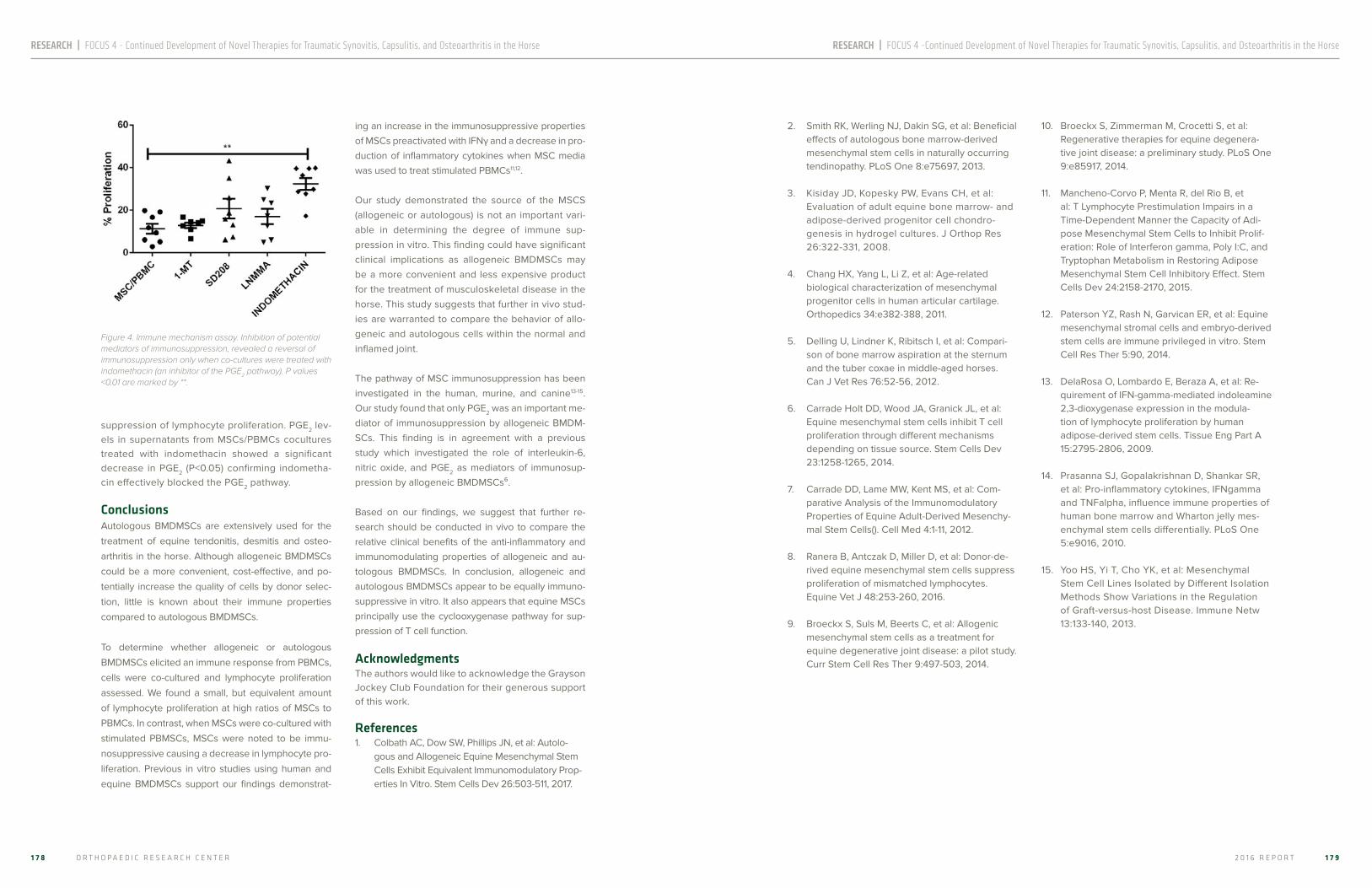
RESEARCH | FOCUS 4 - Continued Development of Novel Therapies for Traumatic Synovitis, Capsulitis, and Osteoarthritis in the Horse RESEARCH | FOCUS 4 -Continued Development of Novel Therapies for Traumatic Synovitis, Capsulitis, and Osteoarthritis in the Horse
suppression of lymphocyte proliferation. PGE2 lev-els in supernatants from MSCs/PBMCs cocultures treated with indomethacin showed a significant decrease in PGE2 (P<0.05) confirming indometha-cin effectively blocked the PGE2 pathway. ConclusionsAutologous BMDMSCs are extensively used for the
treatment of equine tendonitis, desmitis and osteo-
arthritis in the horse. Although allogeneic BMDMSCs
could be a more convenient, cost-effective, and po-
tentially increase the quality of cells by donor selec-
tion, little is known about their immune properties
compared to autologous BMDMSCs.
To determine whether allogeneic or autologous
BMDMSCs elicited an immune response from PBMCs,
cells were co-cultured and lymphocyte proliferation
assessed. We found a small, but equivalent amount
of lymphocyte proliferation at high ratios of MSCs to
PBMCs. In contrast, when MSCs were co-cultured with
stimulated PBMSCs, MSCs were noted to be immu-
nosuppressive causing a decrease in lymphocyte pro-
liferation. Previous in vitro studies using human and
equine BMDMSCs support our findings demonstrat-
ing an increase in the immunosuppressive properties of MSCs preactivated with IFNγ and a decrease in pro-duction of inflammatory cytokines when MSC media was used to treat stimulated PBMCs11,12.
Our study demonstrated the source of the MSCS (allogeneic or autologous) is not an important vari-able in determining the degree of immune sup-pression in vitro. This finding could have significant clinical implications as allogeneic BMDMSCs may be a more convenient and less expensive product for the treatment of musculoskeletal disease in the horse. This study suggests that further in vivo stud-ies are warranted to compare the behavior of allo-geneic and autologous cells within the normal and inflamed joint.
The pathway of MSC immunosuppression has been investigated in the human, murine, and canine13-15. Our study found that only PGE2 was an important me-diator of immunosuppression by allogeneic BMDM-SCs. This finding is in agreement with a previous study which investigated the role of interleukin-6, nitric oxide, and PGE2 as mediators of immunosup-pression by allogeneic BMDMSCs⁶.
Based on our findings, we suggest that further re-search should be conducted in vivo to compare the relative clinical benefits of the anti-inflammatory and immunomodulating properties of allogeneic and au-tologous BMDMSCs. In conclusion, allogeneic and autologous BMDMSCs appear to be equally immuno-suppressive in vitro. It also appears that equine MSCs principally use the cyclooxygenase pathway for sup-pression of T cell function.
AcknowledgmentsThe authors would like to acknowledge the Grayson Jockey Club Foundation for their generous support of this work. References1. Colbath AC, Dow SW, Phillips JN, et al: Autolo-
gous and Allogeneic Equine Mesenchymal Stem Cells Exhibit Equivalent Immunomodulatory Prop-erties In Vitro. Stem Cells Dev 26:503-511, 2017.
2. Smith RK, Werling NJ, Dakin SG, et al: Beneficial effects of autologous bone marrow-derived mesenchymal stem cells in naturally occurring tendinopathy. PLoS One 8:e75697, 2013.
3. Kisiday JD, Kopesky PW, Evans CH, et al: Evaluation of adult equine bone marrow- and adipose-derived progenitor cell chondro-genesis in hydrogel cultures. J Orthop Res 26:322-331, 2008.
4. Chang HX, Yang L, Li Z, et al: Age-related biological characterization of mesenchymal progenitor cells in human articular cartilage. Orthopedics 34:e382-388, 2011.
5. Delling U, Lindner K, Ribitsch I, et al: Compari-son of bone marrow aspiration at the sternum and the tuber coxae in middle-aged horses. Can J Vet Res 76:52-56, 2012.
6. Carrade Holt DD, Wood JA, Granick JL, et al: Equine mesenchymal stem cells inhibit T cell proliferation through different mechanisms depending on tissue source. Stem Cells Dev 23:1258-1265, 2014.
7. Carrade DD, Lame MW, Kent MS, et al: Com-parative Analysis of the Immunomodulatory Properties of Equine Adult-Derived Mesenchy-mal Stem Cells(). Cell Med 4:1-11, 2012.
8. Ranera B, Antczak D, Miller D, et al: Donor-de-rived equine mesenchymal stem cells suppress proliferation of mismatched lymphocytes. Equine Vet J 48:253-260, 2016.
9. Broeckx S, Suls M, Beerts C, et al: Allogenic mesenchymal stem cells as a treatment for equine degenerative joint disease: a pilot study. Curr Stem Cell Res Ther 9:497-503, 2014.
10. Broeckx S, Zimmerman M, Crocetti S, et al: Regenerative therapies for equine degenera-tive joint disease: a preliminary study. PLoS One 9:e85917, 2014.
11. Mancheno-Corvo P, Menta R, del Rio B, et al: T Lymphocyte Prestimulation Impairs in a Time-Dependent Manner the Capacity of Adi-pose Mesenchymal Stem Cells to Inhibit Prolif-eration: Role of Interferon gamma, Poly I:C, and Tryptophan Metabolism in Restoring Adipose Mesenchymal Stem Cell Inhibitory Effect. Stem Cells Dev 24:2158-2170, 2015.
12. Paterson YZ, Rash N, Garvican ER, et al: Equine mesenchymal stromal cells and embryo-derived stem cells are immune privileged in vitro. Stem Cell Res Ther 5:90, 2014.
13. DelaRosa O, Lombardo E, Beraza A, et al: Re-quirement of IFN-gamma-mediated indoleamine 2,3-dioxygenase expression in the modula-tion of lymphocyte proliferation by human adipose-derived stem cells. Tissue Eng Part A 15:2795-2806, 2009.
14. Prasanna SJ, Gopalakrishnan D, Shankar SR, et al: Pro-inflammatory cytokines, IFNgamma and TNFalpha, influence immune properties of human bone marrow and Wharton jelly mes-enchymal stem cells differentially. PLoS One 5:e9016, 2010.
15. Yoo HS, Yi T, Cho YK, et al: Mesenchymal Stem Cell Lines Isolated by Different Isolation Methods Show Variations in the Regulation of Graft-versus-host Disease. Immune Netw 13:133-140, 2013.
Figure 4. Immune mechanism assay. Inhibition of potential mediators of immunosuppression, revealed a reversal of immunosuppression only when co-cultures were treated with indomethacin (an inhibitor of the PGE2 pathway). P values <0.01 are marked by **.
1 7 8 O R T H O P A E D I C R E S E A R C H C E N T E R 2 0 1 6 R E P O R T 1 7 9

RESEARCH | FOCUS 5 - Validation of Rehabilitation and Physical Therapy Techniques for Musculoskeletal Disease RESEARCH | FOCUS 5 -Validation of Rehabilitation and Physical Therapy Techniques for Musculoskeletal Disease
Take home messageThe universal goniometer (UG) had the lowest coef-ficient of variation (CV) and correlated most strongly with radiographic angles. However, none of the de-vices accurately represented radiographically mea-sured stifle joint angles. Additional veterinary studies are indicated prior to the use of novel goniometers. Further investigation into a superior goniometer for the veterinary market is warranted.
IntroductionGoniometry is frequently used as an objective out-come measure during rehabilitation to assess the clinical progression of orthopedic patients in veter-inary and human medicine.2,3 Goniometry is consid-ered a simple, reliable, low-cost, and time-efficient outcome measurement readily available for practical clinical application.4,5
Currently used tools for measurement of joint range of motion include radiography, the commonly used plastic UG, and more specialized technologies such as electrogoniometers, bubble inclinometers, fluo-roscopy, and kinematic gait analysis.5–8 Regardless of the device used, these methods require time to obtain measurements and some require additional equipment that may not be widely available to clini-cians.5,9 The challenges inherent to veterinary prac-tice require devices and methods that allow observ-ers to accurately, reliably, and efficiently measure joint angles in a broadly diverse set of patients.⁶
Smartphone-based applications provide clinicians with alternative goniometers that are readily avail-able, easily transportable, can allow for sharing of patient data, and can be less expensive than some other specialized devices used for range of motion measurement. Such applications are increasingly considered acceptable alternatives for various in-
dications in human medicine.5,11,12 However, to our knowledge, the use of smartphone-based appli-cations has not yet been evaluated in dogs in the peer-reviewed literature.
The purpose of the study reported here was to eval-uate the reliability and accuracy of 3 novel goniom-eters (2 smartphone-based applications and a novel digital goniometer) for angle measurements of the stifle joint in dogs and compare the results with those obtained by use of a UG, with radiographic measure-ments used as the gold standard. We hypothesized that there would be no differences in these variables among the 4 goniometry devices.
MethodsEight canine cadaver hind limbs from medium-sized dogs euthanized for reasons unrelated to this study were utilized. Limbs were excluded if palpable ortho-pedic abnormalities of the hind limbs were present. The limbs were individually mounted on wooden platforms, simulating lateral recumbency, in three random stifle angles within the published normal range of motion, resulting in a total of 24 angles. This design allowed the limb to be raised in a lateral posi-tion off the wooden platform allowing circumferential palpation of the limb mimicking a clinical setting (Fig-ure 1). The lateral incisions were cosmetically sutured in a simple continuous subcuticular pattern.
Radiographs were obtained of the limbs at each in-dividual angle prior to goniometric measurements being performed. The limbs remained in each posi-tion/angle until all observers completed their mea-surements of the respective angle. The nuts were then loosened from the bolts affixing the tibia to the wooden platform, the tibia was moved to a different angle and the nuts were tightened. The limbs were then radiographed and goniometric measurements were obtained in their new position. This sequence
Assessment of novel digital and smartphone goniometers for measurement of canine stifle joint angles
This is a summary of an article by Drs. Freund, Kieves, Hart, Foster, Jeffery and Duerr published in the American Journal of Veterinary Research 2016;77(7):749-55.¹
of limb fixation, radiography, and goniometric mea-surement was performed on each limb in each of the three random stifle angles per limb.
Four goniometers were used in this investigation (see Figure 2). All applications were installed on a single iphone that was used throughout the dura-tion of the study. Three evaluators, trained in the use of each novel goniometer and experienced in utilizing the UG in daily practice, performed all mea-surements. The evaluators were given written and verbal instructions on the use of the methods and allowed the opportunity to practice with the devic-es prior to beginning measurements. The order in which the evaluators used each goniometer was designed such that no device was used repeatedly for measurement of subsequent angles. After com-pleting a set of measurements with one device and prior to measuring with the next, the presentation of the limbs was randomly rearranged by one of the au-thors that did not perform any measurements. This was performed out of view of the evaluators, in an at-tempt to eliminate any recall of previously measured angles. This resulted in 96 measurements that were independently performed by three evaluators in trip-licate fashion, for a total of 864 measurements.
For the assessment of bias, the mean of the three replicates of the radiographic angle was treated as the true value, and the mean of the three repli-cates of each angle measured by each observer as the measured value. In order to determine the ap-propriate statistical method for estimating bias, the
relationship between measured and radiographic values was assessed for each device using simple linear regression. The correlation coefficient (R) was <0.99 for all goniometers, therefore Deming regres-sion was used to assess the relationship between the angle measured by each device, and the radio-graphic value. Constant bias and proportional bias were considered present if the confidence interval for the y intercept or the slope of the regression line did not overlap 0 or 1 respectively. The regression equation, measured value = (slope*radiographic val-ue) + y-intercept, was used to predict the measured angle when the radiographic value was 50 or 100 degrees. Bias at these angles was then calculated using bias (%) = (measured value-radiographic value)/radiographic value*100. Total error observed (TEo) was calculated at 50 and 100 degrees using the
Figure 1: Drawing illustrating the cadaveric set up: (A) Model design as seen when viewed from above; (B) Model design as seen when viewed from the side.
Figure 2: The goniometers used in this investigation: (A) 12-inch plastic protractor universal goniometer, smartphone and HALO (B) HALO (C) Smartphone-based application iHandy Carpenter (D) Smartphone-based application DrGoniometer
1 8 0 O R T H O P A E D I C R E S E A R C H C E N T E R 2 0 1 6 R E P O R T 1 8 1

RESEARCH | FOCUS 5 - Validation of Rehabilitation and Physical Therapy Techniques for Musculoskeletal Disease RESEARCH | FOCUS 5 -Validation of Rehabilitation and Physical Therapy Techniques for Musculoskeletal Disease
equation TEo(%) = 2CV(%)+bias (%), and compared to a total allowable error (TEa) set by the authors at 5% based on previously reported differences in progno-sis after tibial plateau leveling osteotomy.4
ResultsRELIABILITY – The UG was the most and HALO the least reliable device, with mean CV of 4.88% (range 2.24-8.34) and 12.71% (range 3.11-24.63) respectively. Mean CV was similar for DrGoniometer (7.37%, range 2.44-20.40) and IHandy (7.57%, range 3.15%-14.46%).
CORRELATION – The UG correlated best with the radiographic measurements, with a correlation coef-ficient (R) of 0.97. HALO had the lowest correlation with R of 0.78. DrGoniometer and IHandy had an R of 0.97 and 0.93, respectively.
BIAS – Deming regression showed evidence of pro-portional and constant bias for all the devices except DrGoniometer for which the confidence interval of the slope and y intercept overlapped 1 and 0 re-spectively. Both UG and IHandy showed a positive constant bias and HALO a constant negative bias. DrGoniometer had the lowest bias at both 50 and 100 degrees, HALO had the greatest bias at 100 de-grees, and IHandy at 50 degrees.
PERFORMANCE ASSESSMENT – All four devic-es exceeded the TEa of 5%, and therefore failed to meet the authors’ requirements for acceptable accuracy to represent radiographic measure-ments. UG performed the best at 100 degrees (TEo 18.51%) and DrGoniometer the best at 50 degrees (TEo 24.69%). HALO performed the least well of the four devices at both 100 and 50 degrees (TEo 36.90 and 46.46% respectively).
ConclusionsWhen evaluating whether a device is useful to de-termine a clinical measurement, several approaches can be considered. Reliability is frequently evaluated by means of the coefficient of variation. Accuracy of a device can be assessed in many different ways, including the correlation coefficient and bias. Calcu-lation of total error integrates both CV and bias and comparison of total error with the total allowable er-ror determines if the device meets the needs clini-
cians or researchers. R describes the strength of a linear association between devices, and is therefore not an ideal assessment of actual accuracy.5 Bias, an expression of inaccuracy, is an assessment of the closeness of a measurement with that of the true val-ue.⁶ The evaluation of bias is beneficial in that it can be used to predict the extent of inaccuracy for differ-ent magnitudes of measurement. If proportional bias were present, this would indicate that the degree of overestimation or underestimation varies for large angles versus small angles. Constant bias, where the degree of error remains independent of the mea-sured true value, would indicate that a device consis-tently under or overestimates the true value.6,7
The results of this study demonstrate that no test-ed novel goniometer performed better than the UG. The UG demonstrated a low proportional and constant bias, percent bias, as well as TEo when evaluated at acute and obtuse angles. For clinical evaluations of progression after implemented ther-apy, the relative lack of variability is likely the most important factor to consider. The UG also had the highest correlation with the true angle as measured on radiographs, estimated by R.
Goniometry is most frequently used to assess clin-ical progression, such as evaluating the success of a treatment by comparing serial stifle joint angles.⁴ Given this purpose, the comparison of a goniom-eter to the radiographic measurement is likely not the most essential outcome measure for device assessment. Rather, how much a device varies between repeated measurements, between pa-tients, and between other devices has the greater potential effect on outcome measures. Therefore, while DrGoniometer had the lowest percent bias, or highest measured accuracy when compared to the radiographic measurement, this does not suggest that DrGoniometer would be the best alternative to the UG. For the novel goniometers, IHandy demon-strated a similar proportional bias to the UG, while HALO resulted in the largest proportional bias of all devices tested. While the UG remains the preferred goniometer, IHandy would produce the most similar results to the UG in clinical use.
AcknowledgmentsThis study was supported in full by the Young Inves-tigator Grant Program in the Center for Companion Animal Studies, Colorado State University.
References1. Freund KA, Kieves NR, Hart JL, et al. Assess-
ment of novel digital and smartphone goni-ometers for measurement of canine stifle joint angles. Am J Vet Res 2016;77:749-755.
2. Jaegger G, Marcellin-Little DJ, Levine D. Reli-
ability of goniometry in Labrador Retrievers. Am J Vet Res 2002;63:979-986.
3. Salamh PA, Kolber M. The reliability, minimal
detectable change and concurrent validity of a gravity-based bubble inclinometer and iphone application for measuring standing lumbar lor-dosis. Physiother Theory Pract 2014;30:62-67.
4. Jandi AS, Schulman AJ. Incidence of motion
loss of the stifle joint in dogs with naturally occurring cranial cruciate ligament rupture surgically treated with tibial plateau leveling os-teotomy: longitudinal clinical study of 412 cases. Vet Surg 2007;36:114-121.
5. Sedgwick P. Limits of agreement (Bland-Altman
method), 2013. 6. Harr KE, Flatland B, Nabity M, et al. ASVCP
guidelines: allowable total error guidelines for biochemistry. Vet Clin Pathol 2013;42:424-436.
7. Flatland B, Freeman KP, Friedrichs KR, et al. ASVCP quality assurance guidelines: control of general analytical factors in veterinary laborato-ries. Vet Clin Pathol 2010;39:264-277.
8. Ferriero G, Sartorio F, Foti C, et al. Reliability of
a New Application for Smartphones (DrGoni-ometer) for Elbow Angle Measurement. PM R 2011;3:1153-1154.
9. Ferriero G, Vercelli S, Sartorio F, et al. Reliability
of a smartphone-based goniometer for knee joint goniometry. Int J Rehabil Res 2013;36:146-151.
10. Vohralik SL, Bowen AR, Burns J, et al. Reliability
and Validity of a Smartphone App to Measure Joint Range. Am J Phys Med Rehabil 2014.
11. Jaeger GH, Marcellin-Little DJ, DePuy V, et al.
Validity of goniometric joint measurements in cats. Am J Vet Res 2007;68:822-826.
12. Govoni VM, Rahal SC, Agostinho FS, et al.
Goniometric measurements of the forelimb and hindlimb joints in sheep. Vet Comp Orthop Trau-matol 2012;25:297-300.
13. Petazzoni M, Jaeger GH. Atlas of Clinical Goni-
ometry and Radiographic Measurements of the Canine Pelvic Limb. 2 ed: Merial SAS, 2008.
1 8 2 O R T H O P A E D I C R E S E A R C H C E N T E R 2 0 1 6 R E P O R T 1 8 3

RESEARCH | FOCUS 5 - Validation of Rehabilitation and Physical Therapy Techniques for Musculoskeletal Disease RESEARCH | FOCUS 5 -Validation of Rehabilitation and Physical Therapy Techniques for Musculoskeletal Disease
IntroductionJoint mobilization and manipulation are considered forms of manual therapy, which involves the appli-cation of the hands to the body with a therapeutic intent. Soft tissue mobilization focuses on restoring movement to the skin, connective tissue, ligaments, tendons, and muscles with the goal of modulating pain, reducing inflammation, improving tissue re-pair, increasing extensibility, and improving function. Joint mobilization is characterized as nonimpulsive, repetitive joint movements induced within the pas-sive range of joint motion with the purpose of re-storing normal and symmetric joint range of motion, to stretch connective tissues, and to restore normal joint end-feel. Manipulation is a manual procedure that involves a directed impulse which moves a joint or vertebral segment beyond its physiological range of motion, but does not exceed the anatomical limit of the articulation. The primary biomechanical differ-ence between joint mobilization and manipulation is the presence of a high-speed thrust or impulse. Spinal manipulation involves the application of con-trolled impulses to articular structures within the axial skeleton with the intent of reducing pain and muscle hypertonicity and increasing joint range of motion.
Objectives of treatmentSoft tissue and joint mobilization are used to assess the quality and quantity of joint range of motion and as a primary means of treating musculoskeletal disor-ders. Subjective assessment of the ease of joint mo-tion, joint stability, and joint end-feel provide insights into the biomechanical and neurologic features of an articulation. Goniometry is often used to objectively quantify and document the amount of flexion or ex-tension present at an articulation. The objectives of soft tissue and joint mobilization are typically to re-duce pain, restore tissue compliance, and to improve overall tissue mobility and joint range or motion. Ma-nipulation is more often used to address localized pain and joint stiffness, with less focus on the sur-
rounding soft tissues.² Manual therapy techniques can also provide an adjunct to therapeutic exercis-es and rehabilitation of neuromotor control, where applied forces are used to induce passive stretch-ing, weight-shifting and activation of spinal reflexes, which help to increase flexibility, stimulate proprio-ception and strengthen core musculature.³ Peripher-al nerve and nerve root mobilization techniques and exercises are also used for post-operative rehabili-tation of low back pain.⁴ Few formal studies exist to support the use of active joint or spinal mobilization techniques in horses.⁵ Most mobilization studies in horses involve a period of induced joint immobili-zation by a fixture or cast followed by allowing the horse to spontaneously weight bear and locomote on the affected limb, without evaluation of specific soft tissue or joint mobilization techniques.⁶
Joint mechanicsThe use of palpation techniques to qualitatively and quantitatively assess joint motion requires an un-derstanding of joint mechanics. Joint motion can be categorized into three zones of movement: physio-logic, paraphysiologic and pathologic (Figure 1). The physiological zone of movement consists of both active and passive joint motion within all possible di-rections of movement (e.g., flexion, extension, lateral bending, and axial rotation). Passive movement of an articulation from a neutral joint position first involves
Joint mobilization and manipulation for the equine athlete
This is a summary of an article by Dr. Kevin K. Haussler published in the Veterinary Clinics Equine.¹
evaluating the range of joint motion that has minimal, uniform resistance. Then as the articulation is moved toward the end range of passive joint motion, there is a gradual increase in the resistance to movement which terminates at an elastic barrier (i.e., joint end feel). The end range of motion begins with any palpa-ble change in resistance to passive joint mobilization. Joint end feel is often evaluated by bringing an indi-vidual articulation to tension and applying rhythmic oscillations to qualify the resistance to movement. Normal joint end feel is initially soft and resilient and gradually becomes more restrictive as the limits of joint range of motion are reached. A pathologic or restrictive end range of motion is palpable earlier in passive joint movement and has an abrupt, hard end feel when compared with normal joint end feel. Each articulation within the body has unique palpatory end feels for each of the directions of joint motion (e.g., flexion, extension, lateral bending, etc.). The goal of palpating passive joint movement is to evaluate each articulation of interest for quality of joint motion, the initiation of resistance to motion and type of end feel, and the amount of motion within each of the principle directions of movement. The paraphysiological space is bordered by the elastic and anatomical limits of an individual joint. Joint motion into the paraphysiologi-cal space occurs only with the application of high ve-locity forces associated with joint manipulation. The anatomical barrier of the joint marks the junction be-tween the paraphysiologic and pathologic zones of movement. The pathological zone is characterized by the application of excessive forces or joint motion which causes an articulation to move beyond its ana-tomical limits and results in mechanical disruption of intra- and periarticular structures and subsequent joint instability or luxation.
Clinical indicationsJoint mobilization and manipulation provide import-ant diagnostic and therapeutic approaches for ad-dressing equine axial skeleton problems that are not otherwise available in veterinary medicine. Most of the current knowledge about equine manual ther-apies has been borrowed from human techniques, theories, and research and applied to horses. Thera-peutic trials of joint mobilization or manipulation are often used because of limited knowledge about the effects of manual therapy in horses. The indications for joint mobilization and manipulation are similar
and include restricted joint range of motion, muscle spasms, pain, fibrosis, or contracted soft tissues. The principal indications for spinal manipulation are neck or back pain, localized or regional joint stiffness, poor performance, and altered gait that is not associated with overt lameness. A thorough diagnostic workup is required to identify soft tissue and osseous pathol-ogy, neurologic disorders, or other lameness condi-tions that may not be responsive to manual therapy. Clinical signs indicative of a primary spinal disorder include localized musculoskeletal pain, muscle hy-pertonicity and restricted joint motion. This triad of clinical signs can also be found in a variety of low-er limb disorders; however, they are most evident in horses with neck or back problems. Clinical signs in-dicative of chronic or secondary spinal disorders in-clude regional or diffuse pain, generalized stiffness, and widespread muscle hypertonicity. In these cas-es, further diagnostic evaluation or imaging should be done to identify the primary cause of lameness or poor performance.
Joint mobilization and manipulation are critical com-ponents in the management of muscular, articular and neurologic components of select musculoskel-etal injuries in performance horses. Musculoskeletal conditions that are chronic or recurring, not readily diagnosed, or are not responding to conventional veterinary care may be indicators that manual ther-apy evaluation and treatment is needed. Joint mobi-lization and manipulation are typically more effective in the early clinical stages of disease processes ver-sus end-stage disease where reparative processes have been exhausted. Joint manipulation is usually contraindicated in the acute stages of soft tissue in-jury; however, mobilization is safer than manipulation and has been shown to have short-term benefits for acute neck or back pain in humans. Manipulation is probably more effective than mobilization for chron-ic neck or back pain and has the potential to help restore normal joint motion, thus limiting the risk of reinjury. It has been theorized that spinal manipula-tion preferentially influences a sensory bed which, in terms of anatomical location and function, is dif-ferent from the sensory bed influenced by spinal mobilization techniques. Manipulation may preferen-tially stimulate receptors within deep intervertebral muscles, while mobilization techniques most likely affect more superficial axial muscles. Only one study
Figure 1: Graphic representation of joint mechanics as it relates to the zones of joint motion.
1 8 4 O R T H O P A E D I C R E S E A R C H C E N T E R 2 0 1 6 R E P O R T 1 8 5

has compared mobilization to manipulation in hors-es and spinal manipulation induced a 15% increase in displacement and a 20% increase in applied force, compared to mobilization.⁴ At most vertebral sites studied, manipulation increased the amplitudes of dorsoventral displacement and applied force, indica-tive of increased spinal flexibility and increased toler-ance to pressure in the thoracolumbar region of the equine vertebral column.
Joint mobilization and manipulation techniquesSelection factors for considering mobilization versus manipulation include the technical training and skill of the practitioner, perceived risks versus benefits, the presence of acute pain and inflammation, and pathoanatomic considerations. Joint mobilization is easier to apply, requires less psychomotor skills, has minimal risks, and can be used in the presence of acute pain and inflammation, compared to manipula-tion. Manual therapy procedures are also dependent on the ability of the patient to relax and the patent response to the applied force. Characteristics of joint mobilization and manipulation include factors related to specificity, leverage, velocity, amplitude, direction, and prestress of the applied force. Additional factors are related to joint position and frequency or oscilla-tion of the applied forces. Levers are used to increase mechanical advantage and assist in applying force to an articulation or body segment to induce joint motion. Long levers include using the limbs or head and neck as levers to induce spinal motion, instead of the inducing motion at one or two individual ver-tebrae by using transverse or spinous processes as short lever contacts. Velocity relates to the speed of the impulse applied to move a vertebra or body seg-ment and displacement is the distance over which the applied thrust is applied. Amplitude refers to the amount of force applied. With long-lever techniques, lower amplitudes of force are required to induce similar joint motion as short-lever contacts. Howev-er, the rationale for using short-lever techniques is to increase the specificity of the applied thrust, since a single vertebral process is contacted on the vertebra of interest with short-lever techniques. With long-le-vers, it is likely that multiple articulations are included between the doctor’s contact and the body segment of interest, which produces a more generalized treat-ment effect. Using a specific anatomical contact is
theorized to address a single articulation; however,
studies on treatment effects indicate that specific
contact techniques produce local, as well as, regional
and systemic effects. The therapeutic dosage of joint
mobilization or manipulation is also determined by
the number of vertebrae or body segments treated
and the frequency of the applied treatments.
SourceKK Haussler. Joint Mobilization and Manipulation for
the Equine Athlete. Vet Clin Equine 32 (2016) 87–101.
http://dx.doi.org/10.1016/j.cveq.2015.12.003
References1. Haussler KK. Joint mobilization and manipu-
lation for the equine athlete. Vet Clin Equine
2016;32:87-101.
2. Haussler KK. Chiropractic evaluation and
management. Vet Clin North Am Equine Pract
1999;15:195-209.
3. Stubbs NC, Clayton HM. Activate your horse's
core: unmounted exercises for dynamic mobility,
strength and balance. Mason, MI: Sport Horse
Publications, 2008.
4. Ellis RF, Hing WA. Neural mobilization: a system-
atic review of randomized controlled trials with
an analysis of therapeutic efficacy. J Man Manip
Ther 2008;16:8-22.
5. Haussler KK, Hill AE, Puttlitz CM, et al. Effects
of vertebral mobilization and manipulation on
kinematics of the thoracolumbar region. Amer J
Vet Research 2007;68:508-516.
6. van Harreveld PD, Lillich JD, Kawcak CE, et al.
Clinical evaluation of the effects of immobiliza-
tion followed by remobilization and exercise on
the metacarpophalangeal joint in horses. Amer
J Vet Research 2002;63:282-288.
RESEARCH | FOCUS 5 - Validation of Rehabilitation and Physical Therapy Techniques for Musculoskeletal Disease RESEARCH | FOCUS 5 -Validation of Rehabilitation and Physical Therapy Techniques for Musculoskeletal Disease
Take home messageSurgical treatment of cranial cruciate ligament dis-ease (CCLD) was associated with a greater number of excellent outcome ratings, and improved owner-as-sessed lameness grade than non-surgical treatment with a stifle orthosis. However, a high percentage of owners in both groups reported positive satisfac-tion. Pet owners selecting stifle orthoses should be advised of the potential for complications including persistent lameness, skin issues, non-acceptance of the orthosis, and the possible need for subsequent surgical intervention.
IntroductionInjury to the cranial cruciate ligament is a common cause for the development of hind limb lameness, de-velopment of stifle osteoarthritis, and associated pain in the canine patient.2-5 Despite the high prevalence, there is still controversy about the best overall course of treatment. Surgical treatment is frequently advocat-ed, and has proven to be effective in the restoration of limb function.5-9 The literature most strongly supports Tibial Plateau Leveling Osteotomy (TPLO) as the sur-gical procedure of choice, allowing return to normal function after surgical treatment of CCLD.⁵ Non-sur-gical management of CCLD has been investigated and is most commonly suggested for dogs weighing less than 15-20kg.10 However, improvement in lame-ness with non-surgical management of CCLD has also been reported in dogs weighing >20kg.11,12
Non-surgical management of CCLD has recently changed with the introduction of canine rehabilita-tion therapy and other novel management options such as stifle orthoses.13,14 Orthoses (also referred to as ‘braces’ or ‘orthotic[s] device[s]’) are medical devices used to support or protect an injured leg,14 and frequently defined as any medical device added to the body to support, align, position, immobilize,
prevent or correct deformity, assist weak muscles, or improve function.13,14 Anecdotally, stifle orthoses are occasionally utilized for non-surgical management of CCLD.14 However, to the authors’ knowledge, no peer-reviewed reports of clinical outcome of stifle orthoses for the management of CCLD are avail-able. The goal of this study was to report outcome and satisfaction of pet owners that chose to employ a custom-made stifle orthosis for the management of their dog’s CCLD, in comparison to owners that elected TPLO.
Comparison of pet-owner satisfaction with stifle orthoses (braces) or tibial plateau leveling osteotomy for the management of cranial cruciate ligament disease in medium to large breed dogs
This is a summary of an article by Drs. Hart, May, Kieves, Mich, Goh, Palmer and Duerr published in the Journal of the American Veterinary Medical Association 2016;249(4):391-8.1
Figure 1: Orthoses (also referred to as ‘braces’ or ‘orthotic[s] device[s]’) are medical devices used to support or protect an injured leg.
1 8 6 O R T H O P A E D I C R E S E A R C H C E N T E R 2 0 1 6 R E P O R T 1 8 7

RESEARCH | FOCUS 5 - Validation of Rehabilitation and Physical Therapy Techniques for Musculoskeletal Disease RESEARCH | FOCUS 5 -Validation of Rehabilitation and Physical Therapy Techniques for Musculoskeletal Disease
Methods The Orthopets’ client database was searched for dogs who received a custom-made stifle orthosis for treatment of CCLD (ORTHOSIS group) from 2008 to 2013. These cases were recruited from veterinarians across the US. Only dogs that had not previously un-dergone surgical intervention for CCLD were includ-ed in the study. Dogs that were treated simultaneous-ly with an orthosis in addition to surgical treatment were also excluded. Similarly, CSU’s hospital data-base was searched for dogs that underwent TPLO surgery for treatment of CCLD (TPLO group) between 2002 and 2013. For each database, only dogs where owners provided an email address were included.
Survey requests were sent via email to the iden-tified clients including questions on the following categories: CCLD presentation (affected limb(s), breed, weight, and age), treatments performed, aftercare, complications, level of client compliance and satisfaction, and considerations made when choosing treatment.
ResultsOWNER RESPONSE - For the ORTHOSIS group, 819 owners were invited to participate in the sur-vey, while 203 owners were invited for the TPLO group. The TPLO group represented owners of dogs presenting between 2002 and 2013, and the ORTHOSIS group cases were identified from or-thoses provided between 2008 and 2013. The re-sponse rate for the ORTHOSIS group was 25% (n = 203/819) and 37% for the TPLO group (n = 76/203; P = 0.003). Owners were not required to answer all of the questions and for some questions multiple answers were allowed; hence subsequent data is reported including the number of respondents for each question.
DEMOGRAPHICS - There were no significant dif-ferences for dog weight between the TPLO group (31.4 ± 10.5 kg) and the ORTHOSIS group (34.1 ± 13.4 kg; P = 0.10), or limb distribution (P = 0.68). All dogs in the study were medium to large breed dogs. Dogs in the TPLO group started showing signs of lameness at a significantly younger age than dogs in the ORTHOSIS group (5.5 ± 2.6 years in the TPLO group compared to 7.8 ± 3.6 years in the ORTHOSIS group; P = <0.001).
TREATMENT DECISION FACTORS - Eighty-one per-cent (n = 162/199) of selected responses from owners in the ORTHOSIS group were “cost”, “convenience”, or “personal preference” as the most important fac-tors for choosing the treatment of their dog com-pared to only 25% (n = 17/67) in the TPLO group. Con-versely, “veterinarian recommendation” was selected as the most influential by 75% (n = 50/67) of the TPLO group owners compared to 19% (n = 37/199) of own-ers in the ORTHOSIS group (P = <0.001).
AFTERCARE - Sixty-three percent (n = 40/63) of the TPLO group and 53% of the ORTHOSIS group (n = 80/152) did not pursue rehabilitation (P = 0.14). When asked why they did not pursue rehabilitation, 46% of the TPLO group (n = 16/35) and 33% of owners in the ORTHOSIS group (n = 30/92) reported “Not advised of necessity” (P = 0.06).
OUTCOME ASSESSMENT/OWNER SATISFACTION - Eighty-five percent (n = 129/152) of owners that pur-sued orthoses for their dogs, and 90% (n = 57/63) of the TPLO group, replied they would repeat the cho-sen treatment again if they were given the choice (P = 0.27). A statistically significantly larger number of owners in the TPLO group rated the treatment as ei-ther “excellent”, “very good”, or “good” (TPLO: 98%, n = 62/63; ORTHOSIS: 86%, n = 131/152) compared to “poor” (TPLO: 2%, n = 1/63; ORTHOSIS: 14%, n = 21/152; P = <0.001). When comparing owner-report-ed lameness after intervention, 98% (n = 62/63) of the dogs in the TPLO group and 88% (n = 128/146) in the ORTHOSIS group showed mild/no lameness (P = 0.01; see Table 2).
COMPLICATIONS - Forty-six (n = 70/152) percent of owners reported their dogs experienced skin issues while wearing the orthosis and 32% (n = 22/69) of those cases required medical attention and multiple adjustments to the orthosis according to owners. In comparison, 5% of owners in the TPLO group re-ported complications (n = 4/76). Owners reported 2 cases of suspected patellar tendinopathy, 1 draining fistula that required removal of the TPLO plate, and 1 acute case of NSAID toxicity. In review of the med-ical records, there were no additional documented complications for the TPLO group. The majority (60%, n = 91/151) of dogs who received an orthosis experi-enced a 1 to 2 week long adjustment period to get
used to wearing the orthosis. Eighty-eight percent (n = 134/151) of owners reported their dog was wearing the orthosis after the initial adjustment period: 43% (n = 65/151) of dog owners reported their dog was wearing the orthosis every day, 15% (n = 23/151) re-ported 3-6 days/week, and 34% reported less than 3 days/week (n = 52/151). Thirty-eight percent (n = 58/151) of owners reported their dog tolerated the orthosis ‘very well – my dog actually seems to like wearing the device’, 33% (n = 50/151) reported ‘well – my dog seemed reluctant to have the device applied but seems to like wearing the device’, and 19% (n = 28/151) reported ‘fair – my dog seems reluctant to have the device applied and does not seem to like wearing it but will tolerate it.’ Eleven percent (n = 17/151) of the owners reported that their dog does not wear the orthosis at all: 7% (n = 10/151) reported their dog did not tolerate the orthosis due to fit issues or skin concerns, 2% (n = 3/151) reported their dog didn’t need the orthosis anymore since the lameness had resolved, and 3% (n = 4/151) reported “other” reasons. Five percent reported their dog never tolerated the orthosis (n = 7/151), and 11% (n = 16/151) of the ORTHO-SIS group reported that their dog received subse-quent surgical procedures on the orthosis-managed limb (TPLO, n = 7; meniscal removal only, n = 4; tibial tuberosity advancement, n = 2; tightrope procedure, n = 2; did not specify, n = 1).
Conclusions Based on the results of this survey and previous re-search, it can be concluded that non-surgical treat-ment is a viable alternative for dogs that cannot undergo surgical treatment for CCLD. If an orthosis is considered as part of this management, owners should be given appropriate counseling regarding potential complications such as skin issues and un-willingness to wear the device, as well as the risk of subsequent surgical intervention. Owners should also be aware that the majority of patients so treated are chronically “orthosis-dependent”. It is also im-portant to note that long-term data on development of osteoarthritis is not available.
References1. Hart JL, May KD, Kieves NR, et al. Compari-
son of owner satisfaction between stifle joint orthoses and tibial plateau leveling osteotomy for the management of cranial cruciate ligament
disease in dogs. Journal of the American Veteri-nary Medical Association 2016;249:391-398.
2. Whitehair JG, Vasseur PB, Willits NH. Epidemiol-
ogy of cranial cruciate ligament rupture in dogs. J Am Vet Med Assoc 1993;203:1016-1019.
3. Wilke VL, Robinson DA, Evans RB, et al.
Estimate of the annual economic impact of treatment of cranial cruciate ligament injury in dogs in the United States. J Am Vet Med Assoc 2005;227:1604-1607.
4. Comerford EJ, Smith K, Hayashi K. Update on
the aetiopathogenesis of canine cranial cruciate ligament disease. Veterinary And Compara-tive Orthopaedics And Traumatology: VCOT 2011;24:91-98.
5. Dismukes DI, Tomlinson JL, Fox DB, et al.
Radiographic measurement of canine tibial angles in the sagittal plane. Veterinary Surgery 2008;37:300-305.
6. Bergh MS, Budsberg SC. A systematic review of
the literature describing the efficacy of surgical treatments for canine hip dysplasia (1948-2012). Vet Surg 2014;43:501-506.
7. Boudrieau RJ. Tibial plateau leveling osteoto-
my or tibial tuberosity advancement? Vet Surg 2009;38:1-22.
8. de Medeiros M, Sanchez Bustinduy M, Radke H,
et al. Early kinematic outcome after treatment of cranial cruciate ligament rupture by tibial pla-teau levelling osteotomy in the dog. Vet Comp Orthop Traumatol 2011;24:178-184.
9. Nielen AL, Knol BW, van Hagen MA, et al.
Genetic and epidemiological investigation of a birth cohort of boxers. Tijdschrift Voor Dierge-neeskunde 2003;128:586-590.
10. Vasseur PB. Clinical Results Following Nonop-
erative Management for Rupture of the Cranial Cruciate Ligament in Dogs. Veterinary Surgery 1984;13:243-246.
1 8 8 O R T H O P A E D I C R E S E A R C H C E N T E R 2 0 1 6 R E P O R T 1 8 9

RESEARCH | FOCUS 5 - Validation of Rehabilitation and Physical Therapy Techniques for Musculoskeletal Disease 2016 ORC FACULTY AND STAFF
11. Chauvet AE, Johnson AL, Pijanowski GJ, et al. Evaluation of fibular head transposition, lateral fabellar suture, and conservative treatment of cranial cruciate ligament rupture in large dogs: a retrospective study. J Am Anim Hosp Assoc 1996;32:247-255.
12. Wucherer KL, Conzemius MG, Evans R, et al.
Short-term and long-term outcomes for over-weight dogs with cranial cruciate ligament rupture treated surgically or nonsurgically. Jav-ma-Journal of the American Veterinary Medical Association 2013;242:1364-1372.
13. Coates J. Evaluation and rehabilitation options for orthopedic disorders of the pelvic limb In: Zink C,Van Dyke J, eds. Canine Sports Medicine and Rehabilitation. Ames, Iowa: Wiley-Blackwell, 2013;306-307.
14. Adamson C, Kaufmann M, Levine D, et al. Assis-
tive devices, orthotics, and prosthetics. Vet Clin North Am Small Anim Pract 2005;35:1441-1451, ix.
2016 ORC FACULTY AND STAFF
1 9 0 O R T H O P A E D I C R E S E A R C H C E N T E R 2 0 1 6 R E P O R T 1 9 1


ORTHOPAEDIC RESEARCH CENTER